THE EVOLUTION OF MAN
Volume II
CHAPTER XVI
STRUCTURE OF THE LANCELET AND THE SEA-SQUIRT
In turning from the embryology to the phylogeny of
man—from the development of the individual to that of the
species—we must bear in mind the direct causal connection
that exists between these two main branches of the science of human
evolution. This important causal nexus finds its simplest
expression in “the fundamental law of organic
development,” the content and purport of which we have fully
considered in the first chapter. According to this biogenetic law,
ontogeny is a brief and condensed recapitulation of phylogeny. If
this compendious reproduction were complete in all cases, it would
be very easy to construct the whole story of evolution on an
embryonic basis. When we wanted to know the ancestors of any higher
organism, and, therefore, of man—to know from what forms the
race as a whole has been evolved we should merely have to follow
the series of forms in the development of the individual from the
ovum; we could then regard each of the successive forms as the
representative of an extinct ancestral form. However, this direct
application of ontogenetic facts to phylogenetic ideas is possible,
without limitations, only in a very small section of the animal
kingdom. There are, it is true, still a number of lower
invertebrates (for instance, some of the Zoophyta and Vermalia) in
which we are justified in recognising at once each embryonic form
as the historical reproduction, or silhouette, as it were, of an
extinct ancestor. But in the great majority of the animals, and in
the case of man, this is impossible, because the embryonic forms
themselves have been modified through the change of the conditions
of existence, and have lost their original character to some
extent. During the immeasurable course of organic history, the many
millions of years during which life was developing on our planet,
secondary changes of the embryonic forms have taken place in most
animals. The young of animals (not only detached larvæ, but
also the embryos enclosed in the womb) may be modified by the
influence of the environment, just as well as the mature organisms
are by adaptation to the conditions of life; even species are
altered during the embryonic development. Moreover, it is an
advantage for all higher organisms (and the advantage is greater
the more advanced they are) to curtail and simplify the original
course of development, and thus to obliterate the traces of their
ancestors. The higher the individual organism is in the animal
kingdom, the less completely does it reproduce in its embryonic
development the series of its ancestors, for reasons that are as
yet only partly known to us. The fact is easily proved by comparing
the different developments of higher and lower animals in any
single stem.
In order to appreciate this important feature, we have
distributed the embryological phenomena in two groups,
palingenetic and cenogenetic. Under palingenesis we
count those facts of embryology that we can directly regard as a
faithful synopsis of the corresponding stem-history. By cenogenesis
we understand those embryonic processes which we cannot directly
correlate with corresponding evolutionary processes, but must
regard as modifications or falsifications of them. With this
careful discrimination between palingenetic and cenogenetic
phenomena, our biogenetic law assumes the following more precise
shape:—The rapid and brief development of the individual
(ontogeny) is a condensed synopsis of the long and slow history of
the stem (phylogeny): this synopsis is the more faithful and
complete in proportion as the original features have been preserved
by heredity, and modifications have not been introduced by
adaptation.
[ 180 ]
In order to distinguish correctly between palingenetic and
cenogenetic phenomena in embryology, and deduce sound conclusions
in connection with stem-history, we must especially make a
comparative study of the former. In doing this it is best to employ
the methods that have long been used by geologists for the purpose
of establishing the succession of the sedimentary rocks in the
crust of the earth. This solid crust, which encloses the glowing
central mass like a thin shell, is composed of different kinds of
rocks: there are, firstly, the volcanic rocks which were formed
directly by the cooling at the surface of the molten mass of the
earth; secondly, there are the sedimentary rocks, that have been
made out of the former by the action of water, and have been laid
in successive strata at the bottom of the sea. Each of these
sedimentary strata was at first a soft layer of mud; but in the
course of thousands of years it condensed into a solid, hard mass
of stone (sandstone, limestone, marl, etc.), and at the same time
permanently preserved the solid and imperishable bodies that had
chanced to fall into the soft mud. Among these bodies, which were
either fossilised or left characteristic impressions of their forms
in the soft slime, we have especially the more solid parts of the
animals and plants that lived and died during the deposit of the
slimy strata.
Hence each of the sedimentary strata has its characteristic
fossils, the remains of the animals and plants that lived during
that particular period of the earth’s history. When we make a
comparative study of these strata, we can survey the whole series
of such periods. All geologists are now agreed that we can
demonstrate a definite historical succession in the strata, and
that the lowest of them were deposited in very remote, and the
uppermost in comparatively recent, times. However, there is no part
of the earth where we find the series of strata in its entirety, or
even approximately complete. The succession of strata and of
corresponding historical periods generally given in geology is an
ideal construction, formed by piecing together the various partial
discoveries of the succession of strata that have been made at
different points of the earth’s surface (cf. Chapter
XVIII).
We must act in this way in constructing the phylogeny of man. We
must try to piece together a fairly complete picture of the series
of our ancestors from the various phylogenetic fragments that we
find in the different groups of the animal kingdom. We shall see
that we are really in a position to form an approximate picture of
the evolution of man and the mammals by a proper comparison of the
embryology of very different animals—a picture that we could
never have framed from the ontogeny of the mammals alone. As a
result of the above-mentioned cenogenetic processes—those of
disturbed and curtailed heredity—whole series of lower stages
have dropped out in the embryonic development of man and the other
mammals especially from the earliest periods, or been falsified by
modification. But we find these lower stages in their original
purity in the lower vertebrates and their invertebrate ancestors.
Especially in the lowest of all the vertebrates, the lancelet or
Amphioxus, we have the oldest stem-forms completely preserved in
the embryonic development. We also find important evidence in the
fishes, which stand between the lower and higher vertebrates, and
throw further light on the course of evolution in certain periods.
Next to the fishes come the amphibia, from the embryology of which
we can also draw instructive conclusions. They represent the
transition to the higher vertebrates, in which the middle and older
stages of ancestral development have been either distorted or
curtailed, but in which we find the more recent stages of the
phylogenetic process well preserved in ontogeny. We are thus in a
position to form a fairly complete idea of the past development of
man’s ancestors within the vertebrate stem by putting
together and comparing the embryological developments of the
various groups of vertebrates. And when we go below the lowest
vertebrates and compare their embryology with that of their
invertebrate relatives, we can follow the genealogical tree of our
animal ancestors much farther, down to the very lowest groups of
animals.
In entering the obscure paths of this phylogenetic labyrinth,
clinging to the Ariadne-thread of the biogenetic law and guided by
the light of comparative anatomy, we will first, in accordance with
the methods we have adopted, discover and arrange those fragments
from the manifold embryonic developments of very different animals
from which the stem-history of man can be composed. I would call
attention particularly to the fact that
[ 181 ]
we can employ this method with the same confidence
and right as the geologist. No geologist has ever had ocular proof
that the vast rocks that compose our Carboniferous or Jurassic or
Cretaceous strata were really deposited in water. Yet no one doubts
the fact. Further, no geologist has ever learned by direct
observation that these various sedimentary formations were
deposited in a certain order; yet all are agreed as to this order.
This is because the nature and origin of these rocks cannot be
rationally understood unless we assume that they were so deposited.
These hypotheses are universally received as safe and indispensable
“geological theories,” because they alone give a
rational explanation of the strata.
Our evolutionary hypotheses can claim the same value, for the
same reasons. In formulating them we are acting on the same
inductive and deductive methods, and with almost equal confidence,
as the geologist. We hold them to be correct, and claim the status
of “biological theories” for them, because we cannot
understand the nature and origin of man and the other organisms
without them, and because they alone satisfy our demand for a
knowledge of causes. And just as the geological hypotheses that
were ridiculed as dreams at the beginning of the nineteenth century
are now universally admitted, so our phylogenetic hypotheses, which
are still regarded as fantastic in certain quarters, will sooner or
later be generally received. It is true that, as will soon appear,
our task is not so simple as that of the geologist. It is just as
much more difficult and complex as man’s organisation is more
elaborate than the structure of the rocks.
When we approach this task, we find an auxiliary of the utmost
importance in the comparative anatomy and embryology of two lower
animal-forms. One of these animals is the lancelet
(Amphioxus), the other the sea-squirt (Ascidia). Both
of these animals are very instructive. Both are at the border
between the two chief divisions of the animal kingdom—the
vertebrates and invertebrates. The vertebrates comprise the already
mentioned classes, from the Amphioxus to man (acrania, lampreys,
fishes, dipneusts, amphibia, reptiles, birds, and mammals).
Following the example of Lamarck, it is usual to put all the other
animals together under the head of invertebrates. But, as I have
often mentioned already, the group is composed of a number of very
different stems. Of these we have no interest just now in the
echinoderms, molluscs, and articulates, as they are independent
branches of the animal-tree, and have nothing to do with the
vertebrates. On the other hand, we are greatly concerned with a
very interesting group that has only recently been carefully
studied, and that has a most important relation to the ancestral
tree of the vertebrates. This is the stem of the Tunicates. One
member of this group, the sea-squirt, very closely approaches the
lowest vertebrate, the Amphioxus, in its essential internal
structure and embryonic development. Until 1866 no one had any idea
of the close connection of these apparently very different animals;
it was a very fortunate accident that the embryology of these
related forms was discovered just at the time when the question of
the descent of the vertebrates from the invertebrates came to the
front. In order to understand it properly, we must first consider
these remarkable animals in their fully-developed forms and compare
their anatomy.
We begin with the lancelet—after man the most important
and interesting of all animals. Man is at the highest summit, the
lancelet at the lowest root, of the vertebrate stem.
It lives on the flat, sandy parts of the Mediterranean coast,
partly buried in the sand, and is apparently found in a number of
seas.1 It has been found in the North Sea (on the
British and Scandinavian coasts and in Heligoland), and at various
places on the Mediterranean (for instance, at Nice, Naples, and
Messina). It is also found on the coast of Brazil and in the most
distant parts of the Pacific Ocean (the coast of Peru, Borneo,
China, Australia, etc.). Recently eight to ten species of the
amphioxus have been determined, distributed in two or three
genera.
Johannes Müller classed the lancelet with the fishes,
although he pointed out that the differences between this simple
vertebrate and the lowest fishes are much greater than between the
fishes and the amphibia. But this was far from expressing the real
significance of the animal. We may confidently lay down the
following principle: The Amphioxus differs more from the fishes
than the fishes do from
1. See the ample monograph by Arthur Willey,
Amphioxus and the Ancestry of the Vertebrates; Boston,
1894.
[ 182 ]
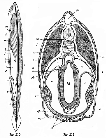
Fig. 210—The lancelet
(Amphioxus lanceolatus), left view. The long axis is
vertical; the mouth-end is above, the tail-end below; a
mouth, surrounded by threads of beard; b anus, c
gill-opening (porus branchialis), d gill-crate,
e stomach, f liver, g small intestine, h
branchial cavity, i chorda (axial rod), underneath it the
aorta; k aortic arches, l trunk of the branchial
artery, m swellings on its branches, n vena cava,
o visceral vein.
Fig. 211—Transverse section of the
head of the Amphioxus. (From Boveri.) Above the
branchial gut (kd) is the chorda, above this the neural tube
(in which we can distinguish the inner grey and the outer white
matter); above again is the dorsal fin (fh). To the right
and left above (in the episoma) are the thick muscular plates
(m); below (in the hyposoma) the gonads (g).
ao aorta (here double), c corium, ec endostyl,
f fascie, gl glomerulus of the kidneys, k
branchial vessel, ld partition between the cœloma
(sc) and atrium (p), mt transverse ventral
muscle, n renal canals, of upper and uf lower canals
in the mantle-folds, p peribranchial cavity, (atrium),
sc cœloma (subchordal body-cavity), si principal
(or subintestinal) vein, sk perichorda (skeletal
layer). |
man and the other vertebrates. As a matter of fact,
it is so different from all the other vertebrates in its whole
organisation that the laws of logical classification compel us to
distinguish two divisions of this stem: 1, the Acrania (Amphioxus
and its extinct relatives); and 2, the Craniota (man and the other
vertebrates). The first and lower division comprises the
vertebrates that have no vertebræ or skull
[ 183 ]
(cranium). Of these the only living
representatives are the Amphioxus and Paramphioxus, though there
must have been a number of different species at an early period of
the earth’s history.
Opposed to the Acrania is the second division of the
vertebrates, which comprises all the other members of the stem,
from the fishes up to man. All these vertebrates have a head quite
distinct from the trunk, with a skull (cranium) and brain;
all have a centralised heart, fully-formed kidneys, etc. Hence they
are called the Craniota. These Craniotes are, however,
without a skull in their earlier period. As we already know from
embryology, even man, like every other mammal, passes in the
earlier course of his development through the important stage which
we call the chordula; at this lower stage the animal has neither
vertebræ nor skull nor limbs
(Figs. 83–86). And even after the formation of the
primitive vertebræ has begun, the segmented fœtus of
the amniotes still has for a long time the simple form of a
lyre-shaped disk or a sandal, without limbs or extremities. When we
compare this embryonic condition, the sandal-shaped fœtus,
with the developed lancelet, we may say that the amphioxus is, in a
certain sense, a permanent sandal-embryo, or a permanent embryonic
form of the Acrania; it never rises above a low grade of
development which we have long since passed.
The fully-developed lancelet (Fig. 210) is about two inches
long, is colourless or of a light red tint, and has the shape of a
narrow lancet-formed leaf. The body is pointed at both ends, but
much compressed at the sides. There is no trace of limbs. The outer
skin is very thin and delicate, naked, transparent, and composed of
two different layers, a simple external stratum of cells, the
epidermis, and a thin underlying cutis-layer. Along the middle line
of the back runs a narrow fin-fringe which expands behind into an
oval tail-fin, and is continued below in a short anus-fin. The
fin-fringe is supported by a number of square elastic
fin-plates.
In the middle of the body we find a thin string of cartilage,
which goes the whole length of the body from front to back, and is
pointed at both ends (Fig. 210 i). This straight,
cylindrical rod (somewhat compressed for a time) is the axial rod
or the chorda dorsalis; in the lancelet this is the only
trace of a vertebral column. The chorda develops no further, but
retains its original simplicity throughout life. It is enclosed by
a firm membrane, the chorda-sheath or perichorda. The real
features of this and of its dependent formations are best seen in
the transverse section of the Amphioxus (Fig. 211). The perichorda
forms a cylindrical tube immediately over the chorda, and the
central nervous system, the medullary tube, is enclosed in it. This
important psychic organ also remains in its simplest shape
throughout life, as a cylindrical tube, terminating with almost
equal plainness at either end, and enclosing a narrow canal in its
thick wall. However, the fore end is a little rounder, and contains
a small, almost imperceptible bulbous swelling of the canal. This
must be regarded as the beginning of a rudimentary brain. At the
foremost end of it there is a small black pigment-spot, a
rudimentary eye; and a narrow canal leads to a superficial
sense-organ. In the vicinity of this optic spot we find at the left
side a small ciliated depression, the single olfactory organ. There
is no organ of hearing. This defective development of the higher
sense-organs is probably, in the main, not an original feature, but
a result of degeneration.
Underneath the axial rod or chorda runs a very simple alimentary
canal, a tube that opens on the ventral side of the animal by a
mouth in front and anus behind. The oval mouth is surrounded by a
ring of cartilage, on which there are twenty to thirty
cartilaginous threads (organs of touch, Fig. 210 a). The
alimentary canal divides into sections of about equal length by a
constriction in the middle. The fore section, or head-gut, serves
for respiration; the hind section, or trunk-gut, for digestion. The
limit of the two alimentary regions is also the limit of the two
parts of the body, the head and the trunk. The head-gut or
branchial gut forms a broad gill-crate, the grilled wall of which
is pierced by numbers of gill-clefts (Fig. 210 d). The fine
bars of the gill-crate between the clefts are strengthened with
firm parallel rods, and these are connected in pairs by cross-rods.
The water that enters the mouth of the Amphioxus passes through
these clefts into the large surrounding branchial cavity or
atrium, and then pours out behind through a hole in it, the
respiratory pore (porus branchialis, Fig. 210 c).
Below, on the ventral side of the gill-crate, there is in the
middle
[ 184 ]
line a ciliated groove with a glandular wall (the
hypobranchial groove), which is also found in the Ascidia and the
larvæ of the Cyclostoma. It is interesting because the
thyroid gland in the larynx of the higher vertebrates (underneath
the “Adam’s apple”) has been developed from
it.
Behind the respiratory part of the gut we have the digestive
section, the trunk or liver (hepatic) gut. The small particles that
the Amphioxus takes in with the water—infusoria, diatoms,
particles of decomposed plants and animals, etc.—pass from
the gill-crate into the digestive part of the canal, and are used
up as food. From a somewhat enlarged portion, that corresponds to
the stomach (Fig. 210 e), a long, pouch-like blind sac
proceeds straight forward (f); it lies underneath on the
left side of the gill-crate, and ends blindly about the middle of
it. This is the liver of the Amphioxus, the simplest kind of liver
that we meet in any vertebrate. In man also the liver develops, as
we shall see, in the shape of a pouch-like blind sac, that forms
out of the alimentary canal behind the stomach.
|
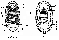
Fig.
212—Transverse section of an Amphioxus-larva, with
five gill-clefts, through the middle of the body.
Fig. 213—Diagram of the preceding. (From
Hatschek.) A epidermis, B medullary tube,
C chorda, C1 inner chorda-sheath, D
visceral epithelium, E sub-intestinal vein. 1 cutis,
2 muscle-plate (myotome), 3 skeletal plate
(sclerotome), 4 cœloseptum (partition between dorsal
and ventral cœloma), 5 skin-fibre layer, 6
gut-fibre layer, I myocœl (dorsal body-cavity),
II splanchnocœl (ventral body-cavity).) |
The formation of the circulatory system in this animal is not
less interesting. All the other vertebrates have a compressed,
thick, pouch-shaped heart, which develops from the wall of the gut
at the throat, and from which the blood-vessels proceed; in the
Amphioxus there is no special centralised heart, driving the blood
by its pulsations. This movement is effected, as in the annelids,
by the thin blood-vessels themselves, which discharge the function
of the heart, contracting and pulsating in their whole length, and
thus driving the colourless blood through the entire body. On the
under-side of the gill-crate, in the middle line, there is the
trunk of a large vessel that corresponds to the heart of the other
vertebrates and the trunk of the branchial artery that proceeds
from it; this drives the blood into the gills (Fig. 210 l).
A number of small vascular arches arise on each side from this
branchial artery, and form little heart-shaped swellings or
bulbilla (m) at their points of departure; they advance
along the branchial arches, between the gill-clefts and the
fore-gut, and unite, as branchial veins, above the gill-crate in a
large trunk blood-vessel that runs under the chorda dorsalis. This
is the principal artery or primitive aorta (Fig. 214 D). The
branches which it gives off to all parts of the body unite again in
a larger venous vessel at the underside of the gut, called the
subintestinal vein (Figs. 210 o, 212 E). This single
main vessel of the Amphioxus goes like a closed circular
water-conduit along the alimentary canal through the whole body,
and pulsates in its whole length above and below. When the upper
tube contracts the lower one is filled with blood, and vice
versa. In the upper tube the blood flows from front to rear,
then back from rear to front in the lower vessel. The whole of the
long tube that runs along the ventral side of the alimentary canal
and contains venous blood may be called the “principal
vein,” and may be compared to the ventral vessel in the
worms. On the other hand, the long
[ 185 ]
straight vessel that runs along the dorsal line of
the gut above, between it and the chorda, and contains arterial
blood, is clearly identical with the aorta or principal artery of
the other vertebrates; and on the other side it may be compared to
the dorsal vessel in the worms.
The cœloma or body-cavity has some very important and
distinctive features in the Amphioxus. The embryology of it is most
instructive in connection with the stem-history of the body-cavity
in man and the other vertebrates. As we have already seen (Chapter
X), in these the two cœlom-pouches are divided at an early
stage by transverse constrictions into a double row of primitive
segments (Fig. 124), and each of
these subdivides, by a frontal or lateral constriction, into an
upper (dorsal) and lower (ventral) pouch.
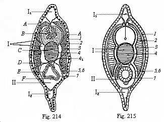
Fig. 214—Transverse section of a
young Amphioxus, immediately after metamorphosis, through the
hindermost third (between the atrium-cavity and the anus).
Fig. 215—Diagram of preceding. (From Hatschek.)
A epidermis, B medullary tube, C chorda,
D aorta, E visceral epithelium, F subintestinal
vein. 1 corium-plate, 2 muscle-plate, 3
fascie-plate, 4 outer chorda-sheath, 5 myoseptum,
6 skin-fibre plate, 7 gut-fibre plate, I
myocœl, II splanchnocœl, I1
dorsal fin, I2 anus-fin.) |
These important structures are seen very clearly in the trunk of
the amphioxus (the latter third, Figs. 212–215), but it is
otherwise in the head, the foremost third (Fig. 216). Here we find
a number of complicated structures that cannot be understood until
we have studied them on the embryological side in the next chapter
(cf. Fig. 81). The branchial gut lies free in a spacious cavity
filled with water, which was wrongly thought formerly to be the
body-cavity (Fig. 216 A). As a matter of fact, this atrium
(commonly called the peribranchial cavity) is a secondary structure
formed by the development of a couple of lateral mantle-folds or
gill-covers (M1, U). The real body-cavity
(Lh) is very narrow and entirely closed, lined with
epithelium. The peribranchial cavity (A) is full of water,
and its walls are lined with the skin-sense layer; it opens
outwards in the rear through the respiratory pore (Fig. 210
c).
On the inner surface of these mantle-folds
(M1), in the ventral half of the wide mantle
cavity (atrium), we find the sex-organs of the Amphioxus. At each
side of the branchial gut there are between twenty and thirty
roundish four-cornered sacs, which can clearly be seen from without
with the naked eye, as they shine through the thin transparent
body-wall. These sacs are the sexual glands they are the same size
and shape in both sexes, only differing in contents. In the female
they contain a quantity of simple ova (Fig. 219 g); in the
male a number of much smaller cells that change into mobile
ciliated cells (sperm-cells). Both sacs lie on the inner wall of
the atrium, and have no special outlets. When the ova of the female
and the sperm of the male are ripe, they fall into the atrium, pass
through the gill-clefts into the
[ 186 ]
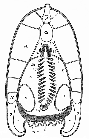 |
Fig.
216—Transverse section of the lancelet, in the
fore half. (From Ralph.) The outer covering is the simple
cell-layer of the epidermis (E). Under this is the thin
corium, the subcutaneous tissue of which is thickened; it sends
connective-tissue partitions between the muscles
(M1) and to the chorda-sheath. (N
medullary tube, Ch chorda, Lh body-cavity, A
atrium, L upper wall of same, E1 inner
wall, E2 outer wall, Lh1
ventral remnant of same, Kst gill-reds, M ventral
muscles, R seam of the joining of the ventral folds
(gill-covers), G sexual glands. |
fore-gut, and are ejected through the mouth.
Above the sexual glands, at the dorsal angle of the atrium, we
find the kidneys. These important excretory organs could not be
found in the Amphioxus for a long time, on account of their remote
position and their smallness; they were discovered in 1890 by
Theodor Boveri (Fig. 217 x). They are short segmented
canals; corresponding to the primitive kidneys of the other
vertebrates (Fig. 218 B). Their internal aperture (Fig. 217
B) opens into the body-cavity; their outer aperture into the
atrium (C). The prorenal canals lie in the middle of the
line of the head, outwards from the uppermost section of the
gill-arches, and have important relations to the branchial vessels
(H). For this reason, and in their whole arrangement, the
primitive kidneys of the Amphioxus
[ 187 ]
show clearly that they are equivalent to the
prorenal canals of the Craniotes (Fig. 218 B). The prorenal
duct of the latter (Fig. 218 C) corresponds to the branchial
cavity or atrium of the former (Fig. 217 C).
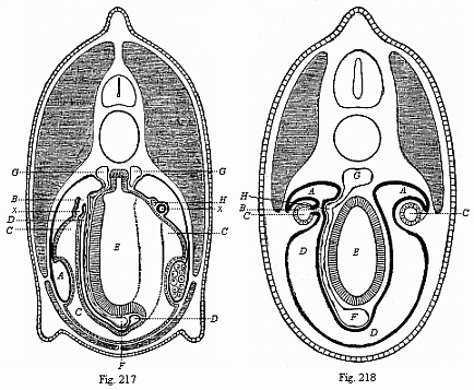
Fig. 217—Transverse section through
the middle of the Amphioxus. (From Boveri.) On the left
a gill-rod has been struck, and on the right a gill-cleft;
consequently on the left we see the whole of a prorenal canal
(x), on the right only the section of its fore-leg. A
genital chamber (ventral section of the gonocœl), x
pronephridium, B its cœlom-aperture, C atrium,
D body-cavity, E visceral cavity, F
subintestinal vein, G aorta (the left branch connected by a
branchial vessel with the subintestinal vein), H renal
vessel.
Fig. 218—Transverse section of a primitive fish embryo
(Selachii-embryo, from Boveri.). To the left pronephridia
(B), the right primitive kidneys (A). The dotted
lines on the right indicate the later opening of the primitive
kidney canals (A) into the prorenal duct (C).
D body-cavity, E visceral cavity, F subintestinal
vein, G aorta, H renal vessel. |
If we sum up the results of our anatomic study of the Amphioxus,
and compare them with the familiar organisation of man, we shall
find an immense distance between the two. As a fact, the highest
summit of the vertebrate organisation which man represents is in
every respect so far above the lowest stage, at which the lancelet
remains, that one would at first scarcely believe it possible to
class both animals in the same division of the animal kingdom.
Nevertheless, this classification is indisputably just. Man is only
a more advanced stage of the vertebral type that we find
unmistakably in the Amphioxus in its characteristic features. We
need only recall the picture of the ideal Primitive Vertebrate
given in a former chapter, and compare it with the lower stages of
human embryonic development, to convince ourselves of our close
relationship to the lancelet. (Cf. Chapter XI)
It is true that the Amphioxus is far below all other living
vertebrates. It is true that it has no separate head, no developed
brain or skull, the characteristic feature of the other
vertebrates.
[ 188 ]
It is (probably as a result of degeneration) without
the auscultory organ and the centralised heart that all the others
have; and it has no fully-formed kidneys. Every single organ in it
is simpler and less advanced than in any of the others. Yet the
characteristic connection and arrangement of all the organs is just
the same as in the other vertebrates. All these, moreover, pass,
during their embryonic development, through a stage in which their
whole organisation is no higher than that of the Amphioxus, but is
substantially identical with it.
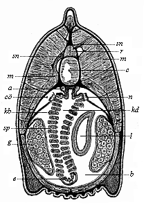 Fig.
219—Transverse section of the head of the
Amphioxus (at the limit of the first and second third of the
body). (From Boveri) a aorta (here double), b
atrium, c chorda, co umlaut cœloma
(body-cavity), e endostyl (hypobranchial groove), g
gonads (ovaries), kb gill-arches, kd branchial gut,
l liver-tube (on the right, one-sided), m muscles,
n renal canals, r spinal cord, sn spinal
nerves, sp gill-clefts.
Fig.
219—Transverse section of the head of the
Amphioxus (at the limit of the first and second third of the
body). (From Boveri) a aorta (here double), b
atrium, c chorda, co umlaut cœloma
(body-cavity), e endostyl (hypobranchial groove), g
gonads (ovaries), kb gill-arches, kd branchial gut,
l liver-tube (on the right, one-sided), m muscles,
n renal canals, r spinal cord, sn spinal
nerves, sp gill-clefts. |
In order to see this quite clearly, it is particularly useful to
compare the Amphioxus with the youthful forms of those vertebrates
that are classified next to it. This is the class of the
Cyclostoma. There are to-day only a few species of this once
extensive class, and these may be distributed in two groups. One
group comprises the hag-fishes or Myxinoides. The other group are
the Petromyzontes, or lampreys, which are a familiar delicacy in
their marine form. These Cyclostoma are usually classified with the
fishes. But they are far below the true fishes, and form a very
interesting connecting-group between them and the lancelet. One can
see how closely they approach the latter by comparing a young
lamprey with the Amphioxus. The chorda is of the same simple
character in both; also the medullary tube, that lies above the
chorda, and the alimentary canal below it. However, in the lamprey
the spinal cord swells in front into a simple pear-shaped cerebral
vesicle, and at each side of it there are a very simple eye and a
rudimentary auditory vesicle. The nose is a single pit, as in the
Amphioxus. The two sections of the gut are also just the same and
very rudimentary in the lamprey. On the other hand, we see a great
advance in the structure of the heart, which is found underneath
the gills in the shape of a centralised muscular tube, and is
divided into an auricle and a ventricle. Later on the lamprey
advances still further, and gets a skull, five cerebral vesicles, a
series of independent gill-pouches, etc. This makes all the more
interesting the striking resemblance of its immature larva to the
developed and sexually mature Amphioxus.
While the Amphioxus is thus connected through the Cyclostoma
with the fishes, and so with the series of the higher vertebrates,
it is, on the other hand, very closely related to a lowly
invertebrate marine animal, from which it seems to be entirely
remote at first glance. This remarkable animal is the sea-squirt or
Ascidia, which was formerly thought to be closely related to the
mussel, and so classed in the molluscs. But since the remarkable
embryology of these animals was discovered in 1866, there can be no
question that they have nothing to do with the molluscs. To the
great astonishment of zoologists, they were found, in their whole
individual development, to be closely related to the vertebrates.
When fully developed the Ascidiæ are shapeless lumps that
would not, at first sight, be taken for animals at all. The oval
body, frequently studded with knobs or uneven and lumpy, in which
we can discover no special external organs, is attached at one end
to marine plants, rocks, or the floor of the sea. Many species look
like potatoes, others like melon-cacti, others like prunes. Many of
the Ascidiæ form transparent crusts or
[ 189 ]
deposits on stones and marine plants. Some of the
larger species are eaten like oysters. Fishermen, who know them
very well, think they are not animals, but plants. They are sold in
the fish markets of many of the Italian coast-towns with other
lower marine animals under the name of “sea-fruit”
(frutti di mare). There is nothing about them to show that
they are animals. When they are taken out of the water with the net
the most one can perceive is a slight contraction of the body that
causes water to spout out in two places. The bulk of the
Ascidiæ are very small, at the most a few inches long. A few
species are a foot or more in length. There are many species of
them, and they are found in every sea. As in the case of the
Acrania, we have no fossilised remains of the class, because they
have no hard and fossilisable parts. However, they must be of great
antiquity, and must go back to the primordial epoch.
The name of “Tunicates” is given to the whole class
to which the Ascidiæ belong, because the body is enclosed in
a thick and stiff covering like a mantle (tunica). This
mantle—sometimes soft like jelly, sometimes as tough as
leather, and sometimes as stiff as cartilage—has a number of
peculiarities. The most remarkable of them is that it consists of a
woody matter, cellulose—the same vegetal substance that forms
the stiff envelopes of the plant-cells, the substance of the wood.
The tunicates are the only class of animals that have a real
cellulose or woody coat. Sometimes the cellulose mantle is brightly
coloured, at other times colourless. Not infrequently it is set
with needles or hairs, like a cactus. Often we find a mass of
foreign bodies—stone, sand, fragments of mussel-shells,
etc.—worked into the mantle. This has earned for the Ascidia
the name of “the microcosm.”
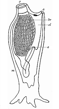 Fig.
220—Organisation of an Ascidia (left view); the
dorsal side is turned to the right and the ventral side to the
left, the mouth (o) above; the ascidia is attached at the
tail end. The branchial gut (br), which is pierced by a
number of clefts, continues below in the visceral gut. The rectum
opens through the anus (a) into the atrium (cl), from
which the excrements are ejected with the respiratory water through
the mantle-hole or cloaca (a); m mantle. (From
Gegenbaur.
Fig.
220—Organisation of an Ascidia (left view); the
dorsal side is turned to the right and the ventral side to the
left, the mouth (o) above; the ascidia is attached at the
tail end. The branchial gut (br), which is pierced by a
number of clefts, continues below in the visceral gut. The rectum
opens through the anus (a) into the atrium (cl), from
which the excrements are ejected with the respiratory water through
the mantle-hole or cloaca (a); m mantle. (From
Gegenbaur. |
The hind end, which corresponds to the tail of the Amphioxus, is
usually attached, often by means of regular roots. The dorsal and
ventral sides differ a good deal internally, but frequently cannot
be distinguished externally. If we open the thick tunic or mantle
in order to examine the internal organisation, we first find a
spacious cavity filled with water—the mantle-cavity or
respiratory cavity (Fig. 220 cl). It is also called the
branchial cavity and the cloaca, because it receives the excrements
and sexual products as well as the respiratory water. The greater
part of the respiratory cavity is occupied by the large grated
branchial sac (br). This is so like the gill-crate of the
Amphioxus in its whole arrangement that the resemblance was pointed
out by the English naturalist Goodsir, years ago, before anything
was known of the relationship of the two animals. As a fact, even
in the Ascidia the mouth (o) opens first into this wide
branchial sac. The respiratory water passes through the
lattice-work of the branchial sac into the branchial cavity, and is
ejected from this by the respiratory pore (a′). Along
the ventral side of the branchial sac runs a ciliated
groove—the hypobranchial groove which we have previously
found at the same spot in the Amphioxus. The food of the Ascidia
also
[ 190 ]
consists of tiny organisms, infusoria, diatoms,
parts of decomposed marine plants and animals; etc. These pass with
the water into the gill-crate and the digestive part of the gut at
the end of it, at first into an enlargement of it that represents
the stomach. The adjoining small intestine usually forms a loop,
bends forward, and opens by an anus (Fig. 220 a), not
directly outwards, but first into the mantle cavity; from this the
excrements are ejected by a common outlet (a′)
together with the used-up water and the sexual products. The outlet
is sometimes called the branchial pore, and sometimes the cloaca or
ejection-aperture. In many of the Ascidiæ a glandular mass
opens into the gut, and this represents the liver. In some there is
another gland besides the liver, and this is taken to represent the
kidneys. The body-cavity proper, or cœloma, which is filled
with blood and encloses the hepatic gut, is very narrow in the
Ascidia, as in the Amphioxus, and is here also usually confounded
with the wide atrium, or peribranchial cavity, full of water.
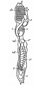 Fig. 221—Organisation of
an Ascidia (as in Fig. 220, seen from the left). sb
branchial sac, v stomach, i small intestine, c
heart, t testicle, vd sperm-duct, o ovary,
o′ ripe ova in the branchial cavity. The two small arrows
indicate the entrance and exit of the water through the openings of
the mantle. (From Milne-Edwards.)
Fig. 221—Organisation of
an Ascidia (as in Fig. 220, seen from the left). sb
branchial sac, v stomach, i small intestine, c
heart, t testicle, vd sperm-duct, o ovary,
o′ ripe ova in the branchial cavity. The two small arrows
indicate the entrance and exit of the water through the openings of
the mantle. (From Milne-Edwards.) |
There is no trace in the fully-developed Ascidia of a chorda
dorsalis, or internal axial skeleton. It is the more interesting
that the young animal that emerges from the ovum has a
chorda, and that there is a rudimentary medullary tube above it.
The latter is wholly atrophied in the developed Ascidia, and looks
like a small nerve-ganglion in front above the gill-crate. It
corresponds to the upper “gullet-ganglion” or
“primitive brain” in other vermalia. Special
sense-organs are either wanting altogether or are only found in a
very rudimentary form, as simple optic spots and touch-corpuscles
or tentacles that surround the mouth. The muscular system is very
slightly and irregularly developed. Immediately under the thin
corium, and closely connected with it, we find a thin muscle tube,
as in the worms. On the other hand, the Ascidia has a centralised
heart, and in this respect it seems to be more advanced than the
Amphioxus. On the ventral side of the gut, some distance behind the
gill-crate, there is a spindle-shaped heart. It retains permanently
the simple tubular form that we find temporarily as the first
structure of the heart in the vertebrates. This simple heart of the
Ascidia has, however, a remarkable peculiarity. It contracts in
alternate directions. In all other animals the beat of the heart is
always in the same direction (generally from rear to front); it
changes in the Ascidia to the reverse direction. The heart
contracts first from the rear to the front, stands still for a
minute, and then begins to beat the opposite way, now driving the
blood from front to rear; the two large vessels that start from
either end of the heart act alternately as arteries and veins. This
feature is found in the Tunicates alone.
Of the other chief organs we have still to mention the sexual
glands, which lie right behind in the body-cavity. All the
Ascidiæ are hermaphrodites. Each individual has a male and a
female gland, and so is able to fertilise itself. The ripe ova
(Fig. 221 o′) fall directly from the ovary (o)
into the mantle-cavity. The male sperm is conducted into this
cavity from the testicle (t) by a special duct (vd).
Fertilisation is accomplished here, and in many of the
Ascidiæ developed embryos are found. These are then ejected
[ 191 ]
with the breathing-water through the cloaca (q), and so
“born alive.”
If we now glance at the entire structure of the simple Ascidia
(especially Phallusia, Cynthia, etc.) and compare it with
that of the Amphioxus, we shall find that the two have few points
of contact. It is true that the fully-developed Ascidia resembles
the Amphioxus in several important features of its internal
structure, and especially in the peculiar character of the
gill-crate and gut. But in most other features of organisation it
is so far removed from it, and is so unlike it in external
appearance, that the really close relationship of the two was not
discovered until their embryology was studied. We will now compare
the embryonic development of the two animals, and find to our great
astonishment that the same embryonic form develops from the ovum of
the Amphioxus as from that of the Ascidia—a typical
chordula.
Title and Contents
Glossary
Chapter XV
Vol. II Title and Contents
Figs. 1–209
Figs. 210–408