Please see the Transcriber’s Notes at the end of this text.
FARM LIFE SERIES
THE FARMER’S VETERINARIAN
By Charles William Burkett
HANDY FARM DEVICES AND HOW TO MAKE THEM
By Rolfe Cobleigh
MAKING HORTICULTURE PAY
By M. G. Kains
FARM CROPS
By Charles William Burkett
PROFITABLE STOCK RAISING
By Clarence A. Shamel
PROFITABLE POULTRY PRODUCTION
By M. G. Kains
Other Volumes in Preparation
HEALTH
A Practical Treatise on the Diseases of Farm Stock: Containing Brief and Popular Advice on the Nature, Cause and Treatment of Disease, the Common Ailments and the Care and Management of Stock when Sick
By
CHARLES WILLIAM BURKETT
Editor of American Agriculturist
ILLUSTRATED
NEW YORK
ORANGE JUDD COMPANY
1914
Copyright, 1909
Orange Judd Company
New York
Printed in U. S. A.

large class of people, by force of circumstances, are compelled to treat their own animals when sick or disabled. Qualified veterinarians are not always available; and all the ills and accidents incident to farm animals do not require professional attendance. Furthermore, the skilled stockman should be familiar with common diseases and the treatment of them. He should remember, too, that the maintenance of health and vigor in our farm stock is the direct result of well-directed management. Too frequently this is neither understood nor admitted, and an unreasonable lack of attention, when animals are ill or indisposed, works out dire mischief in the presence of physical disorder and infectious diseases. A fair acquaintance with the common ailments is helpful to the owner and to his stock. This leads to health, to prevention of disease, and to skill in attendance when disease is at hand.
The volume herewith presented abounds in helpful suggestions and valuable information for the most successful treatment of ills and accidents and disease troubles. It is an everyday handbook of disease and its treatment, and contains the best ideas gathered from the various authorities and the experience of a score of practical veterinarians in all phases of veterinary practice.
C. W. BURKETT.
New York, June, 1909.
[vi]
[vii]
| Page | |
|---|---|
| Introduction | |
| Facing Disease on the Farm | 1 |
| Chapter I. | |
| How the Animal Body is Formed | 9 |
| Chapter II. | |
| Some Physiology You Ought to Know | 21 |
| Chapter III. | |
| The Teeth as an Indication of Age | 34 |
| Chapter IV. | |
| Examining Animals for Soundness and Health | 39 |
| Chapter V. | |
| Wounds and Their Treatment | 54 |
| Chapter VI. | |
| Making a Post-Mortem Examination | 62 |
| Chapter VII. | |
| Common Medicines and Their Actions | 69 |
| Chapter VIII. | |
| Meaning of Disease | 82 |
| Chapter IX. | |
| Diagnosis and Treatment of Disease | 92 |
| Chapter X. | |
| Diseases of Farm Animals | 101 |
[viii]
[ix]
| Page | ||
|---|---|---|
| 1. | Health | Frontispiece |
| 2. | Common Sheep Scab | 3 |
| 3. | Hog House and Feeding Floor | 5 |
| 4. | Poulticing the Throat | 8 |
| 5. | How a Cell Divides | 10 |
| 6. | Bones of Skeleton of a Horse | 16 |
| 7. | One of the Parasites of the Hog | 18 |
| 8. | Circulation and Digestion | 22 |
| 9. | Diseased Kidney | 25 |
| 10. | Stomach of Ruminant | 27 |
| 11. | Circulation of Blood in Body | 30 |
| 12. | Lumpy Jaw (jaw bone) | 36 |
| 13. | Bad Attitude Due to Conformation | 41 |
| 14. | Ewe Neck | 46 |
| 15. | Anatomy of the Foot | 49 |
| 16. | Fractures | 54 |
| 17. | Bandaging a Leg | 57 |
| 18. | Rickets in Pigs | 63 |
| 19. | Round Worms in Hog Intestines | 66 |
| 20. | Tetanus Bacilli | 71 |
| 21. | Ready for the Drench | 81 |
| 22. | Bacteria As Seen Under the Microscope | 85 |
| 23. | Result of Bone Spavin | 90 |
| 24. | Feeling the Pulse | 94 |
| 25. | How Heat Affects Growth | 96 |
| 26. | Diseases of the Horse | 102 |
| 27. | Lumpy Jaw (external view) | 105 |
| 28. | Where to Tap in Bloating | 118 |
| 29. | Bog Spavin | 122 |
| 30. | Horse Bots in Stomach | 124 |
| 31. | Colic Pains | 138 |
| 32. | Retention of the Urine | 141 |
| 33. | Curb | 145 |
| 34. | Fistulous Withers | 156 |
| 35. | Foot Rot in Sheep | 160 |
| 36. | Founder | 163 |
| 37. | Bad Case of Glanders | 170 |
| 38. | Ventral Hernia | 180 |
| 39. | An Attack of Cholera | 182 |
| 40. | The Result of Hog Cholera | 186 |
| 41. | Kidney Worms in the Hog | 205 |
| 42. | Liver Fluke | 207 |
| 43. | Lockjaw | 209 |
| 44. | Lymphangitis | 215 |
| 45. | Natural Presentation of the Foal | 225 |
| 46. | Abnormal Presentation of the Foal | 227 |
| 47. | Quittor | 235 |
| 48. | A Cattle Bath Tub | 241 |
| 49. | Side Bones | 244 |
| 50. | Splint | 248 |
| 51. | Twisted Stomach Worms | 252 |
| 52. | Tuberculosis Germs | 264 |
| Health and Disease | Plate 1 | |
| Making Post Mortem Examinations | Plate 2 | |
| A Victim of Tuberculosis | Plate 3 | |
| Exterior Points of the Horse; Castration | Plate 4 | |
| Texas Fever | Plate 5 | |
| A Typical Case of Foot and Mouth Disease | Plate 6 | |
[1]
To call a veterinarian or not—that is the question. Whether your horse or cow is sick enough for professional attendance, or just under the weather a little, is a problem you will always be called upon to face. And you must meet it. It has always faced the man who raises stock, and it is a problem that always will. Like human beings, farm stock have their ailments and troubles; and, in most cases, a little care and nursing are all that will be required. With these troubles all of us are acquainted; especially those who have spent much time with the flocks and the herds on the farm. Through experience we know that often with every reasonable care, some animals, frequently the healthiest-looking ones, in the field, or stable, give trouble at the most unsuspected times. So the fault is not always with the owner.
There is no reason, however, why an effort should not be made, just as soon as any trouble is noticed, to assist the sick animal to recover, and help nature in every way possible to restore the invalid to its usual normal condition. The average observing farmer, as a rule, knows just about what the trouble is; he usually knows if treatment is beyond him, and if not, what simple medical aid will be effective in bringing about a recovery with greater dispatch than nature unaided will effect.
Now, of course, this means that the farmer should be acquainted with his animals; in health and disease their actions should be familiar to him.[2] If he be a master of his business he naturally knows a great deal about his farm stock. No man who grows corn or wheat ever raises either crop extremely successfully unless he has an intimate knowledge of the soil, the seed, the details of fertilization and culture. He has learned how good soils look, how bad soils look; he knows if soils are healthy, whether they are capable of producing big crops or little crops.
So with his stock. He must know, and he does know, something as to their state of health or ill health. With steady observation his knowledge will increase; and with experience he ought to be able to diagnose the common ailments, and not only prescribe for their treatment, but actually treat many of them himself. Unfortunately, many farmers pass health along too lightly and the common disorders too seriously. This is wrong. The man who deals with farm animals should be well acquainted with them, just as the engineer is acquainted with his engine. If an engine goes wrong the engineer endeavors to ascertain the trouble. If it is beyond his experience and knowledge he turns the problem over to an expert. It should be so with the stock raiser. So familiar should the owner be with his animals in case of trouble he ought to know of some helpful remedy or to know that the trouble is more serious than ordinary, in which case the veterinarian should be called.
All of this means that the art of observing the simple functions should be acquired at the earliest possible moment—where to find the pulse of horse or cow, how many heart beats in a minute, how many respirations a minute, the color of the healthy nostril, the use of the thermometer and where to place it to get the information, the character of the[3] eye, the nature of the coat, the passage of dung and water, how the animal swallows, the attitude when standing, the habit of lying down and getting up—all of these should be as familiar to the true stockman as the simplest details of tillage or of planting or of harvesting.
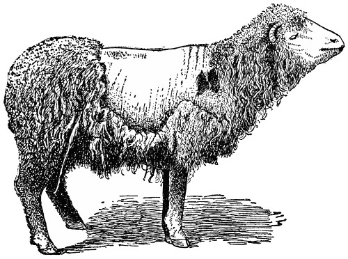
COMMON SHEEP SCAB
Here is an advanced case and shows how serious the trouble may become. A very small itch mite is the cause. The mites live and multiply under the scurf and scab of the skin.
Moreover, the stockman should be a judge of external characters, whether natural or temporary. He should have a knowledge of animal conformation. If to know a good plow is desirable, then to know a good pastern or foot is desirable. If the art of selecting wheat is a worthy acquisition, then the art of comparing hocks of different horses is a worthy accomplishment also. If experience tells the grower that his corn or potatoes or cotton is strong, vigorous and healthy or just the reverse,[4] observation and experience ought also to tell him when his stock are in good health or when they lack thrift or are sick and need treatment.
Few farmers there are, indeed, who are not acquainted with crop diseases. Smut is readily recognized when present in the wheat or corn or oat field; so colic, too, should be recognized when your horse is affected by it. The peach and the apple have their common ailments; so have the cow and pig. In either case the facts ought to be familiar. So familiar that as soon as diagnosed and recognized prompt measures for treatment should be followed that the cure may be effected before any particular headway is at all made. Handled in this way, many cases that are now passed on to the veterinarian would never develop into serious disturbances at all.
The old saying, “Prevention is better than cure,” is both wisdom and a splendid platform on which to build any branch of live stock work. Every disease is the result of some disturbance, somewhere. It may be improper food; the stockman must know. Moldy fodder causes nervous troubles in the horse. Cottonseed meal, if fed continuously to pigs, leads to their death. Hence, food has much to do with health and disease. Ventilation of the stable plays its part. Bad air leads to weakness, favors tuberculosis, and, if not remedied, brings about loss and death. Fresh air in abundance is better than medicine; and the careful stockman will see that it be not denied.
[5]
Good sanitation, including cleanly quarters, wholesome water and dry stables, has its reward in more healthy animals. When not provided, the animals are frequently ill, or are in bad health more or less. As these factors—proper food, good ventilation, and effective sanitation—are introduced in stable accommodations, diseases will be lessened and stock profits will increase.
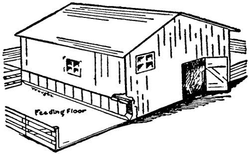
HOG HOUSE AND FEEDING FLOOR
This convenient hog house is inexpensive, and the feeding floor at the side insures cleanliness and thorough sanitary conditions. A sanitary hog house should be one of the chief improvements of the farm.
As disease is better understood it becomes more closely identified with germs and bacteria. Hence, to lessen disease we must destroy, so far as possible, the disease-producing germs. For this purpose nothing is better than sunlight and disinfectants. Sunlight is itself death to all germs; therefore, all stables, and the living quarters for farm animals, should be light and airy, and free from damp corners and lodgment places for dust, vermin, and bacteria. Even when animals are in good[6] health, disinfection is a splendid means for warding off disease. For sometimes with the greatest care germs are admitted in some manner or form. By constantly disinfecting, the likelihood of any encroachment by germs is greatly lessened.
Fortunately we have disinfectants that are easily applied and easily obtained at small cost. One of these disinfecting materials is lime, just ordinary slaked lime, the lime that every farmer knows. While it does not possess the disinfecting power of many other agents, it is, nevertheless, very desirable for sprinkling about stables and for whitewashing floors, walls, and partitions. When so used the cracks and holes are filled and the germs destroyed. Ordinary farm stables should be whitewashed once or twice each year, and the crumbled lime sprinkled on the litter or open ground. It is not desirable to use lime with bedding and manure, for the reason that it liberates the nitrogen contained therein. Hence the bedding and manure should be removed to the fields as frequently as possible, where it can be more helpful to the land. Thus scattered, the sunlight and purifying effects of the soil will soon destroy the disease bacteria, if any are present in the manure.
Another splendid disinfectant is corrosive sublimate, mercuric chloride, as it is often called. Use one ounce in eight gallons of water. This makes one-tenth of one per cent solution. In preparing this disinfectant, allow the material to stand for several hours, so as to permit the chemical to become entirely dissolved. This solution should be carefully guarded and protected, since it is a poison and, if drunk by animals, is liable to cause death. If infected quarters are to be disinfected, see that[7] the loose dirt and litter is first removed before applying the sublimate.
Carbolic acid is another satisfactory disinfectant. Usually a five per cent solution is recommended. It can be easily applied to mangers, stalls, and feed boxes. Enough should be applied so that the wood or iron is made wet and the cracks and holes more or less filled. Chloride of lime is a cheap and an easily prepared disinfectant. Use ten ounces of chloride of lime to two gallons of water. This makes a four per cent solution, and should be applied in the same way as the corrosive sublimate.
Formalin has come into prominence very recently as a desirable disinfectant. A five per cent solution fills the bill. Floors and cracks should be made thoroughly wet with it. By using one or more of these agents the living quarters of farm animals can be kept wholesome, sweet, and free from germ diseases. In fact, the use of disinfectants is one of the best aids of the farmer in warding off disease and in lessening its effects when once present.
Many diseases are introduced into a herd or flock by thoughtlessness on the part of the owner. I have known distemper to be introduced into stables and among horses, Texas fever and tuberculosis into herds of cattle, and hog cholera among hogs, because diseased animals, when purchased, were not separated off by themselves, for a short time at least. If this were done, farmers would lessen the chance of an introduction of disease into their healthy herds. Consequently quarantine quarters should be provided; especially is this true if new[8] animals are frequently purchased and brought to the farm where many animals are raised and handled. These quarantine quarters need not be expensive, and they ought to be removed far enough from the farm stock so that there may be no easy means of infection. When newly purchased animals are placed in the quarantine quarters they should be kept there long enough to determine if anything strange or unusual is taking place.
POULTICING THE THROAT
The picture shows how to apply a poultice to the throat.
[9]
The cell is the unit of growth. It is so with all forms of life—plant or animal, insect or bacterium. In the beginning the start is with a single cell, an egg, if you please. After fertilization has taken place, this single cell enlarges or grows. Many changes now occur, all rather rapidly, until the cell walls become too small, when it breaks apart and forms two cells just like the first used to be. This is known as cell division. As growth increases, the number of cells increases also—until in the end there are millions.
—The cell is very small. In most cases it cannot be seen with the naked eye. The microscope is necessary for a study of the parts, the nature and the character of the cell.
In the first place the cell is a kind of inclosed sac, in which are found the elements of growth and life. Surrounding the cell is a thin wall known as the cell membrane. In plants this cell wall is composed of cellulose, a woody substance, which is thin and tender in green and growing plants, but hard and woody when the plant is mature.
Within the limits of the cell is the protoplasm, the chief constituent of the cell; locked up in this protoplasm is life, the vital processes that have to do with growth, development, individual existence.
Embedded within the protoplasm is another part known as the nucleus and recognized under the microscope by its density. Around the nucleus is[10] centered the development of new cells or reproduction—for the changes that convert the mother-cell into offspring-cells are first noted in this place.
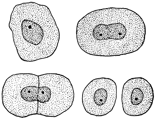
HOW A CELL DIVIDES
The simple steps in cell division are pictured here. Starting with a single cell, growth and enlargement take place, ending finally in cell division or the production of two individual cells.
So much for plant cells. Is this principle different in animals? For a long time it was thought that plants and animals were different. But upon investigation it was discovered that animals were comprised of cells just as plants. And not only was this discovered to be true, but also that animal cells corresponded in all respects to plant cells. Hence in animals are to be found cells possessing the cell walls formed of a rather thick membrane, the granular protoplasm or yoke, and the nucleus established in the yoke.
[11]
The ovum, known as the female egg, is composed of the parts just described. If it is not fertilized when ripe it passes away and dies. If fertilized in a natural way, it enlarges in size and subsequently divides into two cells; and these, passing through similar changes, finally give rise to the various groups of cells from which the body is developed.
—The body is, therefore, a mass of cells; not all alike, of course, but grouped together for the purpose of doing certain special kinds of work. In this way we have various groups, with each group a community performing its own function. The brain forms one community; and these cells are concerned with mind acts. The muscle cells are busy in exerting force and action. Another group looks after the secretions and digestive functions, while another group is concerned solely with the function of generation and reproduction. And so it is throughout the body.
Both individual cells and group cells are concerned with disease. One cell may be diseased or destroyed, but the surrounding ones may go on just the same. It is when the group is disturbed that the greatest trouble results.
—The cell always possesses its three parts—membrane, protoplasm, and nucleus. But there is no rule as to the size or shape. Cells may be round or oblong, any shape. Substances pass in and out of the cell walls; and they are in motion, many of them, especially those that line the intestines and the air passages, and the white corpuscles of the blood. More than this, some cells, Dr. Jekyl-like, change their appearance and shape, send out finger-like bodies to catch[12] enemies or food, and even travel all around in the body, often leaving it altogether.
The animal body contains five forms of tissues: Epithelial, in which the cells are very compact, forming either thin or thick plates; the connective tissue, by which many organs are supported or embedded; muscle tissue, either smooth or striated, and in which the cells are in fibers that contract and shorten; nerve-tissue, that has to do with nerve and ganglion cells by which mental impulses are sent; and blood and lymph tissue or fluid tissues.
The first group is intimately connected with the secretory organs, or those organs which secrete certain substances essential for the proper work of the body. Thus we have salivary glands, mucous glands, sweat glands, and the liver and pancreas. Connective tissue includes fibrous tissue, fatty tissue, cartilage and bone. The fibrous connective tissue is illustrated when the skin is easily picked up in folds. Fatty tissue occurs where large amounts of fat are deposited in the cells. Cartilage is found where a large amount of firm support is required. With muscle we are all familiar; it is the real lean meat of the body.
—The blood is a fluid in which many cells are to be found. The fluid is known as serum or blood-plasma and the cells as corpuscles, and are both red and white. The red cells give the characteristic color. When observed under a microscope, they appear as small, round disks. They are of great importance to the body work. Because of the coloring matter in them the oxygen of the air is attracted when it comes in[13] contact with the blood in the lungs. Oxygen is in reality absorbed, and on the blood leaving the lungs it is distributed to all parts of the body. The oxygen supply of the body is, therefore, in the keeping of the red corpuscles.
White corpuscles have a different work; they guard the body by picking up poison, bacteria, and other undesirable elements and cast these out through the natural openings of the body. Compared with the red cells, they exist in far less numbers and may wander about through all parts of the body.
Lymph is a fluid in which a few cells, lymph corpuscles, are suspended. These cells are very much like the colorless corpuscles of the blood, only no red blood cells are present. But the lymph attends to its own business; it bathes the tissues and endeavors to keep them in a healthy condition.
—Without a covering the delicate muscles would be unprotected. The skin serves in this capacity. It does still more; out of it is exuded poisonous substances, perspiration, and, at the same time, the skin is a sort of respiratory organ, through which much of the carbonic acid formed in the body escapes.
The skin possesses two general layers, the cutis and sub-cutis; in the first is contained also epidermis. Developed in the skin are the outer coverings like hair, wool, feathers, horns, claws, and hoofs.
The framework of the body undergoes a gradual development from birth to maturity. It represents the bony structure of the body; and on it all other[14] parts depend for support and protection. The brief summary of its parts and work that follows here has been adapted from Wilcox and Smith.
—This consists of a backbone, skull, shoulder girdle, pelvic girdle, and two pairs of appendages. The backbone may be conveniently divided into regions, each comprising a certain number of vertebræ. The cervical vertebræ include those from the skull from the first rib. In all mammals except the sloth and sea cow the number of cervical vertebræ is seven, being long or short, according as the neck of the animal is relatively long or short. The first and second cervical vertebræ, known as the atlas and axis, are especially modified so as to allow free turning movements of the head.
The next region includes the dorsal or thoracic vertebræ, which are characterized by having ribs movably articulated with them. The number is 13 in the cat, dog, ox, sheep, and goat; 14 in the hog; 18 or 19 in the horse and ass, and six or seven in domestic poultry. In mammals they are so joined together as to permit motion in several directions, but in poultry the dorsal vertebræ are more rigidly articulated, those next to the sacrum often being grown together with the sacrum. The spines are high and much flattened in all ungulates, long and slender in dogs and cats. They slope backward, forming strong points of attachment for the back muscles. Several ribs, varying in number in different animals, meet and become articulated with the breast bone or sternum. The sternum consists of seven to nine articulated segments in our domestic mammals, while in fowls the sternum is one thin high bone furnished with a keel of varying depth. The lumbar vertebræ lie between the dorsal[15] vertebræ and the sacrum. The number is five in the horse, six in the hog, ox and goat, and seven in the sheep. The sacrum is made up of a certain number of vertebræ, which are rigidly united and serve as an articulation for the pelvic arch. The number of sacral vertebræ is five in the ox and horse, four in sheep and hogs, and 12 to 17 in birds. The caudal or tail vertebræ naturally vary in number according to the length of the tail (7 to 10 in sheep, 21 in the ox, 23 in hogs, 17 in the horse, 22 in the cat, 16 to 23 in the dog).
In ungulates the anterior ribs are scarcely curved, the chest being very narrow in front. The number of pairs of ribs is the same as the number of dorsal vertebræ with which they articulate.
—This part of the skeleton is really composed of a number of modified vertebræ, just how many is not determined. The difference in the shape of the skulls of different animals is determined by the relative size of the various bones of the skull. In hogs, for example, the head has been much shortened as a result of breeding, thus giving the skull of the improved breeds a very different appearance from that of the razorback.
The shoulder girdle consists of a shoulder blade, collar bone and coracoid on either side. The fore leg (or wing, in case of birds) articulates with the socket formed by the junction of these three bones. In all the ungulates the shoulder blade is high and narrow, the coracoid is never much developed, and the collar bone is absent. In fowls all three bones of the shoulder girdle are well developed, the collar bone being represented by the “wish bone.”
—This consists of three bones
on either side, viz., ilium, ischium, and pubis. The
first two are directly articulated to the spinal[16]
[17]
column, while the pubic bones of either side unite
below to complete the arch. The three bones of
each side of the pelvis are present in all our
domestic animals, including the fowls.
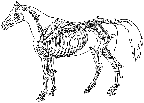
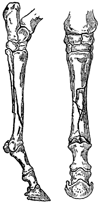
BONES OF THE SKELETON OF A HORSE
1 Face Bones, 2 Neck Bones or Cervical Vertebræ, 3 Scapula or Shoulder Blade, 4 Humerus or Arm Bone, 5 Radius or Bone of Forearm, 6 Carpus or Knee, 7 Shank Bone or Cannon, 8 Upper Pastern, 9 Lower Pastern, 10 Coffin Bone, 11 Ulna or Elbow, 12 Cartilages of the Rib, 13 Costæ or Ribs, 14 Dorsal Vertebræ or Bones of Back, 15 Lumbar Vertebræ or Bones of Loin, 16 Candal Vertebræ or Bones of Tail, 17 Haunch, 18 Femur or Thigh Bone, 19 Stifle Joint, 20 Tibia, 21 Tarsus or Hock, 22 Metatarsal Bones, 23 Upper Pastern Bone, 24 Lower Pastern Bone, 25 Coffin Bone.
—There is one formula for the bones of the fore and hind legs of farm animals. The first segment is a single bone, the humerus of the fore leg, femur of the hind leg. In the next segment there are two bones, radius and ulna in the fore leg, tibia and fibula in the hind leg. In the dog, cat, and Belgian hare the radius and ulna are both well developed and distinct. In ungulates the humerus is short and stout, while the ulna is complete in the pig, rudimentary and behind the radius in ruminants and firmly united with the radius in the horse. Similarly with the hind leg the fibula is a complete bone in the pig, while in the horse there is merely a rudiment of it, attached to the tibia.
—The mammalian skeleton has undergone the greatest modification in the bones of the feet. In the horse there are only six of the original ten wrist or carpal bones, and, since there is but one of the original five toes, the horse has also but one metacarpal or cannon bone. Splint-like rudiments of two other metacarpal bones are to be found at the upper end of the cannon bone, or at the “knee” joint. Below the cannon bone, and forming the shaft of the foot, we have the small cannon bone, coronary bone, and coffin bone—the last being within the hoof with the navicular bone behind it. The stifle joint of the horse corresponds to the knee of man. The “knee” of the horse’s fore leg corresponds to the hock of the hind leg, both being at the upper end of the cannon bone. The fetlock joint is between the large and small cannon bones,[18] the pastern joint between the small cannon or large pastern bones, and the coffin joint between the coronary and coffin bones. The horse walks upon what corresponds to the nail of the middle finger and middle toe of man.
In pigs four digits touch the ground, the first being absent and the third and fourth larger and in front of the second and fifth. In ruminants the third and fourth digits reach the ground, while the second and fifth do not. In dogs the first digit appears on the side of the leg, not in contact with the ground.
ONE OF THE PARASITES OF THE HOG
The thorn-headed worm attached to the anterior part of the small intestine often causes death. Not more than five or six are usually found in a single animal.
In fowls the wing, which corresponds to the fore leg of mammals, shows a well-developed humerus, radius and ulna, while only one carpal and one metacarpal bone remain, along which the wing feathers are attached. In the leg the femur and tibia are strong bones, but the fibula is a mere splint. The tarsal bones are absent, while the shank consists of a metatarsal bone (really three bones fused together), to which the four toes are articulated.
[19]
—The muscular system is too elaborate, the number of muscles too great, and their modifications for different purposes too complex for consideration in detail in the present volume. All muscles are either striped or unstriped (as examined under the microscope), according as they are under the immediate control of the will or not. The heart muscle forms an exception, for it is striped though involuntary. The essential characteristic of muscle fibers is contractility, which they possess in high degree. The typical striped muscles are concerned in locomotion, being attached at either end to a bone and extending across some movable joint. The most important unstriped muscles are found in the walls of the intestines and blood vessels.
—In so far as our present purposes are concerned, the nervous system may be disposed of in a few words. The central nervous system consists of a brain and spinal cord. The microscopic elements of this tissue are peculiarly modified cells, consisting of a central body, from which fibers run in two or more directions. The cell bodies constitute the gray matter, and the fibers the white matter of the brain and spinal cord. The gray substance is inside the spinal cord and on the surface of the brain, constituting the cortex. The most important parts of the brain are the cerebrum, optic lobes, cerebellum, and medulla. There are twelve pairs of cranial nerves originating in the brain and controlling the special senses, movements of the face, respiration, and pulse rate. From each segment of the spinal cord a pair of spinal nerves arises, each of which possess both sensory and motor roots. The sympathetic nervous system consists of a trunk on either side, running[20] from the base of the skull to the pelvis, furnished with ganglionic enlargements and connected with the spinal nerves by small fibers.
—These include the nose, larynx, trachea or windpipe, and lungs. The trachea forks into bronchi and bronchioles of smaller and smaller size, ending in the alveoli or blind sacs of the lungs. In fowls there are numerous extensions of the respiratory system known as air sacs, and located in the body cavity and also in the hollow bones. The air sacs communicate with the lungs, but not with one another.
—These consist of kidneys connecting by means of ureters with a bladder from which the urethra conducts the urine to the outside. In the male the urethra passes through the penis and in the female it ends just above the opening of the vagina. The kidneys are usually inclosed in a capsule of fat. The right kidney of the horse is heart-shaped, the left bean-shaped. Each kidney of the ox shows 15 to 20 lobes, and is oval in form. The kidneys of sheep, goats, and swine are bean-shaped and without lobes.
—This consists of ovaries, oviducts, uterus or womb, and vagina in the female; the testes, spermatic cords, seminal vesicle and penis, together with various connecting glands, especially prostate gland and Cowper’s gland, in the male. In fowls there is no urinary bladder, but the ureters open into the cloaca or posterior part of the rectum. The vagina and uterus are also wanting in fowls, the oviducts opening directly into the rectum. The male copulating organ is absent except in ducks, geese, swan, and the ostrich.
[21]
A close relation exists between the soil, plant, and the animal. One really cannot exist without the other to fulfill its destiny. A soil without plant or animal growth is barren, devoid of life. The soil comes first; the elements contained in it and the air are the basis of plant and animal life. The body of the animal is made up of the identical elements found in the plant, yet the growth of the plant is necessary to furnish food for animal life. The plant takes from the soil and from the air the simple chemical elements, and with these builds up the plant tissue which, in its turn, is the food of the animal.
The animal cannot feed directly from the soil and air; it requires the plant first to take the elements and to build them into tissue. From this tissue animals get their food for maintenance and growth. Then the animal dies; with its decay and decomposition comes change of animal tissue, back to soil and air again; back to single simple elements, that new plants may be grown, that new plant tissue may be made for another generation of animal life.
Thus the plant grows out of the soil and air, and the decay of the animal plant life furnishes food for the plant that the plant may furnish food for the animal. Thus we see the cycle of life; from the soil and air come the soil constituents.
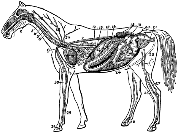
CIRCULATION AND DIGESTION
1 Mouth, 2 Pharynx, 3 Trachea, 4 Jugular Vein, 5 Carotid Artery, 6 Œsophagus, 7 Posterior Aorta, 8 Lungs, 9 External Thoracic Artery, 10 Left Auricle, 11 Right Auricle, 12 Diaphragm, 13 Spleen, 14 Stomach, 15 Duodenum, 16 Liver, upper extremity, 17 Large Colon, 18 Left Kidney and its Ureter, 19 Floating Colon, 20 Rectum, 21 Anus, 22 Bladder, 23 Urethra, 24 Small Intestine, 25 Cæcum, 26 Venous Supply to the Foot, 27 Posterior Tibial Artery, 28 Internal Metatarsal Vein, 29 Internal Metatcarpal Vein, 30 Posterior Radial Artery, 31 Metacarpal Artery, 32 Vertebral Artery, 33 Superior Cervical Artery, 34 Anterior Dorsal Artery.
—Before the single
simple elements were taken into the plant, they[22]
[23]
were of little value. The animal could not use
them for food, they could not be burned to furnish
heat, and they stored up no energy to carry on any
of the world’s work. What a change the plant
makes of them! So used, they become the source
of the animal food, and, as food, they contain five
principal groups with which the animal is nourished.
These five groups are the air, water, the
protein compounds, the nitrogen free compounds,
such as starch, crude fiber, sugar and gums, and
the fat or ether extract, as it is called.
Before these different constituents of the plant can be used as food for animals, they must be prepared for absorption into the system of the animal. This preparation takes place in the mouth, œsophagus tube, the stomach, and the intestines, aided by the various secretions incident to digestion and absorption. Any withholding of any essential constituent has its result in inefficiency or illness of the animal.
Withhold ash materials, for instance, from the food, or supply an insufficient quantity, and the fact will be evidenced by poor teeth, deficient bone construction and poor health in general. Let the feeding ration be short in protein, and the result will be shown in the flesh and blood. Let the carbohydrates and fat be withheld or supplied insufficiently, and energy will be denied and a thrifty condition will not be possible.
The supply of these different constituents in the proper proportion gives rise to the balanced ration; and is concerned in a treatise of this kind only in so far as it has to do with disease or health. For,[24] remember this fact: live stock are closely associated with right feeding. If foods be improperly prepared, or improperly supplied, or the rations poorly balanced, with too much of one constituent and too little of another, the effect will be manifest in an impoverished condition of the system. That means either disease, or disease invited.
Not only must these facts be considered, but other matters given recognition also. The greater part of the trouble of the stockman in the way of animal diseases is due to some disturbance of the digestive system, or to the water supply, or to ventilation, or to the use to which the animal is put from day to day. Attention to the details of digestion has its reward in thrifty, healthy stock; a lack of this attention brings trouble and either a temporary ailment or a permanent disease.
—Food is taken in the mouth, where it is masticated by means of the teeth, lips, cheeks, and the tongue. While the process of mastication is taking place there is being poured into the mouth large quantities of saliva, which softens the food and starts the process of digestion. The active principle of saliva is a soluble ferment, called ptyalin, that converts the starch of food into sugar. The amount of saliva that is poured into the food is very great, being often as much as one-tenth of the weight of the animal. This ferment is active after the teeth have been formed, which explains why it is not advisable to feed much starchy food to children before their teeth have begun development.
The food, after being ground and mixed with the saliva fluid, goes to the stomach. With the horse and hog the stomach is a single sac not capable of holding very large quantities of food; with the[25] cow and sheep, on the other hand, we find a large storehouse for holding food—a storehouse that is divided into four compartments, the rumen or paunch, reticulum, omasum, and the abomasum. The first three communicate with the gullet by a common opening. The cud is contained in the first and second stomachs, and, after it has been masticated a second time, it passes to the third and fourth, and to the bowels, where the process of digestion is continued.
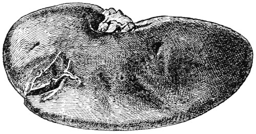
DISEASED KIDNEY
The kidney of the hog is pictured here. As a rule it is usually impossible to diagnose kidney troubles in hogs and similar lower animals.
—From this it will be noticed that chewing the cud is an act in the process of digestion; it refers only to rechewing the food so as to get it finer and better ground for digestion. While in the stomach the saliva continues the digestion of the starchy matter and is assisted by the gastric fluid that pours in from the lining of the stomach, which converts the protein or albuminoids into peptones. The fatty matter is not acted upon at this point. There are three constituents of gastric juice, which affect the changes in the food. These are pepsin, rennet, and acid. With rennet you are acquainted. It is used in the kitchen, in the making of cheese, and is obtained from the stomach of[26] calves or other young animals. Pepsin, also obtained directly from the stomach, is now a conspicuous preparation in medicine. The food, after leaving the stomach, goes into the bowels and is acted upon by secretions of the liver and pancreas or sweetbreads. It should be noted in passing that no secretion enters the first three divisions of the ruminant’s stomach. It is only in the fourth or true stomach that the gastric juice is found.
—While food is in the stomach it is subjected to a constant turning movement that causes it to travel from the entrance to the exit or intestines. When it passes into the small intestines it is subjected to the action of bile and pancreatic juices, which have principally to do with the breaking up of the fat compounds. Both resemble, to a certain extent, saliva in their ability to change starch into sugar.
The secretion of the bile comes from the liver and the pancreatic juice from the pancreas or sweetbreads, and both are poured into the intestines near the same point, so that they act together. The ferments they contain act in the following ways: They change starch into sugar, fat into fatty compounds, they curdle milk, and convert protein compounds into soluble peptones.
The process of digestion is finally ended in the intestines, where absorption into the system takes place. There is no opening at all from the bowels into the body, but the digestive nutriment is picked up by the blood when handed into the body from the intestines by means of countless little cells called villi, that line the walls of the intestines. These villi cells have little hair-like projections extending into the intestines, which constantly move; these protrusions, as they move about, catch on to[27] the digested nutriment, draw it into the cells themselves, where it is handed on to the blood, when it is later on distributed to all parts of the body. You can realize that an immense number of these absorption cells are present when the length of the intestine is considered. In the ox the intestine is nearly 200 feet long. After the nutriment is drawn from the food the undigested portions are voided periodically as feces or dung.

STOMACH OF RUMINANT
The four main divisions of the ruminant’s stomach are pictured here. The first three divisions are the store-houses for food until it is fully prepared for the fourth stomach or abomasum.
—Digestion, therefore, is a dissolving process; food is admitted to the system by means of cells. You remember that[28] all plant food first passes into a soluble state before it can enter the roots and be conveyed to the parts of the plants that require additional food for growth. In the case of plants the entrance is by means of the root hairs. In the case of the animal, entrance in the body is by means of the villi cells that line the intestines. From this we see that digestion is both an intricate and delicate process. Any loss of appetite, any disturbance of the digestion work, and any irregularity of the bowels bear decided results, one way or the other, to the rest of the system; and any disturbance of the body at other points, although having no direct relation to the digestion system, sooner or later affects the digestion and in so doing causes additional trouble.
Directly affecting digestion may be improper food, either liquid or solid; and over-exercise or not enough of it may prove troublesome, for exercise is clearly related to digestion. When the digestion process is disturbed, air or gas may accumulate in the stomach or bowels and give rise to colic or hoven. A watery action of the intestines, due to inflammation or irritation, may lead to dysentery and enteritis; or some obstruction like a hair-ball or a clover fuzzy ball, or the knotting of the intestines, may occur, temporarily or permanently impairing digestion so seriously often as to cause death itself.
As water in the plant is the carrier of plant food throughout the plant, so is blood the carrier and distributor of food in the animal. When food is absorbed, it either passes into the lymphatic system or into the capillaries of the blood system.[29] If in the former, it is carried to the thoracic duct, which extends along the spinal column and enters one of the main blood vessels. If collected by the capillary system, it is carried to the portable vein, thence to the liver and finally to the heart, where it meets with the blue blood collected from all parts of the body.
At this point, the blood contains both the nutriment and the waste matter of the body. Before it can be sent through the body again the waste material must be thrown out of the system by means of the lungs. This is accomplished by the heart forcing to the lungs the impure blood with its impurities collected from all parts of the body and also the nutriment collected from the digestive tract.
The chief organs, therefore, of the circulatory system are the blood and lymphatic vessels containing respectively blood and lymph. The only difference between these two materials is in the fact that lymph is blood without the red-blood corpuscles. The body, after all, really depends upon this lymph for nourishment, since it wanders to all parts of the body, surrounds all the cells in all of the tissues and in this way carries to the cells the very kinds of food that they need.
—The blood vessels have no openings into the body at all. In this respect the blood system is like the digestive system; it is separate and distinct in itself. The blood, however, does creep through the walls of the blood vessels. In so doing the blood corpuscles are left behind and lymph is the result.
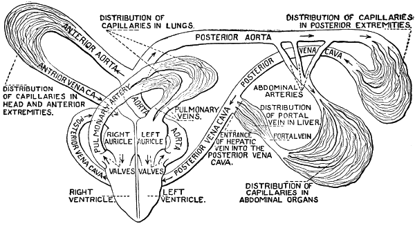
HOW THE BLOOD CIRCULATES THROUGH THE BODY
The center of the blood system is the heart. It
is the engine of the body. Going out from it is the
great aorta, which subdivides into arteries and[30]
[31]
farther away further subdivides until there is a
great network of little arteries; these in turn become
very tiny and take the name of capillaries.
Thus the red blood, by means of arteries and capillaries,
is carried to all parts of the body. This
plan of distribution would not be complete unless
some way were provided for the return of the blood
to the heart and lungs for purification. And just
such an arrangement has been provided. Another
kind of network collects this scattered blood at the
extremities into separate vessels, which gradually
increase in size and finally empty their possessions
into the heart. These are the veins of the body,
and have to do with the impure blood of the body.
—The power back of blood distribution is the heart. It is an automatic pump, as it were, that sends blood to the lungs and through the arteries to all parts of the body. The heart is divided into four divisions: the left and right ventricles and the right and left auricles. The right auricle receives the blood from the upper half of the body through a large vein and the lower half of the body through another large vein, and the blood from both lungs empties into the left auricle through two left and two right pulmonary veins. The large arteries of the heart which carry the blood from the heart to the different organs arise from the ventricle.
The blood always flows in the same direction. It goes into the auricle from the veins, and from this into the ventricle. It then passes into the arteries, then to the veins and then to the capillaries.
The action of the heart is very much like a force pump; the dark blood flows into the right auricle, which contracts; when this is done, the blood is[32] forced into the right ventricle; this in turn contracts and forces the blood into the lungs, where oxygen is taken on and carbonic acid gas and other impurities are thrown off. From the lungs the blood, now red and pure, passes into the left auricle and thence into the left ventricle, from which it is forced into the aorta to be distributed to all parts of the body.
We now see the close connection existing between the digestive system and the circulatory system. The digested food in the intestines is gathered in by villi cells. The question can now be asked, What do these cells do with this nutriment or digested food? They pour it into the absorbent vessels or lymphs, as they are called; these in turn empty the assimilated stores of food into larger and still larger vessels, which continues until the whole of the nutritive fluid is collected into one great duct or tube, which pours its contents into the large veins at the base of the neck, from whence it is carried into the circulatory system, the very basis of which is the blood.
The dark and impure blood, after returning to the heart, is sent to the lungs. It is, when collected from the body, just before being sent to the lungs dark, dull and loaded with worn-out matter. It must now be sent to the lungs, where it may be spread over the delicate thin walls of millions of vesicles, to be exposed to the air, which is inhaled by the acts of breathing. The blood gives off the broken-down material and carbonic acid gas very readily. It is both unpleasant and disagreeable, and the blood cells find it very unattractive.
[33]
The cells of the blood, however, have a great attraction for oxygen, consequently the cells absorb oxygen with greediness, so that when the blood returns to the heart it is fresh and bright and ready to take its journey back over the body again. This is done just about every three minutes. This endless round continues until stopped forever by death.
The relation existing between the animal and plant functions is brought to light in another way. When the plant was building tissue it released oxygen and exhaled it into the air. At the same time, by means of leaves, it gathered in the carbonic acid to use in plant building. Of course this was got from the air. The animal in performing its functions and in building its tissue inhales oxygen from and exhales carbonic acid gas into the air. Thus it is that animals take up what is unnecessary to the plant and the plant uses what is waste and poison to the animal.
[34]
When a colt is born the first and second temporary molars, three on each jaw, are to be seen. These are large when compared with the size of those that later replace them. In from five to ten days after birth the two central incisors or nippers make their appearance. In three or four weeks the third temporary molars appear, followed within a couple of months by an additional incisor on each side of the first two, both above and below. The corner incisors appear between the ninth and twelfth months after birth. This makes the full set of teeth—twenty-four in number.
There is now no change in number, although there is considerable change taking place all the time; the incisor teeth, in rubbing against each other, are more or less worn, giving rise to the expression “losing the mark.”
The two molars present at birth remain until the animal is about three years old, at which time they fall out of their sockets by the protrusion of the second set, or permanent molars.
This change from temporary to permanent teeth takes place usually without difficulty and without trouble. The permanent teeth push their way up from below crowding those in view. While this pushing and crowding is going on the temporary teeth are losing ground, for the reason their roots are being absorbed, and a time comes when the cap only is left attached to the gums. This cap drops[35] out and the new or permanent tooth soon is established in its place.
According to the observation of Mayo, the temporary incisors are replaced by permanent teeth as follows: “The two central incisors are shed at about two and a half years, and the permanent ones are up ‘in wear’ at three years. The lateral incisors are shed at three and a half and the permanent ones are up and in wear at four years. The corner incisors are shed at four and a half and the permanent ones are up and in wear at five.
“The molars are erupted and replaced as follows: The fourth molar on each jaw (which is always a permanent molar) is erupted at ten to twelve months; the fifth permanent molar at two to two and a half years, and the sixth usually at four and a half to five. The first and second molars, which are temporary, are shed and replaced by permanent ones at two to three years of age. The third temporary molar is replaced by a permanent one at three and a half years. In males, the canine or bridle teeth are erupted at about four and a half years of age. At about five years of age a horse is said to have a full mouth of permanent teeth.”
Horsemen make use of the “mark in the tooth” for determining the age between five and eleven. In examining teeth you observe that two bands of enamel are to be seen; one exterior, that surrounds the tooth, the other interior, which is termed the casing enamel. It is this latter, or “date cavity,” that is used to tell the age.
[36]
The mark in the tooth is occasioned by the food blackening the hollow pit. This is formed on the surface by the bending in of the enamel, which passes over the surface of the teeth, and, by the gradual wearing down of the enamel from friction, and the consequent disappearance of it, the age can be determined for a period of several years.
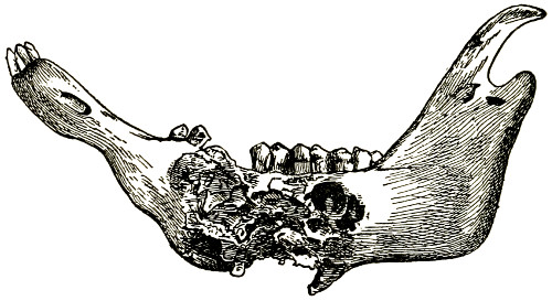
LUMPY JAW
The disease is caused by the ray fungus. The result is local tumors in the bones and other tissues.
When a horse has attained his sixth year the mark on the central or middle incisors or nippers of the lower jaw will be completely worn off, leaving, however, a little difference of color in the center of the teeth. The cement which fills the hole produced by the dipping in of the enamel will be somewhat browner than that of the other portions of the tooth, and will exhibit evident proofs of the edge being surrounded by enamel.
At seven years the marks in the four middle incisors are worn out and are speedily disappearing in the corner ones. These disappear entirely at the[37] age of eight; thus all marks are obliterated at this age on the lower jaw; the surface of the teeth are level and the form of the teeth changes to a more oval form.
The marks on the upper jaw are still present, since there has been less friction and wear on them. At nine the marks disappear from the central upper incisors, at ten from the adjoining two, and at eleven from the corner teeth.
To tell the age of the horse beyond this period is difficult and uncertain, except by those very much experienced in performing the undertaking. The shape of the teeth, the color and the condition all enter into the determination but there is no fast and fixed rules after the marks have disappeared.
Cattle have no incisor teeth on the upper jaw. They have eight incisors on the lower jaw. According to Mayo, the temporary incisors are as follows: “The central incisors or nippers are up at birth, the internal lateral at one week old, the external lateral at two weeks, and the corner incisors at three weeks old. They are replaced by permanent incisors approximately as follows, though they vary much more than in the colt: The central incisors are replaced at 12 to 18 months; the internal laterals at about two and a half years; the external laterals at three to three and a half years; and the corner incisors at about three and a half years. In the horned cattle, a ring makes its appearance at three years of age, and a new ring is added annually thereafter.”
[38]
Sheep, like cattle, have no incisor teeth on the upper jaw. Like cattle, they have eight incisors on the lower jaw when the mouth has reached full age. The change of the teeth occurs as follows: At birth the lamb has two incisors, followed by two more very soon. At the end of two weeks two more are out, making six incisors in all. At three weeks of age two more have appeared, completing the appearance of the temporary or milk teeth.
The permanent begin to replace the temporary teeth between one and one and a half years. The two central milk teeth are first replaced by two longer and stronger teeth. The lamb is now known as a yearling.
At two years the two teeth adjoining the central incisors are replaced by permanent ones; at three the two adjoining these are replaced, making now six permanent incisors.
Between four and four and a half the last two permanent incisors appear and the sheep then has a full mouth.
[39]
In purchasing farm stock, it is a good plan to deal with reputable people only. Leave the horse trader alone. He knows too many tricks, and if you are a stranger to him you can be pretty certain that he will try one on you—just for fun.
Fortunately farmers sell to strangers more frequently than they buy of them, and when they seek new stock they deal largely with breeders, who, like themselves, are farmers and not given to the tricks of low and disreputable methods; nevertheless, every purchaser of stock should be familiar with animal form and able to recognize defects and faults when he sees them. This is as much his business as to breed, raise or feed the stock on his farm.
Know what form you want; draft and speed represent different types, so do dairy and beef. With all classes of farm stock there are a few points that are desirable in all stock. One of these is width between the eyes. No animal of any breed or class possessed of a narrow forehead is at all perfect. A wide forehead is one of the absolute beauties.
These are desirable characters of all farm animals; they represent culture and refinement and good breeding. The purchaser or breeder, therefore, should not only know conformation, but he should know quality.
[40]
Our breeds of horses may be divided into three general classes. Those used for speed, those for draft and those with a mixture of the two—a general purpose sort of horse. The speed or trotting horse has its distinct type; it has been evolving and developing through a long series of years.
Briefly, its conformation may be described as follows: A wide forehead, fairly long head, a long neck that is thin and agile, a narrow chest as you look at it from the front, but very deep as you look from the side, long sloping shoulders, rather long back, a long horizontal croup, small barrel, fairly long forearm, long cannon bones and feet that are well shaped and perfect in every respect. Looking at the animal from the side it should be as high over the hips or higher than over the withers.
The draft horse, on the other hand, has a different conformation. There is not that elongation of his parts, although there is a symmetry of parts and of proportion. There should be the width between the eyes; the clean, neat face; a graceful neck, which should be shorter and more heavily muscled than that of the speed horse. The chest should be wide, both from the front and side, the back short but heavily muscled, the croup strong and not so horizontal as with the speed type, the quarters heavily muscled and the cannon bone short.
The feet should be as perfect as those of the speed horse. In both types the knee should be thick, deep, and broad and the hocks wide. The narrow hock is not so well able to stand heavy strain, consequently curb diseases readily follow[41] where the conformation shows narrow hocks. Another difference between the two types is found in the muscles. The speed type throughout has long, thin, narrow muscles—muscles that stretch a long way and contract quickly.
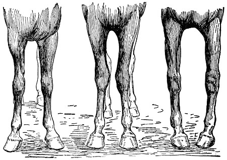
BAD ATTITUDES DUE TO CONFORMATION
In the first, the toes are turned out. The middle picture shows in-kneed attitude and the third shows in-turned toes. Whether standing or traveling, the appearance is unpleasant and mitigates against the value of the animals.
With the draft horse it is different: the muscles are shorter, but they are heavy; they are less quick in their action, but they are more powerful. In both types good proportions are always desirable. The width between the eyes should be as much or more than one-third the length of the head. The distance from the point over the shoulders to the ground should be about equal to the distance from the point over the hips to the ground; and in turn this distance, whatever it is, should be about equal[42] to the length of the horse from the point of the shoulder to the point of the buttock.
Looking at the horse in front if a line be dropped from the point of the shoulder it should halve the fore leg, the knee, the cannon, and the hoof. And the width of the third hoof, if placed between the two front feet, should give the attitude that is desirable.
Looking at the horse from the rear, the same attitude is to be observed. Of course, many horses do not possess these qualities and proportions; and because they do not is the very reason that their beauty, efficiency, and value are less.
In going into the stable look the animals over quietly. Observe how they stand, breathe, eat, and act generally. Are they nervous? Does one swing his head from side to side? Does he kick, paw, put back his ears, or does he have any of the other common stable vices that are unpleasant and undesirable? As you look about and pass back and forth, you will get the evidence of these stable vices, if such are to be found.
Look particularly for cribbing, wind sucking, kicking and crowding. Pawing is just as bad. If you want animals with good stable manners pass by those possessing these ugly faults. The next step is to examine the animals individually; those that “look good” to you. No doubt you will find some that do not interest you for one reason or another. These need no further attention, unless you have overlooked some fact, in which case your attention will likely be called to it.
[43]
In making the individual examination, go up to the animal in the stall, place your hand on the hip, and gently press it. If no stringhalt afflicts the horse, he will move over, allowing you to pass into the stall. The same applies to the cow. If well trained, she will make room for you by moving over at the same time, if you do this on the proper side, and she will put back her hind foot, as if she were about to be milked.
This casual observation would not be possible if force were used or the animal excited by loud commands or by a whip or strap. The halter teaches its lesson also. A heavy rope or leather suggests that the animal has a pulling back vice, a habit you want to avoid. Light halters for horses and cattle are to be preferred to chains, heavy leather, or ropes.
Now that you have seen all of the animals for sale, ask the owner to lead them out of doors for a more careful examination. In this you will inspect the animal very carefully in order to be certain of the conformation, defects, and blemishes, and to acquaint yourself specifically as to health and disposition.
Cast your eyes over the animal, front, side, and rear. Pass around the animal, keeping some distance away. By so doing you can judge of type and conformation, of proportions and attitudes; for each of these is important. A beefy-looking cow, with a thick neck, square body and small udder will not suit you for milk. Neither will a cow with a long, thin neck, open, angular body, thin thighs, and[44] heavy, deep paunch meet your needs if you are seeking breeding stock for beef production.
If you are examining a horse, keep in mind the purpose for which you are selecting. Remember the long, thin neck, very oblique shoulder, long cannon, long back, and long thin muscles are not adequate for draft. On the other hand, if you want a horse for road purposes, avoid the heavy muscles, the short neck, the heavy croup, and the heavy thighs. These mean draft—an animal for heavy work.
The milk cow should have a very soft, mellow skin, and fine, silky hair. The head should be narrow and long, with great width between the eyes. This last-mentioned characteristic is an indication of great nervous force, an important quality for the heavy milker. The neck of the good dairy cow is long and thin, the shoulders thin and lithe and narrow at the top. The back is open, thin, and tapering toward the tail. The hips are wide apart and covered with little meat.
The good cow is also thin in the regions of the thigh and flank, but very deep through the stomach girth, made so by long open ribs. The udder is large, attached well forward on the abdomen, and high behind. It should be full, but not fleshy. The lacteal or milk veins ought also to be large and extend considerably toward the front legs.
The beef cow is altogether different: she is square in shape, full and broad over the back and loins, and possesses depth and quality, especially in these regions. The hips are even with flesh, the legs full and thick, the under line parallel with[45] the straight back. The neck is full and short, the eyes bright, the face short, the bones of fine texture, the skin soft and pliable, and the flesh mellow, elastic, and rich in quality.
In other words, a beef cow is square and blocky, while the dairy cow is wedge-shaped and angular. The one stores nutriment in her body; the other gives it off. The one is a miser, and stores all that she gets into her system; the other is a philanthropist and gives away all that comes into her possession.
It will be seen, therefore, that the two types are radically different. This difference is due to breeding, not to feeding, nor to management. If you are seeking good milk cows, you must look for form and conformation. If you are looking for beef cows, you must also look for form and conformation, but of a different kind. With this knowledge to back you up and to guide you, you are now ready to make an examination of animals that will meet your purpose.
After making these general observations you are now ready to examine the animal. Begin with the head. How is the eye? Dull, weak, without animation? If so, be on your guard. The good eye shows brightness, intelligence, and it must be free from specks. By placing the hand over the eye for a few moments you will be able to detect its sensitiveness to light. Do you find any discharge of any kind from the eye? If so, some inflammation is present. Try to ascertain the cause.
—A large, open nostril is desirable. Look for that character first. Now[46] observe the color of the lining. To be just right, it should be healthy-looking, of a bright rose-pink color, and it should be moist. A healthy nostril is one free from sores, ulcers, pimples, and any unpleasant odor. Be careful here; an unscrupulous dealer can very easily remove discharges and odors by sponging and washing, and you may be deceived.

EWE NECK
The neck is one of the beauty points of the horse. In purchasing animals look carefully to conformation and quality. Let these also be guiding principles in breeding.
—Always look in the mouth; you have the tongue, teeth, jaws, and glands to see. Naturally, you, like every other person, consider the teeth first; you want to be certain of the age. This feature is discussed elsewhere in this book, and all in addition that needs to be said is in reference to the shape of the teeth,[47] whether or not they are diseased or worn away by age or by constant cribbing of the manger. Of course these facts you will think of as you examine the mouth.
Give the tongue a second of your time. If it is scarred and shows rough treatment a harsh bit is likely the cause, due to its need in driving and handling.
Then give a thought to the glands while here. Enlarged glands may indicate some scrofulous or glanderous condition of the system.
—A beautiful neck and throat is an absolute beauty in the horse or cow. The skin should be thin, mellow, and soft, and the hair not over thick nor coarse. Look for poll-evil at the top of the neck and head. See if swellings, lumps or hard places are to be found at the sides of the neck, or underneath joining the throat. I have found such very frequent with dairy cattle; and cases are not unusual with horses.
Frequently scars are to be found on the sides or bottom of the neck. These may be due to scratches caused by nails, barb-wire or some similar accident, and again they may have been caused by sores, tumors, or other bad quality of the blood.
—Passing the side, look over the withers for galls or fistulæ, the shoulders for tumors, collar puffs, and swellings. Observe at the same time if there is any wasting of the muscles on the outside along the shoulder.
Now the back. Is it right as to shape? Do you find any evidence of sores or tumors? Look for these along the sides and belly. Now stoop a bit and look under; do you find anything different from what is natural? In males look for tumor or disease of the penis; do the same with the scrotum,[48] and, in case of geldings scrutinize carefully to see if they be ridgelings.
While making this examination, if the animal is nervous and fretful, you can help matters along if an assistant holds up a fore leg. Take the same precaution when examining the hind quarters and legs. By doing so, you will avoid being kicked and can run over the parts more quickly and satisfactorily.
Before leaving the body observe if the hips are equally developed, and the animal evenly balanced in this region. Both horses and cattle are liable to hip injury, one of the hips being frequently knocked down. Make sure that both are sound and natural.
—Now step to the front again for a careful examination of the front legs and feet. Starting with the elbow, examine for capped elbow; now the knee. It should be wide, long, and deep, and at the same time free from any bony enlargements. The knees must stand strong, too. Is the leg straight? Do you observe any tendency of the knee to lean forward out of line, showing or indicating a “knee sprung” condition? Just below the knee, do you find any cuts or bunches or scars due to interference of the other foot in travel? Look here also for splints; follow along with the fingers to see if splints are present—on the inside of the leg.
Be particular about the cannon. The front should be smooth—you want no bunches or scars. Just above the fetlock feel for wind puffs; and note if about the fetlock and pastern joints there are any indications of either ringbones, bunches, or puffs. Now look for side bones; if present, you will find them just at the top of the hoof. They may be on either side. Sidebones are objectionable, and are[49] the lateral cartilages changed into a bony structure.
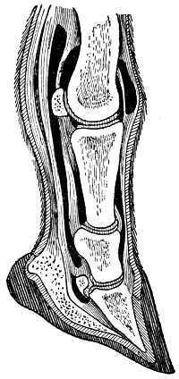
ANATOMY OF THE FOOT
The delicate nature of the foot is readily recognized when the various parts are considered in their relation to each other.
Give the foot considerable attention. The old law of the ancients, “no feet, no horse,” is certainly true in our day. You can overlook many other imperfections and troubles in the horse, but if the feet are bad you do not have much of a horse. A good foot is well shaped, with a healthy-looking hoof and no indication of disease either now or ever before.
See that the shape is agreeable. A concave wall is not to be desired, and the heels are not to be contracted. The wall should be perfect—no sand cracks, quarter crack, or softening of the wall at the toe of the foot.
—These are both troublesome and cause much lameness. A healthy frog, uninjured by the knife or the blacksmith or other cause is very much to be preferred.
—In examining these regions give the hocks of the horse special attention. No defect is more serious than bone spavin. You can, as a rule, detect this by standing in front of the horse just a little to the side. If there is[50] any question about the matter, step around to the other side and view the opposite leg. This comparison will let you out of the difficulty, as it is very unusual that this defect should be upon both legs at the same point and developed to the same degree.
A spavin is undesirable for the reason that it often produces serious lameness, which frequently is permanent. As it is a bone enlargement, it is something that cannot be remedied. If you are seeking good horses, better reject such as have any spavin defect.
In this same region between the hock and the fetlock curbs troubles are located. They appear at the lower part of the hock, directly behind. You can readily detect any enlargement if you will step back five or six feet. The curb, while it may not produce lameness, is altogether undesirable. It looks bad; it shows a weakness in the hock region and often is caused by overwork, consequently the animal with curb disease is one that has not measured up to the work demanded of him.
Just above and to the rear of the hock the thorough-pin disease appears, and just in front of and slightly toward the inner side of the hock bog spavin is sometimes to be found. Lameness may come from either of these diseases. Small tumors, puffs and other defects frequently show themselves on the hind legs and the best way is to reject animals having them. While some of these may be caused by accident, the most of them are the result of bad conformation, due to heredity, unimproved blood and bad ancestors.
Lameness comes from many causes; maybe from[51] soreness, from disease or from wounds. And lameness is hard to detect. Frequently it seems to be in the shoulder, when in fact it is a puncture in the foot. Again it may seem to be in the fetlock, but the trouble is in the shoulder or fore leg. You must examine for lameness both in the stable and out of the stable. If you find the horse standing squarely upon three feet and resting the fourth foot, you should be suspicious. If you move the horse about and he assumes the same attitude again and still again, you can be certain that he is assuming that position because he wants to rest some part of that member.
In testing out the horse for lameness, let no excitement prevail. Under such excitement the horse forgets his lameness or soreness for the time being, and you do not note the trouble. A quiet, slow walk or trot on as hard a road as possible is a desirable sort of examination to give.
The free breathing of a horse may be interfered with, and for two reasons. Roaring or whistling, as it is called, is a serious disease of the throat, and, at the same time, an incurable disease. The second disease is known as heaves or bellows, and is also a most serious disease, because it is also incurable. By the use of drugs relief may be given temporarily, but no permanent cure follows. Unscrupulous dealers will resort to dosing for the time being, or until a sale is made.
You should guard against this trouble, however, for it is one of the most serious that a horse can have. Upon this subject, Butler has the following to say: “To test the wind and look for two serious conditions and others which may be present,[52] the animal should be made to run at the top of his speed for some considerable distance—a couple hundred yards or more. Practically this run or gallop should be up hill, which will make the test all the better. After giving the horse this gallop, stop him suddenly, step closely up to him and listen to any unusual noise, indicating obstruction of the air passages, and also observe the movements of the flanks for any evidence of the big double jerky expulsion of the air from the lungs characteristic of heavers.”
No examination is complete that does not make a test of the paces. You want to know how fast the horse can walk, how he trots or paces or how he takes some other gait. Some horses make these movements very gracefully; others very unmannerly. A well-acting horse is one that moves smoothly, regularly, who picks up his feet actively and who places them firmly in their position regardless of the ground or gait. Some horses have a rolling movement of the legs. Avoid these. Others step on the toe or heel. These, too, should be avoided. They suggest some defect or bad conformation.
The testing of the paces brings all parts of the body into play and assists in catching other blemishes or defects that you may have overlooked in your previous examination. It gives you another opportunity to examine the wind, to observe the respiration, the heart beatings, the condition of the nostril after work; it shows you also how the animal takes his pace and how he stands. All of this will be of value as indicating the soundness and health of the individual under observation.
[53]
Now, as a last factor of your examination, consider the uses to which the animal is put. If you are looking for breeding animals be sure to know that the udder is not injured. Of what use is a cow with a bad udder? How often do we find a quarter of the udder destroyed or a teat cut or so badly mangled as to be of little use! Some udders are dead, heavy, fleshy; some are diseased, lumpy; and even though the animal is otherwise good you must reject her.
If the udder is good, superior in many respects, and shows great milk production, you can often afford to overlook other defects, especially if the result of accident.
In the case of horses, a disease or blemish due to accident may be overlooked, if the work to which the animal will be subjected does not interfere, let us say, for breeding purposes. The horse has good conformation, good quality, is healthy and very superior, but unfortunately a leg was broken. Shall she be rejected as a breeder? No heavy work will be required of her—she is wanted for colt raising. Take her; of course you will pay less for her. This accident interferes in no way with her value for breeding purposes. Many cases of accidental injuries are similar to this example among cattle and horses.
A good rule is to reject those having defects or blemishes that interfere with functional activity or the work to which you wish to put them. Then, as breeders, reject all with constitutional defects, as bad feet, narrow hocks, coarse disease-appearing bones, and bad conformation and scrubby character.
[54]
FRACTURES
When a bone is broken into two or more parts it is said to be fractured. These may be straight across, up and down, or oblique. Ordinary fractures are easily treated by splints, but sometimes fractures are so serious as to destroy the value of the animal.
The stockman has all sorts of wounds with which to deal. He may guard his animals with the care and caution of a mother and still find constant bother and worry to face in the daily management of his stock. Today it may be a wound caused by a nail puncture in the foot; tomorrow a cut occasioned by a fence; and then almost immediately another, the result of a kick or a hook; with patience nearly exhausted, now follow bruises of many sorts and unexplainable lacerations.
These troubles occur on the best managed farms. There is but one thing to do: meet each case as it occurs and lend such assistance as you can that nature may repair the wrecked tissue at the earliest possible moment.
[55]
Wounds fall into four classes: the clean-cut kind made by something sharp; the torn or lacerated, where ragged edges are left; the bruised, the result of continued pressure or kicks or a knock; and the punctured, like the entrance of a nail or splinter or gunshot.
The latter class is the most difficult in treating, for the reason of the greater penetration that may likely occur. In the case of gunshot, the wound may be on the surface, or it may extend entirely through the region attacked, or even penetrate some vital organ like the heart or the lungs or bowels, and either immediately or within a few days be the cause of death. Fortunately such wounds are rare. The stockman may never have to deal with them at all. There are punctured wounds that are common, however; some, indeed, frequently lead to death. A nail wound is the most serious, perhaps. It is likely that more cases of tetanus or lockjaw are due to nail punctures than to all others combined.
After this class comes the lacerated kind. These heal slowly; the tissue being torn and bruised is repaired only through the sloughing off of the injured and now superfluous parts. As a result, even with the most attentive surgical help, the injured part develops its exposed sore, ending finally completely healed, but permanently marked. Bruises may be equally bad, long delayed in healing and very painful. Do you remember the stone bruises of boyhood days? How long it required to develop! And the pain! I shall feel mine for ages to come.
[56]
The clean-cut wounds, if not too serious, are the least difficult in treating.
The flow of blood is usually associated with ordinary wounds; other than with some bruised and punctured wounds this is always true. Frequently a nail puncture gives off no blood or it is not noticed. However, the blood is present, for, from the very nature of the trouble, blood rushes to the seat, this being nature’s way of repair. Your first step, therefore, is to check the excessive blood flow.
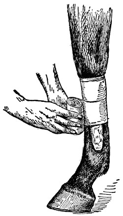
BANDAGING A LEG
The method of applying the bandage is shown here. The bandage may be wrapped directly over the hair or over cotton saturated with an antiseptic and placed over the wound.
If left to itself the blood might do it. Blood has the trick of coagulating or clotting; and this in time will check the flow. But you can assist in forming the clot very simply by applying some finely ground material that the blood may be held on the spot. Absorbent cotton is the best material to use. In case this is not available, use something of like nature—something that is clean, not stored up with germs. Tea is good, as is flour also. Cold water acts favorably, and for the slight, ordinary surface wounds water is usually sufficient. A few drops of some antiseptic in the water, if available, is always advisable, for the freshest water carries its full quota of germs, some of which may cause trouble. A tiny bit of alum powder will be found both effective and not painful.
—After the flow of blood has been stopped, cleansing the wound is next in order. All dirt should be carefully removed, the injured flesh cleansed, the torn tissues brought together and stitched, if need be, and antiseptics applied. The water used in bathing the wounded flesh should contain an antiseptic, that the germs[57] present may be destroyed and no live ones admitted by water in cleansing the wound. Any good commercial antiseptic will do; or the old common ones, like corrosive sublimate, one part in a thousand parts of water, or carbolic acid, a teaspoonful in a quart of water. Some powdered antiseptic like iodoform is very desirable for dusting into the wound.
—Unless the wound is of little consequence it should be covered and bandaged that no foreign elements be admitted and that some pressure may be given to keep the broken parts together. To secure this effect absorbent cotton, slightly moistened with the antiseptic, should be laid on the wound, and firmly fastened by strips of clean cotton cloth.
By winding this bandage around and about the wound, dressed in this careful way, the wound will be protected, germs will be kept out and nature, thus reinforced, will be enabled to make a rapid recovery. Unless the bandage is disturbed in some way there is no need of changing it under twenty-four or thirty-six hours. If, for any reason, the bandage is displaced, dress as before, and bandage again.
[58]
—When a cut wound is deep or large, stitching is sometimes required, that the broken parts may be brought together for more rapid healing. Nothing is better for this than a coarse needle and heavy thread. Before stitching, however, the wound should be bathed as previously described. The needle and thread should be soaked in the antiseptic, that no germs may be introduced by means of them.
Now you are ready to make the stitches. Place the needle about an eighth to a quarter of an inch from the edge of the wound across to the opposite side. Bring the two ends together and tie, leaving the lips of the wound as close together as possible. If more than a single stitch is necessary, proceed in the same way, placing the second stitch about three-quarters of an inch from the first one; continue as with the first stitch if more are necessary.
In case a needle and thread are not available, pins may be used in the emergency. Insert the pin through the two edges and bring the lips together, making them fast by a thread or cord carried from one end to the other several times, alternating to the right and left as presented by the figure eight. Sometimes the wound enlarges and becomes feverish. If such becomes very severe, remove the fastenings and bathe the wound very gently, using a mild antiseptic wash of tepid water in which carbolic acid has been placed.
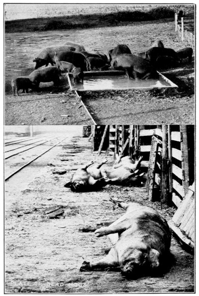
HEALTH AND DISEASE
In the upper picture the pigs are treating themselves. Below are shown hogs which died during shipment to market.
Avoid any breaking of the healing tissue and do not have the washing solution too strong, else it may injure the delicate tissue growth. A teaspoonful of carbolic acid to a quart of water is strong enough. With lacerated wounds the treatment is very similar. If the wound goes bad and becomes spongy add a tablespoonful of acetate of lead and[59] a tablespoonful of sulphate of zinc to the antiseptic solution and apply twice daily.
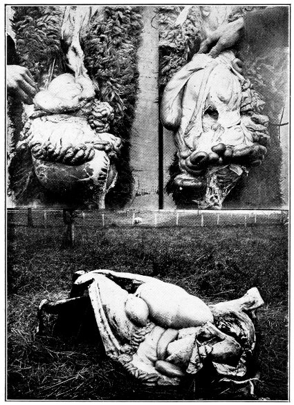
MAKING POST MORTEM EXAMINATIONS
The upper right hand picture shows the intestines of a healthy sheep. On the left nodule disease is discovered. The bottom picture illustrates how a carcass may be opened for the examination.
—These very frequently cause trouble. You have no way of observing the wound and your only way of judging is from the way the animal walks or acts, and if the hoof is unduly hot. Locating lameness in the stifle joint is a common but inexcusable error, as the action resulting from lameness in the two parts is entirely different. The so-called gravel which is said to enter the sole of the foot and then to work out at the heel is usually the working out of the pus or the matter resulting from a nail puncture or a bruise.
If an animal becomes suddenly and severely lame and there be no evidence of any injury to any other part of the leg, such as swelling, heat and pain upon pressure, it is always well to look for puncture in the foot. If the animal stands with the lame foot extended and when walking places the lame foot well forward and brings the well foot up to it, the evidence of puncture is still stronger.
To examine the foot properly the shoe should be removed. It is not sufficient to merely scrape the bottom of the foot clean, for if the nail has pulled out and the horn sprung back in position, all trace of its entrance may have been obliterated. To examine the foot properly, tap the hoof with a hammer or knife and the exact spot may be definitely located. If the injury is of a few days’ standing, additional heat in the hoof and, perhaps, slight swelling of the coronet may also be present.
In treating such wounds, pare away only such parts of the hoof as necessity requires and introduce a bit of cotton cloth rolled as a string by means of a probe of some kind. Both probe and cotton must be treated with the antiseptic solution.[60] This solution should be a little stronger than for flesh wounds. Make the solution by using a teaspoonful of carbolic acid to only a pint of water. After the cotton has been inserted a few times and withdrawn, each time a fresh cord being used and fully saturated, leave the last one in for a few hours and then repeat the treatment. This should be done three or four times each day.
The main point in the treatment of nail puncture of the foot is to provide free exit to all matter that may collect and keep the parts as clean as possible. If this be done, the matter will not be compelled to work out at the heels, and no separation or loss of hoof will occur. Often a very severe wound is made and the treatment acts slowly.
In case proud flesh accumulates, it should be burned away by a hot iron. After this operation has been performed, the cavity should be filled with balsam of fir and cotton placed over it, a piece of heavy leather fitted to the foot and held fast by the replaced shoe. This will usually end the difficulty. A veterinarian should be called in case the wound is severe or goes bad as the treatment progresses.
—In treating bruises a different procedure is necessary. The broken tissue is concealed—beneath the skin and usually under the surface muscles. Bathing with water and acetate of lead—a quart of water and two tablespoonfuls of the acetate—will tend to lessen the inflammation. In time you may have to open the swelling for the pus to get out. After doing so, inject some wash for cleansing, using one quart of water and a tablespoonful of chloride of zinc.
If the swelling remains, apply twice each month a salve made by using one teaspoonful of biniodide[61] of mercury and three tablespoonfuls of lard. Wash occasionally, using the chloride of zinc solution.
—Cleanse the wound with a wash composed of one tablespoonful of acetate of lead, one tablespoonful of sulphate of zinc, four tablespoonfuls of tincture of arnica and one quart of water. Use this wash frequently, every hour or so, during the first day. After that three or four applications will be sufficient. The sore should be kept lower than the skin during the healing process. If it tends to crowd up, apply a tiny bit—as much as you can place on a one-cent piece—of bichloride of mercury. This will assist in getting an even heal and the skin will grow over, leaving no blemish or swelling.
—If the wound has been treated as suggested above there is no possibility of any trouble from maggots. These come from a lack of cleanliness and neglect. Of course, an animal often gets a wound and the owner is not aware of the mishap. Wounds, more or less infrequently treated, those made as the result of castration, occasionally get infected with maggots.
When, for any cause, maggots are present, they must be got rid of at once. A good plan is to use chloroform, either by spraying or by throwing it in the wound in small drops from a sponge.
The danger from maggots can usually be avoided if a mixture composed of one tablespoonful of turpentine, three tablespoonfuls of tar and two tablespoonfuls of lard or fish oil be smeared all around the border of the wound.
[62]
Even on the best-managed stock farms some animals do get sick and die. Good care and good nursing may be given, but the sick animal frequently does not recover—death often follows very quickly, before you have an opportunity to observe the development of the disease or to secure the services of a veterinarian. Then, again, after a lingering sickness an animal dies, the disease being known or unknown as the case may be.
In any event, a post-mortem examination is usually desirable, if for no other reason than that it serves to familiarize you with the organs of the body. With a little experience you can become quite proficient in examining a dead animal, and you can soon learn the difference between healthy and unhealthy organs, between diseased and normal tissues and the relation of the internal parts to the whole body. A post-mortem examination thus enables you to know the cause of the disease—where it is located or whether death is the result of accident or of some fatal disturbance of the system.
This examination should be made as soon after death as possible; the longer the delay the greater the changes due to decomposition of the body and its decay back to the original elements from which it has come. Soon after death the stiffening process takes place. This is known as rigor mortis. It may occur within an hour after death and again it may not be complete until twenty-five or thirty[63] hours have passed. Soon after the death stiffening has occurred the tissues soften and decomposition rapidly follows.
In making a post-mortem examination, in case the animal has not been moved, the position of the body is to be observed. Look all about you. Is there any evidence of a struggle? Does either the body or the ground appear as if spasms have taken place? It may be a case of poisoning. If such be true, the outward appearance may be further substantiated by the internal condition. If inflammation and irritation of the stomach and bowels are observed, this evidence helps to confirm the first observation.
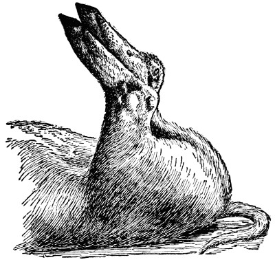
RICKETS IN PIGS
Rickets in pigs is due, as in man and other animals, to an improper development of the bone, the result of insufficient mineral matter in the food. The bones are weak and bend or break. It frequently appears after the pigs are weaned. An abundant supply of wood ashes, charcoal, lime and salt is always good for hogs.
[64]
The appearance of the struggle, however, is not enough to establish a case of poisoning; for struggling is a death characteristic of many diseases. Of course, in making this preliminary examination you will note if death could have been the result of some other reason. Has some obstruction had anything to do with the trouble? Maybe the animal has been caught in some way and not being able to move about has starved to death, or maybe some over-exertion has had something to do with the trouble.
Many animals choke, and, not being able to relieve themselves, die. Thousands of farm animals, especially in the West and Southwest, die annually from cold, and not a few from heat. All these things enter into the case and must be considered in reaching a reasonable conclusion.
—The next thing to do is to observe the discharges from nose, mouth and other natural openings of the body. External scars and wounds often bear a close relation to the disease and these should be considered in examining the carcass. How do the eyes look? Is there a discharge from the ears? Is the swelling of the abdomen and the bloating more pronounced or different than should be the case in ordinary death? Practice will indicate the lesson that each of these teach.
—Farm animals are often killed by stray shots from the guns of hunters and trespassers. A casual observation will indicate if death has been due to this. Again, animals may die from distemper or be eaten up with lice or troubled with itch or mange—you will note these facts as you go along with your work.
[65]
In the South, where Texas fever is so prevalent, you should look for ticks, as these bring death to thousands of animals each year. Look for the wee tiny ones—they cause the trouble. When cattle are fairly covered with the large ticks death does not ordinarily follow, since the animal has practically become immune to the poison caused by the tick. These large ticks, however, are filled with blood and nutriment, both obtained from the animal, and hence they may rob the animal of blood and nutriment that it ought to have itself.
—The skin is now to be removed, so that the color of the tissues and the nature of the blood may be noted. If the blood be thin or black, with a disagreeable odor, you can expect some germ trouble like blood poisoning or an infectious and contagious disease. If the white tissues are yellow you may be reasonably certain that the liver has not done its work as it would have done had it been in a thoroughly healthy condition.
In removing the skin and making other observations be cautious that you do not prick your fingers with the knife, since you may convey in this way disease to yourself. If by accident a cut or prick is made, cauterize the wound at once, so as to destroy any germs transmitted in this way to you.
The next step is to examine the internal organs. To do this, place the animal on its side, remove the upper front leg and the ribs over the chest region. The ribs should be removed as near as possible to the backbone so as to give an unobstructed opening over the important organs. This large opening[66] now allows you free access for examination, and an unimpaired view all about the vital organs, if these are entirely exposed.
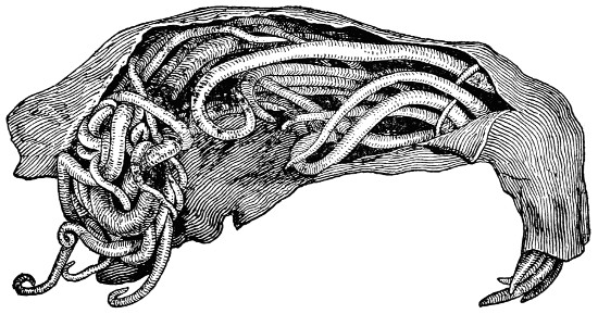
ROUND WORMS IN HOG INTESTINE
An infestation with intestinal worms, as shown here, leads to unthriftiness and a loss of flesh. These worms may be expelled by giving turpentine in doses of one teaspoonful in milk for three days in succession.
While making this opening, observe the watery fluid as it escapes. If a large quantity is present, dropsy or a rupture of the bladder is indicated. If the trouble is due to the latter, an odor in the urine will be quickly noted. When the fluid is red in color, it indicates the presence of blood or some inflammation of the abdomen or the bowels. A large amount of watery fluid in the chest cavity is an indication of some lung trouble; this is further indicated by the tiny attachments running between the lungs and the chest wall.
—If the stomach and intestines be abnormally red, congestion is indicated, and if they be quite dark, even purple in color, you may be sure that some kind of inflammation has[67] been the trouble. You will note also if the stomach is hard and compacted; and, if so, indigestion may have been the trouble. The intestines will also show if they be hard and compacted or in any otherwise bad condition. Pass the hands along to see if the intestines are knotted in any place or if nails are present in the stomach. It is not likely that the nails have been the direct cause of death, but this fact helps to indicate the condition of the digestion trap.
Often hair balls or parasites will be found; either may clog up the channel and may be the immediate cause of death. I have on more than one occasion found that the fuzz of crimson clover, accumulating in the intestines of horses, rolls up into a hard, compacted ball, and not being able to pass out, becomes an obstruction in the passageway and ultimately causes death.
—The urine tells its tale also; a very disagreeable odor indicates some disturbance; and a brownish or dark-red color may indicate a local disease or a constitutional breakdown. Texas fever in cattle produces a very dark or reddish urine, Azoturia in horses, a similar color. Gallstones or gravel are often found in the bladder, and these frequently cause serious disturbance, if not death.
—Look the lungs over carefully. See if the natural color is present and if the soft, spongy constituency responds to the same kind of touch as does the thoroughly healthy lung. In health the lungs are a very light pink color. If inflammation has been present this will be indicated by the dark color and the hard density.
When the lung is cut apart with the knife further observation should be made. A marble appearance[68] indicates inflammation and hard lumps or tubercles indicate tuberculosis. These tubercles, when cut open, show pus and a cheeselike material, yellow in color—a true indication of the disease.
—You should feel the heart to know if it is natural or not, or to see if any of the valves are broken, or if some inflammation has been back of the trouble. The sides of the open cavity should be observed before leaving. Is it spotted, speckled? Are pink spots seen about the ribs? This is an indication of hog cholera, and in itself may lead to a correct interpretation of the disease.
[69]
The common medicines used in treating farm animals are named in the following list, together with origin, action, use, and dose.
Tincture of aconite is derived from the root of a plant. When used, the heart beats more slowly and the blood pressure is decreased, making the medicine desirable in cases of inflammation.
Dose: For horses and cattle, from 10 to 30 drops, and sheep and hogs 5 to 10 drops.
This is usually bought in a powder form. It is brown in color and bitter in taste. Considerable time transpires before action in the bowels takes place. Allow at least 24 hours. It is a physic and blood purifier.
Dose: For horses, 4 to 5 tablespoonfuls; cattle, 4 to 8 tablespoonfuls; sheep, 1 to 2 tablespoonfuls; and pigs, 1 to 2 tablespoonfuls.
This mineral salt is used in washes for sore mouth and throat, and cleansing wounds. It may be dusted into wounds in powder form, and is both drying and healing.
Dose: Use a tablespoonful to a pint of water.
[70]
This preparation is made from dried berries and ground. It stimulates digestion, sweetens the stomach, and serves as a tonic and appetite maker.
Dose: For horses and cattle, a tablespoonful, and for sheep and pigs, a teaspoonful.
For wounds, sprains, and bruises, tincture of arnica is both cooling and restful. It is made from the dried flowers of a plant, and is for external use. Apply three or four times daily.
This medicine comes from the mineral kingdom and is very powerful. In using better get it in some standard medicinal form such as Fowler’s Solution. It is used as a tonic when the stomach is bad and the system run down.
Dose: Fowler’s Solution; for horses and cattle, 2 tablespoonfuls; sheep, 1 teaspoonful; pigs, one-half teaspoonful. In giving to stock mix with 4 tablespoonfuls of whiskey, and either use as a drench or add to mash or gruel.
This is a tincture made from a plant. When used it soothes, softens, and relaxes the parts to which applied. It checks inflammation and relieves pain, but must be carefully used.
Dose: For horses and cattle, 1 teaspoonful; sheep, 10 drops; pigs, 5 drops.
[71]
This preparation, taken from a mineral, is not used internally. It is a powerful caustic. Its principal use is for curing thrush in horses’ feet.
TETANUS BACILLI
How the germs look under the microscope. The poison produced by them is one of the most violent known in disease.
This comes as a white crystal or powder, and is used to quiet the nerves when some trouble like lockjaw has set in.
Dose: For horses and cattle, 1 teaspoonful; sheep and hogs, one-half teaspoonful.
This bright red powder is used chiefly for blistering purposes. It is excellent when a spavin or[72] splint or ringbone is just beginning. In preparing, use one part of the mercury to nine parts of vaseline or lard. Remember, it is a poison, and must be carefully handled, as is true of some other preparations of mercury.
The camphor of commerce is in the form of a gum obtained from a tree by boiling and evaporation. It is used in mixtures for coughs, sore throat, and heaves. It is good also for colic and diarrhœa and assists in lessening pain. It should be given in water.
Dose: For horses, 2 to 4 teaspoonfuls; cattle, 4 to 5 teaspoonfuls; pigs and sheep, 2 teaspoonfuls.
This is in the form of powder, and is an irritant. For use it should be thoroughly mixed with lard or vaseline. One teaspoonful of the cantharides to 4 tablespoonfuls of lard or vaseline. When so prepared it is excellent as a blister. It can be applied for sweat thickenings or lumps on any part of the body that is not on the bone. It should not be used on curbs or tumors and is not used internally.
This is got from coal tar and petroleum. When full strength and pure it is in the form of crystals, but is generally bought as a liquid. It is a disinfectant and an antiseptic, and while used internally for some purposes, is largely used internally in washes and solutions. Its principal use is in bathing[73] wounds and sores. Care should be taken not to have a wash contain too much of the acid, as it will burn the wound and stop the healing action. It is a corroding poison taken internally. It should be just strong enough to kill bacteria; say, 1 part to 1,000 parts of water. A very good healing salve is made when 5 drops of pure carbolic acid is used to 4 tablespoonfuls of vaseline.
This is used externally as an antiseptic and disinfectant. Dissolve 1 part to 100 parts of water. It is a preparation of mercury, is poisonous, but excellent for bathing wounds and open sores.
This oil is pressed from castor beans. It is a mild physic similar to raw linseed oil. It is not used much for live stock.
Dose: For horses and cattle, 1 pint; for sheep, one-quarter pint, and for pigs, 4 tablespoonfuls.
This is a heavy white powder and a mineral. Its principal action is as a physic, and it has a cleansing effect on the liver. Hence it is used for all kinds of liver troubles. When dusted in old sores, it is splendid for healing and drying up.
Dose: For horses, one-half to 1 teaspoonful; cattle, 1 to 2 teaspoonfuls; sheep and pigs, one-eighth teaspoonful.
This oil is made from seeds, and is one of the most powerful physics known. It should never be[74] used until milder physics do not respond. Use it as a last resort.
Dose: For horses, 15 to 20 drops; cattle, 30 to 40 drops; sheep, 5 to 10 drops; and pigs, 2 to 3 drops. In giving, it is best to use in connection with raw linseed oil; of the linseed oil use 1 pint for horses and cattle and one-quarter pint for sheep and pigs.
This chemical is most easily used when purchased in pencil-like sticks. It is never given internally, but is used to burn warts and growths by wetting the stick and rubbing it over them. It is also used for burning poisonous wounds to kill the poison. It is commonly employed for dishorning calves. When a week or ten days old, and the button of the horn is just appearing, rub the potash over the horn. This usually insures destruction of the horn substance. Wet the stick of potash. See that drippings do not run down the animal’s head. In order to protect the fingers, when using, wrap paper around the stick.
This is the product of coal tar and comes in the form of a thick, dark fluid, and, like tar, is harmless. It is frequently used as the basis of salves for wounds, scratches, and like troubles. It is a very effective remedy for killing lice, ticks, or fleas, and is used as a remedy when sheep are afflicted with mange and scab.
Dose: Use from 2 to 4 tablespoonfuls to a pint of water and shake well before using. Make up a small quantity at a time, as creolin thus made loses[75] its value after exposure. For disinfecting purposes, 1 part of creolin to 100 parts of water is satisfactory.
This is the root of a plant, dried and ground. It is used principally as a tonic, and is very bitter; commonly found in condition powders and is given to animals that are weak and run down. If used alone, give twice a day in the food and place on the tongue with a spoon.
Dose: For horses and cattle, 1 tablespoonful; for sheep, a teaspoonful; pigs, one-half teaspoonful.
This is a dried root ground fine, secured from a plant, and acts as a stimulant, relieving gases that accumulate in the stomach. It is an excellent ingredient to use in colic and indigestion preparations. If given alone, doses may be repeated every two or three hours.
Dose: For horses and cattle, 1 tablespoonful; sheep, 1 small teaspoonful; pigs, one-half teaspoonful.
This salt is frequently used in combination with gentian, equal parts of both, and in other recipes for condition powder. It cleans the blood and builds up the system after weakening diseases. A common preparation is made by using one-half of powdered gentian and one-half of hyposulphite of soda. Mix all together and give two or three times a day to the animal needing it.
[76]
Dose: For horses and cattle, 1 tablespoonful; sheep, 1 teaspoonful; pigs, one-half teaspoonful.
This dark brown tincture is not often used internally, but is used as a sweat blister and for blistering thickened glands. In using, take a feather, painting the iodine on the lump until it blisters; when the blister appears, grease the part; after two or three days have passed, wash the lump with warm water and soap and blister again.
This white powder is obtained from the mineral kingdom. When given internally it acts as an absorbent. It is commonly used in cases of dropsy of the belly. In administering, use equal parts of ground gentian root and give twice a day.
Dose: For horses and cattle a teaspoonful; for sheep and pigs, one-half teaspoonful.
This oil is obtained from flaxseed, and is excellent when a mild physic is desired. The easiest and most effective way of giving to animals is in the form of a drench. About 1 pint should be used for horses and cattle. Raw linseed oil is usually preferred to the boiled.
This is made from opium and is used both internally and externally. It is commonly used[77] where there is pain, hence it is excellent for relieving pain and spasms and assists also in checking inflammation.
Dose: For horses and cattle, 4 to 6 teaspoonfuls; sheep and pigs, 2 to 4 teaspoonfuls.
This powder comes from ground seeds, and is used as a nerve stimulant. It is very efficacious for strengthening weak, debilitated animals. A common way is to mix equal parts of gentian and powdered nux vomica thoroughly together. This may be given as a drench, or in the feed or placed at the back of the tongue with a spoon.
Dose: For horses and cattle, 1 teaspoonful three times a day; for sheep and pigs, one-half teaspoonful.
This comes in the form of white penciled sticks. It is excellent for burning off warts, proud flesh in cuts and growths on any part of the body. Just wet the stick and rub it on the parts. Of course, be careful that your fingers are protected from the chemical. It is a poison taken internally.
This is frequently called saltpeter, and comes as a white crystal or powder. It is used for kidney, lung and blood troubles. It has a very acute action on the kidneys, causing them to secrete an extra amount of urine.
Dose: For horses and cattle, 1 teaspoonful; sheep and pigs, one-half teaspoonful.
[78]
This is commonly known as blue vitriol or bluestone. It is excellent when given internally for checking discharges, especially those of a chronic catarrhal nature. It may also be used as a wash for wounds, when a weak solution is made, and may be dusted on the wound every day or two in case proud flesh forms.
Green vitriol, or copperas, as it is commonly known, is a splendid mineral tonic, and is commonly used in combination with gentian, equal parts of the two. Use when the system is badly run down. It is also excellent as a worm powder.
Dose: For horses and cattle, 1 teaspoonful three times a day; sheep and pigs, one-half teaspoonful.
This is frequently called acetate of lead. It is seldom used internally, but quite generally externally for healing washes, particularly for the eye.
This sweet-tasting and smelling preparation is obtained from alcohol, and is in the form of a clear liquid. It acts upon the kidneys and skin and is commonly given in the drinking water of animals. It is used in combination with other medicines for colic and indigestion. It thus acts upon the bowels and stomach and relieves pain and dissipates the gases. In giving to animals mix in a pint of lukewarm water and give as a drench.
[79]
Dose: For horses and cattle, 2 to 4 tablespoonfuls; for sheep and pigs, 1 to 2 teaspoonfuls.
This is the ordinary turpentine known by all. It is excellent in cases of acute indigestion and colic, and is destructive to bots and the long round worms in horses. When used externally it is as a liniment. When used internally a small quantity is given with raw linseed oil.
Dose: For horses and cattle, 4 tablespoonfuls; for sheep and pigs, 1 tablespoonful.
The two common salts used for live stock are Epsom and Glauber. Epsom salts are most frequently used, the chief action being as a physic. Aloes take the places of salts for horses, as it is believed these are much better than the mineral salts. In giving salts to cattle, the drench is most satisfactory.
Dose: Use 1 quart of warm water in which place 1 tablespoonful of ginger and 1 tablespoonful of common soda. To this add 1 pint to 11⁄2 pints of salts and dissolve by shaking or stirring. For sheep and pigs, one-quarter of this amount is sufficient.
This yellow powder is well known and is a great medicine when given internally. It acts on the blood and purifies it. It is excellent also for killing parasites or germs in the skin, hence it is good for all diseases. When used internally it is best to combine with gentian root. Give once a day for a short period.
[80]
Dose: For horses and cattle, 1 tablespoonful; sheep and pigs, 1 teaspoonful.
—Laudanum, 16 tablespoonfuls; aromatic spirits of ammonia, 12 tablespoonfuls; sulphuric ether, 2 tablespoonfuls; tincture of aconite, 10 drops; ginger, 16 tablespoonfuls. Dissolve in a pint of water. From 10 to 20 tablespoonfuls of this can be given in one-half pint of water. If relief is not secured, repeat in a half hour, follow with a third dose, then with another, giving the doses one-half to one hour apart.
—Powdered cantharides, 2 teaspoonfuls; gum camphor powdered, 2 tablespoonfuls; lard, 8 tablespoonfuls. After thoroughly mixing, rub in 5 to 10 minutes, depending on the severity of the blister desired.
—Gum camphor powdered, 2 tablespoonfuls; biniodide of mercury, 2 teaspoonfuls; lard, 8 tablespoonfuls. This should be rubbed in from 5 to 10 minutes.
—Belladonna, 2 tablespoonfuls; pulverized opium, 2 tablespoonfuls; gum camphor, pulverized, 2 teaspoonfuls; chloride of ammonia, 2 tablespoonfuls; sulphur, 4 tablespoonfuls. An easy way to give this is to mix with molasses and flour until a paste is secured.
—Laudanum, 8 tablespoonfuls; aconite, 4 tablespoonfuls. This is excellent for sprains, and relieves the pain and soreness when applied to a part where there is much inflammation.
—Raw linseed oil, one-quarter pound; crude petroleum oil, one-quarter pound; neat’s-foot oil, one-quarter pound; pine tar, one-quarter pound. Mix well and apply every night[81] with a brush all over and under the hoof. A little in the hair above will do no harm. Clean out the hoof before applying.
—Aloes, 8 teaspoonfuls; common soda, 1 teaspoonful; ginger, 1 teaspoonful. Dissolve these in a pint of lukewarm water and give as a drench. The horse should be allowed rest the day following its use.
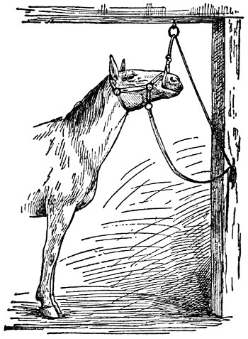
READY FOR THE DRENCH
A simple device for giving drenches to horses.
—Epsom salts, 1 pound; ginger, 1 tablespoonful; common soda, 1 tablespoonful. Dissolve in a quart of lukewarm water and give as a drench. It is a splendid general physic for cows, and can be given at any time when they are thought not to be thriving as they should.
[82]
Any departure from a normal condition is disease. The body, composed of different organs and parts, is in a healthy state when each of these performs its natural functions. Thus the normal mind is concerned with normal mental acts; any disturbances of the brain or spinal cords is immediately manifested in the action of the animal; likewise frequently a disturbance elsewhere may later have its effect on the mental system.
Disease may result from some external cause like from a wound, from food causing poison or derangement of the digestive system, from water introducing impurities, from parasites that disturb normal functions, disorganize tissue or produce toxines, or from other abnormal conditions—all of which interfere with the normal functions of one or more organs, regions, or parts.
In most cases the disturbances are readily recognized. Swellings, bruises and wounds are located at a glance. When blood passes from nose, ears or intestines, a key to the trouble is at hand. Coughs have their story. And vomiting, diarrhœa, convulsions, spasms, abnormal breathing or temperature each indicates at what points an abnormal condition is evident.
—Some diseases lead to disturbance throughout the entire body. For instance, pus may accumulate at some point from which it finds its way into the blood, in the end reaching to other parts of the body that in time also become affected.
[83]
Those diseases, with which fever is associated, are general in nature. The nerve centers are influenced, the body heat is increased and a weakened condition prevails. Back of this are the disease poisons—chemical poisons or germ poisons.
When the temperature of the body, as a result of fever, rises too high certain life principles are changed and death immediately follows. A temperature of 106° or 107° is very high, and, therefore, very dangerous. In treating disease the temperature is watched, that the course of the fever may be followed. Treating a fever, then, is helpful and a natural part of the treatment of the disease itself. The basis of the curative process rests upon the principle of proper circulation and the excretion of the impure substances.
In the first place most diseases arise from mismanagement. The very principles at the bottom of good health receive no consideration and little thought. On some farms it is seldom that a case of disease is heard of; on others, stock are under treatment at all times. Where order prevails, where cleanliness is appreciated, where disease-producing conditions are never allowed to accumulate or even gain an introduction, health is the rule and disease the exception. When the latter appears, it is due to some outside influence that gave it admission.
The greatest mischief in handling farm stock comes from improper food, filthy or impure drinking water, bad ventilation of stables, overwork, or lack of exercise and poor sanitary conditions.
Disease, therefore, is largely due to causes within control of the owner of the farm stock. True, one[84] source of trouble is due to mechanical causes: horses get nail punctures, legs and necks and head are cut in fences, blows bring bruises. But whose fault? Certainly not the animal. Old boards with nails ought not to be left in all sorts of places, fences should be protected, and stable fixtures, gates and harnesses should be in such order that only in rare cases will injury result.
—Poisonous materials and poisonous plants cause death to thousands of animals annually. Of great importance to the stock interests is the rapid destruction of these harmful products. Fortunately in the older sections these are about eliminated now, and we are also understanding more about the molds that lead to bad results when moldy forage is given as feed to farm animals. In time disease will be considerably lessened when only clean, wholesome food finds its way into the mangers and feed racks—then disease will depart and more rapid gains will come.
—Despite caution and care, health is often disturbed because of hereditary influences. Thanks to science, we know now that many of the old bugbears of the past, and once so entrenched, have become dislodged, and their true import set right before the owner. Tuberculosis, for instance, once so dreaded in both man and beast, is now known not to be handed down from parent to progeny; it is a germ disease, pure and simple, and gets its start just as many other ailments—through breath, or drink, or feed. There are hereditary troubles, however, that continue down through many generations. The narrow hock of the horse invites curb diseases; the narrow chest is a good breeding ground for tuberculosis[85] germs; straight pasterns are bad for the feet; poor conformation is not consistent with efficiency or easy functional activity.
These examples clearly show that form and type and physical characteristics have roles to play in animal economy and in health to which the wise stockman will give heed.

BACTERIA AS SEEN UNDER THE MICROSCOPE
a, Spirillum. b, Micrococcus. c, Micrococcus. d, Streptococcus. e, f, g, h, Rod-shaped bacteria. i and j, Divisions.
—In addition to the above causes of disease, another class is before us ready to inflict its injury at all times. Indeed, it is a class of the greatest importance. I refer now to parasites, bacteria, and germs, which cause more loss to live stock than all others combined. Think of hog cholera, a germ disease; of tuberculosis, a germ disease; of stomach worms, parasites; of staggers, a mold disease; of abortion, a germ disease; and hundreds of like nature, all due to parasites and germs, disease agents that disturb and destroy[86] the delicate organs or exposed regions, as the case may be, regardless of age, value, or breed.
Of course, remedies and treatment are being worked out to meet these individual diseases as they occur. Nevertheless, the best treatment is prevention. It is far better to prevent than to cure; and that is the line of action especially for this class. Indeed, it is far easier to understand the simple laws of prevention than the complicated curative processes. Especially is this true since germs are known and isolated, and their rapid destruction with air, sunlight, and disinfectants understood and available.
Enough has now been said to indicate that disease originates as a reaction between the cause of it and the body. Withhold food, and starvation—the disease—follows. Withhold fresh air and oxygen, and the tissue breaks down; disease results as a reaction from the normal use of air and the demands of the body for oxygen. Allow bacteria admission to the body and settlement in the tissues or organs most agreeable to each particular one, and these will grow, multiply, and, unless overcome by the natural resistance of the body, will conquer and destroy, causing sooner or later death and decay.
—Many diseases that now yield to no curative treatment are being met by inoculation. By this method the body is reinforced by serum injections, that disease germs and infections may be warded off, or in case of attack, be so fortified against the disease germs as to destroy them or render them inactive.
[87]
—An infectious and contagious disease may affect a herd or flock, destroying few or many. Some may never be affected and yet be subjected to exposure and contagion; such are immune and resist this particular disease. Others may suffer a mild attack, but throw it off with no disastrous consequence; such are strong and their organs ably fortified against any injurious inroad by the disease. On the other hand the majority in a flock or herd is not so able to throw off the disease for the reason of being predisposed by nature to such attacks; their very susceptibility invites attack, and if the infection is intensely virulent the affected body will most likely yield and death follow.
Each disease possesses its own peculiar characteristics, which are more or less conspicuous in each individual case. Then, too, some diseases develop quickly and end quickly. Others run a course of several weeks; and still others several months or even years. The first class is acute, the second chronic. In both kinds nature is at work endeavoring always to effect a cure; and, unless other complications arise, the result of improper food, bad sanitary quarters, bad air, or conditions not conducive to health, recovery will, in most cases, result. The great drawback to rapid recovery comes from the outside influences that counteract the curative processes of the body itself. Good nursing, good air, proper food, are back of rapid recovery.
Most diseases have been carefully studied, and their course of development has been mapped out. Our veterinarians know, in a general way, how fever[88] acts in live stock. If an animal is inoculated with Texas fever germs, the veterinarian knows the course of the disease beforehand. In a general way, he knows when the fever will begin, how long it will last, when it will be at its highest point, and when it will disappear. He knows all of this, even before he makes the inoculation. Yet no disease invariably runs the same course in different individuals. In fact, the virulence of bacteria have much to do with the course; mild cases occur usually when the germ is weak, and severe cases when the germs are very virulent. This explains why some attacks of measles or Texas fever or hog cholera are more fatal than other attacks in other places, or at other seasons of the year.
—It is in rare cases only that a regular course is not followed by most diseases. Take an infectious disease. The period of incubation comes first; this follows up the infection. During this period, no change in the animal is observed. He seems well, acts well, and does his work well. Nevertheless, all the time, during this period of infection, the germs are developing, multiplying, gaining headway, and so entrenching themselves that illness and disorder will soon follow. The period of infection varies in different animals and in different diseases. It may take two or three weeks for development, or as few as two or three days.
Following the period of infection comes the period of eruption. At this stage the typical characteristics are observed. At the next step the disease reaches its height with the animal under its complete dominion. But only temporarily. If properly nursed and treated, with most diseases, the animal will pass through the period and recover.
[89]
The final stage is the period of improvement. The battle that has been waged between the body and the disease is now about ended. The disease germs have been routed and the body has been victorious. All that now remains is the clearing away of the débris. In this case it is scattered throughout the body system. The damage that has been done is to be repaired and left, if possible, as near to the original condition, as the nature of the disease will allow. The period of improvement will vary in different diseases and in different animals. Recovery may occur in a few days, in some cases, and in others weeks and months will be required. A change of feed or pasture or work is usually necessary if the most rapid recovery would be had. In some cases, nothing other than absolute rest will suffice.
After the disease has run its course, the body usually returns to its former normal condition. There seems to be a limit to what the disease can do. A healthy body may be attacked, but, in the end, disease retires, having used itself up. There are diseases, however, that leave their marks in many ways. And these become permanent marks. With many of these all of us are acquainted. Smallpox is one. The pits over the face record the fierce battle that was fought. The same is true of wasted tissues, with scars that conspicuously mark the track along which blood poison has traveled. The shrunken hoof of the foundered horse tells the adverse termination of that disease.
While recovery may be more or less complete, the effect is to seriously injure the worth and value of the individual. There is a long list of this kind.
[90]
RESULT OF BONE SPAVIN
Pictured here is a natural hock free from disease and a diseased hock, the result of bone spavin. The bone is seriously affected and the easy action prevented.
Other diseases act differently in another way. They progress slowly, are not noticeable at first, but in the end are incurable. Take glanders as a typical case. It quietly and silently develops, often taking months or years in reaching the stage of eruption or before it becomes apparent. During all this time, and even after the disease is recognizable,[91] the animal goes on about his duties with no apparent trouble. The disease, however, is progressing all the time; in the end it conquers its victim, the final stages are reached, and the animal dies.
The stock raiser is concerned with different diseases in so far as they mean slow or rapid recovery, and particularly if they be contagious or not. His entire herd will be impaired if glanders is introduced into it. One tuberculosis cow will convey the disease to all susceptible individuals in the herd to which she belongs, especially if stabled in a tight barn during the winter seasons when little or no ventilation is intentionally provided.
[92]
Some diseases are not difficult to diagnose. Those resulting from wounds or knocks are easily located, and their treatment readily outlined. Others, however, are not so easy. Something is observed as wrong, the animal acts strangely, does not take to its food, is fretful, stands or walks unnatural—what is the matter? The stockman must ascertain the trouble, and the quicker the better.
A review of the past few days is desirable. Where has the animal been? What kind of food has it had? With what strange fellows has it associated? Has it been put to excessive work or exposed to unusual weather or conditions? What infectious diseases are prevalent in the community? These and other questions will occur; in some instances the answer will be at hand.
The stockman should at least know the fundamental principles of health and of any departure from them that indicate disease. Hence a superficial examination of the animal, as a whole, is in line of diagnosing the disease. Note the general condition of the body. The thermometer will advise you rightly. Is there pain? If possible determine this point and locate the seat of it. Is the circulation natural? An examination of the pulse will tell you if the blood is racing rapidly or gliding slowly, and whether regular or rough. Is[93] the respiration as it should be? Count the number a minute that you may know if the number is more or less, or is as it should be. On listening to the lungs, heart, and blood vessels, certain sounds are heard which change with disease—normal and heart murmurs. Whether or not an organ contains air can be determined by percussion, since solid organs, the lungs, for instance, in pneumonia, give a different sound from those containing air as they are normally. Air-containing organs—lungs and intestines—may thus be distinguished from the solid ones adjoining them. In this way their varying size in health and disease may be determined.
Your examination should go further and include the natural discharges—the dung, the urine, the nose moisture and the “look of the eye.” In cases of fever the urine is scanty and deeply colored. In Texas fever, for instance, the urine is dark red. In azoturia in horses, it varies from a light color to a deep brown or black. The nature of the dung should be observed, if watery or dry, soft or hard, scanty or profuse.
—Stand at the left side of the horse and run the finger along the lower jaw until you come to the point where the artery crosses the jaw on its lower edge. This will be found about two inches forward from its angle. Right here is the large muscle and at the front edge the pulsations may be caught. To get the pulse of the cow, stand at the left side, reach over the neck and take it from the right jaw.
In the horse the normal pulse beats are from 35 to 40 per minute and may go to 100 in disease. In the cow the pulsations run from 45 to 50 in health. The pulse relates its story very accurately[94] and, with practice, can be constantly used in diagnosing the nature of the ailment. For instance, a soft pulse, one that is easily compressed by the finger, indicates bronchitis. A hard pulse, one not easily depressed by the finger, indicates acute inflammation. A hard pulse may be quick and bounding and forceful. An irregular pulse, one that beats fast for a time, then slowly, indicates a weakened heart condition. A slow, full pulse, one that comes up gradually to the finger touch, indicates some brain trouble.
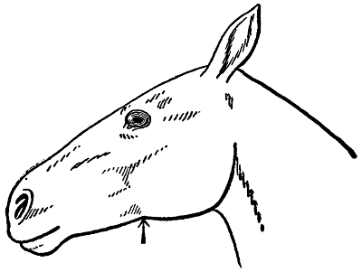
FEELING THE PULSE
The heart beat, as it is called, may be felt by placing the finger over any of the superficial arteries. The submaxillary artery as it passes under the edge of the lower jaw close to the bone is a convenient vessel for the purpose.
—While the heat of the body may be surmised by touch and feeling this is not a reliable guide as to the temperature. A self-registering thermometer, inserted into the rectum, is the only reliable means for getting this desirable information. In a state of health the temperature of the horse ranges from 100° to 102.5°.
[95]
When the temperature rises, inflammation is indicated. A fall in temperature below normal denotes loss of strength, vitality, and death. If the temperature rises three or four degrees above normal, the case is serious, and a rise of five or six is very dangerous. Animals seldom survive when the rise reaches above 107° or 108°.
A good clinical thermometer should be in the possession of every stockman. It costs but little, and its aid in recognizing and treating disease is helpful, if not absolutely indispensable.
—In breathing two movements are observed—taking in and sending out the air. In health the respiration is usually constant, ranging from 10 to 14 in the horses, and from 15 to 20 in cattle. Breathing is faster in young animals; and exercise increases the number of respirations per minute.
Any disease of the respiratory organs will cause the breathing to be short and rapid and labored. If the number of respirations seem more than normal, some disturbance is indicated. If the pulse is faster at the same time, illness is at once indicated, and the trouble should be sought at once.
The first effort in treating disease is to remove the cause. This is sometimes done very easily. Mange and lice are quickly destroyed by washes and disinfectants.
Bright, fresh, wholesome food and pure water easily replace bad food and water to the permanent good of the stock. Cattle ticks quickly disappear when the grease brush is applied. And so in every direction you take to fight the disease: find the[96] cause and then remove it, and half the battle is fought.
If disease-producing germs cannot be killed at the moment, it is still possible to diminish their number or to modify their virulence. Thus to open an abscess is to remove the pus-producing bacteria, and hence to hasten recovery. To wash a wound or open sore with antiseptics is the simplest way to remove, diminish, and destroy the evil of the sore.
HOW HEAT AFFECTS GROWTH
At the end of 24 hours in a but seven bacteria have developed, the temperature being 50 degrees. In b 700 have developed in the same time, but in a temperature of 70 degrees.
—When disease sets in a battle begins. One combatant is the disease itself, the other the body. Your work is to render assistance to the body. In many cases your help will not be needed. In others you can render incalculable aid. Here is where medical aid begins and ends: to care for and nurse and make the body strong that it may be victorious, quickly, if possible, but without fail, in the end. Medicines are helpful if they diminish the work of the diseased organ, giving in this way time for the body cells to bring about a cure. Therefore rest and quietness are advisable, that no organ may be called upon for any effort but normal function and repair. A disease of the heart calls for absolute rest, of the intestines for little or no irritating or bulky or hard food, of the lungs for no exposure. At times it is advisable to check the activity of an organ, in which case a[97] drug may be given, like opium, to quiet the intestines, or like aconite, to diminish the rate of the blood flow.
In the same way external assistance may be rendered; as, for example, sweating—to throw off poison in the tissue juices; and blanketing—to maintain an even temperature and to protect from chill and draught.
Medicines are conveyed into the body as drenches, balls, enemas, and injections under the skin or into the veins. There is nothing mysterious about any of them.
—The practice of giving medicines in a ball is a very old one, and has much to recommend it. Many nauseous agents as aloes, opium, arsenic, asafetida, are thus conveyed to the stomach without causing annoyance and disgust to the patient. The balls are wrapped in paper, dough, or gelatin capsules, and may weigh an ounce or two. In giving a ball the following plan is usually followed: Hold the ball between the thumb and first two fingers. Now seize the tongue at about its middle and gently draw it out to the side of the mouth, in such a way that the right hand may be inserted into the mouth and the ball placed far back on the tongue, when the hand is withdrawn, the tongue replaced and the halter or strap wrapped around the jaws until the ball is swallowed.
—The drench is usually employed for liquid medicines. It is best to dilute the medicines with water, milk, or oil that they may more readily reach the stomach and[98] at the same time exercise no injury to the structures through which they pass.
In giving a drench exercise as much patience as possible. To horses it should be given slowly. If there is any disposition to cough, lower the head, and then proceed as before.
—These are made of a variety of things, bread, bran, and linseed meal being the most common. Any substance that will hold water and retain heat will serve the purpose.
—These are made with mustard and water, cold water being the most desirable. Mix to a thin paste. If the part to which the plaster is to be applied is covered with thick, long hair, a very thin plaster will more quickly soak into the skin. This kind of plaster is most commonly applied to the throat, the windpipe, the sides of the chest, the abdomen and over the region of the liver. To get the best effect for the last named, apply on the right side at a point four or five inches behind the back ribs.
—The first step in blistering is the clipping of the hair over the diseased part, and the removal of dirt and scurf attached to the skin. The blister is to be worked into the skin, and usually ten minutes of rubbing will be necessary to produce the desired results.
In the course of twenty-four hours blisters will form, and some swelling in the region is likely to be manifest. On the third day bathe the part with warm water and soap. After drying, apply vaseline, lard, or sweet oil. The blister should be repeated if the results of the first blister do not bring about a cure.
—The hot iron is a very useful agent in treating many cases of chronic lameness and bone[99] diseases. In performing such an operation have the iron at a full red and white heat and touch the part gently with just sufficient pressure to make a distinct impression. But one leg should be fired at a time.
It is desirable to shave the hair closely to the skin before applying the iron. The day following the firing spread over the wound any common wound oil like neat’s-foot oil or vaseline. Daily applications are called for until the swelling subsides. Unless a period of rest is given after the operation, the best results will not be had. Many bone diseases return, or are never cured, because complete recovery never occurred in the first place. Work and exertion only aggravate the cases, often leaving them in a worse condition than before the firing.
In the first place keep them clean. If necessary wash them daily, especially the parts liable to get filthy and dirty. In fever cases a gentle sponging, every few hours during the day, is desirable. Vinegar added to tepid water is very good.
Animals in feverish or chilly condition can be assisted by blankets and bandages. These are very helpful in warding off congestion of the internal organs and in maintaining an even temperature of the body. Any warm rug or blanket that is clean and light will serve.
In bandaging the legs, endeavor to get an equal pressure at all points. A long roll is, therefore, best, and several layers should be wrapped around the member. It is a good plan to remove the bandage, replacing with another at least once a[100] day, and two a day are better. When a bandage is removed, the skin should be washed and rubbed with the hand and fingers, and the covering replaced as promptly as possible.
—During sickness only easily digestible food should be provided. Offer something different from the ordinary, and let it be prepared in an appetizing form. Nothing is better than gruels and mashes. These are soft, nourishing, appetizing, and easily digested. When active nutrition is demanded, milk and eggs can be added to the ordinary gruels or mashes.
Water should be available at all times. Small amounts at frequent intervals are better than large amounts at intervals far apart. In a few instances only is it best to withhold the water. In treating dysentery, diarrhœa and diabetes water is usually withheld, but in most diseases a free use is allowable and desirable.
[101]
—The expulsion of the fetus at a period too young to live exterior of its mother is known as abortion. This ailment may afflict cows, mares, sows, and ewes, but is most common among cows.
Abortion may be divided into two classes, namely, accidental and contagious. If we had nothing but the accidental form of abortion to contend with we would hear very little about this disease, owing to the fact that it is perfectly natural for animals to carry their young full time, regardless of how much they may be punished or abused while in this condition if their system be free from the germs of contagious abortion. On the other hand, contagious abortion is a very destructive disease, causing heavy losses to the stockmen of the United States as well as to other countries. Contagious abortion is divided into two classes, namely, acute and chronic. Cows afflicted with the acute form of abortion may lose from one to three calves. Cows, after passing from the acute to the chronic form of abortion, may carry their calves full time, but are as badly affected with the germs of contagious abortion as they were in the acute form, when they were losing their calves.
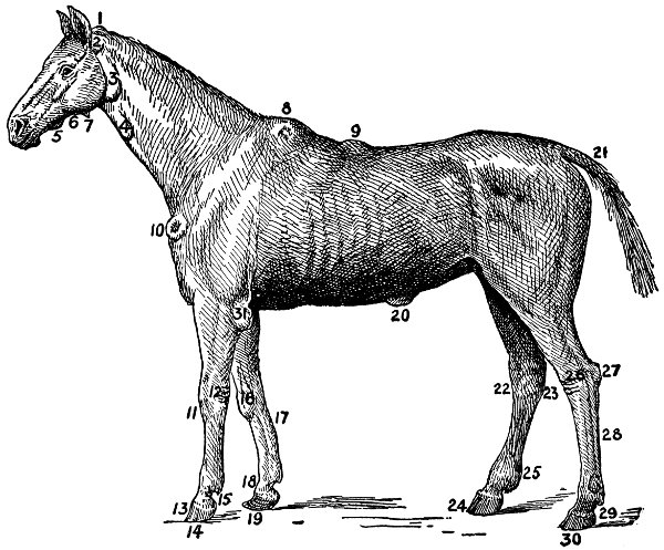
DISEASES OF THE HORSE
1 Poll Evil, 2 Swelling by Bridle Pressure, 3 Inflamed Parotid Gland, 4 Inflamed Jugular Vein, 5 Caries of the Lower Jaw, 6 Fistula of Parotid Duct, 7 Bony Excrescence, 8 Fistula of Withers, 9 Saddle Gall, 10 Tumor Caused by Collar, 11 Splint, 12 Malanders, 13 A Tread on the Coronet, 14 Sand Crack, 15 Quittor, 16 Knee Bunch, 17 Clap on Back Sinews, 18 Ring Bone, 19 Foundered Foot, 20 Ventral Hernia, 21 Rat Tail, 22 Spavin, 23 Curb, 24 Quarter Crack, 25 Thick Leg, 26 Malanders, 27 Capped Hock, 28 Swelled Sinews, 29 Grease, 30 Sand Crack, 31 Tumor of Elbow.
Perhaps the greatest damage brought about with
cattle afflicted with the chronic form of abortion is
the shortage of milk. Animals afflicted with accidental
abortion show very few marked symptoms
before they abort. Animals afflicted with contagious[102]
[103]
abortion have a number of marked symptoms,
namely, little red patches of infection on the lining
of the vulva, and there may also be present a
catarrhal discharge. The sheath of the herd bull
in the acute form of the disease has a catarrhal discharge,
while the symptoms of calves is a swelling
of the glands of the throat from ear to ear. These
last named symptoms do not appear in accidental
abortion.
Owing to the fact that the germs of contagious abortion are found in the mothers’ blood, in the genital organs of the cow and the bull, and in the stables wherein they are housed, it has been positively decided that the only reliable and effectual treatment for contagious abortion is the hypodermic treatment, which destroys the germ in the mother’s blood. The genital organs of the cow and bull should be washed out with the antiseptic solution made of 1 pint of corrosive sublimate to 1,000 parts of water, and the germs contained in the stables wherein afflicted animals are housed should be destroyed by disinfectants. In this way the disease is met at every turn, and it is impossible for the disease of contagious abortion to exist when thus handled.
—A collection of pus in a new-formed cavity in the body. It has a well-defined wall surrounding it. An abscess is the result of entrance of micro-organisms into the body. They may have entered through wounds or into the hair follicles, or abscesses may result from infectious diseases, as strangles or distemper in the horse. At the seat of the abscess formation swelling occurs, the part feels warmer than the surrounding tissue, is painful to touch, and hard. These conditions are due to the inflammation of the part. Later it becomes[104] soft, less sensitive, and fluctuates, which shows that it is coming to a head, or that the pus is collecting. If the skin is white it will show a yellow color in the center, which is usually raised above the surface, and the hair falls out. This soon breaks and discharges pus.
It is advisable to hasten the ripening of the abscess by hot applications in form of poultices, or a large pack of cotton saturated with hot bichloride of mercury 1 part to 1,000 parts of water, or use some one of the coal tar dips 1 part to 50 parts of water. The application of a light blister will often hasten ripening. When the pus has collected or the abscess has come to a head, it should be opened at the lowest part in order to give free drainage to the pus contained within.
Great care should be used in opening abscesses—not to cut blood vessels which might be in the vicinity. In case the abscess breaks of its own accord, it is often necessary to enlarge the opening, in order to give free drainage for the pus. If the abscess is large or deep-seated it should be washed out each day with bichloride of mercury 1 part to water 1,000 parts, or with a 2 per cent solution of some one of the coal tar dips. After it is opened do not apply bandages, as they prevent the free escape of pus. Do not allow the opening to close until it heals from the bottom; or, in other words, as long as it secretes pus, for there is danger of its breaking out again. If the opening is too high up, or not large enough, it may result in a running sore or fistula.
—Called lumpy jaw, because of the frequency of the swelling located on the jaw. It is due to the entrance of a specific organism, a[105] fungus, into the tissues. This causes an inflammation, with an increase in the amount of tissue, as shown by the enlargement and in which an abscess is formed. Adult cattle are the only animals commonly affected with this disease, but occasionally nearly all classes of domestic animals may be affected. A number of cases have also been reported in man, but the disease in cattle, being localized to a small region of body, usually the head, there is little danger of transmission from animal to man in eating beef.
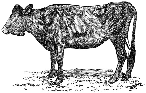
LUMPY JAW
An exterior view showing location of lumpy jaw.
The symptoms are recognized by the characteristic tumor, usually observed on the jaw, either of the bone or of the soft tissues in that vicinity. It may, however, affect the tongue, or, in fact, nearly any of the organs of the body. Its development is more or less of a slow, constant growth, beginning with a very small nodule, but, when allowed to run[106] its course, may reach the size of a cocoanut, or larger. On reaching some size, it usually ruptures and from it discharges a thick, yellowish pus. It is to be distinguished very largely by its commonly affecting cattle, its location, its slow growth and its firm, hard consistency, and finally a discharge of pus from it.
Treatment consists, if of small size in the soft tissues, of complete excision by the knife. But, if of larger size, or when the bone or large blood vessels are involved, recourse should be had to the internal administration of iodide of potash from one to two teaspoonfuls in a drench of a quart of water, or, in some instances, it may be given in the drinking water once daily. This should be continued for a week or ten days, when the treatment should be discontinued for a like time, and, if necessary, repeated several times.
—This is a condition resulting from the failure of the mother to pass the membranes after the birth of her young. It happens most frequently in cases of abortion, or when birth occurs before time. There is usually more or less of a mass of the membranes hanging from the opening, which occasionally reaches to below the hock, or even to the ground. When fresh it looks somewhat like the intestines, but if exposed to the air for some time it is grayish in color, especially when it begins to decompose. The odor is very offensive, and the discharge soils all the hind parts of the animal. In these cases the health of the animal suffers, and fever frequently results, with a loss of appetite and flow of milk. The fever and inflammation of the parts may go so far as to cause the death of the animal.
[107]
The afterbirth should never be allowed to remain over three days in the cow, nor over twenty-four hours in the mare. In the mare, sow, or bitch gently pulling on the membranes, at the same time twisting them easily, will often bring them out without injury to the animal. With the cow it is different. Here the membranes are “buttoned” on in tufts, and the pulling, and especially the twisting, usually makes matters worse and injures the uterus.
After removing the membranes there always remains in the uterus a quantity of fluid, which should be washed out with water a little cooler than the blood of the animal, adding about a teaspoonful of carbolic acid or other good antiseptic to each gallon of water and mixing well. The hands and arms of the operator should be absolutely clean, and during the operation should be kept covered with carbolized oil or carbolized soap and water. In mares, especially, care should be taken not to injure the parts, as inflammation sets in very much quicker than in the cow. Several gallons of the above solution should be injected as soon as the condition is noticed, and a warm bran mash fed to the animal occasionally will help her general health.
—A deficiency of red blood corpuscles. The animal is scanty of flesh, hide bound and in a general run-down and debilitated condition. The disease is sometimes called hollow horn. Treatment consists of better food and care. The feed should be of a nature such as will enrich the blood and build up the system. Food of a succulent nature, like roots, green grass, or ensilage, will help out. A tonic, made as follows, will be helpful: Two teaspoonfuls of sulphate of iron, 1 teaspoonful of powdered nux vomica, and 4 tablespoonfuls of[108] ground gentian root. Add this to the food each day for a week or ten days.
—An acute, infectious disease of plant-eating animals, which, under favorable conditions, attacks flesh-eating animals as well. It is caused by a microbe which enters the circulating blood and by multiplication therein causes its rapid destruction, and the death of the animal. The disease is as old as human history. It exists in all countries and in all latitudes. It was formerly very destructive to human life, as well as to animals. There is no disease which attacks more different kinds of animals than anthrax, nor one which is more deadly. Also, there is no disease which is harder to deal with from the sanitary point of view; nor harder to stamp out. The reasons for this will be shown later on.
Soil is the prime factor in preserving and propagating the microbe, when it is naturally wet, impermeable, and rich in decomposing animal and vegetable matter. The microbe of anthrax may enter the body by several channels. It may be taken in with the food or drink. It may be breathed into the lungs. It may enter through abraded surfaces on the skin. It may be inoculated into the body by biting insects.
There are several forms of the disease and these are determined by the modes of entrance of the virus. One form, which occurs especially in sheep and cattle, at the commencement of an outbreak, and which is characterized by the suddenness of its onset and its high degree of fatality, is known as the apoplectic, or fulminant form. Without showing any previous symptoms, an animal will suddenly be seized with loss of appetite, trembling, uneasiness, irregularity of movements, difficult[109] breathing, blueness of the nostrils, bellowing, convulsions and hemorrhages from the natural openings. Death may occur in a few minutes or in four or five hours.
Another type is known as anthrax fever, or internal anthrax. Here we have distinct symptoms, the most important being high fever of from three to four degrees, excitability and restlessness. Blood may ooze in drops from the nose, eyes, or ears, and from inside of the forearm or thigh, in sheep. There will be trembling, prostration, numbness of the loins, thirst, grinding of the teeth, colicky pains, bloating, bloody discharges, palpitation of the heart, difficult breathing, blueness of the visible mucous membranes, jerking of the muscles of the back and neck, and rolling of the eyes. The animal will die in comatose state, or in convulsions, and death will occur in sheep in about a day. Cattle will live from two to five days, and horses from one to six days.
A third form is external anthrax, which manifests itself in swelling of the tongue, throat, rectum, and skin in cattle; and of the tongue, throat, neck, shoulders, withers, flank, or thigh in horses. These swellings have a firm, doughy feeling, are not painful generally, and show a marked tendency to gangrene. They never suppurate. If cut (this should never be done), they discharge a pale, straw-colored liquid. In this may be found the microbe.
The rapidity with which putrefaction occurs in an anthrax carcass is very marked. Another characteristic is, the blood loses its property of clotting, is dark and tarry, and does not become light in color by contact with air, like normal blood. In fulminant cases, however, these characters are not[110] so well marked. Other signs of the disease, if a farmer should be so unfortunate as to open an anthrax carcass and thereby spread the infection on his farm, will be great enlargement of the spleen, or milt, and also of the liver. Bloody patches in the tongue, throat, lungs, stomach, and intestines, caul, skin, and muscles, or in fact in almost any part of the body, will be plainly visible.
and disposal of the carcass are the most important procedures in an outbreak of anthrax, from a sanitary standpoint. Medicinal treatment is of little value. A vaccine has been discovered that is very effective in preventing the disease. This has been used very successfully in both this and European countries. If a case of anthrax is suspected, call your veterinarian at once. The disease will not pass through the air from a sick animal to a healthy one, but the discharges which invariably occur during the progress of the disease all contain the microbe, and everything soiled by them is infectious material and capable of spreading the disease.
When an animal is infected, remove at once to the burial lot and tie it near the place it is to be buried, to save handling and scattering the infection. When it dies, dig the grave. Then saturate the animal with kerosene or coal oil and set it afire. By means of ropes tied around the fetlocks turn the animal, saturate the other side and fire that, and also the soles of the feet. When every hair has been burned off, dissolve a one-pound carton of chlorinated lime (freshly opened) in sufficient water to make a fluid that will just pour from the cup. Fill the nostrils with this, also the mouth and eyes, which should be pried open with a stick dipped in solution. Saturate some cotton or rags[111] with the lime, and plug up the nostrils or mouth. Treat the rectum likewise. Turn the animal into its grave, sprinkle the ground on which it has stood and laid with a strong solution of chlorinated lime, and shovel the top layers of this soil into the grave. Follow this with the grave soil, banking it up, as in human graves. In cases where the animal is found dead, the same method is to be pursued, except that the animal is hauled to the grave on a sled (never dragged over the ground). In these cases, also, the place where it died must be disinfected by the same means, after hauling out all loose material and burning the same, as near as possible to the place where the animal died. It would also be necessary to disinfect the sled and all tools which came in contact with the carcass.
—A ruptured blood vessel in the brain; usually causes unconsciousness, at least for a time. The control of certain muscles is lost and a general dullness prevails over the animal. In case the apoplectic attack runs a favorable course, the muscles come more or less under control again and the patient in time may recover. It is in rare cases only, however, that animals recover to an extent to be worth much after being affected with apoplexy. Fortunately the disease in animals is rare.
—This is a very peculiar affection of the horse, in which the animal shows a special form of lameness upon exercise, after having remained idle for a day or two. The cause is not definitely known, and yet the circumstances under which the disease develops are rather constant, such, for instance, as an animal[112] in vigorous condition, fed liberally upon nitrogenous feed, remaining idle over Sunday, a holiday or at other times. Upon being taken out the following morning the animal usually shows an excess of energy, but before going far begins to go lame in one or both hind limbs until, if urged further, becomes completely paralyzed behind, going down and unable to rise. He also shows considerable pain, as though he might be suffering from some form of colic, with a profuse sweating. On reaching this point the animal usually ceases to void the urine, which, when drawn, appears a very dark brown or coffee color. The pulse and breathing are somewhat accelerated, and frequently there is considerable nervous excitement. The muscles of the loin and thigh are tense and rigid.
The treatment should begin as soon as the lameness shows itself. After a few hours of rest, the distress will be over. The more exercise given the animal after the lameness begins, the more severe the trouble, and the more energetic means of treatment required. In a case showing signs of nervous excitement, it should receive 2 tablespoonfuls of bromide of potassium every three or four hours until becoming quiet. Sweating should be induced by blanketing the animal well, preferably using blankets wrung out of hot water and covered with a dry one. Allow all the water the animal will drink and give it 4 tablespoonfuls sweet spirits of niter three times a day if bladder is not paralyzed. If unable to void the urine, the bladder must be emptied three times daily. A laxative or purgative should be given early in the disease. If the animal remains somewhat stiff, give a teaspoonful in the feed twice a day of the following: Powdered nux vomica, 4 teaspoonfuls; powdered sulphate of iron,[113] 6 teaspoonfuls; powdered gentian root, 6 teaspoonfuls.
—Failure to breed is usually due to an acid secretion of the genital organs, to the germs of contagious abortion, retention of the afterbirth, or to an abnormal condition of the sexual organs of either the male or female. The acid secretion of the genital organs prevents conception by destroying the semen of the male; the germs of contagious abortion set up a catarrhal inflammation and discharge, which also prevents conception; retention of the afterbirth, whether it be removed by force or permitted to slough away, usually leaves the womb in a diseased and catarrhal condition, effecting a discharge; impotency may be due to excessive use of the male, or to advancing age in both male and female.
Any unnatural discharge irritates and scalds the mouth of the womb so that when the discharge ceases the mouth of the womb heals, and it is impossible to make a cow or mare breed without mechanical interference. This kind of treatment is conducive to fertility by increasing the blood supply to the part. Mechanical contrivances are now on the market for the purpose of dilating the mouth of the womb. These increase the probability of pregnancy. If the womb be opened just before service, many troublesome cases can be corrected. This is done by inserting the oiled hand and arm into the vagina, finding the opening into the womb, and gradually dilating it by inserting one or more fingers until the passage is open and free.
—Just why bones become soft and frequently are absorbed in normal animals is not known, unless it is due to an absence of some essential bone constituent in the food or water.[114] The disease shows that the bone is absorbed and its structure softened. As a consequence, the bone enlarges, becomes spongy and light.
The disease usually starts as a swelling in the head, hence the name. Often the lower jaws are enlarged, and, as the disease progresses, the legs become affected. At the same time the animal loses weight. The treatment consists of nourishing foods, rich in the mineral constituents. Better consult a veterinarian when the disease is first noticed.
—See Actinomycosis.
—Often cattle show large bunches over the knees. These may be soft or hard. In cattle these big knees are caused by hard floors, in lying down and getting up. Big knee in horses is a little different, being more in the nature of spavin or ringbone, and in this case occurring at the knee joints. In cattle the bunch may be localized in the flesh and skin. With horses, it is an attack on the bony structure. When first noticed a blister may be used.
—See Lymphangitis.
—Frequently germs get into the udder, and, as a result, bitter milk or blue milk or bad milk results. Sometimes the bad taste of milk is due to the odor in the stable or to the food that the cows get while pasturing. Turnips give a bad taste to the milk, as does garlic or wild onions. If the bitter taste or the blue milk is due to disease germs, then the remedy lies in the destruction of these germs. Just after milking, and each quarter thoroughly emptied, inject a warm solution of boric acid.
—A germ disease affecting turkeys and chickens. It is characterized by a dark purple appearance in the comb and wattles. Fowls attacked[115] by the disease show dullness and laziness; at the same time indigestion disturbances and diarrhœa is observed. The best treatment is to kill the fowls affected just as soon as they become affected. This will prevent the disease from spreading. It is advisable to burn the bodies of the dead so as to prevent the spreading of the germs. Thorough disinfection is necessary.
—An infectious disease produced by the blackleg bacillus, a parasite which lives and propagates in the soil of infected districts and in the bodies of diseased animals. Certain kinds of soil are very favorable to the existence of the parasite, and such, when once infected, easily remain so permanently and thus constitute the source of the disease. Years ago blackleg was regarded as a form of anthrax. This has been proved erroneous, however, for blackleg and anthrax are two distinct and independent diseases, each being caused by a specific germ. One diseased animal does not transmit the disease directly to a healthy one. When caused, it is the result of self-inoculation, that is, by the germ entering a wound in the skin or mucous membrane of the body, produced on the legs while the animals are roaming over the fields, or at the mouth while grazing; these are the places by which the blackleg germs get into the system.
An animal dying of blackleg is fairly alive with germs, which remain in virulent condition for a long time. It behooves the farmer, therefore, to completely destroy this kind of dead; not by burying, for then the germs remain in the soil. The best way is to burn the animal right on the spot where it died. If the animal is moved to another place, the infection is spread, thereby, and not only the death place, but the grass over which the animal[116] has been moved, should be thoroughly disinfected that no germs may survive. The disease is characterized in the appearance of large swellings on various parts of the body, usually on one of the upper portions of the legs, and never below the hock or knee joints. Swellings vary in size, and are always formed by the presence of gas that has collected in the tissue just beneath the skin. This gas is a product of the germ. You will notice a peculiar crackling sound when you pass your hand over these swellings. When punctured with a knife these swellings emit a bloody fluid possessing a disagreeable and sickening odor.
Associated with the disease are loss of appetite, high fever and lameness. Death follows just a few days from the time of attack. So far no medicinal treatment for cure has been discovered. Stock should not be admitted to infected regions. The only safe practice in regions where blackleg is prevalent is in the use of protective inoculation or vaccination. Such vaccination renders the animals immune, and even if attacked, there is almost no appearance of the disease at all.
—The blackleg vaccine now so well known is made from diseased flesh taken from a calf that has died from blackleg. This flesh, after being dried and powdered, is then properly prepared and injected into the animal. There are two kinds—a weak and a strong vaccine and single and double vaccine. The single vaccine requires but one inoculation. The latter is believed to be superior and gives better protection. The vaccine is usually available from the state experiment stations, or can be obtained through your veterinarian. About the only skill required in[117] doing the work is in having the instruments thoroughly cleaned and disinfected. A hypodermic syringe is used and the injection made on the underside of the tail, a few inches from the tip, or just beneath the skin of the neck or shoulder. The point of the syringe should not puncture the muscle at all; simply pick up the skin and draw it away from the muscle and admit the fluid in the loose space between the two.
When vaccinated, the treatment is supposed to last about a year. If calves are vaccinated the operation should be repeated at about the age of yearling. Two periods of vaccination are suggested: when turned to pasture in the spring or when turned to dry food in the fall. Full directions as to the use of vaccines always accompany the preparations and further detail is unnecessary here.
Preventive medicines cannot be relied upon, although a common one is used throughout the West, made as follows: 4 ounces of sulphur, 1 ounce of saltpeter, 2 pounds of sulphate of iron, and 1 pound of air-slaked lime. After being thoroughly pulverized and mixed, this is added to one-third of a gallon of common salt and used in the place of salt.
—See Concretions or Calculi in Urinary Organs.
—See Staggers.
—This disease, sometimes called hoven, is characterized by the distention of the paunch or rumen, and is due to the accumulation of gas. It most frequently occurs when cattle or sheep are pastured on clover or alfalfa, especially if it is moist just after a rain, or when dew is on the ground, and when not accustomed to[118] fresh green food. I have known of many cases where cattle have bloated from eating alfalfa hay during the winter season.
There is no mistaking the disease. The animal shows pain, goes off to itself, and breathes with difficulty. Colic is often associated with bloating. The most characteristic symptom, however, is the excessive swelling due to the gas. The bloating is noticed even over the back of the animal, the gas continues to form, and, unless relief is secured, the animal will choke and die as the result. Or some suppression of the vital processes will occur, even rupturing, with the same fatal ending.
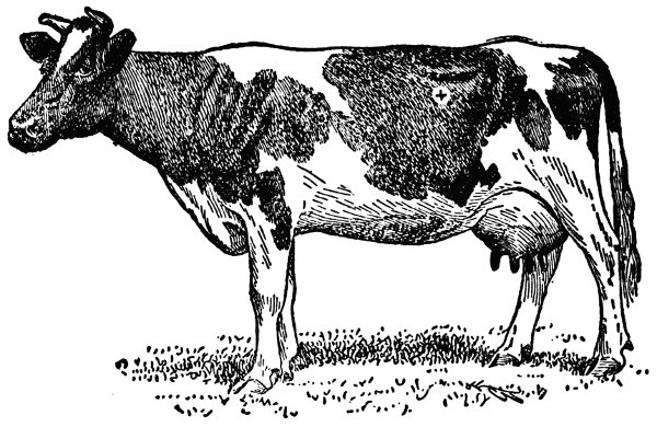
WHERE TO TAP IN BLOATING
Insert the trocar and canula, or if these are not available a knife may be used. Make the puncture downward and forward and plunge the instrument into the rumen.
Bloating may take one or two forms; a mild case in which recovery gradually follows, and a very severe form, where the only salvation is in tapping[119] to release the gas. If it is an ordinary case of bloating, not very severe, ordinary remedies will give relief. Turpentine in doses of 8 or 10 tablespoonfuls is good. Some use 4 tablespoonfuls of hyposulphide of soda dissolved in water, with excellent results. Some veterinarians give doses consisting of 4 tablespoonfuls of aromatic spirits of ammonia in water as a drench. Ginger is frequently given, as much as 4 tablespoonfuls diluted in warm water as a drench. To keep the animal moving about is excellent.
In severe cases it is advisable to tap with the trocar and canula. Indeed, tapping is the last resort if you would save the animal. These are inserted on the left side of the skin and pushed into the rumen or paunch, the incision being made about half way between the point of the hip and the last rib. In introducing the trocar push in and down.
After the insertion is made, the trocar is withdrawn and the canula is left in to furnish an opening through which the gas can escape. In case the canula gets clogged with partially digested feed, insert the trocar so as to push away the material and withdraw it again. If the trocar and canula are not available, then use a pocket knife. Of course, be careful that the incision is not made too large.
Just a few simple precautions are suggested here as a prevention of this trouble. There is always danger from bloating when cattle or sheep are turned into green pastures, especially when not accustomed to such feed and especially when wet. It is advisable, therefore, to keep stock from the pasture until later in the day when the dew has disappeared. Stock should have their regular morning feed just as usual before being turned on[120] the pasture. They will have less greedy appetites, will not like to gorge themselves, and hence the trouble will not be brought on.
—When blood poisoning results from the entrance of bacteria into the circulation, it is termed septic infection. This means that the disease may be communicated to a healthy animal by inoculation. Thus, an operator in making a post portem examination may bring on blood poisoning because of an accidental prick of the skin. An animal may step on a nail or get a splinter in a muscle or under the skin, and become self-inoculated, in time becoming affected with septic infection. Consequently bacteria are the direct inducing factors. The chemical poisons produced as a result of the work of these bacteria, as those of putrefaction, may induce what is known as septicæmia. On the other hand where pus is produced, as in the abscesses which follow upon neglected wounds in joints, a form of blood poisoning is produced known as pyæmia. In either case blood poisoning may result, become very serious and may cause the death of the victim.
At first chills may be noticed, then a rise of temperature, quick respiration, rapid but weak pulse, and much prostration. All the time the appetite is disappearing, until it becomes lost. The mucous membranes of the eyes and nose take on a yellowish, red tint often showing spots or blotches of blood and the tongue becomes coated and clammy.
Quick treatment is necessary in every case of blood poisoning. As soon as noticed, the source should be treated with disinfectants, thereby arresting the supply of morbid matter. A strict employment of antiseptics, so as to destroy the bacteria, is the first essential. We look upon the[121] prick from a rusty nail, or wound from a wire fence, or a dirty stable splinter, as matters of frequent occurrence, yet a great deal of danger lurks among these. They should be avoided as much as possible and in all cases immediately treated. As soon as the poison is admitted to the blood or tissue, the disease germs multiply and soon are present in great numbers. Had the wound been cleansed with an antiseptic like carbolic acid in the beginning, it would have been a simple matter and the poison would have been neutralized, and the ingress of the invaders made unattractive, if not altogether impossible.
In all cases of blood poisoning, look to a systematic and constant application of suitable lotions to the injured parts, to careful nursing, and to nourishing food. If the appetite has completely departed, it is often advisable to force food like eggs and milk into the stomach, so that the strength of the patient may never be dissipated or weakened. With this treatment should go pure fresh air, cleanliness and much sunshine. It usually is advisable to call a veterinarian as early as possible.
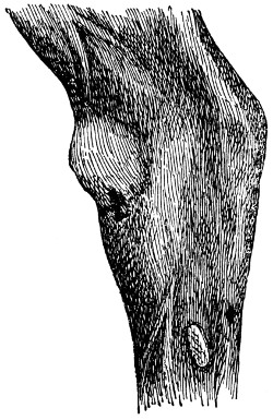
BOG SPAVIN
The bulging outward of the soft tissues of the hock joint is due to the secretion of joint oil or lubricating liquid in abnormal amounts.
—Sometimes, just after calving, bloody milk is observed. The cause is generally due to a rupture of the small blood vessels in the vicinity of the cells that secrete the milk. It may be due to a tiny accident of some kind or it may be the result of disease, localized in the udder. Bathing the udder with hot water will prove helpful and, until the milk is normal, frequent milkings are desirable. If the condition prevails for any length of time and the cow is not a very good one, it is just as well to fatten her and send her to the butcher.
—A condition of the urine[122] peculiar to certain diseases like Texas fever in cattle and azoturia in horses. In the latter disease the urine is quite turbid and dark in color, sometimes almost black.
—A round, smooth tumor at the front and on the inside of the hock. It is the result of sprains, bruises, or other injuries. When these injuries occur, too much joint oil is secreted, causing a bulging of the ligament. Lameness seldom accompanies a bog spavin. If lameness be present other structures are certain to be affected, and some pain and heat will be noticed, together with a stiffness of the joint.
Treatment consists of applications of cold water to the affected parts and a lotion made of 2 tablespoonfuls of acetate of lead in a quart of water. A blister made of 1 teaspoonful biniodide of mercury and 4 tablespoonfuls of lard rubbed in a little with the fingers and repeated in ten days or two weeks and continued for some months will correct the trouble. Wash the part having received the blister twenty-four hours after application. It is also advisable to tie the horse’s head while the blister is on, so that he cannot bite the part.
[123]
—See Spavin.
—The larvæ or grub of all common bot flies are thick, fleshy grubs and pass their life in some portion of the body. When they are fully developed they leave the body by some route and bore into the ground, where they go through another stage of their development known as the pupa stage. When this stage is completed they crawl out of the ground as a fly ready to deposit eggs.
—Everyone is familiar with the common nit fly and the yellow nit that is attached to the hair on almost all parts of the horse, but especially on the chest and legs. The young larvæ or even the egg may be transferred from these regions of the body into the mouth by the horse biting these parts. The grub passes into the stomach where it attaches itself to the mucous lining and continues its development. The bot is not so dangerous as it is popularly supposed to be. They may, when attached in large numbers to the right side of the stomach, interfere with digestion and be responsible for some of the digestive disorders and colics. They are uniformly present in the stomach of all horses that are kept in the open where flies can get at them. A carefully groomed animal may be free from them. The eggs may be destroyed by rubbing the body with a rag wet with kerosene. One of the most common remedies for bots, and at the same time the most useless, is a mixture of molasses and milk. Bots are hard to dislodge from the stomach until they have completed their development there and pass out of their own accord. Half-ounce doses of turpentine three hours apart until three doses are given, followed by[124] an ounce of powdered aloes as a physic, is a good remedy and easily administered. Mix the turpentine with half a pint of milk or gruel and give on an empty stomach. Carbon bisulphide is a good remedy. Take two drachms or one-fourth of an ounce of this and shake with a pint of cold water and drench. Repeat this every two hours until an ounce of bisulphide is given, then give a physic of aloes. These remedies should be given on an empty stomach.
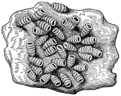
HORSE BOTS IN STOMACH
The bot fly lays its eggs on the hair of the horse. These, taken into the stomach, hatch out and give rise to horse bots or young maggots that attach themselves to the walls of the stomach. After becoming grown they loosen themselves and pass out with the feces.
—It is now believed that eggs are deposited near the feet and that the grub is taken into the mouth and becomes[125] partially developed in the digestive tract. It then burrows through the tissue until it reaches the region of the back. The only treatment that will amount to much is to destroy the grub as it is developing under the skin. If farmers and stockmen will systematically do this they can soon lessen the damage done. The heel fly annoys cattle, and the grub, when it escapes from the back, leaves a hole in the best part of the hide, causing loss in this way. After the grubs are in the back no treatment helps the animal very much; but the grub can be killed, thus preventing their developing into flies that would annoy other cattle. The grubs may be squeezed out and destroyed. Mercurial ointment may be rubbed through the hole and kill the grub, or chloroform, or creoline, may be injected into the grub with a hypodermic syringe. It does not require very much time to look after the number of cattle usually found on a farm.
is a very troublesome pest at times, and always causes trouble and annoyance to the flock when present, and occasionally causes considerable financial loss. The fly attacks sheep during the warm months, July and August generally being the worst. The presence of fly in the flock is easily told by the behavior of the sheep. The fly looks much like a house fly, only it is longer and it always attempts to lay its eggs just inside of the opening of the nose. As soon as the fly begins to get near the nostril the sheep will begin to run, will hold their noses close to the ground, and frequently huddle together as closely as possible for protection. When the fly does succeed in depositing the larvæ it begins immediately to work its way up the cavity of the nose and finally gets[126] into the small cavities in the head, where development goes on. It is during this period of development in the head that most of the damage is done. As the grubs grow larger a discharge from the nostril is noticed, which may soon become very thick and sticky, gumming up the nostrils and making breathing difficult. The sheep will often carry their heads low, but will frequently raise their heads and point their noses straight up.
The treatment may be either preventive or surgical. The first is within the reach of everyone owning sheep. Where only a few sheep are owned each individual should be caught and a mixture of tar and lard, or oil of tar and lard, applied to the nostril with a brush. This can be done in a short time and should be repeated every ten days or two weeks during the warm months. Narrow salt troughs may be made and the edges smeared with tar so that the sheep will get tar on their noses when they take salt. Turpentine may be applied high up in the nostril by means of a feather. Begin the preventive treatment early in the spring or whenever you know by the action of the sheep that the fly is bothering them, and you will have better success than to wait until the sheep are affected and undertake to cure them.
—See Bot Flies.
—See Heaves.
—A common disease of domestic animals attacking the bronchial tubes. It may be chronic, but is usually acute, and may affect one side or both. The most frequent causes of bronchial catarrh are colds. A sudden cooling of a heated body by drenching, by the breathing of cold damp air, may all bring on the disorder. Dust, smoke or gas, when inhaled, often produces the[127] same trouble. Acute bronchitis usually sets in with a sudden rise of the temperature of the body, and the animal seems to have a chill. This may be quite violent at times. The cough is noticed very much as with people, being short, dry, and husky. Later on, as the disease progresses, a frothy mucus follows the cough. Associated with the disease is a loss of appetite, constipation, and pains in the chest and rattling in the chest and throat. A favorite position of the horse is standing and of other stock that of lying down. Good care is essential in the treatment. That means, with good treatment, dust, smoke, and bad air are to be severely avoided. Plenty of good ventilation, but no draft; and warm, well-lighted quarters are very desirable. The animal should be blanketed to be kept warm in the early stages and a compress placed over the chest, with blankets over the compress. Frequent changing of this compress is desirable, say a change every hour or two. When the animal is suffering from a chill, stimulants are excellent. A tablespoonful of whiskey in a pint of water and given as a drench every half hour or hour will be helpful.
After the chill period is passed, small doses of tincture of aconite, say 10 to 15 drops, in a little water as a drench will assist in discharging the mucus. When the animal has become at ease, a mustard plaster applied to the lungs will help you somewhat. From now on the treatment should be good nursing and good food. Boiled flaxseed and gruel will be very helpful. A very helpful preparation may be made of the following: Nitrate of potash or saltpeter, tartar emetic, ground gentian root, equal parts. A half pound or pound in[128] all should be mixed thoroughly, and then a teaspoonful given three times each day. When all danger is passed, continue the careful handling and allow two or three weeks’ complete rest.
—Bunches are most generally enlargements of the bone. They are most serious in the region of a joint. They are caused, as a rule, by some injury, bruise, or wound. When first noticed they should be treated with a blister to insure a hasty absorption of the enlarged parts.
—Occasionally animals are burned or scalded so badly as to subject them to considerable pain. This may be relieved by the use of a strong solution of common baking soda. Following the use of this, apply an ointment made of one part of carbolic acid to 50 parts of vaseline. If vaseline is not available, then use in its place linseed oil.
—See Mammitis.
—A diseased condition of the udder, with the secretion of milk altered, the udder hot, dry, and caked, and the glands inflamed. The trouble may be due to external injury, to germs entering the teats or to the milk being kept for too long a time in the udder. As soon as noticed the udder should be bathed in hot water and massaged for several minutes. After being dried with a cloth rub on a salve made of 2 tablespoonfuls of gum camphor dissolved in 12 tablespoonfuls of lard. At the same time give 4 tablespoonfuls of saltpeter morning and night for two or three days. See also Mammitis.
—When a new-born calf comes into the world weak, puny, and listless, and dies in a few hours after scouring, bawling, and blatting and has sunken eyes and bloated belly soon after death, the disease by stockmen is called “calf[129] cholera.” Many calves so affected are really “living abortions.” They have just enough life at birth to exist a few hours and show the symptoms described, and such calves are usually the offspring of cows that, during pregnancy, have been incompletely nourished upon timothy or swale hay, or coarse fodder, without an adequate supply of other foods to balance the ration; or similar calves may come from fat, flabby, corn-stuffed, beef-bred cows.
The trouble may be prevented by proper feeding of the pregnant cow, but there is no cure. A majority of such cases, however, are due to germ infection. Cows affected with contagious abortion may produce affected calves; the afterbirth and navel cord are invaded by the germs in such cases and the calf is improperly nourished in the womb. In other instances, calf cholera is due to filth germs entering the calf’s system by way of the raw navel cord stump at birth, or the mouth when the calf nurses from a manure-contaminated udder.
Prevent infective cases by providing a clean, fresh-bedded, disinfected, whitewashed, sunlighted, ventilated pen for the new-born calf, and immediately wet its navel with a 1⁄500 solution of corrosive sublimate and repeat the application twice daily until the cord dries up, drops off and no raw spot remains. Also wash the hind parts of the cow and her udder with a two per cent solution of coal tar disinfectant before the calf is allowed to suck for the first time and repeat the washing twice daily for at least a week. Isolate affected calves. Bury or burn the dead.
—See White Scours.
—Malignant growths, the cause or causes of which are not known; nor can it be said the disease is infectious. While a very serious[130] disease among human beings, it is, fortunately, however, more rare among farm animals. The only treatment worth while is in surgical removal of the growths. If this be done when the tumors are first noticed and when they are small, their further appearance may not result. It is a good plan, if the growths persist in presenting themselves, to eliminate the affected animal from the herd. With cattle, it is possible to prepare them for market long before any cancer growths may reappear, and in this way the full market value may be secured with no danger when consumed.
—Frequently horses, in lying down, press the foot or the shoe against the elbow. This, in time, causes inflammation and ends in a tumor or shoe boil. The diseased condition is difficult to repair, as there is little flesh or muscle at the joint of the elbow where the trouble starts. Treatment consists of opening the boil and allowing the fluid to escape. In case the swelling is hot and painful, an application of lead acetate will prove comforting and helpful. In preparing the lotion, use 2 tablespoonfuls of acetate of lead to a quart of water. There is no objection to injecting a little of this into the opening. An injection of a little tincture of iodine once a day into the opening is desirable also. In treating cases of this kind, it is a good practice to wrap about the horse’s foot a pad of straw or hay for cushioning the foot. This prevents the wound from being further bruised, otherwise the cure may be greatly delayed, if not indefinitely postponed.
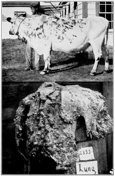
A VICTIM OF TUBERCULOSIS
This cow, reacting to the tubercular test, was killed. The bottom picture shows the extent to which tuberculosis had affected her lungs. At least ten per cent of the cattle in the United States have this dreaded and destructive disease.
—An inflammation resulting in a separation of the cap from the point of the bone of the hock. Cases of this kind are the results of[131] kicks or bruises. In the early stage, use 2 tablespoonfuls of lead acetate in a quart of water and bathe the injured part. When there is no longer any temperature, apply a blister composed of 1 teaspoonful of biniodide of mercury and 6 tablespoonfuls of lard. Apply this every week or ten days for several months.
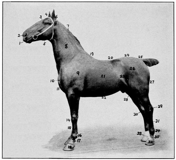
EXTERIOR POINTS OF THE HORSE
1 Lip, 2 Nostril, 3 Forehead, 4 Poll, 5 Cheek, 6 Ear, 7 Mane, 8 Neck, 9 Shoulder, 10 Point or Shoulder, 11 Breast, 12 Forearm, 13 Arm, 14 Knee, 15 Cannon, 16 Fetlock, 17 Pastern, 18 Foot, 19 Withers, 20 Back, 21 Side, 22 Underline, 23 Flank, 24 Croup, 25 Tail, 26 Haunch, 27 Thigh, 28 Stifle, 29 Hock, 30 Point of Hock, 31 Cannon, 32 Foot, 33 Coronet, 34 Fetlock, 35 Pastern.
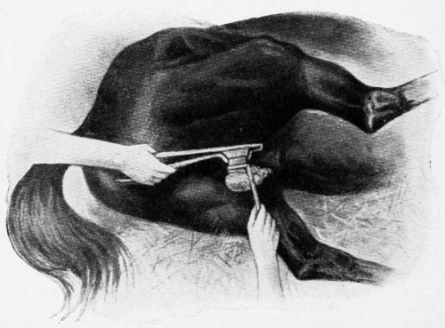
CASTRATION
—An enlarged condition of the knee most commonly found in cattle. It is caused by cattle getting up and down on hard floors. It is usually seen in stables where stanchions are used. A baggy tumor forms at the front and just below the knee. In some instances this tumor becomes very large and the cow walks about or moves with great difficulty. Where hard floors are covered with bedding, no trouble of this kind results. Applications of hot water are excellent. Liniment is also very good. Where the tumor has long existed and is stubborn an opening should be made at the bottom so that the fluid may be discharged. A little tincture of iodine injected into the opening once a day is good and at the same time an application of iodine rubbed over the outside will assist in reducing the trouble. Use one part of iodine to eight parts of lard and continue this treatment for a month or two.
—The removal of the testicles from male animals. Castration is practiced upon all the domestic animals. Only those male animals possessing desirable characteristics are retained entire. The operations are generally performed when the male animals become troublesome. In horses the time is usually at one to three years old; in cattle one to three months old; sheep at one to four months and pigs two to four months old. Dogs,[132] as a rule, become worthless if castrated. Cats grow to an enormous size when castrated.
—In the castration of all the domestic animals some general suggestions will be beneficial. (1) Secure the animal so he cannot injure himself or the attendants. (2) Do the castration during the early spring. (3) Give the animal exercise after castration. (4) Boil the instruments before operating, using warm water and any good hand soap. (5) Disinfect the skin over the scrotum before operating with corrosive sublimate 1⁄1000. (6) Wash the hands of the operator with soap and water, then disinfect with corrosive sublimate. (7) Great care should be exercised that no corrosive sublimate be left that stock may drink, as it is a deadly poison.
When the instruments have been boiled (sharp castrating knife and emasculator), cast (throw) the animal as carefully as possible. Secure the hind legs so they will not hinder the operator. The operator having his hands clean and the scrotum washed and both his hands disinfected, and also the region to be operated upon, the animal is ready for the operation. The lower testicle is grasped with the left hand and with the right hand an incision is made over the testicle, down to the testicle. The testicle is pulled upon until the cord is seen. Then the emasculator is used to crush the cord. This emasculator should be placed on the cord as high up as possible. Some like their horses castrated proud. This consists in leaving part of the testicle. This last method is not safe, as it allows the testicle to become infected and form what is commonly known as water seeds. A tumor grows on the cord and may become the size of a man’s head.
[133]
After the testicle is removed, then enlarge the first incision (cut) that was made through the skin so as to give plenty drainage. This incision should be about eight inches long for horses. By having a large incision the upper part can heal first, and there will be good drainage until the scrotum entirely heals. If possible turn the castrated horse out to pasture after the operation, and it will exercise sufficiently to keep the parts from swelling. Do not keep the animal in a dirty stable after it is castrated, as there is so much danger from infection in the dirty horse stable. If the horse is broken it can be put to light work a week after the castration.
Bulls do not need to be thrown to be castrated. The incision is made over each testicle, and the operation carried out in the same way as with the horse. Bulls are not so susceptible to infection as the horse.
—Commonly known as a cold, catarrh is recognized as an inflamed state of the upper portions of the air passages, with more or less discharge from the mucous membranes. The eyes often sympathize with this deranged condition, with a watery state as the result. The causes of catarrh or colds in animals are very much the same as those causing the same disturbance in human beings; as with people, so with animals, the malady should be remedied as quickly as possible. Bad air is one of the most frequently observed causes; consequently pure cold air with proper blankets to keep the body warm is considered the best treatment for simple catarrh when unaccompanied with other troubles.
One of the common symptoms is dullness and loss of appetite. The hair stands out and looks[134] rough, a slight cough may be noticed and sometimes a rattling is heard in the head. For cattle a mild dose of physic, consisting of one-half pound Epsom salts and 4 tablespoonfuls of sweet spirits of niter mixed in a pint of lukewarm water and given as a drench, is about all that is necessary. If the cold hangs on, mix together one-half pound of nitrate of potash or saltpeter and one-half pound of gentian root and give a teaspoonful of this three times a day until the animal is better. Of course good food should go along with this treatment. The horse should be fed soft food like bran mashes and be kept quiet in a well-ventilated stable. If the cold hangs on with him, mix one-half pound of saltpeter or nitrate of potash, one-half pound of sulphur, and one-half pound of ground gentian root and give a teaspoonful morning, noon and night.
—See Scab in Cattle.
—A disease fatal in violent attacks and not well understood as to cause. It is believed to be non-contagious, although frequently extensive outbreaks occur, suggesting that it may be contagious. The symptoms are not well defined, due, perhaps, to the fact that other diseases are included under the general name. Horses of all ages of both sexes are affected, and temperament and physical condition have nothing to do with susceptibility to the disease. Likewise mules are affected and the mortality among them is equally as great as among horses. The most acceptable belief as to cause centers around a bacterial organism that works in the membranes of the brain. However, some writers attribute the disease to ergot, smuts and molds supposed to be taken with the food. Moldy corn and moldy hay are believed to be associated with the disease. The[135] symptoms are staggering gait, partial or total inability to swallow, various muscular contractions and delirium.
Treatment is seldom effected, especially in violent cases. Mild forms frequently respond to cathartics, blisters on the neck, spine and throat. These give some relief. Small doses of aconite are also believed to be helpful. Some writers place choking, distemper, grass staggers, and blind staggers along with this brain disorder.
—See Anthrax.
—See Navicular Disease.
—Chicken or fowl cholera is a germ disease, and contagious. It attacks poultry of all kinds. Diarrhœa is a prominent symptom of the disease. Bad food or improper food may aggravate the trouble, but the germ introduced into the system either in food or drink, is at the bottom of it. At first the droppings will take on a whitish color. Diarrhœa will then result. The discharges will then become thin and watery, to be at times frothy and greenish in appearance. Fowls thus attacked soon lose their appetites and become stupid and take on a sickly appearance. The head drops toward the body, the eyelids fall, and the fowls stand around as if doped. Some recover, but, unless checked, the flock will be materially injured.
Of course dead fowls must be burned at once and lime and other disinfectants used to keep the disease from spreading. The well birds must be kept apart from the infected quarters. Care must be exercised that infection be not carried either by visitors or attendants from the sick to the healthy quarters. A common remedy consists of 1 part of sulphate of iron to 50 parts of water for drinking purposes. Another common remedy is to mix a[136] tablespoonful of sulphate of iron, 2 tablespoonfuls of dried blood, and 2 tablespoonfuls of tincture of opium with a pint of water. This is given in the food in doses of 1 or 2 tablespoonfuls of this mixture three or four times a day to each sick bird.
—Horses frequently choke from too rapid eating of oats, and cattle are very commonly troubled on attempting to swallow apples, turnips, or small pieces of ear corn. In either of these cases much distress is occasioned and serious danger. In treating the horse, the best treatment is to give it a little oil, after which rub the hand up and down the gullet to scatter the accumulated oats. Sometimes it is necessary to make an incision in the gullet through which the material is removed. Better have a veterinarian do this. When food lodges in the gullet of cattle, suffocation soon follows if it is serious and in the upper part of the gullet. When such objects have lodged near the stomach end there is less immediate danger. Of course the first treatment is to try to force the object down by using the hand, if at all possible. If this cannot be done a probang should be used. The probang should be very limber, so as to bend easily, and it should be used with great caution. Cattle often are killed by the accidental puncture of the gullet as the probang is pressed down toward the mouth of the stomach. Consequently no unyielding article like a broom handle or even a buggy whip should be used. If a regular probang is not available, a rope a little less than one inch in diameter can be inserted and gently worked down the gullet. Before using the rope, grease it well and make a knob at the end to be inserted. This knob can be made of cotton strings or muslin cloth.
[137]
—See Navicular Disease.
—See Catarrh.
—Colic is an inflammation of the bowels characterized by a spasmodic contraction of the intestinal walls. It is a very common disease in horses, and occasionally cattle and lambs are affected with it. Both the small and large intestines may be afflicted or only one of them. There are many causes, but feed and water are the controlling factors. An animal just stopped from hard work and given a large quantity of cold water, especially after eating, may be quickly troubled. And the animal hot from work, on drinking very cold water, often gets colic. Then, too, a change of food, or a change from dry feed to green food or eating some root crop when the animal is not used to it, may bring on the disease.
Then, again, some horses and cattle are more given to colic than others. Some individuals are never troubled, and others are almost constantly under its influence. If much inflammation sets in, a very serious case is on your hands. Two kinds of colic are known—the spasmodic, a contraction, commonly known as cramps of the bowels; and wind or flatulent colic or bloating. Some authorities add a third, and call it worm colic.
—This kind of colic is first noticed when the horse begins to paw with his forefeet, cringes, bends his head around as if looking at his side, lays on the ground and rolls as if in pain; then he stands quietly for a while and repeats these performances again. During the time between the spasms the animal is more at ease and frequently eats a little. When the spasms come on again the shifting about and the rolling are repeated. If the[138] cramps are severe the animal breaks out with sweat. The pulse is accelerated when the spasms are on, ranging from 60 to 65 beats a minute. If inflammation has set in, the pulse instead of rising and falling remains more constant and is high all the time.
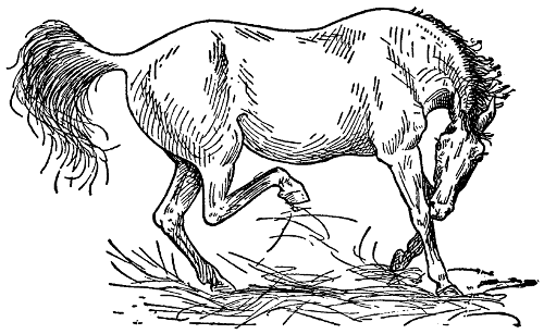
COLIC PAINS
A common attitude with colic. When seized with pains the horse paws, scrapes the ground with his front feet, stamps and strikes the belly with the hind ones, lays back his ears and looks around to his flank.
When the spasms are on, pressing the bowels seems to relieve the pain and please the animal, but if inflammation is present the pressure seems to increase the pain. The best treatment is to relieve the pain with an opiate, and next to obtain a free action of the bowels by a purge. Many prescriptions have been suggested, among which is the following: 4 tablespoonfuls of sweet spirits of niter, 4 tablespoonfuls of laudanum, 1 tablespoonful of ginger and 1 tablespoonful of common soda. These are added to a pint of warm water and given as a drench.
[139]
—This form of colic, though not so acute, is much more constant than the preceding form. The body is swollen in the region of the bowels, the gas extending quite generally through the region. There is also a tendency to inflammation. The pulse will be noticed as more rapid, and at the same time more feeble, the breathing will be more pronounced, and the animal less steady on its feet. In treating the patient it is advisable to unload the rectum with greased hand and arm, and the admission of warm water with soap in it, is also likely to be beneficial. A little turpentine mixed with the soap and water is good. The intestine is to be cleaned out as far as the arm will reach, but a violent purge is unwise, as that only intensifies the inflammation. Naturally the first thing is to mildly open the bowels. For this give 15 or 20 tablespoonfuls of linseed oil and 5 or 10 tablespoonfuls of spirit of turpentine. If the case continues, it is advisable to call a veterinarian, and it may be necessary to use the trocar and canula. If the instrument is sterilized, no great risk attaches to the operation, while immediate relief is secured as the gas passes out through the tube, and the distention is visibly reduced. An excellent mixture for this kind of colic consists of 6 tablespoonfuls of chloral hydrate, 6 tablespoonfuls of laudanum, 3 tablespoonfuls of sulphuric ether, 2 tablespoonfuls of turpentine, and 10 tablespoonfuls of ginger. Of this give 2 or 3 tablespoonfuls in a half pint of warm water and repeat every half hour for 3 or 4 doses and then place the doses an hour apart until all danger has passed.
When there is a good deal of gas with considerable swelling an excellent drench is made of 2 tablespoonfuls of powdered aloes, 4 tablespoonfuls[140] of spirits of ammonia and 4 tablespoonfuls of sulphuric ether. This should be mixed with a pint of water and given promptly. In case of considerable pain use this: 4 to 6 tablespoonfuls of hydrate of chloral and eight tablespoonfuls of sugar mixed in a pint of water and give as a drench.
—The collection of solid mineral matter in the urine may become lodged in the kidney, the ureter (duct leading from the kidney), the bladder or urethra (the duct leading from the bladder). All animals are more or less subject to these conditions, and yet are not so affected as they are sometimes thought to be. Many a case of so-called kidney colic is in reality an affection of the digestive system. The cause for these mineral accumulations perhaps varies under different conditions, yet the most common circumstance under which they occur is during the time when animals are fed exclusively or largely upon dry feed such as exists in the winter time where silage is not fed. Wheat bran has been attributed as one of the most sourceful means of bringing on this trouble. When it is fed with succulent feeds and an abundance of water allowed these disorders do not occur.
The symptoms do not differ a great deal from some forms of colic, due to stomach or intestinal disturbances, especially in the frequent attempts to empty the bladder. The animal usually shows more or less pain from the restless condition, looks around at the flank, dribbles his urine frequently, which is occasionally blood stained. There may be a complete obstruction of the passages, in which case no urine is voided.
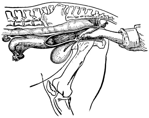
RETENTION OF THE URINE
By means of a catheter the greater portion of the urine can be drawn off. The operation is shown in the picture.
with the location of the trouble,[141] in which little can be accomplished when the gravel or stone is located in front of the bladder. If within the bladder, not obstructing its outlet, it is not likely to make its presence known. Agents should be given, however, to overcome the pain and to relieve the frequently existing spasm at point of obstruction, as far as possible, which may allow passage of stone. Give 4 tablespoonfuls of laudanum or chloral to a dose and repeat in two or three hours if any pain or trouble is still indicated. In inducing the animal to drink liberal quantities of water the condition may be somewhat relieved by making the urine more watery in character and possibly dissolving a portion, allowing the remainder to pass along its course. When the[142] obstruction occurs within the urethra the removal should be made by incising through the tissues on to or near the obstruction, removing by forceps and suturing up the wound. A skilled operator is required for this, hence the veterinarian should be called.
—An infrequent movement of the bowels with the dung hard and dry. The animal is said to be bound up or costive. Bad food, improper feeding, lack of exercise, all contribute to the trouble. Treatment is in the line of laxative and succulent food, such as wheat bran, green grass, silage and linseed oil meal. If the case is one requiring immediate action give any of the usual purgatives, but do not continue their use as a regular thing. If green grass is not possible, nor silage available, give one or two teaspoonfuls of the following mixture in the food three times a day: Equal parts of ground gentian root, powdered nux vomica, powdered ginger and sulphur.
—Small swellings or tumors on the sensitive heel in the triangular space between the bars and the wall of the heel. These are found in the fore feet only, and almost always on the inside heel. They are caused most frequently by bad shoeing or from wearing the shoe for too long a time. These growths do not always cause lameness, although, as a rule, they do. They are, however, always sensitive to pressure and usually appear as tumors of a hard, corny character. Neglected corns are liable to fester and must then be laid bare by the knife and be poulticed. Neglect of this treatment results in the matter or pus finding its way up through the coronet. Thus quittor may result.
Give the foot a careful dressing by paring the heel, and bathe the corn with a weak carbolic acid[143] solution. After doing this, place a fold of muslin over the corn and then over all a bran and linseed poultice. A complete rest from work, hard roads and shoes should now be given the animal until the corn has entirely disappeared. When the feet are again shod, leather should be used as a protection. Many corn salves are recommended, but unless the corn be removed and the pressure taken from the wound, there can be no cure, even though the tumor is pared away.
—When cattle are allowed to run in stalk fields it frequently happens that a large per cent die from various causes. All these troubles are classed under the one term—cornstalk disease. In some western fields where there is a second growth of cane stalk late in the fall an early frost will at times develop in the stalk a deadly poison (hydrocyanic acid), which kills the animal in a very few minutes after eating it. This poison has not been found in the cornstalk.
In the last year or two some of our state experiment stations have been investigating several molds which seem to affect not only cattle but horses as well. These molds grow quite abundantly upon cornstalks, alfalfa, and other forage crops. The death of a great number of animals has been traced directly to the feeding of such affected fodder, hay, or corn. These molds, however, must have a certain amount of moisture for their growth, and it has been shown that when the feeds have been properly harvested and sheltered no trouble has resulted. Only in materials exposed to the weather, allowing the development of these lower forms of plant life, has serious trouble been found.
In the treatment of these troubles nothing reliable can be given, as the disease usually comes on[144] without any warning and the animal dies suddenly. Much of the trouble can be avoided by allowing the animals only a limited amount of the feed or in the stalk field a few hours only each day. It is necessary that plenty of pure water should be given frequently and enough of other roughage to keep the animals from gorging themselves on the fodder.
—An infectious disease passed from one cow to another. It affects herds in all parts of the world and is similar to smallpox in the human being, only it is not so fatal. When first affected the cow is feverish, slacks somewhat in the milk flow, and presents little red pimple-like spots around the teats. In a day or two these become enlarged and become blisters, containing within a watery fluid, which, if not broken, dry up themselves and form scabs, leaving the teat in time perfectly natural. Ordinarily, special treatment is not given. There is no objection, however, to providing a simple tonic composed of one-quarter pound saltpeter, one-quarter pound sulphur, and one-quarter pound ground gentian root. Give a teaspoonful of this night and morning in a mash. The teats should be bathed, just before milking, with any common disinfecting solution. If the sores are slow in healing, sweet oil, to which is added a little carbolic acid, will soon correct the trouble.
—See Sand Cracks.
—A habit of biting the manger or other objects, often sucking in the air at the same time. This bad habit is frequently called wind sucking. It is the result of a habit formed when young. There is really no cure when the habit is once formed, but different measures may be employed to lessen the fault. A broad strap firmly[145] placed around the neck brings the desired effect with some individuals.
—This bad habit usually begins in colt days. It may arise from a sore tooth. The colt, to relieve the feeling, bites the manger, and in so doing acquires the habit. When hanging on to the manger, air is sucked in and this frequently brings on colic. The best treatment is to break up the habit. Examine the mouth first to see if anything is wrong with the teeth. Muzzle while standing in the stable. The old cribbers never give up the habit.
—See Colic.
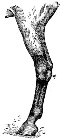
CURB
While common to all varieties of the horse, curbs are most frequently seen in the lighter breeds and especially in roadsters and trotting horses.
—A sprain or injury to the ligament situated on the back part of the hock joint. Anything that puts too much stress on this part, such as holding back heavy loads going down hill, or backing up too heavy loads, or the hind legs slipping too far under the horse’s body, may cause curb disease. It is also caused by kicks or by the whiffletree striking against the back of the hock joint.
There will be swelling and heat in the part and lameness. In some cases there will be swelling,[146] but no lameness. If the swelling is hot and tender to the touch, mix half an ounce acetate of lead and two ounces tincture of arnica with one quart of water. Shake up and apply a little to the swollen part three times a day and continue until the heat and swelling disappear. If there should be any swelling after the heat and lameness have disappeared, mix 1 teaspoonful of biniodide of mercury with 4 tablespoonfuls of lard. Rub on a little with the fingers, let it remain on for 24 hours, then wash off with warm water and soap and repeat the blister in three weeks if needed. In cases where there is swelling, but no heat or lameness, the lotion would be of no use, but the above blister should be used as directed. In old or long standing cases of curb, if the animal is not lame, it is best to let it alone, as medicines would be of no service.
—In man there are two forms of this trouble seen rather frequently, but among domestic animals only the insipid form is common. It is often simply a sign of some other disease, but not infrequently occurs under similar circumstances; such as certain forms of indigestion, the result of eating musty or damaged feed. The most characteristic symptom, of course, is the frequent urination of liberal quantities of urine. Associated with this is usually an unabating thirst. The animal loses flesh rapidly, the flanks are tucked up, the coat is dull, languid and staring, and great weakness is shown. If not relieved, the animal may die from exhaustion. In the second form of diabetes, the distinguishing feature is the presence of sugar in the urine.
If in a working animal it should be laid off from work. Search should be made for the cause of trouble. If any of the food appears[147] suspicious it should be substituted with wholesome food. To relieve the ardent thirst and assist recovery, a teaspoonful of the crystals of iodine should be given in a ball of linseed or other pasty material. It may be desirable to repeat this in three or four days. Also give in the drinking water 4 tablespoonfuls of bicarbonate of soda three times daily.
—See Dysentery.
—See Obstetrics.
—There are only two satisfactory methods of treating animals with a dip. The first is hand treating, where the number of animals are few and easy to handle. In hand treating the animal the dip is applied with scrubbing brushes, sponges, etc., and all parts of the body liable to infection should then be thoroughly and vigorously rubbed. If hand treating is properly performed it is an excellent method. The second method consists of immersing the diseased animals in the dipping solution. There are two forms of vats in use for this purpose. The cage vat is designed for comparatively few cattle. As its name implies, it consists of a cage in which the animal is placed and then lowered into a vat containing the dip. Where a large number of animals are to be dipped, the swimming vat is very popular. The animals are forced to pass through the vat, which contains sufficient dip to completely immerse them when they plunge into the solution.
The coal-tar dips are made from some of the products of the distillation of coal tar. When mixed with water they form a milky emulsion, having a strong odor of coal tar. The coal-tar[148] preparations, in addition to being used as parasiticides, have become very popular disinfectants in hospitals. These preparations are used with good success on all open wounds, where a disinfectant is required. In poll evil and fistulous withers they are extremely valuable, owing to the fact that in addition to their power as a germicide they have been perfectly safe to place in the hands of persons not accustomed to handling drugs, because of their non-poisonous nature. They have been found quite efficient when used in three per cent solution.
—Some cattle breeds are hornless. Most, however, are not. Removing the horns is done quickly and is more humane than to permit them to remain, by which death frequently follows to stock and even to people. The dishorning machine is intended for animals whose horns are not removed when young. The simplest method of dishorning is to use a stick of caustic potash. Apply it to the small horn button when a calf is a few days old. Moistening this and rubbing the potash over the skin will permanently destroy the horn tissue and no horns will result.
—See Strangles.
—A condition in which the fluid portion of the blood escapes from the blood vessels and collects in the body cavities or under the skin. Any sluggish condition of the blood occasioned by disease or faulty nutrition may induce this collection in various parts of the body. Dropsy is, therefore, not a disease, but a symptom of some other disease. This being the case, treatment depends upon the original disease, upon the nature of which depends in turn the possibility of permanent or temporary cure.
[149]
A mild attack of dropsy is indicated when the legs of a horse swell up, due to lack of exercise and poor circulation as occasioned by standing in the stable. The first thing, of course, is to start better blood circulation. Hand rubbing is good; bathing with hot water acts similarly. Any medicine that stimulates the action of the kidneys will prove helpful. Saltpeter is excellent for this. Use once a day for three or four days in succession, and give 4 tablespoonfuls at a dose. In connection with this treatment supply the animal with succulent or laxative food, that the bowels may be kept free and open. Any of the tonic condition powders will help.
—See Obstetrics.
—An inflammatory, non-contagious disease of the skin in which eruptions may occur in the form of vesicles, pustules, crusts, scales, or simple redness. Its principal victims are animals fed rich food, the penalty being associated with some gastric or intestinal disturbance. Treatment is both external and internal. The former should be in the nature of washes for cleanliness and healing. Tar soap is recommended. A wash made of 4 tablespoonfuls of carbonate of potassium dissolved in a quart of water is also excellent. After a good rub with this, wash off with warm water.
If itching causes any distress, prepare a wash consisting of 2 tablespoonfuls of acetate of lead, 8 tablespoonfuls of tincture of opium and a quart of water. Where scales have formed and the skin is thick and scurvy, rub in a little with the fingers some biniodide of mercury and vaseline. Use 2 teaspoonfuls of the mercury and 8 tablespoonfuls of the vaseline. One application will do the work. If the case is bad, several parts being affected, treat[150] only one part at a time with the mercury salve. Be certain to have the animal tied so that he cannot get his mouth to the treated region.
For internal treatment let the physic come first. For horses, mix 4 tablespoonfuls of aloes, 4 tablespoonfuls of ginger and 4 tablespoonfuls of soda carbonate dissolved in a pint of boiling water. Let cool to proper temperature and give as a drench. For cattle, give a pound of Epsom salts and 4 tablespoonfuls of ginger in water as a drench. Following the physic should come a good blood tonic. To prepare this, mix 16 tablespoonfuls each of nitrate of potassium and sulphate of iron. Give in doses of 11⁄2 tablespoonfuls daily in a bran mash until all is used.
—An inflammation of the lining membrane of the large intestine near the rectum, accompanied with straining, discharge of blood, and fever. Poisonous and irritating food causes it, stagnant and foul water favors its development, but any exposure to cold or excessive heat or overwork may bring it on. In cattle the acute form is attended with shivering, arching of the back and tenderness about the loins. The animal grunts, yawns, grinds its teeth, and, at short intervals, discharges from its bowels a thin, ill-smelling dung mixed with blood and pus. The thirst is excessive, the animal is dull and stupid, and loses flesh rapidly. After the disease has gone on a few days, the hide becomes rough and unhealthy, the teeth loose, the dung bloody and fetid, the eyes sink in the head and dropsical swellings appear about the lower jaws and legs, and usually the creature dies exhausted. For acute dysentery, when seen early, give horses a drench consisting of 15 tablespoonfuls of castor oil, 8 tablespoonfuls of laudanum, and 1 pint of[151] linseed oil. The rectum and lower bowel should be washed out with large injections of simple warm water. For chronic forms 10 grains of calomel, a teaspoonful of opium, and 4 tablespoons each of gentian and chalk are advised. These are to be mixed and given either as a ball or as a drench once a day. Six tablespoonfuls of laudanum in a pint of boiled starch every two hours until the straining ceases, is also very good. When cattle are affected, remove from grass or other succulent food, put on a dry diet and give a pint of linseed oil every day until recovery. If the action of the bowels does not cease promptly, give 2 tablespoonfuls of powdered alum and 2 tablespoonfuls of powdered ginger in a quart of milk once or twice a day until the discharge moderates. An excellent medicine is 10 tablespoonfuls of castor oil and 4 tablespoonfuls of laudanum mixed with linseed gruel and given as a drench.
—See Inflammation of the Bowels.
—See Fits.
—See Influenza.
—A parasitic fungus that grows on different species of grass and produces in one stage of its development black or purple enlarged spurs causes ergotism. The disastrous effect of ergot seems to appear in the late fall and winter, when hay or straw infected with ergot are continuously fed. The animals will be troubled with irritation of the bowels and a sloughing off of the extremities. Frequently the animals lose parts of their tails or ears or hoofs. In others, gangrenous sores appear. In the early stages of the poisoning the symptoms are not clearly marked. The best treatment is secured by an entire change of food, so as to remove the cause, and then[152] to follow with good laxative food. Of course, medicinal treatment will not be satisfactory if an important part of the animal like the hoof were to be destroyed. So much expense would be connected with keeping the animal until a new hoof had been formed that it is better at the beginning to destroy the animal unless very valuable. Where sores only manifest themselves such treatment as given an ordinary wound will be efficacious, provided food absolutely free of ergot is supplied.
—An inflammation of the skin and tissues beneath. Owing to a blood poison, it is characterized by a swelling and hardness of the affected parts which has a tendency to spread and form abscesses. In horses and cattle, erysipelas is nearly always the result of wounds and generally of those in the legs of animals weakened by hard work and poor food, or else in young animals whose blood is vitiated by the poison of glanders or some other animal contamination. The disturbance is noticed on the third or fourth day after the injury in the immediate neighborhood of the wound. The skin is swollen, smooth, hot, tender, and painful. The swelling gradually extends around it, sometimes deep into the muscles. The surface is hard and tense, but often when the finger is firmly pressed upon it and withdrawn a depression is left. In severe cases chills occur, the pulse is weak and quick, the breathing hurried, the bowels constipated and the urine scanty and highly colored. There is considerable thirst, but no appetite. A brisk purge is the first step in treating. Follow the purge with tincture of chloride of iron, 4 teaspoonfuls in a pint of water. Give this every three or four hours. At the same time give internally 4 tablespoonfuls of hyposulphite of soda in a pint of water three times a[153] day. Externally bathe the wound with the following mixture: Tincture of chloride of iron, 4 tablespoonfuls, and alcohol one pint. Another good ointment is sugar of lead 4 tablespoonfuls in a pint of water. This should be applied with a wet cloth to the diseased parts.
—See Glanders.
—Any rise in temperature above the normal. It is, as a rule, a symptom of the body’s reaction to some form of infection. It is, therefore, not a disease in itself, but an indication of some disorder occasioned by infection or poison. To treat fever is not so necessary as to remove the cause that brought about the disturbance in the first place. It follows from this that fever is not a cause, but a result. Germs come first, and fever is only a sign that tells of their presence. Another thing brought to light in reference to fever is this: Germs are less active, their vital energy is weakened and their power lessened when the heat in the body is increased. Consequently they are less active in their destructive tendencies as the temperature rises. Fever is, therefore, a provision of self-defense, and the body’s plan of bringing its forces together to battle against the germ foes that have invaded it.
Just what degree of temperature is to be considered is difficult to establish. Many things enter into the problem, like exercise, age, food, and mode of living. In general, however, any special rise above the normal, whatever that may be, is the signal of danger and infection. A rise of a degree or two indicates a mild disturbance, hence a mild fever; an elevation of two or three degrees indicates a slight fever; of four or five, of considerable fever; and if six or seven, of high fever. When the elevation reaches 108 degrees, the limit of life[154] has just about been reached. In some diseases there is a regular alternative between morning and evening temperatures. In others, the course is continuous, with slight variations, while in others the course is intermittent. In this last named it varies at different portions of the day, but reaches a normal at a certain time each day.
The pulse-rate usually bears a certain relation to the height of the disease. Consequently the pulse should be taken in connection with the fever height indicated by the thermometer. A fast pulse and a high fever in general is more serious than a high fever with a pulse only slightly above the normal number of beats. There are exceptions to this however, as, for instance, in cerebro-spinal meningitis. In the early stages of fever, the development cannot at the moment always be decided. In many cases little treatment, if any, will be necessary. The caution should be observed, nevertheless, of ascertaining the cause of the disturbance, if possible. In any case, simple cathartics can be given, good air provided, nourishing feed supplied, and time allowed for careful observation of the system and of the actions and movements of the animal.
—A chronic discharge from some tubelike channel, with no tendency to heal. Fistulæ are most common in horses. They may be located on the withers (fistulous withers), on the side of the face (tooth fistulæ), on the breast bone (sternal fistulæ), or on the lower jaw (salivary fistulæ). Fistulous withers are caused from some external injury (the animal rolling on a rock, ill-fitting collars, the saddle pressing on the withers, or from being struck by a club). Tooth fistulæ are caused by a decayed tooth. The pus in trying to get out of the[155] body takes the easiest course and eats through the bones of the face and escapes, causing a chronic discharge. A sternal or breast fistula is caused by some sharp object being run into the breast and striking the breast bone, injuring it and causing decay and pus formation. A salivary fistula is caused by an injury to the tube which carries the saliva from the gland to the mouth.
—At first a large swelling appears on one or both sides of the withers. In about a week this enlargement becomes soft, and the fluid contained in it can be distinctly felt. If left to itself the swelling gets larger and softer, and in a month or so breaks and discharges the contents. The fluid that comes from the swelling is first thin and streaked with blood; later it contains yellow-appearing masses. The last material is the pus. The sack that formed at the time the fistula was caused is a hard, firm membrane. This keeps the wound from healing. For this reason the discharge becomes chronic. The wound may heal and there will be no pus discharged for a month, then the old opening will be broken and the pus will flow out again until the sack is emptied. This healing of the wound and then breaking again may be kept up for years, unless the disease is properly treated. As a general rule, the affected animal runs down in flesh.
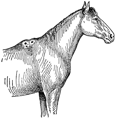
FISTULOUS WITHERS
Sometimes only the skin and tissue immediately under it become affected. In such cases little trouble need be anticipated; but if the cause is not removed, the deeper structures, muscles and bones, may become diseased.
Treatment for fistulous withers consists of opening the swelling and inserting muslin strips that have been dipped into terchloride of antimony. Insert one and remove, inserting another and leave in the opening for three or four hours. Repeat this operation every four or five days for a month. In addition rub on the outside of the swelling once every two weeks a mixture made of 2 teaspoonfuls[156] of cantharides and 4 tablespoonfuls of lard. The tooth fistula usually calls for the removal of the tooth and thorough disinfection of the opening from the face through to the mouth. With a sternal fistula the diseased bone may need to be scraped and then antiseptic washes used daily. The salivary fistula is more difficult to treat. Better have the veterinarian to examine, and an operation may be necessary.
[157]
—Some horses are subject to fits, and with them it is incurable. These should not be driven, because, when the attack comes on, injury may result to the animal itself and to the occupants in the carriage. The cause of the difficulty may be overfeeding, bad circulation or indigestion. When an attack occurs the best treatment is to throw cold water over the head. If this attack is repeated you had better consult a veterinarian.
—See Colic.
—Fleas are always a nuisance and always disagreeable. They live in dry, filthy quarters and associate with dogs, hogs, and chickens. To keep fleas away or to destroy them when at hand, clean the quarters occupied by the animals, destroy the bedding and add lime and disinfectants. Dogs may be washed in a creolin solution of, say, 2 tablespoonfuls of creolin to each pint of water. To disinfect chicken, hog, and horse pens use in a hand spray any of the so-called sheep dips or other preparations manufactured for lice, itch, mange, or insect troubles.
—These pests are a nuisance on every farm. While they do not directly cause death they greatly worry and irritate farm stock, especially in summer, and in this way greatly affect the results whether along dairy or beef lines. It would be impossible to estimate the misery these pests inflict on the stock of the country during a single year. Aside from the pain that flies inflict on domestic animals, they are carriers of disease, both to the human family and the beast family. A great many common infectious diseases are spread by flies, including such serious diseases as typhoid fever and tuberculosis. The only treatment is in way of prevention. As the breeding places are in filth and[158] manure, it follows that if these be destroyed or removed, and not permitted to accumulate, the floods of flies will disappear. The fly remedies now on the market are excellent. When sprayed about the stable premises and on the animals the flies stay away until the application evaporates. Darkened stables are not attractive to flies, and by this means the nuisance and annoyance is minimized.
—See Liver Flukes.
—This malady generally affects ruminants, but, although found most often in cattle, sheep, and goats, it may be transmitted to swine, and, in some instances, to horses, dogs, cats, birds, or human beings. In most cases where proper disinfection is made the animal recovers in about 15 days. The most dangerous thing about foot and mouth disease is the fact that it spreads so rapidly. The virus which transmits the disease may be carried by railroad cars, bedding, feeds, dairy products, dogs, cats, birds, or persons. A dog running through a pasture may be the means of infecting a whole herd.
The cause of the disease has not been satisfactorily determined, but it is definitely known that the virus which reproduces the disease comes from the ulcers and natural secretions and excretions of the body, such as milk, saliva, perspiration, feces, urine, and exhalation. The contagion is not harmful when dried. Infected animals lose the power of transmitting the disease when the ulcers of the mouth, feet, and udder have healed.
In from three to five days after infection the animal has a moderate fever. The appetite is lost and the mouth is kept closed. There is a dribbling of saliva, and in two or three days yellowish-white[159] spots the size of a hemp seed appear on the gums, the lower surface of the tongue, lining of the mouth and on the lips. These eventually attain the size of a silver dollar. They run together, burst and form painful, foul-smelling ulcers. At this stage the saliva is more profuse and ropy and the animal makes characteristic smacking noises with the mouth.
Infected animals lose flesh rapidly, in some cases as much as 100 pounds in eight or ten days. The milk is thick, yellowish-white, has a bad taste, and is with difficulty made into cheese or butter. The reduction in milk yield during the sickness and for some time after recovery is 50 to 75 per cent.
Usually, a short time after an appearance of the disease in the mouth parts, there is a redness, heat and swelling of the skin at its junction with the hoof and especially between the toes and upon the soles of the foot. Similar ulcers to those on the mouth appear on the feet and soon burst. The animal becomes lame and moves stiffly and lies down a great deal. These ulcers ordinarily heal up in one or two weeks.
In some cases the animal dies suddenly, in others lingers a few hours with difficult breathing and discharge of blood from the nose, and finally dies of paralysis of the heart and lungs. In still other cases emaciation and reduction of milk flow is the only bad result. Sometimes ulcers form at the root of the horn and cause the horn to drop off.
Owing to the nature of the disease, its contagion and danger, treatment should be in line of prevention and in destruction of infected animals. While the disease yields to treatment, our best suggestions when the disease is suspected is in notification to the state officers and in securing the services of[160] a veterinarian who will be able to advise what is best to do.
—See Wounds and Their Treatment.
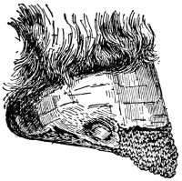
FOOT ROT
A disease usually associated with sheep. It is sometimes so serious that the entire hoof rots away.
—A chronic inflammation of the foot, marked by ulceration, softening of the hoof, lameness, and the discharge of a sticky material which has a very fetid odor. It is a contagious disease, and is produced by a germ that lives in the soil and gains entrance to the feet through wounds and surfaces chafed by barbed grasses and stones, or by gritty clay, which becomes lodged between the toes and hardens there.
The first symptom is a slight lameness. If the affected foot be examined, that part just above the horny part of the cleft of the foot, either in front or behind, will be found inflamed, feverish, and moist. Erosions or ulcers soon appear, generally on the heel. These penetrate the foot and burrow beneath the horny parts, causing fistulous tracts from which exudes a foul-smelling pus possessing an odor sufficiently characteristic to indicate the disease in a flock, even without a close examination. In time, the foot becomes greatly overgrown and deformed, the hoofs increasing in length and curling upward. In bad cases, the suffering is so great the animal lies down most of the time, but when only the front feet are diseased, it will crawl around on its knees.
[161]
That the disease is contagious is shown by the fact that it generally starts in one foot and spreads to the others, and, at the same time, the feet of other sheep in the same flock become diseased in the same way, the outbreak covering a period of several months. In cases that recover spontaneously the foot is deformed and the joint is stiffened. It is only in virulent outbreaks where all the feet are diseased, or where some complication, such as maggots, is present, that deaths occur.
Having as its cause a microbe, it is proper to take measures of prevention as well as cure. In purchasing sheep, it is highly advisable to keep them isolated for a week, as a test. All overgrown hoofs should be trimmed. Sores or wounds, from any cause, should be carefully disinfected daily. Low, boggy lands should not be used as pasture for sheep, and dirty, unsanitary pens should be made sanitary, as these all predispose to an outbreak of the disease.
As treatment, first isolate all affected animals. Mild cases are best treated by making the sheep stand for several minutes daily in a trough containing a disinfectant, or, better still, by arranging the trough of suitable length with fenced-up sides and a widened entrance, so the sheep can be easily started into the inclosure and made to wade through the disinfectant.
In bad cases and where the hoof is underrun with pus, the horn and all overgrowths must be cut away so as to expose the diseased parts to the action of the disinfectant. The foot should then be dried, dusted with finely powdered burnt alum, and bandaged to keep out the dirt. This antiseptic treatment of the feet must be kept up daily as long as the disease exists. Any of the following may[162] be used: 1 pound chloride of lime to 12 quarts of water; 1 pound of pure carbolic acid to 4 gallons of water; a solution of creolin; a coal-tar disinfectant of the same strength; or any good sheep dip containing these substances in the proper amounts.
—An inflammation of the sensitive or soft structures between the hoof and bones of the foot. The popular belief that founder is to any extent in the legs and chest is probably an error. The disease is in the feet, and those symptoms which make it appear as a stiffness in the legs and shoulder are but the natural results of soreness in the feet. The same statement might be made regarding those cases which are popularly described as “stove up in the shoulder.” Instead of the soreness being in the shoulder in these cases, it is generally in the feet, or at least below the knee.
It is somewhat difficult to explain how those influences or causes which are known to produce founder bring about that condition, but observation shows clearly that an irritation of the digestive tract, or in fact, any extensive irritation of any mucous surface, may produce an inflammation of the sensitive laminæ of the feet; that is, founder. Therefore founder may be produced by a change of feed or excessive feeding, a change of work or excessive work which results in exhaustion, large quantities of feed or water when warm or fatigued, sudden changes of temperature such as cooling too fast when sweating, and a long drive on hard roads, especially without shoes. Excessive purging or diarrhœa may also produce it. Founder also occasionally results from irritation of foaling, but this is not common.
[163]
There is no essential difference in the nature of the disease determined by the particular agent or condition which causes it. “Water founder,” and that produced by over-feeding, concussion, or extreme fatigue are, in so far as the character of the disease is concerned, one and the same thing.
FOUNDER
In bad cases of founder the foot shrinks from the wasting of the sensitive substances. A typical foundered foot is pictured here.
in the fore or hind feet or in both; but generally the fore feet are those affected. A stiffness and disinclination to move are perhaps the first symptoms noticed. The position in which the animal stands is characteristic. The fore feet will be placed well forward, so that the weight will be borne by the heels, while the hind feet are brought well up under the body in order to take as much weight off the front feet as possible. This position gives a rather unsteady appearance to animal, and the hind feet are frequently shifted in order to maintain as steady a position as possible. From this fact founder is frequently mistaken by inexperienced persons for a disease of the kidneys. The body temperature is usually considered increased; that is, there is fever—as it is generally expressed—due to inflammation in the feet. As is usual in the first stages of inflammation, the pulse beat is increased in frequency and force. An increase of heat in the feet, with a manifestation of pain when the hoofs are tapped with a hammer, are, when taken with all the foregoing facts, sufficient evidence of founder.[164] When founder occurs in one foot, however, as it sometimes does, the diagnosis may be more difficult for the inexperienced. When it occurs only in the hind feet the position which the animal takes will not be different from that taken with founder in only both fore feet but from different causes. The hind feet are brought well forward under the body, but for the purpose of throwing such little weight as is borne on them on the heels.
—Remove the shoes and apply moisture to the feet. The latter may be done by standing the animal in water five or six inches deep each day, several hours at a time, or by the application of a poultice of wheat bran or some such material, or by wrapping the feet with cloths and keeping them thoroughly saturated with water. The animal should always be encouraged to lie down and take the weight off his feet, which is beneficial. When this occurs, a poultice of some sort must be used to apply moisture to the feet. It may be applied by the use of a sack large enough to envelop the foot and hold sufficient of the poultice to retain the moisture for some time. This application of moisture to the feet should be continued until the severity of the inflammation and the lameness have subsided.
Unless the founder be due to excessive purgation, a quart of raw linseed oil should be given as a purgative. During the first 48 hours from 30 to 40 drops of tincture of aconite may be given every three or four hours. Four tablespoonfuls of nitrate of potash (saltpeter) should also be given three times a day in the feed or on the tongue. If the lameness continues after the acute symptoms have subsided, a rest of several weeks on a soft pasture and the application of a blister around the[165] top of the hoof are recommended. The following mixture has been useful as a blister: Red iodide of mercury, 1 part; lard, 4 parts; cerate of cantharides, 4 parts. Apply around the top of the hoof, except at the heels, and rub for 10 to 15 minutes. The animal should be tied so that it cannot get its mouth to the blistered part for several hours after the medicine has been applied.
—In a majority of cases the above treatment will be followed by a good recovery, but an animal once foundered is probably more likely to suffer from a subsequent attack. If the lameness does not entirely disappear in a week or ten days, it is seldom that a complete recovery takes place. In such cases the animal is likely to remain unfit for road work and to continue to show more or less soreness. These are the cases that are later said to have “chest founder,” or “stove up in the shoulder,” owing to the fact that the muscles of the chest waste away from lack of free use.
In some cases still more serious results follow an acute attack of founder. The inflammation may be so severe that there is separation between the hoof and structures, the formation of pus, and a descent of the central organs of the foot, which causes a bulging of the sole. In such cases, even though recovery takes place to such an extent that it is advisable to allow the animal to live, it is not fit for work, and can only be used for breeding purposes.
—See Chicken Cholera.
—A symptom caused by worms in the windpipe; oftenest seen in young chicks and turkeys. Birds droop, cough, and lower their wings.[166] A feather moistened, but not dripping, with kerosene or oil of turpentine is the commonest remedy. Cleanliness of food, water and quarters is the great preventive. Poultry men who keep their chicks on ground not used for chick raising the previous year, and who insist on strictest cleanliness, report highly satisfactory results in avoiding gapes.
—A swelling, accompanied by inflammation of the udder. It may be caused by kicks or blows, by germs getting into the udder, or as a result of holding the milk too long. Do not use the milk when the udder is affected. For garget rub with hot camphorated oil twice a day. Give as medicine 8 tablespoonfuls of hyposulphite of soda each day, either in the feed or in a drench. Keep up the treatment for two weeks.
—A rather uncommon disease in domestic animals and the result of a disturbance in the stomach, with inflammation following, caused by irritating substances, usually of a poisonous nature. A common symptom is nausea and pain like colic. Indeed, the ordinary outward signs of colic are observed. At first the pulse is strong, which weakens, and runs rapidly, from 80 to 100 beats a minute. As the disease progresses the pulse becomes irregular and the animal dull and listless. Treatment consists of simple agents. If the disturbance is due to some potassium compound, give oil; if to ammonia, give vinegar; if from turpentine, give oil and opium, the opium in teaspoonful doses every couple hours. After recovery, let only easily digested food be provided.
—A disease of the brain due to a worm in the brain substance. This worm, known as the bladder worm, is a form of the tape-worm of the dog at an early stage of its existence. The[167] eggs of this worm, on being swallowed, are hatched in the stomach, from which they enter into the circulation, finally lodging in the brain and spinal cord. Those that lodge elsewhere, as in the heart and lungs, grow for a time and then disappear. The most conspicuous symptom is the staggering, stupefied condition of the affected animal.
In walking, if a single side is affected, a circle is described. The feet are raised as if the animal did not see well. In many cases blindness results. The growth of the worm is somewhat rapid. In about three weeks after the appearance of the disease a softened condition of the skull results, which may be found by pressing the fingers over it. From this it will be observed that there is practically no treatment for animals affected. Occasionally the skin is accidentally broken over the point where the worm is encysted, out of which it emerges and the sheep recovers.
Treatment, therefore, is along the line of this natural recovery. Find the soft spot by pressing the fingers over the skull, then introduce the trocar and canula. Withdraw the trocar, apply a syringe to the canula, and withdraw the contents of the cyst within. Of course, inflammation of the brain may set in and the sheep die from this, or another worm may be present and grow, thus causing continued disease. Inasmuch as the bladder worm of sheep is a stage of the tape-worm of the dog, it follows that destroying all affected sheep, so as to prevent the dogs from becoming reinfested from it, is the only really safe and satisfactory method of warding off the trouble.
—A contagious disease peculiar to the horse, ass, and mule, and may be communicated to human beings, and also sometimes to carnivorous[168] animals in menageries, by means of infected horse flesh, and also by means of inoculation to field mice, guinea pigs, dogs, cats, goats, rabbits, and sheep. Pigs are not readily susceptible and cattle appear to be immune. Like all diseases of a contagious or infectious character, glanders is due to a specific organism, known as the bacillus malleus.
The external manifestations of glanders differ and consequently the disease is spoken of as glanders or farcy, depending upon the symptoms presented. The disease is known as glanders when the horse suffering from it has a discharge from the nose, ulcers on the septum nasi (the partition dividing the nasal cavities) and enlarged submaxillary glands, and is known as farcy when the affected animal has farcy “buds” or ulcers on the skin, and corded lymphatic vessels running from one “bud” to another. In farcy, the corded lymphatics, “buds” and ulcers on the skin are very apt to be on the inside of one hind leg or the other, but may appear on the inside of a fore leg, or on the neck or body.
Farcy was, in olden times, thought to be a different disease from glanders, and was believed by many to be curable, while glanders has always been generally believed to be incurable, but it is now known that farcy is simply one manifestation of glanders. It has been found that a horse with glanders may give another farcy, and vice versa. Guinea pigs inoculated with the discharge from a glandered horse’s nose will develop glanders, and pure cultures of the glanders bacillus can be obtained from them, and in a similar way if guinea pigs are inoculated with the discharge from a sore on a horse with farcy glanders may be produced in these little experimental animals, and upon post mortem examination pure cultures of the glanders bacillus can[169] be obtained from the lesions of the disease produced in them. Glanders and farcy may again be divided into two forms, acute and chronic glanders, and acute and chronic farcy.
In the acute form the disease develops rapidly, the lesions form more speedily and with greater rapidity than in the chronic form and the animal loses strength and condition and dies within the course of a few weeks, sometimes in the course of a week or two. It is not unusual to meet with an animal showing symptoms of both glanders and farcy, especially in the acute form.
In the chronic form the symptoms are not so well marked, and a horse may go for months keeping in fairly good condition and able to do its work, the disease developing very slowly, and at times showing a tendency to recover; yet such an animal is a source of danger to other horses, and also to the man taking care of him or driving him. A horse with chronic glanders, or farcy, may give the disease to another in an acute form, especially if the other one is more susceptible for some reason, such as a less strong constitution or being run down by hard work.
Post mortem examination of horses with glanders, or farcy, nearly always reveals the presence of glanders nodules or tubercles in the lungs, and, in many instances, there is no doubt but what a horse may have the tubercles of glanders in his lungs for some time before showing outward symptoms of the disease, and in many cases the primary lesions of the infection occur in the lungs. A horse with lung glanders may be a source of danger to other horses and cause disease in them and yet go unsuspected for some time. A case is said to have occurred in Boston a number of years ago where a[170] hack horse lost eight successive mates with glanders; he was finally killed and his lungs were found to be full of glanders nodules, and yet he never showed any external symptoms of glanders. Such cases could be cited in large numbers if space permitted, but one example will answer.
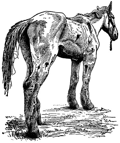
BAD CASE OF GLANDERS
The farcy form is shown here. The animal has not long to live. Except for experimental purposes, every horse having glanders should be killed as soon as the disease is discovered.
A horse with lung glanders may have a little dry, spasmodic cough, may look somewhat unthrifty,[171] and if the temperature were taken it might be slightly above normal, say, 101 degrees to 1011⁄2, the normal temperature being 100 degrees. Yet such an animal might do its work, last for a long time and not be suspected as a source of danger until several cases had occurred in the stable, for which it was difficult to account.
While a well-marked case of glanders or of farcy is not difficult of diagnosis, there are many obscure cases which escape detection for some time. If a horse has a well-marked discharge from one or both nostrils, with characteristic chancres visible upon the mucous membrane of the septum nasi, and hard enlarged submaxillary glands in the intermaxillary space, it is not a difficult matter to diagnose such a case, and any horseman ought to recognize it. The same is true of a well-marked case of farcy. When the lymphatic vessels on the inside of a leg, especially a hind leg, are swelled and corded, with a chain of farcy buds along their course, some of which have gathered and broken, leaving a discharging open ulcer in the skin, it is quite evident that the animal is suffering from farcy.
A peculiarity of glanders seems to be a tendency for the symptoms to appear on the left side; in many cases of glanders the discharge and ulceration is in the left nostril, and the left submaxillary gland is enlarged; and in a large number of the cases of farcy met with it is the left hind leg that shows the lesions of the disease. In obscure cases of glanders or farcy the diagnosis is not always so easy, even for experts, and then other methods for determining the trouble have to be resorted to. These are the guinea pig test and the mallein test. The guinea pig test consists of inoculating one or two of these little animals with the discharge from[172] a suspected horse’s nose, or from a farcy sore. If they should develop glanders it would be proof positive that the suspected horse had this disease; if they do not develop glanders it is not always positive proof that the suspected horse is free from the disease. Sometimes more than one test is necessary, or another method of diagnosis may have to be resorted to. This is the mallein test.
Mallein is a product made from cultures of the glanders bacillus analogous to tuberculin as made from cultures of the tubercle bacillus, and is used for testing horses for glanders much as tuberculin is used for testing cattle for tuberculosis. A horse infected with glanders will react to a mallein test in much the same way as a cow infected with tuberculosis will react to the tuberculin test. It is not customary in some states to kill a horse that reacts to mallein unless it shows some clinical evidence of disease. All horses that show clinical evidence of glanders or farcy in some states are killed by the state authority, and the law requires persons knowing or suspecting cases of this kind to report in writing to the chief of the cattle bureau of the state board of agriculture or to the inspector of animals in the city or town where the disease is believed to exist, except in some cities where the city board of health has full charge of glanders and farcy. Anyone selling, removing, transporting, or concealing a horse knowing or having reasonable cause to believe it has glanders or farcy is in most states liable to a heavy penalty.
In stables where glanders exists, in some cases, all the horses are tested and divided; the reactors are separated from the non-reactors, and those that react are tested once a month until they cease to react, or show physical indications of glanders and[173] are killed. Used in this way mallein seems to have a curative effect on incipient cases, and has been very successfully used in freeing infected stables from the disease. When a horse is killed because it has glanders or farcy the stall should be thoroughly disinfected where it has been kept, as well as the harness, blankets, currycomb and other utensils, and anything that cannot be easily disinfected ought to be destroyed. Public watering troughs where the horse has been watered should be emptied and cleaned out, and the blacksmith ought to disinfect his shop where the horse was shod.
There are various diseases that may be taken for glanders or farcy, and there have also been numerous instances where glanders has been taken for something else; for instance, chronic nasal catarrh. What many old-time veterinarians used to call chronic nasal catarrh or nasal gleet, were, in many instances, if not in nearly all, cases of chronic glanders, and when one of these cases of nasal gleet was rounded up in a locality, glanders disappeared in that neighborhood.
A horse with a chronic discharge from the nose as the result of a decayed tooth may sometimes be mistaken for a case of glanders, and also a horse with distemper or strangles; but the latter generally recovers soon, and in strangles the gland under the jaw softens and breaks and discharges while in glanders the gland remains firm and hard and generally not sensitive to manipulation.
There is a disease that has been troublesome in Pennsylvania and parts of Ohio the last two years called suppurative lymphangitis or epizootic lymphangitis, which may be mistaken for farcy, but animals suffering from it do not react to mallein, and guinea pigs inoculated with the discharges do[174] not develop glanders. There is not much glanders in the Eastern states, except in the cities, and the disease is not of a great deal of interest to farmers, except to avoid purchasing animals with it at some of the unreliable sales stables. Where a case occurs on a farm, except on some market gardener’s farm near a city, it is found, as a rule, that the horse was purchased at some unscrupulous dealer’s stable in the city, and, in some instances, other horses on the farm are infected, and the farmer not only loses his new acquisition, but has two or three other horses killed besides that have become infected.
Farmers buying new horses at city sales stables ought to endeavor to deal with only reputable concerns, and to avoid cheats. It is well to remember that a person cannot get something for nothing, and it is not likely that anyone can buy a horse for $50 to $75 because it is afraid of elevated railroad trains that would otherwise be worth $300 to $500, or because a widow lady wants a good home for her late husband’s old pet. Anyone buying horses from a fake coal company, or a humbug ice company, or an unknown express company that is just going out of business, is liable to invite a serious disease to his farm.
—A collection of pus, or other fluid containing gravel or dirt. It occurs most frequently in the foot, and is associated with the horse and mule almost exclusively. The cause may be from a bruise, but more frequently it is due to a punctured wound of the foot by nail, wire, or other pointed object. Nearly always there will be dirt carried into the wound with the offending object or shortly after its removal. This dirt, infected with germs, sets up an inflammation of the sensitive[175] structures causing more or less lameness. In many instances the nail hole becomes closed up and the collected matter may have to seek an outlet above the hoof. To determine the trouble a very careful examination of the hoof should be made, looking for any opening leading into the foot, often detected by discoloration of the part, or at an over-sensitive point in the foot.
Treatment should consist in making or enlarging the opening at a dependent part of the hoof, if possible, so that all secretion formed in the wound can find a ready escape to the outside. Without free opening there is danger of tetanus (lockjaw) developing. The wound should be thoroughly cleansed, and washed with some mild disinfectant, after which a small quantity of oil of turpentine should be injected, and the wound packed with calomel or iodoform and covered with a pledget of cotton. If the wound is very deep or extensive it may be beneficial, after thoroughly cleansing the foot, to apply a hot bran or flaxseed poultice. Use poultice for several days and change daily.
—A form of eczema that attacks the skin of the heel and fetlock. Sometimes the disease becomes so severe as to crack open, from which blood oozes out. A crust forms and later on becomes painful and disagreeable. To remove the scurvy part that is noticed first, apply a poultice, made of wheat bran or linseed meal. Change the poultices two or three times during the day. After removal each time wash with warm water, in which has been put some carbolic acid or creolin, and then apply the poultice again. After the poulticing is ended apply a salve made of 4 tablespoonfuls of oxide of zinc and 8 tablespoonfuls of vaseline. If indigestion seems to be associated in any way, give[176] the horse a dose of physic, aloes being best for the purpose.
—This condition is the presence of the larva (worm stage) of the sheep bot fly, located in the frontal sinuses (cavities) of the head. The trouble is confined to sheep and occasionally goats. The so-called “grub” of the horse is found in its stomach, while the “grub” of cattle is found along its back just underneath the skin. The adult fly, which lays the living “sheep grub,” is of a yellowish-gray color, slightly larger than a house fly. During the warmer part of the summer days the fly goes about depositing its young in the nose of the sheep. The young then work their way upward into cavities of the head between the eyes, but not into the brain cavity. Here they attach themselves to the lining, remaining when unmolested for some ten months, then lose their hold and are sneezed out to the ground. Burrowing into the ground they enter the pupa or dormant stage, when, after a month or six weeks, they emerge as adult flies to replenish their kind.
When few grubs are in the head little trouble may be observed, but if more numerous may cause free discharge of dirty white or yellowish, thick fluid, loss of appetite, frequent coughing and sneezing, tossing of head and weakened gait, and the animal may become too weak to rise, and finally dies. With a special instrument (trephine) bore a hole into the cavity containing grubs and remove them with forceps. When they are present every year the sheep should be protected by keeping the nose smeared with tar during summer months. This can be done by causing sheep to lick salt from holes in a trough after placing tar about the holes.
[177]
—True hair balls are seldom found in other animals than cattle, resulting either from licking themselves or others; but different kinds of indigestible balls or concretions are frequently found in cattle and other animals, particularly the horse, in the stomach or intestines. Dust balls are occasionally formed when animals are fed upon mill cleanings. In sections where crimson clover is fed, and frequently in over-ripe condition in large quantities, balls are formed of parts of the indigestible heads. Again, calcareous or mineral matter may accumulate about an indigestible substance as a nucleus. These are not well-defined, in many instances, and the balls are often present without making it known. So long as they do not irritate the bowel too much, or do not occlude the opening from one portion of the bowel to another, they are likely to escape notice. In case they do obstruct the bowel they become serious obstacles, the greater number of these cases terminating in death. The symptoms then become those of colic from obstruction. In many cases no relief can be given, but attempts should be made to cause the obstruction to pass by giving mild purgatives and copious enemas.
—The term “heaves” is used to describe that disease of the horse which otherwise is known as “broken wind,” or technically as “emphysema of the lungs.” This ailment, which is incurable when thoroughly established and to which a tendency is inherited by the offspring of an affected sire or dam, is characterized by the following symptoms: Double, bellows-like action of the abdominal muscles in breathing; short, suppressed cough, usually accompanied by passage of gas from the rectum; gluttonous appetite; harsh, staring coat[178] of hair; pot belly; weakness; lack of endurance, sweating, panting, or staggering during work; dilated nostrils; frequent passage of gas and soft, foul-smelling feces when starting from stable.
The disease begins with indigestion, affecting in time the pneumo-gastric nerve of the stomach and then the branch nerves running to the lungs. At first the air tubules and vesicles of the lungs become dilated (aneurism); later they may break down into large air spaces and the surrounding lung tissues become involved (interlobular emphysema). Air then is easily inhaled, but is exhaled with difficulty and the effort causes cough and expulsion of gas (flatus).
The distress may be relieved by treatment, but perfect recovery is impossible when the lungs have become badly affected. Treat by substituting wet oat straw for hay in winter and grass for hay in summer. Allow double the usual rest period after a meal. Work when stomach is not distended with food. Do not feed hay at noon. Use lime water to wet all food. Once or twice a week give raw linseed oil in a bran mash to open bowels. Give half an ounce of Fowler’s solution of arsenic night and morning. Do not breed from affected horses.
—The horse that is stricken with heat exhaustion or which falls from heat, apoplexy or “sunstroke,” is sick or out of sorts at the time of attack; otherwise he would withstand heat and work. The middle horse of a three-horse team suffers most and is apt to succumb to the ill-effects of the combined radiation of heat from his mates and direct rays of the sun. Attacks are most apt to happen on the third or fourth day of a spell of intensely hot weather characterized by mugginess, electrical storms and moisture-saturated[179] air. At such times the horse that has indigestion, a heavy, unhealthy coat of hair, a skin or kidney trouble or any affection of the brain or heart is the one that must be most carefully watched and worked.
With the hope of preventing attacks feed light rations, no corn, no mashes, no ground feed other than bran; avoid green grass, unless the horses are on it all of the time; do not feed hay at noon; allow cool, pure drinking water often when horses are at work; keep stables clean, darkened, screened, and ventilated; shade the polls of the horses’ heads during work time and in such a way that air passes freely under the shading device.
In sunstroke the horse falls and soon succumbs. In heat exhaustion he lags, stops sweating, pants, staggers, skin is dry, nostrils dilated, membranes of eyes and nostrils red. High fever is present. Treat by keeping cold, wet packs to the poll of head or letting a stream of cold water run over it. Shower body with cold water from a sprinkling can. Stand horse in shady place under a tree where air passes. Give stimulants freely in water as a drench every hour at first, then less often as symptoms abate. A suitable stimulant is whiskey in half pint doses, or a mixture of one part of aromatic spirits of ammonia and two parts each of alcohol and sweet spirits of niter. Dose is two ounces in half pint water. Do not bleed horse or give aconite. Give half ounce doses of saltpeter in water twice daily as horse recovers. Call the veterinarian in sunstroke cases.
—A protrusion of any portion of the bowels or their coverings through a break in the walls of the abdomen. A rupture, for that is the popular term, is most common in horses. Often[180] at birth they are seen near the navel. These disappear in a few months without any treatment being required. In mature horses the usual causes are blows, kicks or some violent effort that tears the muscular structure.
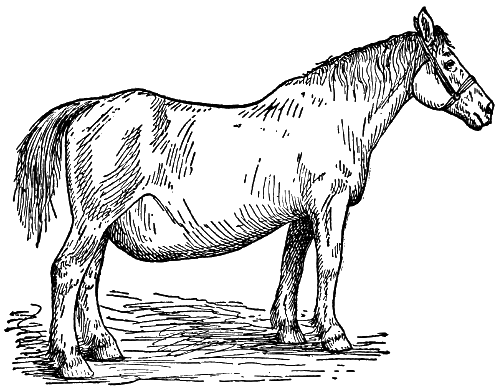
VENTRAL HERNIA
It may occur in any part of the abdomen and varies in size with the extent of the rupture.
The characteristic symptom is the bulging out of the gut, tumorlike; and this often can be slipped back where it belongs. If the rent be not closed, even if the gut is returned, the least bit of strain is liable to force it out again. Some kinds of hernia cause immense pain and the animal shows it.
In treating, work the gut back to its place. This done, place a pad—a flat piece of wood or leather will do—over the wound and fasten in such a way as to keep it in place. This should be worn for a month until recovery is complete. Such treatment[181] will not serve in all cases of hernia. An operation may be necessary, which should be made only by a skillful veterinarian.
—This is not a disease at all, but an indication of poor health, more particularly of poor nutrition; usually the result of indigestion, improper food, worms or want of proper exercise. The skin is hard, rough, papery, and cannot be picked up from the body with ease. When the attempt is made, it suggests that the body is too large for the skin. Of course treatment is in the nature of better food, that proper nourishment may be secured. A good physic will be proper to start with and then follow with a tonic, easily assimilable food of a nature that will properly nourish the body.
—A sound produced in the act of breathing while the air is being expelled from the lungs during forced respiration. It is a fluttering sort of a sound. When horses are trotting or pacing the sound is essentially a nasal one, and is not to be regarded as a state of unsoundness. It is rather a measure of excitability, and associated with horses of much spirit and good breeding.
—A disease of the hip, caused usually by some injury as from a fall or kick. A slight swelling is observed just over the hip, and lameness when the animal walks or trots. In severe cases, the horse will hop and catch the lame leg. The best treatment is absolute rest. Frequent applications of hot water are good. After each application bathe with a solution made of 4 ounces of water, 2 ounces of tincture of opium, 2 ounces of tincture of arnica and an ounce of belladonna. If the lameness continues, use a blister[182] made of 2 teaspoonfuls of cantharides and 4 tablespoonfuls of lard. Allow the blister to remain for an entire day, then wash off with soap and water and apply lard or vaseline. Repeat in a couple of weeks if necessary. If the lameness disappears, give the horse rest for several weeks.
—A fracture at the point of the hip. The most common cause is striking the point of the hip against a door post or pole. Sometimes a kick is responsible. While recovery follows, as a rule, from the very nature of the fracture, there is no treatment that will remedy the broken point. After the soreness has passed no inconvenience results; only a blemish is observed.
—The term hog cholera has become quite ambiguous, partly on account of new discoveries concerning the cause of the disease and partly on account of what have been supposed to be two different but curiously related diseases being generally included under this general term. Until within a year or two we have supposed that there were two infectious diseases of hogs recognized under the general terms of hog cholera and swine plague. It now seems probable that we will be able to do away with the term swine plague entirely.
The disease considered here answers to the following requirements: (a) Infectious by association or other natural exposure; (b) the animal before death and the carcass after death show certain accepted symptoms which are clearly recognized as pertaining to cholera; (c) the blood is virulent and capable of reproducing the disease on inoculation into susceptible hogs; (d) attack and recovery confer immunity. It is to be understood that we might easily have diseases among swine[183] where characteristic “a” or even “b” might be present and yet the disease be not true hog cholera.
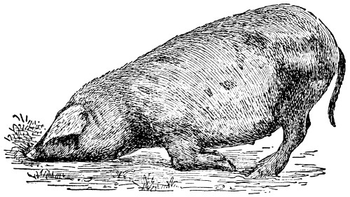
AN ATTACK OF CHOLERA
One of the familiar attitudes assumed when the hog is affected with cholera. When this far along, not many cases of recovery are observed.
Until within recent years American authorities, bacteriologists and veterinarians alike, have very generally accepted a certain germ, the bacillus of Salmon and Smith, as the specific cause of hog cholera and another somewhat similar germ as the cause of what was supposed to be a distinct but curiously related disease—swine plague. But within a few years workers in the Federal bureau of animal industry have apparently demonstrated that hog cholera is caused by a living germ so small that it passes easily through germ filters which remove all known forms of the bacillus of Salmon and Smith.
It may be interesting to note further that this new germ is so small as to be invisible to the highest available powers of the best microscope. That it is a living organism and not a chemical[184] poison may be very easily demonstrated. The curious relations to this disease of the old bacilli of hog cholera and of swine plague are not well understood, but it seems quite possible that they may play some part in the later development of the disease after the disease processes have been started by the invisible germ. While our old theories and supposed information concerning the cause of hog cholera have been very much disturbed by newer work, it is important to remember that hog cholera is now just as much as before to be recognized as a distinctly infectious disease. It is important to remember also that this infection is absolutely necessary, or there can be no cholera no matter how susceptible animals may be. There can be no cholera without this primary and specific cause any more than there can be plants in our wheat fields without the previous presence of mustard seed. Conditions of soil and climate may favor a rank growth of mustard. Conditions of feed and keep may favor the development and spread of hog cholera. They may decrease resistance and increase susceptibility, but cannot originally cause the disease. It is a rather common experience that hogs kept closely housed and fed, especially with such foods as corn, offer less resistance than do other hogs. In our vaccine work we frequently find hogs of this type which die readily under inoculation with blood of low grade virulence. Hogs of hardier type may become slightly sick or not sick at all with inoculation from the same infectious material. Pampered show herds appear especially susceptible to both natural infection and artificial inoculation.
The farmer, and for that matter the public in general, should bear in mind that the cause of hog[185] cholera is a living organism capable of enormously rapid self-multiplication—actual, though very minute particles of matter. This, fully understood, makes it apparent that infection may be carried in any way that other fine particles of matter may be carried. It thus becomes very apparent that the infection may be carried by sick hogs or upon the legs and bodies of hogs not sick; it may be carried in wagon boxes, in hog racks, in stock cars, or upon shoes and clothing of people. It is very evident that the infection may be carried down stream, especially in small creeks, and give rise to other outbreaks.
So far as the sick hog is concerned, we are quite sure that the blood and the manure are thoroughly infectious and there can be no question concerning the infectiousness of fresh carcasses of dead hogs. Perhaps we should say first of all that we rarely get all of the accepted symptoms of hog cholera plainly shown in one case. It is important to bear in mind that cases vary in virulence from those of very chronic type where hogs live for weeks and finally die or recover, to very acute cases where they die overnight.
The hog coming down with cholera is usually sluggish at first, lying around in the shade and refusing feed. The hair may become rough. The eyes early show symptoms of inflammation, with a sticky discharge. There is usually a suppressed cough. The gait may become irregular and uncertain, especially with the hind legs. After these preliminary symptoms have been shown for a time, the skin becomes red, changing to purple, especially noticeable in white-haired hogs. The hog is then usually within a very few days of death.
As already explained, not all cases are typical. Sometimes hogs die in an outbreak of cholera from[186] undoubted hog cholera, and yet the ante mortem or post mortem symptoms show very little upon which to base a diagnosis. But we may easily demonstrate that these were cases of cholera by injecting their blood into susceptible hogs and by thus producing typical cholera.
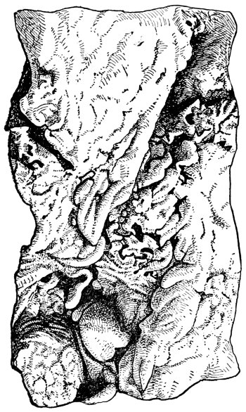
THE RESULT OF HOG CHOLERA
A post mortem of a hog dying from cholera will show ulcers like those pictured here. Look for them in the large intestine.
At the autopsy of an ordinary case of cholera the first and perhaps the most striking thing seen is the purpling of the skin. On opening the carcass small blood spots may be found under the skin and in the fat cut through. The glands along the intestines are intensely inflamed. The mucous membrane of the stomach is frequently thickened and roughened and in chronic cases there may be ulcers. On opening the intestines we see areas here and there of intense inflammation in the acute cases or numerous ulcers in cases of more chronic type. In[187] very acute cases we find areas intensely inflamed, even bloody in places. The slow chronic cases develop characteristic hog cholera ulcers. These may appear at almost any point on the lining membrane, but more particularly in the blind pouch and around the point where the small intestine connects with the large intestine. On stripping off a very thin transparent membrane covering the kidneys, a typical case of hog cholera will usually show minute red spots on the surface somewhat resembling the covering of a turkey egg, which gives the common name of turkey egg kidney of hog cholera.
—Clearly there are certain things which the owner of healthy hogs in a hog cholera district should do and a good many things which he should not do. The same is equally true for the man who has sick hogs in a neighborhood where there are uninfected herds. The owner of healthy hogs and his family should keep away from public stock yards, from all pens and yards on other farms whether sickness among hogs prevails or not. It may easily occur that a neighbor’s hogs may appear well but have recently received the infection and be already capable of scattering the disease. We do not know at what period in the development of this disease infected hogs become capable of disseminating hog cholera.
During a hog cholera season the owner of healthy hogs should institute something in the way of private quarantine and pleasantly, perhaps, but firmly, ask visitors, especially stock buyers and threshing machine crews, to keep at a reasonable distance from the pens and yards. It is safer for one man to have exclusive care of healthy hogs during the hog cholera season, and this man should be very careful where he goes with reference to possible[188] infection. Special fencing or other provisions should be made wherever practical to keep dogs out of the pens and yards, for, under certain conditions, dogs become very active agents in spreading the disease.
The owner of a healthy herd should be very careful about buying in hogs for feeding or breeding purposes, and, in the Western states especially, all public stock yards and stock cars must be regarded as possible sources of spread. Hogs coming into the herd for breeding purposes, if by rail, should be shipped in other than stock cars, and should not be unloaded so as to go through stock yards. All new hogs coming on to a farm where the disease has not appeared, should be kept carefully apart from the herd for from two to three weeks after arrival. The disease may thus have time to develop, if the animals have been infected before shipment or en route. It is decidedly worth while to be careful about clean feeding, for it seems probable that this is a common method by which infection enters the body. This being the case, troughs and feeding floors should be frequently disinfected with steam, boiling water, or a very dilute corrosive sublimate solution (1:1,000 dissolved in water), with the troughs subsequently rinsed out with plain water. Or the troughs and feeding floors may be disinfected with any of the coal tar disinfectants if they are used in sufficient strength. These are not poisonous in any probable quantity which hogs would get.
—The farmer should be especially careful about buying hogs out of stock yards. Some years ago a certain Minnesota farmer purchased a lot of feeders from Sioux City and took them home to his farm. In about two[189] weeks his hogs commenced dying. A little later hogs previously on the farm began dying. In a little while he was losing hogs at the rate of 25 a day, losing a total of about 200. This loss of 200 hogs was scarcely a drop in the bucket—too small for consideration in comparison with the loss which this outbreak cost the state, for, with some others coming into the state from Iowa and Nebraska, this outbreak cost the state, as carefully estimated, about $1,250,000 during that one year. As soon as the Minnesota farmer here referred to realized that he had cholera and was liable to lose a large portion of his herd, he shipped out a lot of fat hogs ready for market. These were yarded for a time in the public stock yards of his town, and one of them died while waiting for shipment. This hog was left for a day or so in the yard. Later a carload of feeding hogs was shipped in from a point in South Dakota, where they had never had hog cholera. These South Dakota hogs were unloaded into the yards where the fat hog had died some time before, and were sold out from there by auction.
It was a very interesting study to follow the resulting outbreaks; but a very serious matter for the owner and for that entire portion of the state. Practically every farmer who bought hogs at this sale, and very many of those who walked around the yards looking at the hogs, but without buying, had hog cholera on their farms in a very uniform period after the sale. Surely the moral of this tale is so self-evident as to need no further suggestion.
—Troughs and feeding floors, at least, and, if practicable, the hog house also, should be kept clean and frequently disinfected during an outbreak. When the outbreak appears to be over,[190] the owner must decide as to just what he will do in the way of disinfection and cleaning up, or whether he will stay out of the hog business for a year and allow the infection to die out. This is, of course, without regard for the possibility of putting in vaccinated and immune hogs. Feeding troughs and feeding floors and the hog house in general, may be disinfected if of reasonably good construction, by a thorough cleaning and then by one of the methods suggested under prevention. If the sick hogs have been kept in an old straw shed or in an old hog house that is about ready to fall down anyway, by all means the best method of disinfection is by burning. Without disinfection or burning the owner cannot be safe in putting in susceptible hogs within much less than a year after the last hog died or recovered. The slow old chronic cases that go dragging around at the end of an outbreak should usually be killed and safely buried, for it is rarely profitable to put such hogs in shape for market. It might possibly be worth while to hold such a one over and nurse them along, in case of valuable brood sows, for hogs having recovered from cholera are usually immune for life.
Brood sows which have had the disease and recovered usually give something more than natural immunity to their offspring. But the degree of immunity so conferred is so variable in degree and uncertain otherwise that it cannot be depended upon as a routine method of establishing immune herds. Yards may be practically disinfected by plowing or by burning off a good layer of straw.
—Generally stated, this vaccine consists of two parts: (a) Blood serum from the body of a specially immunized hog; and (b) virulent blood serum from the body of a hog[191] about to die from cholera. The general theory upon which this double vaccine is used is that of giving the animal an infectious disease and at the same time a treatment which enables the animal to resist the infection. When the hog is through with it he is in exactly the same condition as though he had gone through a natural exposure and recovered.
—We start this work with certain hogs that are immune usually because they have passed through an outbreak. It has been shown that when such immune hogs are treated with large injections of virulent blood under the skin or into a vein, that they do not usually become sick, but their own blood develops a peculiar property that gives protection to other hogs that are naturally susceptible.
When the blood or rather blood serum from this specially treated immune hog is injected into the bodies of healthy susceptible hogs, the latter becomes likewise immune, but the immunity so gained lasts only a short time, possibly four to six weeks, and is then gradually lost. If we give a small injection of virulent blood at the same time, or soon after the immunizing serum is given, then the treated hog becomes immune for a long period, perhaps for life.
—The specially immunized hog which produces this immunizing serum is known as a hyperimmune, and to save words will be hereafter mentioned as such. The simply immune hog may be prepared for producing serum in either one of three ways. (1) By three rapidly increasing doses of virulent blood serum injected under the skin at intervals of seven to ten days; (2) by one enormously large injection of virulent serum under the[192] skin; (3) by injecting virulent blood in smaller doses directly into the blood circulation.
In this work an ordinary immune hog weighing 100 pounds is given a quart of very virulent blood, a teaspoon of which similarly injected would kill a hog that was not immune. In other words the immune, and especially the hyperimmune hog, have developed certain properties in their blood antagonistic to hog cholera virus.
—We have two possible methods of vaccinating or immunizing susceptible hogs (a) Serum only. This is by the injection under the skin of serum from the body of a hyperimmune hog and gives immediate but temporary immunity lasting, as already stated, several weeks. If this animal, during the period of immunity, is exposed to natural infection, he becomes protected for a very long period, perhaps for life. (b) Simultaneous. The second method of vaccination consists of injecting immunizing blood serum into one thigh and a small amount of disease-producing serum at the same time, or soon after, into the other thigh, thus giving the animal the cholera and a cure for it at the same time. If the immunizing serum is potent and the virulent serum is really virulent, then the animal so treated becomes permanently immune.
The serum-only method is usually preferred in actual outbreaks and for hogs not yet sick, because this gives immediate protection, and the hogs, being naturally exposed, usually develop a permanent immunity. The simultaneous method of vaccination is preferred where we are very confident of the serum’s potency against the virulent blood, and for hogs that have not yet been infected. It may yet be found wise to use this method even in outbreaks.
[193]
—Every intelligent stockman who reads this will probably ask if there is not danger of scattering cholera by this simultaneous vaccination into districts where it has not yet appeared. A considerable amount of direct evidence on this point is better than any amount of theorizing and personal opinions. This evidence all agrees that unless the vaccinated hogs become distinctly sick as a result of the vaccination (which can occur, and does very often), that there is practically no danger of disseminating the disease. This is especially true since all hogs on the farm are supposed to have been treated and are immune, and, therefore, incapable of developing cholera and so spreading the disease. It does occur, even with good serum, perhaps, that an occasional hog may become a little sick, and very rarely even die, as a result of vaccination. But with good serum given in standard dose and virulent blood also given in proper dose, the risk of this is so small that it may be safely disregarded and especially when all hogs on the farm or that may be exposed with such sick hogs have been treated.
—A common term to denote a diseased condition of the blood. The horn is not hollow and never is. The old quack method of boring a hole in the horn with a gimlet and squirting turpentine into the orifice is both cruel and ridiculous. While in fact the temperature of the horn is low, it is because of the general poverty of the blood of the animal. There is no merit in this kind of treatment. The most common symptoms are general debility, scanty flesh, scurvy coat and coarse hair. The appetite is also irregular and at times greedy. Treatment is in line of better food and general improvement of the system. If[194] lice are found on the body, they must be destroyed by disinfectants and washes. A tonic, consisting of 2 teaspoonfuls of sulphate of iron, 1 teaspoonful of powdered nux vomica and 4 tablespoonfuls of ground gentian root given each day in the food or as a drench, will be very helpful in toning up the system and in enriching the blood. The most important factor of the treatment, however, is in nutritious, wholesome food.
—See Sand Cracks.
—A small insect about half as large as the common house flies, and very much like them in appearance. Horn flies swarm about the head and settle near the base of the horn, where they bite and cause much irritation. They also attack cattle on the back and sides and flank. The fly mixtures that are commonly advertised, and applied by means of a hand-spray, are excellent for keeping the pests away. A good home mixture to apply at the base of the horns is made of pine tar, kerosene, and fish oil. Use this in equal parts, and apply with a brush.
—See Bloating in Cattle.
—See Water in the Brain.
, also called rabies and mad dog, is an infectious disease caused by some invisible organism. The disease is transmitted from one animal to another by the bite of an animal which is suffering with the disease or by direct inoculation. It is more common in the dog than any other animal, from the fact that dogs run at large and have a tendency to bite other dogs with which they come in contact while they are suffering with the disease.
The dog shows two forms, furious and dumb. In the furious form the animal at first seeks dark[195] places, but is usually restless and will move from one place to another. This condition lasts for a day or two, after which time he becomes more restless and may go 30 miles in a day. He will drink water, eat sticks, stones, and bite other dogs, horses, and cattle, less often man. This condition will last from one to four days, and then the dog becomes partly paralyzed, so that he can no longer swallow, or his legs may be affected, so that he will lie in one place, and usually dies after a few days longer. In the dumb form, the animal seeks dark places, is rather restless, the throat and lower jaw become paralyzed, he is unable to swallow or to close his mouth and, therefore, cannot bite. Sometimes they will change from one form of symptoms to the other.
In the horse the symptoms vary somewhat from those in the dog. The horse is restless, usually violent and will kick and bite, oftentimes showing sexual excitement. He may break his teeth on the manger and oftentimes bites his own flesh at the place where he has been bitten by the dog. The symptoms usually develop in from eight to twenty-eight days after the animal is bitten, but may not develop for six months. The disease runs its course in from two to ten days, with a fatal termination.
There is no treatment for the disease after the symptoms have developed. In case man is bitten he should take the “Pasteur” treatment, which is a preventive, and it should be taken in a very short time after being bitten. After the symptoms begin to show it is too late to take treatment.
—See Water in the Chest.
—A continued distention of the rumen caused by large quantities of undigested material lodging in the[196] rumen. Inflammation often results, with distress and pain manifest. If relief is not attained the walls of the rumen become paralyzed. Associated with the disturbances the animal is dull, the left side swollen, the breathing and pulse increase and the back aches. When lying down, the left side is always up. In treating, cold water dashed over the back and loins is recommended. A strong physic of Epsom salts and ginger will aid in stimulating the secretions and may bring relief. If gas accumulates so as to threaten the life of the animal, the trocar and canula should be used. If these are not available, use the knife, as described for hoven or bloat. In some cases the impaction becomes so pronounced as to resist ordinary treatment, when extreme measures will be necessary if the animal is to be saved. Better call your veterinarian and open the rumen in order to remove the contents with the hand. The operation is as follows: At the point midway between the point of the hip and the last rib, and down about four inches from the backbone, an opening is made large enough to admit the hand. After the opening is made the edges are stitched to prevent any material from getting between the skin and the rumen wall. Now remove the greater part of the accumulated material; this done, the rumen, the muscles and the skin are each in turn stitched, the wound dressed and the animal given stimulating medicines. A splendid tonic consists of 4 tablespoonfuls each of ginger, tincture of gentian and tincture of iron. Give this tonic daily and until the animal has fully recovered.
—Failure to digest food with abdominal pains and indisposition resulting. Bad food and improper management are back of the[197] trouble in most instances. Mild cases require no treatment. A light, laxative diet is desirable for stubborn cases. If possible turn the animals on fresh grass. Jamaica ginger is generally prescribed for indigestion. Give 8 tablespoonfuls in a pint of warm water three times a day as a drench. Follow this with condition powders, or some good digestive tonic. After recovery see that the diet is varied and that laxative and succulent foods are supplied.
—As the name indicates, this is an infectious trouble frequently extending over considerable areas and occurs among both horses and cattle. It is very similar in its action to ordinary pneumonia or inflammation of the lungs. However, it does not seem to be so acute in its action. The same treatment is applied to cases of this kind as to ordinary pneumonia. When its presence becomes known, it is wise to remove all healthy animals to some other quarters. This lessens the danger of infection to healthy animals. After the disease has run its course, remove all litter and manure from the stables, thoroughly air out, admit as much sunlight as possible, and disinfect all walls and floors. A coat of whitewash on the ceiling and walls is desirable. The floors should be literally wet with disinfectant fluid, which should be admitted to all cracks and open spaces.
—See Swamp Fever.
—Sometimes this disease is called enteritis. It frequently follows severe cases of colic. It is the result of inflammation caused by indigestible material lodging in the stomach and intestines of animals. It may, however, result from other things that irritate the bowels. When first noticed, a general depression[198] prevails, with signs of pain in the bowels; breathing is quickened and frequently a chill shows itself. The horse acts very much as if he had a case of colic. As the disease progresses the pain increases and the pulse rises. In a few hours the pain becomes very severe and the animal is in great agony all over; he breathes heavy, the legs and ears are cold and clammy and the pulse very high. In severe cases the pulse reaches to 100 and 105 beats a minute. The horse now is very ill indeed. He shows great weakness. It is very unlikely that he will survive more than a day or two. The disease usually runs from ten to fifteen hours, and unless there is a change for the better, death results.
When far advanced there is little likelihood of successful treatment. Success lies only in early work, taking the disease in time. A satisfactory drench is made of 4 tablespoonfuls of tincture of laudanum, 10 to 15 drops of tincture of aconite, 1 tablespoonful of common soda, and 1 tablespoonful of ginger. These are mixed in a pint of warm water and given as a drench. Repeat this every hour until the animal gets relief. A mustard plaster gives relief when applied to the belly. A physic is not considered advisable, as it increases the inflammation—just what is not wanted at all.
The most rational treatment consists in allaying the pain. Opium in teaspoonful doses every hour until the pain is relieved is helpful. Some veterinary practitioners use 10 grains of morphia and 4 tablespoonfuls of chloral hydrate in syrup and water for each dose. This dose is repeated every two or three hours until the symptoms abate.
The diet should be carefully watched in diseases of this kind. Bran mashes made with linseed tea or slippery elm bark are suitable. Boiled food is[199] better than uncooked food. Good water frequently and in small quantities is desirable. Skimmed milk is excellent and may be fed for a week or two at a time. This food often effects a cure without any other aid.
—This is a common disease in farm stock. The disease occurs most frequently in late fall or winter or early spring, and is due to exposure while the animal is still warm and hot; bad ventilation influences it. Authorities now generally believe it to be a germ disease and infectious. One of the first things noticed is the shivering of the animal and then a fevered condition; the animal seems to be hot, then cold; a peculiar breathing is noticed; the pulse quickens, ranges from 60 to 70 beats a minute; the eyelids on the inside take a scarlet hue. The animal does not eat, stands up much of the time with the head down and the ears lopped over; a grating sound is noticed when the ear is placed to the chest. Frequently distress is experienced in the bowels; constipation follows and the temperature rises gradually until it reaches 105 degrees, which is reached about the sixth or seventh day. If recovery does not follow the appetite will disappear, the mouth become cold, the breath heavy and disagreeable and the pulse feeble, frequently not noticeable at all.
After the case assumes a more favorable aspect, an effort should be made to keep the animal comfortable and in as good condition as possible. It is therefore advisable to keep it well blanketed, the legs bandaged and rubbed. The patient should be kept also in a warm stall where good air is available. Good food that is nourishing and easily digested should be provided. Sweet milk is good,[200] and raw eggs mixed in the gruel are excellent also. A compress over the lungs does much good. The compress should be made out of heavy cloth, frequently rinsed in cold water and then placed over the lungs where they are covered with heavy, dry cloths. On recovery, rub the sides of the chest so as to thoroughly dry the surface. A mustard plaster, after the compress has been removed, is quite generally used. A stimulating medicine may be given during the early stages. Use a drench, consisting of 8 tablespoonfuls of whiskey to 4 tablespoonfuls of sweet spirits of niter. If the animal is in very great distress, give a drench every two or three hours consisting of 8 to 10 drops of Fleming’s tincture of aconite, 2 tablespoonfuls of laudanum mixed with a pint of cold water.
After the animal is on the road to recovery, stop the use of these medicines and give a tonic consisting of nitrate of potash or saltpeter and ground gentian root, half and half. Give a teaspoonful three times a day. While the animal is sick, a little boiled flaxseed mixed with a soft food will keep the bowels regular. It is not wise to give purgatives, hence it is wise to give an injection consisting of warm soapy water, so as to empty the bowels. From two to four weeks of rest and care should be allowed for complete recovery.
—A specific disease of the horse affecting the mucous membrane of the air passages. When the mucous membrane of the eyelids is affected, pink eye results. Sometimes the mucous membrane of the intestines is affected, in which case colic or inflammation of the bowels results. The common cause is exposure to cold. If no work be required, plenty of fresh air be supplied, no drafts admitted and careful nursing otherwise, the disease[201] will run its course in from two to three weeks and no medicines will be necessary. In cases where considerable cough prevails, the custom of putting a piece of camphor about the size of an egg in a pail of boiling water and holding the horse’s head over it from a quarter to a half hour at a time is to be commended. The bowels should be kept free and open. Any of the ordinary purgatives will do. If weakness occurs, give 4 tablespoonfuls each of tincture of ginger, ground gentian root and sweet spirits of niter in a half pint of water three times a day. Two tablespoonfuls of nitrate of potassium given once or twice each day in the drinking water is also desirable. As the trouble abates, the medicines suggested before may be dropped and in their place a teaspoonful of sulphate of iron and a tablespoonful of ground gentian root may be given daily in a bran mash or oatmeal gruel.
—Intestinal worms may be classed as large and small. The large worms inhabit the small intestines, and the small ones the large intestines, the larger class of worms being more readily reached by worm destroyers than are the smaller ones, as the small intestines begin at the stomach and as remedies leave the stomach, the worm soon receives the dose prepared for it, while if one dose has to pass through about 60 feet of intestines before reaching the smaller worms in the larger intestines, much of the worm remedy is lost by mingling with the food, and diluted by mixing with the digestive fluids. Thus what is a remedy for the large species of worms will have little effect upon the smaller ones.
As a farmer’s dose for the larger species of worms, none, perhaps, is better than the following: Oil of turpentine, 2 ounces; extract or oil of male[202] fern, one half ounce, mixed with 4 ounces of castor oil and 8 ounces of pure raw linseed oil, with half a pint of new milk, and given after the horse has fasted for about 14 hours. Repeat the dose in a week; then follow with two worm powders, common smoking tobacco, eight ounces; powdered worm seed, 6 ounces; powdered sulphate of iron, 4 ounces; mix with one-half pound each of salt and granulated sugar. Every morning before the horse is fed any other food, place a heaping tablespoonful of the powder in four quarts of wet wheat bran and allow the horse to eat it; continue for ten days and the horse will be practically rid of worms of the larger species. Colts should receive smaller doses in proportion to age.
The small worms need the worm powder to be given in the wheat bran every morning for fully two weeks. Then follow with an ounce dose of barbadoes aloes and a tablespoonful of ginger given by mixing with about 12 ounces of warm water and a gill of common molasses; wait a week and repeat the powder treatment and follow with the aloes. In a case of the very small or rectal worms (pin worms) always use rectal injections, a good enema being made by steeping for two hours one pound of quassia chips in a gallon of soft water; strain and add two ounces of common hard soap; use the whole at once, using at about blood temperature after the soap has dissolved. Repeat in three days and continue as long as worms are being brought away by the enemas.
—See Stomach and Intestinal Worms in Sheep.
—See Scab in Cattle.
—Until of recent date the disease in the human so common at certain seasons of the[203] year was unknown among animals, or, at least, if present had never been discovered by the veterinary profession. But be that as it may, we are now finding it in plenty among horses of all ages, from colts up to aged horses; very prevalent among sheep, and quite frequent among cattle. The early writers on veterinary science usually attributed the cause to gall stones. But that theory can hardly be tenable in this country, where we find it essentially more prevalent on low, marshy soils or on the hill lands that have been long unplowed, where animals are pastured, or hays are cut. The general symptoms of it are a general dullness, hanging of the head as though it ached, or pressing the head, if the animal be a bovine or sheep, against the barn or stall. The tongue will be found dry or covered with a thick, sticky slime. The membranes of the eyeball of a yellowish cast. In horses the tongue will usually have a black coating. The appetite in all animals is capricious. They will eat well one day and scarcely touch food the next. As a rule, they will manifest great thirst, yet will drink but little. There are exceptions to this, however. The voidings are not uniform. Sometime the urine is quite high colored; at other times not. But, as a rule, it is scanty. The feces are sometimes quite hard and covered with a shiny slime. At other times there will be extreme looseness of the evacuations. These last symptoms are to be well considered in using a treatment when the voidings are hard and slimy. In case it is a horse that is ailing, a physic of aloes should be given, one ounce being the dose for a thousand pounds of horse, and two teaspoonfuls of podophylin. Give this dissolved in water and pour down as a drench, and follow with a bitter tonic for from two to four weeks, or until the[204] voidings are normal and all scurf is removed from the tongue. As a tonic for this none is better than a mixture of powdered gentian root, six ounces, powdered golden seal 2 ounces, powdered sulphate of iron 4 ounces, well mixed in 1 pound of common salt. Give in the feed a tablespoonful in ground oats three times a day, until improvement takes place. Then drop to twice a day and later once a day. In case of the bowels being very loose always give a pint dose of a mixture of castor oil 4 ounces, pure raw linseed oil 12 ounces. Then follow with the tonic powder named. The symptoms in cattle are quite similar to those of the horse, except the bovine’s eyes usually discharge some, yet not profusely, and there are frequently puffy swellings beneath their lower jaws. In case their bowels are abnormally loose, give the oil as for the horse. If constipated give from one to two pounds of Epsom salts at one dose as the physic, with the podophylin added as for the horse, and follow with the same tonic powder. In the case of sheep, which are by nature constipated animals, nothing equals a ten-grain dose of calomel, followed the next day with a four or six-ounce dose of Epsom salts (sulphate of magnesia), and as sheep are reluctant to eat any tonics in their feed, we are compelled to pour their medicine down them. Mix together 4 ounces each of the tincture of gentian, golden seal, ginger and iron, and give a tablespoonful twice a day in a half pint of water. But always give the calomel, as it will clean out the liver of a sheep as no other known agent will. The symptoms are much the same as in cattle. Begin treatment early or success will not follow.
—The hog is mostly affected with these worms, although they have been found[205] in the dog also. Death does not, as a rule, follow the infestation unless in an aggravated form. Obviously there is no remedy.
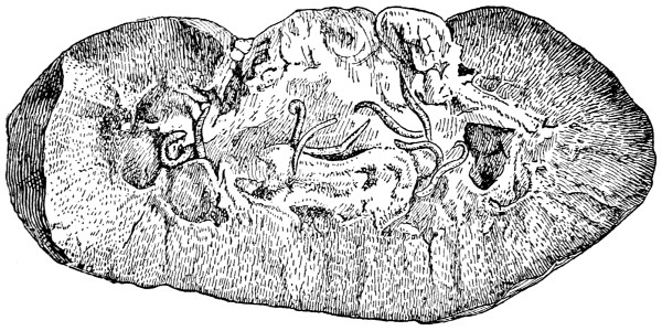
KIDNEY WORMS IN THE HOG
While worms are occasionally found in the kidneys, they do not frequently cause disease or death.
—A condition in which the knees bend forward as the result of contraction of tendons located along the back of the leg. In aggravated cases the tendons should be cut. If this is to be done only a skilled surgeon should be allowed to perform the operation.
—See Founder.
—Farm animals, especially those housed in stables more or less infested with insects and vermin, are commonly troubled with lice. Animals in good health resist the insects, but those already in a non-thrifty condition do not fare so well. Lice cause a good deal of annoyance to farm stock, inasmuch as they bite the skin, suck out blood, and thus cause considerable irritation. Lice can be seen[206] with the naked eye. Infestation, as a rule, takes place in filthy quarters, and the best means of disinfecting such places is by the use of a spray of kerosene. One of the best means of applying this to hogs consists in rubbing posts, which are constantly smeared with kerosene. In this way the hogs are induced to treat themselves. Infected hogs may also be treated by pouring the kerosene directly over the infested parts, like the neck, shoulder and back. Dipping tanks made of cement or wood are frequently located in the run-yards, in which is placed some disinfectant fluid. Hogs use these small tanks as wallows, and in this way they disinfect themselves.
For horses and cattle a good remedy is made as follows: Boil for an hour 8 tablespoonfuls of arsenic, 8 tablespoonfuls of soda ash and 16 tablespoonfuls of soft soap in two gallons of water. After being prepared by boiling, add enough water to make two gallons. When cool, wet the animal all over with a little of it, using a brush or currycomb to get it into the skin. Another good remedy is made of boiling stavesacre seeds, 1 part to 20 parts of water, for an hour and let it simmer for another hour; then add water to make it up to the original bulk. This applied to the affected parts brings quick relief. It is advisable to repeat the application in a week or ten days, so as to catch any new lice from any eggs that were not caught by the first application. A very common treatment is secured by mixing a pint of linseed oil, 8 tablespoonfuls of oil of tar, and 8 tablespoonfuls of sulphur. This is then rubbed on the affected parts once a day for two days and allowed to remain for a few days, after which it is washed off with soap and water. In serious cases,[207] the application should be repeated within a week or so.
—These are parasites usually found in the liver or its ducts. At times they are present in great numbers, giving rise to a serious disease called liver rot. When the fertilized eggs are discharged in the excrement of diseased animals and fall in fresh water they hatch out and are taken into the body by sheep and cattle, either in the food or drink. In a short time thereafter they have entrenched themselves in the liver of cattle or sheep.
LIVER FLUKE
A few liver flukes in an animal causes little trouble, as the injury is largely mechanical anyway. No peculiar symptoms are conspicuous when only a few flukes are present. The greatest damage is done when hundreds of flukes develop in a single individual. In these cases the flow of the bike is checked. As result the health becomes impaired and the usual penalties of malnutrition follow. Swelling of the jaws and diarrhœa are often noticed in connection with the disease.
When the host is badly infected with the flukes and in a badly run-down condition the trouble is always serious, and medicinal treatment is of little real value. Tonics and good food may be given to help along—but death usually follows. Salt is helpful as the flukes are sensitive to it. If an animal that has succumbed to the disease be examined, the liver will be observed to be fairly rotten as a result of the inroads of the parasites.
[208]
Treatment is in line of prevention only. Clean, pure fresh water, free of the eggs or the parasites, is necessary if the trouble is to be eradicated. The old ponds, ordinarily filled with stagnant water, should be drained. They harbor many bad parasites, and their harm is far beyond their value. When water for sheep and cattle is taken from pure streams or wells the trouble from liver flukes and other parasites is reduced to a minimum.
—This disease, very frequently called tetanus, is an infectious disease in which the body muscles are spasmodically contracted or stiffened. The muscles that move the jaw are frequently affected and the animal is unable to open the mouth. Because of this condition the disease is commonly known as lockjaw.
The spread of the disease does not occur through healthy animals coming in contact with animals having tetanus, but by inoculation. The germ of tetanus is present in the soil, manure and dust. It enters the body by way of wounds, especially punctured and bruised wounds. The injury may result from stepping on a nail, and the germs are planted in the deeper structures of the foot. Such a wound usually has poor drainage, the horn of the hoof closing the mouth or opening. Here the germs grow and produce a poisonous toxin that is said to be the most powerful produced by any bacteria. This toxin acts on the nerve centers of the brain and spinal cord, causing extensive spasmodic contraction of the body muscles.
Tetanus sometimes occurs in the absence of any noticeable wound. It may be in such cases that the seat of the infection is a slight abrasion of the skin, or the lining membranes of the respiratory and digestive tracts. The tetanus bacillus is a[209] slender, spore-producing bacterium. The spore is located at one end of the rod in the form of a round head, that gives the organism a pin shape, hence the name of pin bacillus. It is very resistant to outside conditions and the action of the chemical disinfectants. It is because of its ability to resist the action of disinfectants and the fact that it develops best when protected or covered by the tissues and wound secretions, that this disease so often follows ordinary wound treatment.
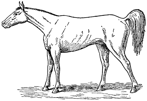
LOCKJAW
Note the rigid, tense position of the muscles.
From a few days to several weeks may lapse from the time of infection with the germs until the development of the stiffness and spasms. Sometimes the wound by which the organism has entered the tissues has healed before the symptoms of tetanus are manifested. In case the symptoms develop a few days after the inoculation the disease[210] is severe or acute in form, and less violent or subacute if the symptoms are manifested after the second week. The above statement does not hold true in all cases, but it may be considered true in a general way.
Of the domestic animals the horse is the most commonly affected. The symptoms shown by this animal are very characteristic. Any person that has had the opportunity to see and examine a horse suffering from tetanus should have no trouble in recognizing the disease in other animals.
is the spasmodic contraction of the muscles. This may vary in the different individuals, depending on the susceptibility of the animal and the quantity of poisonous toxin present in the system. There is at first a slight stiffness of the muscles of the back, neck, head, and limbs, and the animal is more nervous than common. A noise in the stable or a slap with the hand may increase the stiffness and contractions temporarily. The contracted condition of the muscles of the eye, are, perhaps, the most noticeable early in the disease. These muscles pull the eyeball backwards, the fatty cushion is pressed on and the third eyelid protrudes, covering at times from one-third to two-thirds of the front part of the eye. In the severe form of the disease the muscles feel hard, especially those of the back and neck, and the animal moves with difficulty. In addition to the muscular symptoms, the respiration and pulse beats are quickened and the body temperature higher than normal. The evidence of suffering from the contracted condition of the muscles is very marked, and, unless supported in some way, the animal may fall to the floor. If the symptoms develop a few days after infection, the animal[211] usually dies. The acute form is very fatal, but in the mild or subacute form the chance for making a recovery is good.
It may be largely prevented by the careful disinfection of wounds, and the use of anti-tetanic serum. In most localities the proper treatment of the wound is a sufficient preventive measure, but in localities and stables where the disease is common the anti-tetanic serum should be used. Ordinary cleansing of a wound, as practiced by most stockmen, is not sufficient to destroy the bacillus of tetanus. The wound must be carefully cleaned, disinfected and prepared for healing. This should be kept in mind when treating a wound, and instead of using an agent that we know little about, we should secure reliable information regarding the different commercial disinfectants and methods of caring for wounds. That class known as tar disinfectants is most commonly used. The better grade belonging to this class should be used.
If anti-tetanic serum is used, it should be injected as soon after the injury has occurred as possible. The injection is made hypodermically, usually beneath the skin on the side of the neck. Large doses of anti-tetanic serum given after the symptoms have developed may assist recovery. However, in the severe form of the disease this treatment is uncertain.
When the animal comes down with the disease, it should be made as comfortable as possible. The quarters should be roomy, quiet, clean, and well ventilated. It is advisable to support the horse with a sling unless the animal is worried or made nervous by it. This prevents his becoming tired and falling down. We should give the animal the best[212] of care in the way of regulating the diet, etc., but should avoid annoying it by our attention. Medicinal treatment is of little benefit and should be given a secondary place. In fact, dosing the animal with medicine, especially if large doses are given, may do more harm than good in the treatment of this disease.
—The word loco is a Spanish word, and means crazy. Loco disease is a disease of the brain and nervous system, especially of horses and cattle, but may also affect other animals. It results from eating any one of a number of poisonous plants called loco which grow upon the dry, sandy prairies of some parts of the Western United States.
In winter and early spring, when there is little or no grass, some animals acquire an appetite for this plant, and soon refuse all other kinds of food. When addicted to the weed an animal loses flesh rapidly, the eyesight becomes affected—often it has no knowledge of distance—and frequently when made to step over a board or rail will jump over it as though it were several feet high. Later, in the course of the disease, the brain becomes more affected and the animal acts more or less crazy, at times quite violent, at others depressed and dull.
Should the animal live through the first attack it may linger for months or even years, but it usually dies as a result of the attack. Frequently some peculiar “foolish” habit follows the animals through life. Some have a nervous fit when excited or warmed up, others will not lead and some you cannot drive at all. There is no cure for the trouble. All that can be done is to prevent the habit from being formed or by removing the animal[213] from temptation and furnishing wholesome, nutritious food.
—See Actinomycosis.
—A filling of the lungs with blood. This is very common with horses in winter and is most frequently due to a chill. Animals that have been put to heavy work, or are in a weakened condition, are frequently susceptible if left standing in a draft while still warm. Sluggishness is noticed, first followed by trembling at the flank, heavy breathing; the pulse will be noted as quick, but weak; a gurgling sound will be noted if the ear is placed against the chest. The best treatment is such as gives quick relief. If at work, place the horse at rest at once in the stable and cover with blanket. Have plenty of fresh air admitted, but do not allow a draft to blow over the patient. Assist circulation as much as possible by rubbing of the legs and apply cold pad to the chest. A mustard plaster applied over the chest is very good. A good drench consists of alcohol in 2 ounce doses, well diluted in water; at the same time another drench consisting of 4 tablespoonfuls of sweet spirits of niter and 2 tablespoonfuls of laudanum, mixed with a pint of water, is also very good. If the conditions indicate that the lungs are full of blood, add 10 drops of Fleming’s tincture of aconite to the drench. The drenches may be given two or three hours apart until relief comes, at which time quiet is advised, although a little gentle walking for exercise is advisable.
From this time on treat the animal as a patient, giving easily digested foods. A tonic consisting of ground gentian root and nitrate of potash, half and half, is excellent. Give a teaspoonful of this in the feed three times a day.
[214]
—See Inflammation of the Lungs.
—It has been proven in years gone by that the common spirits of turpentine, when mixed with salt in proportions of a gill of turpentine to four quarts of common fine salt and placed in a covered box so constructed that sheep and calves can get their head in and eat the salt (yet the salt be protected from the weather), will practically prevent an infection. Some have advised the mixing of a half pint of sublimed sulphur with the salt and turpentine. There can be no objection to the sulphur when added in the proportions named. This remedy is not a cure but a preventive. In fact there is no cure, as these worms are in the bronchial tubes and lungs, where no worm destroyer can reach them directly. But when the lamb or calf daily partakes of even a few drops of turpentine, the whole system becomes, to an extent, infected with the turpentine, and as the young worms come into existence, their home in the lungs becomes a very unhealthy home for them and they fail to mature. In some cases mature worms have been removed by injecting a mixture of turpentine, chloroform and olive oil into the windpipe, using about a teaspoonful of this mixture. Its effect is to stupefy the worms that it touches, and they may be coughed out by the suffering lamb or calf. The fumes of burning sulphur has also been advised by some veterinarians. But both remedies are as liable to kill as cure, and are by no means always successful. The farmer’s business should be to prevent, not cure, diseases of this class; therefore prepare the salt box.
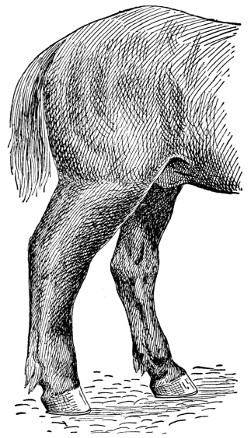
LYMPHANGITIS
This kind of inflammation is usually seen in the hind legs. It is most frequent in heavy draft horses, or in coarse plethoric individuals. It occurs most frequently after a short period of idleness.
—An inflammation of the[215] lymphatics, usually of the hind legs. Hence the name “big legs.” It is the result of too rich feeding, and too little work in many[216] cases on the one hand, or of overwork and insufficient food on the other. Lymphangitis often follows other diseases like distemper, influenza, or pneumonia, in which cases the system is weakened and the lymphatics in abnormal condition. It shows itself after a short period of idleness and rest. It usually begins with a chill and a rise of temperature, which may be as much as 105 degrees, depending on the intensity of the attack. One or both hind legs may show swelling and be so stiff and sore after standing during the night as to be moved only with difficulty when the horse is taken out of the stable in the morning. The horse in moving seems able to bear little or no weight on the affected leg. At the same time, the pulse is full and throbby, respiration is fast, the bowels are constipated and the appetite is lost.
In some cases the legs swell to an enormous size. If the inflammation is not relieved in a few days, the glands get badly diseased and blood poison may result. The disease, however, if taken in time, is easily treated. If it is caused by overfeeding, change this; give more exercise. When the disease is first noticed, give the horse 4 tablespoonfuls of aloes, 4 tablespoonfuls of carbonate of soda and 4 tablespoonfuls of ginger. These should be dissolved in a half pint of boiling water, then mixed with a half pint of cold water, and then given as a drench. If the pulse is fast, it may be made easier and slower by giving 20 to 30 drops of tincture of aconite, every couple of hours. A couple of tablespoonfuls of nitrate of potash in the drinking water three times a day will increase the urine. This is desirable to do in this disease. The leg should be bathed for at least a half an hour and then dried and a wash consisting of 2 tablespoonfuls of acetate[217] of lead, 8 tablespoonfuls of tincture of opium, and a quart of water should be applied to the legs. This should be rubbed in well with the hand every hour. In from 20 to 30 hours, a great change for the better will be noticed the inflammation will have been reduced; the pain will have disappeared and the bowels will be loose and active.
From now on give general exercise at frequent periods, during the day. In cases caused by overwork or too little food or those following debilitating diseases, like influenza or distemper, the treatment should be more stimulating; therefore, nutritive foods and tonics are best. Good hay and oats and other feed of a laxative nature should be furnished.
A preparation, consisting of 4 tablespoonfuls, each, of tincture chloride of iron, tincture of gentian, and ginger in a pint of water three times a day will be found both stimulating and nourishing. If the disease has progressed so far that the legs break and show that matter is formed, wash them with warm water and follow with acetate of lead, sulphate of iron and carbolic acid. Use 2 tablespoonfuls of each in a quart of water and apply twice each day. If the swelling hangs on use Fowler’s solution of arsenic, 4 tablespoonfuls to a dose in a bran mash once a day. Continue this for four or five weeks. A salve made of 2 teaspoonfuls of iodide and 8 tablespoonfuls of vaseline should also be rubbed on the leg twice a week.
—See Hydrophobia.
—The grubs of the ordinary flesh-flies so common about stables and houses. The adult fly deposits the minute larvæ in fresh meat, in wounds, and frequently in dirty wool. These become the maggots so well known about the farm.[218] The distress caused by these when present in a wound is considerable, and they endanger life.
The best treatment is in line of cleanliness. Keep old wounds clean by means of antiseptic washes and tag the sheep that no filth and dirt may accumulate. If for any reason maggots are found, open the infected part and remove, if possible, both the maggots and sloughed tissue. Old sores or wounds, if they will not lend themselves to complete removal of the maggots, should be treated with a solution of carbolic acid and water. On some, turpentine can be used. Chloroform may be sprayed on, or injected into the wound with almost instant results. After the maggots are destroyed follow up the treatment with a good disinfectant until the wound has healed.
—An eruption of the skin above the feet in horses. The disease at first is very much like eczema. In time the watery fluid dries up and the sore parts become covered with hard crusts and scabs. The sore spots should be washed with some good disinfectant and repeated frequently enough to destroy the infection. A moderate purge is advisable. See that only wholesome food is provided.
—Inflammation of the mammary gland or udder. The disease is frequently called caked bag and garget. In the last named, the milk secretion is altered and appears as a thick or a stringy fluid. Heavy milkers are most commonly affected. The udder becomes swollen, hot and somewhat tender just before calving. The swelling may extend forward along the belly. It often gets so severe as to require treatment. It is in this sense physiological. In a few days after calving, as a rule, the swelling disappears and the normal[219] condition is regained more quickly if the calf is allowed to suck the cow. In the first stages bloody milk is secreted and often pus is formed in one quarter or more of the udder. The udder should be carefully milked, cleaned, and, if the milk ducts are closed, it may be necessary to use a milk tube. This should be used cautiously so as not to injure the tissue of the udder and should be perfectly clean before inserting, otherwise serious inflammation may result. In bathing, use hot water for 15 to 20 minutes at a time, after which rub dry and apply an ointment made by dissolving 3 tablespoonfuls of gum camphor and 4 tablespoonfuls of fluid extract of belladonna to a pint of clean, fresh lard. This ointment should be applied three times a day.
A more serious form of the disease is known as contagious mammitis, and is due to invasion of the gland by bacteria. In cases of this kind the inflammation is more extensive and the disorder calls for more careful treatment. Since the milk contains bad bacteria, it is necessary to destroy them so as to prevent spreading of the disease. The milker should have clean hands and should wash them in a disinfecting solution before milking another cow. The milk tube may be necessary in withdrawing the milk. After the milk has been removed from the udder, inject a solution of peroxide of hydrogen or dioxygen or a solution of carbolic acid, 1 part to 50 parts of boiled water. After the solution has acted for a few minutes, it should be milked out. The external treatment for contagious mammitis should be similar to that of ordinary mammitis.
—See Scab in Cattle.
—It is a remarkable fact that this disease occurs most commonly in cows which[220] calved easily. This is explained by the fact that in such cases the os uteri remains relaxed for a greater length of time than it does in cases of difficult parturition. Milk fever generally occurs in cows which are heavy milkers, and great eaters. Keeping the animals in permanent stables, and feeding large quantities of rich food while they are giving no milk are predisposing causes.
The disease makes its appearance usually in from 24 to 48 hours after parturition. It seldom occurs after the third day, and some authors state that it has never been recognized before the starting of the milk secretion. The most salient symptoms to the average layman would, perhaps, be the anxious expression of the animal, bellowing and mounting into the manger. Later they become very weak, stagger and fall, and are unable to rise. The members are usually extended in a rigid position. A rattling or whistling noise is heard in case the larynx is paralyzed. The feet, ears and horns feel cold to the touch. When a case is going to recover we see improvement as early as the second or third day. Recovery is usually complete at the end of from two to five days.
Milk fever is one of the cases where the old maxim, an ounce of prevention is worth a pound of cure, is doubly applicable. If proper precautions were taken a large number of cases could be prevented. Give the pregnant animals daily exercise, and decrease their allowance of food.
—Make the cow comfortable. Now give her a small dose of Epsom salts from one-half to one pound, depending on her size. This should be given as a drench. Animals afflicted with this ailment swallow with difficulty. Use care that the drench does not get into the lungs.[221] Perhaps the most satisfactory medical treatment is to use what is known as the Schmidt treatment. This is nothing more than injecting into the udder a solution made by dissolving in one quart of clean boiled water 3 teaspoonfuls of iodide of potash, after stripping all milk from the udder. A very satisfactory way is to get a rubber tube, attach it to a common milking tube which is placed into the teats in turn and pour the solution into the tube by means of a funnel. By massaging the udder the solution can be worked into each quarter in a short time without difficulty.
In case iodide of potash is not available, inject air into the udder after drawing out the milk. I have known of many cases where air has been forced into the udder by means of a bicycle pump, and the animal recovered in a very short time. If the disease does not respond to the treatment with readiness, repeat in a few hours, say, anywhere from five to ten hours after. Cold water or ice on the head is advisable. The use of stimulants is also recommended. Whiskey can be given in doses of 10 to 15 tablespoonfuls and jamaica ginger 6 to 8 tablespoonfuls. Milk the cow frequently and massage the udder, bathing in hot water.
After the cow is on the way to recovery, withhold milk-stimulating foods for a few days and give some tonic like gentian and nux vomica, half and half, 2 or 3 tablespoonfuls two or three times a day.
—See Azoturia.
—When a cold or simple catarrh is neglected it may run into a chronic condition giving rise to nasal gleet. A thin, bluish discharge comes from the nose; and the membranes of the nostrils, instead of being moist and pink in[222] color, take on a leaden hue. The coat at the same time shows unthriftiness. In such cases the face and head may swell because the accumulated materials fail to pass out. When these bunches are tapped with the fingers, a dull sound is heard. Treatment consists of isolating the animals and giving them good care, nutritious food and well-ventilated quarters. A bucket, filled with boiling water, in which a half cup of turpentine is placed, and held under the nose to steam the nostrils and face, is excellent. Any sort of blanketing that will hold the steam about the head is very good to have at hand at the time. For internal treatment give a teaspoonful of sulphate of copper three times daily in a small bran mash; following this drop the copper sulphate and give 2 tablespoonfuls of Fowler’s solution of arnica twice a day in the mash. Should the bulges on the face become large, it will be necessary to open them. Often a part of the bone requires sawing out to get effective results. In these severe cases it is best to have your veterinarian make the operation.
—A disease of the navicular bone and the structures surrounding it. It is called “coffin joint lameness.” This bone is situated at the back and inferior part of the coffin joint, and acts as a pulley over which the flexor tendon of the foot passes. Horses with upright pasterns are most liable to it, as more weight is thrown on this joint. Those shod with calkins on their shoes, which prevent the frog from coming in contact with the ground, therefore causing a shock to this joint, are also very liable to it. Some horses have hereditary tendency to this disease. Nails penetrating too deep through the sole, or anything that will cause inflammation of this joint,[223] is likely to produce navicular disease. The most prolific cause is bad shoeing. By degrees the inflammation in a chronic form extends to other parts, causing a shrinking of the soft parts, resulting in contraction of the foot.
The lameness may appear suddenly and perhaps immediately after the horse has been shod, and is then usually thought to be the fault of nailing on the shoe. It is likely in this case that the smith has pared the sole and frog too thin, and that the part has suffered from a bruise by the horse stepping on something hard. After a rest it may disappear, to return after the next drive. Sometimes the disease is of very slow progress in one or both fore feet. The first thing that is noticed is that the animal points its toe, and if both are affected, first one, then the other. The animal may not be lame, but it does not step out so well as it used to, and by degrees the part gets more tender, until the animal begins to go lame, and the lameness gradually gets worse. There is a form of this lameness where the animal shows stiffness and lameness when first taken out of the stable, but, after being driven for a short distance, it passes off, and after it stands for awhile it will start off lame again. If this disease lasts for some time the muscles of the chest and shoulders seem stiff and may shrink. This has been called “chest founder” by horsemen. This is brought about by the soreness of the feet. The horse is afraid to step out, giving it the appearance of being stiff; the muscles of the chest and shoulders will shrink from want of proper action, caused by the feet being sore. If there is heat and tenderness in the hollow of the heel or a redness of the sole, and an absence of any other disease of the foot or leg, we may consider with[224] almost a certainty that it is a case of navicular or coffin joint lameness. The result is contraction of the foot.
Take off the shoes, so that the frog will rest on the ground, then poultice the feet with bran, made up with cold water if it is a recent case, but if it is of some months’ standing hot water is better than cold; put the poultices into bags made a little larger than the foot; put about two inches deep of the bran mash into the bag, then put the foot in and fill in all around as high as the fetlock, and tie the bag above the fetlock and around the ankle to keep it well on the foot. Wet this several times a day and change it once daily. Continue this for two weeks, and see that it is properly done; if not, it will be of no service. Then blister the coronet with cantharides 2 teaspoonfuls and lard 4 tablespoonfuls. Repeat in three weeks, and give the animal a long rest.
—See Bot Flies.
—Nodules resembling those of tuberculosis found in the intestines of sheep, are due to the presence of parasitic worms. Profuse diarrhœa and a pronounced anemic condition prevail. A post mortem examination of the intestines discloses the presence of numerous nodules in the intestinal walls. If the worm is present, no treatment is possible, for the reason that any medicine that would affect the worm would also affect the tissues and lead to their destruction. Prevention, therefore, is the only means of overcoming the disease. Sheep must be kept off infested pastures, and infested pastures must be plowed and given over to cultivated crops. Give lambs only clean pastures to graze over. This means crop rotation in connection with sheep husbandry.[225] No feed that has been tramped over by infected sheep should ever be supplied to lambs or sheep not infested with the disease.
—Difficult parturition is common in some females. And frequently others, less bothered as a rule with any difficulty at this period, deliver their offspring only after great labor and much difficulty. When such cases occur close vigilance not only frequently hastens delivery, but often saves the life of either the mother or offspring or both.
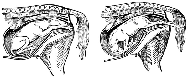
NATURAL PRESENTATION OF THE FOAL
In either of these cases delivery follows in the usual order without delay or injury to the mother.
In many instances the trouble is seated in the womb; the neck of the womb remains closed, and even though long-continued and vigorous efforts are made, the offspring does not arrive. In cases of this kind assistance can be rendered which quickly removes the difficulty. First oil the hand and forearm and work the fingers into the passage, gently pressing it open. If the womb does not yield to this treatment saturate a sponge or cloth with extract of belladonna and rub it around the neck, leaving it thus for a little while. On removing the sponge the passage will open.
[226]
—The natural position of the fetus at birth calls for the fore feet forward with the head resting on the knees. The fore feet, therefore, in a normal delivery, are first presented and then the head. If the fetus is not unduly large, the mother will likely force the delivery without assistance. In case the struggle is extended gentle assistance will be in order. This can be rendered by a gentle pull on the legs and head. If this does not bring the offspring, you can consider that something is wrong. However, do not be hasty, just give time. Mares usually deliver in a few minutes and cows often require an hour or so after labor begins. If you conclude that something is wrong oil the hand and arm. Shove the fetus back and ascertain, if possible, the trouble. If this examination shows dropsy of the abdomen—water in the belly—puncture the abdomen with a knife in order that the fetus may be delivered. If the trouble is with the head—water in the brain—puncture the head that the water may run out, and then remove the arm and hand. When the struggle pains come on again, give a gentle pull and delivery will follow.
Frequently the position is changed. Sometimes but one fore foot appears with the head, making it impossible to deliver the offspring. When a case like this occurs, shove the fetus back and bring the unpresented leg forward where it belongs, and then likely no further trouble will result. If the legs are in proper place but the head turned backward, it will be necessary to push the fetus back into the womb and bring the head forward in position. In case the head resists your efforts, adjust a noose over the head, and while you work with your hand inside, have an assistant gently pull on the rope, in order to draw the head into the proper[227] position. After the head and fore feet are put in natural position, delivery will follow without further difficulty.
When all four feet appear together it is necessary to push the fore feet back into the womb just as far as it is possible to force them. This done, pull now on the hind feet and bring the fetus out, hind feet first. It is always a mistake to attempt delivery with the head first when delivery has proceeded as suggested in cases of this nature.
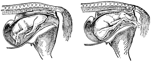
ABNORMAL PRESENTATION OF THE FOAL
Delivery is not possible in either of the cases here illustrated. Where such occur assistance must be rendered. See article on obstetrics for treatment.
Where delivery is attempted with the hind legs foremost, it is regarded as safe, provided the feet come out as they should. If any difficulty is encountered, shove the fetus back, straighten the legs, and then with the renewal of the labor struggles assist the mother by a gentle pull on the hind legs.
Another common presentation is where you feel nothing but the tail, rump and hips. Adjust the fetus for proper delivery by shoving the hind end upwards and towards the front of the womb, then slip the hand down and get hold of the foot of[228] the hind leg and lift upwards and backwards until the legs are brought out into the passage. Now repeat the work for the other leg and the job is done.
It is always a good plan, after difficult parturition, especially when any abnormal discharge appears, to wash out the womb with warm water in which a little carbolic acid or creolin is placed. Use this daily for a few days.
—The worms are found in the horse in two periods of existence. The mature worms are usually found attached to the mucous membrane of the intestinal wall of the large intestine, with the head sunk deep for the purpose of sucking blood, which gives them the brown or red color. The immature are found sometimes in the same organs, in a small capsule covering, in small pellets of manure, in cavities or cysts, varying in size from a pin-head to that of a hazel nut, in the walls of the intestines, and also in the arteries and other structures of the body.
When present in the kidneys or in the arteries leading to the kidneys, or in the surrounding tissues, a horse is especially sensitive to pressure over the loins. They have been known to cause paralysis. When found in the brain, an animal, when working, suddenly begins to stagger, the eyes become fixed, and the horse shows many of the symptoms of “blind staggers.” When the large arteries of the abdomen are affected, and this is their favorable location in the circulatory system, the animal is frequently subject to colic, which often results in death. This is also the case when found in great numbers in the intestines.
From a thorough investigation of a great many cases, both before and after death, the conclusions[229] are drawn that the parasite evolves a poisonous substance (toxin), which, in many instances, stupefies the brain or parts of the nervous system of the horse, and in that way causes coma, paralysis and death of the animal.
Prevention is the best treatment. Hay and fodder from swampy land are to be looked upon as suspicious. Pastures which are subject to overflow should be avoided. Medicinal treatment consists of a prolonged, careful use of some of the essential oils or other vermifuges. The ordinary spirits of turpentine has proved a fairly good common remedy. An ordinary animal will stand 8 tablespoonfuls of turpentine given in a pint to a quart of raw linseed oil, thoroughly mixed. If the animal is badly affected, the above dose may be given night and morning for two or three days, then omit for a week or two and repeat. The remedy should be discontinued as soon as the animal shows signs of irritation of the kidneys.
—A loss of power over some of the muscles due to a disordered state of the brain or nerves. This may result from disease or injury or some irritation. In horses and cattle the hindquarters are not infrequently affected in this way, the result of indigestion from constipation or from attacks of colic. The animal shows weakness in one hind limb, moving it with difficulty when the opposite limb may then become affected. If the attack is very severe, the animal falls on its haunches and may not be able to rise. Temperature, pulse and respiration, all are rather normal. Treatment should be directed to remove the cause of the disease. When there is colic or constipation, give purges. A half teaspoonful of extract of nux vomica, given in a pint of milk twice a day, is[230] very good. Pouring cold water from a height and then immediately hot water sometimes greatly strengthens the muscles and has its use in treating. Rubbing the parts with mustard stimulates them, and in some cases good results. Paralysis resulting from injury usually disappears as the part returns to its normal state.
—These are living plants or animals that live temporarily or continually in the bodies of other plants or animals and draw their nourishment from their host. It is doubtful if there is a single farm animal that does not harbor parasites at nearly all times during its life. There may be many of these in the same individual at the same time. Parasites may be harmful or not, as the case may be. Parasites may be divided into two classes—plant parasites and animal parasites. The bacteria and molds are the most important among the former, whereas in the latter certain minute protozoa, certain forms of insects and certain worms are the most commonly met. Such diseases as staggers, tuberculosis, and typhoid fever are the result of bacterial diseases, while Texas fever is an example of the protozoa class; and then the insects and worms are types with which we are all acquainted. When a disease is caused by either, discussion will be found under the name of that disease.
—See Milk Fever.
—See Obstetrics.
—An inflammation of the membrane which lines the abdominal cavity and which also invests the abdominal organs. It may be caused from some exposure to cold after some weakening disease. Some injury to the abdomen[231] or belly may cause it, or it may start from some inflammation that has attacked the stomach, liver, intestines, or the spleen. When attacked, a slight pain is felt and the animal lies down, stretches himself, sweats freely, and moans. Then he rises, walks about somewhat, and all the time breathes heavy and shows much weakness. The pulse runs up between 75 and 100 beats a minute. In time the legs and ears get cold. A good treatment is a pint of raw linseed oil, 4 tablespoonfuls of laudanum, and 10 drops of aconite. Mix these and give as a drench. A mustard plaster for the abdomen and something hot for the back are desirable. In two hours, if the pain continues, give 4 tablespoonfuls of laudanum and 10 drops aconite in a pint of lukewarm water. Use as a drench.
—A contagious epidemic disease of the horse affecting the animal all over and particularly the membranes of the air passages. There is general debility, considerable cough, and a general discharge from the nostrils. The transparent covering of the eyeball becomes inflamed. At times the disease is very fatal, many horses succumbing to it. It is most common in the spring.
One of the symptoms is the general weakness of the animal. He hangs his head, and trembles; has little appetite and appears cold. The eyes show a watery discharge and later a stare coat. The pulse at first is weak, but quick, and later rising to 80 or 90 beats a minute. At this stage the temperature is high, around 103 to 105 degrees. The breathing is accelerated to about 50 times a minute. The bowels do not act, or act very poorly, and the urine is very scanty. In treating, first isolate the animal and disinfect the stables to prevent spreading. Any of the common disinfectants will do.
[232]
Good nursing is necessary. Keep the horse warm with blankets. Give him soft, nourishing food. The eyes should be bathed three or four times a day with hot water. A little boric acid, say, a teaspoonful to a half pint of water, is good to use as a wash for the eyes and nostrils. To keep the kidneys active and to reduce the fever, give a tablespoonful of nitrate of potash dissolved in water two or three times a day. If the horse is very weak, one-half glass of whiskey in a pint of gruel three times a day is stimulating and helpful. It is better not to give any physic of any kind. After recovery, the horse should be given little or no work. A long rest of several weeks is necessary.
—The covering of the fetus, commonly called the afterbirth. As a rule, this comes away with the birth of the offspring. Occasionally in the cow it remains attached to the walls of the uterus, and if not removed will cause trouble, if not sickness and death. Soon after the birth of the calf, if the afterbirth remains, decomposition sets in and as a result the system is more or less poisoned. The first symptoms observed are the offensive odor, the reddish discharge and the decrease in the milk flow.
If the afterbirth does not come away of itself, assistance is necessary. Do this during the first or second day, or the third day at the latest. To remove the afterbirth, tie up the cow and fasten her in a way that she cannot jump around. Now introduce the hand and arm, after careful washing and disinfecting and oiling, into the uterus and gradually and gently break the buttons or attachments from the walls of the uterus with the fingers. With patience these will come away and the whole[233] membrane be removed. An occasional injection is advisable. Use some good disinfectant in the water, flush out thoroughly.
—This disease occurs in the chest cavity and is found inside the ribs and over the lungs. It is caused very much in the same way as inflammation of the lungs, like exposure to cold, standing in a draft, and cooling when warm. Some injury to the ribs may also cause the trouble.
In the early stages the animal is noticed to shiver, the pulse is quick and strong, and there is great pain. The breath is heavy, and this is noticed as far back as the flanks. While the animal may lie down, its disposition is to stand up most of the time. There is an inclination to cough, but this is suppressed, because of the pain occasioned by it; therefore the cough really ends in a groan rather than in a normal cough. The extremities of the body become cold.
The best treatment endeavors to prevent the disease from developing. Do just as you would in a case of inflammation of the lungs. Mustard plasters for the chest on each side are good. Keep the body well covered, including the legs and neck; have good ventilation in the stable, but keep the patient out of any draft.
As soon as the disease is noticed, mix the following in a pint of cold water, and give as a drench: Ten drops of aconite, a half teaspoonful of belladonna and two tablespoonfuls of laudanum. These should be given every two hours until the pain subsides. If the animal seems to be weak, and needs a stimulant, give 4 tablespoonfuls of spirits of niter and a half glass of whiskey. This may be given in a pint of cold water mixed with the gruel and given as a drench three or four times a day.
[234]
At the same time use the following medicine to improve the kidney action: One-fourth pound of saltpeter or nitrate of potash and one-fourth of a pound of gentian root. These are to be mixed well together and a teaspoonful given three or four times daily. Soft foods are desirable. A small amount of water should be given frequently. Small quantities at a time are preferable to large quantities at infrequent intervals.
—This is a very contagious disease of cattle introduced in this country from Europe. At one time it was a very serious menace to the cattle industry. Thanks to the very aggressive work of the United States Department of Agriculture, the disease has, so far as is known, been eradicated from this country. No cases of the disease have been reported during the past dozen years.
—See Inflammation of the Lungs.
—A swelling or soreness at the top of the head. Usually it is caused by an injury, like bumping the head in a doorway, or from a bruise made by the halter or bridle. It is first noticed by a swelling or soreness, which frequently causes trouble by forming an abscess; sometimes this works down and even affects the bone. Treatment is very simple if handled in time. Remove the cause and then bathe with warm water and vinegar twice a day and apply a liniment of some kind. If the abscess is formed, it should be opened with a knife at the lowest point to remove the matter. From now on for a few days bathe the opening with warm water in which has been added some carbolic acid or creolin. If the case causes much trouble, you had better consult a veterinarian, as bad cases frequently[235] leave the neck stiff so that the animals are not able to eat off the ground.
—See Sand Crack.
—A name given to a fistulous opening upon the heels and quarters of the coronary band, and is caused by treads, pricks in shoeing, bruises, and suppurating corns. Any injury which will cause suppuration within the foot will usually cause matter to form at the coronet, and may result in quittor. The disease is indicated by a swelling upon the coronet where the hair and hoof meet, great lameness, and a discharge of thin or thick curdy pus. There may be one or a number of small openings leading down into the sensitive part of the foot. The parts surrounding the quittor swell and become hard and take on an unhealthy action and are difficult to cure, and may be permanently diseased.
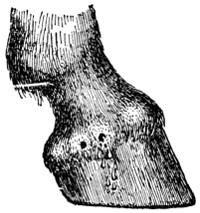
QUITTOR
Fistulous wounds on any part of the coronet are usually the result of a tread or bruise. If neglected serious trouble may result.
Clean the foot and put it into a bran poultice for several days, then remove any horn that may be pressing on the sore part. If it is at the heel remove the crust with a knife; if it is in front of the hoof rasp it thin. Then probe the opening at the top to find the depth and direction. Put a grain of bichloride of mercury into tissue paper and roll it into a cone and press it down to the bottom of the opening. Treat all the openings in the same way. Put the foot into a bag to protect it from[236] injury and let it alone for three days, then clean out the openings and put in some more of the bichloride of mercury, and so on for two weeks, or until the parts become healthy and the hard swelling has decreased; then make up a bath of chloride of zinc one ounce, cool water one gallon; put the foot into this twice a day for twenty minutes at a time. As soon as the openings are healed blister the coronet with the following: Mix 2 teaspoonfuls of cantharides with 4 tablespoonfuls of lard; repeat in two weeks if necessary. When it is time to put on the shoe and work the horse, a bar shoe will be best. If the animal has much fever in the early stages of the disease give a dose of aloes, and follow this by giving 2 tablespoonfuls of nitrate of potassium twice a day in bran mash. Later in the disease give a teaspoonful of sulphate of iron once a day in bran mash as a tonic.
—See Hydrophobia.
—A disease which affects the muscles or joints, wandering from one part of the body to another. It affects nearly all animals, including the horse, ox, dog, hog, and sheep. Rheumatism of the muscles is usually due to catching cold, while rheumatism of the joints is often due to some micro-organism.
Stiffness, which usually comes on suddenly, is a characteristic symptom. The animal may be able to move only with great difficulty. The joints may crack when moved, the affected muscles are hard and painful to touch, the soreness may shift from one part to another; and the animal sometimes makes a quick recovery, only to be followed by another attack in a short time or perhaps never again. These symptoms may be associated with a rise in body temperature and increased pulse.[237] The disease may last for a long time or only for a few days. In chronic cases the muscles decrease in size in the parts affected. In the dog it is very painful when caused to move and he will howl, or even howl when he thinks he is going to be moved. In sheep it seldom occurs except in young lambs. Pigs are often affected in the legs or back, sometimes becoming paralyzed in the hind legs.
usually shows very rapid swelling, increased heat, and is very painful. The animal is often so lame that it will not put any weight on the foot of the affected limb.
For horses and cows, treatment consists of local applications of alcohol 50 parts and oil of mustard 1 part, rubbing it in well; or spirits of camphor. Give at the same time internally 1 teaspoonful of potassium iodide twice daily and not to exceed 12 doses; or salicylate of soda 4 tablespoonfuls daily. Keep the animal warm and in a well-ventilated stable. Pigs or dogs, according to size, should be given from 4 to 16 grains of salol, also using the above local applications.
—A growth of bone on the pastern bone, just above the hoof. It causes lameness when it interferes with the joint or the passage of any of the tendons. Some horses are predisposed to bony diseases from the least injury, while others are not, and in selecting mares for breeding purposes the former should be rejected. This disease results from strains, bruises, or injuries to the cartilage of the joints. When the membrane of the bone or cartilage becomes inflamed there may be great lameness for several months before any enlargement takes place, and it is somewhat difficult to detect. The absence of other diseases of the foot, with some heat in the pasterns, and soreness on[238] pressure or moving the joints indicates this disease. In other cases the enlargement may make its appearance for some time before the horse becomes lame, and in some cases it may never cause any lameness, but should always be looked upon with suspicion, as in the majority of cases it sooner or later causes lameness. Ringbone is more difficult to cure on the fore feet than on the hind ones, as the pasterns are more upright on the former than on the latter, and, besides, the horse’s fore legs have to bear two-thirds the weight of the body.
The horse should have rest, and the shoes should be removed and the foot pared level. If there is heat in the part, keep it wet with the following lotion by means of a bandage saturated with it: Acetate of lead half an ounce and water one quart. Continue this for a few days, then apply a blister composed of cantharides 2 teaspoonfuls, biniodide of mercury 1 teaspoonful and lard 8 tablespoonfuls. Rub on a third of this with the fingers. It is not necessary to cut off the hair if the blister is well rubbed in. Let it remain on for 24 hours, then wash off and rub on a little lard. Repeat every second week until three blisters have been applied. Keep the horse’s head tied while the blister is on so that he cannot get his mouth to the part. The horse should have a few months’ rest after this treatment. If it does not cure the animal it is best to have him fired by a qualified veterinarian.
—This is common in the domestic animals, especially in calves and young cattle, and is contagious. It depends upon the presence of a vegetable parasite, which develops and grows rapidly when it finds a suitable place for development. Ringworm may affect any part of the body, but its favorite seat is around the eyes, the face,[239] ears, and neck of cattle, and sometimes the back and hindquarters.
A gray crust appears on the skin, and the hair drops out. This keeps spreading in the form of a ring until around the eyes, the side of the face, ears, or neck may be covered with it. It appears in the same way on the back, hips, and inside of the hind legs. It does not seem to affect the health of the animal, as it is found in the well-kept as well as those poorly kept.
First remove the crusts by washing with warm water in which one ounce of carbonate of potassium has been put to every quart of water. A brush should be used in washing the parts. Then use the following: Iodine 2 teaspoonfuls and vaseline 4 tablespoonfuls. Rub a little of this on with a gloved hand. Repeat in three days. Or mix carbolic acid 1 ounce with 2 ounces of alcohol and apply a little of this to the parts with a feather once or twice; this last is very effective.
—A disease, due to the wasting of the larynx; is characterized by loud, unnatural sounds after any violent exertion. The disease sometimes follows distemper and influenza or a local injury to the throat. Once established the disease is incurable. In its early stages repeated light blisters may help. A common blister can be made of a half teaspoonful of cantharides, a half teaspoonful of biniodide of mercury and 4 tablespoonfuls of vaseline or lard.
—A disease of the mucous membrane in fowls. It is of the nature of an inflammation, with a discharge from the eyes and nostrils usually accompanying. Damp and unsanitary quarters favor the development and spread of roup. It is clearly a germ disease, and, therefore, contagious. It is[240] spread by means of infected quarters and fowls. All discharges must be destroyed by disinfection, and the diseased fowls quarantined off by themselves. The dead should be burned. Keep the quarters light and airy; admit an abundance of sunshine and fresh air. Feed wholesome, nutritious food, that the poultry stock may ward off the disease. The best treatment is that which prevents spreading to healthy fowls. If an outbreak occurs, disinfect thoroughly, liberally, and continuously. Antiseptics administered about the head will usually break up the disease. Creolin is good—say, 1 part to 100 parts of water. Kerosene is also recommended.
In a sense, roup is the result of neglected colds. The birds sneeze, and manifest their uneasiness as animals do with common colds. A teaspoonful of pure carbolic acid to each gallon of drinking water is an excellent preventive and can be provided at small cost.
—A crack found in any part of the wall of the foot. The crack is due to over-exertion. When the hoof is dry and hard and brittle, the crack usually begins at the top and extends downward. Frequently the sensitive tissue creeps into the crack, causing pain, and from which blood frequently issues. When a crack is first seen, the feet should be poulticed with linseed meal for a few days. This will remove the inflammation and soften the hoof. The next step will be to pare out a piece of the hoof at the top, separating it completely from the coronary band a half inch or so on each side of the crack down to the quick. Fill this hole with tar. A bar shoe attached so as not to rest on the wall where the crack is located is very helpful.
[241]
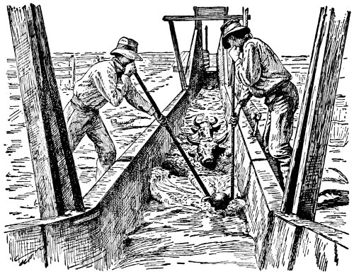
A CATTLE BATH TUB
The tank here shown is used for dipping the cattle for treatment of mange. The dipping tank is now generally used throughout the West.
—Scab or itch, sometimes called mange of cattle, is caused by a minute mite that lives upon the surface of the skin, burrowing into it. Other animals are not attacked by this parasite, although a similar one does afflict sheep. So long as cattle are doing well on grass, no disturbance is noticed. As soon, however, as they are placed on dry food and cold weather sets in, the disease appears, and, if the cattle do poorly, develops into a very aggravating form. Old cattle are less troubled, the attacks being more frequently on calves and yearlings and two-year-olds out of condition. In the early stages the itching of the skin in the region of the neck or shoulders is first noticed. This is indicated by the[242] animals digging at the skin with teeth and horns and the constant rubbing against posts or barbed wire or anything that may give relief at the time. The disease gradually spreads along the back, sides and outside of legs. In the early stages the coat looks rough, the skin has a scurvy appearance. In time, the hair comes off or is rubbed off, presenting bald patches of thick, glazed and wrinkled skin. After the hair comes off the parasites leave these regions, seeking other quarters and then the hair grows in again. There is a dejected and debilitated condition in animals thus afflicted and they fail rapidly in flesh. Their appetites are poor and most of their time is expended in scratching themselves.
Scab spreads rapidly through a bunch of cattle, especially if they are not thrifty, and disseminates itself through a herd in four to six weeks. The thrifty, vigorous animals resist the infection for some time, but they gradually succumb. The disease is spread by direct contact and by contact with infected quarters. While the mites will live a week or ten days in protected places, they are almost immediately destroyed by direct sunlight. As soon as the disease is discovered in a bunch of cattle, the infected animal should be isolated and the infected quarters and rubbing posts disinfected with a 5 per cent solution of carbolic acid. Infected animals should be well fed and cared for, and be salted with a mixture of 1 pound of flowers of sulphur mixed with 10 pounds of common salt. External treatment is necessary to affect a cure. If a large number of cattle are affected, a dipping wash through which the animals must swim in the dip is the best means for destroying the mites.
The most efficient remedies, considering cost, are the coal tar products advertised as dip solutions.[243] A homemade dip that is both cheap and effective for treating a small number of animals may be made of 3 pounds of flowers of sulphur, 21⁄2 pounds of unslaked lime, 15 gallons of water. In making this unslaked lime into a thick paste, sift in the sulphur and stir well. Put this mixture in a kettle with, say, five gallons of water and boil for at least half an hour—a longer time is better. When the chocolate-looking mass settles, the clear liquid is drawn off and water enough is added to make 15 gallons. The dip will be more effective if used when warm, just a bit hotter than the normal heat of the body. After the animals are dipped, they should remain in the solution about two minutes. This will be time enough to thoroughly saturate the scabs and destroy them. A couple of ablutions are required for complete eradication. When no treatment is resorted to, the dip should be applied with a scrubbing brush, cloth or sponges and all scabs and crusts should be thoroughly saturated. Warm sunny days are preferable for this kind of work.
—A diseased condition at the attachment of the navel cord soon after birth. It is a good plan just after birth to apply some septic powder to the navel at the breaking point. If trouble arises, apply a solution of carbolic acid, 1 part to 20 parts of water, after using some hydrogen peroxide. A little iodoform and alum, mixed half and half, make a good dusting powder to use also.
—See Bot Flies.
—On either side of the coffin bone there is a cartilage which may in certain cases become hardened by deposits of mineral matters, which may thus lead to lameness. Side bones are[244] situated on one or both sides of the leg and bulge above the upper portion of the hoof. They may be the result of inflamed conditions, bruises or troubles like corns or hoof cracks. Slipping on the stony pavement is a frequent cause, as well as the great weight of the bodies in heavy horses. If the wagon tongue falls on the foot at this point, the cartilage may be injured and induce the disease. The swelling is first noticed just above the hoof or near the heel. Lameness soon follows.
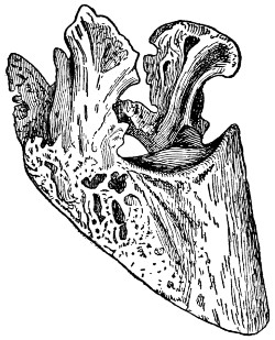
SIDE BONES
When the cartilages on either side of the foot of a horse just at the top of the hoof and close to the heel turn to bone, side bones are the result.
The treatment usually recommended for side bones consists in the free use of cold foot baths or cold water bandages for a week or more. Tincture of iodine applied to the swollen parts is very good. A blister applied after the water applications have been made for a week or so, is used by many veterinarians. The blister is made of 2 teaspoonfuls of cantharides mixed with 4 tablespoonfuls of lard. It is rubbed in well with the fingers and allowed to remain for 24 hours, when it is washed off and applied a second time the following week. These applications are continued until the lameness disappears. If this does not bring permanent relief,[245] then firing of the injured parts and several months’ rest will be necessary.
—Some kinds of food cause an unnatural flow of saliva. Fresh crimson clover hay is one of these. Of course the continual flow of saliva is undesirable and unpleasant. It is unnatural and should be checked as soon as possible. This can be accomplished by changing the feed and then washing the mouth out with alum water. If a change is not observed soon, give a good physic. For horses use 8 teaspoonfuls of bitter aloes, a teaspoonful of common soda and a teaspoonful of ginger. Mix these in a pint of water and give as a drench. For cattle, dissolve a pound of Epsom salts, a tablespoonful of common soda and a tablespoonful of ginger in a quart of lukewarm water and give as a drench.
—See Colic.
—This disease, known in common language as bone spavin, is an enlargement of the hock joint similar to a ringbone about the coronary joint. It may affect the hock joint in such a way as to cement the small joints together, not causing lameness, and apparently no blemish, but the free movement of the limb is impaired. Any condition which favors sprains, such as fast driving over hard or uneven roads, unequal paring of the hoof, thus causing the weight to be unequally distributed in the joints, and severe labor in early life, or blows, bruises, or any injuries to tendons, ligaments, or joints may cause spavin. In addition to these causes may be mentioned sprains caused by jumping, galloping, or trotting animals faster than they are accustomed to; also straining by starting a heavy load, slipping on an icy surface or sliding on a bad pavement.
[246]
If the patient is examined before any bony growth has developed, inflammation will be detected on the inside of the hock joint at the junction of the cannon bone and the joint. While in the stable the horse prefers to rest the diseased leg by setting the heel on the toe of the opposite foot with the hock joint flexed. In traveling the patient is very lame when first taken out of the barn, but after traveling for a short distance goes sound. The diseased leg is not lifted clear from the ground, but nicks the toe in the middle of the stride, which is very noticeable on a pavement. A strained horse becomes very lame after being allowed to stand for even a very short time, then moved again.
Preventive treatment consists in keeping horses’ feet trimmed properly, not overworking colts while young, careful driving on hard or uneven roads, and avoiding all injuries that are liable to strain tendons, ligaments or joints of the limbs. Even after a spavin has developed it may be cured by proper treatment of the feet, and applying a fly blister. The fly blister is prepared by mixing thoroughly 4 tablespoonfuls of pulverized cantharides, 4 tablespoonfuls of biniodide of mercury and 8 ounces of lard. The hair is clipped over the spavin and the blister applied with considerable rubbing. The horse’s head should be tied so as to avoid his biting the part blistered. A second application of the blister is to be used about a month after the first. If blistering fails to cure the spavin, point-firing may be resorted to. It is necessary to “fire” rather deeply to secure good results, care being taken not to fire into a joint. After firing, a fly blister should be rubbed into the holes where the hot iron has been used.
[247]
—The removal of the ovaries to prevent breeding. Cast the animal on her right side. Give an anesthetic to prevent pain. When the animal is unconscious, free the limbs sufficiently to remove any pressure from the abdomen. Now pinch up a fold of the skin in the left side, midway between the prominent bone of the haunch or pelvis, and the last rib, about 4 inches below the backbone. Make an incision in the skin 5 or 6 inches long; now do likewise with the abdominal muscles until the lining membrane of the abdominal cavity is exposed. This membrane is then punctured and an incision made as long as that in the skin and muscles. Now kneel down in close contact with the cow’s back and insert the arm, passing the hand within the brim or cavity of the pelvis. By so doing both ovaries can be secured and detached. This ended, the operation of uniting the abdominal muscles follows by means of stitches and sutures.
Great care is necessary in having the instruments boiled and washed in antiseptics, and in having the fingers, hands, and arms severely clean and well saturated with a strong antiseptic solution. The operation should be made out in the open where neither dirt nor dust are to be found. Extreme care about germs will remove much of the risk associated with the operation.
In spaying a sow, she is laid on an inclined board with the hindquarters up. The operator stands at the back of the sow. The hair is first clipped from the skin where the incision is to be made, high up in the flank and midway between the haunch and the last rib. The incision needs to be just large enough to admit the two fingers. Ovaries are located, pulled through the opening in the flank, and removed by tearing off with the fingers. The[248] flank incision is then closed by the necessary number of stitches.
This operation is sometimes performed in mares. But being rather uncommon the process is less understood. In this case it is best to call your veterinarian or someone in the community well skilled in the operation. In all cases of spaying let severe cleanliness be the rule and practice, from the very beginning to the very end.
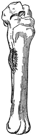
SPLINT
—Splints occur more commonly in the heavier breeds of horses than in those that are light in the bone below the knee. It is rare that splints occur anywhere except on the inside of the front cannon bone, although they are sometimes seen on the outside of both the front and hind legs. Any enlargement of the bone occurring on the inside of the leg between the knee and fetlock comes under the name of splint. The usual cause is concussion, that is, the impact of the foot on the hard road. It may be the result of other causes, such as a blow, a twisting strain or faulty conformation. Some animals are more liable to splints than others. It is, after all, to a certain extent, dependent upon heredity. At first the splint is hard to detect. If you notice a young horse going lame while doing road work, it is well to examine for splints. While working there seems to be no lameness at all, and when standing there seems to be no pain,[249] but when put to a trot the horse shows lameness and may raise and lower his head.
If taken in time, a splint can be cured. The first thing to do with an animal suffering from a splint is to give the animal rest and place in such quarters where there is a soft floor, preferably the ground, and when so quartered one very frequently effects a complete cure. The application of cold water bandages acts well. If treatment of this sort fails, apply a blister of red iodide of mercury, 1 tablespoonful to 2 tablespoonfuls of lard. This blister should be applied with rubbing every day from two to four days, or until the area is well blistered. Then wait until the little scabs fall off, and if the animal is still lame, repeat the application of this blister. To apply the blister, clip off the hair over the enlargement and wash with vinegar to remove grease, then rub in blister with ends of fingers. Keep the animals tied short for two to four days in order to prevent rubbing or biting the leg. Four days after the last application of blister, wash carefully with warm water and soap and over it apply every day or so a little lard, to prevent drying and also to loosen the scabs.
—Injuries to the ligaments of joints, tendons, or muscles. They are caused by violence, as twisting, or from over-exertion; also sprains are often the result of overwork. If an animal is worked until tired or exhausted he is unable to use the proper muscle force, and more strain has to be borne by the ligaments, resulting in sprains, which often occur in young horses or even in old horses, when put to work after long periods of rest. Swelling, heat, soreness, and partial or complete loss of the use of the part, which is shown by the degree of lameness, characterize the disorder.[250] Sprains are most common in the legs, at the fetlock joint, in the tendons just back and above the fetlocks, but may occur in any part.
The first and most important thing in the treatment of sprains is rest, as sprains are a long time in making a complete recovery. In the early stages, that is, before swelling has taken place, applications of cold water should be used, applications of hot water, or hot packs of water, 1,000 parts, and bichloride of mercury 1 part, are very good. This will relieve the pain and reduce the swelling. Applications of liniments are also very good. Should there be great heat and soreness in the part, it is well to use cold applications. Never blister in the early stages. A blister may be used after the swelling has gone down, and the part has become cold, from two to four weeks after the injury occurred. This should be followed by rest for some time after all lameness has disappeared.
—Staggers in horses is an affection of the brain showing itself usually in one of two forms—sleepy or stomach staggers and blind or mad staggers. In the first form the stomach is at fault. Sudden change of feed, moldy or dirty food heavy work or fast driving right after a heavy meal or severe exposure is liable to cause indigestion in the stomach and this is reflected to the brain, causing the animal to act dull or sleepy, sometimes showing symptoms of serious colic, with gas forming from the fermentation of the food, frequently resulting in death.
Blind or mad staggers is an inflammation of the brain and may affect any of the lower animals. In the beginning of this form the symptoms closely resemble those in the stomach form, but as the inflammation progresses the animal becomes blind[251] and violent and may roll, paw, kick, wander around in a circle, usually going only one way, either to the left or right, or it may walk or run in a straight line as near as possible for hours at a time—paying no attention to injuries received in its travels. In either case the animal may be drenched once daily with a quart of raw linseed oil or a pound of Glauber salts, dissolved in water, which sometimes gives relief.
is mostly caused by the young stage of a tapeworm which infests sheep dogs. The dog eats the infected brain of the sheep and the sheep eats the egg of the tapeworm after it has passed through the dog. After the egg hatches in the stomach of the sheep the young worm passes through the bowels and other organs or tissues or circulates through the blood and reaches the brain, where it develops and causes an inflammation, resulting in disease. It is most common in young animals, rarely occurring in sheep after their second year.
Prevention is about the only practical way of handling this trouble. The grounds should be thoroughly drained, allowing the animals only pure, fresh water to drink. It may be necessary to change pastures for a year or two. The brains of all sheep killed and the heads of all dying with the disease should be burned.
—If a box of salt is kept covered in some place frequented by the sheep, to which they are allowed to help themselves, and if said salt is saturated with spirits of turpentine in proportions of a gill to every four quarts of salt, it will wonderfully help to keep the worms from multiplying. It is well, also, to have another box of larger size, where[252] sheep can help themselves at will, filled with tobacco stems. These stems should be cut up in inch lengths and from time to time a quantity of wheat bran should be put on top of the stems. When this is done the sheep soon instinctively learn to use tobacco, and no young intestinal worm or stomach worm, except the tapeworm, can stand the diet. This will not kill mature worms. It will only prevent the worm family multiplying to the extent of injuring the health of sheep.
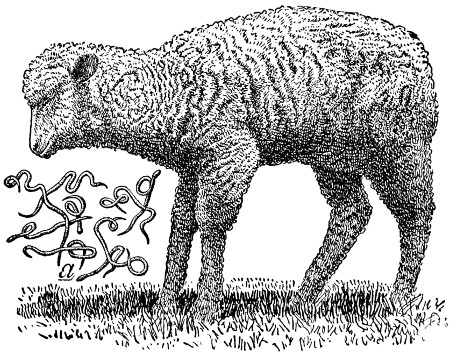
TWISTED STOMACH WORMS
A common attitude observed when sheep are afflicted with twisted stomach worms. The animal loses in flesh, and unless relief is found in time, dies. The parasite is shown in the illustration.
But no sheep owner should feel wholly satisfied by preventive treatment of stomach worms. Twice a year the whole flock should be drenched with some agent which will destroy the mature worms.[253] There are two very inexpensive drenches which will quite effectually do this. The one is gasoline, the other coal tar creosote. The objection to gasoline is that it needs to be so extremely carefully used or sheep will be killed by it. The dose is 1 tablespoonful (never more at one dose) to a mature sheep; mix with not less than 4 tablespoonfuls of raw linseed oil (never boiled oil); then add a half pint of sweet milk. In giving, set the sheep up on its haunches and shake the liquids well together until the last minute it is administered, or the gasoline will separate and, if it enters the stomach in the unmixed form, it will seriously injure and may kill the sheep.
There is no direct vermifuge that will as effectually kill all species of worms in a sheep’s stomach and intestines as will gasoline; yet the coal tar creosote or the more refined class of sheep dips, if given after a full 12-hour fast, before the flock is turned to pasture in the spring, and again about November, will destroy a large number of the mature worms. All lambs born in April or May should be drenched about August or September following, to be certain of ridding them of worms that may later cause their death. The dose of any of the sheep dips is a dessertspoonful mixed in a full pint of water.
—See Concretions or Calculi of Urinary Organs.
—This trouble, commonly called colt distemper, affects horses, and rarely mules and donkeys. It is such an infectious disease that nearly all horses contract the disease when colts and usually remain immune to future exposures. The cause is a very small organism or germ which enters the system when a healthy colt comes in[254] contact with a diseased one or when fed and watered in infected vessels. The seat of trouble is largely restricted to the respiratory organs, occasionally causing difficulty in breathing, owing to swelling in region of throat or to accumulations in air passages.
The symptoms start out with more or less sluggishness. The animal eats little, and does not care to take much exercise. A little watery discharge frequently appears from the eyes, and about the same time a watery discharge from the nostrils, which soon becomes thicker and more yellow in color. Usually the glands between the lower jawbones become enlarged and undergo suppuration with a rupture of them and free discharge of pus. The temperature of the animal may be slightly or very greatly increased from 103° to 105°. The pulsations may also be considerably quickened. When complications do not occur this disease usually runs its course in two weeks, leaving the animal little the worse for having passed through the affliction.
The milder forms of this disease will need little or no treatment other than careful feeding and nursing. A laxative diet, with something green, if possible, should be given. The colt should be placed in clean, airy, and comfortable quarters, but not in a draft. To hasten the suppuration of the glands a poultice of hot bran or flaxseed may be applied to that region, and as soon as softening can be detected within, puncture the gland containing abscess with a clean knife blade and allow the escape of the collection of pus. During the course of the disease the animal should not be worked and care should be taken that it be not exposed to conditions likely to produce a cold.
[255]
—Stringhalt is an involuntary contraction of the muscles that bring the hind leg or legs forward. The cause of stringhalt is a deranged condition of the nerves supplying the muscles, causing the leg or legs to be brought up with a jerk. In slight cases of stringhalt it is necessary sometimes to turn the animal round from right to left, and from left to right, in order to make him show signs of stringhalt, the symptoms of the disease being exhibited as he turns one way only. This disease sometimes comes on suddenly, but generally develops slowly. It is an unsoundness, and depreciates the animal’s value and makes him unfit for hard work or fast driving. There is no sure cure for stringhalt; the animal can sometimes be relieved by giving him one ounce bromide of potassium at a dose twice a day in bran mash, and continuing it for one week, then skipping a week and giving again. It can sometimes be relieved by cutting the tendon or tendons of the affected muscles, but the operation should be performed by a qualified veterinarian.
—See Heat Exhaustion and Sunstroke.
—This disease, by some called infectious anemia of horses, is produced by an invisible organism, which is transmissible to horses, mules, and asses. About the first symptoms noticed are a general weakness of the animal; it tires very easily and is not able to do any work. The loss of flesh is apparent in spite of the voracious appetite which the animal has at times. The appetite usually remains good until death, but the feed seems to do the animal no good. The temperature is very irregular. Some days it runs quite high, at times to 107°; again it is below normal. An[256] animal may have several attacks of the trouble, but each succeeding attack seems to be more severe. The blood becomes thin, and the circulation impaired, and frequently there appears a swelling under the chest or abdomen, or an enlargement of one or more legs. It is quite easy to recognize the trouble, especially in the advanced stages. The slow progress at the beginning, remittent fever, progressive emaciation and anemia, unimpaired or ravenous appetite, staggering gait, and excessive urination are usually all present to a greater or less degree. Recovery takes place only when treatment is begun early and when the disease is not too acute.
In treating, absolute rest until fully recovered is one of the primary requisites, and purgatives are to be avoided. For the fever, the United States Department of Agriculture recommends an antipyretic of quinine 40 grains, acetanilide 2 drams, and powdered nux vomica 30 grains, four times daily. Cold water sponge baths and frequent copious rectal injections of cold water also aid in reducing the fever. After the fever subsides the following is recommended: Arsenious acid, 2 grams; powdered nux vomica, 28 grams; powdered cinchona bark, 85 grams; powdered gentian root, 110 grams. These should be well mixed and one-half teaspoonful given at each feed of the affected animal.
As in the case of all other infectious diseases, the healthy should be separated from the sick horses, and thorough disinfection of the infected stable, stalls, litter, and stable utensils should be used by mixing six ounces of any one of these chemicals with one gallon of water. One of the approved coal-tar sheep dips might also be used to advantage in a five per cent solution, and should be applied liberally to all parts of the stable, and sufficient lime may be[257] added to the solution to make the disinfectant area conspicuous.
From the fact that the disease is more prevalent during wet seasons, it is always best to guard against allowing the animals to graze upon swampy land or to drink from ponds of stagnant water. The spread of the disease has been traced along creeks from one farm to another, which would suggest avoiding these places also. The draining of the low, swampy lands is especially recommended.
—Wasting of the muscles covering the shoulder blade of the horse is commonly called “sweeny,” and the cause may be any strain, sprain, jerk, or bruise of the parts due to a bad fitting collar, or to awkward steps of a colt plowing for the first time, and especially when worked in the furrow. The great nerves of the shoulder are affected, and in consequence nutrition is impaired and the muscles waste away. A similar condition may affect the muscles of the hip, or of the space between the stifle and hip.
Lameness seldom is a prominent feature in shoulder sweeny. Ordinarily the wasting comes on some time after the causative injury; then the skin alone appears to cover the bone (scapula) and the animal may have little power for work. In this connection it should be remembered that wasting of the shoulder muscles also may be due to any chronic lameness or soreness of the foot, or leg, between foot and shoulder. Wasting (atrophy) of muscles occurs when the muscles for any reason are not fully exercised. It, therefore, is important to make sure whether the cause is in the foot or in the shoulder before commencing treatment.
[258]
Treatment consists in stimulating flow of blood to the poorly nourished parts, and if this can be done the muscles gradually grow in again and regain their normal development and power. An old-fashioned plan is to make incisions in the skin and then blow up the parts with air to separate the skin from the bone. This should not be done. Setons (rowels) of tape may be inserted under the skin, but they leave scars. Better treatment consists in rubbing the parts twice daily with a stimulating liniment, or blistering at intervals of three weeks with cerate of cantharides, after removing the hair. A suitable liniment may be made by mixing together four ounces of druggist’s soap liniment, one ounce each of aqua ammonia and water to make one pint.
—See Hog Cholera.
—The flat worms of domestic animals. They are most serious and common in sheep. Treatment is only partially satisfactory. To get any reasonable result food must be withheld for several hours before the medicine is given. Use the following: 1 teaspoonful of ethereal extract of male fern in four ounces of castor oil. It is desirable to keep the sheep inclosed, so that the ground can be disinfected after the worms are expelled, otherwise infection will occur right over again.
—See Lockjaw.
TEXAS FEVER
The annual loss to the South, because of the cattle tick, extends into many millions of dollars. Investigations show that a complete extermination can be effected at a cost of $6 per farm.
—The earliest accounts that we have of this disease date back to 1814. It was found that cattle driven from a certain district in South Carolina to other parts of the state would infect others with the disease, while they themselves seemed to be in perfect health. The disease is known by various names in the different sections of the country. It is often called[259] red water, Spanish fever, Australian tick fever, and murain.
A TYPICAL CASE OF FOOT AND MOUTH DISEASE
The disease shows itself about the mouth, the feet and the teats. When an outbreak occurs all affected animals should be destroyed and all quarters thoroughly disinfected.
This is a specific fever, and is characterized by the peculiarity among animal diseases that animals which scatter the infection are apparently in good health, while those which sicken and die from it do not, as a rule, infect others.
When the cattle are brought into the infected districts they usually contract the disease during the first of the summer, and if they are adult cattle, particularly milch cows or fat cattle, nearly all die; calves are more likely to survive. The disease is one from which immunity is acquired, and, therefore, calves which recover from the disease are not again attacked, as a rule, even after they become adult.
When the disease is prevalent or scattered beyond the infected district the roads, barns and pastures are dangerous until freezing weather, when the disease disappears and cattle can be kept in the grounds or driven over the roads without catching the disease. The midwinter months is the only time that cattle can be safely driven from an infected area to a non-infected area without spreading the disease.
—Texas fever is caused by an organism which lives within the red-blood corpuscles and breaks them up. It is not a bacteria, but a protozoa, and belongs to the lowest forms of the animal kingdom. How it gets into the blood corpuscles is not known. The fatality is due not so much to the loss of blood corpuscles as to the difficulty which the organs have in getting rid of the waste products arising from this wholesale destruction.
—After a period of exposure, which may vary from 13 to 90 days, the[260] disease first shows itself in dullness, loss of appetite and a tendency to leave the herd and lie down alone. A few days before these symptoms appear the temperature rises from 103° to 107°. There is little change in temperature until death or recovery.
—The presence of small ticks on the udder or escutcheon is a very important sign in herds north of the Texas fever line. The watery condition of the blood. The spleen or milt very much enlarged, and filled with a blackish pulp. Enlargement of the liver, and its color changed to a mahogany color. The distended gall-bladder, caused by an excessive amount of bile in it.
(Boophilus bovis) is the carrier of this disease. Its life history is quite simple. It is unable to come to maturity and reproduce its kind unless it becomes attached to the skin of cattle, whence it may obtain its food. The eggs laid on the ground by the female tick after falling off the cattle begin to develop at once. The time required for hatching varies considerably, according to the temperature. In the heat of summer about 13 days, and in the fall, under the same conditions, from four to six weeks. On pastures these little creatures soon find their way on to cattle. They attach themselves, by preference, to the tender skin on the escutcheon, the inside of the thighs, and on the base of the udder. When very numerous they may be found on various parts of the body. They remain clinging to the cattle until mature, and then fall off and lay their eggs and hatch more new ticks.
—The spread of Texas fever can be prevented by two ways—sanitary[261] arrangements and by vaccination. Where the cattle are infected with the tick, the ticks can be killed by smearing the animals with a solution capable of killing the ticks without harming the cattle. In large herds a large vat of crude petroleum is used to immerse the cattle in. In small herds smear the cattle with a mixture of equal parts of cottonseed oil and crude petroleum.
How to rid the pastures of the tick without killing the vegetation on them has for a long time been the problem. Divide the pasture in two parts by a double parallel line of fence with a 10-foot space between, to prevent ticks from crawling across. One of these pastures is then kept free of cattle for two winters and one summer. After the second winter it will be free of ticks and ready for tickless cattle, when the other pasture is abandoned for the same time.
Vaccination is for the purpose of immunizing cattle that are brought from a non-infected district to an infected district. Calves about six to eight months old should be used, as they are more immune than adult cattle. The immunity is caused by introducing the germ into the blood in a weakened form. This may be done in two ways—by placing virulent young ticks on the calves or by artificial vaccination. When this is practiced, it should be done in two or three inoculations, as it gives better results. The intervals should be about three weeks. The amount of virulent blood should be small the first time and increased in the following treatments.
The inoculation always results in a more or less serious attack of the fever upon the animal treated. Some may die, but the proportion of deaths resulting among animals taken directly into the infected[262] district is large to the proportion of deaths resulting from vaccination. Medical treatment for this disease has proven unsatisfactory in the acute form, although in some chronic cases some good results may have been obtained by medical treatment.
—See Lymphangitis.
—An enlargement situated on the sides and upper part of the hock joint of the horse, arising from a derangement of the sheath of the back tendon. The fluid with which it is filled can be pressed from one side to the other, hence the term thoroughpin. It seldom causes lameness. For treatment mix a teaspoonful of biniodide of mercury with 4 tablespoonfuls of lard. Rub on a little with the fingers, let it remain on for 24 hours, then wash off and rub on a little lard or vaseline. Repeat the blister every third week until the enlargement disappears. The horse should have rest while under treatment.
—A diseased condition of the secreting surface of the fatty frog in the foot. In severe cases the horny part often detaches from the sensitive tissue within. Bad shoeing is a common cause of the trouble, or anything else that prevents the frog from coming in contact with the ground. Lameness is sometimes associated with the disease. Treatment consists of careful cleaning, followed with linseed meal poultices if lame. After the foot is made dry, insert calomel into the little cavities. The calomel can be kept in and the dirt kept out by using paper or cloth plugs. Follow this treatment until normal condition is attained.
—This disease is limited in its action to pigs. Its cause is not definitely known. It is recognized by a peculiar contraction of the diaphragm in young pigs. While the pig may eat fairly well[263] the disturbance is associated with digestion. Such patients like to lie around and take very little exercise. The disease is more common where one kind of food like corn is fed. The old common method was to cut off the ear. The common practice now is to give a purgative so as to relieve the stomach and bowels of accumulated material. The food should be changed and from 1 to 2 tablespoonfuls of Epsom salts should be given. The jerking movement of the muscles may be relieved or stopped by using laudanum, say, four drops to 1 or 2 teaspoonfuls of aromatic spirits of ammonia in a half pint of water.
—See Texas Fever.
—A disease caused by the trichina, a minute worm that affects people, hogs and rats. People become affected with the disease from raw or partly cooked pork. These worms are killed by thorough cooking or by the process of hot pickling and curing meat products.
Hogs become affected through eating offal and rats about the slaughterhouses. Hogs that are fed on green grass and other wholesome food, free from these minute worms, are less likely to have trichinæ embedded in their flesh and muscles. Hogs do not seem to be bothered with the trichinæ, but people suffer very severely, as both soreness in the muscles and fever result.
A few days after eating the trichinæ, the worms multiply very rapidly in the digestive tract, from which they migrate to other parts of the body and work their way through the tissues. There is no remedy in way of treatment when affected. Prevention is the one cure. Inasmuch as five to ten per cent of hogs are affected, it is advisable that all[264] pork or ham be eaten only after most thorough cooking.
—Tuberculosis is a disease resulting from the growth of tubercle bacteria in the tissues of the animal. The bacteria, or germs, of tuberculosis, usually gain entrance to the organs of the body by being taken in with the food. Sometimes they penetrate through the membranes in the throat and get into the glands of the head. Sometimes they are taken into the digestive tract, where they pass through the walls of the intestines into the lymph channels and are carried through the large lymph vessel into the blood circulation. In some cases it would seem that the bacteria get into the lungs on particles of dust that are inhaled.
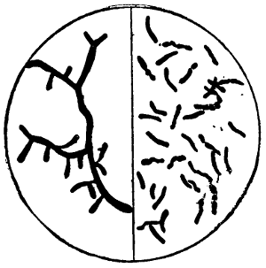
TUBERCULOSIS GERMS
These germs may be inhaled in the lungs with the air, admitted to the stomach and intestines with food and drink, or established in the flesh by inoculation through broken skin or mucous membrane.
After getting into the body, tubercle bacteria multiply in the tissues to which they have been carried and produce the changes in them which we find on the examination of an animal suffering with tuberculosis. Tuberculosis, therefore, is simply the outcome of the growth of the tubercle bacteria in the organs.
—Tuberculous areas may be found in almost any part of the infected animal, but the organs that are usually affected are the lymphatic glands, either in the throat, the bronchial glands or those about the intestines and on the liver; the lungs; the liver; the kidneys;[265] intestines; udder and generative organs. The membrane covering the lungs (pleura), the heart (pericardium), and intestines (peritoneum), are frequently affected. It often happens that a large mass, or masses, of tuberculous tissue grow over one or more of these membranes. The most peculiar thing about bovine tuberculosis is the fact that frequently an animal will appear to be perfectly well, but when slaughtered will be found to have a large number of tuberculous areas or masses on the membranes or in its organs. The reason for this is that the diseased area is not at a vital point.
The organ or membrane affected depends upon the one to which the germ is carried. Usually animals are infected in but one organ in the beginning, and from this diseased area the germs spread through the blood vessels or lymph channels to other organs. When the diseased area is restricted to one organ or part, it is called “localized” tuberculosis, because it appears at the point where the seed or germ was first planted. When the germs spread through the circulation from this first or primary diseased area to other organs and set up new tuberculous growths, the condition is called “generalized” tuberculosis. When cattle are slaughtered for food, if they are found to be afflicted with localized tuberculosis, the flesh is considered to be fit for food, but if the disease is generalized the carcass is condemned.
vary according to the location of the disease. If it is in the glands of the throat it is suggested by their enlargement. If it is in a gland about the lungs, which, because of its enlargement, presses on the œsophagus (gullet), there might be bloating. If the disease is in the lung tissue there would be, after it is sufficiently[266] advanced, coughing and perhaps difficult breathing. If the disease is in the liver, it cannot be readily distinguished until it is far advanced. If the disease is in the udder it manifests itself usually by the organ becoming firm or hard, and when the tissues are sufficiently broken down the milk from that quarter will be changed in appearance; sometimes it is thick, containing pus, sometimes thin and watery. It is very difficult to diagnose tuberculosis from the symptoms, as many other causes may give rise to similar manifestations.
As tuberculosis is caused by a specific germ, the disease is spread by the germs escaping from the diseased animals and getting into the bodies of healthy ones. The tubercle bacteria escape from the infected animal with some one or more of the natural discharges of the body. For example, if the cow has a bad tuberculous area in the lung, the bacteria may be discharged into one of the air tubes and coughed up into the mouth. Some of them will escape with the saliva and infect mangers or pastures. Some of them may be swallowed and escape from the body with the feces. If the disease is in the udder the germs will escape with the milk. There are some observations which indicate that sometimes the bacteria will escape with the milk where the udder is not affected. After the bacteria leave the diseased animal and are left in the manger, or in the pasture, or on the surface of water in the drinking trough, they can be readily taken up by healthy cattle that eat or drink after them. If they escape with the milk, calves and pigs that are fed with it readily become infected. After the germs get into the body of the healthy animal they will multiply and produce the disease, just as the seed of a noxious weed will, if blown[267] into a new field, germinate and produce the weed there. Tuberculosis spreads from animal to animal on the same principle that weeds spread from one field to another.
In order to prevent the spread of tuberculosis it is simply necessary to prevent healthy animals from coming in contact with the diseased ones or eating or drinking after them.
As tuberculosis cannot be readily detected by a physical examination until the disease is far advanced in the organs affected, it is necessary, in order to determine which animals have the disease, to apply some test or to find the germs of the disease in their excretions. The simplest test that has thus far been discovered is the action of tuberculin. When tuberculin is injected under the skin of the animals affected with active tuberculosis the animals respond by a rise of temperature, which follows a somewhat definite curve. By means of this test it is possible to pick out the infected individuals so that they can be separated from the healthy ones. The test should be repeated in from six months to a year in order to detect any new cases which might have developed from latent or arrested ones. We cannot always get all of the infected animals with the first test any more than we can always remove every weed from the garden by one hoeing.
of tuberculosis consists in separating the animals that are infected from the well ones and keeping them for breeding purposes. The calves are removed from their dams as soon as born and fed with the milk of healthy cows, or the pasteurized milk of the infected ones. It has been found that but a small percentage of calves that are raised under proper precautions from[268] such animals have tuberculosis. By this means a sound herd of cattle may be developed from tuberculous animals. This method was introduced by Prof. Bang of Copenhagen, and it has been found to be very effective in Denmark and other countries in Europe. It has been applied with much success in a large number of individual herds in the United States. Its success depends entirely upon the care which is taken in keeping tubercle bacteria away from the calves.
In purchasing cattle for dairy or breeding purposes it is important that they should be taken from herds that are free from tuberculosis. The sound herd is the unit to be dealt with. Animals from such herds are far more reliable than non-reactors from tuberculous herds.
—Abnormal growths of tissues. There are many kinds of tumors. They are named from the kind of tissue of which they are composed, as fibrous and fatty. Just why tumors should develop is not known. Treatment is in the direction of direct removal; this means they are to be cut out with a knife. Another method is to tie a strong cord around the stem of the tumor, thus shutting off the blood supply. As soon as this is effected, there will be a sloughing away, with a sore remaining, which is to be treated as in an ordinary wound. Some tumors are burnt off with caustics. Arsenic or corrosive sublimate are commonly used, either singularly or combined. Better consult a veterinarian about the removal of tumors on valuable animals.
—Bunches form on the cords of pigs after castration as a result of infection from dirty instruments or hands during the operation; or from leaving the[269] cord too long, thus increasing the liability of its becoming infected. These tumors continue to grow, and in the worst cases attain the size of a man’s head. Cut down on a tumor the same as in a simple case of castration. Separate the skin from the tumor and then swallow up the cord with the hands. Cut the cord off as high up as possible. The wound may be healed by the use of any of the common disinfectants. A teaspoonful of carbolic acid in a quart of water may be used once daily until the pigs are healed. Pigs should be kept in a clean pen after the operation.
—These are lumps in the skin of cattle, caused by grubs or warbles. A simple treatment is to cut the skin and squeeze out the grubs where the lumps are noticed. If all the grubs are killed in this way, there will be no mature flies to cause trouble later on. See article on Bot Flies.
—The cause of these little tumors of the skin is not definitely known. They occur on all domestic animals, appearing most frequently on horses and cattle. Pure acetic acid, dropped on the wart until it is saturated and softened, destroys in the early stages. Warts about which a small cord may be tied are most easily treated in that way. After they have sloughed off, apply a little terchloride of antimony with a feather or cotton. When the scab forms, remove it and apply the chemical again. With a couple of applications the spot will be lower than the surrounding skin. Now use an ointment, made of 4 tablespoonfuls of oxide of zinc and 8 tablespoonfuls of lard. Apply this daily until the sore spot is healed. Sometimes a form of warts suddenly appears on colts and calves and scatter themselves about the lips, nose[270] and face. They are common and appear and disappear suddenly. No treatment is necessary.
—Dropsy in the brain. A condition characterized by an accumulation of fluid in the brain. The disease is either congenital or arises during the first years of life. When it occurs the best thing is to kill the young individual at once.
—Often after a case of pleurisy a reaction comes and a very large quantity of water settles in the chest cavity, anywhere from two to four pailfuls. When the disease comes on the animal has difficulty in breathing; takes in the breath quickly. There is a constant biting at the flanks; the pulse increases to a hundred beats a minute. If you place your ear over the chest you will likely hear no sound at all. Best treatment is wholesome food, boiled flaxseed, and blisters for both sides of the chest. Use strong mustard plasters. A good medicine to use is one-fourth of a pound of saltpeter or nitrate of potash, one fourth of a pound of ground gentian and one-fourth of a pound of sulphate of iron. These should be mixed and then 1 teaspoonful given every four hours. You had better consult a veterinarian. Other complications set in so readily that help may be secured in other ways. Some veterinarians puncture the chest so as to draw off the surplus water that has accumulated.
—Calves of several days or weeks old suffer from indigestion, which is indicated by thriftlessness, and then scouring. The discharges are white, sour, curdled and frequent at first and then become watery, greenish and offensive, passing in stream often. Calves live some[271] days and fast lose flesh, showing all the symptoms of ill health.
One of the commonest causes is feeding dirty, souring or decomposing factory skim milk in large quantities at long intervals; even sweet skim milk so fed may produce the trouble. To prevent scours give calves a perfectly clean, airy, sunny pen and yard attached. Separate any calf that scours. Avoid dirty, dark, damp, poorly ventilated pens in which scouring calves have been. Give all food from clean, scalded, sun-dried vessels. Feed small quantities of food often; and in milk mix lime water freely two or three times a week as a preventive; and daily when scouring has been experienced. Also see that the udders of cows nursing calves do not become contaminated with manure or other filth.
Wash udders with a two per cent solution of coal tar disinfectant before any calf is allowed to suck for the first time, and then repeat to keep the udders clean. Also disinfect the navel of each calf at birth with a 1⁄500 solution of corrosive sublimate and repeat the application twice a day until the navel is perfectly healed over. At the first sign of scours give castor oil shaken up in milk. Two to 6 tablespoonfuls is the dose according to the size and age of the calf. Follow two or three times daily with a 1 to 2-teaspoonful dose of a mixture of one part of salol and two parts of subnitrate of bismuth in milk or water. For calves scouring on skim milk mix in each pint of milk 1 teaspoonful of a mixture of half an ounce of formaldehyde in 151⁄2 ounces of distilled water, to be kept in an amber-colored bottle.
—See Colic.
—An accumulation of synovia in the cavities between the tendons of the legs, especially[272] between the back tendons and the bone just above the fetlock joint. The bulging out is on each side of the tendon. Horses subjected to severe exertions, like hard work on the roads, are most frequently affected. The puffs or galls seldom cause lameness or interfere with the usual work. Unless treated the puffs will become thicker and harder and sometimes solidified. When this happens lameness occurs. In the early stages, pads and bandages, if applied so as to cause pressure, will tend to remove the galls. If this treatment is not sufficient, then use a teaspoonful of biniodide of mercury, and 4 tablespoonfuls of lard. When mixed, these should be rubbed on with the fingers. After 24 hours remove with water and soap and repeat every other week until the puffs disappear.
—See Cribbing.
—See Intestinal Worms in Horses and Sheep; and Stomach Worms.
—Hogs with worms in the intestines run down in condition, become very thin and lank, back is arched, eyes dull, refuse feed, walk stiffly, and appear lifeless. The worms may be very numerous, in bad cases completely filling the intestines. The pigs die if not treated. To secure the best results, affected hogs should receive individual treatment. Twenty-four hours before administering treatment very little feed should be given them. Then give the following medicine as a drench to each 100-pound hog; larger or smaller hogs should receive a dose in proportion: 4 tablespoonfuls of oil of turpentine, one-half teaspoonful of liquor ferri dialysatus and 6 ounces of raw linseed oil. If necessary, repeat the dose in four days.
[273]
Inconsistent spelling and hyphenation have been retained.
The Plates have been added to the List of Illustrations.
Jekyl-like (page 11) and post portem (page 120) have not been corrected.
Page 110 ff.: not all entries are listed in alphabetical order, this has not been corrected.
Changes and corrections made:
Page 21: Æsophagus changed to Œsophagus
Page 183: ... characteristic “a” or even “b” ...: Letter “b”
was invisible in the source document
Page 186: On open- the carcass ... changed to On opening the carcass ...
Page 201: Intestinal Worms in Horses: capitalised as other section headings
Page 275: Nail Punctures changed to Punctures, Nail.