|
|
LARYNGOSCOPY AND RHINOSCOPY. By CARL SEILER, M.D.
DISEASES OF THE NASAL PASSAGES. By HARRISON ALLEN, M.D.
NEUROSES OF THE LARYNX. By HOSMER A. JOHNSON, M.D., LL.D.
ACUTE CATARRHAL LARYNGITIS (FALSE OR SPASMODIC CROUP). By ABRAHAM JACOBI, M.D.
PSEUDO-MEMBRANOUS LARYNGITIS. By ABRAHAM JACOBI, M.D.
DISEASES OF THE LARYNX. By LOUIS ELSBERG, A.M., M.D.
DISEASES OF THE TRACHEA. By LOUIS ELSBERG, A.M., M.D.
TRACHEOTOMY. By GEORGE M. LEFFERTS, A.M., M.D.
DISEASES OF THE BRONCHI. By N. S. DAVIS, M.D., LL.D.
BRONCHIAL ASTHMA. By W. H. GEDDINGS, M.D.
HAY ASTHMA. By W. H. GEDDINGS, M.D.
DILATATION OF THE BRONCHIAL TUBES, CIRCUMSCRIBED AND DIFFUSED. By SAMUEL C. CHEW, M.D.
EMPHYSEMA. By SAMUEL C. CHEW, M.D.
COLLAPSE OF THE LUNG (ATELECTASIS). By SAMUEL C. CHEW, M.D.
CONGESTION AND OEDEMA OF THE LUNGS (HYPOSTATIC PNEUMONIA). By SAMUEL C. CHEW, M.D.
HÆMOPTYSIS. By WILLIAM CARSON, M.D.
PULMONARY APOPLEXY. By WILLIAM CARSON, M.D.
ABSCESS OF THE LUNG. By WILLIAM CARSON, M.D.
GANGRENE OF THE LUNG. By WILLIAM CARSON, M.D.
CROUPOUS PNEUMONIA. By ALFRED L. LOOMIS, M.D., LL.D.
CATARRHAL PNEUMONIA. By WILLIAM PEPPER, M.D., LL.D.
PULMONARY EMBOLISM. By BEVERLEY ROBINSON, M.D.
PULMONARY PHTHISIS (FIBROID PHTHISIS OR CHRONIC INTERSTITIAL PNEUMONIA). By AUSTIN FLINT, M.D.
SYPHILITIC DISEASE OF THE LUNG. By EDWARD T. BRUEN, M.D.
PNEUMONOKONIOSIS. By EDWARD T. BRUEN, M.D.
CANCER OF THE LUNGS. By EDWARD T. BRUEN, M.D.
PULMONARY HYDATIDS. By EDWARD T. BRUEN, M.D.
ACUTE MILIARY TUBERCULOSIS. By JOHN S. LYNCH, M.D.
DISEASES OF THE PLEURA. By FRANK DONALDSON, M.D.
DISEASES OF THE SUBSTANCE OF THE HEART. By WILLIAM OSLER, M.D.
ENDOCARDITIS AND CARDIAC VALVULAR DISEASES. By ALFRED L. LOOMIS, M.D., LL.D.
CYANOSIS AND CONGENITAL ANOMALIES OF THE HEART AND GREAT VESSELS. By MORRIS LONGSTRETH, M.D.
CARDIAC THROMBOSIS. By BEVERLEY ROBINSON, M.D.
NEUROSES OF THE HEART. By AUSTIN FLINT, M.D.
DISEASES OF THE PERICARDIUM. By J. M. DACOSTA, M.D., LL.D.
THE OPERATIVE TREATMENT OF PERICARDIAL EFFUSIONS. By JOHN B. ROBERTS, A.M., M.D.
DISEASES OF THE AORTA. By G. M. GARLAND, M.D.
DISEASES OF THE CORONARY, PULMONARY, SUPERIOR MESENTERIC, INFERIOR MESENTERIC, AND HEPATIC ARTERIES, AND OF THE COELIAC AXIS. By ELBRIDGE G. CUTLER, M.D.
DISEASES OF THE VEINS. By ANDREW HEERMANCE SMITH, M.D.
THE CAISSON DISEASE. By ANDREW HEERMANCE SMITH, M.D.
DISEASES OF THE MEDIASTINUM. By EDWARD T. BRUEN, M.D.
DISEASES OF THE BLOOD AND BLOOD-GLANDULAR SYSTEM. By WILLIAM OSLER, M.D.
DISEASES OF THE SPLEEN. By I. EDMONDSON ATKINSON, M.D.
DISEASES OF THE THYROID GLAND. By D. HAYES AGNEW, M.D., LL.D.
SIMPLE LYMPHANGITIS. By SAMUEL C. BUSEY, M.D.
AGNEW, D. HAYES, M.D., LL.D.,
Professor of Principles and Practice of Surgery in the University of Pennsylvania, Philadelphia.
ALLEN, HARRISON, M.D.,
Emeritus Professor of Physiology in the University of Pennsylvania, Philadelphia.
ATKINSON, I. EDMONDSON, M.D.,
Professor of Pathology and Clinical Medicine and Clinical Professor of Dermatology in the University of Maryland, Baltimore.
BRUEN, EDWARD T., M.D.,
Assistant Professor of Physical Diagnosis in the University of Pennsylvania; Physician to Philadelphia (Blockley) Hospital; Lecturer on Pathology in the Woman's Medical College, Philadelphia.
BUSEY, SAMUEL C., M.D.,
Attending Physician and Chairman of the Board of Hospital Administration of the Children's Hospital, Washington, D.C.
CARSON, WILLIAM, M.D.,
Physician to and Clinical Lecturer at the Cincinnati Hospital, Cincinnati.
CHEW, SAMUEL C., M.D.,
Professor of Materia Medica, Therapeutics, and Clinical Medicine in the University of Maryland, Baltimore.
CUTLER, ELBRIDGE G., M.D.,
Clinical Instructor in Auscultation in the Harvard Medical School; Physician to Out-Patients, Massachusetts General Hospital.
DACOSTA, J. M., M.D., LL.D.,
Professor of Theory and Practice of Medicine in the Jefferson Medical College, Philadelphia.
DAVIS, N. S., M.D., LL.D.,
Professor of Principles and Practice of Medicine in the Chicago Medical College, Chicago.
DONALDSON, FRANK, M.D.,
Clinical Professor of Diseases of the Throat and Chest in the University of Maryland, Baltimore.
ELSBERG, LOUIS, A.M., M.D.,
Late Professor of Laryngology and Rhinology in the New York Polyclinic and in Dartmouth Medical College; Physician to Charity Hospital, Blackwell's Island (Throat Wards), New York.
FLINT, AUSTIN, M.D.,
Professor of the Principles and Practice of Medicine and Clinical Medicine in the Bellevue Hospital Medical College, New York.
GARLAND, G. M., M.D.,
Formerly Professor of Thoracic Diseases in the University of Vermont, and Assistant in Clinical Medicine in Harvard Medical School.
GEDDINGS, W. H., M.D.,
Aiken, South Carolina, and Bethlehem, N.H.
JACOBI, ABRAHAM, M.D.,
Clinical Professor of Diseases of Children in the College of Physicians and Surgeons, New York, etc.
JOHNSON, HOSMER A., M.D., LL.D.,
Emeritus Professor of Practical Medicine in the Chicago Medical College, Chicago.
LEFFERTS, GEORGE M., A.M., M.D.,
Professor of Laryngoscopy and Diseases of the Throat in the College of Physicians and Surgeons, New York; Consulting Laryngoscopic Surgeon to St. Luke's Hospital, etc.
LONGSTRETH, MORRIS, M.D.,
Physician to the Pennsylvania Hospital, Philadelphia.
LOOMIS, ALFRED L., M.D., LL.D.,
Professor of Pathology and Practice of Medicine in the University of the City of New York.
LYNCH, JOHN S., M.D.,
Professor of Principles and Practice of Medicine in the College of Physicians and Surgeons, Baltimore.
OSLER, WILLIAM, M.D.,
Professor of Clinical Medicine in the University of Pennsylvania; formerly Professor of the Institutes of Medicine in McGill University, Montreal.
PEPPER, WILLIAM, M.D., LL.D.,
Professor of the Theory and Practice of Medicine and of Clinical Medicine in the University of Pennsylvania, Philadelphia.
ROBERTS, JOHN B., A.M., M.D.,
Professor of Applied Anatomy and Operative Surgery in the Philadelphia Polyclinic and College for Graduates in Medicine.
ROBINSON, BEVERLEY, M.D.,
Clinical Professor of Medicine in the Bellevue Hospital Medical College, New York.
SEILER, CARL, M.D.,
Instructor in Laryngoscopy in the University of Pennsylvania; Professor of Acoustics and Vocal Physiology at the National School of Oratory, Philadelphia.
SMITH, ANDREW HEERMANCE, M.D.,
Professor of Therapeutics and Clinical Medicine in the New York Post-Graduate Medical School; Physician to the Presbyterian Hospital, New York.
| FIGURE | |
| 1. | LARYNGEAL MIRROR |
| 2. | HEAD REFLECTOR |
| 3. | SEILER'S ELECTRIC ILLUMINATOR FOR THE LARYNGOSCOPE |
| 4. | POSITION OF HAND IN HOLDING THE LARYNGEAL MIRROR |
| 5. | SECTION OF THE HEAD, SHOWING THE POSITION OF LARYNGEAL MIRROR IN THE PHARYNX |
| 6. | ELSBERG'S SPONGE-HOLDER AND EPIGLOTTIS FORCEPS |
| 7. | LARYNGEAL IMAGE DURING RESPIRATION |
| 8. | LARYNGEAL IMAGE DURING PHONATION |
| 9. | LARYNGOSCOPIC DIAGRAM, SHOWING VOCAL CORDS WIDELY DRAWN APART, AND THE POSITION OF THE VARIOUS PARTS DURING QUIET BREATHING |
| 10. | THE SAME, SHOWING APPROXIMATION OF VOCAL CORDS AND POSITION OF THE VARIOUS PARTS DURING VOCALIZATION |
| 11. | VERTICAL SECTION OF THE HEAD |
| 12. | NASAL SPECULUM |
| 13. | BOSWORTH'S NASAL DILATOR |
| 14. | SEPTOMETER FOR MEASURING THICKNESS OF NASAL SEPTUM |
| 15. | FOLDING TONGUE DEPRESSOR |
| 16. | COHEN'S TONGUE DEPRESSOR |
| 17. | JARVIS'S RHINOSCOPIC MIRROR AND TONGUE DEPRESSOR |
| 18. | RHINOSCOPIC IMAGE |
| 19. | SHOWING ANTERO-POSTERIOR SECTION OF BONES OF THE FACE IN POSITION |
| 20. | BELLOCQ'S CANULA |
| 21. | ALLEN'S NASAL FORCEPS |
| 22. | THE GALVANO-CAUTERY SNARE |
| 23. | DOUBLE BATTERY AND FLEMING ELECTRODE FOR USE IN NASAL DISEASES |
| 24. | TWO ELECTRODES OF PECULIAR SHAPE, FOR USE IN NASAL DISEASES |
| 25. | ACUTE TRACHEITIS: ANTERIOR WALL |
| 26. | SAME CASE AS FIG. 25: POSTERIOR WALL |
| 27. | TUBERCULOUS ULCERATION OF THE TRACHEA, AS SEEN DURING LIFE |
| 28. | SAME CASE AS FIG. 27: POST-MORTEM APPEARANCE |
| 29. | SYPHILITIC ULCERATION OF TRACHEA, AS SEEN DURING LIFE |
| 30. | SAME CASE AS FIG. 29: POST-MORTEM APPEARANCE |
| 31. | PAPILLOMA OF TRACHEA |
| 32. | INVOLUTION OF TRACHEA, DUE TO ANEURISM |
| 33. | A TYPICAL CASE OF LOBAR PNEUMONIA IN THE ADULT: RECOVERY |
| 34. | LOBAR PNEUMONIA, WITH CRISIS MARKED BY EVENING EXACERBATIONS REACHING NEARLY THE HIGHEST PYREXIA OF THE SECOND STAGE |
| 35. | A TYPICAL CASE OF LOBAR PNEUMONIA IN A CHILD: RECOVERY |
| 36. | A CASE OF LOBAR PNEUMONIA IN A BOY TEN YEARS OLD, IN WHICH TEMPERATURE WAS TAKEN EVERY FOUR HOURS |
| 37. | A TYPICAL CASE OF SENILE LOBAR PNEUMONIA |
| 38. | CROUPOUS PNEUMONIA IN THE ADULT, TERMINATION OF, IN PURULENT INFILTRATION |
| 39. | ACUTE LOBAR (CROUPOUS) PNEUMONIA IN A CHILD: RECOVERY |
| 40. | ACUTE LOBULAR (CATARRHAL) PNEUMONIA IN A CHILD: RECOVERY |
| 41. | SPHYGMOGRAPHIC TRACING OF AORTIC OBSTRUCTION (AFTER FOSTER) |
| 42. | SPHYGMOGRAPHIC TRACING OF AORTIC REGURGITATION |
| 43. | SPHYGMOGRAPHIC TRACING OF AORTIC OBSTRUCTION AND REGURGITATION |
| 44. | SPHYGMOGRAPHIC TRACING OF MITRAL OBSTRUCTION |
| 45. | SPHYGMOGRAPHIC TRACING OF MITRAL AND AORTIC OBSTRUCTION AND REGURGITATION |
| 46. | SPHYGMOGRAPHIC TRACING OF MITRAL REGURGITATION |
| 47. | SPHYGMOGRAPHIC TRACING OF MITRAL AND AORTIC REGURGITATION |
| 48. | SPHYGMOGRAPHIC TRACING OF TRICUSPID REGURGITATION |
| 49. | ROBERTS'S PERICARDIAL ASPIRATING TROCAR |
| 50. | SPHYGMOGRAPHIC TRACING OF NORMAL PULSE |
| 51. | SPHYGMOGRAPHIC TRACING OF RIGHT AND LEFT RADIAL PULSE IN ANEURISM OF THE AORTA |
| 52. | ARCH OF AORTA DURING EARLY FOETAL LIFE |
| 53. | SARCOMATOUS TUMOR OF ANTERIOR MEDIASTINUM |
| 54. | SECONDARY MYELOID SARCOMA OF MEDIASTINUM |
| 55. | RESECTION OF STERNUM FOR REMOVAL OF ENCHONDROMA |
The laryngoscope is a combination of instruments designed for the examination of the interior of the larynx and upper part of the trachea, while the rhinoscope is a similar combination of instruments designed to explore the posterior nasal cavity; and both are comparatively recent inventions.
HISTORY OF THE LARYNGOSCOPE.—In medical literature before the middle of the eighteenth century no mention is made of an instrument or apparatus resembling the laryngoscope, but recent excavations at Pompeii have brought to light small polished metal mirrors attached to slender handles which are supposed to have been used to inspect the cavities of the human body. The first authenticated attempt at laryngoscopy and rhinoscopy was made by the distinguished French accoucheur M. Levret in the year 1743, who invented, among other surgical instruments, an apparatus by means of which polypoid growths in the cavities of the nose, throat, ear, etc. could be seen, and a ligature be passed around them for their removal.1 This apparatus consisted mainly of a polished metal mirror which "reflected the luminous rays in the direction of the tumor," and on whose surface the image of the growth was seen to be reflected. The great value of this apparatus for the diagnosis and treatment of nasal and laryngeal diseases was, however, not recognized, and it shared the fate of many other valuable discoveries which were made before the world was ready to receive them: it was forgotten.
1 Mercure de France, 1743, p. 2434.
In 1807 a certain Dr. Bozzini, living in Frankfort-on-the-Main, published a work describing an apparatus which he had invented for the illumination and examination of the cavities of the human body.2 This apparatus consisted of a peculiarly-shaped lamp and of a number of metal tubes, polished on their inner surface, of various shapes and sizes adapted for the different cavities of the body. The one intended for the examination of the larynx was bent near its end at a right angle, and had a mirror placed at the bend, which served to throw the light downward toward the opening of the larynx when the tube was inserted into the mouth. When reflected light was to be used, the interior of the tube or speculum was divided into two portions by a longitudinal septum, and two mirrors were inserted at the bend—one for the reflection of the light downward, and the other for receiving the reflected image. This invention of Bozzini was treated, however, with derision by the medical profession, probably on account of the extravagant descriptions given of it in the papers, which were not verified by its performances.
2 "Der Lichtleiter," Philipp Bozzini, Med. und Chir. Dr., Weimar, 1807.
In 1825, Cagniard de Latour, an investigator of the physiology of the voice, made some unsuccessful attempts to examine the living larynx.3
3 Physiologie de la Voix, par Ed. Tournié, Paris, 1865.
[p. 20] Senn of Geneva in 1827 endeavored to examine the larynx of a little girl suffering from an affection of the throat by means of a small mirror which he had made and which he inserted into the pharynx, but he failed to see the glottis, because, as he says, the mirror was too small, and because he used neither direct nor reflected light to illuminate the cavity below the mirror.4
4 Journal de Progrès des Sciences, etc., 1829.
In the year 1829, Benjamin Guy Babington published5 an account of what he called the glottiscope, an apparatus which consisted mainly of two mirrors. One of these was small and attached to a slender stem, and was used to receive the image, while the other, an ordinary hand-glass, was used to reflect the rays of the sun or ordinary daylight upon the smaller mirror in the fauces. This combination was essentially the same as is used at the present day in the laryngoscope, with the difference that we now use artificial light in most instances, and a concave mirror instead of a plane one for reflecting the light.
5 Lond. Med. Gazette, 1829, vol. iii.
While Babington was still engaged in perfecting his instruments, a mechanic named Selligue, who suffered from an affection of the throat, in 1832 invented a speculum for his physician, Bennati of Paris, with which the latter was able, as he asserted,6 to see the vocal cords. This instrument was similar to the one invented by Bozzini, and consisted of a double speculum bent at right angles and carrying two mirrors—one for illuminating the cavity, and the other for reflecting the image. Selligue was rewarded for his efforts by a complete cure of his affection.
6 Recherches sur le Mécanisme de la Voix humane.
A number of others worked in the same direction, and endeavored to see the interior of the larynx in the living subject by employing different apparatus and methods of illumination. Thus, in 1838, Baumès of Lyons described a mirror the size of a two-franc piece (11/8 inches in diameter) as useful in examining the larynx and posterior nares.7 Then Liston in 1840 used a dentist's mirror,8 and Warden of Edinburgh employed a prism of flint glass attached to a long stem as a laryngeal mirror.9 In the latter part of the same year Avery of London employed a speculum with a mirror in its end for examining the larynx, using as an illuminator a concave reflector with a central opening, which was supported by a frame to be worn on the head of the operator.10
7 Compte Rendu des Travaux de la Société de Médecine de Lyons, 1836–38.
8 Practical Surgery, 1840.
9 Lond. Med. Gazette, vol. xxiv. p. 256.
10 Med. Circ., June, 1862.
Up to this time all efforts at laryngoscopy had been made with a view to diagnose diseases of the larynx, with the exception of those made by Latour. In the year 1854, however, Signor Manuel Garcia of London, without any knowledge of previous efforts, conceived the idea of studying the changes in the larynx during phonation in his own throat. For this purpose he placed a small dentist's mirror against the uvula and reflected the rays of the sun into his mouth and upon the small mirror by means of a hand-glass held in the other hand. By arranging his position in relation to the sun in such a manner that he could see the reflected image of the small mirror in his throat in the hand-glass, and in it the illuminated image of his larynx, after a few ineffectual attempts his efforts at auto-laryngoscopy were crowned with such success that he was enabled to study the movements of the vocal cords during phonation, and accurately describe the registers of the voice in a paper read before the Royal Society of London in 1855.11 Although Garcia was the first who practised laryngoscopy successfully, his communication to the Royal Society attracted little attention, and would have been forgotten if it had not been that, in 1857, Tuerk of Vienna, having heard of Garcia's paper, began to use the laryngeal mirror on the patients in the K. K. Algem. Krankenhaus for [p. 21] diagnostic purposes.12 At first he was not very successful in his attempts, and began to experiment with laryngeal mirrors of different sizes and shapes. While thus engaged Czermak borrowed Tuerk's mirrors, and modified them until he succeeded in the greater number of cases in seeing the vocal cords,13 using artificial light for illuminating the larynx. Meanwhile, Tuerk continued his experiments, and also succeeded in almost all cases of throat disease which came to his department of the hospital in seeing the interior of the larynx and in treating the lesions. Both Tuerk and Czermak improved their apparatus, and especially the latter, who by substituting artificial light for sunlight, and by inventing a number of different illuminating apparatuses, has given us the laryngoscope in the form in which it is used at the present day. It is but natural that Tuerk should have claimed priority in the successful use of this instrument, and in consequence of this claim a controversy was carried on for a number of years in the medical press between him and Czermak, which at times became quite spirited, but which left Czermak master of the field. In the winter of 1858–59, Madam E. Seiler, having heard of Czermak's experiments, had a laryngeal mirror constructed from his description, practised laryngoscopy successfully on herself and others, among them the writer, with a view to study the physiology of the voice. Her efforts being crowned with success, she was able not only to verify Garcia's observations in regard to the registers, but also discovered the so-called head register of the female voice, as well as two small cartilages in the vocal cords.14
11 Proc. Royal Society of London, vol. vii. No. 13, 1855.
12 Zeitschrift der Ges. der Aerzte zu Wien, April, 1858.
13 Wien. Medicin. Wochenschrift, March, 1858.
14 Altes und Neues, Leipzig, 1861.
HISTORY OF THE RHINOSCOPE.—Rhinoscopy, or the art of viewing the naso-pharyngeal space by placing a small mirror behind the velum palati, naturally suggested itself almost as soon as any attempts at laryngoscopy were made, but in the literature we find that Bozzini was the first to clearly express the idea.15
15 Loc. cit.
A number of years later Wilde endeavored to see the opening of the Eustachian tubes by means of a small mirror: an account of these experiments he published in his famous work on the diseases of the ear.
In 1836, Baumès used the rhinoscope, and claimed to have seen ulcerations in the naso-pharyngeal cavity.16 It remained, however, for modern times to develop this field of research, and it is again Czermak whom we have to thank for the perfection of this valuable means of diagnosis.
16 Loc. cit.
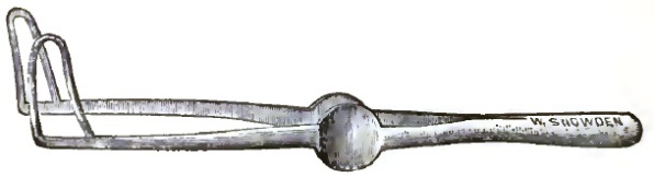
THE LARYNGOSCOPE.—The laryngoscope as it is used at the present day, both by the specialist and the general practitioner of medicine, consists of a so-called laryngeal mirror and of an illuminating apparatus more or less complicated. The laryngeal mirror is a small circular glass mirror mounted in a metal frame varying in size from ¾ inch to 1½ inches in diameter, and attached to a wire stem at an angle of 120°. This stem, about 4 inches in length and about 1/10 inch in thickness, should be soldered to the back of the mirror in such a manner that the rim of the frame forms the angle with the stem, and should not be below it, as this would increase the diameter of the instrument without increasing its reflecting surface. The stem is made to slide into a hollow handle of wood, ivory, or ebonite, and is clamped at any desired length by a set-screw. This arrangement is preferable to having the stem permanently fixed in the handle, inasmuch as the stem can be pushed entirely into it, thus economizing space and rendering the instrument more portable, and also allowing an adjustment of the length of the stem when in use. The handle should be 4 inches in length, and of the thickness of an ordinary lead-pencil (Fig. 1).
| FIG. 1. |
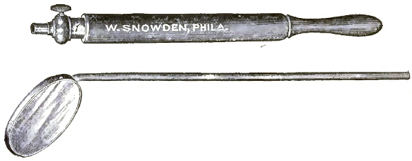
|
| Laryngeal Mirror. |
Mirrors of various shapes have been used, but it has been found that the circular form is the one most easily borne by the patient, and can be used in [p. 22]a greater number of cases than any other shape, at the same time giving the largest reflecting surface for its size. However, in cases where an hypertrophy of the tonsils is present an oval mirror can be introduced between the protruding glands more easily than a round one.
This laryngeal mirror, however, would be of little or no value as an instrument of diagnosis if used by itself, for in order to see the cavity of the larynx it must be illuminated, lying as it does far below the level of the back of the tongue; and this cannot be done satisfactorily by merely allowing ordinary daylight to fall into the oral cavity. It becomes, therefore, necessary to use a stronger light to illuminate the larynx, and for this purpose either direct or reflected artificial or sunlight may be used.
Direct illumination, by allowing a strong artificial light or sunlight to fall into the patient's mouth, although it is used by several of the eminent laryngologists of Europe, is both inconvenient and unsatisfactory, because the observer must either place his head in the path of the light in order to be able to see the surface of the laryngeal mirror, as in the case when sunlight is used, or he must place the lamp, candle, or other source of light between himself and the patient, which materially interferes with the freedom of his motions. For these reasons reflected light is now almost universally employed in laryngoscopy.
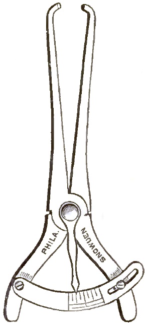
Reflected light may be obtained by throwing the light of a lamp, candle, gas-jet, or light from any other source into the mouth of the patient by means of a round concave reflector. This concave mirror—which, when made of glass, should be silvered and not backed with amalgam—is from 3 to 4 inches in diameter, and should have a focus of from 12 to 14 inches. The metal frame in which it is mounted is attached by means of a ball-and-socket joint to some contrivance by which it can be supported on the observer's head or be attached to the source of illumination if a stationary artificial light, such as a gas-lamp, is used at the physician's office.
A variety of devices for fastening the reflector on the head of the observer is in use, among which the head band, introduced by Cramer, will be found the most serviceable. It consists of a broad strap of some strong material which passes around the head and is fastened at the back by a buckle. To the part of the band or strap resting on the forehead is attached a padded plate, to which the reflector is fastened with its ball-and-socket joint (Fig. 2). The reflector usually either has a small hole in the centre or a small space in the centre is left unsilvered. This opening is intended to be brought before the pupil of one or the other eye of the observer in such a manner that the line of vision and that of light have exactly the same direction. Using the reflector in this way like the reflector of the ophthalmoscope, it is easier to obtain the image of the larynx well illuminated, but with the great disadvantage of [p. 23]monocular vision, which makes all objects appear on the same plane and prevents a correct interpretation of distances—a very important point in laryngoscopy. It will therefore be found more advantageous to place the reflector on the forehead, and from thence reflect the light into the patient's larynx. Both eyes may thus be employed in viewing the laryngeal image, and a correct idea of the relations of parts in regard to distance may be formed.
| FIG. 2. |
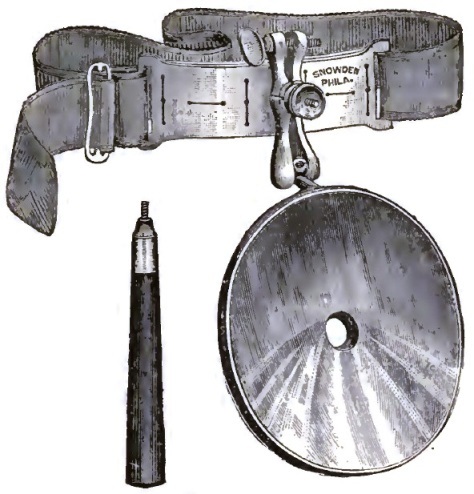
|
| Head Reflector. |
The line of vision and the path of the beam of light in order to obtain the best results should be in the same plane as though the light emanated from the pupil of the observer; but practically the position of the reflector upon the forehead is nearly as good as when the hole in it is brought before the eye, because a line drawn from the pupil of the eye to the laryngeal mirror, and a line from the reflector upon the forehead to the mirror, do not form an angle sufficient to make any very great difference in the reflection of the light downward, and very little difficulty will be experienced in obtaining the desired image.
The head reflector should be concave when artificial light or ordinary daylight is used, but be plane when direct sunlight is employed, for the concentration of the sun's rays by a concave reflector produces so much heat as to become painful to the patient.
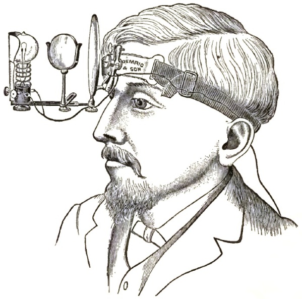
THE SOURCE OF LIGHT.—As an artificial source of light a candle, coal oil lamp, gas-flame, or incandescent electric lamp suffices for ordinary purposes. But frequently it is desirable to have a much stronger light than can be obtained without concentration, and several forms of apparatus for concentrating artificial light have been constructed and are in use. Among these, Tobold's lamp and Mackenzie's light concentrator are the most convenient and most universally used.
Tobold's lamp consists of a brass tube containing several lenses, which are placed, one before the other, at such distances as to give the greatest possible amount of concentration of light. The back part of the tube is closed, while near the end two large holes are cut in its sides opposite to each other, through which the chimney of the lamp projects. The whole is fastened by means of clamps to a stand, to which is also attached a jointed arm bearing the reflector. This apparatus is used either in connection with a student's lamp or with an argand gas-lamp, and it will be found very convenient to have it mounted upon a gas-bracket which can be raised and lowered and swung from side to side.
Mackenzie's light concentrator consists of a cylinder of sheet iron about 6 inches long by 2½ in diameter. Near one end a hole is cut in the side of the cylinder, and a short piece of tube holding a condensing lens is attached to the edge of the hole. This lens, which is plano-convex with a spherical curve, and of 2½ inches diameter, is placed with the plane side toward the light. [p. 24]This concentrator is intended to be slipped over the chimney of an argand burner, and should be so adjusted that the centre of the flame corresponds with the centre of the lens. It may, however, be used in connection with a student's lamp, incandescent electric lamp, or even a candle, giving in all cases a very satisfactory light, which, however, must be reflected from the head mirror into the patient's mouth.
The best light, however, when the examinations are conducted in the office of the physician, is the electric incandescent light, which presents numerous advantages over the gas or oil lamp. It is more brilliant and whiter than any other suitable artificial light, giving off neither gases nor heat, nor does it consume the oxygen in the room; and since the introduction and perfection of storage batteries it has become available and convenient for use in private houses. Numerous experiments which the author has carried on for some time have resulted in the application of this form of light for laryngoscopy in two ways which are both very satisfactory. The incandescent lamp is mounted upon the universal gas-bracket in place of the argand burner, and either the Tobold lamp or Mackenzie's light concentrator is slipped over it, so that it comes opposite the centre of the lens. In fact, the electric lamp is substituted for the gas-burner, and the whole apparatus is used as described above. The arc light may also be used in the same manner, but does not give as satisfactory results on account of its unsteadiness.
| FIG. 3. |
|
| The Author's Electric Illuminator for the Laryngoscope. |
The second method is to mount the electric lamp on the head mirror in such a way that it projects a little from the surface and is a little to one side of the centre of the reflector (Fig. 3). The light is then thrown forward in a cone, and can be directed with great ease into the mouth of the patient. Since thus the source of the light moves with the mirror, the observer can follow the motions of the patient more easily; and if, in the first place, an easy position of the head has been assumed when adjusting the light, much less [p. 25]fatigue is experienced by the examiner with this apparatus than when the light is reflected from a stationary source. Still another mode of using the incandescent lamp, which was suggested by Trouvé, is to mount the lamp within a tube one end of which is closed by a plano-convex lense, while the other end is covered by a metal cap carrying in its centre a ball-and-socket joint, by means of which it is fastened to the frontal plate of the head band. In this way the light with its condensing apparatus is carried on the forehead like the head mirror.
Sunlight is certainly the best source of light for the illumination of the interior of the larynx and nasal cavities, but, unfortunately, it is not available at all times and in all localities. When it can be obtained, however, the student should not neglect the opportunity, and should not be deterred from using it for examination by the little extra apparatus and trouble necessary.
The most convenient plan is to place a small plane mirror mounted upon a stand in such a manner that it can be turned in any direction, such as a small toilet-glass, in the direct rays of the sun coming through a southern window. Then turn the mirror until the reflection falls upon a second plane mirror supported by a jointed arm and placed in a distant corner of the room, and in front of the chair upon which the patient is seated, with his back to the first mirror. The light from the second mirror is then thrown into the patient's mouth in the same manner as when a light concentrator is used. The second mirror may also be mounted on the head band and used as a head reflector, but this latter plan is not as satisfactory, because the reflected light from the first mirror is apt to strike the observer's eye and temporarily blind him.
Sunlight, as well as the light from the oxyhydrogen and electric-arc lamps, is white, and therefore shows us the parts in their natural coloring, which is claimed as a great advantage over all other sources of light. It is true that the yellow rays which are predominant in all other artificial lights make the mucous membrane appear redder than it really is, and the observer may be led to believe that a congestion exists if the patient be examined by white light first, and then by yellow light on different occasions. But as all our knowledge and appreciation of shades of color depend upon a comparison with a standard, it makes no difference whether this standard, as in the case before us, is a little redder when viewed by yellow light or not so red when seen by white light. This advantage of the white light is, therefore, not of much practical value, and the expense and difficulties connected with the use of the oxyhydrogen or electric arc-light for laryngoscopy fully outweigh any advantage which can be claimed for it.
THE ART OF LARYNGOSCOPY.—Before entering upon a description of the details of the art it will be necessary to clearly understand the optical principle upon which the use of the laryngoscope is based, and, further, to remember that the object to be viewed is situated below the straight path of light and vision. The optical law referred to is, that "The angle of incidence is equal to the angle of reflection," and consequently, in order to illuminate the cavity of the larynx and to see its details, the laryngeal mirror must be placed in such a position in the fauces that the light is reflected downward. The light rays forming the laryngeal image will then be reflected from the surface of the laryngeal mirror into the eye of the observer. It should always be borne in mind that the image seen in the mirror is a reflected one, like the image of one's self seen in a looking-glass, so that what appears to be right is left, and vice versâ. On account of the difference in height of the parts forming the image, and because the mirror must be placed above and slightly behind the opening of the larynx, the picture appears reversed in an antero-posterior direction. The same holds good when viewing a drawing of a laryngeal image.
[p. 26]POSITION OF PATIENT AND OBSERVER.—The relative positions of the patient, observer, and the source of light are of very great importance, especially to the beginner, and a want of proper adjustment will often make it extremely difficult, if not impossible, to obtain the desired view of the larynx. The patient having been seated upon a chair, or better still upon a piano-stool, the source of light is placed upon a table at his right, at such a height that the centre of the flame is on a level with his eyes and a few inches behind. The observer then takes a seat directly in front of the patient, and, separating his knees, places his feet on either side of those of the patient, thus being able to grasp the patient's knees with his own should occasion require him to do so. This position is preferable to the one in which the knees of the observer are either on one side or the other of the patient's knees, because then the observer, in order to throw the light from the head mirror into the mouth of the patient, has to assume a constrained position which very soon becomes fatiguing. Under no circumstances should the patient be allowed to grasp the observer's knees, for then the latter is powerless to restrain the struggles of his patient, and cannot quickly leave his seat should vomiting occur. When the examination is made at the physician's office or wherever it is practicable, it is of advantage to have a head-rest, such as photographers use, for the patient's head.
The positions having been taken, the observer places the head reflector upon his forehead a little above the left eye, and by rotating it upon its ball-and-socket joint reflects the light from the lamp- or gas-flame upon the patient's face so that the circle of light is bounded above by the tip of the nose and below by the tip of the chin. It is of great importance that the adjustment of the reflector should be made by means of its joint, and not by rotating or inclining the head, for it is necessary that the head should have an easy position which can quickly be resumed should it become necessary to move the head. It requires considerable practice to quickly reflect the light from the head mirror in any desired direction, and it is therefore well for the beginner to practise this by throwing the light upon a spot on the wall before he attempts to examine a patient, as he will thus save himself, as well as the patient, unnecessary annoyance. If a light concentrator be used which supports the reflector on the jointed arm, this of course is not necessary, but the practice with the head mirror will even then be found advantageous, because when a patient is to be examined in the sick room a light concentrator cannot usually be employed, and the physician has to fall back upon the head mirror for illuminating the laryngeal cavity.
When the reflector has thus been properly adjusted the patient is required to incline his head backward and open his mouth as wide as possible, when it will be found that the centre of the circle of light falls upon the root of the uvula. A careful examination of the oral cavity, the anterior and posterior pillars, the tonsils, and the wall of the pharynx should be made before the laryngeal mirror is introduced, not only because the condition of these parts often imparts valuable information, but also in order to be sure that no infectious sores be present which might contaminate the instruments to be introduced. The laryngologist cannot be too careful to prevent the carrying of infectious material from one patient to another; and if he should by this preliminary examination discover a specific sore, he should use only such instruments as are reserved for this class of cases, and which are kept in a separate box or drawer of the instrument-case.
Everything being in readiness, the laryngeal mirror is held over the lamp, with the glass side down, for a few seconds until it is warm, so as to prevent the condensation of moisture on its reflecting surface, and is then introduced in the following manner: The handle is held between the thumb and fore finger of the right hand like a pen-holder (Fig. 4); the hand is bent [p. 27]backward upon the wrist and held below the chin of the patient. Meanwhile, the protruded tongue is grasped between the folds of a napkin or towel held in the left hand, and gently but firmly pulled out of the mouth. Great care should be exercised to prevent the frænum of the tongue from coming in contact with the sharp edge of the front teeth, for this soon becomes very painful and may prevent a successful examination. Many laryngologists are in the habit of letting the patient hold his tongue, which becomes necessary when operations or applications are to be made to the larynx; but for the purpose of examining only it is better for the observer to hold the tongue, as he thus gains more control over the movements of the head of the patient.
| FIG. 4. |
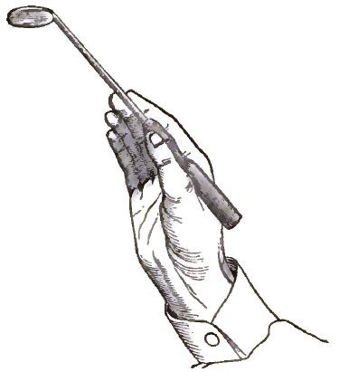
|
| Position of Hand in holding the Laryngeal Mirror. |
The mirror is now rapidly introduced into the mouth of the patient, without touching the tongue or the palate, and carried backward until its rim touches the wall of the pharynx, when it is lifted upward, carrying on its back the uvula, and the stem is brought into the angle of the mouth, so as to be out of the line of vision (Fig. 5). In this position the light of the reflector will fall upon the reflecting surface of the laryngeal mirror, and will be reflected downward so as to illuminate the laryngeal cavity and reflect the laryngeal image into the eye of the observer.
| FIG. 5. |
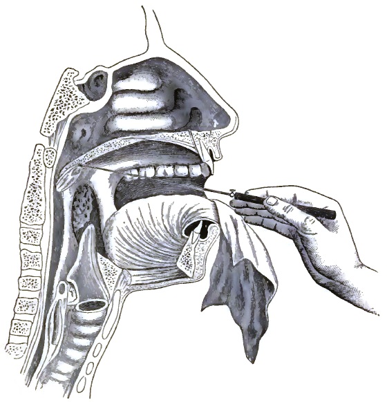
|
| Diagram of Section of Head, showing the Position of Laryngeal Mirror in the Pharynx. |
[p. 28]There are, however, numerous obstacles and difficulties which must be overcome to successfully practise laryngoscopy—obstacles which are partly due to the want of skill on the part of the operator, and partly to over-sensitiveness and want of control of the patient, or, finally, to abnormal positions of the parts. Taking them up one by one, in the order named above, the reader will soon learn to overcome these obstacles by practice and careful attention to details.
As has already been pointed out, a satisfactory view of the laryngeal image cannot be obtained if the position of the light, of the patient's head, and of the observer is not properly arranged; further, if the laryngeal mirror is either too cold or too hot. In the former case the moisture of the breath will condense on its reflecting surface and render it non-reflecting, and in the latter case the patient will feel the heat and will object to the presence of the mirror in the fauces. The examiner should therefore carefully test the temperature of the mirror on the back of his hand before introducing it. Many laryngologists are in the habit of testing the temperature by placing the mirror against the cheek, but this is a dangerous practice, for a slight scratch or abrasion of the skin from shaving may be inoculated with infectious material from a specific sore, and the writer knows of more than one instance in which such infection has occurred; while a scratch on the hand is not so likely to be overlooked, and therefore the danger is much less. Pulling too hard upon the tongue, so that the frænum becomes injured by the edge of the teeth, is another obstacle, for the patient will not bear the pain thus occasioned. Touching the tongue or palate in the act of introducing the mirror, besides coating the reflecting surface with the secretions of the mouth, causes in most patients gagging, and should therefore be avoided. When the mirror has been introduced it should be held very still, and if it becomes necessary to rotate it, this should be done slowly and steadily, because the slightest trembling motion of the rim of the mirror resting against the wall of the pharynx produces gagging and cuts the examination short at once. It is therefore advisable to steady the hand holding the mirror by placing the third finger against the cheek of the patient, or, better still, against the thumb of the hand holding the tongue.
Undue irritability of the fauces is of very rare occurrence, and is almost invariably produced by one or the other of the above-mentioned mistakes of the examiner. When it does exist independently, it can in a measure be overcome by letting the patient drink a large draught of ice-water immediately before introducing the mirror, and by holding the mirror so that it does not touch either the pharyngeal wall or the palate. In this manner but a very unsatisfactory view of the larynx can be obtained, and it is better to overcome the irritability by practice on the part of the patient—i.e. by introducing the mirror frequently and removing it before gagging sets in, and by directing the patient to introduce a teaspoon into the fauces before a looking-glass several times a day. Even the most obstinate cases can thus be educated to allow of a lengthy examination. No matter how tolerant a patient may be, however, the mirror should never be left in the fauces after the first symptoms of gagging show themselves, but should at once be removed. It is better in all cases to leave the mirror in the mouth but a short time and to introduce it frequently, thus studying the different parts of the image one after the other, than to attempt to see everything at once. In laryngoscopy, as in many other arts, not only the hand, but also the eye, must be educated to appreciate all the details and the variations from the normal.
Among the malformations of the parts which present obstacles to laryngoscopy are, in the first place, hypertrophied tonsils, which by narrowing the space in the fauces make it impossible to introduce the ordinary-sized mirror. A smaller mirror or one of oval shape can, however, usually be slipped past the [p. 29]enlarged glands and the desired image obtained. An elongated uvula does not exactly prevent a view of the larynx, but it materially interferes with a good image, because its end by hanging below the rim of the mirror is seen in the reflecting surface and obscures part of the image. Removal of the uvula by surgical means is of course the best remedy.
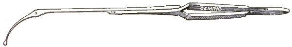
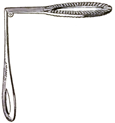
The third and most serious obstacle presented by malformation or malposition of parts is a pendent epiglottis—i.e. an epiglottis which by being bent too far over covers the laryngeal opening and prevents a view. This obstacle exists to a certain extent in most cases that come under observation, but is easily overcome by letting the patient sound the vowel sound of eh, which causes a rising of the epiglottis and opens the laryngeal cavity to view. There are some cases, however, in which this expedient does not sufficiently raise the epiglottis to obtain a glimpse of the vocal cords, and only the arytenoid cartilages are seen, from the motion and color of which we can often obtain valuable information in regard to pathological processes. In these cases, when it becomes absolutely necessary to see the whole extent of the vocal cords, we may succeed by causing the patient to laugh in a high key, but when this fails the only resource left is to lift the epiglottis by grasping its upper margin with a pair of curved forceps especially designed for this purpose and called epiglottis forceps (Fig. 6). If this instrument is not at hand, the same object may be attained by clasping the edge of the epiglottis with a bull-nose forceps, to which is fastened a string weighted at the other end by a small weight, such as a rifle-bullet. The string with its weight hanging out of the mouth of the patient makes traction upon the forceps, and thus the epiglottis is raised. In cases of operation within the laryngeal cavity this method of raising the epiglottis is even preferable to the epiglottis forceps, because it leaves the hands of the operator free to use the mirror and the instrument to be used in operating.
| FIG. 6. |
|
| Elsberg's Sponge-holder and Epiglottis Forceps. |
AUTO-LARYNGOSCOPY.—There is perhaps no better method for the beginner to overcome the difficulties besetting laryngoscopy than to practise the art on himself, for then only will he be able to appreciate to its full extent the necessity of observing all the minute details described above, as the pain and inconvenience which he inflicts upon himself by his false movements will teach him better, and enable him to attain proficiency in the use of his instruments quicker than any other method of practice. Nothing need, for auto-laryngoscopy, be added to the stock of instruments necessary for the examination of others, except a stand to which the reflector is fastened and a small toilet-mirror. The observer seats himself beside a table upon which, at his left, is placed the lamp a little behind his head and the centre of the flame on a level with his eyes. The stand, an ordinary retort-stand, is placed in front of him, and to it is fastened at the proper height the reflector. On the same stand, and immediately above the reflector, is attached the plane mirror in such a manner that it can be inclined at an angle. Inclining the head slightly backward, the observer then by watching his face in the plane mirror directs the light upon his mouth by moving the reflector upon its ball-and-socket joint until the circle falls upon his mouth. He then opens his mouth as wide as possible, grasps his protruded tongue between the folds of a towel or [p. 30]napkin held between the thumb and fore finger of the left hand, and introduces the laryngeal mirror with the right hand in the manner described above. The laryngeal image as it appears on the surface of the laryngeal mirror is reflected by the toilet-glass above the reflector, and can be seen in all its details by the person practising auto-laryngoscopy. By substituting a perforated mirror for the toilet-glass the student can demonstrate the image to others in his own person if the observers look through the perforation in the mirror.
Before giving a description of the laryngeal image it will be well, for the sake of completeness, to mention the fact that of late photography has been employed to reproduce this image, both in this country by T. R. French of Brooklyn17 and by Lennox Browne of London, England, with very gratifying results. The writer himself several years ago made experiments in this direction, which, however, were not very satisfactory in their results. The method employed by French is a very simple one, and it will be best to give his own description of the process:
"The camera consists of a box 4½ inches long, 17/8 inches wide, and ¾ of an inch in thickness. The back opens upon hinges, and admits of the introduction of either the ground glass or the plate-holder. On the anterior face a tube 11/8 inches long is attached, in the outer end of which the lens is placed. This lens has a focus of 1¼ inches. At the side of the tube a part of the handle of a throat mirror is fixed, and into that the shank of the throat mirror is passed and fastened by a thumb-screw. The shank of the mirror is somewhat curved, and is attached to the side of the frame holding the mirror. The object of this is to allow the lens being held opposite any part of the opening of the mouth, and also to prevent the possibility of a shadow being cast upon the mirror. In the front part of the box is a shutter made of lead and perforated with a hole just the size of the lens. The shutter is held in position by a lever acting as a key on the anterior face of the camera.
"The apparatus is used in the following manner: A reflector, either plane or concave, attached to a head band, is arranged over the left eye so that the pencil of sunlight from the solar condenser is received upon it and thrown into the mouth. The patient, with the head inclined slightly backward, now protrudes the tongue and holds it well out between the fore finger and thumb of the right hand. The throat mirror with the camera attached, held in the right hand of the observer, is placed in position in the fauces, and the light adjusted so that the larynx can be seen with the observer's left eye to be well illuminated. If, now, the tongue does not mount above the level of the lower edge of the lens and the lower edge of the mirror, it may be taken for granted that when the plate is exposed the picture received upon it will be nearly the same as that seen with the left eye in the throat mirror. The photograph is taken by pressing upon the key with the index finger; this releases the shutter, which in falling makes an instantaneous exposure amounting to perhaps one-seventh of a second.
"In using condensed sunlight with a small camera it is important to throw the circle of light from the inner side of the reflector, that nearest the nose; for in this way a part of the larynx exposed to the lens of the camera may be illuminated which cannot be seen with the eye. To ensure this it is best to cover the outer half of the reflector with black silk. On account of the parallax or displacement of the image due to the difference in point of view between the eye and the camera, some skill is necessary in managing the illumination so that the part which it is desired to bring out will be exposed to the lens if not to the eye."
17 Archives of Laryngology, vol. iv. No. 4.
THE LARYNGEAL IMAGE.—When the mirror is introduced and is held in the proper place, and the light is reflected downward, the laryngeal image [p. 31] will appear on the surface of the mirror. As it is, however, so different from what might be expected after having examined a larynx removed from the body, it requires a detailed description, and the student will do well to refer to the diagrams frequently while examining patients, to make himself familiar with the details he sees, and to recognize them when they are altered by disease or when they are slightly different in shape in different individuals. Figs. 7 and 8 represent the image of the larynx in the act of respiration and of phonation as it appears on the surface of the mirror, while Figs. 9 and 10 are diagrammatic, and are intended to represent the same.
| FIG. 7. |
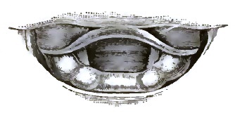
|
| Laryngeal Image during Respiration. |
| FIG. 8. |
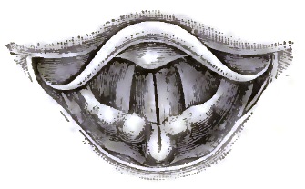
|
| Laryngeal Image during Phonation. |
| FIG. 9. |
|
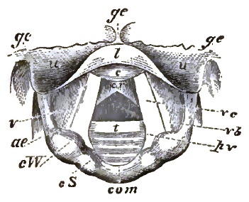
| Laryngoscopic Diagram showing the vocal cords widely drawn apart, and the position of the various parts above and below the glottis during quiet breathing. g. e. Glosso-epiglottic fold. s. u. Upper surface of epiglottis. l. Lip or arch of epiglottis. c. Protuberance of epiglottis. v. Ventricle of the larynx. a. e. Ary-epiglottic fold. c. W. Cartilage of Wrisberg. c. S. Cartilage of Santorini. com. Arytenoid commissure. v. c. Vocal cord. v. b. Ventricular band. p. v. Processus vocalis. c. r. Cricoid cartilage. t. Rings of trachea. (From Mackenzie.) |
| FIG. 10. |
|
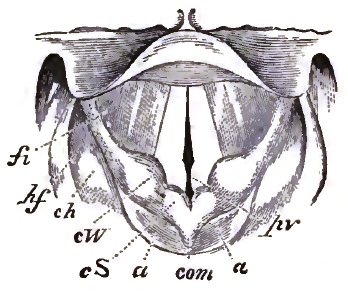
| Laryngoscopic Diagram showing the approximation of the vocal cords and arytenoid cartilages, and the position of the various parts during vocalization. f. i. Fossa innominata. h. f. Hyoid fossa. c. h. Cornu of hyoid bone. c. W. Cartilage of Wrisberg. c. S. Cartilage of Santorini. a. Arytenoid cartilages. com. Arytenoid commissure. p. v. Processus vocalis and cartilages of Seiler. (From Mackenzie.) |
The first detail to attract the eye is the epiglottis, which appears as a yellowish-red arch reaching from side to side across the image. It is thicker in the middle than at either end, and a protuberance is usually seen in the centre pointing forward. This arch is the upper margin of the epiglottis, and the protuberance is the tubercle, situated near the insertion of the epiglottis into the thyroid cartilage. The shape as well as the color of the epiglottis is very variable in different individuals, being sometimes rounded as in the drawings, sometimes rolled up like a dried leaf, sometimes notched in the centre, and sometimes presenting a point at this place. However, all these variations in shape have nothing to do with any pathological process, and may therefore be termed normal. The color of the organ also varies from a bluish-yellow to a pink-red, and these variations are also normal, being due to a greater or less thickness of the tissue covering the cartilage, which by shining through imparts its bluish color to the tissue. The superficial blood-vessels also are more prominent in some individuals than in others, and may not be noticeable in some cases.
[p. 32] Immediately behind the epiglottis we see two pit-like depressions, separated from each other in the middle by a fold of mucous membrane and bounded on either side by similar folds less prominent. These folds are the glosso-epiglottic ligaments, and serve to connect the tongue with the epiglottis, while the depressions are the glosso-epiglottic grooves, in which we usually find the foreign bodies which have accidentally been swallowed.
The ends of the epiglottic arch are lost in folds of mucous membrane, which run forward and inward to meet in the median line some distance in front of the epiglottis. Along their course several nodules of different size are noticed, which are symmetrically situated on either side. The one nearest to the epiglottis is the cartilage of Wrisberg, a small cartilaginous nodule imbedded in the tissue. The larger one, situated at the end of the fold of mucous membrane, is the arytenoid cartilage, and a third small nodule is noticed close to the arytenoid cartilage between it and the cartilage of Wrisberg, which is called the capitulum Santorini. The folds of mucous membrane are termed the aryteno-epiglottidean or ary-epiglottic folds. Their color is normally of a pinkish-red, and does not vary much in different individuals.
The arytenoid cartilages forming the ends of the ary-epiglottic folds are movable, approaching and separating alternately during the act of respiration, while during phonation they are pressed against each other, thus obliterating the space between them which is seen when they are separated. This space is the inter-arytenoid space or commissure, and is formed by the lateral walls of the arytenoid cartilages and the upper margin of the posterior portion of the cricoid cartilage. The mucous membrane in this commissure is very loosely attached to the deeper structures, and is thrown into folds by the approximation of the arytenoid cartilages. Its color is much lighter than that of the ary-epiglottic folds, due to the shining through of the cricoid cartilage. Outside of the ary-epiglottic folds and the inter-arytenoid commissure is the tissue forming the posterior and lateral walls of the oesophagus (not shown in the diagrams), and near the epiglottis a space called the pyriform sinus is noticed between the ary-epiglottic folds and the wall of the oesophagus.
Running from the epiglottis to the ary-epiglottic folds are two broad bands, one on either side, covered with mucous membrane and of a pinkish-red color, which are lost on either side in the tissue forming the walls of the laryngeal cavity, while toward the middle of the image they present concave and tolerably sharp edges. These are the ventricular bands, which were formerly termed the false vocal cords, and which form the lip to the opening of the ventricle of the larynx. Between the ventricular bands filling up the central portion of the image are seen the vocal cords, two bands of a pearl-white color which are attached to a cartilaginous process of the arytenoid cartilages, and run from these parallel with each other to the angle of the thyroid cartilage immediately below the tubercle of the epiglottis. These present sharp edges toward each other, and follow the motions of the arytenoid cartilages to which they are attached, so that when in inspiration the cartilages are separated the edges of the vocal cords are also some distance apart, forming, together with the inter-arytenoid commissure, a triangular opening called the glottis. That portion of the opening which is bounded on either side by the edges of the vocal cords alone is called the membranous portion, while the base of the triangle is termed the cartilaginous portion, being bounded on either side by the vocal processes of the arytenoid cartilages. This portion is readily distinguished from the membranous portion by its slightly yellow color, and by the fact that a very obtuse angle is formed at the junction of the two portions when the glottis is wide open during respiration. Through the open glottis the lower edge of the cricoid cartilage and several of the rings of the trachea can usually be seen, and there are a few cases in which even the bifurcation of the trachea can be dimly illuminated, showing in the [p. 33] laryngeal image the openings of the bronchi. The distance is, however, too great for bright illumination, and nothing can be seen distinctly, so that it is of little value in a diagnostic point of view. During phonation the glottis is narrowed to a slit by the approximation of the arytenoid cartilages and inner edges of the vocal cords, and, as has already been stated, the inter-arytenoid space becomes obliterated. In the higher notes of the female voice, the so-called head tones, the cartilaginous portion of the glottis remains closed entirely, while the membranous portion appears as an elliptical opening which is diminished in its longitudinal diameter with each rise in pitch. This becomes possible because of the presence in the vocal cords of a slender rod-like cartilage attached to the end of the vocal process, which can readily be seen in the female larynx, but which is only rudimentary in the male.
This description, intentionally, has been made without reference to the anatomical relation of the parts, but to give a clear idea of what is seen in the laryngeal mirror. The reader should therefore always bear in mind that the laryngeal image, being a reflected one, is reversed, and that, on account of giving a bird's-eye view of the larynx from a point above and behind the organ, distances are materially diminished; and the image is also reversed in an antero-posterior direction, so that the epiglottis appears to be posterior when in reality it is anterior.
RHINOSCOPY.—Rhinoscopy, or the art of inspecting the nasal cavities and the naso-pharyngeal space, is divided into two portions—viz. anterior and posterior rhinoscopy; and it will be convenient to observe this division in the following description of the methods employed. But before proceeding with the description it will be well to briefly review the topographic anatomy of the parts, because in most works on general anatomy the nasal and naso-pharyngeal cavities are discussed in a few sentences, and they are rarely if ever examined in the dissecting-room, so that the student has but a very imperfect knowledge of the relation of the parts belonging to these cavities. (See Fig. 11.) The nasal cavities, which are wedge-shaped, with a narrow arched roof, extend from the nostrils to the upper portion of the vault of the pharynx. Their outer walls are formed by the nasal process of the superior maxillary and lachrymal bones in front; in the middle, by the ethmoid and inner surface of the superior maxillary bones; behind, by the vertical plate of the palate bone and the internal pterygoid process of the sphenoid and the turbinated bones. These latter run before backward, three on each side, and are designated as the inferior, middle, and superior, the latter being the smallest of the three. The sinuses or spaces between these turbinated bones are called meatuses; so that the space between the floor of the nose and the lower turbinated bone is called the inferior meatus, the one between the lower and middle turbinated bones is the middle meatus, and the one between the middle and superior turbinated bones is the superior meatus.
| FIG. 11. |
|
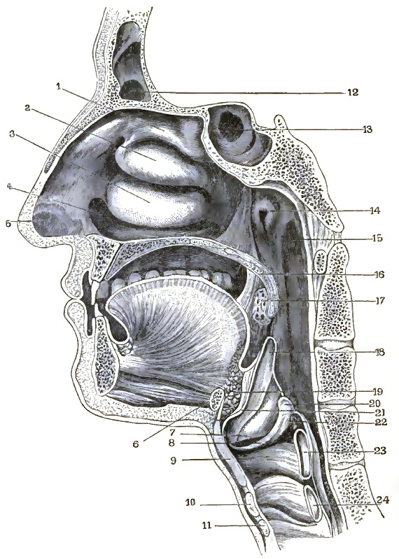
| VERTICAL SECTION OF HEAD, SLIGHTLY DIAGRAMMATIC. |
| 1. Superior turbinated bone. 2. Middle turbinated bone. 3. Lower turbinated bone. 4. Floor of nasal cavity. 5. Vestibule. 6. Section of hyoid bone. 7. Ventricular band. 8. Vocal cord. 9 and 23. Section of thyroid cartilage. 10 and 24. Section of cricoid cartilage. 11. Section of first tracheal ring. 12. Frontal sinus. 13. Sphenoidal cells. 14. Pharyngeal opening of Eustachian tube. 15. Rosenmüller's groove. 16. Velum palati. 17. Tonsil. 18. Epiglottis. 19. Adipose tissue behind tongue. 20. Arytenoid cartilage. 21. Tubercle of epiglottis. 22. Section of arytenoid muscle. |
The nasal cavities are separated from each other by a septum or division wall composed of the perpendicular plate of the ethmoid bone and the vomer posteriorly and the cartilaginous septum anteriorly, thus presenting a smooth surface as the inner wall of each cavity. The floor is formed by the palatine process of the superior maxillary bone and by the palate bone, and runs in a slanting, downward direction from before backward. The roof is formed by the nasal bones and nasal spine of the frontal in front, in the middle by the cribriform plate of the ethmoid, and posteriorly by the under surface of the body of the sphenoid bone. Directly communicating with the nasal cavities are other cavities situated in the bones of the skull, the lining mucous membrane of which no doubt is largely affected by the pathological processes in nasal diseases: these are the antra of Highmore, large triangular cavities situated in the body of the superior maxillary bone and communicating with the nasal cavities by an irregularly-shaped opening in the middle meatus; [p. 34] then the frontal sinuses, two irregular cavities situated between the two tables of the frontal bone. The communication between them and the nasal cavities is established by the infundibulum, a round opening in the middle meatus, and finally the sphenoidal cells or sinuses, found in the body of the sphenoid bone, communicating with the nasal cavities by small openings in the superior meatus. That portion of the nasal cavities which projects beyond the end of the nasal bone is surrounded by cartilages forming the alæ of the nose.
In the cartilaginous septum of the lower animals we find a small cavity lined with mucous membrane, called after its discoverer Jacobson's organ, the minute anatomy of which has lately been described by Klein.18 This [p. 35] organ in man is, however, only rudimentary. The nasal cavities are lined with mucous membrane, which varies greatly in thickness in different localities, and which materially decreases the size of the cavities in the living subject from that seen in the denuded skull. This mucous membrane is covered by ciliated epithelium in man, with the exception of that portion which lines the vestibule—i.e. that portion of the cavity of the nose surrounded by cartilage only—which is covered by pavement epithelium.
18 Quarterly Journal of Mic. Science, January, 1881.
In the lower animals we find that in the olfactory region the ciliated epithelium is either absent, or that ciliated and non-ciliated epithelium alternates in patches.19 The author has not been able to find a statement in the literature on the subject as to the kind of epithelium found in the accessory cavities in man, but it is very probable that the mucous membrane of the frontal sinuses and the antra of Highmore is covered with ciliated epithelium; otherwise it would be difficult, if not impossible, for the secretions of that mucous membrane to pass through the narrow channels into the nasal cavities. The color of the normal nasal mucous membrane is of a light pink shade in what is termed the respiratory portion, while it is of a yellowish hue in the olfactory region, that portion of the mucous membrane which covers the roof and the outer walls of the nasal cavities down to the upper margin of the middle turbinated bone and the septum down to about the same level. It is in this region that the nerve-ends of the olfactory nerve are distributed. Immediately beneath the mucous membrane, and between it and the periosteum of the bony walls and the perichondrium of the cartilaginous portion of the septum, we find a tissue which bears a striking resemblance to the erectile tissue of the genital organs.20 It is composed of a network of fibrous tissue, the trabeculæ of which contain a few organic muscular fibres. Its meshes of various sizes and shapes are occupied by venous sinuses lined with endothelium. These are supplied with blood by small arterioles and capillaries, which are quite numerous in the fibrous tissue and can readily be demonstrated under the microscope. In this arrangement of elements of the nasal mucous membrane we find a ready explanation of the fact that liquids of greater or less density than the serum of the blood when introduced into the nasal cavities produce pain, for we have here the most favorable conditions for osmosis, which will cause either a contraction or a distension of the sinuses. In the larger masses of fibrous tissue between the sinuses or caverns we find imbedded the glands, with their ducts opening out between the epithelial cells of the mucous membrane. There are two kinds of glands in this region, which have been described by Klein21—viz. serous and mucous glands.
19 Haenle, Anatomy des Menschen, vol. ii.
20 Haenle, loc. cit.
21 Loc. cit.
This cavernous erectile tissue is most abundant at the lower portion of the septum and of the lower turbinated bones; and, although it has been recognized and described as true erectile tissue by Haenle, Virchow, and others, yet to Bigelow of Boston belongs the honor of having first called attention to the part which this tissue plays in nasal diseases. He gave to it the name turbinated corpora cavernosa.22 The expansion of the nasal cavities formed by the alæ of the nose is termed the vestibule, which is lined with pavement epithelium and forms the entrance to the cavities proper. The naso-pharyngeal cavity extends from the posterior ends of the turbinated bones and the edge of the vomer to the line where the velum palati touches the pharyngeal wall during the act of deglutition or phonation. In this cavity we find the openings of the Eustachian tubes, two crater-like elevations, with a pit-like depression of variable size and shape, one on either side; and a collection of glands with a central duct-like opening disposed on the roof and posterior wall of the cavity. This gland was named by Luschka23 the pharyngeal [p. 36]tonsil. The openings between the edge of the vomer and the lateral walls of the naso-pharyngeal cavity are termed the posterior nares.
22 Boston Med. and Surg. Journal, April, 1875.
23 Der Schlundkopf des Menschen.
ANTERIOR RHINOSCOPY.—Anterior rhinoscopy is a very easy and simple procedure, and is practised as follows: The patient is placed in position as for laryngoscopy, and the light directed upon his face so that the centre of the circle of reflection from the head mirror falls upon the tip of the nose. The examiner then elevates the tip of the nose with his left hand, resting the fingers on the forehead of the patient, and lifts the ala away from the septum with a slightly bent probe, when he will be enabled to see a considerable distance into the nasal cavity. It is, however, better to employ a speculum instead of the bent probe, because the parts then are seen in their usual relation to each other, and are not distorted by the forcible traction necessary when the probe or a dilator is employed. The nasal speculum (Fig. 12) is best made of hard rubber and shaped like the ordinary ear speculum, except that the narrow end is oval instead of round. This instrument is to be introduced by a sort of rotatory motion until the end has passed the edge of the vestibule, when it will remain in position, displaying the interior of the nose. Great care should be exercised, when introducing the speculum, not to scratch the mucous membrane of the septum, for this will give rise to pain and start hemorrhage, both of which are to be avoided as much as possible. When applications are to be made to the mucous membrane of the septum or turbinated bones, or when operations are to be performed within the cavity, it is best to employ an instrument called a nasal dilator, of which there are a large number of different forms, the most satisfactory of which is shown in Fig. 13. The dilator is introduced by compressing the blades between the thumb and fore finger, and pushing them into the nostril until their ends have passed the edge of the vestibule. The pressure is then removed, and the spring separating the blades holds the nostril open; the handle or stem of the instrument, hanging down, need not be held or supported, as the blades press sufficiently upon the tissues to retain the instrument in position. If the pressure is too great, however, it will soon produce pain, and the patient will object to the use of the instrument.
| FIG. 12. |
|
| Nasal Speculum. |
| FIG. 13. |
|
| Bosworth's Nasal Dilator. |
The view obtained both by the speculum and the dilator is rather limited, and usually comprises only the anterior portions of the lower and middle turbinated bones, together with the cartilaginous portion of the septum. In order to get a good view of the lower and middle meatus and of the floor of the nose the patient's head should be inclined forward or backward as occasion requires. The student should, however, not be satisfied by simply inspecting the parts, but should aid the eye by the sense of touch, for pathological changes are of common occurrence, and their nature, whether soft and fleshy or hard and bony, erosions of the mucous membrane, or deep ulcerations, can often only be determined by the aid of the probe. In the same manner can the permeability of the meatuses be determined better than by inspection [p. 37] only. In cases where it becomes necessary to determine whether the anterior portion of the septum is of normal thickness, or whether a projection seen through the speculum is due to localized deflection, an instrument called the septometer is of great assistance (Fig. 14). This instrument is similar to the one used by mechanics to determine the diameter of a piece of wood or iron being turned on the lathe. In using it the long straight shanks are introduced one in each nostril, and, being closed upon the septum, the rounded points are gently moved up and down and backward and forward over the bulging portion of the septum. The motion of the index attached to the curved shanks of the instrument accurately indicates the relative thickness of tissue grasped between the points in the nose. By means of this instrument we can thus ascertain whether we have to deal with a deviation or a localized thickening of the septum; for if it is a deviation the index will move but slightly, while it will travel a considerable distance when the points pass over a thickened portion.
| FIG. 14. |
|
|
Septometer for Measuring Thickness of Nasal Septum. |
Although simple in its details, anterior rhinoscopy is often made difficult or altogether prevented by obstacles which are mostly due to malformation of the parts, such as deviation of the cartilaginous portion of the septum, exostoses from the superior maxillary bones reaching into the nasal cavity, adhesion between the anterior portion of the lower turbinated bone and the septum, nasal polypi, anterior hypertrophies of the mucous membrane, and so forth; or they may be due to faulty instruments, as too much pressure in the spring of the dilator; or, finally, they may be caused by want of care in the handling of the instruments, as when the septum is scratched by the edge of the speculum and hemorrhage ensues.
POSTERIOR RHINOSCOPY.—Posterior rhinoscopy is much more difficult than laryngoscopy or anterior rhinoscopy, and requires more patience and dexterity on the part of the examiner than either of the former, because but very few persons have control over the movements of the velum palati, and in most of these the upper portion of the pharyngeal wall is so sensitive that the slightest touch with an instrument gives rise to reflex cough and to gagging. In many cases, however, with patience and skill the naso-pharyngeal cavity and the posterior portion of the nasal cavities can be illuminated and inspected. To do this the patient is placed in the same position as for laryngoscopy, except that the head is not inclined backward, and after the mouth is opened as wide as possible the light from the reflector is thrown into the oral cavity. The tongue is then depressed with a tongue depressor. This instrument in its simplest form in which it is daily used by the practitioner for examining the fauces is the handle of a spoon. For laryngoscopic or rhinoscopic purposes, however, the spoon is not to be recommended, because the hand holding it must be on a level with the mouth, thus obstructing the view and light. An instrument has therefore been constructed which obviates this difficulty. It consists of a leaf-shaped blade of silver or German silver bent at right angles and inserted into a flat wooden handle. The lower surface of the blade is slightly concave, and ribbed so as to take a better hold of the slippery back of the tongue, and from the bend is about 3 inches in length. It is introduced into the mouth as far back as possible, and pressed upon the back of the tongue while the hand of the examiner is below the chin of the patient. For the sake of convenience in carrying the instrument the blade has been so hinged to the handle that it will fold up against the latter and will [p. 38]open at a right angle with it (Fig. 15). A more elegant and lighter instrument of the same description has lately been introduced in which the handle is also made of metal, and, like the blade, is heavily nickel-plated, and which when folded can be carried in a pocket-case. Soon, however, the metal tongue depressor becomes tarnished by the secretions of the mouth or by the substances used for applications to the throat, and then presents an appearance disgusting to many patients, who will not on that account submit to its use. For the sake of greater cleanliness, J. Solis Cohen devised a tongue depressor made of hard rubber, which is known as Cohen's tongue depressor (Fig. 16). It consists of a piece of ebonite bent upon itself, either end being a little over 3 inches long. The bend being more than at right angles, the hand holding the instrument rests underneath the chin of the patient; but if a different curve be desired for any particular case it can easily be obtained by placing the instrument for a little while in hot water. When soft it can be bent into any shape, which it will retain when cooled by immersion in cold water. Great care should be exercised not to carry the blade of the instrument too far back, as then gagging will at once set in. In cases where the tongue resists the pressure of the tongue depressor, it is better to exert but a gentle pressure upon the back of the organ, under which it will slowly recede, than to try to subdue it by force, for in the latter case it will unavoidably slip from under the blade of the instrument, and the desired space in the fauces is not obtained. With children the writer has found the fore finger of the left hand to be the best means of depressing the tongue, for the little patients as a rule have a horror of the formidable-looking instrument.
| FIG. 15. |
|
| Folding Tongue Depressor. |
| FIG. 16. |
|
| Cohen's Tongue Depressor. |
After the tongue has subsided into the floor of the mouth a small laryngoscopic mirror is introduced into the pharyngeal space behind the velum palati, with the reflecting surface upward, and is held there without touching the wall of the pharynx. The handle of the mirror, as in laryngoscopy, is brought into the angle of the mouth, so as to be out of the line of vision. As is usually the case, the velum palati at the approach of the mirror will rise and apply itself to the posterior wall of the pharynx, when of course the naso-pharyngeal space, being shut off, cannot be illuminated. Under these circumstances the velum must be made to hang down as in the act of nasal respiration, which is most easily accomplished by telling the patient to breathe through his nose. It is of course impossible to do so when the mouth is open, but the patient, not being cognizant of the fact, will make the attempt, and the palate will come down, permitting illumination and inspection of the naso-pharyngeal space and the posterior nares. In those cases in which this [p. 39]expedient fails it becomes necessary to forcibly pull down the velum by means of a blunt hook made by bending a silver laryngeal probe, or to tie it down by passing small elastic bands through the anterior nares and bringing the ends through the mouth and tying them over the upper lip. The smallest black rubber tubing is admirably suited for this purpose, as it can be introduced without an instrument. When the palate is pulled down with the palate hook, or when operations in the naso-pharyngeal space are to be performed, the patient must hold the tongue depressor himself, so as to leave the other hand of the operator free. Few persons can do this, however, satisfactorily, and it will be found more convenient to use Jarvis's tongue depressor and rhinoscope, as modified by the writer (Fig. 17). The instrument consists of a stout wire, which, after having been forked or divided at some distance from its insertion into the handle, forms the loop for the tongue depressor. The two branches then cross each other, and are bent to form another loop at an angle to the larger one. The ends of the wire are somewhat flattened and press against each other, thus closing the smaller loop and forming a sort of pincette, which can be opened by pressing the sides of the larger loop toward each other. The ends of the pincette are perforated by a small hole, which receives a pin attached at right angles to the short shaft of a small mirror, thus forming a hinge, so that the mirror can be placed at any desired angle with the handle or stem. The spring of the pincette cannot be made strong enough to prevent a change of the angle of the mirror by coming in contact with the pharyngeal wall, and therefore a ratchet was placed at the shaft of the mirror where it hinged to the end of the pincette, and a small steel spring, coming from one of the branches of the wire where they cross each other to form the small loop, by engaging in the teeth of the ratchet holds the mirror at the angle given to it before introducing. The large loop acts as a tongue depressor, so that with this admirable instrument the examination of the post-nasal cavity can be made with one hand, leaving the other free for the manipulation of other instruments. In order to be able to exert more pressure upon the tongue and to bring the hand out of the line of vision, the handle may be attached to the stem at an angle like the one in the folding tongue depressor. Except in cases of cleft palate the naso-pharyngeal cavity cannot be illuminated in its whole extent, and must be studied in parts, which when placed together in the mind of the examiner form the rhinoscopic image, a slightly diagrammatic drawing of which is seen in Fig. 18.
| FIG. 17. |
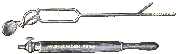
|
| Jarvis's Rhinoscopic Mirror and Tongue Depressor. |
| FIG. 18. |
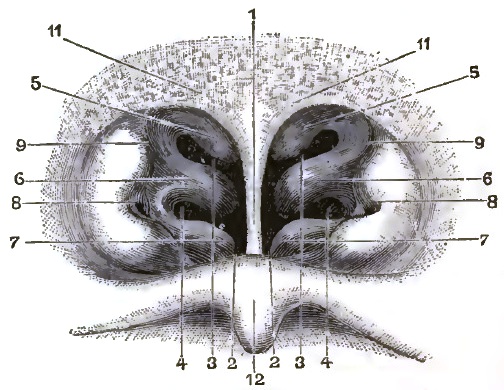
|
| RHINOSCOPIC IMAGE. |
| 1. Vomer or nasal septum. 2. Floor of nose. 3. Superior meatus. 4. Middle meatus. 5. Superior turbinated bone. 6. Middle turbinated bone. 7. Inferior turbinated bone. 8. Pharyngeal orifice of Eustachian tube. 9. Upper portion of Rosenmüller's groove. 11. Glandular tissue at the anterior portion of vault of pharynx. 12. Posterior surface of velum. |
THE RHINOSCOPIC IMAGE.—In the middle of the drawing we see a triangular plate with its apex downward; this is the posterior margin of the vomer or nasal septum. On either side we notice curtain-like folds projecting against the septum; these are the posterior aspects of the turbinated bones. On either side of these and on the margin of the drawing we notice pointed elevations projecting toward the interior of the cavity, with a crater-like [p. 40]depression on their apices; these are the lateral pharyngeal walls with the orifices of the Eustachian tubes. Above we see the vault of the pharynx, and below the posterior surface of the velum palati with the uvula.
Another method of examining the laryngeal and naso-pharyngeal cavities, which is especially valuable in cases where neoplasms or impacted foreign bodies hide the parts forming the laryngoscopic and rhinoscopic images, is by means of digital palpation. Even where no obstruction is present the beginner will do well to resort to this method in all cases, for he will thus become better acquainted with the topography of the parts than by inspection only. The procedure is not as difficult nor as disagreeable to the patient as might be imagined, and needs but little description.
When the laryngeal cavity is to be examined by palpation, the head of the patient is thrown back, and steadied in that position by the left hand of the examiner while he introduces the index finger of the right hand into the mouth and slides it along the back of the tongue until the tip comes in contact with the upper margin of the epiglottis. Passing downward along its lateral margin on either side, the ary-epiglottic folds and the tips of the arytenoid cartilages can be felt, and likewise the upper surfaces of the ventricular bands. The vocal cords are, as a rule, too low down to be reached by the tip of the finger. An examination of this kind should of course be made quickly while the patient is holding his breath, so as not to obstruct respiration too long, which in cases of narrowed glottis by neoplasms might give rise to serious results. When the naso-pharyngeal space is to be explored by the finger, the patient's head is bent forward, and the index finger is gently pushed upward between the velum and the pharyngeal wall. When this is accomplished, the velum is drawn forward and the finger pushed along its posterior aspect until the different portions forming the rhinoscopic image are reached and explored by the sense of touch.
Coryza is an acute inflammation of the mucous membrane of the nasal chambers. The disease is ordinarily idiopathic, but may be produced by irritative vapors, pollen, or dust. In the idiopathic form the symptoms of coryza are often preceded by malaise, with chilly sensations, and in severe attacks with headache. The attack itself is divided into two stages: that of determination or congestion, and that of exudation. In the first stage the excessive quantity of blood flowing into the arterio-venous network and the capillaries of the nasal mucous membrane distend them and obstruct the nasal chambers.
The symptoms are referable either to such obstruction of nasal respiration—in which group are included oral respiration, sensations of distension, and throbbing in the nose—or to reflexes, such as frontal headache, attacks of sneezing, and dull aching pain in the teeth.
The first stage lasts for a period varying from a few hours to several days, and is followed by the stage of exudation. This is characterized by a free watery or mucoid discharge from the nasal chambers, and by the cessation of the symptoms due directly or indirectly to pressure of the layers of swollen mucous membrane against each other. The discharge at first is watery, and is doubtless composed of transuded liquor sanguinis. It is followed by a mucoid fluid, which in severe or neglected cases may assume a purulent character. In many instances, even in mild cases, the discharge becomes muco-purulent toward recovery. The second stage is associated in children and adults of delicate constitution with excoriations of the nostrils.
Suppuration may take place in nurslings and in old people. It would appear that in coryza, as it exists in the northern countries of Europe, the beginning of the second stage is apt to be marked by free suppuration.
Acute coryza may involve the sinuses of the face, particularly the maxillary sinus. The involvement of the frontal and sphenoidal sinuses, while possible, is infrequent. Pharyngitis, laryngitis, and occasionally acute aural catarrh, often coexist with the disease.
The symptoms of coryza are so distinctive that the diagnosis is easily made. But since any obstructive or catarrhal state of the nose is described by patients as a cold in the head, it is necessary for the medical attendant to distinguish the various diseases so denominated. Acute coryza may be confounded with angiose hypertrophy; with the obstruction to nasal respiration due to deflection of the nasal septum or to an inflamed soft polypus; with catarrhal irritation affecting surfaces which are already enlarged by hyperplasia or which are undergoing atrophy; or with the effects of operative interference in the nose.
In angiose hypertrophy the swollen membranes will contract under a mild [p. 42] current of electricity or by change in the position of the body. Both chambers are rarely involved at the same time. Reflexes are of infrequent occurrence. Obstruction to nasal respiration due to a deflected septum arises from causes which are insignificant and do not affect the constitution. The genuine influenzal or catarrhal element is absent. In an inflamed soft nasal polypus an attempt at inspiration will, as a rule, detect the presence of the growth. In diffuse multiple polypi the case is different. Many persons who are reputed to take cold readily, or who may be said never to be free from cold, are really sufferers from neglected polypi. Persons suffering from atrophic catarrh always speak of an exacerbation of their symptoms as a fresh cold, and describe the disease itself as a cold. The sense of fulness, the throbbing, the heat, and the characteristic discharge of coryza are absent. A fresh cold in atrophic catarrh is an attack of inflammation (often catarrhal in character, it is true) which affects the involved surfaces, but is attended with an increase of plastic exudation and accompanying fetor.
It is a common occurrence for patients who have had a cautery application made or a polypus removed to return after a few days' absence with the report that they have contracted a cold. While the condition may be an attack of acute coryza, the chances are in favor of the symptoms being excited by the manipulation or the reaction from the operation. The symptoms are mild in character.
TREATMENT.—The treatment of coryza is both local and constitutional. The local treatment consists in applications of agents which tend to constrict the vessels of the nasal mucous membrane. In the first rank of such agents may be named cocaine, which in a 2 per cent. or a 4 per cent. solution will often give notable relief by overcoming the sense of obstruction. Individuals will be found in whom the effect is of short duration, and in some persons I found the medicine to have no effect whatever. In more favorable subjects the relief will be acknowledged for a period varying from four to six hours. Next in rank may be named a current of constant electricity (say from six to ten cells) passed through the cheeks. Should neither of the above-named agents be available, inhalations of iodine vapor, a few drops of chloroform rubbed upon the palms and inhaled, or the inhalation of the spirits of ammonia may be recommended. Toward the later stages of the disease detergents and mild astringents are well borne. The constitutional treatment includes the administration of diaphoretics and minute doses of opium, especially in the early stages of the disease. Coryza is commonly self-limited, and by far the larger number of cases do not come under the care of the physician.
Chronic nasal catarrh embraces those more or less persistent affections of the nasal chambers whose symptoms resemble those of acute coryza. The term catarrh is inexact. It is used to include several diseases associated by a single characteristic—namely, the existence of an increased amount of mucous secretion upon the affected membranes.
In order to understand the varieties of nasal catarrh, it is necessary to have clear conceptions of the uses of the nasal chambers. The normal performance of the function of respiration demands that when the mouth is closed the currents of air should pass through the nose. These currents, however, do not sweep over the entire nasal surfaces, but are confined to those portions which answer to the inferior meatus and the space bounded within by the septum, without by the median surface of the inferior turbinated bone, and above by the under surface of the middle turbinated bone. In the lower mammals this space is separated posteriorly by a transverse bony lamina which [p. 43]effectively excludes the upper portion of the nasal chambers from the tract just named. Anteriorly, at the termination of the inferior meatus and the middle turbinated bone, the tract is in freer communication with the upper spaces. The passage thus briefly defined may be called the respiratory tract, and when it remains patulous no serious interference with nasal respiration can occur.
The transverse diameters of the tract are subject to frequent changes, owing to the erectile character of the mucous membrane in its walls. But as long as the surfaces do not touch one another obstruction cannot exist. The passage, even when narrowed to a chink or line intervening between the median and lateral walls of the tract or between the floor and the roof of the inferior meatus, is sufficient evidence that there is room for the transit of the currents of air. The membranes themselves are subject to changes in form which are dependent upon the degree of development of their erectile tissue.
There is doubtless a disposition on the part of the erectile tissue to grow in the direction of the least resistance, and thus to occupy, by a process of compensative hypertrophy, the spaces left as the result of variations or defects in development in the bones composing the framework of the nasal chambers. The greater development of the erectile tissue may in this way be found on the side answering to the larger respiratory tract, which may therefore be more apt to suffer from changes in the conditions of nasal breathing than the chamber having the smaller tract. The erectile tissue acts as a monitor to the throat and lungs by presenting warm surfaces over which the air passes, thereby having the temperature raised before it enters the throat and lungs. It also acts by occluding the chamber, and thus aids in shutting out irritant vapors and dust. The lower animals possess a higher degree of development of the tissue at the point where the adducted ala presses against the septum. This point answers to the position of the organ of Jacobson. With man, the locality of the adduction corresponds to the junction of the premaxillary with the maxillary portion of the nasal chambers, and is often the seat of a delicate band of mucus extending across from the inferior turbinated bone to the septum.
That portion of the nasal chamber above the respiratory tract may be called the olfactory tract. It does not appear to be involved in the diseases under consideration, or, if it is, no clinical signs or symptoms are presented with which the author is acquainted. It will therefore receive no attention in this article.
For convenience the varieties of chronic catarrh may be classified as follows:
FIRST VARIETY—that dependent on defective nasal respiration.
This variety is caused by—
(a) Osseous obstruction in the nasal chamber.
(b) Membranous obstruction in the nasal chambers from compensatory hypertrophy of the erectile tissue, alone or with hyperplasia.
(c) Obstruction arising from hypertrophy of the adenoid tissue in the pharyngeal vault.
(d) Contracture of the levator palati muscles.
SECOND VARIETY—that dependent on structural changes in the component parts of the nasal chamber.
This variety is associated with—
(a) Chronic inflammation of the nasal mucous membrane without hypertrophy of the erectile tissue.
(b) Atrophy of the turbinals and their associated mucous membrane.
(c) Necrosis of the bones which enter into the framework of the nasal chambers.
FIRST VARIETY.—Defects in nasal respiration induce hyperæmia, distension of the erectile tissue, hyperplasia of the mucous membrane, and [p. 44] inevitable distress in the nose. A sense of fulness across the bridge of the nose and at its sides is complained of. Frontal headache may be present.
(a, b) When the septum is deflected and the left nasal chamber is narrowed, the labor of sustaining nasal respiration is thrown on the right side. This arrangement invites a flow of blood to the already large turbinals, and creates obstruction which is frequently referred to the right side, although both are alike affected. Thus, subjects in which the initial obstacle is osseous complain of distress caused by cavernous-tissue hypertrophy of the lining membrane of the opposite side. This represents a very common class of cases.
When the septum is not deflected, but projections from it impede the current of air, there may be either unilateral or bilateral obstruction, dependent upon the shape of the septum itself. Hypertrophy of the cavernous layer of the mucous membrane usually coexists. These cases are numerous, but less common than those last described.
Infrequently, cases are seen where the distress is occasioned by defects of the osseous structures not accompanied by cavernous hypertrophy.
Treatment of the above disorders consists in restoring nasal respiration by removing obstructions, whether they be osseous or membranous. The septal projections may be drilled or filed away, or, if marked deflection of the anterior portion be present dependent upon a malposition of the triangular cartilage, an operation simple in character may be performed for its correction. This consists in severing the connection of the lower margin of the cartilage with the maxilla and slipping the partially free cartilage to a new position. The details attendant upon the operation need not be here given. The reduction of the hypertrophied membranes can be best accomplished by cauterization. The most efficient method is by means of the electric cautery. The electrode used should be flexible and of small size. The points which most frequently require cauterization are the premaxillary portion of the inferior turbinated bone, the under surface of the same, and the septum at the maxillary spur. Rarely the inferior surface of the inferior turbinated bone at the palatal region requires attention. The applications are best made over small surfaces at a time, and should be repeated at intervals of from two to three days until all suspected points have been at least once cauterized. Not infrequently, the effect of the cauterization at one spot will cause constriction to take place in the vessels of the entire mucous surface, so that while this condition lasts it is impossible to tell what additional points of the membranous obstruction demand removal. At the following visit, however, the vessels have become relaxed, the membranes are again turgescent, and if obstruction now occurs it can easily be detected.
The galvano-cautery can only be used in the nasal chamber in patients who are earnestly seeking relief and are willing to assist the physician in all his efforts. With the tractable, intelligent subject it can with proper care be limited exactly to the spot intended. It is scarcely necessary to observe that any erratic or unexpected motion of the head will sear unaffected and sensitive surfaces. The interior of the vestibule is perhaps the most sensitive of these, and should always be protected by the use of the nasal speculum. No additional protection is needed, though in the judgment of others, among whom may be mentioned E. Shurly of Detroit, Michigan, an ivory shield passed in the nose parallel to the electrode is a necessary safeguard.
The pain of the application is generally slight, and can be in part annulled by a previous application of a 4 per cent. solution of cocaine. Some annoyance is acknowledged on the following day from the pressure of the eschar. Traumatic congestion of the entire mucous surface of the corresponding chamber is at the same time detected, and is usually sufficiently decided to produce some of the effects of acute coryza. This condition will spontaneously terminate in from thirty-six to forty-eight hours. The most annoying features [p. 45]following an application of the galvano-cautery which has been too freely made do not belong to the group just indicated, but rather to reflex disturbances. Pains are occasionally excited in the teeth, in the temple, eye, nape of the neck, and the middle ear. On one occasion in the writer's experience a unilateral reflex excitation of the entire opposite side of the body occurred, and a prickling sensation, followed by numbness, ensued, which lasted for twenty-four hours. Very rarely a congestion of the pharynx, of the larynx, and the larger bronchial tubes ensues, which can scarcely be directly attributable to the application, yet it has followed in a sufficient number of cases to lead me to believe that the two are in some remote way associated. Perhaps such a condition is analogous to the slight irritation of the respiratory tract following excision of the tonsil. Careful use of the galvano-cautery will obviate the conditions above described. They are important to remember as serving as limitations to the use of this valuable agent.
(c) It will be seen that osseous obstruction in the nasal chamber and hypertrophy of the cavernous nasal tissue often coexist. More rarely, a third element occurs as a complication, or it may be found independently of all other morbid processes. I allude to the presence of hypertrophy of the adenoid tissue in the pharyngeal vault. When this tissue is only moderately developed, it need not, and does not, interfere with nasal respiration; but when it projects downward to such a degree as to lie within the axis of the lower portion of the posterior nares, it produces the same effect upon nasal breathing as though obstruction existed within the chamber. The growths can be easily detected, as a rule, from behind by the aid of the rhinal mirror, but it should not be forgotten that they also can be seen from in front, provided the chamber is free from obstruction along the respiratory tract. In some individuals the ribbed or lobate structure of the mass can be discerned, but more often its presence is revealed by the minute points of light reflected from the lobules. If it be a matter of doubt whether these points of reflection are within the nasal chamber or beyond it in the pharyngeal vault, the patient may be requested to swallow, or to pronounce the letter e; when, if the point of reflection is within the nasal chamber, it will not change its position, but if it be within the naso-pharynx, it will be moved slightly from side to side, or it may for a moment disappear.
The symptoms of nasal catarrh which are provoked by the presence of such a growth can be alone successfully treated by the removal of the offending mass. In young individuals—say, from twelve to eighteen or twenty years of age—the finger inserted into the naso-pharynx from behind can often break down the growth. Slight hemorrhage follows this procedure, and the tags of imperfectly-destroyed tissue can be subsequently treated by caustics and powerful astringents. In the event of the patient proving intractable, the growth may be reached from in front through the nasal chamber, and the galvano-cautery can be used by passing the electrode backward through the nostril until it meets with resistance, which is invariably at the pharyngeal vault. Should this method of treatment not be permitted by an undisciplined or nervous person, the prolonged use of a glycerole of iodine may gradually reduce them in size; but no definite result can be promised from such treatment.
(d) Very rarely, through inordinate elevation of the soft palate owing to over-action of the levator palati muscles, the passage of communication between the naso-pharynx and the oro-pharynx is inadequate. Consequently, the nasal chamber is imperfectly ventilated, and its secretions, not flowing backward or being displaced to the normal extent, become semi-inspissated, and create obstruction by lodging in the respiratory tract, either in the premaxillary or palatal portions. To successfully combat this condition it is evident that no local treatment is demanded, either in the nose or the naso-pharynx, other than to increase the tonicity of the pharyngeal and palatal [p. 46] muscles. Very frequently in such cases there is a symmetrical atony in the muscles last named, which demands the internal use of strychnia and iron and the application of galvanism.
PROGNOSIS.—When nasal catarrh has proved to be dependent on defective respiration, the removal of the causes entering into this condition may with reason be expected to effect recovery. The prognosis, therefore, is favorable. In young persons, in whom reparative power is present in the highest degree, and in whom a secondary hypertrophy of the cavernous tissues is least developed, a prompt cure may be obtained by removal of the osseous or other forms of obstruction. In adults, however, the prognosis is less favorable, especially with those who have approached or passed middle life, and who have contracted vicious habits of breathing, which are likely to persist even after the removal of their causes. It is also tenable that in such subjects the mucous lining of the cranio-facial sinuses has become involved. Should anosmia persist after the capacity of the chambers has been augmented—in a word, should this condition not be dependent upon obstruction, but upon changes in the olfactory surfaces—the prognosis is less favorable than in any of the cases of the above-named group.
Treatment will, however, always secure amelioration of the symptoms, and few cases occur which cannot be greatly improved. The general health is invariably benefited. Should a tendency to asthma exist, it is apt to disappear, the complexion clears, and in adolescence the rate of general development is accelerated.
SECOND VARIETY.—The group of nasal diseases included under this head is not a natural one, since it embraces disorders characterized by a negative feature—viz. absence of obstruction to nasal respiration. Nevertheless, it is convenient to consider under a single head a number of relatively infrequent disorders in which there is invariably an underlying constitutional cause. Subjects of disorders herein embraced are not merely sufferers from insufficient oxygenation of the tissues, but have impaired general vitality or possess a decided constitutional taint, whether specific or otherwise. The nasal condition is simply the most prominent of the local manifestations.
Three distinct disorders are herein named: first, chronic inflammation of the nasal mucous membrane; second, atrophy of the turbinals and their associated mucous membrane; third, necrosis of the bones entering into the framework of the nasal chambers.
(a) Inflammatory thickening is a rare affection. It is more frequent in males than in females, and in persons of a sedentary occupation than in those who are actively employed. Those subject to it are apt to have light-blue or gray eyes and auburn or sandy hair. On examination, the chambers may be found free from peculiarities of bony structure, capacious, and without hypertrophy of the cavernous tissues, yet the membranes be of a deep-red color and of cushiony consistence, yield bright reflexes, and the shank of the instrument introduced into the nose is mirrored upon them. The most conspicuous alteration is not seen on the turbinals, but on the septum. The parts are very vascular, and the most moderate manipulation will often end in free capillary oozing. The discharge, though moderate in quantity, is inclined to be purulent, and resembles semi-coagulated albumen. Quite frequently, in the examination of a neglected case, minute flecks of this modified secretion are seen scattered over the septum and the inferior turbinated bone. Rarely, the discharge is maintained by the presence of a morbid growth or inflammatory products, either in the nasal chamber or a chamber accessory to it. The discharge then appears to consist of pure pus mixed with the normal secretion of the nose, and, thus rendered viscid and tenacious, it excites by its presence a condition of the lining mucous surface quite similar to that above described.
[p. 47] Under excitement, as after an attack of coryza, the discharge becomes more serous in character, and is occasionally of a chocolate color from its admixture with blood. It is without odor. There is no obstruction to respiration except during sleep, when, in aggravated cases, mouth-breathing may be established. Thus, the patient will often complain of an obstruction which is never present at the time of the examination. He further complains of a sense of dryness in the nose, with some pharyngeal irritation. The palato-pharyngeal and palato-glossal muscles are weak and often asymmetrical; the tonsils are small, but the adenoid tissues are generally unaffected. In a dry atmosphere, especially if it be loaded with irritating particles, the pharyngeal irritation is increased—a complication which is probably due to the inspired air passing too rapidly through the capacious and imperfectly-guarded nasal chambers and throat. Although I have carefully searched for all indications of aural complications, especially for the symptoms of progressive dry catarrh, I have never detected them but in a single instance.
The prognosis is to be guarded, although a careful course of treatment and proper care of the general health will greatly improve, if not entirely cure, the disease.
TREATMENT.—This consists in the application of nitrate of silver, either in strong solution or in the solid stick, to the under surface of the inferior and middle turbinated bones, of washing the parts with a dilute solution of carbolic acid, and of passing through the cheek tissues a constant electrical current of a strength of from five to ten cells. Tonics and alteratives should not be neglected, and an outdoor life, as far as is practicable, should be enjoined. The galvano-cautery may be used to destroy any nodules of tissue which resist other treatment. All applications are well borne, if indeed we may not look upon the condition of the surfaces as partially analgesic, and thus far of unfavorable significance. It is certain that indurated tags of oedematous and chronically inflamed mucous membrane overlying a bone, such as the middle turbinated or the alveolar line about the necks of the teeth, will never yield to anything but the most powerful astringents. Upon such tissues the most concentrated solutions of nitrate of silver are never caustic. The premaxillary portion of the inferior turbinated bone is frequently seen hopelessly infiltrated, and it must then be destroyed by the electro-cautery. When a discharge of a pus-like character exists, careful search should be made for the cause. If a tumor or foreign body be found, it should be removed, but if the cause lie in one of the outlying spaces of the nasal chamber, it is evident that the above treatment is palliative only.
(b) In atrophy of the nasal mucous surfaces and turbinals we have, as in the last-named group, spacious chambers, a purulent discharge, pharyngeal irritation (in many cases), and always associated a thin and relaxed, if not a paretic, condition of the velal muscles. These cases might be looked upon as an advanced stage of the preceding affection, since it may be surmised that the stage of infiltration has been succeeded by one of atrophy. The mucous membranes are everywhere pale, and closely bound to the underlying bony framework. The discharge is purulent and confluent; where in contact with the air it is desiccated, but where protected, as by crust-like surface-layers, it is semi-fluid and tenacious. There is, consequently, no disposition for the discharge to escape from the nose, and it accumulates until the sense of obstruction induces the patient to remove it by artificial means. When first seen, the nasal chambers are frequently so fully occupied with discharge as to conceal the characteristic appearances of the mucous surface. This prolonged retention induces incipient decomposition of the mass, which gives rise to the odor so characteristic of this group of cases.
The subjects of atrophic catarrh (ozæna) are never in robust health. They are, as a rule, of spare habit, anæmic, and with family histories which, while [p. 48]not distinctive, indicate that the affection is, to some degree at least, hereditary. A few cases have come under my notice in which all the general features of atrophic catarrh were present, but with very slight although confluent discharge, unaccompanied by fetor. Such cases are, strictly speaking, examples of atrophic catarrh, while they could not, under the old nomenclature, be included under the head of ozæna.
The prognosis is unfavorable for entire recovery, but treatment systematically pursued will make the patient entirely comfortable to himself and others—will arrest the progress of the disease and vastly improve the general health. As in other forms of nasal disease, should anosmia be present the prognosis is less favorable.
| FIG. 19. |
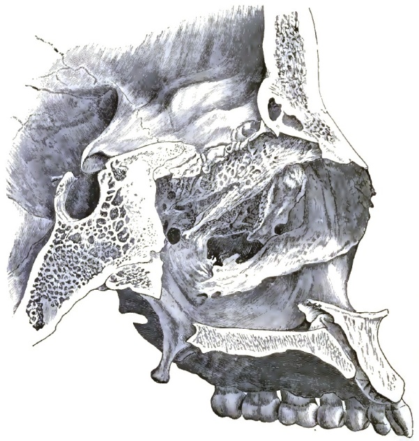
|
| Antero-posterior section of the bones of the face in position, showing the premaxillary portion of the floor of the nose greatly elevated above the plane of the remaining portions. In ozæna, as mentioned in the text, a disposition of parts may exist similar to that delineated, and cause discharge to collect and undergo offensive decomposition. |
TREATMENT.—The parts should be carefully cleansed—an act which, while imperfectly accomplished by either the syringe or the douche, is, in my judgment, best performed by the galvano-cautery. This instrument, the one relied upon for the subsequent treatment of the case, is to be selected for its initiation. The largest speculum which the nose will admit being placed in position, a spiral-looped electrode is introduced cold into the nose and held against one of the crusts. When heated it will effect so firm an attachment to it as to enable the mass to be withdrawn with great ease. In patients with [p. 49]whom the palatal portion of the floor of the nose is depressed below the level of the maxillary a considerable quantity of discharge may lie concealed from observation. When, after the removal of all visible crusts, the fetor persists, it is reasonable to suppose that the palatal depression is filled with decomposed pus and mucus. To test such a condition, the electrode should be appropriately curved and introduced. I have been surprised at the quantities of discharge which can in this way be withdrawn from a locality which, as far as I know, cannot be cleansed in any other way.
With the removal of the crust relief is at once experienced, and if the discharge could be removed as fast as it forms the disease would not really be a source of offence. The general health would also improve, from the fact that an atmosphere tainted with a burden of decomposition would no longer be breathed. But in practice this cannot be attained, and it is imperative, after the chambers have been entirely cleansed, to cauterize the lining membrane throughout. I have been in the habit of beginning such cautery treatments with the middle turbinated bone, passing thence to the inferior turbinated bone, then to the roof of the nose in front of the sphenoidal sinus, and lastly to the septum. Small surfaces only should be covered at a single treatment, so that it may take a month or six weeks to finish a single series of applications. This treatment is almost always well borne, nothing ever ensuing beyond a slight headache or a temporary establishment of a serous discharge. Notwithstanding that the condition in question is one of atrophy, the reparative power of the mucous membrane remains apparently unaffected. At all events, no danger from sloughing is to be dreaded after such extensive destruction of tissue. The thin eschars separate within from three days to a week, leaving a healthy mucous membrane beneath. In one instance the cauterization had extended to a sufficient depth to expose the bone, and yet from this denuded surface no exfoliation took place, the parts healing rapidly and satisfactorily. No other local treatment is relied upon for fetid atrophic catarrh than the one mentioned. No disinfectant washes are required if the discharge is removed as described. Should the patient be so situated as to be unable to report regularly for its removal or treatment, a wash composed of one part of Labarraque's solution to sixteen parts of water may be ordered with advantage, or a solution of carbolic acid, gtt. j to fluidounce j, with a little glycerin, may be snuffed up the nose twice a day, or listerine, diluted one-half with water, may be used with advantage. The general health, of course, should be cared for, and any complications met. I have found that during the winter months arsenic and cod-liver oil are well borne, associated with minute doses of Lugol's solution. For adolescents earthy and the calcareous phosphates are indicated, and for all abundant exercise and careful dieting. When the symptoms have been relieved, the patient should be requested to report once a month, for it is not to be expected that all symptoms will disappear, and some point of advice can be advantageously offered at this interval.
(c) Necrosis in the nasal chamber is a cause of catarrh, inasmuch as the fragments of bone lying within the nose excite irritation and induce discharge. I have never seen a case of this form of disease which was not due to syphilis. The remains of syphilitic angina are apt to be present, and the general manifestations of constitutional syphilis are well developed. The septum is more frequently affected than the turbinals.
Discharge due to necrosis can be readily distinguished from that arising from any other cause by the presence of detached fragments of denuded bone, by the characteristic fetor, and by the history of the case.
The prognosis is favorable, for all symptoms will cease upon the extraction of the fragments, or at least those which remain are of an entirely different character, and are due to the resultant imperfections of the septum, and consequent irritation arising from the too free entrance of air into the [p. 50]nose. I have seen in one case an extensive tumefaction and infiltration of the tissues covering the middle turbinated bone at the same time that the septum was breaking down. These masses require treatment with the galvano-cautery and astringents after the dead fragments have been removed.
| Cases in which interference with nasal respiration is a conspicuous symptom: | ||
| Due to deflection of nasal septum (common). Due to angiose hypertrophy of the mucous membrane (common). Due to tumors lodged in the nasal chamber. Due to adenoid hypertrophy in the naso-pharynx. Due to over-activity of the levator palati muscles (rare). |
||
| Cases in which discharge is a conspicuous symptom: | ||
| Due to hyperplasia of the mucous membrane over the turbinated bones
(common). The discharge when flowing backward is described as a
dropping; when forward, as a running at the nose. The discharge is
either mucoid or muco-purulent. Due to tumors lodged in the nasal chambers or appendages. The discharge is usually excessive. When due to myxomata (polypi) the discharge is mucoid (common). In inflammatory complications of the same the discharge is muco-purulent (common). When due to neoplasms other than myxomata the discharge is purulent, and rarely muco-hæmic (rare). |
||
| Cases in which retention of mucus in the nose or upper part of the throat is a conspicuous symptom: | ||
| Due to retention of inspissated mucus at
the roof of the naso-pharynx (common). Due to the mucous secretion of the nose and throat being excessively tenacious (rare). |
||
| Cases in which fetor is a conspicuous symptom: | ||
| Odor putrid. | Due to retention and decomposition of plasmic exudation from
atrophied bone and mucous membrane (common). Due to necrosis of the bones within or bordering upon the nose (rare). Due to decomposition of muco-pus in the maxillary sinus (rare). |
|
| Odor musty. | Due to partial decomposition in small patches of desiccated mucous crusts (common). Due to morbid secretion unaccompanied by profound alteration in the structure of the nose (rare). Due to ulcerations of the mucous membrane (rare). |
|
| Cases in which a sense of dryness is a conspicuous symptom: | ||
| Due to ineffective erectile tissue permitting air imperfectly warmed
to enter the nose and the pharynx (often met with in neurosis). It
is caused by temporary constriction of the erectile tissue or by
the atrophy of the tissue. Due to neurosis. Neurotic patients will often complain of a sense of dryness in the nose and the naso-pharynx when all the conditions of excessive mucoid discharge are present. |
||
| Cases in which hyperæsthesia exists, so that slight lesions that in any way interfere with the nasal functions form the basis of persistent complaint (not infrequent). | ||
Epistaxis, or nose-bleed, is a form of local hemorrhage perhaps of more frequent occurrence than hemorrhage from any other mucous surface of the body. This is doubtless owing to the extreme vascularity of the lining membrane of the nose and the special arterio-venous (cavernous) spaces of the turbinated bones; and the bleeding may be said to be of grave character in proportion as these spaces are involved. In some individuals a special disposition to nasal hemorrhage exists. From the fact that the affection is transmitted from parent to offspring, and is frequently found in all members of a given family, this form of hæmophilia is probably dependent upon some structural peculiarities in the cavernous spaces.
The causes of epistaxis are both local and general. Among the local causes may be included traumatism, either from blows or other injuries, attempts on the part of the patient to relieve irritation by picking the nose, or from the [p. 51]use of cutting or other instruments in the hands of the surgeon. Septal ulcerations in this way are often accompanied by moderate bleeding. In a case reported by R. G. Curtin the nasal branch of the ophthalmic artery was thought to have been ruptured. Among the general causes the most frequent is undoubtedly the depressed state of the system preceding or accompanying typhoid and other anæmic states. Thus, among the prodromes of typhoid fever epistaxis holds a conspicuous position. It is also seen in chlorotic females, especially in those suffering from that phase of anæmia known as Grave's disease. It also occurs in vicarious menstruation and in local facial or encranial congestions. In those disorders of nutrition accompanied by a tendency to capillary extravasation, such as purpura and scurvy, the nasal mucous surface participates in the general disorder. In a case of the former disorder coming under the notice of the writer the blood had forced its way out in large quantities by every capillary avenue.
TREATMENT.—Epistaxis when a symptom of a dyscrasia is of course to be treated as a local expression of a general condition. In typhoid fever, scurvy, and purpura or anæmia the bleeding is a sign of the general distress, and requires no special local method of treatment. Epistaxis when of local character should be treated, first, by removing the cause; second, by diminishing the flow of blood to the part; third, by cold and astringent washes to the affected surface; and, fourth, by compression.
First. Should the bleeding be kept up by fragments of bone impinging upon or lacerating the mucous membrane, they should be restored as far as possible to their natural position and retained there by appropriate apparatus. If they are entirely denuded of their periosteum and mucous membrane, they should be removed. Foreign bodies should be extracted, and if septal ulceration be present it should be carefully treated, the crusts removed, the ulcerated surfaces touched with nitrate of silver in stick, and the nasal chamber plugged from in front to exclude the outside air.—Second. The position of the body is of great importance in treating epistaxis. The recumbent position is no doubt to be preferred. The patient often holds one arm elevated or ties a cord about the proximal end of a limb. These innocent accessories to treatment may be permitted, since they are based upon well-known physiological principles, although it must be said that the bleeding can in all instances be checked without their aid. Cold applications to the nape and sides of the neck are often of service. Various internal remedies, such as ergot, gallic or sulphuric acid, and erigeron, may be administered with good effect in addition to the local measures.—Third. Astringent washes, such as a solution of alum—about drachm j to the pint—will often check a moderate degree of capillary bleeding without other aid. Tannic or gallic acid may also be used. Should these measures fail, the Monsel solution may be used on pledgets of cotton carried up to the bleeding spots. In Curtin's case, already quoted, a pledget saturated with the solution of the perchloride of iron placed over the nasal branch of the ophthalmic artery promptly arrested the bleeding. In lieu of these styptics the platinum wire loop of the galvano-cautery battery may be used. The writer has often succeeded in checking bleeding after a removal of a polypus or the use of the galvano-cautery when the exact position of the hemorrhage is known by laying upon the affected spot a little square of patent lint. It acts much as in checking the bleeding from a leech-bite.—Fourth. Compression of the mucous lining of the nose and exclusion of these surfaces from the air—a method familiarly known as plugging the nose—is the dernier ressort in the treatment of epistaxis, and is to be relied upon in the event of failure of other methods. This failure is, however, relatively infrequent. Observers agree in describing the procedure tedious and rather disagreeable, as much to the operator as to the [p. 52] patient, who has already been exhausted by loss of blood and the previous measures resorted to for his relief.
| FIG. 20. |
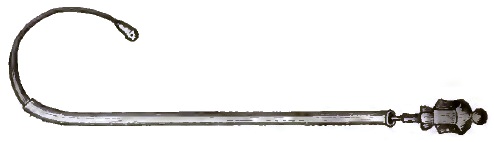
|
| Bellocq's Canula. |
The instrument usually relied upon for this purpose is known as Bellocq's canula (Fig. 20). This little instrument consists of a hollow curved tube of metal fashioned somewhat like a Eustachian catheter, and bearing within it a flexible and adjustable metallic band which carries at its extremity an eyelet. Any one who has used the Eustachian catheter will recall the number of instances in which it could not be passed, or if passed the frequency in which great distress followed. If this be true of the Eustachian catheter, it is also true of the Bellocq canula, the difficulty in the case of the catheter, indeed, being the lesser of the two, inasmuch as the physician has a number of sizes to select from. Conceding, however, that the instrument (with a long stout thread passed through the eyelet of the stylet) has been placed in position in the nasal chamber, one end of the thread is seized within the mouth and brought out between the lips, while the other, carried by the instrument, is withdrawn through the nose and is allowed to hang from the nostril. The two ends of the thread are now tied firmly together, and a pledget of lint or cotton, fashioned somewhat after the shape of the posterior naris, is tied to the thread. Traction is now made upon the nasal portion of the thread until the plug is firmly lodged against and within the posterior naris. The remaining portion of the oral thread is now cut off close to the velum, and the free end of the nasal thread secured by adhesive plaster to the integument. The nostril should next be stopped from in front by pledgets of lint or absorbent cotton. The size of the nasal chamber and naso-pharyngeal varies so markedly that a rhinoscopic examination is of use in fixing upon the size of the plug. If it be too small, it will be drawn entirely within the nose, and possibly beyond the bleeding spot. If it be too large, it will partially or entirely occlude the posterior naris of the opposite side, and thus by interfering with nasal respiration greatly increase the distress, or by pressure against the Eustachian fossa and velum interfere with the hearing and with deglutition. The plug should be retained in position until a purulent mucus appears within the nose: this is usually about the third day. The plug now usually becomes a little loose, and can readily be withdrawn by pushing it back into the pharynx, where it is seized with forceps. Too long retention of the plug in position is followed by great fetor and the free formation of muco-pus—conditions which tend to debilitate the patient.
D. Hayes Agnew informs me that he for a long time practised stopping nasal hemorrhage by plugging the chamber from in front. Strips of patent lint four inches long by half an inch wide are employed for this purpose. They are gradually pushed into the chamber until the entire space is filled as far as is practicable. An essentially similar method is described by F. H. Bosworth in his Manual of Diseases of the Throat and Nose.
These may be said to include the myxoma, sarcoma, fibroma, carcinoma, also the true hypertrophies and submucous inflammatory thickening.
[p. 53] The myxoma, more commonly known by the name of soft or gelatinous polyp, is the most prominent of the morbid nasal growths. It occurs ordinarily in small pedunculated seed-like masses, ranging in size from that of a grain of wheat to a grape. The most common seat is on the anterior portion of the middle turbinated bone and on the median surface of the inferior turbinated bone at the palatal portion. Instead of being pedunculated, they may be sessile; that is, each tumor may have a base equal to, if not exceeding, any diameter of the tumor.
The symptoms of nasal polypus are of three kinds: (1) those arising from obstruction of the nasal respiration; (2) those arising from the irritation excited by their presence; (3) the symptoms, reflex in character, manifested at points beyond the limit of the nasal chambers.
(1) The polypi necessarily tend to obstruct the respiratory tract of the nasal chamber. The first symptoms are of this character, and as a rule furnish the first intimation to the patient that trouble exists. Difficulty of nasal respiration is acknowledged, accompanied with a sense of tension and fulness, which is found to be worse during damp weather than when the air is dry and bracing. If the growths are freely pendulous, the act of blowing the nose may change the position of the mass and secure temporary freedom from distress. Incidental to obstruction, an intonation of the voice is often present. Loss of smelling and of taste is a frequent result of the mass interfering with the movement of the odoriferous particles. The loss of the sense of taste is dependent upon the loss of the sense of smelling.
(2) Polypi when large enough to press against the membranes of the nose excite an increased flow of mucus. As a rule, this flows forward, and is removed by the handkerchief. The quantity of fluid thus escaping is often very great. Patients often report the necessity of carrying about with them for a single day's use from eight to ten handkerchiefs. In the turgesence excited by an attack of coryza the mucus becomes thicker and of a yellowish color. Occasionally a sensation of dropping of mucus from the nose into the throat is a source of complaint.
(3) The reflex symptoms belonging to the presence of nasal polypi are, as a rule, referred to the forehead. This is especially the case if the growths involve the middle turbinated bone. When the tumors are so located, and have not impinged upon the respiratory tract, the symptoms of obstruction may be absent, and those of mucus excitement so moderate as not to excite attention, while the tension in the forehead, especially over the frontal bos, is pronounced. This sensation is intensified by prolonged inclination of the head forward, being especially aggravated in the acts of writing at a desk, working at a sewing-machine, kneeling at prayer, etc. Occasionally tinnitus aurium and suffusion of the conjunctivæ are present.
Neglected polypus ends in deformity of the nasal chambers and bones of the face. The face assumes a peculiar expression called by the older observers frog face. This is rarely if ever seen in this country, owing doubtless to the fact that the sufferers from nasal polypus seek medical advice in the early stages of the affection. Moderate degrees, however, of deformation of the turbinated bones are often seen.
Since the symptoms of soft nasal polypus are produced entirely by mechanical means, they can be closely imitated if not replaced by other morbid states of the mucous membrane. A hyperplastic state of the membrane over the middle turbinated bone will give rise to all the symptoms of a sessile polypus in the same situation. It is well to remember that this condition of the membrane often coexists with polypus, and of course will persist after the polypus has been removed. It follows that a guarded prognosis should always be made in case of sessile polypus. A tedious course of treatment of the indurated and chronically inflamed membranes may be required after the [p. 54]tumors have been removed before a cure is effected. The prognosis of soft polypus is more favorable as to the immediate results of treatment than in sessile polypus. The liability to recurrence can be materially lessened by carefully conducted after-treatment.
The diagnosis of soft pedunculated polypus is readily accomplished if the examination is made by aid of an appropriate speculum, the rhinal mirror, and a powerful light. Even without these aids the tumors can be seen by direct sunlight within the nostril if they are entirely occluding the chambers, and even in the event of nothing being visible by such inspection the movement of the masses by the act of blowing the nose will be noticed. The fact that the nasal obstruction is aggravated by damp weather seems to assist the physician in framing a diagnosis.
The diagnosis of sessile polypus requires a careful use of all the aids of rhinoscopy. They can be distinguished from hyperplasia of the mucous membrane by their lobulated form, and from the fact that the probe can move them slightly from their base. They can be distinguished from adenoid growths at the root of the pharynx by the fact that they remain unmoved during the act of swallowing.
The disease is not apt to recur if the treatment is thoroughly carried out.
The treatment of soft polypus consists in their removal. All observers are now agreed on this point. Injection by astringents and acetic acid—a process that at one time held out much promise—has been generally abandoned. In removal of the polypus one of two methods may be resorted to: that by avulsion, and that by the use of the snare. Avulsion is effected by forceps adapted for this special use. With such an instrument the polyps can readily be seized and removed. The rule that nothing should be seized which is not seen is subject to no exception. In no other way can the operator be secure against accidents. Incautious operators have frequently torn away strips of mucous membrane or portions of the turbinated bones in their crude attempts to remove these growths. Severe hemorrhage and death through violent lacerations of the ethmoid bone near the cribriform plate, and subsequent extension of the inflammation thereby excited to the membranes of the brain, have been known to follow these crude surgical procedures.
| FIG. 21. |
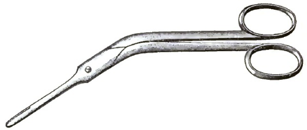
|
| The Author's Nasal Forceps. |
W. C. Jarvis of New York has modified the wire snare for application to the nose for the removal of polypi and hypertrophied tissues, and reports that it is a safer, more expeditious, and less painful method of operation than the forceps, which he unqualifiedly condemns. His instrument, while undoubtedly an ingenious adaptation of the principle of the snare, and a valuable addition to our means of treating nasal affections, cannot, in my judgment, take the place of the forceps in removing nasal polypi. As the aurist finds both the forceps and the snare useful in removing growths from the external meatus of the ear, so I am sure the physician will need both in the treatment of nasal polypi. In many cases the malformations of the nasal septum are such that [p. 55] I have been unable to use the snare where the forceps could be used with relative ease. I find when the loop is quickly drawn the same amount of bleeding follows as when the forceps are used. When it is slowly drawn, the sitting is tedious, and both the patient and attendant find the process wearying. The amount of blood lost when the forceps are properly used is not considerable, and is always under control. F. H. Bosworth1 describes the operation as extremely painful. So far from this being the case in my experience, I find the patients complain greatly of the constriction of the wire loop on the pedicle of the polypus, and invariably prefer the forceps. I must add that this preference was in no way influenced by myself, for I was disposed at one time to agree with the writers who have of late criticised the method of removal of the polyps by avulsion.
1 A Manual of Diseases of the Throat and Nose, 1881, p. 241.
No matter which of the methods be accepted, the treatment of polypus resolves itself into two simple propositions. When one or two large polypi are present in a capacious nasal chamber, the removal of the growths either by avulsion or snaring is a simple matter, and can often be accomplished in a single sitting. When numbers of small polypi are scattered over a large surface, particularly if they grow from the sides of the middle turbinated bone, the treatment is tedious, and even after the growths are removed a series of applications are required to cure the thickened and infiltrated mucous membrane.
Sarcoma, fibroma, and carcinoma are infrequent causes of nasal disease. When located in the nasal chambers they do not present any characters with which I am familiar which distinguish them from the expressions they assume in other parts of the body. When involving the respiratory tract they alike create symptoms by obstruction, by excitement of the secretions, and by the reflexes due to the involvement of the branches of the fifth pair of nerves. When situated in the olfactory track the obstruction to nasal respiration is absent, but the reflex symptoms are pronounced: the patient is liable to depression of spirits and to frontal headache. Encroachment upon the orbital, pharyngeal, and encranial spaces is common in the last stages.
Perhaps the most common way in which these morbid growths induce symptoms referable to the nose is by obstruction of the respiratory tract by the incursions of a mass originating at a point beyond the limits of the nasal chambers. In this way a growth in the pharynx may close one or both choanæ, or protrude into the nose from the spheni-palatine space by breaking down the ascending plate of the palatal bone as it forms the median wall of this space; or the growth may project inward from the superior maxilla.
In one case under my care, of obscure growth high up within the nose, which ended fatally by involvement of the membranes of the brain, a tenacious mucus of a dark chocolate color was withdrawn from the nose into the throat. The peculiar color of the mucus was found to be caused by a mixture of blood. In my judgment, this peculiar mixture of blood and pus was significant. The blood and mucus had not been mixed in the nasal chamber to cause the chocolate or rusty hue, for then we would have had the appearance customary in epistaxis of bright blood and frothy mucus mechanically held together. The even dissemination of the blood through the mucus would point to the conclusion that the blood had escaped in small quantity at the time of the formation of the mucus. Why such mucus does not constantly form in inflammatory states of the mucous membrane of the nose, as it does from the pulmonary mucous membrane in pneumonia, I am not prepared to say. But existing as it did in a case where a deep-seated disease was present may be accepted as a fact in some way connected with the invasion of a morbid growth in and upon the nasal mucous surface.
The pharynx is always in a state of hyperæmia when morbid growths of [p. 56] the above groups are present in the nose. The front of the velum is apt to be covered with a great number of minute papillæ, which, however, are often seen in anæmic individuals, and are not therefore pathognomonic.
The treatment of the growths enumerated and the general conduct of the cases are subjects for the general surgeon, and a consideration of them here would be out of place.
It may, however, be well to describe a few instruments which have been found useful in the large group of cases where cauterization is the principal treatment indicated. Foremost among these is the instrument shown in Fig. 22, which combines advantageously the essential features of the galvano-cautery and the wire snare.
| FIG. 22. |
|
| The Galvano-cautery Snare described in the text: 1, the cable of the battery; 2, the canula (which is not shown in full length); 3, the platinum wire; 4, the vulcanite carriage, with screws holding the ends of the platinum wire in metallic contact with the hinge-connections, by which the current is transmitted from the battery; 5, a slotted barrel of aluminium; 6, a movable nut on the screw; 7, a small portion of the screw disengaged from the slotted barrel; 8, milled stationary screw-head. |
| FIG. 23. |
|
| The Double Battery employed by the Author: The two sets of plates are seen united by a flat band of metal. The case which encloses the two separate batteries opens in front, displaying the cells, the plates (which are seen pendent over the cells), and the treadle. Above the figure of the battery lies a figure of the Flemming electrode handle and the electrode in position. |
It is well known that a loop of wire which is steadily narrowed has great power in severing the attachment of tumors and other outgrowths. When of a large size, it may be sufficiently powerful to pass through bony structures, as well as the softer parts of the body. The principle of the snare has been employed both in the throat, the ear, and the nose; but when my attention was first directed to this subject the forms available were too large and heavy for the delicacy of manipulation demanded in removing small tumors lodged in the narrower recesses of the nose. Moreover, no snare that I could then find would permit the galvanic current to pass through the loop at the time it was being narrowed. I was led, therefore, to inquire into the practicability of an instrument which would at once be light, be of small size, and yet be sufficiently powerful to remove that class of hypertrophied tissues and polypoid growths which are of such frequent occurrence in the nasal chambers. The instrument shown in Fig. 22 combines these qualifications, and satisfactorily performs the service for which it was designed. The only feature of an essential character which may be said to be novel is the fact that the platinum wire (3, Fig. 22) forming the snare is covered with a uniform coat of copper, excepting alone the portion forming the loop, which is bare. As a consequence of this arrangement the current of electricity from the battery is conducted through a double canula (2, Fig. 22) by means of the copper. The length of the instrument being about 9½ inches, and its weight less than ½ ounce, delicacy of manipulation is not interfered with. Besides possessing all the features of the cold wire snare, it has the additional advantage of securing a more rapid and painless operation, without any hemorrhage. Sessile (pyramidal) or resilient growths may be removed by first burning a groove of any depth into them, after which the loop is drawn while the current is passing through it. For this task the cold wire snare is obviously incompetent. Growths of unusual size or hardness may be destroyed by the same procedure, and nodules no larger than a grain of wheat may also be excised with great nicety. [p. 57]It will be seen that failure to remove at least a portion of the growth attacked is an event exceedingly unlikely to occur. I have been particularly struck with the facility with which hypertrophies of the inferior turbinated bone can in this way be treated; and if cocaine be freely applied before the operation, it constitutes, in my judgment, the most speedy and the least painful of any means by which such conditions can be reduced. By using a canula with a curved end it is easy to snare growths situated on the posterior portion of the inferior turbinated bone. The current passing through the battery (Fig. 23, B) to the instrument can be interrupted by any of the numerous devices with which the practical electrician is familiar; or the treadle of the battery can be depressed and locked by the lever-catch, and the interruption of the current be determined by the pressure of the finger [p. 58]on the knob in the handle (Fig. 23, A). This is under all circumstances desirable, since the weight of the cells is sufficient to demand considerable force to be exerted by the foot—always enough to destroy the delicacy of the manipulation of the instrument.
| FIG. 24. |
|
| Two Electrodes of peculiar shape in use by the Author. |
An electrode which is wrapped nearly to its distal end (Fig. 24), and used either in a straight or a curved form, is of great advantage in reaching growths within the naso-pharynx. The straight form can be thrust directly back through the nasal chamber, and the curved form can be passed from the oro-pharynx to the naso-pharynx without danger of burning the posterior border of the soft palate.
DEFINITION.—Disorders of sensation or motion, or of both sensation and motion, due to disease, first, of the centres from which the nerves of the organ are derived; second, to disease along the track of the nerves; third, to disease in the terminal distribution of the nerves; fourth, to reflected irritation from neighboring or distant parts; and fifth, to myopathic change. This last condition is not necessarily a neurosis; it is nevertheless a cause of modification of the function of the parts to which the nerves are distributed, often a result of paresis or paralysis, and therefore inseparably associated with the neuroses of the organ. Disorders of innervation, depending upon structural disease of the larynx, such as ulceration or tumor, are not included in this definition.
ANATOMICO-PHYSIOLOGICAL CONSIDERATIONS.—The framework of the larynx consists of cartilages securely but rather loosely articulated with each other. The movements of these cartilages produce changes in the position and tension of the soft parts. The thyro-cricoid articulation allows ginglymoid and sliding motion; the aryteno-cricoid, rotatory and sliding motion; the hyo-thyroid, ginglymoid motion. The physiology of the muscles of the larynx is quite complex, since nearly all have fibres taking a number of different directions, and the changes in the form and positions of the parts depend upon the combined action of different muscles and parts of muscles which may be individually brought into action to produce the required results. The muscles may, however, be roughly divided into groups: 1. Constrictors of the superior strait; 2. Dilators of the superior strait; 3. Adductors of the vocal cords; 4. Tensors of the vocal cords, external, internal; 5. Relaxers of the vocal cords; 6. Abductors of the vocal cords.
The superior strait of the larynx is closed by the action of the oblique portions of the arytenoideus, acting in conjunction with the ary-epiglottici, into which some of its fibres are continued, thus drawing the cartilages of Santorini downward and inward and approximating the ary-epiglottic folds and depressing the epiglottis; while the thyro-epiglottici complete the closure by further depressing the epiglottis. Fibres of the latter muscle, acting alone, may dilate the superior strait by drawing apart the ary-epiglottic folds.
The transverse portion of the arytenoideus and the superior fibres of the crico-arytenoidei postici approximate the arytenoid cartilages. The crico-arytenoidei laterales, and also in a slight degree the external fibres of the thyro-arytenoidei, rotate these cartilages, turning their vocal processes inward: the action of the latter two muscles as adductors is imperfect unless the arytenoids are drawn backward and fixed by the arytenoidei postici.
The tensor group comprises a number both of the extrinsic and intrinsic muscles of the larynx. The crico-arytenoidei postici draw the arytenoids back, external rotation, and consequent abduction, being prevented by other muscles. The anterior fibres of the crico-thyroid and those fibres of the [p. 60]sterno-thyroid inserted anterior to the crico-thyroid articulation approximate the cricoid and thyroid cartilages, and thus tighten the vocal bands. The posterior fibres of the crico-thyroid slide the thyroid upon the cricoid, lengthening the antero-posterior diameter of the larynx. This muscle, acting as a whole, also compresses the alæ of the thyroid with the same effect. The constrictors of the pharynx have a similar function. The hyo-thyroidei, acting in conjunction with the elevators of the hyoid bone, draw the thyroid forward and tilt it downward upon the cricoid. The form and internal tension of the vocal bands are greatly influenced by the thyro-arytenoidei, especially their inner fibres, while the ascending fibres of the muscle draw the inferior portions of the vocal bands upward and prevent the sagging of their edges. This muscle, acting alone, has been thought to cause extreme relaxation of the vocal bands. Modern research renders this statement of relaxation doubtful. The contraction of those fibres of the sterno-thyroidei inserted posteriorly to the crico-thyroid articulation tilts the thyroid upward, and thus relaxes the tension of the bands.
The crico-arytenoidei postici rotate the arytenoid cartilages outward, separating the vocal processes, and, acting in conjunction with the posterior fibres of the crico-arytenoidei laterales, draw the cartilages outward and downward.
In the cadaveric condition, which is one of relaxation of all the laryngeal muscles, the glottis is neither closed nor widely open; the epiglottis is erect against the dorsum of the tongue; the arytenoid cartilages are slightly separated, so that the glottic opening is a triangle with the base posteriorly, as in the act of inspiration, but the separation is much less than in the act of breathing. This condition is met with in paralysis affecting all the muscles of the organ.
The nerves of the larynx are derived from the pneumogastrics. The superior laryngeal is mainly a nerve of sensation for the parts above the edges of the vocal bands. There are some notable exceptions to this statement: a branch, external, descends to the crico-thyroid muscles and is motor in its function. Filaments from the superior laryngeal endowed with motor functions are also distributed to the folds extending from the arytenoids to the epiglottis; these are the ary-epiglottidean bands, and are concerned in the movements of the epiglottis. It is probable that the arytenoids are also in part supplied by the superior laryngeal; in other words, that both the superior and inferior laryngeal nerves are mixed, branches from the spinal accessory, as well as from the pneumogastric proper, entering to each of these nerves. Beclard1 states that the one, the spinal accessory, is a nerve of phonation; the other, the pneumogastric, is a nerve of respiration. The sensations of the mucous surfaces below the glottis depend upon filaments from the pneumogastrics returned along with the motor fibres from the spinal accessory. The two orders of fibres go to make up the recurrents. The relations of the recurrents themselves to the large vessels, as well as to the bronchial glands, are of importance. At the point of their origin they are in close relation with the aorta and right subclavian; they are also in close relation with the top of the lungs. Disease of these organs and structures, especially of the large blood-vessels, such as aneurism of the aorta or subclavian, disease of the glands, tumors, abscess, traumatism, etc., may modify or completely destroy the functions of the laryngeal nerves. In short, anything or any condition by which pressure may be made upon the pneumogastrics or recurrents may become a cause of nervous disturbance in the larynx. In addition to this general source of innervation, Elsberg2 describes a special centre of sensation for the throat in the medulla oblongata. He also describes three kinds of sensibility in the larynx—tactile, dolorous, and reflex. Rossbach3 details experiments from which he concludes that there are nerve-cells in [p. 61] the mucous membrane of the larynx which preside over the function of secretion. The larynx is endowed with at least two kinds of sensibility: the one tactile—when exalted it becomes painful; the other, reflex sensibility, is double. First, there is as a result of excitement a contraction of the subjacent muscle, and there follows closure of the glottis. This is seen in the application of irritants to the parts, such as solutions of nitrate of silver or other escharotics. There is no cough, but great difficulty of inspiration. Expiration is free and easy. There may follow some degree of pain for several hours. It will be seen that the phenomena are the same as those observed in the irritation of other mucous surfaces. The irritation is immediately translated into motion; this motion is probably reflex, but not necessarily through the centres, such as the brain or cord. The motion is of the subjacent muscles. Second, the mechanical irritation produced by the presence of a drop of water or a morsel of food in the larynx results in violent and explosive cough. The cough persists until the offending drop or body has been removed. This kind of sensibility calls into action distant muscles. There is no spasm of the adductors of the glottis, as in the case of the application of caustics. It is probable that the filaments of the nerves, the irritation of which gives rise to spasm, are distributed more generally than those which preside over reflex action at a distance and produce cough. The one set of functions are designed probably to protect the organ from the intrusion of foreign bodies; the other for their expulsion, as well as for the removal of the secretions of the parts or of matter brought up from below. The hypothesis of a third form of sensibility, as described by Elsberg—namely, the dolorous—seems hardly to be demanded for the larynx more than for all other mucous surfaces subject to pain. The nerve-cells of Rossbach in the mucous membrane may be peculiar to the larynx and trachea, as he claims, but further observations are required for the demonstration of this as a special histological fact distinguishing laryngeal from other mucous surfaces.
1 Dic. Eng. des Sci. med.
2 Int. Med. Cong., 1881.
3 Ibid.
There is some difficulty in grouping the derangements of the sensibility of the larynx, for the reason that in many cases the perversion of this function is only a symptom of some other disease of the organ. Probably in all cases the trouble is, in fact, an expression either of disturbance in the structures of the larynx, involving more than the sensory nerves, or it is the result of change in structure or function of neighboring or distant parts. Various attempts have been made to classify these disorders according to the kind of perversion and also according to the cause of the trouble. Elsberg, in a paper presented to the International Congress, London, 1881, p. 224. vol. iii., makes an attempt at a scientific classification based upon anatomico-physiological facts. That there is yet much to learn in regard to these facts, especially the physiological facts, will be admitted by every one at all familiar with the literature of the subject. Elsberg, under the term of dysæsthesia, makes two principal divisions—namely, first, disorders having reference to the quantity or intensity of the sensation; this embraces simple hyperæsthesia and simple anæsthesia. The second grand division relates to the quality of the sensation, and includes only paræsthesia or sensory delusions. These grand divisions are still further subdivided.
In fact, we have to do with exaltation of sensibility simply, with sometimes pain; second, with delusion of sensation; and, third, with lost or diminished [p. 62]sensation. For all practical purposes, therefore, we may adopt this arrangement, but should consider it as only provisional, as has been well observed by Schnitzler. These conditions are described under the terms hyperæsthesia, with or without pain; paræsthesia; anæsthesia.
DEFINITION.—Exalted sensibility of the larynx, not necessarily associated with pain or other disorders of function. This condition is rare, but it is nevertheless met with. We sometimes find that the larynx is abnormally sensitive to touch or to an irritant, even though there is no marked inflammation. The symptoms and history justify the consideration of the condition apart.
ETIOLOGY.—Predisposing causes are probably to be found in the general condition of the nervous system. Persons of a highly susceptible nervous organization are, other things being equal, more prone to this affection. Certain habits of life, such as confinement to the house or want of exercise in the open air, excessive use of the voice in singing, especially in unnatural keys or after unnatural methods, have seemed to me to predispose to the exaltation of the sensibility of the organ. It must be confessed, however, that so little is accurately known of the history of the disease that we are left in much doubt as to the rôle of these conditions in the production of the abnormal state. The exciting causes of hyperæsthesia of the larynx are the long-continued action of the predisposing causes—acute and chronic inflammation, mechanical and chemical irritants, etc. So far as my own experience goes, the use of the voice in an unnatural key, or perhaps rather the strain upon the parts by efforts to force the organ to perform the function of phonation in an abnormal manner, has more frequently been assigned by the patient as the cause than any other one thing. I have seen quite a number of singers who have by an effort of the muscles, apparently, produced an intensified irritability of the mucous surfaces. It is possible that in rare instances there may be an exalted activity of the receptive centres, and that the local trouble in the larynx is only a manifestation, in the distribution of the nerves, of the central disease. In such cases, however, the disorder should reach all the parts supplied by the pneumogastrics. Inflammation of the pharynx, soft palate, posterior nares, and perhaps of the structures of the ear, have an influence over the sensibility of the parts below, probably through the relations of the glosso-pharyngeal and other nerves to the laryngeal branches of the pneumogastrics. E. F. Ingals of Chicago has seen a case of laryngeal hyperæsthesia produced apparently by a varicose condition of the vessels about the base of the tongue. Frankel, Tornwaldt, Bayer, Schnitzler, A. H. Smith, Glasgow, and others have reported cases in which there were symptoms of hyperæsthesia or of reflex motor disturbances due to trouble in the nose or pharynx. The general health has much to do with the development of the local trouble. Asthenia is associated so frequently with hyperæsthesia of other parts that we should expect to find this relation also in the larynx.
SYMPTOMS.—The symptoms of hyperæsthesia of the larynx are in part involved in the definition of the affection—exalted susceptibility to the touch, intolerance to the presence of mechanical irritants, a sensation of discomfort in the presence of chemical agents, such as gases or impure air, and, when the exaltation is excessive, positive pain. This pain may be only a soreness or tenderness or it may amount to neuralgia. This last form of exaltation is rare. When present it has been considered a special disease and treated as a separate affection. Von Ziemssen and Mackenzie regard it as a variety of hyperæsthesia. Schnitzler, Jones, Wagner, and Mackenzie report cases. The [p. 63]pain is said to be not confined to the larynx, but to extend up toward the ear and along the course of the superior laryngeal nerve. In two cases observed by the writer the pain not only extended along the course of this nerve, but into the pharynx and posterior nares as well. In these cases the patients were both singers, and both had adopted with great enthusiasm a new method by which the abdominal muscles were brought into action at the expense of the muscles of the thorax. The pain was always aggravated by any effort to sing, but more especially by any return to the method noted. The pain not unfrequently extended to the face as well as to the ear.
Neuralgia of hysterical origin, according to Thaon,4 is more frequently met with on the left side than on the right. Instead of being general, it is not unfrequently limited to points or circumscribed patches.
4 Proceedings Laryng. Cong., Milan.
COURSE AND TERMINATION.—The course of the affection is very uncertain. In the neuralgic variety the pain may be transient, passing away in a few days or hours even, but generally there are frequent recurrences extending through weeks or months. Simple exaltation of the common sensibility is much more persistent and more uniform in its character.
Hyperæsthesia of the larynx is so largely dependent upon the general health that not only is it very irregular in its course and duration, but its termination is equally uncertain. It can hardly be said to be a cause of death, as it does not involve structures necessary to life. It disappears occasionally without treatment. When complicated with other affections, such as acute or chronic inflammation, alterations of the function of the pneumogastrics, with disease of the thoracic viscera or with general derangements of the nervous system, its course and termination must depend largely upon the persistence of these complications.
PATHOLOGY.—So far as the pathology and morbid anatomy have been studied, there is no appreciable change of structure. This is true, of course, only of those cases which are not complicated. Whether the primary lesion is in the mucous membrane, denuding, pinching, or otherwise modifying the terminal portions of the nervous filaments, or whether there is an alteration of the conducting portion of the sensory nerves, or, in fine, whether there is some lesion of the receptive centres, it is impossible in most instances to say. It is probable, however, that in some cases the first morbid fact has been an alteration in the nerves themselves. The cases induced by unnatural methods of using the vocal organs are apparently of this character.
The diagnosis, prognosis, and treatment will be considered in connection with Paræsthesia.
Closely connected with hyperæsthesia of the larynx is a form of sensory delusion consisting of the impression that some foreign substance is lodged in the organ or that there is some alteration an the structure of the parts. This is known as paræsthesia.
ETIOLOGY.—The first variety of sensory delusion depends on a primary injury to the parts. A bone or pin or some other foreign body, perhaps having lodged in the parts for a short time, has left a persistent impression upon the mucous surfaces. It is possible that in some instances there may have been no foreign body in the parts, as we have in many cases only the statement of the patient. Local inflammations, small in extent, may possibly have left the parts in a morbidly sensitive condition justifying on the part of the subject the hypothesis of a foreign body.
The second variety of paræsthesia is the expression of some disturbance in [p. 64] a distant part. It is usually hysterical in its character or a variety of hysteria associated with neurasthenia. It belongs to the same class of phenomena as the sensory delusions in other parts of the body. The globus hystericus is one of its forms. Thaon5 says that hysteria may give rise to neuralgia as well as to other forms of hyperæsthesia of the larynx. It also, according to this author, produces that form of paræsthesia in which there is a sense of a bone or pin or some foreign substance in the larynx. The general condition of asthenia, and especially of neurasthenia, may be assigned as a predisposing cause. The local injury in the one case and the general hyperæsthetic condition in the other, with some determining fact, such as the mental impression or an apprehension of trouble in the larynx, constitute the exciting causes.
5 Proceedings of the International Congress of Laryngology.
SYMPTOMS.—It usually comes on after an injury or as a result of the presence of a mechanical obstruction or irritation, the presence of a bone or pin being frequently invoked as an explanation of the feeling. In a few cases the sensation is suggestive of an alteration of the structure of the parts. Patients are inclined to think that they have a tumor or that there is some deformity. In the first class of cases there is a sense of pricking or of scratching in the larynx. This is not constant in locality or in intensity. There will be times, occasionally days, in which the sensation may be entirely absent, after which it returns with great severity, the patient insisting that the cause of the trouble has simply changed its location—in other words, that there is a migratory body in the throat. That form of paræsthesia in which the sensation is that of a tumor or malformation is also irregular in the mode of its manifestation or kind of disturbance. Like the other forms, it comes and goes, changes its location, and undergoes modification in its character. It may be associated with neuralgia.
DIAGNOSIS.—Hyperæsthesia and paræsthesia are recognized by the symptoms already described and by the aid of the laryngoscope. The mirror reveals the fact that the parts are normal in structure and that there is no foreign body present. The mucous membrane may be hyperæmic or anæmic, but is not the seat of any active inflammation. The excessive sensibility and pain of the larynx in ulceration of the parts will be excluded from this group of troubles by the revelation of the laryngeal mirror. Cases of pain or perverted sensation dependent upon the disorders of the nerve-centres usually involve the whole range of functions supplied by the pneumogastrics, and will generally be recognized by this fact. Such cases can hardly be called local, and do not belong to the group of affections embraced in this article.
PROGNOSIS.—The prognosis of simple paræsthesia of the larynx is not grave. Though it may exist for a long time, it, so far as we know, does not terminate in death. While it sometimes results in recovery without treatment, it in a large proportion of cases yields only to both local and general treatment. Its duration is uncertain. Paræsthesia coming on after the presence of a foreign body in the organ may last many months and then gradually disappear. This result will be largely aided by the moral support which is gained if we can convince the patient that the sensation is entirely a delusion.
TREATMENT.—For the purpose of meeting local indications in hyperæsthesia we may apply with a brush or by the means of the atomizer a solution of morphine and alum of the strength of 15 centigrammes of morphine and 2 grammes of alum to 50 grammes of water, or to this may be added 20 centigrammes of carbolic acid and 10 grammes of glycerin. Of this solution an application may be made each day with the hand-atomizer. The hand-atomizer is preferable to the steam-atomizer, for the reason that we know in the use of the former the strength of the solution. In the use of the steam-atomizer the medicated solution is diluted with the water of the steam, and we are [p. 65]ignorant as to the strength of the application. The method of application by the use of the atomizer is to be preferred to the brush or sponge probang, for the reason that we produce by it no mechanical irritation of the parts. The brush or sponge can hardly be used without giving pain or discomfort. In addition to the solution above indicated, solutions of borax, of sulphate of zinc, of tannin and glycerin with chloroform, of nitrate of silver not too concentrated—2 to 10 centigrammes to 30 grammes of distilled water—tincture of aconite, solutions of the bromides, cocaine and other anæsthetics, may be used with benefit. In many cases the administration of general tonics along with the local treatment will be of the greatest value. The application of electricity to the parts through the surfaces—that is, from one side of the larynx to the other—will add to the efficacy of other local treatment. The strength of the current should not be so great as to give rise to any discomfort. The current should be continuous, and should be repeated every day for several weeks if the disorder does not yield sooner. In cases which have been induced by vicious habits of living or of exercise of the organ there should of course be an entire change of the habits. The producing cause should, if possible, be removed. The exposure of the parts to anything which gives rise to pain is to be avoided. If hyperæsthesia has been induced by unnatural methods of singing or of speaking, these should be remedied.
In neuralgia the general treatment for that affection is indicated. Quinine and iron have especially been found useful. In the hysterical variety of both hyperæsthesia and paræsthesia general treatment is of more value than local measures. General tonics, moral support, such as will be secured if we can convince the patient that there is really no serious trouble with the organ, but that it is only a morbid sensation, will be of the greatest value. In these cases change of climate, change of occupation, diversion by new associations, with expectation of recovery on the part of the patient, often bring about the most satisfactory results. The diagnosis should be certain and the physician should be able to speak with confidence in the matter. This will go far toward effecting a cure. For the purpose of diminishing the general irritability of the system bromine in some of its combinations, potassium, sodium, iron, quinine, etc., may be useful.
DEFINITION.—Diminished sensibility of the mucous surfaces dependent upon lesion of the nerve-centres, alteration of the conductivity of the nerve-trunks, or upon disease in their terminal distributions. It is usually bilateral, but may be limited to one side. This alteration of the sensitive condition of the mucous membranes is usually observed after diphtheria. It is also met with in bulbar paralysis. In this last condition it is only one of the phenomena of paresis or paralysis involving several different organs. It is not, therefore, properly a disease of the larynx, and the consideration of it will not be embraced in this article. It has been stated that hysteria is frequently accompanied with anæsthesia of the larynx. Von Ziemssen, Chairou, and Schnitzler have published cases. It seems very improbable that this condition of the organ is so generally present in hysteria as is claimed by Chairou. It is, however, certain that anæsthesia as well as hyperæsthesia of the larynx exists as a complication of hysteria. In the later stages of all exhaustive diseases, as cholera, etc., the sensibility of this organ is either diminished or abolished. This is not, however, a true paralysis in the sense in which we generally use the term. It is only one of the manifestations of the general failure of the life-forces. The special senses, the reflex functions, all share in this paresis, this severing of the relationships of life. Anæsthesia of the larynx is usually [p. 66]confined to the parts supplied by the superior laryngeal nerves, and is sharply limited by the edges of the vocal bands. If there is anæsthesia of the parts below these bands, it is of much less significance and hardly requires our consideration.
ETIOLOGY.—So far as we know, there are no predisposing causes. The chief exciting cause of this affection is unquestionably diphtheria. It is, in fact, a sequel of diphtheria. It will hardly be necessary to repeat here what the reader will find fully discussed in the sections devoted to diphtheritic inflammation of the fauces and adjacent parts: we are mainly concerned with the phenomena. Just how this morbid process produces paralysis is not known. It is believed by some observers that the disease is produced by the alteration of the nutrition of the parts during the progress of the diphtheria. It is stated that the parts most nearly related to the seat of the exudation are most likely to become involved. This is thought to sustain the theory of the direct propagation of the morbid changes from the mucous surfaces to the nerves and muscles. That the paralysis following diphtheria is not, however, produced alone in this manner seems to be made evident by the fact that distant parts, parts which have not been at all involved in the disease, do nevertheless become affected with paralysis. This paralysis develops when the general health and the nutritive changes are all improving. It is quite evident, therefore, that the loss of power in the laryngeal muscles, as well as the altered sensibility, in part at least, must be due to some lesion of the nerve-centres. In addition to the causes above noted, anything which impairs or destroys the function of the superior laryngeal nerve may produce this affection. In the anæsthesia from hysteria we know only the fact, but do not know just how the derangements of the nerves in a distant part, or in the nerve-centres perhaps, are so reflected as to change the function of this organ. The hyperæsthesias, the paræsthesias, and the anæsthesias of hysterical character are all probably produced in the same manner. Anæsthesia in bulbar paralysis is easily understood, but need not, for the reasons already given, engage our attention.
SYMPTOMS.—This condition is usually associated with paresis or paralysis of the muscles of the part. One of the first symptoms of loss of sensibility is, therefore, a failure of the constrictors of the larynx to protect the organ from the intrusion of foreign substances in the form of food and drink. Particles swallowed find entrance into the respiratory tube, and this with no sense of discomfort. If the paralysis is complete both above and below the glottis, the intrusion of these substances is not recognized. There may be no cough or spasm to indicate the fact. In the mean time, the particles of food descend into the bronchi, and may become the exciting causes of broncho-pneumonia. It is often noticed after tracheotomy for diphtheria that food and drinks gaining access to the respiratory tract are discovered at the tracheal opening. In several cases within the knowledge of the writer this fact has led the operator to fear that the posterior wall of the trachea had been opened. In all cases in which the pharynx is in a state of paresis a careful examination should be made by means of the laryngeal mirror.
There are no subjective symptoms, and this fact makes it probable that the affection is more common than has been supposed. The patient complains neither of pain nor of any other discomfort. This statement is only true, however, when there is simple loss of sensation. There may be paræsthesia associated with partial anæsthesia. In such cases there will be noted the usual symptoms of paræsthesia. In hysterical forms of anæsthesia the appearance of the parts is often variable from day to day. The location of the disordered function is well defined at the time of one examination, while at the next the condition may be quite different. It is stated by Thaon6 that [p. 67]in one-sixth of the cases of hysteria the larynx is in some way affected. The epiglottis is more usually the seat of the affection in the hysterical variety. Several authors have noted that with the laryngeal disorder there is often a zone of modified sensation beneath the chin and on each side of the larynx. This sometimes amounts to absolute loss of cutaneous sensibility.
6 Loc. cit.
COURSE AND TERMINATION.—According to Mackenzie, Von Ziemssen, and others, the anæsthesias following diphtheria usually terminate in recovery. It is quite possible, however, that the literature of the subject does not give us elements on which to base an opinion. I am inclined to think that cases die from this disorder in which the nature of the affection is never recognized. It is quite certain that paralysis of the fauces is not unattended with danger. It is also probable that in many of these cases the real danger is not so much from the loss of muscular power in the pharynx, and consequent inability to swallow, as from the fact that the larynx is not protected from the introduction of foreign substances, that the intrusion of these substances is not recognized, and the consequent disorders of the lungs become the cause of death more frequently than has been supposed.
DURATION.—Paralysis of the sensory nerves of the larynx usually lasts only a few weeks. When a result of diphtheria it disappears with the motor trouble with which it is associated. As a complication of hysteria, or rather when hysterical in character, it may last indefinitely. When dependent upon changes in the centres from which the pneumogastrics are derived it has a history commensurate with that affection.
The PATHOLOGY AND MORBID ANATOMY have been suggested in the discussion of the cause and symptomatology of the disorder. The question of the local or general changes in the diphtheritic variety is noted in the history of the disease.
The DIAGNOSIS is made mainly by the examination with the laryngoscope. The probe will at once determine the presence or absence of the sensibility of the mucous membrane of the parts. In addition to touch, electricity may be employed. In these cases the alteration involves both the tactile and reflex sensory functions. There will therefore be neither cough nor spasm resulting from a mechanical irritation. The surfaces are usually quite normal in color and form. The epiglottis is erect, abnormally so, and there will often be more or less paresis, or even complete paralysis, of the other muscles of the organ. In some cases the difficulty in deglutition due to derangement of the reflex functions may be also suggestive of alterations of sensation in the parts within the larynx, but it is only a suggestion.
The PROGNOSIS is usually favorable, but for the reasons given above this should be accepted with some degree of reservation. The diphtheritic varieties share in the uncertainty of other forms of paralysis in that disorder. The hysterical forms are not dangerous, but may continue so long as the primary affection persists.
TREATMENT.—This should be both local and general. The local treatment consists almost entirely in the application of electricity. Both the galvanic and faradic currents are recommended. In my own practice I have been accustomed to resort to the galvanic, but modified by the introduction of a shunt or switch, so as to produce a wave of electricity. The manner in which this is accomplished is to connect in the circuit a coil such as that used for the faradic current. This takes out of the direct current, with each closure of the circuit in the coil, a portion of the quantity of the current, and without entirely interrupting the working circuit gives a wave of electricity, producing, so far as I can judge, the results of both the primary and secondary currents. There is not the shock of complete interruption, while there is the stimulus of the irregular quantity. The electrode which will be found most convenient is that devised by Mackenzie or some modification of it. It [p. 68]should be applied through the parts from one side of the larynx to the other by placing the tip or point of the instrument in one of the pyriform sinuses over the superior laryngeal nerve. A double electrode will often answer better, placing one point in one sulcus, while the other is in contact with the mucous membrane of some other part of the organ or in the opposite sinus; that is, on the other side of the larynx. The current then passes through the parts and stimulates all the tissues between the two poles. The application should be made every day, and for several minutes at each sitting, interrupted, of course, as required by the variable condition of the parts. The current should not be so strong as to produce positive pain. This is not easily reached, however, for the reason that the response is slow and uncertain. The strength of the current should be tested upon the normal surfaces of the patient, or, better, upon the mucous membranes of the operator, before applying it to the morbid parts.
In case a reliable tangent galvanometer is used, much more certainty can be reached than when the strength is determined solely by the sense of touch. With this exhibition of electricity there should also be administered such remedies as are best calculated to restore the general strength of the patient—quinia and iron, with the bitter tonics, and especially strychnia in what would be considered large doses (.003–.005 grammes), two or three times a day, with interruptions every few days. In the hysterical cases, as well as those following diphtheria, electricity is often of great value.
Attention should also be given to the proper treatment of any local trouble in the viscera of the abdomen or pelvis. Uterine disease, if present, as it frequently is, demands attention. It is believed by some authorities that the unilateral disorders of the larynx dependent upon ovarian irritation generally manifest themselves upon the side corresponding to the diseased ovary. It is, however, rare to meet with complete unilateral anæsthesia. In addition to the use of these measures, change of surroundings, especially in the hysterical variety, diversion by new associations, new occupations, etc., are to be secured whenever practicable.
Disorders of motion are perhaps more complex than those of sensation. They may be divided into two general groups—1st, exalted action; 2d, diminished or arrested action. The first group is susceptible of a subdivision: first, those in which the sensory functions are exalted as well as the motor. In some of these cases the real disturbance is very probably hyperæsthesia rather than increased irritability of the nerves going to the muscles. Generally, however, the morbid phenomena are mixed; the two sets of nerves are both in a state of over-action. Spasm, for instance, may be the result of excessive activity of the sensory function coupled with the exaltation of the motor impulses, or exaggerated irritability. Second, the spasm or exalted activity of the muscles may be entirely independent of sensory impressions, possibly, in some instances, dependent upon muscular conditions, but generally only the local expression of some central nervous trouble. Chorea may be cited as an example. The diminished action of the motor system may also be due to either a want of the sensory common or special impressions; or it may be due to failure of the motor centres or some interruption of the continuity of the conducting media; or, lastly, it may be for the reason that the muscles themselves are so changed that they do not respond to the normal stimuli, such as the [p. 69]commands of the will or reflex impressions. It will be seen from this brief statement that the subject of motor derangements is one of much complexity. From the very nature of the complications it is often impossible to satisfactorily analyze the symptoms and to determine with certainty, in a given case, whether we have to deal with a simple or a compound result. We may, it is true, in some instances arrive at approximately correct conclusions by resorting to the physiological methods of testing the muscle by galvanism and faradism. In other instances we may by a careful study of the history of the disease reach at least a provisional opinion. We must, after all, admit that much will in many of these derangements remain to be conjectured.
There is quite a difference among authorities as to the place in the classification of disease of the larynx which should be assigned to spasm as met with in childhood, and which is also occasionally encountered in adult life. It is not possible, perhaps, in the present state of knowledge, to separate in every instance those cases in which there is disorder of the circulation and nutrition of the larynx from those in which the spasm is the result of disturbance simply of innervation, or in other cases the reflex manifestations of nervous irritation elsewhere. Generally, however, this can be done. I have for a long time been accustomed to consider the affection known as spasmodic croup to be a mild inflammation of the larynx, and that it differs from the same affection in the adult for the reason that the lumen of the tube is smaller, the cartilages are more yielding, and the susceptibility of the parts is greater, and further for the reason that the nervous system in childhood is always more prone to spasm than in the adult. Stridulous laryngitis, however, is a real disease, and is for the reasons above given a neurosis, even though it is an inflammation. It is entitled to a separate description for the reason that the symptoms are so well marked and differ in so many particulars from those of ordinary inflammations. That there is, besides, a true spasm of the muscles of the larynx, independent of inflammation, by which the vocal cords and the constrictors are brought into action and possibly kept in a state of tonic contraction, is possible.
In a majority of instances of laryngeal spasm there is a degree of inflammation, as above stated, or at least a degree of congestion of the mucous membranes. It is certainly true, however, that in exceptional cases there are no indications of such a condition of the parts, so far as we can determine by ante- or post-mortem study. It seems to be evident, then, that under this name of spasm of the larynx or of some synonym of it many careful observers have recorded facts and have grouped them with the thought that the functional derangement was the main trouble. The real difficulty appears to be that the spasm is in fact a symptom—a symptom of perhaps several different disorders, but so prominent and creating so much alarm that it has seemed for the time being to be the disease itself; and yet in most cases there is a mild form of inflammation, local in its extent, and producing, so long as there is no interference with the function of respiration, no general disturbance. It is perhaps appropriate to include in the discussion not only the purely nervous cases, but also those conditions in which, while there is hyperæmia, and probably always some derangement of secretion, nevertheless the symptoms and dangers concern mainly the motility of the muscles of the organ.
The disease occurs both in children and in adults. There is, however, in its etiology, course, and terminations quite a marked difference, as observed before and after puberty. We shall therefore consider, first, spasm of the glottis in children; second, in adults.
SYNONYMS.—Laryngismus stridulus, False croup, etc.
ETIOLOGY.—Predisposing Causes.—The disease occurs most frequently in children from a few months to two or three years old. It is occasionally met with in those still older and up to puberty. It seems to be more often encountered in patients of a strumous habit than in those of a healthy constitution. Rickety children are especially liable to the affection: the German pathologists especially insist upon this factor. Patients of a nervous temperament predisposed to general spasms are especially predisposed to this affection in the larynx. It is a general law that muscles weakened either by disease or by fatigue or by deficient nutrition are especially irritable. In them mechanical as well as other forms of stimuli produce local contraction with great readiness. These contractions are, it is true, rather the expression of the condition of the muscles than of the nerves. The muscular condition must, however, be regarded as a predisposing cause of the spasm. In the same way, perhaps—namely, by the inherited tendency to lower forms of vitality, weakened muscular power—we may account for the fact that family history of similar conditions, such as false croup in other members or in the parents, should be considered as among the evidences of predisposing tendencies to spasm of the glottis.
Sex has in this affection, as well as in most laryngeal diseases of children, a predisposing influence. Mackenzie has collected in all, from different sources, 8248 cases. Of these, 5378 were boys and 2870 girls—a proportion of nearly 2 boys to 1 girl. In adults the reverse holds good, females being much more frequently seized than males. It is certain that season has something to do with the development of the disease, but this influence should be regarded rather as a producing than a predisposing cause.
Dentition, worms, weaning, or anything which produces an irritation of the alimentary canal may also, by exciting the reflex irritability of the nervous system, become predisposing causes of laryngismus. The influence of dentition has, however, been probably over-estimated.
The exciting causes of spasm of the glottis are not well defined. In a few cases we are able to definitely fix upon something as the occasion of the attack. It is possible that there may be some central lesion, and this may be well defined. This is rare, however. It is nevertheless true that the onset is generally preceded by some derangement of the general health. There has been for a day, or perhaps only for an hour or two, a slight cold, a little hyperæmia of the respiratory mucous surfaces, or disturbances of the digestive tract, or the child has been unusually fatigued or excited from play or study. The secretions have in other cases been deranged. No one of these causes has perhaps been of sufficient gravity to attract the attention of the mother or nurse. The indisposition, if it has been noticed at all, has been regarded as only one of the many ephemeral troubles that so often occur in infancy, and no anxiety has been felt. Of all these possible causes, the one most frequently invoked after the attack is a cold, slight, it is true, but nevertheless, in the light of the subsequent history, evidently a mild form of inflammation of the laryngeal mucous membranes.
SYMPTOMS.—Spasm of the glottis usually takes place at night. It is true that some authorities deny that this is the case. Stefen says "that it is quite as likely to occur during the day as night." In a great majority of instances, however, it will be found that the attack occurs after the child has been asleep. During the day there has been perhaps a slight disturbance of the general health, a little inclination to cough, or there has been a catarrh of the fauces or bronchial mucous surfaces; nothing, however, of a serious character has been observed. At midnight or later the little one awakes with a crowing or [p. 71] whistling inspiration. It starts up in bed, and evidently experiences great difficulty in breathing; this difficulty is manifestly in inspiration; expiration is easy and free. The eyes are prominent, the lips blue, the surface often bathed in perspiration; pulse frequent, small, at times irregular; there is, if the child be old enough to reason in the matter, great alarm; there is often cough, and this cough is characteristic: it is a hoarse, metallic, barking, peculiar cough, described as croupy. If the spasm is limited to the larynx, the other muscles not being affected, the patient clutches at whatever it can reach, and often seizes the throat as though there was something there to tear away. The general surface becomes cyanotic and all the symptoms of asphyxia are present. The voice, though not generally extinct, is altered; it becomes hoarse, or husky, as it is called; in a few minutes the severity of the attack is passed, and the little sufferer sinks exhausted into a sleep more or less disturbed. A second attack may occur the same night, or there may be nothing more to alarm the attendants till the next night. The second attack, if it occurs, as it generally does, on the succeeding night, is less severe than the first; the third still more mild; and this generally ends the case for the time being. During the intervals—that is, during the day—the patient in a majority of cases is up, and seems to be but slightly affected by the seizure of the night before. There will perhaps be a slight cough, with some loss of appetite and indisposition to engage in play. This is the most usual type of the disease. In a few cases there is more marked derangement of the general health. The spasms are more severe; the cramp is not confined to the laryngeal muscles, but involves other parts, such as the muscles of the chest and the extremities. During the intervals of the attack there is perhaps a little fever, the digestive tract is disordered, the cough may be marked during the day, there may be an increase in the secretions of the respiratory surfaces. Attacks may recur during the day and for several days; the cough may retain its croupy character, and the voice may continue to be hoarse.
COURSE AND DURATION.—Spasm of the larynx is usually a transient phenomenon, lasting only from a few seconds in the milder cases to several minutes in the more severe forms of the disease. The attacks are intermittent. The seizures are relieved by intervals of comparative relaxation of the muscles of the parts. Even in the intervals there is, however, a degree of contraction of the constrictors, so that the relief is not absolute. Two or three days elapse before the attack may be said to have entirely ceased. In the severer forms the consequences of the spasm may continue even for a still longer time. There are usually no sequelæ. When the patient has recovered there is nothing left of the disease, though there is often a predisposition to a recurrence; the same causes that produced the first attack, or even slighter causes, may produce a second. These causes are generally persistent; the seizures are therefore usually repeated.
PATHOLOGY.—In cases dependent on central disease the pathological changes are to be sought for outside of the larynx. In rickets and other morbid conditions which by reflection produce spasm of the glottis the pathology proper is distant and not in the organ; there is only an excess of motility in the nerves and muscular apparatus. Efforts have been made to differentiate spasm and false croup, but the confusion is only equalled by the disagreement as to the relation of diphtheria to true croup. It is probably true that the cramp is generally due to some excess of motility in the system at large, and that the larynx is the seat of pathological changes that determine the spasm in that organ. This is especially true in those cases associated with rickets, derangement of the alimentary canal, etc. It seems to be a fact, nevertheless, that in a majority of cases the mucous membranes are, as already stated, the seat of a very mild inflammation. Or perhaps we should say they are slightly hyperæmic. So far as we can judge from [p. 72] examination in cases which have terminated fatally, as well as from ante-mortem observation, there is no structural change of tissue to be recognized by the naked eye, unless it be, during life, a slight fulness of the vessels. There is a change, however, in the form of the organ, at least at the entrance to the larynx. The constrictors are in a state of action, so as to partly close the superior opening to the larynx, and the epiglottis is rolled so as, in some instances, to become almost a tube. I have repeatedly recognized this in the image seen in the laryngeal mirror. Cohn reports a case of impaction even of the epiglottis in the vestibule of the larynx (p. 627). This fact is also suggested by the difficult inspiration and the altered voice and cough. In young children the yielding character of the cartilages probably adds largely to the obstruction produced by spasm of the muscles about the vestibule.
DIAGNOSIS.—The diseases with which spasm of the larynx is most likely to be confounded are true croup, simple inflammation of the larynx, foreign bodies in the larynx, and possibly, in the absence of the history of the case, tumor situated in the glottis or along the vocal cords.
It will readily be distinguished from true croup by the fact that in the one case, true croup, the attack is insidious: the patient has been sick some time, usually several days before spasm occurs; there is also fever, with usually more cough; the voice is altered before the appearance of spasm; the first seizure is slight, almost imperceptible, and the subsequent attacks become more and more severe; dyspnoea is continuous. All these facts are in marked contrast with the picture of an attack of spasm of the glottis as we have attempted to describe it. In the one case the most alarming symptoms are at the beginning. There is an explosion of morbid phenomena, each recurrence less alarming till complete convalescence is established. In the other disease the symptoms and dangers are constantly increasing in severity, till at last the spasms become as fearful as the initial seizure in laryngismus. The morbid anatomy of the two diseases is also widely different; and this difference can be recognized during life. Simple ordinary inflammation of the larynx may give rise to hoarseness and cough; the hoarseness is, however, different from that in laryngismus. There is fever, and the hyperæmia of the organ can be readily recognized. The disease is progressive, does not present its most alarming symptoms at the beginning, and spasm, if it occurs, is a late event.
It is possible that spasm of the larynx might be mistaken for a foreign body in the organ. It will be remembered that the attacks of spasm usually occur at night after the child has been asleep. The history of foreign bodies in the larynx reveals what we should expect—namely, that the accident almost always occurs during the day. In a great majority of cases this history also furnishes reliable information of some substance or object which was in possession of the child, and which has disappeared. The dyspnoea is more continuous and the course and symptoms more variable. There will therefore be no great difficulty in any case, and in most cases no difficulty at all, in making a certain diagnosis as between these two conditions. In a few cases of laryngeal tumor the symptoms are very similar to those of the disease under consideration. The attacks in the case of a pedunculated tumor on the vocal cords may take place at night and may be intermittent. The rarity of this affection in children in comparison with spasm of the larynx, and the further fact that in the case of tumor there is a more continuous disturbance of respiration, make the differentiation easy. Paralysis of the adductors gives rise to more dyspnoea during sleep, but the history and laryngeal mirror make the diagnosis easy and certain.
PROGNOSIS.—The large majority of cases of spasm of the larynx recover. Statistics show that there are deaths from this disease, but in proportion to [p. 73]the number attacked I think the mortality is small; how small we do not know. The confusion in classification is so great that we cannot place much dependence upon published statistics. In our climate I think most observers will admit that a patient seldom dies from this affection unless there be associated with it some morbid condition of a serious nature.
TREATMENT.—The immediate and pressing indication in spasm of the larynx is for something to relax the constrictors and allow the act of inspiration to be accomplished without embarrassment. For the accomplishment of this purpose three methods of treatment may be resorted to: First, heat; second, emetics if there be time; third, anæsthetics and antispasmodics. Of all these measures, the first is the most easily applied, and will probably in a great majority of cases prove efficient. It is usually within the reach of the attendant or nurse. It can in any event do no harm. This fact is not to be overlooked, as the symptoms are so alarming that friends and physicians are often tempted to do too much. Heat may be applied by means of cloths dipped in hot water (110° F., or even more) applied to the neck and chest of the patient, or the child may be placed in a bath of 105° F., while the head is kept cool by cloths wet with cold water. This treatment may be continued till the spasms yield. The second of the measures suggested is usually safe, and may be resorted to along with the first. Those agents should be selected which act with most promptness, and the doses should be adapted to the age and condition of the patient. Alum, sulphate of zinc, sulphate of copper, are perhaps the best, but by no means the only ones. Ipecacuanha, by the relaxing effect which it has upon the muscular and nervous system, may be useful not only in overcoming the spasm, but in preventing the recurrence of the attack. Antimony is unsafe, and the other emetics are quite as useful in relaxing the muscles. The third of the measures suggested should be used with great caution. It may be doubtful whether, in fact, anæsthesia is ever indicated in simple spasm of the muscles of the larynx. The dyspnoea renders it very difficult to produce full anæsthesia, and without this the relaxing effect is not reached. In cases in which there is serious disease outside of the larynx there should be appropriate treatment directed to the extrinsic trouble. During the intermission—that is, during the day following the spasm—attention should be directed to the condition of the digestive and excreting organs as well as to the respiratory tract. In malarial districts I have thought that quinia given in antiperiodic doses the morning after the seizure has been of benefit in preventing or diminishing the severity of the next spasm. In addition to these measures, for the prevention of the subsequent attacks bromide of potassium or bromide of sodium in 3 to 5 grain doses may be given once in three to six hours after the spasm has ceased. Five grains of chloral, as advised by Mackenzie, given at bedtime the night after the attack, will also diminish in a certain number of cases the severity of subsequent seizures, or possibly entirely prevent them. Musk, myrrh, camphor, castor, and other similar antispasmodics are theoretically indicated, but, in fact, are of but little if any value. If the disease is central, involving the floor of the fourth ventricle, the local and general spasms are only symptoms, and the treatment must be directed entirely to the preservation of life. It should be remembered in this connection that in the floor of the fourth ventricle the pneumogastric and the glosso-pharyngeal, as well as filaments of the spinal accessory, have their origin. The range of distribution of these nerves marks to some extent the range of the morbid phenomena in disease of central origin. It may of course be true in any given case that only a small portion of the central gray matter is involved, but as a rule the organic change in one of the nerves at the point of origin does give rise to disorder of function of one or both of the others.
General tonics and attention to hygienic conditions are of great [p. 74] importance for the purpose of giving vigor and regularity to all forms of nervous and muscular activity.
The affection is usually bilateral; that is, all the muscles guarding the vestibule of the larynx, and probably in most cases the adductors of the vocal cords, are involved. That this is not always true, however, I am convinced by a case now under observation in my own practice. The patient is an adult, and I have been able to determine by laryngoscopic examination that the muscles on the left side are the seat of the spasm. The epiglottis is drawn downward and backward on that side. The top of the left arytenoid cartilage is drawn forward, while the similar parts of the right side remain in their normal position except the change necessarily produced in the epiglottis. This condition is not constant, and is not a paralysis of the opposite side. This is the only case that I have seen, and I do not know of any similar case on record. Nothnägel7 reports a case of spasm of the adductors upon making an effort to phonate. The cords were normally separated in inspiration, but at the first effort to speak they closed firmly, leaving no line of opening between them. The attack seemed to have been produced by a powerful impression made upon the nervous centres. It seems probable that it was hysteria. Krishaber describes a form of what he calls spasm of the larynx in adults, which seems to be rather a local manifestation of a central disease than a neurosis of the larynx. It is in many respects similar to epilepsy. The danger, even in cases in which life is threatened, is not from asphyxia, but from the arrest of the functions of circulation and respiration—an arrest of the effort even to breathe. It hardly seems proper to include this among the troubles of which we are treating. He calls it ictus laryngé.
7 Deutsch. Arch. für klin. Med.
ETIOLOGY.—It is certain that the same causes that produce spasm in childhood are efficient in the adult, though there is an absence of some of the conditions that render the disease so frequent in infancy. The cartilages have become more firm, and consequently are not so easily moved by the action of the constrictor of the vestibule of the glottis; the size of the cavity in proportion to the necessities of the body for air is larger; the control of the voluntary over the automatic actions of the muscles of mixed function is greater; the reflex irritability of the nervous apparatus is less. These facts all render the probability of spasm in the adult much less than in the child. On the other hand, the development of the generative organs, and the widespread influence which they have upon the respiratory and circulatory as well as upon the central nervous system, introduces a new factor as a cause of motor disturbances of the larynx. This new element is a reason for the fact that in adults the predisposing influence of sex is reversed: after puberty the disease occurs more frequently among females than among males. The hysterical character of many of these cases may be inferred from this preponderance of one sex over the other among the subjects attacked.
This fact has been seen and described by Charcot, Lefferts, and others. Irritation along the track of the nerves, morbid conditions of the mucous surfaces, or muscular irritability, may be each a cause of spasm.
SYMPTOMS.—The symptomatology of spasm in the adult does not differ in any material respect from the phenomena observed in children. It is in the rarity and the comparatively milder character of these symptoms that the difference is to be found. The attacks occur at night, as in children, but, so far as I have observed them, they may also take place during the day. When very severe they occasion great alarm to the patient, and for this reason [p. 75]produce a profound impression, not only upon the physical, but also upon the mental and emotional, state.
The duration and termination of the affection are about the same as in children. In the mortality-tables we find every year a certain number of deaths from spasm of the larynx in adults. It is probable that among these there are quite a number which should be placed elsewhere. A patient may die from spasm of the larynx, which spasm is produced by an ulceration, by a tumor, by the presence of a foreign body in the organ. As in children it is quite certain that the deaths reported as from spasm of the larynx include many that should be referred to central or other diseases, so here the immediate cause of death is not unfrequently given instead of the real and essential cause. This fact makes it difficult to reach anything like a definite conclusion as to the termination of the disease; only this can be said: the great majority of cases recover.
PATHOLOGY.—With the exception of those cases in which there is disease of the central nervous system or along the course of the nerves, we know nothing of the morbid anatomy of this affection. In fact, there is no appreciable alteration of the tissues or of the relations of parts; the spasm is to be considered as a symptom of disease, and not as the disease itself, or necessarily even as a sign of morbid structure in the organ.
DIAGNOSIS.—In adults we can make the diagnosis certain by the aid of the laryngoscope. This can be done in a certain number of cases in childhood, it is true, but not with the same ease as in those who have reached more mature years. Ulcerations, benign and malignant growths, and foreign bodies may each or all produce spasm, but the existence of such causes is revealed by the mirror, and excludes such cases from the group under consideration.
TREATMENT.—This does not differ in any essential respect from that suggested in spasm of the larynx in children. Attention to the condition which has been instrumental in the production of the affection, the use of antispasmodics, such as bromides, chloral, myrrh, musk, camphor, ether, chloroform, etc., will meet the urgent symptoms, while the use of tonics, such as vegetable bitters, quinine, iron, cod-liver oil, with attention to a proper hygiene, constitutes the general treatment.
The question of tracheotomy in spasm of the larynx should be considered. It is sometimes stated that there is never in simple spasm a justification for this operation, and that the other means at our control are always adequate to meet the indication. Krishaber, Thaon, and others are of this opinion. Gougenheim and Schnitzler think it is sometimes required. While in a very large majority of cases of uncomplicated spasm of the larynx the spasm will yield to the measures recommended, it is nevertheless true that there are cases in which this result is not realized. The slowness of the action of some of the drugs, the difficulty in securing their introduction into the system, their absence at the time of the attack, and the delay in their administration,—all these facts may render it absolutely necessary to resort to an operation for the purpose of saving the life of the patient. It is, however, rare that this necessity will occur. In one case recently in my own practice I think a life was lost for want of the operation. The trouble was, as I thought, of hysterical origin, and at the time of the consultation did not threaten life. There was free movement of the vocal cords, and the vestibule of the larynx was not obstructed. Spasm of the constrictors occurred at night, and did not continue for a great length of time. There was certainly not paralysis of the abductors of the glottis. I directed an antispasmodic, and advised that if the spasm returned the next night a physician in the neighborhood should be sent for. The spasm did recur, and the physician was called, but before he reached the house the patient was dead. No post-mortem was held, and [p. 76]the question of the morbid anatomy could not be determined with any degree of certainty. From the fact that there had not been spasm till the night previous to the consultation, that she was an adult female previously in good health, with no organic disease, no tumor, no ulceration, no paralysis, and with a perfectly healthy condition of all the parts of the organ as revealed by the mirror, I am led to believe that the cause of death was simple spasm of the larynx. It is possible that this was one of those cases described by Krishaber and Charcot under the name of ictus laryngé or laryngeal vertigo, and that the death was due to some central disease; but the description given by the attendants was that of true spasm of the muscles of the larynx, and it is more probable that, as in Cohen's case, there was impaction of the epiglottis in the vestibule. The question of the operation should be considered in severe spasm which does not readily yield to the ordinary means. It is certain, I think, that life may sometimes be saved by a timely opening of the trachea.
E. F. Ingals suggests tubage of the larynx in cases of spasm threatening death. If the physician is present at the time of the dangerous symptoms, this may be attempted. A large-sized catheter or one of Schrötter's dilators may be used with no danger to the patient, and possibly with the result of saving life.
There is a kind of disturbance of the motor function of the larynx which has been described as chorea. The derangements of phonation and of respiration are such as we should naturally expect from want of co-ordination of the muscles concerned in speaking and breathing. There may be a true chorea of the laryngeal muscles when there is no other indication of the disease. Lefferts, in the first volume of the Transactions of the American Laryngological Association, reports three cases which he designates chorea of the larynx. They were all characterized by spasm of the muscles concerned in phonation. It is to be observed, however, that all three were women in early life, and that there were no other choreic troubles mentioned. There were, so far as the histories indicate, no hysterical phenomena present, if we assume that the laryngeal trouble was not of that character. In the recital of these cases the author seems to think that the evidence that the patients were not simulating is a sufficient proof that the troubles were not hysterical. This will not, I think, be accepted as adequate proof of the absence of hysteria. It is certainly possible that the patients were all three really choreic, but there is at least in the fact of the sex, the absence of other manifestations of this disease, and, so far as the author informs us, no antecedent history of rheumatism or other morbid conditions so frequently preceding chorea, a doubt as to the nature of the affection. Chorea affecting the muscles of the throat and of respiration is, I think, not unfrequently met with, but there is in these cases, so far as I know, such well-marked symptoms of the origin and nature of the trouble as to leave no reasonable room for doubt.
Cases of unmistakable chorea limited to the laryngeal muscles have been seen by Knight, Roe, and others. Chorea or spasm of the expiratory muscles alone may occur. I have the records of one such case, an adult male. I was unable to say certainly that the larynx was the only part involved. After a full inspiration there followed a series of short, jerky, expiratory acts till the movable air in the thorax was all expelled. For a few breaths the respiration was regular and full, when the same phenomena were repeated. There was no organic disease. There was forcible closing of the glottis during the [p. 77]spasmodic expiratory efforts. The patient recovered under treatment by arsenic.8
8 It may not be easy in all cases to distinguish between the true choreic cases and the hysterical affections. Knight of Boston has given special study to choreic troubles of the larynx. He recognizes three varieties: The first includes those cases in which the adductor and expiratory muscles each side of the larynx are involved; second, in which the laryngeal muscles alone are involved; third, in which the expiratory muscles alone are involved.
TREATMENT.—This should be the same as for other forms of chorea.
Besides this ataxic condition we have hysterical disturbances of the motor functions, which are of various kinds according to the muscles involved. A constant effort to clear the throat, as it is called, is sometimes met with—a scraping of the throat, by which there is produced a rough, harsh sound similar to that which is heard in some of the inflammations of the organ. At other times the form is that of cough—a cough which is almost constant, and which is not associated with disease of the mucous surfaces of the thoracic viscera. This cough is sometimes almost continuous for days, and months even. It occurs at intervals of a minute or more, with the same character of hoarseness and roughness, without any interruption, except during sleep, when the breathing is free and easy. I saw a few years ago a little patient who had a cough of this nature which lasted several weeks, when it was replaced by the peculiar rasping, scraping effort mentioned above. The patient was a girl of fourteen years and had not developed. The moral effect of a severe case of typhoid fever in a younger sister, followed by the confinement of the mother, effected a cure. It is not at all uncommon to find that certain patients suffering from uterine troubles are also affected with laryngeal derangement of this character. A lady was seen by the writer a few months ago who had a rough, harsh cough, with attacks of asthma. There was no evidence of thoracic disease, and I learned that she had had this cough from the time of her last confinement. I advised her to consult a gynæcologist, who found that she had a laceration of the cervix uteri. For this she was operated upon, and from the time that she recovered from the immediate effects of the operation she had no more asthma or cough. It had been purely hysterical.
Cohen reports in his work On Disease of the Throat (p. 627) an epidemic of hysterical cough in a school for girls near Philadelphia. The cough was peculiar in character. The neighbors called them the barking girls. Cough of this character may be dependent upon other conditions than hysteria. Irritations reflected from other parts, as the ear and naso-pharynx, have been noticed.9
9 Cohen, p. 636.
E. F. Ingals reports a case of an adult female whose voice had been abnormal for several years. It had been preceded by measles. Upon laryngoscopic examination the ventricular bands were seen to be approximated during the effort of phonation, while the true or vocal bands were, when last seen, moderately separated. The voice was not extinct, but hoarse, low in pitch. The true cords could not be seen during phonation on account of the closure of the false cords. This could hardly be considered as chorea, but there must have been an irregularity of muscular action, something between chorea and hysterical ataxia. There were no other abnormal movements of the larynx.
TREATMENT.—For these hysterical forms of trouble the treatment should be such as to correct, if possible, the morbid conditions upon which they [p. 78] depend. Under the subjects of Anæsthesia, Hyperæsthesia, and Paralysis this has been sufficiently discussed.
The function of the muscular apparatus concerned in respiration and phonation depends mainly upon the action of the recurrent nerves, as stated in the paragraph devoted to the Anatomico-physiological Facts. Disease of the centres in or near the floor of the fourth ventricle, where, in close proximity, the pneumogastric fibres of the accessory and the glosso-pharyngeal nerves take their origin, may be the sole cause of a paralysis of these muscles. Disease along the course of the nerves anywhere between this centre and the termination of the nerves may give rise to the same result. Change in the structure or function of the nerves at the point of their contact with the muscles in some instances may possibly be the sole cause of the paralysis. Alteration of the muscles themselves, such as atrophy or degeneration, produces a like effect. In certain cases both the nerves and muscles are involved in the morbid processes, but in some instances, even where there are undoubted changes in the muscles, these changes are secondary, the result of the long inactivity of the muscles. It is possible to group these morbid conditions with reference to the nerves involved; but it frequently happens that several different conditions are present at the same time, and groups of muscles supplied by different nerves are simultaneously involved. It is therefore difficult to classify these troubles with reference to the nerves by which the parts are supplied. The further fact that of individual muscles or parts of muscles supplied by the same nerve-trunk some are affected, while others are intact, renders this effort to make a physiological classification still more unsatisfactory. As a rule, however, we may state in general terms that diseases of the superior laryngeal nerves produce paralysis or paresis of the external tensors of the vocal cords, the crico-thyroids, and, to a certain extent, of the constrictors of the larynx. Diseases of the recurrent nerves produce paralysis or paresis of the other muscles of the organ. If the disease of the nerve is of one side only, we have, as a rule admitting of only a very few exceptions, a unilateral impairment of the motor functions of the parts. In the case of the loss of power of individual muscles or parts of muscles it is by no means easy to find a satisfactory explanation. It seems probable that in some instances the reason is to be sought in the centres, but in a great majority of cases the muscles are degenerated or the nervous filaments of the particular parts are in a morbid condition.
Notwithstanding this difficulty of classification, the troubles of respiration and phonation due to the complete or partial paralysis of the muscular apparatus are, for the convenience of study, divided into groups. These groups are based either upon the seat of the primary lesion or upon the kind of disturbance or the symptoms of the case. Neither method of grouping is satisfactory. We must content ourselves with a provisional arrangement. With the single exception of the arytenoideus, the muscles are double and symmetrical; paralysis may therefore be general or partial, unilateral or bilateral.
The causes, symptoms, or terminations vary with this general or partial, double or single, character of the affection. We propose, therefore, to consider these motor derangements under the following heads, which in the main follow the classification of Mackenzie and most other writers upon the subject:
1. Paralysis of the whole larynx—of one-half of the larynx; [p. 79]
2. Paralysis of the constrictors of the larynx;
3. Paralysis of the adductors of the vocal cords: (a) unilateral, (b) bilateral, (c) central;
4. Paralysis of the tensors of the vocal cords: (a) internal, (b) external, (c) unilateral, (d) bilateral;
5. Paralysis of the abductors of the vocal cords, openers of the glottis: (a) unilateral, (b) bilateral.
Paralysis of all of the muscles of the larynx gives rise to a position of the parts which has, as before stated, been called the cadaveric condition. The vocal cords are neither abducted nor adducted. The opening of the glottis is sufficiently wide to admit of easy respiration, but the cords are so far apart as to make phonation impossible. The effort to articulate is not attended with any change in the position of the vocal bands. In respiration there is no additional widening of the glottic chink. The superior portion of the larynx is also in a peculiar condition. The epiglottis is erect, standing against the dorsum of the tongue; the vestibule of the larynx is widely open; deglutition is difficult.
ETIOLOGY.—So far as we know, the causes are to be found either in central disease or hysteria. When the cause is in the centres, there is almost of necessity functional lesion of other parts of the muscular apparatus, especially of the parts supplied by the glosso-pharyngeal nerve. There will, therefore, be dysphagia. It is possible that the central lesion may be very circumscribed; in such cases we may have paralysis of individual laryngeal muscles or parts of muscles. These cases are probably very rare, and the indication of more general paralysis is, in fact, the point upon which the diagnosis of central disease depends. Tumor or other disease along the track of the spinal accessory before it unites with the pneumogastric may produce the same effect. When the affection is upon one side only the paralysis is also unilateral. There are, as before noted, exceptions to this statement. In these instances it is probable that the innervation of the affected part or side is supplied by branches from the opposite trunk. Such cases have been reported by George Johnson, Lefferts, and others. It has also been found that injury or paralysis of one recurrent nerve is sometimes followed by bilateral paralysis. Schnitzler reports a case in the Wiener Med. Report for 1882. The left recurrent was compressed by aneurism of the arch of the aorta; the right was normal. There was, however, bilateral paralysis. Experiment by Tourgues10 demonstrated the fact that powerful excitation and consequent exhaustion of one of the pneumogastrics may result in paralysis of the other. This result is in accordance with facts seen occasionally in traumatism of one of the pneumogastrics.
10 Reported in the Gazette de Montpellier, Nos. 35 and 36, 1882.
A pure, uncomplicated paralysis, in which all of the muscles of the larynx are implicated, and in which no other muscles are concerned, will almost always be found to depend upon some lesion of the pneumogastrics or the spinal accessories after they leave their point of origin. Whether the paralysis is dependent upon the lesion at one point or another, the symptoms are the same so far as the larynx is concerned. The vocal cords are in a state of absolute rest between abduction and adduction; the effort at phonation gives rise to no contraction of the tensors; the arytenoids leave the cartilages slightly separated; and the state of the organ is that of muscular death.
When the lesion upon which a paralysis of the muscles of the larynx depends is below the point at which the superior laryngeal nerves leave the [p. 80] pneumogastrics, the paralysis is limited to the phonators and respirators. The muscular bands and fibres by which the glottis is constricted are, in part at least, still capable of being thrown into contraction. This condition of recurrent paralysis may be due to a disease of the nerve-trunks, tumor pressing upon the nerves, cicatricial tissue by which the nerves are compressed, aneurism of the arch of the aorta or right subclavian artery, disease of the apex of the lung, especially of the right side, pleuritic adhesions, or, in fact, any injury or lesion along the trunks of the recurrents or pneumogastrics. The paralysis may of course be partial or complete.
The SYMPTOMS vary according to the extent of the muscular disability. In case of complete paralysis of one side there may be aphonia, but not dyspnoea. The glottis admits a sufficiency of air, but does not close so as to allow of the vibration of the cords. Where there is complete paralysis on one side only, the voice is not necessarily entirely suppressed, but it is changed in its quality; it becomes rough, weak, and in its use gives rise to great fatigue. In long-continued cases there is in part a compensation for the want of motion of one of the vocal bands. The muscles of the sound side act with increased vigor, so as to carry the sound cord at its posterior extremity beyond the median line. The result is, that the two cords are brought so near each other that phonation is possible. The arytenoid of the non-paralyzed side is drawn forward beyond its fellow. The cord upon the affected side is less tense than that on the healthy side. The vibrations are therefore not equal; the pitch is different; the voice is therefore unnatural, rattling, uncertain.
As we proceed to discuss the lesions in individual muscles or sets of muscles we shall have occasion to refer to these etiological considerations, as well as to some of the symptoms noted with partial or complete loss of power of the whole group of muscles of the organ.
Complete paralysis of the muscles, by which the vestibule of the larynx is closed, is rare. The partial paralysis of these muscles is, however, by no means uncommon. As we have already endeavored to show, it is probable that the motor functions of the muscular fibres in the ary-epiglottic folds—the superior constrictors—are mixed. Probably both the superior and inferior laryngeal nerves are concerned in their movements. It is not, therefore, easy to group these disorders according to the nerves involved, as has been done by Von Ziemssen, Mackenzie, and others.
Partial paralysis of the constrictors may be due to deficient power of one or both of the laryngeal nerves, superior or inferior. The parts involved are the arytenoids, transverse and oblique, and the muscular fibres in the folds going from the arytenoid and from the thyroid cartilages to the epiglottis.
The ETIOLOGY of this form of paralysis associates itself with that of anæsthesia of the parts—namely, the arrest of motor impression in the centre, obstruction along the course of the nerve, disease in the nerve itself, in its trunk or termination, or, lastly, myopathic changes rendering the muscle incapable of responding to the nervous influences. Disease in the centres may possibly affect only these muscles; the disorders of motion may be well defined and local in extent, but usually, in case of central disease, there is a complication of external manifestations and we have a wider range of disturbances. The most common cause of this loss of power is diphtheria.
SYMPTOMS.—The symptoms of paralysis of the constrictors of the larynx are for the most part mechanical. The failure to close the vestibule of the organ in the act of swallowing allows food or drink to pass into the larynx, [p. 81] and, as there is usually anæsthesia of the parts also, the invasion of the larynx is not perceived; no reflex irritation is produced, no cough for the extrusion of the offending matter, which may descend into the trachea, and, reaching the bronchi, may become the agent in the development of a bronchitis or a broncho-pneumonia. The secretions of the mouth overflow the borders of the laryngeal opening and fall into the tube below. Fluids are swallowed with greater difficulty than solids. The voice is not altered except in cases where the crico-thyroids, one or both, are involved, as in complete paralysis of the superior laryngeal nerve. The effort to close the glottis, as in the preliminary act of coughing, is accomplished with difficulty. The sound of the cough is somewhat altered. This is for want of the reinforcement to the adductors furnished by the closure of the vestibule of the larynx. Upon laryngoscopic examination the epiglottis is seen to stand erect against the dorsum of the tongue. The ary-epiglottic folds are lax or wide apart. With this is loss or diminished sensibility of the surfaces. There is little or no change in the color of the membranes. The secretions are normal in quality, and only slightly in excess in quantity.
The course of the disease is in cases of diphtheritic origin like that of anæsthesia from the same cause. The termination, except in rare instances, is recovery. In cases of central origin the local symptoms in the larynx are almost necessarily associated with disorders of other parts. The progress and termination will depend upon the nature and extent of the central lesion.
The PATHOLOGY of this form of paralysis is probably multiple. When of diphtheritic origin it has been believed to consist in a change of the nerves along the trunk or in their distribution, or an alternation of nutrition due to the local changes in the larynx or pharynx during the progress of diphtheria, or to both of them. It is also probable that it is in many cases as much a myopathic as a neuropathic trouble. In other words, during the progress of the diphtheria the muscles, as well as the nerves, have undergone a change in their nutrition; and this local change in the peripheral portions of the nerves, along with this degeneration of the muscles, goes to make up the pathological anatomy and constitutes the essential local morbid condition.
There is, however, abundant reason to think that in some cases at least the influence of the diphtheria in the production of paralysis reaches far beyond the parts which are the seat of the local manifestations of the trouble, or even the centres from which these nerves are derived. It is well known that the extremities may be affected, and that other muscles become involved which can have no direct and immediate relation to the tissues which have been attacked with the diphtheria. It seems therefore evident that there must, at least in certain cases, be a general derangement of the centres, or that there must be some other explanation for the impairment of the muscular power than that which ascribes its loss solely to the local and poisonous action of the morbid deposit or to the defective nutrition of the parts. It is probable that there is in these cases a widespread influence, a constitutional trouble, which, like the disease itself, is general and not local except as to its manifestations.
A pure, uncomplicated paralysis of the adductors of the vocal cords is extremely rare. When present it is marked by symptoms and signs which are easily recognized. A partial paralysis of an hysterical nature is, however, not unfrequently encountered. The etiology of paralysis of the lateral crico-arytenoid muscles is in most instances the same as that of the other muscles of the larynx. There may be a morbid condition of the centres in the fourth [p. 82]ventricle, from which the spinal accessory takes its origin. It is certainly possible in theory that certain fibres ultimately distributed to these muscles may alone become diseased in their course along the trunk of the nerve. There may be change in the final distributions by which the function of the nerve is arrested. There may be myopathic change in the muscle itself, rendering it non-responsive even to normal nerve-impressions. All of these causes are theoretically possible. In fact, however, we know but little of the real causes which operate in any given case. Mackenzie, Von Ziemssen, and others ascribe it in some instances to catarrh from exposure to cold. There is developed a hyperæmia of the mucous surfaces of the supraglottic space. The structures beneath are involved in the tumefaction as a result. The voice is impaired or lost; the aphonia, which was at first due to the mechanical difficulties in the way, persists after the local inflammation has subsided. The vocal cords remain permanently apart, even though there is no swelling to prevent the arytenoids from approaching each other. Gerhardt attributes this form of paralysis in certain cases to a rheumatic inflammation affecting either the articulations or the muscles themselves. Trichina have been found in one or both muscles, producing a paresis. Syphilis, central or laryngeal, may account for a number of cases. When the loss of power is due to local syphilitic trouble, there is, however, usually a recognizable change in structure, something more than a simple paralysis.
It would seem strange to find a rheumatism so localized as this hypothesis implies. Mackenzie has met with a case in which the paralysis was unilateral and toxic, due to lead-poisoning. He thinks there may be other cases of similar origin, and suggests arsenic also as a possible cause. In his case he compares this paralysis of the lateral crico-arytenoids to the loss of power in the extensors of the forearm in well-marked cases of lead-poisoning. The affection was limited to the adductor muscles. Seifert and Lublinsk in Berlin. klin. Woch. also report cases. The adductors only were affected. The very few cases in which this form of paralysis has been carefully noted do not supply us with the material for a more exact opinion as to the causes of the trouble.
SYMPTOMS.—The symptoms of this form of paralysis are for the most part such as depend upon the mechanical relation of the parts. There is no pain; there is no dyspnoea, except in cases in which there is a catarrh of the larynx; there is no cough. There is however, complete aphonia. There may be an exception to this statement when the paralysis is unilateral. It is possible that where one cord comes to the median line, and the other is affected only with paresis, in the course of time the cord on the sound side may pass beyond the median line and render phonation possible. In such cases, however, the voice is not normal in quality.
Upon inspection with the laryngoscope the glottis is seen to be widely open. The cords approximate the lateral walls of the supraglottic space. Upon an effort to phonate the cords remain immobile. If the constrictors are unaffected, the act of laughing is still possible, from the fact that a partial occlusion of the lumen of the tube is accomplished by the action of the borders of the laryngeal opening and by the approximation of the false cords. In case of unilateral paralysis of course there is motion of the cord upon the sound side, leaving one-half of the glottis open. It has been stated by Von Ziemssen that there is sometimes an anæmic condition of the mucous surfaces. When present, this is probably only a contingent phenomenon, the evidence of a slight alteration of the circulation in the tissues. It is true that the permanent immobility of the parts ought to diminish the activity of the circulation in the muscles, and perhaps also in the neighboring structures. On the other hand, the surfaces have been found hyperæmic. Probably no importance should be attached to the surface condition as a means of diagnosis.
[p. 83] The course, duration, and termination of this form of paralysis must depend largely upon the cause. When the disorder depends upon a catarrh, we may expect that the trouble will disappear, or at least be mitigated, as the local affection is relieved. If of syphilitic or rheumatic origin, it should disappear pari passu with the primary disease. So far as we know, there is no danger to life, the loss of voice being the only important result.
The DIAGNOSIS is easy. The laryngoscope will enable the observer to differentiate it from all other affections by which the voice is destroyed. It is possible that disease affecting the articulation of the arytenoids, and thus preventing their movement, might give rise to a doubt. A careful examination in such cases will, however, generally reveal the fact of tumefaction or other evidence of structural change.
Closely allied to the paralyses which we have just been considering are the affections of the glottis of hysterical origin.
If the cases of true paralysis of the lateral crico-arytenoid muscles are rare, it is equally true that a partial arrest of the action of these muscles, and temporary for the most part in duration, is not unfrequently met with. The etiology of these cases seems to be much more within our knowledge than that of those of which we have just been speaking; at least the conditions under which they occur are much better known. For the most part they occur in females. They are met with in patients of nervous temperament, generally adults, though I have seen one case in which the subject was still undeveloped. There are very generally the evidences of hysteria in some of its various manifestations. We may therefore assume that the disease is functional in its nature and that it is reflex in origin. It has been said that, as it is not dependent upon any disease of the muscles or nerves of the larynx, so far as we know, it should not be classed among the paralyses. For the same reason it should not be considered as a neurosis of the organ, but of the system in general. But it is a neurosis of the larynx, and therefore ought to be placed here. In addition to this, it is in its symptoms identical with or very similar to the true paralyses dependent upon alteration of the nerves or of the muscles of the part.
The etiology of the affection has already been suggested in the definition. A disturbance of the functions of the uterus, or possibly of other portions of the nervous system, may be so reflected as to materially interfere with the action of the muscles of the larynx. It is possible that the affection may occur in males, as other troubles called hysterical sometimes do. That the uterus is not always the source of the reflex disturbance is certain. I have very recently seen a case in which there was unquestionably an intermittent partial paralysis of the adductors of the muscles in an adult man. It seemed to be dependent upon the condition of the stomach. Whenever there was flatulence or an accumulation of gases in the stomach, the voice became husky, requiring great effort and expenditure of air in phonation, and then extinct. Examination with the laryngoscope showed the cords in the condition of adduction. In the effort to speak there was a very slight approximation of the vocal bands, but not enough to admit of their vibration. With the recovery from the disorder of the stomach this condition disappeared. I have seen one other case similar in character. I think we may therefore assume that the trouble can be produced by any affection which creates a disturbance of the pneumogastrics, and which by reflex action interferes with the proper functions of the spinal accessory.
The disease is always bilateral. Its advent is generally sudden. The symptoms are first and almost solely loss of voice. The aphonia may from the beginning be persistent, or there may be intervals when the patient speaks with ease. In some cases the patient is able to whisper; in others this power is also lost: in the effort to phonate there is absolutely no sound. There is [p. 84]no pain, but there is often cough: this cough is hoarse, like that which has been described under a previous heading. The general health is in some cases apparently perfect, but in a majority of instances there will be found some disturbance of the viscera of the abdomen. Perhaps in all cases this is true, but so slight that we are obliged to look carefully in order to find it. Upon inspection with the laryngoscope the cords are seen to be separated, but not so widely as in complete paralysis of the adductors from other causes. There is no marked morbid condition of the mucous surfaces. The secretions are not affected. It is possible that there may be at the same time a partial paralysis of the pharyngeal muscles, so that there is also dysphagia. In a few instances there is a paræsthesia of the parts above. The dysphonia or aphonia is then associated with a feeling as though there was a foreign body in the throat. In efforts at phonation the cords usually move slightly toward the median line, but not enough to enter into vibration. When this condition of things is observed, and there is no other cause for the explanation of the loss of voice, we may with safety assume that we have to do with an hysterical paralysis of the adductors.
The duration of this form of motor disturbance is uncertain. It may terminate suddenly after a short duration or it may continue indefinitely. It is a cause neither of dyspnoea nor asphyxia. It always ends finally in recovery. This statement is possibly subject to an exception in cases in which there are other diseases present and when these diseases are of themselves dangerous to life.
The pathology and morbid anatomy are dependent upon the length of time during which the muscles have been in a state of inaction. It is possible that the muscles may degenerate or lose their power to act with the normal vigor, or there may be a simple atrophy of the muscles, as in a case reported by Mackenzie. So far as I know, this alteration of the muscles is very seldom found in hysterical paralysis. When degeneration or atrophy does exist, it is probably a result, and not a cause, of the paralysis. So far as we know, there is no antecedent change in the larynx. This must of necessity be the case, since the disease is reflex, and not primarily in the organ of speech. Why the morbid influences are manifested in this organ to the exclusion of others we do not know. In fact, we do not know that this is the case. So far as we can judge from the records of similar cases found in the literature of the subject, we may safely believe that there is in nearly all of the patients some other disorders of motility, but the derangements of speech are so striking that these have masked all minor troubles.
The intimate relation between the organs of expression, of which speech is one of the most important, finds in these cases a striking illustration. The quality of the voice is modified by emotion. The evident relation of the generative functions to this psychical state is well known. This fact explains the association of these troubles so frequently encountered in the study of the morbid conditions of the larynx. It is true that the disturbance is not always limited to the phonators, but it is nevertheless more frequently met with in these muscles than in the muscles of respiration. Emotion and the expression of emotion go together. Their morbid conditions are therefore associated.
The function of this muscle is to approximate the arytenoid cartilages. Its paralysis leaves the posterior borders of the cartilages separated, even though the vocal processes are by the action of the lateral crico-arytenoids made to approach the median line. There is left a triangular opening at the base of the cartilages, through which the air escapes in the act of speaking. This, the [p. 85]cartilaginous portion of the glottis, remains patent even though the anterior three-fourths of the space be closed. The result is generally, but not always, a loss of speech. The air whistles through this opening, but phonation is difficult or absent. The causes are to be sought in the derangements resulting in the loss of power of the other muscles. Upon examination with the laryngoscope the triangular opening is readily seen. The ligamentous portion of the glottis is seen to close in the effort to speak, while the cartilaginous portion is widely open. There is no other morbid condition necessarily present. The trouble is frequently associated with paralysis of the adductors of the two sides—that is, the lateral crico-arytenoids. In these cases there is complete separation of the cords throughout the whole length.
The DIAGNOSIS is easy except in instances where there is ankylosis of the articulation of the cartilages. Even in these cases a careful study of the parts, as revealed by the mirror, will enable the observer in most instances to recognize evidence of structural disease on the walls of the larynx. There will also be a history of some antecedent affection, such as syphilis or tuberculosis, or possibly arthritis. The course and termination of this form of paralysis depend largely upon the etiology in any given case.
It will be remembered that these are in two groups, the internal and external.
The internal are the thyro-arytenoids. While their function is in part still a matter of discussion, it is very generally conceded that they have to do with the form and tension of the cords. Their paralysis produces a very marked derangement of the functions of the larynx as the organ of speech. They act ordinarily along with the crico-thyroids, but from the fact of their separate innervation it would seem very probable that they should be the seat of special functional derangements. In fact, it is true that their paralysis in a limited number of cases is found to be quite independent of any disturbances of the external tensors.
ETIOLOGY.—In addition to the general causes of laryngeal paralysis, the use of the voice in an unnatural or too high a key or the too long-continued use of the organ may result in a temporary or even permanent impairment of the power of these muscles. Their exposure to the causes of inflammation, lying as they do so near the surface of the mucous membranes, subjects them to the morbid influences of the catarrhal troubles to which the glottis is liable. They are probably more frequently affected than the literature of the subject would lead us to suppose, as in many cases the disease is temporary.
SYMPTOMS.—These consist mainly in the alteration of the voice. It is hoarse, the register is lower, the quality is uneven. Occasionally a note is, if not lost, uttered with difficulty; some letters, such as the aspirates, requiring the careful adjustment of the glottis, are articulated with great uncertainty. There is what has been called a rattling of the voice. It is quite impossible to sing or to speak long in a high key; even prolonged ordinary conversation gives rise to fatigue, for the reason that there is so great a waste of air in the effort. The pressure upon the under surface of the cords in their relaxed condition forces its way upward and through the glottis without throwing them into normal vibration.
DIAGNOSIS.—The laryngeal mirror reveals the glottis only partly closed. There is an oblong opening extending from the thyroids to the base of the arytenoid cartilages. The vocal processes even are not brought to the median line, but are so far apart as to leave a noticeable slit between them. It seems from this fact that these muscles are therefore the aids of the lateral [p. 86]crico-arytenoids in the rotation of the cartilages on their bases. In the effort at phonation the cords are seen to move with difficulty. The disease may be unilateral or bilateral.
This form of paralysis in course and termination does not in any essential respect differ from other paralyses of the larynx. The duration is therefore very uncertain, and will depend largely upon the cause of the affection.
This is a rare disease, but is present in complete paralysis of the superior laryngeal nerve. It is then associated with anæsthesia of the superior portion of the glottis, as well as paresis of the depressors of the epiglottis, and generally of the constrictors of the vestibule of the larynx.
ETIOLOGY.—It may be the result of injury to the external branch of the superior laryngeal in its distribution to the muscles. It may be caused by diphtheria. It is possible that the motor fibres of the superior laryngeal nerve may be alone involved, while the sensitive portion is still normal. Cases of partial paralysis are recorded by Von Ziemssen, Gerhardt, and others.
The SYMPTOMS are such as we should expect in diminished tension of the vocal bands: lowering of the pitch of the voice, with inability to reach the higher notes. There ought to be, therefore, hoarseness. Acute paralysis of this muscle has been known to produce aphonia (Ramon).
DIAGNOSIS.—It is said that this form of paralysis gives rise to a well-recognized condition which may be seen in the laryngeal mirror. The cords are described as wavy, irregular in their relation to each other, like the position of two pieces of ribbon, which, having an attachment at their extremities near to each other, are allowed to fall into folds. This condition, if ever present, is, I am convinced, very rare. It is probable that the descriptions have been given to correspond with what ought to be seen, rather than what is actually seen, in the mirror. There is said to be a slight depression of the vocal processes in the act of inspiration, and a corresponding elevation of them in the act of expiration and phonation. The diminished tension should produce this change in position. The disease may also be recognized by placing the finger upon the edge of the crico-thyroid muscle during the effort to speak. The muscle acts so strongly in the healthy condition that it may be easily felt; in paralysis this contraction is wanting.
The course and duration of the disease must depend upon the cause and complications. When the muscles suffer in common with the sensory apparatus supplied by the superior laryngeal nerve, as in the case of diphtheria, there is reason to expect that it will disappear with the other morbid phenomena.
The functions of these muscles render any loss of their power as glottis-openers a matter of importance. It will be remembered that they are so situated that they not only rotate the arytenoids, turning the vocal processes away from each other, but they also serve to fix the cartilages, giving them a firm support as points of attachment for the vocal cords. The outer fibres tend also to draw the body of the arytenoids away from each other, as well as to fix them in a postero-lateral position. They are, more than any other of the muscles of the larynx, organs of respiration. They are also in constant action: with each inspiration they contract, and during expiration they [p. 87] fall into rest. In this respect they resemble the other muscles of respiration and the central organ of the circulation. In some respects they also resemble the muscles of the heart in the degenerative changes to which they are subject. Their antagonists are the lateral crico-arytenoids. When both sets of muscles are paralyzed, the glottis is in what is known as the cadaveric condition; that is, the vocal cords are neither widely separated nor parallel to each other. There is an opening of a triangular shape as in the act of easy inspiration, not sufficiently approximated to admit of speech, but sufficiently open to admit of free inspiration. With this understanding of the physiology of the parts, we can readily appreciate the results of the loss of power of these muscles. As stated by Bosworth, the especial danger is in the integrity of the adductors, tending for the want of antagonism to keep the glottis closed. Of all the muscles of the larynx, these are therefore the most important so far as life is concerned.
The disease is progressive (Lefferts, Semon, Bosworth).
The first symptom which attracts attention is generally inspiratory dyspnoea while taking active exercise. The difficulty continues to increase till there is constant difficulty in the act of inspiration, usually with spasm. The dyspnoea is more marked during sleep than when awake. Death may occur at this period of the disease before the gravity of the trouble has been recognized. As a rule, tracheotomy will be required to prolong life, after which the dangers to the patient are passed.
The ETIOLOGY of this form of paralysis presents some peculiar problems. In all paralyses of the individual muscles we are obliged to invoke nerve-changes in special nerve-cells in the centres from which the individual nerves have their origin—changes along the course of the nerves; or, on the other hand, some myopathic change in the muscles themselves. In the muscles now under consideration we have a special function—namely, respiration—involved. The disorder is usually limited to these muscles alone. If it becomes general, it commences here. The phonators not being involved, it is probable that in a part of the cases reported the essential cause of the paralysis must be ascribed to disease in a centre in the brain, or at least along the course of the nerve near its origin. Other cases are evidently due to pressure on the pneumogastrics or recurrents. This view has been proposed by Bosworth. Von Ziemssen and others have thought that syphilis enters very largely into the pathology of this group of cases. There has been noted, as confirmatory of this proposition, that other symptoms of central disease have been in a few instances observed. Diseases affecting the recurrents have been known to affect these muscles alone: Ingals reports cases. On the other hand, it is quite certain that in a large majority of the cases recorded there has been no satisfactory cause assigned. In nearly all of the post-mortems there has been found a degeneration of the muscles. This is as we should expect to find it where the structures have been for a considerable time in a state of inaction. The histological change may possibly be in any case only the result of the paralysis, and not the cause of it. In a few instances there has been discovered a degeneration of the nerve-trunks by which the parts are supplied. As to the causes by which the muscles may become affected, we can imagine that the exposed position suggested by Mackenzie renders them peculiarly liable to mechanical injuries from hard substances forced down the oesophagus. They are subjected to changes of temperature produced by hot and cold drinks and food. Their relation to the seat of local inflammation of a specific as well as of a non-specific character renders them liable to become involved in morbid processes. The fact that the disease occasionally occurs after diphtheria, as I have in two instances demonstrated, gives additional weight to this hypothesis. The fact probably is that there are several varieties of the affection. The want of more accurate information as to the [p. 88]previous history, as well as to the immediate antecedents of the attack, renders it impossible as yet to differentiate the cases due to one or other of these causes. For the present, then, we may conclude that paralysis of these muscles may depend upon either disease of the centres, disease along the track of the nerves, pneumogastric or recurrent, or to disease of the peripheral branches or fibrils, or to disease of the muscles themselves.
SYMPTOMS.—These are at first so slight that the trouble is usually not recognized till it has reached such a stage that the act of inspiration is either attended with fatigue or there is stridor which annoys the patient or alarms his friends. Soon afterward there begins to be a dyspnoea, a difficulty in breathing, especially during any active exertion and during sleep. The voice in the mean time remains normal. Expiration is free. The general health is usually undisturbed. There may be a catarrhal affection of the mucous surfaces, but if so it is quite accidental. Spasm supervenes. There is at times great difficulty of breathing, and, finally, the effort becomes so great that the patient becomes alarmed. Upon examination with the laryngoscope the vocal cords are seen in close proximity to each other even during the inspiratory effort. In fact, they are, by the pressure of the air upon their upper surfaces, brought closer together during inspiration than during expiration. They seem to act as valves which are closed by the weight of the atmosphere upon their wide, flat upper surfaces, pressing them against each other. Hence the inspiratory stridor and dyspnoea. The act of expiration is a passive one in health, and in this condition the air is easily forced out by pressing the cords away. The order of the movements of the cords is therefore changed—in the normal condition wide in inspiration, narrow in expiration; in this disease narrow in inspiration, and while not wide, at least wider, in expiration than in inspiration. In other respects the parts are normal. There is nothing to suggest the trouble except the closure of the glottis during inspiration.
The course and duration of the disease are in a large majority of cases chronic. Once established, it tends to persist. The cases of diphtheritic origin should be excepted from this statement. In those forms in which the trouble is entirely in the muscles of the part life may, so far as we know, be continued indefinitely. Where the trouble is central it is probable that the cause has a tendency to involve other parts of the brain, and in this way to lead to other, and possibly dangerous, complications. Of this, however, we know but little. The paralysis is not directly the cause of death, except as it closes the glottis. The dangers are therefore mechanical. When the patient has once been placed in a condition of safety by the operation of tracheotomy the local paralysis no longer endangers life.
Mackenzie, Von Ziemssen, Cohen, and in fact almost all writers upon the diseases of the larynx cite and publish cases by the way of illustration of the symptoms, course, and termination of this class of troubles. They are now so numerous that it would seem to be hardly necessary to do more than to give the conclusions which the recorded instances suggest. Fortunately, this form of laryngeal disease is rare, and when present it is easily recognized. The treatment is clearly indicated. In all cases in which the inspiratory difficulty is marked tracheotomy should be performed, even though suffocation does not seem to be imminent. The treatment for the radical cure of the disease must be in the main the same as that required in other forms of laryngeal paralysis.
TREATMENT OF PARALYSIS OF THE LARYNX.—The grouping of these disorders for the purpose of description has, for the reasons already given, been based largely upon symptoms. For the purpose of treatment we may properly divide them with reference to their causes. With these in view, we have, first, those cases in which the cause of the affection is within the [p. 89]cranium—central disease; second, those in which the loss of power is the result of disease or pressure along the course of the nerves outside the cranium and before reaching the larynx; third, those in which there is disease of the structure of the larynx itself, nerves or muscles; fourth, those in which the cause is to be found in some distant part—reflex paralysis; fifth, those of toxic origin. This last includes paralysis after typhoid fever, diphtheria, etc., as well as those produced by lead, arsenic, mercury, and possibly copper and other toxic agents.
Diseases of the base of the brain or medulla are for the most part not amenable to treatment. They are generally organic and progressive. The exception to this statement, or at least the most notable exception, is syphilis. The influence of this disorder in the production of paralysis of central origin must be admitted, but it seems to have been by many authorities overstated. The coincidence of paralysis with an earlier infection does not by any means justify the inference that the one disease has been produced by the other. When, however, there is reason to think that this relation may exist, antisyphilitics should be administered. In a few cases this treatment has been followed by marked improvement of the laryngeal disease.
Cases dependent upon malignant growths within the cranium are absolutely beyond the reach of treatment. Paralysis dependent upon bony tumors, even though they are benign in character, are also for the most part beyond the reach of surgical interference. If the paralysis is complete—that is, if all the muscles are involved—there are no indications for any operative procedure. If, however, only the nerves that supply the posterior crico-arytenoids are involved, as occasionally happens, tracheotomy should be resorted to even though the dyspnoea is not urgent. This operation places the patient in a condition of temporary safety, and gives time to resort to other means if the indications for their use can be found.
The second group of cases includes all those in which the cause of the paralysis is due to the presence of disease of the nerve-trunks, or to pressure upon the nerves between their emergence from the cranium and their terminations in the muscles of the larynx. Malignant growths and benign tumors situated along the tract of the nerves, and pinching them, are readily recognized, and when not contraindicated by other facts they should be removed. Enlargement of the thyroid gland may in some cases press upon the nerve and cause paralysis. This is occasionally relieved by appropriate treatment directed to it. Among those means which have occasionally been found efficacious for this purpose iodine or some of its compounds, and especially electricity in the form of galvanism, seem to be entitled to the most confidence. For paralysis dependent upon cicatricial pinching of the recurrent nerve-trunks relief may possibly be obtained by dissecting out the bands by which the nerves are compressed. This is hardly indicated for the partial derangements which do not endanger life, as in unilateral paralysis of the recurrent. Where the trunk of the nerve is entirely obliterated nothing can be done, and in many cases of injuries along the trunk of the recurrent it will be impossible to know that the nerve has not been destroyed in the mechanical lesion.
Paralysis caused by pressure upon the intra-thoracic portion of nerve is beyond the reach of surgical interference. When this is aneurism, disease of the apex of the lung, or pleuritis, as may possibly happen, the paralysis or paresis must of course have a history coeval with the thoracic disease. The causes themselves are unfortunately persistent and tend to terminate in death; the paralyses are therefore persistent and beyond the reach of medical or surgical relief. In cases where the posterior crico-arytenoids are especially involved, tracheotomy, as in the same condition from intra-cranial disease, should be performed. It is certainly true that there may be a morbid [p. 90]condition of one or both of the pneumogastrics or recurrent nerves without macroscopic changes in their structure; in such cases the use of the faradic current together with general tonics is indicated.
The third group is made up of those cases in which there is disease of the nerves or muscles of the larynx itself. It seems to be true that in most of these patients there is a derangement of the general nutrition; but this is not all: there is also a special morbid condition of these special structures. For degeneration of the muscles of the larynx there is probably no remedy; for atrophy there may be something done by different methods of exercising the muscles. The use of electricity when the muscles are still responsive to the current should be attempted. Regular applications by which they are thrown into action may result in the improvement of their nutrition. The use of them so far as they are phonators, without carrying it to the extent of producing fatigue, is also indicated. In addition to these local measures, tonics for the purpose of improving the general condition may be administered. Strychnia, with the purpose of stimulating the centres, will also be found in some cases useful. When the disease is partial, as in the case of the posterior crico-arytenoids, such operative measures as have been already indicated must be resorted to. The purpose is to prolong life, even though we cannot cure the disease.
The fourth group, the paralyses of reflex origin, are generally within the reach of treatment; at least, they usually recover. They depend for the most part, as will be remembered, upon some disorder of distant organs. There is primarily no disease of the larynx, and not necessarily even a secondary disorder of its structures. It is true that long inaction may result in atrophy of the muscular structure, but this is, I am convinced, a rare exception to the rule that in hysterical paralysis there is maintained a complete integrity of the muscles of the organ, even though the parts have been for years in a state of inaction. For some reason, the nutrition is maintained much better than in paralysis from other cases. The trophic nerves are evidently not involved. The treatment should be both local and general. It should be directed to the larynx and to the distant part upon which the motor disorder of the larynx depends. So far as the larynx is concerned, we know of nothing better than electricity. The faradic current, by which the muscles are stimulated and the nervous energies awakened, seems to be most useful. The method of applying electricity to the larynx may be varied according to the nature of the case and the age of the patient. In young children the current should be directed through the walls of the larynx from side to side or from before backward. It should be repeated every day if possible. In adults the current may with advantage be passed through the larynx from within outward or from one side to the other. This may be accomplished by the use of Mackenzie's laryngeal electrode. The instrument is either single or double. Armed with a sponge and bent to the proper curve, one pole is introduced into the larynx, the other placed upon the neck, and then by pressing a spring the circuit is closed, permitting the current to pass through the parts from one pole to the other. In using the instrument with two electrodes, as in paralysis of the arytenoids and constrictors, the instrument with two branches, each armed with a sponge, and to which the two poles are attached, is introduced with one branch in one of the depressions in one side of the larynx, and the other on the opposite side in the corresponding depression. The circuit is now closed as before, with the muscles between the two poles as part of the circuit. The electrodes may be carried down into the organ and the stimulus applied directly to the vocal bands. In some cases the first shock is followed by distinct phonation; in others repeated applications are necessary; while in still others all efforts of this kind fail entirely. Both the galvanic and the faradic current may be used. When the object [p. 91]is to stimulate the dormant energies of the nerves or muscles, the faradic is probably the more useful; if it is desired to modify the nutrition of the parts, the galvanic is preferable. The strength of the current should be carefully tried upon the surface of the hand of the operator before introducing it into the larynx. The shock to the nervous system from the dread of the operation has sometimes resulted in the recovery of the voice before anything has been done. The morbid spell is broken and the patient speaks. This is true in spasm even, as shown in a case reported by Lefferts, where it was thought that tracheotomy was necessary for the purpose of saving life. The patient, frightened at the thought of the operation, recovered, and respiration became easy. There was no reason to think that the case was one of simulation.
For the general condition, which is usually one of asthenia, nerve-stimulants are indicated, and the bitter tonics, with iron and strychnia, good generous diet, outdoor exercise, change of surroundings, travel, moral impressions, in short everything that tends to promote general good health,—these are among the most important requirements. If there is local uterine trouble, this of course requires attention, or if there is any other derangement which serves as the point of departure for the morbid phenomena, this will also demand consideration. In fact, no organ suffers alone. There is a community of function and there is a community of suffering. This subject has been perhaps sufficiently discussed in the consideration of the treatment of hysterical disorders of sensation and of spasm, to which the reader is referred.
The fifth group comprises paralyses toxic in their origin. When the cause is typhoid fever or diphtheria, we may confidently expect the paralysis to disappear with the other manifestations of adynamia. Time and tonics, with attention to diet, and in the more protracted cases electricity, will generally be all that is required. Cases depending upon the toxic effects of lead or arsenic demand the treatment appropriate for the other manifestations of these forms of paralyses. The iodide of potassium internally, with attention to the general health, and especially to the functions of the excreting organs, constitute the most important measures. In addition, strychnia may be administered, and the faradic current applied through the larynx. It is certainly possible that laryngeal paralysis may be produced by arsenic, as shown in the case reported by Mackenzie, and probably also by copper or mercury. Such cases, however, must be exceedingly rare. The potassium iodide, as suggested for lead-paralysis, may be resorted to in case mercury is supposed to be the cause. For arsenic- and copper-poisoning the reader is referred to articles upon these subjects elsewhere. Cases in which there is evidence of a local lesion due to syphilitic intoxication should receive both local and general treatment.
PATHOLOGY.—Catarrhal inflammations of the mucous membrane and the submucous tissue of the larynx are of frequent occurrence. They are either general or local; that is, confined to the epiglottis or the vocal cords, etc. The affected parts are red (only less so where the elastic fibres are developed to an unusual degree and capable of compressing the dilating capillaries) and more or less tumefied. Sometimes small hemorrhages occur. The secretion is either changed in character or in quantity. It is either mucous or purulent, or (mainly in passive congestions produced by interrupted venous circulation) serous. The epithelium is either thrown off or accumulated in some spots, particularly on the vocal cords, so as to form whitish conglomerates which may become the abode of schizomycetæ. The muciparous follicles are enlarged and dilated; to this condition is due the granular form of laryngitis, with the nodulated condition of the epiglottis or the fossæ Morgagni or the inferior vocal cords.1
1 Ziegler, Pathol. Anat.
When the catarrhal process is of longer duration, the capillaries and small veins become permanently enlarged; round cells are deposited between the epithelium and cellular tissue; the cellular tissue becomes hypertrophied; papillary elevations are formed on the vocal cords. The disintegration of the epithelium and the bursting of the tumefied muciparous glands lead to the formation of erosions and ulcerations; the chronic swelling and hypernutrition of the muciparous follicles to their destruction by cicatrization or simple induration; and to atrophy of the mucous membrane.
Many of the specific causes of inflammation of the larynx exhibit no peculiar alterations of their own. Scarlatina, measles, and exanthematic typhus are complicated with either a catarrhal (in most cases) or a diphtheritic laryngitis. Variola, however, has a peculiar form of its own, with red, pointed, whitish stains or nodules, consisting of a cellular infiltration or of a deposit upon or into the upper layers of the mucous membrane, composed of necrotic epithelia and pus-corpuscles or of coherent membrane. Hemorrhages or abscesses are but rare, and chondritis seldom results from it. Even syphilis has not always changes which are characteristic. The laryngitis accompanying it is often but catarrhal, without anything pathognomonic about it. But whitish papules consisting of granulation-tissue (plaques muqueuses), gummata often changing into sinuous ulcerations, particularly on the epiglottis and posterior wall of the larynx, also perichondritis with loss of cartilage and deep cicatrization, such as are not found in either carcinosis or tuberculosis of the larynx, are frequently met with. Typhoid fever shows different forms of laryngitis, from the catarrhal to the ulcerous. Epithelium is thrown off at an early period of the disease; erosions and ecchymoses follow; rhagades on [p. 93] the margins of the epiglottis, and a deposit on the anterior wall of the larynx and the vocal cords, consisting of epithelium and round cells, are frequent. That they should be mixed with micrococci and bacteria is self-understood. Not so that these bacteria are to be considered as the cause of the disintegration which is taking place, the less so as no specific typhoid bacterium has been demonstrated, and several varieties of them are found both in the mouth and in these ulcerations. These changes are apt to terminate in ulceration of the epiglottis and false vocal cords; these will extend in different directions, and to the deeper tissue down to the cartilage.
In tuberculosis, laryngitis is a frequent occurrence. In most cases it is secondary to the pulmonary affection, and due to the direct influence of the contagious sputum—according to Heinze, however, not to contagion, but to the influence of the infected blood. In other cases it appears to develop spontaneously, before any pulmonary affection is diagnosticated, and may then be due to some poison circulating in either blood or lymph. Tubercular laryngitis, according to Rindfleisch, commences in the excretory ducts of the muciparous glands. That this is so in a great many cases is undoubted. The first changes visible are small cellular subepithelial infiltrations or real subepithelial tubercles, which, while growing, undergo gaseous degenerations and ulcerate. These ulcerations are either flat and small or deeper with an infiltrated edge, and are apt to terminate in secondary nodulated infiltrations and abscesses. Large tumors are not met with, but oedema and phlegmonous inflammations are by no means rare.
ETIOLOGY.—The predisposition varies according to individuals, ages, and seasons. Some mucous membranes appear to be more sensitive than others. The hereditary transmission of peculiarities of structure of all or some tissues or organs is apparent, in the case of laryngitis, in the fact that many children in the same family or the children of parents who were sufferers themselves are affected. Children are more liable than adults, infants more than children: 20 per cent. of all the cases are met with under a year, 25 from the first to the second, 15 from the second to the third. Not many occur after the twelfth year. The narrowness of the infant larynx and the looseness of its mucous membrane afford full play to injurious influences, such as dust, cold and moist air, changing temperatures, hot vapors and beverages. Colds, though their nature and effects can hardly be said to be understood, are certainly amongst the main causes. Perspiring surfaces afford frequent opportunities. One of the principal causes is insufficient clothing—more amongst the well-to-do than amongst the poor. The latter have this blessing in their misfortune, that they are protected uniformly if at all, and have their skins hardened by exposure. The bare necks and chests, the exposed knees, the low stockings and thin shoes of the children of the rich, old and young, are just as many inlets of laryngeal catarrh, inflammatory disease, and phthisis. Persons suffering from nasal catarrh or pharyngeal catarrh are liable to have laryngitis. Thus, not only rachitis, with its influence on lymphatic glands and the neighboring mucous membranes, but also acute infectious diseases, such as whooping cough, measles, influenza, erysipelas, hay fever, tuberculosis, syphilis, typhoid fever, and variola, are as many causes of laryngitis. That over-exertion of the voice should produce laryngitis seems probable, but experience does not teach that those babies who cry most are most subject to laryngeal catarrh.
SYMPTOMS.—Acute laryngitis is a frequent disease, and has always been. Still, in 1769, Millar mistook it for a sensitive neurosis, considering it as identical with spasm of the glottis, and recommended antispasmodic treatment. Guersant understood its nature better. He first (1829) used the names false croup and stridulous laryngitis. Acute laryngitis is attended with but little fever in the adult, but with a high elevation of temperature in [p. 94]the young. In all, it yields a number of symptoms, part of which are uncomfortable only; others are liable to become dangerous.
Seldom without any catarrhal premonitory symptoms of other parts of the respiratory tract, sometimes, however, without any, there is a burning, tickling, irritating sensation in the larynx—a sense of soreness in it and the lower portion of the pharynx. Sometimes these sensations amount to actual pain, to difficulty of deglutition, and to the sensation of the presence of a foreign body. Speaking, coughing, cold air, increase the discomfort and pain. Hoarseness, sometimes increasing into aphonia, follows soon after, is seldom simultaneous with, the first appearance of cough, but lasts longer than the latter, which is, according to the severity of the case or the stage of the disease, changing between loose and dry, hoarse and barking. Inspiration is apt to become impeded, mainly in infants and children. In these it is often sibilant. It is followed by a reflex paroxysm of cough, with interrupted and brief expirations, during which the forcible compression of the thorax may result in cyanosis. The principal attacks are met with at night amongst children. Quite suddenly they wake up with a dry, barking cough, interrupted by considerable dyspnoea, which is great enough sometimes to give rise to much anxiety. They toss about or cling to a solid body, raise themselves on their knees, breathe with great difficulty, exhibit cyanosis in its different hues, perspire very freely, and yield all the symptoms of the strangulating attacks of membranous croup, its over-exertion of the sterno-cleido-mastoid muscles and supraclavicular and diaphragmatic recessions not excepted. These attacks occur but rarely during the day; on the contrary, well-marked remissions are quite common in the morning. Their occurrence during the night is best explained by the facility with which mucus will enter the larynx from above during the reclining posture, the increasing dryness of the pharynx during sleep, perhaps also the nervous influence depending upon the relative diminution of oxygen and increase of carbonic acid in the respiratory centre, leading to spasmodic contractions.
Some of these grave attacks of sudden dyspnoea are explained by the participation of the submucous tissue in the morbid process. When that occurs, adults also, who as a rule do not suffer from dyspnoea in laryngeal catarrh, are badly affected. The symptoms are rigor, high temperature, pain, hoarseness or aphonia, a barking cough, labored expectoration—which is sometimes bloody—dyspnoea, orthopnoea, cyanosis. In some cases, to which the name of laryngitis gravis or acutissima has been given, the symptoms grow urgent to such a degree that tracheotomy alone is capable of saving life.
Otherwise, the severity of the symptoms does not go parallel with the local lesions. Particularly in children, hoarseness, cough, and dyspnoea are liable to be grave, while the local hyperæmia is not intense at all. A pharyngeal catarrh is very apt to increase the suffering. Complications with tracheitis or bronchitis are liable to prolong the course of the disease and to render respiration—which is not accelerated in laryngeal catarrh—more frequent. Otherwise, the disease runs a favorable course. Remissions of the severe attacks which may occur in several successive nights take place in the morning. Expectoration, which in the beginning was either absent or scanty, becomes soon more copious and mucous; the hard, barking, loud cough grows looser with increasing secretion. In most cases the violence of the affection is broken in from three to five days, and the disease runs its full course in a week or two. But hoarseness may remain behind for some time; in rare cases aphonia has become permanent and relapses are frequent. Not infrequently children are presented who are reported to have had croup five or ten or more times. In some families all the children are subject to laryngeal catarrh, and hereditary influence cannot be doubted.
The very worst complication of laryngitis is oedema of the glottis. It [p. 95] affects both the mucous membrane and the submucous tissue of the larynx. It is met with on the inferior (posterior) surface of the epiglottis, in the ary-epiglottic folds, and on the false (inferior) vocal cords, the submucous tissue of which is of a very loose structure normally. Amongst its causes—which may be various (foreign bodies in the larynx, injuries, mechanical and chemical irritants of any kinds; typhoid, tubercular, variolous, syphilitic ulcerations; erysipelas of the neighborhood, inflammations of the parotids or tonsils, suppuration in the pharynx, thyroid body, and cellular tissue of the neck)—both catarrhal and croupous laryngitis are not at all uncommon. This is particularly so when they are complicated with cardiac and renal anomalies, pulmonary emphysema, and compression of the veins of the neck by glandular swellings; also with changes in the structure of the walls of the blood-vessels. The last-named pathological conditions are alone capable of giving rise to chronic oedema of the larynx, which is by no means so fatal, but still dangerous.
In glottic oedema the dyspnoea is both very great and very sudden. First, it is inspiratory only, but soon becomes both inspiratory and expiratory. The swelling is felt distinctly by the examining finger; the laryngoscope is neither required nor advisable.
DIAGNOSIS.—It is by no means easy in all cases. When laryngeal diphtheria (membranous croup) happens to be frequent, the most experienced diagnostician will meet with occasional difficulties. The sound of the barking, explosive, tickling cough locates its origin in the larynx, but the affection may be very mild or very severe. Expectoration in small children is not pathognomonic; even when it is copious it is not brought up, but swallowed. Fibrinous expectoration would settle the diagnosis of a croupous process. Depressing the tongue with a spoon or spatula and producing the movements of vomiturition often reveals the presence of a tough, viscid mucus rising from the larynx. It renders the catarrhal nature of the laryngitis positively clear. The frequency or volume of the pulse is of no account in diagnosis; it is too variable. Of more importance is the temperature, at least in children. Uncomplicated sporadic croup has no increase, or very little; catarrhal laryngitis is mostly attended with high fever. In very many cases this symptom has guided me safely, in spite of the statements of the books. The stenosis of catarrhal laryngitis comes on very suddenly, in diphtheritic laryngitis mostly slowly. In the former it is not of long duration; remission sets in soon, and is more complete than in membranous croup. An attack of stenosis occurs mostly in the night, and is apt to return with the same vehemence after a fair remission after twenty-four hours. The frequency of relapses in catarrhal laryngitis in children who have been affected before must, however, not prejudice in favor of the catarrhal nature of an individual case, for not infrequently will those who have had many attacks be taken with membranous croup some other time. In the latter the main symptoms—viz. stenosis, hoarseness (or aphonia), and cough—will mostly develop simultaneously and in equal proportion; the unproportionality of these symptoms—for instance, much stenosis and cough, but little hoarseness, or barking cough and hoarseness with little stenosis—would speak for catarrh. The laryngoscope, when it can be used—viz. in the adult and very docile children—reveals redness of the mucous membrane of the pharynx and all or part of the larynx; also tumefaction of the epiglottis or fossæ Morgagni or ary-epiglottic folds. Sometimes the inferior part of the larynx only is affected; Ziemssen has described a severe form under the name of hypoglottic laryngitis. The vocal cords can be watched easily. Their proportionate and parallel contraction is often interfered with.
Tubercular laryngitis, particularly when there is no pulmonary tuberculosis, is not easily diagnosticated by the local changes only. The long duration of [p. 96]hoarseness and fever, increasing emaciation, and the knowledge of the presence of tuberculosis in the family are more conclusive than local examinations can be.
PROGNOSIS.—The termination of catarrhal laryngitis in the adult is almost always favorable. Still, relapses are frequent, and it may become chronic, with permanent tickling of the mucous membrane and submucous tissue. In children it is mostly favorable; still, it is doubtful, because of the frequency of complication with, or transmutation into, bronchitis, pneumonia, or glottic oedema, and because of the facility with which in a prevailing epidemic the catarrhal laryngitis becomes diphtheritic. The elevation of temperature is not a very significant symptom in regard to prognosis. The danger does not increase with the temperature at all. On the contrary, those cases which set in with a high temperature will, as a rule, terminate soon and favorably. When, however, the temperature rises again after having gone down to the normal or nearly normal standard, complications or extension of the catarrhal or inflammatory process must be expected. Catarrhal secretion from the nasal mucous membrane, which was dry in the beginning, is a favorable symptom; so is the looser and moister character of the cough.
TREATMENT.—Whatever plays an important part in the etiology of the disease ought to be carefully avoided. The feet must be kept warm under all circumstances, nothing being more injurious to health in general, and to that of the respiratory organs in particular, than cold and moist feet. Shoes and stockings must be kept dry, the latter changed when wet, and of slowly-conducting material. No part of the body must be kept uncovered, and the dresses of children made the particular object of care on the part of the family physician. Linen must not be in immediate contact with the skin, cotton—or, still better in all seasons, wool—being required for the undergarment. At the same time, the hygiene of the skin requires attention. Regular washing or bathing need not be mentioned as a requisite, as it is self-understood. What, however, cannot be insisted upon too much is this, that the skin must get accustomed to cold water. The whole body must be exposed once a day to cold water—washing or bathing—and well rubbed off afterward with a thick towel. Young infants and those who are very susceptible to colds begin with tepid water, the temperature being lowered from day to day. Even children of three or four years enjoy, finally, a morning bath at sixty or sixty-five degrees F. in winter. Such as do not get easily warmed up under the succeeding friction may mix alcohol with the water they use for washing and sponging purposes, in the proportion of 1:5–8. Sea-bathing also makes the skin more enduring, to such an extent that exposure to cold air has no longer any damaging influence. In fact, cold air without wind is easily tolerated even by those who have a tendency to respiratory disorders, while wind and draught must be avoided. From this point of view the change of climate sometimes required for such as suffer from catarrhal laryngitis must be instituted. It is not always necessary to select a very warm climate; undoubtedly, many of the winter resorts are badly selected, for the very reason that they are too warm. On the other hand, great elevations are not advisable. The sudden atmospheric changes and fogs of high mountains are injurious.
Patients suffering from catarrhal laryngitis or a tendency in that direction must avoid all irritation of the pharynx and larynx. They must not smoke, or talk too much or too loud. Those few clergymen who suffer from clergymen's sore throat in consequence of speaking only will remember that they can speak just as forcibly when speaking less vehemently. The use of alcoholic beverages, unless greatly diluted, is prohibited. Catarrh of the nares and pharynx must get cured. The former will get well in most cases under the use of salt water. A tepid solution of 1 or ½ per cent. of table-salt [p. 97]in water, snuffed up copiously (a tumblerful) from the hand of an adult patient, or a similar solution in a small quantity injected through each nostril of a child, twice or three times a day for weeks and months in succession, will often remove a laryngeal as well as a pharyngeal catarrh. Care must be taken that the fluid passes the whole length of the nasal canal. It must be applied in the fauces, and will then be ejected through the mouth or a small portion of it swallowed. Many a severe nasal catarrh requires no other treatment. Some chronic ones require the use of a spray of nitrate of silver in a solution of ½–1 per cent. every other day, or of a 2 per cent. solution of alum daily. Where both the pharyngeal and nasal catarrh are complicated with, or kept up by, enlarged or ulcerated tonsils, these organs must be resected. The combination of these two measures, exsection of the tonsils and nasal injections, has proved very beneficial in a great many cases.
The treatment of an acute case requires great care. Avoid injurious influences. The patient must keep silent and quiet in bed. The temperature of the room is to be about 70° F., the air moistened by vapor, which must not be allowed to get cold before it reaches the patient.
When swelling and dyspnoea are considerable, particularly in those grave cases attended with swelling of the submucous tissue, the application of an ice-bladder or ice-cloths will be found beneficial and agreeable. But the cases in which these applications are indispensable are but few. In most of them the necessity of subduing intense inflammation is less urgent than the advisability of increasing the secretion of the congested larynx. For that purpose warm poultices, but of light weight, act very favorably. Inhalation of warm vapors either constantly or at short intervals, or of muriate of ammonium or spirits of turpentine, will prove beneficial. The latter is evaporated from the surface of boiling water, on which a small quantity, from a teaspoonful to a tablespoonful, may be poured every one or two hours. The hydrochlorate of ammonium is evaporated, 10 or 20 grains (1.0 gramme), every one or two hours by heating it on a hot stove or otherwise. The white cloud penetrates the air of the whole room, and, while not uncomfortable to the well, serves a good purpose in liquefying the viscid and tough secretion of the mucous membrane. The internal administration of liquefying and resolvent remedies may properly accompany the external applications and inhalations. Amongst them I count the alkalies, mainly bicarbonate and chlorate of potassium or sodium and the hydrochlorate of ammonium. A child of two years will take daily a scruple (gramme 1.0–1.5). The iodide of potassium will also have a good effect and counteract many a predisposition to chronicity. A child may take from 8 to 15 grains a day (gramme 0.5–1.0). Hydrochlorate of apomorphine, gr. 1/50–1/30 (0.001–0.002), dissolved in water, a dose to be given every two hours or every hour, is quite sufficient to act as a fair expectorant without being enough to produce emesis. Antimonii et potassii tartras has been used more extensively in former times than at present. An adult would take gr. 1/20–1/15 every two hours. Children ought to be spared the drug, as it is depressing, produces unnecessary vomiting now and then, even in small doses, and, what is still worse, diarrhoea. The other antimonial preparations, such as kermes mineral and the oxysulphuret of antimony, are less depressing and less purging, but also less effective; and there are but few cases where a good substitute could not be found. For the purpose of increasing secretion the hydrochlorate of pilocarpine has been recommended. It certainly has that effect, but its indications become doubtful in many cases where the saving of strength is of paramount importance. I shall return to this subject in my remarks on the therapeutics of membranous laryngitis.
Derivation is of great service when well directed. Local depletion must be avoided. A purgative in the beginning is beneficial—a dose of calomel [p. 98] as good as, or mostly better than, anything else. Diaphoretics and diuretics act quite well; the best of them all are warm beverages of any kind. They need not come from the apothecary's nor be very unpleasant to take—water not too cold, Apollinaris, Selters, or Vichy, hot milk, tepid lemonade in large quantities and very often. Sinapisms have a good effect. When not kept on longer than a few minutes—long enough to give the surface a pink hue—they may be applied every hour or two.
Some urgent symptoms may require symptomatic treatment. When secretion is copious, but too tough, and expectoration insufficient because of both the character of the mucus and the incompetency of the respiratory muscles, ipecac in small doses or camphor is indicated. A child's dose of the latter would be gr. ¼–½(gramme 0.015–0.03) every one or two hours. In these cases the hydrochlorate of ammonium may be combined with the carbonate (ammon. chlorid. drachm ss. (2.0); ammon. carbonat. scruple j (1.25); extr. glycyrrh. pur. scruple ij (2.5); aq. pur. fluidounce iij (grammes 100.0)—teaspoonful every hour). When the difficulty of expectoration is excessive an emetic may be resorted to. It is true that infants and children vomit with less straining and difficulty than adults, but, still, the practice of flinging emetics around is too common. The unpleasantness of getting up in the night because of a pseudo-croup in a distant patient's baby is not a correct indication for encouraging the indiscriminate use of emetics. When they are required, antimonials ought to be excluded from the list. Ipecac, sulphate of zinc, sulphate of copper, turpeth mineral are preferable.
In urgent cases the hydrochlorate of apomorphia may be used hypodermically (six or ten drops of a 1 per cent. solution in water). Cases of such urgency, and so excessive dyspnoea coupled with cyanosis, as to necessitate tracheotomy are but very rare. But once in thirty years and in many more than four hundred tracheotomies have I been compelled to operate for a case of catarrhal laryngitis. Still, a few such cases are on record. The best-known amongst them is that of Scoutetten, who operated successfully on his own daughter six weeks old.
Narcotics prove quite beneficial, particularly in complications with pharyngeal catarrh. A dose of gr. j–jss of Dover's powder (gramme 0.05–0.1) at night will secure rest for several or many hours to a child of two or three years; an adult is welcome to a dose of 10 or 12 grains (0.6–0.75). When the irritation is great during the day, it is advisable to add a narcotic (acid. hydrocyan. dil., min. j; vin opii, min. viij–xij; codeine gr. 1/3–½, or extr. hyoscyam. gr. ij–iij—daily) to whatever medicine was given. I am partial to the latter, giving it up to gr. viij–x (0.5–0.6) to adults daily in their mixture, retaining the single dose of opium or morphine to be taken for the night. At that time a single larger dose is rather better than several small ones. Narcotics cannot be dispensed with in all those cases in which—as, for instance, in tubercular laryngitis—deglutition is very painful because of the catarrhal and ulcerous pharyngitis. Bromide of potassium has a fair effect, but frequently fails, and the administration of morphia before each meal is sometimes an absolute necessity.
That complications, such as bronchitis, have their own indications is self-understood. The general rules controlling the treatment of laryngitis are not interfered with by them. Oedema of the glottis, however, when occurring during an attack of laryngitis, has its own indications, and very urgent ones indeed in all acute cases. In chronic cases a causal treatment is required according to the etiology of the affection as specified above. In acute cases it is not permitted because of want of time. The danger of immediate strangulation is often averted only by a deep scarification or the performance of tracheotomy.
Chronic cases require all the preventive measures enumerated above and [p. 99] the internal use of iodide of potassium or sodium (scruple j–scruple iiss = gramme 1.25–3.0 daily, for adults), and tincture of pimpinella saxifraga three or four teaspoonfuls daily. When it is given it ought to have an opportunity to develop its local effect on the pharynx also by giving it but little diluted, and not washing it down afterward (tinct. pimpinella saxif., glycerin. aa, teaspoonful every two hours). In these cases, while the local salt-water treatment recommended above is indispensable, the nitrate-of-silver spray mentioned in that connection is here again referred to as very beneficial indeed. But the solution of 1 per cent. is the highest degree of concentration allowable. Conducted through the nose, it will reach the larynx better than through the mouth. When both accesses are rather difficult the application must be made directly to the larynx.
PATHOLOGY.—Pseudo-membranous laryngitis is characterized by the presence, on and in the mucous membrane, of a pseudo-membrane of a whitish-gray color, various consistency, and different degrees of attachment. It has been called croupous when it was lying on the mucous membrane without changing much or at all the subjacent epithelium and could be removed without any difficulty. It has been called diphtheritic when it was imbedded into the mucous membrane and was difficult to remove. This difference exists, but it does not justify a difference of names except for the purpose of clinical discrimination; for the histological elements of the two varieties are the same, and the difference in their removability is explained by the anatomical conditions of the territory in which they make their appearance. The membrane consists of a net of fibrin studded with and covering conglomerates of round cells, mixed with mucus-corpuscles, epithelial cells more or less changed, and a few blood-cells. The fibrinous deposit is either quite superficial or lies just over the basal membrane or on layers of round cells originating from the basal membrane. It is continued into the open ducts of the muciparous follicles, filling them entirely in the worst cases, or meeting the normal secretion of mucus in the interior of the duct. The principal seat of the pseudo-membrane is that mucous membrane which is covered with pavement epithelium; thus it is that the tonsils are the first, usually, to exhibit symptoms of diphtheria. But cylindrical epithelium is by no means excluded. However, while pavement epithelium is generally destroyed by the diphtheritic process, the cylindrical epithelium is frequently found unchanged, or but little changed, on top of the mucous membrane under the pseudo-membrane.
The nature and consistency of the pseudo-membrane in the larynx is best studied by the light of the study of its anatomy. There is a great deal of elastic tissue in both epiglottis and larynx; the mucous membrane of the latter is thin, and sometimes folded on the vocal cords. The epithelium of the epiglottis is pavement; only at its insertion it is cylindrical. In the larynx it is also pavement on the true vocal cords and in the ary-epiglottic folds, and fimbriated toward the fossæ Morgagni and trachea. Lymph-vessels are but scanty on the epiglottis, still more so in the larynx. Of acinous muciparous glands there are none on the epiglottis, none on the true vocal cords; they are more frequent in and round the fossæ Morgagni, with cylindrical epithelium in the glandular ducts. The trachea and bronchi contain a good many elastic fibres, less connective tissue, fimbriated epithelium, some lymph-vessels, but no lymph-glands, and acinous muciparous glands in large numbers. Wherever the pavement epithelium membrane is abundant the membrane is firmly adherent and imbedded into the mucous membrane. Where it is cylindrical and plenty of acinous glands secrete their mucus, they are loosely spread over the mucous membrane, from which [p. 101] they can be easily removed; while the histological condition of both the imbedded and the loose membrane is exactly the same.
Before the membranous deposit takes place the surface is in a condition of catarrh. Round the membrane the mucous membrane is red and slightly swollen. Not always, however, is that so. Particularly, the epiglottis may be covered on its inferior surface with a solid membrane or be studded with tufts of membrane, without much or any hyperæmia. The same can be said of the larynx, which is supplied with but a scanty distribution of blood-vessels and a sufficient network of elastic fibres to counteract the dilatation of blood-vessels peculiar to the catarrhal and inflammatory processes.
In uncomplicated cases of membranous laryngitis the membrane is confined to the larynx. Dozens of years ago—viz. before 1858, when diphtheria began to settle amongst us, never, it appears, to give up its conquest again—that took place in most cases. But since that period we meet with few such simple cases. As a rule, the membrane makes its appearance in the pharynx first, from there to descend into the larynx, and not infrequently into the trachea and bronchi. In other—fortunately, but few—cases the membrane is formed in the bronchi and trachea first, and invades the larynx from below.
Other organs suffer but consecutively and from the results of impeded circulation only. Thus, in post-mortem examination hyperæmia of the brain, liver, and kidneys, and bronchitis, broncho-pneumonia, or pulmonary oedema, are met with. Only those cases of membranous laryngitis which are complicated with general diphtheria yield the additional changes of the latter.
ETIOLOGY.—Intense irritants will produce an irritation on mucous membranes. In the larynx the product is, according to the severity of the irritation, either a catarrhal or a phlegmonous or a croupous laryngitis. The irritating substances may be mechanical, chemical, or thermical. Heubner produced diphtheria of the bladder by cutting off, temporarily, the supply of circulation. Traumatic injury of the throat and larynx will soon show a croupous deposit. Caustic potassium, sulphuric acid, caustic ammonium, corrosive sublimate, arsenic, chlorine, or oxygen, applied to the trachea or larynx, produce croupous deposits.1 Inhalations of heat, smoke, and chlorine have the same effect. These, however, are not the usual causes of croup. Cold and moist air is a more common cause, mainly during a prevailing epidemic of diphtheria. In former times, which are unknown to the younger generation of physicians, when no such epidemics existed, the only form of diphtheria occurring now and then was the local laryngeal diphtheria called pseudo-membranous croup. It was then a rare disease, while at the present time it is of but too frequent occurrence. In my Treatise I have explained at some length the relations of the two (p. 128).
1 A. Jacobi, Treatise on Diphtheria, p. 111.
Age has some influence in its development. The disease is not frequent in the first year of life; between the second and seventh years almost all the cases are met with. There are families with what appears to be a general tendency to croupous laryngitis. It may return. Even tracheotomy has been performed twice on the same individual.2 It is contagious. In the same family, from a case of croup, either another case of laryngeal croup may originate or another form of diphtheria will develop in other members of the household. It is not so contagious, it is true, as generalized diphtheria must be, for the infecting surface is but small in uncomplicated membranous croup, and the membrane not so apt to macerate and be communicated. Boys appear to be affected more frequently than girls. But the previous constitution makes no difference.
2 Treatise, p. 27.
SYMPTOMS.—Membranous laryngitis begins sometimes with but slight symptoms of catarrh, sometimes without them. Nasal, pharyngeal, and laryngeal catarrh may precede it a few hours or a week, with or without fever and with [p. 102] a certain sensation of pain or uneasiness in the throat and a moderate amount of cough and hoarseness. This condition has been called the prodromal stage of membranous laryngitis, though it is just as natural to presume that the changes in the mucous membrane merely facilitated the deposit of false membrane. The latter is more apt to develop on a morbid than on a healthy mucous membrane. The membranous laryngitis proper dates from the time at which, with or without an elevation of temperature, a paroxysmal cough makes its appearance—first in long, afterward in shorter intervals—which is increased by a reclining posture, mental emotions, or deglutition. At an early period this cough, which is very labored and gives rise to dilatation of the veins about the neck and head, is complicated with hoarseness, which gradually increases into more or less complete aphonia. Respiration becomes audible, sibilant, with the character of increasing stenosis. Inspiration becomes long and drawn; expiration is loud; head thrown back; the scaleni, sterno-cleido-mastoid, and serrati muscles are over-exerted; above and below the clavicles and about the ensiform process deep recessions take place in the direction of the lungs, which are expanded with air, but incompletely; dyspnoea becomes the prominent symptom, and occasional attacks of suffocation render the situation very dangerous and exciting indeed. These sudden attacks of suffocation are due—besides the permanent narrowing of the larynx by the membranes, which gradually increase in thickness—to occasional deposits of mucus upon the abnormal surface of the larynx and vocal cords, by partly-loosened false membrane, which now and then become audible, yielding a flapping sound, by oedema in the neighborhood, and by secondary spasmodic contractions. They are mostly met with in the evening and night; there is often a slight remission in the morning, which rouses new hopes, which soon, however, prove unfounded. Meanwhile, the pulse becomes more frequent in proportion with the increase of dyspnoea, and finally irregular; the temperature rises but little, and usually only when the throat or other organs, which are in more intimate connection with the lymph circulation than the larynx, are participating in the exudative process; and the laryngeal sounds become so loud as to render the auscultation of the lungs impossible. The glands of the neck are not swollen when the process is confined to the larynx. Now and then small or larger, rarely cylindrical, pieces of false membranes are expectorated, with or without any amelioration of the condition. In this condition the patient may remain a few hours or a few days.
Then the dyspnoea will rise into orthopnoea; the anxious expression and bearing of the little patient—for the vast majority of the sufferers are children—becomes appalling to behold; cyanosis increases; the head is thrown back; the larynx makes violent excursions upward and downward; the abdominal muscles work in rivalry with those of the thorax and neck; the surface is bathed in perspiration; still, consciousness is retained by the unhappy little creature tossing about and fighting for breath, and in complete consciousness he is strangled to death. Now and then the carbonic-acid poisoning renders the pitiful sight a little less appalling to the powerless looker-on by giving rise to convulsions or anæsthesia and sopor, which finally terminate the most fearful sight, the like of which the most hardened man, the most experienced medical attendant, prays never to behold again.
Besides the brain symptoms just mentioned, but few other organs give rise to abnormal function. In the kidneys the stagnant circulation results in albuminuria—in the bronchi and lungs, in hyperæmia, inflammation, and oedema.
The symptoms described above are the same both in those cases which are strictly localized and those which descend from the pharynx. In the latter there is fever only when the pharyngeal diphtheria was attended with it. The process descending into the trachea and bronchi changes the symptoms [p. 103] but little, as far as the laryngeal stenosis is concerned, for it is the latter which destroys by suffocation. Only when tracheotomy has been performed, and the immediate danger of suffocation has been removed, the further progress in a downward direction gives rise to a new series of symptoms. After the temporary relief procured by the operation dyspnoea will set in anew, not always, however, of that intense degree of the laryngeal stenosis; respiration will become dry and loud again, and a little more frequent than in the uncomplicated laryngeal cases. Death will finally also result, either from suffocation or from the symptoms I enumerated above.
Lastly, when membranous laryngitis is but the terminating development of extensive membranous bronchitis, the symptoms differ from those described above in this, that the laryngeal symptoms last but a short time. For days or weeks no symptoms but those of an ordinary bronchial and tracheal catarrh were observed: all at once the process reaches the larynx; in a few hours the very last stage of croupous stenosis is reached; even tracheotomy does not relieve the symptoms. Or the fibrinous bronchitis was extensive enough to give rise to a sufficient number of symptoms before the larynx was reached. Amongst them is, foremost, frequency of respiration, because of its insufficiency; diminution of respiratory murmur over the area supplied with the affected bronchi; sometimes localized absence of respiratory murmur, while the percussion sound is sonorous. Another complication is emphysema, either subpleural or pulmonary. It is not frequent, except in combination with fibrinous bronchitis. The increase of respiratory movements is quite sudden, percussion sound tympanitic, and auscultation negative. Pulmonary oedema is quite frequent; it is the result of the rarefaction of air in the bronchi, the consecutive dilatation of the blood-vessels, and the effusion of serum by intravascular pressure. Every severe case is accompanied with it; in every tracheotomy it is met with coming up into the incision. Oedema of the glottis is less common, but it is met with in the same manner and with the same symptoms which characterize the glottic oedema of catarrhal laryngitis.
PROGNOSIS.—It is not favorable even in the simple and uncomplicated cases. Infants and children under two years almost invariably die. The percentage of average mortality rates very high—from 80 to 90 and more. It is probable that some recent therapeutical advances have reduced it, will reduce it, considerably. Tracheotomy is known to do so certainly, as from 20 to 45 out of 100 operations prove successful. The previous condition of the patient is of very little account in regard to the course and termination of the disease; no constitution protects or saves. The more the disease is local the better the prognosis. When fever makes its appearance, it means a complication, such as extending diphtheria or bronchitis or bronchi-pneumonia, and impairs the chances of recovery. The expectoration of membranous shreds or whole membranes does not improve the prognosis much, as the new formation of membranes may be very rapid indeed. I have seen new membranes rising to a formidable extent in from two to seven hours. The prognosis is improved when the cough becomes looser, expectoration more purulent, pulmonary respiration become audible again after having been covered by the laryngeal noises, rhonchi become moist, and portions of lungs which before were inaccessible to air by clogging membranes are reopened. Increasing debility, frequent and irregular pulse, are ominous symptoms. Even more so is the failure on the part of emetics to take effect.
DIAGNOSIS.—It may be quite difficult to diagnosticate croupous from catarrhal laryngitis, particularly in those cases where the former is not complicated with any visible exudative process in the fauces. In membranous laryngitis stenosis begins gently (except in those cases which ascend from the bronchi) and increases gradually; there are, it is true, remissions in the [p. 104]morning (mostly), but they are but slight, and the subsequent evenings are worse than the previous ones. It increases from day to day until a slight cyanotic hue of the lips is followed with more general cyanosis. There is no fever or very little, except in the cases of generalized diphtheria. The character of the cough does not change; perhaps it becomes more dry and suppressed after a while. Hoarseness does not improve, but increases steadily into aphonia. Expectoration is but scanty; now and then a small portion of mucus from the lower portion of the respiratory tract, now and then shreds of membrane, are expelled.
In catarrhal laryngitis stenosis begins abruptly and suddenly, and is often at its height a few minutes after the commencement of the attack. Remission sets in soon, is more marked, sometimes complete, and a new attack, just as sudden as the first, may occur in the next night. Real cyanosis is but rarely developed; when it is, it changes soon into a more normal condition. Catarrhal laryngitis in the child is a febrile disease. In it the cough changes after a little time, some moisture mixes with the expectoration and changes both cough and articulation; also, the voice is not equally husky; now and then a clear note comes in. Close inspection of the throat exhibits sometimes a thick, viscid mucus floating up and down with the excursions of the larynx in catarrh. It never has any membranous expectoration.
Local oedematous swelling of the ary-epiglottic folds, with or without membranous deposits in some other parts of the larynx, yields all the symptoms of membranous croup with its dangers and death-rate. The effect of this oedema is partial paralysis of the vocal cords. Thus, inspiration is impeded, as in membranous obstruction; expiration, however, is free and the voice intact to a certain extent. This local oedema may be detected by palpation.
General oedema of the larynx (glottis) is fortunately rare. The attack is very sudden; there is no cold, no hoarseness, no choking cough, no membrane; there is only dyspnoea, gasping, asphyxia, sopor, and death, unless relief is given almost instantaneously.
The presence of a foreign body has been mistaken sometimes for membranous laryngitis. The history is a different one; there was no prodromal catarrh; the children were taken suddenly while playing or eating.
The laryngoscope would be a great aid in diagnosis if it could be used during the distress of a membranous laryngitis. Still, it has been employed by Ziemssen, Rauchfuss, and others. But the opportunities are rare.
TREATMENT.—The objects of treatment differ with the various stages of the disease. The inflammatory symptoms of the commencement, the completed exudation, the maceration and disintegration, and also the expectoration of the pseudo-membranes, and, finally, the asphyctic stage, have each their own indications. If there is anything which must not be recommended, it is depletion. Fortunately, there are but few practitioners left who still apply leeches or employ more general depletion, but these few are still doing too much harm by their practice and teaching. The application of ice, however, in bags over and near the larynx, and of iced cloths frequently changed, combined with the swallowing of small pieces of ice from time to time, is apt to be beneficial in well-nourished, hearty children. Such as have been anæmic, with thin muscles and pale mucous membranes, do not bear it so well.
The most powerful and reliable preventive and solvent, thus far, is hydrargyrum. It is true that many voices have been raised against it, but from Bard, Bretonneau, and Billard to Rauchfuss, Ch. West, Lynn, Pepper, and others, the remedy has had its admirers. Large single doses of calomel have been given by some, amounting to 15–30 grains (gramme 1.0–2.0), but that treatment has not found many friends. In small and frequent doses it has been of good service to me both in fibrinous laryngitis and bronchitis, [p. 105]particularly in the latter; gr. ¼–½ may be given every half hour or every hour. Tartar emetic is liable to develop so many unfavorable effects that even doses—in combination with calomel—of 1/100 of a grain require great caution. The most reliable mercurial preparation, in my experience, and the least hurtful, is the corrosive chloride. In the stomach it combines with the chloride of sodium, is absorbed without being changed, and transmuted into an albuminate during its circulation in the blood. Babies of tender age bear one-half of a grain and more, daily, many days in succession. Salivation and stomatitis are exceedingly rare after its use. Gastro-intestinal disturbances are not at all frequent; diarrhoea, if observed at all, is very moderate, and can be avoided or removed by the administration of mucilaginous and farinaceous food or a mild dose of an opiate. But the administration of the bichloride requires care in regard to its solution. A fiftieth of a grain may be safely given to a baby a year old every hour, but it must be dissolved in one-half of a tablespoonful or a whole tablespoonful of water. The solution of a grain in a pint of water is about correct. In those very rare cases in which no preparation of mercury is borne internally the inunction of sufficient and frequent doses of the oleate of mercury may take the place of the internal administration or alternate or be combined with it. The blue ointment is not so effective as the oleate. The subcutaneous injection of the corrosive chloride may be added to the modes of administration if no time must be lost in introducing as much as possible of the drug into the system. Now and then, however, the subcutaneous tissue of the child does not tolerate it well in that form, though the solution may be not larger than 2 per cent.3 The cyanide of mercury, in doses of a hundredth of a grain every hour, has been warmly praised by A. Erichsen and C. G. Rothe.
3 The Medical Record, May 24, 1884.
The large mortality in croup and the inefficiency of remedial treatment have been the reasons why the recommendations of remedies have been very numerous. Alkalies were held in great favor during different periods of our literature, mainly the carbonate and bicarbonate of potassium (and sodium), in daily doses, to a child, of ½ drachm or 1 drachm or more; also the chlorate of potassium or sodium. As an adjuvant it may be useful; as an antidiphtheritic or antimembranous remedy it must not be regarded. What it can do is to heal or prevent a catarrhal stomatitis and pharyngitis. The best and most reliable is probably the iodide, in larger doses than are usually given. One or two drachms daily (grammes 4.0–8.0) are well tolerated when sufficiently diluted. Benzoate of sodium was recently recommended for its supposed antifermentative and antibacteric effect; its practical utility is but very limited; not even its antifebrile effect is anything but reliable. Lime-water has not fulfilled in my hands the promises made by others—neither its internal use nor spray nor inhalation. The most certain mode of introducing lime particles into the larynx is, after all, the inhalation of slaked lime, which allows a quantity sufficient to be somewhat effective to enter the respiratory organs. Its comparative inefficiency has been acknowledged by those who add 1 per cent. of the liquor of caustic potassium or sodium to the lime-water.
Quinia, in doses of 15 or 30 grains (grammes 1.0–2.0) daily, has been recommended by Monti for the same indications, mainly in the commencement of febrile cases. It has been claimed that cold applications, to be changed every hour or two according to the Priessnitz or hydropathic plan, had a great power in macerating and disintegrating mucous membrane. Many of the successful cases of these, as of all other specialists, are undoubtedly the result of the convenient substitution of a grave diagnosis for a milder one. The effect of such applications in laryngeal catarrh, like that of warm applications, is undoubted. Vesicatories applied to the neck over the larynx are never [p. 106] useful—frequently injurious by the sore surface becoming the seat of a pseudo-membrane.
Inhalations of warm vapor are decidedly beneficial, but atomized water is not of equal value. Thus, Richardson's atomizer is not so useful as Siegle's inhaler or other apparatuses working on the same plan.
Lactic acid, in solutions of 1:10 or 25 (Monti's solution of 1:200 is certainly too weak), has been applied by means of a sponge, inhaled, or thrown in from an atomizer for the same purpose. Good results have been reported, failures also; and still, recoveries are rushed into print much more readily than failures. The same may be said of the local applications of glycerin, boric acid, carbolic acid in solutions of 1 or 2 per cent., salicylic acid, iodoform, and hypermanganate of potassium; also of bromine (bromine and potas. bromid. aa) 1:water 500, or a stronger solution.
Tannin, dry or with glycerin, is rather more injurious than it can be useful. It is apt to coagulate the mucus contained in the pharynx and the upper part of the larynx, and to render the dyspnoea graver than before. Such an aggravation of symptoms must be carefully avoided, though it be but temporary. The same must be said of alum, which has been used solid, in finely-powdered condition, down to a 3 per cent. solution in water.
Spirits of turpentine are inhaled either from an inhaling apparatus or by saturating the air of the room. Water is kept boiling constantly on a stove, oven, or alcohol lamp (not on gas, which consumes a larger quantity of oxygen), and a tablespoonful of the spirits of turpentine is poured hourly or in shorter intervals upon the boiling surface.
Hydrochlorate of ammonia can be used in the same manner as described in the article on Catarrhal Laryngitis.
Hydrochlorate of pilocarpine was introduced into the treatment of diphtheria and pseudo-membranous croup some years ago, and recommended as no less than a specific. It increases, physiologically, the secretion of the skin, the mucous membranes, the lachrymal and muciparous glands, the kidneys. It also depresses the heart's action. In all cases in which the latter effect is to be feared the drug is contraindicated; thus in septic diphtheria, in pseudo-membranous croup with great asthenia, in general debility and anæmia. By increasing the secretion of the mucous membranes it is expected to macerate the pseudo-membrane and raise it from its bed. This can be accomplished wherever the membrane is deposited upon the mucous membrane—that is, whenever the number of muciparous follicles is large and the epithelium is cylindrical. This is not so on the vocal cords, and thus the floating effect of pilocarpine cannot be obtained exactly where it is most needed—that is, on the vocal cords, where the pseudo-membrane is more intimately imbedded into the tissue than, for instance, on the posterior wall of the fauces or the trachea and bronchi. Still, pilocarpine may be tried, in combination with other modes of treatment, as long as the heart's action is competent and the general condition satisfactory. It is dissolved in water; its dose, for a child a year old, 1/30 grain (2 milligrammes = 0.002) every hour. A subcutaneous injection every four or six hours of 1/60 grain (three drops of a 2 per cent. solution) will prove very effective for good and evil. I believe it has rendered me good service in some well-marked but mild cases of pseudo-membranous laryngitis, which it either aided in healing or prevented from getting worse.
Emetics have their distinct indication. It is irrational to expect any relief from them when the larynx is narrowed by firmly-adhering pseudo-membranes. Their indication depends on the possibility of removing something which acts as a foreign body. This something can be either mucus or loose or partially loose membrane. The peculiar flapping sound produced by the latter admits of or requires the administration of an emetic. Above I have stated which [p. 107]of them ought to be selected. Turpeth mineral in a dose of from 3 to 5 grains, repeated in six or eight minutes, acts quite well. Hypodermic injections of apomorphine may be required in urgent cases.
The introduction of catheters into the larynx, according to the methods of Horace Green, is a dangerous proceeding and ought not to be indulged in. It gave the idea to Loiseau and Bouchut to force a tube into and through the larynx, full of pseudo-membrane, for permanent use until the pseudo-membrane would have disappeared. This tubage was rendered ridiculous at once by the assertion of Bouchut (1858) that children suffering from croup who were supplied with this laryngeal tube were not only relieved at once, but expressed their gratitude in audible oratory. Still, there are some cases on record of more recent date in which tubage is reported to have been attended with success. It is not very probable, however, that a larynx which admits of no air, because of its being clogged with firm pseudo-membrane, should be willing to admit and endure the presence of a tube.
Massage of the larynx has been recommended by Bela Weiss. It consists in systematical gentle pressing and kneading of the larynx by the physician while sitting behind the patient. He asserts its satisfactory influence not only in catarrhal but also in diphtheritic (croupous) laryngitis.
The inhalation of oxygen has proved rather advantageous in my hands in a few instances. The most memorable case of the kind I have mentioned elsewhere. It was that of a child on whom tracheotomy had been performed. The pseudo-membranous process, however, invaded the bronchi, with the result of producing dyspnoea, cyanosis, and convulsions. Whenever a current of oxygen was introduced into the lungs through the canula both cyanosis and convulsions would cease, and returned when its supply was stopped.
But if no medication will have proved successful, the symptoms of stenosis, dyspnoea, cyanosis, and the supra- and intraclavicular and epigastric recension increase steadily to an alarming extent. When the pulse becomes frequent and intermitting, even without the presence of asphyxia and anæsthesia, air ought to be introduced into the lungs by tracheotomy. No positive rules can be laid down as to the length of time one ought to wait before performing it. No subdivision of the disease into several stages is of any benefit in selecting the exact period in which the trachea must or may be opened. No alleged contraindication to the performance of tracheotomy, whether the tender age of the patient or a complication with either an inflammatory or an infectious disease, must be considered valid. The one strict indication for the performance of tracheotomy is when the diagnosis of pseudo-membranous laryngitis is undoubted, the increasing dyspnoea, cyanosis, and approaching asphyxia, with the certainty that a well-directed and sufficient medicinal treatment has been, and in all probability will be, useless. Even under these circumstances there is no mathematical certainty. The matured experience of a well-informed and thoughtful physician will commit but few errors. If there be the slightest doubt, the operation ought to be preferred to suffocation.
The operative procedure and the surgical treatment after the performance of tracheotomy will form the subject of a special article in this work. In this place a few remarks upon the medicinal and dietetic treatment in that period of the disease must suffice.4
4 Cf. The Med. Rec., May 24, 1884.
The nutrition of the patient has generally suffered much. Before the operation but little food was taken, still less was digested, and the operation itself and the anæsthetic have added to the previous weakness or exhaustion. Moderate feeding and stimulation are therefore to be commenced soon. Vomiting after chloroform I have seldom seen to last long or to be embarrassing under these circumstances. Feeding and stimulation are the more necessary [p. 108]the more the hungry lymph-vessels are liable to absorb injurious material when not supplied with healthy food.
Is internal treatment required? The general treatment must be continued. If it consisted in the administration of hydrargyrum, either internally or externally, it must be continued. If its effect was not sufficient to clear the larynx and to render the operation unnecessary, it will or may be sufficient to complete its effect in the next day or two, to prevent the process from descending or the membranes becoming too many or too thick. No changes ought to be made in the treatment unless there be changes in the symptoms. Not infrequently the first symptoms of broncho-pneumonia come on within a few hours after the operation, recognizable by frequent pulse, respiration frequent beyond proportion, and physical symptoms. The stomach is not very reliable. Quinine answers best hypodermically. From 6 to 10 grains may be injected at once. The preparation which has served me best in the last few years is a solution of the carbamid in five parts of water. If an additional remedy is required, from 20 to 30 grains of sodium salicylate may be given in the course of three or four hours, in hourly doses, to reduce the temperature. Tincture of digitalis will prove advisable at the same time when the heart appears to require it. Strychniæ sulphas will act as a powerful nervine; 1/25 grain may be given to a child two years of age every two hours, until four or five doses shall have been taken. The rest of the treatment of the complications depends on their nature and character. It is not the name of the disease which has to be treated, here as in every case, but the individual patient.
In regard to stimulants I have but little to say. I use alcohol in the most pleasant shape, preferring brandy or whiskey. I use a great deal of camphor, 10 to 40 grains daily, or in cases of urgency Siberian musk, from 2 to 5 grains, every half hour or hour, until from 15 to 20 grains have been taken in cases of collapse or great prostration.
Of the diseases of particular portions of the larynx, those of the epiglottis deserve especial attention in a work designed for general practitioners, on account of the comparative ease of recognizing and treating them if understood, and the promptness their management requires. They occur more frequently than is generally supposed, their symptoms are often erroneously ascribed to other affections, and they may lead to extensive disease in the respiratory apparatus, sometimes of a very serious character. Adjacent portions of the root of the tongue and pharynx or of the larynx are apt to be coaffected. In diseases which commence in the pharynx, usually the lingual surface, and in such as spread upward from the larynx only the laryngeal surface, of the epiglottis is involved mainly or exclusively.
Before describing the affections of the epiglottis a few words must be said of the manner of using the tongue-spatula. Physicians almost without an exception press the tongue from above downward and from before backward; but in order to bring the epiglottis into view in the majority of instances the proper method is just the opposite of this—viz. from below upward and from behind forward. Place the spatula far back, lift up the base of the tongue, and draw it forward. The usual manner of depressing the tongue—no matter how good or bad an instrument may be used, and an ordinary spoon-handle serves the purpose better than most of the so-called tongue-depressors—pulls upon and irritates the pharyngo-glossal fold, and often hides the epiglottis instead of bringing it into view, besides producing intolerance and intractability. The blade of the tongue-spatula should be long (at least four, still better five, inches), slightly curved downward, not more than from half an inch to one inch wide, and joined to the handle at an obtuse angle.
1. Acute inflammation of the epiglottis is usually caused by taking cold, exposure to draughts, wet, sudden changes of temperature, etc. The symptoms are local pain and difficulty of swallowing; in severe cases also some dyspnoea and dysphonia. Only occasionally there is a hemming cough, and that a peculiar one, induced (usually voluntarily) by a feeling of a foreign body at the root of the tongue. The diagnosis is made by means of the tongue-spatula and laryngeal mirror, the epiglottis being seen to be inflamed and swollen. When the lower portion, the so-called cushion of the epiglottis, is affected, the mirror is required for diagnosis. In this case suppuration is apt to occur. The prognosis is good with attention; neglected epiglottitis may cause great discomfort, and even death. Treatment must be antiphlogistic and supporting. For mild cases systemic and dietetic regulation suffices, with externally either hot fomentations or cold applications as the patient can best bear. Severer cases require in addition leeches and ice to the part; and cases of threatened suppuration, medicated and unmedicated steam inhalation, and, when necessary, lancing of the abscess through the [p. 110]mouth under guidance of the mirror. After the acute inflammation has subsided, local treatment may become necessary to hasten or produce complete restoration, as will be noticed in Chronic Epiglottitis.
Inflammatory oedema of the epiglottis will be considered under the head of Laryngeal Oedema.
Chronic inflammation of the epiglottis is usually the result of uncured acute epiglottitis or of laryngitis. The main symptom is dysphagia. The epiglottis is found swollen and more or less discolored. Not only tongue-spatula and laryngeal mirror, but also the finger carefully introduced into the mouth, may ensure the diagnosis, especially if the upper portion be affected: then the thickened epiglottis is seen and felt as a peculiar rounded tumor at the base of the tongue. Oedema is distinguishable from chronic inflammation by both sight and touch. As to prognosis, it must be observed that the process of restoration is slow and that there is always danger of acute exacerbation. The treatment consists in attention to the general health and habits and in local applications. The latter are indispensable, and should be made by means of an instrument (Elsberg's applicator or the like) carrying a little wad of cotton or sponge. Some prefer a brush: to such individual preference no objection need be made, but powders and sprays are not advisable. The remedies to be applied should be in liquid form, and belong pharmacologically to the class of alteratives. Iodine, iodoform, and silver nitrate in solution are most useful. In subacute inflammation (see above) potassium bromide and chlorate, respectively, in saturated aqueous solution, may be applied once a day, or a saturated solution of iodoform in sulphuric ether, or ten grains of crystallized silver nitrate dissolved in an ounce of water, every other day. In chronic epiglottitis the tincture or compound solution of iodine, the ethereal solution of iodoform, and the watery solution of silver nitrate, in degrees of concentration varying according to the severity of the case and the individuality of the patient (the choice of either of the three agents, the repetition of the same, or the change from one to the other depending upon the effect produced), should be accurately applied to the part affected by means of the laryngeal mirror or the tongue-spatula.
2. The most frequent, and at the same time the most neglected, morbid condition of the larynx is erosion of the free edge of the epiglottis. Louis has called attention to the epiglottic erosions in connection with tubercular phthisis: he found them present in about one-sixth of the patients who died of that disease, and they are caused, in his opinion, by the constant passage of pus over the part. Horace Green was the first who pointed out that they are also frequently met with independently of tubercular disease. According to him, "These instances, for the most part, have been found occurring in those cases in which a persistent, teasing cough, following chronic follicular disease or common catarrhal inflammation, has obstinately resisted all the ordinary measures for its arrestment. On depressing the tongue in such cases by means of the ordinary bent spatula or tongue-depressor, so as to bring the epiglottis into view, this cartilage has been found frequently inflamed, vascular, and its superior border marked at one or more points by distinct erosions. In much the largest proportion of cases these erosions make their first appearance on the left superior edge of the epiglottis. Next in frequency they will be found occupying its centre, and occasionally, but very rarely in comparison with the two preceding locations, they have been observed upon its right border. These erosions are not readily detected, at first, by the inattentive observer, as they are quite small, are only slightly depressed, with a pallid base, sometimes a little reddened, and with whitish, linear edges. The surrounding mucous membrane is generally inflamed, its delicate network of superficial vessels is red and injected, and the epiglottis itself more or less thickened." Sometimes epiglottic erosions exist without [p. 111]much cough, and certainly a cough can exist without erosions; but the two seem frequently to act interchangeably as cause and effect; and certain it is that a cough, from whatever cause, once firmly established, when such erosions have supervened rarely if ever yields so long as the erosions continue, and often stops when they are cured. According to my experience, the left and right sides of the upper border are affected with about the same frequency, and oftener than the centre. The erosions are catarrhal in their nature, even in tubercular subjects; in non-specific cases they degenerate exceedingly rarely into ulcers—i.e. they may exist for years without involving any tissue below the epithelium unless the patient is or becomes syphilitic or phthisical. They often produce symptomatically, especially in the beginning, more hemming than cough. The diagnosis is easy on thorough inspection of the epiglottis. Prognosis is generally favorable, except in phthisical cases; in others, although they sometimes prove exceedingly obstinate, they usually yield with surprising promptness to topical treatment. In specific cases, and even in chronic naso-laryngeal catarrh, they are apt to recur, however. A cotton wad dipped in a strong solution (gr. xxx–drachm j ad ounce j water) of either silver nitrate or gold chloride must be brought accurately into contact with the eroded spots once in twenty-four or forty-eight hours; ordinarily only a fortnight's treatment is necessary, except for the frequently accompanying (or underlying) catarrhal condition of a more or less large extent of the upper respiratory mucous membrane. In very severe cases a few applications at longer intervals of a still stronger solution (drachm j–drachm ij), or even of the solid silver or gold preparation, may be required.
3. Epiglottic ulcerations differ from erosions in the fact that the latter are confined to the epithelium, while the former involve also deeper structures. It has been asserted by some observers that an erosion is always the first stage of an ulceration, and by others that the one never passes into the other. I believe that both of these extreme assertions are incorrect; but if it were possible to distinguish, clinically or pathologically, every case of superficial ulceration from erosion, I might incline to agree with the latter. Histologically, epiglottic ulceration affects the mucous membrane, glands, or cartilage. Most frequently it seems to originate in the follicles. As Horace Green has long ago pointed out, "At first an enlarged or pimple-like follicle appears on the border of the epiglottis, surrounded by an inflamed and highly-injected portion of mucous membrane. Soon the follicle softens, and degenerates into an ulcer with irregular edges and an inflamed and reddened circumference. In many instances these ulcers remain for some time superficial, destroying only the mucous membrane; in others they penetrate deep into the fibro-cartilage, and occasionally they result in the total destruction of the epiglottis." Sometimes the ulcer seems to originate in the superficial layer of the mucous membrane, the molecular death proceeding from the surface downward; these are the cases which in the beginning cannot be distinguished from erosions. Both these kinds of ulceration of the epiglottis occur without, and with, grave constitutional affections, but the cartilaginous tissue usually, though not invariably, remains intact except in phthisis, syphilis, and cancer. Lupus, lepra, and glanders also give rise to ulceration, and sometimes to much accompanying thickening of the epiglottis. The seat of the ulcers is, as a rule, on the upper border and laryngeal surface of the epiglottis, only exceptionally on the lingual. Together with ulcers on the laryngeal face those on the lingual face are found, but not vice versâ. Ulcers of the epiglottis are usually small, but numerous, worm-eaten in appearance, and frequently pass to other laryngeal structures. Though occasionally resulting from tuberculosis, syphilis, and other constitutional affections, they also occur as primary disease due to catarrh and local injury, but may become the antecedents, and in many instances the exciting cause, of other grave maladies. Indeed, I quite agree [p. 112]with Horace Green that they are often "not only among the earliest manifestations of thoracic diseases, but are themselves in many instances the true exciting cause of these affections; and furthermore, this postulate once established, that we have it in our power, by timely topical medication, to arrest, positively, cases of disease which otherwise would, and in many instances which do, terminate fatally."
The symptoms vary with the seat and extent of ulceration. Cough and the sense of irritation in the throat are usually present. "In several instances all the prominent rational signs, with some of the earlier physical manifestations, of pulmonary disease have been observed to follow long-continued ulceration of the epiglottis; all of which symptoms have been seen to disappear after these lesions have been healed." When the upper border is extensively affected, and still more when either surface, especially the lower portion of the laryngeal surface, be involved, there is difficulty of swallowing; the pain is due often as much to surrounding inflammation as to the epiglottic lesion. In some cases the voice also is affected.
The diagnosis of the existence of an ulcer is easily made when the epiglottis can be seen not only with the spatula, but also with the laryngeal mirror. Its origin and nature are, however, not always easily recognized, and the patient's general condition and history, as well as the appearance of the ulcer, must be taken into account. The diagnosis of catarrhal epiglottic ulceration must be made only after other underlying conditions, as phthisis, syphilis, malignant disease, lupus, lepra, and glanders (see the articles on those subjects), have been excluded. The prognosis is good, except in cases of phthisis, syphilis, etc., or in which already a great deal of the cartilage has been destroyed; and even in these cases appropriate treatment will often give the patient much comfort. Appropriate constitutional treatment must be instituted in all cases in which the constitution is affected.
Topical treatment consists in the application of alteratives, astringents, stimulants, or sedatives, as the case may call for. Some cases may require once or more times touching with solid silver nitrate; watery solution of this remedy, varying in strength from gr. x to drachm ij to the ounce; solution of gold chloride of similar strength; of iron pernitrate and perchloride drachm ss–drachm j to the ounce; of zinc chloride (gr. x–drachm ss to the ounce); a solution of iodine in olive oil (gr. x–xxv ad ounce j with a few grains of potassium iodide), or of iodoform in sulphuric ether (drachm i–drachm ij ad ounce j); carbolic acid in glycerin (gr. v ad ounce j) or Magendie's solution of morphine, or a mixture of morphine and syrup of tolu (gr. 1/8–½ to a few drops),—have most frequently been beneficial in my hands. In many cases in which the pain on swallowing has been so great as to make deglutition almost impossible, I have succeeded in temporarily anæsthetizing the parts before a meal by applying, after cleansing them, a watery solution of cocaine hydrochloride (gr. xx ad ounce j). If, in spite of all, the difficulty of swallowing threatens the patient with starvation, feeding with the oesophageal tube must be resorted to.
DEFINITION.—Infiltration of a fluid or semi-fluid into the submucous connective tissue of the larynx.
SYNONYMS.—Oedema of the glottis (often incorrectly so called, as will presently be seen), Oedematous laryngitis, Phlegmonous laryngitis, Submucous laryngitis, Dropsy of the larynx, Angina laryngis infiltrata, Angina laryngea oedematosa, Angine infiltro-laryngée, etc.
CLASSIFICATION.—Cases of laryngeal oedema are classified as to their occurrence into acute and chronic, corresponding generally to inflammatory [p. 113] and non-inflammatory; as to the nature of the infiltration, into serous, purulent, sanguineous, sero-purulent, sero-sanguineous, etc.; as to the extent of the infiltration, into diffuse and circumscribed (the latter often leading to abscess-formation, and then called laryngeal abscess rather than laryngeal oedema, differing, however, from perichondric abscess); and as to the seat, into epiglottic, supraglottic, infraglottic, and glottic. When epiglottic, it implicates, besides the upper border, often the glossal, hardly ever the laryngeal, surface; in supraglottic, the ary-epiglottic folds, arytenoid region, ventricular folds, or ventricles are involved; in glottic, the interfibrillar connective tissue of the thyro-arytenoid muscle is infiltrated, very exceptionally, if ever, the submucous tissue of the vocal bands themselves;1 and in infraglottic, the submucous connective tissue down to the first ring of the trachea. Glottic oedema occurs extremely seldom, but the designation oedema glottidis is often used, no matter what portion of the larynx is affected. Laryngeal oedema usually affects both sides; occasionally one side more than the other, still more rarely one side exclusively.
1 Such a case has been positively reported, or I would deny the possibility of its occurrence.
ETIOLOGY.—Laryngeal oedema is seldom, if ever, idiopathic. Usually it accompanies or follows either some disease or injury of the larynx2 or neighboring structures or a constitutional affection. Acute oedema may be caused by catarrhal or diphtherial pharyngo-laryngitis; irritation from scalds, burns, caustics, foreign bodies (especially sharp ones), or other trauma; laryngeal ulcers, especially syphilitic and tuberculous; laryngeal perichondritis, tonsillitis, parotitis, or inflammation of cervical tissues on the one hand, and pyæmia and septicæmia, endocarditis, erysipelas, small-pox, scarlatina, measles, typhoid fever, typhus, or acute Bright's disease of the kidneys on the other. "It has ensued upon deglutition of very cold water and upon prolonged vocal efforts" (Cohen). Perichondritis and chondritis, tuberculous, syphilitic, carcinomatous, or typhoid ulcerations of the larynx, especially when deep-seated or extensive, are sometimes attended with acute, but more often with chronic, oedema. Non-inflammatory or chronic laryngeal oedema is sometimes part and parcel of general dropsy in consequence of heart, kidney, or lung disease: Horace Green has reported a case occurring in a man who had hydræmia from great losses of blood from hemorrhoidal tumors; and it is sometimes due to some impediment to free venous circulation in the laryngeal tissues, from paralysis of the walls of the vessels, mechanical obstruction, tumors of the thyroid body or in the mediastinum, etc. compressing the jugular veins, compression of the superior vena cava, etc.
2 According to Sestier, who has written (in 1852) the most elaborate treatise extant on the subject, four-fifths of all cases occur in other laryngeal affections.
Cohen mentions cases to show that acute iodism and mercurialization may cause laryngeal oedema. He also says that although occurring in individuals in good general health, it is more apt to take place in those of impaired constitution or recently convalescent from acute diseases; and in some instances there would appear to be some peculiar predisposition toward its occurrence the nature of which is not understood, for examples are on record of more than one attack in the same individual. Under all these circumstances the immediate exciting cause, when apparent, seems to be exposure to cold and moisture.
Laryngeal oedema is not a disease of childhood; exceptional under five years, it is very rare until after ten. Most cases occur between eighteen and thirty-five. After the sixtieth year it is again rare; and it occurs more rarely in women than in men.
SYMPTOMATOLOGY.—The symptoms of laryngeal oedema vary with the seat and degree—that is, according to the class to which the case belongs. [p. 114] Increasing interference with breathing is the most prominent symptom. Interference with swallowing, though not always present, is the next prominent. Sometimes the occurrence is so sudden, insidious, or overwhelming that the patient dies before aid can be procured. Such was Boerhaave's case of a man who during dinner suddenly spoke with a changed voice, which his companions took as a joke, and in a few minutes fell dead; Rühle's case of a servant-girl, who, a trifle hoarse, went out lightly clad on a cold morning and suffocated while going up stairs on her return; and the case of a patient of mine with subacute catarrhal laryngitis, who rode out behind a fast horse on a cold afternoon, and died, within ten minutes after entering his own house, from serous infiltration of the upper aperture of the larynx. A number of similar cases have been reported, but usually the disease runs its course less rapidly. When the ary-epiglottic folds are the seat of the oedema, the patient experiences either suddenly or gradually a difficulty of inspiration, while the expiration may be at first unimpaired, and with increasing sensation of constriction of the throat or of the presence of a foreign body, hoarseness, and stridor, but often without dysphagia, the most threatening paroxysms of suffocation supervene. When the epiglottis is the main seat, while respiration is also more or less impeded, swallowing is rendered painful, difficult, and sometimes impossible without choking and regurgitation through the nares, and the voice roughened and sometimes extinguished. When the arytenoid region is also affected, respiration and deglutition are still worse, aphonia is complete, the sense of irritation at the upper aperture of the larynx often amounting to pain, and the patient with great effort expectorates slightly. In oedema of the ventricular folds there is early aphonia and gradually increasing dyspnoea, which affects both expiration and inspiration, sometimes the former even worse than the latter. This makes the sufferer's efforts to breathe most frightful to witness, the feeble inspiration being accompanied by a slow whistling sound, and the expiration, despite most violent exertion, almost entirely shut off. Glottic oedema is, as before said, exceptional; when it occurs to any great extent apnoea ends the case unless operative relief is immediately afforded. In infraglottic oedema, which is exceedingly rare and chronic in nature, there is steadily increasing dyspnoea, wheezing, cough, and abundant expectoration.
In acute cases of supraglottic and epiglottic oedema the suffocative paroxysms may last several minutes, and recur at irregular intervals of a few hours with increased intensity. If not relieved, patients become wildly excited or terror-stricken; they may throw the chest forward, open the mouth, grasp the throat outside or thrust their hands into it, and make convulsive movements in their struggles for breath; with protruding eyes and flushed face they become cyanotic, the extremities cold, the pulse small and frequent; coma supervenes, and death. In chronic cases the symptoms are not so violent, though they may steadily progress to impending strangulation, but for a long time the dysphagia gives the patient much more distress than the dyspnoea.
In circumscribed acute cases leading to the formation of an abscess there is usually pain in a particular spot, and often general feverishness, in addition to all the symptoms before mentioned, according to the seat of the oedema. Sometimes the suffering in laryngeal abscess at its height is very intense. Perforation into the pharynx, oesophagus, or even externally, may take place, but usually the pus points into the larynx. When the pus is evacuated either spontaneously or by incision, violent choking, coughing, and hawking may occur, but after it is evacuated all dangerous symptoms usually rapidly subside.
In sanguineous infiltration the symptoms do not differ from serous or purulent oedema under the same circumstances. Hemorrhagic infusion is usually [p. 115] sudden, and the resulting stenosis often fatal. Muscular spasm or paralysis sometimes coexists with laryngeal oedema, and greatly adds to the interference with respiration.
PATHOLOGY AND MORBID ANATOMY.—The seat of the morbid process being the connective tissue, those localities of the larynx in which this tissue is most abundantly interposed between the mucous membrane and the cartilage are most liable to infiltration. I must say from my own experience that the epiglottis—particularly the glosso-epiglottic region—is most frequently affected,3 next the ary-epiglottic folds, then the arytenoid region, and then the ventricular folds. The ventricles and the vocal bands are very rarely involved. Infraglottic oedema is still more rare, and is never an extension of the supraglottic. The disease is never a primary one, and, though seated in the submucous connective tissue, it may have started with inflammation of either the overlying mucous membrane or the underlying perichondrium. Effusion of blood is generally limited to traumatic cases, but has ensued from mercurialization, small-pox, and typhus; purulent infiltration and abscess formation is the result of phlegmonous inflammation and breaking down of the tissue, occurring especially in the cushion of the epiglottis and in the ventricular and ary-epiglottic folds; but as a rule the effusion in laryngeal oedema is of a serous or sero-purulent character.4 In infraglottic oedema it is said to be fibrinous.
3 According to Sestier, the ary-epiglottic folds are affected in nearly every case, either alone or together with other parts.
4 In 90 cases Sestier found the infiltration serous 60 times, sero-gelatinous 6, sero-purulent 9, sero-purulent with plastic lymph 4, purulent 8 times, sero-sanguineous twice, and sanguineous once.
The mucous membrane covering the oedematous structures is tense and discolored; except in very inflammatory conditions it is yellowish, shimmering, and pallid. On cutting into the diseased parts often but little exudation takes place, and sometimes even squeezing between the fingers does not suffice to cause disgorgement.5 After the fluid is evacuated the parts collapse and the mucous membrane is left wrinkled and folded.
5 In 23 autopsies Sestier found that incisions into the oedematous structures made the liquid run out either without any or with slight pressure 10 times; with repeated pressure, with difficulty and only in small quantity, 6 times; and not at all, in spite of repeated incisions and pressure, 7 times.
DIAGNOSIS.—With the laryngoscope, the spatula, and the finger the seat, the degree, and often the nature of the infiltration can be determined. A successful laryngoscopical examination may sometimes require in such cases more than ordinary skill, and there is often so much tumefaction that the parts are not easily recognizable. The epiglottis may appear as a thick roundish tumor, or be of a more or less indistinct horse-shoe shape, overhanging the laryngeal aperture; the ary-epiglottic folds may be converted into large lateral cushions pressing against the arytenoid bodies, or be merged with the latter into huge, irregularly pear-shaped, oval, or globular masses; and the ventricular folds may be immensely tumefied, or else, by means of the swelling and the being pushed into a horizontal position of the whole lateral lining of the upper laryngeal cavity, may be obliterated altogether. Glottic oedema never occurs except with supraglottic, and the upper surface of the vocal bands may look elevated, arched, and bladder-like, even if only the thyro-arytenoid muscles are infiltrated. In infraglottic oedema there is usually neither epiglottic nor supraglottic oedema; pads are seen underneath the vocal bands, either ring-shaped or projecting from side to side toward the middle line, and fill up to a greater or less degree the rima glottidis. The oedematous parts have sometimes a pinkish, but usually a yellowish, translucent or semi-translucent aspect. Accumulation of pus lessens the translucency and sometimes makes the yellow more marked. Sanguineous [p. 116] infiltration shows a bluish-red or livid discoloration. In chronic oedema the color is lighter, sometimes a dirty gray.
I have already explained the proper method of using the spatula. It reveals in all cases, sometimes best during retching, the epiglottis, and in many cases the ary-epiglottic folds. With the finger these parts can be touched, and all the more easily when they are swollen; but great care must be exercised to avoid provoking by digital examination a suffocative paroxysm. When felt by the finger the peculiar elasticity or fluctuation present is unmistakable.
PROGNOSIS.—Laryngeal oedema is always a very dangerous condition—in a chronic case less so than in an acute one. The prognosis depends largely upon the causative or accompanying disease. The more local the oedema and the more promptly medical, and in most instances surgical, aid can be had, the more favorable is the prognosis, though uncertain even then. Sometimes a rapidly fatal attack supervenes in a mild, chronic, or apparently convalescing case. In abscess formation it is generally favorable unless the underlying disease makes it the reverse.
TREATMENT.—Antiphlogistic treatment of every sort has been recommended against this dread disease. Its frequently rapid course usually necessitates primarily topical measures. Even fifty years ago, when bleeding and tartar emetic were in vogue, Ryland entirely discountenanced these, and said: "Our chief reliance must be placed on the local detraction of blood by means of a large number of leeches applied in the vicinity of the larynx; on the use of blisters, which should never be put on the front of the neck, as their operation will interfere with the subsequent performance of tracheotomy should such a step be necessary, but on the back of the neck or the upper part of the chest; and on the internal administration of large doses of calomel, which, either by their purgative effect or by their specific action on the general system, tend to check the inflammation in the glottis and to promote the absorption of the effused fluids. These remedies can only be of use during the early stages of the disease, and experience shows but too plainly that even then we have far more reason to anticipate failure than success."
Many years ago it was proposed to catheterize the trachea for the purpose of allowing air to reach the lungs in this and other diseases in which the larynx is obstructed; and more recently Hack has shown the great benefit of using, under sight by means of the laryngoscopic mirror, Schrötter's dilating hard-rubber tubes in acute as well as chronic laryngeal oedema. According to him, they do good not only symptomatically, but also curatively.
Furthermore, we can employ, under the guidance of the mirror or of the finger, scarifications of the infiltrated structures by means of the laryngeal lancet, or in its absence of a long bent, sharp-pointed bistoury covered, except for a quarter of an inch or so from its point, with adhesive plaster. (For the epiglottis the ordinary gum lancet will often do.) An abscess is opened in the same way. When the bleeding following scarification is excessive we use ice internally or externally, or both; when bleeding is insufficient, steam inhalation, hot fomentations, etc. To promote absorption we make topical applications, either before or certainly after the scarification, of a saturated solution of iodoform in sulphuric ether (drachm ij ad ounce j), or of a strong watery solution of silver nitrate (scruple ij–drachm j ad ounce j). Astringents, especially tannin and alum, applied in the form of spray to parts that cannot otherwise be reached, are advisable; and antispasmodics and narcotics (potassium bromide and morphine) should not be omitted in cases complicated with muscular spasm, etc. The internal administration of fluid extract of jaborandi in drachm doses or the hypodermic injection of pilocarpine is highly lauded as promoting absorption; also diaphoretics, purgatives (salines and croton oil), [p. 117] etc. From the beginning the patient's general functions must be regulated and his strength supported by tonics and nutritives, and any underlying disease amenable to treatment must of course be attended to. The slow swallowing of pieces of ice is often of great benefit. In every case that does not visibly improve by the vigorous carrying out of the treatment hitherto detailed, especially the catheterization by means of Schrötter's tubular dilators, the ultima ratio—viz. tracheotomy, particularly inter-crico-thyroid laryngotomy—must be resorted to without waiting until the patient has lost much ground by the impediment to respiration. One of the lessons taught us by pathological investigation is that epiglottic, supraglottic, and glottic oedema does not extend beyond the upper surface of the vocal bands: therefore, while in infraglottic oedema, and when the two conditions supraglottic oedema and infraglottic coexist, tracheotomy should be performed, in the other cases the air-passage should be opened by introducing a tube through the inter-crico-thyroid membrane. This operation is, especially for the general medical practitioner, much easier, safer, and quicker of performance, and answers in those cases all purposes. This important lesson is not heeded by any of the recent authors on the subject. Indeed, Cohen expressly says: "The trachea is to be opened in preference to the larynx, as being at a greater distance from the seat of the disease and less liable to involvement, as well as for the reason that the disease occasioning the oedema may be extending low down in the larynx, and therefore exist at the very point usually selected for laryngotomy." Supraglottic oedema does not extend to the region of the inter-thyro-cricoid membrane, and the tube may therefore safely be there introduced.
DEFINITION.—Inflammation of the laryngeal perichondrium and cartilage.
SYNONYMS.—Phthisis laryngea of the older authors, Laryngitis affecting the cartilages, Deep-seated ulcerative laryngitis, Caries cartilaginum laryngis, Vomica laryngis, Perichondric laryngeal abscess, Necrosis laryngis. (Some of these names refer to the product or terminal stage of the disease.)
ETIOLOGY.—Laryngeal perichondritis and chondritis occur either as idiopathic or as symptomatic or secondary affections. Even the former, caused by so-called catching cold or exposure to cold and wet while the system is in a state of lowered vitality, may have a septicæmic basis; it is much more rare than the secondary. Rühle has remarked that arytenoid perichondritis may probably sometimes start in the crico-arytenoid articulation, and in an instance which has come under my observation this certainly seemed to have been the case. Authors state that occasionally the inflammation commences in the cartilaginous tissue itself, instead of in its investment; this is hardly conceivable. Perichondritis must always precede chondritis, but it always causes the cartilage to become involved in the morbid process. Quite often perichondritis and chondritis constitute an extension of a particular ulcerative disease of the mucous and elastic membranes. In the great majority of cases the causes are tuberculosis, syphilis, diphtheria, cancer, lupus, typhus and typhoid fever, small-pox, or else traumatic occurrences, especially suicidal throat-cutting, decubitus or other pressure upon the part—as, for instance, the frequent introduction in an aged subject of the oesophageal sound observed by Ziemssen, and overstrain of the voice alleged by Flormann. At least three cases are reported (viz. by Porter, Lawrence, and Eppinger) in which the disease has been ascribed to the administration of mercury, and Graves and Stokes remark that in broken-down constitutions, [p. 118] where large quantities of mercury have been used, chronic laryngitis is very apt to terminate in ulceration of the cartilages.
The disease occurs oftener in men than in women, and oftener between the twentieth and fortieth years than at any other age.
SYMPTOMATOLOGY.—I distinguish three stages of laryngeal perichondritis and chondritis—viz. the inflammatory, suppurative, and necrotic. The symptoms of the first stage are obscure: the main one is pain, usually of a boring, burning character, localized according to the precise cartilage affected, which is increased by functional or other movement of the part and by pressure from the outside. To the pain there are gradually added—also depending somewhat upon the precise seat of the inflammation—cough, dysphonia, and dysphagia. In cricoid perichondritis—especially when, as is generally the case, the posterior surface of the plate of the cricoid cartilage is affected—there is sometimes inflammatory reddening of the pharyngeal mucous membrane which may extend upward to the palate. Inflammatory swelling of some part of the cartilaginous framework may be recognizable in the first stage of the disease by means of the laryngoscope.
The suppurative stage is attended with more swelling of the part affected, due to accumulation of pus and to collateral oedema. Pain, dysphagia, or dysphonia, and sometimes irritative, harsh cough may be much augmented; but, above all, dyspnoea now appears, which sometimes so rapidly increases that the patient dies asphyxiated unless tracheotomy is performed.
During the necrotic stage the symptoms of laryngeal stenosis sometimes persist, and sometimes cease with the expectoration of quantities of pus containing possibly a part, and occasionally the altered whole, of the affected cartilage: with continued purulent expectoration the patient's strength fails, the breath becomes very fetid, and hectic fever and death may supervene.
Swelling of cervical lymphatic glands, though by no means always present, has been observed in the early and sometimes only in the later stages of the disease.
The course of the disease, whether idiopathic or secondary, is either acute or chronic. It tends either toward abscess-formation, which predominates, or toward new growth of tissue; for a time sometimes the one, sometimes the other occurs, and, as a rule, during the former the process is more acute, and during the latter more chronic: the proliferated tissue, after being produced, may break down and increase the amount of pus. When acute, the three stages of the disease follow each other rapidly, if, indeed, the third be not cut off by the death of the patient. When chronic, the pus collected is very apt to burrow and to make fistulous passages and openings internally and externally. At various points also perichondric hypertrophies, ecchondroses, and exostoses are apt to occur.
The inflammatory stage can terminate by more or less complete resolution, though usually some enlargement of the cartilages permanently remains; recovery can also take place in the later stages, and leave deformities and produce cicatricial contractions.
PATHOLOGY AND MORBID ANATOMY.—The perichondrium of the larynx is diseased comparatively oftener than that of any other region of the body; which, aside from other causes, is partly due to the fact that the laryngeal cartilages become with increasing age normally vascular and ossified. The morbid process never affects at one time the whole of the cartilaginous framework of the larynx, and usually only one cartilage, or even only a limited portion of one cartilage, except in the case of the cricoid and arytenoid, which are sometimes together implicated. Perichondritis does not spread easily. The cricoid is most frequently affected, next the arytenoid, far less often the thyroid, and exceedingly rarely the epiglottis.
As already remarked, the inflammation of cartilage and perichondrium [p. 119] has a great tendency to suppuration—occasionally, though rarely, proliferation and hypertrophy; or, on the other hand, and more frequently if the inflammation is a slowly progressing one, the processes leading to ossification take place. The suppurative stage follows the inflammatory quickly unless the latter has been comparatively very slight. A great abundance of pus collects between the cartilage and its investing membrane. As the former is thereby denuded and separated from its nutritive vessels, it must become necrotic. Exfoliated pieces of cartilage are generally found in the abscess. Caries of adjacent tissues is apt to take place, and oedema of the surrounding connective tissue, and sometimes far-reaching destruction, before the perichondrium bursts or becomes destroyed over a large extent. In cricoid perichondritis, the plate mainly being affected, the abscess projects mostly toward the oesophagus and the trachea, or it points outwardly when the narrow portion is involved; the opening when the abscess has burst is frequently large, and shows a portion of the necrosed cartilage; sometimes there are a number of perforations. In arytenoid perichondritis the abscess bulges either into the interior of the larynx or into the adjacent pyriform sinus; bursting usually occurs at the posterior portions of the ventricular folds or near the posterior vocal process, and the undermined edges may disclose the dead cartilage. In thyroid perichondritis either the interior of the larynx, the pyriform sinus, or the outside of the neck is encroached upon.
In the course of the necrotic stage of the disease the laryngeal framework may cave in, and a stenosis be produced which may quickly put an end to the patient's life unless tubage—as explained under the head Oedema—or tracheotomy be performed. A loose piece of dead cartilage getting into the rima can produce the same fatal effect. Smaller or larger pieces of necrosed cartilage, sometimes partially or wholly ossified, have been expectorated, or, post-mortem, found lying in the respiratory passage, looking dirty-yellowish or blackish. Fistulous openings may take place in the larynx, pharynx, and in the skin covering these parts. Gaucher has reported an extraordinary case in which a perichondritic abscess of the thyroid cartilage had opened into the vertebral canal, as well as externally by the side of the sterno-cleido-mastoid muscle. If the perichondritis has followed deep-going ulcerative destruction of the mucous membrane, the perichondritic abscess bursts more easily, and less burrowing of the pus usually takes place.
In the rare termination of healing of the necrotic stage of perichondritis the loss of cartilage-substance is supplied by connective-tissue granulation emanating from the perichondrium. Cohen has reported a case in which there was apparently a reproduction of the whole cricoid cartilage, the necrosed original one remaining in the interior of the larynx as a foreign body.
Just as laryngeal stenosis is the grave danger during the continuance of the disease before the perichondritic abscess has opened from its protrusion into the laryngeal cavity, together with the accompanying oedema, and from the undermining of soft parts by burrowing pus, and after the abscess has opened from exfoliated pieces of cartilage blocking the interior, or, when eliminated, from caving in of the laryngeal framework, so laryngeal stenosis is the grave consequence of the disease from remaining deformity, cicatricial contraction, ankylosis of the crico-arytenoid articulation, etc. An open perichondritic abscess may also lead to extensive gangrenous destruction, and occasionally to subcutaneous emphysema.
Under the microscope the first stage of perichondritis is marked by the appearance in the fibrous basis-substance of the perichondrium of more or less coarsely granular corpuscles, the so-called inflammatory corpuscles. As to their origin, it is well known that Virchow taught that they are produced by the enlargement, division, and subdivision of the connective-tissue corpuscles, while Cohnheim claimed that they are nothing but emigrated [p. 120]colorless blood-corpuscles: in point of fact, most of them arise from the liberation of the living matter contained in the basis-substance, by the liquefaction or melting out of the non-living ingredient, and the increase and division of this matter into medullary or inflammatory corpuscles which constitute the so-called inflammatory infiltration. So long as the corpuscles remain connected by filaments of living matter, the inflammatory process may terminate by a new formation of basis-substance in hyperplasia—i.e. in the new formation of connective tissue. When, on the contrary, the inflammatory corpuscles are torn apart and become suspended in a liquid exudate, they constitute pus, and then the termination of the inflammatory process is in suppuration; that is to say, usually in an abscess.
The perichondrium and cartilage are normally so closely connected that the one tissue passes gradually into the other without definite boundary-line, and the cartilage participates in the inflammatory process by a liquefaction of its basis-substance, reappearance of the living matter therein contained, and the formation of more inflammatory corpuscles. So long as the inflamed perichondrium remains in living connection with the cartilage, both tissues may participate in the new formation of a dense connective tissue, and hyperplasia be the result of the perichondritis and chondritis. Should, on account of suppuration at the boundary of the cartilage, the vascularized portion of the perichondrium become detached, the cartilage, being itself devoid of blood-vessels, will become dead. Its corpuscles will shrivel, and together with the lifeless basis-substance become disintegrated. Pieces of necrotic cartilage may be found lying in the surrounding pus, and, though usually chondritis has preceded the necrosis, the latter may ensue without previous change of the cartilage tissue, especially if the perichondritis runs its course to suppuration rapidly; but in every case suppurative perichondritis precedes necrosis of the cartilage.
After the elimination of necrosed portions cartilage is as a rule replaced by newly-formed dense fibrous connective tissue. Some clinically-observed cases, aside from the remarkable case of Cohen already mentioned, indicate, however, that, exceptionally, new formation of cartilage may occur from hyperplastic perichondrium, in the same manner as new bone is sometimes formed from hyperplastic periosteum after osseous necrosis.
DIAGNOSIS.—The inflammatory stage may be suspected, rather than positively recognized, from the peculiar pain if the laryngoscope (or, in the rare case of thyroid perichondritis, palpation) reveals enlargement of a part of the cartilaginous structure without much injection of the mucous membrane. The presence of other symptoms mentioned, and in the case of cricoid perichondritis the localized pharyngeal reddening, make the diagnosis more probable. During the suppurating and necrotic stages the diagnosis becomes certain from the symptoms I have described, especially expectoration of fragments of necrosed cartilage, together with direct examination. The laryngoscope may show the abscess; sometimes the finger or a probe can detect fluctuation, and frequently through an opening the probe detects the necrosis. The movement of one or both vocal bands may be affected either mechanically from purulent accumulation, or from articular ankylosis, or from interference with muscular attachments or action, or with innervation. In my hand, and in that of others, a probe introduced through an external fistula has been seen in the larynx; others have been able to inject colored fluid and find it in the interior.
PROGNOSIS.—Except in slight cases death is more apt to take place than recovery. If tracheotomy has saved the patient from impending death, ultimate prognosis is still unfavorable in severe cases. In idiopathic, traumatic, and syphilitic cases the prognosis is of course better than in others in which we have to face grave dangers of the underlying disease as well. The [p. 121]remaining laryngeal stenosis after recovery makes the prognosis bad as to the doing away with the tracheotomy-tube, although it is far more favorable at the present day than it was previous to Schrötter's success with dilating measures.
TREATMENT.—Throughout the disease the patient's general health and strength must be carefully attended to, tonics and stimulants used according to circumstances, and the underlying condition of secondary perichondritis, such as syphilis, etc., treated secundum artem. Locally, the treatment during the first stage must be antiphlogistic, by leeches, ice, etc., and soothing, especially by inhalations. Afterward, abscesses must, if accessible by means of the laryngoscope, be opened. Artificial feeding, through either an oesophageal or a rectal tube, may become necessary. Schrötter's hard-rubber tubes may be inserted to conduct air to the lungs, but tracheotomy, not laryngotomy, must be performed if, in spite of this tubage, suffocation threatens.
The methodical dilatation of post-perichondritic laryngeal stenosis requires special bougies, catheters, hard-rubber tubes, pewter plugs, and dilators which are not to be found in the ordinary armamentarium of a medical practitioner; but the proper and frequently successful use of these can be acquired with patience and perseverance when a case of the kind presents itself for treatment.
DEFINITION, SYNONYMS, AND CLASSIFICATION.—Under the name chronic laryngitis are brought together a number of different diseases of the larynx which have the character in common that they are more or less inflammatory and chronic in their course. The various conditions of chronic inflammation of the mucous membrane (chronic laryngeal catarrh) prominently belong to this category, but the chronic inflammation of every other constituent tissue of the larynx, except cartilage and perichondrium, is included.
The synonyms refer mostly to individual etiological and other factors not applicable to all cases, as clergymen's laryngitis, phthisical laryngitis, and many of the designations of different classes.
Chronic laryngitis frequently involves more than one tissue, but usually one prominently. Histologically, the following kinds of chronic laryngitis have been distinguished: viz. catarrhal, when simply or principally the mucous membrane is affected; granulous or glandular, when the muciparous glands; submucous or parenchymatous, when the connective tissues underneath the mucous membrane are prominently implicated; and muscular, when there is chronic inflammation of the muscular tissue. According to the seat, there will be supraglottic, glottic, and infraglottic chronic laryngitis. There have also been described atrophic, hypertrophic, and polypoid chronic laryngitis; dry and blenorrhoeic or hypersecreting chronic laryngitis; simple, fetid or ozænic, and ulcerative; phlebectasis laryngea, trachoma, etc.
ETIOLOGY.—Chronic laryngitis is caused in many ways. Frequently it follows uncured or neglected acute laryngitis. It is apt to occur in persons whose avocations or habits lead them to strain or otherwise abuse their vocal organ, to work in an impure or irritating atmosphere, or to use tobacco or alcohol excessively; and it may depend upon or be an extension of chronic inflammation of either the naso-pharyngeal or tracheo-bronchial mucous membrane. Secondarily, it accompanies all long-continued laryngeal affections, such as phthisis, syphilis, lupus, etc. Males suffer more often than females, and middle-aged persons more often than either children or the very old. Boys at the time of puberty are liable to become affected.
SYMPTOMATOLOGY.—The diseases comprised under the collective name of chronic laryngitis give rise to various symptoms, of which the chief are [p. 122] morbid sensations in the region of the larynx and alteration of the voice. Unless ulceration have occurred, the morbid sensations hardly amount to pain, except on acute exacerbation from catching cold or after long-continued use of the voice. They consist in a sense of dryness or of pressure, in a tickling or in an unnatural feeling that cannot be definitely described in words. Though not acute, they are sufficient to make the patient constantly conscious of their existence and to induce fruitless efforts at clearing the throat, etc. The alteration of the voice varies from occasional unsteadiness or veiling, or a loss of power or purity of tone, to different degrees of hoarseness, dysphonia, and even aphonia. In singers and public speakers the disease interferes sometimes with professional vocal efforts only, ordinary conversation not being affected. The voice is best, sometimes worst, after a night's rest, and in either instance changes after moderate use for worse or better as the case may be; but long-continued exercise is always harmful. The voice is comparatively easily fatigued, and then the vocal organ becomes positively painful.
In addition to the two chief and constant symptoms there are others that may or may not be present, and which sometimes assume even greater prominence than the modification of the voice. Thus, secretion, which in most cases is very slight, glassy grayish, and viscid, is occasionally very abundant, yellowish, or darkish, or more rarely still mixed with streaks of blood and in clumps, though not sticky or dried into scabs, and is sometimes so fetid that the patient's breath is exceedingly malodorous. Cough, which in most cases is either absent or comparatively trifling, barking, or hacking, occasionally is the most troublesome of all the symptoms. Dysphagia is sometimes present even in simple or mild cases. In severer cases, in the later stages, especially in syphilitic and phthisical chronic laryngitis, swallowing becomes painful and difficult, or even impossible. Dyspnoea occurs only from accumulations of phlegm in the larynx, and is then lessened after expectoration, or it may depend upon the diminished lumen of the laryngeal cavity on account of thickening of the walls, as it is especially apt to do in subglottic chronic laryngitis, or on account of so-called polypoid hypertrophies in simple cases, gummata or cicatricial tissue in specific cases, etc. Dyspnoea may become so urgent as to require tracheotomy.
PATHOLOGY AND MORBID ANATOMY.—In catarrhal chronic laryngitis there is congestion of the mucous membrane, dilatation of the blood-vessels, and altered secretion. The mucous membrane becomes, as a rule, hypertrophied, tougher, and more firmly connected with the subjacent tissues. Laryngeal venous congestion (so-called phlebectasis laryngea) is occasionally, though rather rarely, met with; and still more rare is a hemorrhage from the surcharged vessels in chronic cases. In granular or glandular chronic laryngitis—i.e. when the muciparous glands are prominently involved in the inflammatory process—they form elevations, making the surface uneven, and the tissues become tenser and more compact. When the submucous connective tissue takes much part in the process the hypertrophy is still greater, and not only may the lumen of the laryngeal cavity become greatly diminished, but projections of various lengths (the so-called cellular polypi and papillary excrescences) are apt to occur. The objective term tuberosa is sometimes added to laryngitis or to the designation for inflammation of a portion of the larynx; as, for example, that of the vocal bands—viz. chorditis tuberosa, when small whitish, tumor-like elevations occur. These, especially on the vocal bands, where they have been described by Tuerck, Elsberg, Cohen, and others, are also called trachomata. In cases to which the name muscular chronic laryngitis is given the muscular tissue has been found prominently hypertrophied. Moura Bourillou has recorded a case in which the striated fibres of the posterior crico-arytenoid muscle were converted into fibrous tissue. In many of [p. 123] the common cases of catarrhal chronic laryngitis the alteration of the voice depends upon paralysis of the muscles—especially the thyroid arytenoid and the arytenoid—directly caused by the transmitted inflammation and by thickening of the overlying mucous membrane. In fetid chronic laryngitis there is usually found excoriation of the mucous membrane, and atrophy. That erosions—i.e. superficial ulcerations extending no deeper than the epithelial layer—frequently occur in the course of catarrh is admitted by everybody, but much unnecessary discussion has been indulged in concerning the question whether deeper ulcerations of the mucous membrane can ever take place under these circumstances. It has been insisted upon that catarrhal ulcerations never occur. This is a mistake, but it is true that catarrhal ulceration is rare unless the patient is greatly debilitated or cachectic. Ulcerative chronic laryngitis in the majority of cases depends upon some cachexia—i.e. tuberculosis, syphilis, lupus, lepra, etc.
Tuberculous chronic laryngitis—laryngeal phthisis proper—frequently accompanies pulmonary consumption. Usually it follows, but occasionally precedes, the latter. Unquestionably, it also occurs, though rarely, without any disease in the lungs. Anæmia of the laryngeal mucous membrane is present from the first, and usually persists throughout. There is a low form of inflammation, swelling of the tissue, and then ulceration, the ulcers being at first small, and afterward coalescing to form larger ones. Much destruction may take place, and more or less oedema is always present. Paralysis of some of the interior laryngeal muscles may also occur, depending alike upon anæmia and oedematous infiltration of the muscular substance, or upon compression of the nerve-tracts by enlarged lymphatic glands (most frequently on the right side) or upon involvement of the nerves—pleuritic adhesions, tuberculous deposits, etc.
Syphilitic chronic laryngitis is a local manifestation occasionally of hereditary, but usually of acquired, syphilis. It may vary from a slight erythematous condition of the mucous membrane to intense inflammatory thickening or destructive ulceration, may be accompanied by laryngeal oedema and pericarditis, and may lead to dangerous adhesions, cicatrizations, and stenosis. The chronic laryngitis occurring in lupus and lepra and in malignant diseases of the larynx partakes of the character of these processes, and is accompanied by their peculiar thickenings, tuberosities, granulations, and ulcerations.
DIAGNOSIS.—Chronic alteration of voice, local morbid sensation, and other symptoms mentioned may lead us to suspect the presence of chronic laryngitis, but are insufficient for diagnosis without mirror examination. The diagnosis can be positively made only by means of the laryngoscope, and even by this means requires care. It is of the utmost importance that the physician make himself perfectly familiar with the appearance of the healthy larynx by the particular illumination he uses for examining patients.
A very able laryngoscopist, Carl Michel of Cologne, confesses6 that he has many times diagnosed chronic laryngitis when none existed, and explains that with inadequate illumination the contours of the small vessels run into one another and make the whole surface which they traverse appear red. In simple chronic laryngitis the redness has a somewhat livid look; in syphilitic chronic laryngitis it is darker and more angry-looking; in phthisical cases it is duller, even though the mucous membrane be congested, while usually it is pale. In both the latter diseases the swelling is greater, the natural contour of the parts more changed, and destruction more imminent than in the simple chronic laryngitis. When oedema is present there is a peculiar transparent or translucent appearance. In subglottic chronic laryngitis, especially when [p. 124]much hypertrophy has already taken place, the color is often quite light grayish instead of red.
6 Practische Beiträge zur Behandlung der Krankheiten des Mundrachenhöhle und des Kehlkoffes (Leipzig, 1880).
Phlegm found in the larynx may have come from the bronchial tubes or the trachea; when it is cleared away by cough or otherwise, the larynx may prove to be unaffected. In all cases of suspected secondary chronic laryngitis, phthisical, syphilitic, etc., the state of the lungs and whole respiratory tract, as well as the general health in every respect, hereditary tendencies, and past diseases, must be carefully inquired into.
PROGNOSIS.—The prognosis of chronic laryngitis is good as to life except in broken-down constitutions, neglected exacerbations, and grave underlying affections; but, even with these exceptions, it can be said to be favorable as to cure only with expert local treatment and if no severer tissue-alterations, usually hypertrophic, have as yet taken place. If the latter have taken place—especially if the submucous tissues are prominently involved—the organ can seldom be restored to perfect integrity. For persons in ordinary vocations and situations in life the recovery that can generally be secured may be entirely satisfactory, but more exacting demands on the speaking and singing voice require special measures, including hygienic precautions, to be carried out carefully, and sometimes to be long continued.
TREATMENT.—Whatever the grade or stage of a chronic laryngitis, the constitutional condition or proclivity of the patient always requires suitable hygienic, dietetic, and therapeutic management. The repair of regional or local morbid conditions may often be confidently entrusted to such constitutional measures; and it is only when these morbid conditions resist the influence of systemic treatment, or are of some special character obviously insusceptible to such influence, that topical medication or actual surgical procedure becomes requisite in addition. The accessibility of the interior of the larynx to instrumental manipulation under laryngoscopic guidance offers great temptations for topical interference. The result is, that the diseased larynx is sometimes submitted to unnecessary, and even injurious, direct attack at the hands of a dexterous manipulator untrained in general practice, and consequently ignorant of the beneficial influence of purely constitutional measures upon many local morbid conditions. While it is highly proper, therefore, to utter a few words of caution, it is equally proper to assert that many local conditions are entirely beyond the control of systemic measures, and require topical treatment.
Constitutional Treatment.—Simple or catarrhal chronic laryngitis, unassociated with special diathesis, is often admirably influenced by the prolonged administration of some preparation of cubeb; the oleoresin being preferred by the writer in doses of from fifteen to twenty-five minims for the adult, three times a day on crushed sugar. This drug being eliminated in part by the bronchial tract, it seems especially adapted to exert upon chronic inflammatory conditions of the aërial mucous membrane that healing process which it is known to exert on mucous membrane elsewhere. Among other useful constitutional remedies from which similar service can be expected may be enumerated compound tincture of benzoin in doses of from thirty to sixty minims for the adult, three or more times daily; fermented infusion of tar or tar beer, several ounces daily; and petroleum mass, one to two grains for the adult, three or four times daily, with pulverized extract of glycyrrhiza in pill or capsule. In cases with deficient secretion ammonium chloride is indicated. In cases associated with impaired digestion, with excess of acidity, the [p. 125]prolonged use of alkaline mineral waters is advisable; preferably, if convenient, at their sources. In cases associated with chronic diarrhoea the mineral acids are indicated. Cod-liver oil, hydrated chloride of calcium, and preparations of iodine and of arsenic are useful in patients of the scrofulous diathesis. Iodoform, one grain for the adult, rubbed up with glucose or some other excipient, in pill or capsule, three times a day, is often useful in patients with the tuberculous diathesis. Specific remedies are required for syphilis. In like manner, any constitutional abnormality is to be systematically attacked.
The functions of skin, kidneys, and intestine are to be maintained as nearly normal as may be, or even a little in excessive action from time to time for derivative purposes. Abstinence is to be enjoined from all exposures or indulgences deleterious to the parts diseased; with as sparing a use of the voice as is compatible with ordinary domestic or social demands, and absolute rest for prolonged periods of days at a time whenever unusual demands have resulted in exacerbating the malady. Under such treatment many cases of simple catarrhal or glandular chronic laryngitis may get well, as has been intimated, without any special local measures.
Topical Treatment.—The difficulty of impressing patients with the necessity of submitting to these hygienic measures and to dietetic restrictions, and for avoidance of occupations or habits which favor or maintain the condition of chronic inflammation, renders topical treatment necessary in many instances. Direct instrumental medication requires the personal attention of the medical attendant. Medication by inhalation or insufflation may be entrusted to the patient or the nurse in most instances. In instituting a course of topical treatment several things must be taken into consideration, such as the condition of hyperæsthesia, hypersecretion, insufficient secretion, congestion, hemorrhagic infiltration, hemorrhage, hypertrophy of tissue or tissue-elements, erosion, fissure, ulceration, and excessive granulation. The first three of these furnish the clue to the nature of the home-treatment, the remainder to that required at the hands of the physician. The home-treatment is to be directed to keeping the parts clean and comfortable; the manipulation of the physician is to be directed toward overcoming special pathological conditions.
Should secretion be defective, alkaline sprays inhaled at regular intervals, for a few minutes at a time, tend to augment secretion and to facilitate the detachment of adherent mucus. For the purpose choice may be made of the following drugs in the proportion of about five grains to the ounce for the adult, dissolved in distilled water or tar-water, with the addition of a sedative when the parts are hyperæsthetic, or an opiate when they are painful: ammonium chloride, sodium borate, sodium bicarbonate, sodium chloride, sodium chlorate, sodium iodide, potassium iodide, potassium chloride. The spray should be propelled by means of compressed air, with what is known as the hand-ball atomizer, in preference to steam, the effect of which is too relaxing in most instances. A few drops of some aromatic or balsamic product will render the spray more agreeable in many instances. Should these agents fail, pyrethrum or jaborandi may be found more serviceable, in the proportion of from one to five minims of the fluid extract to the ounce of water.
Should secretion be excessive, astringents are indicated; and choice may be made from alum, five grains to the ounce of rose-water; tannic acid, two or three grains; zinc sulphate or zinc sulphocarbolate, two grains; lead acetate, two grains; ferric chloride, one grain; and silver nitrate, half a grain to the ounce. Personal supervision of the initial inhalations is requisite to ensure proper use of the spray. Whether the medicament is to be propelled directly into the larynx by means of a tube with a vertical tip to be passed beyond the tongue, or to be inhaled by efforts of inspiration from spray projected horizontally, will depend upon the skill of the individual using it. Hard-rubber [p. 126] spray-producers are furnished with series of tips, so that either method may be employed. When the horizontal tip is used, the instrument should be held some distance from the mouth, so that the spray may be deflected into the larynx by the act of inspiration. When the tube is placed within the mouth most of the spray becomes condensed upon the pharynx, and very little can be drawn down into the larynx. As metallic tubes are liable to become reduced by certain remedies—ammonium chloride, for instance—tubes of glass or of hard rubber are to be preferred.
Should a steam apparatus be employed, the patient should remain housed for half an hour after inhalation, except in very warm weather. In cases of hyperæsthetic mucous membrane the home inhalation of volatile remedies daily is often useful. Compound tincture of benzoin, camphorated tincture of opium, oil of pine, oil of turpentine, terebene, eucalyptol, creasote, carbolic acid, may be inhaled from a bottle containing hot water or from a special inhaler, a few drops of chloroform being advantageously added when there is a good deal of irritative cough. A few drops of the more pungent volatile substances, such as terebene, eucalyptol, and creasote, may be dropped on the sponge supplied with the perforated zinc respirator of Yeo of London, and the apparatus be worn for an hour or longer continuously. In cases with excessive secretion and in syphilis, ethyl iodide is indicated as a remedy appropriately administered by this method. When the parts are very irritable, a respirator of this kind or some similar contrivance, or a fold or two of woollen or silk gauze worn in front of the mouth and nose while in the open air, will often protect the tissues from too cool an atmosphere, and enable the patient to bear exposure with comfort.
Topical treatment of a more decided character being required, the physician usually chooses between powder and solution. Powders are usually propelled by a puff of air through a properly curved tube, whether from a rubber ball, a reservoir of compressed air, or the mouth. The mouth allows the most delicate and accurate application, but the mouthpiece should be protected by a valve from receiving a return current when the patient coughs. Solutions may be applied by means of pipette, syringe, brush, cotton wad, or sponge, according to indications. A fragment of sponge securely fastened to a properly-bent rod or pair of forceps is the safest and most effectual material for positive contact against a limited surface, and a brush the best for painting larger surfaces. The use of the cotton wad involves a slight risk of leaving a detached shred of fibre in the larynx, but renders the manipulation less unpleasant to the patient than the use of the sponge, and is less irritating to the mucous membrane. Spasm of the larynx is usually excited the first time that a medicinal application is made within it, and even death by suffocation has followed the incautious use of powerful agents. Hence strong solutions should not be used until the tolerance of the parts has been sufficiently tested by weak or innocuous ones. The remedies which have been employed topically for intra-laryngeal medication seem to include every available medicinal agent that could be mentioned, from rose-water to the incandescent cautery. The list of really useful ones is not very long. Those upon which the most reliance is placed by the writer comprise tannic acid (a saturated glycerite), zinc sulphate (thirty grains to the ounce of rose-water), and silver nitrate (forty to sixty grains to the ounce) in obstinate and protracted cases of simple chronic laryngitis; iodine and carbolic acid, singly or in combination (one grain or more to the ounce of glycerin), and chinoline tartrate or salicylate (five or more grains to the ounce), in cases attended with infiltration; iodoform (finely pulverized or in recent saturation in sulphuric ether) in ulcerative or proliferative tuberculosis; and iodoform and acid solution of mercuric nitrate (one part to ten or twelve of water) in progressive ulcerative syphilis resisting appropriate constitutional treatment. Other [p. 127]astringents in the simple varieties; resorcin in the glandular, hypertrophic, polypoid, and tuberculous varieties; chromic acid and incandescent metal in the circumscribed hypertrophic and in the polypoid varieties; and zinc chloride and copper sulphate in the syphilitic varieties,—proffer additional resources. These applications are to be made at intervals of one day or more, according to results. Hyperæsthesia and pain, whether of the larynx or of parts adjacent, can usually be subdued by the local anæsthetic effect of solutions of erythroxyline hydrochloride (2 per cent. or stronger) applied at intervals of a few hours, or even by the fluid extract or a strong aqueous infusion of the erythroxylon-leaves. Before the anæsthetic effect of this drug was known, morphine powder (one-eighth to one-fourth of a grain, alone or associated with tannin or with iodoform) or aqueous solutions of morphine salts and of aconite were employed to relieve pain and obtund sensitiveness. The oleate of morphine (2 to 4 per cent. solution) and the oleate of aconitine (2 per cent. solution) are similarly useful. Morphine, by its constitutional influence, is preferable to erythroxyline in some instances, though less prompt in its effects. Where ulcerative processes at the top of the larynx or thereabouts entail odynphagia, these preparations should be used before administering nourishment. The use of erythroxylon products may be entrusted to the nurse or to the patient with comparative safety. Morphine and aconite should be applied only by a medical attendant or an exceptionally skilled nurse. Before any medicinal curative or reparative agent is applied the parts should be thoroughly cleansed of suppurative and secretory products. This may be done with sprays of alkaline solutions—five or more grains of sodium borate or bicarbonate, for example—dissolved in pure water, in tar-water, or in an emulsion of coal tar. An excellent agent, especially in the presence of pus, is hydrogen dioxide, usually furnished in a 10-volume solution which should be diluted with two or more parts of distilled water. It is likewise disinfectant and gently stimulant to mucous membrane. The manipulations by the physician preparatory and medicatory should be performed laryngoscopically, otherwise the entire procedure must be haphazard.
Neoplasmata and fungous growths may require removal should they interfere with respiration. In the presence of stricture, surgical interference by tracheotomy may become requisite. Elsberg, according to the testimony of his assistant, Schweig, seems to have been particularly favorable to the performance of this operation in obstinate cases of ulcerative laryngitis of whatever character, and even in protracted non-ulcerative cases, for the purpose of securing physiological rest to the parts, although the procedure might not be indicated to relieve any embarrassment in respiration. The writer's experience in tracheotomy as a factor in producing rest has not been favorable, such a result being usually defeated by the cough so frequently following a tracheotomy, no matter how well-adjusted a tube may have been inserted. His recommendation, therefore, is limited to cases of embarrassment to respiration due to stricture or constriction unamenable to intra-laryngeal interference.
DEFINITION.—Neoplastic formations, benign and malign, in the interior of the larynx, in its cartilaginous framework, in its investment-tissues, or upon the exterior of the organ.
ETIOLOGY.—Inflammation of the mucous membrane, local irritation or injury, ulceration, cell-proliferation, and excessive granulation seem to be the exciting causes of benign neoplasms. They follow on laryngitis, whether catarrhal, syphilitic, tuberculous, exanthematic, toxic, or traumatic. They [p. 128]are quite common, so to speak, several thousands of cases being on record, and as many or more probably being unrecorded. Heredity does not seem to play any special part in their production. They are occasionally congenital, and may be developed at any age; but they are encountered the most frequently in subjects between the ages of thirty and sixty years, probably because of the greater exposure to laryngitis attending the activity incidental to the prime of life. Males are affected far more frequently than females, probably on account of greater exposure to sources of laryngitis. Benign growths are sometimes followed by malign growths in recurrence, and are sometimes converted into malignity by irritation, whether physiological, mechanical, or instrumental. Malign growths are attributed to cold, chronic laryngitis, and traumatism as the initial exciting causes. Butlin suggests a cryptogamic origin. They are far more common in males than in females, and occur chiefly between the ages of twenty-five and seventy, but they have been noted as occurring exceptionally much later, and even as early as the first year.
PATHOLOGY AND MORBID ANATOMY.—By far the greater number of laryngeal morbid growths belong histologically to the category of benign neoplasms, but the important location they occupy often renders them clinically malign. By far the greater number of benign growths are papillomas, perhaps fully two-thirds, although Elsberg has reported that but 163 instances were papillomas out of 310 seen in his own practice.7 This has been an exceptional experience. Then we have fibromas, myxomas, adenomas, lymphomas, angeiomas, cystomas, ecchondromas, lipomas, and composite neoplasms. Laryngeal morbid growths, too, occasionally undergo the fatty, colloid, or amyloid degenerations. Papillomas are frequently multiple, and most frequently sessile, but the other benign neoplasms are most frequently single and are more often pedunculated. All this class of morbid growths affect the anterior half of the larynx more than the posterior. They are most frequent on the vocal bands or very near to them, although they may occupy any portion of the larynx. They vary in size from the smallest protuberance to a bulk sufficient to block up the cavity of the larynx and even project above it. The dimensions of the greater number of papillomas vary from the size of a pea to that of a small mulberry. Other benign neoplasms rarely reach the bulk attained by papillomas.
7 Archives of Laryngology, p. 1, New York, 1880.
Malign growths are far less common than benign ones. They comprise both sarcomas and carcinomas. Sarcomas occur in the varieties of spindle-celled, round-celled, giant-celled, mixed-celled, fibrosarcoma, lymphosarcoma, and myxosarcoma. Some attain only the size of small beans, and few exceed the size of a pigeon's egg. The majority of them are primary growths. Most of them originate in the interior of the larynx, whence they may extend by contiguous infiltration, even penetrating the laryngeal walls. The vocal band and the ventricular band are the most frequent seat. The epiglottis is a common seat. These growths appear either in irregular, smooth, spheroid masses, or nodulated, mamillated, and dendritic. They are much the more common in males, and occur chiefly in subjects between the ages of twenty-five and fifty. Their growth is slow for a year or more, and then becomes more rapid.
Carcinoma is much more common than sarcoma. It is most frequently primary, and primarily limited to the larynx, but occurs likewise in extension of carcinoma of the tongue, palate, pharynx, oesophagus, or thyroid gland. It rarely extends to the oesophagus or penetrates the laryngeal walls.
Squamous-celled carcinoma or epithelioma is the commonest variety, large spheroidal-celled or encephaloid being much less frequent, and small spheroidal-celled and cylindrical-celled occurring still more rarely. Intrinsic [p. 129] laryngeal carcinoma is usually unilateral at first, and most frequently in the left side. Its most frequent seat is at the vocal band. It rarely occurs below this point, and when it does, as in the five cases analyzed by Butlin,8 it seems to be at some point just beneath. Extrinsic laryngeal carcinoma usually begins in the epiglottis, and sometimes occupies that structure only. It may begin in a cicatrix in the skin.9 Carcinoma is the more common in males, chiefly in subjects between the ages of fifty and seventy. It has occurred within the first year, at three years, and as late as at eighty-three years. Carcinoma is liable to extend by infiltration of tissue and destroy all the contiguous and overlying tissues, so that it may extend into the pharynx or even externally; the large spheroidal-celled variety presenting the most frequently progressive ulceration into contiguous tissue, and the squamous-celled, intrinsic ulceration. Hemorrhage is frequent. Perichondritis, abscess, necrosis, and fistula take place in old cases.
8 On Malignant Disease of the Larynx, p. 36, London, 1883.
9 Cohen, Transactions American Laryngological Association, p. 113, 1883.
SYMPTOMATOLOGY.—Small growths in localities where they neither provoke cough nor interfere with voice or respiration may run their course for a long time without giving rise to any symptoms at all. Growths of larger size, pedunculated growths, and growths located upon important structures give rise to interference with voice, respiration, or deglutition as may be—to cough, and even to pain. Dysphonia is due to mechanical interference with vibrations of the edges of the vocal bands; aphonia, to mechanical interference with their approximation; diphthonia, to mechanical interference at an acoustic node. These manifestations may be permanent or intermittent. Dysphonia is one of the earliest symptoms of carcinoma, and is usually continuous for a number of months before any other indication. Aphonia in carcinoma is often due to nerve-lesion. Dyspnoea is due to some considerable mechanical occlusion of the respiratory tract, whether by the growth itself or in consequence of oedema or of intercurrent tumefaction. It is inspiratory rather than expiratory, and subject to aggravation at night. As with the dysphonia, it varies with the size, location, and mobility of the growth and the position of the head and neck. It may be intermittent or permanent; be slight or severe; or it may terminate in apnoea by spasm, by mechanical occlusion of the calibre of the larynx, or by impaction of the growth at the chink of the glottis. Marked encroachment on the breathing-space is not accompanied with as marked dyspnoea as in acute processes, the parts seeming to acquire tolerance during the slow growth of neoplasms.
Dysphagia is due to a growth at the top of the larynx or on some portion of its pharyngeal surface. It is quite frequent in carcinoma, preceding dysphonia in the extrinsic varieties. It may be associated with regurgitation of food, drink, or saliva into the larynx, provocative of paroxysms of suffocation. Cough is due to growths which project from the vocal bands or press upon them, or to hemorrhage or accumulation of secretory or suppurative products. Hemorrhage, cough, and expectoration of bloody and fetid masses are indicative of carcinoma. Pain is usually due to intercurrent conditions. Aches in the part and sensations of the presence of a foreign substance are more frequent. Intense pain is exceptional in benign neoplasmata; it is often an early symptom in carcinoma, in which it is apt to radiate toward the ears and along the neck. Epileptic seizures and vertigo are sometimes occasioned by reflex influence. Exceptionally, large growths may produce change in the external configuration of the larynx. The general health is not much involved in benign growths, unless they interfere seriously with important physiological functions. Impaired health is far less manifest in sarcoma than in carcinoma. Emaciation, pyresis, and marasmus eventually occur as constitutional manifestations of malign growths.
[p. 130] DIAGNOSIS.—Laryngoscopic inspection usually reveals the growth and furnishes the best means of diagnosis. Intra-ventricular and subglottic growths may elude detection. Palpation is sometimes available, especially with children. Palpation with probes under laryngoscopic inspection is sometimes requisite to determine the mobility of a growth, its form and seat of attachment, and even its size. It seems, too, to discriminate a neoplasm from an eversion of a ventricle. While the histological character of a growth cannot be definitively decided by laryngoscopic inspection, the varieties present a series of characteristics sufficiently pronounced for approximative discrimination. Papillomata are often multiple, usually sessile, and usually racemose or dendritic. Some are white, but the majority are red, and the tinge varies from one extreme of the tint to the other. Some are as small as the smallest seeds; most of them have a bulk varying from that of a pea to that of a berry; some of them are so extensive as to appear to fill the larynx or even project above its borders. They are far the most frequent in the anterior portion of the larynx, and are often located upon a vocal band. Fibromata are most frequently single, smooth and pedunculated, and red. Some are white or gray. Some are vascular. When fully developed they vary in size from small peas to large nuts. They are more frequent upon a vocal band. Their development is slower than that of papillomata. Myxomata are usually single, smooth, pyriform, and pedunculated. They are usually red or reddish. Their ultimate size varies from that of grains of rice to that of Lima beans. They are most frequent at the commissure of the vocal bands. Angeiomata are usually single, reddish or bluish, vary in size from that of small peas to that of berries, and are most frequent on the vocal bands. Cystomata are usually globular, sessile, translucent, and white or red. They are most frequent in a ventricle or on the epiglottis. Their size varies from that of hempseed to that of peas. Ecchondromata are usually developed in the posterior portion of the larynx. Other benign growths are very rare, and do not seem to present special features for recognition by laryngoscopic inspection. Sarcomata are usually present as sessile, hard, well-circumscribed growths, smooth or lobulated. Some are dendritic on the surface, but not to the extent noticed in papillomata, and their location at the posterior portion of the larynx would suggest their true character, for papillomata rarely occupy this position except in tuberculosis. Superficial ulceration occurs in some cases, but is not extensive. There is no peculiarity in the color of the mucous membrane, which may be paler or redder than is normal. The lymphatic glands are not involved.10 Carcinomata present first as diffuse tumefactions in circumscribed localities, gradually undergoing transformation into well-formed growths, then nodulation, and then ulceration. Meanwhile, especially in extrinsic varieties, the submaxillary and the cervical lymphatic glands become successively involved and tumefied. Squamous-celled carcinoma becomes pale, wrinkled, and nodulated, and sometimes dendritic. Large spheroidal-celled carcinoma becomes nodulated, dark, and irregularly vascular, and finally ulcerated, perhaps at a number of points. In the ulcerative stage of carcinoma of the epiglottis and of the interior of the larynx discrimination is requisite from syphilis and from tuberculosis. In all cases of doubt as to malignancy, laryngoscopic inspection should be supplemented by microscopic examination of fragments detached for the purpose. The early detection of sarcoma may lead to surgical measures competent to save life—a remark applicable, perhaps, in a far more limited degree to intrinsic carcinoma.
10 Butlin, op. cit., p. 14.
PROGNOSIS.—The prognosis is usually good in benign growths submitted to proper surgical treatment. Left to themselves or treated medicinally, the prognosis is bad both as to function and to life. Such growths are occasionally expectorated after detachment during cough or emesis. Some [p. 131] occasionally undergo spontaneous absorption. Some remain without change for years. Most of them enlarge and compromise life as well as function. Recurrence occasionally follows thorough removal, and this recurrence is occasionally malign in character. Repullulation frequently follows incomplete removal. The prognosis is favorable in sarcomata, provided thorough eradication can be accomplished by surgical procedure. Incomplete removal is followed by repullulation or recurrence. Unsubmitted to operation, sarcoma will destroy life either mechanically by apnoea or physiologically by asthenia.
The prognosis is unfavorable in carcinoma. Recurrence takes place as the rule despite the best devised resources of surgery. Intrinsic carcinoma offers some hope of success to the surgeon; extrinsic carcinoma, little if any. Life is shortest in the large spheroidal-celled, and longest in the small spheroidal-celled variety, other conditions being equal. Death may take place by apnoea or asthenia, as in sarcoma, or by hemorrhage, collapse, or pyæmia. Submitted to tracheotomy at the proper moment in cases in which death is threatened by occlusive dyspnoea, life is prolonged and suffering mitigated. The fresh lease of life is longest in the squamous-celled variety.
TREATMENT.—The essential treatment is surgical, and to surgical works the reader must be referred for details. Suffice it to say that when a benign growth is small and does not embarrass respiration, it need not be attacked at all, unless its interference with the voice deprives the patient of his means of livelihood. The majority of benign growths are accessible to instruments passed through the mouth. Some require external incision into the larynx, whether partial or complete. The intra-laryngeal procedures in vogue include cauterization, both chemical and by incandescence, incision, abscission, crushing, brushing, scraping, and evulsion. According to the character and location of the growth, direct access from the exterior is practised by infra-hyoid pharyngotomy, by partial or complete thyroid laryngotomy, mesochondric laryngotomy, cricoid laryngotomy, complete laryngotomy, laryngo-tracheotomy, or tracheotomy, as may be indicated.
The thorough eradication of sarcomata usually requires a direct access by section of the thyroid cartilage or even of the entire larynx. This procedure failing or appearing insufficient, partial or even complete laryngectomy may be necessary. Temporizing is of no avail.
The treatment of carcinoma is palliative, unless it be decided advisable to attempt eradication, which may offer some chance of success in intrinsic carcinoma still confined to the larynx. Laryngectomy may be unilateral in some instances, and must be bilateral in others. Unilateral laryngectomy is the more hopeful. Eradication proffers no hope in cases of extrinsic carcinoma in which the growth has passed the boundaries of the larynx. After recovery from the laryngectomy an artificial appliance may be adjusted to the parts for the purpose of supplying a mechanical method of producing sound in the larynx for speaking purposes. Should no radical procedures be instituted, treatment is relegated to general principles, with prophylactic performance of tracheotomy in the presence of dangerous occlusion of the larynx. The voice should be used but little. All sources of laryngitis should be avoided. Ergot or hamamelis may be given to restrain hemorrhage, and morphine to relieve pain and secure sleep. Sprays can be used to keep the parts free from morbid products. Erythroxyline may be applied to produce local anæsthesia as required. Semi-detached portions of growth may be removed from time to time. Nourishment may be given by the bowel when necessary, and so on as in other diseases of the larynx in which the functions of respiration and deglutition are seriously impaired. Medicinally, arsenic may be given in the early stages, as that drug is conceded to possess some slight retarding influence on the growth of carcinoma.
Lupus is rare in the larynx. It usually occupies the structures above the vocal bands. It is most frequent in females, and usually associated with cutaneous lupus.
ETIOLOGY.—Scrofulosis and syphilis seem to be the predisposing causes. Climate may have some influence. The reason of the special proclivity of the female is undetermined. Of 9 reported cases, records of which are before the writer, 8 were in females.
PATHOLOGY AND MORBID ANATOMY.—Laryngeal lupus is usually an extension of the disease from the upper lip or the nose, extending along the nasal passages, pharynx, and palate. Destructive ulceration takes place, with irregular cicatrization and the formation of hard nodules of hyperplastic tissue of irregular conformation, varying from the size of hempseeds to that of small peas, similar to the cutaneous buccal and pharyngeal nodules.
SYMPTOMS.—These include dysphonia, dyspnoea, dysphagia, and cough. Pain is exceptional.
DIAGNOSIS.—Laryngoscopic inspection reveals the characteristic nodulation, the nature of which is inferred from the coexistence of external lupus. The disease may be confounded with lepra, syphilis, tuberculosis, or carcinoma. Discrimination from syphilis is the most difficult, and is predicated chiefly on its slow progress and on the absence of constitutional manifestations.
PROGNOSIS.—This is unfavorable. The reported cures seem to have occurred only under the influence of antisyphilitic treatment.
TREATMENT.—The prolonged use of cod-liver oil and of potassium iodide seems to be more beneficial than any other systemic treatment. Destruction of the nodules and ulcerated tissues is indicated when the diseased structures are sufficiently circumscribed and accessible. This may be done with the sharp spoon or with the electric cautery. Silver nitrate and iodine have been lauded as topical remedies.
Lepra is rare in the larynx.
ETIOLOGY.—Its cause seems to be climatic. In Europe it is most frequent in Norway and Sweden, and in America in Cuba and the West Indies.
PATHOLOGY AND MORBID ANATOMY.—It is always associated with cutaneous lepra, and usually with lepra of the nasal passages and the pharynx. According to Schroetter's observations, laryngeal lepra occurs as small connective-tissue nodules on the epiglottis or in the interior of the larynx, or as uniform thickenings, general or circumscribed. These may lead to stricture. Extensive ulceration may ensue.
SYMPTOMS.—Dysphonia, aphonia, dyspnoea, cough, and local anæsthesia are the main symptoms. Pain is infrequent.
DIAGNOSIS.—This depends upon the external manifestations of lepra and the laryngoscopic detection of the characteristic thickenings and nodulations.
PROGNOSIS.—This is unfavorable.
TREATMENT.—This must be conducted on general principles. Elsberg commended iodoform topically and gurgun oil internally.
Disease originating in or confined to the trachea is rare. It hardly ever follows tracheotomy unless the shape of the canula or its relation to the windpipe be improper; the normal tracheal mucous membrane probably resists cadaveric disintegration longer than any other mucous membrane of the body. But morbid processes of the larynx often extend downward, and those of the bronchial tubes still more frequently upward, so that the trachea is found affected in connection with both. Indeed, in what is ordinarily simply called bronchitis (see article on BRONCHITIS) the windpipe is seldom free from the inflammatory condition.
We shall here consider Inflammation, Ulceration, Morbid Growths, Stenosis, and Dilatation (hernia, fistula). Tracheotomy may have to be performed in any of these diseases to prevent impending suffocation, and in some to gain access to the part for further treatment. (See article on TRACHEOTOMY.)
Tracheitis is either simple or complicated, and acute or chronic.
DEFINITION.—Inflammation of the windpipe limited to the mucous membrane.
SYNONYMS.—Catarrhal tracheitis, Tracheal catarrh.
Its ETIOLOGY may be gathered from the corresponding sections on Catarrhal Laryngitis and Bronchitis.
SYMPTOMATOLOGY.—In acute catarrhal tracheitis local irritation is complained of, varying according to the severity of the case from a mere tickling sensation to soreness and pain. This morbid sensation is increased by pressure on the part, and with it there is cough and expectoration—the former either brassy and hacking, or paroxysmal and violent; the latter at first scanty, but very soon more copious than when the larynx alone is affected, although much less so than when the inflammation involves the bronchial tubes at the same time. The sero-mucous secretion gradually becomes muco-purulent or even purulent. When inflammation is confined to the trachea there is no alteration of the voice, and, except in children, in whom the calibre of the windpipe is proportionately small, usually no or only very slight dyspnoea. In mild cases there are no constitutional disturbances. Severe cases are accompanied by [p. 134] the febrile symptoms of a bad cold. The disease runs its course in from a few days to a week or two.
Uncured or too frequently repeated attacks of acute catarrh of the windpipe lead to chronic tracheitis, occasionally with considerable hypertrophy of the mucous membrane. In mild cases the cough and expectoration are less than in the acute disease, but persist, with exacerbations in cold, damp weather; in other cases the cough is more frequent, and the expectoration either thick, glutinous, and scanty, or else thin, frothy, or glairy, semi-transparent, and abundant. The separation by forcible paroxysmal coughing of accumulated adherent tough secretion from the tracheal mucous membrane has been observed to cause not only slight dyspnoea, but even the dangerous suffocating attacks of foreign bodies in the larynx. In color the sputa vary from gray to green and yellow; occasionally they are streaked with blood; sometimes they are without taste or odor; sometimes they are nauseous and fetid. Frequently patients with chronic tracheitis complain of "a sort of tightness at the root of the neck." In some cases a sense of dryness in the region of the trachea is the principal or the only symptom complained of, and this may alternate with, or even actually coexist with, occasional hypersecretion of tracheal or bronchial mucus.
In chronic bronchitis and senile pulmonary emphysema mucorrhrea and cough usually depend to some extent upon the chronic tracheitis that is present.
PATHOLOGY AND MORBID ANATOMY.—The pathological characteristics of simple tracheitis are hyperæmia, active or passive, swelling, and increased secretion of mucus. There is no fibrinous exudation.
Acute inflammation causes the mucous membrane to become softened, swollen and red, either uniformly or in points or patches, frequently with ecchymoses and catarrhal erosions, more perceptible in the lower than in the upper portions of the trachea. Scanty secretion sometimes lies upon the surface in pearl-like drops, which might be mistaken for solid elevations only that they can be wiped off.
In chronic inflammation the redness is more dull, reddish-blue or grayish; the secretion, sometimes more scanty and sometimes more abundant, is puriform and usually spread out over larger portions of the surface; and the glands are enlarged and prominent, with their ducts so dilated that their mouths are readily visible, sometimes, to the naked eye, and always with a low-power lens, and the rest of the tissue is hypertrophied, especially at the back wall of the trachea. Catarrhal tracheal ulcers are exceedingly rare, superficial, and of but slight extent, but they do occur, and are usually situated on the intercartilaginous membrane.
DIAGNOSIS.—Tracheoscopy, a modification of laryngoscopy, can alone determine with certainty whether, and to what extent, the trachea is inflamed. Unfortunately, very few practitioners have as yet mastered this method of examination, which, though really not more difficult than laryngoscopy, requires greater illumination (necessitating under some circumstances a mirror of longer focal distance) and different relative position of patient and operator. (See article by Seiler.) Figs. 25 and 26 show the tracheoscopical images of a case in which there was intense acute tracheitis. The anterior wall is seen in Fig. 25, and the posterior in Fig. 26; on both, but especially the latter, clumps of phlegm and ramifying injected blood-vessels are distinctly seen. In many cases, by means of the stethoscope, either dry sonorous or mucous râles may be heard over the windpipe; at other times we may be aided in coming to a conclusion by the presence of dysphagia—increased when the chin is raised and diminished when the chin is pressed on the chest, as pointed out by Hyde Salter—and by the morbid sensations, increased by pressure, in the region of the windpipe when there is cough and expectoration.
[p. 135]| FIG. 25. |
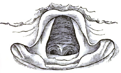
|
| Acute Tracheitis: anterior wall. |
| FIG. 26. |
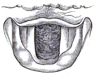
|
| Same case as Fig. 25: posterior wall. |
PROGNOSIS.—Simple tracheitis, though occasionally not without danger in extremely young and very old patients, rarely if ever destroys life. Under good hygienic circumstances it frequently gets well of itself, and it does not usually produce sufficient swelling or hypertrophy to cause stenosis. It is, however, when severe, an annoying disease, apt to recur, and, unless properly managed, difficult to eradicate.
TREATMENT.—Tracheitis is treated very much like bronchitis confined to the larger tubes, only that local measures are more prominently applicable, especially in chronic cases. Frequently, when acute, the disease may be arrested by a Dover's powder, a warm bath, and a diaphoretic drink at night, with hygienic attention, regulation of systemic functions, and soothing applications, such as inhaling simply vapor of water or medicated water, or using warm-water poultices externally. Expectorant mixtures, containing ipecacuanha, sanguinaria, squills, or senega, may be given, according to the age and condition of the patient, with matico and the like, when the secretion is abundant, and with ammonium acetate or sodium bromide (potassium carbonate or ammonium carbonate where there is depression) or tincture of aconite (especially when fever is present), or a very minute quantity of tincture of veratrum viride, when there is much dryness. Inhaling the steam arising from a pint of hot water (160–170° F.) containing 10 grs. of extract of conium, 1 drachm of compound tincture of benzoin, and half a drachm of ammonium sesquicarbonate, or inhaling nebulized solution of potassium bromide, 10 to 20 grains to the ounce, or fumes of evolving ammonium chloride or of nitre-paper, is very serviceable, as well as placing a mustard plaster or a hot poultice on the upper part of the chest (not directly over the windpipe) and on the back of the neck or between the shoulders. Some patients require for several days to take daily from 8 to 10 grains of quinia sulphate, then a smaller quantity, care being taken not to discontinue the remedy suddenly. Smoking eucalyptus-leaves, with much inhalation of the smoke, is useful in protracted cases. In chronic as well as acute tracheitis not only balsamic, anodyne, and astringent inhalations either of vapors, or of liquids nebulized by the various spray-producers are in vogue, but also insufflations of powders, injections of liquids, and touchings with the sponge or cotton-wad probang or tracheal applicator. Powders should never or only rarely (as, e.g., morphia, 1/16–1/8 of a grain, when the cough is troublesome, etc.) be blown into the trachea; injections and touchings should be made use of only after the operator has acquired the necessary skill to apply them by means of the mirror. A few drops of a solution of silver nitrate, varying in strength inversely as the chronicity of the case from 5 grains to 60 to the ounce of water, thus accurately applied at proper intervals of time, have proved successful in otherwise intractable cases. In chronic tracheitis general tonic treatment must be combined with the local, and attention be paid to possible coexistent cardiac and [p. 136] broncho-pulmonary affections or other morbid conditions. In some cases it is advisable to administer potassium iodide; in rheumatism, sodium salicylate; in gout, colchicum. The utility of producing alkalinity of the blood (as by giving alkaline mineral waters to drink, etc.) has received a new and direct support by Rossbach's recent observations of diminution of the blood-supply and of the secretion in the tracheal mucous membrane of cats whose blood was made alkaline by injecting sodium carbonate into the femoral vein.
Patients subject to tracheitis should observe all the precautionary measures of so-called bronchitics as to sponging, bathing, and friction of the body, wearing a respirator, clothing, exercise, habits, etc.
Under this heading are here classed together all inflammatory conditions of the windpipe differing from simple or catarrhal tracheitis. In these, other tissues may be affected as well as the mucous membrane. In exanthematous, erysipelatous, and exudative tracheitis the mucous membrane is prominently involved; in oedematous and phlegmonous tracheitis, the submucous connective tissue; and in perichondritic and chondritic tracheitis, the cartilages and their investing membrane. The latter forms are connected with suppurative and ulcerative processes, and, unless traumatic, almost never occur, except in phthisical and syphilitic tracheitis. I shall speak of them under the head of Ulceration.
The tracheitis of measles and scarlatina consists in an acute catarrh, with sometimes considerable desquamation of epithelium, erosion, and capillary hemorrhage. In cases of small-pox in which the larynx is affected, the same disease may extend into the trachea, varying in severity from a congestion of the mucous membrane to an intense pustular process. Erysipelas of the larynx may also involve the windpipe, and when it does is exceedingly dangerous. More than half a century ago Gibson observed in an epidemic of erysipelas that when it spread to the trachea it generally proved fatal.1 Tracheal oedema is extremely rare even when the larynx is oedematous. Phlegmonous inflammation and abscess have been observed in a few instances. Tracheal diphtheria is usually an extension of diphtherial disease of the larynx. Without entering into a discussion of the nature and cause of diphtheria, as either a local or general disease, it is here sufficient to refer to the fact that while in simple inflammation of mucous membrane no fibrinous exudation takes place, certain poisonous irritations lead to the exudation of lymph which infiltrates the tissue and may form a pseudo-membranous deposit upon it: experiments have proved that ammonia, chlorine, and, certainly, bacteria, are able to produce this. In laryngo-tracheal diphtheria or croup the disease most frequently commences in the pharynx, occasionally in the larynx, and much more rarely in the trachea.
1 Transactions of the Edinburgh Medico-Chirurgical Society, vol. iii., 1828.
The treatment of each of these forms of complicated tracheitis is the same as the treatment of the corresponding form of laryngitis.
Tracheal ulcers are just as multiform as laryngeal ulcers, but far more rare. Like inflammation, they may occur by extension from above or below, [p. 137] and only those following localized morbid conditions are certain to have arisen in the trachea. Under the head of Inflammation it has been stated that simple catarrhal ulceration does occasionally occur; of this there is really no doubt, but some writers have denied it and thrown the whole subject into great confusion. It is true, however, that a tracheal ulcer has usually a so-called dyscratic base, and either is diphtherial or phthisical (tuberculous) or syphilitic or lupoid or leprous or carcinomatous, or else comes from extraneous causes; as, for instance, from traumatic ulceration or extension or perforation from neighboring abscess, etc. There are two kinds of ulcers—viz. one in which the molecular death of tissue proceeds from the surface inward, and another in which it proceeds from within to the surface. Catarrhal ulcers, as well as ulcers from decubitus after tracheotomy, from pressure of the canula, belong to the first kind; when involving only the epithelium or the epithelium and the layer immediately underneath it the name erosions is given them; and if it were true that catarrhal erosions never penetrate to the deeper structures, it would be justifiable to say that there are no catarrhal ulcers, but only erosions: they do, however, penetrate, and sometimes to great depths. In the second kind of ulcers the epithelium is at first normal or intact, and the loss of substance of underlying tissue in consequence of inflammatory processes in the mucosa, submucosa, or perichondrium affects the epithelium secondarily. This occurs whenever, from any cause, there is primarily caries of cartilage or suppuration of submucous tissue, especially in typhoid conditions, in phthisis, and in syphilis.
| FIG. 27. |
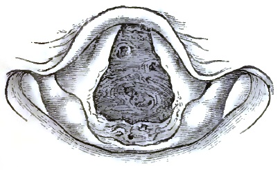
|
| Tuberculous Ulceration of the Trachea, as seen during life. |
| FIG. 28. |
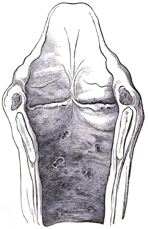
|
| Same case as Fig. 27: post-mortem appearance. |
| FIG. 29. |
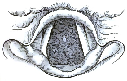
|
| Syphilitic Ulceration of Trachea, as seen during life. |
| FIG. 30. |
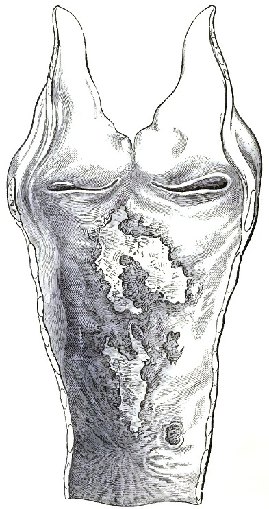
|
| Same case as Fig. 29: post-mortem appearance. |
The seat of tracheal ulcers is usually the posterior wall and the lower portion, unless the upper portion is affected by extension from the larynx or by pressure from a tracheotomy-tube. They are found also in other portions, and sometimes are so numerous that they give to the membrane a sieve-like appearance. Occasionally they denude some of the tracheal rings. In shape they vary, being mostly irregularly circular or oval, and excavated or scooped out; in size they vary from that of a pin's head to that of a marble. In tuberculosis they are generally small and numerous, have a pale background, and are occasionally confluent, while in syphilis they are usually isolated and large, very destructive, and apt to cause contractions or other deformities by [p. 139] partial or extensive cicatrization. Such contracting ray-like cicatrices have more than once produced fatal stenosis.
The SYMPTOMS are frequently obscure, but local pain and irritation are usually, purulent or muco-purulent sputa are sometimes, present. The diagnosis is difficult unless tracheoscopic examination reveals the condition. Fig. 27 shows the tracheoscopical image, and Fig. 28 the post-mortem appearance, of a case of tuberculous tracheal ulceration on the upper portion of the front wall, while Figs. 29 and 30 show the image during life and the appearance after death of a case of syphilitic ulceration. In Fig. 30 the posterior wall is seen with the ulcers, and below them a star-shaped cicatrix.
The PROGNOSIS generally depends upon the underlying disease, and is grave because the latter is. Perforation may take place, as well as cicatrization and hypertrophy, and either process may lead to a fatal issue. In a number of instances post-mortem examination has shown that tracheal ulceration may produce surprisingly great ravages before destroying life.
TREATMENT, like the prognosis, depends somewhat upon the disease underlying the ulceration. Pain is relieved by anodyne, and cicatrization promoted by alterative inhalations, as of nebulized glycerated solutions of morphine, ethereal solution of iodoform, iodinic preparations, oil of solidago, citronella oil, etc. Catarrhal ulcers heal without special treatment with the subsidence of the catarrhal inflammation. In syphilitic ulceration, stenosis from cicatrization is to be dreaded, and specific constitutional treatment is the main reliance. The internal administration of cod-liver oil has been found of service in nearly all cases of tracheal ulceration, especially in phthisis, lupus, etc. Appropriate general treatment must be combined with the local.
DEFINITION.—Tumors, benign or malignant, growing from the wall and projecting into the interior of the windpipe. Inversion of the mucous membrane forming a protrusion into the interior will be spoken of under the head of Stenosis; and tumors of other organs extending into the trachea, such as cancer of the oesophagus, lymphatic glands, thyroid body, etc., are excluded from consideration under the present head.
FREQUENCY OF OCCURRENCE.—Aside from post-tracheotomic granulation-tumors, which with careless tracheotomy or after-treatment occur often, the disproportion in the frequency of laryngeal and tracheal morbid growths is even greater than that of other laryngeal and tracheal affections. I have met with only eight instances of tracheal morbid growths, strictly so called, in a special practice during more than twenty-five years. This is exclusive of post-tracheotomic vegetations and tumors from contiguity.
ETIOLOGY.—Local irritations and chronic inflammatory conditions seem often, if not always, to be the forerunners of tracheal tumors, but the real cause of the latter is unknown. Recently it has been suggested (see the article on LARYNGEAL TUMORS) that the ever-present bacilli play a rôle in the production of morbid growths as well as in that of other diseases. As it is known that some parasitical organisms on plants use up their nidus very slowly, with the formation of peculiar excrescences, while others very rapidly destroy the tissue of their host, it would be easy to suppose that some such difference in the micro-organism causing the tumor determines its benign or malignant character.
Post-tracheotomic vegetations may arise from the irritating pressure of a [p. 140] tracheotomy-tube, especially from the use of a fenestrated tube or a tube ill fitted to the patient. Some observers are of opinion that such tumors existed before the performance of the operation, and, indeed, led to it, even though the supposed reason may have been laryngeal or some other tracheal disease. While it cannot be denied that such may have been the case sometimes, there is no doubt that in other instances—and not only in those in which the vegetations "always grow from the cicatrix" (Petel)—they are truly caused by the operation, or by the wearing of the tube, especially if it be in any way unsuitable as to size, form, etc.
SYMPTOMATOLOGY.—The symptoms of tracheal tumors are local irritation; tickling or other morbid sensation, sometimes inducing and sometimes not inducing cough; and encroachment upon the breathing-space—dyspnoea—depending on their precise seat, size, and rapidity of growth. It is usually difficult for the patient to specify the beginning of his trouble, because, on account of the large size of the windpipe, dyspnoea generally comes on very gradually. An accidental catarrhal condition of the tracheal mucous membrane from a cold usually first arrests the patient's attention. The very great diminution of the calibre of the tube that the patient can bear when the tumor enlarges slowly is sometimes astonishing. Unless the tumor is pedunculated (so that expiratory efforts can throw it up into the larynx), which is generally not the case, expiration and inspiration are equally affected, both becoming gradually more and more labored and noisy. Sometimes the act of swallowing large morsels brings on an increased dyspnoea; sometimes respiration is accompanied by a sort of valvular sound. Cough is frequently, but not always, present, and depends, together with expectoration, upon either coincidental catarrhal condition or irritation from the tumor: in the latter case it is essential, dry, and persistent, and may vary with the position of the patient. Sputum may be bloody and even contain shreds of the tumor, as in similar cases of laryngeal growth. With increase of the tumor the voice becomes weak and suffers in extent of range, as in other cases of tracheal stenosis; the same is true of the diminished rising and falling of the larynx. The course and duration of the disease vary considerably with its nature. I have observed a tracheal fibroma to remain stationary for eight years, when the patient died from other causes and the diagnosis was confirmed post-mortem; and, on the other hand, a cancer to grow so rapidly that the patient died from suffocation within five months of its first causing the slightest symptom. If not relieved, suffocatory paroxysms, with or without consequent bronchitis and pneumonia, lead to a fatal termination.
PATHOLOGY.—As in the larynx, so in the trachea, the pathological character of neoplasmata is generally that of papilloma. Of my eight cases, all observed during life, four were papillomatous (two examined microscopically after successful extirpation, one post-mortem, and one in situ macroscopically only), one was a fibroma, microscopically examined, one an osteo-chondroma, one a sarcoma, and one a carcinoma, the three last having been examined post-mortem.
Of non-malignant tracheal tumors observed by others, the large majority were papillomata; next in number come fibromata. Aside from these two kinds of tracheal tumor, the cases recorded in literature are the following: Rokitansky more than thirty years ago described tracheal enchondromata found after death; and Cohen discovered in the corpse of a phthisical patient a number of small enchondromata on the central portions of the tracheal cartilages. Steudener, Demme, Wilks, Chiara, and Eppinger have observed, post-mortem, tracheal osteomata. Gibbs has described a tracheal cystic tumor2 seen with the laryngoscope; Müller, under the guidance of Gerhardt, a myxo-adenoma observed tracheoscopically and carefully studied [p. 141]during life and after death; and Eppinger has recorded a case of post-mortem tracheal adenomata and cysts, Simon having previously found three similar tumors on dissecting a new-born tigress. Virchow speaks of the occurrence of retro-tracheal retention-cysts, and Gruber has observed several; but there can be no doubt that at least some of the tumors thus described are nothing but circumscribed dilatations of the tracheal mucous membrane—practically, dilated mucous glands. As to malignant tumors, in addition to my two cases Schrötter has reported two cases of sarcoma, and Labus one of fibro-sarcoma, while Rokitansky, Klebs, Koch, Schrötter, Langhans, and Mackenzie have described cases of carcinoma.
2 Cohen questions whether this was a cyst or an abscess. It burst spontaneously.
Cases of cancer of the oesophagus, which involve the trachea—excluded, as before stated, from present consideration—are, comparatively speaking, by no means rare, and are apt to establish a fistulous communication between the two tubes.
DIAGNOSIS.—The symptoms mentioned are those common to nearly all cases of tracheal stenosis, and will be referred to again under that head. Tracheoscopy alone makes the diagnosis certain; unless when the seat of the disease is ascertainable without, its nature is shown by the expectoration of portions of the tumor. The first case of tracheal tumor ever diagnosed during the patient's life was observed by means of the mirror by Tuerck in 1861; but it is very difficult in the mirror to estimate distances as to depth, and unless the number of tracheal rings above a tumor can distinctly be counted, a growth in the lower cavity of the larynx may readily be mistaken for one in the trachea, and vice versâ. Catheterism of the trachea shows the distance at which the tumor is situated, sometimes very accurately, but it is dangerous unless performed under the guidance of the mirror, and even then requires great care. The introduction without the mirror of a probe or sound for the same purpose is still more dangerous and unjustifiable, while with the mirror it is perfectly safe in proper hands. Localized protrusion of the mucous membrane into the interior is the condition which most simulates tracheal tumor. (Compare Fig. 32.)
| FIG. 31. |

|
| Papilloma of Trachea. |
The pathological nature of a tracheal tumor can sometimes be determined in situ with more or less probability. Without microscopical examination it is not always possible to say whether a growth is benign or malignant unless the mass has advanced to ulceration, and then specific disease must be excluded by the history and concomitant symptoms. Papillomata have a peculiarly uneven surface; fibromata are usually more smooth. With equally good illumination, tumors of the trachea resemble tumors of the larynx, and may be similarly differentiated. The former are almost always non-pedunculated, or at least none of those hitherto observed have had a long pedicle. Their seat is generally the posterior wall, or the cicatrix of the anterior wall after tracheotomy. In Fig. 31 is seen the tracheoscopic appearance of one of my cases of tracheal papilloma.
PROGNOSIS.—The prognosis is always unfavorable in malignant cases, and also in non-malignant when the tumor grows rapidly or has already attained a large size. The introduction of the laryngoscope has bettered the prognosis, inasmuch as in many cases early recognition enables us, by performing tracheotomy, to prevent sudden death from suffocation, and also because by the aid of the mirror removal has been accomplished through the natural passages.
TREATMENT.—Removal of a tracheal tumor through the natural passages [p. 142] by means of either cutting or cautery instruments requires so much special ability on the part of the operator that it need not be described in detail in a work designed for general medical practitioners. When the tumor is situated above a point at which tracheotomy can be judiciously performed, no physician worthy of the name should hesitate to lay open the trachea in any case in which suffocation is impending. Removal of the tumor by surgical operation after opening the windpipe may be attempted or not according to circumstances, but in all cases palliative measures by sedative inhalation and otherwise may be resorted to, and the patient's general health, especially in malignant cases, must be kept up as much and as long as possible.
DEFINITION AND PROXIMATE ETIOLOGY.—Stenosis is narrowing or more or less occlusion of the windpipe. It is either stricture or constriction from within, or compression from without, or both combined. Constriction within the trachea is due to swelling or thickening or cicatricial displacement of the mucous membrane or other tissue, inversion of its walls, or morbid growth or foreign body in its interior. Compression from without is due to goitre (which has in some cases prevented viability) or other disease of the thyroid body; aneurism; abscess; enlarged bronchial glands or cervical lymphatics; disease of the sternum, clavicle, or vertebræ; mediastinal tumor; cystic, emphysematous, or other tumor of neighboring tissue; or foreign body. According to Rose's observations of goitre,3 compression of the trachea leads to fatty degeneration of the cartilages and their subsequent softening and absorption; after which, the windpipe having become membranous throughout and no longer patulous, death can easily—in some positions or flexion of the body, etc.—take place.
3 Der Kropftod und die Radicalcur der Kröpfe, Berlin, 1878.
In acute tracheitis, though there is swelling of the mucous membrane, the large size of the tube usually obviates stenotic symptoms, while chronic tracheitis does occasionally lead to sufficient contraction to interfere with respiration; but generally stenosis is the result of syphilis, and frequently follows ulceration and cicatrization. In a case recorded in the Bullétin des Sciences médicales for January, 1829, the lumen of the trachea was reduced to two lines.
| FIG. 32. |
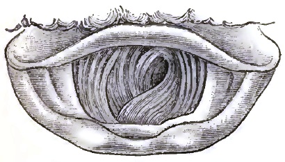
|
| Involution of Trachea, due to aneurism. |
SYMPTOMS AND DIAGNOSIS.—The main symptom is the peculiar, gradually increasing dyspnoea; once observed, it is recognized without much difficulty. There may also be mucous râles; cough rough and sibilant; attempts at clearing the throat without expectoration, or occasionally with some expectoration, which is at first light-colored, then streaked with blood, and at last purulent, but never abundant (unless accidentally complicated by catarrh), and always difficult to eject; perhaps occasional pain, but constant disagreeable sensation (tightness) in the trachea just above the sternum. Tracheoscopy settles the diagnosis. The tracheal rings are seen either as diminished circles or arcs—sometimes concentrically placed, sometimes in two different directions, as shown in a case of tracheal stenosis from [p. 143] compression causing protrusion of the mucous membrane into the interior, represented in Fig. 32, or else constricting bands are visible.
As to the dyspnoea, both inspiration and expiration are affected—frequently, however, the former more than the latter, as is shown by pneumatometry. The head is thrown forward and the chin up; the larynx moves up and down less energetically than in health (while the respiratory movements of the larynx are abnormally increased in laryngeal dyspnoea); the thorax is less expanded than normally, especially its upper portions.
As to catheterization and probing, see the remarks under the head of Morbid Growths.
PATHOLOGY.—The pathological changes in cases of stenosis vary with its cause. In the great majority of cases of stricture from within, syphilis—antecedent ulceration followed by cicatrization—has produced the stenosis; in compression thyroid disease, and next often aneurism, is the cause. The stenosis is most frequently situated in the lower, next in the upper, and least in the middle, portion; more often than the latter alone the whole tube is affected.
PROGNOSIS.—This is rather favorable with timely and proper treatment unless a continuing active cause be irremovable; without treatment, however, the cases almost invariably terminate fatally from pneumonia, tracheal spasm, apnoea as before explained, etc.
TREATMENT.—When the symptoms are urgent and the stenosis is not too low down, tracheotomy must be performed. Sometimes a very long and flexible tube may be introduced with success in case of very low stenosis, but more often tracheotomy is disappointing on account of the stenosis extending too low down even when its beginning is higher up.
Stricture, especially when the symptoms are not very urgent, may be relieved by dilatation through the natural passages, with, or if possible without, previous tracheotomy. The cure of compression implies removal of the compressing tumor or disease. Soothing inhalations, such as of hops, benzoin, etc., diminish irritation and give temporary relief.
Dilatation of the trachea is either confined to the tube (when the synonym tracheaectasy is applied to it) or is diverticular. In the former case it may involve only a part or else the whole extent of the windpipe. Whenever free respiration, especially expiration, is chronically impeded, some portion of the air-tract below the obstruction is apt to become dilated; thus, a bottle-shaped dilatation is sometimes found immediately below an annular contraction. On the other hand, tracheaectasy may extend upward from bronchiectasy. It has been observed post-mortem to a slight extent in public criers, trumpeters, etc., and in old coughers from laryngeal disease, chronic bronchitis, pulmonary emphysema, etc., but without giving rise to distinct symptoms during life.
Diverticular dilatation forms an air-containing tumor which either looks into the oesophagus or is discernible on the outside of the neck. Though rarely met with, it ought to be thought of in all appropriate cases, and when pointing externally ought always to be recognized by the careful practitioner. It is either hernial, glandular, or fistular—three pathological conditions which have hitherto been confounded. On account of the construction and position of the trachea there can be but little protrusion outward without previous [p. 144] dilatation. Unless there be a deficiency of the cartilaginous rings, only the posterior wall, which is always unsupported, and to a slight extent also the intercartilaginous membranous portions, are liable to tracheal hernia. This is properly called tracheocele; but the various terms aërial goitre, aërial bronchocele, pneumatocele, tracheal air-cyst, tracheal retention-cyst, internal tracheal fistule, subcutaneous or incomplete fistule of the trachea, have been indiscriminately used as synonyms of tracheocele, and have added all the more to the confusion, as some of them originated, no doubt, as correct appellations of the particular cases to which they were applied. Aside from the occasional occurrence, both congenital and acquired, of tracheo-cutaneous fistule, complete and incomplete, and the still more rare occurrence of hernia of entire portions of the mucous membrane, the cases of diverticular dilatation of the trachea—or saccular tracheaectasy, as it may be called—are glandular, as found by Rokitansky more than fifty years ago. Virchow seems to regard all such glandular dilatations as retention-cysts (see Morbid Growths), but although retro-tracheal retention-cysts doubtless do occur (Gruber has reported two unquestionable instances), and although the tumors now under consideration do in fact sometimes contain a little mucus in addition to air, they do not constitute cysts or adenomatous new growths, but are simply distended portions of the tracheal mucous membrane, respiratory glands, whether the dilatation be caused, as Rokitansky thought, by traction (Zerrung) and hypertrophy of the mucous glands, or, as Eppinger suggests—and which is more likely—mainly by increased intra-tracheal air-pressure. There must, however, I think, coexist some deficiency or weakness of the cartilaginous or other tissue, either congenital or acquired.
When the dilatation is retro-tracheal only, the symptoms are very obscure, and diagnosis during life is at best uncertain. In one such case under my care, confirmed (death having occurred from another cause) by post-mortem examination, there was some dysphagia and slight alteration of the voice. In all other cases the characteristic and unmistakable sign of the disease is the peculiar intermittent, or, at all events variable, aërial cervical tumor. It increases and diminishes with forcible expiration and inspiration, and attains its largest size during violent coughing, hawking, blowing of the nose, or other expiratory effort. Occasionally the voice is considerably affected. The tumor, especially by the manner in which it can be made to temporarily disappear and reappear, can usually be easily differentiated from subcutaneous emphysema and goitre, the only two conditions with which it might be confounded. In the fistular variety the opening into the trachea can sometimes be seen by means of tracheoscopy.
Aside from the deformity which the tumor may cause, it sometimes induces laryngeal spasm and dyspnoea; otherwise it is of no gravity.
As to TREATMENT, methodical and continued compression by applications of astringent collodion or by mechanical means is the only palliative measure applicable; when suffocatory attacks call for it, tracheotomy must be performed.
The operation of tracheotomy, or the artificial opening of the air-passage—using the term in its modern acceptation as including all of the five incisions that are both anatomically and surgically possible, either singly or in combination, between the lower border of the thyroid cartilage and the upper edge of the sternum (incisura jugularis sterni), and reserving the term laryngotomy to denote the division of the thyroid cartilage alone—fulfils two important and usually urgent indications: First, in allowing the respiratory current free access to the lungs in cases where the laryngeal obstruction is of such a sudden or of so progressive a character as to either immediately or remotely threaten the life of the patient; and, secondly, in affording a ready means of direct access to those portions of the air-tract which lie below the level of the glottis, and thus permit not only of the direct extraction of such foreign bodies as may accidentally have found their way within the air-passage, but of neoplasms here located and of occluding diphtheritic membranes. Catheterization and aspiration of the trachea are likewise both rendered not only possible, but easy of execution. Both general indications mentioned often coexist, and are met by the operation in a large class of cases; the first alone plays its important life-saving rôle in many.
The disease or accident which renders the operation necessary varies greatly, and upon this variation depends not only the surgeon's decision as to the precise time at which the opening into the air-tube must be made, but also the precise point at which the operation should be performed. These general questions I treat of in detail. The special indications may conveniently, but somewhat arbitrarily, be arranged as follows, in groups, which I have attempted to make complete, although some of the conditions, being purely surgical, do not strictly come within the compass of this essay:
A. Acute inflammatory diseases of the larynx and trachea:
1. Acute oedema of the larynx.
2. Erysipelatous and exanthematous laryngitis.
3. Acute perichondritis, with abscess.
4. Diphtheritic croup.
B. Chronic affections of the larynx and trachea:
1. Syphilitic laryngitis.
2. Phthisical laryngitis.
3. Chorditis vocalis inferior hypertrophica.
4. Carcinoma of the larynx or trachea.
5. Non-malignant growths of the larynx or trachea.
6. Tumors overlying the superior aperture of the larynx.
7. External compression of the trachea by tumors of the neck or chest.
8. Strictures of the larynx or trachea.
C. Neurotic diseases:
1. Paralysis of the abductors of the vocal cords.
2. Spasm of the adductors of the vocal cords. [p. 146]
D. Traumatic conditions:
1. Foreign bodies in the larynx or trachea.
2. Impaction of foreign bodies in the pharynx or oesophagus.
3. Fracture of the larynx. Rupture of the trachea.
4. Scalds and burns of the larynx.
5. Incised and gunshot wounds of the throat.
6. Poisonous bites inflicted by certain insects about the mouth or neck.
7. Suffocation from the passage of blood, fluids, etc. into the air-passages (tracheotomy, with aspiration of the windpipe and artificial respiration).
8. Suffocation from the acute collection of either mucus or serum in the bronchia (ditto).
9. Suffocation from the inhalation or development of poisonous gases (tracheotomy, with artificial respiration).
Finally, although it pertains alone to the province of the surgeon, I may allude to the temporary tracheotomy and "tamponing of the trachea" which has been recommended—and certainly found efficient—in preventing the entrance of blood to a dangerous degree into the lower trachea and lungs during the performance of certain operations in the neighborhood of or upon the air-passages, such as resection of the upper jaw, the extirpation of large nasal and naso-pharyngeal polypi, removal of the tongue, subhyoidean pharyngotomy, laryngotomy, and extirpation of the larynx.1
1 For the details of this procedure consult Schüller, Die Tracheotomie, etc., Stuttgart, 1880.
All-important as a preliminary to the operation itself is a thorough knowledge of the surgical anatomy of the region upon which it is proposed to operate; and this not alone in the adult, but especially in the child, where essential differences often exist. Possible anomalies also are not to be forgotten.2 The assurance of the surgeon depends upon this knowledge: mere, manual skill will not compensate for its want; the success, both immediate and remote, of the operation is in great measure the reward of its possession.
2 See Pilcher, "The Anatomy of the Anterior Median Region of the Neck," Ann. of Anat. and Surgery, Brooklyn, April, 1881.
It will be remembered that the trachea commences at the inferior border of the cricoid cartilage, directly opposite to the lower edge of the fifth cervical vertebra, and reaches thence downward, in the median line of the neck, until it bifurcates opposite to the third dorsal vertebra. In its upper part it is nearly subcutaneous, and is surmounted by the prominent ring of the cricoid cartilage (easily identified, even in the young child), above which, in turn, lies a slight depression (the crico-thyroid space) between the cricoid and thyroid cartilages. As the trachea descends in the neck it recedes gradually, lying at the episternal notch about one and three-eighths of an inch from the surface. Throughout the whole of this course it is in relation with important structures. In its cervical portion it is covered by the sterno-hyoid and sterno-thyroid muscles, and in the median space, which is usually distinct between them, by layers of the deep cervical fascia. It is also crossed by the isthmus of the thyroid gland, which lies between the second and fourth tracheal rings; by the arteria-thyroidea ima, when present, and below by the plexus formed of inferior thyroid veins with their tributary and communicating branches. In the latter region, but more superficially, are some communicating branches between the anterior jugular veins. The innominate and left carotid arteries are also anterior to it in the episternal notch as they diverge from their origin. Laterally, the trachea is in relation with the common carotid artery, the lateral lobes of the thyroid body, the inferior thyroid veins, and the recurrent laryngeal nerves. The thoracic portion of the trachea is covered by the manubrium sterni, with the origins of the sterno-hyoid and [p. 147]sterno-thyroid muscles, by the left innominate vein, and by the commencement of the innominate and left carotid arteries. Still lower, the transverse portion of the arch of the aorta crosses, and the deep cardiac plexus of nerves lies in front of it. Posteriorly, throughout its length, it rests upon the oesophagus.
In performing, then, either the superior or inferior operation of tracheotomy, after cutting through the skin and superficial cervical fascia—which is really loose areolar tissue containing fat—the superficial layer of the deep cervical fascia is reached, and immediately below it more or less adipose tissue and the two anterior jugular veins lying in an inferior tracheotomy to either side of the wound, which is always made in the median line. As a matter of fact, these various layers are rarely demonstrable, and the surgeon proceeds irrespective of them until he reaches this point in his operation—viz. the muscles which overlie the trachea. These may overlap in the median line, and have to be retracted after having been separated; or, again, a thin line of connective tissue marks a slight interval between their inner edges, and is readily seen and dissected through if the operator has kept his incision vertical and strictly in the median line of the neck—a matter so important to the success of his operation that I do not hesitate to again allude to it. The muscles separated and gently retracted, together with the overlying tissues, toward the sides of the wound, the upper edge of the isthmus of the thyroid gland overlying the second and third, perhaps fourth, rings of the trachea, is always seen in a superior tracheotomy—its lower edge very frequently in the inferior operation. The isthmus is adherent to the trachea and to the larynx through the deep layer of the deep cervical fascia, but is capable of being slightly displaced or pushed upward or downward as the case may be, and thus kept from obscuring the operative field. This being done, the deep layer of the deep cervical fascia is seen covering and strongly adherent to the tracheal wall together with the thyroid veins. A few touches of the knife, carefully avoiding the blood-vessels, serve to clear it away, and the tracheal rings are clearly exposed.
In carrying out this dissection, which has been described as occurring in an ordinary and uncomplicated adult case, several matters must be borne in mind; and especially is this true if the operation concerns infants. In them, for instance, the thymus gland rises half an inch above the level of the sternum, and is frequently to be found as late as the sixth or seventh year. In both adults and children the innominate artery occasionally comes into view in an inferior tracheotomy, obliquely crossing the lower portion of the right half of the trachea. It is relatively higher in the child than in the adult. The left innominate vein is also often observed when the trachea is opened low down.
Certain abnormalities of the blood-vessels have been alluded to above. The commonest consists in the existence of a thyroidea ima artery, which when present usually arises from the innominate trunk, but sometimes from the right common carotid or the aorta: it passes to the thyroid body directly in the median line of the neck and close to the trachea; again, the place of the anterior jugular veins may be taken by a single central vessel, almost sure to be wounded during the operation if it exist (Mackenzie).
In performing the operation through the thyro-cricoid membrane (thyro-cricotomy) or through the cricoid cartilage alone (cricotomy), the same tissues are met with, and the same dissection is necessary in the earlier stage of the operation, as have been described in the operation of superior or inferior tracheotomy; but the parts are more superficial, adipose and cellular tissue less abundant, blood-vessels much less numerous, and the operation very much simpler. The thyroid gland of course does not come into view, [p. 148]and the crico-thyroid artery, a very small vessel, needs no attention in the dissection.
I have here and elsewhere included under the general term tracheotomy five distinct operations, having for their object the opening of the air-passages, which are surgically possible between the lower border of the thyroid cartilage and the upper edge of the sternum. In this classification I have followed that of Schüller, and its simplicity, but exactness, and the avoidance of the old confusion of different terms which results from the use of one intelligently employed, seem to me to commend it. These five operations are—1. Thyro-cricotomy, or the opening made through the crico-thyroid membrane alone. 2. Cricotomy, or the division of the cricoid cartilage alone. 3. Superior tracheotomy, the incision being made above the point where the isthmus of the thyroid gland crosses the trachea and below the cricoid cartilage. 4. Median tracheotomy, when, the isthmus being displaced or torn through, the trachea is opened immediately below its site. And 5. Inferior tracheotomy, the incision being made below the point of crossing of the isthmus of the thyroid gland, and at varying distances, dependent mainly upon the age of the patient and size of the parts, above the sternal notch.
Rarely, I am bound to admit, is the field of all of these operations as distinctly limited in practice as is here indicated, and one, perhaps two, are rarely selected. Thyro-cricotomy (old term laryngotomy) is often indicated, and cricotomy and median tracheotomy are sometimes performed as here described. Superior tracheotomy is commonly a combination of at least two of the methods—viz. the division of the upper rings of the trachea and the cricoid cartilage as well. It may even, probably frequently does, trench also upon the thyro-cricoid membrane (thyro-cricotomy) and upon the field of a median tracheotomy, the isthmus being pushed downward or even cut or torn through. The latter operation and cricotomy are, I believe, rarely if ever done from choice. Finally, inferior tracheotomy is a common method. As here described, it meets a large number of indications, and, despite its superior difficulties over the higher operations, is therefore necessarily often chosen; not infrequently, however, does it invade the median region, the isthmus of the thyroid being pushed upward.
Which of these operations shall be selected in a given case depends upon the particular conditions which render it necessary, and likewise, to some extent, upon the age of the patient. Durham summarizes the question very fairly. Thyro-cricotomy (old term laryngotomy) is by far the easiest operation to perform, and its execution is attended by least risk; therefore it is the operation to be preferred in any sudden emergency when suffocation threatens, and especially where the surgeon is alone with the patient. Generally, it is not as applicable as the others, especially in early childhood, on account of the limited dimensions of the thyro-cricoid space. It cannot be recommended in cases of acute or extensive diseases or injuries of the larynx, nor is it likely to be of much service if a foreign body is in the trachea or bronchus. On the other hand, it is probably the best operation to adopt in cases in which foreign bodies are impacted in the larynx, in cases of limited chronic disease or contractions of the superior laryngeal parts—usually the result of syphilitic ulceration—and in cases in which respiration is impeded by intra-laryngeal growths which cannot be removed by the natural passages.
Cricotomy, combined with superior tracheotomy (old term laryngo-tracheotomy), is not a difficult operation, and may be advantageously practised, especially in children; in the adult it meets many indications. Holmes recommends it the more urgently, in preference to an inferior tracheotomy, the earlier the age of the subject may be.
Inferior tracheotomy is comparatively difficult to perform, and during its performance dangers may have to be encountered greater and more numerous [p. 149] than those met with in either of the other operations. This is true certainly of children. As regards young children, Holmes states that after the age of five or thereabouts the surgeon can, if he prefer it, open the trachea below the isthmus of the thyroid gland. He himself does not recommend the operation before puberty. In the case, however, of a foreign body loose in the windpipe of a child, where a large opening is required, it can hardly be obtained above the thyroid body and below the cricoid. To cut through the isthmus of the thyroid (median tracheotomy) is, in early life at least, a doubtful proceeding when it is of large size, on account of its vascularity, and the incision must be made below it—in other words, an inferior tracheotomy.
When the operation of tracheotomy shall be performed is a question which the experience and individual views of the surgeon, based on experience, must decide in each case. The doubt always arises in the mind of the inexperienced operator whether the symptoms are sufficiently urgent to render the operation necessary. To him these general rules may be given: The immediate indication for the operation is to be looked for in the thorax. It is the recession of the lower part of the sternum and contiguous ribs and the retraction of the intercostal spaces and clavicular fossæ at each act of inspiration. He must not wait until lividity of the lips and blueness of the fingernails prove that the blood is being imperfectly oxygenated (Mackenzie). Let him remember also that, aside from the immediate and imminent danger of sudden suffocation, a remote one exists and increases the longer he postpones his operation and allows the struggle for air to continue—viz. vascular engorgement and oedema of the lungs, especially in young children; the production of all those conditions which allow, and even predispose, the lung after the operation to fall an easy prey to the inflammatory processes.
The instruments necessary for the performance of the operation of tracheotomy are few and simple, and are such as may ordinarily be found in any small operating-case. A scalpel, a probe and sharp-pointed bistoury, dissecting and artery forceps, a tenaculum, a grooved director, two small retractors, scissors, and a dilator for the tracheal wound, are necessary. To these may be added the needles and thread, waxed ligatures, sponges, and tape. The tracheal tube is elsewhere described. A faradic battery, good suction syringe, and a large flexible catheter may render good and timely service if at hand.
It is true that many other and more or less complicated instruments have been devised for the purpose of facilitating the operation; and other methods, aside from that of the knife, have come of recent years into vogue; but, still, simplest means, as above given, have in the experience of most surgeons been proven to be the best. This statement, undeniably true for all surgical measures, is especially so for the operation under consideration, which is often necessarily undertaken without opportunity for elaborate preparation and under the most adverse and inconvenient circumstances. The more familiar, therefore, the surgeon is with his instruments, the better and more certain will be his work.
Holding this view, it is unnecessary for me to more than briefly mention such instrumental aids as the grooved tenaculum of Chassaignac, the groove serving to guide the operator's knife into the trachea; the sharp double hooks of Langenbeck, which, after being caught in the tracheal walls to either side of the site of the intended incision, are sprung apart after the latter is made, thus dilating the wound and rendering the introduction of the tube easy; the tracheotome of Thompson, a pair of curved cutting forceps, the blades of which are caused to open by a screw after they have been plunged through the tracheal walls; that of Garin, a forceps with curved blades—one, the longest and sharpest-pointed, being made to penetrate the trachea, the instrument then opened, and both blades cut their way to the desired extent of [p. 150] incision; finally, the tracheotome of Maisonneuve, a curved dilating hook with cutting inner edges. Its point is entered between the first and second rings of the trachea and brought out again between the fourth and fifth; the handle is then carried under the chin, so that the blades are made to cut through the trachea and the skin between the points of insertion and exit, after which, upon pushing a spring, the two halves of the hook separate, and the canula is introduced between them (Thornton). And the trachea-stretcher of Marshall Hall, by means of which a portion of the trachea is cut out and the opening kept patent.
None of these instruments have been proven to possess any practical worth; on the contrary, their use, especially that of the latter forms, has in more than one instance been attended with disastrous results.
To obviate the danger of serious hemorrhage during the performance of tracheotomy, both the galvano-cautery knife and the thermo-cautery instrument of Paquelin have been recommended within the past few years, and a number of operations placed upon record. The procedure is the same whichever means be used. The skin and soft parts overlying the trachea are usually alone cut through by means of the cautery-knife, the cartilaginous rings of the tube, when reached, being divided with the ordinary knife. This fact alone speaks against the thoroughness attainable by means of these methods; but, still more important, neither has been found reliable in checking hemorrhage, and in several instances the operator has been obliged in haste to lay aside his cautery apparatus and turn to the ordinary and better-known means to complete his operation. The healing of the tracheal wound made by the cautery is slow: erysipelatous inflammation may attack the wound as the result of the burn, and extensive sloughing of the edges is not unknown, while the resulting cicatrix is large, strong, and contractile, and has caused, in one case at least, a stenosis of the trachea. In the face of these facts he must indeed be an enthusiastic advocate who would recommend the procedure. Mackenzie justly remarks that the use of the thermo-cautery for opening the air-passage merely introduces an unnecessary complication into the operation.
The choice of a proper tube, one suited to meet the special indications in a given case and specially adapted to the age of the patient and the calibre and position of his trachea, is no unimportant matter, and may do much not only to facilitate the immediate success of the operation, but likewise prevent the occurrence of those possible unfortunate results, ulceration, fatal hemorrhage, abscess, pneumonia, and pyæmia, no lack of which are recorded in our literature.
Although the number and variety of mechanical devices and forms of tracheal tubes that have from time to time been devised by the inventive ingenuity of operators is large, the choice practically centres upon one of two forms. The first, and the one most commonly used, is but the original canula of Trousseau, modified by Roger, in that the tracheal portion of the tube is detached from the collar or neck-piece, and moves freely with the movements of the patient; and by Obré, by the important device of an inner tube to prevent clogging of the outer or original tube by mucus. Starting upon this essential basis, the instrument-maker has perfected the instrument of to-day. It is a silver tube, double throughout, the inner tube projecting at the lower or tracheal end beyond the outer—an important point, as it prevents any possible permanent occlusion by mucus or blood-crusts, membranes, and the like at this point, removal of the inner tube at once clearing the end of the outer one. The curve of both tubes should correspond to the arc of a quadrant, and the outer is fastened to a transverse collar or shield by means of two small projections or pins upon its sides which lie under small wire bridges upon the shield after it has passed through an opening in the [p. 151]transverse neck-collar large enough to permit of its free movement during the respiratory movements of the trachea, as well as during the forcible action caused by cough. The ends of this collar or shield curve slightly backward to correspond with the curve of the neck, and are perforated by, preferably, large oval openings, instead of the usual small, inconvenient slit, through which the tapes are passed which hold the tube in position by encircling the neck. To this same shield is fastened, by means of a small turn-screw or a revolving collar, the end of the inner tube, which is thus prevented from being forced out of the outer tube by coughing or any motion of the patient. Upon the upper or convex surface of the outer tube a small ovoid opening is usually made for the purpose of permitting the expiratory current to pass upward (the inner tube being removed) into the larynx and render phonation possible; also, the free opening of the outer tube being closed, to allow of respiration being carried on through the larynx and natural passages—often an important matter, as the case progresses toward recovery, in instances where the operation of tracheotomy has been performed on account of laryngeal obstruction.
A set of these tubes, which can now be readily obtained, should consist of four, with the following diameters: No. 1, one centimeter; No. 2, nine millimeters; No. 3, seven millimeters; No. 4, five millimeters: their length is of course in relative and fixed proportion to these measurements. A tube should always be selected less in diameter than the trachea operated upon: to seek to introduce one of the same calibre is not only unnecessary, but cannot fail to be dangerous. Tubes constructed upon the same principles as that just described (Lüer's) are made of hard rubber instead of silver (Leiter): their lessened cost is their principal recommendation, added to the one that they are more easily kept clean and sweet than the silver tubes. The fact that they are necessarily made much heavier and thicker than the latter is a disadvantage, the lumen of a hard-rubber tube being smaller than that of a silver tube of corresponding external diameter. The objection urged against them, of their great danger of breakage, I have not found borne out by experience. Tracheal tubes are also constructed of platinum, and recommend themselves on the score of lightness.
The main objection to any of the forms of tube just described exists in the nature and shape of their curve, which not infrequently causes the lower or tracheal end to lie in contact with the anterior tracheal wall, or its convexity with the posterior, and irritate, even ulcerate, them. This misfortune is entirely obviated by the canula of Durham, the second of the two forms to which I have called special attention, and which is essentially a right-angled tube, made of four sizes, with a long horizontal portion, varying from 7 to 4 centimeters, and short vertical portion, of from ½ to ¾ of an inch in length and slanting slightly backward. The former portion is capable of being lengthened or shortened in any sized tube by means of a screw arrangement attached to it as it passes through the usual neck-collar or shield; and the vertical tube can thus be correctly adapted to the particular depth at which the trachea naturally lies in a given case from the surface; and not alone this, but also to the condition of the overlying parts, whether thin or fat, swollen or otherwise. Once in position, the vertical portion of the tube remains in the long axis of the trachea, and does not touch its walls to any injurious degree. Owing to its right-angled shape, the angular and descending portions of the inner tube of this canula are necessarily made upon the lobster-tail principle, with joints—a possible disadvantage, as they can become clogged with mucus and may become detached. Other modifications and improvements exist in this Durham canula over the older one first described, which add to its utility, but need not here be dwelt upon. Suffice it to say that the tube is an excellent one for its purpose, and is deservedly highly [p. 152]spoken of and recommended by those who have had experience in its use. Its cost is an objection.
The other forms of tracheal tube need but passing mention. The bivalve canula of Fuller is made in two lateral segments, fastened to a collar and tapering when closed to a point, so that introduction of the apparatus through the tracheal wound is made easy. Once introduced, an inner complete canula is slid into its place, thus separating the two outer halves and rendering the whole round and compact. It has been criticised unfavorably on account of the danger of hemorrhage that it is likely to cause through pressure on the tracheal walls by the sharp edges of the outer canula. In Gendron's canula the same lateral blades are separated after introduction by means of a screw fastened on a transverse bar.
Soft-rubber canulas were introduced to the profession not long since by Morrant Baker for subsequent use after the operation of tracheotomy, the usual tube having been worn meanwhile for a few days. Being soft and flexible, they are certainly safe and comfortable for the patient, but their thickness and the absence of any inner tube are, especially the latter, serious disadvantages. They are not, I believe, generally used. Finally, the long, flexible tracheal tube of König was devised by its author to meet the indications in cases where the trachea is compressed from without by tumors, and where a long canula that is flexible, but at the same time rigid enough to resist pressure, becomes a necessity. It is made in the form of the ordinary tracheal canula, only larger, some three or more inches of the centre of the descending portion of the tube being constructed of spirally-twisted silver wire.
It may not be out of place to remind at this point that a tracheotomy is not infrequently performed, of necessity, very hastily, and in the absence not only of a tracheal tube, but likewise of other and even more essential instruments. The lack of the former need never be a barrier to the prompt performance of the operation, for the ready wit of the true surgeon will show him various ways out of his temporary difficulty. A thick goosequill fastened by threads passed through its outer end makes an efficient improvised canula. A bit of elastic catheter answers the same purpose. Retractors for the edges of the tracheal wound, made of wire—silver if it be at hand, a couple of hairpins if it be not—and connected together by an elastic tape which passes around the neck, will not only answer a good temporary purpose in holding the tracheal wound dilated, but have been recommended by Martin—in a more elegant form, it is true—as a proper method of treatment after opening the trachea. Finally, one or more stitches passed through the cartilaginous edges of the wound, and attached to the soft parts beyond it, will serve to secure its patency, at least temporarily.
If a patient be doomed to wear a tube constantly in his trachea, the instrument described above can be removed at a suitable interval after the operation and its place supplied by a single tube of the same size and form as has been found adapted in the case. In the convexity of this permanent tube an ovoid opening should be made to allow of the passage to the larynx of the respiratory current, in part at least, and to its mouth a pea-valve may be fitted which shall admit air on inspiration, and not allow it to escape on expiration, thus doing away with the necessity of the patient's closing the opening of his tube with his finger each time that he requires to speak. Several forms of these valves have been devised, but practically they are of little use, are annoying to the patients, and, as a rule, not tolerated by them.
How shall the operation of tracheotomy be performed? An answer to this question necessitates a short description of the operative steps of the different procedures that is given in the order in which, I believe, the operations are, as a matter of experience, found to occur in practice—viz. 1st, superior [p. 153]tracheotomy, combined or not with cricotomy; 2d, thyro-cricotomy and, 3d, inferior tracheotomy. Certain preliminaries are common to all.
The patient should be extended upon a table covered with one or two thicknesses of blanket and of suitable height, which has been placed sideways in front of a window if the operation is done by daylight. (At night several candles tied together afford a better and safer light than a kerosene or oil lamp.) The surgeon stands at the right side of his patient and facing the window. Of his two assistants—and the value of trained assistance in this operation is inestimable—one faces him, without obscuring the light, and is prepared to use the sponges, hand the instruments, manipulate the retractors, and render such direct assistance as may be required. The second sits at the head of the table and holds the head of the patient steadily, the neck being well extended and thrown backward over a small round pillow (or, better, a wine-bottle wrapped in a towel) which has been placed beneath it. The head must be held directly in the median line of the patient's body, and even in that of the operating-table. The assistant's attention must never waver from this important duty. In certain cases too great inclination of the head backward serves to increase the urgent dyspnoea, or even to check respiratory efforts. This effect he must watch for, and be prepared to relieve instantly by raising the head. His duties also include the preliminary administration of an anæsthetic, and its use during the operation if required. That such use is safe in this class of operations is now generally admitted, but it is not always necessary. The operation is not an exceedingly painful one, and I have often performed it, with the adult patient's consent, without using any anæsthetic (sometimes freezing the skin over the site of the incision before making it), he submitting rather than undergo any addition to the sense of urgent dyspnoea from which he is already suffering. In children anæsthetics—ether being more commonly employed, although chloroform is often used—are much more necessary, often indispensable. Their effects are speedily manifested when asphyxia is present in any marked degree, and but little of the vapor need be inhaled. The administration, always to be carefully watched and profound anæsthesia avoided, renders breathing easier in many instances, certainly lessens laryngeal spasm, and may be discontinued early in the operation when the air-tube is or has been nearly reached by dissection. Any slight risk attending their use is more than outweighed by the safety and precision which they ensure in the more difficult and delicate steps of the operation (Sands). If the patient be already insensible or if death be imminent, their use, of course, is contraindicated.
The operator having previously decided which operation he will perform, and after carefully identifying the position of the various parts, the larynx especially, marking them with ink upon the skin if he chooses, now steadies the loose skin over the site of his intended incision, and then makes it, freely, firmly, cleanly, and exactly in the median line. If it be for a superior tracheotomy, combined or not with cricotomy, the operation I shall first describe, it must extend from just at the notch of the thyroid cartilage downward for about four inches. A free external incision is very desirable in all cases. The subcutaneous tissue now rapidly dissected through by the careful use of the knife, the veins as met with either being pushed to one side or, if they cross the line of incision, cut if small, then twisted or immediately ligated, or if large doubly ligated and then cut between the ligatures, the interval between the sterno-hyoid muscles is sought for and found, then separated by the blade or handle of the knife and held apart by retractors at the side of the wound. It is important that the faint whitish line of connective tissue which marks the interval between the muscles be recognized, otherwise it happens that the operator passes through the body of one of them, deviates at once from the median line, and approaches the side of the trachea [p. 154]instead of the front. The ring of the cricoid cartilage above and the upper edge of the isthmus of the thyroid gland below can now be either seen or felt by the finger in the wound between them; and about the latter lies more or less connective tissue and numerous small veins. As a rule, careful touches of the point of the knife, or, as some operators prefer at this stage, its handle or the use of a blunt director, serves to dissect up piecemeal or tear through and clear this away, the veins again being pushed out of the way, or if necessary cut and tied, and all parts held aside by removing and replacing freshly the retractors from time to time as the dissection proceeds, until the ring of the cricoid and the upper rings of the trachea come plainly into view; that is, are seen, not alone felt. During this dissection, especially if the handle of the scalpel be used, too much pressure must not be made upon the trachea. More than once I have known it to cause sudden suspension of the respiration, probably by exciting reflex spasm of the larynx. If the isthmus of the thyroid gland extend far upward, it must be pressed downward, its facial attachments to the cricoid and trachea cut or torn through, and may require to be held downward in the lower angle of the wound by an additional retractor. The upper rings of the trachea having been thus well cleared of their overlying parts, the next step of the operation follows. I am in the habit of now removing the retractors and allowing the trachea, which may have become displaced by them, to resume its normal position, the head of the patient being meanwhile readjusted. All this takes but a few seconds. A tenaculum is then implanted in the median line, either just below the edge of the thyroid or the cricoid cartilage, if the latter is not to be severed, and held firmly by the assistant at the head of the table, thus steadying and elevating slightly the trachea and rendering the incision into it certain. The retractors are now reintroduced at the sides of the wound, and the operative field is clear and steady. A glance having shown that all bleeding has ceased, another that the tracheal dilator and tracheotomy-tube lie ready at hand, the operator plunges a straight-pointed bistoury through the tracheal wall at the level of the third or fourth ring in the median line, and cuts quickly upward until the cricoid cartilage is reached, if he proposes, as in the adult can usually be done, to limit his operation to a superior tracheotomy. If not, as in the child, and the cricoid cartilage must be cut through to gain sufficient space for the introduction of the tube, it also is severed by prolonging the incision upward to the thyro-cricoid membrane. A hissing of escaping air, with the bubbling of a little blood and paroxysms of cough, follows the incision and shows that the trachea has been fairly opened. The tracheal dilator is now introduced, the lips of the tracheal wound separated, and the canula slipped neatly into the windpipe (unless in the case of a foreign body), and secured a moment or two later, when respiration is fairly established, by tapes passing around the neck. The tenaculum and retractors are removed at the same moment that the tube is slipped into place.
Many different methods have been recommended for the dilatation of the tracheal wound and to assist the introduction of the canula. The dilator (Trousseau) which has been mentioned surely answers all purposes, and is simple and easily used. An ordinary dressing forceps will likewise do the work if introduced closed and afterward opened. More complicated procedures are unnecessary.
Thyro-cricotomy requires that the superficial incision be so made over the larynx that the thyro-cricoid space shall lie in the centre of one, about two inches long, made in the median line. Following now the dissection just described, the thyro-cricoid membrane is easily reached and quickly seen as soon as the sterno-hyoid muscles are retracted. It should then be divided transversely close below the lower edge of the thyroid cartilage, the wound dilated, and the tracheotomy-tube slipped into place.
[p. 155] Inferior tracheotomy demands that the external incision be free. In children, and in adults with a short neck, it should extend from the cricoid cartilage to just above the sternum. The subsequent steps of the operation are as for superior tracheotomy, with but slight differences. The anterior jugular veins may come into view, but can generally be avoided. If they are joined by a transverse branch, this is necessarily cut through after being doubly ligated. After the thyro-hyoid muscles are separated, the rings of the trachea are much less distinctly felt at first than in superior tracheotomy, being covered by more connective tissue and numerous veins. These inferior thyroid veins, especially if large, are the great obstacle in the way of this operation, and much care is necessary in order to avoid them, which should be done if possible. The lower edge of the isthmus of the thyroid gland, which presents to a variable extent above in the wound, does not, as a rule, offer any obstruction. The thymus gland present in infants is easily pulled downward and out of the way. The trachea at length fairly exposed and all bleeding controlled, the left fore finger of the operator is placed in the lower angle of the wound to securely protect the large blood-vessels here located, and the incision made through some three tracheal rings from below upward.
It may happen that in either a superior or inferior tracheotomy no time will be allowed for careful and slow dissection as here described. In such instances Durham advises that the surgeon grasp the trachea between the fore finger of his left hand on the left side and the thumb on the right, and make uniform, steady, deep pressure, thus firmly securing it and at the same time protecting the large vessels of the neck. The fingers thus placed are not to be moved until the trachea is reached, which is accomplished by rapid incisions confidently made. The pressure of the fingers causes the wound to gape and the trachea to advance. The latter reached, it is caught by the tenaculum and the operation completed as before described.
The operation of median tracheotomy may require a word. As has been stated, that part of the trachea covered by the isthmus of the thyroid gland is very commonly encroached upon in performing either or both superior and inferior tracheotomy, the isthmus being slightly displaced from its site. Other than this the site here mentioned would rarely be selected as the point for opening the trachea. Certain conditions, it is true, might render it necessary, but they would be rare. The danger lies in the hemorrhage which, theoretically at least, is to be expected when the isthmus of the thyroid gland is either torn or cut through; but opinions vary very greatly as regards this danger. With a thin, narrow isthmus in children I have frequently, in performing superior tracheotomy, cut my way through to a sufficient extent to clear a suitable space upon the trachea through which to introduce a tube without difficulty or danger. I should not recommend the procedure, however, were the isthmus to be seen to be, when reached, thick, wide, and exceedingly vascular, but at the same time believe that the danger even here of cutting into it is much overestimated.3 Roser's recommendation to apply a ligature to the isthmus on either side of the median line previous to its division is not generally applicable. Hueter has shown that the fibrous capsule of the thyroid gland enclosing it and its blood-vessels is firmly attached to the trachea and sides of the larynx, and that from the isthmus this fascia extends upward over the larynx (fascia laryngo-thyroidea), and thus prevents, in a measure, attempts at displacing the gland downward. Bose4 recommends that this fascia be divided transversely over the anterior convexity of the cricoid cartilage, when a director can be passed behind the isthmus, to lift it from the trachea and depress it far enough to expose three or four of the [p. 156]rings: the capsule of the gland thus remains unbroken and no hemorrhage occurs. The procedure certainly merits trial; twice it has succeeded well in my hands.
3 See Foulis, "Some Points on Tracheotomy," Glasgow Med. Journ., vol. xv. No. 2, p. 123.
4 Archiv für klin. Chirurgie, vol. xiv. p. 137.
Cricotomy, the division of the cricoid cartilage alone, is an operation which, as far as I am aware, is rarely ever performed. The objection urged against it, however, that in the adult the elasticity of the cricoid cartilage is so great that a wound through its ring cannot be made to gape sufficiently to allow of the introduction and retention of a canula without discomfort and danger of necrosis of the cartilage, is not borne out by experience. In children the objection cannot of course be urged.
The description of the operative steps which has been given, and which comprises the routine in an ordinary and easy cure, should not mislead. The operation is not always as simple and safe as would appear from what has been said. At times complicated and difficult, at times dangerous in practice from the delay involved, it demands in all, but especially in certain urgent cases, a trained hand and eye, sound anatomical knowledge, coolness, self-reliance and presence of mind on the part of the operator. Despite the greatest caution, and even in apparently favorable cases where time for dissection and deliberation is allowed, certain mishaps may occur which complicate the operation to a serious, dangerous, or even fatal degree. Some of these, as will be seen, are avoidable with care, but others may happen that are not only unavoidable, but totally unforeseen, and from their very suddenness all the more embarrassing.
Accidents may occur during the dissection of the soft parts overlying the larynx and trachea, and the importance of carefully determining by palpation the location of the various parts prior to making the preliminary incision, and of studiously preserving their relation and location during the dissection, cannot be overestimated. Neglect of this precaution has in more than one instance led to the air-passages being opened through the thyroid cartilage or thyro-hyoid membrane, instead of at the intended point. It should not be forgotten also that the natural laxity of the several layers of connective tissue of the neck is much increased by their division, and that the trachea, being naturally freely movable, is thus very easily displaced from its normal position during the act of dissection; especially will this happen when unskilful attempts are made to hook aside or retract the divided structures during the operation. Thus it may easily occur that the entire trachea is drawn to one side and entirely lost, or, more commonly, is turned upon its vertical axis, and finally opened at the side instead of anteriorly in the median line. It may not be opened at all, either being altogether missed by the surgeon in his dissection, which is continued past it, even down to the vertebral column, or the tracheal tube may be passed into the tissues lying in front of the trachea, under the mistaken idea that the latter has been incised. Persistence in keeping to the median line during dissection—a golden rule in the operation of tracheotomy—will render the first accident impossible; the second may be avoided by hooking up the trachea, as has been described, before incising it. If the opening into the trachea has not been made large enough to receive the tube, as often happens to the young operator, and even to the experienced when he fears to extend his incision on account of the proximity of the thyroid isthmus, no resource remains but to carefully enlarge it, pushing the thyroid isthmus or veins from before the course of the knife. If the opening be small, and be lost both to touch and sight, a second should at once be made, especially in urgent cases, and no time lost in searching for the first. This opening must be made directly in the median line, otherwise the canula will stand awry in the wound and be easily dislodged from its position in the trachea. If the first opening made is faulty in this respect, it is better to at once make a second. It may seem unnecessary to [p. 157] warn the surgeon against thrusting his sharp-pointed bistoury too far inward at the moment of incising the trachea; but as a matter of fact it has been driven through both anterior and posterior walls, and even through the oesophagus, until it has struck the spine. The converse, or a too superficial incision, is an accident more likely to occur, the point of the knife not being made to penetrate the mucous membrane of the trachea, which is probably swollen and thickened. No relief in such cases follows the incision, and an attempt to introduce a tracheal tube may cause it to pass between the mucous membrane and tracheal walls into the submucous tissue, thus stopping up the tube as it progresses. The disastrous result of such an accident can readily be foreseen unless the complication be quickly appreciated as to its nature, the tube withdrawn, and the incision completed. Much more frequently will a somewhat similar accident occur in the operation of tracheotomy for croup or diphtheria. The pseudo-membrane overlying the walls of the air-passage is not penetrated, but pushed before the knife, which has properly incised the walls of the tube; the introduction of the canula now crowds this membrane still farther back toward the posterior tracheal wall, and a complete tracheal stenosis is added to the pre-existing laryngeal one; sudden and urgent dyspnoea follows, and prompt relief alone wards off fatal suffocation. Fortunately, in such instances the forcible efforts at respiration and struggles of the patient are often sufficient to break through the occluding membrane and allow the respiratory current to pass. Violent cough often follows, and more or less of the membrane is forced out through the tube. Should these events not come instantly to pass, the surgeon must not wait for the efforts of the patient, he being often cyanosed and unconscious at this point, but by passing an elastic catheter down through the tracheal tube break through the occluding membrane forcibly. The occurrence of such an accident is always denoted by absence of respiration through the canula and by alarming asphyxia, and its cause needs but little reflection to be appreciated.
Much the same train of events happens if during the introduction of the canula large portions of the false membrane are completely detached and drawn down into the lower trachea by the violent inspiratory efforts of the patient, or stripped up from the mucous membrane and pushed downward into the air-tube. No time should be lost in either case in removing the tracheal tube, dilating the tracheal wound by forceps or otherwise, and in endeavoring to clear the trachea by seizing the obstructing membrane with forceps. If this be unavailing, the suction-syringe must be adapted to the mouth of the canula and the trachea cleared by aspiration. A large elastic catheter may take the place of the canula. Sands recommends in such instances as the foregoing that another opening should be freely made below the first one in the trachea, when respiration will probably be re-established. The success of this procedure of course depends upon the depth to which the false membrane has been drawn in the trachea.
Schüller regards the moment at which the trachea is opened as the most important and most dangerous of the whole operation. Certain of the accidents which may occur at this period have been detailed; others remain to be spoken of, one of which at least—viz. hemorrhage—requires special mention. Even before the tube is cut into it may cause an important question to arise for the surgeon's decision. A bleeding, often copious and persistent, which arises during the course of the operation from the accidental or unavoidable wounding of the thyroid veins, especially when they are large and numerous, the patient unruly, and perhaps with a short fat neck, and the fact that having wounded one the blood flows so over the parts as to obscure and increase the chance of wounding others, constitutes one of the commonest difficulties met with in the operation of tracheotomy. Hemorrhage arising from a wound of the thyroid isthmus is much rarer, and neither, as a rule, need be [p. 158]feared if due care and promptitude be exercised. But should it occur in a case in which the urgency of the dyspnoea allows of no time in which to employ the ordinary methods by ligature, torsion, pressure, or otherwise of checking it, shall the incision be made and the risk boldly incurred of blood passing to a dangerous degree into the trachea, and this in the face of the oft-repeated advice—the, in some quarters, absolutely given rule—that the trachea is never to be opened until all hemorrhage has ceased? I hold that it unquestionably should be, and that he who waits in many instances until the former moment will have to wait until his patient is dead. Durham truly says that it is useless to let the patient die from suffocation while attempting to prevent death from loss of blood; and yet this has been done.
In any case, then, where there is great venous congestion, marked venous bleeding, and little time, the patient being on the point of suffocation, the surgeon should carefully but boldly proceed and complete his operation in spite of the hemorrhage, opening the trachea and introducing the canula even though the entire field of his operation be obscured by blood. The tracheal opening once made under such circumstances, the patient, if the blood which enters the windpipe be not coughed up again, may be turned upon his face, so that the blood will gravitate toward the tracheal opening and the lips of the latter compressed about the rigid tube; or the blood may be aspirated from the trachea by means of the suction-syringe through an elastic catheter in the wound or the tracheotomy-tube by the operator's mouth, according to the urgency of the case. These measures answer for the slighter cases, but where the patient has suffered from urgent impending suffocation before the opening of the trachea, the entrance of the blood and its suction downward by the first inspiration may make it complete, and the danger is great. Still, the choice lies between the two evils, and the advice given above holds good. To the treatment there recommended will now have probably to be added artificial respiration and faradization. Comfort in any case may be taken in the fact that the re-establishment of respiration through the tracheotomy wound quickly relieves the pulmonary capillaries and the right heart of their distension, the venous circulation resumes its natural course, and the venous bleeding, perhaps alarmingly free, ceases almost immediately or is readily checked by pressure.
Where time is afforded and despatch in the operation is not a necessity, the trachea should not be opened until all hemorrhage has ceased. This, as a rule, is readily controlled by the usual measures, and in a large percentage of operations is not excessive. A direct fatal hemorrhage is very rare; likewise an arterial hemorrhage of any extent, especially if the possible anomalous position of certain arteries, such as the thyroidea ima, be borne in mind and care in making the incision exercised. Nothing but gross carelessness on the part of the surgeon and entire loss of presence of mind can account for the opening of the carotid or innominate arteries, as has been done. During the performance of the low operation of tracheotomy the finger of the operator must more or less frequently be pressed into the lower angle of the wound, and his anatomical sense constantly on the alert.
The entrance of air into a vein during the operation is a possible accident, especially when it is much enlarged and imbedded in dense tissue, as sometimes occurs in malignant disease of the throat or when large tumors of the parts exist. Should such an unfortunate complication occur, the proper treatment, according to Erichsen, should be compression of the wounded vein with the finger and its immediate ligation if possible; compression of the axillary and femoral arteries and a recumbent position for the patient to favor cerebral circulation; and, lastly, artificial respiration.
At the moment of opening the windpipe two conditions may suddenly [p. 159] supervene, both of which need, as may usually be easily done, differentiation from the asphyxia produced by the entrance of blood into the trachea. The first of these is the apnoea which not unfrequently arises in children suffering from urgent dyspnoea the moment that a free opening is made and the air-stream rushes unimpeded into the lungs. The condition lasts but a moment or two, and need excite no alarm. The second is based upon the fact that the operation itself not seldom excites an alarming asphyxia, probably by provoking laryngeal spasm. The introduction of the tube serves to promptly relieve it.
Finally, I may refer to those rare but unfortunate and unpreventable cases where the introduction of a tracheotomy-tube after a carefully conducted operation fails to give relief. Such instances are reported by several authors, and depend upon the existence of some unascertained pathological lesion, such as the presence of a stricture of the trachea below the site of the operation, compression of this tube from without or a tumor within, stricture of the primary bronchi, or some similar condition. A careful preliminary examination and study of the case will in the majority of instances do much to fix the indications for the operation and perhaps account for the surgeon's failure.
The operation itself having been practically completed with the introduction of the canula, the after-treatment of the case now becomes the important consideration. This naturally varies in accordance with the accident or disease which has rendered the opening of the trachea necessary. In the instance of a foreign body lodged in either larynx or trachea the tube may at once be removed as soon as the former is removed or expelled. Indeed, the introduction of the tube is often unnecessary, as the offending article flies out through the wound as soon as the trachea is opened. The only contraindication would be to this rule when the foreign body is of a sharp and irritating character, and has been impacted in the larynx, especially of a child, and consequent inflammation and swelling of the parts may confidently be looked for. Should the operation have been called for on account of laryngeal or tracheal obstruction due to syphilis, both constitutional and local treatment are indicated, the latter varying with the special conditions presented, and being fully described in the section of this work treating of that subject. The patient not infrequently is obliged to wear the tracheal tube permanently. In croup and diphtheria the first efforts of the surgeon after introduction of the tube should be directed toward the removal of such shreds of the membrane as present through the tube or may be reached by forceps introduced through it into the air-passage. Large quantities may thus often be gotten away, to the manifest relief of the patient. A pseudo-membrane covering the vocal cords and causing glottic stenosis has thus also more than once been removed through the wound. A feather carefully passed through the tube into the trachea, by exciting cough and through its mechanical effects, is of assistance in promoting the expulsion of membrane lodged in the trachea below the wound. The use of an elastic catheter and aspirating syringe for the same purpose is advised by Roux and Hueter. In any case, constitutional treatment as well is indicated, and other measures—viz. the inhalation of steam, direct local applications, and the like—such as may meet the views of the particular operator.
Granted that the operation has been performed to meet the indication in cases of sudden and urgent dyspnoea arising from the passage of blood into the trachea or the accumulation of serous fluids in the lower air-passages, as well as in cases of dangerous intoxication from the effects of poisonous gases and narcotics, aspiration of the trachea in the former instances, followed by artificial respiration in all, and perhaps the catheterization of the trachea in the latter, as advised by several recent writers, will tax the surgeon's energies as the primary consideration after his operation. The catheter may be first used for the purpose of aspiration in the former cases, if [p. 160]necessary, then for the injection of air, it here taking the place of the natural upper air-passages.
In cases of acute laryngeal oedema, certain chronic inflammatory processes, neoplasms in the larynx or trachea, and injuries or wounds of the air-passages, the proper treatment, aside from that of the necessary tracheotomy, will suggest itself on ordinary surgical principles, or is elsewhere specially treated of in this work in connection with the subjects themselves.
Aside from these special indications for after-treatment, which must be met as they arise, there are certain general rules for the management of any case after the tracheotomy-tube has once been inserted: they relate mainly to the care of the patient, the dressing of the wound, and the care of the canula.
A variable period of intense and exhausting suffering from dyspnoea having probably preceded the operation, the sooner the patient is allowed to seek refreshing sleep the better; and this may be allowed if there be no danger of hemorrhage. Nourishment of a fluid character and stimulants, if necessary, are to be allowed in quantities and at times dictated by good judgment. The patient's first attempts at swallowing must be watched and directed, as the fluids frequently pass in part for a short time into the larynx, and may appear at the tracheal wound. If the condition persist, it may be, no other apparent cause existing, because the tracheal tube is too long and presses on the posterior wall of the trachea, thus interfering with deglutition. For the first day or two at least a competent nurse must be in attendance, and the care of the tube entrusted, after explicit directions, to her. For the first twenty-four hours the secretions usually need to be constantly cleared from the mouth of the inner tube as they are coughed up by the patient, and the tube itself occasionally removed and thoroughly cleaned in carbolized water (or water to which a little borax or potash has been added) by means of a bristle brush, such as is used for cleaning pipes. As the case progresses, the secretions are not as profuse or annoying, and the patient learns to assist himself, in caring for his tube and to remove and replace the inner one. Attempts at using the voice are to be abstained from, and a slate or pencil and paper used until, if the case progress favorably, the third day, when he may be shown how to produce it by closing the outer fenestrated tube (the inner being removed) with the finger. The outer tube does not require usually to be removed, except in diphtheria, for cleansing until the third or the fourth day, prior to this it being done by means of a feather. The removal of the tube should always be done by the surgeon himself, and the occasional danger of its difficult reintroduction, caused by the swelling of the parts, not forgotten. At the same date, the wound sutures may be cut and removed. After its first removal the outer tube is taken out, cleansed, and replaced at each daily dressing, which consists in the washing of the wound with carbolized solutions, the application of adhesive strips, if necessary, across it after the sutures have been removed, and the insertion between the neck-plate or collar of the tracheotomy-tube and the skin, upon which it presses, of a layer of sheet lint covered by a little simple cerate or like dressing. The tapes attached to the canula for fastening it about the neck need changing, and care must be taken to regulate each day their degree of tension about the neck in proportion to the amount of inflammatory swelling attendant upon the wound through the soft parts overlying the trachea.
The patient, during, especially, the first few days after the opening into the trachea has been made, should be kept in a well-ventilated room with a uniform temperature. There is rarely any occasion, except in cases of croup and diphtheria, when it may be advisable, to envelop him in steam. Some surgeons place a small wad, two or three layers of gauze, wrung out frequently in hot water, over the mouth of the tube for the first day or two. A [p. 161]large, coarse sponge answers the same purpose; and the precaution seems to me to be a good one, preventing, as it does, air of a low temperature from entering the lungs, and rendering it moist and free from adventitious particles. The difficulty is in keeping it in place.
The question as to the final removal of the canula is a difficult one to answer here, depending as it does upon the various causes for which the operation was originally performed. In certain cases, as will be seen from what has been said, its sojourn in the trachea will only be from a few moments to a few hours; while, on the other hand, in cases, for instance, of severe syphilitic disease of the larynx, with cicatricial stenosis of its cavity, the tube, once introduced, has to be worn during the lifetime of the patient. Between these extreme limits the period varies greatly. As a general rule—perhaps from the fourth or fifth day to the end of the first week—an attempt to cause the patient to breathe through the natural passages, the outer end of the outer fenestrated tracheal tube being closed, will partially succeed. Each day will now make success greater; the voice in part returns, and a period is soon reached when the outer tube may be closed with a cork (at first during the daytime only) and respiration carried on entirely through the larynx. The speedy removal of the tube and the closure of the tracheal wound then follow as a matter of course. I have never found it necessary to employ any of the various forms of after-treatment canulas, and believe them to be unnecessary. The original tube, preferably a fenestrated one, as heretofore described, is to be worn until convalescence is established, then permanently withdrawn.
The tube should be removed at the earliest safe and practicable moment. Its lengthened sojourn is not devoid of danger, as will be shown; and an atrophy of the laryngeal muscles, especially the abductors of the vocal cords, may follow their prolonged disuse, or at least inactivity, thus giving rise to a narrowing of the glottic opening perhaps inconsistent with respiration.
The wound, covered by granulation-tissue if the tracheotomy-tube has been worn for any length of time, quickly closes, when the latter is removed, and needs to ensure this but a few narrow strips of adhesive plaster to be passed across it and attached to the side of the neck, to prevent the air being forced out through it during the first day or two when the patient coughs or attempts to speak.
In cases where the tube has been worn for a long period, and the edges of the opening have firmly cicatrized, their freshening by the knife or scissors is a necessary preliminary to their being brought together by means of a suture or two.
The wound in the trachea closes not by the formation of a cartilaginous, but rather of a dense connective tissue, and the cicatrix is so smooth and small as to be with difficulty discernible. The cicatrix remaining externally upon the neck need be but slight and linear, and cause no disfigurement, especially if the wound have been properly treated and watched during the healing process.
Among the complications and accidents which may occur after a tracheotomy successfully performed,5 none is commoner, and none, perhaps, is more to be feared, than the broncho-pneumonia which may develop at any time within the first three or four days, and especially in those cases where the operation has been rendered necessary by a diphtheritic inflammation of the throat or air-passages. Bronchitis is common when much blood has escaped into the trachea during the operation. The periodical and careful auscultation of the chest is therefore desirable, in order that the earliest physical signs of these morbid conditions may be detected.
5 See Parker, "On Some Complications of Tracheotomy, with Illustrative Cases," Lancet, Jan. 24, Jan. 31, and Feb. 7, 1885.
[p. 162] Secondary hemorrhage is rare: should it occur, the wound must be opened, enlarged if necessary, and the bleeding vessel sought for and secured. A slight hemorrhage may be checked by pressing the parts firmly about the tracheal tube and the use of styptics locally.
When the pathological condition of the parts has demanded that the canula be worn for a long time, and in cases where sufficient care has not been taken to select one suited to the age of the patient or to the particular form of operation that has been chosen, perhaps to the needs of the special case, an ulceration of the anterior or posterior wall of the trachea, the result of the pressure of the lower edge of the tube or of its upper posterior and convex side, may occur. Usually, it happens on the anterior wall, rarely on both, and the main trouble to which they give rise lies in the repeated hemorrhages that proceed from the laceration of granulation-tissue, in changing the canula, for instance, and the descent of the blood into the trachea and lungs. Cases of extensive ulceration, with erosion of the large vessels at the root of the neck, and fatal hemorrhage, have been reported. Considerable care should then be exercised in so adapting a canula to a special case that it will lie as free as possible within the lumen of the trachea. Ulceration of the tracheal walls, it is claimed, never occurs with the right-angled canula of Durham. Occasional change of form in the canula or the use of canulas with rounded extremities (perforated with numerous slits) is often advisable when the tube is worn for a length of time.
Another complication following the prolonged sojourn of a tracheal tube—rare, it is true—is the development of a mass of granulation-tissue, a veritable tumor, which may occlude the lumen of the trachea and lead to serious disturbances of respiration. The growth usually occurs about the inner edges of the tracheal wound, extending thence inward and upward or downward, as the case may be, and is most frequently met with, perhaps, after tracheotomies undertaken for diphtheria, although it may occur as a result of the ulcerations mentioned above, and develop even from the cicatrix in an old and perfectly-closed tracheotomy wound. The size of the mass, its location, and the amount and manner of its interference with the respiratory current vary much, but the condition must ever be regarded as a troublesome, even dangerous, one, and may always be suspected when attempts at the removal of the canula temporarily or permanently are followed by sudden and urgent dyspnoea.
The exuberant granulation-tissue which forms about the outer edges of even a recent tracheotomy wound, and occasionally renders the reintroduction of the tube difficult, as well as closing the wound while it is out, is a much simpler matter, and is easily remedied by cutting it away with the scissors or checking its formation by caustic applications.
A subcutaneous emphysema not infrequently occurs as the result of poor surgery and delay at the time of introducing the tube into the windpipe, or may come on later when the tube fits the tracheal wound incompletely. In either case it need excite no apprehension, and usually quickly subsides. Cervical cellulitis is a more serious matter, but is fortunately rare if unconnected with disease of the cartilages of larynx or trachea. It probably depends upon injury to the tissues and a too extensive opening up of the intermuscular strata at the time of the operation. Should the complication arise, the tendency to the burrowing of pus must be prevented by free drainage and, if necessary, incisions. The other surgical indications are to be treated on general principles.
When the incision necessary for the introduction of a tracheotomy-tube has been made through healthy tissue, necrosis of the cartilage in contact with the tube belongs to the rarest of the complications of the operation. The simple traumatic perichondritis set up by the operation shows no tendency to [p. 163]eventuate in death of the parts. Equally rare is cicatricial contraction of the trachea as the direct result of the operation. That it may follow the healing of the extensive defects sometimes left by the syphilitic and other processes can readily be understood; and the same defects, involving as they occasionally do the loss of large amounts of tissue and destruction of important parts, may eventuate in the formation of an aërial fistula during or after the healing process is completed. The occurrence of such a fistulous opening as the result of a simple and uncomplicated tracheotomy wound could only be regarded as the evidence of unskilful surgery and after-treatment. The various plastic operations undertaken for the repair of such defects are described in the works on general surgery, notably in the able monograph of Schüller. Dislodgment of the canula out of the trachea as the result of an insufficiently long tube, or of neglect to fasten the tapes which hold it properly about the neck, so that it slips during coughing or the movements of the patient, is an accident which may not for the moment attract the attention of an inexperienced surgeon unless laryngeal dyspnoea is urgent. The patient breathes quietly, the air passing by the sides of the tube, which apparently is correctly placed. The simple test of ascertaining whether air be passing through the canula or not, or of making a trial whether the patient breathe as well when the finger closes the opening of the outer tube, as he will do if the tube is out of the trachea, will decide the question. Should the tube have slipped, it is of course at once to be replaced.
The breaking off of a portion of the inner canula, and the terminal piece falling down the trachea—several instances of which have been reported during recent years—is more apt to happen with the right-angled canula of Durham, the inner tube of which is necessarily made up of segments held by small rivets: these become in time loosened and the piece that they held detached. The outer tube of the hard-rubber canula also has become detached from its collar and dropped into the trachea. An occasional inspection of the condition of the tube is therefore desirable.
DEFINITION.—Inflammation of some part or of the whole of the mucous membrane lining the bronchial tubes between the bifurcation of the trachea and the alveoli or air-cells of the lungs. The inflammation may vary in grade from simple hyperæmia, with increased irritability, to the most intense engorgement, exudation, and tumefaction of the membrane, and in activity from the most acute and rapidly-progressive to the most chronic and protracted in duration.
SYNONYMS.—By the earlier writers the disease was called Peri-pneumonia notha, Angina bronchialis, and sometimes Erysipelas pulmonis. More recently it has been called Catarrhus suffocativus, Catarrhus pituitosus, Catarrhus bronchialis, Bronchial catarrh, and Bronchitis; Fr. Bronchite; Ger. Bronchialentzundung. Adopting the simple name of bronchitis, acute and chronic, in the further consideration of the subject I shall group the cases as they occur in general practice under the heads of Catarrhal, Mechanical, Capillary, and Pseudo-membranous Bronchitis.
HISTORY.—During all the earlier periods of medical history bronchitis was generally confounded with inflammation of the membrane lining the larynx and trachea on the one side, and with pneumonia and pulmonary phthisis on the other. Among the earliest writers who gave more accurate descriptions of bronchitis as a distinct disease were Badham, J. P. Frank, and Broussais, in the latter part of the eighteenth century. Full and accurate descriptions of the disease, differentiating it from inflammation of other parts of the respiratory organs, were not given, however, until the discovery of auscultation by Laennec, and its practical application aided by percussion to the physical examination of the chest. This important addition to the previous means for studying the exact location and extent of all diseases within the chest, and the largely increased attention given about the same time to the study of morbid anatomy, soon led to as accurate an appreciation of the existence and extent of disease in any part of the organs of respiration and circulation as in any of the structures of the human body.
ETIOLOGY.—The causes of bronchitis, like those of all other acute diseases, may be divided into two classes—namely, predisposing and exciting. The first embraces all those influences that are capable of rendering the mucous membrane of the air-passages more susceptible to impressions, whether by direct increase of the irritability of the structure or indirectly by altering the quality of the blood and the tone of the smaller blood-vessels. The second embraces such influences only as are capable of exciting a direct increase of irritability of the lining membrane of the bronchial tubes, with congestion of [p. 165] blood in its capillaries. Among the most common predisposing causes may be mentioned age, sex, occupation or modes of life, and climatic influences. As a general rule, the several grades of bronchitis are more prevalent during childhood and old age than during the active period of adult life. The British Registrar-General's Report for 1868 contained 33,258 deaths attributed to bronchitis, being 1344 for every million of inhabitants. Of the whole number, 10,550 died during the first three years of life, and 18,485 over forty-five years of age, leaving only 4223 to occur between the ages of three and forty-five years. This, however, is very far from indicating correctly the relative prevalence of the disease at the different periods of life, for the reason that the disease is far more fatal both in early life and in old age than in the early and middle periods of adult life.1 During the months of February, March, and April, 1882, in San Francisco, there were 65 deaths reported from bronchitis, of which 37 were of children under five years of age, 25 adults over forty years, and only 3 persons between five and forty years. During the same months there were reported 154 deaths from bronchitis in the city of Chicago, with about the same ratio in regard to age. In the city of Philadelphia, during the seven years from 1862 to 1869, the deaths from bronchitis at all periods of life aggregated 969, of which 495 were of children under five years of age, 14 over five and under fifteen years, and 460 of persons over fifteen years of age.2 These and similar mortuary statistics have led to the very general adoption of the opinion that early childhood and old age are pre-eminently susceptible to attacks of bronchitis. Yet my own clinical observations and records relating to the time and number of acute and subacute cases of bronchitis coming under my own care lead to a very different conclusion. By reference to those records I find a larger number of cases occurring between the ages of ten and thirty years than at any other period of life. Thus, during the first six months of the present year (1882) I recorded 59 cases of primary bronchitis; that is, cases not arising secondarily as complications of other diseases. Of this number, only 5 were children under ten years of age, 38 between ten and thirty years, and 16 over forty. It is probable that similar results will be obtained by all who will take the trouble to record the whole number of cases, instead of simply the number of deaths. The statistics of mortality in relation to this disease are deceptive, not only in regard to relative susceptibility of the human system to attacks at the different periods of life, but also in regard to the ratio of mortality of the disease itself. It is generally conceded that the chief mortality from this disease occurs during infancy or early childhood and in old age, cases rarely terminating fatally in youth or the more active period of adult life. Careful examination of cases will show that this fatality at the extremes of life is owing mainly to the greater tendency of the inflammation at those periods to extend directly from the bronchioles into the lobules of the lungs, thereby complicating the bronchitis with lobular pneumonia; and in more than half the cases reported under the head of bronchitis the fatal result was caused by the pneumonia instead of the bronchitis.
1 See Reynolds's System of Medicine, Amer. ed., vol. ii. p. 318.
2 See A Practical Treatise on the Diseases of Children, by J. F. Meigs, M.D., and William Pepper, M.D., 4th ed., p. 189.
Neither recorded facts nor my own clinical observations show any decided difference in the susceptibility of the sexes to attacks of bronchial inflammation.
Those occupations which confine the parties pursuing them much indoors, and at a temperature either too warm or too cold, strongly predispose to attacks of inflammation of the membrane lining the respiratory passages. Habitual exposure to a warm, confined air invites free exhalation from both the bronchial and cutaneous surfaces, with increased susceptibility, and [p. 166]consequently renders the individual more susceptible to all external impressions. Habitual passive exposure indoors to a low temperature represses the exhalations and causes the retention of some of the products of tissue-change which by their presence in the blood render the individual more liable to attacks of inflammation on the supervention of any exciting cause. For the same reasons the habitual wearing of too much warm clothing on the one hand, or too little on the other, predisposes to attacks of bronchial disease. Another error of importance is the unequal adjustment of clothing to different parts of the cutaneous surface. In children especially we often see an abundance of warm clothing over the whole body, while the legs and feet and neck have but a single covering, and sometimes none. And even adult women often go out loaded with warm clothing, while their feet and ankles are protected only by thin shoes and stockings. All those occupations that surround the workmen with an atmosphere filled with irritating gases, floating particles of stone, metal, or charcoal, or with the dust from grain and many vegetable substances, increase the liability of such workmen to attacks of all grades of bronchial inflammation.
It is universally conceded that bronchitis, as well as inflammation of all other parts of the mucous membrane lining the air-passages, prevails most in such countries as are characterized by a cold, damp, and variable climate. This can be well illustrated by comparing the prevalence of this class of diseases in that belt of our own country lying north of the fortieth parallel of latitude and east of the Rocky Mountains with the prevalence of the same class in the belt south of the thirty-third parallel and bordering upon the Atlantic and Gulf of Mexico. In the former the summers are comparatively short, with brief periods of high temperature, the winters cold, and the transition seasons, spring and autumn, long and exceedingly variable, with a predominance of cold and dampness. In the latter all the conditions just mentioned are substantially reversed. Perhaps the earliest reliable statistics we have bearing upon this subject are those collected by Samuel Forrey from the several military posts occupied by the United States Army, and given in a series of articles in the American Journal of Medical Science, and subsequently in an octavo volume, on the climate of the United States and its influence over the prevalence of diseases. The valuable facts presented by Forrey were added to by Daniel Drake, and given in full in his large work on the topography and diseases of the great interior valley of this continent. From these sources we learn that the average annual number of attacks of inflammation of the mucous membrane of the respiratory passages in every 1000 soldiers at Fort Snelling, in Minnesota, latitude 44° 53' N., was 600. At Fort King, fifty miles from the Gulf of Mexico, latitude 28° 58' N., the annual number of attacks average only 101.2 in every 1000 persons. Again, at Madison Barracks, near Sackett's Harbor, New York, the average number of attacks for every 1000 persons was 637.2, while at Key West, Florida, the average number of attacks was 208.9, and at Baton Rouge, Louisiana, only 207.2. Lest it should be thought that these five posts had been selected for the purpose of showing the most extreme contrasts, it may be added that Drake, after a laborious comparison of the statistics at all the military posts in the great interior valley from Fort Snelling at the north to Fort Jessup in Louisiana, the most southern, makes the "ratio of decrease in bronchial inflammations" as we pass from the north to the south as 31.5 for each degree of latitude.3 A similar comparison of the statistics of all the posts on the Atlantic Slope from Madison Barracks to Key West gives nearly the same results. The general inference here drawn concerning the much greater prevalence of bronchitis in the colder and more variable climate of the northern belt of our country [p. 167]than in the southern is fully corroborated by all the facts to be gathered from observations in civil life.
3 See A Systematic Treatise on the Principal Diseases of the Interior Valley of North America, etc., etc., 2d Series, pp. 795, 796.
A study of these same military statistics, representing the mean ratio of the prevalence of diseases of the respiratory passages for a period of ten years at nearly all the posts, will justify some other inferences of interest besides the one just stated. According to this general inference or rule, which is assented to by all the authors within my reach, the three important factors in the climates most favorable for producing bronchial inflammation are cold, variableness, and dampness, the latter being emphasized by most writers as of predominating influence. Yet the tables before us show that the highest ratio of prevalence of inflammatory attacks of the mucous membrane of the respiratory passages in the northern part of the interior valley was at Fort Snelling, in the immediate vicinity of St. Paul, Minnesota, being 600 attacks for every 1000 soldiers, while the lowest ratio was at Fort Dearborn, on the site now occupied by the city of Chicago, being only 102 for every 1000 soldiers. Looking at the posts in the eastern part of the northern belt of country, Madison Barracks, at Sackett's Harbor, at the eastern end of Lake Ontario, gives a ratio of 637 attacks for every 1000 soldiers, while Fort Niagara, at the mouth of the Niagara River, near the western end of the same lake, gives a ratio of only 355. Again turning to the posts in the southern belt of country, the tables show at Fort Jessup, in the interior of Western Louisiana, a ratio of 432.8, while at Fort Jackson the ratio was only 47.5 and at Fort King 101.2. As Fort Snelling is on the high rolling prairie of the interior of Minnesota, noted for its cold and dry air, and Fort Jessup on the elevated arid plateau between the head-waters of the Sabine and the Red River, they cannot be noted for a high degree of atmospheric moisture. On the other hand, Fort Dearborn was located on the south-west shore of Lake Michigan, on the borders of a low and wet prairie with a substratum of impervious clay, giving all the conditions favorable for the prevalence of a high degree of atmospheric moisture. And Forts Jackson and King are both on low alluvial lands only fifty miles from the Gulf. Again, Fort Niagara is surrounded by all the conditions favoring a high degree of atmospheric moisture, certainly equal to those surrounding Madison Barracks in nearly the same latitude, and yet the ratio of attacks in the latter was nearly double those in the former. It is evident, therefore, that there exists some important factor in the climatic relations of the inflammatory affections of the respiratory passages besides temperature, humidity, and changeableness. A glance at the topography of the whole country will show that each of the posts giving a high ratio of attacks—namely, Madison Barracks and Forts Snelling and Jessup, to which may be added Forts Gratiot, Crawford, and Wood—are so located as to be exposed to the prevalence of unusually severe winds or atmospheric currents either from the north-east or the north-west and west, with certain relations either to high mountain-ranges or ocean-currents. For instance, from Madison Barracks the open valley of the St. Lawrence River extends in a north-easterly direction to the Atlantic Ocean, where the cold ocean-current is from the north, favoring the pressure of cold atmospheric currents directly up the valley from the north-east, reaching its termination at the eastern end of Lake Ontario with but little diminution of force. The mountains of Northern New York, Vermont, and New Hampshire seem to prevent the deflection of these currents to the south, and help to keep them directly in the line of the valley. That the high ratio of attacks of bronchial and catarrhal affections at Madison Barracks is largely due to the influences here described is corroborated by the fact that the same class of diseases are much more prevalent in the province of Quebec, through which the valley of the St. Lawrence extends, than in the province of Ontario, as shown by the Registrar-General's Report in reference to the several [p. 168]military posts in the Canadas. Turning to Forts Snelling and Crawford at the north and Jessup at the south, we find them so situated in relation to the great mountain-chains to the west as to be fully exposed to the cold and strong atmospheric currents that sweep over the Plains from the north-west and west with such force as to justify the popular title of blizzards. Without consuming more time in details, it may be said that the force and direction of atmospheric currents have quite as much to do with the development of inflammations of the air-passages, including all grades of bronchitis, as either temperature or humidity.
As might be inferred from what has already been said in relation to the influence of climatic conditions, season of the year is also found to exert a marked influence over the prevalence of bronchial affections. Those parts of the year characterized by a low temperature, high winds, and frequent thermometric changes are accompanied by the highest ratio of prevalence of inflammations of the respiratory passages. Thus, the statistics compiled from the records of all the military posts by Drake show an average ratio for the four quarters of the calendar year of 119.8 for the first quarter, 72.7 for the second, 48.7 for the third, and 99.6 for the fourth.4 This corresponds closely with the results of clinical records kept under my own observation through a series of years.
4 See Drake on the Principal Diseases of the Interior Valley of North America, p. 792.
That tubercular deposits in the lungs, cancerous growths, emphysema, and previous attacks of bronchitis, all strongly predispose the patient to further attacks of the last-named disease, is proved by universal clinical experience.
EXCITING CAUSES.—Exposure to sudden and extreme changes in atmospheric temperature from warm to cold is almost universally regarded as the chief exciting cause of inflammation of the bronchial as of all other parts of the mucous membrane of the air-passages. More accurate and detailed observations, however, show that such changes of temperature are seldom productive of diseases of this class unless accompanied by coincident high winds and humidity. My own studies concerning the relations between special meteorological conditions and the prevalence of particular diseases have led me to the following conclusions in regard to bronchitis and inflammation of the mucous membrane of the air-passages generally:
First. Many sporadic cases are caused, at any and all seasons of the year, by exposure of limited portions of the cutaneous surface to cool or cold currents of air while the rest of the body is well protected. Females going out with thin shoes and stockings or sitting before open windows with low-necked dresses, and children out on cold days with naked legs from short trousers and defective stockings, afford many and familiar examples of bronchitis from this cause.
Second. The sudden transition from a protracted period of intense dry cold to a higher temperature with increased atmospheric humidity. Almost every winter season, in the northern belt of the United States, east of the Rocky Mountains, is characterized by several periods of steady dry, cold air, varying from one to three weeks in duration, during which the mercury in the thermometer often descends more than 20° C. (8–10° F.) below zero, and which generally ends in a sudden change in the direction of the winds and a marked elevation of temperature, constituting what is popularly called a thaw. Such changes are very uniformly accompanied by a general prevalence of catarrhal affections of the air-passages, including many cases of bronchitis. This class of cases occur principally in the months of December, January, and February.
Third. The occurrence of those cold north-east winds that during the latter part of autumn and early part of spring so often sweep over the whole extent of our Atlantic coast and press up the valley of the St. Lawrence to [p. 169] the great interior lakes, and the still more severe currents that come during the same seasons from the north-west and west, over all the wide plains that intervene between the great mountain-chains to the west and the upper lakes and Mississippi River to the east, are also accompanied by a high ratio of prevalence of bronchial affections, as has been already shown from the records of the several military posts. Most of these severe storms of wind are accompanied by either snow or rain and a marked increase of ozone or active oxidizers. In some of the severe snowstorms from the north-east, occurring in the latter part of February and in March, I have found an unusual amount of free ammonia. Whether either the ozone or the ammonia has had anything to do with the production of the bronchitis cannot be determined until the observations and records now being made under the auspices of the American Medical Association have been continued for a few years, by which adequate data will be furnished for reliable deduction.
Besides ordinary meteorological conditions, bronchitis may be produced by inhaling irritating substances, such as steam, irritating gases, steel-dust, or minute particles of other metals or stone in workshops, and the dust encountered in handling grain, etc. The disease has often occurred in epidemic form without the presence of an obvious exciting cause. It also frequently occurs in connection with certain general fevers, more particularly with typhoid, measles, influenza, and pertussis. It also sometimes, though more rarely, accompanies rheumatism, constitutional syphilis, and erysipelas. The presence of tuberculous and cancerous deposits in the lungs almost always provokes more or less bronchial inflammation during some part of their progress.
SYMPTOMATOLOGY.—The most common form of acute bronchitis, by many writers styled catarrhal bronchitis, acute bronchial catarrh, etc., presents considerable variety of symptoms, according to the extent of the membrane involved and the intensity of the inflammatory process. As a general rule, the disease commences with slight chilliness or unusual sensitiveness to slight changes of temperature, accompanied by a sense of soreness and oppression behind the sternum and sometimes across the whole chest, with a frequent and rather dry, harsh cough. In many cases there is during the first day or two coincident congestion of the membrane lining the nostrils, fauces, and larynx, causing sneezing, with some feeling of soreness in the throat and hoarseness, also a heavy dull pain in the head, much increased by coughing. By the second day a moderate general fever has supervened, characterized by dryness and moderate heat of the skin, flushed face, slight increased frequency and fulness of the pulse, more sense of oppression and soreness in the chest, with a continuance of harsh, dry cough, which often causes soreness in the epigastrium, radiating laterally in the direction of the attachments of the diaphragm to the inner surface of the ribs. On the second or third day the inflamed membrane begins to be less dry and the paroxysms of coughing bring up a scanty expectoration of a tenacious, somewhat frothy mucus, which gradually increases until about the fourth or fifth day, when it becomes more opaque, sometimes yellowish, and much more easily expectorated. At the same time that the expectoration changes to a more opaque condition, the general febrile symptoms begin gradually to abate, and the cough is accompanied by less sore pain both in the chest and head.
In the milder class of cases, the decline in all the general symptoms is so rapid that by the seventh or ninth day, convalescence is established. But in the more severe cases the more important symptoms may continue through [p. 170]two weeks, and convalescence not be complete until the end of the third week. And in some of the cases the inflammation does not disappear on the subsidence of the febrile symptoms, but degenerates into a chronic form, causing a continuance of cough, with some muco-purulent expectoration and slight soreness in the chest, through an indefinite period of time. The disease is most likely to take this course when it occurs in young persons having a scrofulous diathesis, or in connection with eruptive fevers or pertussis, or in the aged afflicted with rheumatism.
During the active stage of ordinary cases of bronchitis the urinary secretion is diminished in quantity, redder than natural, and deficient in chloride of sodium, and the bowels are inactive. But after the crisis of the disease is passed, as indicated by the character of the expectoration, the renal and intestinal discharges soon return to their normal condition.
The results of auscultation and percussion in ordinary bronchitis, limited to the membrane lining the larger bronchial tubes, are mostly negative. In some instances during the first or dry stage, the respiratory or vesicular murmur may be slightly harsher or more dry than natural, and after the exudation or secretion of mucus, as indicated by expectoration, there may be some coarse, moist râles, which are removed temporarily by coughing, but return again in a little time. These râles are heard much more in cases occurring either in infancy or in old age than in youth or the middle period of adult life. Percussion elicits only the natural degree of resonance throughout the whole course of the disease, except in those rare cases in which complete occlusion of the bronchial tube has taken place, causing exclusion of air from certain lobules of the lungs, and consequently a shade of dulness on percussion over such lobules.
By mechanical bronchitis is meant those cases in which the inflammation is caused by the direct action of mechanically irritating substances floating in the inspired air, as fine particles of steel and other metals, particles of stone, charcoal, and various vegetable powders and fungi. Such substances, when inhaled, are liable to impinge on the surface of the bronchial membrane and produce direct irritation and inflammation, both acute and chronic.
Cases originating from this class of causes differ from ordinary acute bronchitis chiefly in the mode of beginning and in the greater tendency to continue in the chronic form. Instead of slight rigors, coryza, and early development of moderate general fever, the patient generally complains first, and for several days, of a sense of tickling or fulness in the air-tubes, with occasional paroxysms of violent coughing and little expectoration. Sometimes particles of the foreign substance that is producing the inflammation may be seen mixed with the mucus or matter expectorated. In many of these cases there is much soreness in the chest and considerable dyspnoea, especially during the night, followed by severe coughing in the morning, and a more free discharge of mucus occasionally containing little streaks of blood, but which is never intimately intermixed with the sputa as in pneumonia. If the patient, by change of occupation or otherwise, ceases to be exposed to the further action of the exciting cause, the symptoms soon begin to abate, and a complete recovery may take place in from two to four weeks. If exposure to the further action of the exciting cause is not avoided, the disease will necessarily assume a chronic form, and in many cases produce such changes as to materially shorten the life of the patient.
By this term is meant inflammation in the smaller bronchial tubes, but not necessarily involving the true bronchioles as they terminate in the air-cells. It may arise from all the causes that are capable of exciting inflammation in the larger and medium-sized tubes. It may occur at any period of life, but is most frequent in infancy and early childhood, and next in persons past the middle period of life.
The chief differences in the clinical history of this and ordinary catarrhal bronchitis arise from the greater obstruction to the ingress and egress of air through the inflamed tubes. The same degree of tumefaction of the membrane that occasions but little obstruction in the larger tubes is capable of completely obstructing many of the smaller ones, and thereby causing much dyspnoea and sense of oppression, with frequency of respiration, accompanied at first by an abundance of dry râles in all parts of the chest, followed later by the complete intermixture of dry sounds and moist submucous râles, the latter caused by more or less exudation or secretion of mucus from the inflamed mucous membrane. The addition of the tenacious mucous exudation to the previous tumefaction of the membrane, often so far obstructs the ingress of air to the air-cells of the lungs that the respirations become short, very frequent and noisy, with blueness of the lips, coldness of the extremities, drowsiness, and soon death from suffocation. This result, however, is seldom met with except in quite young children and in persons enfeebled by age or by previous disease.
In cases which do not thus tend to an early fatal result from the direct obstruction of the bronchi the respirations continue frequent, in young children sometimes reaching 50 or 60 respirations per minute, with much dyspnoea and restlessness; the pulse is quick, but not in proportion to the respirations; the expression of countenance is anxious and often slightly bloated, with a leaden hue of the prolabia; the wings of the nose expand and the chest heaves with each inspiration, giving a great variety of dry, whistling sounds generally throughout the whole chest, which after the first two or three days become mixed with sharply-defined submucous râles, and in the later stages give place to the latter entirely. The cough is frequent and inefficient, on account of the difficulty of getting sufficient air to make it satisfactory. The temperature varies from 38° to 39.5° C. (101–103° F.), seldom rising above the latter figure unless complicated with lobular pneumonia. The urine is generally scanty and deficient in the chlorine salts, and the bowels are inactive. The labored efforts of breathing in many cases make the upper and anterior part of the chest appear more prominent than natural, and even more resonant on percussion on account of temporary emphysema from over-distension of the air-cells in those parts, while in some parts of the lower and posterior portions there is less expansion and less resonance than natural from the occlusion of some of the bronchi and the partial obstruction of others leading to those parts of the lungs.
Between the third and fifth days usually the mucous exudation, which up to that time had been scanty and tenacious, becomes more abundant and more opaque, and in two or three days more assumes a distinct muco-purulent character and is much more easily expectorated. As that which comes from the smaller bronchial tubes is less mixed with air, and consequently less frothy than that which comes from the larger tubes, the two qualities of matter may often be recognized in the same mouthful of sputa; and if the whole be placed in water, that from the smaller tubes will drop lower in the water, or sink to the bottom if detached from the other, which floats freely upon the surface.
In acute cases, at the same time that the expectoration becomes more opaque and more easily dislodged by coughing, all the more important [p. 172] symptoms begin slightly to improve, and by the end of the second week convalescence is fairly established. Many cases, however, are less acute, slower in progress, and do not reach convalescence in less than two or four weeks; and many of this class manifest a strong tendency to continue indefinitely in a chronic form, more especially in persons past the middle period of life. In some of the cases that do not continue in a chronic form, the bronchial membrane is left in a condition of such susceptibility that the attack is renewed on the slightest exposure to the exciting causes.
Although many systematic writers on practical medicine make no mention of this form of bronchitis except as a complication of general rheumatic fever, yet cases both of acute and chronic inflammation of the bronchi, of unmistakable rheumatic character, have so often come under my observation that I am constrained to recognize it as a distinct form of disease. In regard to the relative frequency of the occurrence of this class of cases, I find in a brief report concerning 965 cases of chronic pulmonary disease, read in the medical section of the American Medical Association by F. H. Davis in 1877,5 the following classification of the cases:
| Chronic catarrhal bronchitis | 403 |
| Chronic rheumatic bronchitis | 283 |
| Chronic bronchitis accompanied by gastric derangement and spasmodic dyspnoea | 119 |
| Chronic bronchitis, modified by syphilitic disease | 37 |
| Hereditary pulmonary tuberculosis | 56 |
| Inflammatory pulmonary phthisis | 67 |
| Total | 965 |
It will be seen that, of the 842 cases of chronic bronchitis included in the table, the writer classes 283, or a trifle more than 33 per cent., as of rheumatic character. That the relative proportion of acute cases of a distinct rheumatic character is less than those of a chronic grade I have no doubt, and yet their number is not so small as to be insignificant or unworthy of careful attention.
5 See Transactions of American Medical Association, vol. xxviii. p. 269, 1877.
They differ in clinical history from ordinary acute bronchitis chiefly in the following particulars: Etiologically, a large proportion of them occur in persons of a rheumatic diathesis, either hereditary or acquired, and at those seasons of the year characterized by a predominance of cold and damp air with frequent changes of temperature.
Symptomatically, they are characterized from the beginning by more continuous dull pain in the chest, often extending to the attachments of the diaphragm, the shoulders, and the dorsal portion of the spine; by more persistent dry, harsh cough, often exhibiting a marked spasmodic character and accompanied by a great aggravation of the pains in different parts of the chest. When the smaller bronchi are involved the stage of dry râles is much more protracted, the dyspnoea and suffocative paroxysms of coughing more uniformly aggravated at night; and when mucous exudation does take place it remains scanty and viscid, rarely presenting a distinct muco-purulent character unless the case is protracted into a chronic form, and sometimes not then. During the active stage the urine is less in quantity and more decidedly acid in reaction than natural, and the bowels generally costive.
When not interfered with by appropriate treatment, these cases run a much more protracted course, and more frequently degenerate into a chronic form, [p. 173] than those of an ordinary catarrhal character. When they are thus allowed to run a protracted course or to continue in a chronic form, they manifest another tendency of great importance—namely, to have the inflammation extend by continuity from the fibrous and muscular structures of the small bronchi into the connective tissue of the pulmonary lobules, inducing sclerosis of the latter tissue and consequent compression or obliteration of the alveoli or air-cells, and permanent contraction of the chest. Much and careful clinical observation has satisfied me that many of the cases now classed by writers as fibrous and inflammatory phthisis began as simple acute or subacute rheumatic bronchitis, which, being renewed at every return of the cold, damp, and changeable part of the year, not only ultimately caused permanent thickening of the bronchial structures, but gradually invaded portions of the connective tissue of the lungs, and induced similar pathological changes in it, constituting the sclerosis just mentioned.
This affection has been described by different writers under the additional names of plastic, croupous or croupal, and diphtheritic bronchitis. The extension of the inflammation and membranous exudation to the bronchial tubes in cases of diphtheria and pseudo-membranous tracheitis and laryngitis or croup, is of frequent occurrence. But as a distinct disease limited to the bronchial membrane it is of comparatively rare occurrence.
In 1854, T. B. Peacock noticed in the Transactions of the London Pathological Society 34 cases collected from European sources; Biermer in 1867 increased the number to 58; Kretschy in 1874 added 10, and Chevstok 4 more cases—making in all 72 cases in Europe. In 1879, W. C. Glasgow of St. Louis read to the medical section of the American Medical Association an interesting report on the subject of plastic bronchitis, in which he notices 23 cases which had occurred in this country, accounts of which were obtained from an extensive correspondence with leading physicians in all parts of the United States, as well as from reference to our periodical medical literature.6 These statistics are certainly sufficient to justify the statement that the disease is of rare occurrence both in this country and in Europe.
6 See Transactions of the American Medical Association, vol. xxx. p. 177, 1879.
The statistics thus far collected show a much greater prevalence of the disease in males than in females, and that the larger number of cases occur between the ages of fifteen and fifty years, although one case is reported by T. G. Simons of Charleston, S. C., as quoted by Glasgow, at four years of age, and Goumoens one at seventy-two. In a large proportion of the cases reported the disease existed in a chronic form. When acute, and affecting a large portion of the bronchial membrane, it is liable to lead to an early fatal termination from obstruction to the ingress of air to the air-cells of the lungs. But in many cases the disease has extended to only a limited number of the bronchi, and recovery has generally taken place in from two to three weeks.
The symptoms differ from those of ordinary bronchitis in only two important particulars—namely, the more violent and suffocative character of the cough, and the actual appearance of shreds, patches, or casts of pseudo-membrane in the matters raised and ejected by coughing. The latter is the only reliable diagnostic symptom by which it can be certainly differentiated from all other forms of bronchial inflammation. When the membranous exudation is discharged in shreds or small pieces, it may readily escape the attention of the physician, and even considerable casts when expectorated are in some cases so surrounded with mucus and collapsed into a slightly yellowish mass in the central part of the mouthful expectorated, that they might be regarded as only [p. 174] a more muco-purulent part of the mucous secretion. If the whole is thrown into water, however, and agitated a little, the membranous patches and casts will be quickly unfolded in such a manner as to be easily recognized. It is distinguished from mucus by placing it in a solution of acetic acid, which causes it to swell, while mucus contracts in a similar solution. It has the appearance of having been formed in concentric layers, and is sometimes cast-off so complete as to present a continuous representation of one or both primary and several of the secondary bronchial tubes. Under the microscope it has the same fibrillated appearance as other pseudo-membranous formations.
Cases of acute and subacute bronchitis belonging to either of the five varieties just described may be protracted until they assume a chronic form, and other cases of each variety are met with which have been chronic from the beginning. This form of the disease is met with in aged persons more frequently than at an earlier period of life. In children it sometimes follows as a sequel of measles and whooping cough, and in adults is often associated with tuberculosis, emphysema, and cardiac diseases.
ETIOLOGY.—Chronic bronchitis is capable of originating from any and all the causes that have been enumerated as capable of producing the more acute forms of the disease, and consequently prevails most under the same conditions of topography, climate, and social relations.
SYMPTOMATOLOGY.—The symptoms of ordinary chronic catarrhal bronchitis differ from those accompanying the acute form of the disease, chiefly in the absence of general fever and the existence of much less pain or feeling of soreness and oppression in the chest. The patient generally complains of a rather harsh, full cough, usually more severe on first retiring to bed at night and on rising in the morning, but occurring at intervals through the day, and accompanied by a mucous or muco-purulent expectoration varying much in its amount and tenacity. In the great majority of cases occurring in young persons and in the first part of adult life, the expectoration is simply a whitish or slightly opaque mucus, more or less frothy from the intermixture of minute bubbles of air, and easily dislodged, especially in the mornings. In old persons and in cases which have continued a long time, the expectoration often becomes more copious and more decidedly purulent, with slight feverishness at night and some loss of flesh.
In all the cases except those last mentioned the general health of the patient is but little impaired, the appetite and secretions usually remaining nearly natural. Those who pursue indoor occupations or are sedentary in their habits will be prone to constipation and imperfect digestion—more, however, from the circumstances just mentioned than from the effects of the bronchial disease. All cases of chronic bronchitis are subject to temporary aggravation by exposure to a cold and damp atmosphere, whether indoors or out, and are also very susceptible to increase from the inhalation of air containing dust or floating particles of solid matter or of irritating gases.
Cases of ordinary chronic bronchitis rarely prove fatal without the intercurrence of some other disease, and yet there is no natural limit to their duration. In many cases the symptoms almost disappear during the warm months of summer, but return with the first period of cold and wet weather of autumn. Such patients usually find permanent relief by changing their residence to a mild and dry climate.
The symptoms of the rheumatic grade of chronic bronchitis differ from those just described mostly in the more severe paroxysmal character of the [p. 175] cough, with either no expectoration or only a scanty quantity of a glairy, tenacious mucus; in the more soreness or dull pain in the intercostal muscles and attachments of the diaphragm; and in the more marked influence of sudden and severe meteorological changes. Perhaps the most marked and distressing cases of this variety of bronchitis are those we occasionally meet with in old persons whose joints, especially those of the extremities, have long been stiffened and sometimes enlarged from chronic rheumatism, and who are harassed and worn from a harsh, suffocative cough, the worst paroxysms of which are almost always during the latter part of the night and the early morning, accompanied by the expectoration of considerable quantities of a thick, viscid, and very tenacious mucus, which is dislodged with so much difficulty that in the midst of the more violent paroxysms of coughing the action of the stomach is reversed and its contents ejected by vomiting. This is very liable to happen just after breakfast, and to occasion the loss of the morning meal. The condition of these patients is very generally ameliorated during the warm months of summer, but on the whole they emaciate and grow more helpless from year to year, until they die either from exhaustion or the supervention of pulmonary sclerosis (fibroid phthisis), endocarditis, or chronic diarrhoea. There is one grade of rheumatic irritation which is liable to attack the fibrous texture of the smaller bronchi and to give rise to a very persistent form of asthma, which increases with every returning cold season of the year; but as asthma in all of its forms is treated in other parts of this work, I only allude to it in this connection.
PATHOLOGY AND MORBID ANATOMY OF BRONCHITIS.—The special pathology of inflammation involving the mucous membrane and other structures of the bronchi does not differ from that of similar grades of inflammation in any other structures of the body. It consists essentially of an increase or disturbance of those properties of living organized matter which regulate the molecular movements constituting nutrition, disintegration, secretion, and cell-evolution to such a degree as to cause accumulation of blood in the capillaries, followed by exudation and increased cell-proliferation, which may organize into plastic material or pseudo-membrane or degenerate into pus, according to the coincident circumstances and condition of the patient.
Consequently, the anatomical changes resulting from acute catarrhal bronchitis are, in the early stage, more or less intense congestion of blood in the vessels, causing redness and tumefaction of the membrane, soon followed by an increased flow of mucus, with increase or proliferation of mucous corpuscles and epithelium-cells, while leucocytes or white corpuscles are seen permeating the capillary walls and penetrating the submucous tissue or mingling with the increased epithelium upon the surface. These several inflammatory products are seen adhering to the surface of the inflamed membrane and in the smaller tubes, often so filling their calibre as to greatly interfere with the ingress and egress of air through them, and of course adding to the dyspnoea that characterizes the capillary form of bronchitis. During the latter stage of the disease pus-corpuscles are seen freely intermingled with the mucus, and, owing to the exfoliation of much of the epithelium, the surface of the mucous membrane often appears irregular, abraded, or ulcerated.
When the inflammation has been protracted into a chronic form, the vessels appear less congested, but the cell-proliferations continue both in the mucous and submucous structures, causing thickening and increased density, with a still more purulent quality of secretion. The bronchial glands are also [p. 176]sometimes found enlarged, and either softened, colored with pigment, or, more rarely, calcified.
In addition to the foregoing changes, in many cases of the capillary form of bronchitis some lobules of the lungs are found collapsed from the complete occlusion of the bronchi leading to them by the accumulation of tenacious mucus with other inflammatory products. And in the same cases the air-cells in other parts of the lungs, more frequently the upper and anterior parts, are enlarged from over-distension, constituting a degree of emphysema.
In very chronic cases, especially of the rheumatic variety, considerable hypertrophy of the connective tissue of the bronchi has been found, and in other cases atrophy of the same tissue, the latter generally accompanied by more or less dilatation of the tubes.
In pseudo-membranous or croupous bronchitis the bronchial tubes are found lined, and in some cases filled, with a plastic exudate. Usually, only a limited number of the bronchi are affected. The tube-casts that may be expelled are generally in the form of balls, which may be unrolled, and which will then be found to be fragments or complete cylindrical casts of the tubes. They are, when expelled, usually yellowish and often sanious. When washed they are white. There are frequently points of enlargement along the casts which are caused either by the presence of air-bubbles within them, or by a more rapid exudation from that point on the bronchus. The largest casts are usually solid and laminated in structure; the smaller ones more frequently are hollow, containing a greater or less number of air-bubbles; the smallest consist of a single solid thread. Under the microscope the casts seem to be composed of a structureless or fibrinous substance holding numerous mucus and pus-cells, more or less numerous globules of fat, and occasional epithelial cells; seldom red blood-corpuscles, although these may be numerous on the surface. The casts are usually moderately compact, firm, and elastic. Toward the end of the disease, however, they may be less firm. In some cases toward the close of life epithelial cells are abundant in them, but in other cases on post-mortem examination the epithelial lining of the bronchi is found nearly or quite entire. The mucous membrane may be much reddened, or, on the other hand paler than normal. The submucous tissues are also sometimes involved in the swelling, and occasionally infiltrated with serum.7
7 For a representation of one of the most complete specimens of pseudo-membranous casts from the bronchi the reader is referred to the paper of Glasgow in the Transactions of the American Medical Association, already referred to.
DIAGNOSIS.—The principal diseases from which acute inflammation of any part of the bronchial mucous membrane needs to be differentiated are pneumonia, pleurisy, laryngitis, tracheitis, and asthma, while it is still more important to keep a clear line of diagnosis between the chronic grades of bronchial inflammation and the earlier stages of pulmonary phthisis and of emphysema. From nearly all the diseases named it is separated by negative evidence or the absence of symptoms and physical signs characteristic of those affections. It neither presents the rusty expectoration or high temperature or fine crepitant râle of pneumonia, nor the acute pains or short stifled cough or friction-sounds of pleurisy in the early stage, and still less is there in the middle and later stages any of the dulness on percussion that characterizes the corresponding stages of the other two diseases. In true asthma the active symptoms are distinctly paroxysmal, without fever or increase of temperature, and the respiration during the paroxysms is slow, with marked prolongation of the expiratory act; while in bronchitis, both catarrhal and capillary, the symptoms are continuous, the temperature increased, and the respirations more frequent than natural. All grades of bronchitis are easily distinguished from laryngitis and tracheitis by auscultation, which enables us [p. 177] to trace all the morbid sounds to the chest in the former, and to the front part of the neck in the two latter.
The great advantage of recognizing pulmonary tuberculosis and other forms of phthisis in the early stage of the disease makes the diagnosis between it and chronic bronchitis a matter of primary importance. This can be readily done by all practitioners who have acquired a reasonable degree of skill in the practice of auscultation and percussion. In all forms and stages of pulmonary phthisis, whether from primary tubercular deposits, pneumonic exudation followed by caseous degeneration, or from interstitial fibroid sclerosis, there is increased vocal fremitus and diminished resonance on percussion; neither of which is present in any grade of uncomplicated bronchitis. It is true that in the advanced stage of some very severe cases of capillary bronchitis there occurs sufficient pulmonary oedema to increase the vocal fremitus and diminish the resonance over some parts of the chest; but the accompanying symptoms and immediately preceding history of such cases are sufficient to separate them from any stage of phthisis. The same remark is applicable to those rare cases in which an attack of pseudo-membranous bronchitis results in the complete occlusion of one or more of the bronchi and the permanent collapse of the pulmonary lobules to which the occluded tubes lead. If in addition to the plain difference in the physical signs already mentioned we remember that in all the forms of phthisis there is progressive loss of flesh, some increase of temperature and acceleration of pulse, with a contraction of the upper and anterior part of the chest, while none of these changes result from bronchitis alone, there should be no difficulty in keeping the line of diagnosis clear between these two diseases. And yet there is probably no more frequent or important error committed in diagnosis than that of mistaking the early stage of pulmonary phthisis for bronchitis. This may arise in part from the fact that bronchitis often supervenes and continues coincidently with phthisis. But the practitioner should remember that whenever there is increased vocal fremitus and diminished resonance in any given case there is some altered condition of the lung-structure, and consequently some form of disease besides bronchitis, however plain the ordinary symptoms of the latter may be at the same time.
From pulmonary emphysema, chronic bronchitis is distinguished chiefly by the abnormally-increased resonance on percussion in the former, especially over the upper and anterior parts of the chest, and the peculiar depression of the spaces above the clavicles and between the ribs at the beginning of the inspiratory act, and their return to over-fulness near its close; while none of these changes accompany any grade of simple bronchial inflammation.
PROGNOSIS.—In the ordinary form of acute and chronic bronchitis there is very little tendency to terminate fatally except when it attacks infants or persons infirm from age. And even when it occurs at these extremes of life the fatal terminations are usually caused by the supervention of lobular pneumonia as a complication, and not from the bronchial inflammation alone. Severe cases of capillary bronchitis are more dangerous, and in young children and aged or debilitated persons often prove fatal before the end of the first week of their progress by the direct obstruction to the entrance of air into the air-cells of the lungs. The pseudo-membranous or plastic bronchitis is still more dangerous. It has been estimated that one out of every five dies. But the statistics concerning the number and character of cases are not sufficient to furnish a reliable deduction of the ratio of mortality.
The duration of acute attacks of bronchitis of all varieties from which recovery takes place is from one to three weeks. Uncomplicated cases of chronic bronchitis seldom prove fatal, neither is there any self-limit to their duration. Many cases undergo marked improvement during the warm [p. 178] months of summer, but suffer a renewal of all the more severe symptoms on the return of the cold and wet weather of autumn. In other cases the symptoms continue nearly the same through all the seasons of the year and until an advanced period of life.
TREATMENT.—There are certain leading objects to be accomplished in the treatment of all grades of inflammation affecting the mucous membrane and connective tissue of the bronchial tubes—namely, (a) to diminish or overcome the morbid excitability of the inflamed part; (b) to relieve the vascular hyperæmia or fulness of blood in the vessels, and thereby limit the amount of exudation or morbid secretion and consequent dyspnoea; (c) to counteract or relieve secondary functional disturbances, such as increased heat and dryness of the skin, diminished renal and intestinal activity, and nervous restlessness; (d) to hasten the removal of such plastic exudations as may have caused thickening and induration of the inflamed structures or formed layers or patches of false membrane on the bronchial surface, and to lessen the tendency to establish a stage of purulent degeneration or suppurative action in the inflamed part; (e) to regulate diet, drinks, exercise, and clothing in such a way as to sustain healthy nutrition and prevent the further action of predisposing and exciting causes.
The first three objects to be accomplished belong more particularly to the early stage of acute and subacute attacks, but are present in some degree throughout the whole course of the disease; while the last two belong to the latter stages of the acute and to all stages of the chronic grades of the inflammation. While the foregoing indications to be fulfilled or objects to be accomplished are present in all the various grades and stages of inflammation of the bronchi, the particular means for accomplishing them will be modified by the age and previous physical condition of the patient, the nature of the predisposing and exciting causes, the extent of the disease, and the stage of its advancement; or, in other words, the nature and extent of the pathological changes already accomplished. For instance: the same remedial agents that would be most efficient in relieving the morbid excitability and the vascular fulness of the first stage of acute inflammation in a young or middle-aged and previously healthy, vigorous subject might be positively injurious, or even fatal, if used in the same stage of inflammation in a subject previously anæmic and feeble or debilitated from age or from causes capable of impairing the quality of the blood and favoring a typhoid condition of the system. Consequently, the practitioner who not only sees clearly the objects most desirable to accomplish, but who most judiciously selects and adjusts the means or agents he uses to the special conditions of each patient, will meet with the highest degree of clinical success.
In the first stage of acute attacks involving the bronchi of both lungs in vigorous adult persons, and especially if the inflammation extends into the smaller tubes, causing much dyspnoea and dry râles, there is no single remedy that will so certainly and speedily check the intense engorgement of vessels in the bronchial membranes, and thereby gain time for the action of other remedies, as one prompt and liberal abstraction of blood by venesection. In cases of a little less severity, and in children, the application of from two to twelve leeches to the upper and anterior part of the chest, the number being regulated by the age of the patient, will be a good substitute for the venesection. And in case leeches are not at hand extensive dry cupping over both the anterior and posterior parts of the chest may be applied with much benefit. Immediately after the venesection, leeching, or cupping, and without these in cases of only ordinary severity, the whole chest may be enveloped in an emollient poultice or in folded napkins wet in warm water and covered with oiled silk. At the same time the following combination may be given internally: [p. 179]
| No. 1. Rx. | Liquoris ammonii acetatis, | (60.0 c.c.) fluidounce ij; |
| Tincturæ opii camphoratæ, | (75.0 c.c.) fluidounce iiss; | |
| Vini antimonii, | (15.0 c.c.) fluidounce ss; | |
| Tincturæ veratri viridis, | (6.0 c.c.) fluidrachm iss. |
M.—Sig. Give to an adult 4 cubic centimeters or 1 teaspoonful in a tablespoonful of water every two, three, or four hours, according to the severity of the case. The same may be given to children, the dose being properly adjusted to the age of the child.
If the tongue be coated, the bowels inactive, and urine high-colored, from 6 to 30 centigrams (grs. j–v) of calomel, according to the age of the patient, may be given, and followed in four or five hours by a saline laxative sufficient to procure two or three evacuations from the bowels. Under the influence of these remedies the high fever and great sense of soreness and oppression in the chest which exist in the first stage of the more acute cases in previously healthy subjects rapidly diminish, giving place to more moist râles, easier breathing, and some expectoration. As soon as such amelioration of symptoms has been obtained, the mixture containing veratrum viride should be discontinued, and the following formula substituted in its place:
| No. 2. Rx. | Syrupi scillæ comp. | (45.0 c.c.) fluidounce iss; |
| Tincturæ sanguinariæ, | (15.0 c.c.) ounce ss; | |
| Tincturæ opii camphoratæ, | (60.0 c.c.) fluidounce ij. |
M.—Sig. Give to an adult 4 cubic centimeters in a little additional water every three or four hours.
If the patient suffers much from severe sore pain in the head, aggravated by coughing, or from nervous restlessness, the addition of bromide of potassium, 16 grams (drachm iv), to the above formula will render it more efficient in relieving these symptoms and in promoting rest. Under such quieting and expectorant influences, aided by a mild laxative when needed, the cough, soreness, and oppression in the chest, and all other active symptoms, diminish from day to day, and convalescence ensues in from seven to nine days.
If after the first three or four days the temperature rises in the evening and the cough becomes more troublesome, interfering with rest during the first part of the night, followed by some sweating in the early morning, a single dose composed of sulphate of quinia from 3 to 6 decigrams (gr. v–x), pulverized sanguinaria-root 3 centigrams (gr. ½), and codeine 16 milligrams (gr. ¼) given between six and eight o'clock each evening for three or four evenings, will often contribute to the rest of the patient and hasten the establishment of convalescence.
Cases are sometimes met with, especially in patients debilitated by previous ill-health or age, in which the fever subsides after the first three or four days, leaving the patient with a feeling of unusual weakness, a deep harassing cough, copious muco-purulent expectoration, and little or no appetite. In such cases tonics and the more stimulating class of expectorants are indicated. A mixture of equal parts of the syrup of Prunus virginiana, syrup of senega, and camphorated tincture of opium, given in doses of 4 cubic centimeters or one teaspoonful every four or six hours, and 13 centigrams (gr. ij) of quinia three times a day, will often cause a rapid improvement in all the symptoms. In some of the cases last described there is added to the other symptoms a troublesome nausea and disposition to vomit with the paroxysms of coughing, in which I have found the following formula a good substitute for the mixture containing the prunus virginiana and senega:
| No. 3. Rx. | Acidi carbolici, | (0.50 grams) gr. viij; |
| Glycerinæ, | (30.0 c.c.) fluidounce j; | |
| Tincturæ opii camphoratæ, | (60.0 c.c.) fluidounce ij; | |
| Aquæ, | (60.0 c.c.) fluidounce ij. |
[p. 180] M.—Sig. Give 4 cubic centimeters (fluidrachm j) or 1 teaspoonful before each mealtime and at bedtime, giving the quinia a little after the meals.
If more anodyne influence is required to procure rest at night, 16 milligrams (gr. ¼) of codeine may be added to the teaspoonful of carbolic acid mixture given at bedtime. If, as sometimes happens in cases of acute bronchitis, both of the catarrhal and capillary varieties, the inflammation invades some of the lobules of the lungs, as indicated by undue rise of temperature, greater expansion of the wings of the nose during inspiration, with short expiration, and diminished resonance with fine crepitation over limited portions of the chest, I have found the most certain and speedy relief to follow the application of a blister over the seat of the pneumonia and the internal use of the following formula:
| No. 4. Rx. | Ammonii chloridi, | (12.00 grams) drachm iij; |
| Antimonii et potassii tartratis, | (0.13 grams) gr. ij; | |
| Morphiæ sulphatis, | (0.20 grams) gr. iij; | |
| Extract, glycyrrhizæ fluidi, | (30.0 c.c.) fluidounce j; | |
| Syrupi, | (90.0 c.c.) fluidounce iij. |
M.—Sig. Give to adults 4 cubic centimeters (fluidrachm j) or 1 teaspoonful, mixed with a tablespoonful of water, every three or four hours until some relief is obtained, and then at longer intervals. For children the doses must be diminished in proportion to the diminution of age. Quinine and laxatives may be used in these cases under the same indications as in uncomplicated bronchitis.
In the severe attacks of capillary bronchitis in young children many writers recommend emetics, and subsequently nauseating doses of antimony or ipecacuanha. But I have not seen sufficient benefit result from emetic doses of these agents to compensate for the early prostration, and sometimes continued gastric irritability, which they induce. I prefer the proper application of leeches at the very beginning, followed by emollient applications to the chest, and the same remedies internally as already mentioned, aided, perhaps, by an earlier use of quinine and digitalis if the cardiac action becomes weak and frequent. In all this class of cases, however, much caution should be exercised in regard to the use of opiates, either alone or in combination with other remedial agents, lest their narcotizing influence should diminish the force and frequency of the respiratory movements too much, and encourage the accumulation of the inflammatory products in the smaller bronchi to such a degree as to produce apnoea or death by the exclusion of air from the alveoli or air-cells of the lungs. And yet just enough of these quieting agents to diminish excitability and allay excessive restlessness is as desirable in children as in adults.
In the plastic or pseudo-membranous form of bronchitis it is an object of much importance, in the first stage, to limit the amount of plastic exudation, and later to hasten the loosening and disintegration or discharge of such layers of false membrane as may have formed on the bronchial mucous surface. For these purposes alterative doses of calomel may be given alternately with the doses of the formula containing the liquor ammonii acetatis already given (see Formula No. 1) during the first twenty-four hours, and subsequently pretty full doses of the iodides of sodium or potassium or of the bicarbonates. In acute cases in children, when the symptoms indicate that the false membrane is loosening and the dyspnoea is great, an emetic that will induce prompt and free vomiting may hasten its expulsion and afford much relief.
In the cases which have been described as rheumatic bronchitis of the more acute or active grade I have seen the most prompt and satisfactory degree of relief follow the administration of the following combination of remedies in the early stage: [p. 181]
| No. 5. Rx. | Sodii salicylatis, | (25.00 grams) drachm vj; |
| Glycerinæ, | (15.00 c.c.) fluidrachm iv; | |
| Vini colchici radicis, | (25.00 c.c.) fluidrachm vj; | |
| Syrupi scillæ compositi, | (45.00 c.c.) fluidounce iss; | |
| Tincturæ opii camphoratæ, | (60.00 c.c.) fluidounce ij. |
M.—Sig. Give 4 cubic centimeters (fluidrachm j) every three or four hours in a little additional water.
In several cases in which this grade of inflammation was located chiefly in the smaller bronchi, causing very distressing and persistent dyspnoea, I have found an equal mixture of the wine of colchicum-root and the acetated tincture of opium, given in doses of 25 to 30 minims every three hours at first, to afford more relief than any other remedies I could use; and after some degree of relief had been obtained, by lengthening the interval between the doses to four or six hours and continuing it a few days, all the symptoms were removed. When the disease occurs in old persons, accompanied by severe paroxysms of coughing and only a scanty and very viscid mucous expectoration, much benefit may sometimes be derived from the use of the carbonated alkalies, such as the carbonate of ammonium or bicarbonate of sodium, dissolved in an equal mixture of the fluid extract of the Phytolacca decandra, liquor ammonii acetatis, and camphorated tincture of opium, in such proportions that the patient will get 3 decigrams (gr. v) of carbonate of ammonium in each dose of the mixture.
It is proper to remark that there are many mild attacks of bronchitis, caused by exposure to sudden and severe meteorological changes, which if seen during the first twenty-four hours can be speedily arrested by a hot or stimulating foot-bath and a full dose of the compound powder of opium and ipecacuanha (Dover's powder), taken in the evening, and followed the next morning by a saline laxative and two or three moderate doses of quinine during the day. Similar results can also be obtained in some cases by the use of any agents that will allay irritability and at the same time produce a free or copious elimination from the skin and kidneys. An efficient diaphoretic dose of pilocarpine, or a full warm bath, followed by two or three moderate doses of quinine, will succeed well if employed in the initial stage of the disease. Unfortunately, but few cases come under the care of the physician until after this stage is past.
TREATMENT OF CHRONIC BRONCHITIS.—Most of the cases of chronic bronchitis are treated satisfactorily by a more moderate use of the same remedial agents that have been recommended in the acute and subacute grades of the disease, aided by a judicious regulation of diet, dress, and exercise. In the great majority of cases of the ordinary chronic catarrhal variety of bronchitis the formula already given, numbered 4, or the one numbered 2, if given to adults in doses of 4 cubic centimeters (fluidrachm j) before each meal and at bedtime, mixed with a tablespoonful of water, will afford the necessary relief without confining the patient to the house. If the bowels become constipated while using either of these prescriptions, the evil may be obviated by taking one of the following pills every evening:
| No. 6. Rx. | Extract. hyoscyami, | (2.00 grams) gr. xxx; |
| Ferri sulphatis, | (2.00 grams) gr. xxx; | |
| Pulveris aloës, | (2.00 grams) gr. xxx; | |
| Pilulæ hydrargyri, | (2.00 grams) gr. xxx. |
M. et ft. pil. No. XXX. If one pill taken every evening does not prove sufficient to prompt one natural intestinal evacuation each morning, another can be taken after breakfast. The patient should adhere to a plain, nutritious, and easily digestible diet, avoiding the use of all varieties of alcoholic drinks, wear good warm underclothes of flannel all the time, and take moderate daily outdoor exercise so long as the strength will permit.
[p. 182] In addition to the several remedies that have been mentioned as applicable to the treatment of the different varieties of acute and subacute bronchitis, there are many others that have been found more or less beneficial in the treatment of chronic cases. Among the more important of these are the iodide of potassium and sodium, the grindelia robusta, eucalyptus globulus, oenothera biennis, cimicifuga racemosa, asclepias tuberosa, balsams copaiba and tolu, gum benzoin, turpentine, cod-liver oil, and the hypophosphites of sodium, calcium, and iron; and a still larger number that have been used for inhalation. As a general rule, when the cough is harsh and the expectoration scanty, with the predominance of dry râles, such remedies as the muriate and iodide of ammonium and the iodides of potassium and sodium, given in conjunction with small doses of antimony and some mild anodyne, will produce the best effects. On the other hand, if the expectoration is abundant and of a muco-purulent character, the balsamic and terebinthinate remedies, given in connection with such tonics as the lacto-phosphate of calcium, phosphate of iron, sulphate of quinia and strychnia with codia, hyoscyamia, or lupulin, at night to procure rest, will afford the greatest relief. In some of these cases I have obtained very good effects from a combination of two parts of the syrup of iodide of calcium with one of the fluid extract of hops, given in doses of 4 cubic centimeters (fluidrachm j) each morning, noon, tea-time, and bedtime.
When chronic bronchitis is complicated with pharyngitis and laryngo-tracheitis, much palliative influence may be obtained by judiciously-directed inhalations, either in the form of vapor or atomization. But when the disease is limited to the bronchi alone, inhalations have much less influence over its progress or in relieving the more distressing symptoms. And unless the nature of the material used is judiciously selected with reference to the particular stage and grade of the disease, the inhalations will be more likely to do harm than good. There are two conditions of the bronchi met with in different cases of chronic bronchial inflammation to which local applications can be made in the form of vapor with much benefit. The first is indicated by an abundant purulent or muco-purulent expectoration, sometimes fetid and at other times not. For such the full deep inhalation of aqueous vapor impregnated with some antiseptic and anodyne will be of great service. One of the best combinations that can be used for this purpose is that of carbolic acid with camphorated tincture of opium in the proportion of 2 grams of the former (gr. xxx) to 90 cubic centimeters (fluidounce iij) of the latter; 4 cubic centimeters (fluidrachm j) of this mixture may be put into 250 cubic centimeters (fluidounce viij) of hot water in an inhaling-bottle and the vapor inhaled freely, five minutes at a time, two or three times each day.
The second condition alluded to is characterized by a persistent, harsh, irritating cough, with little or no expectoration, indicating a sensitive and congested condition of the mucous membrane with diminished secretion. Such cases may generally be much relieved by adding to the antiseptic and anodyne mixture just given some one of the oleo-resin or balsamic preparations, of which perhaps none are more efficient than that which is known in the shops as oil of Scotch pine. Four cubic centimeters (fluidrachm j) of this may be added directly to the quantity of the other ingredients already given, and then used in the same manner. The combination thus used appears to allay the morbid sensitiveness and speedily establishes a better secretory action.
There is another important class of cases met with most frequently in persons of both sexes between twelve and twenty years of age. They present a narrow, imperfectly-developed chest, with so sensitive a condition of the bronchial membrane that every trifling exposure to cold and damp air renews the vascular hyperæmia and cough, until both become permanent and the morbid process extends into the connective tissue of the pulmonary lobules, [p. 183]establishing what some call interstitial pneumonia and others fibroid phthisis. In the earlier stage of all this class of cases the systematic daily practice of full, deep inhalations of pure atmospheric air, coupled with a judicious exercise of the muscles of the chest and arms, will do more to remove all symptoms of bronchial disease and preserve the general health of the patient than all the medicines that have been hitherto devised. There is much evidence in favor of using compressed air for inhalation in these and some other cases of bronchial inflammation. The late F. H. Davis of this city, who during his brief professional career gave much attention to the treatment of diseases of the respiratory organs, and had good opportunities for clinical observation, says, when speaking of the same class of young subjects, that "the inhalation of compressed air for from five to ten minutes once or twice a day produced marked and rapid improvement in all the cases. The size of the chest on full inspiration was increased from one-half inch to one inch in the first month, and a habit of fuller, deeper breathing and a more erect carriage was established."8 But he adds, with proper emphasis, that the inhalations to be permanently curative must be continued faithfully for many months, and be accompanied by a judicious regulation of all the habits of life.
8 See paper read before the Chicago Society of Physicians and Surgeons, April, 1877, on "The Respiration of Compressed and Rarefied Air in Pulmonary Diseases."
Every physician of much practical experience knows, however, that, in defiance of all the remedies and methods of treatment hitherto devised, there are many cases of chronic bronchial inflammation which will continue, and be aggravated at every returning cold season of the year, so long as the patient lives in a climate characterized by a predominance of cold and damp air with frequent and extreme thermometric changes. And yet a large proportion of these, by changing their residence to a mild and comparatively dry climate, either greatly improve or entirely recover. Consequently, in all the more severe and persistent cases such a change is of paramount importance, and should be made whenever the pecuniary circumstances of the patient will permit. Probably the best districts in our own country to which the class of patients under consideration can resort are the southern half of California, the more moderately elevated places in New Mexico and the western part of Texas, Mobile in Alabama, Aiken in South Carolina, and most of the interior parts of Georgia and Florida. My own observations lead me to the conclusion that the unfortunate invalid, suffering from any grade of chronic bronchial inflammation, can find in some of the regions named all the relief that could be gained in the most celebrated health-resorts on the other side of the Atlantic. But adherence to strictly temperate and judicious habits of life, with regular daily outdoor exercise, is essential to the welfare of the invalid in whatever climate he may choose to reside.
In the foregoing pages I have said nothing concerning the management of those cases of asthma, emphysema, interstitial pneumonia, etc. which often occur either as complications during the progress of bronchial inflammations or as sequelæ, simply because they will all be fully considered in the articles embracing those topics in other parts of this work.
SYNONYMS.—Asthma convulsivum (Willis); Spasmus bronchialis (Romberg); Asthma nervosum; Krampf der bronchien.
DEFINITION.—A violent form of paroxysmal dyspnoea, not dependent upon structural lesion; characterized by wheezing respiration, with great prolongation of the expiration, and by the absence of all symptoms of the disease during the intervals between the attacks.
HISTORY.—Derived from the Greek [Greek: asthmatnô] to gasp for breath, the term asthma was employed by the older writers to designate a variety of affections of which embarrassed respiration was the most prominent symptom, thus including a great number of diseases which a more extended knowledge of pathology has since distributed among other nosological groups. By the earlier authors simple embarrassment of breathing was designated as dyspnoea; if attended with wheezing it was called asthma; while those forms in which the difficulty in respiration was so great as to prevent the patient from lying down were appropriately styled orthopnoea (Celsus). Ignorant to a great extent of pathological anatomy and unprovided with the improved methods of physical diagnosis which we now possess, they described as asthma not only the dyspnoea due to cardiac and pulmonary diseases, but also that occasioned by affections of the pleura and greater vessels. Covering such an extensive range of territory, it was found necessary to subdivide the disease into a number of varieties, each author classifying them according to his conception of the cause, seat, and nature of the trouble. Some of these—e.g. a. dyspepticum—still find a place in medical literature, but the vast majority of them, having ceased to be of any practical significance, have been discarded, and are now only interesting as examples of the crude and fanciful notions which prevailed in an age during which science rather retrograded than advanced.1 Of the writers of this period, Willis in the seventeenth century is especially worthy of notice as being the first to describe the nervous character of asthma. Without discarding the accepted forms of the disease, he mentions another variety, characterized by spasmodic action of the muscles of the chest, to which he gave the name asthma convulsivum.
1 "Van Helmont, discarding the ancient doctrine of the four humors, attributed asthma to an error of the Archeus, which he conceived to be enthroned in the stomach and to constitute the source of all diseased as well as of all healthy phenomena. This principle, he supposed, sent forth from the stomach a peculiar fluid, which, when it became diseased, gives rise to a morbid state of the parts to which it was conveyed. He moreover imagined that this fluid sometimes mixed itself with the male semen, and thus formed a compound which, as one of its constituents is the means provided by nature for the propagation of the species, possesses the power of generating a disease of hereditary character. Thus, when this compound was conveyed to the articulations, he affirmed it produced gout, and when it took its direction to the lungs it then occasioned asthma" (Baltimore Med. and Surg. Journ. and Review, Baltimore, 1833, p. 300).
The improvement in physical diagnosis resulting from the brilliant discoveries of Auenbrugger and Laennec greatly curtailed the domain of asthma. [p. 185] With the aid of auscultation and percussion it was discovered that most of the cases hitherto regarded as asthma were only symptoms of some organic disease. Many distinguished authorities, particularly of the French school, went so far as to declare that there existed no such disease as asthma, and that in every case the dyspnoea and other phenomena described under that name were merely symptoms of some organic affection.
Although very generally received at first, it was not long before this too-sweeping reform encountered opposition from various quarters. Cases were observed with marked asthmatic symptoms in which, after death, the most careful examination failed to reveal the slightest trace of textural lesion. The discovery by Reisseisen of muscular fibres even in the minutest bronchi, and the demonstration of their electric contractility by Longet and Williams, afforded a ready explanation of these cases, and led to the opinion—which has since been generally received—that asthma in the modern acceptation of the term is simply a neurosis. The more recent theories in regard to the nature of asthma will be more fully discussed in the portion of our article devoted to the pathology of the disease.
SYMPTOMS AND COURSE.—The following description of an attack of asthma by Trousseau, who was himself an asthmatic, is perhaps the best that has ever been written: "An individual in perfect health goes to bed feeling as well as usual, and drops off quietly to sleep, but after an hour or two he is suddenly awakened by a most distressing attack of dyspnoea. He feels as though his chest were constricted or compressed, and has a sense of considerable distress; he breathes with difficulty, and his breathing is accompanied by a laryngo-tracheal whistling sound. The dyspnoea and sense of anxiety increasing, he sits up, rests on his hands, with his arms put back, while his face is turgid, occasionally livid, red, or bluish, his eyes prominent, and his skin bedewed with perspiration. He is soon obliged to jump from his bed, and if the room in which he sleeps be not very lofty he hastens to throw his window open in search of air. Fresh air, playing freely about, relieves him. Yet the fit lasts one or two hours or more, and then terminates. The face recovers its natural complexion and ceases to be turgid. The urine, which was at first clear and was passed rather frequently, now diminishes in quantity, becomes redder, and sometimes deposits a sediment. At last the patient lies down and falls to sleep."
The next day the patient may feel well enough to pursue his accustomed avocation, and may remain free from all symptoms of the disease until another attack comes on; but more frequently he is confined to the house, if not to bed, the slightest exertion being sufficient to cause dyspnoea; and during the following night there is a repetition of the paroxysm.
If unchecked by treatment, the disease may continue for days, weeks, and in some instances even for months, the paroxysms often increasing in severity until, as in other nervous affections, it ultimately wears itself out.
There is no regularity in the occurrence of the attacks. In some cases they recur every few days, while in others there may be an interval of weeks or months between the seizures. Even in the same case, although the individual paroxysms of the attack may come on at the same hour, there is, except in rare instances, no regularity in the recurrence of the attack itself; and when it does recur at a certain time it is almost always due to some cause which, as in hay asthma, exerts its influence only at that particular period.
In the great majority of cases asthma comes on without any warning whatsoever, but occasionally it is preceded by certain sensations which to the experienced asthmatic are a sure indication that an attack is impending. With some it is only a feeling of ill-defined discomfort; others complain of various disorders of the digestive system—a sense of dryness of the mouth and pharynx, uncomfortable distension of the epigastrium with eructation of [p. 186]gases from the stomach, and more or less obstinate constipation. A troublesome itching of the skin often precedes the attack. Some experience a feeling of constriction around the throat; a profuse secretion of clear urine is a symptom of this stage. Frequent gaping, frontal and occipital headache, are mentioned; but far more constant than all of these are certain symptoms indicative of a mild grade of acute catarrh of the respiratory organs—coryza, with swelling of the Schneiderian membrane and discharge from the nostrils, sneezing, redness of the conjunctivæ with increased lachrymation, and later, as the irritation extends downward, more or less cough.
The attack almost always comes on after midnight, and, as a rule, between the hours of two and six o'clock in the morning. Salter states that nineteen out of twenty cases occur between two and four A.M. There are, however, occasional exceptions to this rule; sometimes the patient is attacked soon after retiring, and Trousseau cites the case of his mother, who always had her attacks between eight and ten in the forenoon, and also that of a tailor, whose paroxysms invariably came on at three o'clock in the afternoon. Indeed, there is no hour of the twenty-four during which the seizure may not take place. Various attempts have been made to explain why it is that the paroxysms of asthma almost invariably occur during the latter half of the night. Many attribute it to a stasis of blood in the lungs caused by the recumbent posture of the patient, while others claim that it is due to a dulling of reflex impression, the patient during sleep failing to perceive the necessity of breathing. Germain Sée, who discredits both theories, inquires why, if the above explanations are correct, does the attack not come on soon after retiring, as is the case with the dyspnoea of cardiac diseases.
The paroxysm of asthma develops very rapidly, but not so suddenly as is claimed by many authors, several minutes to half an hour or more elapsing before it attains its full height.2
2 Germain Sée in Nouveau Dictionnaire de Médecine et de Chirurgie, tome iii. p. 617, Paris, 1865.
The patient, experiencing an urgent desire for breath, instinctively places himself in the position most favorable for the ready admission of air into the lungs. If in bed he sits up, and, resting on his hands or grasping his knees with them, he so fixes the body that the muscles of respiration may work to the greatest advantage. The shoulders are drawn up and the head thrown back. The expression of the face is one of great anxiety—pale at first, then red, and as the attack increases in severity assumes a dusky, bluish tint; the mouth is partially opened, the nostrils are dilated; the eyes, the conjunctivæ of which are much injected, are prominent, with a wild, staring look; and the forehead is moist with perspiration. Others in their desperate struggle for breath spring from the bed, throw open the window, and, regardless of everything save what they believe to be impending suffocation, recklessly gasp in the cold night air. Sometimes the sufferer prefers to kneel before a table or some other article of furniture, supporting his head with his hands. Whatever posture he assumes, he is actuated by the one impulse of placing himself in the position that will enable him to use to the greatest advantage the muscles of respiration and their auxiliaries. The sterno-cleido-mastoid muscles are contracted to the utmost, and, projecting like hard cords, with the aid of other muscles draw the chest upward. The patient instinctively avoids every unnecessary exertion as having a tendency to aggravate his dyspnoea; he speaks but little, and when questioned usually replies with a motion of the head.
In ordinary respiration the inspiratory movement is twice as long as the expirium, the latter, except in forced expiration, being a purely passive act. In asthma this rule is reversed, the expiratory movement being four or five times as long as the inspirium, and is often so slow that it fills the whole of [p. 187]the pause which usually intervenes between the completion of one respiration and the beginning of another. It is sometimes so slow "that it seems as though the lung would never empty itself." In the desperate struggle for breath the respiratory muscles are exerted to the utmost in futile endeavors to expand the chest; with each inspiration there is an elongation of the thorax, but no lateral movement. The chest moves up and down, but there is no expansion; "the muscles tug at the ribs, but the ribs refuse to rise" (Salter), the walls of the chest remaining immovable.
Notwithstanding the all but tetanic contraction of the diaphragm, there is during each inspiration a sinking in of the epigastrium, and in severe cases also of the spaces above and below the clavicles. During expiration the abdominal muscles, especially the recti, are hard and tense, the pressure thus exerted being sometimes sufficient to expel the contents of the lower bowel and bladder.3 The transversus is also tightly contracted, and a cross furrow above the umbilicus indicates that the contraction of its upper half is opposed to the contents of the abdomen forced down by the distended lung (Biermer). Although the dyspnoea is great, there is no increase in the frequency of the respirations so long as the patient remains quiet, but, on the contrary, they are often less frequent than in health. This slowing of the respiration is also observed in the dyspnoea from laryngeal stenosis in croup, etc.; but in these cases we do not have the prolonged expiration which is so characteristic of asthma (Biermer). At every breath which the patient takes there is a peculiar wheezing sound which may be heard distinctly all over the room; it is usually heard only during expiration, but some authors (Biermer) claim that it is also audible during inspiration.
3 Bamberger's case, as quoted by Riegel, Ziemssen's Pathologie u. Therapie, Leipzig, 1875, Band iv. 2, S. 282.
On auscultating the chest it will be found that the ordinary vesicular murmur is either entirely absent, or if heard it is only over very limited areas. In the place of it we have an endless and ever-changing variety of dry sounds, such as whistling, cooing, mewing, snoring, etc., technically styled sibilant or sonorous ronchi. They are usually equally diffused over both lungs, but are sometimes confined to one. The sibilant râles afford an index of the degree of spasm, being in mild cases equally audible during both inspiration and expiration, while in severe attacks they are louder during expiration (Biermer). That the vesicular murmur cannot be heard is due not only to its being masked by the louder ronchi, but also to the absence of the condition necessary for its production, the spasmodic constriction of the bronchial tubes or their plugging with tough, viscid mucus preventing the entrance of sufficient air to produce the sound. Sometimes a hitherto occluded tube becomes pervious, and we have vesicular respiration where a moment before only dry sibilant râles were heard. Usually at the close of the attack, when cough sets in, there are occasional moist râles. These become more frequent as the expectoration becomes more abundant. Frequently, however, the paroxysm terminates much more abruptly, the spasm relaxes, and the air rushing through the tubes gives rise to puerile respiration.
During the paroxysm there is, even in the early stages of asthma, more or less distension of the lungs, measurement of the chest showing that its circumference is four to eight centimeters greater than before the attack (Beau). This transitory emphysema, which must not be confounded with that due to structural changes observed in old cases, disappears with the attack, and the lung returns to its normal condition. This distension causes the exaggerated resonance obtained by percussion which is one of the most constant symptoms. At the base of the lung, especially posteriorly and laterally, there is a peculiar modification of the percussion sound to which Biermer has applied the name Schachtelton, from its resemblance to the note produced by striking [p. 188] an empty pasteboard box. Besides this exaggerated resonance, it will be found that the line of dulness on the right side, which marks the upper border of the liver, is fully two inches lower during the paroxysm than before, and that the area of cardiac dulness is somewhat diminished by the overlapping of the distended lung-tissue (Riegel). Another peculiarity elicited by percussion, and to which Bamberger was the first to direct attention, is that in some rare cases instead of moving vertically the line of hepatic dulness remains unchanged during both acts of respiration.
Toward the close of the attack the congested mucous membrane of the bronchi begins to secrete, and there is more or less cough. The matter expectorated consists at first of little balls of tough, semi-transparent mucus not much larger than a pea. It is exceedingly tenacious, and is raised with great difficulty. Examined under the microscope, the sputum is found to consist "of a great number of corpuscles, some of which are polyhedral in form with rounded angles; they are pale, homogeneous, and slightly granular. At first sight they resemble pus-corpuscles, but they are much larger, less circular in form, and have no nucleus. In addition to these corpuscles there are others which are oval, elongated, fusiform, and sometimes linear in shape, but all of them appear to be of the same nature and possess the same refracting power as the corpuscles first mentioned. They are all of them agglomerated in a sort of viscous matter."4 The expectoration often contains blood, and in some rare instances profuse hemorrhages have been known to occur. Sometimes the matter has particles of soot and coal-dust intermingled with it, the so-called carbonaceous sputum (Sée). In addition to the cells above described, the sputa contains small yellowish-green masses or threads in which are imbedded the peculiar octahedral crystals which Leyden has ingeniously connected with the etiology of asthma, and to which we shall again have occasion to refer.5 Ungar has recently also discovered crystals of oxalate of lime in the sputa.
4 Germain Sée, Nouveau Dictionnaire de Médecine et de Chirurgie, pp. 612, 613; also, Salter, Asthma, its Pathology and Treatment, Am. ed., p. 944.
5 Riegel, in Ziemssen's Handbuch d. Pathologie u. Therapie, vol. iv. 2, pp. 268, 285.
Laryngoscopic examination reveals more or less congestion of the air-passages. "In ordinary respiration the glottis is widely open during inspiration, and at each expiration the arytenoid cartilages approach each other so as to narrow the glottis; but in the labored respiration of asthma the glottis is fixed in the condition of expiration; that is, the glottis is narrowed, and the air enters and is expired through the same narrow space."6
6 Steavenson, Spasmodic Asthma, p. 23.
The embarrassment of respiration and the pressure of the air in the distended alveolæ by impeding the capillary circulation of the lungs prevent the left auricle from receiving its full supply of blood; hence the pulse is small and weak during the paroxysm, but regains its natural volume as soon as its immediate effects are over. The action of the heart, like every other phenomenon of asthma, is subject to constant variation. At one moment it beats tumultuously, while at the next its action may be so feeble as to cause temporary syncope (Sée). The venous blood, unable to overcome the obstacles to its passage, is forced back into the vessels, causing distension of the cervical veins and the jugular pulse sometimes observed in severe attacks. The bluish hue of the face in bad cases is due to cyanosis resulting from insufficient aëration of the blood. The paroxysm is unattended with fever, the temperature, if altered at all, being rather below than above the normal. Coldness of the face and hands is quite a common symptom in protracted cases.
In addition to the nervous sensations described among the premonitory symptoms, patients have been known to suffer from disturbances of a more [p. 189] serious nature during the paroxysm. In some instances there is complete loss of consciousness, and Riegel7 states that such cases have been known to have tetanic convulsions of the trunk and extremities.
7 Loc. cit. p. 285.
The course of an attack of asthma is in most cases quite typical, the paroxysms recurring nightly for an indefinite period, and usually increasing in severity until, as in epilepsy and other nervous diseases, it finally exhausts itself. On awaking from the sleep which usually succeeds the final paroxysm the patient, unless the attack has been very mild and of short duration, feels weak and exhausted, but there is no tendency to the recurrence of the dyspnoea; on the contrary, he may expose himself with perfect impunity to the causes which at other times would be certain to produce an attack. The chest feels stiff and sore, the cough and expectoration diminish, and in a few days disappear, and if the disease has produced no organic lesion the patient returns to his usual state of health.
DURATION.—The duration of asthma, except in young persons and in those rare cases in which the cause can be discovered and removed, is very indefinite, the disease lasting for years, if not for life. As the patient grows older the attacks become less severe, but are more frequent. Sometimes a case which has recurred for years and defied the most energetic treatment will all at once recover of itself.
SEQUELÆ.—Although bronchial asthma is essentially a neurosis, and therefore purely functional in its character, it is rare for it to continue for any great length of time without causing some organic affection of the lungs or heart.
The most common sequel of asthma is emphysema. The bronchial tubes being more or less completely closed, either by contraction of their muscular fibres or by plugs of thick, viscid mucus, the air pent up in the parts beyond the obstruction is subjected to the negative pressure produced by the exaggerated inspiratory act, becomes rarefied, and, in obedience to the diminished resistance induced by the partial vacuum in the thorax, causes distension of the air-cells. This condition continues until, the tubes having again become pervious, the natural elasticity of the lung-tissue, aided by the expiratory muscles, forces out the air and permits them to return to their natural size. This is the transitory emphysema to which we have already alluded. Germain Sée8 regards it as analogous to the paralytic emphysema which occurs the moment the pneumogastric is divided. With repeated attacks the air-cells lose their elasticity and remain permanently dilated. Owing to the constant distension, the walls of the alveolæ become more and more attenuated, until, finally giving way, two or more of them coalesce, forming one large cell. The symptoms of this condition are the same as those of ordinary vesicular emphysema.
8 Op. cit., p. 637.
Owing to partial occlusion of the afferent bronchi and the altered conditions of pressure mentioned, the blood accumulates in the capillaries during the paroxysm, the lung-cells do not receive their adequate supply of air, and oxygenation is imperfect. In the early stages of the disease this congestion is only temporary, and disappears with the removal of the obstruction, but in those cases in which the attacks are severe and frequent the vessels lose their contractility and remain permanently congested.
The state of chronic congestion just mentioned is occasionally attended with serous exudation into the interalveolar tissue, which by pressing upon the adjacent air-cells causes their obliteration. This oedema, with the remains of the compressed air-cells and the viscid mucus stagnating in the finer tubes, forms the little islets of carnified tissue known as lobular pneumonia.
The most frequent change observed in the bronchial tubes in old cases of asthma is hypertrophy of their muscular fibres, causing thickening of their [p. 190] walls and diminished calibre. In other cases they are dilated, but this condition is due more to the concomitant bronchial catarrh than to the asthma.
Obstructed in its course through the lungs, the venous blood accumulates in the pulmonary artery, and, pressing back upon the right ventricle, excites it to increased action, which in the course of time leads to hypertrophy of its muscular fibres and dilatation of its cavity.
In the early stages of asthma, the face is usually pale during the intervals between the paroxysms, but when the latter become more frequent the impeded circulation causes stasis in the peripheral vessels. The imperfectly-oxygenated blood gives the face a dusky hue, and in severe cases it may become bluish or even violet-colored. The eyes are prominent, owing to the enlargement of the orbital veins (Sée), and the conjunctivæ congested and watery.9
9 For a description of symptoms of the above-mentioned secondary affections the reader is referred to the articles on EMPHYSEMA and HEART DISEASE.
ETIOLOGY.—Predisposing Causes.—Every one is not liable to asthma, and the fact that out of a large number exposed to its exciting causes only a few are attacked justifies the assumption that there is an inherent tendency to the disease. That this tendency is hereditary in its nature is conceded by every prominent writer on asthma except Lebert, who believes this to be only occasionally the case. Thus, of 35 cases collected by Salter, heredity could be traced in 14, of whom 7 inherited the disease from the father, and the remainder from grandparents and other relations. Ramadge gives an instance in which the disease appeared in four generations: an asthmatic father had four children, three of whom inherited the disease; one of the daughters married, and of her two children one became asthmatic; the other escaped, but the disease reappeared in one of her children.10
10 Germain Sée, op. cit., p. 668.
The hereditary tendency may skip one generation, as is the case with Steavenson,11 who inherited asthma from his grandfather, his father's generation having been entirely free from the disease. In other cases it may alternate with some other neurosis or with gout or rheumatism; for instance, the children of an asthmatic father may be epileptic or gouty and the grandchildren asthmatic, or the asthmatic tendency may develop in one child of an asthmatic family and the gouty diathesis in another. It is by no means necessary for the hereditary transmission of the disease that the father should be asthmatic when the child is conceived, as there are many cases recorded in which asthma developed in children whose fathers had completely recovered before they contracted marriage and never had any subsequent return of the disease.
11 W. E. Steavenson, Spasmodic Asthma, London, 1882, p. 8.
All authorities agree that asthma is much more frequent among males than females. Of Théry's cases, 60 were females and 80 males. The more recent statistics of Salter show that the males exceed the females in the proportion of two to one. This undue frequency of a purely nervous disease among males appears at first to be at variance with the generally-received opinion that such affections pertain rather to the female sex; but on investigating the ages at which the attacks first come on it will be found that between the fifteenth and thirtieth years—that is, during the period when sexual function is most active—the proportion is reversed, females being attacked much oftener than males.
Asthma occurs more frequently in childhood than at any subsequent period—a fact which may be explained by the great susceptibility of young children to catarrhal affections of the air-passages and to the frequent occurrence at that age of measles and whooping cough (Salter). Of 225 cases collected by Salter, 71 occurred before the tenth year, and of these, 10 began during the first year, the youngest of them being only fourteen days old at the time of [p. 191]the attack. From ten to twenty it occurs less frequently than at any other period of life, but from that age to the fortieth year there is a steady increase in the number of cases. During the next decade, from forty to fifty, the disease diminishes in frequency, and from that period on the number of cases continues to grow smaller and smaller with advancing years, comparatively few commencing after the fiftieth year.
The following tabular statement, compiled by Salter, shows the comparative frequency of asthma during the various periods of life:
| From | 1 to 10 | years, | 71 | cases. |
| " | 10 to 20 | " | 30 | " |
| " | 20 to 30 | " | 39 | " |
| " | 30 to 40 | " | 44 | " |
| " | 40 to 50 | " | 24 | " |
| " | 50 to 60 | " | 12 | " |
| " | 60 to 70 | " | 4 | " |
| " | 70 to 80 | " | 1 | " |
These figures demonstrate the fallacy of the popular idea that old people are especially liable to asthma. Its prevalence during the later periods of life is due to the fact that while, on the one hand, the affection rarely causes death, on the other it is scarcely ever curable except during childhood, and thus the cases contracted at different ages accumulate and form a large aggregate as life advances.
Those cases occurring in childhood and late in life are likely to be associated with more or less bronchial catarrh, while those which come on when the body has attained its fullest development are almost invariably purely nervous in character.
The period of life at which asthma commences is an important element in the prognosis of the disease, the cases occurring in early childhood being likely to end in recovery, while those coming on later in life are exceedingly protracted in their course and liable to lead to organic diseases of the heart or lungs.
Asthma does not appear to be influenced by the seasons, some authors claiming that it is most frequent in summer, while others maintain that the greatest number of cases occur in winter.
Exciting Causes.—Bronchial asthma being a neurosis of the pneumogastric nerve, its exciting causes may be divided into those which act upon the nerve directly, and those which are reflected from more remote parts or organs.
In the first class the irritant may act upon the nerve at its origin in the medulla oblongata or upon some part of its continuity. Various poisons, organic or inorganic, when introduced into the system may so change the character and composition of the blood as to interfere with the nutrition of the respiratory centre, and thus cause more or less embarrassment of respiration; but the attacks of dyspnoea due to these causes are more continuous than those of ordinary asthma, and are wanting in many of the symptoms which we have described as characteristic of that disease. These forms of dyspnoea are usually the result either of some constitutional disease or of some poison introduced into the system, both of which act by diminishing the proportion of red corpuscles in the blood. Of this we have examples in the dyspnoea sometimes observed in syphilis and malarial fever and in lead and mercurial poisoning—the so-called a. saturninum and a. mercuriale. It is true that there have been instances in which the paroxysms of asthma have come on at regular intervals and have yielded to quinine, but it is not regarded as proved that such cases were due to malarial poisoning (Sée).
Enlarged bronchial glands pressing upon the pneumogastric nerve may cause asthma, and this explains why it is so frequent in children after attacks of measles and whooping cough (Williams and Biermer). Others have remarked that asthma is often coincident with hypertrophied tonsils (Schaeffer). In the great majority of cases the exciting cause does not act directly upon [p. 192]the pneumogastric nerve, but upon the skin or some other remote organ, whence it is transmitted to the nervous centre and reflected back through the nerves of respiration to the bronchi.
Biermer believes that the irritant in many cases, instead of being directly transmitted to the medulla oblongata, causes a fluxion to the exposed mucous membrane. He thinks that the absence of catarrhal symptoms is more apparent than real, the evidences of congestion being unappreciable during the early stages of the disease. According to Riegel,12 the action of the irritant may be explained in one of three different ways—viz. 1st, both the spasm and the fluxion may be the common result of the irritant; 2d, the catarrh may cause the spasm; or, 3d, the spasm may secondarily produce catarrh.
12 Op. cit., p. 256.
Although cold may not be so frequent a cause of asthma as was formerly supposed, low temperature undoubtedly acts as an irritant upon the terminal branches of the respiratory nerves, especially the pneumogastric, and in the manner just described may produce spasmodic contraction of the bronchi. The effect of cold is of course much more deleterious when it is associated with sudden changes and diminished barometric pressure, high winds from the east and north being particularly prejudicial. Aside from its meteorological characteristics, the locality itself exercises a potent influence in the production of asthma; and here, again, we have an example of the capricious character of the disease. A patient who for years has suffered with asthma may change his residence and find immediate relief, but of the special factors which engender the disease in one place and cure it in another we know as yet but little. It is, however, a generally acknowledged fact that removal from the country to a crowded city will often diminish the severity and frequency of the attacks, and English writers mention numbers of cases of asthma which have been permanently cured by a prolonged residence in the foggy atmosphere of London. A very slight change is often sufficient to afford relief, and sometimes removal to another part of the same city is all that is necessary. The town of Aiken in South Carolina is divided by a ravine into two sections: the elevation, soil, and exposure are alike in almost every respect, but persons have been known to suffer severely with asthma on one side and to enjoy perfect exemption from it on the other. A gentleman who resides at Bath in the same neighborhood is perfectly free from asthma at his home, but invariably has an attack as soon as the train begins to cross the Savannah River at Augusta, which is only a few miles distant. More remarkable still is the case mentioned by a French writer of a young man who was unable to sleep in the front rooms of a house without having a paroxysm, but who did not experience the slightest inconvenience when he occupied the back rooms.
Although removal to the city frequently affords relief, there are exceptions to the rule, and many cases are recorded where a change of residence to the country has effected a cure. Ozone, of which but little is as yet known, is supposed by some to be a cause of asthma, and it is not unlikely that the relief afforded by removal to a large city may be partly due to the relatively small proportion of this agent in the atmosphere of crowded localities.
Dust of various kinds, the pollen of plants, certain vapors, gases, smoke, and the emanations from many species of animals, have all been known to excite attacks of asthma. Some persons are so sensitive that the simple act of brushing their clothes is sufficient to bring on a paroxysm. Others are unable to inhale the perfume of roses, lilies, heliotropes, and many other flowers without suffering with an attack. The dust of hay will often cause paroxysms even in those who are not hay-fever subjects. Since Cullen first published the case of an apothecary's wife who had asthma whenever ipecac was powdered in her husband's shop numerous cases of a similar nature have [p. 193]been recorded. Ramadge relates the case of an employé in the East India Company who was compelled to relinquish a lucrative appointment because the smell of tea always provoked a paroxysm of asthma. Many persons are unable to come into close proximity with horses, rabbits, cats, and other animals without suffering, and Austin Flint of New York experienced great inconvenience when absent from home from sleeping upon feather pillows. In his case the asthmatic attack was not brought on by all pillows, but what it was that made one kind more active than another he was unable to determine.
In persons predisposed to bronchial asthma the eating of any indigestible substance may of itself be sufficient to cause an attack, and even an ordinarily full meal, if partaken of late in the day, may have the same effect. Dyspepsia in its various forms and the presence of irritating substances in the intestinal canal are such frequent causes of asthma that they have led to the establishment of several special varieties of the disease—e.g. a. dyspepticum, a. verminosum.
Asthma is frequently due to uterine and ovarian disorders, the so-called a. uterinum.
Voltolini of Breslau has described cases which were evidently due to the presence of naso-pharyngeal polypi, the attacks disappearing with their removal and reappearing with their renewed growth. These statements have been confirmed by subsequent cases observed by Haenisch. Attention has lately been directed to a number of cases in which the asthmatic paroxysm was found to be associated with catarrh of the naso-pharyngeal and laryngo-tracheal mucous membrane. In such cases it is thought that the irritation caused by the pressure of the swollen mucous membrane upon the adjacent nerves is conveyed through them to the pneumogastric, and thus provokes the bronchial spasm. Daly, Roe, Harrison Allen, Hack, and others have traced the paroxysms of hay asthma to an hypertrophied condition of the mucous membrane over the turbinate bones and septum of the nose, which renders it peculiarly susceptible to the action of the irritants which cause that troublesome affection, and have succeeded in curing many cases by simply removing the diseased tissue.
Mental emotion, if sufficiently powerful, may sometimes prevent the occurrence of the asthmatic paroxysm; thus, Steavenson, referring to his own case, states that although subject to frequent attacks he never had one on going up for an examination; and the writer is acquainted with a patient whose attack of hay asthma could frequently be checked by an exciting game of cards.
Asthma, like other neuroses, is much more frequent among the educated and refined than among the coarser and more ignorant classes of society, and those leading luxurious lives are more liable to the disease than those of simple and frugal habits. Of the various professions, those which involve much exertion of the voice furnish the largest contingent; hence it is common among public speakers, clergymen, and lawyers.
In former days the retrocession of cutaneous eruptions was supposed to play an important rôle in the production of asthma, but of late years this theory of causation has found but few advocates among intelligent physicians, the only author of any prominence who still adheres to it being Waldenburg, who has proposed to designate such cases as a. herpeticum.
PATHOLOGY.—We have elsewhere alluded to the various theories with which the older writers endeavored to explain the phenomena of asthma, and need not here refer to them again.
The first step toward a truly scientific theory of the pathology of asthma was the discovery by Reisseisen of the smooth muscular fibres of the bronchial tubes. These fibres are found not only in the large and medium-sized bronchi, but even in those of the smallest calibre, Kölliker having [p. 194] demonstrated them in bronchioles 0.18 millimeter in diameter. It was ascertained by Williams that by irritating the lung he could cause contraction of these fibres, and Longet subsequently proved that the same effect could be produced by galvanizing the pneumogastric nerve. Guided by these important discoveries, most modern pathologists have arrived at the conclusion that bronchial asthma is a spasmodic contraction of the middle and finer bronchi, dependent upon some derangement in the function of the pneumogastric nerve. This, the so-called spasmodic theory, is not entirely new, Willis, as we have before stated, having described as early as 1682 a variety of asthma which he believed to be the result of a "spasmodic action of the muscles and nerves of respiration," and to which he applied the term "asthma convulsivum." Although revived from time to time, it was not until some two hundred years later, and after Romberg had definitely settled the question of the essential character of the disease, that the spasmodic nature of asthma received general recognition. Bergson adopted it in his prize essay in 1840, and ten years later it found a warm supporter in the person of Hyde Salter, whose valuable contributions have added so much to our knowledge of bronchial asthma. The theory that asthma is due to spasm of the bronchial muscles met with but little opposition until 1854, when Wintrich, after a series of experiments, arrived at conclusions directly opposed to those of Williams and Longet in regard to the contractility of the muscular fibres of the bronchi, and refused to accept the spasm theory on the ground that it afforded no rational explanation of the phenomena of asthma. He believed that the various symptoms of that disease were due to tonic spasm either of the diaphragm alone or of the diaphragm and the other muscles of respiration. These experiments of Wintrich were so carefully conducted, and his standing as a specialist in respiratory diseases so high, that his theory found many supporters, and might perhaps have been generally accepted had it not been for the distinguished French physiologist, Paul Bert, who in 1870, with improved methods of scientific research, succeeded in demonstrating that Williams and Longet were after all correct in their statements as to the contractility of the bronchial muscles.
One of the most zealous advocates of the spasm theory of asthma, and at the same time its most learned expositor, is Biermer,13 whose classical lecture on that disease, which appeared a short time after the publication of Bert's experiments, is perhaps the most satisfactory work ever published on the subject. He defines bronchial asthma as a "neurosis depending upon tonic spasm of the bronchial muscles and caused by faulty innervation of the pneumogastric nerve." He claims that this theory is confirmed by clinical experience—that the suddenness with which the attack comes and disappears, and the long and forced expiration with the sibilant râles and other evidences of stenosis which accompany it, admit of no other explanation. In support of this view he calls attention to the rapidity with which the paroxysm yields to chloral, all of its symptoms disappearing within from five to ten minutes after the administration of a moderate dose of that agent. Wintrich and his supporters, besides denying the contractility of the bronchial muscles, object to the spasm theory that the distension of the thorax and descent of the diaphragm, both constant symptoms, are incompatible with spasmodic closure of the bronchial tubes, and that constriction from such cause by impeding the entrance of air into the alveolæ would be more likely to cause diminution in the size of the thorax than its enlargement, and that the diaphragm, instead of descending, would be drawn upward. Biermer acknowledges that this to a certain extent is true, and concedes that constriction of the tubes would interfere with both acts of respiration, but claims that it does not do so [p. 195] to the same extent in the two movements. The spasmodic constriction acts as a sphincter which is readily overcome during inspiration, but prevents the escape of air during expiration, the latter movement being slower and less complete than the former. Were the expiratory pressure exerted upon the contents of the alveolæ alone, it would readily overcome the spasmodic constriction of the bronchi, but it also compresses at the same time the bronchioles. "When the bronchi are spasmodically contracted, they are subjected during expiration to the general pressure of that movement plus the pressure of the spastic contraction of the bronchial muscles. The walls of the bronchioles being soft and compressible, the expiratory pressure, instead of overcoming the obstruction and opening them, would tend to close them all the more tightly." He calls attention to an analogous condition which obtains in capillary bronchitis, when, owing to swelling of the mucous membrane and to the accumulation of secretion in the tubes, the alveolæ are cut off. Here, too, the expiratory pressure is often sufficiently powerful to overcome the obstruction, but if under these circumstances it is too feeble, collapse of the lung ensues. When, on the other hand, the inspiration is strong enough to overcome this obstacle, air enters the alveolæ, and, being imprisoned there, causes inflation of the air-cells as in asthma. That collapse of the lung does not occur in the latter disease is due to the fact that the inspiratory act is always sufficiently powerful to overcome the spastic contraction of the bronchioles.
13 A. Biermer, "Ueber Bronchial Asthma," Sammlung klinischer Vorträge, No. 12, Leipzig, 1870.
The air entering the lung during inspiration is pent up by the spastic constriction of the bronchi, which, acting as a valve, admits of its passage in one direction, but impedes its escape during expiration, and thus causes inflation of the air-cells and insufficient aëration. Owing to the distension of the alveolæ the thorax is expanded and the diaphragm forced downward. A tetanic spasm of the diaphragm lasting for hours, such as that which Wintrich describes, and with which he endeavors to explain the descent of that muscle as well as the other symptoms of asthma, is not only improbable, but is contrary to clinical experience. If the diaphragm were thus spasmodically contracted, it would remain fixed in one position, but Biermer has demonstrated that there is more or less rhythmic movement of that muscle even during the paroxysm; but if no movement of the diaphragm were observed, it would still be no proof of tonic spasm of that muscle, as its immobility might be due to other causes. According to Biermer, the inflation of the lungs and their insufficient ventilation afford a satisfactory explanation of the most important symptoms of asthma, as Breuer14 has shown, in his paper on the automatic regulation of respiration through the pneumogastric nerve, that various embarrassments of respiration must be corrected by some suitable modification of the act itself; hence when, as in asthma, the lung is unable to empty itself, the expiratory act must be strengthened and prolonged to overcome the obstruction occasioned by the spasmodic constriction of the bronchial tubes; whereas incomplete filling of the lung would necessitate increased inspiratory effort. According to Biermer, "expiratory dyspnoea is as characteristic of obstruction of the finer tubes," be it from spasm, as in asthma, or from stoppage with viscid mucus or from swelling of their lining membrane, as in bronchitis, as the same condition during inspiration is of narrowing of the larger air-passages—an important point in differential diagnosis to which we shall again have occasion to refer. He is unable to explain the relationship between bronchial spasm and catarrhal hyperæmia of the air-passages, but believes that it may be accounted for as follows: "Either the bronchial fluxion causes the spasm—that is, that there exists between them a causal connection—or the hyperæmia and the spasm are the [p. 196] joint effect of the exciting (centripetal) nerves; in other words, both are due to reflex action."15
14 "Die Selbsterneurung der Athmen durch den N. vagus," Sitzungsbericht der K. K. Akademie der Wissenschaften zu Wien, Bd. lviii. Abtheilung ii., Nov., 1868.
15 In presenting Biermer's theory the writer has drawn freely upon that author's well-known lecture on "Bronchial Asthma," as published in Volkmann's Sammlung klinischer Vorträge, loc. cit.
Another explanation of the phenomena of asthma is that proposed by Lebert,16 who, although he concedes that bronchial spasm is an all-important factor, denies that it of itself is sufficient to account for the sudden and enormous inflation of the lungs observed in that disease. He doubts the possibility of a valvular closure of the bronchi, as claimed by Biermer, but believes that the bronchial spasm, which he regards as primary, causes secondary spasmodic contractions of the diaphragm and of the inspiratory muscles of the neck and chest. The spasm of the diaphragm he believes to be tonic in its character, but not continuous, thus meeting Biermer's objection to the Wintrich theory, that tonic spasm of that muscle lasting longer than a few minutes would inevitably cause fatal asphyxia.
16 Klinik der Brustkrankheiten, 1ster Band, 2te Hälfte, p. 438.
Theodor Weber,17 rejecting the above theories on the ground that neither bronchial spasm nor tonic contraction of the diaphragm is capable of explaining why catarrhal secretion should come on at the close of an attack in which at the commencement there was no catarrh, attributes the phenomena of asthma to sudden swelling of the bronchial mucous membrane, the result of dilatation of its blood-vessels produced through the agency of the vaso-motor nerves; thus reviving the fluxionary theory of Traube. In support of this theory he cites the result of Von Loven's18 experiments, which prove that irritation of the sensory nerves is followed by reflex engorgement of the territory to which they are distributed. Weber considers that this engorgement of the bronchial mucous membrane is somewhat similar to the acute swelling and stoppage of the nostrils to which many persons are subject—a closure which often does not last longer than a few moments, and which is attended with increased redness and swelling of the Schneiderian membrane. The mucous membrane of the nostril and that of the bronchi being both parts of the respiratory tract, and somewhat similar in structure, he concludes that the process in the nostrils is analogous to that which occurs in the bronchi during the asthmatic paroxysm. As additional proof of the correctness of his hypothesis he cites the fact that such occlusion of the nostrils is often the precursor of the asthmatic attack, and in some cases continues throughout the paroxysm. See investigations of Daly, Roe, Allen, and Hack, further on.
17 "Ueber Asthma Nervosum," Tageblatt der 45 Versammlung deutscher Naturforscher u. Aertze in Leipzig, etc., 1872, p. 159.
18 Naturforscher u. Aertze in Leipzig, etc., 1872, p. 159.
The idea that asthma is due to swelling and engorgement of the bronchial mucous membrane appears to have been confirmed by the tracheoscopic observations of Stoerk.19 On examining the air-passages with the laryngoscope, he could see the mucous membrane of the trachea as far as visible (that is, to the bifurcation) grow red with the onset of the paroxysm, and resume its normal appearance after the termination of the attack. He opposes the spasm theory, denies the correctness of Biermer's conclusions, and adopts Weber's explanation of the asthmatic phenomena. He agrees with Wintrich that spasm of the diaphragm occurs, but claims that it results from the tension to which it is subjected by the inflated alveolæ: the diaphragm being forced downward by the distended lung, its fibres are stretched, and the result is a tonic spasm of that muscle. His objections, although well stated, are not sufficiently conclusive to cause us to accept his opinion in preference to that of Biermer and other supporters of the spasm theory.
19 Mittheilungen über Asthma bronchiale, etc., Stuttgart, 1875.
[p. 197] Max Schaeffer maintains that asthma is due to bronchial fluxion, as advocated by Weber, but claims that the hyperæmia is followed by spasm of the bronchial muscles, the former being primary and the latter secondary. He also, with many other recent writers, believes that asthmatic attacks are often associated with pathological conditions in and about the upper air-passages, such as naso-pharyngeal and laryngo-tracheal catarrh, polypi, hypertrophied tonsils, and enlarged cervical glands; all of which act as irritants, which, being transmitted through the neighboring nerves to the vagus, induce the bronchial spasm.
Among the older and discarded theories is that of Bree, who in a work published at the commencement of the present century expressed the opinion that the dyspnoea of asthma was simply an effort on the part of nature to rid the bronchial tubes of an irritating substance supposed to have accumulated in them previous to the attack. He believed that this materia peccans was thrown out with the expectoration which occurs toward the close of the attack. He regarded the violent efforts made by the respiratory organs to expel this offending substance from the bronchial tubes as similar to the tenesmus of dysentery or the painful contractions of the bladder when irritated by a rough calculus. Bree was unable to define more clearly the nature of this offending substance, but of late years another writer, Leyden,20 has discovered in the sputa of asthmatics certain peculiar crystals to the irritating effects of which he attributes the various symptoms. These crystals had been observed previously by Charcot in the blood of leukæmic patients, and subsequently by Neumann in the medulla of the bones of patients who had died of that disease. Leyden describes the expectoration in asthma as tough, grayish-white, and very frothy. Imbedded in a transparent hyaline mass are a number of small bodies, some thread-like, others in the form of little plugs or flakes. Under the microscope these little bodies are found to consist of a mass of brownish cellular detritus containing large numbers of crystals. These are colorless, octahedral in form, with sharp points, and vary greatly in size, some of them visible at once, while others are seen only with the highest powers of the microscope. Their composition has not been determined, but is supposed to be a substance resembling mucin. Leyden's idea is that the sharp points of these octahedral crystals irritate the terminal ends of the pneumogastric nerve in the mucous membrane of the bronchi, and that this irritation, being transmitted to the nervous centre, is reflected back, and thus causes spasm of the bronchial muscles. It seems, however, that these crystals are not peculiar to bronchial asthma, having been also found in chronic catarrh and other affections of the bronchi.21
20 "Zur Kentniss des Bronchial Asthmas," Virchow's Archiv, Band liv., 1871.
21 Not being able to obtain the original paper, the writer is indebted for the greater part of what he has written in regard to the Leyden theory to the treatises on asthma by Knauthe in Eulenburg's Encyclopædie der gesammten Heilkunde, and by Riegel in the work already quoted.
Of the different theories of bronchial asthma which have just been presented, that of Biermer, although unsatisfactory in many respects, offers the best explanation of the pathology and symptoms of that disease.
PATHOLOGICAL ANATOMY.—Bronchial asthma being a purely functional neurosis, the organs involved present no anatomical changes specially characteristic of that affection. It is true that in cases of long standing, in which, owing to oft-repeated attacks, the air-cells have become distended and their walls attenuated, we find the lungs in the condition which will hereafter be described as emphysema, but these, as well as the evidences of chronic catarrh observed in these cases, are due to the secondary affections, and not to the primary disease.
As previously stated, a certain amount of hyperæmia of the mucous [p. 198] membrane of the larynx, trachea, and bronchi may be observed during life with the aid of the laryngoscope; but whether this condition leads to permanent tissue-changes observable after death is exceedingly doubtful.
In the pneumogastric nerve pathologists have as yet been unable to discover, either at its origin or along its course to the lungs, any alteration in structure capable of explaining the phenomena of bronchial asthma.
DIAGNOSIS.—The suddenness of the attacks; the occurrence of the paroxysm usually in the latter half of the night; the slow, labored expiration, with the whistling, wheezing sounds which accompany it; the expectoration of catarrhal sputa toward the close of the attack; the normal respiration and absence of all signs of disease during the interval between the paroxysms,—are the features by which a case of simple uncomplicated asthma may be readily recognized. When these symptoms are present in their integrity in an otherwise healthy subject, there is no difficulty in arriving at the diagnosis; but, unfortunately, the picture is not always complete. The asthma may be complicated with organic disease of the heart or lungs, while primary disease of these organs, as well as certain affections of the nervous system, may produce symptoms closely resembling those of bronchial asthma, and from which it is very essential to distinguish them.
The following are some of the affections which may be mistaken for bronchial asthma:
1. Bronchial catarrh may be accompanied with more or less difficult respiration, but even in its worst forms it never causes the severe attacks of dyspnoea observed in bronchial asthma, and, as Riegel justly remarks, the severity of the symptoms in the latter disease are out of all proportion to the insignificance of the physical changes.
The dyspnoea of bronchitis comes on more gradually, the attacks being dependent upon a variety of accidental circumstances; whereas the asthmatic paroxysm usually occurs quite suddenly in the night without any apparent cause. The cough in bronchitis is severer and the expectoration more abundant than in asthma; the latter is also different in quality, becoming purulent as the disease advances, whereas in asthma it seldom loses its mucous character. These points of difference and the presence of the other symptoms of bronchitis are sufficient to differentiate that disease.
2. Emphysema is frequently associated with asthma, either as a cause, as is believed by many, or as an effect of that disease. It is often exceedingly difficult to determine whether the emphysema when present is the cause of the dyspnoea (symptomatic asthma), or whether the inflation of the air-cells and other symptoms are not the result of the bronchial spasm: a careful inquiry into the history of the case will often decide the question. The points of difference between the two diseases are very similar to those to which we have just called attention as the distinguishing features between the dyspnoea of bronchitis and the true asthmatic paroxysm. The suddenness with which the attack comes and goes, the severity of the symptoms compared with the insignificance of the local lesions, the absence of dyspnoea in the intervals between the attacks (in uncomplicated cases), are all the reverse of what is observed in emphysema. In that disease the attacks develop more gradually; there is always more or less shortness of breath, and the evidences of changes in the structure of the lung are quite marked.
3. Dyspnoea resulting from cardiac disease is often very severe, but may be distinguished from bronchial asthma by the presence of the various murmurs and other physical signs by means of which that class of diseases is recognized. The asthmatic paroxysm, as a rule, comes on when the patient is most quiet, usually during sleep. The attack of cardiac dyspnoea, on the contrary, is always brought on or aggravated by physical exertion, mental excitement, or some other apparent cause. In asthma the respiration during [p. 199]the intervals between the paroxysms is quite natural; in cardiac dyspnoea there is always more or less embarrassment. Pain in the region of the heart, in many cases quite severe and extending down the left arm, may direct attention to that organ as the source of the dyspnoea.
4. Spasm of the glottis, croup, oedema of the glottis, tracheal stenosis, are all attended with more or less violent attacks of dyspnoea. We are indebted to Biermer for having directed attention to an important symptom by means of which all these affections may be distinguished from bronchial asthma. In the latter, and in all other diseases causing narrowing or obstruction of the finer bronchi, the dyspnoea is during the expiration, but if the impediment be in the larger air-passages the dyspnoea will be during the inspiration. "Dyspnoea during expiration is just as characteristic of narrowing of the finer bronchi as the same condition during inspiration is of croup and other forms of laryngeal stenosis." In croup the neck is extended and the head thrown back. Notwithstanding the violent inspiratory efforts of the patient, the lungs are but partially filled; the air in them becomes rarefied, causing a yielding of the less-resisting parts of the thorax—e.g. the supraclavicular space, the lower portion of the sternum, and adjacent costal cartilages—and a sinking in of the abdomen. During expiration, which is accomplished quickly and with comparative ease, the thorax resumes its natural form. In bronchial asthma, on the contrary, the head is thrown forward, and the shoulders fixed in such a position as to enable the muscles of expiration to work to the best advantage. The thorax, instead of sinking in, is expanded and abnormally round, giving on percussion the peculiar pasteboard-box sound (Schachtelton) which Biermer has described as characteristic of inflation of the alveolæ. In croup the sibilant râles are heard during inspiration, while in asthma they are more pronounced during expiration.
5. Spasm of the diaphragm is another affection from which it may be necessary to distinguish bronchial asthma. This rare disease, which is almost always associated with hysteria, is characterized by a short inspiratory movement, during which all the muscles of inspiration are brought into action, and we have the same sinking in of the more yielding portions of the thorax which has just been mentioned as one of the distinguishing features of laryngeal stenosis. After this the thorax remains fixed for a few seconds, the muscles of inspiration remaining in a state of contraction. There then ensues a quick and powerful expiratory effort, accompanied by a sound not unlike that of hiccough; then another inspiration, with a repetition of the above symptom; and so on until the attack is over. It will be seen from this description that this affection resembles singultus more than asthma, and that there is but little likelihood of its being mistaken for the latter disease.
6. Paralysis of the posterior crico-arytenoid muscles, like croup, spasm of the glottis, and all other affections which produce narrowing of the larger air-passages, is distinguished by the dyspnoea being inspiratory, and not expiratory. The function of the posterior crico-arytenoid muscles being to enlarge the glottis, the result of their being paralyzed would be to lessen the opening through which the air passes to reach the lung; and in viewing the cords in such a case with the laryngoscope it will be found that the opening is reduced to a narrow chink. Another distinguishing feature is that the dyspnoea is continuous, and, unlike bronchial asthma, does not come on in paroxysms.
7. An affection which, like asthma, comes on in the night during sleep is the condition known as nightmare, and, like the former disease, is characterized by labored breathing. To distinguish it, it is only necessary to awaken the patient, when the immediate cessation of all symptoms will at once remove all doubt as to the nature of the affection.
8. Through carelessness or ignorance intercostal neuralgia has been [p. 200] sometimes mistaken for asthma. Pain along the course of the nerve and the presence of the points douloureux, which Valleix has described as characteristic of neuralgic affections, are sufficient to establish the diagnosis.
9. Embolism of one of the middle or larger branches of the pulmonary artery is also characterized by great embarrassment of respiration, but is not likely to be mistaken for asthma by any one at all familiar with the two affections. The cachectic appearance of the patient, the intense anxiety depicted on his countenance, the evidence of cardiac disease or of some affection of the vessels, the weakened cardiac impulse, the thready and at times irregular pulse, together with evidences of more or less pulmonary oedema, are sufficient to distinguish this form of dyspnoea from that of asthma.
PROGNOSIS.—As there is no well-authenticated case of death from uncomplicated asthma, the prognosis quoad vitam may be regarded as absolutely favorable. That death never occurs during the severe paroxysms of asthma may be due to the action of the deficiently aërated blood upon the respiratory centres, and bronchial spasm, causing relaxation when the symptoms have become most threatening. The asthmatic, if his case be incurable, may live for a number of years, and even attain to extreme old age, but his life will be one of intense suffering, which becomes more intolerable as he advances in years. Sooner or later, bronchitis, emphysema, or heart disease is developed, which in its turn may lead to renal disease and dropsy.
Such is the almost invariable result in middle-aged and elderly persons; in the young, however, the chances of recovery are much more favorable. Salter22 states "that in youth the tendency is invariably toward recovery, whereas in one attacked with it after forty-five the tendency is generally toward a progressive severity of the disease and the production and aggravation of those complications by which asthma kills." The favorable result in childhood he attributes to the recuperative power of youth: growth and change, being more rapid than later in life, enable the system to repair during the intervals whatever damage may have been sustained during the paroxysms.
22 On Asthma, Am. ed., p. 168.
There is another class of cases in which, owing to our being able to recognize and remove the cause, the prognosis is quite favorable: thus, if it has been discovered that the disease is due to some local influence, change will often effect a cure, and the patient will remain well as long as he remains in the locality which agrees with him, but generally relapses if he ventures to return to the place where he first contracted the disease. The same may be said of that form of asthma in which the disease is due to some trade or pursuit necessitating the inhalation of irritating dust or gases: the indications are obvious. Cases in which the paroxysms have been traced to the presence of nasal polypi or to a tumor pressing upon the course of the pneumogastric nerve have been promptly cured by the removal of these growths. In all these cases it is presupposed that there is no organic disease, for the presence of any one of the serious complications we have mentioned would dissipate all hope of cure.
In arriving at a prognosis it is all-important to inquire into the severity and frequency of the attacks, as violent paroxysms at short intervals soon lead to incurable complications. It is also essential to ascertain the condition of the patient during the intervals between the paroxysms: if at that time he feels well and does not suffer with shortness of breath, we may infer that as yet no organic change has occurred; if, however, he complains of more or less dyspnoea during the intervals, we may safely conclude that some organic disease has set in and that the case is incurable. Salter attaches great importance to the persistence of expectoration during the intermissions, regarding it as indicative of bronchitis, and therefore as an unfavorable indication: to use his own words, "Spitting is one of the worst signs in asthma."
[p. 201] Briefly, those cases may be regarded as favorable in which the patient is young and has no inherited tendency to the disease, is free from the many complications of asthma, and in whom the attacks are light and occur at long intervals. On the other hand, all cases may be regarded as unfavorable in which the patient has reached or passed the middle period of life, has inherited a tendency to asthma, if the attacks are severe with short intervals, or if he has some one or more of the secondary affections of the disease.
TREATMENT.—The treatment of bronchial asthma consists of measures to mitigate and relieve the paroxysms and prevent their recurrence.
A. Of the Paroxysm.—A patient suffering with an attack of asthma will generally instinctively assume the position in which he can use the muscles of respiration to the greatest advantage, but if found in the recumbent posture he should be advised to sit up in bed and grasp the knees with his hands, so as to gain a position which admits of the more ready entrance of air into the lungs. In severe cases it is better to have him rise from the bed and support the head with the hands, the elbows resting on a table in front of him. An ingenious suspension-apparatus, intended to promote the comfort of persons suffering with severe dyspnoea, was extensively advertised several years ago, and may possibly still be furnished by the instrument-makers. It consists of a cross-piece suspended from the ceiling, to which straps are attached for supporting the shoulders without in any way pressing upon the chest; it is also provided with a band for the support of the head. In severe and protracted cases, when, notwithstanding the patient's exhaustion, he is unable to rest upon pillows, such an arrangement might afford great relief. If not undressed, the clothing should be so arranged as to interfere as little as possible with the respiratory movements. An abundant supply of fresh air is essential, and to secure this one or more windows should be thrown open.
Asthma being the most capricious of diseases, remedies often acting differently in each individual case, it is well before commencing treatment to follow Salter's advice and inquire of the patient what remedy has usually afforded the most prompt relief in previous attacks, and thus avoid the risk of prolonging suffering by using remedies which, although apparently indicated, may in his case, owing to peculiar idiosyncrasies, prove to be useless or even injurious.
We have seen that the disease is often due to some special cause, such as the inhalation of an atmosphere laden with the perfumes of certain flowers, with ipecac, dust, etc., the removal of which, if practicable, should of course precede all attempts at treatment. The condition of the stomach and bowels should be inquired into, and if found overloaded they should at once be relieved, the one by an emetic and the other by enema.
In the absence of any hint afforded by the previous experience of the patient the choice of the remedial agent will depend upon the severity of the attack. In the majority of cases, when severe, no remedy will afford such prompt relief as the subcutaneous injection of morphia. To be effective, the dose should be a full one, a fourth to a third of a grain, either alone or, if there is likelihood of this occasioning nausea, combined with one one-hundredth to one-eightieth of a grain of sulphate of atropia. The writer is aware that the use of opium and other hypnotics in bronchial asthma is discouraged by one of the most distinguished authorities on that disease, Salter, who claims that they are not only worthless, but often injurious. He believes that sleep tends to promote the paroxysm, reflex action being much more active then than during the waking hours, and that any agent which induces such a condition is necessarily contraindicated—that, in his opinion, in addition to exalting reflex action, it acts prejudicially, as "by lowering sensibility it prevents that acute and prompt perception of respiratory arrears which is the normal stimulus to those extraordinary breathing efforts which are necessary to restore the balance." These objections, although supported by [p. 202]scientific evidence, are insufficient to cause the abandonment of an agent which in the hands of others has proved so prompt and efficacious in relieving the terrible sufferings of asthma, and Salter himself admits that since writing the above he has had cases in which it has been of signal service. A serious objection to its use is that the dose has to be increased as the patient becomes accustomed to its use. In confirmation of its marked beneficent effects, I give the following extract from Steavenson's treatise on asthma. Describing his own experience, he says:23 "Sedatives and antispasmodics I should consider most serviceable drugs, but above all in value I should place the hypodermic injection of morphia. This has never failed to relieve an attack in myself, and I have never seen it fail in other patients. The objection to it is that if often used the dose must be increased; but it is better to increase the dose of morphia than suffer the agonies of asthma and allow those organic changes in the constitution to take place which I have described when speaking of the pathology of the disease. I have now used morphia for five years, but my attacks are so quickly relieved and so reduced in frequency that I have never yet had to increase the dose I commenced with—namely, one-sixth of a grain."
23 Op. cit., p. 29.
Having administered the morphia, other measures for the relief of the patient should be resorted to. The feet and hands should be immersed in hot water to which a small quantity of mustard has been added. Dry cups between the shoulder-blades or sinapisms over the chest or epigastrium often afford marked relief.
If, on account of the existence of an idiosyncrasy on the part of the patient or from other causes, opium cannot be employed, we have in chloral hydrate a substitute which is almost as efficacious and perhaps even more prompt. Next to morphia, it is the most valuable remedy, and many esteem it superior to that drug, over which it possesses the advantage of not being followed by the disagreeable effects which so often succeed the administration of opiates. It should be given in doses of thirty or forty grains, and repeated if the paroxysm does not yield.
The inhalation of chloroform has long been esteemed as a potent agent in overcoming the bronchial spasm. One would naturally suppose that the use of such a powerful sedative as chloroform would be a dangerous proceeding in a disease which, like asthma, is attended with so much embarrassment of respiration and circulation; but experience does not justify this fear, and Salter, who has used it with good effect in 12 out of 13 cases, assures us that he has administered it "in the very agony of the worst attacks; that, so far from fearing it under such circumstances, it has been able to relieve the intensest asthma that nothing else would reach; that he has given it, and that he has never seen any bad effects from it." He goes on to state that as chloroform relaxes the bronchial spasm, and thus removes the cause of the "asphyxial stoppage, the intensity of the apnoea, so far from being a reason against the administration of chloroform, is the great reason for its immediate employment." He considers neither muscular weakness of the heart nor valvular disease as any contraindication to its administration, provided the circulation is not materially affected. According to Stokes, the paroxysm is not entirely suppressed by chloroform, but returns as soon as the patient passes from under its influence; hence it must be repeated as occasion may require. It should always, if possible, be given at the commencement of the paroxysm, and should never be allowed to produce complete insensibility, nor should so seductive a remedy be left in the hands of the patient. The danger of the self-administration of chloroform is only too well attested by the frequent accounts in the journals of persons found dead in their beds from the effects of that agent, death in such cases being usually due to the patient's [p. 203]unconsciously leaving the handkerchief over the mouth and continuing to inhale the chloroform after having become insensible. When given sufficiently early, a few whiffs may be all that is necessary to overcome the paroxysm; and this repeated as soon as it threatens to return, will often enable us to control the symptoms without resorting to larger quantities.
An old and still very popular treatment—said to have been introduced by an American, Nicholas Frisi,24 in 1843—consists of the inhalation of the fumes of burning saltpetre or in smoking cigarettes made of paper which has been soaked in a saturated solution of that substance. Inhaled into the bronchi, it is supposed to act as an anæsthetic, and produces relaxation of the constricted bronchial muscles. In point of efficiency these inhalations rank quite high, and are probably more generally used than any other remedy. Aside from the relief which they undoubtedly afford, this method derives much of its popularity from being within easy reach of the patient himself. The preparation of the papers is exceedingly simple: A sheet of bibulous paper is dipped into a saturated solution of the nitrate of potassa prepared with cold water; after drying it is divided into strips of the size required. These papers are burnt before the patient, the windows and doors of the apartment having been previously closed to prevent the escape of the fumes. Nitrate of potassa has been prepared in a variety of other ways for the use of asthmatic patients, one of the most convenient of which is the Kidder pastilles so extensively used in this country. Another method is to roll the paper prepared as above into cigarettes, the smoke of which is inhaled by the patient. The nitre is best used early in the attack, but is also beneficial when the paroxysm is at its height. The efficacy of this treatment is attributed by Germain Sée to the formation of protoxide of nitrogen and carbonic acid gas, which act as an anæsthetic, and perhaps also to the particles of carbon in the smoke floating in the air, a smoky atmosphere being beneficial to many asthmatics.
24 Germain Sée, op. cit., p. 709.
The smoking of the Datura metel having been found efficacious in asthma in India, Anderson of Madras in 1802 sent some of the leaves to Gen. Gent, an English officer, by whom they were introduced into England. Simms of Edinburgh, believing that the Datura stramonium might prove equally good, tested it with such good results that it soon came into general use, not only in asthma, but in other forms of dyspnoea. This is the ordinary Jimson or Jamestown weed which is so widely distributed over the Southern, Middle, and Northern States, and, like nitrate of potassa, is much used, not only by the profession, but largely as a household remedy for asthma. The dried leaves are either smoked in a pipe or in the form of a cigarette. The effects, however, are quite uncertain, sometimes acting like a charm, while at others it affords no relief; its physiological action is that of a sedative. Of late years another species of Datura has been introduced—the Datura tatula. Its properties and uses are similar to those of stramonium, but it is supposed to be less narcotic.
Belladonna and its alkaloid, atropia, are often used in the treatment of asthma, but their action is uncertain and often unsatisfactory. The three last-mentioned remedies are also used in combination, as in the well-known Espic cigarettes, the formula for which, according to Trousseau, is as follows, viz.:
| Rx. | Fol. belladonnæ, | gr. vj; |
| Fol. hyoscyami, | gr. iij; | |
| Fol. stramonii, | gr. iij; | |
| Fol. phillandrii aquatic. | gr. j; | |
| Ext. opii, | gr. ¼; | |
| Aq. lauroceras, | q. s. |
[p. 204] The leaves, after being cut up, should be thoroughly mixed, after which they are moistened with the cherry-laurel water, in which the opium has been previously dissolved. The wrapper of the cigarette is also soaked in the same solution and dried. One or two of these cigarettes should be smoked during the attack. Abbott has been very successful with belladonna applied as a spray (drachm j of the extract to one ounce of water) when the spasm threatens.
Tobacco is a powerful depressant, and in those who are unaccustomed to its use is an invaluable remedy in asthma. In the uninitiated it excites nausea, vertigo, cold sweats, and other symptoms of relaxation which Salter not inaptly compares to those of sea-sickness. "The moment this condition can be induced the asthma ceases, as if stopped by a charm." It may, however, be asked whether the remedy is not worse than the disease. Those who retain a vivid recollection of the horrible consequences of their first smoke will hesitate before prescribing tobacco for one unaccustomed to its use. There are many who, not wishing to lose the beneficial effect of tobacco in asthma, never smoke unless a paroxysm threatens.
Lobelia, like the above also a depressant in its action, was formerly much employed in asthma. It is still used, but its effects are disagreeable and by no means certain.
The intimate nervous connection which exists between the lungs and stomach would naturally lead us to anticipate good results from emetics. In asthma, as in laryngismus stridulus, an emetic often affords prompt relief and arrests the paroxysm. The nausea which precedes the act of vomiting, acting as a depressant, causes relaxation of the spasm, while the emesis by unloading the stomach removes an important source of irritation. Like tobacco and lobelia, remedies of this class are only beneficial when pushed far enough to produce the symptoms of depression and collapse to which we have alluded; these once established the relief is usually complete. Tartar emetic and ipecacuanha are the representatives of this class most used in asthma. Tartar emetic, owing to the excessive and long-continued depression which it occasions, is now rarely employed, having been almost entirely superseded by ipecacuanha, which is equally efficacious and more prompt. Its effects also disappear more rapidly than those of antimony. Like other remedies intended to cut short the paroxysm, ipecacuanha should be given as early as possible. It should be taken in full doses of at least twenty grains.
Bromide of potassium, as is well known, acts upon the vaso-motor nerves, causing contraction of the arterioles of the brain and spinal cord, and thus inducing a state of partial anæmia which results in a lessening of the irritability of these organs, quieting muscular spasm and inducing sleep. These effects would naturally lead to its employment in spasmodic asthma. Although occasionally used with success in shortening the paroxysm, it is better adapted, as suggested by Riegel, for use during the intervals, when, if given continuously, it sometimes diminishes the severity of the paroxysms and causes them to recur less frequently.
Nitrite of amyl, a most valuable addition to our materia medica, has been extensively used in the treatment of asthma, but the reports of the results attained are too contradictory to admit of our forming any just estimate of its merits. The general opinion is that it relieves the dyspnoea and makes the patient for the time being more comfortable; and this accords with my own experience. The usual method of administration is to drop one or more minims upon a handkerchief and to inhale the vapor. It is also used internally, and, in the single case that has come under my observation, with benefit. The following case, reported by Pick and cited by Riegel,25 is instructive as showing the favorable effects of nitrite of amyl: "The case was that of a medical student who from his youth onward had suffered with [p. 205]asthmatic troubles, which increased as he grew older and had proved rebellious to all remedies. Nothing except expectorants and narcotics afforded him the slightest amelioration of his symptoms. On inhaling nitrite of amyl he experienced immediate relief, which lasted for some time after the inhalation. He was enabled to breathe deep and with comparative ease. The relief afforded was but transitory, but, on the other hand, was so sure that the patient resorted to it whenever the attack came on." The same writer reports two other cases in which he succeeded by means of nitrite of amyl in relieving the paroxysms and in increasing the interval between them.
25 Op. cit., p. 295.
More agreeable to the taste and at the same time more effectual than the potassium iodide is hydriodic acid. It is best administered in the form of a syrup, preferably that prepared by Gardener of New York.
Salter, who appears to have had more experience with alcohol than any other writer, narrates the case of an elderly Scotch lady who, having exhausted all the known medicines and other agents used in asthma, was finally relieved by full doses of whiskey. This was invariably successful, but the dose, of course, had to be increased as the disease grew older. He also mentions another case in which nothing except chloroform afforded any relief. This he describes as the severest he has ever witnessed. "I have never seen or heard of spasms so violent or that seemed so nearly to put life in peril. His most intense spasms he calls 'screaming spasms,' from the strangling cries that the want of breath compels him to make. At the time of which I am speaking he lived on the same street with myself, and, although his house was half the length of the street from mine, his nurse has often assured me that if the doors had been open I could have heard his screams at my house at night. All remedies except the chloroform had failed, when one day his nurse advised him to try brandy. It afforded him almost instantaneous relief. He took enormous quantities of it, the first day a quart, and in the course of two months as much as twelve gallons. The spasm invariably stopped as soon as he took it, and for the last five months that he was under observation he had only what he called a 'thickness, a tight, constricted breathing,' several times during the night." Salter is particular in stating that the brandy should be given strong and hot.
Another stimulant highly recommended by Salter is coffee. In stating his objections to the use of opium it will be remembered that one of his reasons for not availing himself of that remedy was that it caused sleep, and that the exaltation of reflex action in that state favored the asthmatic paroxysm. Coffee, being a strong excitant of the nervous and vascular system, has the contrary effect and keeps the patient awake. It should be prepared as a strong infusion without the addition of either sugar or milk and given some time before the expected paroxysm. Administered in this manner, he claims that coffee will relieve two-thirds of all cases of asthma. The relief afforded is, however, very unequal, being in some cases complete, while in others it is only slight and transitory.
Quebracho in the form of an extract has been much used of late years in the treatment of asthma and other affections attended with dyspnoea. It has been found quite useful in mild cases.
The induced electrical current has been recommended by Schaeffer as a means of cutting short the paroxysm. His method is to place one pole on either side of the neck immediately below the angle of the jaw and in front of the sterno-cleido-mastoid, so as to cover the course of the pneumogastric and sympathetic nerves. The current should be sufficiently strong to enable the patient to feel the passage from one side of the throat to the other. It is applied for fifteen minutes twice a day for six days, twelve sittings being usually sufficient to afford relief. When the current is first applied it not [p. 206] infrequently causes dilatation of the pupils, but this is succeeded by contraction when the treatment begins to manifest its beneficent effects.
B. During the Intervals between the Paroxysms.—The diet and daily regimen of the asthmatic should be most carefully regulated, the best and most skilfully directed treatment being of little avail if these important matters are neglected.
The asthmatic patient should be encouraged to pass much of his time in the open air, but the amount of walking he should do will of course depend upon his strength and freedom from secondary affections of the heart and lungs. In a case of simple uncomplicated asthma the more the patient walks the better he will feel; but this is not to be construed to mean that he is to walk until exhausted; on the contrary, his walks should at first be quite short, proportioned to his strength and wind, and then gradually extended, but under no circumstances should he be allowed to overfatigue himself. With a view to keeping the skin in the best possible condition the body should every morning be sponged with water, the temperature of which must be suited to the condition of the patient. If he be feeble and anæmic, the water should be tepid, but whenever admissible cold is to be preferred. After the bath it is essential that the skin be thoroughly rubbed with a coarse towel until it becomes slightly reddened. The cold bath properly used not only invigorates the system generally, but by enabling the body to stand the vicissitudes of temperature diminishes the risk of the patient's taking cold.
The intimate relations existing between the lungs and stomach, and the fact that asthmatics usually suffer at the same time with dyspepsia, make the question of diet an all-important one. Their meals should consist of good, nutritious food, rigidly excluding all heavy, indigestible substances, such as cheese, nuts, dried fruits, etc. The meals should be taken at regular hours, and, as asthma almost always comes on at night, it is important that the principal repast should be in the morning or early part of the afternoon, and that any food taken between that and the hour for retiring should be of the lightest possible description. The more empty the patient's stomach, the better will be the chances of his passing a good night. Alcoholic drinks, coffee, and other stimulants should only be allowed when prescribed as medicines, as they have a tendency to aggravate the hyperæmia of the air-passages, which is one of the prominent features of the disease. Constipation should of course be carefully guarded against.
Aside from the apparently well-established fact that asthmatics do well, and often remain so, in the damp, foggy air of crowded cities, we have no means of determining beforehand what locality will suit a case of asthma. Change of climate in such cases is a mere matter of experiment, but when such change is determined upon the patient should at first try a place which is in every respect the reverse of the one he has previously lived in. If his former residence was in a city, he should remove to the country; if the old place was dry, the new one should be damp; if he has lived in a flat, low country, let him try the mountains; and vice versâ. As already stated, removal from the pure air of the country to the foul, smoky air of a city densely populated often affords complete relief, but so soon as the patient returns to his old home the asthma reappears and is as bad as ever.
As regards its capriciousness as to locality, I quote the following interesting case from Salter's work on asthma: "G. C——, a confirmed asthmatic, a native of a city in Scotland in which he resided, having been a sufferer for many years, came to London in 1838 for the sake of receiving the best medical advice. He took apartments in the centre of the city of London, somewhere near St. Paul's. His intention was to wait for an attack, and as soon as one came on to present himself to his physician, that he might witness it and have a clear idea of the state he was in. He waited six weeks, much to [p. 207]his mortification, not only without experiencing one, but without any difficulty of breathing whatever. His health altogether improved; he slept well and gained flesh. Being tired of waiting, he went back to Scotland without having seen his physician at all, and, to his great disappointment, he had not been in his native city many days when he was attacked in the usual way, and continued to suffer just as before his visit to London. Subsequently, finding it necessary on matters of professional business frequently to visit London, he experienced the same result on all occasions as at his first visit—perfect immunity from his disease. To use his own expression, 'he felt in London like a renewed man.' On his first arrival in town he was in a miserable state: he could not move without feeling his shortness of breath distressingly; he got no rest at night, and was seldom able to lie down in his bed. But in London he could do anything—eat, drink, sleep. The consequence was he gained flesh and strength, and went back to Scotland looking quite a different man. This was the invariable result."
Having once found a place which agrees with him, the asthmatic should remain there, as change of climate when no good is effected often does harm.
Arsenic has long been a favorite remedy in asthma, and is undoubtedly of great value in a number of cases. It was used in the form of a vapor by Dioscorides, and, notwithstanding its poisonous properties, has always occupied a prominent place in the therapeutics of diseases of the air-passages. In Styria and other parts of Lower Austria arsenic is habitually eaten by many of the peasants to enable them to breathe more readily while climbing over their elevated mountains and to endure the fatigue incidental to their long pedestrian journeys. The same habit is said to prevail in China, where, however, it is not taken internally, but is smoked mixed with tobacco. Its physiological effects are thought to be due to the increased oxidation of the blood which it promotes, as is proven by the great increase of urea observed after its administration. The blood thus oxygenized stimulates the vital centre, and thus the nerves and muscles of respiration are incited to increased activity, as a result of which the respirations become freer and more easy. Those who believe in the herpetic diathesis derive an additional indication for its administration from the good effects which it manifests in cutaneous diseases. It is best administered in the form of liquor potassii arsenitis (Fowler's solution), giving at first only three drops in a wine-glassful of water after each meal, and increasing the dose one drop each day until the patient takes thirty drops in twenty-four hours. Should any toxic symptoms supervene—pain in the stomach or diarrhoea, puffiness of the lids or redness of the conjunctiva—the arsenic should be at once suspended, and not resumed until they shall have subsided. Thus given, it is quite safe. Trousseau recommends its use in the form of cigarettes, which are prepared as follows: "Twenty grains of the arsenite of potassium are dissolved in half an ounce of water, and a sheet of bibulous paper soaked in this solution until it is all taken up. The paper is then dried and divided into twenty equal pieces, which therefore contain one grain arsenite of potassium each. Each paper is then rolled in the form of a cigarette. In smoking them the patient should endeavor to inhale the smoke into the bronchi. He should take only four or five whiffs once a day."
Iodide of potassium often affords most satisfactory results in the treatment of asthma, but in many cases it fails entirely. It is a drug which must be given for a long period at a time, occasionally for weeks, before it manifests its effects, and want of perseverance may account for its failure in many cases. It forms one of the chief ingredients in Aubrée's antiasthmatic elixir, the formula for which is somewhat uncertain. According to Trousseau, it is as follows: [p. 208]
| Rx. | Rad. polygalæ, | gr. xl; |
| Coque c. aqua fervida, | ounce iv ad ounce ij; | |
| Filtrat, adde Potass. iodid. | drachm iv; | |
| Syrup, opii, | ounce iv; | |
| Spts. vin. gallic. | ounce ij; | |
| Tr. coccionellæ, q. s. ad coloraud. | ||
| Filtra. | ||
Of this Trousseau states three tablespoonfuls are taken "in the morning fasting, at noon, and in the evening, until the asthma disappears." Each dose contains no less than forty-five grains of the iodide of potassium and four-fifths grain of extract of opium. Aubrée himself always insisted that each dose should be followed by a "tablespoonful of chocolate pastille, which neutralizes the irritating action of the iodide of potassium."26
26 Trousseau, op. cit., p. 656.
A remedy resembling in its effects the one just mentioned is nitro-glycerine. It is administered in the form of a one per cent. alcoholic solution, in doses of half a drop, increased to three should the smaller dose prove inefficient. Its effects manifest themselves in from three or four minutes to a quarter of an hour, and disappear within an hour after its administration. The dose should be increased with great caution, as a single drop of the above solution has been known to produce alarming symptoms. The euphorbia pilulifera, much lauded by Australian physicians for its wonderful effects in bronchial asthma, promises to rank as an invaluable remedy in the treatment of that disease. It is best administered in the form of a decoction prepared by steeping one ounce of the fresh, or half that quantity of the dried plant, in two quarts of water, and simmering it down to one quart. The dose of this decoction is three or four wineglassfuls during the day, the last dose preferably in the evening, after supper.27
27 Boston Medical and Surgical Journal, 1885, p. 66.
Leyden, whose theory has been mentioned elsewhere, has proposed a new treatment based upon the solubility of the Charcot crystals in chloride of sodium and carbonate of sodium. A solution of one part of these salts in one hundred parts of water should be inhaled twice daily in the form of a spray.
Oxygen has often been used in asthma, but is now seldom administered except in cases associated with great anæmia.
Sée gives the following statistics of the results of the treatment with compressed air in asthma and its secondary affections. Bertin used it in 15 cases of emphysema, all of which he cured, and in 92 cases of nervous and catarrhal asthma with emphysema, of which 67 were completely and 22 partially cured, while it was only unsuccessful in 3 cases. Of Sandahl's 77 cases of asthma with emphysema and bronchitis, 57 were much relieved, and of 14 uncomplicated cases, all were completely relieved. Compressed air may be applied either by placing the patient in a pneumatic cabinet or by means of the portable apparatus of Waldenburg. It must be remembered, however, that in the cabinet the compressed air acts upon the whole body, while in the portable apparatus only the air-passages and alveolæ are subjected to pressure; hence if the latter is used the amount of pressure must be considerably diminished. Notwithstanding the success claimed for this method of treatment, it should be used with caution, and if the case is complicated with emphysema it should either be regarded as contraindicated, or, if employed, the pneumatic cabinet should be used and not the portable apparatus. In the former, or "air-bath," the exterior pressure of the compressed air acts as an auxiliary to "the elasticity of the thorax and to the abdominal gases in" expiration, and at the same time, by compressing the vessels outside the thorax, aids the venous circulation. The same force exercised on the inner surface of the [p. 209] tubes tends to lessen the hyperæmia of the bronchial mucous membrane (Moeller).28 When the portable apparatus is used, expiration in rarefied air causes retraction of the thorax, and thus in a measure overcomes any tendency to emphysema. A better plan than to use either singly is to combine the two—to expire into rarefied and inspire compressed air—which may be readily accomplished with several of the improved portable apparatuses.
28 Thérapeutique locale des Maladies de l'Appareil respiratoire, Paris, 1882, p. 283.
The inhalation of sulphuretted hydrogen as practised at Eaux Bonnes, Cauterets, Aix-la-Chapelle, and other sulphur baths, is said to have cured some cases, while in many others great benefit is claimed to have been derived from its use; but allowance must be made for exaggeration in many of the reports published.
In giving the treatment of asthma no allusion has been made to Grindelia robusta and other recently-introduced remedies, partly because the writer has had no experience with them, and again where he has tried them they have given negative results.
SYNONYMS.—Hay fever; Hay cold; Summer catarrh; Catarrhus æstivus (Bostock); Freuhsommer katarrh (Phoebus); Autumnal catarrh (Wyman); Rose cold; June cold; Pollen fever; Pollen catarrh (Blackley). Fr. Catarrh de foin; Catarrh d'été; Ger. Roggen Asthma.
DEFINITION.—A form of catarrh caused by some irritant floating in the atmosphere; appearing in the spring, early summer, or autumn; attacking persons predisposed every year at the same time, the patient being at other periods free from the disease; characterized by symptoms resembling those of influenza, the chief of which are sneezing, redness, swelling, and increased secretion of the conjunctivæ and of the mucous membrane of the whole respiratory tract from its commencement in the nostrils down to the finest bronchi; frequently culminating in more or less severe attacks of asthma.
HISTORY.—Bostock, an English physician, is entitled to the credit of having been the first to recognize and describe this peculiar affection, for although, prior to his time, Heberden1 had alluded to symptoms which are now supposed to be referable to hay asthma, and Cullen had noted the fact that some persons have asthma oftener in summer than in winter, neither of these writers recognized the true nature of the disease.
1 Commentary on the History and Cure of Diseases, 4th ed., London, 1816, chap. "Destillatio," p. 113.
Bostock's first description of hay asthma appeared in the form of a paper, "Case of a Periodical Affection of the Eyes and Chest," which he read before the Medico-Chirurgical Society in London in 1819.2 This was a description of his own case. Nine years later he gave the details of 18 additional cases and mentioned 10 others.3 In the second paper, having noticed that the disease as known to him, the American rose or June cold, prevailed only in the late spring and early summer, he styled it catarrhus æstivus. Rejecting the popular theory, that hay asthma is due to the emanations from hay, flowers, etc., he maintained that heat was the real cause of the disease.
2 Medico-Chirurgical Transactions, London, 1819, pp. 161–165.
3 Ibid., London, 1828, pp. 437–446.
It appears singular, in view of its frequency at the present time, that notwithstanding the attention which had been directed to it only 18 cases should have been collected during the nine years which intervened between the publication of the first and second articles by Bostock, and tends to prove that in those days the disease could not have been as common as at present. That this was indeed the case is rendered all the more probable by the indisputable fact that, owing to the more general education of the people and to the requirements of a so-called advanced civilization, other nervous diseases are certainly much more frequent than they were formerly. The great prevalence of hay asthma among the educated is a further proof of the correctness [p. 211]of this conclusion. It must, however, be remembered that diagnosis did not then occupy the position it now does, and it is not unlikely that it was often overlooked or confounded with other diseases.
During the five years which succeeded the publication of Bostock's second paper no less than five treatises on hay asthma appeared in England, some of them by the most prominent medical men of that period. They are remarkable as showing the great diversity of opinion entertained at that early date as to the etiology of the disease. Thus, Macculloch4 (1828) attributed it to the air of hot-houses and green-houses, while Gordon5 (1829) attributed it to the flowers of grasses, particularly those of the Anthroxanthum odoratum, and suggested that grass asthma would be a more appropriate name than hay asthma.
4 An Essay on the Remittent and Intermittent Diseases, London, 1828, vol. i. pp. 394–397.
5 London Medical Gazette, 1829, vol. iv. pp. 266–269.
Even as late as 1859 the disease appears to have been scarcely known in Germany, for Phoebus, who has since published a most excellent work on the subject, on being consulted by a colleague suffering from hay asthma frankly confessed that he was unacquainted even with the name of the disease. This incident, and the belief that he had before him a comparatively unworked field, stimulated him to investigate the disease. By addressing circulars to the various medical societies and hospitals, not only in his native country, but also in other parts of Europe, as well as by personal interviews with patients and by publishing requests for information in the various medical journals, he collected a large number of cases and gained much valuable information concerning the disease. The results of his assiduous and painstaking labors were published in 1862 in the form of a valuable work,6 which, although over twenty years old, is still regarded the best authority on the spring variety of hay fever.
6 P. Phoebus, Der Typische Freuhsommer Katarrh, Geissen, 1862.
Previous to the year 1859, when Phoebus's circulars directed attention to it, hay asthma seems to have been almost unknown in France, as, with the exception of a single case by Cazenave of Bordeaux (1837), who described it as a new disease, we find previous to that date no mention of it in French literature.
The first case of hay asthma published in America, a typical one of the autumnal form of the disease, is recorded by Drake in his work, The Principal Diseases of the Interior Valley of North America, p. 803, published in 1854.
It will be seen by this brief summary of the history of hay asthma that the disease was first recognized in England in 1819, where in 1828 it became generally known, and that at the time of the publication of Phoebus's work (1862), with the exception of one or two isolated cases in France and the United States, England was the only country in which it was generally known and understood. Since the publication of Phoebus's valuable work numerous additions have been made to the literature of the disease, but with the limited space at my disposal I can only refer to a few of the most important that have appeared in the last two decades.
In no country has the subject of hay asthma attracted more attention than in the United States, and in no other has its study been rewarded by the discovery of so many new and interesting facts. To Morrill Wyman of Cambridge, Mass., we are indebted for the first elaborate American work on hay asthma, or rather the autumnal variety of that affection, which Wyman believes to be a distinct disease in no way connected with rose cold, June cold, and other forms which appear in the late spring and early summer.7 He had previously described the disease in his lectures as early as 1854, and [p. 212]also in a paper read before the Massachusetts Medical Society in 1866. Being himself a sufferer from it, he naturally devoted much time and attention to its study, and his work may be justly considered the most valuable contribution to the literature of the disease which has appeared since that of Phoebus. Another American work on hay asthma is that of the late Beard of New York.8 He elaborates the nervous theory of the disease, and establishes three varieties—the first appearing in the spring, the second in midsummer, and the third in autumn. In 1877, Elias Marsh of Paterson, N.J.,9 read an exceedingly valuable paper before the New Jersey State Medical Society, in which he describes a series of experiments which led him to believe that hay asthma is caused by the pollen of plants. In Europe the best treatise on the subject that has been published of late years is undoubtedly that of Blackley of Manchester, who by a series of ingenious and carefully-conducted experiments claims to have found in the pollen of certain plants the true cause of the disease. To all of these works we shall again have occasion to refer in the course of this article.
7 Autumnal Catarrh, Cambridge.
8 George M. Beard, M.D., Hay Fever and Summer Catarrh, New York, 1876.
9 "Hay Fever or Pollen-Poisoning," an essay read before the New Jersey State Medical Society by Elias Marsh, M.D., Paterson, N.J., 1877.
ETIOLOGY.—In scarcely any other disease is there such a diversity of opinion in regard to the cause as in hay asthma. We have seen how Bostock and his contemporaries differed on this point, he attributing it to heat, while of the others one claimed that it was caused by the air of hot-houses and green-houses, and another insisted that it was neither of these, but the flowers of certain grasses. Since that period other theories of causation have been advanced, but the same diversity of opinion as to its origin which marked its early history continues even at the present day.
In treating of the etiology of hay fever the various causes may be divided into two classes—viz.:
Predisposing Causes.—The fact that hay asthma is frequently transmitted from one generation to another, so well established by Wyman, is now very generally admitted, and will become more apparent in the future, as in estimating this feature it must be remembered that we have to deal with an affection which seventy years ago was entirely unknown and which has only recently become generally recognized. That the fact of the hereditary transmission of the predisposition is becoming every year more generally accepted is made apparent by the replies to two sets of circulars addressed to hay-fever patients in different years. Thus, Wyman, whose circular was issued at least eight years ago, received 18 affirmative replies out of 80, a little less than 25 per cent.; while to the writer's circular, issued in 1882, there are 25 affirmative replies out of 66. Numerous instances have been recorded where the disease attacked not only two, but even three, generations of the same family.
Hay asthma appears to be much more prevalent among males than females, the proportion being 3 males to 2 females. There is no apparent reason for this discrepancy other than that males are as a rule more exposed to the vicissitudes of weather, and that the restless energy with which many of them carry on their avocations predisposes to the disease.
The causes which produce hay fever act alike upon many thousands, an infinitesimal percentage of whom are attacked. There must therefore be some individual peculiarity which predisposes certain persons to the affection, but, aside from the facts that those attacked are usually of a nervous temperament, and that the respiratory mucous membrane of many of them is extremely sensitive, and that the vascular erectile tissue over the turbinated bones and lower portion of the septum is often hypertrophied,10 there are no [p. 213]known peculiarities by which it can be recognized. What races are subject is a question which thus far has received but little attention. To the writer's knowledge, the only well-established fact relative to race susceptibility is that negroes are exempt from the disease, and that in India (Blackley) it does not occur among the natives.
10 Roe, The Pathology and Radical Cure of Hay Fever, 1883, p. 9.
Statistics show that it is much more common in youth and middle age, and that comparatively few are attacked after forty, as will be seen by referring to the following table:
| Age when First Attacked. | Wyman's Cases. | My Own Cases. | Total. |
| Under 10 10 to 20 20 to 30 30 to 40 40 to 50 After 50 |
11 11 25 8 11 2 |
10 17 13 11 5 1 |
21 28 38 19 16 3 |
Wyman is of the opinion that females are attacked later in life than males.
Without knowing the numerical proportion which the various professions and occupations bear to each other, it is impossible, even with the aid of statistics, to determine which of them is most subject to hay asthma; but the annexed table shows conclusively that those who do brain-work are much more frequently attacked than those who earn their living by manual labor:
| Wyman. | My Own. | Total. | |
| Statesmen Clergymen Jurists and lawyers Physicians and medical teachers Dentists Pharmacists School-teachers Students Military officers Authors, editors, etc. Mechanical engineers Bankers Bank officers Merchants Brokers Manufacturers Clerks Artisans Farmers and gardeners Butchers Laborers |
1 6 6 8 1 0 3 6 3 0 0 3 2 11 0 12 1 1 4 1 0 |
0 3 2 4 0 1 0 1 0 1 1 1 1 7 1 3 1 1 211 0 1 |
1 9 8 12 1 1 3 7 3 1 1 4 3 18 1 15 2 2 6 1 1 |
It will be seen by the above that of 100 cases, only 12 were engaged in outdoor pursuits, and that the remaining 88 followed occupations necessitating confinement within doors and entailing more or less intellectual effort; which proves conclusively that the earlier writers on hay asthma were correct in regarding it as a disease of the more cultured classes of society. The writer agrees with Wyman that the large increase in the number of hay-fever sufferers may in a great measure be attributed to the circumstance that many [p. 214]who were formerly pursuing agricultural and mechanical pursuits are now engaged in occupations which require more or less intellectual effort.
11 One of these was an amateur and highly educated.
To determine the value of temperament I have followed Beard's example, and in my circular of inquiry propounded two questions: 1st, the temperament of the patient's family; 2d, his own temperament. To the first query I obtained replies which showed that the nervous temperament predominated in 28 out of 37 cases; or, in other words, the family temperament was more or less nervous in two-thirds of the cases. As regards the patients themselves the temperament was as follows:
| My Own. | Beard. | Total. | |
| Sanguine Nervo-bilious Nervous Nervo-sanguine Nervo-lymphatic Lymphatic Sanguino-bilious Bilious Sanguino-lymphatic Bilio-lymphatic |
8 5 23 9 0 3 4 7 0 0 |
18 23 67 27 3 0 5 29 1 1 |
26 28 90 36 3 3 9 36 1 1 |
It thus appears that the nervous element predominates in no less than 157 out of 233 cases.
Other diseases do not appear to predispose to hay asthma, nor, on the other hand, is that affection a cause of any other disease. The question whether naso-pharyngeal catarrh is more common among hay-fever subjects has, after careful investigation, been decided in the negative.
Exciting Causes.—It is generally conceded that the suggestion of a large number of remedies in the treatment of a disease is good evidence that no effective curative agent has as yet been discovered. This observation regarding therapeutics equally applies to etiology, a long array of causes usually developing the fact that great uncertainty exists as to the real causative agent. Hay fever affords a most striking proof of the truth of this remark. The simple enumeration of the various agents which have been accused of causing the attacks would cover several pages. An example of the multiplicity of its supposed causes is afforded by the replies to the question in Beard's circular, "What is the cause of your attacks?" no less than thirty-three agents being accused of causing the disease. Of these I propose to confine myself to a few of the most prominent.
Early in the history of hay asthma heat was considered its chief cause, Bostock, its first describer, having held that view, as have also many of his successors. It is now generally conceded that heat of itself is not a cause, although by promoting vegetable growth and causing dust it may still be regarded as an indirect factor in its etiology. That heat of itself is not a cause is proved by the occurrence of the disease not during the intensely hot weather of midsummer, but in the late spring and early fall. It, however, undoubtedly produces a temporary aggravation of many of the symptoms. This appears to be especially the case in the autumnal variety, as those who have the disease in the spring seldom complain of any ill effects from heat.
"Strong light, sunshine, especially when it falls upon the face, will produce a violent paroxysm of sneezing, and the other symptoms then follow in quick succession; and moving from shade to sunshine, even when not otherwise annoying, will do the same." This is the opinion of Wyman, and coincides with that of Phoebus, Abbott Smith, and others, and is amply confirmed by [p. 215]the experience of the writer. This applies also, though in a less degree, to artificial light, especially gas-light.
Dryness of the atmosphere, by promoting dust, may be regarded as an indirect cause. Hay-fever patients agree almost unanimously that their symptoms are aggravated on clear, bright, dry days, and that they feel most comfortable in damp and cloudy weather.
There is no evidence to show that electricity is in any way connected with the etiology of hay fever.
Ozone is certainly not a cause, as hay-fever patients feel best on the sea-coast and ocean, where ozone is most abundant.
Long before hay fever was recognized by the medical profession hay was supposed by the general public to be the cause of the disease. In England especially, but also in the north of France and in Switzerland, this opinion prevailed very generally. Some suppose that the dust which it contains is the real cause, while others attribute it to its peculiar odor. In those susceptible to its influence it appears to make but little difference how they come in contact with it, whether in an open field where it is mowed, by driving behind a wagon loaded with it, or by entering a stable or loft where it is stowed away. It is not, however, the cause of the autumnal variety, as it is harvested in the temperate regions of North America, where this form of disease is most common, in June or early in July, which is six or eight weeks earlier than the period at which the attacks commence. That hay is a cause of the earlier variety of the disease is evident from the experience of numerous intelligent invalids, who trace it to that agent from the fact that the outbreak coincides with the blooming or harvesting of hay, and that removal from the locality in which they are exposed to its emanations is followed by relief. It must be remembered, however, that hay does not consist of dried grass alone, but that it contains other plants and flowers, as well as a large amount of dust.
The flowers of grass, especially those of the Anthroxanthum odoratum, may be regarded, like hay, as one of the causes of hay fever—a fact that was early recognized by Gordon and others. Blackley12 cites the case of an Indian medical officer of high rank, whose statement is as follows: "I have suffered from hay fever for about thirty-five years; I have had it both in India and in England. The period at which the attacks come on is not fixed, the date of the attacks depending more on the grass ripening late or early than on any other circumstance. They always begin toward the end of the hay season, when the grass is fully in flower, and cease slowly and gradually—not directly—on gathering in the grass."
12 Hay Fever, its Causes, Treatment, etc., p. 47, London, 1880.
Rye, oats, and wheat in bloom may also be ranked among the exciting causes of hay fever.
Indian corn in bloom often causes symptoms of hay fever, but that it does so only in certain cases is evident from the fact that the disease does not exist in some places where large quantities of corn are raised (Wyman).
Geraniums, roses, heliotropes, and other sweet-scented flowers often bring on attacks. The bean in bloom and elderflowers are also regarded as causes.
Ragweed, also known as Roman wormwood, Ambrosia artemisiæfolia, a weed which extends almost over the whole of the United States, is a powerful cause of the autumnal variety, but, like all the other agents which have been accused of causing hay fever, is by no means general in its action, many patients being able to inhale the dust shaken from the flowers with perfect impunity even during the critical period. On those susceptible to its influence it will act not only during the hay-fever season, but also at other periods of the year. Wishing to study the plant, I procured during the fall several [p. 216]specimens of it and placed them between the leaves of a large quarto volume. During the winter my wife, who is a sufferer with hay fever, accidentally opened the book, and, seeing the plant, not knowing its nature, picked it up and smelt it. She immediately began to sneeze, the eyes and nose itched intensely, there was profuse lachrymation; in short, all the symptoms of a mild attack of hay fever supervened, the effects of which lasted until the following morning. The case is interesting from the fact that in this instance the experiment was made unconsciously, and the effects could not therefore be attributed to the imagination, the patient being entirely ignorant of the nature of the plant. The prevalence of autumnal hay fever appears to coincide with the blooming of the ragweed, and conforms to the geographical distribution of that plant, which grows wherever the disease prevails, while in exempted localities it is seldom found or never seen. In Bethlehem, N.H., a diligent search was made for it for two days by a botanical friend without his finding a single specimen, although in the neighboring town of Littleton, which is within sight of Bethlehem and is not exempt, the plant is quite abundant. Marsh states that he saw none of it in New Brunswick nor at Moosehead Lake.
Dust of various kinds is more frequently designated by invalids themselves as the cause of their disease than any other agent. Thus, in reply to his question as to the cause of hay fever, Beard received 104 replies assigning dust as the cause, while 540 attributed it to thirty other agents. All kinds of dust, both in and out of doors, are accused, but that of railway-cars is supposed to be the most potent.
There is but one case on record in which animal parasites were the cause of an attack—that of Bastian, who while engaged in the spring investigating the anatomy of the Ascaris megalocephala, one of the parasites of the horse, noted that its emanations not only in the fresh state, but after having been kept in spirits for two years, invariably caused itching about the eyelids, irritation of the conjunctivæ, with continuous sneezing and other symptoms resembling hay fever. These symptoms ceased after two months, and did not return until the following spring. He finally became so sensitive that the wearing of the coat in which he had worked during the examinations was sufficient to bring on the symptoms.13
13 Salisbury in Infusorial Catarrh and Asthma attributes hay asthma to an animalcular organism, the asthmatos, but his assertions have not as yet been confirmed by other investigators.
Helmholtz, himself a sufferer from hay fever, discovered that the secretion of his nasal mucous membrane contained during the attack a number of vibriones, and, never being able to find them there at other times of the year, concluded that they were the cause of the disease. Binz of Bonn having discovered that quinine was inimical to the vibriones, Helmholtz supposed that that agent would be the proper one to employ in the treatment. He used it with success, injecting a saturated solution into the nostrils, the injection each time affording marked relief.
THE POLLEN THEORY.—Believing from his own experience and that of others that hay fever was due to the pollen of certain plants, Blackley of Manchester instituted a series of ingenious and instructive experiments to prove the correctness of his conclusions. In his first set of experiments a very small quantity of the pollen of various plants was applied to the lining membrane of the nostril. That of the Lolium italicum produced at first a slight feeling of anæsthesia at the point to which the pollen had been applied, followed "by a feeling of heat which gradually diffused itself over the whole cavity of the nostril and was accompanied by a slight itching of the part. After some three or four minutes a discharge of serum came on and continued at intervals for a couple of hours." The mucous membrane became so swollen [p. 217] as to partially occlude the nostrils and impede the entrance of air. When rye was used the symptoms were much more violent, and were attended by violent and long-continued fits of sneezing. With wheat and oats the effect was equally decided. The same experiment was tried with other orders of plants with varied success, some of them being very active, while others were found to be quite inert. One grain of the pollen of Alopecarus pratensis was applied to the fauces, causing itching and diffused redness. That of the Lolium italicum rubbed into the abraded skin of the forearm, as in vaccination, produced itching and swelling.
Marsh,14 who has repeated Blackley's experiments in America, gives some very interesting facts in regard to the pollen of the Ambrosia artemisiæfolia. On the 5th of August, 1874, he placed a few sprigs of the ambrosia in full bud, but without open flowers, in a glass of water in his office. The next day the flowers were open, and on handling the plant for the purpose of preparing some microscopic specimens from it, the pollen was freely scattered around. This caused in him severe coryza of twenty-four hours' duration, with occlusion of the nostrils and serous discharge. On August 13th he repeated the experiment, this time intentionally applying some of the pollen to the nostrils. This produced such severe symptoms that he had to have recourse to a hypodermic injection of morphia for their relief. These, however, continued into his regular attack, which should have been due a few days later.
14 Op. cit., p. 14.
Having proved that the pollen of certain plants was capable of producing hay asthma, Blackley next turned his attention to the determination of the amount of that substance floating in the atmosphere of different places and at various periods of the year. The plan which he found best adapted to his purpose was to expose slips of glass to the open air for a given length of time, so as to allow any solid matter the air might contain to deposit upon the glass. On each of these slips a space of one centimeter square was made sticky by covering it with a mixture of water, proof spirit, and glycerin. These were exposed to the atmosphere for twenty-four hours, and then placed under the microscope and the number of pollen-grains adhering to the moistened square counted. These slides were exposed at the height of four feet nine inches above the ground, "the average breathing-level," and were placed in a grass meadow four miles south-west of Manchester. The experiment was begun early in April, 1866, and continued until the 1st of August. Only a small quantity of pollen was found during the first month. On May 30th it appeared in much larger quantities, and continued to appear on most of the days until August 1st. Barometric pressure did not influence the deposit of pollen, but whenever the air was drier the quantity was increased. A fall of rain, especially if attended with lowering of temperature, had the effect of materially lessening the number of grains. The largest quantity of pollen was obtained on June 28th, the day after the highest temperature of the season, showing that a large deposit of pollen coincides with, or follows, a marked rise in temperature. Fully 95 per cent. of the pollen collected belonged to the Graminaceæ, but this would not apply to other localities and countries, in which that of other plants would naturally predominate. These experiments were quite successful in demonstrating that the rise and progress of the disease corresponded with the amount of pollen present in the atmosphere. A third set of experiments was made by attaching the glass slides to kites, to determine the amount of pollen present in the air at different altitudes. These experiments revealed the fact that grass pollen was much more abundant at elevations of 500 to 1500 feet than near the surface of the ground. Marsh also investigated this portion of the subject, only, instead of attaching the slides to kites, they were placed in the attic windows: he arrived [p. 218] at conclusions in regard to the pollen of ambrosia similar to those which Blackley had reached with reference to the Graminaceæ.
The experiments of Blackley justify the belief that the cause of the early form of hay fever, which prevails in England, is to be found in the pollen of a number of plants, especially grasses and grains, which bloom in the late spring and early summer, while those of Marsh prove conclusively that the Ambrosia artemisiæfolia, or Roman wormwood, is certainly one, and probably the chief, cause of the American or autumnal variety of the disease.
GEOGRAPHICAL DISTRIBUTION.—Both varieties of hay fever prevail in the United States, but the late variety is much more frequent, and may be regarded as peculiar to this country. The distribution of the early form of the disease is much more extensive. It is quite frequent in Great Britain, and, according to our present knowledge, it extends over France, Belgium, Holland, Switzerland, Italy, Russia, and in the plains of India (but only among foreign residents). Further investigations will probably show that it also extends over the other temperate regions of Europe. As before stated, the autumnal form is confined to the United States, where it prevails much more extensively than was formerly supposed. Commencing in Florida, where it is quite rare, it extends northward up to Eastport, Maine. Its northern border is defined by Wyman15 as follows: "From the St. Croix, south of Houlton in Maine, or about the line of 600 feet elevation above the sea-level, the line of exclusion turns eastward, following approximately the border of the elevation just mentioned, excluding the interior lakes of Maine, which are about 1000 feet above the sea, and, descending toward the south, strikes the White Mountain region at its northern portion. Thence, turning toward the St. Lawrence River and running along the height of land which divides the waters falling into the Atlantic from those falling into the St. Lawrence, parallel to the St. Lawrence, it strikes that river north of Lake Champlain." Thence along the southern border of the Great Lakes to the south of the island of Mackinaw, between Lakes Huron and Michigan. "It then crosses the lake and runs north of Lake Winnebago to St. Paul, Minn., leaving the Lake Superior copper-regions beyond its influence." From this point the line is undetermined, but there is evidence to show that the disease occurs in Colorado. The statement of previous authors, that the disease does not prevail in California, is confirmed by a statement recently made to the writer by Hatch, secretary of the Board of Health of that State, who adds that several parties have removed there to avoid the disease. Southward, the line runs along the Mississippi River to New Orleans, where the disease prevails. The southern and eastern borders are the Gulf of Mexico and the Atlantic Ocean.
15 Op. cit., p. 63.
SYMPTOMS AND COURSE.—No better description of an attack of the autumnal form of hay fever has ever been written than that of Wyman, who, being himself a sufferer from the disease, has had exceptional opportunities for studying it in all its details. I therefore extract the following from his work:16
"All the cases agree in the time of annual return, about the 20th of August, varying but a few days from this date in different years. By some individuals it is believed to be remarkably punctual, being first noticed on precisely the same day of the month, and, it is even asserted, at the same hour of the day. It is first perceived as a slight itching in the palate and in all parts about the roof of the mouth, soon followed by similar sensations, apparently in the Eustachian tube, extending from the throat into the ears, and inducing the sufferer to attempt relief by swallowing and by rubbing his tongue against the back part of the hard palate, and by pressing and rubbing the external orifice of the ear to give motion to the parts within. There is often a sense of tension about the forehead, especially over the eyes in the region of the [p. 219]frontal sinuses. In a day or two the nostrils are affected; there is irritation of the lining membrane, sneezing, and a stuffing and obstruction of the nostrils. This obstruction is peculiar; it occurs in paroxysms of short duration, one or both nostrils becoming suddenly obstructed, and in two or three minutes as suddenly relieved; at other times the obstruction is more prolonged. But, however complete, it is in many individuals almost immediately relieved by active exercise, rapid walking, leaping, or any movement indeed which gives warmth to the extremities.
"At first these attacks occur only in the morning or on first rising; as the disease advances they occur later in the day, but still in short paroxysms. At this stage the discharge from the nostrils is limpid and almost free from mucus; it is often very copious, especially during or immediately following attacks of sneezing. Holding down the head is often accompanied by a rapid dropping of the same fluid without sneezing. With this trouble in the nostrils come watering of the eyes and itching along the edge of the lids and in the conjunctivæ generally, but most at the inner corners. This irritation occurs also in paroxysms of a few minutes' duration. It is so intense that it is difficult for the sufferer to refrain from rubbing the eyeballs violently, which soon relieves them, notwithstanding that such treatment increases the turgidity of the vessels until the whole conjunctival surface is of a nearly uniform red. The eyelids are swollen, their edges red and inflamed; the small glands are also inflamed, and in some cases pustules or styes form and break, leaving an excoriated surface which heals slowly. The whole face is often red and swollen, especially in the morning. The senses of taste and smell are much impaired, in some cases almost abolished; and at times there is partial deafness, with a sense of obstruction of the internal ear. The lining membrane of the external tube is sometimes much irritated, even to the extent of producing a thin discharge, without evidence of the irritation extending to the tissue beneath. Swallowing is interfered with, especially when the nostrils are so obstructed as to prevent the perfect motion of the parts necessary to this act. The lining membrane of the mouth, tonsils, and pharynx partakes of the general irritation, and becomes red; and sometimes there is soreness of the throat. The lips become dry, cracked, and swollen. The skin is easily irritated and excoriated, and the excoriations are not so readily healed as in health. Many also suffer from itching of the skin, especially of the scalp, back, and chest, at times accompanied by a slight papular eruption. During some portion of this period there is chilliness, or rather sensitiveness to cold; more or less pain or sense of oppression in the head; the appetite diminishes; there is lassitude and weakness, the skin hot and dry, with other signs of a febrile movement.
"Toward the end of the second week to these symptoms are added irritation of the membrane lining the air-tubes; a frequent and dry cough, commencing with a sense of tickling in the upper part of the windpipe, but little relieved by the cough or only after long coughing; and the expectoration of a small quantity of transparent, glairy mucus. The severity of these bronchial symptoms depends much upon the condition of the atmosphere: if dry and dusty, the cough is much worse; dampness and a rainstorm give relief.
"During the third week the affection of the lungs gradually increases; the cough, still with very little expectoration, is more troublesome, especially in the night, sometimes compelling the patient to spend an hour or two sitting up, and not infrequently is spasmodic in its character, producing convulsive retching or even vomiting.
"The disease may now be assumed to be at its height. It is in this stage also that in some cases asthmatic symptoms appear, and, although they are sometimes severe, are not long continued. At the end of the third week the catarrhal symptoms diminish, the tickling of the fauces ceases, the eyes and [p. 220]nose improve; but the cough is apt to continue longer, and the heart's action is easily accelerated by exercise, and the pulse is sometimes intermitting. The skin is dry and warmer than natural.
"During the fourth week in September these symptoms gradually diminish, and by the end of September or the first frost are nearly gone, leaving weakness and a more or less altered state of the mucous membrane of the air-tubes, the effect of the prolonged irritation, from which the patient, if otherwise in good health generally soon recovers."
16 Op. cit., p. 9.
The spring form of the disease, known as June cold in the United States and as hay fever in England, differs from the late variety in the time of its occurrence, the attack coming on, as its name implies, in the late spring, usually between the 15th of May and the 15th of June, sometimes much earlier; one of my patients reporting that she commences to sneeze as early as the middle of April. The attacks in this variety usually cease during the first or second week in July, although a few continue on into August—a fact which induced Beard to establish a third or middle form of the disease. The symptoms are essentially the same in both varieties, but are much less severe in the early form, which is also of shorter duration. They differ as to cause, the spring variety being usually due to newly-mown hay. It occasionally happens that one person has both forms of the disease, or that a person who has hitherto had the early form fails to have it in the spring and is attacked in autumn.
INDIVIDUAL SYMPTOMS.—There is occasionally a stage of incubation, lasting about a week, during which there is slight feverishness and undue susceptibility to nervous impressions. The patient often experiences a feeling of lassitude and weakness; the digestion is disturbed, as indicated by a coated tongue, want of appetite, and constipation; he is disposed to be wakeful, and when he does sleep his rest is often disturbed by unpleasant dreams.
The first effect of exposure to the irritant is itching of the nose, slight in the beginning, but increasing in severity as the disease advances, until it at last becomes unbearable. The mucous membrane is red and swollen, the swelling being often so great that it interferes with the passage of air; a watery discharge sets in, which, although slight in the early stages, soon becomes copious, and in severe cases is so abundant that it actually streams from the nostrils. Sometimes, when both nostrils are stopped, if the patient changes his position and lies on the side the uppermost nostril will become free. These symptoms are attended with sneezing—not the sneezing of an ordinary coryza, but powerful sternutatory efforts repeated in quick succession and utterly uncontrollable. In one case which has come under my observation the sneezing invariably brought on menstruation in advance of the regular period, and on some occasions caused abortion.
These symptoms just mentioned often appear and disappear with great rapidity, especially in the early stages of the disease, and are usually worse in the morning on awakening.
Itching of the eyes begins at the inner canthus and generally extends over the greater portion of the conjunctiva, slight at first, but becoming more troublesome as the disease progresses. There is also redness of the conjunctiva, sometimes of the lids alone, at others extending over the whole mucous membrane, and giving to the eyes a bright-red appearance. The lids in severe cases are not infrequently oedematous, lachrymation is greatly increased, and the tears, trickling down the face, are liable to cause excoriation of the skin. Pustules and styes often form on the lids. There is more or less photophobia, according to the severity of the attack.
Owing to the occlusion of the nostrils the patient is often compelled to breathe through the mouth, thus causing an uncomfortable drying of the mucous membrane. There is a peculiar itching of the hard palate, which [p. 221] the patient attempts to relieve by rubbing the roof of the mouth with the point of the tongue. This itching sensation extends over the pharynx, posterior nares, and upward through the Eustachian tubes to the ears, causing a disagreeable irritation, which the patient tries to alleviate by thrusting the tip of the finger into the external meatus. The mucous membrane of the pharynx is red and swollen. The dryness observed early in the attack gives place later to increased secretion, which is sometimes quite abundant. On the anterior surface of the velum of one of my female patients I observed a hard papule about the size of a lentil, which she assured me was always coincident with the attack, and never appeared at any other time.
In addition to headache, which is quite common, patients frequently complain of a heaviness and fulness, also of a peculiar sensation as though the head were constricted by a band. This latter symptom I have found present in about one-half of the cases investigated.
Itching of the skin is quite common, especially of the face, between the shoulder-blades, and over the sternum, and is frequently accompanied by a slight vesicular eruption and occasionally by urticaria.
The whole respiratory tract is in a state of catarrh, but there is very rarely any cough during the first week. This usually commences in the second week, and at that time is short and dry, and becomes every day more frequent until the third week, when it changes its type and becomes paroxysmal. During the first three weeks there is little or no expectoration, and what there is consists of small transparent glutinous masses. About the fourth week the irritation reaches the finer bronchi, and in many cases there is more or less asthma, which, like ordinary bronchial asthma, usually comes on at night. The asthma is sometimes quite severe and long-continued. Wyman states that very few escape cough. This does not accord with the writer's experience, as in 65 of his cases 15 had no cough.
Hay-fever patients suffer greatly from mental depression, complain of lassitude, and their capacity for intellectual labor is diminished. They are often troubled with insomnia, and when such patients do sleep it is in a fitful way, and their rest is often broken by unpleasant dreams.
NOMENCLATURE AND CLASSIFICATION.—The various terms used to designate this disease are all misnomers, and up to the present time none has been devised which conveys any idea of the true character of the disease. Hay fever is incorrect, because hay is only a cause in a limited number of cases, and fever is by no means a prominent symptom. Hay asthma should be discarded, for asthma is far from being a constant accompaniment of the affection. Autumnal catarrh or early spring catarrh only serves to designate the time at which the two forms usually appear, but conveys no idea of the disease in its entirety; while the term pollen catarrh or pollen fever is objectionable on the ground that, although the disease is most frequently produced by that agent, there are causes other than pollen which may excite it.
Hay fever is variously classified by different authors, some, like Thorowgood and Beard, regarding it as a neurosis, while others (Bostock, Phoebus, and Wyman) appear to regard catarrh as its distinguishing feature. Zuelzer has recently classed it among the acute infectious diseases, but assigns no reason for placing it in that group.
DIAGNOSIS.—To any one at all familiar with the symptoms of the disease the diagnosis of hay fever is quite easy. Its distinctive features are: It appears at the same time every year (the early form about the 1st of June and the later about the 20th of August); the severity of the local symptoms which usher in the disease—sneezing, stoppage of the nostrils, the inflamed condition of the eyes, and above all the itching of the nose, eyes, skin, and mucous membrane of the root of the mouth. A detailed differential diagnosis [p. 222]is not as important now as it was formerly, when, as in the days of one of its early describers, Phoebus, "Man sah sie nicht, wo sie war, und sie sah, wo sie nicht war."
PROGNOSIS.—The number of elderly persons with hay fever, many of whom have passed the allotted threescore years and ten, and the fact that no one has ever been known to die from the disease, affords conclusive evidence that it does not shorten life. On the other hand, when once affected, except in those cases relieved by operative procedure, the patient remains subject to it during the remainder of his life. A few isolated cases are said to have recovered, but such a result is extremely rare. It is thought by some that a prolonged residence in the South may mitigate the disease, and eventually cure it, but this assertion lacks confirmation. It does not, like bronchial asthma, lead to secondary affections, the interval between the attacks giving the organs time to recuperate, nor does it predispose to other diseases.
TREATMENT.—Aside from its surgical treatment, to which I shall refer farther on, the only effectual remedy for hay fever consists in removal to a region which is exempt from the disease. By going to such a locality before the attack occurs, and remaining there throughout the critical period, complete immunity from the disease may be secured. The time of departure and return must be determined by the previous experience of the invalid in regard to the date upon which his former attacks have commenced. As the disease seldom comes on exactly on the same day every year, but often varies three or four days, he should be in his place of refuge at least a week before the usual time for the attack, and should remain until he can return with perfect safety. This is usually about the middle of July in the early variety, and after the first frost severe enough to kill vegetation in the autumnal form.
In the milder form which occurs in the spring the seashore affords considerable relief, except when the wind is from the land. It is therefore uncertain, and is only indicated when the circumstances of the patient prevent his visiting one of the exempt localities. On the eastern coast of the United States there are several places of this character, such as the Isles of Shoals, a group of rocky islands with little or no vegetation off the coast of New Hampshire, the climate of which is very like that of the ocean; and Fire Island, near New York. Similar to the above, but much more exposed to land influences, are Mount Desert and Nantucket.
The ocean itself affords complete exemption, and a sea-voyage is the surest means of avoiding the disease. It is true that persons have been known to be affected with hay fever even in mid-ocean, but in such cases it is more than probable that the cause of the attack could have been traced to the cargo. A case of this character came under the writer's observation during a voyage from New York to Charleston during the month of September, and was evidently caused by hay, a number of bales of which were stowed on the forward deck of the vessel. It makes comparatively little difference what particular voyage is undertaken, provided the vessel's course does not bring her too near land; but for most hay-fever patients a trip to Europe is to be preferred, especially for those suffering with the autumnal form, as by going to that country, where this variety does not exist, they avoid the necessity of remaining nearly two months on the water. A voyage to California is almost as good, and for the same reasons.
Whether this applies to the so-called June or rose cold, which is quite common in Great Britain and prevails to some extent on the Continent, has not as yet been definitely determined, but it is more than probable. Whether patients who have contracted the disease in Europe would escape in America is exceedingly doubtful. Two of the cases reported to the writer, who were first attacked (with the early form) in Europe—the one in Switzerland and the other at Florence—continued to have the disease after their return to [p. 223]this country; while, on the other hand, an English lady who was subject to the disease at home escaped entirely during her residence of three years in the Southern States. Of the exempt regions in the United States, the one most frequently resorted to, and which at the same time affords the surest relief, is that of the White Mountains of New Hampshire—not the whole of it, but a certain portion, which is bounded on the west by a line drawn from Littleton to Lancaster (but not including the former place, which is only partially exempt), on the north by Canada, on the south by Franconia, Crawford House, and Jackson, while to the east it extends as far as Bethel in Maine. Of the various places contained within this territory, Bethlehem and Jefferson, Whitefield, White Mountain House, Fabian's, Twin Mountain House, Crawford House, Glen, Gorham, and Mount Washington, may be regarded as entirely exempt; Franconia Notch almost equally so; while Dalton, Lancaster, and Bethel must be ranked as uncertain. Another exempt region extends to the north and east of the one just described, and comprises the lake region of Maine. Petoskey in Northern Michigan, at the head of Little Traverse Bay, is said to afford almost entire relief, and is resorted to by a large number of patients from the Western and South-western States. There are also several places in Vermont which offer more or less immunity, such as Mounts Mansfield and Stow, both of which, however, are inferior to those first mentioned. Canada, with the exception of a few cases reported at Toronto, St. Catherine's, and at a few places near its southern border, appears to be exempt. The same may be said of the Adirondack Mountains and Pottersville on Schroon Lake and Marquette. The Catskill Mountains and several places high up on the Alleghanies, such as Cresson, Pa., Oakland and Deer Park in Maryland, afford relief in many cases. Colorado is said to be exempt, but several patients who have gone there failed to obtain relief. California is free from the disease, and many hay-fever patients have escaped their attacks by removal to that State. I know of no place in the Southern States which affords relief except Florida, where the disease is rare; several cases have been entirely relieved during their residence there. In others, however, the experiment was unsuccessful.17
17 Two patients in their replies to the writer's circular claimed to have been entirely exempt—the one (early form) at Beaufort, and the other (autumnal) at Mount Airy, Habersham county, Ga. Wyman mentions four cases that were relieved at or near Beaufort.
The relief obtained by resorting to an exempt locality after the attack has begun is very prompt, all symptoms of the disease disappearing within a few days after the arrival of the patient. While residing at Bethlehem, N.H., I was called one evening to see a German who had just arrived on the train from Fall River. His condition was most pitiable: his eyes were fiery red, the nose and face were terribly swollen, while the water streamed from both eyes and nose. The asthma was at its height, and his struggles for breath were fearful in the extreme. A quarter of a grain of morphia was injected into the arm, and after providing other means for his comfort I left him for the night. The next morning, while preparing to pay him an early visit, the patient himself appeared at my office, bright and cheerful, and so much changed that I at first failed to recognize him. A single night had served to dissipate all traces of his hay fever.
Unfortunately, a journey to the mountains, and a residence there of six or eight weeks, are not within the reach of every one afflicted with the disease; and for these unfortunates something must be done to relieve, or at least mitigate, their sufferings. If unable to visit any of the exempt localities, a sufferer may secure a certain degree of comfort by exposing himself as little as possible to the exciting causes of hay fever. As it is well known that heat and dust aggravate the symptoms, the windows of the apartment occupied [p. 224]by the patient should be so arranged as to exclude the sunlight and every precaution taken to avoid the presence of dust. He should eat good, nutritious food, avoiding the use of all stimulants, except perhaps a little light wine at dinner. Anything which induces dyspepsia must be carefully guarded against, and care taken to keep the bowels regular.
Blackley18 advises as a surer method of excluding the irritant (pollen) the hanging of a curtain of thin calico before the door and fitting into the lower portion of one of the windows a screen made of two layers of thin black muslin enclosed in a square frame. When in use both curtain and screen should be moistened with a solution of carbolic acid, ten grains of the acid to one pint of water. For those who are compelled to go out he has devised a very ingenious respirator. Having taken an exact cast of the nasal passages from the margins of the alæ and septum to the inferior turbinated bones, he constructed with the aid of these, by means of the galvano-plastic process, cases of silver fitting exactly all the folds and depressions of the cavity. Several layers of platinum wire, 0.001" to 0.007", were arranged in the cases. The sieve thus formed was moistened before using with a 1/10 per cent. solution of carbolic acid. To prevent the pollen from coming in contact with the eyes, they were protected with spectacles provided with accurately-fitting gauze guards. The result of wearing this apparatus was an almost perfect freedom from unpleasant symptoms.
18 Op. cit., p. 267.
In the absence of any specific, the medicinal treatment of hay fever is necessarily confined to palliative measures. Debility being one of the prominent symptoms, tonics are indicated, and in this way quinine, at times regarded almost as a specific, may be of use. It should be given in doses of one or two grains three times a day before and during the attack. Thus administered, it is undoubtedly of great utility in many cases. Arsenic, whether in the form of Fowler's solution or the iodide of arsenic, as suggested by Blackley, may also be used with advantage. Galvanism, which was used successfully by Hutchinson of Rhode Island, is strongly recommended by the late Beard. He advises that the negative pole be placed at the epigastrium "and the positive applied a moment over the forehead and on top of the moistened head, then over the front and back of the neck, and down the upper and middle of the spine." The current used should be mild and the sittings short. The writer has had no personal experience with this method of treatment, nor has it been generally adopted.
The injection into the nostrils of a saturated solution of quinine by Helmholtz, although apparently useful in his case, has not met with like success in the hands of others.
The troublesome itching and burning of the eyes and face are most readily relieved by bathing the parts at first in tepid and then in cold water, repeated several times a day, and with mild astringent collyria, such as a strong infusion of tea or of one or two grains of sulphate of zinc to an ounce of rosewater. If the lids be much inflamed and the skin excoriated, the following ointment may be applied:
| Rx. | Bismuth. subnit. | drachm ss; |
| Ungt. simpl. | ounce j. | |
| M. Ft. ungt. | ||
The pharyngeal symptoms are best controlled by chlorate of potassium as a gargle, or, better still, in the form of the compressed tablets now prepared by many of our druggists. The treatment of the asthmatic symptoms differs in no way from that which we have recommended for the paroxysms of BRONCHIAL ASTHMA, the details of which were fully described in the preceding article.
In 1880, Harrison Allen of Philadelphia published an article19 directing [p. 225] the attention of the profession to the fact that many cases of chronic nasal catarrh which had resisted the ordinary methods of treatment could be readily cured by restoring the permeability of the nasal passages.
19 Am. Journal of Med. Sciences, January, 1880, Philadelphia.
In April, 1882, William H. Daly of Pittsburgh, Pa., in a paper20 read before the American Laryngological Association, gave the histories of three cases of hay fever which he had succeeded in curing by means of operative procedure. In each of these cases the tissue over the inferior and middle turbinated bones was hypertrophied, and in one case it was so extremely sensitive that the slightest touch with the probe was sufficient to excite a violent paroxysm of sneezing. In these the diseased tissue was removed with the galvano-cautery or by the application of glacial acetic acid.
20 "On the Relation of Hay Asthma and Chronic Naso-pharyngeal Catarrh," Archives of Laryngology, vol. iii. No. 2.
The following year (1883) a much more elaborate article21 on the same subject was published by John O. Roe of Rochester, N.Y. After describing the highly vascular and somewhat erectile tissue covering the inferior turbinated bones and lower portion of the septum, the turbinated corpora cavernosa of Bigelow, he calls attention to its great susceptibility to the action of irritants, whether applied locally or to some remote portion of the body, citing as an example of the latter the swelling, and sometimes almost complete closure, of the nostrils supervening after exposure of the body to the action of a current of cold air. In this situation the tissue is liable to become hypertrophied, and in that state its susceptibility is greatly increased. If, when in this condition, it is exposed to the action of pollen, dust, or any other irritant, the substance produces a local irritation which is reflected through the sympathetic nerves to other parts of the respiratory tract; and it is to this reflected irritation that Roe attributes most of the phenomena of hay fever. He regards it as analogous to certain forms of laryngeal catarrh which, according to the recent testimony of many distinguished laryngologists, are clearly traceable to disease of the nasal cavity. Applying this theory to the treatment of hay fever, he removed the hypertrophied tissue in five cases, and in every instance succeeded in preventing a recurrence of all symptoms of the disease. His operation consists in the removal of the diseased tissue by means of Jarvis's wire écraseur and the galvano-cautery, caustics having proved less effective. The wire snare is best adapted for the removal of the tissue over the posterior portion of the turbinated bone, where, owing to its being pedunculated, it is readily caught in the wire loop. Over the anterior portion of the turbinated bone, as well as over the septum, the growth is more sessile, and is best destroyed by means of the galvano-cautery. To avoid inflammatory reaction and to guard against other unpleasant symptoms it is advisable to remove only a small portion of the growth at a time. After each operation the part should be sprayed with warmed vaseline to allay the irritation occasioned by the burning, and this should be continued until the surface is sufficiently healed over to admit of a repetition of the operation. The cauterization should be repeated until every trace of the diseased tissue is removed.
21 The Pathology and Radical Cure of Hay Fever, New York, 1883.
Prior to the publication of Roe's article Harrison Allen had operated successfully on two cases, the histories of which he has not as yet published, but has kindly communicated by letter to the writer, together with a description of his method of operating. This latter differs but little from that of Roe, except as regards the time at which the operation should be performed, Roe maintaining that the operation should never be performed when the patient is suffering from an attack of hay fever, while Allen considers this immaterial, and does not hesitate to operate even when the symptoms are at their height. If symptoms of hay fever recur after the operation, the nares should be [p. 226] carefully examined, and if, as is usual in such cases, any remnants of hypertrophied tissue be discovered, these should be at once removed. The operation is not regarded as a very painful one, and a patient of Allen's upon whom he had operated during an attack assures me that he left the doctor's office feeling much better than when he entered it. This is mentioned because hay-fever patients are excessively nervous, and timidity on their part has hitherto prevented many of them from availing themselves of this form of treatment.
It will be seen that, thus far, the operation has been performed in but ten cases, but the results have been so uniformly successful as to justify the belief that it is capable of relieving many cases of this hitherto intractable disease. Whether this hypertrophied condition is present in every case, as claimed by many, or in even the majority of cases of hay fever, has not as yet been determined; and until further observation shall have decided this question it will be impossible to form an opinion in regard to the general application of this method of treatment.
DEFINITION.—Enlargement of the calibre of a bronchial tube or tubes, whether confined to a limited portion of one tube, or reaching throughout a great part of its extent, or involving several or many tubes.
SYNONYM.—Bronchiectasis, from [Greek: bronchos], a bronchial tube, and [Greek: echtasis], an expansion.
HISTORY.—The change in the physical condition and size of a bronchial tube, designated as bronchial dilatation, never occurs as a primary affection, but is always the result of some preceding disease, especially of chronic bronchitis or fibroid phthisis. The full consideration of its pathological origin belongs, therefore, to the natural history of those causative affections.
Later writers have in general followed Laennec's description of the different varieties of bronchial dilatation; which, indeed, can hardly be improved upon, for such was the accuracy of that great clinician and pathologist as an observer that nothing was likely to escape him as regards physical conditions, though he may sometimes have been in error as to the theoretical explanation of what he saw. Previously to Laennec's observations dilatation of the bronchial tubes was, as he remarked himself, almost entirely overlooked both by pathologists and practitioners. The reason of this is evident from the considerations that a smaller tube when dilated would, except to the most careful examination, closely resemble a larger tube of normal size, and that a large dilatation might be mistaken by the ear at the bedside and by the eye at the necropsy for a pulmonary vomica.
Two principal forms of bronchial dilatation are met with. In the first, or diffused bronchial dilatation, known also as the cylindrical form, the tube is uniformly enlarged in calibre, so that, whereas in the normal state it would have admitted only a fine probe, in its enlarged condition it may be of the size of a goosequill. In this state it may be readily mistaken, when seen by itself, for a larger tube; but the alteration is conspicuous when the tube is seen to be larger than the branch from which it is given off. In the second or circumscribed form, which is also termed sacculated dilatation, a pouch-like or fusiform distension occurs in the continuity of a tube. In a third form, which is far less common, several successive enlargements are met with in the course of one tube, which thus presents a beaded appearance. It happens at times that all of these different varieties of dilatation may be encountered in the bronchial tubes of the same lung. The second, or sacculated, form is the most common, especially in young persons.
ETIOLOGY.—In both of the more common forms of bronchial dilatation the previous existence of bronchitis is to be regarded as the chief causative agency, though other conditions may serve to increase the dilatation when it has once been established. Laennec's observations led him to connect the [p. 228]occurrence of bronchitis with the production of dilatation of the bronchial tubes, though his explanation of the mechanism of this production was erroneous, inasmuch as he considered the accumulation of secretion in the affected tubes, and the forcible inspiratory efforts made in coughing to dislodge this accumulation, to be the direct causes of the enlargement. The part played by bronchitis in producing dilatation is, however, less immediate and mechanical than Laennec held it to be. It may, in a general way, be considered the direct cause of the cylindrical and the indirect cause of the saccular form of dilatation.
The long continuance of chronic bronchitis gives rise to weakness and atony of the bronchial walls, so that they yield to the pressure brought to bear upon them in the violent or protracted and repeated respiratory efforts that are made in coughing. In such cases the tubes which are themselves affected by the inflammatory process may yield throughout a greater or less extent of their continuity, and thus the cylindrical form of dilatation may be established. The same mechanism may be supposed to give rise to the beaded variety of the disease if the inflammatory action should be greater at several points along the course of a tube, with intervals of tissue in a healthier or less atonic state.
In the saccular form, on the other hand, the dilatation does not occur in the portion of the tube which is chiefly affected with the inflammatory process, but is the consequence of a local capillary bronchitis involving the ultimate ramifications of the affected tube and occasioning collapse of a portion of the lung. This collapse operates in two ways in causing a pouch-like dilatation of an adjacent bronchus—partly through the atmospheric pressure within the affected tube, tending to fill the space created by the collapsed portion, and partly by the traction of this collapsed lung-tissue outside of the tube.
In addition to the part played by bronchitis and atelectasis of the lung in occasioning bronchial dilatation, another important factor in its production is to be found in the condition described by Corrigan in 1838 as cirrhosis of the lung, and since recognized as interstitial pneumonia or fibroid phthisis. In this affection there is formed around the blood-vessels and terminal bronchi, as well as around the air-vesicles, a hyperplasia of the connective tissue, which, as is the case with connective-tissue formations in other situations, ultimately contracts, obliterating the air-cells, smaller bronchi, and blood-vessels, and thus converts the lung-tissue into a tough, fibrous mass. By the contraction thus produced the bronchial tubes of a larger size, which have been previously weakened by bronchitis and have lost their elasticity, are subjected to traction on all sides, and thus become dilated. Dilatations of all forms may thus be produced, cylindrical, sacculated, or beaded, according to the amount of lung involved in the contracting process and to the degree and situation of the bronchitis which favors the dilating action.
The determining causes, then, of bronchial dilatation are—1st, chronic bronchitis; 2d, atelectasis; and, 3d, fibroid phthisis or cirrhosis of the lung.
SYMPTOMATOLOGY.—The general symptoms of bronchial dilatation, as well as the course and duration of the affection, are such as belong to the pulmonary diseases favoring its production, especially chronic bronchitis and fibroid phthisis. The cough and dyspnoea of these diseases are aggravated by bronchial dilatation; but these symptoms, together with the impairment of nutrition, are due rather to the underlying affections than to the mere fact of dilatation. Increased and fetid expectoration, which often occurs in bronchial dilatation from retained and altered secretion, is by no means characteristic of this condition, since it may occur where no sign of dilatation exists.
There is generally some degree of dulness on percussion over a dilated [p. 229] bronchial tube, due to the condensation of the lung-tissue surrounding it, and varying in extent and degree with the amount of that condensation, and also with the amount of secretion retained within the tube. Sometimes, however, increased resonance of a tympanitic character is observed, especially if the dilatation be of the saccular form and near the surface of the lung. Such differences in the percussion sound are analogous to what occurs over a pulmonary vomica, which will generally give a dull sound, though, if the cavity be superficial and thin-walled, it may yield a tympanitic resonance. On auscultation bronchial respiration may be heard along the course of tubes affected with cylindrical dilatation when they are free from secretion; and this is more intense in proportion as the tube is more dilated and the lung-tissue around it more condensed. Bronchophony and increased vocal resonance also occur, and if mucus be present in the dilated tubes coarse moist râles will be heard. In a saccular dilatation there may be true amphoric breathing, with the gurgling sounds heard in a vomica. In some cases there is an alteration in the appearance of the chest-wall, which is retracted by the shrinking of the condensed lung beneath.
Now, of the auscultatory signs that have been mentioned, the bronchophony and increased vocal resonance, together with the percussion dulness, belong also to pneumonia, which, however, at least in its acute form, can be distinguished from bronchial dilatation by the previous history, the febrile movement, and the general phenomena of the case, and by the fact that the tubal breathing of pneumonia, besides being less persistent, is most frequently met with in the lower part of the lung, and that of bronchial dilatation in the upper part.
But the diagnosis between a dilated bronchus and pulmonary phthisis is in some cases a very difficult problem, the signs of the cylindrical form closely simulating those of the stage of deposit in phthisis, because involving the same physical condition, and those of the saccular variety corresponding often with the auscultatory signs of a cavity. In the former case there may be the same localized dulness on percussion, the same bronchial or broncho-vesicular breathing, and the same sinking or contraction of the chest-wall apparent on inspection. In the latter case there may be equally in saccular dilatation and in a vomica amphoric breathing, gurgling, and pectoriloquy. In the establishment of the diagnosis between these two conditions Austin Flint, Sr.,1 justly attaches importance to the circumstance that there is in general a greater degree of percussion dulness over a cavity than over a dilated bronchus, so that a relatively greater prominence of the auscultatory signs as compared with the degree of dulness makes the diagnosis of dilatation more probable. But the most important evidence on the point is to be gotten from the history of the case. If in a case where the auscultatory signs would leave the examiner in doubt there were found loss of flesh, fever, night-sweats, quickened pulse, and the other general phenomena belonging to phthisis, the existence of this affection would be rendered probable in the highest degree, and the auscultatory signs should be taken as corroborating an opinion founded on the general symptoms.
1 Dis. of Resp. Organs, p. 353.
Positive evidence, again, may be furnished by a microscopic examination of the sputa; the discovery of particles of lung-tissue or the so-called bacillus tuberculosis pointing clearly to phthisis. Conversely, the absence of the general symptoms of phthisis would, in a case presenting the above auscultatory signs, render it probable that they are due to bronchial dilatation. Long-continued cough and abundant expectoration are the chief symptoms common in both forms of disease. There are, however, some cases in which even with the most careful examination and weighing of evidence the physician will be left in doubt, inasmuch as in some cases of otherwise [p. 230]well-marked phthisis the usual constitutional symptoms are absent or imperfectly declared. In such exceptional cases the estimate of probabilities is to be based on the fact that while bronchial dilatation is comparatively rare, pulmonary phthisis is extremely common.
PATHOLOGY AND MORBID ANATOMY.—Enlargement of the bronchi may be met with throughout almost the entire extent of a lung; when limited to a part of the organ the change most frequently occurs, according to Laennec, Rokitansky, and other observers, in the superior lobe and toward the anterior border. The tubes of the third or fourth order in respect to size are most frequently affected, the primary bronchi being never involved except in association with tracheal dilatation.
In the different forms of dilatation the bronchial walls are found in various states. In the cylindrical variety they are for the most part thickened and hypertrophied, both as to the mucous and the fibrous coats; the mucous membrane being in a catarrhal state, covered often with muco-purulent discharge, and easily broken down and detached, while underneath the white fibrous coat is sensibly thickened.
In the sacculated form, on the other hand, the bronchial wall generally presents a thin and atrophied appearance, the mucous membrane undergoing but little change, except that the stretching to which it has been subjected gives it a smooth and shining look. This difference in the degree of thickening of the bronchial walls in the two forms of dilatation is in part due to the fact that in the saccular variety the enlargement in calibre is far greater than it is in the cylindrical form for a corresponding extent of a tube, so that its wall is much more stretched and attenuated, and thus the tendency to hypertrophy which has play in the cylindrical form is more than overcome in the saccular. But the chief reason of the difference in the state of the walls in the two forms of dilatation is found in the different modes in which they are respectively brought about, as already described.
DIAGNOSIS.—It has been shown that the determination of the existence of bronchial dilatation is at times one of the most difficult problems in diagnosis, from the fact that the auscultatory signs belonging to it may be equally met with in other affections, especially in pulmonary phthisis. The diagnosis is to be established, when this is possible, only by a careful consideration of the physical signs in connection with the general symptoms, so that the sources of doubt arising from the one set of phenomena may be as far as possible corrected by the other. These signs and symptoms, and the various affections to be discriminated by them, have been sufficiently set forth under the head of Symptomatology. While in this way a clear conclusion may be reached in many cases, yet there are others in which, notwithstanding the utmost care, there may still be a doubt as to whether the symptoms and signs indicate a dilated tube or a pulmonary cavity.
PROGNOSIS.—The prognosis of bronchial dilatation is directly connected with that of the affections which chiefly give rise to it—viz. chronic bronchitis and fibroid phthisis. When chronic bronchitis has lasted long enough to cause dilatation, it is seldom if ever cured, and, though improvement may take place from time to time in its symptoms, yet the dilated bronchi can hardly undergo diminution in their size. And in fibroid phthisis, while the progress of the disease is often very slow, yet it is on a downward grade, and the connective-tissue contraction giving rise to the dilatation increases with the advance of the malady.
TREATMENT.—The treatment of cases of bronchial dilatation resolves itself in great degree into that of the underlying and causal diseases on which it depends. As regards methods specially directed to the areas of dilatation, they consist of alterative, astringent, stimulant, and antiseptic remedies, either administered by the stomach or used by the process of inhalation. Cough [p. 231]may be allayed with the syrup of lettuce containing in each dose from 1/8th to ¼th of a grain of sulphate of codeia or 10 or 12 drops of the spirit of chloroform. If expectoration is very profuse, sulphate of atropia, in the dose of 1/100th to 1/80th of a grain, or the extract or tincture of belladonna, may be used. Turpentine and eucalyptol have a controlling influence over this symptom, and are specially beneficial if the bronchial secretion is fetid. They may be given by the mouth in the dose of minim v–xx in emulsion, and applied also by inhalation of their vapor or by spray. Inhalations of solutions of carbolic acid, minim j–x to an ounce of water, are more effective than anything else in checking fetor of the expectoration and the breath. This agent may also be administered by the mouth in the dose of fluidrachm j–iv of a 1 per cent. solution.
DEFINITION.—The term emphysema is derived from [Greek: emphysaô], to inflate, and signifies an increased amount of air in a part or the whole of one or both lungs. Accordingly as the situation of this excess of air is (a) in the air-vesicles or (b) in the connective tissue between the lobules, emphysema is divided into Vesicular emphysema and Interlobular or extra-vesicular emphysema.
HISTORY.—These two affections are different pathologically and anatomically, vesicular emphysema being a much more common and important affection than the interlobular form. The distinction between the two forms was first drawn by Laennec. Previously to his time the essential difference between them was unknown; and, as the accurate diagnosis of the disease can be made only by auscultation, its existence was no doubt very often entirely overlooked. It has been remarked by Rokitansky1 that "had Laennec done nothing else for medical science, his discovery of this diseased condition, and of the causes giving rise to it, would have sufficed to render his name immortal."
1 Path. Anat., vol. iv. p. 53, Am. ed.
Vesicular emphysema may be defined as an absolute or relative increase in the amount of air contained in the vesicles of a part or the whole of one or both lungs. As a substantive disease it occurs in two principal forms—hypertrophic and atrophic; but besides these it is met with as a secondary affection due to other diseases and limited to certain areas of the lungs, sometimes acute and sometimes chronic in its production and duration. It will therefore be best to consider the disease under the following different forms:
1st. Acute lobular emphysema;
2d. Chronic lobular emphysema;
3d. Hypertrophic lobar emphysema;
4th. Atrophic lobar emphysema.
This form of the disease is the result of the rapid distension beyond their natural size of air-vesicles which had previously been healthy. It is most frequently met with in children and as the consequence of bronchitis or [p. 233] whooping cough. The paroxysms of cough occurring in these affections, especially in the latter, are attended by deep inspirations, by which the vesicles are directly distended, and by violent expiratory efforts, with closure of the glottis, so that the air is forced into those portions of the lungs where there is least resistance, particularly at the apex and along the margins. In a large proportion of cases of acute lobular emphysema, when the distending cause is removed by the cessation of the cough, the vesicles return to their normal size through their natural elasticity, which has not been destroyed. But in some cases, when the cough has been of unusual violence or of very long duration, the change may be permanent through loss of this elasticity, and thus a form of chronic lobular emphysema is produced.
SYMPTOMS AND SIGNS.—Unless emphysema of this form is extensive and extreme in degree, it is not attended with symptoms additional to those of the affections giving rise to it. When very great it may occasion increased percussion resonance.
TREATMENT.—The treatment is only what is required by the causal affections.
In many cases emphysema is confined to a limited number of lobules, especially at the apices, the anterior borders, or about the base of the lung; and being gradual in development and permanent in duration, it is then termed chronic lobular emphysema. This is the form frequently met with in the different varieties of pulmonary phthisis, in which its development seems supplementary to the incapacitation of other portions of the lung. The lobules nearest to the surface of the lung or immediately beneath the pleura are found to be most distended, so that they often project beyond the adjacent surface.
Chronic lobular emphysema is chiefly of interest in connection with the other pulmonary diseases which give rise to it. The mechanism of its production is like that of acute lobular emphysema, but the diseases occasioning it being chronic the emphysema to which they give rise is equally permanent. At the apex of the lung, its most common situation, it is very often associated with tubercle in a calcareous state. The changes accompanying this deposit of tubercle favor the loss of elasticity in the vesicles of the apex, and the violent expiratory efforts, with closure of the glottis, occurring in the attacks of cough to which phthisical patients are subject, force the air into this part especially, and also into other regions of less resistance, and thus occasion permanent distension of the vesicles.
SYMPTOMS AND SIGNS.—The signs of this form of emphysema are so often masked by those proper to phthisis that the detection of the former is difficult or impossible. This, however, is of no practical importance in respect to treatment. At times the distension of the vesicles at the apex is so great as to produce bulging in the supra-clavicular region and to overcome the dulness due to deposit by the resonance it occasions.
TREATMENT.—No special treatment beyond that of the causative affections is required.
This is a substantive affection, and is much the most important form of the disease, both in its origin and development and in the consequences to which it leads. Though sometimes limited to one lung, or even to a single lobe of [p. 234] one lung, yet it more commonly involves the greater part of both lungs, which are increased in size, as shown by the alteration of the contour of the chest during life and by the appearance of the organs after death. This enlargement of a lobe or of a whole lung is of course the aggregate of the increase in size of the individual vesicles, the changes in which form the pathological units of the disease.
ETIOLOGY.—In no disease is the study of etiology as throwing light on treatment, both medicinal and hygienic, of more value than in emphysema, the important question being as to whether it takes its origin from some immediate mechanical cause acting upon the healthy cell-walls, and thus distending them, or whether they suffer such distension only when they have been previously weakened by some degenerative process in their tissue. The importance of determining this point correctly with reference to treatment is obvious.
In partial and lobular emphysema the change may have been wrought by causes mechanical in their nature and directed specially to the affected parts, such as have been already referred to; but in the general diffused or lobar form of the disease, in which by degrees the greater part or the whole of a lung is involved, we are almost compelled to assume the existence of some degenerative process or tendency coextensive with the malady and determining its existence. That any one form of degeneration is present in all cases has never been proved; indeed, it may be said to have been disproved. Rainey's view, that the change in the air-cells is essentially dependent on fatty degeneration of their walls, was based mainly on observations made upon a single case, and, although favored by the eminent authority of C. J. B. Williams, it has not been substantiated. The same thing must be said of Sir William Jenner's teaching, that fibroid degeneration is the essential lesion. Though both fibroid and fatty changes are found in not a few cases, yet in others a careful examination has failed to detect either the one or the other of them, so that neither can be regarded as the essential condition explaining all cases. Nevertheless, it is probable in the highest degree that a degenerative change of some kind, due to imperfect or perverted nutrition of the cell-walls, always exists in general lobar emphysema, though its nature may sometimes elude observation.
In cases of well-marked emphysema there may be no discoverable morphological changes in the walls of the alveoli, though, as remarked by Hertz,2 "a tissue-relaxation may be present in the lung without our being able to recognize any corresponding microscopic abnormality."
2 Ziemssen's Cyclop., vol. v. p. 373.
It may be said, then, that while in partial or local emphysema the alteration in the air-vesicles may be effected by extraordinary efforts brought to bear upon healthy cell-walls, in general or lobar emphysema, on the other hand, it may be produced by ordinary efforts acting upon weakened and diseased cell-walls. The morbid change is probably not in all cases alike, being sometimes fatty, sometimes fibroid, degeneration, and in other cases of a kind not ascertained.
In addition to other considerations, the markedly hereditary nature of emphysema in not a few instances would of itself render the existence of some constitutional predisposing cause highly probable. On this point A. T. H. Waters3 quotes the observations of Greenhow and Jackson. Out of 42 cases collected by Greenhow, 23 showed an hereditary tendency, and in 28 reported by Jackson, 18 were of emphysematous parentage. In stating his belief that substantive or general emphysema is the result of some degenerative process, Waters bases it on the following considerations: 1st. The high degree of development which the disease often reaches, without any [p. 235]previous history of violent or long-standing cough, in connection with either bronchitis, whooping cough, or any similar affection. 2d. The frequency with which the disease attacks the whole of both lungs, and the uniform character of the morbid changes often observed throughout all parts of the lungs. 3d. The hereditary nature of the disease, as shown by observations. 4th. The manner in which the disease is influenced by certain remedial measures which are known to act beneficially on other diseases attended with degeneration of tissue.
3 Diseases of the Chest, pp. 122, 123.
As to the nature of the immediate exciting cause of emphysema, whether in the general or local form, different views have been maintained. The most important of these are the inspiratory and expiratory theories.
The former of these theories, that in accordance with which the disease is referred to inspiratory action, was maintained by Laennec, and under the influence of his authority was at one time generally accepted. In accordance with this view, the existence of bronchitis is an important factor in the production of emphysema, as undoubtedly it often is in the lobular form. The dilatation of the air-vesicles was attributed to their over-distension by inspiratory efforts allowing the free entrance of air, the escape of which was impeded by bronchial mucus. Inspiration was thus regarded as a more powerful act than expiration, which was considered too feeble to drive the air beyond the accumulated mucus. In this way the air was supposed to accumulate in gradually increasing amount within the cells, which thus became distended.
But in opposition to this view it has been shown by Hutchinson's researches that Laennec was wrong in supposing inspiratory power to be greater than that of expiration; and it is further opposed by the researches of Mendelssohn and Traube, and those of Gairdner, which have shown conclusively that the presence of a pledget of mucus in a bronchial tube, so far from causing distension of the air-vesicles to which it leads, must ultimately ensure their collapse. The collapse thus occasioned, which is most common in the lower parts of the lungs, may lead, partly perhaps through inspiratory pressure, to vicarious emphysema in the upper portions, which receive a relatively larger quantity of air, in accordance with Williams' theory of negative inspiratory pressure.
It is true, then, as maintained by Laennec, that bronchitis may occasion emphysema, but the emphysema does not occur in the vesicles to which the affected tubes directly lead, nor from the force of inspiration applied to these vesicles, as Laennec taught, but in other portions of the lungs.
The expiratory theory affords a more satisfactory explanation of emphysema than does the inspiratory theory, and one more completely in accordance with the physiology of respiration and the anatomy of the thorax.
In ordinary expiration, in which the lungs are uniformly and equably compressed by the chest-walls, there is nothing tending to force air into one part of these organs more than into another, and thus produce emphysematous dilatation. But in forced expiration, such as occurs in the act of coughing, it may be plainly seen, if the chest be uncovered, that the air is driven upward to the top of the lungs, so as to produce a perceptible bulging in the supra-clavicular region. This bulging is notably increased in the coughing-spells of emphysematous subjects; and this fact is urged by Sir William Jenner both as throwing light upon the expiratory act as a principal factor in the disease, and as accounting for the special frequency of emphysema in the upper parts of the lungs. The explanation of this phenomenon is found in the circumstance that in the strong expiratory efforts of coughing the abdominal muscles force the diaphragm upward, and thus compress the lungs from below; at the same time the strong lateral anterior and posterior thoracic walls resist pressure, while the superior part of the thorax, covered over [p. 236]with fascia, but not completely protected by a bony structure, offers least resistance. To this unprotected part of the lungs and to the free margins and borders, which contain normally the smallest amount of air, will the strong currents produced by violent expiratory efforts be driven, so as to cause distension of their vesicles. Thus, the frequent coughing-spells of bronchial catarrh, so commonly associated with emphysema, give rise to the expiratory efforts which are the immediate cause of the emphysema.
While, therefore, it is probable that in some cases and to a certain degree inspiration may have a share in occasioning emphysema, yet expiration is to be regarded as a more important and more frequent factor in its production. This, at least, is probably the case in partial and lobular emphysema, and in some instances of the lobar form where the disease gradually spreads throughout a lobe. But in rapidly-diffused and extensive lobar emphysema such an explanation cannot always be admitted, because sometimes the disease advances steadily, so as to involve the greater part of one or both lungs without the occurrence of any paroxysms of cough which could distend the air-cells by their violent expiratory efforts. In such cases the only distending force would seem to be that of ordinary inspiration, which, while it might have no effect upon healthy lung-tissue, may easily be supposed to exercise sufficient dilating power upon air-cells, the walls of which are in a state of degeneration, and, thus being unnaturally weak, yield to pressure.
SYMPTOMS AND SIGNS.—One of the earliest symptoms of emphysema is shortness of breath; and, though at first it may not be very marked, yet as the disease advances it becomes more and more urgent, especially on going up stairs or walking up hill. Distension of the stomach by a full meal is likely to induce it, and even a slight degree of bronchial catarrh may render it extremely distressing. This symptom is due chiefly to two causes: First, the obliteration of numerous capillaries in the pulmonary system, occasioned by the thinning and destruction of the cell-wells in which they ramify, interferes with oxygenation, so that an increased number of inspiratory acts is required to supply the deficiency, and thus respiration is hurried; and, secondly, the impairment of the natural elasticity of the air-vesicles prevents the expulsion of their contents; the residual air remains, therefore, unchanged, and cannot supply oxygen to the blood; and thus increased expiratory efforts are made in order to expel the stagnant air and obtain a fresh supply. Notwithstanding this increase of both inspiratory and expiratory action, the movements of the chest are but slight. As far as bronchial catarrh is a cause of dyspnoea in emphysematous patients, improvement may take place in the warm dry weather of summer, when this symptom is often much mitigated.
Cough is a very constant symptom, varying in degree with the extent of bronchial catarrh. The act of coughing is feeble and expectoration is effected with difficulty—so much so that sometimes the retained secretion threatens suffocation.
Asthma occurs in paroxysms, and as a distinct phenomenon from the dyspnoea which is more or less constant. The asthmatic seizures often come on in the night after the patient has been asleep; they are characterized by orthopnoea and constriction in the chest, and generally subside with free expectoration.
The physical signs of emphysema are highly characteristic and of great importance. On inspection a peculiar conformation of the chest is observed when the emphysema has lasted for some time, the departure from the normal form gradually increasing in the progress of the disease until, in advanced cases, a degree of deformity is produced which is strikingly characteristic. In the earlier stages, or if the emphysema is local and partial, the alteration in the chest-wall consists only of a prominence corresponding with the dilated [p. 237] portion of the lung. But when the disease is general and occupies a considerable portion of both lungs, a rounded, convex, or barrel-like form of the thorax is produced, most noticeable in the upper part, and due to increased prominence of the ribs. The thoracic portion of the spine becomes more curved, and thus throws the shoulders forward, producing a stooping attitude. The intercostal spaces at the upper part of the chest are frequently effaced by the pressure of the enlarged lung, while at the lower part the depression of these spaces may be increased, especially during inspiration, by the action of the diaphragm. The enlargement of the thorax as a whole is chiefly due to the changes in its upper part, the lower part appearing sometimes by contrast to have lessened in volume. This, however, is in most cases apparent rather than real; but in some instances the dimensions in the lower part of the chest are actually lessened.
The respiratory movements in well-marked emphysema are characteristic and peculiar. The dilatation of the chest which is sought to be accomplished by muscular action is small and disproportioned to the amount of effort put forth, notwithstanding that the need for air keeps the sterno-mastoid and scaleni muscles in constant action. The reason of this is that, the lungs being distended nearly to their utmost capacity, there is but little room for further expansion. As there is only slight enlargement on inspiration, so with expiration the walls of the thorax contract but little at their upper part.
The result, therefore, of their muscular efforts is that the ribs are lifted and the sternum carried forward, so that the whole chest rises and falls in respiration as if its walls formed a solid case. But the character of respiration is by no means the same in all cases of emphysema. More than forty years ago Stokes4 called attention to the different modes of breathing in different cases accordingly as there is or is not displacement of the diaphragm; and his observations have more recently been reaffirmed by Waters5 and others. In the one class of cases the diaphragm retains its normal position and the upper part of the chest is very prominent, probably because the disease is chiefly in the upper portion of the lungs. Here there is but little descending movement of the diaphragm in inspiration and the abdomen remains flat. In the other class the diaphragm has been displaced and pushed downward by the enlarged lungs, which have probably been involved in the disease throughout their whole extent. In these cases the abdomen is protruded more or less with every inspiration. The difference between the two types of breathing is important, as in the latter class of cases there is more advanced and extensive disease than in the former, the symptoms being more urgent, and especially the dyspnoea greater. Inspection of the chest shows that the movement of inspiration is more quickly accomplished than that of expiration, which is prolonged, labored, and often wheezing in character.
4 Diseases of Chest, 2d ed., p. 173.
5 Diseases of Chest, p. 140.
Percussion and auscultation furnish signs of the utmost importance for determining the existence of emphysema which are in direct accordance with the physical conditions giving rise to them.
Increased resonance on percussion is observable over all portions of the lungs when the disease is general, but it is most marked at the upper part and along the anterior borders. When the disease is partial, the increased resonance is limited to the portions of the chest-wall over the affected areas. This sign is of course due to the greater amount of air in the distended vesicles. In very marked cases the resonance sometimes loses the vesicular and approaches the tympanitic character. There is very little, if any, further increase of the resonance on full inspiration. This is unlike what occurs in health, and is due to the fact that the capacity of the distended lungs is not relatively increased in emphysema, as it is in health, by the act of inspiration.
[p. 238] Over the cardiac region the normal dulness on percussion is lessened or entirely superseded by resonance from the overlapping of the heart by the distended lung. In partial emphysema the heart may escape this encroachment and its area of dulness may not be lessened; and even in some rare cases where the disease is general and far advanced the same thing may be observed, from the lung being bound by pleuritic adhesions, so that it cannot expand in the direction over the heart. But, as a very general rule, it will be found in hypertrophic emphysema that the normal præcordial dulness is lessened or absent. When this is observed the heart is in some cases forced downward, its beat being felt most distinctly in the epigastrium; and in other cases it is carried directly backward, so that its impulse can hardly be detected at all.
Over the posterior wall of the chest percussion gives a clear note at a lower level than in health, because the dilated lung extends farther down toward the bottom of the thorax.
The signs afforded by auscultation are highly characteristic of emphysema, and, like those of percussion, in direct relation with the physical condition of the lungs. The respiratory sounds are notably feebler, because the amount of air entering and leaving the lungs at each act of respiration is less than in health. The distended lungs can admit only a small amount of air at each inspiration, and from their diminished elasticity they can expel but a small amount at each expiration. This feebleness is directly proportioned to the degree of the disease, or, in other words, to the amount of distension; for the greater the distension, the less movement of the lungs and the less play of air. If the disease be unequally advanced on the two sides of the chest, the respiratory murmur will correspondingly vary, being feebler on the side where the disease is most advanced.
Besides this change in intensity, there is also an alteration in the rhythm of the respiratory acts corresponding to what has been referred to above as observable on inspecting the chest. The ratio of inspiration and expiration is always changed in well-marked emphysema—so much so as to be in many instances reversed, the expiratory occupying more than double the time of the inspiratory act. Inspiration is short and quick, because the air enters freely and the limit of the possible expansion of the lungs is speedily reached. Expiration is prolonged, because there is a loss of their normal elasticity, and an effort is made by voluntary action of the expiratory muscles to expel the stagnant residual air. This alteration in rhythm is eminently characteristic of emphysema when the disease is far advanced and occupies a considerable portion of the lungs. Feebleness of respiratory murmur is an earlier sign than alteration in rhythm, and may be observed before any marked prolongation of the expiratory act occurs and before there is any very positive increase of resonance on percussion. Hence it is of great importance if not otherwise explicable, as it sometimes is by unusual thickness of the chest-walls, because it indicates, taken by itself, an early stage of emphysema in which treatment may be most likely to be beneficial. It is sometimes found in very advanced stages of emphysema that the respiratory sounds are almost totally inaudible; but in general, while both murmurs are feeble, expiration is more appreciable than inspiration. If, however, the disease is associated with bronchitis, either constantly or intermittingly, the proper auscultatory signs of the accompanying affection may be observed, though modified by the emphysema. Thus, moist and dry râles according to the stage of the bronchitis, sibilant or sonorous, subcrepitant or mucous râles according to the size of the bronchial tube involved, may be heard, the abnormal sounds being notably prolonged during expiration.
It can hardly be doubted that the sign referred to by Laennec as "perfectly pathognomonic of emphysema," and described by him as "the dry [p. 239] crepitant râle with large bubbles" (râle crépitant sec à grosses bulles), is in most cases, if not always, dependent upon coexistent bronchitis. Certainly, many cases of emphysema are met with in which, in the absence of bronchitis, no such sound is heard. The signs or combination of signs which are indeed "perfectly pathognomonic of emphysema" are increased resonance upon percussion, associated with marked feebleness of respiration and prolonged expiration. This association of signs is always indicative of emphysema, because it can be explained only by the physical conditions involved in this disease.
Auscultation of the cardiac region gives results corresponding with those afforded by percussion and palpation. When the lung is distended sufficiently to overlap the heart, the sounds belonging to the latter organ will be more or less indistinct and distant, and sometimes scarcely audible. If the heart be pushed to the right or downward instead of being driven backward, the sounds may still be distinct, but they are out of place and have their greatest intensity under the sternum or at the epigastrium. The proper signs of hypertrophy or dilatation of the heart, which may be revealed on post-mortem examination, and the mechanism of which will be referred to farther on, are to a great degree masked during life; for the overlapping lung prevents the detection of increased cardiac dulness by percussion or increased impulse by auscultation.
Palpation of the chest serves to confirm the evidence supplied by inspection. The effacement of the intercostal spaces, the lessened mobility of the ribs, and the situation of the apex-beat of the heart are signs of importance of which the sense of touch takes cognizance.
COMPLICATIONS AND SEQUELÆ.—Bronchitis is one of the most frequent of the affections complicating emphysema. In the partial form of the malady it often sustains, as has been already seen, a direct causal relation to the emphysema. When the disease is diffused and general, bronchitis is sooner or later almost always encountered, and is then of a congestive rather than an inflammatory type, being often unaccompanied by fever, and in part due to interference with the circulation through the smaller bronchial arteries. For, as some branches of these vessels are distributed in the interlobular areolar tissue, and others ramify upon the walls of the smallest bronchial tubes, a constant pressure may be made upon them by the dilated air-vesicles, and this obstruction of the circulation through them may occasion passive congestion. The bronchitis accompanying advanced emphysema is generally attended with free secretion, amounting in some cases to a bronchorrhoea so profuse as seriously to imperil life by suffocation, the danger being increased by the difficulty in expectorating that exists. The discharge from the bronchi is often in such cases of a muco-purulent character. So urgent is the danger sometimes arising from this complication that unless it be relieved death may quickly ensue. The face and other portions of the surface become livid or leaden, the whole body more or less cool, the pulse weak and hurried, and copious râles are audible even without applying the ear to the chest. Life is threatened both by the accumulation in the respiratory passages obstructing the entrance of air, and by the tendency to the formation of heart-clots from the embarrassment to the pulmonary circulation and the consequent malaëration of the blood.
Another very common complication of emphysema is asthma, which, indeed, is sure to occur in greater or less degree of violence and at longer or shorter intervals in all cases where the disease has become extensive. The attacks often come on in the night, arousing the patient from sleep. The tendency to a nocturnal occurrence of asthma may be due to the recumbent position favoring passive congestion of the lungs, and to the diminished activity of the respiratory process during sleep when it is not aided by [p. 240] voluntary effort. From both these causes an irritation may be set up determining reflex spasm of the bronchi. Moreover, the paroxysmal occurrence of asthmatic attacks is an illustration of the general law in accordance with which morbid neurotic conditions frequently occur intermittingly, though the eccentric cause of them is constantly existing, as witnessed in the subjects of epilepsy or angina pectoris. The frequent recurrence of these attacks of spasmodic asthma is in all probability the cause of the hypertrophic state of the muscular tissue in the bronchial tubes which is often met with as a part of the morbid anatomy of emphysema.
The structural alterations of the heart that occur in emphysema are the results, more or less directly, of the mechanical conditions involved in the disease. Earliest in the sequence of changes affecting this organ are non-compensative hypertrophy and dilatation of its right chambers; and by some writers it has been maintained that the alterations due to emphysema are found only on this side of the organ. This, however, has been completely disproved by extended observations, and it has been shown that left hypertrophy and dilatation, while not such direct consequences of emphysema as the corresponding changes on the right side, are yet frequently encountered, and are plainly due to the disease in the lungs.
The hypertrophy and dilatation of the right chambers of the heart are easily understood when it is considered that the constant pressure of the enlarged air-vesicles of the emphysematous lungs interferes more and more with the circulation through the pulmonary capillaries, and that there is thus a constant impediment to the onward course of the blood from the pulmonary artery, and a continuous backward pressure within the right ventricle and auricle. The effort to overcome this pressure leads to hypertrophy, and ultimately, as this effort is less and less effective, to dilatation of the right chambers.
It would appear as though the readiness with which the alterations on the right side of the heart may be explained has led, if not to their being more frequently observed, yet at any rate to their being more emphasized, than are the corresponding changes on the left side. Some writers have referred only to those on the right side, giving the correct explanation of them, but making no mention of the similar condition on the other side. Thus, Rokitansky6 refers to the obstruction to the circulation occasioned by the expansion of the air-cells in pulmonary emphysema as one of the causes of dilatation of the right ventricle and auricle, but says nothing of similar changes on the left side. Other pathologists, however, as Lebert and Gairdner, have shown that at least in long-standing emphysema the left side is also not infrequently involved in disease.
6 Path. Anat., vol. iv. p. 130.
What explanation, then, is to be given of those changes in the left chambers which, if less frequent than hypertrophy and dilatation on the right side, are yet certainly not uncommon? Evidently, they cannot be referred to obstruction in the pulmonary circulation; for this, while producing backward pressure into the right compartments, must, on the contrary, lessen the amount of blood received by the left chambers, which therefore have no excessive labor thrown upon them from this cause, and so cannot become hypertrophied in such a manner.
The explanation is probably to be found partly, as suggested by Waters,7 in the altered position of the heart occasioned by the emphysema, and partly in the remora of the venous circulation.
7 Diseases of the Chest, p. 152.
There are thus two factors to be considered, the first of which applies to the right heart as well as to the left. As to this first, the more extensive the emphysema the greater is the degree of displacement that the heart [p. 241] undergoes; and as the normal position of the ventricles with reference to the arteries emanating from them offers the easiest course to the blood-currents, any departure from this position causes an embarrassment, and consequently increased labor, in the left chambers as well as the right; hence one explanation of the hypertrophy on both sides. As to the second factor, the obstruction to the general capillary circulation necessitates an increased effort of the left ventricle to overcome it; and so, as far as it is concerned, another cause of hypertrophy is in operation.
It is frequently observed in advanced emphysema that there is a marked disproportion between the forcible heart-beat and the feeble radial pulse, the former being due to the hypertrophy, and the latter to the small amount of blood received and propelled by the heart.
Besides these changes in the size of the heart and the thickness of its walls, constituting hypertrophy or dilatation as the case may be, a displacement of the entire organ is a not uncommon consequence of emphysema. The direction of this displacement may vary, so that it may be either directly backward, the heart being overlapped by the distended lung, or it may be downward or to the right of the sternum. A much greater degree of displacement of the heart may result from the pressure of pleural effusion than from emphysema of the lung; but when due to pleurisy it is generally of shorter duration and admits of perfect restoration, whereas when caused by emphysema it is usually permanent. The writer has at present under his care a case of extreme displacement of the heart to the right, the apex-beat being felt and seen to the right of the sternum; but in this patient, while extensive supplementary emphysema of the left lung, due to the almost complete incapacitation of the right lung, has probably had a share in causing the displacement, yet a more important cause of it has been contraction of the right side of the chest, the result of absorption of an old pleural effusion which has left the lung bound back and adherent. This case closely resembles one reported by Stokes as presenting "the singular phenomenon of the displacement of the heart to the right side, consequent on the removal of an effusion of the right side."8
8 Diseases of the Chest, p. 467.
Dropsy is to be regarded as one of the most notable complications and consequences of emphysema; for when the disease is of long standing the loss of balance between the arterial and venous circulation occasioned by the obstruction to the passage of blood through the lungs gives rise ultimately to effusion of the serum, which is first seen in the lower extremities, and may subsequently become general.
In consequence of the disturbances in the circulation and respiration which have been considered, it is not surprising that the nutritive function should be impaired, as is found often to be the case in the subjects of old emphysema, who present a cachectic and anæmic appearance, partly due to malaëration of the blood, and partly to imperfect performance of the assimilative functions occasioned by passive congestion of the alimentary tract. Still another cause may be found, as suggested by Hertz,9 in the insufficient supply of the elements received from the lymph through the imperfect emptying of the thoracic duct into the distended left subclavian vein.
9 Ziemssen's Cyclop., vol. v. p. 382.
There has been much discussion as to the connection between emphysema and pulmonary phthisis, some pathologists having held that the two affections are incompatible with each other, and that emphysema may thus exercise a prophylactic influence against phthisis. Careful and extensive observations furnish no valid grounds for such a belief. So far as supplementary emphysema is concerned, it is a common thing to find emphysematous patches at the bases and along the margins of lungs the apices of which are tuberculous. In such cases the increased inspiratory labor thrown upon some portions of the [p. 242]lungs in consequence of impaired function of other parts accounts for the emphysema. But, besides this common condition, cases are met with in which the emphysematous portions are themselves beset with tubercle. Such a case is reported by Waters,10 in which an emphysematous lung was found studded with tuberculous matter, which on microscopic examination was seen in the air-sacs and ultimate bronchial tubes.
10 Diseases of the Chest, p. 156.
While emphysema ensures no absolute immunity from tuberculous diseases of the lungs, yet the physical condition involved in it does lessen the liability to tuberculous deposit, which is favored by active hyperæmia, and active hyperæmia is not apt to occur in an emphysematous part of a lung. It likewise lessens the liability to such pulmonary affections as hæmoptysis, oedema, and perhaps pneumonia. The diminished pulmonary circulation occasioned by the shrinking and obliteration of the capillaries explains the infrequency of hæmoptysis. The same cause, together with the smaller amount of interlobular areolar tissue that the emphysematous lung contains, lessens the liability to oedema, because there are both less blood from which the serum can be effused and less of the tissue in which it can be collected and held. And the infrequency of pneumonia in an emphysematous lung is owing to the absence of conditions favoring hyperæmic changes.
DURATION AND TERMINATIONS.—No definite limit can be assigned to the duration of emphysema, as the progress of the disease varies very much in different persons according to the underlying cause, and according also to the care taken in avoiding those influences which promote its development, such as physical exertion or exposure to cold and damp. Many persons with extensive emphysema, if they can secure favorable climatic conditions, and thus escape attacks of bronchial catarrh, will live on for years in comparative comfort, whereas in others the disease may advance with rapidity to a fatal issue if their situation in life necessitates hard work or exposure to causes that induce frequent attacks of bronchitis. The immediate cause of a fatal termination is generally either apnoea resulting from extensive bronchitis, or asthenia from impaired action of the heart, or both of these conditions together.
PATHOLOGY AND MORBID ANATOMY.—From examinations made at various stages of the disease in those who have died of emphysema it is seen that the earliest change is a dilatation of the air-sacs, which become gradually more distended, their walls growing thinner, until they may yield at some points and perforations occur. As the disease advances the perforations become larger and more numerous, until the walls are so far destroyed that several sacs or even lobules are blended together, forming only one cavity. The alveoli may be dilated to the size of a mustard-seed, or even a pea, without undergoing rupture, and may thus become visible by the unaided eye; but when the emphysematous spaces are as large as a hazelnut or small walnut they consist of numerous air-sacs, or even of several lobules, fused together by the atrophy and breaking down of the interalveolar and interlobular tissues. When the cavities thus produced by the fusion of several sacs or lobules are in the subpleural portion of the lung, they will sometimes project beyond the adjacent surface, so as to form appendages of the size of a small walnut which appear to be connected with the lung by a pedicle. It is remarked by Waters that perforation of the cell-walls is much more common in lobar than in lobular emphysema, even though the dilatation of the sacs may be as great or greater in the latter than in the former affection; which is due, no doubt, to the fact that the extensive and diffused changes in the lobar form are dependent upon a degenerative process, in consequence of which the walls are specially prone to give way.
All the changes just referred to, from the earliest and slightest degree of distension to extreme attenuation and perforation of the walls, with final [p. 243] coalescence of several sacs and the formation of appendages, may be met with at the same time in different parts of the same lung. The most advanced changes are found most commonly at the apices and free margins of the lungs, while in the deeper parts an earlier stage only may have been reached.
The blood-vessels in the cell-walls are diminished in calibre by the atrophy of these walls and by the constantly-increasing air-pressure, so as to admit only the watery part of the blood; and thus is explained the pigmentary change in the surrounding tissues where the blood-corpuscles collect. Ultimately, many of the vessels are obliterated, and the backward pressure thus induced extends to the pulmonary artery, and thus gives rise to hypertrophy and dilatation of the right side of the heart, as already explained. It is this pressure on the vessels in the alveolar walls that causes also passive hyperæmia of the bronchial mucous membrane, and thus produces a tendency to bronchitis, which so often occurs as a consequence of emphysema, while, again, primary bronchitis is frequently a factor in the production of the disease. The principal change in the bronchial tubes, in addition to the hyperæmia and softening of their mucous membrane due to coexisting bronchitis, is a hypertrophic thickening of their muscular coat, the result probably of repeated spasmodic action in the asthmatic attacks.
DIAGNOSIS.—The chief points by which the diagnosis of emphysema is determined have already been referred to under the head of Symptoms and Signs. The most important of these are the auscultatory signs; for, although the general symptoms and history of the case may point with probability to the nature of the malady, yet if these alone be regarded other affections may easily be confounded with it.
The auscultatory signs proper to emphysema are increased resonance upon percussion, feeble respiratory murmur, and prolonged expiration. Any one of these physical signs may be met with in other affections than emphysema, but when they occur conjointly they point only to this disease. In addition to them the alteration in the form of the chest-wall, so that it becomes rotund or barrel-shaped, and the asthmatic character of the breathing, are important indications. The diseases most likely to be mistaken for emphysema are phthisis, bronchitis, pneumothorax, and pleural effusion.
In the early stage of phthisis feebleness of respiratory murmur with prolonged expiration might suggest the existence of emphysema; but, apart from the fact that these signs at any time when a doubt might be felt are generally confined to the top of the lung in phthisis, the diminished percussion resonance, the bronchial or broncho-vesicular breathing, the bronchophony or bronchial whisper, and increased vocal resonance and fremitus—all of them proper signs of phthisis and all wanting in emphysema—would by their presence or absence clearly establish the differential diagnosis between the two affections. In more advanced phthisis, when softening has taken place and a cavity exists, difficulty in discriminating between the two diseases could hardly arise.
Emphysema is so frequently associated with chronic bronchitis and with intercurrent attacks of acute bronchitis that it is often important to determine whether these latter affections exist independently or are complications of the emphysema. The question is in general settled by the history of the case and by the conformation of the chest, showing whether previous dilatation of the air-cells has taken place or not; as also by the presence or absence of the special signs of emphysema when those of the bronchial affection are encountered.
Capillary bronchitis, from the urgent dyspnoea attending it and the vesiculo-tympanitic resonance which it sometimes presents, especially in the upper and anterior parts of the chest, may possibly be mistaken for emphysema, from [p. 244] which, however, it may be distinguished by the quickened pulse and high temperature that belong to this form of bronchitis, as also by the rapid diffusion of the subcrepitant râle over both sides of the chest in capillary bronchitis; whereas this sign is absent or less marked in emphysema. Moreover, capillary bronchitis is most common in childhood, when diffused emphysema is less frequently met with.
Pneumothorax is characterized by distension of the chest and increased percussion resonance—signs which belong also to emphysema; but the possibility of error is avoided by the consideration that whereas in emphysema the respiratory sound is feebler than natural, in pneumothorax it is strongly exaggerated and amphoric in character; and there are also the additional signs of metallic tinkling and the plashing noise or "Hippocratic succussion sound" made by moving the body backward and forward. Moreover, even as regards the sign in which the affections would appear to resemble each other, a difference may be observed on careful examination; for the percussion note of pneumothorax is purely tympanitic, while in emphysema the increased resonance has still a vesicular character to some degree. Pneumothorax, again, is always a unilateral affection, and emphysema is almost as constant in its occurrence on both sides of the chest.
It might appear that there would be little liability to confuse emphysema with pleural effusion, in view of the very general presence of dulness on percussion in the latter affection and of resonance in the former. But in some cases of fluid effusion in the chest a degree of tympanitic resonance is met with, more especially in children. J. Lewis Smith remarks that "as a rule in the pleuritis of children, at a certain stage of the effusion, percussion produces a sound which is either decidedly tympanitic or which partakes of the tympanitic character."11 In both affections, moreover, there may be enlargement of the chest. The doubt, if it arise, may be settled by the consideration that in emphysema the altered resonance and the enlargement are on both sides; whereas in pleurisy these signs are in general on one side only; and, further, the enlargement is more marked at the top of the chest in emphysema and at its base in pleural effusion.
11 Diseases of Children, 5th ed., p. 607.
In concluding the account of the diagnosis it may be said that when the history of a case, the frequent or constant occurrence of dyspnoea, and the more or less rounded conformation of the chest make the existence of emphysema probable, this probability may be converted into a certainty by the discovery of resonance on percussion, feeble respiratory murmur, and prolonged expiration.
PROGNOSIS.—The circumstances, apart from treatment, which especially affect the prognosis of emphysema are the form in which the disease occurs and the ability of the patient to secure immunity from influences which may increase the malady itself or the attendant bronchitis, such as hard work, great exertion of the respiratory organs, and exposure to cold and damp.
Acute supplementary emphysema, even when it affects considerable portions of both lungs, may entirely disappear and the vesicles be restored to their integrity on the removal of the underlying cause. Thus, the vicarious dilatation of air-cells following acute bronchitis or whooping cough in children may leave no sign of its previous existence after recovery from these diseases. In general, the shorter the duration of the causal diseases, the more likely is the emphysema to disappear; for if it be maintained for a considerable time, the elasticity of the cells may be so damaged that they may never return to their natural size.
In hypertrophic lobar emphysema the prognosis in most cases is unfavorable as regards perfect recovery; while yet the disease may not materially shorten life, and with proper care may be compatible with a fair degree of [p. 245] comfortable existence. And, indeed, even in this form of the disease, provided it do not affect a great extent of lung and have not been of very long duration, there is in some cases ground for hope of ultimate recovery, with restoration of the air-cells to their normal condition. Modern methods of treatment have rendered the prognosis in such cases somewhat less unfavorable than it was once held to be.
TREATMENT.—The treatment of emphysema comprises several distinct objects: 1st, the arrest of the degenerative changes which may be going on in the walls of the air-vesicles, and which favor their dilatation; 2d, the restoration, as far as is possible, of the integrity of the lungs, so that they may resume their natural size; 3d, the relief of bronchitis, asthma, and dropsy, which are associated as secondary affections with the primary disease.
To meet the first of these indications, the arrest of degenerative change, iron is among medicinal agents the one most to be relied upon; for, though neither it nor any other means has power to restore loss of tissue or to reproduce integrity of structure when several alveoli are fused into one cavity by the breaking down of their partition-walls, yet by enriching the blood it may improve the nutrition of these cell-walls so that the tendency to dilatation and rupture may be checked. Iron steadily administered in small doses is the best means for effecting this end, and if the patient object to one form of the metal after using it for some time, it may be changed for another. The best preparation of the drug is probably the tincture of the chloride, and one of the best forms for administering this medicine is the mixture of acetate of iron and ammonium (Basham's mixture) introduced into the U. S. Pharmacopoeia of 1880. This is especially valuable, when any dropsical effusion exists, on account of its gentle diuretic action. In addition to iron, other agents promotive of nutrition, such as cod-liver oil and the hypophosphites, may be used with the same view. Stomachic tonics, such as the simple bitters and pepsin, may be useful by aiding digestion and nutrition; and at the same time, by preventing the formation of flatus, they may relieve the dyspnoea caused by upward pressure on the diaphragm. That real benefit may be derived from such measures is beyond doubt; and it is to be feared that some practitioners, in their conviction that no cure can be wrought in those parts of the lung which have actually undergone wasting and rupture, have to too great an extent neglected the use of means which may at least prevent the advance of similar changes in other parts, and thus tend to stay the progress of the disease.
Deep and hurried respiration will increase the air-pressure within the yielding vesicles; for this reason active exercise is objectionable, especially walking up hill, and the use of wind instruments is to be strictly prohibited. Indeed, as regards this last cause of respiratory pressure the patient's inability to practise is in general warning enough, but in the early stages of the affection a caution against it may be necessary.
The suggestion of the use of strychnia against emphysema is not founded on a correct knowledge of the mode of action of this drug; for, although it may stimulate muscular contractility, it has no influence upon the elasticity of the air-cells and no power to restore them to their natural size. Whatever benefit may result from it is due solely to its action on digestion and the improvement in nutrition to which it may thus contribute.
The second indication of treatment, the restoration of the dilated air-cells to their natural size, is possible, if at all, only at an early period of the disease or in portions of the lung which have not gone beyond a moderate degree of cell-dilatation. An enlarged space formed by the fusion of several cells cannot be lessened in size by any means, medical or mechanical, and the loss of respiratory power from the destruction of the cell-walls in which oxygenation is effected does not admit of permanent relief. Where, however, such [p. 246]destruction has not yet taken place and distension is not extreme, there is reason to believe that a return of the cells to their natural size may in some cases be accomplished. The inhalation of condensed air has been recommended with this view; and no doubt good may result from it, due chiefly to the retardation of the breathing and of the heart's action which it occasions, while dyspnoea is relieved by the larger supply of oxygen taken in at each inspiration. This improvement in respiration causes more complete tissue-metamorphosis, and thus aids nutrition and all the functions.
Still greater benefit is to be derived from the exhalation into rarefied air—a measure which acts upon mechanical principles, and has been found to give relief not only to the symptoms of emphysema, but to the organic disease itself; for the retention and stasis of the residual air, which is far larger in amount in emphysema than it is in health, serve at once to keep up the dilatation of the cells and to increase the dyspnoea; and therefore any means which will effect the withdrawal of this air will favor the return of the cells to their normal size, and at the same time relieve the dyspnoea. This benefit is accomplished by the method of expiration into rarefied air, which acts by suction—or pneumatic aspiration, as it may be termed—drawing out the air from the distended vesicles, and relieving them of the continual presence and pressure of this air. Better results would appear to be gotten from the conjoint use of the two methods—the inspiration of compressed air and expiration into rarefied air—than from either one alone.
By the persistent use of these means in cases which have not advanced so far as to defy all treatment not only may the symptoms of dyspnoea, cough, asthma, and impaired nutrition be improved, but the size of the chest may be diminished, as shown by measurement; and this can result only from the return of the distended air-cells, in some degree at least, to their normal capacity.
The apparatus best fitted to effect this double purpose is that of Waldenburg, as modified by Tobold.12 The method of using it is simple, and can readily be understood by examining the instrument. It must be said that the most valuable action of this apparatus consists in the withdrawal of the air from the cells which it effects, for this tends to produce an organic change for the better—viz. the diminution of the enlarged cells by a sort of suction; while its other action, the supply of condensed air, gives relief to symptoms mainly. In emphysema the expiratory act is relatively more impaired than the inspiratory, and the apparatus is best adapted to the relief of this greater deficiency. Henry Saltzer, formerly of Germany and now of Baltimore, has recently obtained very favorable results from its use in emphysema, not only as regards the dyspnoea and other symptoms, but also in the way of lessening the size of the chest as determined by measurements.13
12 This instrument is made by Messrs. J. Reynders & Co. of New York.
13 A reference to Saltzer's observations and measurements may be found in Weil's Handbook of Topographical Percussion, pp. 107, 108, Leipzig, 1880.
The third indication of treatment has reference to the complications of emphysema. Of these the most common, and one of the most important, is bronchitis, which is to be treated in the same way as when it occurs as an independent affection. Expectorants to promote and remove secretion and agents to allay cough are very important means, because the retention of secretion and the effort of cough to expel it cause a strain upon the air-cells, and thus increase the emphysema. The local use, by inhalation or spray, of opiates, belladonna, hyoscyamus, and other agents of this class, is often most serviceable by giving relief to the cough without disturbing digestion. As bronchitis is in many emphysematous patients a very chronic affection, and is attended with submucous thickening in the bronchial tubes and consequent diminution of their calibre, the iodide of potassium is an agent of special [p. 247] value for its relief. Whether the influence of this remedy is due to a sorbefacient power or to some other unexplained mode of action, there is no doubt of its great value in chronic bronchitis, so that for this complication of emphysema it claims a very high rank among medicines. The rapidity with which relief is afforded to the cough and dyspnoea of bronchitis, and to the asthmatic paroxysms attending it, by full doses of 10 or 15 grains of iodide of potassium at intervals of four hours, makes it probable that its action is partly neurotic in character. It is remarked by Austin Flint, Sr., that when the iodide has effected a marked improvement in the chronic bronchitis he has known the characteristic deformity caused by the emphysema to be notably diminished.14
14 Clinical Medicine, p. 131.
A dangerous symptom which sometimes arises in the course of the chronic bronchitis accompanying emphysema is profuse bronchial catarrh, which may destroy life by producing apnoea, the surface becoming cold and the pulse feeble and vanishing as the patient seems to be drowning in his own secretion. In this condition the writer has in several instances found prompt and unmistakable benefit from the hypodermic injection of hydrobromate of quinia, and he would strongly advise the use of this agent. The solution he has employed is of the strength of 4 grains of the salt to 20 minims, and of this 15 to 20 minims has been the dose given. Under the action of this remedy the pulmonary capillaries would appear to be so toned that further effusion is checked, and the gasping and cyanotic condition has been speedily succeeded by comfortable breathing. For the same symptom Waters advises the use of moderately large doses of turpentine (drachm doses in aromatic water every two hours) on a plan suggested by Sir D. Corrigan of Dublin.15
15 Diseases of the Chest, p. 172.
As bronchitis has so much power to produce emphysema when the conditions favorable to its occurrence exist, and to increase it when already established, everything tending to prevent it is of great importance. With this view the avoidance of cold and wet, and, when practicable, recourse to a mild climate in winter, are advisable.
The attacks of asthma to which emphysematous patients are subject are to be treated in the same way as the purely spasmodic form occurring independently of discoverable organic disease. If the difficult breathing has come on suddenly and the patient is not laboring under advanced dilatation of the heart, prompt relief may be given by a hypodermic injection of morphia; but if the heart is much dilated, this might endanger too great depression. Chloral is generally unsafe for the same reason. The bromides in full doses may be serviceable in the less severe attacks, and the tincture of lobelia in doses of 10–20 drops every fifteen minutes until slight nausea is felt is often of great benefit, as is also the smoking of stramonium-leaves.
The dropsy met with in advanced stages of emphysema may be so prominent a symptom as to require special treatment. Its cause is found in dilatation and weakness of the right chambers of the heart, which result from obstruction to the circulation through the lungs when compensative hypertrophy is no longer efficient, for then these give rise to passive congestion of the liver and kidneys and remora of the general venous system, with dropsical leakage, seen first and chiefly in the lower extremities. Treatment is therefore to be directed chiefly to increasing the tone of the heart; and for this purpose digitalis is most useful, as it is in other forms of cardiac dropsy. The chief indication of its beneficial action is seen in the better action of the kidneys consequent upon the increased impulsive force given to the heart. When acting favorably, marked relief both of the dropsy and the dyspnoea may be obtained from the use of this agent in the dose of 2 to 4 drachms of the infusion or 10 or 15 drops of the tincture every three or four hours. If [p. 248] the stomach should not bear the digitalis, as is sometimes the case, or if it fail to act or lose its power, the fluid extract of convallaria, recently introduced as synergistic with foxglove, may be employed as a substitute for it.
Under similar circumstances, if the patient's strength will admit of it, great benefit will sometimes result from a mercurial purge, by which passive congestion of the portal system may be relieved and the upward pressure of an engorged liver in some degree lessened.
This disease differs from the hypertrophic form of emphysema in the circumstance that the bulk of the affected lungs has undergone diminution from waste or atrophy of their tissue. Absolutely, the lungs may contain no more air than they should in health—they may even contain less—but, relatively, there is an increased amount of air in them in consequence of the diminished amount of the lung-tissue. Such relative increase of air in a given area of the lung may be very considerable from the atrophy and destruction of the cell-walls, the alveoli coalescing so as to form cavities, while the individual air-cells are not dilated. The entire lung, however, is shrunken, the chest-wall correspondingly depressed and contracted, and the thoracic muscles atrophied. The function of the affected lungs is impaired in consequence of their loss of size and the diminution of the respiratory movements. This is of course especially noticeable when exertion is made, while under other circumstances there may be little or no embarrassment of breathing unless the disease is far advanced and has involved a large amount of both lungs. But, in general, this form of disease causes less distress and is a less formidable affection than hypertrophic emphysema. In some cases a mingling of the two forms is found, as when a person the subject of general atrophic emphysema has a local vesicular dilatation developed at the top and margins of the lungs.
The shrunken state of the lungs in atrophic emphysema prevents the heart from being overlapped, so that the area of cardiac dulness is not lessened, as it is in the hypertrophic form; and as the general waste of the system is attended with a diminution of the amount of blood, dilatation of the right ventricle, and consequent dropsy, are not apt to occur, as they are in hypertrophic emphysema.
ETIOLOGY.—Atrophic emphysema is always due to constitutional causes. It is found chiefly in old persons or in those in whom impaired nutrition has produced the degenerative changes of old age. Hence it is described by some writers as senile emphysema or senile atrophy of the lungs.
SYMPTOMS.—Of the general symptoms of atrophic emphysema, apart from those which belong also to the hypertrophic form, the most marked are—first, the lessened size of the thorax; and, second, the character of the dyspnoea, which is not urgent, and is not apt to occur except on making exertion. The blood is lessened in amount from the general impairment of nutrition, and is therefore adapted, so to speak, in quantity to the diminished aërating space. Percussion in general gives exaggerated resonance, from the relative increase of air in the lung and the thinness of the thoracic wall, which thus vibrates more perfectly. In some cases, however, from loss of elasticity in the cartilages of the ribs, the resonance is even diminished. On auscultation there are found somewhat prolonged expiration and, in general, feeble inspiratory murmur—signs which belong also, but in greater degree, to true hypertrophic emphysema, from which, however, the atrophic form is to be distinguished by the contraction of the chest that is seen throughout its entire contour.
In some cases of hypertrophic emphysema there may be, it is true, an appearance of partial contraction of the chest-wall, since where the [p. 249] emphysema has produced a marked bulging of the upper portion of the thorax the part below may seem by contrast to be contracted. But in the atrophic form of the disease no distension is seen at any part of the chest-wall, the whole surface being more or less sunken and contracted. Even in hypertrophic emphysema with distension of the thorax, when the disease has lasted a long time there may be some degree of wasting of the lung-tissue; but this condition does not constitute true atrophic emphysema, which is such from the beginning without any preceding stage of hypertrophy.
DIAGNOSIS.—The diagnosis of atrophic emphysema is to be made by the physical signs studied in connection with the conformation of the chest.
PROGNOSIS.—The prognosis of this affection is hopeless as regards a cure, since the organic change is due to the degeneration of age; yet the disease may continue for years without materially or at all affecting the duration of life.
TREATMENT.—The atrophied lungs can never be restored to their integrity; treatment is therefore limited to the use of tonics and nutriment in order to hold in check the process of waste; and to the relief of bronchial catarrh, which is apt to be attended with profuse purulent secretion. The agents best suited to these two purposes have already been considered.
Interlobular or extra-vesicular emphysema is, as has been previously stated, an affection differing anatomically and pathologically from the form of disease already described. In the vesicular form air is present where it normally belongs, but in undue amount; in the interlobular form it is present where it ought not to be—that is, in the meshes of the connective tissue between the lobules, beneath the pleura, and around the bronchial tubes and pulmonary vessels. These situations may be reached by the air through a rupture of the vesicles, and thus in some cases vesicular may be associated with interlobular emphysema, the rupture having occurred from violent cough; or the emphysematous infiltration may be gaseous, as the result of gangrene occurring during life or of decomposition after death.
DIAGNOSIS.—The presence of air in the connective tissue of the lungs cannot be determined by any signs or symptoms; if, however, it should be discovered in the subcutaneous tissue of the neck, face, or chest, giving rise to puffiness and crackling of the integument, its presence in the areolar tissue of the lungs may be suspected, especially if there be coexisting vesicular emphysema, the air having passed into the mediastinum and thence into the tissue beneath the skin.
The existence of interlobular emphysema is not, in general, of serious significance, as the air commonly disappears from the subcutaneous tissue in a few days; whence it may be inferred that it likewise disappears from the connective tissue of the lung, the opening which had admitted it there having become closed. If present in large amount in the lung-substance, it may, however, increase the difficult breathing of an emphysematous subject by compressing a number of the air-vesicles. Or, again, if the interstitial emphysema be subpleural, the bulla may burst, and the air, escaping into the cavity of the chest, may occasion pneumothorax, or even hydro-pneumothorax, from the resulting inflammation. Such an occurrence is, however, very uncommon.
Even when the diagnosis of interlobular emphysema is established, no treatment is needed or practicable.
DEFINITION.—The term atelectasis is derived from [Greek: atelês], incomplete, and [Greek: echtasis], expansion, and designates a condition in which the lung has failed to expand or has returned in part or throughout its whole extent to the state of non-expansion which is normal in foetal life. In the former case the state is one of congenital atelectasis, and is of course met with only in the new-born; in the latter it is acquired atelectasis, or collapse of the lung, a portion or portions of the organ which have once been expanded having the air excluded from their alveoli, so that these collapse and return to the pre-natal state. To this condition of acquired atelectasis the term apneumatosis, from [Greek: a] negative, and [Greek: pneumatôsis], filling with air, was applied by Fuchs in 1849, and it has since been adopted by Graily Hewitt.
HISTORY.—For a long time this affection was regarded as a peculiar form of pneumonia, for the reason that at post-mortem examinations patches of collapsed lung-tissue were found which appeared to have undergone solidification. Inasmuch as the condition was most frequently met with in young children, and the supposed solidification was often limited to certain lobules of the lung with intervening healthier spaces, it was described as the lobular pneumonia of children.
The secondary nature of the affection, and the fact that it is very generally preceded by bronchitis, and sometimes by catarrhal pneumonia, were pointed out by Barthez and Rilliet in 1838. Some other important distinctions between this affection and general or lobar pneumonia had been referred to by various writers, but it was not until 1844 that its true nature was satisfactorily elucidated by Bailly and Legendre, who showed, by blowing air into the lungs after death, that the lobules supposed to be hepatized were not really solidified by exudation, but had simply collapsed for want of air.
ETIOLOGY.—The congenital atelectasis of new-born children may be due to original feebleness, to protraction of labor interfering with the blood-supply through the cord, or to obstruction of the air-passages by mucus or other substances. In any case, it is the result of non-expansion of the chest, so that the lungs are not unfolded. This constitutes atelectasis in the strict sense.
Acquired atelectasis, apneumatosis or collapse of the lung, is an affection most frequent in early infancy, though not limited to that period of life, since bronchitis with defective innervation and great impairment of strength, the essential factors in the production of the disease, may occur at any period of life.
It is probably in almost every case secondary to bronchitis, and due to the occlusion of the smaller bronchi by the presence of mucus allowing the egress, but impeding the ingress, of air, so that the lobules to which they lead are gradually evacuated of air, and thus finally collapse.
Obstruction of a bronchial tube by a foreign body or by the pressure of a [p. 251] morbid growth within the lung may produce collapse of the lobules to which such tube leads, a smaller or larger part of the lung being involved in proportion to the size of the obstructed bronchus. Such cases are, however, very rare, and they more closely resemble the condition brought about by the pressure of a pleural effusion giving rise to the state of carnification, which is, in effect, an atelectasis involving the greater part or the whole of a lung, and not limited to certain lobules nor taking place lobule by lobule.
The principal cause of lobular collapse is no doubt bronchial catarrh, the action of which is aided by impairment of the general strength and of muscular respiratory power; for the natural elasticity of the lung-tissue would favor the exit and oppose the entrance of air unless it were counterbalanced by muscular action in inspiration. If, then, this inspiratory action is lessened, the requisite amount of air will not enter the alveoli, and that which they already contain will be in part driven out, and perhaps in part absorbed into the blood, by the pressure to which it is subjected. Deficient innervation and lower vital power are thus important elements in determining collapse, which is most common in very young infants or in those who, though somewhat older, have had their nutrition impaired by malhygienic influences or by other diseases.
The mechanism of the production of lobular collapse by the presence of mucus in the bronchial tubes has been well explained by the classical observations and experiments of Gairdner and of Hutchinson. They showed that the physical result of collapse is in part due to the force of expiration being greater than that of inspiration, and in part to the anatomical formation of the bronchial tree. As to the former of these causes, it was shown by the experiments of Hutchinson, already alluded to in the article on EMPHYSEMA, that the force of expiration capable of being applied for the overcoming of obstruction in the bronchial tubes is greater than that of inspiration—in opposition to the teaching of Laennec, who regarded the inspiratory as the greater force. Repeated efforts to clear the bronchial tubes of accumulated secretion by the forced expiration of coughing must therefore remove air from the alveoli in greater amount than it can be returned to them by inspiration, and so they must ultimately be evacuated of their contents and consequently collapse.
The second mechanical cause to which Gairdner refers is found in the shape of the bronchial tubes, which taper in size as they advance toward the air-cells. The mucus contained within a tube may in consequence of this shape act as a ball-valve, being displaced forward in the direction of the greater diameter by the expiratory efforts, thus allowing the exit of air, the entrance of which will be impeded because inspiratory action will at once close the valve. This valve-action of a plug of mucus is well illustrated and proved by the experiments of Mendelssohn and Traube. In one of these a shot was introduced into the left bronchus of a dog, and in two days the left lung was found collapsed and the right one in a state of supplementary emphysema. The collapsed lung was afterward distended by inflation. In a like manner pledgets of mucus may establish an air-pump action that will empty the cells to which the obstructed tubes lead and cause them to collapse. It is, moreover, not improbable that a portion of the contained air is absorbed by the blood-vessels, as is maintained by Fuchs.
As a predisposing cause age has a remarkable influence in producing atelectasis, the condition being much more frequent under five or six years of age than after that time. This is explained by two considerations: The first is the greater prevalence of catarrhal affections of the air-passages in young children than in other subjects; the second is the fact that the chest-walls in a child are more pliable and less firm and resistant than those of an adult, so that when the diaphragm descends in inspiration a portion of the chest-wall [p. 252] may sink in, and the lung immediately beneath such portion will not expand to meet the costal wall as it does in older persons. According to Graily Hewitt, the part at which the chest-wall is most depressed is "at the junction of the cartilages with the ribs, and the ribs which more especially exhibit this want of power to resist the atmospheric pressure are those just above and below the nipple, the fourth to the seventh inclusive."1
1 Reynolds's Syst. Med., vol. iii. p. 872.
The principal cause of collapse involving an entire lobe or the whole lung is the presence of liquid in the thorax in the form either of inflammatory serous effusion, empyema, or hydrothorax. The admission of air into the cavity of the chest by perforation of the lung or by a penetrating wound of the thorax may also lead to the same result by allowing atmospheric pressure on the lung. In such cases the lung may again expand on the absorption or withdrawal of the liquid or air, but it sometimes remains permanently compressed and carnified.
SYMPTOMS.—It is probable that atelectasis in very limited degree may exist without being discovered or suspected, the amount of lung involved being insufficient to interfere by its loss of function with respiration or to give rise to appreciable symptoms.
In congenital atelectasis the symptoms are obvious from the moment of birth, and all point to obstructed or imperfect respiration; but they vary in degree. Should expansion of the chest not take place at all, the heart, which at first may be felt feebly beating, will soon stop, and death will occur. In other cases, in which the atelectasis is not absolute, but yet expansion is not accomplished sufficiently for respiration to be kept up, the infant is more or less cyanotic, especially about the lips and face and at the extremities. The movements of the thorax are slight in degree, and the cry is weak and suppressed, and at last inaudible. In such cases death usually occurs in a few hours, but sometimes life is protracted for several days. The symptoms then are like those of acquired atelectasis or collapse of the lung.
In this condition—which, as already stated, is generally the result of bronchitis occurring in debilitated children—the symptoms show malaëration of the blood. Sometimes they are gradually developed, and sometimes they occur quite suddenly, according to the rapidity with which the collapse spreads through the lung and the number of lobules involved in it.
The signs of bronchitis are present before the occurrence of collapse, and are more or less mingled with those pointing to the collapsed state. The hurried respiration so often met with in bronchitis is increased by the collapse of any considerable numbers of lobules in the lung. The evidences of imperfect oxygenation of the blood, which in children are often apparent in bronchitis, are greatly augmented on the occurrence of collapse, the breathing becoming more rapid and oppressed, the working of the alæ nasi increased, and the dusky hue of the surface spreading and becoming deeper. The character of the respiration is modified in a very remarkable way, as pointed out by George A. Rees of London, in consequence of the pliable and yielding condition of the chest-walls in early childhood. When the upper part of the chest is elevated in inspiration and the diaphragm descends, the space thus produced cannot be filled by the lungs in consequence of their partially collapsed state; and for this reason the intercostal spaces and the lower end of the sternum are sunken by the atmospheric pressure at each inspiratory act. This character of breathing may also be observed in older subjects of collapse as regards the depression of the intercostal spaces, though in less degree than in children, in consequence of the greater rigidity of the thorax after childhood.
As collapse of the lung in very limited degree may be unattended with general symptoms, so likewise it may have no positive auscultatory signs. A [p. 253] moderately extensive tract of the lungs must be affected in order to produce these to an appreciable extent. This amount cannot be stated exactly, but, according to Gerhardt, it is from an eighth to a sixth of one lung.2
2 Ziemssen's Cyclop., vol. v. p. 332.
Dulness on percussion, varying in degree and extent with the number of affected lobules and their nearness to each other, is a very constant sign of collapse; but it must be kept in mind that if the collapsed lobules are disseminated or central the dulness may be hardly observable. Sometimes there is difficulty in detecting dulness, because from the bilateral character of the bronchitis the collapse of lobules may take place in about equal degree on both sides, so that one side cannot be contrasted with the other. Ordinarily, however, there is a difference in the degree of dulness between the two sides, because the affection is more extensive in one than in the other; and in general the loss of resonance over the collapsed lobules is determinable without comparison of the two sides. Not uncommonly, patches of dulness are found with intervals of comparatively clear resonance.
On auscultation the respiratory sounds are feeble or entirely absent in an area in which a number of adjacent lobules are involved together in collapse.
When a considerable part of a lobe is affected, bronchial breathing may sometimes be heard, but this is in general less marked than the degree of dulness and the amount of condensation would lead the examiner to expect, because the breathing is too feeble to give rise to the vibrations necessary for the production of this sign.
An important indication of lobular collapse is the rapidity with which the signs just described are developed; a part or parts of the lung which had been clear on percussion and normal in respiratory character becoming in a day, or sometimes in a few hours, dull and nearly silent to the ear. This very suddenness with which the physical signs are developed in a case of bronchitis or catarrhal pneumonia in a child points very plainly to the occurrence of collapse of the lung.
PATHOLOGY.—The pathological appearances in collapse of the lung vary according to the extent of tissue involved in the change, and also according to the cause which has induced it. In the disseminated lobular form which is due to bronchitis the collapsed portions are chiefly seen on the surface and at the margins of the lung, and they extend more deeply into the organ as it becomes more involved in the atelectatic condition. On the surface or on a section the collapsed patches are depressed somewhat below the surrounding parts and are of a darker hue, so that they are readily seen as dark-red or purplish spots surrounded by the lighter healthy tissue. The contrast is sometimes enhanced by the fact that the non-collapsed parts are even paler than natural from the vicarious emphysema that has been established in them.
The consistence of the affected part varies in different cases. If the change has occurred without previous congestion, the texture may be somewhat flaccid; but if there has been hyperæmia, the part will be leathery, non-crepitant, and resisting pressure. If no crepitation can be detected the part will sink in water from the complete expulsion of air from the affected lobules. A cut surface is smooth and does not present the granular appearance of a hepatized lung, nor can exudation-matter be pressed or scraped from it.
The collapsed lobules may be made to swell up and resume their normal appearance and rosy color by forcing air with a blowpipe into the bronchus leading to them. This is so generally true, at least, that it has been regarded as a certain test by which to discriminate between atelectasis and pneumonic consolidation when there may be a doubt at a post-mortem examination as to which condition exists. In general, the attempt to inflate will succeed when the air is directed into a collapsed lobule; but the test is of less value than it was once held to be because it has been shown, on the one hand, that lobules [p. 254] which have been collapsed for some time will not always expand under the inflating force, and, on the other, that in recent catarrhal pneumonia the alveoli may for a time still be inflated with air.
Meigs and Pepper, while stating that in general the results of the attempt to produce inflation are altogether different in the two conditions, yet hold, in accordance with Gairdner's teaching, that "partially pneumonic lung may be inflated when the affection is recent and combined, as it frequently is, with bronchitic collapse; while in the latter lesion—i.e. collapse of lobules—in its purest forms complete inflation is often very difficult or impossible after the collapsed state has been of some duration."3
3 Diseases of Children, p. 143, 4th ed.
Nevertheless, the test is of value when applied along with others; for, as stated by J. Lewis Smith, "the inflated pneumonic lung is more solid and resisting when pressed between the thumb and fingers than is the collapsed lung."4
4 Diseases of Children, p. 570, 5th ed.
The chief differences between the two conditions are—1st, the color, which in collapsed lobules is purplish or livid, and in pneumonia reddish-brown; 2d, the microscopic appearance, showing the alveoli filled with cell-proliferation in pneumonia and free from change in collapse; and 3d, the state of the adjacent pleura, which is inflamed and often covered with lymph in pneumonia, while it is entirely healthy in non-complicated collapse.
The bronchial tubes present the appearances met with in bronchitis, being more or less congested, showing a softened state of their lining membrane, and containing liquid mucous secretion and sometimes firmer pledgets which have caused the obstruction.
As regards changes in the heart, extensive atelectasis may prevent closure both of the foramen ovale and of the ductus arteriosus. From the obstruction to the flow of venous blood offered by the collapsed portions of the lungs the right ventricle may become so distended that a portion of its blood may still be forced through the ductus arteriosus, and another portion backward into the auricle and through the foramen ovale, so that both of these channels may be kept pervious.
DIAGNOSIS.—Congenital atelectasis, if complete, cannot be mistaken for any other condition occurring at birth, and is sufficiently denoted by the signs already described.
Imperfect expansion of the lungs continuing for some days after birth might suggest patency of the foramen ovale from the purplish hue of the surface common to both conditions. The expansion of the chest and the resonance that it yields on percussion in the cardiac affection will be sufficient to discriminate them except in those cases in which they exist together.
Acquired atelectasis or collapse of the lung may require to be distinguished from bronchitis, from pleural effusion, and from catarrhal pneumonia.
Even uncomplicated bronchitis is in children sometimes accompanied with so much dyspnoea as to cause apprehension that collapse of lobules has taken place, but the absence of percussion dulness, either diffused or in patches, will exclude the supposition.
From pleural effusion collapse of the lung may be distinguished by the fact that the dulness due to pleurisy is generally on one side only, that it is more intense and diffused than that of collapse, and that its line of demarcation may often be made to shift with the position of the patient.
Catarrhal pneumonia is in general distinguishable from collapse by the history, course, and symptoms of the disease, especially the sudden rise of temperature that belongs to pneumonia; as also by the auscultatory signs. The percussion dulness of pneumonia is more extensive than that of collapse, and is accompanied with bronchial breathing; whereas in collapse the respiratory sounds are feeble and mingled with moist râles.
[p. 255] PROGNOSIS.—In congenital atelectasis, if there be no expansion of the lungs within the first few minutes after birth, the prognosis is generally bad. In some apparently hopeless cases, however, the persistent employment of means tending to arouse the respiratory function, and especially of those acting through a reflex influence, is crowned with success. The prognosis varies according to the amount of unexpanded lung; for even when some respiratory efforts have been made, if the air enter only a limited extent of the lungs, the infant will drag on a feeble existence for perhaps a few days, and then perish from apnoea and exhaustion. When the lungs are once fully inflated the danger from congenital atelectasis is past.
In acquired collapse of the lung the prognosis is dependent both upon the number of lobules involved and upon the amount of strength possessed by the patient. A larger amount of disease may be recovered from if the nutrition and nervous system be not much depressed, while a smaller amount may prove fatal in less favorable conditions of the general system. Much also depends upon the extent and duration of the coexisting bronchitis, and the degree to which it has affected the constitutional powers.
TREATMENT.—In the treatment of congenital atelectasis the main endeavor must be directed to arousing the respiratory function; and this is best accomplished by means acting reflexively through the centres of respiration. Sprinkling the chest and back with cold water, the application of cold water to the spine by a sponge or by affusion, or the alternate use of cold and hot water in the same way, will often induce a deep inspiration by which the lungs will be unfolded and respiration perfectly established. If this be not fully accomplished, it is of the utmost importance that the child should be carefully watched as long as the atelectasis continues in any degree, and that the same means should be again resorted to when the failure of respiration is threatened. The temperature of the surface should be maintained by artificial heat and woollen wrappings, as a depression below the normal standard easily takes place, and serves to lower all the vital processes and increase the difficulty of keeping up respiration.
In acquired atelectasis treatment must to a great degree be directed to the superinducing bronchial catarrh. Counter-irritation of the chest may be practised with Stokes's liniment, which consists of equal parts of oil of turpentine, acetic acid, and camphor liniment, or with mustard poultices prepared with special reference to the sensitiveness of a child's skin by mixing the mustard with a double portion of flour or Indian-corn meal. With the same view, dry cups may sometimes be advantageously used.
Expectorants are serviceable by relieving the bronchitis, the best being the syrup or wine of ipecacuanha in the dose of 5 to 10 drops, or the muriate of ammonia in the dose of 1 to 3 grains in simple syrup or syrup of liquorice, every two or three hours.5 These agents may modify the inflammatory state of the bronchial mucous membrane, and thus prevent the extension of the collapse. If bronchial secretion be profuse, the question of the use of emetics becomes very important. When employed judiciously with reference to the real needs of the case, they may be eminently beneficial, acting partly by removing the accumulation in the bronchi which may have occasioned the [p. 256] collapse and may favor its further extension, and partly perhaps by the deep inspiration which precedes emesis serving to expand the collapsed lobules. It must be remembered, however, that there is always a tendency to failure of the vital powers in acquired atelectasis, and that this may be dangerously increased by emetics of a depressing character. The best for the purpose are alum, sulphate of zinc, and ipecacuanha. The repetition of the emetic must be determined by its effect on the breathing and on the patient's strength.
5 One of the following formula may be used:
| Rx. | Syr. ipecac. | drachm i–ij; |
| Syr. prun. virginian. | drachm vj; | |
| Ammon. muriat. | drachm ss; | |
| Aquæ, | ounce j. M. |
Dose, teaspoonful for a child of three to six months.
Or,
| Rx. | Ammon. muriat. | drachm ss–drachm j; |
| Syr. glycyrrhiz. Aquæ, aa |
ounce j. M. |
Dose, as above.
Tonics and supporting measures are always called for in the treatment of atelectasis, in view of the fact that the condition is essentially dependent on failure of constitutional strength. Milk, wine-whey, and animal broths are appropriate articles of food; alcoholic stimulants are generally required; and in emergencies, if sudden increase of prostration occur, the carbonate of ammonia in the dose of 1 or 2 grains may be given.
During the whole course of the malady such tonics as quinia or the compound tincture of cinchona or one of the soluble salts of iron may be administered.
DEFINITION.—Increased density of certain portions of the lungs, which are of a reddish color, with brown or yellowish-brown spots scattered throughout the indurated tissue.
SYNONYMS.—Pigment induration; Congestive carnification.
HISTORY.—This affection is a form of passive congestion of the lungs, in regard to which it is somewhat uncertain whether the morbid process is simply one of congestion or whether along with this an inflammatory element is likewise present. It is beyond question, however, that the changed condition of the lung is primarily and chiefly congestive, and that it originates from causes which produce congestion.
ETIOLOGY AND MORBID ANATOMY.—The etiology and morbid anatomy of this affection are so closely related that they are best considered together. The most important fact both in the etiology and pathology of brown induration of the lungs is that it is gradually brought about as the consequence of obstruction to the pulmonary circulation from disease of the mitral valve, either constrictive or regurgitant in character. Interference with the return of the blood to the left side of the heart is in this way produced, with consequent stasis in greater or less degree within the pulmonary capillaries.
The most marked changes observed in lungs which have undergone this form of congestion are that they do not collapse when the chest is opened, and that they are more compact and less elastic and crepitant than healthy lungs. On section they present a reddish color interspersed with spots of yellowish- or reddish-brown, which sometimes are of a very dark hue.
Microscopic examination shows an increased size of the capillaries of the lung, which seem to encroach upon the air-cells and thus lessen their capacity. Whether the walls of the alveoli have themselves undergone thickening is a question about which different opinions have been entertained. Rokitansky states that "when stasis has continued for a longer period the walls of the air-cells and the interstitial tissue become swollen, so that the former may become perfectly impermeable to air;"6 and although, in the passage quoted, he is writing of pulmonary congestion in general, and not of this form in particular, yet, as he is describing a stasis which has continued for some time, the observation would seem applicable to the affection under consideration.
6 Path. Anat., vol. iv. p. 59.
Wilson Fox affirms that he has found alveolar thickening in considerable tracts in this affection, with a distinct increase of fibrous tissue in the walls [p. 257]of the alveoli; but this change, he goes on to say, is not uniformly present, and in some places the alveoli are found filled with epithelial products like those of catarrhal pneumonia.
The true explanation of the condition is probably this: that, beginning as a passive congestion, such as might be expected to result from the mitral disease with which it is almost constantly associated, the affection afterward assumes an inflammatory condition of a low type with epithelial proliferation, and in some cases with thickening of the alveolar walls and the interlobular connective tissue. Passive hyperæmia is, however, always the basis of the disease. The brownish spots visible in a section are caused by the leakage of blood from the congested capillaries into the alveoli or interstitial tissue without the occurrence of any large extravasation. The blood thus exuded undergoes pigmentary change, with the production of hæmatoidin, the shades of color varying accordingly as the exudation has been recent or of longer duration.
The failure of the lungs to collapse is due to the encroachment of the dilated capillaries on the air-cells, and perhaps to the thickening of the cell-walls and the partial occupation of the cells themselves by epithelial products.
SYMPTOMS.—The general symptoms and the physical signs of this affection are of the same character as those that occur in other forms of pulmonary congestion. Dyspnoea is felt, especially on making exertion; and this may be attributable in part to the associated cardiac disease as well as to the condition of the lungs. Loss of resonance on percussion and feebleness of respiratory murmur are observable; and when the condensation is great bronchial breathing may be heard.
DIAGNOSIS.—It is evident that there is nothing in these signs distinctive of this particular form of congestion, which is, in fact, not diagnosticable with absolute certainty during life. The probability of its existence may, however, be inferred if along with the above symptoms and signs a presystolic or regurgitant mitral murmur is heard, showing constriction or incompetency of the mitral valve.
PROGNOSIS.—The prognosis of this affection is of course always unfavorable, because the condition depends upon mechanical disease of the heart of an incurable nature. Temporary improvement may, however, sometimes take place under proper treatment.
TREATMENT.—Such treatment must be used as serves to support the weakened heart and hold in check the tendency to dilatation. With this view digitalis or convallaria may be employed, with tonics and alcoholic or ammoniacal stimulants as occasion may require. Counter-irritation over the lungs may be used and expectorants may be given. If dyspnoea be urgent, the preparations of ether, such as Hoffman's anodyne, or the carbonate of ammonia, may be administered.
Congestion and oedema of the lungs are often found together, but they are different morbid conditions, and each may occur independently of the other. It is best, however, to consider them in connection with each other.
DEFINITION.—By congestion of the lungs is meant an active or passive hyperæmia of the pulmonary vessels, which are surcharged with blood.
Oedema of the lungs signifies an effusion of fluid consisting mainly of the serum of the blood into the air-vesicles and, to some extent, into the pulmonary connective tissue. Congestion is at times the determining cause of oedema, but the latter condition may arise from causes not tending to produce the former.
HISTORY AND ETIOLOGY.—As pulmonary congestion and oedema are almost always secondary and dependent affections, their etiology is an essential part of their history, so that these subjects will be best considered together.
Active congestion of the lungs may result from any cause producing an increased afflux of blood to these organs, such as hypertrophy or functional over-action of the heart, or the sudden recession of the blood from the surface and perhaps from other internal organs, such as may take place under the influence of cold. Violent exercise, rapid walking up hill, or even mental excitement, may in some impressible subjects suffice to produce it.
Why vascular congestion should occur in a greater degree and more readily in the lungs than elsewhere from the effect of cold is sufficiently evident when it is considered that the pulmonary capillaries are not supported by surrounding tissue, as those of other parts are. And for the same reason the direct action upon them of cold air or of certain irritant gases, such as ammonia or chlorine, may suffice to cause an undue afflux of blood to them.
How far a neurotic influence exercised reflexively through the vaso-motor system may serve to produce active congestion has not yet been fully determined; but it is probable that the sudden pulmonary congestions which have been known to follow the drinking of a large quantity of cold water when the body is heated may be attributed to such an action.
Passive congestion may be occasioned by a retardation of the blood-flow from the lungs; as, for example, by a hindrance to its onward passage through the left chambers of the heart in consequence of obstructive valvular disease, especially a great degree of mitral or aortic stenosis. So also mitral or aortic incompetency, by allowing the blood to be crowded backward in the pulmonary veins, may interfere with its passage through the lungs, and in this way set up passive hyperæmia.
By some writers mere weakness of the heart is spoken of as a cause of [p. 259] passive congestion of the lungs; but it can hardly be regarded as such apart from influences affecting the blood itself or the tonicity of the pulmonary vessels; for it is to be considered that while weakness of the left chambers of the heart might impede the onward course of the blood received from the lungs, yet at the same time the right chambers, if weakened in a corresponding degree, would send less blood into those organs, and then the conditions of passive hyperæmia would not exist. It is well known, moreover, that cardiac weakness coming on suddenly as in syncope, or gradually as in various asthenic diseases, may be present without the occurrence of any signs of pulmonary congestion. Yet it is not impossible that there may be a disturbance of the balance between the actions of the right and left sides of the heart, and that thus passive congestion of the lungs may result from a relatively greater weakness on the left than on the right side of the heart, so that the left auricle and the pulmonary veins may be obstructed, and backward pressure produced while the right ventricle is still sending blood into the lungs.
It is probable, however, that, in addition to the propulsive power exercised on the blood by the contraction of the heart, another agency affecting its passage through the lungs is the interchange of gases in respiration; and therefore any interference with the reception of oxygen and the elimination of carbonic dioxide may tend to retard the blood-flow, and thus favor stasis or passive congestion. In this way the inhalation of impure air, especially air containing an undue amount of carbonic dioxide, may occasion passive hyperæmia.
Pulmonary oedema is never a primary affection, but is always due to some preceding disease. In the first place, it may, as already stated, take its origin directly from congestion of the lungs, the walls of the obstructed vessels allowing the transudation of serum, which will collect in the air-cells and connective tissue and also in the mucous membrane of the terminal bronchi. In an early stage it may be present in the walls only of the alveoli without being effused into their cavities.
Another cause of pulmonary oedema is obstruction of the circulation of a part of a lung, such as may take place in pneumonia or miliary tuberculosis, the vessels of other parts becoming distended by backward pressure, so that the serum of the blood will exude into the air-cells or interstitial tissue. When this occurs in pneumonia it may be a most alarming and dangerous complication.
Still another and very frequent cause of pulmonary oedema is Bright's disease in its different forms, in which the oedema occurs as a part of the general dropsy incident to these affections. In acute congestive nephritis it may come on very rapidly, constituting acute pulmonary oedema. Hertz remarks that an acute oedema may take place in the course of an acute nephritis, as has been reported by Lebert, but that such an occurrence is not frequent.1 The writer of this article has himself seen several cases of acute pulmonary oedema occurring as a part of the dropsy of scarlet fever.
1 Ziemssen's Cyclop., v. p. 279.
More frequently it is met with in chronic albuminuria, and varies in amount from time to time, as dropsical effusions elsewhere do in this condition.
Attacks of asthmatic dyspnoea are not uncommon in the course of Bright's disease, especially in cases of chronic contracted kidney. They are described as uræmic asthma, and are referred by some writers to the action of the depraved blood on the centres of respiration. This explanation may be correct in some cases, but it seems likely that they are due in part to dropsical oedema of the bronchial mucous membrane, the connective tissue, or the air-cells. A weakened condition of the heart, such as is apt to occur [p. 260]in advanced periods of Bright's disease, has probably some share in determining the oedema.
In any case of oedema, according to its situation, whether it is in the connective tissue, the bronchial mucous membrane, or the air-cells, and according also to the amount in which it is effused, it will interfere more or less with breathing. If there be interstitial infiltration with swelling of the bronchial mucous membrane, lessening the calibre of the tubes, there may be merely some embarrassment of respiration; but if the effusion invade any considerable number of the air-cells, urgent dyspnoea will be produced. Oedema is generally most abundant at the lower part of the lungs, and is not uncommonly associated with pleural effusion, the two conditions being due to the same cause; and then the interference with respiration is greater and more perilous.
SYMPTOMS.—It is possible that a slight degree of pulmonary congestion may exist when the circulation is hurried without the occurrence of any other symptoms except moderate acceleration of the breathing. Under such circumstances, however, the existence of congestion cannot be proved. When it is brought about in greater degree, either by over-action of the heart or sudden recession of blood from other parts, the earliest and most prominent symptoms are a sense of oppression in the chest and quickened, laborious respiration, which may rapidly increase until the dyspnoea becomes most urgent and distressing. The heart's action grows more hurried, the pulsations in the carotid and temporal arteries are strongly felt, and the face is deeply flushed. Cough is always present, at first dry in character and afterward accompanied with expectoration of frothy mucus, which may be tinged with blood or may be even mingled with a considerable amount of bright-red blood.
The different appearances of the expectoration are probably due to the fact that in some cases the distended pulmonary capillaries allow the transudation of blood-corpuscles, and in others they are actually ruptured by the strain, so that pure blood escapes from them.
If the congestion is due to weakened action of the heart, with remora of the venous circulation, and is passive in character, the symptoms may be less acutely developed and less urgent than they are in the active form; indeed, in some cases in which very considerable portions of the lung are involved there may be no excessive dyspnoea while the patient is quiet, in consequence of the organism having become gradually accustomed to the imperfect respiration.
As the congestion increases, however, and the lungs become more affected, the signs of malaëration are more conspicuous. Dyspnoea is more oppressive, the face and surface generally, especially the lips and extremities, become cyanotic and cold, and the patient perishes from apnoea and from coma occasioned by oedema of the brain or medulla or stasis of blood in the cerebral veins, the respiratory centres being paralyzed. With the occurrence of somnolence the efforts to free the air-passages from fluid by coughing and expectoration grow less and less as the sensibility is obtunded.
When the congestion is not very extensive the amount of air in the lungs is not lessened sufficiently to materially affect the percussion note, which may remain resonant, though it may have a somewhat tympanitic quality. The vesicular murmur is still heard, but it is rather rough in character. When the general symptoms indicate a graver degree of congestion there will be corresponding changes in the physical signs; resonance will be lessened, or even replaced by dulness, in consequence of the filling of the alveoli with serum or blood; and the respiratory murmur will be completely masked by coarse and fine mucous râles. If the dulness is very marked, bronchial breathing and bronchophony may be observed. Elsewhere in parts not [p. 261] involved in the congestion exaggerated or puerile breathing may be heard from the supplementary action that takes place there.
The physical signs may vary as to their situation with the patient's position as the blood in the congested vessels and the serum in the alveoli and connective tissue gravitate from side to side. But when the change described as hypostatic pneumonia has taken place, and the affected portion of the lung has become condensed in texture, position has little or no influence on the physical signs, which will still remain even when the affected side is kept uppermost.
When oedema of the lungs is produced by serous effusion invading the air-cells, there is some degree of dulness on percussion, especially at the lower part of the chest. Respiratory murmur is feeble or suppressed, and fine moist râles are heard, with an intermixture at times of the true crepitant râle. These signs are generally heard on both sides, but when an area of oedema is due to pneumonia the signs may be present only on the affected side.
COURSE AND TERMINATIONS.—Acute congestion of the lungs depending on over-action of the heart or a sudden recession of blood may cause death in a short time, or may disappear, either spontaneously or under appropriate treatment, almost as suddenly as it has come on. The abatement of the symptoms is generally attended with profuse serous expectoration, and sometimes with hemorrhage, by which the congested vessels are relieved, so that they return to their natural state.
When acute oedema of the lungs is due to Bright's disease in the acute or one of the chronic forms, it is often quickly fatal, though if properly treated it may disappear. When a consequence of chronic renal disease it is apt sooner or later to return. Chronic passive hyperæmia and chronic oedema of the lungs admit of only temporary relief, because they are occasioned by such diseases of the heart or kidneys as are themselves generally incurable; and they are very sure to recur, even though they may be relieved for a time. It is not uncommon in cases of this sort to see the symptoms of chronic oedema suddenly aggravated by the occurrence of an acute attack, which is the immediate cause of death.
PATHOLOGY AND MORBID ANATOMY.—The pathological appearance of a congested lung varies according to the form of the congestion and the manner in which it has been occasioned. Acute congestion may occur very suddenly from some of the causes that have been mentioned, and may disappear with equal rapidity, leaving no traces behind. But sometimes, from the extent of the congestion, respiration is interrupted to such a degree that life is quickly destroyed. In such cases the affected portion of the lung is of a dark color from being engorged with blood, which flows from it if an incision is made. The part is heavier and crepitates less than normal lung-tissue. The bronchial mucous membrane is apt to be hyperæmic, as might be expected from the communication that exists between the pulmonary and bronchial vessels, and the tubes themselves are filled with mucus and sometimes with frothy and bloody serum.
Where the tonicity of the pulmonary vessels has been impaired by sickness, age, or other debilitating influences, passive congestion of the lungs is very likely to ensue if the heart become weakened; and as the effect of gravity will aid in determining the stasis of the blood, the resulting congestion is in life most marked in the lower and posterior regions of the lungs, where the changes are chiefly found after death. As gravity may thus determine the congestion to one part of the lungs, so a change in the patient's position may cause it to disappear from where it was first manifest and to appear in another part which has become most dependent. The condition thus brought about is known as hypostatic congestion. One of the consequences of passive [p. 262] hyperæmia thus induced is a transudation of the serum of the blood into the air-cells and connective tissue of the lungs; and this is one way in which pulmonary oedema may be occasioned. When hypostatic congestion has lasted for some time, it may no longer be affected by changing the patient's position; and when this is the case it may be accompanied by exudation of fibrin into the air-cells and by proliferation of epithelium, thus producing the condition termed hypostatic pneumonia.
All three of these states may be present in one lung at the same time, one portion being passively congested, another oedematous, while the most dependent part may be the seat of hypostatic pneumonia.
The congested parts of the lungs are very dark in color, in some cases almost black; blood flows freely from a section through them, and serum exudes from the alveoli and interstitial tissue when oedema exists. If the altered condition of the lung has lasted for some time, the texture of the affected part may be so firm as to resemble that of the spleen; whence this change is sometimes termed splenization. In this condition dark-red points consisting of extravasated blood may be seen scattered about. If the state already described as hypostatic pneumonia exists, the affected part is still more firm and dense in texture, and presents a granular appearance on section from the exudation of fibrin which has probably taken place, so that it resembles a portion of a lung that has been the seat of an inflammatory process from the first.
DIAGNOSIS.—The diagnosis of pulmonary congestion in its different forms, and of pulmonary oedema, is in general not difficult if the symptoms of the causative diseases are carefully observed. Acute pulmonary congestion coming on suddenly, and not preceded by any other affection, needs to be distinguished from the early congestive stage of pneumonia, which it somewhat resembles from the slightly impaired resonance on percussion and the dyspnoea that may occur in both diseases. The chief points of distinction between the two affections are the absence in congestion of initial chill, of pain in the side, and of rise of temperature; all of which are in general present in pneumonia. As the case advances the divergence between the two affections will be wider.
The diagnosis of acute oedema and of chronic congestion and oedema is based upon the physical signs belonging to them, taken in connection with the symptoms of cardiac and renal disease with which they are associated.
Capillary bronchitis bears some resemblance to pulmonary oedema, since in both affections there are moist subcrepitant râles; but in capillary bronchitis there is no such loss of percussion resonance as occurs in pulmonary oedema, and, moreover, fever is not present in oedema, as it is in the inflammatory affection. The character of the expectoration is also different in the two diseases, being thicker and more tenacious in bronchitis and serous or watery in oedema. From hydrothorax, oedema is distinguishable by the shifting line of dulness and by the absence of râles in hydrothorax.
PROGNOSIS.—Acute congestion of the lungs is always a serious affection, and, as already stated, terminates fatally in some cases in a short time. In the majority of instances, however, it disappears spontaneously or under suitable treatment, and the lungs are in general restored to their integrity. It may result in pulmonary hemorrhage, from which recovery may take place, or which may give rise to hemorrhagic infarction, the blood being drawn into the alveoli.
Passive congestion being a secondary affection, its prognosis depends upon the diseases which occasion it.
In pulmonary oedema the prognosis is always very grave. When occurring suddenly as a consequence of acute congestive nephritis, it may wholly [p. 263] disappear under proper treatment, and if the kidney affection is likewise cured there will be no further return of the pulmonary complication. When it comes on in the course of chronic renal disease, it may disappear and recur from time to time, but it is apparently not often the direct cause of death by itself. Sometimes, however, it is associated with cerebral oedema and other conditions which together occasion a fatal termination. When due to pneumonia, oedema adds very much to the gravity of the affection, and may be the immediate cause of death.
TREATMENT.—The treatment of acute pulmonary congestion consists in the use of means to check the undue flow of blood into the engorged lungs. Of these the best, if the patient be seen promptly and the strength of the pulse admit of it, is general bloodletting, by which the mass of the blood is lessened and the action of the heart and pressure within the blood-vessels are lowered, so that both the amount of blood in the hyperæmic vessels and the force with which it reaches them will be diminished.
This measure may be also useful in the way of preventing or checking acute pulmonary oedema by lessening the blood-pressure. Should venesection be thought inadmissible, cups may be applied to the chest in front or behind, and at the same time the volume of the blood may be temporarily lessened by placing ligatures around the thighs, so as to check the flow of blood in the veins near the surface. Revulsion from the congested vessels of the lungs may also be effected by mustard foot-baths or the application of mustard poultices to the chest. Aconite may be serviceable by controlling over-action of the heart, and may be given in the dose of 1 or 2 drops of the tincture of the root at intervals of half an hour until some effect on the circulation is produced.
It is of importance to remove any blood or serum that may be present in the air-cells and smaller bronchi; and for this purpose one of the quickly-acting and non-depressing emetics may be given, such as apomorphia hypodermically or the sulphate of zinc or turpeth mineral by the mouth. Respect must be had to the condition of the patient's strength in ordering an emetic, since if there be much prostration, or if the interference with respiration has seriously depressed the heart, more harm than good might result from its use. Expectorants may somewhat later supplement the action of emetics, or serve to keep up the good effects gotten from them by helping to remove the residual fluids from the air-passages. Among the best of these are the syrup of senega and the carbonate or hydrochlorate of ammonium.
Passive congestion of the lungs, being dependent upon a weakened condition of the circulation, requires the use of means to sustain and reinforce the heart's action. The alcoholic and ammoniacal stimulants are here of great importance, and digitalis may be of sovereign efficacy, especially in cases where the congestion is associated with dilatation and attenuation of the heart. The power possessed by this drug of increasing arterial pressure, and thus producing diuretic action, may render it further serviceable when the congestion is accompanied with oedema, as in this way the serous infiltration may be absorbed and removed. From 10 to 20 drops of the tincture or from 2 to 4 drachms of the infusion of digitalis may be given every two hours until some effect on the pulse or the kidneys is noticed. If the stomach should not bear digitalis well in either of these forms, as is the case with some patients, the alkaloid digitalin in the dose of 1/60 grain may be given. The convallaria recently introduced as synergistic with digitalis may be substituted for it, and in the dose of from 20 minims to 1 drachm of the fluid extract it will be found not uncommonly to be an efficient heart-tonic. Like digitalis, too, it possesses diuretic power from the increased arterial pressure that it occasions.
Passive pulmonary congestion may assume a chronic form in connection with chronic cardiac and renal disease, and without presenting urgent [p. 264] symptoms may cause almost constant embarrassment of respiration in greater or less degree. Under such circumstances the preparations of iron are helpful by enriching the blood and increasing the tone of the heart. One of the best preparations is the mixture of acetate of iron and ammonium,2 known as Basham's mixture, which combines diuretic with chalybeate action. This may be given in the dose of from 1 to 4 drachms.
2 U. S. Pharm., 1882.
It is of great importance in all cases of passive congestion and of hypostatic pneumonia to change the patient's position from time to time, so as to counteract the influence of gravity and relieve dependent portions of the lungs.
Pulmonary oedema occurring in an acute form in the course of either congestive nephritis or chronic renal disease may seriously imperil life, and therefore it demands prompt and bold treatment. When it results from acute nephritis, it is more immediately dangerous than when dependent on chronic disease of the kidneys; yet in this acute form it may admit of perfect cure if proper remedial measures be at once instituted. Cups may be applied to the loins with the view of relieving the engorged kidneys and enabling them to resume their work of removing fluid from the body. In cases where the strength of the pulse is sufficient, it may even be good practice to abstract blood by the lancet to the amount of six to eight ounces. According to Oppolzer,3 this treatment may be proper even when somnolence indicates oedema of the brain, provided there be no irregularity of respiration or intermission in the pulse—signs which contraindicate bloodletting.
3 Ziemssen's Cyclop., v. p. 285.
Active diaphoretics are among the best medicinal agents to be employed, their good effects being due to their derivative action and to the large discharge of fluid from the skin which they occasion, thus promoting the removal of what is effused in the lungs. The fluid extract of jaborandi in the dose of from 20 minims to a drachm, or the hypodermic injection of 1/8 to 1/6 grain of nitrate or muriate of pilocarpine, frequently causes prompt and profuse perspiration. The writer is confident that he has seen life saved by the use of this drug when it has been in urgent peril from pulmonary oedema. In the absence of this agent, or along with it, the hot-air bath, which can almost always be extemporized in an efficient form, may serve to promote or increase fluid discharge from the skin. If the patient's strength is sufficient, one of the hydragogue cathartics may be given, and among them the most prompt and active is elaterium in the dose of 1/12 to 1/8 grain every four hours. The action of this drug must be carefully watched and its depressing tendency guarded against by the use of alcoholic stimulants.
When pulmonary oedema results from weakness of the heart, as in dilatation of that organ, or from chronic renal disease, all lowering measures must be avoided. Bloodletting, whether general or local, would still further depress the heart, and by increasing the hyperæmia of chronic Bright's disease would favor the further effusion of serum into the lungs. Dry cupping over the chest before and behind may be serviceable as a revulsive measure. Stimulants and tonics are called for, and digitalis or convallaria is directly indicated from the special power possessed by these agents of improving the cardiac tone and promoting the action of the kidneys by increasing blood-pressure. Digitalis has been thought objectionable when there is much irregularity of respiration, and perhaps it would be safest to postpone its administration until this symptom is relieved by the use of alcohol, ammonia, musk, or other prompt diffusible stimulants.
The writer has had repeated opportunities for observing the value of quinia given hypodermically in checking effusion of serum into the air-passages, and he would strongly recommend its use in the treatment of pulmonary oedema in the form of the hypodermic injection of the solution of hydrobromate of [p. 265]quinia of the strength of 4 grains to 20 minims. Of this solution 10 to 20 minims may be injected at once. If such a solution cannot be obtained, a full dose of 10 to 15 grains of the sulphate of quinia may be given by the mouth.
As in the case of passive congestion of the lungs, so in oedema, advantage may be gained by changing the patient's position from time to time, so as to prevent the constant gravitation of fluid to the same portion of the affected organs.
The word means, literally, spitting of blood, from two words, [Greek: haima], blood, and [Greek: ptyô], I spit.
SYNONYMS.—If we go back far in the history of medicine, we find many synonyms, such as Hæmoptoe, Emptoe, Emptoica passio, Pneumorrhagia, Hæmorrhagia pulmonis, Crachement de sang, etc., etc.
DEFINITION.—Bronchial hæmoptysis is the spitting or expectoration of blood which has been effused into the bronchi or bronchioles from the bronchial vessels. Pulmonary hæmoptysis is the spitting or expectoration of blood which has been effused into the air-cells, the inter-alveolar and interlobular tissues. This distinction is not always practicable in diagnosis or practice. It may, however, serve for a grouping of some well-known clinical forms of hæmoptysis.
It is not possible to give indications by which the origin of blood in the lungs may be positively determined except by a reference to other symptoms than the hæmoptysis. In general, bronchial hemorrhage is characterized by a bright-red, fresh color, is aërated, unmixed, and uncoagulated. In pulmonary or parenchymatous hæmoptysis the blood is dark, non-aërated, and coagulated to some degree, and often alternates with a mixed blood and mucus sputum. These distinctions are not reliable, and must be supplemented by all of our clinical resources in the case before us. The author maintains that in the hæmoptysis of phthisis the hemorrhage in the large majority of cases is both bronchial and pulmonary. The typical parenchymatous hemorrhage is found in hemorrhagic infarction and pulmonary apoplexy, which, compared with phthisis, are rare occasions for hæmoptysis. This general statement will form the basis of what follows in this exposition.
HISTORY.—Historically, there are not many phases in the doctrine of hæmoptysis. Controversy has been chiefly confined to its relations to phthisis as cause or effect.
The simplicity and directness of observation of the ancients give a special interest to their views of hæmoptysis. They believed that it was oftener cause than effect. They found a warranty of that opinion in what they thought was a direct conversion of blood into pus, and in the irritating qualities of the latter. Hippocrates'1 fundamental statements are, "Ex sanguinis sputo, puris sputum malum;" "Ex sanguinis vomitione tabes et puris purgatio per superiora purgatio;" "Ex sanguinis sputo puris sputum et fluor, ubi autem sputum retinetur moriuntur." Another statement of his is given in translation by Peter:2 "When some of the veins of the lung are ruptured the hemorrhage is in proportion to the size of the vessel; a part, on the contrary, unless the vein be very small, diffuses itself in the lung, putrefies [p. 267] there, and after having putrefied forms pus. As a result, it is at one time true pus, at another pus mixed with blood, and another time it is pure blood, which is rejected; and if the vein was very full it is from it that the mass of the blood comes, and thick pus, mixed with putrefied pituitous secretion, is expectorated."
1 Edition 1696, book 7, p. 1141, Aphorisms 15 and 16, 80 and 81.
2 Clinique médicale, tome 112, p. 243. The precise locality of this quotation is not given by Peter, but it is from Hippocrates' Opera, ed. Kuhn, Leipsic, 1825, vol. ii. p. 178.
Thomas Young3 gives the following sentences from Hippocrates' Predictions and Aphorisms: "The most dangerous consumptives are cured by a rupture of the great vessel which corrodes the lungs;" "Purulent expectoration after hæmoptysis is dangerous;" "In some cases consumption originates from an effusion of blood into the lungs without hæmoptysis, especially after a strain or accidental injury; a collection of phlegmatic humors form around it by causing pain and cough, with purulent and bloody expectoration." All of these quotations show the Hippocratic doctrine distinctly, that the hæmoptysis where it appeared in a case was mostly the cause of the subsequent phthisis, and that phthisis ab hæmoptoe was not only one of the most common, but one of the most dangerous forms.
3 A Practical Historical Treatise on Consumption Diseases, London, 1815, p. 111.
The doctrine that blood effused into the lungs became pus, and produced corroding and ulcerating effects, appears in many other prominent authors between the Hippocratic writings and the nineteenth century. Celsus4 (30–40 A.C.) says: "Hæmoptysis is one of the causes of purulent expectoration." Galen5 (131–201 A.C.) says: "Phthisis is lung ulceration;" and he thinks "that in the greatest number of cases it originates in a mechanical way, through tearing of the tissue by means of an outpouring of blood in consequence of a catarrh or strain." This extract would imply that he thought the hæmoptysis in many cases secondary, but that when it did occur it had the effect which Hippocrates attributed to it, that of producing "purulence of the lungs." Sylvius6 (1614–1672) says: "Hæmoptysis is one of the causes of purulent expectoration."
4 Young, op. cit., p. 128.
5 Waldenburg, Die Tuberculose, p. 19.
6 Waldenburg, op. cit., p. 28.
Morton's7 (1689) language partly is: "Decantatum istud medicorum adagium, quod pus sequitur sanguinem;" and then, translated, he says: "It (the adage) appears to have originated in the fact that 'purulence' of the lungs, or phthisis pulmonalis, usually follows hæmoptysis more quickly and oftener than any other disease." In the sentence immediately following he suggests this result may be due to a putrefaction of clots that the hæmoptysis has left behind in the lungs, or to a copious effusion of humors from the whole body to the tender lungs, or to an erosion of some vessel.
7 Phthisiological Ed., 1727, lib. 111, chap. v. p. 95.
Another theory appears in Hoffmann's language,8 and was probably suggested, directly or indirectly, by Sylvius's description of tubercles: "The blood is easily extravasated into the pulmonary vesicles, stagnates there and putrefies, corrodes the neighboring parts, and finally destroys the air-passages or they are converted into nodes or tubercles." The blood becomes tubercularized, and the phthisis ab hæmoptoe is established. This idea is found at different periods, and we find a recent French author arguing against this hypothesis.
8 Peter, loc. cit., p. 244; Young, p. 211, Opera; Hoffmann, Physico-medica, Geneva, 1740.
The reversal of these ideas is generally acknowledged as the results of Laennec's energy and genius, yet similar opinions to his had been expressed by French and English physicians. Bayle does not place phthisis ab hæmoptoe in his classification. Desault,9 one of Laennec's countrymen, near a hundred years before him, "insists that tubercles constitute the essence of consumption, being generally anterior to hæmoptysis." Mudge10 says that hæmoptysis is often the consequence of the obstruction produced by tubercles. A [p. 268] preparation for the positive opinions of Laennec is discernible in these and other authors. His views on this particular topic were opposed by Andral. The latter modified his earliest expressions to some extent.
9 Young, p. 220, Desault sur les Maladies venériennes, la Rage et la Phthisic, Bordeaux, 1733.
10 Young, loc. cit., Radical Cure for a Recent Catarrhic Cough, London, 1779, 2d ed.
The next important historical epoch in the causative relations of hæmoptysis and phthisis is in the energetic protests of Niemeyer. They were in some respects a return to the Hippocratic doctrine, in that he asserted the predominance of hæmoptysis as cause; but he gave the doctrine a basis better adjusted to a better pathology, in that he made the important element of inflammatory lesions the medium between the effusion of blood and the final purulence or ulceration (ulcus pulmonum) of the ancients. He energetically advocated the doctrine of the positive effect of effusion of blood in the bronchi or pulmonary substance in producing disorganization of the lungs, without reference to any hereditary or predisposing element or existence of tubercles.
Jaccoud11 calls attention to the fact that Graves had anticipated Niemeyer in the partial revival of the Hippocratic doctrine and the teachings of Morton and Hoffmann on phthisis ab hæmoptoe.
11 Clinique médicale, vol. ii. p. 302, Graves.
In a recent work12 there is a general adhesion to the modern modifications of the Hippocratic doctrine in regard to the pathogenetic relations of hæmoptysis and phthisis. There is a decided rejection of the causative influence of tubercle in producing hæmoptysis.13 "The connection between pulmonary hemorrhage and tubercle stands on no pathological proof;" "From all the evidence I have been able to obtain on this point, tubercle seems to have been very unjustly credited with hemorrhage."14 He differs from others in attributing much more to the hæmophilic constitution in the production of hæmoptysis. Other phases of the history of hæmoptysis might be given. We shall allude to two only: one is the classification of the varieties. Alexander of Tralles treats of hæmoptysis under three heads: 1, Hæmoptysis by rupture; 2, by erosion; 3, by dilatation. Bricheteau15 makes four divisions: 1, constitutional; 2, accidental; 3, succedaneous; 4, critical and symptomatic. These two classifications show in themselves their origin, in that the one is representative of a local, and the other of a constitutional, pathogenesis.
12 On Pulmonary Hemorrhage, Reginald E. Thompson, London, 1879.
13 Page 32, op. cit.
14 Page 33, op. cit.
15 Maladies chroniques de l'Appareil respiratoire, Paris, 1851, p. 523.
The last historical phase is the therapeutic one. We find in the practice of the present day survivals from the ancient authors. Morton recommended ligatures around the limbs to arrest hemorrhage, and bark to prevent hæmoptysis from becoming phthisis.16 Venesection, which to some extent is a modern remedy, was frequently practised by the older physicians. Erasistratus17 recommended ligatures, applied to the limbs in several places, to prevent the return of the blood to the lungs; Asclepiades thought this practice founded on an erroneous theory, but experience is in its favor. The head should be kept high, the face wetted with water, the room cool, and the patient perfectly at rest. Lietaud (1765) is cautious of employing astringents or purgatives, but recommended ligatures to the limbs and cold to the scrotum. A drachm of rhubarb was given by Fernelius in hæmoptysis. Bryan Robinson18 (1752) relates a case in which an emetic of ipecacuanha, taken three times a week, kept off hæmoptysis for eight years, while tar-water constantly brought it on. Marryat (1758, London) "advises two grains of tartarized antimony, and as much of the sulphate of copper, in half a spoonful of water." Ipecacuanha was frequently employed in hæmoptysis by the practitioners of the centuries preceding the nineteenth.
16 Young, pp. 201, 202.
17 Op. cit., p. 128.
18 Op. cit., p. 156, ed. 1660.
As an important preface to the subjects considered in this article we introduce an account of the vascular supply of the lungs.
[p. 269] Before entering into a statement of the distribution of the minute vessels to the lungs it is desirable, in view of the possible diseased connections between the larger bronchial and vascular trunks, to recall some points of the topographical anatomy of the latter. "The root of the left lung passes below the arch of the aorta and in front of the descending aorta. The bronchus, together with the bronchial arteries and veins, the lymphatics and lymphatic glands, is placed on a plane posterior to the great blood-vessels. The pulmonary artery lies more forward than the bronchus, and to a great extent conceals it, while the pulmonary veins are placed still farther in advance." The left bronchus "in passing obliquely beneath the arch of the aorta is depressed below the level of the pulmonary artery, which is the highest vessel."19 Practically, the chances of abnormal communications lie in the relations of the aorta, more especially the different parts of the arch, to the left bronchus and pulmonary artery, and to the trachea, of the innominate artery to the trachea, and of the glandular structures at the root of lung to the pulmonary artery.
19 Quain's Anatomy, vol. ii. pp. 897, 898.
The encroachment of aneurism of the subclavian artery on the lung, and consequent communication between it and the bronchus, is another form of accidental or extraneous hæmoptysis.
A recognized classification of the vascular systems of the lungs is into—1st, functional; 2d, nutritive. To the first belong the pulmonary arteries and veins, and to the second the bronchial arteries and veins. Both physiological and pathological experience justifies this division.
Notwithstanding the great attention and labor bestowed upon the circulation of the lungs, there are still unsettled some important points. We adopt from Küttner20 some of the anatomical data applicable to our subject. The branches of the pulmonary artery follow uninterruptedly the bronchial ramifications. The mutual relations of the artery and bronchus are such that the larger vessel lying in any preparation of the lung directly next to the bronchus, and running in the same direction, can be pronounced to be a branch of the pulmonary artery. In the lungs of the embryo both lie in the same connective-tissue sheath that originates at the root of the lung, enters with them into the root of each lobule, and there spreads out. In the lobules both run not only closely alongside of each other; there appear also branches of the pulmonary artery on the bronchus itself, and press on to the mucosa of the same.
20 "Beiträge zur Kentniss der Kreislaups-verhältnisse der Saugethierlunge," Virchow's Archiv, vol. lxxiii. p. 476, etc.
With the appearance of the terminal bronchiole this relation is changed. The bronchial artery, as such, ceases; the pulmonary artery—or rather its lateral branches—exclusively surround the alveolar diverticulæ on their external surfaces. At the point where the terminal bronchiole is developed into the infundibula the corresponding trunk of the pulmonary artery divides into a number of branches—"pinselförmig;" each infundibulum receives its stem, which spreads itself after the manner of a feather on its external surface. The terminal branches of the pulmonary artery cover the terminal alveoli. On every lung in which the infundibula and lobules are well distributed the terminal branches of the pulmonary artery extend beyond the borders of the infundibula and lobules into the interlobular and subpleural connective tissue, and here either lose themselves in a capillary distribution or extend to the periphery of an adjoining acinus, being lost in its capillaries.
One peculiarity of the pulmonary artery is that from a large trunk relatively fine lateral branches come. From a vessel of 0.136 mm. come branches of 0.033, 0.016, 0.011, 0.010 mm. The finest disappear immediately as vasa vasorum; the larger pass to the perivascular or peribronchial connective tissue [p. 270]and become capillary, or they appear on the surface of the immediately adjoining lobules and disappear in the capillary paths of the alveoli.
The terminal branches of one and the same principal artery behave differently according as they are distributed to the connective tissue or to the alveoli. In the first case they form wide meshes and narrow tubes, and are not different from the capillary terminations of the body in general. In the other case the meshes are narrow; the vessels in all of the pulmonary capillaries are wide. If these vessels are followed from their origin to their final termination, it will be seen that a considerable part of the pulmonary artery is spread in the interlobular connective tissue; that it is not exclusively a secretory vessel; that the capillary network of all the lobuli are in anastomotic connection.
An anatomical investigation shows that between the branches of the pulmonary artery no anastomoses exist. It is, however, proved that under certain conditions connections between the larger branches of the artery may occur. This artificial connection is favored through peculiarities of terminal branching: wherever two parallel branches of the pulmonary artery are followed, it will be seen that the terminal branches lie alongside of each other without anastomosis. One can be convinced of that, and, further, that the capillaries of only one or two alveoli separate them. These unusually short capillaries between two arteries are those in which differences of pressure in one or the other artery produce wide connections. Küttner agrees so far with those observers who think that between the larger branches of the pulmonary artery no wide anastomoses exist already formed. In this sense the pulmonary artery can be designated a terminal artery; on the other side, however, it must not be forgotten that such connections can arise at any time, and the artery there loses the type of a so-called terminal artery.
He further remarks that the vascular-district supply of the pulmonary artery is not so limited as Cohnheim and Litten believe; that, more than that, some branches of it pass from one lobule to the adjoining one; that others are distributed in the subpleural and interlobular connective tissue and in the bronchial wall.
If the lung of an animal be injected from the pulmonary artery, there is produced a complete filling of the vessels of the bronchial wall and into the subepithelial layer—a fact the more interesting that a similar event can scarcely be produced by a filling of the bronchial artery.
Pulmonary Vein.—Only at the root of the lung do the bronchus, pulmonary artery, and pulmonary vein lie close to each other. In the continuance of the same the artery and the bronchus remain close by each other, but the vein pursues its own course. The branches of the same are, from the hilus to their capillary termination, situated in the interlobular connective-tissue paths. They form on the external margins of the lobules wide blutbuchten, in which the veins of the infundibula enter with short stems. The artery lies intralobular—the vein interlobular. The bronchial veins connect not only with branches of the azygos and superior cava, but also with those of the pulmonary vein.
Bronchial Vessels.—The variety of origin of the bronchial arteries is notable. Whatever their origin, they follow with their chief trunk the bronchus into the parenchyma of the lung, and give off insignificant lateral branches to the connective-tissue layers. There is still another kind of artery, which divides independently in the connective tissue of the lungs, without resting on the bronchial walls; they come from the oesophageal, mediastinal, and pericardial arteries, branch in the mediastinal pleura, appear with these at the hilus of the lung, and form partly an independent mesh of pleural arteries, and partly spread themselves in the interlobular connective tissue.
All the vessels of the serous membranes of the diaphragm can contribute [p. 271] in many ways blood to the hilus of the lung: the unusually fine-branched arteries appear in this way to be in condition to compensate for obstructions (or lesser). The bronchial arteries in comparison with the other vessels of the lungs give off sparingly lateral branches; among the most interesting are the branches which spring directly from the trunks of the bronchial artery, pass through the peribronchial connective tissue, appear at the adjoining infundibula, and lose themselves in capillary terminations.
The capillary districts of the bronchial arteries pass immediately into those of the pulmonary. It is a fact that besides the pulmonary artery the bronchial artery provides the infundibula and alveoli with blood. If the bronchial artery springing from the intercostal and internal mammary arteries be ligated or cut, leaving open the vessels of the mediastinal pleura, and the lung be injected from the abdominal aorta, a mere inspection will show a filling of the parenchyma of the lung; anastomoses between the pleural arteries and the intra-acinous trunks of the pulmonary arteries can be recognized. There is an anastomotic connection between the pleural branches of the pulmonary and bronchial arteries.
The bronchial, as also the pulmonary, artery can be filled by means of the fine arterial branches from the mediastinal pleura.
The principal branches of the bronchial arteries go to the bronchi; at the alveolar passage they here stop as such; their capillaries become continuous with those of the pulmonary artery. The greater part of the few collateral branches nourish the submucous peribronchial and perivascular connective tissue, the nerves, the lymphatic vessels; the smaller part enter the alveoli of other bronchial systems and become capillary.
The branches going to the lung with the mediastinal pleura spread themselves in the pleura and interlobular connective tissue, nourish the large subpleural and interlobular lymphatic vessels, but lose themselves in capillary distribution on the alveoli and infundibula.
The pleural and bronchial arteries anastomose partly with each other and partly with branches of the pulmonary artery.
With reference to the branches of the pulmonary artery going to the bronchi, it may be said that they, without giving special branches to the external layer of the bronchi, press on to the basal membrane and form a compact capillary network in common with the proportionately few branches of the bronchial artery.
Amidst differences of opinion, as between Küttner, Lalesque, and Cohnheim and Litten, there is a concurrence as to the chances of supplementary function by anastomoses between channels that are ordinarily separate. Küttner admits a modified form of terminal arrangement in the pulmonary artery, but at the same time claims an amount of potential connection that is liable to come into actual operation and suspend, if not destroy, the terminal type.
The correlation of both functional and nutrient vessels is so intimate that we believe there is no conclusive argument against the actual transfer of office from one to the other in certain strained conditions of disease; Virchow's experiment proves it.
The wonderful delicacy and distensibility of the enormous network of pulmonary vessels (relation of uncovered space in the alveoli to that covered by the vessels being 50 out of 200, Kuss); their capacity of response to great variations of supply and tension; the prompt supplementary function proven by Litten21 to belong to the tracheo-oesophageal, pericardial, phrenic, and pleuro-mediastinal arteries, and their equilibrium under the sensitive changes of the aortic system; the slower submission of the lesser circulation to the peripheral impressions, which markedly affect the aortic system; the facts [p. 272] verified by Lichtheim22 that on closure of any portion of the pulmonary artery the same quantity of blood will pass through the portion remaining open as before; that this is brought about through increase of pressure in the sections still open, and through the simultaneous increased rapidity of circulation and distension of the vessel walls; and that this mechanism is able to compensate for obstruction of three-fourths of the pulmonary artery,—are important factors in the anatomical and physiological relations of hæmoptysis.
21 "Ueber den Hämorrhagischen Infarct," Zeitschrift für klinische Medicin.
22 Die Störungen des Lungenkreislaufs, by L. Lichtheim, Berlin, 1876, p. 65.
ETIOLOGY.—The natural history of hæmoptysis is practically that of phthisis: exceptions to this will be noted hereafter. As heredity is largely a determining influence in the latter, it may be assumed that it qualifies its principal symptoms. More or less uniformity prevails in the transmission of normal or abnormal conditions, and we seem to find an illustration of the latter in the correspondence between the percentages of hereditary phthisis and those of hæmoptysis in such cases. Reginald Thompson23 says that "out of 1064 cases of well-marked inherited phthisis, 426 suffered from hæmoptysis." In his calculation he omitted all those in which the disease began with hæmoptysis. Had these then been included, they would have raised the percentage over that shown by the figures, which is slightly above 40. The rate would not then be much below that given as an average of cases of hereditary phthisis. This percentage of cases of hæmoptysis in hereditary phthisis is a sufficiently uniform transmission to prove the influence of heredity. Its influence is shown not only in the number of transmissions, but in the transmission of types; so that, as we have a family type of phthisis, we may have a family type of hæmoptysis, such as the cases where all the phthisical members of a family are subject to hæmoptysis of uniform characteristics, instances where the same uniformity in type is transmitted, and instances where the phthisical heredity appears to have its survival in moderate and transient attacks of hemorrhage.
23 The Causes and Results of Pulmonary Hemorrhage, p. 110.
Atavism is also seen in some family histories. We have in view such an instance, where the marked hæmoptysical tendencies of one generation skipped the next to reappear in the third.
A special study of the relation of cases of copious hæmoptysis to different forms of heredity has been made by Reginald Thompson. His table is as follows:
| Age at which attack commenced. | ||||||||||||||
| Cases. | 5 | 10 | 15 | 20 | 25 | 30 | 35 | 40 | 45 | 50 | 55 | 60 | 65 | |
| Mother | 123 | ... | ... | 27 | 30 | 34 | 18 | 8 | 2 | 3 | 1 | |||
| Father | 102 | 1 | 4 | 16 | 22 | 24 | 19 | 9 | 4 | 1 | ... | 1 | 1 | |
| Non-hereditary | 105 | ... | 2 | 14 | 20 | 24 | 16 | 12 | 8 | 5 | 2 | |||
He claims that this table shows that of the cases of direct heredity, cross-heredity, and non-heredity, those who were the subjects of cross-heredity—that is, those from the mother—were more liable to copious hæmoptysis than either the cases of direct heredity or of non-heredity; and the numbers of the two latter so closely correspond as to show that heredity from the father has little influence as regards hemorrhage. The conclusion he draws from the table is that "an heredity is drawn from the mother which differs from that derived from the father, and to this must be attributed the excess of cases of copious hæmoptysis."
[p. 273] This difference will be seen in the following table, which shows the number of cases occurring before and after thirty:
| Cases. | Before age of 30. | After age of 30. | |
| Mother | 123 | 91 | 32 |
| Father | 102 | 65 | 37 |
| Non-hereditary | 105 | 60 | 45 |
He thinks the explanation is to be obtained from the statistics of hæmophilia, which show a large proportion of transmissions from mothers to sons, and that we have here a strong argument connecting copious hæmoptysis, not with tubercle, but with hæmophilia.
His next table is one of 125 cases of double heredity, calculated upon the same basis as the others, that of 400:
| Cases. | 5 | 10 | 15 | 20 | 25 | 30 | 35 | 40 | 45 | 50 | 55 | 60 | 65 | |
| Double heredity | 125 | ... | 3 | 20 | 40 | 23 | 15 | 12 | 9 | 1 | 1 | 1 | ||
| Calculated to | 400 | ... | 10 | 74 | 128 | 75 | 48 | 37 | 29 | 3 | 3 | 3 | ||
which shows a close approximation to the table of cases of cross-heredity from the mother, and that the calculated number for 400 cases of double heredity are almost identical with that of the actual number of 400 cases of cross-heredity between the ages of fifteen and twenty-five—in the first case being 202, in the second 203—and the calculated number of cases before thirty amount to 287, not quite equal to actual number for cases of cross-heredity, which is 294. He concludes that these cases do not show a greater tendency to hemorrhage than is shown in cases of direct and non-heredity. We may accept these figures and calculations as important without endorsing the conclusion which they are intended to sustain—viz. that such hæmoptyses are essentially of hæmophilic origin. It may be stated as a general opinion that hæmophilia does not especially manifest itself in pulmonary hemorrhages, and that hæmophilic families are not specially liable to phthisis.
The hemorrhagic diathesis, as distinguished from the specific bleeders' heredity, does not often manifest its activity through the lungs, and, as correlative, phthisis does not often show hemorrhages in other organs than the lungs.24 Leudet has met in 244 cases of phthisis 9 times hemorrhages in other organs than the lungs; oftenest by the intestine, the skin, the nasal mucous membrane; more rarely by the brain and urinary organs; 10 times between the muscles of the abdominal walls. These considerations suggest that the phthisical hæmoptysis is distinct from that of hæmophilia or the hemorrhagic diathesis, and has an independent origin.
24 "Rémarques sur la Diathese hémorrhagique," Mém. Soc. de la Biologie, 1859, p. 179.
Some facts in regard to the previous diseases of patients admitted into the Brompton Hospital with phthisis are given in the second medical report of that institution, which may have a bearing on the special features, such as hæmoptysis. Among 1973 patients admitted, 275 were found to have suffered with well-marked attacks of rheumatism, and 16 had acute symptoms of it while in the hospital, making a larger number than of any other disease, fevers coming next with 238. The connection of forms of hemorrhage with [p. 274]so-called rheumatism suggests a possible influence of that kind in favoring hæmoptyses during the evolution of phthisis. We know of no facts collected with the view of studying this relation. If such a conclusion were sustained, it would tend to confirm the view connecting hæmoptysis with hæmophilia or the hemorrhagic diathesis.
Williams25 gives a statement qualifying the assumption that the hemorrhagic variety of consumption specially originates in family predisposition, for in 72 cases out of 1000 tabulated cases of phthisis family predisposition was present in only 25 instances. This percentage is scarcely small enough to exclude a predisposition.
25 Pulmonary Consumption, p. 157.
Considering hæmoptysis in this aspect, as a result of heredity, does not account for all the cases with which we meet. We are surprised occasionally by the appearance of pulmonary hemorrhage where heredity of phthisis cannot be traced. Such persons present the aspect of a vulnerable state; they yield readily to a phthisical invasion. Some of the so-called cases of phthisis ab hæmoptoe are found in this class, yet they may have inherited a phthisical predisposition, brought about by various degenerating influences acting on their ancestors, such as antihygienic surroundings, bad air, insufficient food, frequent childbearing, and excessive nursing. The heredity is not in special symptoms, but in a predisposition which needs only some exciting cause for a specific symptomatology that may be carried forward to the next generation.
One individual may himself yield to the same degenerating influences, and live with more or less of an acquired predisposition until similar exciting causes reveal his specific weakness. Another may find that he has a phthisis directly acquired from a single attack of severe illness without the aid of any element of heredity or of the acquired predisposition. The gradation would then be inherited predisposition, acquired predisposition, and acquired phthisis. Hæmoptysis may find its origin in these several relations of heredity. Combined, they represent the law of uniformity and the law of variation in hereditary transmissions. If these general observations be correct, they show that the ordinarily stated percentage of transmission of hæmoptysis in inherited phthisis does not express the totality of influence operating in the production of hæmoptysis. Something must be subtracted from the so-called non-inherited phthisis and added to the inherited form.
When we attempt to express the relation of acquired or non-inherited phthisis to hæmoptysis, we find no sufficient data. Thompson's table above given is assumed by him as showing that the influences superinducing hæmoptysis in the non-hereditary class are equivalent to the heredity operating through the father, which is quite subordinate.
R. Thompson26 states that out of 1064 of his cases of well-marked inherited phthisis, 426 had hæmoptysis; of 1016 when phthisis was not known to be inherited, 558 had hæmoptysis.
26 Loc. cit., p. 110. In a later work Thompson (Family Phthisis, London, 1884) states that the general effect of the paternal inheritance is to reduce the number of cases of copious bleeding for the total period of life, but an excess is observed for the special period between twenty and twenty-five; that in the inheritance of the females from the father the number of cases of bleeding is large, the number of the copious cases being twice as many as the moderate. The effect of double heredity upon males was to make the cases of copious bleeding numerous, and that nearly half the total number of cases were disposed to bleed. In females there was an increase in the number of cases of moderate amount. As regards acquired phthisis among males, that hæmoptysis is a well-marked feature, and nearly three-fourths are cases of copious bleeding; as to acquired phthisis among females, that the number of cases is considerably smaller, the reduction being marked in the cases of copious bleeding.
[p. 275] In a collection of cases of phthisis taken from the Cincinnati Hospital records, amounting to 1266, there were of
| Cases of | hæmoptysis | 475, | or 37.51 | per cent. |
| " | phthisis, with family history of same | 332 | ||
| " | hæmoptysis in same | 127, | or 38.25 | " |
| " | acquired or non-inherited phthisis | 934 | ||
| " | hæmoptysis in same | 348, | or 37.25 | " |
In 10 cases there was a family history of hæmoptysis; that is, of a general family peculiarity in that direction. These percentages show no great difference between the relations of inherited and non-inherited phthisis to hæmoptysis, the inherited exceeding by 1 per cent. that of the non-inherited form.
Without here attempting a comprehensive statement of what the predisposition, transmitted or acquired, is, we may mention two influences of important force—a tendency to fragility of vessels and to the catarrhal disposition. It is sufficient to speak of the fact that in no other disease of the lungs than phthisis have we as a common feature this vascular fragility. It differentiates the disease and the symptoms. If it appear in any of them, it should at once excite a suspicion of the phthisical constitution. That it does appear in some such cases without ulterior effects does not invalidate the general statement. It may be put down as a part of the phthisical habit directly concerned in the liability to hæmoptysis. The proof of this proposition is more clinical than anatomical.
The attempt to prove that it is hæmophilic rests upon the application of a few histological examinations of hæmophilic vessels to the phenomena of phthisical hæmoptysis. The assumption of identity has only the doubtful force of analogy. Histological examinations of the vessels in the earliest stages of phthisis and hæmoptysis are too rare to afford sufficient data. In the latter stages the condition is too complex, because of positive inflammatory and ulcerative processes.
Although alterations in the vessels in the early stage of simple inflammation cannot be histologically demonstrated, yet they must exist in order to allow diapedesis. So with early phthisis: in the pre-hæmoptoic stage the alterations are not demonstrated, yet that such disorder of function must have accompanying structural change underlying the phenomena of the initial hæmoptysis is in accordance with physiological and pathological doctrines, and has much consistent clinical force. When we presuppose a delicacy of, or injury to, the blood-vessels of a part, there is the imminence of not only a rupture, and consequent hemorrhage, but of those changes which, leading through stasis and congestion, come to be inflammatory, and which affect still further the vascular structures and adjoining parenchyma.
Besides these changes initiated in the blood-vessels, there are others of close relation to the phthisical constitution, which begin in the vulnerable epithelial elements of the bronchial mucous membrane and of the air-cells. They are the evidences of the dispositio catarrhalis, which received its name from the old observers, and the validity of which has been confirmed by modern pathological and clinical researches.
It is responsible for the great susceptibility to catarrhal affections of the bronchi and air-cells which lays the foundation for chronic catarrhal pneumonia.
These two elements, of fragility of vessels and of the catarrhal tendency, are the tangible instruments of heredity. They are also the factors of the acquired predisposition.
The Vienna Hospital reports, running through more than fifteen years, and embracing more than 20,000 cases, give as the ages most liable to hæmoptysis those between eighteen and twenty-nine years. No statistics as to sex are given.
[p. 276] Of 1266 cases of phthisis27 taken without selection from the records of the Cincinnati Hospital, there were
| Between | 10 | and | 20 | years | 81; | in this | class, | hæmoptysis | 20 |
| " | 20 | " | 30 | " | 459; | " | " | " | 208 |
| " | 30 | " | 40 | " | 363; | " | " | " | 143 |
| " | 40 | " | 50 | " | 191; | " | " | " | 62 |
| " | 50 | " | 60 | " | 110; | " | " | " | 28 |
| " | 60 | " | 70 | " | 45; | " | " | " | 9 |
| Over | 70 | " | 7; | " | " | " | 1 | ||
| Age not stated | 10; | " | " | " | 4 | ||||
| 1266 | 475 | ||||||||
27 We desire to acknowledge the services of Walter A. Dun, then a resident physician at the Cincinnati Hospital, in collecting these cases from the hospital books.
Ware in Mass. Med. Soc. Proc. gives ages in 317 cases:
| 43 | up to | 20 | ||
| 139 | between | 20 | and | 30 |
| 77 | " | 30 | " | 40 |
Of Pollock's28 351 cases of profuse hæmoptysis,
| 43 | were under | 20 | years of | age. | ||
| 168 | between | 20 | and | 30 | " | " |
| 100 | " | 30 | " | 40 | " | " |
| 40 | over | 40 | " | " | ||
These figures from widely-different sources testify to the fact that the greatest number of cases of hæmoptysis occurs between twenty and thirty years of age, or at least with a variation of only about a year from those extremes. The possibility of hæmoptysis, if we may judge by cases reported, lies anywhere between sixteen days of age and the limit of life. A case of hæmoptysis in a child sixteen days old is alluded to in Nouv. Dict. de Méd. et de Chirurg.29 The oldest on the list of the Vienna Hospital reports is seventy-two. In our Cincinnati Hospital list we have 7 over seventy years. Others have reported cases beyond these figures.
28 Prognosis in Consumption, p. 311.
29 Vol. xxix. p. 391.
A case of death from pulmonary aneurism and hæmoptysis in a child aged two and a half years is reported in London Path. Soc. Trans.;30 also one by Powell31 of a child seven months old from a similar rupture—illustrations of the remark that children are subject not so much to initial as to terminal hæmoptysis.
30 Vol. ii. p. 35.
31 Med. Times and Gaz., June, 1874.
As to the relations of sex to the amount of hæmoptysis, we have the Table XI. from the second medical report of the Brompton Hospital:
| Males. | Females. | Males. | Females. | Total. | |
| Below drachm j in quantity. | 843 | 700 | 55 | 83 | 1681 |
| From drachm j to drachm iv. | 616 | 482 | 34 | 69 | 1201 |
| From ½ oz. to 4 oz. | 429 | 268 | 13 | 21 | 731 |
| Above 4 oz. | 343 | 153 | 9 | 7 | 512 |
| Entirely absent. | 588 | 193 | 74 | 65 | 920 |
| 2819 | 1796 | 185 | 245 | 5045 |
These results correspond with the general one stated by Williams,32 that large hemorrhages occurred in 34.76 per cent. of males, and in only 17.67 per [p. 277] cent. of females. In the above table, where quantities of blood above four ounces were noted, the male figure is more than double that of the female. As regards exemption, it is stated that of the cases of decided phthisis which had been free from hæmoptysis, about five-sevenths were males, and under two-sevenths females. In general it may be said that females are more liable to small and males to the larger hemorrhages.
32 Treatise on Consumption, p. 156.
Of 268 females in our Cincinnati Hospital list, 44 had hæmoptysis—about 13 per cent. Of 998 males, 431 had hæmoptysis, or about 43 per cent.
In the second Brompton Hospital report33 it is stated that "many of the most violent attacks of this nature (sudden fatal terminations) have depended on a sudden rise in the temperature." The peculiar prevalence of hæmoptysis on the coasts of some warm countries has long since been noted. Archibald Smith,34 in giving his practical observations on the diseases of Peru, says: "There appears to be a general predisposition to this disease, hæmoptysis." An intelligent individual, himself a sufferer while then a resident on the lower portion of the North American south-western coast, has given me the same statement. Pasley35 says at least 10 per cent. of the cases of phthisis in Trinidad which die in the hospital terminate in a profuse hæmoptysis; the quantity of blood varies from 15 to 70 or 80 ounces, and the duration of life from the beginning of the hæmoptysis till the end five to fifteen minutes. Of Ware's cases,36 83 were in winter, 101 in spring, 69 in summer, 102 in autumn, or 185 in autumn and winter, 170 in spring and summer. In four months of warm weather, June, July, August, and September, 97 cases; in October, November, December, and January, 134; in February, March, April, and May, 124—an average of 129 for the eight cold months, an excess of 32 cases, or about 33 per cent.; in the transition seasons, spring and autumn, 101 and 102. The highest numbers were in March and November, 38 and 39; lowest number in June, 18. These are the only figures obtainable as to our climate, and they do not agree with those given by R. E. Thompson's table,37 showing the prevalence of hæmoptysis as to months in 1000 well-marked cases:
| Jan., | Feb., | Mar., | Apr., | May, | June, | July, | Aug., | Sept., | Oct., | Nov., | Dec., |
| 67 | 61 | 90 | 95 | 112 | 130 | 128 | 64 | 64 | 55 | 81 | 63 |
The summer months of June and July show the largest numbers, and the months of December, January, and February lesser numbers. The decrease in August is explained in great measure by the diminution in attendance. It is possible that other elements of climate besides temperature may account for this difference between American and English figures.
33 Page 17.
34 Edinburgh Med. and Surg. Journ., vol. liv., 1840.
35 Brit. Med. Journ., Jan. 10, 1880, p. 53.
36 Mass. Med. Soc., 1860.
37 On Pulmonary Hemorrhages, p. 114.
A more correct opinion as to the effect of altitude is developing. Archibald Smith38 more than forty years ago testified to the good effects of removal from the coast to the high sierras of Peru in cases of phthisis with hæmoptysis. His own instances of great improvement following removal to high levels, 5000 to 8000 feet, are conclusive. He also gives instances where renewals of hemorrhagic attacks followed the return to the coast. It was the custom for physicians to send their consumptive patients to the sierras without reference to their hemorrhagic attacks.
38 Loc. cit.
Similar testimony is accumulating in this country. The Colorado regions are supplying, through their physicians, much material bearing upon the effect of altitudes on hæmoptysis. H. K. Steele of Denver, Col., writes, June, 1883, that "it is the opinion in the profession generally, and I endorse it, that this country acts beneficially in the hæmoptysis of phthisical patients." Jacob [p. 278] Reed, Jr., says39 that not only does the ascent to this altitude (6000 feet) not predispose to pneumorrhagia in consumptives, but that "hemorrhagic cases do well here; in most cases the bleedings becoming less frequent, in many cases ceasing altogether." By letter May, 1883, he says these cases number between 500 and 600, and he feels justified in the positive statement that not only does altitude not precipitate hæmoptysis, but that "those suffering from this symptom are benefited here, their bleeding becoming less frequent and less in quantity." He makes an exception of florid cases in active progress or old cavities waking up to new action.
39 "Altitude in Reference to Pneumorrhagia," an analysis of 70 cases, read at the eighth annual convention of the Colorado Med. Soc., 1878, p. 66.
Denison,40 after an analysis of 90 cases of hæmoptysis out of 202 of phthisis, says: "The advantages of high altitudes are pre-eminently for hemorrhagic cases in the first stage, while hemorrhagic cases with excavations, especially if the bleeding has been recent and softening is in progress, should be interdicted from going to great elevations."
40 Rocky Mountain Health Reports, p. 140.
He also quotes Herman Weber41 as recommending "alpine climates, not only as a prophylactic measure against hæmoptysis, but also as a means to promote the cure of the effects of the inflammatory processes resulting from pulmonary hemorrhage."
41 Hæmoptysis as a Cure of Inflammatory Processes and Phthisis, with Remarks on Treatment.
These statements are sufficient to show that the view formerly prevalent, and still more or less so, that high altitudes have the effect of prolonging or favoring hæmoptysis, is not altogether correct. It should be understood as applying to the extreme heights of 10,000 or 15,000 feet, and that rapidity of transfer and unusual exertion are necessary and qualifying considerations. Jourdanet42 places the region of safety in phthisis about the mid-point between the level of the sea and the snow-line. The preservative level is lower in Alpine than in American regions. The line of perpetual snow in Mexico being about 4500 meters, the preservative zone would be 2250 meters. In Switzerland, where the line of snow marks 2700 meters, the same zone would be 1350 meters. No such definite limitations are attainable as regards hæmoptysis, but a correspondence of zones might be conjectured.
42 Influence de la Pression de l'Air, vol. ii. pp. 183, 184, 213.
The belief that pregnancy in some way favors hæmoptysis is a very old one. It has been more or less accepted by modern writers. Trousseau43 gives his observations to the effect that there are women who during pregnancy, and others who during nursing, spit blood. His belief was that such hæmoptyses were not symptomatic of pulmonary tubercle nor of cardiac disease, but he classifies them as cases of hemorrhagic deviation. Peter44 speaks of a gravid pulmonary hyperæmia, proven in part by his determination of increased local temperatures in the lower intercostal spaces. Some of his cases do not sustain his theories, and can properly be referred to puerperal accidents, such as emboli in phlegmasia alba dolens. Such cases as we have met with in connection with pregnancy or lactation have had hereditary or acquired tendencies to phthisis. We have under view a case where hæmoptysis always recurs during pregnancy and where there is a family history of phthisis. A brother has pulmonary hemorrhages preceded by inflammatory attacks, which stand in a relation to him corresponding to the pregnant hyperæmia of the sister. The well-known effects of pregnancy or prolonged lactation in developing phthisis are a sufficient explanation of this class of cases.
43 Clinique Medic. Trans., vol. i. p. 531.
44 Leçons de Clinique médicale, vol. ii. p. 664, 2d ed.
Many exciting causes are assigned by patients in explanation of a dreaded [p. 279] event, and some are otherwise misinterpreted. Their mode of action is not intelligible unless we keep in view the anatomical, physiological, and pathological data heretofore given. Numerous cases occur where no exciting causes can be found, such as those coming on in the quietude of sleep. The insidious agencies of the predisposing causes must be responsible. A study of many cases will show that the alleged causes have become operative only after a considerable time has elapsed, during which a congestive or inflammatory condition has appeared, the expression of a latent tendency. Dancing in a warm room or speaking long in the open air, followed in twenty-four hours by bleeding, are such instances. The physical effort was only so far instrumental as it gave a chance for the development of a potential diathetic condition. It was not the direct cause. Falls, frights, blows on the chest, heavy lifting, playing on wind instruments, and emotional excitement are sufficient to bring on a hemorrhage by direct influence, and by so increasing arterial tension as to overcome the resistance of vessels already weak. It is not intended to maintain the impossibility of rupture of healthy vessels under some circumstances, but the large provision made in the great distensibility of the pulmonary vessels and in the supplementary functions already alluded to make it necessary to be cautious in such admissions. The fact that hemorrhages from the direct causes are sometimes not followed by phthisical effects does not necessarily disprove their diathetic origin.
The effect of blows on the chest in producing hæmoptysis and phthisis has been the subject of medico-legal examinations in suits for damages. In all of such cases within our knowledge there has been the element of fright or great emotional excitement, and hence a complexity of causation. An hereditary tendency to phthisis was also present—a fact which diminished the force of the plea that the blow was alone responsible for the injuries to the health of the parties concerned.
Diseases or injuries of the brain may be mentioned as causes of pulmonary hemorrhage, which may occasionally be shown by hæmoptysis. Experimental pathology has recently thrown much light on these cases. (See PULMONARY APOPLEXY, infra.)
It may be worth while, in view of recent researches, to refer to a form of hæmoptysis closely associated with a newly-discovered parasite, named Distoma Ringeri,45 after Ringer of Tamsui, Formosa, who discovered the parasite, but did not at once recognize its etiological relation to the endemic hæmoptysis. In a post-mortem of a man dead from rupture of an aortic aneurism he found the parasite lying on the lung-tissue, probably escaped from a bronchus. There were some small deposits of tubercle, no cavities, and slight congestion of the lungs. Manson found that these parasites were associated with a frequently-recurring hæmoptysis. Baely of Tokio46 discovered the parasite, probably before any others. It is quite common in North Formosa and through Japan. Manson says:47 "Endemic hæmoptysis can be readily diagnosed. There is a history of irregular, intermitting hæmoptysis, associated with a slight cough, and in the intervals of more active bleeding the expectoration once or several times a day of small pellets of viscid, brownish mucus. Examination of a small portion of the sputum with the microscope at once settles the diagnosis, sometimes as many as twenty parasites being found in a single field." Further examination is necessary to determine the manner in which this parasite produces the hæmoptysis.
45 The Filaria sanguinis hominis and certain New Forms of Parasitic Disease in India, China, and Warm Countries, p. 134, by Patrick Manson, Amoy, China.
46 London Lancet, Oct. 2, 1880.
47 Ibid., p. 143.
The association of bacilli tuberculosis with hæmoptysis is proven by a number of examinations. These will be referred to in their diagnostic relations at another page. It is not intended here to imply an etiological relation, [p. 280]because as yet our knowledge does not point to the blood-vessels as being the special or usual habitat of bacilli or the place of their most destructive efforts.
Hydatids of the lung are a cause of hæmoptysis which may come from congestion accompanying their growth, or from their rupture and consequent opening of blood-vessels.
Before proceeding farther we shall refer more fully than before to the conditions prepared by heredity, age and sex, etc. for the action of the exciting causes. The agencies were stated to be the peculiar vulnerability of the vascular and epithelial structures of the lungs. When the morbid imminence is reinforced by an infective element, as in phthisis, certain results follow which make easy the action of the incidental causes. Because of the enormous vascularity and great delicacy of structure of the lungs, and their liability to external influences, slight external irritants in such constitutions produce more than what follows in other cases. Instead of a transient hyperæmia or mild catarrhal inflammation, we may have that fluxionary hyperæmia of which an early outcome is hæmoptysis. Experimental pathology explains such occurrences by demonstrating that while a normal vessel, as in the mesentery, will require a pressure of seventy millimeters of mercury to produce extravasation of its contents, an inflamed one will not stand more than twenty-five millimeters. If catarrhal pneumonia proceed in its phthisical form, it adds its elements of danger.
After its early stage of congestion we have the initiative processes extending from the epithelial structures of the bronchi and alveoli to the alveolar wall, which becomes thickened. By means of the double pressure of abundant epithelial and fibrinous products retained within the alveolar cells, and of the increased growth in the alveolar walls, obliterative endarteritis and obstruction of blood-supply follow, the final result of which may be destructive changes opening the way for softening and ulceration, and consequent hemorrhage.
The same early hyperæmia accompanies the development and growth of tubercle, whether it come from the bronchioles, the blood-vessels, or alveolar walls. Trasbot48 says: "A tubercle is found to be developed along a small artery, most frequently at the angle formed by a terminal division of the vessel—some around a capillary, around which it forms a kind of bead, or in the network of an anastomosis, which envelops it on every side. Vessels are more numerous in the tissues round the nodules and in the septa or interstices of the large masses than in the healthy connective tissue: there the vascularity is often so great as to be mistaken for inflammation."
48 Quoted by Creighton Bevine, Tuberculosis in Man, p. 133.
Hamilton,49 speaking of the formation of tubercle in the alveolar wall and cavity, says: "Capillary blood-vessels, filled with blood-corpuscles, are drawn into it, and in this stage are distinctly visible. They are all much engorged, and occasionally minute extravasations are visible, the blood-corpuscles being thrown into the alveolar cavity." The chance of an earlier obliteration of vessels is greater under these circumstances than where the process begins in any other structure. The final result is that combination of catarrhal and tubercular products characteristic of mixed phthisis.
49 The Pathology of Bronchitis, Catarrhal Pneumonia, Tubercle, etc., 1883.
As increased vascularity accompanies and surrounds the nascent tubercle, so vascular neo-formations accompany or are intermixed with the obliterated zone of vessels, as long since described by Guillot and recently substantially confirmed by Ewart. The former says:50 "There result numerous and inextricable anastomoses, which extend incessantly, and of which the whole forms a mass of vessels proportional in extent to the age of the tubercles and cavities that they entirely surround."
50 L'Expérience, vol. i. p. 553.
[p. 281] There ensues a connection between this system and that forming on the false pleural membranes, and a supplementary function of supply for regions outside of the area of obliterated vessels and for walls of cavities is established. Considering the want of vitality of new formations generally, it is quite probable that these become sources of hemorrhage occasionally. Successive extensions of the diseased regions reduce the amount of this supply, so that the converse follows—comparative anæmia of the lung involved and diminished tendency to hæmoptysis in some of its forms.
These observations, involving considerations of predisposition and its tangible forms, do not apply to the important class of cardiac hæmoptysis. The factors here are increased venous tension, pulmonary hyperæmia of mechanical rather than vital origin, sclerosed or atheromatous vessels, capillary ectasis, and embolic obstruction of the pulmonary artery with resultant infarction, etc.
SYMPTOMS.—The definition requires that the blood be pure or unmixed, yet the coarse physical appearances may vary a good deal. The color is usually a bright red, but may be dark or venous in hue. There is sufficient inconstancy in color to prevent its being reliable in distinguishing the special source of the bleeding, though usually the bright color is of bronchial and the dark of pulmonary origin. If bright red at the onset, it loses some if not the whole of its brightness as the attack progresses or is subsiding, sometimes because of retention in the air-passages. The mass is more or less frothy, and varies in density and specific gravity, in diffluence or adhesiveness, the latter quality increasing in proportion to congestive or inflammatory conditions, whether in the early or later stages. This is dependent on the increase of the plastic, fibrinous, or reactive elements in the blood and adjoining tissues. The mass may lie in a circumscribed or in a splashy form in the bottom of the vessel, circumstances of distance and force of ejection, as well as of physical quality, producing the variations. The quantity varies greatly, both as to the amount at each act of expectoration and as to the amount during all of them. The whole amount throughout an average attack of initial hæmoptysis might be placed at about one and a half to two ounces. Such would be called moderate but decided hemorrhage. The extremes would range between a teaspoonful and several pounds, and the time consumed in the attack may vary between the time taken up by one or two ejections and several months. The intervals between the successive ejections will vary from a few minutes to twenty-four hours or more in a case lasting a week. The manner of ejection is sometimes by a single effort of hawking or throat-scraping or clearing, sometimes by a slight hacking cough or by a vigorous effort of expulsion; at other times the outflow is so rapid through the mouth and nostrils that it resembles vomiting and may suggest a hemorrhage of the stomach.
The effect which a severe attack may have on the patient is often notable. He becomes pale out of proportion to the amount of blood that he has lost; the pulse is full, bounding, and corresponds to what is called the hemorrhagic pulse. This is sometimes due to the mental shock, but again it is independent of any excitement on the part of the patient, or of even any sort of constitutional disturbance, as fever. We have seen it in full development in connection with a profuse hæmoptysis and a temperature of 105°. It has been noted also as part of the phenomena of hemorrhage produced by septic influences upon the vaso-motor system. Walshe's dictum is no doubt true, that there is a calm and excited variety quoad cardiac action.51 In the former there is little vascular or mental excitement or debility, and the patient does not willingly yield to the necessary restraint.
51 Diseases of the Lungs, p. 330.
Feebleness is an accompaniment, sometimes to a degree disproportionate to the amount of blood lost, and is an element in the shock which the patient [p. 282] feels at so unexpected an event. The early part of the attack is usually without fever. This comes on later as a part of the reaction phenomena, and becomes then a very important prognostic symptom. We have known it, however, to range as high as 105° before the hemorrhage appeared, and without any reduction by a most obstinate continuance of the depletion.
Many cases occur without premonition. In a proportion there are symptoms precedent to the outbreak. The significance of these is often not perceived until the hæmoptysis appears. Certain subjective symptoms are common. A sense of burning, which is substernal or unilateral, corresponding to that lung which is then or shall afterward show itself affected; soreness within the same bounds; dyspnoea, rarely grossly objective; slight hacking cough for variable periods, and, more immediately antecedent, a salty taste in the mouth,—are some of these. They have their origin in a state of hyperæmia or irritation which has its outcome in catarrhal processes or hemorrhage. Which it may be will depend on certain predisposing as well as the immediately operative causes already mentioned.
Of the objective states, some importance may be attached to characteristics of the individual, such as the brunette complexion, dark hair and eyes, or to external correspondences with others of the family known to have been similarly affected.
More than the usual care is necessary in the physical examination, particularly in the use of percussion. Palpation and auscultation can be safely applied, but there might be greater difficulty in getting the patient into a good position for the actual examination. In the hæmoptysis of incipient phthisis the physical signs most usually found are deficient expansion and resonance and vesicular murmur at either apex. These are evidences of causes that had been in operation before the hæmoptysis, and indicate important physical changes at the region where they may be found. Yet they do not necessarily indicate that the bleeding has its origin at that place. Add moist bubbling râles, and the presumption becomes almost a certainty that you have found the locality of the hemorrhage. If these subside as the amount of blood expectorated gets smaller, the inference is still stronger. Successive increments of physical signs would indicate that the bleeding had been correctly located and that the lesion which gave origin to it was progressing. A proportion of cases occur where no physical sign can be found even after careful examination, so that it happens sometimes that at the period of most importance for diagnosis physical signs are not available, and when they are most distinct in the advanced cases the diagnosis is already established. They may even become embarrassing by their abundance. The true significance of the physical signs cannot be determined until the attack has subsided entirely. The termination of an attack is usually by disappearance of the congestion of which the symptoms related were the expression. The soreness and oppression beneath the sternum, the dyspnoea and fever, are relieved. The persistence of cough would not necessarily augur badly, because there is apt to be some catarrhal secretion which necessitates it. The general result is relief. If the termination is to be unfavorable, there will be an evident increase of constitutional symptoms, especially of fever, as in the case alluded to above with the high temperature. There will be a slower return to the pre-hæmoptic state and an increase of the physical signs, and you may have apparently a case of phthisis ab hæmoptoe. The impetus in a large majority of cases is from the constitutional elements which initiated the symptoms, rather than from the local cause, blood within the air-passages. Clinical experience proves that there are cases where serious and rapid injury to the lungs has followed closely upon an hæmoptysis. It is admissible to classify such as phthisis ab hæmoptoe only, in the sense that the effusion of blood in the remote parts of the lungs has brought about catarrhal pneumonia, which in those predisposed [p. 283]ends in phthisis. Sommerbrodt's52 experiments proved that the healthy animals recovered from the catarrhal pneumonia.
52 Virchow's Archiv, vol. lv. p. 192.
To determine the genuineness of any such special case, we should be able to include inherited or acquired predisposition; to prove priority of the hæmoptysis to cough, dyspnoea, and fever, and that these followed soon after the bleeding; and to show that the age at the time of the occurrence was not the phthisical age. If a direct or mechanical cause can be found for the bleeding, the proof would be still stronger. Most of the cases depended on to prove phthisis ab hæmoptoe or hemorrhagic phthisis (Powell) do not answer to these requirements. In the 8 cases reported by Sokolowski53 are summarized these features, and they give strong support to the conception of a phthisis ab hæmoptoe. The mode of termination by sudden death is by syncope, and suffocation cannot be said to be very frequent. There have not been more than 3 suddenly fatal cases (within a half hour) in the Cincinnati Hospital records in a period of fifteen years: 22 cases are given in the second medical report of Brompton Hospital, where the cases of phthisis are very numerous; Powell's table54 has 15 cases, which happened at the Brompton Hospital between February, 1868, and November, 1870. The cases which we have collected as occurring since that amount to about 20. T. Williams55 says that of 198 patients who died, 4 died of profuse hæmoptysis. Thompson56 says that of 383 deaths occurring in the hospital (Brompton) during three years, 26 were from fatal bleeding—a percentage ranging between 2 and 6 in the two series.
53 Berlin. klin. Wochenschrift, 30 Sept., 1878.
54 Vol. xxii., Lond. Path. Soc. Tr.
55 Med.-Chir. Trans., vol. liv.
56 Loc. cit., p. 115.
The symptomatology given above is a general one. Looked at with reference to the varieties of hæmoptysis, the assignment would be to the earliest or initial attacks. Assuming five varieties—1, the simple or idiopathic; 2, the congestive; 3, the ulcerative; 4, the cavernous; 5, the extra-pulmonary—it would belong to the simple or to the congestive form.
Under the first may be included those cases which occur without any heredity or traceable cause, are not accompanied by fever, soreness, dyspnoea, or physical signs, and which further observation shows are not followed by pulmonary disease. Such cases are rare, yet clinical records afford them. Time is so important an element in the diagnosis that the presumption would be against such a classification at the time of the call for treatment. They have probably developed the hemorrhagic element of phthisis, and by otherwise vigorous constitutions are protected from its further evolution.
The congestive form is the one with which we most often meet, and is essentially the expression of the predisposing element mentioned as one of the agencies of heredity. Unlike the idiopathic variety, it has its positive symptoms, so familiar to the practitioner. Special clinical forms, as the hæmoptysis of pregnancy, the so-called vicarious cases, the earliest attacks in the hemorrhagic variety of phthisis, the hæmoptysis of plastic bronchitis (which has a phthisical element in it), that of hydatids of the lung preliminary to the opening of the hydatid, and probably others, such as cancer of the lung, may be placed in this category. Hysterical hæmoptysis is a term of doubtful propriety, because facts show that the tubercular diathesis has close affinities with the neuropathic heredity,57 and hence that the hæmoptysis arises from the tubercular and not the neuropathic element. From this point of view it has its congestive origin, and can be properly classified under this head.
57 J. Grasset, Brain, vols. vi. and vii.
The ulcerative form is familiar to us in the second stage of phthisis. It is more subordinated to the constitutional features, fever, hectic, and debility, to the purulent expectoration, and to the easily-determined physical signs. [p. 284]Notwithstanding the apparently increased chance of profuse hemorrhage, the quantity of blood is often quite small and apt to be accompanied with a mixed sputum. It is not so florid as in the congestive form. Some of the most copious hemorrhages in this stage arise from the presence of the hemorrhagic diathesis or are found in persons of full and plethoric habit. They will recur at intervals of once or twice a year for many years, and some of them finally cease, with a remainder of physical signs. The physical signs usually indicate nothing more than consolidation of the lung for a long time. They are dulness, bronchophony, bronchial breathing, and mucus or crackling sounds over a limited area in the upper part of the chest. In the slow cases of pulmonary fibrosis there is now and then a small amount of ulcerative action to produce hæmoptysis. We have seen cases fatal by a suffocating quantity without discovery of the actual source.
In cavernous hæmoptysis there are striking facts which give this class a great interest. It includes most of the suddenly fatal cases which shock families or hospital inmates. It comes from rupture of small aneurisms in the walls of old cavities. A less dangerous form is that from small granulations or vessels in the walls of recent cavities or from small vessels in their trabeculæ. The elucidation of hemorrhage and death from pulmonary aneurism is of the later acquisitions in our knowledge.58 A distinction between the ulcerative and pulmonary aneurism forms is not always practicable. A detection of the aneurism by auscultation has not been recorded, though it is at times quite large. In the latter form you may have, as in the former, repeated attacks of hemorrhage before this fatal one. The most decisive indication in favor of the aneurismal source of the bleeding, besides frequent and abundant hemorrhage, would be the proofs of a chronic cavity. In Powell's59 15 cases of fatal hæmoptysis 3 were without discoverable source; of the other 12, 3 were immediately fatal; in the remaining 9 the previous attacks of hæmoptysis occurred at periods varying from eighteen months to two days. The aneurisms were all in the left lung except 2: 6 occurred in individuals with family histories of phthisis; 3 with such histories; 2 are negative or doubtful of fatal hæmoptysis.
58 Williams says that Peacock and Fearn of Derby were the first to record instances of pulmonary aneurism in England. Stark in his works edited by J. C. Smyth, 4th Lond. ed., 1788, p. 31 (quoted by Young, loc. cit., p. 331), relates a case of diseased lungs in which sudden death took place from the bursting of an aneurism of the pulmonary artery.
59 Vol. xxii., Path. Soc. Trans., London.
We have a table of cases collected from reports made since Powell's—in all 21. In 10 the aneurisms were in the left lung, 8 were in the right, and in 3 the place of the aneurism was not designated; 16 were in males, 4 in females, and 2 not noted. The relation of heredity to phthisis was not noted, except in 1, which was affirmative. In 2 there was no previous attack of hæmoptysis. The longest interval between the first and fatal attack was four years: 7 were immediately fatal. From both collections we have 33 aneurisms of the pulmonary artery in cavities, 20 being in the left lung; 10 were in the right. Most of the aneurisms were situated in the upper lobes, as might naturally be expected. Powell's opinion was that there were good grounds for saying that the more chronic and quiescent the cavity, and the more unilateral the disease—the more nearly, in short, it approached the type of fibroid phthisis—the more probable it was that the hemorrhage, if it occurred in any quantity, proceeded from a pulmonary aneurism. Taking 15 cases from our list the duration of which could be fairly named, the average was about seventeen months. The average duration of Powell's cases was about twenty-four months. Most of our cases were bilaterally affected, and only 2 were positively stated to have been of the fibroid variety. Yet, practically, [p. 285]the clinical features enumerated by Powell form the best standard by which to determine the source of the fatal hemorrhage. Copious hæmoptysis, with great chronicity and quiescence of phthisis and cavernous physical signs, points to aneurism of the pulmonary artery within the cavity.
In the class of extra-pulmonary hæmoptysis are included those cases of ulceration and rupture of aneurisms of the aorta and its branches into some portion of the air-passages, and the necessary discharge of blood therefrom. Experience justifies a classification of this kind. Cases have occurred where the pulmonary symptoms and signs have been so prominent as to have obscured those of the coexistent and causal aortic aneurism until the fatal hæmoptysis revealed the mistake. Still others of simultaneous tubercular disease of the lungs and aortic aneurism are reported. J. W. Ogle60 reports a case where the patient had had cough for seven years, at first attended with hæmoptysis, dyspnoea, and palpitation, and afterward consolidation of the left lung, and where death was produced by rupture of aortic aneurism into the right bronchus. Bronchitis and pneumonia have been treated without suspecting the real cause until a similar event occurred. Janeway and Loomis61 also give instances of aortic aneurism and phthisical deposits with doubtful diagnoses in the same persons. We have seen an instance where illness began with cough, frothy and then purulent expectoration, then loss of flesh and strength and pain in side, fever to 102, dulness below right clavicle, and then a number of large hemorrhages, and finally a fatal one, all of the hemorrhages depending on an aneurism of the internal carotid artery discharging into the mouth. The chances of these irregular clinical associations must, then, be borne in mind. Careful examination only will enable us to eliminate the doubtful features.
60 Lond. Path. Soc. Trans., vol. xvii. p. 104.
61 N.Y. Med. Rec., vol. vii. p. 304.
In a collection of 33 aortic aneurisms discharged through the air-passages, 9 had histories of hæmoptysis previous to the last one. These discharges were more or less copious, and, considering the physical signs of phthisis obvious in some, and recollecting that aneurisms were not recognized, the clinical features were such as to produce if not justify a diagnosis of intrapulmonary hæmoptysis. Of the 33, 16 opened into the left bronchus, 14 into the trachea, 2 into the right bronchus, and 1 is given without special designation of the point of communication. Of 2 aneurisms of the arteria innominata, both opened into the trachea. Aneurisms of the subclavian have also been known to have discharged through the apex of the lung. These clinical and anatomical facts point to a large predominance of symptoms and lesions connected with the left lung where the pulmonary organs are at all affected. In our own table, while 18 had marked lesions and symptoms pertaining to the left, only 6 had them connected with the right lung. These figures are too limited to be decided, but so far as they go they tend to prove a greater amount of left-lung lesion in extra-pulmonary than in cavernous hæmoptysis.
So far we have considered the symptoms and classification of phthisical hæmoptysis. There remain those other forms of pulmonary hæmoptysis connected with cardiac disease and hemorrhagic infarction. Practically, these are reduced to the first variety, as cardiac disease is the question we have most frequently to consider in this connection. We are justified in assuming the parenchymatous origin of cardiac hæmoptysis, because it rarely appears until chronic valvular disease has prepared the way for its occurrence by its well-known degenerative effects on the pulmonary circulation whereby thrombosis appears, and because at those advanced periods emboli are often injected into the pulmonary artery capable of producing hemorrhagic infarction and consequent hæmoptysis. This latter is accompanied by aggravation of symptoms already serious—increase of dyspnoea, cardiac perturbation, and probably cough. If the hemorrhage be copious, shock may appear, and varies [p. 286] according to the size of the obstructed vessel and the amount of hemorrhage. The patient may have some premonitions, but not of the kind noted in the initial hæmoptysis of phthisis, such as the superficial soreness, burning, or pain localized in the substernal regions. The hæmoptysis, after it has begun, continues more regularly, at shorter intervals, and for a longer time, with the coarse appearance of the blood already mentioned, such as dark, non-aërated, coagulated sputum. The quantity may equal that from the most typical bronchial or broncho-pulmonary hæmoptysis in phthisis; usually it is not copious. Fever is not an ordinary accompaniment, but may develop in consequence of increased structural lesion, as from pneumonic infiltration around a large infarction. It has not then the typical range of ordinary pneumonia, seldom going beyond 100 or 101. The physical signs exclusive of the primary cardiac lesion are those pointing to limited infiltration of lung-tissue about the middle or lower region of the lung. We have limited areas where percussion is dull, almost as much so as over pleuritic effusion, and where the respiration is very feeble or suppressed, and later a bronchial breathing adjoining as a consequence of pneumonic complication. There may be several of these areas, varying in size. Sometimes the localization by physical signs is impossible because of the hemorrhage or infarction being small and deep-seated. Pain becomes a localizing symptom when the infarction is superficial and the pleura becomes involved. The form of valvular disease most likely to produce hæmoptysis is mitral disease, especially mitral obstruction disease.
Beside infarctions originating in cardiac disease there are others of peripheral origin, as in the puerperal condition from phlegmasia dolens. Hæmoptysis is a rare symptom in such cases, but when it does appear it has the same basis. It is seldom severe, and soon merges into an expectoration of pneumonic character, with the clinical forms of embolic pneumonia, or possibly of abscess or gangrene of the lung.
PATHOLOGY.—Incidentally, the pathological relations of hæmoptysis have been already indicated as being connected with phthisis and cardiac disease—principally with the former. If phthisis be an infectious or specific disease, as a large and growing professional opinion claims, hæmoptysis has its specific relations with it. Few symptoms have greater differentiating force than it has. Its occurrence, outside of well-known cardiac or dyscrasic disease, removes any case of primary pulmonary disease from the category of simple inflammation. There may be much more congestion in bronchitis, more catarrhal products in simple catarrhal pneumonia, and more fibrinous or croupous exudation in pneumonia, than in the primary stages of phthisis, and yet no hæmoptysis appear. The mechanical conditions are present in greater degree, but the infective element is wanting. Its closest affinity is with apex pneumonia or alveolar catarrh, yet probably most of such cases occur without it. A blood-dyscrasia contributes an important element in the pathogenesis of hæmoptysis.
In cardiac hæmoptysis the pathology is more simple. Extreme mechanical conditions of obstruction and reversal of the circulation are reinforced by nutritive changes of the vessels and heart, until the so-called cardiac cachexia is established. There is no infective element, and such cases are seldom if ever followed by phthisical destruction.
MORBID ANATOMY.—Reference has already been made to anatomical changes having direct or indirect relation to hæmoptysis, such as those in the blood-vessels. The anatomical basis of the slight hemorrhages of the early stage of phthisis is seldom if ever discoverable. The belief in vascular fragility and congestion with special origin rests much more upon clinical reasoning than demonstration. The large hemorrhages are now and then fatal within short periods of time or instantly, and we then have the opportunity of noting the general appearance of the lungs.
[p. 287] It is notable that cases are not very frequent where the source of the bleeding has not been found by the most careful search. The general appearance varies according to the length of time that has elapsed since the bleeding which preceded death.
In the cases immediately fatal the tubes are filled with fresh blood, which has stained the mucous membrane and has changed the general surface of the sections of lung into a dark, mottled, or patchy color. The greater amount of blood is to be found in the lung from which it has primarily come, but in the more profuse hemorrhages, and particularly where there has been time for the struggles of the threatening suffocation, much blood may either overflow or be inhaled into the other lung and carried into the extreme portions of the air-sacs. If the flow be not overwhelming, the patient may survive long enough to allow other effects from the blood, which has by gravitation or insufflation been carried into certain parts of the lung. We are indebted to Reginald E. Thompson62 for the most important study of the secondary effects of the blood thus remaining. He says that the relics of blood are to be found in the presence of hard nodules, often deeply, though not always, pigmented. They are mostly found at the summit and middle part of the upper lobe, the middle axillary region, between the third and fifth ribs, close to the pleura, the anterior inferior border, and the middle part of the base corresponding to the summit of the arch of the diaphragm. "Absorption, decoloration, and fibrination go on; the outlying portions of the blood disappear; the central nodules become hard and white, and alone remain to show what has taken place." They are in some cases of varying color, slight red or of an ivory white, mottled with old blood-pigment, around the bronchioles especially, and in the shape of small black granules. Microscopically, they consist of "a group of alveola firmly packed with a semi-opaque, homogeneous fibrinous material, and there is some thickening of the alveolar tissue and also of the interlobular tissue, which thickened tissue forms the limiting capsule."
62 Op. cit., p. 46, etc. These researches are an important epoch in the history of hæmoptysis.
The ultimate fate of these nodules is variable. Sometimes they go on to formation of cavities, or softening occurs around the periphery or in the centre, and leads to general liquefaction of the nodule, or they may separate from the surrounding tissue by traction. Sometimes the effect of retention of the blood in the air-passages is a catarrhal pneumonia, with the ordinary anatomical proofs of it referred to in the paragraph on modes of termination of hæmoptysis.
Accepting the observations, we have the demonstration of a phthisis ab hæmoptoe.
The morbid anatomy of cases fatal from rupture of aneurisms of the branches of the pulmonary artery has been made prominent by the researches of Rokitansky and Rasmussen.63 He describes small sac-like aneurisms and ectasias situated in the vessels running along the wall of the cavity. The aneurisms have the shape of a bag and an even surface. The walls of the unbroken aneurisms are of great thickness, and those of the broken ones thin. The opening is always found on the most protruding part of the sac; the edges are thin; their size varies from a pea to a small orange. Powell64 says a microscopic section taken from a specimen in an early stage shows new connective-tissue elements, causing induration affecting the whole thickness of the wall and obscuring the distinction between the coats. The wall is brittle, becomes thinner from want of support, and yields to an inciting cause, with rupture and death as the result.
63 Edinburgh Med. Journal, 1868–69.
64 Trans. London Pathological Society, vol. xxii. pp. 54, 55.
[p. 288] The morbid anatomy of cardiac hæmoptysis is found mostly in two conditions—that of degenerated, atheromatous, varicose blood-vessels, brought about by the condition of chronic obstruction and increased venous tension in valvular disease; and in that of pulmonary infarction.
The first prepares the way for diapedesis or rupture, and consequent hæmoptysis. The rupture takes place in the parenchyma, or, as the anatomical details formerly given make probable, from the blood-vessels of the bronchial mucous membrane also.
Pulmonary infarction is recognized by a dark, dense, pyramidal or wedge-shaped area of varying size situated at the surface of the lung, with the base of the pyramid coming to the pleura. It is found oftener in the lower lobes and in multiple form. It is caused by an embolic obstruction of a terminal branch of the pulmonary artery; sometimes by a thrombosis or by both. A venous reflux from the neighboring districts is supposed to fill the empty vessel, and after a certain time has elapsed changes are supposed to have occurred in their walls by which the blood escapes into the air-cells and interstitial tissue. Litten's65 explanation, sustained by his experiments, is that the venous reflux, after a closure of the pulmonary artery, is by no means necessary to the formation of an infarction. The infarction fails if the pulmonary artery and the bronchial artery, and those arteries lying outside the lungs, but in circulatory connection with them—the pleural—are simultaneously shut off. If the whole arterial supply be thus taken away, but a living connection be maintained by means of the veins, an infarction does not follow, while it immediately follows if, at the same time with the open veins and closed pulmonary arteries, the collateral or supplementary circulation be kept free. A venous reflux cannot occur so long as a circulation in the capillaries of the lung is sustained by collateral arterial branches. The explanation is that in an unobstructed circulation the entire resistance which is offered to the blood-stream in the capillaries of the lung is overcome by the pressure existing in the pulmonary artery, which, corresponding to the greater width of the capillaries, is much less than the pressure in the corporeal arteries. If the pulmonary artery becomes suddenly impermeable, the pressure in the collateral arteries, which originates partly from the bronchial artery, and partly from those outside of, but in connection with, the lungs, as the pleural, etc., is sufficient to prevent a venous reflux, but not sufficient to overcome the entire resistance in the lungs and to drive the blood beyond the capillaries into the left auricle. Then follows an accumulation and stasis of the blood in the capillaries and smaller veins, and hence results at first a hyperæmia and later a diapedesis. Litten makes another important change in Cohnheim's doctrine: he maintains that the hemorrhage appears before the integrity of the vessel-walls is impaired.
65 Zeitschrift für klin. Med., vol. i. p. 148, Berlin, 1880.
Other fatal cases find their anatomical basis in the softening and ulcerating processes, which while forming cavities are liable to open vessels of greater or less size in their walls or trabeculæ.
The condition of the heart in phthisis is one which has an effect in influencing the occurrence of hæmoptysis. The general statement by Peacock, that the weight of the heart in phthisis, though less than in acute diseases, is greater than that in other chronic diseases, needs to be modified somewhat, as he did not make a distinction between different forms of phthisis. The more the case approaches the fibroid variety the more likelihood of some increase of size, particularly in the right ventricle. Spatz,66 a later authority, gives as the result of his examination that phthisis diminishes the size of the left ventricle—that an absolutely compensatory hypertrophy of the right ventricle, which is apparent in special cases, does not as a rule exist, although [p. 289] the resultant decrease does not throughout stand in relation to the decreased weight and volume of the whole body in phthisis. The ratio between the depth of the left ventricle and circumference of the aorta is diminished; and, as this is not compensated for by hypertrophy of the walls of the ventricle, arterial tension diminishes and the pulse becomes soft and small. The chance of rupture of weak vessels by relatively excessive tension is thus much weakened in the later stages of phthisis.
66 Deutsches Archiv für klinisch Med., vol. xxx. p. 154.
Another element capable of modifying the hæmoptysical features of phthisis is claimed by Jaccoud67 as existing in the insufficiency of the tricuspid valve, which compensates the increased tension in the field of the pulmonary artery arising from obstruction of a considerable part of it. The amount of blood passing from the right ventricle is thus, by a reflux, proportioned to the area of obstruction in the artery, and the tension is reduced so as to prevent rupture of the weak vessels. His conclusions are based on 18 cases of measurements of the tricuspid orifice. They varied from 111 to 130 millimeters. The evidence obtainable during life was a systolic murmur heard at the ensiform cartilage and cervical venous reflux.
67 Clinique médicale, vol. ii. p. 346, etc.
DIAGNOSIS is mostly called for with regard to the chances of hæmatemesis. Inspection of the blood is naturally an early point for attention. Its bright-red color, frothy look, freedom from extraneous matter, and its coming up by coughing are strong evidences easily acquired of its pulmonary origin. Corroborative circumstances are the family history of phthisis or hæmoptysis, the presence of pulmonary, or in fewer instances of cardiac, physical signs, the immediately premonitory symptoms spoken of before. Fever, the age of the patient, and the continuance of the discharge of blood in its later gradations of color and mixture of catarrhal elements, inspection of the mouth, fauces, and larynx, would exclude those possible sources. Each has its limitations, but together they are conclusive as against hæmatemesis. Recent and accumulating experience attributes some diagnostic value to the presence of bacilli tuberculosis in the expectorated blood. Hiller68 reports 6 cases of hæmoptysis in which the blood showed in bacilli: 3 were completely initial. The bacilli were easily demonstrated by preparations and also by inoculation on guinea-pigs. They have also been found in the blood of cases of acute tuberculosis by Weichselbaum.69 Resort may be had to the well-known tests for the presence of the elastic tissue of the lung in sputum.
68 Centralblatt für die med. Wissenschaft, March 24, 1883.
69 Wiener med. Wochenschrift, No. 13, 1884.
As positive data for hæmatemesis we have the dark color of the blood, its firmer clotting, greater density and want of aëration, acid reaction, the presence of extraneous matters of food and drink, their ejection by vomiting, and pain or uneasiness at the epigastrium. As corroborative we have the less frequent occurrence of hæmatemesis, the individual history of gastric disease, such as ulcer of the stomach or presence of hepatic cirrhosis from intemperate habits, and the history of a blow on the abdominal surface: discharges of blood from the bowels are more likely to occur in hæmatemesis.
Hæmoptysis may be simulated, as by scratches or cuts on some part of the internal surface of the throat or mouth. The blood is then likely to be thinned by secretion from the mouth. Inspection would detect the imposture. The chance of blood from an epistaxis being swallowed and afterward ejected by vomiting is to be remembered. Cardiac hæmoptysis is distinguished in most cases by the presence of symptoms and physical signs of valvular, usually mitral, disease in a considerable degree of advancement. These are so pronounced as to exclude phthisical disease. Other and fewer cases occur where the hæmoptysis is the first evidence of the cardiac disease, and they require a careful exclusion of all the features of tubercular disease, so as to be able to [p. 290]arrive at a correct conclusion. There are no conclusive considerations pertaining to the amount and character of the blood. In the severe and copious hemorrhages there is likely to be present a marked shock.
PROGNOSIS.—Hæmoptysis usually implies phthisis existing or imminent, and yet it has in general a favorable effect on its course. This applies more to its first stage than subsequently, and more to the small than to the large hemorrhages. The gravity of the small ones increases in proportion to their frequency. The family and personal equation is of more importance than the mere quantity. We may have a slight hæmoptysis and a large increase of the morbid condition following it, and the reverse, the result depending on the individual tolerance of and susceptibility to reaction. As in the second stage the reactive elements are more potent, the small hemorrhages then are less beneficial. They are the index of activity in the destructive lesions, and yet may relieve the accompanying congestion. The easiest appreciable symptom of the progress of the disease is the fever. We may fail to properly interpret physical signs because of want of familiarity with the individual case before us. If besides more fever there be more cough, dyspnoea, and debility, the prognosis increases in gravity. These remarks will apply with more force to the large hemorrhages than the smaller ones, and are guides for prognosis in all the clinical forms of hæmoptysis. In the special clinical form, the hemorrhagic variety of phthisis, bleedings recur often and in large quantities during years, and some of the cases end with final recovery. The fever and constitutional irritation give way under seemingly very unfavorable conditions. The fact that a great part of them have no history of heredity, and that they come on at a late period of life, may account for this, because they thus escape the influences which heredity and age are known to impose upon the other classes of phthisical subjects. Some interesting conclusions have been drawn from the history of cases of profuse hemorrhages. Pollock70 thinks that they shorten the duration of the first stage and lengthen the duration of the second and third. Out of his 351 cases, 204 occurred in the first three months of illness: 45 had remained in the first stage when examined, 142 having undergone softening, while 164 had cavities. Of 286 cases of profuse hæmoptysis classified by Williams,71 the number of cases in the first stage was 187, and the percentage of deaths was 13.95; 65 cases were in the second stage, and the percentage of deaths was 24.61; 31 were in the third stage, and the percentage of deaths was 67.74, showing increased effect of hemorrhages upon pulmonary structures advancing in destructive processes and upon constitutions being progressively undermined by them. In other clinical varieties the symptom is so clearly subordinated to the general process that it loses its prognostic importance in the established disease. There is an imminence of fatal hemorrhage in many of them, as in fibroid phthisis, cancer, abscess, gangrene, and hemorrhagic infarction of the lungs. In extra-pulmonary hæmoptysis or in that from rupture of pulmonary aneurism there is seldom opportunity for prognosis.
70 Elements of Prognosis in Consumption, p. 139.
71 Pulmonary Consumption, p. 150.
If the condition be recognized, we can but say that the fatal attack is liable to come at any moment. In cardiac hæmoptysis the hemorrhage is an event coming toward the close of organic and obstructive changes which are not much within our control. There are minor degrees, as shown by expectoration of single small masses of dark coagulated blood and by the absence of marked aggravation of the symptoms, which do not prognosticate unfavorably for the immediate, but do show impending dangers of a future, attack. Morbid anatomy shows traces of a recovery from a number of premonitory threatenings. The elements of a serious prognosis are the appearance of a shock, increased dyspnoea, a large amount of hæmoptysis, increased perturbations in the heart-action, and increased areas of dulness or râles at certain parts of [p. 291]the lung other than the usual sites of consumptive disease. These and other evidences of constitutional initiation are not as available as in the other varieties mentioned.
TREATMENT.—In the cases of the mildest form very little more need be done than to keep the patient quiet. His apprehensions may require attention. They may be allayed by assuring him that the hemorrhage will be more of a security than a danger, because it is the expression of a local congestion that will be relieved by the discharge. We have found that a large dose of quinine (ten or fifteen grains) will answer the double purpose of a nervous sedative and of controlling the congestion and hemorrhage if the latter object be necessary. This suggestion becomes still more applicable in the severe forms of hæmoptysis. The dose may be repeated within twenty-four hours if needed. If congestion be manifested by its symptoms of substernal heat, soreness, oppression, dyspnoea, and cough to a greater degree, and if the hemorrhage is becoming copious and the hemorrhagic pulse developed, and the temperature elevated, the necessity of a more active interference is evident. Absolute quiet in bed, fresh air, a calm and equable behavior on the part of the family or friends in attendance so that no excitement may be reflected to the patient, are essential. The medicines selected should be such as may control the vascular excitement, and hæmostatics. Ergot will fulfil such indications. It has its limitations in its unpleasant taste, but it should be pushed to the points of tolerance. Of the fluid extract one teaspoonful should be given every hour or two until some effect is observed in slowing the pulse or checking the hemorrhage. If the stomach rebel, ergotin pills may be substituted in doses of three to five grains at the same interval. Should all the resources of ergot medication be required or the above mode of use fail or disagree, hypodermic injections may be added. Two to three grains of the extract of ergotin would form a proper dose, to be repeated every one or two hours. It has been quite the exception in our experience to have serious irritation follow the use of it in this way. Failure in this and other uses of ergot will follow because we do not administer it with sufficient freedom.72 Another most valuable hæmostatic is turpentine. It should also be given freely. From ten to thirty drops in an emulsion or in sugar may be given every two to four hours, according to tolerance and to the threatening character of the case. The ergot and turpentine are best alternated at intervals of one to three hours, according to the requirements of the attack. Some preparation of opium is often required to quiet cough—morphine or codeine, one-fourth grain of the former and one-half grain of the latter, repeated at intervals until their effects are obvious. By adding the use of broken ice and the external application of cold compresses frequently repeated, and, if time and strength permit, the inhalation of persulphate of iron spray twenty or thirty minims in half an ounce of water, we get a plan of treatment adapted to the urgent cases. Some recent reports have confirmed the confidence of the ancients in the use of ligatures. They may be applied to both lower limbs. A dozen dry cups may be applied to the chest. There is no occasion or time for the use of many medicines, but if a general plan, such as the above, must be changed, acetate of lead in doses of two grains every two hours would be an excellent substitute, due regard being had to the possible toxic effects from too long continuance of it in such doses; it is usual to add a little opium to it. Gallic acid is an effectual remedy for the control of different kinds of hemorrhages. Like ergot, it is usually given in too small quantities. Twenty to thirty grains must be given every two to four hours. [p. 292] It is better borne by the stomach, and can often be continued longer, than the medicines above mentioned.
72 A medical friend, T. C. Minor of Cincinnati, has in his own case used three or four drachms of the fluid at a dose, with the effect of reducing his pulse twenty beats in a few hours.
We have already noted ipecacuanha as one of the survivals of ancient practice. It has had warm advocates among modern physicians. Graves places vivisection first and ipecacuanha next in his plan of treatment. Trousseau strongly recommended it. Peter and the French practitioners also strongly endorse its use in the severe forms. We have no doubt of its efficacy. It is important to exclude if possible the existence of a pulmonary aneurism or any such source of blooding, as there are no special means by which this can be done. It is a good rule to use the ipecacuanha in the cases of early or first-stage hæmoptyses. We would give it as it is given in dysentery. Precede its administration half an hour with thirty drops of laudanum, then give ten grains in water. If vomiting comes on, repeat it in an hour, and again, if hemorrhage continue, in two hours. The usual experience is that tolerance is established after two or three doses. It has also an application in small doses of one-quarter to one-half a grain in the milder forms, with irritative cough and slight fever.
Graves calls attention especially to the excellent effect of opium in all kinds of passive hemorrhage, hæmoptysis as well, but insists that it should be given only after vivisection has been performed or when the hæmoptysis has become rather passive, or in scorbutic and similar cases. His direction on one occasion to a physician, in a case of protracted bleeding of the gums, was, "Go home and give two grains of opium immediately, and then half a grain every hour until the bleeding stops." A combination applicable to the persistent bleeding recurring day by day is the sulphate of magnesia made soluble in rose-water by the free use of dilute sulphuric acid—one teaspoonful of the former, fifteen drops of the acid, one-half to one ounce of the rose or plain water. Many other remedies might be mentioned, and among them atropia. After the bleeding has ceased it is necessary to be assured as to the condition in which the lung has been left, and to counteract, if needed, any persistence of irritation. Fever is the most valuable evidence as to this point. If it exist, the use of quinia and ergot had better be continued freely. A three-grain ergotin pill about three times daily, and five grains of quinia morning and evening, can be tolerated two or three weeks. Local irritation should be applied if physical signs or pain warrants it.
DEFINITION.—Escape of blood into the pulmonary parenchyma, with laceration of its substance.
SYNONYMS.—Hemorrhage (pulmonaire) foyer (Jaccoud); Diffuse pulmonary apoplexy or Diffuse pneumorrhagia (Fleich); Diffuse pulmonary apoplexy (Loomis); Pneumo-hemorrhagie (Gendrin), etc.
HISTORY.—Latour1 is quoted as being the first to use the words, "apoplexie du poumon."2 Yet Duguet3 also quotes from Frank that Dolocus had a long time before employed it. It is known that cases had been described long before this, as by Corvisart in 1808, Allan Burnes in 1809, among those of this century, and by Prosper Martiano, Bonet, Morgagni, Haller, etc., among the more ancient authors.4 Again, Laennec gives the weight of his authority, and establishes Latour's use of the name, until, as the synonyms show, modern usage has almost abandoned it. Among the multitude of those who have treated of pulmonary apoplexy, we will have filled the requirements of this brief historical statement by mentioning Virchow and his pathological investigations into embolism, and also Cohnheim,5 and later Litten's6 studies on infarction, which have some indirect connection with pulmonary apoplexy.
1 Histoire philosophique et médicale des Hémorrhagies, 1815, passage misquoted in L'apoplexie pulmonaire by Duguet.
2 Op. cit., pp. 220, 222, and 224.
3 Op. cit., p. 11.
4 Duguet, p. 10, etc.
5 Untersuchungen über die Embolischen processe, 1872.
6 Zeitschrift für klinisch Medicin, Erster Band, 131.
ETIOLOGY.—Predisposing Causes.—The male sex affords predominance of cases, because of greater liability to accidents, to the various forms of ulcerative destruction of lung-tissue, and to aneurisms of the aorta and pulmonary artery.
The adult age is most exposed for similar reasons. Ogston's statistics7 support in a general way, but not with strictly technical force, their quotation by Herz.8 Omitting the last 4 of his 20 cases (2 of which were from poisoning, 1 from fracture of skull by a fall down stairs, and 1 from drowning), the average for males (12) was 56.4, and 55.3 for females (4).
7 Brit. and For. Med.-Chir. Rev., vol. xxxvii., 1866, p. 459.
8 Ziemssen's Handbook, vol. v. p. 298. Ogston says (p. 465) it did not appear, however, that any distinct rent of their substance had taken place—to any extent, at least. "When we consider that the area of the extravasation was sufficient to involve often one or two entire lobes, and that death was in most of them very sudden, the cases may be adopted as showing the action of causes similar in kind, if not in degree, to those operative in undoubted pulmonary apoplexy."
As more efficient predisposing causes than either age or sex, may be mentioned aneurisms of the aorta and pulmonary artery, amyloid degeneration of bronchial and pulmonary vessels, the influence of Bright's disease in producing disease of blood-vessels, and atheromatous diseases of the pulmonary artery.
Exciting Causes.—Penetrating and contused wounds of the chest by their [p. 294] direct mechanical effect, and diseases and injuries of the brain through the medium of the nervous system, may produce pulmonary apoplexy, the result in the latter case being usually an infiltration or small infarction.
SYMPTOMATOLOGY.—Pulmonary apoplexy is the least common of the two forms of distinctive pulmonary hemorrhage, the other being pulmonary infarction, already treated of under HÆMOPTYSIS. A proportion of cases is associated with mitral disease in its most advanced stages. At that time we may expect pneumorrhagia, but whether from infarction or apoplectic laceration even the event can only occasionally determine. In the latter, if hemorrhage makes its appearance it will be copious and generally overwhelming; at other times the hemorrhage may not appear, and the patient dies suddenly with possibly other indications of the internal flow. The physical signs cannot be relied on, for often the pulmonary tissue is already changed by the long-continued obstruction of circulation. Rupture of aneurism, particularly of aorta, in the great majority of cases takes place into a bronchus, and not into the parenchyma. In case of wounds of the contused variety a laceration of parenchyma occurs at times sufficient to produce marked hæmoptysis. If the blood be not ejected, there are no certain indications of what has happened. If the case be seen immediately after the accident, such physical signs as moist bubbling râles on the margin of an area of feeble or suppressed vesicular murmur, possibly attended with a dull, high-pitched percussion note over that area, would afford a strong presumption in favor of ruptured lung and consequent hemorrhage.
The same signs later might be due to a limited traumatic pneumonia. If the internal hemorrhage, whether traumatic or spontaneous, has made its way through the pleura, then, if the patient live long enough for examination, besides such symptoms as great oppression and exhaustion, the physical signs peculiar to pleural effusion may appear to a limited degree. This opportunity seldom occurs, as such a rupture produces almost invariably a fulminant and rapidly fatal result.
COURSE, DURATION, AND TERMINATION.—The course of the lighter cases is much like that of pulmonary infarction, and that of the severe forms too brief for observation. As to termination, it is quite possible there are cases of laceration so limited as to allow complete recovery, but clinical experience shows that pulmonary apoplexy is usually fatal.
PATHOLOGY AND MORBID ANATOMY.—With branches of the pulmonary vessels weakened by long-continued heart disease, or with such vessels as are found with chronic nephritis, a sudden increase of tension in them from unusual effort or excitement will precipitate a fatal rupture. Sometimes the progress of the degeneration is so insidious and complete that a rupture may occur without obvious exciting cause. This is also the natural history of aortic aneurism. An examination soon after the laceration will show a mass of blood, usually coagulated, sometimes partly fluid, lying in an irregular cavity with walls of the lacerated lung-tissue. After a longer time the lung-tissue beyond the walls of the hemorrhagic focus becomes oedematous to a certain extent. A contraction of the cavity, with change of contents, may proceed to the extent that an encapsuled mass of very small size will remain as the final result (Rokitansky).
DIAGNOSIS from bronchial hemorrhage by the probable existence of phthisical conditions and history. The quantity of blood ejected may be profuse in either case, and therefore be no criterion. From pulmonary infarction, as the other form of pulmonary hemorrhage, by the larger amount of hæmoptysis. If there be no hæmoptysis, a presumption would exist in favor of apoplexy in case of extreme dyspnoea or a fatal result. The associated diseases or causes being similar, no inference from the medical history would be reliable except in case of injuries.
[p. 295] PROGNOSIS.—As we meet with it in recognizable form, the result is almost invariably fatal. A qualification is allowed because of the experience of such a pathologist as Rokitansky, who describes a process of cure in a few cases. The prognosis in such would be determined partly by the severity of the antecedent or accompanying disease, as in heart lesions, and partly by the increased respiratory distress, pain, exhaustion, and hemorrhage.
TREATMENT.—As has been before intimated, a case of pulmonary apoplexy distinctive enough to be diagnosed is usually one that is beyond the reach of treatment. Remedies that may relieve dyspnoea, exhaustion, and hemorrhage are those to be relied on. External irritants, as turpentine-stupe stimulants, ergot, turpentine internally, and such other remedies as are of known effect in the treatment of the associated heart troubles and of the incidental pulmonary infarctions.
DEFINITION.—A circumscribed suppuration of the lung, resulting in a cavity.
SYNONYMS.—Abcés du poumon; Lungenabscess.
HISTORY.—The ancients described abscess of the lung, and placed it among the terminations of the inflammation of that organ. They believed that if the inflammation did not resolve itself by the fourteenth or twenty-first day its termination was to be by suppuration. Hippocrates, Van Swieten, and others are mentioned among those who maintained these views and consequent frequency of such cases which prevailed until physical methods and pathological investigation proved their incorrectness. J. P. Frank, Bayle, and Cayol1 are given credit for a partial reversal of this opinion. Avenbrugger,2 a pupil of Van Swieten, in describing vomicæ, divides them into two kinds—the ichorous and the purulent. By the purulent vomica he means an encysted abscess of the chest resulting from the conversion of an inflamed spot into a white, thick, glutinous, fatty matter. When these communicate with the bronchi and discharge any of their contents by expectoration, they are called open; otherwise, close or shut. He gives symptoms and signs belonging to the respective varieties. Corvisart, in his comments on these propositions, says: "In fact, the purulent vomica is always the result of an inflammation, more or less acute, of the lung." He makes distinctions between the various kinds of purulent vomica and the ichorous vomica. Laennec, as in many other subjects of which he treated, has the credit of placing this one on its modern basis, at least so far as the frequent termination of pneumonia in abscess is concerned. Among English writers Stokes deserves especial mention. Abscess was the fifth and the last of the stages of pneumonia, according to his classification. He treated largely of the perforating abscess. Traube, Trousseau, and Leyden are among those who have contributed largely to the elucidation of the subject. The latter has especially claimed for this subject a more prominent place in the literature of practical medicine, and has strongly enforced his views.
1 Nouv. Dict. de Méd. et de Chirurgie, tome xxix. p. 394; and Leyden, "Ueber Lungenabscess," Sammlung klinische Vorträge, von Richard Volkmann, Nos. 114, 115.
2 On Percussion of the Chest, a translation of Avenbrugger's original treatise by John Forbes, with comment by Corvisart, 1761–1808, pp. 38, 43, etc.
ETIOLOGY.—Predisposing Causes.—Everything tending to debilitate the constitution may become a factor in the production of abscess of the lung. Senile constitutions, Bright's disease, chronic alcoholism, diabetes mellitus, and insanity are some of the predisposing causes.
Exciting Causes.—These may be divided, as in the case of gangrene, into the pulmonary, or those originating in the lung or pleura, and the intra-pulmonary, or those originating outside of the lung or pleura. Among the former are included pneumonia and empyema, perforating and discharging into the [p. 297] lung, or one variety of Stokes's perforating abscess, pulmonary apoplexy, and suppurating bronchial glands, opening up a passage through the lung and bronchial tube. Either croupous or catarrhal pneumonia may be associated with or terminate in pneumonia. Among the latter, or extra-pulmonary class are included cases of embolism from the right heart, producing infarction, or from the systemic veins. These emboli carry with them the productive capacity of suppuration. Abscess external to thoracic walls, as in deep-seated mastitis, will at times perforate the walls and enter the lung. Abscesses of the liver not infrequently perforate the diaphragm, and are discharged through the lung.
Foreign bodies in the bronchi may ulcerate through them and produce suppuration of the lung, which may finally open a way externally through the chest-walls.
SYMPTOMATOLOGY.—The symptoms of abscess of the lung, as may be inferred from the enumeration of causes, are divisible into two categories—one including those symptoms with which the abscess may be associated, but which do not necessarily prognosticate it; and the other including the symptoms which indicate the abscess as a fact accomplished. In the simplest and most frequent clinical form, that following pneumonia, the early symptoms would be those of a severe and irregular form, as shown by very troublesome and uncontrollable cough or unusual pain or respiratory embarrassment, high fever, but at that time fairly typical pneumonic temperature, great prostration, etc. These may all diminish in due time, and mostly do without suppuration following. A return of pain, dyspnoea, fever, and general distress should awaken suspicion, yet they may be the result of an extension of pneumonia to other portions of the lungs. Rigors and sweats and increased depression would point to a suppurative process and under such circumstances to the lung as the locality. We cannot, then, positively predict an abscess. It is suspected when a more or less copious eruption of purulent discharge occurs suddenly, and sometimes the discharge is so abundant and pus-like that any other alternative than abscess is very remote; at other times the discharge is small in quantity. The proof of physical cavernous signs is the final step. This is often difficult. A slightly greater increase of lung-density, probably at the middle or upper part, with imperfect bronchial breathing, the appearance of a few moist râles or crepitus, the gradual increase of these and merging into coarser crepitus, and revelation of more or less of the cavity signs, is the physical history of many cases of abscess of the lung. Others have a much more pronounced course, such as the cases of so-called gangrenous abscess—a sort of connecting link between gangrene and abscess of the lung. The breaking down of tissue is ostensibly very sudden, and the cavernous signs are very soon unmistakable. Other clinical forms are the pyæmia, to be distinguished by the antecedent history, which will reveal a source for infectious emboli. The abscesses are usually multiple and small, so that their precise locality cannot be made out. The proof of infectious transportation is sudden pneumonic symptoms, as pain, tinged and finally purulent expectoration. Rupture into pleura may occur and produce empyema. Rupture of hepatic abscess and discharge through the lungs is also a clinical form shown by this antecedent event, pointing to hepatic inflammation. The egress of the pus is sometimes through a narrow track, and not by a reservoir within the pulmonary tissue; at other times the lung is really excavated. The discharge of pus is usually copious and paroxysmal. Leyden recognizes as his third class a form of chronic abscess, or one coming on during a case of chronic pneumonia and bearing great resemblance to a variety of phthisis. Its general symptoms are much the same as in the acute variety, differentiated by the element of time.
COURSE.—It may be said, in a general way, that the etiology has much to [p. 298] do with its course. If the cause be pneumonia, the course will be such as the detail of symptoms already given shows. In some unknown way the natural course of the disease is interrupted, and what promises to be an average case is followed by the characteristic features of abscess. If pyæmia be the precedent condition, a peculiar form of pneumonia, embolic in origin, appears, and abscesses again follow. Greater septicity and rapidity of destruction are probable sequences. The perforating abscesses are subject to modifying influences of mechanical effect, such as gravitation and the resistance of tissues, and have their peculiar course, which is often marked by great chronicity.
TERMINATION.—In the course of seventeen years the reports of the Cincinnati Hospital show that there have been 6 cases of abscess of the lung treated there. Of these 4 died and 2 were discharged as improved. These figures show the infrequency of such cases, and also represent a greater mortality than probably occurs in the non-hospital class. We know of no large statistics which show what is the percentage of recoveries. Our own experience in private practice gives a majority of recoveries. They were cases following typhoid fever, croupous and catarrhal pneumonia, and hepatic abscess. A termination in a chronic cavity now and then happens: perforation of the pleural cavity, with subsequent pyo-pneumothorax, discharge externally through an intercostal space, or even extension into the abdominal cavity, are among the actual events of such abscesses.
DURATION.—The duration of an ordinary case is subject to wide variations between one and six months. A few cases are recorded of several years' duration. Previous constitutional condition has much to do with this element. The degree of infectiousness in the pyæmic class is important as to time. The abscesses become a subordinate condition in the fate of the case. In this connection we may also refer to Leyden's third variety, a so-called chronic abscess.
PATHOLOGY.—A close parallelism, etiologically and otherwise, is observable up to certain points between gangrene and abscess of the lung. Both are products of, or associated with, pneumonia. That which finally determines whether the result shall be gangrene or abscess is unknown to us. In the article on GANGRENE OF THE LUNG some investigations are referred to which point to a probable solution in the existence of specific forms having special pathogenetic force. The tendency of experimental and clinical investigations is to connect the suppurative process closely with the product of specific germs. Ogston in 65 cases of acute abscess found micrococci present in all of them. Obstruction of blood-vessels in the centre of the pneumonic area or on the margin of the abscess walls is an important anatomical element in the production of abscess, and it is claimed that it is often due to colonies of micrococci within their calibre; so that it is probable that there are both mechanical and biological or chemical influences at work. If the view of the zymotic and infectious character of pneumonia be tenable, the contingency of an abscess developing in its course would seem not very remote. Yet the proportion of cases of abscess from pneumonia is not more than 2 per cent. Leyden's high authority supports the idea of the essential and specific differences in the chemical and morphological peculiarities of gangrene and abscess of the lung, but the subject is as yet on a hypothetical basis.
MORBID ANATOMY.—The fresh cavity, generally in the upper lobe, has rough, ragged, and irregular walls, and may have bridles of the more resistant structures, as bronchi and vessels, crossing it. Such a cavity is quite likely to contain portions of undissolved parenchyma or more or less malodorous pus. The older cavity becomes smoother walled, and of more regular limits and cleaner contents. A gradation from granular hepatization through congested to crepitant tissue is almost uniform in the varieties of abscess, [p. 299]whether simple or pyæmic. In addition, some peculiarities are observable in the latter. These are usually several, varying in size from a pea or less to a walnut, some round and others wedge-shaped; others lying superficially and forming slight elevations on the pleural surface. In proportion to the curative progress the cavity will contract and disappear, occasionally leaving behind a cicatricial mark. A lining pus-secreting membrane will sometimes form, resulting in such a limitation of morbid action and such a disappearance of reactionary symptoms as to make the disease entirely local, but quite chronic.
DIAGNOSIS.—The more or less sudden and copious expectoration of pus, without a specially offensive odor, in the course of a case having up to that time the history of a pneumonia, would be considered as due to the development of an abscess in the lung. Some degree of fetor in breath and expectoration is observed, but it is far different from that of gangrene. The detection of the débris of lung-structure in coarse particles, and the microscopic discovery of elastic lung-tissue, are important diagnostic points in contradistinction from the solution of tissue that gangrene usually effects upon the parenchyma of the lung. According to Leyden's3 very complete investigations, the microscope reveals fatty crystals, mostly in roundish fragments, of the size of the epithelium of the lung and of a brilliant structure; pigment-débris of a yellowish-brown or brownish-red color; hæmatoidin and bilirubin, which Traube thought were due to hemorrhagic infarction, but which Leyden has observed in all of his cases; and, lastly, micrococci, in the well-known form of the round, granular micrococci colonies, which differ from those in gangrenous fragments in that they show very little movement and do not give the iodine reaction.
3 "Ueber Lungenabscess," Volkmann's klin. Vorträge, p. 994.
Difficulties of diagnosis arise in the case of an empyema discharging through the bronchi, or of an encysted empyema discharging through the third or fourth intercostal space in front; also, between abscess of the superior portion of the liver and one in the base of the lung, or between the latter and a pyo-pneumothorax. Very careful study of the history in each case is of the first importance. Where this is not attainable the difficulty is often much increased. In the case of the empyema the discharge is more profuse at each time, the whole amount in a given period is much greater, and the time of opening is much delayed beyond that of the pneumonic abscess. Trousseau gives the case of a child who brought up for more than six months 200 grammes of pus daily. He makes children an exception to the rule as to the late opening of the pleural abscesses. In the encysted empyema discharging either internally or externally the difficulties are greater. A portion of the lung-tissue may be so near behind the deposit of matter as to make the physical signs confusing if the pus has opened externally. Some of these and of the interlobular deposits it is almost impossible to diagnose.
In hepatic abscess opening into the lung and bronchi the discharge is copious, dirty brown, paroxysmal, and will generally, on careful observation, show the bile color or its chemical reactions or some microscopic débris of the liver. In Leyden's third class, or the chronic abscess arising in the course of chronic pneumonia, the history is so much like that of some forms of phthisis as scarcely to serve in diagnosis. He thinks there are some macroscopic and microscopic appearances which may serve for diagnosis. There are in the expectoration dark and compact pieces of greenish-black color, not unlike plugs of pus, and larger, black-pigmented fragments of parenchyma, from a pin's head to a hempseed in size. Microscopically, they consist of a close and strongly-pigmented parenchyma, which seldom reveals alveolar structure. They show fatty degeneration and cholesterin plates. This class of cases is mostly without fever. The application of the bacilli-tuberculosis test would seem to offer some assistance in diagnosis.
[p. 300] PROGNOSIS.—A grave prognosis may be formulated if there be a history of feeble constitution, and especially if it be further impaired by habits of intemperance, if the patient belong to either extreme of age, if there has been a recent debauch, or if there be wide variation from the typical form of pneumonia. Variations will be shown in such a complexus of symptoms as follows: fever of low grade, subject to extremes in range; feeble and frequent pulse, but not so marked as in gangrene of the lung; dyspnoea, objective and subjective; typhoid depression; tongue dryish; delirium; copious and fetid or difficult expectoration; physical signs of extensive lesion, such as a large cavity with a large outlying pneumonic area. A favorable prognosis would be conditioned on the appearance of a fewer number of these symptoms or on their evolution in a milder form.
The capacity of the patient to endure a long-continued suppurative discharge is principally determined by his natural vigor and his ability to assimilate food, other elements, such as extent of injury to the lung, being the same. A well-defined superficial cavity would be more favorable, because within surgical relief.
In the pyæmic variety the force of the infectious element will determine largely the result. Chills and sweats are important prognostic elements in such a case.
In the secondary abscesses of either the empyematous or hepatic variety prognosis is grave—more so in the latter than in the former, because surgical procedure would be more promising in the former, and because of the implication of an organ so liable to destructive inflammation as the liver. A long and tedious course of suppuration is possible in either. The dangers in an established abscess arise from liabilities to septic infection and exhaustion consequent on want of reparative power and persistent suppuration.
TREATMENT.—The treatment of abscess differs little if at all from that of gangrene of the lung. The tendencies of the two diseases toward exhaustion and infection are similar, but are less pronounced in the former. The same remedies are necessary in both, such as stimulants, tonics, antiseptics, anodynes, and expectorants internally, inhalations and drainage externally; brandy and malt liquors as stimulants; nourishing and concentrated food at frequent intervals; quinine as tonic and antiseptic; carbolic acid and turpentine as most valuable antiseptics (the latter being also an excellent stimulant); eucalyptus in cases of profuse as well as fetid discharge; carbonate of ammonia, senega, as expectorants; morphine and codeine or anodynes to control cough; carbolic acid for inhalation; and in cases of definitely localized cavities a free opening to be made with antiseptic injections.
Successful cases of surgical interference are reported, and such treatment is now recognized as proper when the system is giving way under septic poison, evinced in chills, sweats, and great prostration, where the purulent discharge is fitful and imperfect, and where the physical signs are clear enough to show the locality of the abscess.
DEFINITION.—Putrid necrosis of the lung-tissue.
SYNONYMS.—Lungenbrand, Gangrene du poumon, Gangræna pulmonum.
HISTORY.—By common consent, Laennec has the credit of first identifying, naming, and classifying gangrene of the lung as a distinct disease; yet Lieutaud1 in 1707 describes imperfectly a case of gangrene of the lung in a child: "the right lung, within and without, appeared entirely putrid." Bayle2 is considered, in his section on his fourth variety of phthisis (phthisis ulcereuse), to have described a rather chronic form of gangrene of the lung. Morgagni, Boerhaave, Stoll, J. Frank, and Cullen considered gangrene as one of the terminations of peripneumonia.3 Laennec's development of the subject has only in a few directions been enlarged. His classification is universally adopted. His description is adopted generally as the most complete. There have been, however, controversies on different points, such as the relation of pneumonia and of the obstruction of the vessels to gangrene of the lung.
1 Historia Anatomica Medica, 1787, Obs. 329, cited by Louisa Atkins, 1872.
2 Bayle, G. L., Recherches sur la Phthisis pulmonaire, 1809–10, p. 30.
3 I. Straus, Nouv. Dict. de Méd. et de Chir., p. 403, etc.
In the pathology and etiology of gangrene Virchow's investigations on embolism and thrombosis opened up important relations; in diagnosis, Traube and Leyden and Jaffee; in medical treatment, also Traube; and in surgical treatment, Haley and Lawson (1879),4 S. C. Smith (1880), E. Bull (1881), Fengar and Hollister (1881), Mosler and Voght (1882). The antecedent development of pulmonary surgery, through important work done by Mosler, Pepper, and others, had prepared the way for special applications of it to gangrene and abscess of the lung. Spencer Wells claims to have suggested similar proceedings nearly forty years ago.
4 Lungen Chirurgie, Mosler, xx. p. 67.
ETIOLOGY.—Predisposing Causes.—Constitutional weakness is a common predisposing influence: it may be a primary condition, but is more often secondary or dependent on some recently-acting debilitating cause, as typhoid fever, chronic lung disease, diabetes, etc. Chronic alcoholism is a cause which, besides its effect on the system at large, may add a special one on the lungs in producing hyperæmia or drunkard's pneumonia.
Of 46 cases we have collected mostly from the Vienna Hospital report, the youngest was nineteen years old and the oldest was forty-seven years. Lebert5 has collected altogether 60 cases, 32 of his own and 28 of others: 19 occurred between twenty and thirty years, and 1 between thirty and forty. Huntington6 gives 32 cases from the Massachusetts General Hospital Record between 1857 and 1875: 9 were between twenty and thirty years, and 12 between thirty and forty; the youngest was ten years old and the oldest sixty-four. It is noticeable that these figures coincide largely with those [p. 302]showing the incidence of phthisis. Louisa Atkins7 gives, as the youngest ages among all the varieties, one of three months and another of two months.
5 Klinik der Brustkrankheiten, vol. i. p. 827.
6 Boston Med. and Surg. Journal, vol. xcv. p. 486.
7 Gangræna Pulmonum bei Kindern, 1872.
Of the 46 Vienna Hospital cases, 43 were male and 3 female. Huntington's cases were males 24, females 8. Of Lebert's own 32 cases, 22 were males; of the 32 others summarized by him, in 4 sex was not mentioned, and of the remainder 17 were males and 11 females. These figures show the large predominance of males in the liability to attack.
Exciting Causes.—They may be classified as pulmonary and extra-pulmonary. The influence of the alcoholic habit has been referred to above among predisposing causes: debauches are a frequent antecedent, especially in hospital cases, by means of resulting pulmonary hyperæmia and drunkard's pneumonia. Its association with croupous pneumonia may be assumed as settled after some warm disputes. The pneumonia of Bright's disease and putrid bronchitis are occasionally causative; bronchiectasies result in it not unfrequently. Extension of diphtheritic inflammation from the tracheal and bronchial mucous membrane is another form. The catarrhal pneumonia secondary to measles may produce it in children.
Embolism is the most frequent cause in the class of extra-pulmonary causes. It may be mechanical or infecting. A bronchial artery may be plugged so as to produce a gangrenous slough from mechanical cutting off of nutrition. Embolism of the pulmonary artery branches is more frequent, and by bringing about infarction and apoplexy may produce gangrene. Of the infecting variety may be mentioned emboli from the peripheral veins, as in surgical or uterine phlebitis, or from cerebral sinuses secondarily involved from otitis. Other causes acting from without on the lungs are foreign bodies, as particles of food passing beyond the trachea into the lungs, as in case of the insane or drunkards, and blows on the walls of the chest. These latter are capable of producing not only the ordinary phenomena of contusion-pneumonia8 but gangrene, and without evidence of external injury or fracture of the ribs.
8 M. Litten, p. 26, vol. v., Zeitschrift für klinische Medicin.
SYMPTOMATOLOGY.—Gangrene of the lung is the termination of a process the beginning and progress of which are not declared or cannot be followed through characteristic symptoms. Even its final occurrence may remain unknown if a communication be not established with a bronchus, which event is followed by the true symptoms, the expectoration and its odor. Whatever symptoms occur previous to that event may occur independent of it. Adopting Lebert's dictum,9 gangrene of the lung is not a pathological unit. As its pathogenesis varies, so does its symptomatology. A feature common to its several varieties is marked constitutional depression and variations from the typical form of the disease in which it occurs. If pneumonia, croupous or catarrhal, be the precedent or associated disease, it will be marked by soft and feeble and frequent pulse, restlessness, dulness or distress of countenance, more or less cyanosis, cool and relaxed skin, possibly delirium, dry tongue, unusual dyspnoea and pleuritic pain, copious prune-juice expectoration, irregular or non-typical temperatures. Along with these functional variations occur some in physical signs, as a lesser amount of dulness or of bronchial breathing, indicative of less structural density and corresponding exudation. A case with such an evolution may afford a presumption of an outcome in gangrene, but appearance of the characteristic expectoration and fetor is necessary to exclude it from irregular forms of pneumonia, which have no such termination. The same general remark applies to the cases of gangrene in bronchiectasic cavities. Perhaps some aggravation of the general condition may excite apprehension, but the characteristic phenomena of expectoration, odor, etc. must decide. If the cause be of embolic origin, we [p. 303]may expect some suddenness and perhaps shock in the beginning, and later the evidences of a more limited inflammation of the lung-tissue, such as circumscribed dulness and modified respiratory sound, which finally end in those indicating destruction of lung-tissue.
9 Op. cit., p. 803.
The macroscopic characters of the expectoration are those of a putrid or fetid liquid of varying shades of color, ashy gray, dirty green, or greenish-yellow, prune-juice, or more or less hemorrhagic. The odor, which is at first so fetid and penetrating, often disappears after the expectoration has been standing a while. It is separable, as first described by Traube,10 into three principal layers. The uppermost, covered with a layer of foam, consists of, first, dirty green, crumbling, confluent lumps; second, of larger, homogeneous, green muco-purulent masses; and, third, of whitish-gray, transparent, mucous masses. The second layer is formed of a colorless fluid. At the bottom is a fine yellowish-white sediment. Microscopically are found fat-acid crystals, many large fat-drops, and finely granular débris, masses of free, black pigment. It is said elastic tissue is nowhere to be found, but to this statement there are no doubt exceptions. Other bodies have been found by Leyden and Jaffee,11 which they named Leptothrix pulmonalis. Kannenberg,12 besides the above forms, found constantly infusoria of the family of monads in the sputa of 11 out of 14 cases of pulmonary gangrene. He considers them peculiar to the processes of decomposition in the lungs.
10 Gesammte Beiträge zur Pathologie und Physiologie, Zweiter Band, p. 452, etc.
11 Deutsches Archiv für klin. Med., Band ii. pp. 488, etc., "Ueber putride Sputa."
12 Virchow's Archiv, Band lxxv.; Zeitschrift für klinische Med., Band i. p. 228.
PROGNOSIS in general is unfavorable. Individually, the gravity of the case is determined by the evidence bearing on previous habits and constitution; by the violence of the onset, as shown in prostration, severe pain in the chest, dyspnoea, persistent and violent cough, delirium, feebleness and softness of the pulse; by the variations from the typical standards of croupous, or especially catarrhal, pneumonia, such as greater amount of septic or infectious or typhoidal element, non-typical and low temperatures in the early stages and also in the stage of disintegration; by the amount of the latter as shown in the physical signs of extensive lesion and in the amount of gangrenous sputum; and by the irritant effect of this in producing bronchial catarrh, and consequent catarrhal secretion, which may of itself become an element of danger in a system already much prostrated. Favorable prognosis is allowable when these conditions are being gradually reversed.
PATHOLOGY.—The pathology of gangrene of the lung is scarcely more settled than it was forty years ago, when Stokes13 published his eighteen propositions, embodying his experience. Obstruction of vessels and inflammatory exudations are present as important pathological conditions, but of themselves are not pathogenetic of gangrene. Other, as yet unknown, elements of putrefactive agency are present. Leyden and Jaffee's observations and those of Kannenberg have been mentioned14 as efforts to throw light upon the pathogenesis of gangrene of lung, but how far the bodies described by them precede, coincide, or follow the familiar clinical phenomena are undetermined questions. Filehne,15 in his experiments to determine the reason of the almost universal absence of elastic fibres in the expectoration of patients with gangrene of lung, comes to the conclusion that there is a ferment which, acting under alkaline conditions, destroys the fibres. The agents of this ferment he does not try to determine. Stokes anticipates the tendency of modern experimental pathology by announcing as an alternative proposition that "a process of putrefactive secretion precedes in many cases the death of lung." The constitutional debility which is so early a symptom prepares the way for such [p. 304]an invasion. In reference to the relation between the septic material and thrombosis in gangrene, Kohler16 affirms that the septic material produces the fibrin-ferment, and thereby capillary thrombosis. Recklinghausen thinks that a special material capable of exciting coagulation has not yet been found in gangrenous substances, but that there may be several factors, such as anæmia, changes in the vessel-wall, imbibition with foreign substances, etc. Other experiments17 and views point toward the conclusion that there are substances formed in various diseased conditions which have the power of ferments and of producing coagulation of blood in the lesser circulation.
13 Dublin Quarterly Journal Med. Science, Feb. 1, 1850.
14 Op. cit.
15 "Sitzungsbericht der Phys. Med. Soc." in Erlangen Schmidt's Jahrbucher, 1877, No. 7.
16 Recklinghausen's Handbuch der Pathologie, p. 136.
17 Wooldridge, Du Bois-Reymond, Archiv Centralblatt für med. Wissenschaften, No. 41, vol. xi. 1874, p. 734.
MORBID ANATOMY.—The circumscribed variety, as it usually appears, is a cavity irregular in outline, with ragged walls, made so by the unequal invasion of the lung-tissue by the gangrenous process. Sometimes the cavity itself contains loose fragments of lung-tissue, or the contents may be of a dirty, greenish, or brownish color, with some of the odor of the expectorated matters. If the case has been a chronic one, the walls are smoother, with a more or less formed lining membrane and the contents of a less characteristic color or odor. The cavity is usually in the right lung, and in the middle or lower portion. There is much variety of statement on this point. The tissue immediately adjacent to the gangrenous walls shows more or less of the products of catarrhal pneumonia. The vessels terminating in the walls are obstructed by coagula. If the gangrenous part come to the pleural surface, belonging to what Fournet calls the superficial variety, it may produce adhesions there, or it may result in perforation, with the result that we have the products of pleuritic inflammation united with the contents of the gangrenous cavity. In multiple foci, some will show the less advanced stages of disease, such as incomplete softening and local inflammatory lesions. In the diffused variety the lesions are sometimes described as being the same except in extent. The demarcations, however, from surrounding tissue are not as well marked; the tissue is soft, breaking easily under pressure, sometimes oedematous, dark or dirty black on surface or on section of lung; at other times the surface is mottled with lighter-colored patches. A secondary result is the production of bronchitis by the contact of the irritant expectoration from the gangrenous cavity.
DIAGNOSIS.—Abscess of the lung is the disease most likely to be confounded with gangrene of the lung. In the former you do not have the same amount of profound constitutional depression; the symptoms have a more frank expression, as it were, because of better precedent conditions usually; the first eruption of matter from the abscess is more copious and sudden than the usual manner of expectoration of gangrenous sputum; the appearance of the contents of the abscess is that more nearly of healthy pus, though the latter has at times a dark dirty brown or hemorrhagic look; the separation into layers is not apt to occur; the odor is usually not so fetid; microscopically, elastic fibres are much more abundant in abscess than in gangrene of the lung. The cavernous physical signs are not reliable in either disease. There are cases in which it is impossible, and in which it is of no practical importance, to make a diagnosis between gangrenous abscess and pulmonary gangrene. Gangrene supervening in phthisical cavities is distinguished by the history of a chronic pulmonary disease in which a cavity has been previously marked. It is phthisis advanced to the destruction of tissue plus the unknown gangrenous element which has found a lodgment in a favorable place. Stagnation of cavity contents, depression of system, etc. are favoring conditions. The same remarks apply to bronchiectasic cavities and putrid bronchitis, physical signs in the latter being additional points of difference.
[p. 305] Our experience proves that the essential SYMPTOMATOLOGY of gangrene of lung, except the débris of lung-structure in the sputum, can occur where there was no gangrenous destruction, either circumscribed or diffuse, no bronchiectasic cavities or bronchial dilatations, and no phthisical cavity. In the case alluded to the gangrenous odor and general characters of the sputum and the separation into layers and the consolidation of tissue were present, but the post-mortem afforded no explanation of the fetid expectoration. It was a case of debauch and alcoholic excesses and exposure.
COURSE, DURATION, AND TERMINATION.—The course of this disease is essentially an acute one. Whatever the early condition be, the gangrenous element hastens its progress, as in cases beginning with the phenomena of acute pleuro-pneumonia. A pre-existing phthisical cavity will take on acute phases, also the septic element will be reinforced, and, as indicated in the enumeration of symptoms, clinical irregularities will be introduced. The early prostration is increased, and the patient dies from exhaustion after a period varying from three days to six weeks from the time the gangrene became manifest. Various complications, such as pleuritis and perforation of pleura and pneumothorax, hemorrhage, or extensive, diffused gangrene may cut short the patient's life within the average period. Occasionally the gangrenous cavity becomes chronic and the patient may live for months in imperfect health. The termination of the circumscribed variety is usually death. Diffuse gangrene is invariably fatal.
TREATMENT should be directed, first, to the known precedent states of constitutional weakness, bad habits, etc., which lay a foundation for unhealthy inflammatory processes, and for the pathogenetic elements that bring about the gangrenous and septic and exhausting conditions; second, to the special symptoms, such as severe pain in the side, harassing cough, dyspnoea, etc. Remedies of the first class are quinia, turpentine, early alcoholic stimulation, carbonate of ammonium, antiseptics, as carbolic acid, etc. An anticipation from the beginning of any irregular form of acute pulmonary disease of its termination in gangrene is impossible, and hence early treatment is necessarily general. It would be properly confined to the use of quinia, which would be useful, either before or after the gangrenous element had developed, in small doses frequently given, unless high temperature or the septic process indicate the use of large ones. Turpentine internally is desirable in proportion to the infectious or typhoidal character of the attack. Its use by inhalation is beneficial when gangrenous destruction has already taken place. Brandy or whiskey in moderate and frequent quantities, one-half to one tablespoonful every three hours, and carbonate of ammonium, meet the requirements which the tendencies to debility indicate. Milk, milk-punch, beef and chicken extracts should be given in the intervals between the administration of medicines. This general plan is applicable throughout the pre- and post-gangrenous stages. In the symptomatic treatment pain and a general respiratory distress often demand attention. Opium is then useful, both in relieving pain and moderating dyspnoea and cough. As these symptoms are often urgent in the later stages of gangrene of the lung, the treatment of the disease harmonizes both in its constitutional and symptomatic aspects. Prescriptions can also be readily prepared which contain remedies that have a decided effect in correcting the fetor of the breath and expectoration, and thus evincing an influence on the putrefactive process or ferment, which has become the prominent feature of the disease. The author recommends that carbolic acid, in the dose of one grain every four hours, be given for that purpose, and also its use by inhalation. Assistance can sometimes be given by putting the patient in such a position on the side as to promote the emptying of the cavity.
Tapping a gangrenous cavity and the introduction of drainage-tubes may be successfully resorted to. The cases suitable for such surgical treatment have [p. 306] been described by Fenger and Hollister18 as those where, "the presence of a gangrenous or ichorous cavity having been ascertained, it is found that notwithstanding an outlet through the bronchi for a portion of the contents of the cavity, it steadily fills up again; the partial evacuation does not relieve the patient; the infection of the healthy portions of the lung from the decomposed contents of the cavity has commenced or is evidently about to take place; the breath and expectoration continue fetid; absence of appetite; increasing weakness, with or without fever, etc." For the steps of the operative procedure reference may be made to the complete directions given by the same authors or to works on surgery. The double opening advised by the above authors would be the most efficient plan.
18 Amer. Journ. Med. Sci., Oct., 1881, p. 385.
SYNONYMS.—English and American: Acute sthenic exudative pneumonia; Primary lobar pneumonia; Vera peripneumonia; and Pneumonitis. Fr. Fluxion du poitine and Fièvre pneumonique; Ger. Pneumonia lobaris and Lobäre Lungenentzündung.
DEFINITION.—Croupous pneumonia is an acute general disease with a characteristic local pulmonary lesion. Anatomically considered, it is an acute inflammation of the vesicular structure of the lungs, resulting in infiltration of the alveoli, with inflammatory products, which renders them impervious to air. This condition is known as hepatization.
HISTORY.—Until the time of Laennec, pneumonia and pleurisy were described as one disease. Hippocrates said that pleurisy was "a disease quickly fatal, and characterized by sputa of various colors." Although these two diseases were undifferentiated, accurate descriptions of the lesions and objective signs of pneumonia have come to us from the earliest medical writers.1
1 Thucydides, The Plague at Athens, B.C. 430.
Much of the early history of this disease is interwoven with the detailed accounts of a great variety of pulmonary symptoms which occurred in the epidemics and plagues which prevailed in Eastern Europe in the centuries just preceding the Christian era, and in Western and Southern Europe during the sixteenth and seventeenth centuries. The black death has been regarded by some as an epidemic of pneumonia. While it is probable that in most of these epidemics the lung was early involved, and that its implication hastened death, yet no proof exists to sustain the belief that they were other than epidemics of typhus fever, dysentery, and those (as yet unknown) fevers which collectively were named plagues. That many of these plagues were complicated by pneumonia there is scarcely room for doubt.
French investigators were the first to separate the pneumonic process from all other morbid processes which occur in the thoracic organs. Valsalva, Morgagni, and Boerhaave gave accurate descriptions of pneumonia, but they did not sharply distinguish it from pleurisy. Bichat and Pinel separated collapse of the lung attending pleuritic effusion from inflammation of the lung-substance.2 Laennec was the first to draw the line sharply between pneumonia and pleurisy, and to him, more than to any other observer, is due the credit of describing pneumonia as a distinct disease. With his labors begins a new era in the history of pulmonary inflammations.
2 Nos. phil., ii., Pinel.
Grisolle's work3 is especially valuable in statistics relating to the climatic element in the development of pneumonia and its comparative frequency among different races. The elaborate treatise on the geographical distribution of pneumonia by Ziemssen has furnished data for a more accurate knowledge of its geographical boundaries. Following in the footsteps of Laennec, [p. 308]Chomel, Stokes, Addison, and many laborers of our own day have furnished the material from which the clinical and pathological history of pneumonia is now being constructed.
3 Traité de la Pneumonie.
Recently, Jürgensen has strongly advocated the infection theory of pneumonia, and has presented strong arguments in support of his opinions. Sturges of London and Cohnheim4 advocate the use of the term pneumonic fever, and the former gives a most interesting general comparison between it and the affections which he regards analogous to it. Careful pathological experiments have recently been made by Heidenhain, Sommerbrodt, Schuppel, and Klebs5 with a view to establish the germ-theory of pneumonia.
4 Leçons de Chir. méd., 1877, p. 17.
5 Arch. für experiment. Pathol., p. 420.
The literature of the past ten years is richer in the development of facts and experimental investigations than all the centuries that have preceded; and these recent experiments, combined with modern statistics and the results of the microscope in pathological histology, have given to croupous pneumonia a separate and distinct place in the list of pulmonary diseases.
MORBID ANATOMY.—Anatomically as well as clinically, there are three recognized stages in croupous pneumonia: 1, Stage of engorgement or congestion; 2, Stage of red hepatization; 3, Stage of gray hepatization, resolution, or purulent infiltration (suppuration).
It has been claimed that the stage of engorgement is preceded by a dry stage, or "stage of arterial injection," in which the lung-tissue is dry and of a bright-red color. It is evident that congestion of the minute branches of the pulmonary artery would not give to the lung-tissue a bright-red color, and if such a condition exists it must be due to injection of the bronchial vessels. It is by no means proven that such injection ever precedes pneumonic engorgement. In the stage of engorgement that portion of the lung which is involved in the pneumonic process does not collapse when the thoracic cavity is opened. The affected portion of lung is distended and firmer than normal lung-tissue, and when pressed upon crepitates less, often remaining indented after the pressure is removed. The lung is not entirely airless, for by pressure the air can be forced from one portion of it to another. Its color is darker than normal, usually being of a brownish-red or purple. There is an increase not only in its actual weight, but in its specific gravity. On section a thin, frothy, blood-stained serum exudes, and sometimes on pressure flows freely from the cut surface; occasionally this exudation is tenacious. When alcohol is added to this fluid, it coagulates into a granular, amorphous mass.
The capillaries around the air-cells are distended, and dark blood oozes from their divided ends. Occasionally, upon close examination, there may be seen beneath the pleura and between the air-sacs small points of blood-extravasation. A portion of lung in this stage, when placed in water, does not float as near the surface as healthy lung-tissue.
When examined with the microscope, the lumen of the alveoli are seen to be diminished by the encroachment of the varicosed and tortuous capillary vessels. As a rule, the air-sacs are uniformly dilated; some, however, may be collapsed—a condition probably due to pressure during the early period of the pneumonic process. The epithelia of the alveoli are swollen, and contain a granular protoplasm with free nuclei. The air-vesicles also contain exfoliated epithelial cells and white and red blood-corpuscles. The serum which escapes into the alveoli from the distended capillary vessels is the fluid in which these cell-elements float. Since the enlarged epithelia often suffer a division of their protoplasm, embryonic mono-nucleated cells are intermingled with the other elements. It is still a disputed question whether the bronchial or the pulmonary capillaries are the chief source of the pneumonic exudation.
Physiology teaches that lung-tissue is nourished by the blood in the [p. 309] ramifications of the bronchial arteries, and that the pulmonary capillaries are the passive media for the interchange of gases. Hence it is claimed that the bronchial capillaries only are implicated in the inflammatory process. Virchow has shown that the pneumonic process can be completely established in places where pulmonic capillaries cannot be traced on account of the plugging of a large branch of the pulmonary artery;6 yet even he admits that secondarily the pulmonary vessels have much to do in the inflammatory process.
6 Ges. Abhang., p. 369, Virchow.
On the other hand, it is claimed that in the early stages of the pneumonic process the parts that are supplied by the bronchial capillaries are not reddened or injected, as they would be were these vessels primarily concerned in the inflammatory process. Reasoning from the above, it would seem that both sets of vessels are involved, but that usually one set is implicated at the very commencement to a greater extent than the other.
It is often difficult, and sometimes impossible, to differentiate between the anatomical appearances produced by pulmonary congestion and oedema and the first stage of pneumonia. In pulmonary congestion and oedema the fluid in the alveoli is serum, and contains none of the pathological cell-elements found in the first stage of pneumonia. The alveolar capillaries are turgid, and in this respect resemble the capillaries in pneumonic congestion, but when a stream of water flows over a portion of lung in the first stage of pneumonia its dark color remains, while in hyperæmia of a non-inflammatory character this is not the case.
On account of its color and its resemblance to liver-tissue the name red hepatization has been given to the second stage of croupous pneumonia. The lung in this stage has a dark liver or mahogany color, and is slightly mottled, the mottling becoming more marked the farther advanced is the hepatization. The color is of a brighter red when the lung is first removed from the body than after it has been exposed to the air.
The volume of the lung is increased—at times so as to bear the impress of the ribs. It is solid and firmer than normal; pressure does not indent but tears it; it is very friable, and its torn surface presents a granular appearance. Its specific gravity is increased. It is airless, and there is an entire loss of crepitation.
Upon section it is seen that the granular appearance of the cut surface is due to the pneumonic exudation which fills the alveoli. This granular appearance is not so well shown on the cut as on the torn surface. The granules can be readily removed from the air-cells by means of a fine needle. A dirty, red, viscid fluid slowly oozes from its cut surface, which is more apparent after the lung has been exposed to the air for twelve or twenty-four hours and has undergone post-mortem changes. At any time this viscid, rusty-looking material may be scraped from the cut surface, or it exudes when a portion of the lung is firmly compressed. A portion of the inflamed lung quickly sinks in water, and small spots of blood-extravasations may be seen scattered here and there throughout its substance. When a stream of water is poured over the cut surface of the implicated lung the color changes from a maroon to a gray or yellow-gray, the usual color of fibrillated fibrin.
Not infrequently the material in the infundibula and air-cells extends into the minute bronchi, but these tubes are rarely completely filled with the pneumonic exudation. When examined under the microscope the alveoli are found filled with a solid material composed of a network of fibrillated fibrin, in whose meshes are leucocytes, red blood-globules, and changed epithelia. These latter are in various forms, usually round or oval. They may, however, become quadrangular, triangular, or irregular. They are granular, and may contain a single nucleus, a nucleolus, or multiple nuclei. These cells finally become granular, and fat-globules accumulate in them. [p. 310] They also become discolored from imbibition of blood-coloring matter, so that in the latter part of the process there is quite an accumulation of pigment-granules, not only in the free cells, but in the fixed epithelia. The larger cells discharge their nuclei into the accumulation of corpuscular elements, and the whole contents of an alveolus present a more or less round shape. The alveolar walls remain unchanged, or are slightly thickened by the capillary turgescence. All of these different cell-elements have been regarded by different observers as characteristic of pneumonia. The red globules give the color to the consolidated lung. The pus-cells are always numerous.
The transition from red to gray hepatization is never well defined. The mottling gradually becomes more marked, so that the affected portion of lung assumes a marbled or granite appearance. As the deep-red color of the second stage fades the density of the pneumonic consolidation becomes less and less, until it is a mere pulp, breaking down under slight pressure. The decoloration is due to the pressure on the blood-vessels, to decoloration of the blood-corpuscles that were present in the second stage, and to fatty degeneration of the other cell-elements which occupied the air-sacs.
The weight and density of the affected lung-tissue are diminished, and toward the end of this stage the lung crepitates. On section a nearly uniform dirty-gray, bloodless surface is exhibited, from which flows spontaneously or upon slight pressure a dirty-white or reddish-gray puruloid fluid. The granular red hepatized look has disappeared or is very indistinct. The amount of oedema in the affected portion of the lung varies in different cases. When it is excessive a large quantity of serum exudes from the cut surface, which then exhibits a smooth, non-granular, glistening appearance, and it does not so readily break down when pressed upon as do other forms of gray hepatization. When examined under the microscope, the alveoli are seen to be filled with numerous round mono-nucleated cells, the intercellular fibrils that bound the elements together having disappeared; in other words (the fibrillated having become granular fibrin), the alveoli are filled with a fluid or semi-fluid mass in which numbers of discrete oil-globules and protein granules are freely mingled.
The granular and fatty elements are due to the rapid degenerative changes that occur in the cell-elements. In this stage leucocytes still emigrate from the blood-vessels. The masses that occupy the alveoli are now shrunken, and between them and the alveolar wall is a layer of fluid, so that in a thin section the contents of the air-sacs are readily lifted out by a camel's-hair brush. All of the affected portion of the lung is rarely in the same stage of the inflammatory process, and to distinguish red from gray hepatization, or the latter from the beginning of some of the conditions next to be mentioned, is often impossible.
The changes which take place subsequent to the stage of gray hepatization, and the modifications due to age, remain to be considered.
Croupous pneumonia may terminate—1, in resolution [recovery]; 2, in suppuration, purulent infiltration; 3, in abscess; 4, in gangrene; 5, and very rarely, in chronic (fibroid) pneumonia.
1. During resolution the lung is moist, lighter than in the stage of hepatization, has a yellow or yellowish-green color, and still shows a marked loss in elasticity. On section, the lung appears to be non-granular, and a tenacious, puruloid fluid escapes when the section is pressed upon. Some oedema may still remain. When examined under the microscope the alveolar capillary vessels are seen to have returned to their normal calibre; the alveolar epithelium is restored; the cells in the air-sacs are degenerated, broken down, and resolved into a detritus. The degeneration of these cells is both fatty and mucoid, and the coloring matter of the blood gives origin to the granular [p. 311] pigment which is scattered throughout the disintegrated and liquefied mass. Some of the pigment is supposed to come from the connective tissue between the alveoli. In this condition the alveolar contents are either expelled by expectoration or undergo absorption, the lung being finally restored to its normal condition.
2. When purulent infiltration or suppuration of the lung occurs, its surface becomes yellow—more so than in any of the preceding conditions; it is soft, moist, and friable, and gives somewhat the sensation of an abscess. It is well described as miry.7
7 Pneumonia, Sturges, 1876, pp. 110, 113.
On section, a diffluent, purulent fluid exudes from a surface whose yellow color is due both to the large number of cells which are undergoing fatty degeneration, and to the anæmia which results from over-distension of the alveoli with these cell-elements.
When examined under the microscope, the cells are found not only crowding the alveoli, but infiltrating the inter-alveolar tissue. This corpuscular infiltration of the alveolar walls may so interfere with their nutrition that they will undergo softening and degeneration. Whether these cells (in all respects resembling pus-cells) have emigrated from the blood-vessels or are the result of epithelial changes is still unsettled. Reason and analogy seem to point to a dual origin. Now and then these cells are pigmented. Occasionally the alveolar walls become thinned, indistinct, and finally rupture.
There has been much discussion over the term suppuration of the lung, but the appearances reported by those who uphold, as well as by those who protest against, the term are identically the same, all agreeing that the "lung is filled with pus."
3. Abscess of the lung, as a termination of croupous pneumonia, is exceedingly rare, and is always preceded by extensive cellular or interstitial oedema; small abscesses are formed by the rupture of several of the alveolar septa. It may follow purulent infiltration. These abscesses vary in size from that of a pea to one which may occupy the greater part of a lobe. They may have a thick, well-defined, irregular wall, their interior being crossed by shaggy shreds of broken-down lung-tissue, or they may form irregular excavations in softened lung-tissue. They may be single or multiple. Several abscesses are often found in the same lobe. They increase in size by peripheral growth or by fusion of several small abscesses.
Abscesses are more common in the upper than in the lower lobes; their frequency is variously estimated as 1 in 30 or 60 cases. These pus-cavities, if of small size, may ultimately close by cicatrization, in which case they may open into a bronchus of sufficient size to allow of the discharge of their contents. Under such circumstances the contents of the abscess are expectorated; interstitial inflammation is set up around their site, which after a time encloses them in a firm connective-tissue wall; contraction ensues, and finally only a line of cicatricial tissue marks their former situation. Or if no such bronchial opening occurs, the abscess becomes encapsulated in firm cicatricial tissue, and the contents undergo cheesy and calcareous transformation. Sometimes these abscesses perforate the pleura and discharge their contents into the pleural cavity, causing pyo-pneumothorax. External fistulous openings have occurred, but they are a rare termination of pulmonary abscess.
4. Gangrene is said to be a termination of croupous pneumonia in about 2 per cent. of cases, but this estimate is based on too few statistics to be wholly reliable. It is met with in bad constitutions where there is very great vital depression, in chronic alcoholismus, and in cases of intense septic poisoning. Interference with the blood-supply, causing the formation of pulmonary or bronchial thrombi, leads to its development.8 While usually limited to a [p. 312]small area of lung-tissue, it may invade large tracts, and be either circumscribed or diffuse.
8 Huss, Pneumonia; Carswell, Ill. El. Forms of Disease.
The gangrenous portion of the lung is changed to a dark, dirty, pulpy mass, sometimes wanting the fetor of gangrene. When the mass has become diffluent, a sort of cavity is formed, in which are found fetid fluid and shreds of gangrenous lung-tissue. Around the gangrenous mass there is a zone of gray hepatized, friable tissue, which in turn is bounded by a zone of red hepatized tissue. When the above-named zones are not present in diffused gangrene, the cavities are large, and shreds of tissue and vascular bands traverse the cavity, which swarms with bacteria. Such a gangrenous mass may lead to sloughing of the pleuræ. It has been denied that a croupous pneumonia can terminate in gangrene, but modern pathologists all support the opposite view. It may be mentioned that gangrene in croupous pneumonia of the horse is of frequent occurrence.
5. Chronic (interstitial fibroid) pneumonia may result when the process of resolution in croupous pneumonia is delayed and the inflammatory process does not advance beyond the stage of gray hepatization. In such cases the walls of the alveoli, and finally the inter-alveolar tissue, become the site of new connective-tissue developments.
The peculiarly hard and oedematous condition that sometimes marks gray hepatization is, by some observers, regarded as an intermediate stage between croupous and interstitial pneumonia. An abundant cell-production in the second or third stage of croupous pneumonia may be followed by shrivelling of the alveolar contents, and subsequent cheesy changes may bring about one form of phthisis. Whether this can ever occur independent of tubercle is still a mooted question. This is called (by those who believe in such an origin of phthisis) cheesy infiltration, and is to be differentiated from tubercular infiltration.
In childhood croupous pneumonia is not of so frequent occurrence as catarrhal. In its anatomical changes it does not differ from croupous pneumonia in adults. In old age the pneumonic changes usually begin in the upper lobes of the lung, and extend downward—the reverse of what occurs in adult life. In the stage of engorgement crepitation is usually wholly absent, and when the stage of red hepatization is reached the color is found much darker than in adults, sometimes being blue or black; the lung is much more strikingly marbled, and on section the granules, in those cases where they are present, are much larger than in adult life. Frequently in senile pneumonia the granular look is absent. Gangrene is a far more frequent termination of croupous pneumonia in old age than at any other period. The highly-rarefied condition of the lungs at this period seems to favor the development of small abscesses.
Croupous pneumonia involves either the whole lobe or a whole lung. Its most frequent seat is the lower lobe of the right lung. Its next most frequent seat is the lower lobe of the left lung, then the upper lobe of the right, the middle lobe of this lung being least frequently involved. Double pneumonia has been variously estimated as occurring in from 5 to 50 per cent. of cases, but in all probability the percentage rarely, even in epidemics, exceeds 12 or 15 (Huss, Grisolle, Barth, Ziemssen). In old age the difference in point of frequency of attack between the two sides is very slight, and some affirm that sthenic is more frequent on the right and typhoid pneumonia on the left side. Double pneumonia is more frequent in the senile period than during adult life.
The average duration of the different stages is as follows: The stage of engorgement lasts from two to three days; the stage of red hepatization, from three to five days; and the stage of gray hepatization, from two to six days. In old age the stages rapidly merge into each other, and suppuration of the [p. 313]lung may occur within thirty-six or forty-eight hours from the onset of the pneumonia, while it is not at all infrequent for complete red hepatization to occur within the first six or eight hours.
The changes in the pleura over a pneumonic lung are quite characteristic. An uneven, thin, downy-looking layer of plastic lymph is spread over the pleural surface, which presents a fine arborescent vascularity. At times this plastic layer may partially conceal the liver-brown color of the pneumonic lung. As the stage of gray hepatization is reached, pleuritic adhesions are apt to be formed, which subsequently undergo absorption, and thus the pleuritic changes follow, to a certain extent, those which are taking place within the lung. The cell-elements in this fibrinous meshwork are chiefly pus- and large endothelial-cells. The pleura itself is opaque, congested, and ecchymotic, and may be so thickened as to give rise to a dull note on percussion after the pneumonia has undergone resolution. If there are adhesions from previous pleurisies, or pleuritic changes that have occurred prior to the lighting up of a pneumonia, they will modify its course and termination.
The right heart is dilated, and on inspection immediately after death it is not unusual to find both ventricles filled with pale, firm clots that insinuate themselves between the columnæ carneæ and sometimes extend into the vessels. The pulmonary vessels running to the affected portion of the lung may be the seat of thrombosis. Pericarditis is so frequently found at the post-mortem of those who die of pneumonia that its occurrence must be regarded as something more than either accident or complication.
The blood is hyperinotic in croupous pneumonia, and, while the amount of fibrin is only slightly increased at the very onset, the amount obtainable goes on increasing until the eighth or ninth day; i.e. as the amount of fibrinous exudation within the alveoli augments, so does the same factor appear in greater quantity in the blood—a circumstance whose opposite condition we should expect to observe. In infectious diseases—e.g. variola—as the temperature rises the hyperinosis increases. In pneumonia, however, the pyrexia and fibrin increase in the blood and bear no relationship to each other. The amount varies greatly in every case; it has reached 10.5 in 1000.
Around the zone of pneumonic inflammation it is not infrequent to discover pulmonary oedema; and in many fatal cases extensive oedema seems to be the direct mode of death.
Temporary compensatory emphysema may occur in the neighborhood of the inflamed lobe or lobes. It may be mentioned that when emphysema has previously existed the pulmonic granules observable in the second stage are of unusually large size.
The bronchial glands are enlarged and congested, and in rare instances they may suppurate. The lymphatics of the lung are choked with fibrin and with red and white blood-corpuscles, and the deeper lymphatics contain products identical with those in the pulmonary alveoli. In both lymphatic vessels and in the lymph-glands (bronchial) at the root of the lung there are always well-marked evidences of inflammation.
The liver and spleen are congested, the latter organ especially presenting the characteristics which are found in it in cases of death from fevers.
Finally, gastro-intestinal catarrh is occasionally observed, and in rare cases it is attended by ulceration and hemorrhage. But there seems no good reason for believing that there is any connection between pneumonia and these intestinal changes. Indeed, most of the observations bearing on this point were made during cholera epidemics. Still, analogous influences might induce both at the same time.
No change in the brain accompanies pneumonia, except congestion. Pus and inflammatory products when found in the meshes of the pia-mater are in [p. 314] all probability due to coexisting meningitis or cerebro-spinal meningitis of an epidemic character.
ETIOLOGY.—The specific cause of croupous pneumonia is as yet undetermined, and the very existence of such a cause is still a matter of conjecture.
Among the predisposing causes age ranks first. There are three distinct periods of life in which the liability to pneumonia is greatest—viz. in early childhood, between the ages of twenty and forty, and after sixty.
Notwithstanding the fact that catarrhal pneumonia is a very common disease in childhood,9 the statement that croupous pneumonia is rare at this period cannot be received.10 From a number of statistical tables it appears that it is five times more frequent during the first two years of life than in the succeeding eighteen.11 It is met with most frequently between the ages of twenty and forty, and after a lapse of twenty years the predisposition to it increases, pneumonia being the most fatal of all acute diseases after the sixtieth year. Nine-tenths of the deaths from acute diseases after the age of sixty-five are from pneumonia. Each year after sixty the liability to it is greatly increased.
9 "Die lobuläre pneumonie ist im Sänglingsalter eine ausserordenich häufige Krankheit" (Vogel, Kinderkrankheiten, p. 222).
10 "Lobäre pneumonie kommt viel seltener vor" (Vogel, loc. cit.).
11 Klinik der Kreislaufs u. Athms. org., Breslau, 1856.
In early life, in what may be denominated the first period, anterior to the second year, males and females are very nearly equally affected. Between twenty and forty, the time when the condition of males and females is most diverse, the proportion is 3 males to 1 female, or at least 2 to 1. After sixty, when the hygienic condition of both sexes again differs very slightly, this proportion is less striking, and the disease is pretty evenly divided between old men and old women; still, the male sex always furnishes more cases than the female. When women work as men do, or when both sexes are huddled together (as in prisons), then the difference between them is lost.
The puerperal state does not seem to increase the predisposition, but pneumonia is more apt to occur at the time of the catamenia.
The general condition of the individual at and before the pneumonic seizure seems to have some predisposing influence, although opinion is divided as to whether the strong and robust or the feeble and sickly are the more predisposed to it. Those who are convalescing from acute and severe illnesses, those who are habitual alcohol-drinkers, and those who are under the influence of malarial poison are far more liable to pneumonia than those who are free from such taints. Enervating habits, poverty, antihygienic surroundings and dyscrasiæ (especially cancerous), and chronic nervous diseases act as predisposing causes. Difficult dentition in children seems to act in a similar manner. Diphtheria, erysipelas, measles, small-pox, and other acute infectious diseases must be ranked as causes predisposing to pneumonia.
Chronic and acute uræmia, pyæmia, septicæmia, and all that class of diseases which depend upon the retention of excrementitious substances in the blood, are also powerful predisposing causes. It is also of frequent occurrence in chronic blood diseases, such as chronic alcoholismus. Suppuration in the abdominal cavity, which opens into the thorax, may lead to a pneumonia. Long-continued, passive pulmonary hyperæmia from any cause becomes a predisposing cause to pneumonia. The pneumonia which frequently occurs during acute articular rheumatism has been regarded by some as metastatic from the joints; but the more reasonable explanation is that the blood-changes in rheumatism predispose to pneumonia.
One attack predisposes to others; as many as twenty-eight attacks have been noted in the same individual, the time between the attacks and the [p. 315] number of them being governed by no rule and subject to the widest variations.
When pneumonia follows a severe blow or injury to the chest or shock from any traumatic cause, the injury (or the shock) must be regarded as a predisposing cause. It is noteworthy to observe how often in the aged fracture of the hip-joint is followed by pneumonia. Within four hours after this injury croupous pneumonia has been established.
The influence of prolonged exposure to intense cold and sudden chilling of the surface of the body as a predisposing cause of pneumonia is still undetermined. Cold does not markedly affect the pneumonia-rate, except in the very old. Nearly nine-tenths of the cases of senile pneumonia occur between November and May. The January and February statistics seldom exhibit the highest pneumonia-rate, as they would were there any direct relationship between pneumonia and cold. In elevated regions north-east winds favor the development of pneumonia, and it is most prevalent in any locality during those periods of the year when there are the greatest extremes of temperature. A continuously low or a continuously high temperature has much less influence in its production than great vicissitudes of temperature. In New York City early spring and winter seem to be the periods when it is most prevalent.
A glance at its etiology shows that it is a disease to which all things predispose that depress the general vitality: this is evidenced by the fact that children and old people are greatly depressed by the intense cold of winter and the chilling winds of March and April. Almost unknown in the polar regions, pneumonia is not an infrequent disease along the Mediterranean coast; and one peculiarity is to be noted here, that in cold as well as in warm climates moderate elevation above the sea-level predisposes to its occurrence.
Rainy seasons or moist districts do not influence the pneumonia-rate to any appreciable degree. Both these conditions have a direct effect in increasing the prevalence of bronchial catarrh, but they do not increase the pneumonia-rate.
The well-established facts that pneumonia occurs oftener among the poor than the wealthy; in the sailor when on shore oftener than when he is on shipboard; in soldiers oftener than among civilians at the same military post,—these are explained on the ground of better hygienic surroundings, better mode of life, nourishment, etc., of the one class as compared with the other. And in studying the predisposing causes of pneumonia one is led more and more to observe that it is the more liable to occur the less resistance individuals are able to offer to some (as yet unknown) specific pneumonic influence, and that depressing influences of whatever kind unquestionably predispose to croupous pneumonia.
The more dense the population in a district, the greater the pneumonia-rate. Hirsch says: "The amount of the mean fluctuation in the mortality from pneumonia is in inverse ratio to the density of the population." When a city has attained a certain size, wind, weather, seasons, and races have but a slight influence in varying the pneumonia-rate. Thus, in New York City from 1840 to 1858 (eighteen years) the mortality from pneumonia was 5.85 per cent., while from 1859 to 1877, inclusive, it was 6.2 per cent.
Before considering the exciting causes of croupous pneumonia, or their relation to its predisposing causes, the question meets us, Is croupous pneumonia an acute specific constitutional (infectious) disease or a local inflammation?12
12 Virchow's Archiv, Bd. lxx., Heidenhain.
That it is not a simple local inflammation appears from the following facts: the experiments with the inhalation of hot air, moist warm air, icy-cold air, [p. 316] vapors of various noxious acids and gases;13 the tracheal injection of caustic ammonia14 and mercury; and traumatism,—have all resulted negatively as exciting causes. And these experiments have all the more weight since they have been conducted not only at different times, and in countries distant from each other, but also because they have been repeated by various pathologists, and always with a similar result—viz. the development of lobular or catarrhal, and not of croupous, pneumonia. Section of the vagi certainly produces hepatization of the lungs, but it is not the hepatization of croupous pneumonia. Its distinctive microscopical characteristics are always wanting in the part of the lung consolidated by such experiments. A strong argument of those who adhere to the local theory of pneumonia is, that cold occupies a prominent place in its production. As exposure to cold and to draughts is a common experience, it is easy to ascribe the origin of any disease to cold.
13 Sityl, K. K. Akad. zu Wien, 867, Reitz.
14 Gendrin, Hist. Anat. des Inflam.
"Close rooms and bad air," says Squire, "more predispose than does outdoor exposure, unless that be prolonged or the individual resistance weakened by fatigue or intemperance." Both wet and cold invariably heighten the bronchitis-rate and exacerbate catarrhal processes, but neither of these can be proven to influence the pneumonia-rate. Statistics show that croupous pneumonia is more prevalent in our Southern States than in our Northern States. The epidemics in the West Indies are as well known as, and have been more devastating than, those in Iceland and in the Norse countries. The prevalence of pneumonia in this continent progressively increases from the pole to the equator.
Hirsch's statistics and statement, that "the amount of the mean fluctuation in the mortality from pneumonia is in inverse ratio to the density of the population," is a strong argument in favor of the view that croupous pneumonia is due to some specific pneumonic infection, for all acute general diseases increase where there is over-crowding.
It is often stated that pneumonia is a far more frequent disease now than it was twenty years ago. That I might arrive at something definite on this point, I have carefully examined the death-reports of England from 1840, also those of New York City, dividing them into two periods of eighteen years each; and I find that the average mortality from pneumonia in England from 1840 to 1858 was 5.57 per cent.; from 1859 to 1877, 4.77 per cent., an actual decrease of 14.3 per cent. In New York City from 1840 to 1858 the average ratio of mortality from pneumonia to all other diseases was 5.85 per cent., and from 1859 to 1877 it was 6.20 per cent., showing an increase in New York of 15.2 per cent. Thus it is shown that while in England pneumonia is on the decrease, in New York City it is on the increase.
Those who advocate cold as a cause of pneumonia base their argument on the seasons of the year when it is most prevalent. The winter and spring are pre-eminently the seasons of pneumonia, but cerebro-spinal meningitis, diphtheria, influenza, measles, and other diseases of similar general character occur with greatest frequency in the winter months, yet it is not now claimed that cold causes them. While it is not to be denied that cold is to a limited extent an exciting cause of pneumonia, the belief that it is the primary or principal cause must be held in abeyance if not altogether rejected. Again, the symptomatology of pneumonia seems to militate against its being a local disorder.
There is no relationship between the amount of lung-tissue involved and the intensity of the symptoms; high fever, delirium or convulsions, and rapid heart-failure are often as well marked when a post-mortem reveals only one lobe to be involved as when a double pneumonia exists. "The local inflammation in its gradual extension and composite character offers no sort of [p. 317] parallelism to the fever which for a while accompanies it." In local phlegmasiæ there is a direct ratio between the amount of surface involved and the attendant constitutional disturbances.
Rarely does a second chill occur when there is an extension of the pneumonic process. "Small consolidations with high fever and severe constitutional symptoms, and extensive infiltrations with a comparatively slight fever and general disturbance, are the rule and not the exception."15
15 Ziemssen's Cyclop. Prac. Med., vol. v. p. 146.
The absence of regular and constant prodromata, the absence of a known period of incubation, of a typical temperature-range, and of characteristic surface phenomena, and the fact that it is not contagious,—these must not be overlooked when we are tempted to regard croupous pneumonia as an acute infectious disease.
The points of resemblance between croupous pneumonia and the acute general diseases are the following: It has an initiatory chill, an orderly pyrexia, and a somewhat typical course, inasmuch as there are in many cases a day of abrupt crisis and a definite duration. The symptoms follow in regular sequence.
There is a peculiar countenance, and here we note a resemblance to typhus and typhoid; there are usually herpetic eruptions; the kidneys are not infrequently the seat of a nephritis; and catarrhal pyelitis is a common condition. The cerebral symptoms greatly resemble the condition that accompanies the exanthems. The peculiarity of its commencement in the very young and old—convulsions in the former and coma and collapse in the latter—serves to point to an alliance with those diseases where a specific morbific agent acts primarily and principally on the nervous system. Etiologically, it often arises under precisely similar circumstances as those which give origin to cerebro-spinal meningitis and diphtheria, to both of which diseases it is also allied, since the pathological changes are distinct from those of any other inflammation.
Again, the influence of septic, miasmatic, and atmospheric conditions is certainly almost universally acknowledged. A good example of this is the sewer-gas pneumonia so often occurring in New York City, and of which frequent mention is made by English writers. Again, there have been frequent epidemics of pneumonia in certain districts in garrisons and on board ship, where over-crowding, bad ventilation, and general antihygienic surroundings prevailed.16
16 In the U. S. Sanitary Commission Memoirs, Russel reports: "The surgeons on duty with the regiments in the barracks (Benton, Mo., 1864) report that men occupying the same bunks with those affected were very much more liable to be attacked than those more remote. Some of the most intelligent surgeons were led to believe that the disease was actually contagious."
During the winter of 1881–82 I remember three instances where two individuals in the same house were simultaneously attacked with croupous pneumonia.
Pythogenic pneumonia is a form which arises under miasmatic influences, and is contagious.17
17 Dub. Med. Journal, 1874, vol. i., Grimshaw and Moore.
"The epidemic form of croupous pneumonia at certain times bears the distinct characteristics of a specific infectious disease."18 Miasmatic and zymotic pneumonia are names which have also been given to this form; and indeed it is now generally acknowledged that croupous pneumonia does occur as an epidemic disease when it is, seemingly, dependent upon a specific contagion. Huss thinks that during a typhus epidemic pneumonia is apt to assume the low typhoid form.
18 Berliner klinische Wochensch., 1879, No. 37, A. Kühn.
Moreover, as in typhoid and cerebro-spinal meningitis, so in pneumonia, we [p. 318] have abortive cases, and forms which are distinguished by the names sthenic, asthenic, malignant typhoid, icteric, etc. Still, a pneumonia epidemic is different from a typhoid or cholera epidemic: it does not sweep over large districts and affect all ages and classes indiscriminately.
Every acute general disease has its complications, and the occurrence in pneumonia of peri- and endocarditis, as well as its cerebral and renal complications, allies it to other acute general diseases.
Cerebro-spinal fever has its characteristic lesion in the membranes at the base of the brain and about the cord; typhoid fever, in the lymph-structures of the intestinal tract; diphtheria commences in and chiefly involves the epithelia; and pneumonia has its characteristic local lesions in the vesicular structure of the lungs. Croupous pneumonia is occasionally met with in intrauterine life, and it is to be remembered that acute general diseases occur far oftener in the foetus than local inflammations. Again, the accepted treatment of pneumonia at the present day is an indication of its specific character. Thus the weight of evidence leads to the opinion that pneumonia is an acute specific general disease caused by a specific poison. The nature and action of the pneumonia-poison may be indicated by the following facts and experiments: Hyperinosis does not seem capable of causing croupous pneumonia; the fibrin increases as the consolidation is completed, and does not antedate either the pyrexia or the hepatization. Excessive bleeding increases the amount of fibrin obtainable from the blood; and when, in pneumonia, we find one lung weighing three pounds more than the other, may not the blood-elements effused into the alveoli have much to do with the hyperinosis?
Pneumonia resembles quinsy19 in its pyrexia, temperature-curves, duration, its constitutional as compared with its local symptoms, and its rapid and abrupt decline. Both have a similar herpetic eruption, and in both the amount of chlorides in the urine is subnormal, the urea (in both) being increased.
19 Sturges, Pneumonia, loc. cit.
An analogy has been noted by some observers between pneumonia and acute rheumatism. Trousseau sees an analogy between erysipelas and pneumonia.20 But apart from their etiology it is difficult to recognize any constant resemblances between them. Sturges places "pneumonia in a middle place between the specific fevers, so called, and the local inflammations," and adds that it has something in common with both. Cohnheim classes croupous pneumonia among the miasmatic contagious diseases.
20 Clinical Lectures, vol. iii. p. 353.
The idea of its being a specific disease dates from the latter part of the eighteenth century:21 it is not by any means a modern thought, although it has within the last ten years received a new impulse and given rise to extended discussions.
21 C. Strackius in Nov. Theo. Morg., 1786.
It seems to me that the resemblance of pneumonia to the acute general diseases is to be found for the most part in its nervous phenomena, and that the complications which render pneumonia dangerous are those which interfere directly with the muscular power of the heart or diminish its nerve-supply.
In order that the influence exerted by an abnormal nerve-supply upon the contractility of the cardiac muscles may be more apparent, let us glance at a few modern physiological facts. When the inhibitory nerve of the heart, the pneumogastric, is cut, the heart beats wildly. When the peripheral cut end is stimulated, the heart stops in diastole. But neither of these phenomena instantly follows the operations, on account of the intervening cardiac ganglia, the part of the vaso-motor system which has its centre in the medulla oblongata. Afferent inhibitory filaments (the depressor branch) of the vaso-motor centre are also in the vagus. Now, by injecting atropine into the blood we so influence these cardiac ganglia (which intervene between the conditions of the vagus and the resulting action upon the heart-beat) that the inhibitory [p. 319] action is entirely checked. Thus an intimate connection is apparent between the local heart-mechanisms, the general vaso-motor system, and some filaments of the vagus. Again, we know that the natural explosive decompositions of the nerve-cells of the respiratory centre may be either augmented or enfeebled according to the condition of the blood supplying this ganglion. Now divide the cervical portion of the pneumogastric, and there results, after a more or less prolonged period, an extensive pulmonary consolidation (hepatization), which is not accompanied by the least sign of heart-failure. It is to be remembered that such pulmonary consolidation has none of the essential pathological characteristics of croupous pneumonia.22
22 Michael Foster, Wagner, Goetz, Heidenhain, Du Bois-Reymond, Ludwig, and Pflüger.
From these experiments the following deductions seem at least reasonable: The tonic influence normally held by the vaso-motor system of nerves over the vascular system is either lessened or destroyed by an altered blood-state or by some morbific agent in the blood introduced from without. The large quantity of blood which would then be retained in the arterioles throughout the body, and which could not be returned to the heart, may cause so great a diminution in the blood-pressure as in itself to cause heart-failure. But in addition, and in connection with this, may not the action of a morbific material in the blood upon the intrinsic cardiac ganglia so interfere with their function, or so act upon the medullary vaso-motor centre itself, that the movements of the heart are deranged and its power is more or less diminished?
It would seem that this materies morbi in the blood may as well act upon both the medullary centre of the vaso-motor system and the ganglia in the wall of the heart as upon either alone. The phenomena of asphyxia are brought about by influences acting solely on the medullary centre. Again, the large amount of urea excreted, the result of excessive tissue-change throughout the body, may also be due to deranged nerve-function.
Klebs23 even claims that he has found the infectious agent—a monas pulmonale—which can be inoculated, with the result of developing croupous pneumonia. This has been credited so far as to lead to the subcutaneous injection of carbolic acid to destroy the pneumonic germ. Incidentally, it may be remarked that it has been shown that the contagion of the pleuro-pneumonia of cattle, according to Parkes, "has been found in the pus- and epithelium-cells of the sputa." The true nature of the pneumonia poison, if one exists, is as little determined as that of the other acute contagious general diseases. But, whatever its nature may be, its primary action seems to be on the nerve-centres.
23 Arch. für exper. Path. u. Pharm., vol. iv., 1875.
SYMPTOMS.—The symptoms of croupous pneumonia may be considered under two heads—Subjective, or rational symptoms; and Objective, or physical signs.
Subjective Symptoms.—In only a small proportion of cases are there prodromata. Grisolle found that prodromata occurred in 50 out of 205 adult cases, or in about 25 per cent.; and Fox says that he finds the proportion to be about 28 per cent. In old age they seem to be more frequent, the proportion being about 60 per cent.24
24 Durand-Fardel, Mal. des Viellards.
There may be for a day or two, or even for a week, preceding a pneumonic seizure a feeling of general malaise, accompanied by anorexia, headache, dull pains in the limbs, back, and lumbar region, vertigo, epistaxis, or slight diarrhoea. Sometimes the skin assumes a slightly jaundiced hue, and there may be flashes of heat accompanied by, or alternating with, slight rigors. Flying pains in the limbs and chest and epistaxis are common in senile croupous pneumonia. When prodromes have existed more than three or four days, they will be vague and undefined.
[p. 320] Rise in temperature as a prodrome is by some thought to be caused by a deep-seated and undiscoverable hepatization. But let us take one example from many in support of a contrary view—viz. the case of an inmate of Bellevue Hospital during the winter of 1880–81. For three days preceding the first appearance of consolidation the temperature ranged at 102° and 103° F. During this time there were several slight rigors followed by flashes of heat.
Wilson Fox25 states that he knows of but one case—the one referred to by Monthus in his Essai sur la Pneumonie double.
25 Reynolds's System, art. "Pneumonia."
In epidemics febrile symptoms and diarrhoea often precede for some two or three days the first sign of consolidation.26
26 The Lancet, vol. ii., 1878, p. 701, Couldrey.
In the great majority of cases croupous pneumonia is ushered in by a distinct chill. Huss and Grisolle found a chill in 80 per cent. of their cases; Fismer and Louis in about 77 per cent. of theirs; and Lebert in over 92 per cent. of his. In 84 out of 100 cases admitted to my ward in Bellevue Hospital, a distinct chill marked the invasion of the disease.
Generally, the patient retires in his usual health, to be seized with a severe chill during the night. The chill lasts from half an hour to two or three hours. Its abruptness and severity are almost characteristic of the pneumonia.
In children, headache, nausea, vomiting, delirium, and convulsions may take the place of the chill; its onset then closely resembles that of the exanthemata, indicating the action of some irritating poison upon the nerve-centres. When these symptoms are not present there will be more or less anorexia, thirst, and a tendency to stupor. The child will awake in the middle of the night with a burning skin, a bounding pulse, flushed face, and hacking cough. When there are convulsions, followed by a loss of consciousness, the pneumonia is usually at the apex of the lung.27
27 Rilliet and Barthez.
If an old person is seized with a severe chill during the night, it is almost a certain indication that pneumonia is developing. Although the chill of invasion is of less frequent occurrence, it is more significant than in adult life. A protracted fit of shivering and pain in the side are the two diagnostic symptoms of acute sthenic senile pneumonia. They occur in about 50 per cent. of all cases, and from statistics taken from the Salpêtrière it seems that in March and April these two symptoms are almost always present.
In the other half of the cases of senile pneumonia the onset is marked by a frequent, irregular respiration, slight rise in temperature, short hacking cough, and signs of great exhaustion. Nausea, vomiting, diarrhoea, and collapse or a semi-comatose condition may usher in a senile croupous pneumonia.
Durand and Fardel give the following statistics of the mode of advent in 35 cases of senile pneumonia: 7 began with distinct rigors; 8 with rigors and pain in the side; 6 with rigors and vomiting; 8 with pain in side alone; and 6 with vomiting only. When a chill is the initial symptom, either in childhood, adult life, or at the senile period, it is rarely repeated.
In adults, following the chill there is usually pain underneath the nipple of the affected side; sometimes the earliest symptoms following the chill are headache, vomiting, and diarrhoea, dyspnoea, a hacking cough, and pain that simulates that of lumbago. Within twenty-four hours after the invasion the aspect of the patient becomes characteristic: there is a rapid rise in temperature, attended with great prostration; the pain in the side is aggravated by coughing and deep inspirations; and the respiratory movements are accelerated. The countenance assumes a dull or anxious expression, with a tendency to lividity; the pulse is accelerated, full, and soft; there is complete anorexia and great thirst; speech is difficult, and often there is great restlessness. The urine becomes scanty and high-colored, the bowels are constipated, and the tongue is dry and covered with a white coating.
[p. 321] These symptoms either increase in severity or are attended by exacerbations and remissions until the day of crisis, which usually occurs between the third and the ninth day; when, if recovery is to take place, there is a sudden remission of all the pneumonic symptoms; the temperature falls abruptly; the surface becomes moist; the flush of the countenance disappears; the pulse and respiration become normal; and the patient rapidly passes on to complete convalescence.
In some cases the decline in the symptoms is gradual and the disease terminates by lysis and not by crisis. In unfavorable cases signs of heart-failure appear within the first few days, and the patient sinks rapidly into collapse and dies.
With this brief outline of the disease I will pass to an analysis of its prominent symptoms.
Respiration.—The respirations are more constantly increased in frequency in croupous pneumonia than in any other acute disease. In most febrile diseases the respirations increase in frequency with the pulse-rate. In pneumonia there is no uniform ratio between pulse and respiration; this is regarded by some as an important diagnostic sign.28 In some cases the respirations will be 80, and the pulse only 90, per minute. The acceleration in the respiration is not in proportion to the amount of lung-tissue involved, but seems to be due to a peculiar condition of the nervous system which existed prior to the pneumonic seizure or is caused by a poison acting upon the nerve-centres. Traube29 thinks that it is due to the pain and to the high temperature. This theory would not explain its occurrence in those cases where the pleura is not involved—i.e. when no pain is present—and yet the shallow, panting, rapid breathing is well marked.
28 Dis. of Lungs, Walshe, 1860, p. 366.
29 Annal. de Charité
In other pulmonary diseases, when there is high temperature, as in acute phthisis, the respirations are not so much accelerated as in pneumonia. The character of the respiratory acts is also peculiar: they resemble the panting of a dog. Accelerated breathing may or may not be accompanied by dyspnoea; in many cases the dyspnoea seems to be independent of it, for extreme dyspnoea is often present where the respirations are but slightly increased in frequency.
In children the acceleration of respiration is more marked than in adults, and the ascent of the chest occurs during expiration, and not, as normally, with the inspiration. The diaphragm is markedly contracted with each expiratory act, and the diagnosis will as often be made by the character of the respiration as by the physical exploration of the chest, for in children the early physical signs of pneumonia are often unsatisfactory.
The hurried breathing prevents a young child from nursing; it takes the mother's nipple for an instant, nurses greedily, and then drops back, gasping for breath.
It is to be remembered that in pneumonia in children the pulse and respiration discrepancy will not be so well marked as in adults: the pulse may be 150 to 160 per minute, while the respirations are 80 or 90. In children there will early be noticed the peculiar expansion of the nostrils which comes on late in adults. In senile pneumonia the chest enlarges vertically during inspiration. The whole act has a panting character, and the expiration is prolonged.
In perfectly healthy old people the inspiratory movements are jerky in character. The lungs become fully expanded only after a succession of interrupted efforts. An exaggeration of what is physiological in old age—i.e. catchy breathing—is the most frequent form of abnormal respiration in senile pneumonia.
Dyspnoea, although frequently accompanying accelerated respiration, is by [p. 322] no means a constant attendant of it. When urgent it is not in proportion to the amount of lung involved, since double pneumonia may be accompanied by less dyspnoea than when but one lobe is involved. It can be due only in small degree either to the diminution in the total breathing capacity, to the pain, or to the rapid and destructive tissue-metamorphosis; for on the day of crisis it ceases, although the lung at this time is not relieved of its obstructive exudation. The most intense dyspnoea usually occurs in those cases where there is extensive nervous prostration, and must always be regarded as a symptom of great gravity.
In secondary pneumonias, especially where there is coexistent disease in any part of the respiratory tract, the dyspnoea is usually more marked than in primary and uncomplicated pneumonia. It differs from the labored dyspnoea of general capillary bronchitis. A diagnosis between these two diseases can often be made by the character of the dyspnoea.
When the summit of the lung is involved, the dyspnoea is always greater than when the pneumonia is at the base. In pneumonia of the apex in children the dyspnoea is so great that the nostrils are widely dilated, the mouth is open, and its corners are drawn downward and outward. In senile pneumonia, even when the respirations are 70 per minute, patients do not complain of difficulty in breathing.
When persons over seventy who have been asthmatic or are the subjects of chronic bronchitis develop a pneumonia, they often suffer less from dyspnoea than before the pneumonic attack. They feel exhausted, are unable to move about, and on lying down to rest often suddenly expire.
Immediately after the initial chill pain is present in over 85 per cent. of the cases. It is of a sharp stabbing character, and is usually located over the seat of the pneumonia; it is intensified by coughing, sneezing, and deep inspirations. In some cases there is tenderness on pressure over the seat of the pain. The pain usually disappears after the third or fourth day of the disease; if it continues until the eighth day, it may be regarded as evidence of pleuro-pneumonia. If the pneumonia is central there will be no pain. In old age, even in a pleuro-pneumonia, pain is never severe. It is rather a dull, uneasy sensation referred to the whole chest, or if localized by the aged patient is referred to the pit of the stomach, the nipple, the loins, the hypochondrium, or even to the side opposite to the one involved.
Cough is generally present within twenty-four hours after the accession of croupous pneumonia. At first it is short, ringing, or hacking in character, and increases the pain in the side. It sometimes entirely ceases just before a fatal termination. In children a hacking cough is more constant than in adults. Within a few hours it becomes painful and urgent, and occasionally assumes a paroxysmal character, resembling whooping cough.
Old people with pneumonia often have no cough. When present it is slight, and may escape the notice of the patient as well as of the physician. When an aged person suffering from chronic bronchitis or asthma, who has had a chronic cough, develops a pneumonia, the cough generally becomes less severe, and may entirely cease.
Expectoration.—The sputum in pneumonia is characteristic. During the first forty-eight hours it is simply frothy mucus; then it becomes semi-transparent, viscid, gelatinous, and tenacious, but never opaque. Streaks of blood often appear early, mixed with the sputa. So tenacious is it that the cup which contains it may be inverted without spilling the mass. It can be drawn out between the thumb and finger into thin strings, and its tenacity undoubtedly is one cause of the difficulty in its expectoration. Its color varies: generally on the second day the brick-dust or rusty sputa are observed; still, there are numerous exceptions. The color is due to admixture of blood which extravasates from the capillaries of the alveoli. The rusty sputa are preceded [p. 323] in some cases by a transient brighter red expectoration. In other cases it is of a creamy-yellow color, resembling in this respect ordinary catarrhal sputa; or, again, it becomes dark and of a prune-juice color. A severe pneumonia may have none other than a purulent sputum.
Prune-juice sputa of an offensive odor are indicative of a depraved state, and occur only in grave forms of pneumonia. In alcoholismus and in those markedly septic forms of pneumonia which are to end fatally, the prune-juice or burnt-sienna sputum is usually present. In some instances prune-juice sputa appear before the physical evidences of hepatization.
A watery and blood-stained expectoration indicates pulmonary oedema and congestion, and is an unfavorable symptom. When a case is tending to a fatal termination, the sputa become scanty, less tenacious, more diffluent, and often of a greenish color. But a greenish color may be present during the stage of resolution, and may temporarily occur in the middle period of a pneumonia, without being indicative of serious changes. It is usually present in the so-called bilious pneumonia when there is jaundice.
Pre-existing or complicating lung diseases may mask or alter the ordinary rusty pneumonic sputa. On the day of crisis, when resolution occurs, the sputa usually become abundant and of a creamy-yellow color. But purulent creamy sputa may occur with a complicating abscess and in some cases of purulent infiltration. During the whole course of the disease there may not be a single characteristic sputum, or it may not be present until the fifth, sixth, or even the twelfth day of the pneumonia. Again, the sputa may continue of a brick-dust hue until the ninth or tenth day. There is frequently an entire absence of expectoration in the pneumonia of acute articular rheumatism and in pneumonia of the apex.
Lastly, the sputa may be more or less pigmented, or when venesection or purgation has been extensively practised expectoration may suddenly cease.
In children expectoration is rarely present, but the brick-dust masses may often be detected in the ejected matter after an attack of vomiting. In senile pneumonia expectoration is never an early symptom, and it is liable during any period of the disease to suddenly cease. Rusty sputa occur in only about 33 per cent. of senile pneumonias. They are at first scanty, gray, and frothy, then yellow or catarrhal (sputa cocta). In severe and fatal cases profuse bloody expectoration may be present at the onset. The reason why the viscid (pathognomonic) sputum of pneumonia is so often absent in senile pneumonia is that the stages pass rapidly into each other, and purulent infiltration takes place very early. The day of crisis is not marked by the same changes in the expectoration that mark the crisis in pneumonia of adult life.
A chocolate-looking serous expectoration usually accompanies the so-called typhoid pneumonia.
When examined under the microscope, the sputum is found to contain swollen epithelia, both spheroidal and columnar, red and white blood-globules, minute spherules of fat, and the other elements which were described as filling the alveoli during the stage of red hepatization. (See Morbid Anatomy.)
Walshe affirms that pus-cells are not found in the brick-dust expectoration of pneumonia. The mucoid cells will often be stained by the liberated coloring matter of the blood, and pigment-granules may be found mingled with the granular débris of its resolving stage. In about 75 per cent. of the cases there will be found in the sputa, when floated in water, casts of the alveoli and bronchioles.30
30 Diagnost. u. Pathognos. Untersuch., 1845, Remak.
The chemical constituents of the sputa are albumen, mucus, and mucin. Different observers have found the sputa to contain tyrosin and sugar. There are two explanations of the acid reaction of pneumonic sputa.31 [p. 324] Verdeil thought it due to the excess of pneumic acid in the inflamed lung. Bamberger claims that it is due to the deficiency in alkaline phosphates.32
31 Gaz. méd., 1851, p. 777, Robin et Verdeil; Chem. Anat. Phys., vol. ii. p. 460 et seq.
32 Wurtzburg Med. Zeitschr., ii., No. 506.
It may be noticed that the following differences exist between pneumonic and catarrhal sputa: catarrhal sputa contain 10 to 14 per cent. of alkaline earths; pneumonic sputa contain no alkaline phosphates. In catarrh the ratio of the soda to the potash is 31 to 20; in pneumonia the ratio is 15 to 41. There is 3 per cent. of sulphuric acid in catarrhal and 8 per cent. in pneumonic sputa.
Early in pneumonia there is an increase of the fixed salts, notably chloride of sodium, in the serum of the blood. It has been thought that from the rapid and excessive cell-transformation in the lung the chloride of sodium is attracted to that organ. In one case where no sodium chloride was found in the urine 10 per cent. of the solid material of the sputa consisted of that salt. Still, the presence of it in the sputa and blood, and its absence from the urine, are facts that still need elucidation.33
33 Beale gives the following analytical table of a case of acute pneumonia:
| Chloride of Sodium. | Per cent. of Solids. |
| Urine | 0.00 |
| Blood from heart | 0.68 |
| Hepatized lung | 2.59 |
| Healthy lung | 1.43 |
The expired air in croupous pneumonia is colder than normal, and, as in many acute general diseases, there is a diminution in the amount of carbonic acid excreted.
Temperature.—The temperature-range of a typical case of croupous pneumonia shows it to belong to the remittent or subremittent type of diseases rather than to the class of febrile disorders marked by a continuous pyrexia. In rare instances it is intermittent.
As in most acute general diseases which are ushered in by a distinct chill, the temperature rises rapidly during the chill. In two or three hours after the chill it may range from 102° to 105° F. After twenty-four hours it is subject to evening exacerbations and morning remissions, but the morning temperature is rarely more than 2° F. lower than the evening. Indeed, the difference in the subremittent type may amount to only ½° F., and in the remittent type to only 1° F. At midnight a second exacerbation may occur, but not so marked as that occurring early in the evening. Occasionally the remissions occur in the evening and the exacerbations in the morning.
| FIG. 33. |
|
| A Typical Case of Lobar Pneumonia in the Adult: Recovery by Crisis. |
The temperature is usually highest on the evening of the third day. In some cases the maximum range may not be reached until a few hours before the crisis, on the fifth or sixth day. [p. 325]In fatal cases, just preceding death, the temperature may reach 107° or even 109° F.
An (average) typical temperature-curve is shown on the preceding page (Fig. 33).
If after the fourth day of a pneumonia an unusual remission is followed by a high temperature-range, either an extension of the pneumonia or the occurrence of some active complication is indicated. If in an otherwise mild pneumonia the temperature suddenly rises to a high point, a grave complication is indicated. The sudden fall of temperature on the fifth or sixth day indicates a crisis and the beginning of convalescence; it may occur in the morning or after the evening exacerbation.
In a typical case it is usual to find the temperature on the morning of the fifth, sixth, or seventh day two or more degrees lower than on the preceding night, and subsequently it falls until a normal, or not infrequently a subnormal, temperature is reached. The crisis may occur by successive and increasing remissions, while the exacerbating temperature remains constant (Fig. 34); and indeed it is common for the remissions to be excessive immediately preceding the crisis.
| FIG. 34. |
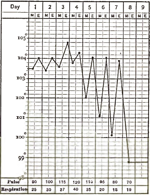
|
| Lobar Pneumonia, where the Crisis was marked with Evening Exacerbations, reaching nearly the highest pyrexia of the second stage: Recovery. |
Just before the final fall the fever may be greater than at any time preceding.34 When the decline in temperature is gradual (lysis), the normal temperature is usually reached by the ninth day, but it may be delayed until the twelfth or fourteenth day. A very slow or protracted lowering of the temperature is attended by a coincident slow disappearance of the physical signs of consolidation. There is no explanation for this, except that it is met with oftenest in the weak, debilitated, and dissipated where venesection has been practised or a depressing plan of treatment has been resorted to.
34 See Fig. 33, where a temperature of nearly 105° F. is followed on the evening of the fifth day by the final fall.
A high temperature persisting after the tenth day indicates purulent infiltration (see Fig. 38).
Pneumonia involving the apex of the lung is usually marked by a higher average range of temperature than when it is confined to the lower lobes. Statistics show that the fifth and seventh days are the days of crisis in the majority of uncomplicated pneumonias. Of 867 cases terminating by crisis, in 677 the crisis occurred before the eighth day. Neither the height of the temperature-range nor the amount of lung involved affects the critical day.
In the form of pneumonia sometimes called bilious—a form that prevails in miasmatic regions—the temperature is markedly paroxysmal.
In children the temperature rises very rapidly, sometimes reaching 106° F. within the first twelve hours. The highest recorded temperature in the pneumonia of children, with recovery, is 106°–107° F. The average temperature [p. 326] of pneumonia at this period of life is 104° F., the range being higher than in adult pneumonia.
In children the day of crisis is oftener the seventh than the fifth day. The fall of temperature during the crisis is somewhat remarkable; it often falls two and a half degrees below the normal, and this exceedingly low temperature may be maintained for two or three days, and yet the child recover.
The accompanying charts show ordinary temperature-curves from children with pneumonia (see Figs. 35, 36).
| FIG. 35. |
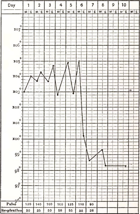
|
| A Typical Case of Lobar Pneumonia in a Child: Recovery. |
| FIG. 36. |
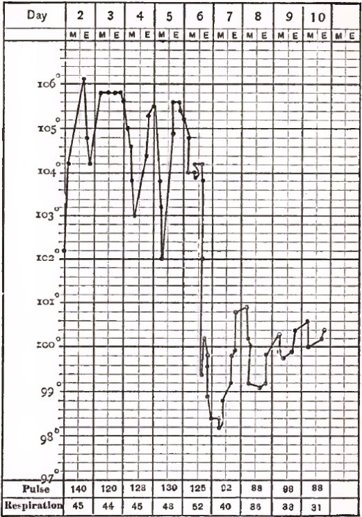
|
| A Case of Lobar Pneumonia in a Boy ten years old, in which thermometrical observations were made every four hours: Crisis on the sixth day. |
In old age it is often difficult to determine the exact day of the invasion of pneumonia except by the temperature. The rectal temperature rises to 103° or 104° F., or even higher, on the first days, and continues at about the initial point for three or four days, with daily morning and evening oscillations of a degree or a degree and a half. The temperature-rise does not begin for several hours after the initial chill, if a chill occur (see Fig. 37).
| FIG. 37. |
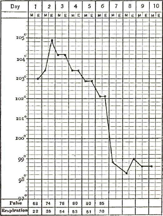
|
| A Typical Case of Senile Lobar Pneumonia: Recovery. |
Relapse in pneumonia is a rare event; it is quite phenomenal for it to occur four days after the crisis. The temperature suddenly rises, but usually returns to normal in three or four days.
Pulse.—The pulse in pneumonia varies with the type and extent, as well as with the stage, of the disease. In an ordinary mild case the pulse-rate is usually between 90 and 120 per minute. When the pulse-rate for any length of time is above 120, the case must be regarded as an exceedingly grave one.
[p. 327] The pulse at the onset of croupous pneumonia is usually full and soft. As the disease progresses it becomes small and feeble. In severe cases, and when the nervous system is markedly implicated, it is rapid, and may be 130 to 140, or even 160, at the onset of the disease. In such cases it will also be small and feeble.
A high temperature is usually accompanied by a rapid pulse, and a low temperature by a moderately frequent, full pulse. At the day of crisis, when the temperature falls, the pulse will fall; and this occurs in the severe as well as in the mild cases.
Subsequent to the third or fourth day in severe cases the pulse, in addition to its frequency and feebleness, may exhibit dicrotism, or it may be jerky, very compressible, and intermittent. Sometimes just before death the pulse becomes markedly slow. The feebleness of the pulse is ascribed by some to cardiac depression, the result of the high temperature; by others it is claimed that the afflux of blood to the left ventricle obstructs, and causes a deficiency in, the aortic circulation. In other words, hepatization is adduced as a cause of the feeble pulse. In chronic wasting diseases, in feeble, weak individuals, or in those already suffering from cardiac disease, weakness of the pulse is a very marked symptom.
I cannot regard a feeble pulse in pneumonia as due to the pulmonary hepatization, for it is not that pneumonia which is most extensive that is accompanied by the greatest heart-flagging. Heart-failure may exist before, or just as, consolidation is beginning. In many pulmonary affections the obstruction to the pulmonary circulation is greater than in pneumonia, and yet there is no heart-failure. The pneumonia with the highest temperature-range is not necessarily the pneumonia in which heart-failure is most marked [p. 328] or earliest to develop. There are many diseases in which there is a much higher range of temperature and yet no evidence of heart-failure occurs.
If a prolonged high temperature is the cause of feeble heart-power by the parenchymatous changes which it induces in its muscular fibres, such a high fever is not met with in pneumonia, and the heart is rarely found at post-mortem to exhibit such changes. May not the heart-failure, as indicated by a rapid, feeble, and intermittent pulse in pneumonia, be due to the presence in the blood of a morbific agent (as in certain infectious diseases) which so affects the nerve-centres which supply the heart that its contractile power is diminished and its rhythm disturbed? The pulse early shows commencing heart-failure by each cardiac pulsation producing a variable filling of the arteries with blood; hence the beats first vary in force, then waves occur, then true intermissions. I have been able to detect this heart-insufficiency by these variations of the pulse within twenty-four hours after the onset of a pneumonia, and occasionally during the initiatory chill.
In children the pulse-rate is greatly increased; it may reach 200 in a minute. It is very small, unequal and irregular, but never intermittent.
In senile pneumonia the pulse is not a reliable indication. The pulse may be only 50, and yet this would be a rapid pulse for the particular case in which it occurs.
In old age, both in health and in disease, the pulse has a fictitious hardness on account of arterial changes. The pulse may not be intermittent or irregular, yet the heart may be very irregular and intermittent in its action. Again, the pulse may be feeble and intermittent and the heart be acting regularly.
Remittence of the pulse is quite common in senile pneumonia independent of cardiac changes. The action of cold upon the surface in the aged is very quickly indicated by the radial pulse lessening its volume and strength, so that if the pulse at the wrist is taken it should be from the arm which has been covered. To avoid error, the pulse in senile pneumonia must be counted at the heart.
The surface of the body may be pungently hot and dry until the crisis is reached, or it may be bathed in perspiration from the onset of the disease. A moist surface has been regarded as a very favorable sign, but when in the height of the disease the parched skin becomes moist and the patient is not relieved, it is an unfavorable rather than a favorable symptom, and is met with more often in fatal cases than in those that recover.
In most cases of croupous pneumonia the expression of the countenance is characteristic. It is one of anxiety, and over the malar bones is a mahogany flush—not, as in typhus fever, diffused, but well defined and circumscribed, so that it is sometimes called the pneumonic spot. While the cheeks exhibit a spot of this dusky hue, the rest of the face may have an earthy pallor. Bouillard states that the pneumonic flush on the cheek is most marked when the pneumonia has its seat at the apex of the lung. Some authorities state that the cheek flushes most or solely on the affected side, while others35 have shown that the cheek on the side opposite to that affected is the one that is usually flushed. In this connection it is interesting to mention the case of Jaccoud, who, suffering from an attack of pneumonia himself, noticed for twenty-four hours preceding the pneumonia signs a flush and a burning sensation in the cheek opposite to the side affected. Usually one cheek is more flushed than the other, and this is undoubtedly due to disturbance of the vaso-motor system.
35 Barthez and Rilliet.
When the impediment to the circulation is excessive, or when vaso-motor disturbance is marked, the lips become cyanosed. At the time of crisis the face becomes paler.
In about one-half the cases pneumonia is attended by an herpetic [p. 329] eruption upon the lips, nose, cheeks, or eyelids. It rarely appears before the second or third day. It may not appear until the crisis is reached. Herpes occurs with varying frequency in different years, but is more commonly met in pneumonia than in any other febrile state. One winter nearly every case of pneumonia in Bellevue Hospital was accompanied by herpes labialis. When sweating exists and involves the entire body, it is very frequently accompanied by sudamina, which are either abundant or sparse, and seem to have a critical significance.
In children, while the surface of the body is hot and dry the extremities are cool. The pneumonic flush instead of having a mahogany tint assumes a bluish-white tint. Cyanosis of the extremities is more frequent than in adults, and herpes labialis more common. All the cutaneous symptoms are exaggerated in children.
In old age the pneumonic flush is often the first objective sign of pneumonia. The eyelids alone are cyanotic. If the face is dusky at first, it subsequently assumes a sallow hue, and the surface-heat, which is greatest in the morning, is succeeded by a cold, clammy perspiration.
Cerebral Symptoms.—The cerebral symptoms in the early stage of pneumonia are not very significant. Headache is usually present at the onset, and may continue throughout the disease. It usually steadily diminishes after the third day. If it is severe in the evening, there will be slight delirium at night—so slight as often to escape notice. Delirium and convulsions rarely occur except in debilitated subjects and in persons of enervating habits. It is most frequently met with in alcoholic subjects, and then it assumes the character of delirium tremens. It is an active, busy, restless delirium: the patient is constantly talking, but seldom in a coherent manner. Sometimes, in those who are not alcoholic subjects, the delirium may assume an active and violent character. Whenever active delirium is present, it is important to make careful and diligent search into the previous habits of the patient.
Pneumonia of the apex is more apt to be accompanied by severe cerebral symptoms than when it has its seat at the base.
Delirium may pass into coma. When delirium and headache are marked symptoms, muscular tremors (subsultus tendinum) are very apt to occur, with insomnia and frightful hallucinations. Indeed, these cerebral symptoms are often so prominent in alcoholic pneumonia, and occur so early, that the pneumonia may be wholly masked, and will only be discovered by the temperature-range and by a careful physical exploration of the chest.
When delirium is present in feeble patients, it assumes a low muttering typhoid type, and a state of stupor is soon reached.
Among the rare nervous symptoms met with in pneumonia may be mentioned photophobia, disturbances of vision, and deafness.
In children the cerebral symptoms are more prominent than in adults, and they do not seem to be influenced by the extent of lung involved. Stupor and restlessness on the one hand, or headache, delirium, and convulsions on the other, may usher in pneumonia in children without any prodromata.
Sometimes children pass rapidly into a semi-comatose condition which has not been preceded by delirium or convulsions. Convulsions are as common in children as they are rare in adults, and occur with greatest frequency and severity during dentition. The convulsions may be general and resemble those of epilepsy (pneumonie éclamptique of Barthez and Rilliet), or they may attack single muscles or groups of muscles, the child occasionally passing into a tetanic or opisthotonic condition.
If convulsions do not occur until late they are quickly followed by a deep and fatal coma. A very rare occurrence is partial paralysis of the muscles which were involved during the convulsive period. Such paralysis is often [p. 330] permanent. Again, the cerebral symptoms may closely resemble those which attend cerebro-spinal meningitis—viz. headache, constipation, great prostration, delirium, convulsions, opisthotonos, and strabismus. As in meningitis, there is a peculiar cry, and all the symptoms may point directly to the brain. These symptoms are most likely to be present in the pneumonia of the apex in children from five to seven years of age.36
36 This form is the pneumonie méninges of Barthez and Rilliet.
In senile pneumonia headache may persist throughout the entire attack; it is usually accompanied by delirium of a mild type, especially when the apex of the lung is involved. These patients are very loquacious and have a constant desire to get out of bed.
Alimentary Tract.—The symptoms referable to the digestive apparatus are neither diagnostic nor important. Nausea and vomiting are not infrequent, and in about 15 per cent. of all cases are among the initial symptoms. Gastric symptoms, when severe and persistent, greatly endanger life. There is no characteristic appearance of the tongue: it may be normal throughout, or covered with a creamy-white fur, which becomes dry and brown as the disease advances. In severe cases and toward the end of the disease the lips and tongue become brown, dry, and cracked, and sordes collect on the teeth. Anorexia is marked at the onset, and the thirst is intense. When convalescence commences the tongue becomes clean and the appetite returns. Occasionally there is a catarrh of the oral mucous membrane. Diarrhoea may occur as one of the initial symptoms. It is most apt to be present when there are nausea and vomiting. As a rule, the bowels are constipated and the stools dry. In young children nausea and vomiting are more common, and in 50 per cent. of the cases usher in the disease. They usually cease on the second day, although they may persist until the crisis occurs. Excessive and violent diarrhoea may precede a fatal termination.
In senile croupous pneumonia the tongue early becomes dry, shrivelled, and covered with a thick brown coating, and is protruded with difficulty. Although these patients do not complain of thirst, they take with avidity fluids that are placed to their lips. As the period of crisis is reached critical diarrhoea is of frequent occurrence.
Loss of strength occurs earlier and is more marked in pneumonia than in any other acute disease except typhus fever. Pneumonia patients become very weak within the first five days. The recovery of strength during convalescence is rapid.
Urine.—The urine at the onset of pneumonia is scanty, high-colored, and of high specific gravity. The amount of urea excreted is twice or three times more than the normal. The excess of urea increases until the crisis, and then suddenly diminishes with the fall in temperature, often below the normal standard.37
37 The daily amount of urea normally excreted is subject to great variations: it ranges between 355 and 460 grains. Parkes gives the result of 25 different observations: the lowest estimate was 286.1 gr. and the highest 688.4 gr.
Uric acid is also increased, and follows the same course as that of the urea. The inorganic salts of the urine, especially the sodium chloride, are diminished, and during the height of the pneumonia may be wholly absent. Much has been written concerning this diminution, which is by no means peculiar to pneumonia, but in no other acute disease is its diminution so constant and marked a symptom. Sodium chloride is probably retained in the system, for when the salt has been administered in large quantities none has appeared in the urine. The reappearance of the chlorides in the urine marks the approach of convalescence, and when the crisis occurs they appear in excess, following an opposite course to the urea and uric acid.
Although these last two ingredients are in very rare cases retained, the [p. 331] same as the chlorides, to appear when the crisis occurs, their retention is usually accompanied by a critical diarrhoea, which is followed by a prolonged convalescence. The diarrhoea is undoubtedly due to the irritation caused by the urea.
Parkes38 states that sulphuric acid is increased and phosphoric acid is diminished, but Huss affirms that both acids are diminished. With the increase of the urea and uric acid, and diminution of chlorides, biliary pigment will appear in the urine, and occasionally the biliary acids.
38 On the Urine.
Slight albuminuria is an ordinary phenomenon of pneumonia, and, though usually met with in the second stage, it may appear at any time. This symptom is present in 35 per cent. of all cases. Its presence is a point of resemblance between pneumonia and other acute blood diseases. The more severe the pneumonia, the more marked is the albuminuria. Some have ascribed its presence to passive hyperæmia the result of the pulmonary obstruction. This is questionable, except in those rare cases where venous engorgement is indicated by cyanosis, enlargement of the liver, jugular pulsation, etc.
In children the amount of urine corresponds to the quantity of fluid taken.
Critical Phenomena.—At the end of the first week, during which all the symptoms have increased in severity, the continued fall of temperature tells us that convalescence is established. As the temperature falls, profuse (critical) sweating occurs. Both of these phenomena may occur to such an extent that for hours the condition of the patient is one of collapse.
In rare cases death has occurred in the midst of these symptoms. The respirations and pulse-rate are diminished in frequency, the pulse being small and frequently exhibiting dicrotism. The cough becomes loose, the dyspnoea abates, the flush disappears from the cheek, the sputum is more copious, and is expectorated with less difficulty; it loses the rusty color from metamorphosis of its hæmoglobin, diminishes in viscosity, and no longer adheres to the side of the vessel, but becomes more opaque, of a creamy consistency, and resembles that of simple bronchial catarrh. When resolution is retarded, the creamy-yellow tint may give place to an almost black hue, on account of the excessive amount of pigment present. As convalescence advances, the sputa become scantier, more mucous, watery, transparent and colorless. At the time of crisis the intense thirst diminishes, the appetite returns, pain in the side subsides, and the patient passes into a quiet, natural sleep, to waken fully convalescent, suffering only from extreme exhaustion.
Epistaxis, hæmaturia, and hemorrhage from the bowels sometimes occur at the critical period, and may be regarded either as accidents or as the result of the defervescence. After the crisis the amount of urea in the urine (which during the height of the disease was augmented) falls to normal or nearly to normal. Sodium chloride appears in the urine as soon as the crisis occurs.
The critical phenomena in children are the same as in adults, and frequently the fall in temperature is so great that for hours after the crisis they lie half unconscious, with a cold surface covered with a colliquative sweat. With the critical sweat there is often a catarrhal flow from the nose. When children have been extremely restless or delirious the crisis is marked by the patient passing into quiet sleep.
In old age, when recovery occurs, it is generally by crisis, and a critical diarrhoea is much more frequent than a critical sweat.
In adults and in children the recovery of strength and flesh is rapid; in the aged the period of convalescence is very prolonged, and often does not begin (when the pneumonia is of the asthenic—typhoid—type) until the fourteenth or fifteenth day; still, complete recovery may be reached.
Symptoms indicating Danger.—When croupous pneumonia is to terminate fatally, dyspnoea is greatly increased; the patient suddenly sinks; the pulse [p. 332] becomes extremely small, rapid, irregular, intermittent, and dicrotic. Large moist râles are heard over the larger bronchi and trachea, while the auscultatory signs of pulmonary oedema become more and more apparent. The sputa become frothy, liquid, and blood-stained, or are entirely suppressed. The respirations become more and more hurried, the face is sunken and livid, the extremities are cold, and the superficial capillary circulation is more and more interfered with, as is indicated by the cyanosis. The body is bathed in a profuse cold perspiration. The fatal issue is usually preceded by coma.
The temperature may steadily rise up to the time of death, or death may occur in the defervescence. In alcoholic pneumonia death is preceded by cerebral symptoms, such as somnolence, numbness of the limbs, a sense of formication, and slight convulsive attacks.
In children death is often preceded by convulsions or coma. If the disease is protracted, death may be preceded by extreme exhaustion and collapse. Cyanosis and extreme rapidity of the pulse are usually present in children just before the fatal issue.
Senile pneumonia may end fatally within a few hours after its onset in a most unexpected manner. The aged patient walks apathetically about, totters to the bed, lies down, and dies. If the pneumonia has existed for a number of days, the signs of a fatal termination are sallowness of the face, a cold clammy skin, expansion of the alæ nasi, and a sudden rise or fall of the temperature. The inspirations become mere gasps, and, following the apathy, the patient gradually lapses into complete coma.
Symptoms which attend the Termination of Pneumonia in Abscess.—Acute pneumonia terminates in abscess in from 1 to 2 per cent. of all the cases. It is therefore a rare termination. It is most frequent in debilitated, weak subjects and in those who have received a depressing plan of treatment. The expectoration is exceedingly copious and fetid, and the sputa are yellowish or yellowish-gray in color, consisting almost wholly of purulent matter. Pigment is usually found in the expectorated masses, and when shreds of pulmonary tissue are present the diagnosis is established. The fever assumes a hectic type and is accompanied by rigors and sweats.
After these symptoms have continued for a time, the patient grows weaker and emaciated, and death results from exhaustion, from asphyxia (when a large bronchus is plugged with pus), or from the discharge of the abscess into a neighboring cavity.
DaCosta states that "pulmonary pneumonic abscesses are at the base of the lung;" Fox locates them "at the apex;" Green, "on the upper lobe;" I have found them in both situations.
The physical evidences of a lung-cavity are the most reliable signs of pneumonic abscess. Abscess is a very rare termination of croupous pneumonia in children. In old age the formation of abscesses is never evinced by any well-marked symptoms. The finding of elastic fibres in the sputa with the physical signs of a cavity are the only diagnostic signs.
Symptoms which attend the Termination of Pneumonia in Gangrene.—Gangrene as a termination of pneumonia has been found in about 14 per cent. of cases.39 This must be regarded as an exceptionally high percentage. Its occurrence is usually accompanied by symptoms of sudden collapse. The pulse becomes rapid, feeble, and intermittent, the face is pale and of a deathly hue, and there is a profuse expectoration of blackish-green masses containing shreds of decomposed and decomposing lung-substance of an exceedingly fetid odor. The breath is fetid and the whole body emits a cadaverous smell. The rapidly-increasing prostration is sometimes accompanied by hemorrhage.
39 In 28 out of 200 cases (Guy's Hospital Reports, Sec. vii., 1848).
The sickening and indescribable odor of pulmonary gangrene is most perceptible after an attack of coughing. Gangrene has its most frequent site in [p. 333] the lower lobes of the lung, and it is here that a careful search must be made for the rather ill-defined physical signs which attend its development. In old age, when a pneumonia is to terminate in a gangrene, typhoid symptoms appear very early, and death occurs with symptoms of the profoundest collapse within five days from the initial chill.
Symptoms which attend the Termination of Pneumonia in Purulent Infiltration.—The symptoms of purulent infiltration differ but slightly from those of the third stage of pneumonia. When resolution does not take place at the period of crisis, and the temperature remains high, accompanied by symptoms of prostration and profuse putrid expectoration, with none of the physical signs of resolution, purulent infiltration is to be suspected. Death may result from exhaustion, or recovery take place after a prolonged convalescence (see Fig. 38).
| FIG. 38. |
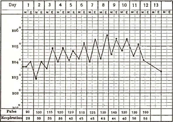
|
| Croupous Pneumonia in the Adult, terminating in Purulent Infiltration: Death on fourteenth day. |
Mild delirium is a frequent symptom during the stage of purulent infiltration. The sputa contain a large number of cells in various stages of fatty degeneration. The temperature has regular evening exacerbations, and often ranges higher than during any preceding period of the disease. The tongue becomes brown and dry, sordes collect upon the teeth, and the patient passes into a typhoid state.
Typhoid pneumonia is a term which has been applied to a variety of croupous pneumonia which is attended by typhoid symptoms. It has also been called asthenic, low, or nervous pneumonia. There are symptoms of extreme prostration from its onset. After well-marked pneumonic symptoms have been present for a few days, the patient passes into a condition of extreme prostration.
There is little or no expectoration, no dyspnoea, no pain, no cough. Sordes collect on the teeth and gums; the tongue becomes thickly coated with black crusts; the pulse becomes small, feeble, and rapid, and there is a tendency to the formation of bed-sores; and then occur stupor, somnolence, and a continuous low muttering delirium. This form of pneumonia is met with most frequently in the aged. In some cases there is marked disturbance of the special senses.
Tremors and subsultus tendinum frequently coexist. It may be accompanied by glandular swellings, by sharp and darting muscular pains, by arthritic symptoms, or by great gastric disturbance. It is not infrequent in epidemics, [p. 334] and it may follow or accompany erysipelas, Bright's disease, alcoholismus, or phlebitis. It is always a grave condition, but recovery is possible. Convalescence, which is very tedious, may commence as early as the twelfth or fourteenth day. Sometimes a modification of typhoid pneumonia accompanies dysentery, intestinal catarrh, or a phlegmonous gastritis. There are great sweating, profuse diarrhoea (colliquative), and high fever. The odor of the sputa resembles that of gangrene of the lungs. Such cases commonly end fatally.40
40 Cyclo. Pract. Med., iii., art. "Gastritis."
Bilious or Gastric Pneumonia.—Croupous pneumonia occurring in malarial districts, accompanied by gastro-enteric or hepatic symptoms, is known as malarial or bilious pneumonia. It has all the characteristic symptoms of pneumonia of a very severe type, but the fever is paroxysmal. The tongue is heavily coated; nausea and vomiting are common, and may persist throughout its entire course; the epigastrium is distended and tender; the skin is jaundiced; the liver is enlarged, and there is usually an exhausting diarrhoea, attended by greenish, black, viscid, and inodorous stools. The hepatic congestion and jaundice are due to a coincident gastro-duodenal catarrh.
Bilious pneumonia may be of a sthenic or asthenic type. The theory that the liver becomes inflamed by extension from the lung is untenable.
The symptoms of bilious pneumonia have frequently led to a diagnosis of typhoid gastric fever or some severe acute affection of the intestinal tract. But a reference to the physical signs will remove all doubts.
Bilious pneumonia runs a more protracted course and has a much longer period of convalescence than ordinary croupous pneumonia. In old age this form is not infrequent. The vomiting is distinctly bilious in character, and at this period of life somnolence and stupor are quite common, and are exceedingly unfavorable symptoms.
Latent Pneumonia.—Pneumonia in adults is seldom latent unless it complicates some disease whose symptoms are so severe, and the attending prostration is so great, as to obscure the characteristic signs of the pneumonia. Intercurrent senile pneumonia is always latent, and Grisolle says that an exploration of the thoracic organs in the majority of such cases gives negative results. If, then, an intercurrent senile pneumonia runs its course without expectoration, without dyspnoea, without the pneumonic flush, and without any of the physical signs of pneumonia, its diagnosis must rest—first, on the extreme frequency of pneumonia in old age; secondly, on the fact that of all the phlegmasiæ of advanced life pneumonia is the one which is oftenest latent; thirdly, that of all the acute diseases in old age pneumonia is attended by the highest range of temperature and the greatest prostration. When an old person has a slight rigor followed by febrile movement, with great prostration, for which no explanation can be found, pneumonia may be suspected, even though all its diagnostic signs are absent.
Intermittent or remittent pneumonia, which is described by some authors as a distinct type, is a form of acute pneumonia in which a malarial element is so pronounced that all the pneumonic symptoms, even the physical signs, undergo distinct intermission, returning each day with increasing severity. Occasionally, instead of the quotidian it assumes the tertian type. During the intermission the temperature may fall to normal. Severe chills and sweating are often present, and the pneumonia is not infrequently double.
The malarial conditions which give rise to this type of pneumonia occur more frequently in our Southern and Western States than in any other part of the world.
PHYSICAL SIGNS.—By studying the physical signs of croupous pneumonia in connection with the different stages of its morbid anatomy, their importance as elements in diagnosis and prognosis can best be appreciated.
[p. 335] Stage of Engorgement.—The physical signs indicative of the first stage of croupous pneumonia are usually present within twenty-four hours after its invasion. If the pneumonia is central, their appearance may be delayed until the third day.
Inspection.—On inspection the movements of the affected side are noticed to be more or less restricted, while the unaffected side moves as in health. In double pneumonia the respiratory movements will assume a costal type, attended by an increase in the abdominal breathing.
Palpation.—On palpation there is more or less increase in the vocal fremitus on the affected side. The degree of increase corresponds to the extent of the engorgement. It must always be remembered that normally the vocal fremitus is more marked on the right side than on the left.
Percussion.—There is slight dulness over that portion of the chest-wall which corresponds to the affected portion of lung: its extent varies with the amount of lung involved. It is never well marked until the end of the first stage, although the pulmonary capillaries are engorged with blood from the commencement. Even at the end of this stage the intensity of the percussion sound, although diminished and muffled, has a slightly tympanitic quality, due to the fact that the exudation has not completely displaced the air in the distended alveoli. Very extensive central pneumonia may fail to give either increase in vocal fremitus or dulness on percussion until the second stage is well advanced. Absolute dulness during this stage is of rare occurrence.
Auscultation.—During the dry stage, which according to some41 is said to precede the exudation stage, there will be noticed a feebleness and unnatural dryness of the respiratory murmur. Sometimes it is harsh, at others feeble and loses the peculiar breezy, rustling quality of the normal respiratory sound. If it is less intense over the affected portion of the lung, it is exaggerated over the unaffected portion. These changes are apt to pass unrecognized unless auscultation is practised frequently and early in the disease. As soon as the engorgement is well marked and exudation takes place into the air-cells, fine crackling sounds are heard at the end of inspiration. These sounds are called crepitant râles, and are regarded as the characteristic sign of the first stage of pneumonia. They resemble those produced by throwing salt on live coals or rubbing the hair in the neighborhood of the ear between the fingers. These râles do not necessarily depend upon the presence of fluid in the alveoli, but may arise from the sudden separation of the alveolar walls at the end of inspiration when they have been agglutinated by a tenacious exudation. They are as numerous as they are minute, are unaffected by coughing, and remain audible over a circumscribed space from twelve to twenty-four hours. Whenever the pneumonic stages follow each other in rapid succession, the crepitant râle may not be heard. It is rarely present in a pneumonia which is developed during an attack of acute articular rheumatism. With the crepitant râle the respiratory murmur is feeble or assumes a broncho-vesicular character.
41 Stokes.
When, as often happens, pneumonia has been preceded by or complicates any other thoracic affection, the crepitant râle will be mingled with the sounds arising from that particular condition. It is said42 that bronchial breathing is sometimes heard in this stage of pneumonia. The voice-sounds undergo slight increase in their intensity over the seat of the pneumonic engorgement.
42 Traube, Annal. der Charité, i. 286.
In children the crepitant râle is frequently absent, and, though it may be heard at the end of a full inspiration after coughing, it is never so fine or distinct as in adults. In children there will be no increase in vocal fremitus if, as often happens, a large bronchus leading to the inflamed spot is plugged with mucus.
[p. 336] In old age the physical signs of adult pneumonia are modified by certain physiological changes which occur in the lungs and chest-cavity of the aged. The more complete bony union of the chest-walls, the curvature of the spine, the rigidity of the bronchial tubes, the rounded form of the chest, and the senile rarefaction of the lungs, give rise to extra resonance on percussion as compared with an adult chest. On account of the great arching of the sternum and the deposition of carbonaceous material at the apex of the lung, the clavicular region near the median line gives a dull percussion sound. The scapular and supra-scapular regions are less resonant than in the adult, on account of the tilting of the scapulæ due to curvature of the spine. There is a loss in the vesicular element of the respiratory murmur, and it resembles the sound produced by a forceful expulsion of air from the compressed lips. When the septa or the alveoli are torn and greatly distended, it has a bronchial character. Its intensity varies: at one moment it is loud, at another hardly perceptible; the variation occurs not only in the same individual, but in different individuals of the same age. The vocal sounds are loud and bronchophonic in character, and have a vibration closely resembling oegophony. It is also to be mentioned that it is almost a physiological condition for old people to have bronchorrhoea; hence mucous râles may be present during the whole period of advanced life, and if one relies on the usual crepitating râles of adult pneumonia for a diagnosis he will be misled.
Inspection and palpation in the first stage of senile pneumonia furnish little positive information. Percussion will give little dulness until the lung has reached the stage of red hepatization, and even then it may be so slight as to pass unnoticed. Very early in the disease the respiratory murmur is feeble and indistinct over the affected portion, while the portion of lung that is not involved assumes, for the time, all the characters of a normal adult respiratory murmur. Again, the breathing over the pneumonia may be intensely puerile and interrupted.
The crepitant râle is rarely present in the first stage of senile pneumonia, but subcrepitant râles and large moist râles resembling those of bronchitis are heard during the whole of this stage. The explanation of the absence of the crepitant râle is to be found in the physiological condition of the air-cells just referred to. Sometimes, on a deep inspiration after violent coughing, fine crepitation is heard, but upon careful examination it will not be found to differ from the râles of capillary bronchitis. It may be stated as a general rule that the feebler and more superficial the respirations the less distinct will be the adventitious sounds.
The physiological rigidity of the bronchi in old age favors the early development of bronchial breathing, which is often the first physical sign of senile pneumonia. One of its peculiarities, when occurring in the stage of engorgement, is that it is most distinct at the root of the inflamed lung.
Stage of Red Hepatization.—The physical signs of the second stage of croupous pneumonia are more diagnostic than those of either of the other stages.
Inspection shows the expansive movements of the affected side to be more markedly diminished than in the first stage, while those of the healthy side are increased. Frequently there is absolute loss of motion over the inflamed lung.
Palpation.—By palpation the vocal fremitus is usually increased on the affected side over the consolidated lung-tissue. In some instances it may be only slightly increased, and in rare instances it will be found less marked upon the affected side than upon the healthy. Palpation may also reveal slight displacement of the heart from the pressure of the distended lung; and in rare cases well-marked pulsation is felt over the affected lung.43
43 Skoda, Stokes, and Graves regard this as the result of increased pulsation of the arteries in the inflamed spot; and Walshe and Fox rather admit it, but Grisolle denies it.
[p. 337] It is evident that the vibrations of the vocal cords can be transmitted from the trachea through the bronchi and lung to the chest-wall, and there is no reason why the cardiac impulse may not likewise be transmitted through a solidified lung to the chest-wall.
If the pneumonia is central, the vocal fremitus may not be increased. It is diminished when there is an abundant pleuritic exudation over the pneumonic lung.
Percussion.—On percussion there will be marked dulness over that portion of the lung which is the seat of the pneumonia, while over the healthy portion, as well as over the opposite lung, there will be exaggerated resonance. The nearer the hepatization approaches the surface of the lung, the more marked will be the dulness. There is a peculiar sense of resistance on percussion over a completely airless hepatized lung which is not present in solidification from other causes. The exact outline of an hepatized lobe can often be traced on the chest-wall.
The tympanitic quality which is sometimes present during the stage of engorgement may continue anteriorly during the second stage, and yet posteriorly the dulness will be complete. A tympanitic percussion sound is sometimes elicited over that portion of lung which is adjacent to the consolidated lobe. When an upper lobe is consolidated, forcible percussion may elicit a tympanitic sound, for the column of air in a large bronchus will vibrate under forcible percussion. The cracked-pot sound (bruit de pot fêlé) is occasionally met with over those relaxed and permeable parts of the lung in the immediate vicinity of the consolidation. When this sound is present over the consolidated portion, it is due to the sudden expulsion of air from one of the larger bronchi. It is most frequent in young persons with thin, elastic chest-walls. The cracked-pot sound in pneumonia is not increased in intensity when the patient's mouth is open.
In basic pneumonia the subclavicular percussion note may be distinctly amphoric in character. Dulness may appear within twelve or twenty-four hours after the onset of a pneumonia, or it may be delayed until the fourth day.
Auscultation.—As soon as the air-cells are completely filled by the pneumonic exudation, the crepitant râle ceases and bronchial respiration is heard over the affected lung. The bronchial breathing is due to the fact that the vesicular element of the respiratory sound disappears on account of the complete consolidation of the vesicular structure, and the tracheal element of the respiration is conveyed to the chest-walls through the consolidated lung. It often has a metallic element, or may sound like the tearing of a piece of linen. Bronchial respiration is more intense in pneumonia than in any other disease.
Laennec taught that bronchial respiration was due to the superior conducting power of condensed lung. Skoda combats this view, and says that bronchial respiration is generated or magnified in caverns and in the bronchi of condensed lung-substance by the air in these cavities and in the bronchi vibrating in consonance with that within the trachea. The condition necessary for this consonance is provided in the circumstance that the air is pent up in confined spaces whose walls reflect the sonorous undulations.
The more complete the consolidation, the more intense is the bronchial respiration. At the commencement of this stage the tubular breathing only attends expiration, while later it accompanies both acts. Pleuritic exudation may mask or render this sound very indistinct. It may in rare instances be absent even when extensive consolidation exists and the pleura is perfectly normal. This can be accounted for in most cases by the plugging of a large bronchus. There are cases in which its absence is inexplicable.
The vocal sounds are increased in intensity and bronchophony is heard [p. 338] over the consolidated lung. The physical conditions of the lung which give rise to bronchophony have the same diagnostic significance as the bronchial respiration, and in all instances its occurrence, its distinctness, its temporary disappearance, and its reappearance are dependent upon precisely the same conditions as are the changes in the bronchial respiration. If the pleural cavity is partially filled with fluid, bronchophony will be indistinct or absent below the level of the fluid, while at its level the voice-sounds will be either bronchophonic or oegophonic.
During this stage the heart-sounds are transmitted to the surface over the hepatized lung with greater intensity than normal.
In children dulness is especially marked in the infra-scapular region of the affected side. Some authors44 speak of a feeling of greater solidity below than above the scapula, which can be detected before the ear can detect actual dulness on percussion. Vocal fremitus may be increased, but it is not reliable on account of the changes in the voice.
44 West.
In old age, inspection and palpation give negative results. Dulness on percussion in old age would be regarded as normal resonance in the adult; hence the percussion sound in senile pneumonia may be only relatively dull. The tubular or bronchial breathing in the second stage of senile pneumonia is more intense than in adult pneumonia. Small gurgles or mucus râles generally persist throughout this stage. Bronchophony is not well marked. On causing the aged patient to cough and expire violently, tubular breathing may be heard where it was before absent.
Stage of Gray Hepatization.—There is no abrupt transition from the second to the third stage of pneumonia, so that the physical signs of the early part of gray hepatization are the same as those of the second stage.
Inspection.—As resolution progresses, expansive motion on the affected side becomes more and more apparent.
Palpation.—On palpation the vocal fremitus will be found approaching normal, its intensity diminishing as resolution occurs.
Percussion.—Dulness on percussion becomes less and less marked, but of all the signs this is the last to disappear. Rare cases are mentioned where it has disappeared in twenty-four hours after the commencement of resolution by crisis. As the percussion sound approaches the normal, a tympanic note is again present in circumscribed spots.
Auscultation.—The bronchial respiration that was present in the second stage gives place to broncho-vesicular breathing. This soon becomes blowing, then indeterminate, and finally approximates to, and merges into, normal vesicular breathing. Bronchophony gives place to exaggerated vocal resonance in connection with the changes in the respiratory and vocal sounds. The crepitant râle returns, but is soon obscured by larger and moister crepitating sounds, "the resolving subcrepitant râle of pneumonia," called also the râle redux. Large and small mucus râles, sibilant and sonorous, accompany the subcrepitant râles, to disappear only when resolution is complete. Not infrequently the bronchial râles that are developed during the stage of resolution are of that character called consonant45 or ringing.46
45 Skoda.
46 Traube.
The physical signs of this stage are all retrogressive, and they disappear in the opposite order to that in which they appeared. In rare instances resolution is so rapid that the subcrepitant râle is not heard. In this class of cases dulness on percussion and bronchial breathing continue for some time after the crisis.
In children, bronchial breathing rarely disappears before the seventh day. It is often accompanied by the subcrepitant râle. When resolution takes place, bronchial breathing and the subcrepitant râle will disappear at the same time.
[p. 339] In old age, inspection, palpation, and percussion give similar results as in adult pneumonia. On auscultation coarse crepitating sounds and loud gurgles are often heard at a distance from the site of the pneumonia. The râle redux is not distinctive of or peculiar to the third stage of senile pneumonia. The sounds heard during this stage are called mucous crepitations, by which is meant liquid crepitating râles produced in tubes intermediate between the bronchioles and the larger bronchi.
If pneumonia terminates in purulent infiltration, the temperature remains high and symptoms of great prostration are developed. The bronchial breathing continues, and becomes more intense, dulness on percussion persists, and sharp, high-pitched râles resembling fine gurgles are abundant.
The occurrence of abscess or gangrene is indicated by the physical signs which attend the formation of cavities in consolidated lung-substance.
No one of the physical signs which is present in the different stages of pneumonia is sufficient for a diagnosis, but the manner and order of their occurrence, and their relation to the symptoms which mark the different stages of the disease, enable one to reach a positive diagnosis in all typical cases. The only symptom of croupous pneumonia which can be regarded as diagnosticated is the sputum.
The physical signs of pulmonary abscess in the aged are very generally wanting. Distinctly localized gurgling and cavernous respiration may, when taken in connection with the rational signs, suffice for an approximate diagnosis, but the great rarity of abscess in old age should make one cautious in its diagnosis. The sputa will greatly aid in such cases.
The physical signs of senile pneumonia are subject to greater variations than ever occur in pneumonia in the adult, and often they do not even follow the course, irregular as it is, which has just been described.
Gray hepatization or abscess may be reached without any distinctive auscultatory signs, even after repeated and careful examination. The râle redux of resolution may be absent, dulness and bronchial breathing being immediately followed by normal (senile) resonance without crepitation. This occurs most frequently in the typhoid variety.
DIFFERENTIAL DIAGNOSIS.—In typical cases of croupous pneumonia (except in childhood and old age) the diagnosis is not difficult. The prolonged chill of invasion, the rapid rise of temperature, the accelerated, panting respiration, pain, cough, characteristic sputum, increase in vocal fremitus, dulness on percussion, the crepitant râle, bronchial breathing, and bronchophony are sufficient to establish the diagnosis.
Croupous pneumonia may be confounded with acute pulmonary congestion and oedema, capillary bronchitis, pleurisy, hypostatic congestion, catarrhal pneumonia (in children), pulmonary apoplexy, meningitis, and typhoid fever.
Pneumonia begins with a chill, while pulmonary oedema has no chill. Pneumonia is a febrile disease, while in pulmonary oedema there is no rise in temperature. In pneumonia there is pain in the side; there is no pain in pulmonary oedema. The sputum in pneumonia is viscid, rusty, and microscopically pathognomonic; pulmonary oedema is accompanied by a profuse watery expectoration. Pneumonia is commonly unilateral, and can occur in any part of the lung, while pulmonary oedema is bilateral, and usually occurs in the most dependent portion of the lung. In pneumonia we have the crepitant, dry râle, while in pulmonary oedema we have subcrepitant râles, larger and more liquid than those in pneumonia. Bronchial breathing and bronchophony occur in pneumonia, and are absent in pulmonary oedema. Percussion dulness is more marked in pneumonia than in pulmonary oedema, and the diseases with which the latter condition is apt to arise will aid us very much in the diagnosis. Urinary symptoms are negative in pulmonary oedema, while in pneumonia the chlorides are diminished or absent.
[p. 340] The stage of resolution in pneumonia is not infrequently mistaken for general capillary bronchitis, but, though the subcrepitant râle is present in both, it is heard all over the chest in capillary bronchitis, while it is confined to a comparatively small space in pneumonia. The expectoration is muco-purulent in bronchitis, and viscid and fibrinous in pneumonia. The temperature is lower in bronchitis (100°–103°) than in pneumonia (104°–106°). Capillary bronchitis is bilateral, pneumonia usually unilateral. Capillary bronchitis does not commonly begin with a chill, like that which occurs in pneumonia, but comes on more insidiously and without pain. Capillary bronchitis gives an exaggerated percussion note, while there is dulness on percussion in pneumonia. There is bronchial breathing in pneumonia, and a feeble vesicular murmur in capillary bronchitis. In capillary bronchitis the cyanotic appearances are very much more marked than in pneumonia, and there is no perversion of the pulse-respiration ratio. The breathing is labored in capillary bronchitis, and panting in pneumonia. In capillary bronchitis there are several slight attacks of chilliness; in pneumonia there is usually only one chill, at the onset.
The chief points in making the diagnosis between pneumonia and pleurisy are the pain, sputum, and percussion note. Pneumonia is ushered in by a distinct chill, followed by a rise in temperature to 104° to 105°, while pleurisy begins with chilliness or a number of slight rigors, and the temperature is lower, rarely above 100°. The dry hacking cough of pleurisy may be accompanied by slight mucous expectoration, while in pneumonia the expectoration is characteristic. In pleurisy the breathing is catching; in pneumonia it is panting. In pleurisy the face is pale and anxious; in pneumonia the cheek bears a dull mahogany-colored flush. In pleurisy the pulse is firm, small, tense, and wiry; in pneumonia it is full and bounding. The amount of chlorides in the urine is not altered in pleurisy, but in pneumonia they are diminished or absent. The pulse-respiration ratio is not affected in pleurisy, while in pneumonia it may fall as low as 2:1. There are no critical days in pleurisy, while in pneumonia crisis occurs about the fifth or seventh day. In pleurisy with effusion there may be bulging of the intercostal spaces, and the heart may be displaced; these phenomena never occur in pneumonia. The vocal fremitus is feeble or absent in pleurisy, while in pneumonia it is much increased. In pneumonia there is dulness on percussion, while percussion over a pleuritic effusion elicits flatness, which changes with the position of the patient. In pleurisy the grazing, rubbing, or sticky friction-sound may be heard with both respiratory acts; in pneumonia we hear the crepitant râle. In pleurisy the respiratory sounds are feeble or absent, as are the vocal sounds, while bronchial breathing and bronchophony are marked in pneumonia. It may be remembered, however, that if adhesions from an old pleurisy bind the lung to the chest, vocal fremitus may be increased in pleurisy. Again, bronchophony and bronchial breathing may exist in pleurisy, but they are always diffuse, never sharp and tubular, as in pneumonia, and are usually confined to the scapular region.
Hypostatic congestion of the lungs is accompanied by copious, watery, blood-stained expectoration. In pneumonia the sputa, though bloody, are rarely watery. Pneumonia occurs anywhere in the lung, and has well-marked rational symptoms; hypostatic congestion occurs in the most dependent portion of the lung, disappears when the patient sits up, is accompanied by no rational symptoms except dyspnoea and expectoration, and usually can be traced to a long-continued recumbent posture in those who are suffering from extensive blood-changes.
It is often difficult to decide whether a child has catarrhal or croupous pneumonia. It is to be remembered that catarrhal pneumonia is always secondary, while croupous is primary. Catarrhal pneumonia usually follows [p. 341] a bronchitis, croupous pneumonia rarely. In catarrhal pneumonia both lungs are involved; in croupous but one, and often only a single lobe. Catarrhal pneumonia is accompanied by a catarrhal sputum, while croupous pneumonia has a viscid, rusty, fibrinous expectoration. There is no day of crisis in catarrhal pneumonia, while croupous pneumonia in children almost always ends in well-marked crisis. In catarrhal pneumonia dulness on percussion is generally confined to the posterior dorsal region, and does not extend so far forward as in lobar pneumonia. Again, the extent of the physical signs and the rapidity of their development in catarrhal pneumonia are in contrast with those of croupous.
The range of the temperature is a most valuable guide in their differential diagnosis, since not only the height of the fever is greater in croupous, but the temperature-curve is different, as seen in the accompanying tracings:
| FIG. 39. |
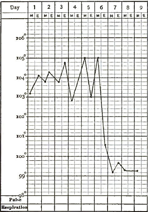
|
| Acute Lobar (croupous) Pneumonia in a Child: Recovery. |
| FIG. 40. |
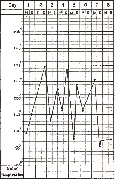
|
| Acute Lobular (catarrhal) Pneumonia in a Child: Recovery. |
Pulmonary apoplexy is rarely met with independent of valvular disease of the heart or pyæmia. It is a non-febrile disease, while pneumonia has marked pyrexia at the onset. In pulmonary apoplexy dyspnoea is very intense and comes on abruptly; in pneumonia it comes on slowly and progressively increases. The expectoration in pulmonary apoplexy consists of small, black sooty-looking coagula, while in pneumonia the viscid fibrinous mass contains numerous cell-elements other than blood-corpuscles. In apoplexy the dulness is distinctly circumscribed, and around it moist râles are heard, while in pneumonia the area of dulness is more extended and râles are heard over the seat of the dulness. The urinary symptoms are negative in pulmonary apoplexy; in acute pneumonia the chlorides are diminished or absent. There is a peculiar acid odor to the breath—an odor like that of tincture of horseradish—in pulmonary apoplexy, never found in pneumonia.47
47 Guéneau de Mussey.
When croupous pneumonia has its seat at the apex of the lung, it may be [p. 342] confounded with the first stage of phthisis. But the history of a chill followed by the characteristic pneumonic symptoms will generally enable one to make the differential diagnosis. Besides, the fever in phthisis is irregular and is subject to irregular exacerbations and remissions. If the signs of consolidation persist with little or no change, if the temperature at no time falls to normal, if there are night-sweats, if emaciation is progressive,—then the case is to be regarded as one of phthisis, even though there may have been pneumonic consolidation complicating it.
In children pneumonia is so frequently accompanied by marked nervous symptoms that it may be mistaken for meningitis. Meningitis is developed insidiously; has but slight febrile symptoms (102–103° F.), which remit with comparatively great regularity; has a pulse which is often slower than normal; has no thoracic symptoms, no dyspnoea nor accelerated breathing; the face is pale and anxious; and the physical signs of pneumonia are absent.
Sometimes a latent pneumonia with typhoid symptoms is mistaken for typhus fever: especially is this the case when the latter is prevailing. I frequently saw cases where such a mistake had been made while in charge of the typhus-fever patients on Blackwell's Island during a typhus epidemic. In these cases there will be active typhoid symptoms, such as dry tongue, delirium, high temperature, etc. The countenance in this pneumonia, although the cheeks may have a purplish hue, does not exhibit that dull, heavy leaden expression so commonly seen in typhus fever. Although there may be delirium in both instances, the delirium in the former disease is of a milder type than in the latter. The characteristic pneumonic expectoration is often absent in this class of cases; therefore it cannot be relied upon as a point in the differential diagnosis. If pulmonary consolidation is a complication of typhus fever, it will not be developed until after the sixth day of the fever, the time when the eruption is visible. If no eruption is present, the pneumonic consolidation may be regarded as the primary affection, and the symptoms which simulated those of typhus fever may be regarded as secondary.
Pneumonia with typhoid symptoms is sometimes mistaken for typhoid fever. It is called typhoid pneumonia. The differential diagnosis is not difficult if one remembers that the pneumonia which complicates typhoid fever does not come on until late in the fever, and the regular history of typhoid fever precedes its development. On the other hand, when the typhoid symptoms are present from the beginning or come on at the end of the second stage of pneumonia, the physical signs of pneumonia will attend or precede the typhoid symptoms. If a patient over sixty years of age with this type of pneumonia is not seen until the second or third week of his sickness, although evidences of lung-consolidation may be found, it will be very difficult to decide whether the pneumonia is or is not complicating a typhoid fever; and under such circumstances a differential diagnosis may be impossible.
PROGNOSIS.—The mortality-rate of pneumonia is shown by the following statistics: Of 12,421 cases treated in the hospitals at Stockholm, 11 per cent. died. In the Vienna hospitals 24 per cent. died. The Basle hospital's report for thirty-two years gives 23 per cent. of deaths, Grisolle reports 59 per cent. of deaths in those over sixty years of age. In the United States medical reports from May 1, 1861, to July, 1866, of 61,202 cases which occurred among the white troops, 14,738 died, or a little more than 24 per cent.; and of 16,133 among the colored troops (for the same period) 5233 died, or nearly 33 per cent. The deaths from all other inflammatory diseases of the respiratory organs for the same period were only one-seventh as many as from pneumonia. The Confederate hospital reports give the rate of mortality from pneumonia for twenty-five months of the same period as 331/3 per cent. Of 255 cases treated in my wards in Bellevue Hospital during a period of four years, the rate of mortality was 34 per cent.
[p. 343] The statistics given of private practice differ remarkably from those of hospital reports, and are somewhat contradictory. Of Lebert's 205 cases, 73/10 per cent. died. Ziemssen lost only 31/3 per cent. of his cases. Bennett (mentioning, however, that no complication existed) lost none of his 105 cases. Brundes of Copenhagen lost more than 21 per cent. of his 142 cases. Wilson Fox gives to pneumonia the fifth, and Walsh the third, place among fatal diseases. The mortality-average from all the published reports to which I have had access gives 20.1 per cent. of deaths.
From such facts it must be admitted that a disease in which death occurs in 1 out of every 5 cases should be classed among the very fatal diseases. But the death-rate varies very much at different times: it is to-day the same as when Andral wrote, nearly fifty years ago. He stated that it varied from 33 to 2 per cent. There can be no doubt but that treatment somewhat influences the variations in the mortality-statistics, but not to such an extent as to account for the great differences in the reports of different observers.
The prognosis depends more upon the age of the patient than upon any other single element. In infancy the mortality is greater than in early childhood, in which period statistics give from 4 to 6 per cent. as the ratio. The period of dentition seems to influence the prognosis in children. Between the ages of forty and sixty the death-rate is from 10 to 25 per cent.; uncomplicated cases will recover. After sixty the prognosis is exceedingly grave, and the greater the age of the patient the less are the chances of recovery.
Statistics do not give pneumonia its proper place among the fatal diseases of old age. My own experience leads me to believe that it is the most fatal of all acute diseases at this period of life, for the large number of autopsies in which it has been found to be the cause of sudden death in individuals of advanced years, and the frequency with which red or gray hepatization is found at the autopsy when pulmonary disease was not suspected during life, must greatly increase the statistical rate of mortality. Many modern authorities, who have had large experience in the hospital practice of the aged, state that nearly nine-tenths of those who die over sixty-five die of pneumonia. Pneumonia is more fatal in females than in males, in the proportion of about 3 to 2.
Statistics vary in regard to the influence of seasons on the prognosis in pneumonia. In some years the proportion of deaths is far greater in summer than in either the spring or winter. And it must be acknowledged that certain as yet unknown atmospheric influences are of the utmost importance in determining the death-rate in different years. Statistics do not show that the mortality-rate is greatest during cold weather.
The prognosis is greatly influenced by the extent of the pneumonia. Double pneumonia is not often recovered from, and pneumonia of an entire lung is more dangerous than when only a single lobe is involved. In pneumonia at the apex in infancy and old age the prognosis is unfavorable. The more feeble the patient at the time of the attack, the less are his chances of recovery. Previous attacks have no influence over the prognosis.
Most authors make mention of certain diseases that complicate pneumonia. Few give condensed statements of their influence on the death-rate. In 255 cases of my own, 87 were fatal and 168 recovered. Of these 255 cases, 124 were complicated and 131 were uncomplicated. Of the complicated cases, 75 died; of the uncomplicated, 12. Of these complications, alcoholism was present in 30 cases, pleurisy in 17, Bright's disease in 13, pericarditis in 9, hypertrophy and dilatation of the heart in 3, peritonitis in 2, fibrinous bronchitis in 1, and rubeola in 1. Lebert in his statistical report on pneumonia states that he lost only 5½ per cent. of his uncomplicated cases and all of his complicated cases. Huss of Stockholm lost 6 per cent. of his uncomplicated [p. 344] and 20 per cent. of his complicated cases. Wilson Fox says that, according to the reports of English physicians, pneumonia complicated by endocarditis is fatal in 75 per cent. of the cases; complicated by pericarditis, in 54 per cent.; by Bright's disease, in 50 per cent.; and by alcoholism, in 25 per cent. Brundes of Copenhagen in 120 uncomplicated cases lost only 62/3 per cent., while of 22 complicated cases he lost all, or 100 per cent. Thus it is evident from my own records, as well as from those of others which I have given, that the rate of mortality in complicated pneumonia is much greater than in uncomplicated.
By a careful study of these complications it is apparent that they all exert a direct influence upon the heart, diminishing its power and crippling its action by obstructing the blood-current from the right ventricle toward the lungs. It is unnecessary to discuss these complications in detail; it is sufficient to state that weakening of the contractile power of the cardiac muscle is an essential feature of endocarditis, pericarditis, Bright's disease, and alcoholismus. In all acute infectious diseases such complications are regarded as dangerous, because they increase the liability of heart-failure when such failure is especially to be feared.
Other complications in addition to those already mentioned which increase the mortality-rate in pneumonia are chlorosis, phthisis, emphysema, laryngitis, oedema glottidis, bronchitis, pleurisy, parotitis, pregnancy, erysipelas, and rheumatism. Bronchitis, pleuritis, and jaundice do not seem to increase the mortality-rate very much, although they certainly influence it; while pregnancy, parotitis, and affections of the joints are very serious complications. Excepting small-pox and cholera, abortion is more apt to occur during the course of pneumonia than in any other acute disease.
A case of pneumonia may be called mild so long as the temperature does not rise above 104° F.; an elevation of temperature above 106° F. for two days renders the prognosis unfavorable. Wunderlich48 says that a gradual rise in temperature after the fourth day is always an unfavorable symptom. A low temperature is dangerous only when the respirations are very much accelerated.
48 In Die Eigenwärme in Krankheiten.
When the pulse is 120 or 130 for two or three days, the prognosis is bad; if the pulse reach 150 per minute, a pneumonia patient rarely recovers. An irregular and intermittent pulse, and one whose tracing exhibits dicrotism, has a most unfavorable prognosis. In children the rapidity of the pulse is not of so much importance, while in old age the pulse is seldom or never a reliable element in prognosis. A feeble, irregular, and intermitted pulse is always an unfavorable symptom.
Prune-juice expectoration is also an unfavorable sign, as it indicates extensive blood-changes or a depraved condition of the patient. If there is an entire absence of expectoration in the second or third stage of a pneumonia, or if it becomes scanty and difficult, the prognosis is unfavorable. Any sudden suppression of the expectoration, with coincident tracheal râles, in any period of the disease, indicates impending death.
In adults delirium is an unfavorable symptom, except when it occurs at the onset of the pneumonia. When delirium occurs late in one who is the subject of chronic alcoholismus, it generally indicates a fatal termination. Convulsions in children with great jactitation, and in old age subsultus tendinum and a tendency to coma, are unfavorable signs. Great exhaustion and signs of prostration, accompanied by a sunken, pallid countenance and cold, clammy perspiration, are always attended with danger. In children bronchial breathing after the seventh day, numerous subcrepitant râles, copious and persistent diarrhoea, and swelling of the veins of the hands, are unfavorable.
In old age a sudden rise or fall in temperature, apathy, somnolence, and a [p. 345] sallow countenance, are all symptoms indicative of great danger. Any complication renders the prognosis unfavorable, and the occurrence of pulmonary oedema or congestion in the unaffected parts of the lung is to be regarded as a forerunner of death.
Purulent infiltration, the formation of an abscess, and the development of gangrene are all attended with danger. Recovery from gangrene is very rare.
Death does not result from heart-clot, for the conditions which favor the forming of the clot precede its formation. The fibrin factors in pneumonia are increased—often 400 per cent. more than normal. The heart is so enfeebled that the right ventricle cannot empty itself; the columnæ carneæ and the chordæ tendineæ whip up the residual blood (already prepared for clotting). Heart-clot, it is well known, is the rule when the death struggle is prolonged and the cardiac contractions gradually become weaker and weaker. Such heart-failure is always the beginning of death.
In seeking for the causes of death in pneumonia, observers have taken the results of their post-mortems as a standard of their observations. One finds oedema of the lungs at the majority of his autopsies, another finds a clot in the heart in most of his fatal cases; hence the conclusion is reached that pulmonary oedema and heart-clot are causes of death in pneumonia. But it must be remembered that in every disease there is a great difference between the cause and the mode of death. If, as a result of the failure of heart-power during the last hours of life, pulmonary congestion and oedema are developed and clots are found in the heart-cavities, it ought not to be assumed that these conditions are the cause of death. Jürgensen states that in fatal cases of pneumonia oedema of the lungs is always present, and heart-clots are frequently met with.
Death may occur, then, from heart-insufficiency, from some of the complicating diseases (cardiac especially), or from asphyxia. In some cases death seems to come from the overwhelming of the system with a poison which acts primarily and principally upon the nervous system. In a few cases fatal collapse has followed an apparently regular, well-marked crisis.
TREATMENT.—The treatment of croupous pneumonia involves not only many unsettled questions in modern therapeutics, but it embraces a history of the therapeutics of inflammation. An heroic, antiphlogistic plan of one period gave place to the rational plan of another, and that in turn to the expectant plan of a later period, while to-day an antiseptic method finds many advocates.
Regarding it as a general disease with characteristic local lesions, and not a local inflammation with constitutional symptoms, its treatment must vary with the constitutional condition of the patient and the type of the disease. When uncomplicated and occurring at certain periods and in certain conditions of life, it will terminate spontaneously in recovery. But when certain complications exist and certain conditions are present, and at certain ages, it is almost necessarily fatal. Any plan of treatment in such a disease, if resorted to indiscriminately, must needs be unreliable and unsatisfactory.
Although there is no doubt that a large percentage of cases of croupous pneumonia will recover without treatment, there is also little doubt but that well-directed therapeutical efforts can save lives and render convalescence less tedious. If it is remembered in the treatment of pneumonia that the pneumonic lung no more requires treatment than do the intestinal ulcers in typhoid fever, and that we are to be governed by the patient's general condition, and not by the physical changes in the lung as indicated by the physical signs, it is evident that all those measures which have been employed for the arrest of a local inflammatory process have no place in our therapeutics. It is for this reason that venesection, which at one time had its stronghold in [p. 346] the treatment of pneumonia, has now fallen into disuse. A summary of the arguments against its practice seems to be conclusive, and the numerous discussions that have so often distracted the most careful and truthful observers are well expressed in the following terms:49 1st. That indiscriminate bleeding immensely increases the mortality of the disease. 2d. That it is specially fatal in old people and in young children, in patients of exhausted constitutions, and in those suffering from chronic diseases, and particularly from Bright's disease. 3d. That it is absolutely unnecessary in the majority of cases of young adults and also young children. 4th. That in the majority of cases it has no influence whatever either in cutting short the disease, in lessening its duration, or in diminishing the pyrexia, but that occasionally these results appear to follow from its use when practised early. 5th. That in the majority of cases it hinders the critical fall of temperature and delays convalescence. 6th. That in the majority of cases, as shown especially by Bennett's and Didel's data, recovery is equally if not more rapid when it is not practised as when it is resorted to. 7th. That in a few cases a moderate venesection may be necessary in the early stages to avert immediate danger of death from asphyxia.
49 Wilson Fox, Reynolds's System of Medicine.
Not only does indiscriminate bleeding increase the death-rate and have no influence over the progress, course, or severity of pneumonia, but it postpones crisis and convalescence, and in the old, young, and enfeebled is very often nothing less than a fatal procedure. In a robust, vigorous individual, in spite of the fact that a free bleeding at the very onset will temporarily relieve the urgency of some symptoms, it nevertheless diminishes by so much the chances of recovery, lessens the power to fight against the disease, and makes the patient far more pregnable to subsequent dangers and complications.
Venesection has no influence upon the temperature. It may sometimes postpone imminent death from asphyxia; and the fact that there is one condition in which bleeding may be practised is no contradiction to the foregoing statements, for venesection is then resorted to on account of conditions that must be treated independently of the coexistent pneumonia, such as sudden engorgement of the heart with blood, attended with all the signs of sudden and extensive pulmonary oedema and congestion. When the patient is vigorous and the above-mentioned emergencies exist, free bleeding gives prompt relief. In no case should more than ten ounces of blood be taken.
A careful study of the pathology of pneumonia not only leads one to the conviction that venesection must do harm, but it strongly contraindicates the employment of all those remedial agents which have been used to arrest a simple pulmonary inflammation. Hence tartar emetic, veratrum viride, aconite, and all other cardiac sedatives which at one time were used so extensively have now fallen almost entirely into disuse, as by their effects they can only add to the burden of a heart already overtaxed by the venous stasis and the lack of arterial blood. They may for a time lower the temperature and diminish the pulse-rate, but they will accomplish this at the expense of heart-power, and will almost certainly favor the earlier and more certain development of that heart-insufficiency which must be regarded as the most powerful death-producing agent in pneumonia. Dangerous collapse has frequently followed the free use of these drugs.
Veratria is claimed to be a cardiac depressant; and this fact should make us hesitate before we administer it in pneumonia. It acts promptly in slowing the pulse, but its effects are only temporary, and when used for a couple of days the larger doses that are necessary to bring about the desired results interfere with the nutrition of the patient, often causing vomiting and diarrhoea. Aconite is also a cardiac sedative, but my experience with it has [p. 347] convinced me that it is in all respects inferior to veratria. Digitalis, which may be regarded as a cardiac stimulant, is to be preferred to either veratria or aconite. It not only lowers the temperature, but lessens the frequency of the pulse, steadies it, and produces in the majority of cases its well-known tonic action upon the heart. Its use in children is sometimes followed by an intermittent pulse, but it is a symptom of no dangerous import.
Calomel and antimony have been almost entirely discarded from the therapeutics of pneumonia: there is no evidence that the former has any influence either upon the progress of the disease or the absorption of the pneumonic exudation. Those who advocated its use believed it to be most advantageous after the patient had been freely bled and large doses of tartar emetic had been given. The latter was also a part of the treatment where bleeding was practised. It exercises a depressant effect upon the heart, and hence, although it may diminish the frequency of the pulse and lower the temperature temporarily, it is an exceedingly dangerous relief, as it is obtained at the expense of vital power. It is absurd, with our present knowledge of the pneumonic process, to discuss what was once claimed for tartar emetic—namely, that it had the power of arresting the pneumonic process as well as preventing pulmonary congestion in the unaffected portion of lung. These drugs, while they at best afford only temporary relief, require most careful watching to guard against their dangerous and prostrating effects.
Expectorants have no place in the treatment of pneumonia, as only a very small portion of the exudative matter in the lung is removed by expectoration. If mucus or other secretions accumulate in the bronchial tubes in sufficient quantities to cause inconvenience, it is in consequence of the exhaustion and a loss of muscular power which attend the disease, for which expectorants will afford no relief.
Counter-irritation, by blisters or other irritants applied to the surface of the chest in the earlier stages of pneumonia, is of questionable utility. Occasionally, blisters may be applied during the third stage to hasten the process of resolution and promote the absorption of plastic exudation. The application of leeches, followed by a linseed poultice or some other soothing fomentation, will often relieve the pain in the side which is so urgent at the onset of a pneumonia. If extensive pulmonary oedema occurs, dry cups applied to the chest will afford relief to the dyspnoea and temporarily remove the oedema.
It has come to be a quite universal practice in this city to encase the chest in a layer of cotton batting or flannel covered with an oil-silk jacket. While this procedure has no direct influence on the course of the pneumonia, it promotes diaphoresis and protects the surface from sudden changes of temperature, and is always grateful to the patient. It is especially serviceable in children.
A pneumonic patient should be kept in bed, as nearly as possible in a horizontal position. Every appliance for promoting rest should be employed. He should be cautiously moved for the necessary examinations of the chest, and should be kept as short a time as possible in a constrained position. If there are evidences of heart-failure, the sitting posture should be avoided and talking should be prohibited. The sick-room should be large, cheerful, and well ventilated, and its temperature should range between 65° and 70° F. In acute febrile disease there is no danger of catching cold from draughts, and the idea of the laity that the moment a person is sick or has a fever he must be put in a dark, close room is one of the superstitions of ignorance. Pneumonic patients always demand air, and the cooler the more grateful it is. Pure fresh air has a marked antipyretic power. It is a good rule to allow adults to regulate the temperature of the sick-room to suit their feelings.
One of the most important things in the successful treatment of pneumonia is a carefully-regulated diet. The food should be fluid or semi-fluid and [p. 348] highly nutritious, such as milk, eggs, beef-tea, and concentrated meat-broths. Milk is to be preferred to all other forms of nutrition. It should be given in small quantities at short intervals.
When not contraindicated wine may always be administered with milk. Such administration of wine is not a part of the stimulating plan of treatment hereafter to be considered, but it is a means of increasing the digestive power of a feeble stomach.
If expectoration becomes difficult, it may be from a loss of muscular power in the bronchial tubes, when stimulants are indicated; or from extreme viscidity of the sputa, when alkalies will be of service. Just here it may be mentioned that alkalies and neutral salines possess a diuretic and diaphoretic power which often affords relief from the pungently hot skin, and may aid the elimination of effete material by the kidneys.
It should be remembered that in the treatment of croupous pneumonia we have to do with a self-limited, acute febrile disease, which usually runs a cyclical course.50 Routine treatment is therefore always harmful.
50 Fernet ("De la Pneumo. franche aigue," etc. Arch. gén. de Méd., 1881, pp. 5–155) has demonstrated the regular and cyclical course of pneumonia. The evolution of the malady is represented by the march of the fever and is figured by the thermometric curve.
The nervous shock which attends the ushering in of a severe croupous pneumonia is greater than in any other acute disease, unless it may be peritonitis, and the important question presents itself at its very onset, What measures shall be employed to overcome or mitigate the impression made upon the nerve-centres by the morbific agent which is operating to produce the pneumonia? The experience of the last few years leads me to the conclusion that during the developing period of the disease, when the pneumonic blow is first struck, and until the pneumonic infiltration is completed (usually for the first four days of the disease), if the patient is brought under the full influence of opium, and held in a condition of comparative comfort by hypodermic injections of morphia repeated at regular intervals, he is placed in the best condition not only for resisting the shock, but also for combating the activity of the pneumonia. Opium does not, when thus administered, interfere with a stimulating or antipyretic plan of treatment which may be demanded, but it does very greatly diminish the chances of heart-failure, cases often recovering under its use which from age and condition of life seemed hopeless. Then the great relief and comfort which it gives to the sufferer in the first four days of his struggles are sufficient to commend it, especially in those cases where pain is severe and the restlessness of the patient is exhausting.
After the pneumonic infiltration is completed opium should be administered with great caution, for paralysis of the bronchi (which it induces), and the consequent accumulation of secretion in the bronchial tubes, may greatly increase the already existing difficulty of respiration.
In all severe types of croupous pneumonia there are two prominent sources of danger: heart-insufficiency and high temperature. There are consequently two prominent indications for treatment—viz. to sustain the heart and reduce temperature.
A large proportion of deaths from pneumonia result directly or indirectly from heart-failure. Alcoholic stimulants, judiciously employed, are the most efficient means which we possess for sustaining a flagging heart, but their indiscriminate use is more dangerous than indiscriminate venesection. It may be that only a few ounces of brandy will be required to carry a pneumonia patient through a critical period, or it may be that its free administration will be required to save life. In the old and feeble, and in those who have been accustomed to the use of alcohol, stimulants may be indicated from the commencement of the attack, and their free use required throughout the whole course of the disease. Each case demands careful study. In no [p. 349] other disease is so much discretion and judgment required in the administration of stimulants as in croupous pneumonia. The pulse, being the indicator of the condition of the heart, must be carefully studied. A frequent, feeble, irregular, or intermittent pulse always indicates heart-insufficiency. The quantity of stimulants to be administered in any case must be determined by their effect upon the pulse. It is advisable to commence their use in small quantities, and carefully watch their effects. If the effect is beneficial, a favorable result will follow within a few hours, and then the quantity to be administered can be increased according to the necessities of each case. It is seldom necessary to give more than six or eight ounces of brandy in twenty-four hours, yet if the necessity of the case demands it may be given in much larger quantity, twelve or twenty-four ounces often being required in twenty-four hours. A dicrotic pulse is a certain indication for the administration of stimulants.
The period immediately following the crisis is the one in which stimulants are usually most serviceable. Delirium is a symptom which calls for their administration, whether it is due to asthenia, pyrexia, or is an expression of blood-poisoning. When muscular tremor and subsultus tendinum are present, alcohol may usually be freely given. A critical collapse in the aged and weak, attended by great prostration and a subnormal temperature, is a condition in which alcohol shows its best effects, and the amount of asthenia will determine the amount of stimulation required.
It has been claimed that carbonate of ammonium in large doses stimulates the heart and prevents the formation of heart-clots by its action on the blood. The cause of heart-clot is the heart-failure, and there is no evidence that carbonate of ammonium prevents the coagulation of the blood when the blood-current is slowed. Besides, large doses of carbonate of ammonium irritate the stomach, and on this account interfere with nutrition, and thus diminish the chances of recovery. As a diffusible stimulant it is inferior to champagne. Moreover, champagne can be administered for a much longer period without causing gastric disturbances.
Camphor and musk have been highly recommended as cardiac stimulants, but they are inferior to alcohol.
Digitalis of late years has been extensively used to counteract heart-insufficiency, but it is very uncertain in its action in the heart-insufficiency of pneumonia, and has seemed to me more frequently to do harm than good. The nervous element of the heart-failure contraindicates its use.
The second important indication in the treatment of croupous pneumonia is to lower the temperature. The plan of applying cold compresses to the chest in pneumonia, though far from being a new one, still has its strongest advocates in the modern school of therapeutics. It is proposed to apply thick compresses wet in ice-water over the seat of the inflammation, changed every five minutes. Some use the Esmarch ice-bag for the same purpose. Patients who were moribund have, it is said, been revived by immersion in a cold bath. The advocates of this treatment claim that the temperature is lowered; that the patient experiences a feeling of relief during the bath; that the pain, dyspnoea, pulse-rate, restlessness, and severity of the attack are all ameliorated; and that the duration of the disease has been cut short by the continued use of cold baths or cold packs.51
51 Rules for the employment of cold as an antipyretic in pneumonia:
Cold Bath.—As soon as the axillary temperature in the evening rises above 103° F., place the patient at full length in a bath with a temperature of 70° F. or 80° F. Gradually lower the temperature of the bath by the addition of cold water or ice until the temperature of the patient begins to fall. It may be necessary to lower the temperature of the bath to 60° F. before the temperature of the patient is affected. After the temperature begins to fall, thermometrical observations must be taken every two or three minutes; the rectal temperature only can be relied upon. If the temperature falls [p. 350]rapidly—that is, two or three degrees in five or six minutes—as soon as the fall reaches 102° F. the patient should be removed from the bath; if it falls slowly, as soon as it reaches 101° F. he should be removed and immediately placed in bed. The patient should never be kept in the bath until the temperature reaches the normal, for it continues to fall for some time after his removal from the bath, and he may pass from a condition of fever into a state of collapse. The duration of the bath should rarely exceed fifteen minutes. While the patient is in the bath cold should be applied to the head by means of a sponge or by an ice-bag.
In the young, the feeble, and the aged the duration of the bath should never exceed five minutes. Once commenced, the baths must be persisted in until the crisis is reached.
Cold Pack.—This is much less effective than the bath, but if the patient is too feeble to be moved it may be employed. The patient should be wrapped in a sheet wrung out of tepid water, and over this a sheet should be applied wrung out of ice-cold water; the latter may be removed as often as it becomes warm. Its application and removal may be continued until the desired fall in temperature shall be obtained.
Cold Compresses.—The method of applying cold compresses in pneumonia is as follows: A cloth of some thickness is to be wrung from ice-cold water and applied every five minutes to the affected side, or an ice-bag is employed instead of the compresses. It is claimed for this method that it not only relieves the local symptoms, but lowers the temperature and hastens the day of crisis.
If cold is to be applied to the chest, either moist or dry, all the disadvantages arising from repeated exposure and frequent changes of temperature can be avoided by the use of the rubber coil, and it should always be employed in preference to wet compresses.
The experience of American practitioners, so far as I have learned, is against this plan of treatment. It is found that under it pneumonia is more liable to extend; that the shock of the cold to the surface causes a nervous depression from which the old and feeble do not rally; that although a reduction of temperature may be effected, heart-insufficiency is more rapidly reached and is more difficult to overcome. Besides, the statistical results of this plan of treatment are decidedly against its use. The above statements do not prohibit cold sponging of the limbs and face if it is grateful to the patient.
If the high temperature in pneumonia is due to rapid tissue-metamorphosis, the result of the action of some morbific agent in the blood, it follows that we must look for an antipyretic which can check this rapid tissue-change. It is claimed with reason that the sulphate of quinia is a sedative to the arterial system, and has a stimulating effect, sui generis, upon the capillary circulation; that it can arrest cell-development, and also check the amoeboid movements of the white corpuscles. Theoretically, therefore, it is a remedy par excellence for the lowering of the temperature in this disease; and clinically and empirically it has been found to reduce temperature more permanently and with greater certainty than any other agent. None of the objections brought against the other antipyretics can be urged against this one, for it possesses the twofold power of reducing temperature and sustaining the heart-power from its action on the nervous system. To act antipyretically, quinia must be given in large doses. From twenty to forty grains must be given within two hours, or the whole quantity may be given at a single dose. The greatest reduction of temperature will be reached in about seven hours after the quinia is taken.52
52 When quinia is employed as an antipyretic in pneumonia it must be given in large doses. The administration of two grains every two hours, or a larger quantity administered in divided doses within a period of twenty-four hours, will not act as an antipyretic; but from twenty to forty grains must be administered within a period of two hours. If the stomach is irritable, ten grains may be given every half hour until the desired quantity has been administered. Usually in from four to six hours after the antipyretic dose has been taken the fall of temperature will begin, and in about twelve hours it will reach its minimum height; then it will remain stationary from twelve to twenty-four hours. After the temperature has once been reduced by the quinia, its administration may be discontinued until the temperature shall again rise to 105°. As a rule, the temperature does not reach as high a point as before the quinia was administered. This mode of administering quinia rarely produces any symptom of cinchonism [p. 351] other than transient deafness after the first dose. In a large proportion of cases the temperature by this method can readily be kept below 103° F.
In Ringer and Gill's experiments with quinia on temperature it took at least twenty grains to produce a fall of a degree. From fifty to eighty minutes were required before the fall occurred, and the effects lasted from forty-five minutes to three hours. Ringer states that in pneumonia the quinia does not readily pass out with the urine, but is delayed in the system for a considerable time.
Lately, antipyrine has been brought before the profession as a valuable and powerful antipyretic. I have used it in both private and hospital practice, and have found it a prompt and efficient means of reducing temperature. It has not seemed to me, however, to have any other beneficial effect either in mitigating the severity or shortening the course of the disease. In two cases its use was followed by collapse, which in one case terminated fatally. My experience has seemed to bear out the belief that this drug is a decided cardiac depressant, and I should for this reason consider it much less desirable as an antipyretic than quinia.
During convalescence tonics and restoratives—iron, quinia, the mineral acids, cod-liver oil, or strychnia—should be administered, and the highest degree of nutrition should be maintained. If bronchitis complicates pneumonia, it may be treated with muriate of ammonium, ipecacuanha, and senega. If severe gastric catarrh occurs, hot fomentations may be applied to the abdomen, and calomel, followed by a saline purgative, may be administered.
Diarrhoea is rarely so severe as to require treatment; five grains of Dover's powder usually suffices to control it.
In the delirium which occurs in alcoholic patients small doses of the tartrate of antimony and potassium are said to be useful. I have controlled this form of delirium best with small doses of hydrate of chloral.
By some, camphor, musk, and turpentine are recommended during the stage of gray hepatization, but it seems to me that the requirements are far better fulfilled by alcoholic stimulants.
In the first stage of senile pneumonia an emetic, when not specially contraindicated, is given in the Salpétrière Hospital. The physicians of the Montpellier General Hospital regard ipecacuanha as an heroic remedy in senile pneumonia. The English regard nitrate of potassium as the most efficacious, while the Germans prefer hydrochlorate of ammonium. Antipyretics are rarely necessary in senile pneumonia; the most important thing is to sustain the heart by stimulants and concentrated fluid nutriment combined with iron and quinia. In senile pneumonia the diarrhoea occurring with the typhoid form must be promptly checked by vegetable astringents.
In children, as in old age, leeches and blisters should never be used. The whole chest should be enveloped in a linseed-meal poultice, to which some anodyne may be added (opium, aconite, or belladonna) if there is severe pain.
In asthenic pneumonia, in addition to the nutritious diet, burgundy, port wine, or brandy should be used, and stimulant embrocations should be applied to the chest. In children the state of the bowels must be most carefully watched. Stimulating expectorants are more frequently necessary than at any other period of life.
In conclusion, I would urge that all remedial measures which tend to paralyze the heart should be excluded from the treatment of pneumonia, and great care should be exercised not to over-stimulate the heart, for over-stimulation often results in paralysis. It must always be remembered that in the milder cases there is necessity for no treatment except a regulated diet and attention to those general hygienic measures which have already been referred to.
I shall not attempt to discuss the treatment of the complications which may occur in the course of a pneumonia, for it is impossible to even mention every contingency that may arise. The rule is to treat the pneumonia so long [p. 352] as it is the controlling disease, and the complication when it shall have become the most prominent and dangerous element in any given case. In prolonged convalescence it is of the utmost advantage that the pneumonic patient shall have a change of scene and climate.
Antiseptics.—The use of antiseptics in the treatment of pneumonia has as yet given no definite results. I have employed hypodermically phenic acid after Declat's method in several well-marked cases of simple pneumonia, without being able to determine that the temperature or course of the disease was at all influenced by its use.
F. Schwarz53 states that the very favorable results which he has obtained in croupous pneumonia can only be due to one thing—i.e. the specific action of iodine, which renders inert the exciting cause of the disease, which he regards as an organism, and that its efficacy is limited exclusively to the very early stage of the pneumonia. He believes that its action in acute lobar pneumonia is the same as Von Willebrandt claimed for it in typhus, typhoid, and in malarial fevers. He even states that he regards iodine as a genuine specific in pure uncomplicated croupous pneumonia if employed within twenty-four or thirty-six hours after the ushering-in chill, that hinders its development and arrests its progress.
53 Deutsche medicinische Wochenschrift, January, 1881, No. 2.
After using benzoate of soda in diphtheria, scarlet and puerperal fever—drachm ij in the twenty-four hours—E. B. Cady54 states that when an epidemic of pneumonia visited his town in Wisconsin he had equally good results from the similar use of this salt in pneumonia, cases recovering which had a temperature of 106° F. and 107° F.
54 N.Y. Med. Record, 1880, July, 3, "Benzoate of Soda in Pneumonia."
Orth55 has recently written an interesting account of the treatment of pneumonia (lobar) with iodine.
55 Allg. med. Centr. Zeitschr., Berlin, 1881, i. p. 181.
T. H. Buckler strongly recommends its treatment with salicylate of sodium and fresh lemon-juice.56
56 Phila. Med. News, 1882, xl. p. 652.
Phenic acid, boracic acid, and the salicylates are highly recommended by many as the best drugs in the antiseptic treatment.
SYNONYMS.—Broncho-pneumonia; Lobular pneumonia. Although numerous other names have been used to designate this affection, it is undesirable to perpetuate them.
DEFINITION.—Catarrhal pneumonia is an inflammation of the parenchyma of the lungs, frequently bilateral, and affecting scattered groups of lobules, which may, however, coalesce, so that considerable areas of lung-tissue become continuously involved. This anatomical distribution explains the name lobular as opposed to that of the lobar or croupous form. As implied by its other titles, it has close associations with bronchial catarrh, and occurs nearly always either as an extension of inflammation from the larger tubes or in connection with capillary bronchitis. In consequence, it is often combined with pulmonary collapse, with which latter condition it was until recently confounded. The affected areas show lesions of the bronchioles, together with a morbid product filling the alveoli, and consisting in varying proportion of altered epithelial cells from the alveolar walls, of cells drawn by aspiration from the bronchioles, and of exudation from the blood-vessels. Catarrhal pneumonia may be circumscribed or diffuse, and acute, subacute, or chronic. Its course and duration vary greatly: at times it terminates fatally in a few days, or runs a lingering chronic course, while recovery rarely occurs in less than fourteen days. The mortality is always considerable, and it acquires additional gravity from its tendency to leave behind it organic lesions of the lungs or even to induce phthisis.
ETIOLOGY.—As catarrhal pneumonia is so closely associated with bronchitis, and so commonly preceded by it, it may be premised that all the causes of bronchial catarrh must be considered as liable to induce this form of pulmonary inflammation, whether they do so by exciting bronchitis, which subsequently extends to the alveoli, or whether, as more rarely happens, they affect simultaneously the lining membrane of the bronchi and of the lobules.
There are, however, several influences which must here be carefully considered, since they have a special tendency to determine the production of the more grave form of disease.
The effect of age in predisposing to catarrhal pneumonia is undoubtedly great, and yet it seems to have been often over-estimated, since by many this has been regarded almost as a disease peculiar to childhood. The great frequency with which young children were formerly held to be affected by this form of pneumonia has, however, been found to be due in part to the fact that many cases of pulmonary collapse were included with it; while, on the other hand, there is strong reason to believe that the frequency with which adults are attacked has been greatly under-estimated in consequence of the failure on the part of the profession at large to clearly recognize this affection. It seems in the highest degree important that more [p. 354]correct views on this subject should be generally received. While it is probable that the more severe and widely-disseminated pneumonias of catarrhal type are commonly recognized now-a-days, it appears undoubted that in very many instances of apparently mild sickness, of acute or subacute character, which are regarded as simple febrile colds or as the result of malaria, the true condition is one of circumscribed catarrhal pneumonia, which, while threatening no immediate danger to life, may if neglected leave lesions of grave significance. Still, it is undoubted that it is during the early years of childhood, and particularly the first five years, that catarrhal pneumonia, and more especially its grave and fatal form, is of frequent occurrence; while the period of next greatest liability is at the other extreme of life, among aged and debilitated subjects.
Under the head of Pathology we shall have occasion to dwell on the relations between defective respiratory power, pulmonary collapse, and catarrhal pneumonia; and it is evident that this connection helps to explain the relative frequency of the latter in early childhood, when conditions of debility are so common, and when rickets not rarely is superadded as an important factor. Another potent cause of the liability of young children to catarrhal pneumonia is the prevalence at that period of life of the infectious diseases, which are apt to be complicated with bronchitis, and which then present a combination of conditions favoring its development. This is especially true of measles, of whooping cough, and of diphtheria, while influenza, which is also frequently complicated with this form of pneumonia, is operative at all ages. Among predisposing causes which operate chiefly at a later period of life must be mentioned organic diseases of the heart and vesicular emphysema. The latter especially has shown itself important in our experience, both as predisposing to the occurrence of catarrhal pneumonia and as adding to the gravity of the attack.
Unquestionably, all states of bad nutrition and depressed vitality render the system much more liable to attacks of catarrhal pneumonia. The bad air of crowded houses or of ill-ventilated public institutions, especially if conjoined with the effect of improper food and of other defects of hygiene, plays an important part in inducing the fatal forms of this disease which are common among children exposed to such conditions. It is equally evident that among adults the effect of overwork, with insufficient sleep and outdoor exercise, is to develop a peculiar sensitiveness and weakness of system which make the ordinary causes of bronchitis capable of exciting a deeper and more serious catarrh. Finally, there are many individuals who possess a catarrhal diathesis—that is, in whom the epithelial layers are especially vulnerable, and when attacked are especially prone to take on cellular proliferation of a deep-seated and obstinate character. Such constitutions, which are frequently found in the subjects of phthisical heredity, furnish a ready soil for the development of catarrhal pneumonia.
Nor must the practical lesson be here overlooked that when acute or subacute bronchitis exists, an additional motive for prompt and thorough treatment is to be found in the fact that undue fatigue or exposure may be followed by an extension of the inflammation and by the onset of catarrhal pneumonia.
PATHOLOGY AND MORBID ANATOMY.—Allusion has already been made to the relation existing between catarrhal pneumonia and collapse of the lung; and the present seems to be the proper place to speak more fully of it, since in order to appreciate the lesions in any case it is necessary to distinguish between those which are the result of the inflammatory process and those which can be explained by simple collapse of the lung-tissue. It is indeed true that in some cases the development of catarrhal pneumonia takes place in areas already the seat of collapse. This is only what would [p. 355] naturally be expected. For the production of both conditions the existence of preceding bronchial catarrh is, if not necessary, at least highly favorable. The folds of the swollen mucous membrane of the smaller tubes come into contact with each other, or else the diminished lumen of the tubes is occluded by the viscid mucus formed as the result of the catarrh. The normal activity and rhythm of respiration is disturbed by the fever and the lowered innervation. During expiration more and more of the air escapes from the alveoli of the affected area through these partly-obstructed tubes, while during inspiration, owing to the less force of that part of the respiratory act and to the shape of the bronchial tree, air cannot enter to replace it. Thus, or by the action of a plug of mucus in a conical bronchial tube, serving as a ball-valve, a condition of airlessness or of collapse is induced in a more or less extensive area. It is not, indeed, to be supposed that the mere occurrence of such collapse serves in any way to excite inflammation of the alveoli. But at the same time it is evident that there will be a strong likelihood that the catarrh which has advanced so deeply into the finer tubes will extend in some spots to the alveoli, and consequently that in a collapsed area of some extent there will be one or more foci of pneumonia developed. Moreover, it must be remembered that the collapsed lung-tissue becomes more or less hyperæmic and disposed to take on inflammatory action, and that the irritating bronchial secretions, the suction of which into the alveoli plays an important part in these affections, would necessarily be less apt to be dislodged by cough and expectoration from areas which had become collapsed. On the other hand, it is evident that when areas of catarrhal pneumonia have occurred directly from extension or establishment of catarrh in air-containing alveoli, the conditions will exist which favor the development of collapse in the surrounding zones of lung-tissue. Thus it happens that while the lesions either of collapse or of catarrhal pneumonia are found separately, it is common to find more or less evidences of alveolar inflammation in connection with collapse, especially if it has lasted any length of time; and still more common to find a considerable proportion of collapse coexisting with catarrhal pneumonia.
A simple practical rule must therefore be here insisted upon: that in all post-mortem examinations of the lungs in cases of catarrhal pneumonia, after careful study of the external appearances, a moderate inflation by means of a blowpipe must be practised, and the effects of this upon the consolidated areas be carefully studied before the lung be incised, in order that any element of collapse may be recognized and eliminated.
The external appearance of the lungs usually presents evident lesions. There are patches or layers of soft lymph on the pleura over the affected areas, and when the former are removed the serous membrane is found roughened, congested, and ecchymosed. On the other hand, while the pleura over a collapsed patch usually presents small ecchymoses, there is rarely any evidence of inflammation. More or less evident signs of vesicular emphysema are also usually present, bearing some proportion to the extent of the pulmonary collapse. When the areas affected are small and scattered, the emphysema is limited to their neighborhood; but when, for instance, both lower lobes are extremely involved, the upper lobes may present a high degree of emphysematous distension. In rare instances subpleural emphysema, from separation of the membrane over a pneumonic focus, may be observed; and even, as in a case published by me some years ago,1 perforation of the separated pleura may occur, leading to pneumothorax.
1 Philada. Med. Times, Aug. 15, 1872, p. 425.
After section of the lungs there will always be found lesions of the bronchial mucous membrane, which presents evidences of catarrh extending as high as the trachea or larynx in some cases, but habitually growing more [p. 356] intense in the finer tubes, where the membrane is reddened and swollen. Frequently the infiltration extends throughout the structure of the bronchial walls, so that the tubes stand out prominently above the surface of the section. Delafield2 has insisted with especial emphasis upon these alterations in the bronchial walls, and on the view that the inflammation extends from the bronchi, not to the group of air-vesicles into which they lead, but directly outward to the peribronchial zones of lung-tissue. In severe cases of longer standing the bronchial tubes often present in addition dilatations, either cylindrical or more rarely globular.
2 "The Pathology of Broncho-pneumonia," Medical News, Nov. 15, 1884, p. 534.
The bronchi contain morbid secretions in the form of clear viscid mucus in the early stage, while later they are filled with creamy pus. In some cases there are also found small subpleural collections of more or less inspissated yellowish secretion contained in dilated alveoli or in small globular dilatations of terminal bronchioles. The most plausible explanation of their nature is, as suggested by Fauvel, that they are caused by the suction of particles of bronchial secretion into the alveoli in the forcible inspiratory effects which follow paroxysms of cough, and especially such paroxysms as occur when whooping cough is complicated with catarrhal pneumonia.
The lung-tissue itself exhibits, associated in varying degrees, congestion, oedema, emphysema, collapse, and pneumonic consolidation. The patches of simple collapse are to be easily recognized by their familiar appearance, being depressed below the surrounding tissue, bluish in color, non-crepitant and solid to the touch, and on section smooth, airless, firm, and not friable. They sink in water. As already stated, they can, when recent, be readily inflated, and thus restored to their normal condition. Such patches are most common at the postero-inferior parts of the lungs. They are mostly pyramidal in shape, and vary in size from a few lines to one or two inches in diameter, though in severe cases an entire lobe, or even an entire lung, may pass into this state of collapse. On the other hand, the areas of pneumonic consolidation appear as slightly prominent nodules, varying in size from that of a pea to that of a hazelnut, which may be distinctly felt with the finger, if occurring in the midst of a collapsed patch, by their elevation above the surrounding depressed tissue. They are usually scattered throughout both lungs, often with some symmetry of disposition, especially in the postero-inferior portions. The surrounding zone of tissue is more or less congested and oedematous, and when the nodules are closely adjacent they may become confluent, so that large portions of a lobe or an entire lobe may become infiltrated. Vigorous inflation will usually show in such cases, however, that the consolidation is not uniform or complete. Section of the lung will show that the most varied stages of the inflammatory process are represented in the different nodules; and this is a highly characteristic feature of the disease. The recent nodules are brownish-red or grayish-red, faintly granular, smooth, friable, and yield on scraping a small quantity of thick reddish secretion. Later they become reddish-gray and yellowish-gray in color, yield a thick, airless, milky substance, and finally grow more firm and dry: the inflammatory product undergoes fatty degeneration, is gradually removed by absorption or by expectoration, and the affected area of lung-tissue is slowly restored to its normal state. This is the course in favorable cases, while in those which run into a chronic form or which terminate fatally at an early period the lesions undergo various modifications. In some instances the inflammatory product undergoes more acute degeneration, with destruction of the pulmonary tissue in the affected area, and the subsequent formation of abscesses, which are not to be confounded with the minute aspiration-abscesses above described. I have notes of autopsies in which the lungs have presented every stage of the process of catarrhal pneumonia, from the nodules of incomplete consolidation to [p. 357] circumscribed abscesses. In other cases the thickening of the walls of the alveoli and of the bronchi, together with dilatation of the tubes, has become marked, and the interstitial changes in the zones of peribronchitic pneumonia extend and induce a slow process of fibroid thickening which results in that form of chronic pneumonia which has been called cirrhosis of the lung and fibroid phthisis. In still other cases the morbid products in the alveoli, with or without an antecedent process of suppuration, undergo caseation; and the presence of the degenerate cheesy foci, associated with alveolar and peribronchial thickening, may lead to catarrhal phthisis with or without true tuberculous formations.
The microscopic examination of the pneumonic nodules shows that the essential condition consists in a morbid accumulation within the alveoli, together with changes in the walls of the vesicles, which become infiltrated with cells in the same way as the bronchial walls. These changes become more marked after the disease has lasted some time. The epithelium lining the alveolar walls is the seat of cloudy swelling, becomes less closely attached, and undergoes proliferation, with the formation of large epithelial elements. The morbid product filling the alveoli is composed in varying proportions of these latter elements, of the richly cellular bronchial secretion which has been sucked in from the bronchioles, of leucocytes, and much more rarely of red blood-corpuscles which have escaped from the pulmonary capillaries, and finally of fibrillated exudation. In contrasting these minute appearances of catarrhal pneumonia with those of the croupous form it is to be noted that in the former the fibrinous element is not constant, or is at most scanty, and that the results of diapedesis, leucocytes, and especially red corpuscles, are much less prominent. At a later period of the process fatty infiltration and degeneration of the alveolar contents usually occur, which is the most favorable change, since it disposes toward evacuation with restitution of the lung to its normal state; but at times a larger proportion of pyoid cells appears, and the alveolar walls become involved and break down, so that small abscesses are formed, or, again, the contents may become inspissated and caseous, associated with nuclear growth in the walls of alveoli and bronchioles.
An account has thus been given of the lesions in fully-developed and disseminated catarrhal pneumonia; but I would again ask attention to the existence of a mild and circumscribed form of the disease, which rarely if ever causes death of itself. In these mild attacks, which occur frequently in adults, the part affected may be the base of the lung, but more commonly it is the root, the apex, or the lower anterior portion of the upper lobe. The anatomical condition is probably one of congestion, with extension of catarrhal inflammation into the alveoli without any preceding collapse, and with a varying degree of implication of the walls of the vesicles and of epithelial accumulation in the alveoli, though the process may not always go on to the production of fully-formed pneumonic nodules, such as above described. Yet it seems to me not only illogical, but eminently unsafe, to regard such cases otherwise than as catarrhal pneumonia, since while under proper treatment and in fairly healthy constitutions they uniformly terminate in resolution, on the other hand, they will, if neglected or if occurring in highly-vulnerable constitutions, run into a subacute form, with more extensive implication of the alveolar walls and peribronchial tissue, and will induce catarrhal phthisis. Allusion will be made again to these cases when speaking of the symptoms and diagnosis of catarrhal pneumonia.
In addition to the pulmonary lesions, the bronchial glands are, with rare exceptions, swollen and congested. In cases of longer standing foci of suppuration have been occasionally noted in them (Steiner), though cheesy nodules are more common. Acute miliary tuberculosis is a comparatively frequent complication. Oedema and congestion of the brain and meninges occur [p. 358] frequently, but are to be regarded as secondary lesions without special significance. It is probable, however, that more numerous examinations, in cases where death has been preceded by grave cerebral symptoms, would reveal the occasional occurrence of circumscribed areas of meningitis, with or without miliary tubercles. The liver is congested in acute cases, while in older ones there is apt to be fatty degeneration, which we have seen occur in irregularly distributed patches, imparting a peculiar mottled appearance to the organ. The kidneys also may be congested, but serious changes in the epithelium are rarely met with. Vastly more common are the lesions of catarrhal inflammation of the mucous membrane of the stomach and intestine. While in acute cases they may be superficial and slight, in those which have run a longer course Peyer's patches are prominent, and the solitary glands are enlarged, and not rarely oval ulcerations exist which may coalesce, so that I have seen quite extensive destruction of the mucous membrane of the colon simulating the effects of dysentery.
SYMPTOMS.—Before entering on a detailed description of the symptoms of catarrhal pneumonia it must be premised that this disease presents a far greater range in its degrees of severity than does croupous pneumonia. In this latter disease, although clinical evidence shows that its extent and course are less uniform than is often assumed, there is a remarkable uniformity in the stages through which the inflammatory exudation passes; but in catarrhal pneumonia, as in all forms of catarrhal disease, it is a marked characteristic that the process varies almost infinitely in different cases, both in the location, the extent, and the degree of development of the lesions. It is difficult to avoid the conclusion that a corresponding variety is presented by the symptoms, and that a complete clinical picture of catarrhal pneumonia must include cases of very mild character and of short duration, as well as those of a more severe and fully-developed type. I propose, therefore, to describe a mild form, an acute form of the ordinary well-developed disease, and also a subacute and chronic form.
The mild form is undoubtedly often overlooked, the attack being regarded merely as a feverish cold or as an ordinary bronchitis. Yet certain peculiarities in the symptoms, the course, and the tendencies of the cases I refer to serve to distinguish them, and enable them to be recognized as of more serious nature. More commonly the attacks occur in young adults whose systems are abnormally sensitive either from original weakness or in consequence of overwork, previous sickness, or the action of other depressing and exhausting causes. After some imprudent exposure there is a slight rigor, followed by headache, flushed, feverish feeling, soreness in the chest, aching in the limbs, and tight, dry, painful cough. A careful examination soon after the onset would reveal the familiar signs of a bronchial catarrh, though even now there might be noted a tendency for the affection to be less diffused than is usual in ordinary bronchitis.
If the patient is not prudent and solicitous about his health, no physician is summoned at once, and not rarely in the course of forty-eight or seventy-two hours the general symptoms have subsided so considerably that the patient feels able to move about, and may be led by pressure of business claims to resume his occupation. He finds himself so weak, however, and the cough is so much aggravated, that medical advice is sought. Distinct fever of remittent type is found, the morning temperature not exceeding 100° or 100½°, while in the evening it rises to 102° or 103°. There is a tendency to perspiration, especially on exertion, while exposure to a cool wind or draught causes a chilly feeling; exertion soon fatigues; sleep is restless; appetite is impaired; the tongue coated; the bowels irregular; and the urine high-colored. Cough is troublesome and somewhat painful, and the chest feels sore and weak. Physical examination will reveal, in the first place, bronchitic [p. 359] râles, dry and moist (sonorous, sibilant, and mucous), on both sides of the chest, though not rarely much more markedly on one side than on the other, or even limited to a portion of one side.
In addition to this, careful auscultation, especially if conducted not only during ordinary respiration, but during the strong inspirations which follow cough, will detect in certain localities subcrepitant râles, associated with feeble respiratory murmur and slightly prolonged and blowing expiration. The percussion resonance or the vocal fremitus or resonance may be only slightly impaired. These signs, which are connected with an extension of catarrhal inflammation into the alveoli and the consequent partial occlusion of certain lobules, may be met with in the subclavicular spaces, at the lower anterior margin of the upper lobes, at the roots of the lungs, or elsewhere. If the patient be confined to bed and suitable treatment be employed, the local and general symptoms will pass away in five to ten days. The cough grows looser, and the sputa, which were at first very scanty and mucoid, grow muco-purulent, and then diminish in amount. There follows a greater degree of anæmia and of weakness than would have been expected from what is apparently so slight an ailment, and especially there remains a marked sensitiveness of the throat and chest, so that after any slight recurrence of catarrh there may be a temporary return of râles at the affected spot, until gradually the general health and the healthy tone of the lungs are restored. But if, on the other hand, the patient persists in keeping about and exposing himself, the febrile process of remittent type will be prolonged, and though the disturbance of general health will gradually subside, repeated renewals of catarrhal irritation will occur, and the local disease will become more deeply seated, will be attended with increased infiltration of the lobules, and if the reaction of the system be greatly depressed will end by becoming chronic. According to my observation, it is in this way—and this fact confers its great importance upon the mild circumscribed form of catarrhal pneumonia now under discussion—that very many cases of pulmonary phthisis begin; and according to the power of resistance of the tissues, and to the tendency of the system to become infected by the products of unhealthy inflammation will be the disposition for the disease to assume this unfavorable development. It is true that the precise anatomical conditions present in the early stages of such cases cannot be demonstrated, since death rarely if ever occurs at that period; but it seems difficult to regard them as differing from those found in partially developed patches of consolidation in more severe and typical cases of catarrhal pneumonia. The constitutional symptoms, the local signs, and the course and results of the affection all indicate that it is not an ordinary bronchial catarrh, but that it is properly to be regarded as a mild type of catarrhal pneumonia. Without pretending to describe minutely all the clinical features of these interesting cases, it may suffice to have called attention to their frequent occurrence and great actual importance, and to the fact that owing to the indifference of the patient or to the hasty examination of the physician their true nature is often overlooked and the disease is allowed to pass far beyond its original character of a local catarrhal trouble.
Acute catarrhal pneumonia in its fully-developed form occurs most commonly in children, especially as a complication of measles or in the course of capillary bronchitis. It is evident, therefore, that the passage from the stage of severe bronchial catarrh to that of alveolar inflammation may be barely perceptible at first. This is especially true because in such cases the development of the pneumonia is usually preceded by a considerable amount of pulmonary collapse. The child is already suffering with fever, rapid shallow breathing accompanied with movements of the nostrils and possibly with inspiratory retraction of the thorax, and with frequent painful cough. No rigor, as a rule, occurs to mark the inception of the pneumonic complication. [p. 360]The fever, however, nearly always rises rapidly, and from 102° or 103°, which has been the maximum during the preceding catarrh, it quickly reaches 104° or 105°, or even higher. It will be promptly noticed also that the respirations become even more accelerated, shallow, and imperfect; in some cases they reach 100 in the minute. The alæ nasi play violently; the elevation movement of the thorax is marked, while expansion is but slight; there is retraction of the base of the chest during inspiration, which is short and quick, while expiration is prolonged and labored. Severe suffocative paroxysms occur from time to time. The cough is frequent and painful, so that adults complain severely of it, while in children it causes moaning or crying. Later, when the nervous symptoms grow more prominent, the cough grows much less frequent and severe, or even ceases. Sputa are rarely raised by children unless with the act of vomiting; they are tenacious, but not rusty colored, though they may be slightly streaked with blood. The pulse soon grows very rapid, 160, 180, or even 200 in young children, and loses force and volume. The appetite is lost, but thirst is extreme. The tongue becomes brown and parched from deficient secretion and from mouth-breathing. Diarrhoea is not uncommon, owing to the frequent presence of intestinal catarrh as a complication. The urine occasionally contains a small amount of albumen; and it is stated (Bednär) that the chlorides persist. The nervous symptoms are prominent. As the dyspnoea increases there is extreme restlessness, the child tossing about incessantly, with slight delirium. Soon the flush on the face yields to a distinct cyanotic appearance, with coolness of the extremities. The restlessness subsides, and there is a tendency to stupor, alternating with spells of active and restless delirium, and finally deepening into coma, at times with rolling of the head, so that there may be a close resemblance to the later stage of tuberculous meningitis.
During the development of these symptoms the physical signs are for the most part unsatisfactory and require great care to determine and to interpret them. As already intimated, inspection shows inspiratory retraction of the base of the chest, increased movement of elevation, with defective expansion. Percussion does not usually give definite results, owing to the fact that the lesions may be symmetrical in the two lungs, and because the pneumonic process is complicated to a very variable extent with the results of pulmonary collapse. In children especially the most gentle and careful percussion is requisite to detect and map out the affected areas. Some assistance may be rendered by the fact that the dulness dependent on collapse is often found in the form of symmetrical elongated areas in either intervertebral groove. The results of palpation are even less satisfactory than those of percussion. If the patches of consolidation are not extensive and are scattered, no change will be detected; and it is only when superficial areas of considerable extent are consolidated that distinct increase of vocal fremitus can be determined. It may be remarked here that, on the contrary, there is impairment of fremitus over areas of pulmonary collapse.
Auscultation usually shows the continuance of the râles due to the preceding bronchitis. In addition to these coarser dry and moist râles there is also heard fine moist crackling over the area of pulmonary consolidation; these fine subcrepitant râles are heard both during inspiration and expiration. Pure bronchial breathing, such as is heard in croupous pneumonia, is by no means constantly present. Over large areas of catarrhal pneumonia, when the small bronchial tubes are comparatively unobstructed, it may exist; but, on the other hand, there may merely be weak diffused blowing breathing.
In adults an equally grave type of acute catarrhal pneumonia is not of such common occurrence. Cases are met with, however, occurring especially in subjects whose systems are depressed—as, for instance, by overwork—in old or feeble persons, or in connection with diphtheria, typhoid fever, or influenza. The [p. 361]disease may then run a course closely resembling that described above as found in children, the rapidly developing interference with aëration of the blood, the speedy failure of cardiac power, and the appearance of grave nervous symptoms all being strongly marked. Such cases constitute a notable proportion of what is commonly styled typhoid pneumonia, especially in the aged, the disease being often in reality catarrhal instead of croupous. I have also met with rapidly fatal catarrhal pneumonia developed during the course of typhoid fever, particularly during the later stages of cases marked by considerable bronchitis and great nervous depression. In one instance the patient, a young man of twenty-six years, who had been much exhausted by mental worry and anxiety, passed through a well-marked attack of typhoid fever with moderate pyrexia, but with decided nervous symptoms. Convalescence seemed established on the twenty-first day, when he was carelessly allowed to sit up in a chair, and while there was exposed to a draught of air; he felt chilly, fever reappeared with cough, but no rusty sputa; centres of catarrhal pneumonia developed in the lower lobe of the right lung, then in the middle lobe; the fever varied from 101½° or 102° in the mornings to 103½° or 104° in the evenings. On the seventh day there was a sudden fall to 99°, with a rise in the afternoon to 106°; centres of inflammation appeared in the left lung. For the next five days there were remarkable fluctuations of temperature, the range being from 100½° or 101° in the morning to 106° and 106½° in the evening. The variations in the pulse-rate were not so marked. Respiration was hurried and imperfect. Nervous symptoms of a typhoid and ataxic nature developed, and death occurred on the twelfth day. Considerable daily fluctuations in temperature, though rarely so regular and extreme as in this case, are often noted in catarrhal pneumonia, and are of some diagnostic importance. I have many tracings to show the remittent though atypical course of the pyrexia of this disease. Such grave cases of acute catarrhal pneumonia are very fatal, even in adults, scarcely less so indeed than in children; and when recovery occurs the convalescence is protracted, and often interrupted by more or less serious renewals of catarrhal inflammation with constitutional disturbance.
As already remarked, the pulse-rate, which soon becomes rapid, 110 to 124, does not vary as much as the temperature; and even during marked remissions of the pyrexia the pulse usually continues rapid. The appetite is greatly diminished or lost; the tongue is coated, often heavily so; vomiting is not often present spontaneously, but may be excited by the spasmodic attacks of cough. The respirations are hurried and superficial, frequently rising to 40, 50, or 60 in the minute in adults, and this rapidity persists during remissions of the fever just as does the rapidity of the pulse. As a rule, it is not possible to observe any marked difference in the movements of the two sides, owing to the irregular distribution of the foci of disease. The cough is frequent and may be painful. It is apt to occur in paroxysms, and the spells may be so severe as to cause alarming interference with respiration, and also to induce serious exhaustion.
The sputa are at first scanty and consist of tenacious mucus, which may possibly show fine blood-points, but which are quite different from the rusty-colored sputa of croupous pneumonia. Later the sputa become more abundant and less consistent, being much affected by the amount of bronchitis attendant.
The results of physical examination are much more satisfactory in adults than in children, owing partly to the less frequency of pulmonary collapse as a complication, and partly to the assistance obtained from the more careful study of the vocal fremitus and resonance possible in the former. Inspection will not show inspiratory retraction of the base of the chest to anything like the extent seen in children, owing to the greater rigidity of the thoracic walls. [p. 362]In the later stage of the disease, however, when considerable infiltration and obstruction of the lungs has developed, such retraction and also an inspiratory depression of the suprasternal space may be noted. Palpation does not give such clear results as in croupous pneumonia, yet careful observation will show relative increase of fremitus over the affected areas. Auscultation of the voice usually gives valuable results. They are not constant, however, nor is it common, even when a considerable area is consolidated, to meet with such bronchophony as in the second stage of croupous pneumonia. Still, it is nearly always possible to detect some alteration of the vocal resonance by comparing corresponding portions of the two sides; and this, as contrasted with the negative results in bronchitis, possesses high value. The respiratory murmur is usually feeble and blowing over the patches of catarrhal infiltration. In some cases it is as intensely bronchial as in the croupous form; but more commonly the greater or less obstruction of the bronchioles renders it weaker and more distant and diffused. I have observed considerable areas of consolidation due to catarrhal pneumonia, over which the respiratory murmur was so feeble as to suggest the presence of moderate pleuritic exudation. Râles are apt to be present at all stages of the disease. Usually they are fine subcrepitant or fine dry crackling râles, audible in both inspiration and expiration; and even over consolidated areas these may be audible, being doubtless transmitted from the fine bronchioles.
As the case progresses toward resolution the râles become larger and looser. It often happens that the râles are variable, changing in character, extent, and position from day to day vastly more than occurs in croupous pneumonia.
Percussion gives valuable data if practised with care over symmetrical areas of the two lungs. From such comparative study alone can satisfactory results be obtained, since in many cases the areas of disease are too small or not sufficiently superficial to yield more than relative dulness. But it must happen rarely that spots are not found where resonance is at least relatively impaired, while of course in some cases actual dulness is readily detected. It has been stated that collapse of the lung is a comparatively rare complication in adults, yet careful study of the physical signs from day to day will occasionally show its existence in a marked degree. It may occur in a striking manner in the subacute catarrhal pneumonia of emphysematous subjects; but in acute cases also considerable areas of the affected lung may quickly pass into a state of collapse. In a fatal case of the acute form in a young man I observed the abrupt development of the signs of pulmonary collapse over the whole lower lobe of the right lung, requiring care to avoid the error of supposing a considerable pleuritic exudation to have supervened, but passing away in the course of thirty-six hours with renewed expansion of the lobe and restoration of the previously existing physical signs.
It is not necessary to give any detailed discussion of the other symptoms of acute catarrhal pneumonia as occurring in adults—the atypical remittent type of fever; the rapid pulse and breathing; the digestive symptoms, anorexia, thirst, occasional nausea, and a comparatively frequent tendency to diarrhoea; the nervous restlessness and depression, with delirium supervening, at first slight, later more active, and toward the close of fatal cases of such violence as to require restraint, alternately with deepening stupor from exhaustion and defective aëration of the blood. Albuminuria may be present in a slight degree toward the close of severe cases. When death occurs in these acute cases it usually does so from the tenth to the sixteenth day. In children it may occur suddenly during or after a violent paroxysm of cough, or an attack of convulsions may be the immediate cause of death. More commonly death is preceded by evidences of increasing intensity of interference with the aëration of the blood, and with deepening stupor and nervous [p. 363]disturbances such as have been described. The degree of cardiac failure present is to be ascribed rather to nervous exhaustion than, as in many cases of croupous pneumonia, to the action of hyperpyrexia on the muscle of the heart. The extreme interference with respiration in catarrhal pneumonia is readily accounted for, not only by the extent of lung-tissue actually involved in the process, but by the associated bronchitis with swelling of the mucous membrane, by the accumulated bronchial secretions, and by the frequent complication with collapse. When recovery is to follow, the disease declines gradually and irregularly, slight recurrences of fever and renewed catarrhal irritation being observed from time to time. These exacerbations may not rarely be traced to atmospheric influences or to trifling indiscretions on the part of the patient. The pain declines gradually; and the pulse-rate also falls, but even after the temperature has become normal some degree of rapidity of the pulse is apt to remain for a considerable time. The physical signs gradually disappear: the respirations, like the pulse, remain somewhat rapid, or at least are for some time readily accelerated; and there is apt to be some cough remaining, with gradually decreasing muco-purulent expectoration. The digestive functions are also apt to be left in an enfeebled condition, and the recovery of full nutrition and health is often slow. A peculiar sensitiveness of the general system is frequently noted after this disease, so that morbid processes, especially of catarrhal type, are readily excited.
As would be expected, catarrhal pneumonia frequently presents much less violent symptoms and runs a much less acute course than above described, so that it may be said to assume a subacute or chronic form.
In children this may occur as the result of an acute attack, the severe symptoms gradually subsiding, and passing into a less violent but persistent type. In other cases the disease assumes this form from the beginning, and such instances are more commonly noted after ordinary bronchitis of moderate severity or after whooping cough. In adults this form also is less common than in children. It is met with as an intercurrent affection in certain cases of phthisis; and not rarely the exacerbations of that disease are due to the development of centres of catarrhal pneumonia which too often become later the seat of an extension of the tuberculous process. It occurs in this form also in the old and cachectic, and doubtless proves the undetected source of death in many cases where the end is preceded by irregular pains and by some signs of hypostatic infiltration of the lungs. In a feeble and exhausted state of the system at all ages it is liable to be induced. At times this is brought about by a series of recurring slight catarrhal attacks, gradually deepening into a subacute process of catarrhal pneumonia; while in other cases a more powerful disturbing cause will in such states of system directly induce this type of the disease. It develops insidiously. There is little or no pain. The fever is highly irregular; the maxima usually occur in the evening and reach 102° or 103°, but there may be such marked remissions as to make the case closely simulate one of intermittent malarial fever complicated with bronchitis, and I have known such an error to be made in repeated instances. In some cases, especially in the old and feeble, there may be very little fever, at least until the disease is more fully developed. The dyspnoea is not urgent; the pulse is not extremely rapid; and cough may actually seem diminished if the disease has originated in the course of severe bronchitis. The physical signs develop slowly, but may eventually appear over considerable areas of lung-tissue. In this way with an irregular fluctuating pyrexia, presenting from time to time marked exacerbations, with an equally varying amount of cough and muco-purulent expectoration, and with marked and progressive debility and emaciation, these forms of catarrhal pneumonia pursue a course extending over many weeks or months. Complete recovery is still possible, after a tedious convalescence. Commonly, [p. 364] however, some permanent lesion of the lungs, as emphysema, dilatation of the bronchial tubes, or circumscribed induration of the lung, will remain as sequels. In a large proportion of cases a fatal result finally follows, more commonly from the passage of the morbid process into pulmonary phthisis usually associated with true tuberculosis; while in some cases acute miliary tuberculosis supervenes and proves rapidly fatal. Undoubtedly, however, cases of chronic catarrhal pneumonia may continue purely as such, with recurring exacerbations at irregular intervals from the development of new centres of disease, until death is finally induced by exhaustion.
COMPLICATIONS AND SEQUELS.—It is needless to repeat what has been said as to the essential connection between catarrhal pneumonia and bronchitis, so that the latter is to be regarded as an invariable symptom and attendant rather than as a complication. As might be expected also, catarrhal laryngitis of varying degrees of severity is of comparatively common occurrence. Especially in cases occurring in connection with measles, where the upper respiratory tract is already inflamed, the increased intensity of the laryngitis may induce so much swelling as to cause some mechanical obstruction to respiration which will arouse fears of pseudo-membranous formation, and which, during the spasms of cough and dyspnoea which are apt to occur occasionally, will closely simulate true croup. Pleurisy rarely appears in such a high degree as to constitute a serious complication. When the areas affected are superficial, there is apt to be circumscribed plastic exudation on the corresponding portions of the pleura. Less frequently quite extensive plastic pleurisy occurs, with layers of exudation sufficiently thick to modify the physical signs; and in still more rare instances does fibro-serous effusion occur. I have noted the occurrence of purulent pleurisy, as has Jürgensen; and in two cases it was found to be associated with subpleural purulent foci, one at least of which had ruptured. In the other cases the purulent character of the pleurisy was presumably due to the constitutional dyscrasia. Allusion has already been made to the occurrence of emphysema and bronchiectasis in connection with catarrhal pneumonia, especially of the subacute and chronic varieties. The observations of Delafield on the tendency of the catarrhal inflammatory process to extend laterally through the bronchial wall into the peribronchial zones of lung-tissue are of special interest in their bearing on the liability to dilatation of the bronchial tubes and to deep-seated circumscribed indurations of lung-tissue as sequels of catarrhal pneumonia.
Gangrene of the lung I have known to occur as a complication in one case of extraordinary severity, but in which recovery ultimately followed a very tedious process of reparation. It was attended with recurring attacks of hæmoptysis. The case occurred in a young man of twenty-four years of age: the lesions existed chiefly over the right back, though there were smaller centres elsewhere; and the spot of gangrene and from which the hemorrhages occurred was near the right root. He was four months in bed; his convalescence extended over a year; evidences of induration at the above spot lasted five years; and now, eight years after the attack, he is in vigorous health, though still with slight cough.
Pneumothorax may occur as a sequel in protracted cases in consequence of the rupture of a subpleural abscess. I have elsewhere reported cases of this, and Steffen has also reported two instances.
Tuberculosis occurs in various ways in connection with catarrhal pneumonia. There may be a development of acute general miliary tuberculosis, owing to the depressing and irritating effect of the disease upon a constitution strongly predisposed to tuberculosis. Or tuberculous pulmonary phthisis may ensue, either directly as a complication or as a sequel to ulcerative changes of inflammatory nature in the lung. Finally, those who have passed through [p. 365]an attack of catarrhal pneumonia are usually left with such vulnerability of system that any predisposition to phthisis or to tuberculosis is very apt to be readily called into activity. It seems highly important to note this close and complicated connection between catarrhal pneumonia, in its various types and even in its mild and circumscribed form, and subsequent organic disease.
Further evidence of the profound disturbance of nutrition often effected by an attack of this disease may be found in the occasional development of marked rachitis, and in the much more frequent establishment of subsequent anæmia and debility, which prove obstinate and are associated with a high degree of susceptibility of the system to morbid influences, and which are doubtless, in some instances at least, dependent upon impaired primary assimilation due to lesions of the intestinal canal, which existed as complications of the original attack of catarrhal pneumonia. It has been mentioned that gastro-intestinal irritation is often present, both in the acute and in the more chronic forms, and this may reach such a high degree as to justify the name of a complication. It has seemed to be especially in these cases, or in those where, owing to the subsequent vulnerability of the system, gastro-intestinal catarrh occurs as a sequel, that the serious impairment of nutrition above mentioned is most likely to ensue.
Lastly, allusion must be made to the frequency with which severe nervous symptoms appear, especially during the later stage of the attack. As has been seen, convulsions are not rare in children, while at all ages active delirium and extreme restlessness, often requiring restraint, are of frequent occurrence. These cannot be attributed, as a rule, to uræmic intoxication, but are to be referred to the high systemic irritation, the great nervous exhaustion, and the marked interference with respiration and aëration of the blood. It is probable also that circumscribed areas of lepto-meningitis, or even of tuberculous meningitis, are of occasional occurrence in these cases.
DIAGNOSIS.—The direct recognition of catarrhal pneumonia in its acute stage is not always free from difficulty, while both in the acute and chronic forms there are certain conditions with which care must be used not to confound it.
In the first place, it is important to recognize at the earliest moment the development of the pneumonic process during acute bronchitis of the finer tubes. In all cases of the latter, especially in children and in patients of debilitated system, this occurrence must be constantly apprehended. Its occurrence may be strongly suspected if sudden rise in the fever and in the rate of respiration and pulse is noted, though if the areas affected are small, scattered, or deeply seated it may not at first be possible to demonstrate it. It must be remembered also that in the capillary bronchitis of children the fever and disturbance of pulse and respiration may be aggravated quite abruptly from extensions of the disease, so that actually it must be recognized that in such cases the presence of small pneumonic centres can only be assumed, but can neither be proved nor disproved. The course of the pyrexia may afford some assistance, since I believe more marked diurnal variations, amounting at times to distinct remissions, will be noted in cases of catarrhal pneumonia than in those of severe bronchitis not so complicated. In adults less hesitation need be felt in admitting the development of pneumonic foci under such circumstances, even though the physical signs are negative. Usually, however, carefully repeated examination will soon reveal the signs of infiltration in irregularly disposed areas; and I suspect it must be infrequent that the close study of the relative physical signs afforded by examination of the corresponding areas on the two sides of the chest will not afford substantial ground for diagnosis.
It must always be remembered that areas of consolidation arising in the [p. 366] course of severe bronchitis of the finer tubes may be from collapse, and not from pneumonia. This is especially apt to be the case in children, but occurs not rarely in feeble adults. The diagnosis of catarrhal pneumonia from mere collapse must therefore be carefully considered. The occurrence of collapse, though it may be marked by sudden and severe increase of dyspnoea, pulse-rate, and distress, is not accompanied by a corresponding rise of temperature; and this is a point of capital importance. Again, the development of the physical signs is usually much more abrupt than where catarrhal pneumonia is occurring. Considerable areas of dulness on percussion appear in the course of twelve or twenty-four hours, between the successive visits of the physician, without corresponding increase of fever; and these areas may subsequently present marked peculiarities, at times disappearing almost as abruptly, to be succeeded by similar areas in other portions of the lungs, though at times also they persist and pass through the changes already described. The physical signs furnish further assistance. Retraction of the base of the chest during inspiration is much more common in collapse, especially when the areas are at all extensive and when they occur in the lower lobes, since there is necessarily a reduction in the volume of the lungs; and this, added to the inability to inflate the affected lobules, induces this important sign, which should always be carefully looked for. The dulness over collapsed lung-tissue is rarely as marked as over extensive areas of catarrhal pneumonia; the vocal resonance and fremitus are diminished; râles are wanting or are feeble and transmitted; and again, it must be mentioned that the physical signs present remarkable variations within short periods of time. It is, however, necessary to suspect the existence of pneumonic areas in cases of severe bronchitis where portions of lung become collapsed, and continue so, while the general symptoms indicate persistence of inflammatory action. The differential diagnosis is therefore in many instances rather as to the relative proportion of these factors than as to the total absence of either.
Catarrhal may be confounded with croupous pneumonia. This error may most readily be made if the case be not seen until a consolidated area of considerable extent is present, since, as we have seen, in some instances the foci of catarrhal pneumonia may chiefly occupy one lung and may coalesce. Even then, however, the dulness of percussion rarely corresponds with the outline of the lobe, and is rarely as complete as in croupous pneumonia, nor are the bronchial respiration, the bronchophony, and the exaggerated vocal fremitus as pronounced, for the simple reason that the consolidation is not so uniform, and that many of the smaller bronchial tubes are more or less obstructed by swelling of the mucous membrane or by the accumulation of viscid secretions. It will rarely happen, moreover, that strong efforts at respiration—induced, if necessary, by having the patient cough during the auscultation, so as to ensure a full inspiration—will fail to develop subcrepitant râles at some point of the catarrhal consolidation. To this must be added the information drawn from the history of the case; the character of the cough and sputa; and, above all, the atypical course of the pyrexia, and the fact that carefully-repeated examinations will show frequent and abrupt variations in the physical signs around the margins of the affected area. If the case is observed during its development, there will be less difficulty in making a correct diagnosis. The process is very rarely unilateral throughout its development; and the evident bronchitis, the development of irregularly scattered foci of partial consolidation in both lungs, and the frequent coexistence of collapse, combined with the absence of the characteristic symptoms and course of the croupous form, make the nature of the case apparent.
The diagnosis of ordinary pleurisy with effusion from catarrhal pneumonia presents no difficulty. But, on the other hand, it is not easy to recognize the [p. 367] occurrence of a moderate pleuritic effusion complicating a catarrhal pneumonia. The fact that the lower lobes of both lungs are apt to be involved in the pneumonic process interferes with the displacement of the heart, and the enfeebling of the respiratory and vocal phenomena may be attributed to bronchial obstruction or to collapse. A careful study of the outline of the dull area, and of the effect upon it of changes in the position of the patient's body, has proved of service. After all, this is a rare complication; but not so rare is the coexistence of plastic pleurisy with catarrhal pneumonia, and this also may give rise to doubt in the diagnosis. An area of dulness appearing near the base and extending with moderate rapidity, attended with bronchial irritation, with irregular fever of slight or of moderate degree, and with some evidences of engorgement of the lower part of the opposite lung, and presenting over the affected area, in addition to marked percussion dulness, bronchial respiration not of intense concentrated type, distant bronchophony, no increase of vocal fremitus, and crackling râles irregularly scattered over the affected area, represent a clinical condition, occasionally met with in adults, which requires care to ensure its proper interpretation. I have observed crackling râles in particular in such cases, which might have been regarded either as intrapleural or as developed in the finest bronchioles. It will be observed, however, that the degree of dulness is excessive for a mere plastic pleurisy; that the respiratory and vocal signs, while not typical of croupous consolidation, are yet far more developed than would be consistent with the presence of a quantity of plastic pleural exudation sufficient to cause such dulness; that any such grade of plastic pleurisy is very rare; and that the general symptoms and the course of the disease are indicative of much more gravity than would attach to such a pleuritic process if it were to exist. It is altogether probable that there has been here a coexistence of catarrhal pneumonia with a moderate degree of plastic exudation on the corresponding part of the pleura.
Again, it is essential to distinguish catarrhal pneumonia from acute miliary tuberculosis with special localization in the lungs and meninges. This diagnosis may present marked difficulties both in children and in adults, but of course chiefly in the former, and especially at a late period of the case, when cerebral symptoms, closely simulating those characteristic of tubercular meningitis, may have appeared. The irregular fever, the marked disturbance of pulse and respiration, with evidence of diffuse bronchial irritation, but out of proportion to the physical signs of consolidation, the occasional vomiting in the early stage, and the appearance of nervous symptoms, are present in both conditions. But in tuberculosis there may be high fever before any marked evidences even of bronchial irritation appear; there is not so much bronchitis to aid in explaining the dyspnoea; there is not so much tendency to pulmonary collapse, and the physical signs present are more persistent; the pulse presents the characteristic successive stages of alteration; vomiting is apt to be more frequent, while the diarrhoea which is often present in catarrhal pneumonia is replaced by constipation; the Cheyne-Stokes respiration is more apt to appear; and, finally, an ophthalmoscopic examination may reveal retinal tubercles. It remains true, however, that in some cases it must evidently be wellnigh impossible to decide whether the case is one of acute tuberculosis, with a high grade of bronchitis, and very probably with some centres of pneumonic infiltration associated, or one of catarrhal pneumonia developing out of a severe bronchitis. It must be remembered, moreover, that even when the case has begun as one of catarrhal pneumonia there is a tendency to the development of tuberculosis, both pulmonary and general; so that it may be found after death that the nervous symptoms, which were reasonably ascribed to congestion, to high temperature, to prolonged and exhausting nervous irritation, and to the effect of imperfectly aërated blood, are in reality connected with the [p. 368] presence of miliary tubercles in the meninges, while at the same time these have also been developing around the pneumonic foci in the lungs.
It is no less important to bear in mind the necessity for close study in distinguishing between chronic catarrhal pneumonia and phthisis. There are not a few cases of the former where the protracted irregular fever of hectic type, the progressive debility and emaciation, the moist râles, the areas of altered percussion resonance, possibly the signs of a dilated bronchus, and the purulent sputa, may closely simulate true phthisis, but yet which microscopic examination of the sputa for bacilli and elastic fibre, and the effect of treatment and climatic change, prove to be merely inflammatory. On the other hand, it appears undoubted, from the standpoint of clinical observation, that in many cases, especially where a predisposition exists, catarrhal pneumonia terminates in phthisis.
DURATION, TERMINATIONS, PROGNOSIS.—The duration of this disease is highly irregular, and care must be taken not to confound the subsidence of the marked general symptoms with a full restoration of the affected areas. A considerable period is required for this latter process to be effected, and during this interval the lung-tissue continues in a highly sensitive and vulnerable state. Speaking with reference to the obvious symptoms, however, it may be said that mild acute cases may terminate in seven to ten days; fully-developed acute cases, in fifteen to twenty-five days; while the subacute and chronic forms may last several or many months.
Death may occur in from two to four days, especially in weak young children, while more commonly the fatal result occurs from the seventh to the tenth day. Of course in the chronic form death may occur after many weeks or months.
The various terminations are in complete recovery; in apparent recovery, but with vulnerable lungs or general system; in partial recovery, but with residual lesions, such as bronchial dilatation or emphysema; or the disease may pass into the chronic form, associated with chronic bronchitis, or it may lead to the development of acute tuberculosis or of chronic phthisis.
The rate of mortality of catarrhal is much higher than that of croupous pneumonia. Excluding the mild circumscribed form, if such is admitted to exist, as I believe it does, the mortality varies from 30 to 60 per cent. It is apparently less fatal when occurring in the course of measles than in connection with some other diseases, as diphtheria or whooping cough. The nature and tendencies of this disease make it evident that debility and frailty of the patient would render catarrhal pneumonia much more fatal. So it is found that in infants within the year death almost constantly follows, and in older children of bad constitution, especially in those who are scrofulous or rachitic and subjected to malhygienic influences, it is almost equally fatal. After puberty the mortality is chiefly influenced by the constitutional state of the subject and by the extent of the pneumonic process.
The greater tendency to pulmonary collapse and to severe capillary bronchitis in young children justifies Jürgensen's generalization, that before the age of puberty the danger from catarrhal pneumonia grows greater in proportion to the youth of the individual. Partly because the disease is more apt to assume the subacute form in feeble and sickly individuals, partly because in this form the pneumonic process is more apt to run into destructive lesions of the lung-tissue or to induce tuberculosis, it is found that the mortality from the subacute is even greater than from the ordinary acute form.
It is needless to detail the special symptoms of unfavorable significance. The most important considerations to guide us in prognosis are, therefore, the age, constitution, and vital resistance of the individual; the extent of the pneumonia and of the associated pulmonary collapse and capillary bronchitis; the degree of gastro-intestinal irritation; the vigor of the circulation [p. 369]and respiration, and the manner in which aëration of the blood is maintained; and, finally, the grade of the fever and the character of the nervous symptoms.
TREATMENT.—It is difficult to lay down definite rules for the treatment of catarrhal pneumonia, as the indications are extremely variable and complicated.
In the first place, it is scarcely necessary to call attention to the importance of guarding against the development of this disease in all cases of bronchitis occurring in children or in delicate adults. This care is essential not only in idiopathic bronchitis, but in those general diseases, such as measles and whooping cough, in which bronchitis is constantly present. As children of bad constitution and those exposed to depressing hygienic conditions, such as over-crowding, bad air, and the like, are most liable to become attacked with this form of pneumonia during the course of a bronchitis, it is especially in such cases that our precautions must be most stringent. They should include a strict attention to the condition of the sick-room, which should be well ventilated, but free from drafts, the temperature not being allowed to rise above 68° or 70°, and the air being kept moist by the generation of steam. The diet must be carefully regulated, so that the child's strength shall be as far as possible maintained, and stimulants must be used if indicated by weakness of the pulse or by a tendency to failure of respiratory power. Stimulating applications should be made to the chest, both to serve as counter-irritants and because they stimulate respiration. It would be manifestly unsuitable to enter here into the details of the treatment of such cases of bronchitis, and the above remarks have been made chiefly for the purpose of calling attention in an emphatic manner to the great importance and value of strict and thorough treatment of all severe cases of bronchitis, especially in children, not only with a view to the prompt cure of the primary disease, but because thus also will the development of the more serious conditions of pulmonary collapse and of pneumonia most surely be prevented.
So soon, however, as the coexistence of catarrhal pneumonia is established the gravity of the disease should be promptly recognized, and the closest attention should be paid to every detail of treatment. The condition of the sick-room as to temperature, ventilation, the absence of drafts, and the suitable moisture of the air must be even more carefully watched. The clothing of the child and the bed-covers must be adapted to the season, the weather, and the patient's habit and strength. It is certainly true that aggravations of the disease are often induced by apparently slight indiscretions in the above respects. It is rarely desirable to employ poultices. Unless skilfully made and dexterously applied, they fatigue by their weight; dangerous exposure is incurred in the frequent changing necessary; and, especially in the case of children, they do not keep their position well. A layer of cotton batting stitched inside of a merino shirt of suitable weight, upon the outside of which oiled silk may be stitched, forms an equally efficient and vastly more comfortable and convenient protection. This should be directed when the bronchitis assumes a severe type, or certainly as soon as pneumonia is suspected. It will not be necessary to change this for a week or ten days, unless copious sweating calls for its more frequent renewal. Among the advantages of this application must be reckoned the fact that it allows us to employ at any part of the chest, and as often as desired, local stimulants or counter-irritants, such as turpentine liniment, mustard plasters, or, what is one of the most valuable, the repeated application of tincture of iodine of suitable strength so as not to cause too severe irritation.
The next most important part of the treatment relates to the restoration and maintenance of the digestive function, which is so commonly disturbed in this disease. No one factor contributes more powerfully to produce vital [p. 370] debility, which in turn rapidly increases the gravity of the lung disease by the failure of respiration and the development of collapse, than does gastro-intestinal disorder. Not only the diet, but the entire medication, must therefore be rendered subordinate to the conditions of the digestive tract. It has been seen that, at the onset of the attack, vomiting and diarrhoea are not rare symptoms, and that throughout the course of the disease the condition of the tongue, of the appetite, and of digestion often shows that a catarrhal process exists in the gastro-intestinal as well as in the bronchial mucous membrane. It is therefore frequently advisable for a day or two to avoid all remedies directed to the condition of the lung, and to address the treatment, dietetic and medicinal, solely to the state of the alimentary canal. Thus it will often be of service to employ minute doses of calomel and bicarbonate of soda or of Dover's powder, as in the following formulas, adapted for children of five years of age:
| Rx. | Hydrargyri chloridi mitis, | gr. j; |
| Sodii bicarb. | gr. xxiv; | |
| M. et div. in Chart No. xij or No. xvj. | ||
S. One every two or three hours until the bowels are moved once or twice.
| Or, Rx. | Hydrargyri chloridi mitis, | gr. j; |
| Pulv. ipecac. composit. | gr. x; | |
| M. et div. in Chart No. xij or No. xvj. | ||
S. One every three or four hours.
During this early stage of cases attended with marked gastro-intestinal irritation it may be desirable to use remedies to allay high fever, for which purpose fractional doses of tincture of aconite by the mouth and quinia by enema or suppository are efficient, while avoiding all risk of injuring the stomach. The diet at first should be carefully restricted: it is not at this time that prostration is to be feared, while by a thorough allaying of gastric irritation and by the establishment of fair digestion an ally of immense value for the later and more dangerous stages is secured. But at all periods of this disease the occurrence of vomiting or of diarrhoea should be the signal for instant revision of the diet and for the omission of any remedy, no matter how strongly indicated on other grounds, which could be regarded as the cause of the disturbance.
Milk, skimmed or whole; gruel, light broths, or beef-tea; junket, arrowroot, or similar light yet nourishing articles, are most suitable. Stimulants are frequently indicated on account of the tendency to failure of the respiration and heart, and owing to the typhoid nervous symptoms. They are required at all ages, especially by young children and by the aged. Children in particular bear relatively large amounts, and respond to their use well and promptly. The form and strength of the stimulant must be adapted to the state of the stomach. Wine-whey and weak milk-punch are often serviceable. Many children will take brandy or whiskey in water, but will refuse the former preparations. Dry champagne has proved highly valuable in many serious cases, especially in older persons, for young children will rarely take it.
Other important indications are to favor expectoration and to stimulate the respiratory forces. These are closely associated, and are of prime importance, since in catarrhal pneumonia the principal danger to life undoubtedly comes from the progressive diminution of the pulmonary area open to respiration, and from the increasing failure of the respiratory muscles to overcome the obstruction to full inflation. It is through this agency that pulmonary collapse extends, that heart failure subsequently occurs, and that carbonic acid poisoning, with its attendant nervous symptoms, is finally developed.
The preparations of ammonia seem to be the most valuable remedies to meet these indications. In adults, where the disease is attended with high fever, the following may be ordered: [p. 371]
| Rx. | Ammonii chloridi, | gr. lxxx; |
| Syr. scillæ vel syr. senegæ, | fluidrachm iij; | |
| Liq. ammoniæ acetatis, q. s. ad | fluidounce iv. |
Ft. sol. S. A dessertspoonful in water every three hours. To this may be added one or two drops of tincture of aconite in each dose, watching carefully for the appearance of its effects; or small doses of morphia or of deodorized tincture of opium may be added, according to the severity of cough or of nervous restlessness. But to children in nearly all cases, and frequently to adults, it is best to give carbonate of ammonia at once, as follows:
| Rx. | Ammoniæ carbonatis, | gr. xlviij; |
| Pulv. acaciæ et sacchari, | aa q. s. | |
| Sp. lavandulæ comp. | fluidrachm ij; | |
| Aquæ, q. s. ad | fluidounce iv. |
Ft. mist. S. One teaspoonful in water every two or three hours for a child five years old.
It may occasionally be necessary, owing to the abundance and the viscidity of the bronchial secretions, to administer an emetic, but this should be avoided if possible. If required, choice should be made of one which will act promptly and decisively without subsequent nausea or relaxation. Such is a combination of alum and ipecacuanha, or of sulphate of zinc and ipecacuanha, which have proved very satisfactory in my hands. Jürgensen recommends apomorphine, administered hypodermically, as the agent which he has found most efficient. The dose of this substance is about gr. 1/12 for an adult, whilst for a child of five years it should not be more than gr. 1/30 or gr. 1/25. The dose may be repeated in 15 or 30 minutes if no emetic action is secured.
I attach great importance to the use of strychnia in catarrhal pneumonia after symptoms of respiratory failure appear. Its value as a stimulus to the muscles of respiration, and possibly directly to the respiratory nervous centre, is established. It may be given alternating with the ammonia mixture, thus:
| Rx. | Quiniæ sulph. | gr. xxiv; |
| Strychniæ, | gr. ¼; | |
| Acid. muriatici diluti, | gtt. xvj; | |
| Glycerinæ, | fluidrachm iij; | |
| Liq. pepsinæ, q. s. ad | fluidounce iv. |
Ft. sol. S. Teaspoonful in water every three or four hours, for a child of five years of age.
But when urgent symptoms arise it may be given in much larger doses and hypodermically, so as to ensure its full absorption and effect. I have thus given in many severe cases, and at times with unquestionably good results, as much to an adult as gr. 1/24 every four hours, day and night, for seventy-two or ninety-six hours.
The fever in catarrhal pneumonia does not demand special treatment nearly so often as in the croupous form. Though the evening maxima may be quite high, yet the occurrence of the morning remissions brings some relief and obviates the necessity for vigorous antipyretic treatment. The nervous system and the heart do not therefore suffer severely and constantly from this cause in this disease. Still, there are not a few cases when hyperpyrexia occurs and demands prompt treatment. If the nervous symptoms are not threatening, and if the respirations are still fairly well performed, it will be proper to try the effect of a few full doses of quinia, or, if that fails, of antipyrine. But if, despite these remedies, or in the event of the stomach rejecting them, or, finally, if more urgent symptoms of nervous and respiratory failure are impending, recourse should be had to cold effusion, particularly if the highly laudatory statements of Bartels, Ziemssen, and Jürgensen be confirmed by further observation. I have not found it necessary, or may have failed to [p. 372] appreciate the necessity, to resort to the external use of cold in catarrhal pneumonia; but the remarks of Jürgensen as to the remarkable influence of cold water dashed upon the surface of the chest or directed against the cervical spine in stimulating deep respirations accord with general observation, and suggest this mode of treatment, especially in cases of sustained high temperature with rapid, shallow, imperfect respirations and defective aëration of the blood.
The nervous symptoms frequently are so severe as to require the administration of sedatives. Remedies of this class must, however, be used cautiously and sparingly. It were unwise to give those which depress the heart and respiration, or, on the other hand, to administer opium in such doses as would blunt perception and lessen cough to an injurious degree. By the use of small doses of opium from the beginning of the attack, however, combined with strict attention to the other details of treatment, it is often possible to prevent the development of severe nervous symptoms which would require powerful sedatives. In cases of extreme restlessness and sleeplessness benefit may be found from the use of such a suppository as the following:
| Rx. | Pulv. assafoetidæ, | drachm j; |
| Quiniæ sulph. | gr. xxx; | |
| Ol. theobromæ, | q. s. |
To be made into twelve suppositories of small size, suitable for a child of five years of age, one of which may be used and repeated in three or four hours.
Enemas of chloral hydrate, from five grains for a child of five years to twenty grains for an adult, may be used without fear of depressing the heart or checking the secretions, and with great relief to the nervous symptoms, especially if tending toward convulsions. In cases of extremely active and restless delirium, where prompt sedative action is demanded, and yet where the use of opiates is forbidden, the hypodermic use of hyoscyamia in doses of gr. 1/80 to 1/100 for an adult may give gratifying results.
In cases which pass into a subacute form a continuance is demanded of every precaution as to the diet, the hygiene of the sick-room, and the use of general tonic remedies. Advantage may then be found from the use of oil of turpentine, which has seemed to me the most valuable alterative and stimulating expectorant under such circumstances.
As the case progresses into the chronic form it becomes necessary to gradually substitute for the more strict and special method of treatment previously employed one in which the maintenance of the general health shall be the prime object. The regulation of the diet, care in dress, the cautious resumption of gentle exercise, and the use of carefully-regulated pulmonary gymnastics so as to favor the full inflation of the lungs and the invigoration of the respiratory muscles, are to be closely attended to.
The condition of the skin demands careful attention also, and dry friction, inunction, and suitable stimulating sponge-baths followed by friction, according to the constitutional condition of each patient, may be cautiously directed with great advantage.
The remedies suitable for the more acute stages may now be replaced by cod-liver oil, arsenic, or iodide of iron. Occasionally alterative expectorants, such as copaiba or yerba santa, with or without an alkali, as muriate of ammonia, will still be found desirable.
Most signal benefit will also be obtained from suitable change of climate, associated with a continuance of careful regimen and treatment; and, indeed, we may be gratified by witnessing a complete restoration to health, with the exception of unimportant residual lung lesions, of cases in which the general symptoms and the physical signs strongly indicated hopelessly incurable organic disease. Not only in the acute, but in the most tedious chronic, cases of catarrhal pneumonia must our efforts be continued to the very close.
DEFINITION.—Pulmonary embolism signifies the transport during life of clots, or of other solid substances appearing within the right heart or systemic veins, from these parts to the trunk or divisions of the pulmonary artery.
SYNONYMS.—Fr. Embolie pulmonaire; Ger. Embolie der Lungenarterie; It. Embolia pulmonare.
CLASSIFICATION.—A rigorous classification of the different kinds of pulmonary emboli or a clear separation of them into distinct orders is very difficult on account of the rôle in producing them, partly mechanical, partly dyscrasic, of some of the affections in which they are likely to occur. This is particularly true of the puerperal state, uterine affections, and fractures.1 Bertin, however, has made of them four divisions, according as the cause is mechanical, dyscrasic, mixed, or undetermined; but these are objected to by Luzzato on the ground of their inadequacy, and he deems it preferable to regard them from the point of view of their medical, surgical, or obstetrical origin. From this latter point of view we also consider it best to consider them until a more satisfactory separation shall be established.
HISTORY.—Latterly, the subject of pulmonary embolism, or the obstruction of this artery by means of a clot which has had its origin in the right heart or one of the systemic veins, has been very carefully studied. Many cases of sudden death are properly and readily explained in this manner, where formerly they would have been doubtful or inexplicable. Sometimes the previous existence of a fibrinous coagulum in the right heart or in the crural vein has been recognized previous to the symptoms indicating plugging of the pulmonary artery; occasionally these coagula have been wholly overlooked, and the sudden cry with intense dyspnoea, pointing to obstruction of the blood-supply to the lungs, is a matter of dread surprise to the beholder. According to Trousseau,2 our knowledge of embolism is due mainly to Legroux. This is not, however, the view of Ball3 nor of Walshe, who attribute the clearest insight into this process to the clinical and experimental observations of Van Swieten, who, half a century before Legroux's time, had injected different coagulating materials into the veins of animals and produced the characteristic symptoms of pulmonary embolism. To Virchow's4 exhaustive researches, however, we are principally indebted for a great deal of what is actually known upon this subject to-day. True it is that contemporaneous writers have added many new facts to those he so well elaborated, but the greater part of credit in this line of study should be awarded to him. After Van Swieten's time the doctrine of pulmonary embolism fell somewhat into disrepute, owing to the lukewarmness of Hunter and [p. 374]Morgagni. Cruveilhier (1842) recognized their existence, but was so much impressed by his doctrine of phlebitis that he believed the majority of pulmonary coagula were autochthonous (formed on the spot), and not the consequence of transport or migration in the venous current. This opinion was strongly combated by Virchow, who held that pulmonary coagula formed some time before death were not the result of an inflammation of this arterial vein (Galien), but always had for origin a migratory clot which came from some part of the venous system.5 This doctrine of Virchow's is certainly too exclusive, and although primary or secondary inflammation of the pulmonary artery is certainly rare, it is certainly not unknown, and when it exists will sufficiently explain the formation of a fibrinous clot. Facts of this kind have been reported by Bumann, Bouillaud, Andral, and more recently still by Lancereaux, who has shown to the Anatomical Society of Paris6 distinct new formations in the pulmonary artery. Until further investigations are made on this point it seems wise to abstain from having an opinion too categoric on one side or the other. Virchow's first studies on pulmonary embolism were published in 1846, and were based upon 11 cases of this disease. All these cases were caused by migratory clots from the heart or one of the systemic veins. In this first publication, and later on (1854–56), Virchow gave the results of numerous experiments in which he had injected bits of fibrin of diverse origin, particles of flesh, and fragments of rubber into the jugular veins, and showed in an admirable description the immediate effects of embolic plugs upon the blood, the arterial coats, and the surrounding pulmonary tissues.7
1 Étude critique de l'Embolie, Paris, 1869.
2 Clinical Lectures, Sydenham Society's ed., vol. iii. p. 414.
3 Des Embolies pulmonaire, Thèse de Paris, 1862.
4 Froniep's Neue Notizen, 1846, p. 910.
5 Bucquoy, Des Concrétions sanguines, Paris, 1863, p. 138.
6 Bullétin, 1861, p. 377.
7 Dict. de Médecine et de Chirurgie, vol. xxix. p. 334.
In 1852, Senhouse Kirkes8 also studied very carefully different cases of embolism, and showed how they were connected with organic disease. Most of these cases, however, showed rather the effects on the brain and the production of right or left hemiplegia than the asphyxic sequelæ of obstruction of the trunk or divisions of the pulmonary artery. In France the doctrine of embolism was at first received doubtingly, and was the subject of animated discussions at the Medical Society of the Hospitals in 1857. In this year Charcot and Ball published the first case of pulmonary embolism which had been observed in that country. Five years later (1862) pulmonary embolisms formed the subject of a remarkable inaugural thesis by Ball, and articles of considerable value were also written in regard to it by Velpeau, Lancereaux, Lemarchand, Trousseau, etc. Spontaneous coagulation of the blood in the right heart and pulmonary artery was considered by Meigs in 1855 to be a frequent cause of death in the puerperal state. Analogous cases, although none of them occurred after childbirth, had five years previously been carefully studied by Paget.9 The cases of sudden death subsequent to confinement are now generally considered to be due to pulmonary embolism.10 Amongst the later sources of information on the subject of pulmonary embolism or its consequences we would direct special attention to the work of Luzzato11 and the theses of Duguet12 and Levrat.13 These and numerous other writers have made researches as to the different varieties of pulmonary embolism, such as the fatty (Flournoy), atmospheric, specific, those following confinement, or traumatism.
8 Med.-Chir. Trans., vol. xxxv.
9 Ibid., vol. xxvii. p. 162, and vol. xxviii. p. 352.
10 Playfair, A Treatise on the Science and Practice of Midwifery, Philada., 1882.
11 "Embolia dell' Arteria pulmonale," Annali Univers. di Med. e Chir., Milano, 1877–79.
12 De l'Apoplexie pulmonaire, Paris, 1872.
13 Des Embolies veineuses d'Origine traumatique, Paris, 1880.
ETIOLOGY.—The great majority of emboli which are found in the pulmonary artery after death come from the systemic veins. They may also be [p. 375] transferred from the right heart. According to Hayden,14 in fact, the migratory clot is usually derived from cardiac thrombosis of the right heart. This opinion, however, is not generally accepted as correct. Of all the veins of the economy, those of the lower extremities give the largest number of emboli. This statement is notably true of the femoral and internal saphena veins. According to Lancereaux,15 the reason why coagula form in the veins of the lower limbs high up and in the cerebral sinuses is the fact that in these locations the action of the vis-a-tergo and thoracic aspiration is scarcely or at all felt. Besides, we know that coagula of these veins are very frequently found in cachectic conditions (tubercle, cancer) and in the puerperal state (phlegmasia alba dolens). As we shall have reason to remark further on, embolism of the pulmonary artery is often due to fragments of cancer, of pus, of a valve, etc., which have made their way into the return blood-current, or indeed have formed there in the first place. The direct cause of the separation of a portion of thrombus is either some mechanical cause or the influence of regressive changes affecting the clot. We are called upon, however, to consider briefly—1, the causes in a general way which predispose to the formation of thrombi; 2, the diseases, dyscrasic and local, in which emboli occur most frequently. Virchow, Richardson, and others have shown conclusively that the blood is prone to coagulate in the vessels—1, whenever it stagnates or is arrested in its course; 2, if there be, by reason of morbid alteration of vascular wall or presence of an embolus, a mechanical obstacle present; 3, if the blood be modified by septic conditions or increase of fibrin. Now, then, in the veins of the lower extremities we have a considerable tendency to stasis—greater indeed than exists elsewhere in the economy—because these veins have to contend against the weight of the blood in the iliac veins, the venæ cavæ, and the right heart. Further, they are often obliged to resist the effects of accidental pressure in the abdominal cavity, or that which takes place in lungs altered by some chronic diseases16 (emphysema, pneumonia, bronchitis). Usually in these veins, as elsewhere, when a thrombus exists there is local inflammation at its level of the walls of the vein. This inflammatory condition itself is dependent upon mechanical injury, change of the blood (gout),17 or the introduction of septic material. At times septic material is introduced into the blood and absorbs from disintegration of a clot. Hence arise typhoid or pyæmic symptoms.
14 Dis. of the Heart and Aorta, Part ii. p. 1029.
15 Gaz. hébdom., 1862, quoted by Bucquoy.
16 Hayden, Diseases of the Heart and Aorta, Part ii. p. 1023.
17 Tuckwell, St. Bartholomew's Hosp. Reports, vol. x., 1874.
I. Amongst the medical causes which frequently occasion pulmonary embolism we should mention diseases of the heart,18 of the lungs, the stomach, the kidneys, and the uterus. Mitral affections which have reached their ultimate period are a fruitful source of pulmonary embolism on account of the effect produced on the right heart. The slowing of the circulation in this condition by digitalis has been wrongly accused by some writers of favoring the production of emboli. Evidently, digitalis does not promote this formation when given with circumspection, as it increases the force of the heart-beats. Sometimes constitutional disease precedes the formation of emboli. This is particularly true of leucocythæmia, which is an efficient cause of it, at times, when sudden death has followed plugging of the trunk and both branches of the pulmonary artery.19 In the convalescence of typhoid fever pulmonary embolism is quite frequent, and follows upon the formation of thrombi in the veins. Such a specimen was presented by Fagge at the meeting of the London Pathological Society on Nov. 16, 1876. The patient died suddenly in the fourth week of an attack of typhoid fever following an attempt to get [p. 376]out of bed. The marked symptom of the case was intense dyspnoea.20 It would appear that emboli are more apt to take place in acute than chronic disorders, because in the former they grow more rapidly, are less intimately attached to the vascular walls, and in consequence are prone to become detached and carried in the current of the circulation.21 Chlorosis has been invoked as a cause of venous thrombosis of the lower extremities,22 but rarely under these circumstances has pulmonary embolism been due to its existence. Hayden23 reports a case of similar formation in advanced pulmonary phthisis. In this instance death occurred, but no symptoms of pulmonary embolism were at any time present. At the autopsy a firm, decolorized clot was discovered in either femoral vein. Although phthisis is a frequent cause of phlegmasia alba dolens, owing to the compression of enlarged lymphatic ganglia or the blood-dyscrasia, it is not often the source of pulmonary embolism (9 cases out of 160 of all kinds, according to Luzzato). A probable explanation of this fact is that the fibrinous coagulum does not form in the veins until a late period of the disease, and death results before it has had time to soften and disintegrate.24
18 Am. Journ. Med. Sci., Oct., 1876.
19 Hayden, Case 124, p. 1030.
20 Lancet, Nov. 24, 1876.
21 At times septic material is introduced into the blood and absorbed from disintegration of a clot. Hence arise typhoid or pyæmic symptoms.
22 Reports of Pathol. Soc. of London, vol. xvi.
23 Op. cit., p. 1024.
24 Dict. de Méd. et de Chirurgie, vol. xxix. pp. 336, 337.
II. Surgical affections are frequently the efficient cause of pulmonary embolism. Thus, out of 160 cases of the latter disease collated by Luzzato, 66 owed their origin to conditions embraced in this division. Wounds, contusions, compressions, diseases of, and all surgical operations upon, the veins are specially liable to be followed by pulmonary embolism. Sometimes the thrombus formed originally in the implicated vein takes place there spontaneously; sometimes it is the direct consequence of a localized phlebitis. Several times the injection of tincture of iron into varicose veins of the inferior extremity25 or into a nævus26 has been the occasion of symptoms indicating sudden obstruction of the pulmonary artery; again, it is an accident, more frequent than is generally admitted, of the operation of transfusion (Vulpian). According to Le Dentu,27 the varicose veins tend to cause stasis of the blood, and thus to favor coagulation. They are, therefore, a predisposing cause of the formation of thrombi, and hence of pulmonary embolism. Extensive burns and frostbite are also efficient causes of venous thrombus, and after this manner predispose to pulmonary embolism. The separation of the placenta after delivery leaves an open condition of the uterine sinuses which is a real traumatism, and which occasions the formation of sanguineous coagula. The irritation of the sinuses may extend to the large extra-uterine veins (iliacs), and lead to further deposits of fibrin which may give rise to pulmonary embolism. In these latter cases the inflammation of the veins, if it occurs, is apt to follow the formation of the coagulum. Cases of pulmonary embolism have sometimes been occasioned by the compression of the lower extremities with Esmarch's elastic bandage.28 Massari indeed cautions his readers against the use of elastic bandages for varicose subjects. Even if their use be deemed advisable, never should the limbs be permitted to remain bandaged during several consecutive hours, for fear lest fibrinous clots be formed. Azam cites a case of cyst into which an injection of iodine was made, and quickly followed by obliteration of the pulmonary artery. Hélie (1837), Gütterbock, and Marjolin (1837) have each related a case of pulmonary embolism following a sprain. The most frequent cause, in this division, of pulmonary embolism is [p. 377]without question fractures. The first case reported is probably one by Virchow in 1846 of an extra-capsular fracture of the thigh which led to a fatal termination by a pulmonary embolism.29 Sixteen years later (1862) a second fact of pulmonary embolism following upon a fracture was communicated to the Institute by Velpeau.30 In 1864, Azam31 read two memoirs—the first before the French Academy of Medicine, the second before the Congress of Bordeaux—in which not only the relation of fractures, but all kinds of traumatisms, to pulmonary embolism was fully considered. In these two articles several illustrative cases were recorded as being seen by Gosselin, Richet, and Labat. In the following years several articles of value appeared on the subject of emboli and of their relations with contusions and fractures. Among authors we should cite the names of Bertin (1868), Durodié (1874), and Besson (1878) as workers in this direction. It has been noted not only that the fractures amongst traumatisms cause a large proportion of cases of pulmonary embolism (16 times in 30 cases), but also that the number of instances of fracture of the leg largely predominate (11 cases).32 The explanation given to the latter circumstance is in part the near proximity of the bones with large veins (Verneuil), and second the dilatation of the veins themselves (Le Dentu). Occasionally the embolism of the pulmonary artery has consisted mainly of fat: in one instance there was fracture of the thigh consequent upon a fall;33 in the other the patient was suffering from a gunshot wound of the left knee.34 In both specimens examined after death under the microscope fatty matter was found in the capillaries and pulmonary arteries. It was probable that the fat had been transported by the veins—in part from the lacerated marrow, in part from the subcutaneous adipose tissue.
25 Soc. Méd. Bordeaux, 15 Nov., 1867.
26 Annales d'Oculistique, 1864.
27 Discussion à la Soc. de Chirurgie, 14 Avril, 1875.
28 Annali Universali di Medicina, Milano, May, 1877; Wiener Med. Woch., 1875, No. 48, quoted from Dr. Dobell's report on Diseases of the Chest, vols. ii. and iii.
29 Traube's Beiträge sur experimentalen Path. und Phys., 1846, Heft 551, quoted by Levrat.
30 Comptes rend. de l'Académie des Sci., 7 et 14 Avril, 1862.
31 Gaz. hébd. de Méd. et Chirurg., 1864.
32 Levrat, Des Embolies veineuses d'Origine traumatique, p. 54, Paris, 1880.
33 Hesch, Anzeiger d. K. K. Gesellschaft der Aerzte in Wien, 1876, No. 3, quoted in Dobell's Reports, vol. cxi., 1877.
34 Dorpater Medicin. Zeitschrift, Bd. vi., Heft iii. and iv., 1877, pp. 326–338, cited by Dobell.
III. The puerperal state has been considered by Behier, Dubreuilh, and others with some degree of reason as one closely resembling a condition of traumatism. In fact, the raw surface with widely-open uterine veins and sinuses which exists so soon as the placenta is fairly separated is analogous to that of a limb which has just been amputated. The differences which present themselves are those which arise from the special state of the patient herself. During gestation, and particularly toward its terminal period, the relative quantity of fibrin to the mass of the blood is greatly augmented. According to Andral and Gavarret, this excess of fibrin may become so great as to reach a third more than the normal quantity. After delivery of the foetus and placenta involution of the uterus begins. This process lasts several weeks, and during this period the blood is filled with effete material. Besides these favoring causes of thrombosis which are special to the puerperal state and mark its blood-dyscrasia, we have the fact of loss of blood, both during and after confinement, as an efficient and well-known cause of this accident. According to Leishman,35 who cites Merriam, this is doubtless the reason why after placenta prævia cases of phlegmasia alba dolens are so frequently observed. Not only does Richardson consider the loss of blood as predisposing toward pulmonary thrombosis, but also syncope or exhaustion in other depressed states of the system. In some such instances, however, we must not ignore possible disease of the myocardium or compression from an abscess of the broad ligament (Charcot and Ball). The coagula formed in the femoral or saphena veins may [p. 378]sometimes extend into the iliacs, venæ cavæ, and become a frequent source of pulmonary embolism. Owing to the rapid softening of clots formed in the uterine veins during septic endometritis, we have a special cause of pulmonary embolism accompanied by toxic phenomena (Virchow). Several of the operations necessary in certain complications of this condition, such as application of the forceps, detachment of the placenta, etc., have been followed by pulmonary embolism (Massari). It is not remarkable that with so many predisposing causes of thrombosis blood-clot should be of frequent formation in the puerperal state.
35 System of Obstetrics, p. 710.
Phlegmasia and pulmonary embolism have been well studied on account of their gravity; the other situations of fibrinous deposit are very imperfectly known. Playfair36 believes that clots may form in the right heart and pulmonary artery, just as they may be produced in other portions of the venous system and under the influence of the same causes. This conviction is opposed to that of Virchow and Bertin,37 who hold that an embolus must be the starting-point of a blood-clot, and that without its presence it cannot form. Virchow, indeed, considers stagnation of the blood as the most essential condition of the formation of a coagulum. It would seem, however, that the action of the heart is so feeble in certain debilitated persons, or in diseases in which there is strong tendency to adynamia, that this objection is at least partially met. Certainly, as Humphrey38 has shown, the pulmonary artery, owing to its numerous divisions and the prominent angles it offers, is favorable to coagulation by its anatomical formation. Moreover, if coagulation may form around an embolus, why cannot similar causes which bring this about also occasion a spontaneous deposit of fibrin? The greater number of cases of pulmonary embolism in the puerperal state occur in young women not many days after confinement (Hennig, Luzzato). Occasionally a case is seen as late as the fifth week. Cases also occur, though exceptionally, during pregnancy. Playfair39 has endeavored to show, partly by post-mortem appearances, partly by the date of the accidents, the distinctions to be drawn between pulmonary embolism and pulmonary thrombosis. After the nineteenth day from the date of delivery the accidents are usually due to embolism, before this date to thrombosis. This would appear to be rationally explained when we consider that the degenerative changes which alter the vascular clot sufficiently to permit its transport from the place of its formation to a distant organ take a certain time to become effected. The causes of the pulmonary thrombosis are those which produce coagulation elsewhere in the vascular circuit during the puerperal state. Pulmonary embolisms are more frequent with women than men on account of affections of the uterus and the puerperal condition (80 to 66, Luzzato). In children pulmonary embolisms generally come from clots first formed in some one of the peripheral veins (renal, umbilical, diploe, etc.). Autochthonous clots in the pulmonary artery may be due to direct pressure from enlarged ganglia of the neck. The great number of pulmonary embolisms form in the vessels of the lower extremities. Thus far, thrombi have not been shown in the larger lymphatic trunks of the body.
36 "The Puerperal State," being Part V. from a Treatise on Midwifery, p. 50, Philada., 1882.
37 Des Embolies.
38 On the Coagulation of the Blood in the Venous System during Life, quoted by Playfair.
39 Lancet, 1867.
SYMPTOMATOLOGY.—The symptoms thus far observed of pulmonary embolism are not usually very full or accurate. Many of the cases occur so suddenly and fill the beholders with such dismay that clinical observations are imperfect. Opposed to this statement we note the fact that what pertains to pathology and morbid anatomy of pulmonary embolism is particularly complete. Nevertheless, for the sake of clearness and in view of accidents really [p. 379]observed, we may divide the cases into—1st, sudden, fatal form; 2d, grave form; 3d, benign form.
1st. Sudden, Fatal Form.—In this category should be placed those instances in which the main trunk or both primary divisions of the pulmonary artery have become wholly obstructed in a sudden, almost instantaneous, manner. Immediately the patient is a prey to the most intense dyspnoea and anxiety; the chest-walls rise and fall in an exaggerated degree and with great rapidity; the heart-pulsations are tumultuous and irregular; there is intense pallor of the face; a groan or cry is heard; there is a vain and brief struggle for breath; and death may occur before aid can be offered, with symptoms resembling those of asphyxia. These rapidly fatal accidents are always deeply impressive, but never so appalling as when they take place in convalescence, when everything appears to be going on well, and there is no reason to apprehend such an occurrence had not numerous recorded facts affirmed their verity. Such cases have been observed40 particularly after fractures of the lower extremities and during recovery after confinement.41 The accidents are not always asphyxic in character, even though they be equally sudden and destructive. According to Trousseau,42 this is true where the embolus is arrested in the right ventricle and is of sufficient size to cause stoppage of cardiac contractions and an attack of fatal syncope. In instances which are not mortal in a few moments, and where the gasping and struggling for breath continue during half an hour, an hour, or more, the excessive pallor gives way to a deeply cyanosed tint of the face. When the face assumes a livid purple hue it has been considered as proof of a condition of spontaneous thrombosis rather than embolism.43
40 Azam, 1re Mémoire, Gaz. hébd. de Méd. et Chirurgie, 1864; Observ. II., reported by Levrat.
41 British Medical Journal, March 27, 1869.
42 Clinique méd., t. iii. p. 715.
43 Such a case is reported by Playfair in Part V. of his Treatise on Midwifery, and is also recorded in Obst. Trans., vol. xii. p. 194.
Although the heart-beats are vigorous at first, they soon become weak, intermittent, and irregular. Similar characters may be noted in the pulse, which is very soon compressible, thready, and at times almost imperceptible. These latter conditions of the cardiac movements may exist from the beginning of the accidents, and may be accompanied by coldness of the extremities and chilly sensations (Cohn). Frequently we observe convulsive movements and foam at the mouth just before death. When these symptoms have been remarked, the question has been raised as to whether the patient was suffering from an epileptic seizure.44 The first impressive effects of pulmonary embolism undoubtedly attach themselves to the respiration and circulation. The nervous system is not always so visibly affected. Frequently the patients preserve complete mastery of their intelligence to the end, and cry out in no doubtful accents, "I am stifling! I am dying!" Occasionally they even point with their fingers to the exact seat of the sudden obstruction in the chest. In a case of Vidal45 the peculiar and painful sensations indicated by the patient in the precise location of the embolus had considerable diagnostic importance. This indication, however, is not always valuable, and may be misleading, as in the case reported by Ormerod, when the patient, a young girl, paraplegic, was attacked suddenly with intense feelings of suffocation and pointed to the throat as being the seat of the obstruction.46 Frequently these suddenly fatal cases occur in the course of an acute or chronic disorder, and usually the terrible phenomena manifest themselves after some movement or effort, as one makes in sitting up in bed or reaching for a desired object. Under these latter circumstances sudden pallor may overspread the features, the heart [p. 380] cease to beat, and the patient expire in a true syncopal attack, without any of the asphyxic appearance previously referred to. Usually, however, the need of air is most acutely felt, the muscles of the neck and thorax are violently contracted, whilst the patient suffers from intense anxiety and oppression. Meanwhile, air enters the lungs freely with each successive inspiration. Percussion and auscultation of the chest do not reveal any notable change in the pulmonary structure, and the peculiar asphyxia which is present results rather from the want of blood to be oxygenated than from the lack of air or sanguineous stagnation. Indeed, Lancereaux affirms that death in all cases of fatal pulmonary embolism follows upon these progressive asphyxic features. Never, according to him, does it occur from a real attack of syncope.47
44 Picot, Les grands Processus morbides, 1876.
45 Ball, Des Embolies pulmonaires, Observ. xxvii., Thèse, Paris, 1862.
46 London Med. Gazette, vol. ix. p. 788, quoted by Hayden, p. 1029.
47 "Comptes rendus de la Société de Biologie," 1861, Dict. de Méd. et de Chirurgie, vol. xxix. p. 365.
The immediate cause of death in these cases is differently regarded by eminent authorities. Virchow48 holds that the heart-beats suddenly stop and death is caused by syncope. Picot and Panum claim that inasmuch as the left heart does not receive any blood from the lungs, the brain cannot be supplied, and thus anæmia of the brain becomes the immediate source of a fatal termination. True it is that owing to the complete obstruction of the pulmonary trunk or its bifurcation no blood can reach the brain, but for a similar reason the coronary arteries cannot be supplied, and indeed the whole arterial system remains empty, whilst the surface of the body becomes livid, owing to marked venous distension. Paget holds to a conservative view, believing that death results at times from anæmia and on other occasions from syncope. It is the belief of the writer that most of the phenomena preceding death are in the majority of cases those of asphyxia, and he recognizes with Bertin49 and Lancereaux that deficient oxygenation of blood is, after all, the essential cause of death. In all cases of very rapid death, Lancereaux believes that the embolic clot must have originated in, and been transported from, one of the large veins of the lower extremities or the pelvis, and that the prolongation of clots formed elsewhere in the venous system, when broken off and carried in the blood-current, are insufficient by reason of their small size to block up completely the pulmonary artery.50
48 Gesamm., Abhandl., 1862, p. 316, quoted by Playfair.
49 Étude critique de l'Embolie, Paris, 1869.
50 Bucquoy, Des Concrétions sanguines, Paris, 1863, p. 147.
2d. Grave Form.—In this form neither the pulmonary trunk nor one or both of its primary divisions had become obstructed. The embolic clot or clots have been carried farther into the pulmonary structure and filled up one or more of the secondary bronchial divisions. Nevertheless, the accidents declare themselves with the same remarkable suddenness, and are accompanied by chilly feelings and pallor of the face, just as we have for a brief period after all great shocks to the system (Levrat). In spite of the rapid occurrence of the accidents, they last a considerable time, and hence we are able to study more carefully the respiratory and circulatory symptoms proceeding from the pulmonary obstruction. The pallor of the face soon passes away, and we have in its place cyanosis of the features and a livid hue of the extremities, and in fact of the entire surface. Sometimes, owing to tricuspid regurgitation, we have a venous pulse rapidly produced in the veins of the neck. The patient constantly suffers from oppression and anxiety, and sighs and utters complaints, whilst he makes powerful and ineffectual efforts to diminish his uneasy sensations by deep and rapid inspirations. Occasionally partial convulsions are noted. At times, also, the patient complains of cephalalgia and vertigo, but rarely shows signs of delirium. After a time the accidents narrated become less, and there is relative ease. Soon, however, there is a recrudescence of the attack, and the anxiety and oppression are even [p. 381] greater than before. A succession of such occurrences may take place, and are attributable to a change of location of the clots. If we examine the chest by our physical means of exploration, the result is little better than negative. Percussion shows no abnormal dulness. There are no abnormal râles, and at most there is only a certain rough timbre of the respiratory murmur. After a short time the heart-beats become irregular and feeble, the temperature falls one or two degrees (Cohn), the body is covered with abundant clammy perspiration, and the patient succumbs. Sometimes death is due, where the accidents are prolonged, as much to the secondary effects in the lungs of the embolus as to the embolic plug itself. The accidents commence, indeed, by intense dyspnoea and oppression, but are soon followed by sanguinolent sputa. Luzzato has mentioned one case where the hæmoptysis was an initial symptom of pulmonary embolism, but it is very probably explained by concomitant chronic cardiac disorder. Whenever we find the local signs of an infarctus, the general condition is apt to become more and more serious, new clots reach the lungs, and death occurs in a few days from asphyxia. Frequently albuminuria and oedema of the extremities are observed. In those instances where the patient recovers the mucous râles and localized dulness caused by the infarctus remain for a while, but the sanguinolent sputa diminish, and little by little respiration becomes more vesicular. The patient is now only exposed to dangers due to ulterior transformations of the infarctus. Occasionally new infarctions may form several times and produce accidents similar to those referred to. If there is no hæmoptysis, an infarctus can only be suspected, and often after death this condition is discovered when during life it was wholly overlooked. Sometimes the pulmonary embolisms, although quite numerous, affect vessels of very small calibre, and remain latent during life or occasion no characteristic symptoms. In those examples in which some of the secondary divisions of the pulmonary artery are filled with embolic plugs there is of course a diminished supply of oxygenated blood sent by the left heart to the brain, and there is likewise an accumulation of carbonic acid in the nerve-centres. The lack of oxygen is not enough to cause rapid death, and the accumulation of carbonic acid produces, no doubt, the symptoms of temporary excitability and the local convulsions which are so often present under these and analogous conditions. Whenever after this period of excitement symptoms of slow asphyxia become apparent, they are due either to an excessive afflux of blood into the free arteries of the lung and the passage of the plasma through their walls into the pulmonary vesicles, or they are caused by a succession of emboli blocking up the remaining vascular twigs. Upon the mechanism of death resulting from pulmonary embolism Jacquemet51 has made a careful study, showing the cases of death attributable to syncope and those solely explained by asphyxia. The only physical sign observed in the region of the chest in fatal cases which would appear without question to be caused by a pulmonary clot is a prolonged basic murmur extending itself to the right and left of the sternum in the direction of the primary divisions of the pulmonary artery. "This sign," says Walshe,52 "I most certainly heard in an old gentleman whose life was brought to a sudden close in the course of an acute affection by coagulation in the pulmonary artery."
51 Congrès médicale de France, 2ème session, Lyon, 1864, quoted by Levrat.
52 Diseases of the Heart, 4th ed., 1873.
Whilst authors have usually insisted with much emphasis upon the habitual fatal termination of pulmonary embolism, especially where the plug fills one or both of the large divisions of the pulmonary artery, they have not referred as a rule to the possibility of the patient's recovery. Now, if the arteries be only partially filled by the plugs, and a current of blood can pass around them, the lungs may be sufficiently supplied with oxygenated blood to sustain [p. 382]life for a while and until the clot can be reabsorbed. That this clot can be reabsorbed in the pulmonary artery is shown by what has been frequently observed in regard to clots which have been contained in other vessels of the body.53 Not infrequently, simultaneous with or following upon54 obstructed pulmonary circulation, the phenomena due to peripheral thrombosis have been observed (phlegmasia dolens). In a somewhat analogous manner, after anxious respiration had occurred, obviously due to pulmonary embolism, a peripheral thrombus previously present has been known to have disappeared.
53 Humphrey, Med.-Chir. Trans., vol. xxvii. p. 14.
54 Case reported by Playfair in Treatise on Midwifery, Part V., amongst those illustrative of recovery after symptoms of pulmonary obstruction.
3d. Benign Form.—This form occurs frequently after the traumatisms as described by Besson.55 Habitually we have few or no symptoms which are at all characteristic. The embolisms are capillary and remain latent. Now and then there may be a sudden attack of difficulty of breathing, accompanied by constriction of the thorax which shall probably be explained in this manner. Sometimes the sputa are slightly covered with blood, and this fact lends additional authority to the diagnosis. According to Ball, the physical evidences of the embolisms in the chest are wholly disappointing. Besson, however, finds distinct evidences of their presence in crepitant and subcrepitant râles and dulness on percussion. Levrat56 believes we may have probable signs of the existence of capillary emboli, and cites as an example a case of traumatism in which there might be present a thrombus, and where there would be sudden hæmoptysis followed by sanguinolent sputa, and yet the examination of the chest remained negative. There are cases reported by Paget, Colin, and Feltz in which fatal terminations, caused by a succession of asphyxic paroxysms, took place just as they do after sudden plugging of the large pulmonary divisions. This is true only when the capillary embolisms are very numerous.
55 Paris, 1878.
56 Thèse, Paris, 1880.
It has been noted that secondary changes of capillary embolisms are not apt to occur in the anæmic and cachectic; in the plethoric and those affected with chronic cardiac disorders the contrary is true. According to the condition of the nervous system, to its greater or less tendency to react, there will be more or fewer chances of the capillary embolisms making their existence known by an attack of suffocation (Luzzato).
PATHOLOGY AND MORBID ANATOMY.—Pulmonary embolism gives rise to different morbid lesions. The nature of these and their extent depend in great measure upon the size, situation, and character of the plug which fills the main trunk of the pulmonary artery or one or more of its divisions. The changes of tissue which take place are of course in close relationship with the length of time which has elapsed since the embolus first migrated. They are also influenced greatly by accidents or complications which have arisen. It shall be our effort first to narrate the important considerations which pertain to simple embolus, and whether it affect a large artery or only a small vessel. After speaking of the simple variety we will refer briefly to septic and fatty emboli and also to those of other nature.
The pulmonary artery may be blocked up by a clot formed in situ. This fact has been shown to be true by many writers—i.e. Lancereaux, Duguet, etc. When a thrombus is present it may be occasioned by an inflammatory condition of the artery (rare), or by a dyscrasic blood-condition, or again by localized compression in the vicinity of the coagulum, as from a tumor. We may find arterial thrombosis during pulmonary phthisis, in pneumonia, in pleurisy, and in cases of cardiac dilatation or degeneration.57 Endocarditis of the pulmonary valve and compression of the neck by enlarged ganglia have been mentioned as causes of these thrombi.
57 Here it is due to relative stasis of the blood.
[p. 383] Ordinarily, a pulmonary embolus is fixed at the point of division of the main vascular trunk. It more or less completely blocks up the calibre of the artery, and is usually situated in the midst of a soft new clot, which also covers it in front and behind. The embolus often manifests its origin from a clot contained in one of the large veins of the lower extremities. One end is rough and excavated, and fits into the coagulum we find lodged there. It is often twisted like a corkscrew, or has on its surface the mark of the valves of the vein from which it has migrated. It is white or yellow in color. If we examine the interior of an ordinary autochthonous clot, we find it softer relatively than the clot of an embolus, and, moreover, no prolongations proceed from it which fail to correspond with any vascular division. The suddenness of the accidents and the disappearance of a previous peripheric clot are strong reasons in favor of the existence of the embolus.
More emboli are carried into the right lung than into the left, on account of the larger size of the artery. The median and lower lobes are also the ones most usually affected. When the right lung is diseased the emboli are then more frequently transported on the left side. After a time an embolus goes through certain transformations. It softens at its centre, owing to degeneration of the white blood-corpuscles. The hematies disappear soon, and the fibrin also changes in structure, becoming soft and granular. This softening at the centre of the embolus must not be confounded with a purulent change which affects certain thrombi which come from or are carried to a focus of suppuration. Whenever an embolus has been a long while in the artery, a neo-membrane forms between it and the arterial wall. This neo-membrane is mainly constituted by fibrillous tissue and here and there some developed vascular twigs. As a whole, it forms a sort of cap or covering for the embolus, and finally it takes up by absorption the granular detritus which forms in the interior of the clot. We perceive from the foregoing statement that a pulmonary embolism may heal, and that the process of its cure differs in no respect from what occurs in the case of a coagulum which disappears by absorption from some other portion of the vascular system, or indeed from the surface of the serous membrane. When the embolic plug comes from a focus of suppuration or gangrene the vascular walls will probably be affected with similar alterations.
In consequence of the obstruction of the main trunk, or of the important branches of the pulmonary artery by embolic plugs, certain effects are directly produced. These are—1st, mechanical; 2d, nutritive; 3d, irritative.
Perhaps, however, before describing these effects in detail it would be well to mention certain anatomical facts with respect of the circulation of the lung which have considerable importance in view of certain morbid lesions to which we shall refer presently. It has now been proven experimentally, by the researches of Cohnheim, Litten, and Küttner, that there are no vascular communications between the pulmonary and bronchial arteries, and, further, that there are no branches coming off from the small divisions of the pulmonary artery by which a collateral circulation can be carried on when the arteries of the third order are obstructed by embolic plugs. It is also further corroborated by the investigations of the authors named that the pulmonary artery is mainly instrumental in keeping up the function of the lungs, whilst the bronchial artery is the artery of nutrition. If the latter were obstructed in any manner, gangrene of the pulmonary structure must surely follow; if the latter be ever so thoroughly closed, no death of tissue will ever result.
The mechanical effects caused by the obstruction of the main artery or of a primary division of it are much less considerable than when a smaller artery is plugged. In the first case the only observable condition is that of anæmia of pulmonary tissue. Occasionally Lancereaux has noticed atelectasis of certain lobules. The pathogeny of this condition is difficult to [p. 384] explain, as air enters the bronchi freely, and it should not be produced without effusion taking place. If life lasts a few hours hyperæmia and oedema of lung-tissue may be caused. The latter conditions are aided if there be existing organic disease of the heart. If, now, the smaller arteries be obstructed by embolic plugs, there is a strong tendency to the formation of hemorrhagic effusions, to which the name infarctus has been very properly given by Virchow. These infarctions vary in size from that of a small nut to that of a pullet's egg, just as they implicate one or more pulmonary lobules. They are situated at the periphery of the lung underneath the pleura. They are conoid in shape, with the apex turned toward the root of the lung. They seem like hard nuts under the surface of the lung when felt with the fingers. Their color is dark-brown or black; their cut surface is granular, even more so than the surface of a lobule solidified by broncho-pneumonia. The capillaries in and around these masses are filled with red blood-corpuscles. The same is true also of the alveoli, in which we find degenerated epithelial cells in large numbers containing granules of pigment. The connective tissue about the alveoli becomes thickened, the alveolar cavities contract, and finally the infarctions are changed into a real fibrous cicatrix, in the same way as they are transformed in other viscera of the body. Prior to this stage, however, we notice that the color of the infarction has gradually changed, and that it has become pale and yellow. This is due to the fatty degeneration of the fibrin contained in the alveoli, and the same affection of the enclosed cells. May any infarctions be restored to a condition of perfect integrity? It is more than doubtful, even if the obstructing plug of the pulmonary artery disappeared very soon, because the pulmonary parenchyma beyond the clot has suffered so much from fatty changes and hemorrhage that the vessels are unequal to their function. At times, owing to the stoppage of the nutritive action of the bronchial artery, the infarction may become a cheesy mass, which soon softens and is expectorated. This leaves a cavernous opening in the lungs. Sometimes the infarction becomes infiltrated with calcareous salts. It cannot be confounded readily with other lesions, especially pulmonary apoplexy, on account of its distinct limitations. Sometimes a lobule affected with broncho-pneumonia and hemorrhage may simulate it closely. The pathogeny or mode of production of the hemorrhage in a more or less limited area of the lung which is concomitant with an embolic plug in one of the branches of the pulmonary artery is difficult to present. This fact may be explained by the different solutions afforded by various authors as to the manner in which the apoplectic condition and the embolus are correlated. Certain writers have affirmed that the embolus itself is but a secondary phenomenon, and the surrounding hyperæmic state is the real cause of its production (Laennec). Later authorities have established that this statement is rarely true, and that the embolus always occurs first and the localized congestion follows closely afterward.
Precisely the way in which the congestion or hemorrhage was occasioned has not been elucidated in a similar manner by all. Virchow years ago (1856) recognized that one or other was due to vascular stasis and reflux of venous blood from neighboring vessels; in other words, the explanation here given was the same as for infarctus of the kidney or spleen. Jürgensen regards infarctus as being similar in structure to lobular pneumonia. It has been also affirmed that owing to incomplete obstruction tissue supplied by the artery was at first anæmiated, and later, by reason of excess of backward pressure from venous trunks, it became congested or hemorrhage was effected. Duguet states that the arterial walls beyond the embolic plug become inflamed, and thus act as a cause of hemorrhage. The first effect, then, of an embolic clot being arrested in the lung is that of anæmia. Soon this state is followed by hemorrhage occasioned in the way I have mentioned. In the lung the [p. 385] hemorrhage means of necessity rupture of a vessel; in the spleen and brain this is not so invariable. Whilst the smaller bronchi are sometimes congested, they are rarely infiltrated with blood. For this reason gangrene is not a frequent sequela of pulmonary infarctus. It is not admissible that hemorrhage should occur without rupture of the vessel in many instances, for the reason that the sanguineous effusion is not always limited to the area supplied by a given vascular division obstructed, nor is it in the centre of the lung conoid in shape. The catarrhal changes in the lungs are very constant, although usually superficial in character and only affecting the epithelium. As Cohnheim58 has pointed out, there is a proneness to degeneration rather than to inflammatory action.
58 Untersuchungen über die Embolischen Processe, Berlin, 1872.
Due consideration being given to the changes of tissue effected by an arrested embolus, we can more fully understand the clinical phenomena connected with them. True it is, however, that the troubles of innervation and respiration thus brought on may pass unperceived, and for the simple reason that the pathological lesion follows, as a rule, only the transport of an embolus into a small arterial division. In a similar way the intensity of the venous reflux is in direct relationship with the functions of the heart and lungs, and if either the diseased hemorrhagic effusion is rendered more certain.
It is probable that a simple embolus cannot be followed by a gangrenous focus in the lung. This result is recognized frequently when the embolus originates in a purulent deposit, whether it be the consequence of an abscess, of puerperal fever,59 of a compound fracture, etc. The gangrenous cavity finally softens, its contents are expectorated, and the pulmonary tissue becomes indurated and cicatrizes around the excavation.
59 Dublin Journ. of Med. Science, May, 1875.
Pulmonary embolism may at times be the occasion of a pneumonic consolidation limited to the area of distribution of an obstructed pulmonary division. Sometimes the consolidation extends beyond this limit, and is seemingly the immediate effect of neighboring irritation. When the consolidation exists near the surface of the lung, it may extend to the pleura, producing considerable effusion and pseudo-membranous deposit upon the visceral layer. Both sides of the chest may occasionally be thus affected.
Capillary emboli of simple nature have long been described. Unless they obstruct a great many vessels simultaneously, they rarely cause death (Feltz). They do not, moreover, produce hemorrhages or infarctus, inasmuch as a collateral circulation is so easily established. The principal sources of these emboli exist outside of the vascular system, and in this variety we find emboli of air, fat, of the débris of new growths, etc.
Since 1866, the period at which Zenker first directed attention to fatty emboli in the pulmonary capillaries as a complication of an accident in which a patient was crushed between two wagons, many observers have noted accidents due to these obstructing bodies. Fatty emboli may follow numerous causes (contusions, suppurations, osteomyelitis, etc.), but are more frequent and fatal after comminuted fractures of the limbs than from any other single cause (Flournoy).
Occasionally the patient will have recovered from the shock following the fracture, when he is suddenly attacked with intense dyspnoea and expires within a few hours. The only effectual remedy would seem to be immediate amputation of the limb above the seat of the fracture. When the vessels of the lungs have been examined in these instances, they have been found to contain elongated masses, several millimeters in length, possessing a particular brilliancy, "disappearing under the action of ether, and becoming a deep, black color with osmic acid."60
60 Déjerine, Le Progrès médical; Med. Record, Jan. 15, 1879.
[p. 386] Specific emboli may be followed by the mechanical effects of simple emboli, but they are also accompanied by specific phenomena which are in relation with the particular focus in which they took origin—i.e. purulent or septic focus, gangrenous cavity, cancerous tumor, etc.
In the region where the embolus is arrested, local alterations of tissue become developed which correspond with the nature of the changes which exist in the spot from which the embolus was derived. Very often these morbid effects are produced without any mechanical results of emboli being occasioned.
Septic emboli are observed in infectious diseases, such as pyæmia and puerperal fever, and are prone to occasion not merely mechanical effects, but equally the suppuration, liquefaction, and finally the absolute destruction of tissue. Cruveilhier has seen pulmonary embolism followed by metastatic abscesses. The formation of these was attributed by him to suppurative phlebitis affecting the capillaries.61
61 Dict. de Méd. et de Chirurgie pratique, vol. xxix. p. 360.
It is admitted to-day that infectious germs causing metastatic abscesses may be transported in the pulmonary vessels without being accompanied by pulmonary emboli. It is equally true, however, that the usual means of transport for these infectious bacteria or micrococci is an embolic plug (Jeannel).
The effects produced by the septic emboli are pneumonic consolidations involving the lobules and going on rapidly to suppuration, and sometimes to gangrene. The coloration of the lobules is red, gray, tending toward yellow as the tissue shows signs of softening. The contents of the abscess are yellow or brown and contain particles of the pulmonary structure. The tissue in the vicinity is gray and infiltrated with pus.
The number of metastatic abscesses is often very considerable. Their size is usually smaller than the infarctus due to simple emboli. The smaller abscesses are found usually near the surface of the lung. When several abscesses unite into one they may attain the size of the fist.
Whenever there exists a gangrenous lesion in some portion of the body, sphacelated débris may be carried from this focus into the venous system, and finally into the lungs. Arrested in some spot of the pulmonary tissue, the embolus will give rise to gangrenous changes similar to those of the region from which it started.62 The infarctus thus produced will assume a dark color, then become gray toward the centre, where it shows signs of softening. Later, under the form of a thick semi-fluid mixture of extreme fetid odor and dark-brownish color, it is expectorated by degrees, and leaves behind a gangrenous cavity. The process of change in this case is due to the proliferation of infectious germs. It may be, however, that the gangrenous particles transported into the lungs have the power in themselves to decompose the tissues by chemical action into more simple elements.63 According to the later researches of Doleris, septic bacteria have been found by him in these putrid infarctions.64
62 This process was first pointed out by Cruveilhier in his work on Phlebitis. It remained, however, for Virchow in his Cellular Pathology (p. 235, ed. Strauss), and later for Billroth in his Surgical Pathology, 1868, p. 395, to give greater development to this belief.
63 Lancereaux, Traité d'Anatomie pathologique, vol. i., 1875–77, p. 14 et seq.
64 Quoted by Levrat, p. 78.
The infecting power of cancer is certainly not equal to that of gangrene. Nevertheless, Lancereaux has shown that cancerous nodules may be produced by metastasis. This belief in the possibility of a simple embolus taking on a cancerous change, and carrying this disease to far-removed parts, has been strongly combated by Cohn. Neither experimental nor human pathology has thus far decided the subject in an absolute manner. Certain it is, [p. 387] however, that the power of emboli from cancerous foci to carry similar disease elsewhere depends partly upon the vitality of the cancerous particles, partly upon the power of receptivity as shown by certain constitutions for developing special diseases, and which relates, after all, to the general question of dyscrasia. Langenbeck has shown that certain animals will die within a few hours after the injection of cancerous juice. On the other hand, it is known that the infective power of the juice only lasts a very brief period. Weber, Luzzato, and others have reported numerous examples of secondary tumors of similar nature developed in the lungs when epithelioma, enchondroma, sarcoma, or carcinoma existed somewhere in the body. Finally, it would appear that emboli containing hydatids in embryo have been the means of transporting these parasites into the pulmonary structure.
DIAGNOSIS.—The sudden commencement of the accidents, especially when a peripheral thrombus has existed previously in one of the large veins of the extremities, renders the diagnosis almost certain. If the patient has been suffering from the effects of a traumatism (contusion, fractures, operation on the veins of the limbs or rectum, etc.), and is almost instantaneously attacked with intense dyspnoea and a feeling of anguish which he refers to the thoracic region, we shall be able usually to eliminate other intercurrent affections and to diagnosticate the existence of pulmonary embolism.
This accident is often confounded with cardiac thrombosis. It may usually be separated from it by the following differential symptoms: Cardiac obstruction from a clot usually comes on insidiously, by degrees; the heart-beats are irregular, tumultuous, muffled, and distant; there may be a murmur from one or other of the cardiac orifices; there is no initial chill; peripheral thrombosis is not present as a rule; there is no sensation of localized obstruction in the chest.
In pulmonary embolism the début may be instantaneous and death follow in a few seconds; or, again, the beginning may be rapid, ushered in by stifling in the chest, a chill, cyanosed face, followed soon by excessive pallor, a distinct sensation of obstacle to breathing in a particular region. Percussion and auscultation may remain negative. The patient may have a succession of similar accidents, and yet finally recover. According to Ball, pulmonary embolism and pulmonary thrombosis cannot be distinguished during life. In one case which he reports where pulmonary embolism should have been present without question the autopsy showed the presence of a thrombus in the pulmonary artery. A succession of chills, general malaise, febrile excitement, the localized phenomena of pneumonia or gangrene of the lung, point indubitably to the existence of septic emboli.
The differential diagnosis between pulmonary embolism and other affections, such as angina pectoris, a foreign body in the air-passages, pneumothorax, etc., may usually be reached without much difficulty. Sometimes the paroxysmal dyspnoea with sensations of great oppression which accompanies mitral stenosis may be mistaken for pulmonary embolism. In these instances the absence of a discoverable cause of the attack in pre-existing emboli, and the presystolic murmur with marked general anæmia, may surely lead to an accurate diagnosis. It must, however, always be remembered that in mitral stenosis it is not infrequent to have cardiac coagula formed in the right auricle, which may become detached and give rise to pulmonary emboli. Under these circumstances a severe localized pain in the side of the chest has considerable diagnostic importance as pointing to the presence of a pulmonary embolus (Cohn).
When there is pre-existing cardiac disease of organic nature a syncopal attack may sometimes occasion doubt with respect of a correct diagnosis. The sudden loss of consciousness, excessive pallor, and absence of pulse will ordinarily, however, confirm the diagnosis of syncope. Rupture of the heart [p. 388] is accompanied with symptoms of syncope rather than those of suffocation (Balzer). Emboli of the bronchial arteries are not accompanied by any characteristic symptoms which will enable us to make a differential diagnosis. There is the same sudden dyspnoea, the initial chill and hæmoptysis, as in pulmonary embolism (Penzold).
PROGNOSIS.—As will be readily understood, the prognosis is sometimes difficult to estimate and varies with many circumstances. Certain emboli, even among those which have occasioned severe symptoms, have never been recognized. Other pulmonary emboli always remain comparatively latent. In this connection we should mention those which take place in the lungs of tuberculous patients. Again, the size and seat of the embolus will always have great importance in regard to the prognosis. If the trunk or primary divisions of the pulmonary artery be suddenly and completely obstructed by emboli, sudden death will surely follow. If secondary divisions of the pulmonary artery are filled up, more or less grave symptoms will usually follow. When emboli are carried into the tertiary or still smaller branches of the artery, they may not occasion any appreciable phenomena other than a moderate and passing dyspnoea. If, however, there be a large number of small emboli carried into both lungs at the same time, it is possible that rapid death may follow their presence. It is true, however, according to certain authors, that even a large embolus blocking up the main trunk of the pulmonary artery may be followed by recovery. Such a case is that of Jacquemier, reported by Ball. Even in this case, whilst the presence of the embolus cannot perhaps be doubted, still the exact size and location may be called in question. And here we may add that in all cases of reported cure of this nature there will naturally and inevitably exist an atmosphere of legitimate doubt about the correct observations and diagnosis of the narrated facts.
What precedes relates exclusively to the existence of simple emboli. Of course if the embolus be of septic origin, it will be followed by the appearance in the lungs of foci of purulent pneumonia or of gangrenous changes of tissue which will finally produce such structural destruction as almost certainly to terminate in death.
TREATMENT.—The majority of those who have studied this subject have recognized how vain are our efforts of treatment in many instances. Pulmonary embolism is one of those accidents which we should always be prepared to admit, however, when its characteristic symptoms show themselves, and should endeavor rationally to combat by the therapeutic means in our power. Even before we have any signs present which indicate obstruction of the pulmonary circulation, we may have those which point in a very certain manner to the existence of a peripheral thrombus. This thrombus may block up completely one of the large veins of the lower extremities, and may, owing to its possible detachment and transport, be a constant menace to life. At times these peripheral thrombi are accompanied by local inflammatory symptoms which belong to phlebitis. This condition of things is not uncommon after fractures or other traumatisms. Frequently there is no evidence of any inflammatory state, and we recognize the thrombus solely by the signs which result directly from obstructed venous circulation and by the existence of a hard, indurated cord which fills the vein at a given level. Now, what are the means we have at our command to prevent the transport of this coagulum, or indeed to dissolve it, or absorb it in its place?
First, if inflammatory signs are present we should endeavor to subdue these by local applications of an emollient character, for the reason that excessive inflammation is apt to produce such changes as cause the disaggregation of the clot, and hence its detachment. In either case, whether there be or be not any local inflammatory condition, we should insist upon absolute repose and quiet. We should not permit the limb to be moved: we should be [p. 389] extremely careful in all our manipulations of it, and only employ those which are absolutely essential. The patient should not be permitted to raise himself in bed, nor even eat or drink without assistance. These counsels are very important, since we know how frequently a very slight movement or exertion has been followed immediately by the transport of the clot, pulmonary embolism, and sudden death. In cases of fractures or severe wounds where such a peripheral clot is discovered the surgeon should be particularly careful in applying bandages and retentive apparatus. The risk of displacement of the clot is greater after several days from the time of the fracture or wound than it is at first, and it is at this period that the most careful attention should be exercised. Instances are on record in which so late as the fifty-seventh day after a fracture of the lower extremity a peripheral thrombus was transported from its original site and caused a fatal termination (Bouchard).
Some eminent writers have thought by employing a suitable medication we might hasten the solution of the peripheral thrombi and thus prevent their migration. With this view Legroux has given the acetate of lead internally and applied it in solution over the seat of the thrombus. Richardson has vaunted the use of the carbonate of ammonium in large and frequently-repeated doses as a solvent of the fibrin. By its means he believes he prevents the fibrin from precipitating from the blood, and further helps it to resorb when it has already become solid. Prevost, Dumas, and Schutzenberger recommend specially the bicarbonate of sodium, taken internally, with a view of rendering the blood more fluid and also hastening the retrogressive changes in the clot by its oxidizing power. According to Boyer, the very object which is thus sought if it were accomplished would result injuriously to the patient, since it would favor the detachment of the clot. Further, the continued use of large and frequent doses of ammonia or soda is prone to lower the general system very much, and in this manner to act to the prejudice of the patient. According to Azam, it would appear that what we most desire to effect is the organization and adhesion of the thrombus to the walls of the vessel. This can best be accomplished by fortifying the patient in every possible way and raising his nutrition to the highest attainable point. Iron, cinchona, the most nutritious food, should be freely given. Further, the greatest attention should be paid to the hygienic surroundings. The air should be purified, and if by chance the patient is suffering from a wound close attention should be given to the renewal of the dressings and the employment of a disinfectant locally applied. One of the reasons for this last counsel is because if the thrombus were detached it is important that it should be free of any septic taint and not lead to specific accidents (purulent pneumonia, gangrenous abscess). In the above enumeration we include the means usually to be employed as preventive measures against the migration of clots.
Is there any other method which can be adopted with any chance of success? Of the surgical attempts we should mention favorably in certain cases, and especially in those where the affected vein is superficial, the adoption of persistent compression between the clot and the heart. This means has been alluded to by J. Hunter65 as far back as 1773. Ligature and section of the vein have also been supported by some writers as suitable operations to bring into use with a like intent. Unfortunately, we are obliged to make a second traumatism in order to carry out this object, and, further, we make by the ligature at least a second coagulation, which may be the origin of the very accident we seek to avoid. Nevertheless, J. Teissier66 of Lyons reports a case [p. 390] observed by himself in the service of Noël Guéneau de Mussey, in which a ligature was instrumental in arresting the onward progress of the clot, which otherwise would have given rise to the accidents of pulmonary embolism.
65 Observations of the Inflammation of the Internal Coats of the Veins, quoted in thesis of Levrat, p. 108.
66 Nouveaux Éléments de Pathologie et de Clinique médicale, t. ii. p. 931, quoted by Balzer.
In the event of pulmonary embolism taking place in spite of all preventive means employed, what shall we do in order to combat this terrible accident? According to Ball,67 there are three indications to be observed: 1. To establish collateral circulation in the lungs; 2. To diminish local congestions; 3. To favor the resorption of the obstacle.
67 Thèse quoted, Paris, 1862.
The first indication cannot be effectually responded to, by reason of the fact that there is no way in which a collateral circulation can be promoted in the lung, owing to its anatomical structure.
The second indication is best observed by the application to the chest-walls of dry cups in large number, mustard poultices, turpentine, blisters. In this place we must consider the propriety of bleeding. As a result of the embolism there is arterial anæmia and venous plethora. This latter condition can be temporarily relieved by venesection. In this method, indeed, we have an immediate help for the distended and burdened heart, and we give time to the system to recuperate somewhat. We should, however, remember that bloodletting establishes a greater tendency in the system to the formation of emboli, and is therefore to be avoided. Moreover, sometimes it is decidedly objectionable on account of cardiac degeneration, anæmia, or great weakness.
When this method is contraindicated we should not hesitate to recur to the use of drastic purgatives (Jaccoud). Digitalis has been recommended, so as to regulate the cardiac action and to increase its power. Bertin has gone so far as to praise emetics and the use of the faradic current over the thoracic parietes. It seems as if these were dangerous methods to employ, since if a portion of the clot is still undetached the efforts caused by these agents would be apt to separate whatever portion remained in its original site. In order that a quantity of oxygen should be inhaled in a given time sufficient to supply the needs of the economy until a greater power of oxygenating the blood is established, the inhalation of compressed air has been vaunted. The objection to this means is merely the one which arises as we reflect how improbable it is that this agent would be at hand in a serviceable form when the sudden accidents of pulmonary embolism take place.
The third indication, to favor the resorption of the obstacle, must be virtually attended to by giving the alkalies in large doses internally. The advantages and objections to this sort of treatment we have already referred to.
After this exposition of the different means to be employed, both as preventive and curative agents of pulmonary embolism, we are obliged to recognize that very frequently they remain ineffectual. Usually the accident takes place in a very sudden manner and when we are least suspecting its advent. When the phenomena do occur which are caused by its presence, they take place so suddenly, and terminate fatally in such a brief period, that we scarcely have the time to employ the remedial agents referred to. Finally, we must admit that in presence of this complication, especially when there is complete obstruction of the trunk or primary divisions of the pulmonary artery, all our therapeutic means are without avail, and we are indeed almost powerless.
DEFINITION.—Pulmonary phthisis is a chronic disease, characterized in its common form, anatomically, by a morbid product within the air-cells, in a large majority of cases progressively increasing and extending, having a tendency to cheesy degeneration and liquefaction forming collections of puriform liquid which, evacuating by ulceration into the bronchial tubes, are followed by cavities, these pathological conditions accompanied by more or less induration from interstitial morbid growth and by small granules called miliary tubercles. A comparatively rare form of the disease is characterized by the great predominance of interstitial growth, leading to notable diminution of the volume of lung by atrophy and to dilatation of the bronchial tubes. The latter form is now commonly distinguished as fibroid phthisis. This will claim separate consideration after having considered the form generally understood by the name pulmonary or pneumonic phthisis.
SYNONYMS.—Classification.—Much confusion, as regards nomenclature and classification, followed the adoption by many of the theory of Virchow that the sole characteristic of tuberculous disease is the presence of the so-called miliary tubercles. According to this theory, the morbid product which constitutes the most marked anatomical feature of the common form of phthisis is simply an inflammatory exudation. Heretofore, pulmonary phthisis and pulmonary tuberculosis were considered as convertible terms, but, adopting Virchow's theory, in a certain proportion of cases pulmonary phthisis is not a tuberculous disease. Hence arose a variety of names denoting non-tuberculous phthisis, such as chronic broncho-pneumonia, chronic lobular pneumonia, catarrhal pneumonia, cheesy pneumonia, etc. These names have shared the fate of the theory from which they originated, the latter, at the present time, having but few supporters in any country. It is convenient to distinguish the morbid product which is characteristic of pulmonary phthisis as a tuberculous product, and it will be so distinguished in this article.
The name acute pulmonary tuberculosis denotes an affection which may be sharply separated from the chronic forms of pulmonary phthisis. The acute affection is characterized by the presence, exclusively or in great abundance, of miliary tubercles. It runs a rapid course and the symptoms are those of an acute disease. The name phthisis implies a chronic affection. In a small proportion of the cases of pulmonary phthisis miliary tubercles become developed in great abundance. In these cases acute pulmonary tuberculosis supervenes upon chronic phthisis. These cases, by those who regarded phthisis in its ordinary form as a non-tuberculous affection, were designated cases of tuberculous phthisis. The fact that in cases [p. 392]of phthisis there is a liability to the supervention of miliary tubercles as abundantly as in cases of acute tuberculosis, is to be borne in mind, but it does not seem necessary to make a distinct variety of the disease on the basis of this fact. In some cases of pulmonary phthisis the tuberculous product is notably large at the outset, and destructive changes in the lungs go on continuously with unusual rapidity. To these cases the names phthisis florida and galloping consumption have been applied.
In view of what has been stated, the classification in this article will not extend beyond a division into the common form of pulmonary phthisis and the form distinguished as fibroid phthisis. The latter form has been designated chronic interstitial pneumonia, chronic pneumonia, and cirrhosis of lung. It is to be understood that reference is had to the common form of pulmonary phthisis, except in that portion of this article which has for its heading Fibroid Phthisis.
HISTORY.—Pulmonary phthisis, in typical cases, is developed so imperceptibly that it might with propriety be included among the so-called insidious diseases. A slight dry cough is the first local symptom. This increases, and after a variable period is accompanied by the expectoration of a small quantity of mucus. The latter becomes gradually more abundant, and has the characters of the sputa in cases of bronchitis. So slow is the increase of those symptoms before they are regarded as of sufficient importance to require attention that not infrequently the patient is unable to state precisely how long they have existed. They are generally attributed to a slight cold which will take care of itself or call for only popular remedies, and the existence of a grave disease may not have been suspected until a physical examination of the chest discloses the fact that the phthisical affection has already made considerable progress. Coincident with or preceding the commencement of cough is often some obvious impairment of the general health, as indicated by diminished muscular strength and endurance, decrease in weight, pallor of the complexion, and lessened appetite. The impairment, however, may not interfere with customary occupations, and may be evident to others when the patient takes no cognizance of it.
In not a few instances hæmoptysis is the event which first awakens suspicion of an important disease. The hemorrhage generally takes place without any apparent causation, and often in the night. It may be either slight or profuse. It may occur but once, or there may be recurrences after intervals of hours, days, or weeks. The cough in some cases dates from the occurrence of hæmoptysis. In other cases the hemorrhage or hemorrhages antedate the cough for a variable period.
From the time when the symptoms and physical signs render the diagnosis of the disease positive the history in different cases presents notable variations. Comparatively, the course of the disease is continuously progressive and rapid in cases of so-called galloping consumption. The characteristics of the disease in these cases are—an unusual degree of cough with abundant expectoration, rapid breathing, frequency of the pulse, persistent pyrexia, chills or chilly sensations followed by exacerbations of fever, profuse perspirations, anorexia, rapid emaciation with decreasing muscular strength, and a fatal termination after a few months. The physical signs in these cases show a large and progressively increasing amount of solidification from the morbid product, followed quickly by destructive changes.
The disease pursues a rapid course, and ends fatally whenever acute tuberculosis supervenes. This may occur in the early part of the chronic phthisical affection or at any period during its course. The supervention of the acute disease sometimes follows a profuse hæmoptysis. The characteristics are high fever, frequency of the pulse, cyanosis, prostration, and death within a few weeks or even a few days. The physical signs which denote a large [p. 393]extent of solidification of lung and the consequent destructive changes are wanting in these cases.
A small proportion only of cases of pulmonary phthisis fall in the category either of galloping consumption or of the supervention of acute tuberculosis. In by far the larger proportion the disease is chronic from the beginning to the end, and a fatal termination takes place after a period averaging from two to three years, the period sometimes extending to many years.
An important distinction, as regards the history of the disease, is expressed by the terms progressive and non-progressive. The disease is progressive when the local and the general symptoms denote more or less activity in the tuberculous process, the physical signs generally showing progressive extension of the pulmonary affection. It is non-progressive when symptoms and signs having the significance just stated are wanting. The disease may become non-progressive early or late, and at any period during its continuance. A stationary condition may continue indefinitely. The symptoms and signs may show processes of restoration—namely, disappearance of the tuberculous product, diminution in size, and the cicatrization of cavities. The disease is then said to be regressive. A regressive course is not extremely infrequent. It is more or less slow and may or may not end in recovery. A stationary condition, regression having taken place to a greater or less extent, is not infrequently observed. This condition may remain because the pulmonary lesions are too great to admit of restoration. In most cases the disease is not steadily progressive. It ceases from time to time to progress, the periods of non-progression varying much in duration. With each renewal of progress the physical signs generally show an addition to the tuberculous product. As a rule, this product does not increase continuously, but, as it were, by successive eruptions after intervals of time which may be either short or long.
Pulmonary phthisis in some cases ceases to progress, and regression continues, recovery taking place from an intrinsic tendency—that is, irrespective of any measures of treatment. This highly important fact has not hitherto been distinctly recognized by medical writers and practitioners. I have established it by having recorded a series of cases in which recovery took place without medicinal or other treatment and without any material change in habits of life.1 In these cases the disease may be said with propriety to be self-limited.2 The weight of this fact in its bearing on prognosis and treatment is obvious. That non-progression and regression ending in recovery may be brought about by judicious measures of management cannot be doubted; in other words, the disease may be arrested in a certain proportion of cases when non-progression and recovery would not have resulted from an intrinsic tendency or self-limitation.
1 Phthisis, in a Series of Clinical Studies, by Austin Flint, M.D., 1875.
2 Vide "Self-limitation in Cases of Phthisis," by Austin Flint, M.D., N.Y., Archives of Medicine, June, 1879.
Pulmonary phthisis proves fatal by undermining more or less slowly the powers of life. The appetite and digestion fail. There is progressive loss of weight and of muscular strength. A greater or less degree of pyrexia is persistent, with diurnal exacerbations and night perspirations, forming what is known as hectic fever. Muco-purulent matter is expectorated in abundance, with fatiguing cough. The respirations are accelerated, and there is often suffering from dyspnoea. The pulse becomes more and more frequent and weak. Oedema of the lower limbs is of frequent occurrence. The patient dies by slow asthenia, the mental faculties usually remaining intact and the patient hopeful of recovery to the last.
The history of the disease in many cases embraces tuberculous affections elsewhere than in the lungs, and other complications. The duration is often [p. 394] shortened by some of these. The more important are tuberculosis of the intestines, tuberculous peritonitis, perforation of lung giving rise to pneumo-hydrothorax, pneumorrhagia, pulmonary gangrene, tuberculous meningitis, and chronic laryngitis affecting deglutition. The less important affections are pleurisy with effusion, thrombosis of the femoral or the iliac vein, a circumscribed non-tuberculous acute pneumonia, chronic laryngitis not affecting deglutition, intercostal neuralgia, and perineal fistula. Profuse hæmoptysis is sometimes a grave event, and may prove the immediate cause of death.
It is impossible to divide the course of pulmonary phthisis into sharply-defined stages based on anatomical changes. Often after death the lungs present in different situations all the changes which intervene between a fresh tuberculous product and cavities. The division into a stage of crudity of the product and a stage of softening is of no practical utility. There are no symptoms nor signs which are reliable for determining when softening has taken place. The existence of cavities can generally be determined by means of the cavernous physical signs, and the disease may be considered as advanced phthisis when cavities are discovered. The term incipient phthisis is used to designate an early period of the disease. Having passed the incipient or early period, and before reaching the advanced stage or stage of excavation, cases may be conveniently grouped according to the amount of the tuberculous affection. In different cases and at different periods in the same case the affection is either small, moderate, considerable, or large. Exact chronological divisions are impracticable.
ETIOLOGY.—Pulmonary phthisis, as a rule, is developed irrespective of any antecedent affection of the lungs. The researches of Louis established the fact that the phthisical affection is very rarely preceded by bronchitis, either acute or chronic.3 My clinical studies have led to the same result.4 That a neglected cold may eventuate in phthisis is a traditional popular error, unfortunately held also by some medical writers and practitioners. The error is to be regretted because it often interferes with hygienic management in cases of phthisis. The name chronic catarrhal phthisis proposed by Niemeyer was based upon this etiological error. It is a matter of common clinical observation that persistent bronchial inflammation leading to pulmonary emphysema, and often accompanied by asthma, involves no liability to phthisis. The long-continued inhalation of coal- and stone-dust, of the oxide of iron, and particles of other substances gives rise to bronchitis and interstitial pneumonia (pneumonokoniosis, anthracosis, siderosis, etc.), but is rarely followed by the common form of pulmonary phthisis. It is common for phthisical patients to suppose, as a matter of course, that their disease originated in a cold. In giving the previous history they often say that they took cold at a certain time. The analysis of carefully-recorded cases shows that very rarely does the disease follow directly upon an attack of bronchitis, notwithstanding that the frequency of the latter, from the law of chances, would involve an accidental concurrence in a certain proportion of cases. Acute lobar pneumonia or pneumonic fever has little or no tendency to eventuate in phthisis. This statement is sustained by the researches of Louis and by my clinical studies. In the rare instances in which phthisis follows either acute pneumonia or bronchitis, the latter diseases act only as auxiliary causes of the phthisical affection if the sequence be more than an accidental connection. This statement applies also to pleurisy with effusion. In certain of the few instances of phthisis apparently having been preceded by pleurisy it is probable that the former was the antecedent disease, occurring early in the history of the phthisical affection and retarding or arresting the progress of the latter. It may be added that there is no ground for supposing that phthisis is ever produced solely by traumatic causes acting upon the chest.
3 Recherches sur la Phthisie, 1825.
4 Phthisis, in a Series of Clinical Studies.
[p. 395] It is an old doctrine that bronchial hemorrhage may be causative of phthisis. This doctrine has been recently revived by Niemeyer and some others. It is disproved by the following clinical facts: in two-thirds of the cases in which hæmoptysis antedates phthisis the development of the latter is after the lapse of a considerable period—weeks, months, or years. The instances are few in which phthisis immediately follows the hemorrhage. The occurrence of hæmoptysis during the course of phthisis, as a rule, is not followed by any increase of the phthisical affection. On the contrary, the local symptoms are not infrequently relieved by the hemorrhage. It is, however, to be remarked that hæmoptysis as a forerunner of phthisis is of much significance. In the larger proportion of cases phthisis follows its occurrence sooner or later. It is to be added, in view of the recent discovery by Koch, that bronchial hemorrhage may proceed from the same local cause which afterward leads to the development of phthisis—namely, the presence of a special micro-organism.
The etiology of pulmonary phthisis not involving any antecedent affections of the lungs nor any appreciable local causes, it would seem to follow that the disease involves either a predisposing or a causative agency elsewhere within the organism; and as, with our present knowledge, the source of this intrinsic agency cannot be localized, it is customary to say that the disease has a constitutional origin. This use of the term constitutional here, as in other instances, expresses an important fact—namely, that the disease is not purely local; that is, attributable solely to extrinsic or any appreciable causes acting on the affected part. At the same time, the term is a confession of the imperfection of our knowledge, inasmuch as it does not specify the nature of the causative or predisposing agency, nor its origin, beyond the statement that it is not local. That the constitutional agency has a special character is a logical inference from the fact that the disease may be said to have such a character. The term vulnerability does not fully express the special character of the constitutional agency. The condition of the constitution which stands in a causative relation to the disease is something more than an undue susceptibility to morbific influences of any kind—a susceptibility giving rise to diseases the nature and seat of which are accidental. The condition is one which has relation both to the character and the situation of the pulmonary affection. Such a condition is expressed by the term cachexia.
It remains to inquire whence arises this phthisical or tuberculous cachexia.
A congenital predisposition or diathesis exists in a certain proportion of cases. This is to be inferred from the number of instances in which several or many members of a household, brothers and sisters, become affected with phthisis. There may or may not be evidence that this predisposition is inherited. An inherited predisposition is to be inferred from the number of the cases in which parents or grandparents were phthisical. While statistical facts show undoubtedly heredity as involving a causative agency, making due allowance for the law of chances, it is important for the physician to bear in mind that a tuberculous parentage involves only a certain measure of liability to phthisis in the offspring. The progenitors of many healthy men and women have been phthisical. There are instances of large families of children in which many have died with phthisis, leaving, however, some who escape this disease and are in all respects healthy.5 The question arises whether in cases of phthisis where there is lack of evidence of a congenital predisposition the diathesis may not be innate. The affirmative answer seems probable in view of the inability oftentimes to find any rational explanation on the supposition that the diathesis has been acquired. Positive data bearing on this question are of course not available.
5 For data on which these statements are based, vide Phthisis, in a Series of Clinical Studies, by the author.
[p. 396] Age has a decided influence on the development of phthisis. Cases in which the ages of patients are between twenty and thirty years greatly preponderate over the number in any other decade of life. Next in order as to the number of cases are the ages between thirty and forty years. The form of tuberculous disease under present consideration is rare under ten years and also in advanced life. All that can be said with our present knowledge in explanation of the influence of age is, that either an existing diathetic condition tends intrinsically to the development of the disease or that the diathesis is likely to be acquired at certain periods of life more than at other periods. Of these two explanations the former is the more rational.
Statistics show that occupations which involve sedentary habits, confinement within doors, especially in small, illy-ventilated rooms, poor or insufficient food, and prolonged mental depression, increase the liability to phthisis. The disease is developed either during or shortly after gestation in a sufficient number of cases to show that pregnancy has a causative agency. Facts appear to show a less degree of prevalence of the disease in most cold and tropical climates than within the temperate zone. It is, however, true, as stated by Ruehle, that "there are regions in all zones which are free from the disease, and, on the other hand, there is no zone in which it is not very prevalent." The prevalence is less in high than in low altitudes. Humidity of the soil has been shown by Bowditch, Buchanan, and others to enter into the etiology. In order to determine how far purely climatic agencies exert an influence either for or against the prevalence of the disease, it is necessary to take into account other associated agencies, together with an innate predisposition; and the latter especially does not admit an exact estimation.
Certain general diseases seem to involve a liability to phthisis as a sequel. This is true of rubeola and pertussis. In cases of diabetes mellitus, phthisis is considered as occurring sufficiently often to show a causative connection. In my own clinical experience, however, phthisis has not been of frequent occurrence in that disease. Typhoid fever in some cases appears to favor the development of phthisis. Some, however, have contended for the reverse of this statement. Certain affections are apparently antagonistic in their influence. In this category are pulmonary emphysema and obstructive or regurgitant valvular lesions at the mitral orifice of the heart. The disease is rarely developed in chlorotic patients. Facts go to show that alcoholism opposes its development. In opposition to current belief, my clinical studies lead me to conclude that they who have had scrofulous disease of the cervical glands in early life are not likely to become phthisical in after years. Contraction of the chest from deformity diminishes the liability to the disease.
The communicability of phthisis is a doctrine dating as far backward as the history of medicine extends. Distinguished physicians in every age have held that the disease may be communicated under circumstances which involve close proximity, as from husband to wife or vice versâ, and from patients to nurses or attendants. The contagion is supposed to be contained in the expired breath. The clinical evidence in behalf of this doctrine is the number of instances which seem to be striking examples of communicability. It is easy to collect a considerable number of such examples. But in order to constitute clinical proof of the doctrine of communicability the number must be so large as not to be accounted for on the ground of mere coincidence. A collection of isolated instances gathered from medical literature or reports from different physicians does not establish the doctrine. Owing to the great frequency of phthisis, mere coincidence suffices to account for a certain number of instances. Moreover, long-continued proximity to cases of phthisis generally involves causative agencies other than a contagium—namely, confinement within doors and mental anxiety. In my collection of 670 recorded cases of phthisis, the number of instances in which there was [p. 397] room for the suspicion of the disease having been communicated either from the husband to the wife or from the wife to the husband amounted only to 5. In one of these instances, a wife, who became phthisical after her husband, had lost two sisters, one of whom was a twin sister, by the disease. It must be admitted that the analysis of these cases, without disproving the doctrine of communicability, fails to lend to it support, for the reason that in such a large collection of cases the number of examples of apparent communicability are so few.
A new and strong impetus was given to the discussion of the doctrine by the discovery of the inoculability of tuberculous disease. Villemin in 1865 demonstrated the fact that this disease could be communicated to rabbits and guinea-pigs by inserting beneath the skin portions of the tuberculous product. The experiments of Villemin and many others have shown conclusively that the insertion of fresh undecomposed tuberculous matter beneath the skin or within the pleural and the peritoneal cavity, or in the anterior chamber of the eye, is followed by an eruption of tubercles in these animals within two or three weeks. If tuberculous matter taken from an animal in which the disease has been produced by inoculation be inserted in another animal, the disease is transmitted to the latter. These results of inoculation, which have been abundantly confirmed in all countries, prove indisputably the communicability, by that mode, of tuberculous disease in certain animals which have a peculiar susceptibility thereto. The fact that the disease is not readily communicated to dogs, cats, and other animals shows a peculiar susceptibility to be an important factor in the successful results of inoculation. The conclusion drawn by Villemin and others from these experiments is that the disease is communicated by means of a specific virus, a term implying the existence of a contagium.
Opposed to this conclusion are experiments which appear to prove that tubercles may be produced in rabbits by inoculating them with various kinds of non-tuberculous matter. By those who adopt the doctrine of a specific virus it is contended either that true tubercles are not produced in these experiments, or that, if followed by the development of true tubercles, the production of the latter is attributable to the derivation of the virus from the laboratories in which tuberculous animals had been confined or to a contagium received directly from these animals. The introduction of non-tuberculous matter was found by Cohnheim and Fraenkel never to be followed by tuberculous disease when the experiments were repeated in places where tuberculous animals had not been confined and the animals on whom the experiments were made were isolated from those affected with tuberculosis. Cohnheim states that inoculation with portions of indurated lung, or of the nodules resulting from peribronchitis, or of the contents of bronchiectasic cavities, will not give rise to true tubercles, for the reason that, although taken from phthisical lungs, they do not contain the tuberculous virus. This distinguished pathologist, at first an opponent of the doctrine of a specific virus, afterward became a strong advocate therefor. He was led to regard a successful inoculation as affording the only criterion and reliable test of tuberculous disease; that the etiology of tuberculous disease invariably involves the presence in the system of this virus; that it exists in a latent form whenever there is an innate predisposition to phthisis; and that it may enter the system in different directions—namely, with the inspired air into the lungs, and even within the skull through the foramen of the ethmoid bone, into the small intestine by deglutition, and into the uterus with the semen. Becoming developed in any situation, the virus may remain localized, or it may be disseminated more or less extensively by means of the lymph and blood. The behavior of the tuberculous virus, according to Cohnheim, corresponds closely to that of syphilis.
Experiments made by Gerlach, Bollinger, Aufrecht, Chaveau, Leisering, [p. 398] Harms, Gunthern and others, have shown that the disease may be communicated by incorporating tuberculous matter with food. Rabbits, guinea-pigs, dogs, calves, swine, sheep, and goats have been rendered tuberculous by these experiments. Klebs, Tappeiner, Parrot, and Puech claim to have communicated the disease by combining with the food the matter of expectoration from phthisical patients. Gerlach and Klebs have seen the disease in animals fed with milk from cows affected with the so-called pearl disease (perlsucht), which is considered to be identical with phthisis. Finally, the disease appears to have been produced by exposing animals to an atmosphere impregnated with fine particles of tuberculous matter by means of an atomizer, and by blowing into the trachea this matter reduced to a fine powder.6
6 For a summary of the experiments relating to the communicability of tuberculous disease by inoculation, by the ingestion of tuberculous matter, and by its inhalation, and for reference, the reader is referred to an article by Wm. P. Whitney in the Boston Medical and Surgical Journal, July 28, 1881; to the article on "Tuberculosis" by Frederick C. Shattuck in supplement to Ziemssen's Cyclopædia of the Practice of Medicine, 1881; to the "Cartwright Lectures," by William T. Belfield, M.D., published in the New York Medical Record in February and March, 1883; and to an article by Surgeon George M. Stemberg, U. S. Army, in the American Journal of Medical Sciences, January, 1885.
It is noteworthy that tuberculous disease may be produced by inoculating with the infiltrated product, with matter from miliary tubercles, or from scrofulous glands in the neck. The identity of these morbid products is thus made evident, assuming that the fact of communicability involves the existence of a specific virus.
The practical importance of the facts already ascertained respecting the communicability of phthisis is obvious. They constitute the foundation for a reasonable supposition that the disease may be communicated to man by means of the meat of tuberculous animals, by milk, and by breathing an atmosphere charged with particles of tubercle. That the instances in which the disease is communicated, however, are rare seems to be a rational inference from the difficulty of obtaining clinical proof of communicability. That susceptibility is an essential factor is made evident by the well-known predisposition pertaining to certain periods of life. It is to be considered that while the communicability of the disease to certain animals is abundantly shown by the experiments to which reference has been made, the existence of a special virus or a contagium is not as certainly established by these experiments. They leave to be settled, by further investigation, the question whether or not the communicability of the disease involves only the agency of a septic matter devoid of the special character expressed by the terms virus and contagium. Without waiting for data sufficient to settle this important question, prudence would dictate the propriety of all practicable precautionary measures.
Still more recently, and since the foregoing remarks on the communicability of phthisis were written, have appeared the remarkable experimental researches of Koch of Berlin. Koch claims to have demonstrated the constant presence in tuberculous products of a specific organism which he calls the bacillus tuberculosis, and that it is not found in non-tuberculous products. This parasite he has isolated, and by cultivation carried through several successive generations. By its introduction, after, as well as before, cultivation, into the pleural cavity, the peritoneal cavity, the anterior chamber of the eye, and in other situations, he produced tuberculous disease, not only in rabbits and guinea-pigs, but in dogs and rats, the latter animals being less susceptible than the former to tuberculous infection. In his experimental observations, animals not inoculated, placed under the same external conditions as those inoculated, did not become tuberculous. The same parasite, alike capable of infecting healthy animals, he found in miliary tubercles, in the cheesy tuberculous deposit, in scrofulous glands, and in the sputa from [p. 399] tuberculous patients. The parasite was found not to have lost its vitality in dried sputa.7
7 For the details of Koch's researches vide his report in the Berliner klinische Wochenschrift, April 10, 1882; vide, also, Verhandlungen des Congresses für Innere Medicin, Erster Congress gehalten zu Weisbaden, 20–22 April, 1882.
The researches of Koch had been continued for two years before the publication of the results in March, 1882. Moreover, his ability as a skilled experimental observer in the study of micro-organisms, and his sincerity as a truth-seeker, are universally admitted. Naturally, the publication of the results of his researches excited at once great interest in all countries. At the present moment (April, 1885) questions connected with the bacillus tuberculosis are more considered than any others relating to medical pathology and etiology. Thus far, the observations of competent medical mycologists are confirmatory of the results of the researches by Koch. It seems to be established that the so-called bacillus tuberculosis is uniformly present in tuberculous products, and as uniformly absent in other morbid products; that it is generally present in the sputa of phthisical patients, and never present in the sputa of non-phthisical patients; and that tuberculous disease in animals may be produced by inoculation with this organism after cultivation has been sufficiently continued to eliminate all else pertaining to the tuberculous product. On these data are based the conclusions that phthisis is an infectious disease—in other words, that it involves in its causation a specific agent capable of self-multiplication; that it is a communicable disease, and that the agent of the communication is the bacillus tuberculosis—that is, this agent is the contagium. The supposition that the presence of the bacillus is secondary to the tuberculous affection is not tenable in view of the fact that the affection is produced by the introduction of this organism after it has passed through several generations by culture out of the body.
As has been already seen, clinical experience fails to furnish positive proof of the communicability of phthisis. There are many striking instances which, taken by themselves, render it probable that the disease was communicated; but, on the other hand, there are so many cases of its development under circumstances not pointing to contagion, and of the number of persons in close proximity to tuberculous patients the proportion of those who become affected is so small, that it has seemed impossible to establish the doctrine of contagion by clinical evidence.
The insufficiency of clinical proof, however, cannot invalidate the demonstration by inoculation. Assuming it to be demonstrated that the disease involves a specific agent, and that this agent is proven to be a contagium by its capability of producing the disease when introduced into a healthy body, the conclusion as to communicability is not to be shaken by the lack of corroborative clinical evidence or by inability to explain certain facts which seem to be inconsistent with that conclusion. Having accepted a demonstrated truth, the endeavor should be to reconcile therewith facts which do not sustain it and which may appear to be opposed to it. It remains to inquire in what way the communicability of phthisis by means of a contagium vivum is to be reconciled with facts furnished by clinical experience.
If we accept the conclusion that a particular parasitical organism is the primary and efficient causative agent in the production of phthisis, the development and multiplication of this organism must require certain local conditions. Without these the parasite is innocuous. The conditions are to its development and multiplication what the peculiarities of soil are to the production of different vegetables. Of the nature of these conditions we are at present ignorant. When they exist the bacillus develops and multiplies; when they are wanting the parasite is incapable of development and multiplication. This dependence of specific morbific agents upon particular [p. 400] conditions is exemplified in other infectious diseases. For example, the contagium of the eruptive fevers, received into the system ever so abundantly, is inoperative in some persons, and, as a rule with rare exceptions, it is never operative after the disease which it occasions has been once experienced. In these instances it is not the contagium itself which has lost the capability of producing the disease, but the conditions for its activity are wanting. Of the nature of these conditions we know as little as of those which are essential to the development and multiplication of the bacillus tuberculosis. The inoculation of animals with tuberculous matter shows that the disease is produced in some species of animals much more readily than in other species, and some animals of the same species much more than others are susceptible to this contagium. These facts are to be explained by variations in different species of animals, and in different animals of the same species, as regards the conditions required for the efficiency of the morbific agent.
The facts in the clinical history of phthisis which denote a constitutional predisposition thereto or a tuberculous cachexia are explicable by reference to the conditions requisite for the development and multiplication of the parasite. A predisposition which may be innate, inherited, or acquired involves the existence of these conditions. The latter may be greater or less in degree. The causative agencies of confinement within doors, humidity of soil, pregnancy, etc. operate by either giving rise to or increasing these conditions. If this view be correct, it is evident that the curative influence of climatic changes, alteration of the habits of life, and other hygienic agencies must be by means of an effect exerted upon these conditions; and probably it is in this way chiefly that remedies are useful. Of the essential nature of these conditions we know neither more nor less than of what consists the tuberculous cachexia. We are, of course, as ignorant of the one as of the other if it be assumed that they are identical—that, in other words, the different expressions have the same meaning. The only difference is this: If phthisis be an infectious and a communicable disease, a contagium enters into its etiology; whereas if the existence of a contagium be denied, it follows that the cachexia is itself sufficient for the causation of the disease.
In connection with the etiology of phthisis a theory which of late years has found favor with many should be referred to. It is, that this disease may be a result of the absorption of caseated non-tuberculous morbid products in different parts of the body. This theory of autochthonous infection derives but little support from clinical observation. In much the larger proportion of the cases of phthisis it is impossible to discover anywhere caseated morbid products which may be supposed to have a causative connection with the disease. To assume that, when not discovered, foci of infection nevertheless are concealed somewhere within the organism is evidently begging the question. On the other hand, how often do suppurations, necroses, and degenerated morbid products occur in different situations without being followed by phthisis!
SYMPTOMATOLOGY AND COMPLICATIONS.—Giving under this head a fuller account of the symptomatology and complications than has been already given in sketching the history of the disease, it will be a convenient arrangement to consider these topics in their relations to the different anatomical systems of the body—namely, the respiratory, circulatory (including temperature), hæmatopoietic, digestive, nervous, and genito-urinary systems.
Symptoms, etc. referable to the Respiratory System.—The dry cough which is the earliest pulmonary symptom in typical cases is to be regarded as an effect of the local irritation caused by the presence of the tuberculous product. This product, increasing and extending, gives rise to circumscribed bronchitis which causes increase of cough with expectoration. The expectoration represents this secondary bronchitis prior to the occurrence of [p. 401] ulceration, the escape of liquefied tuberculous product, and the existence of cavities. The quantity and the characters of the matter expectorated depend on the degree and the extent of the bronchial inflammation, the latter depending on the extent of the phthisical affection. Different cases present wide variations in these respects. The frequency and severity of the cough depend in a great measure on the quantity of the matter of expectoration and its adhesiveness. The matter expectorated, at first semi-transparent mucus, becomes muco-purulent, the characters pertaining to mucus and pus being combined in varying proportions, as in cases of chronic bronchitis. Nummular sputa—so called from the resemblance in form to a coin when lying on a flat surface, the edges often serrated—are considered as casts of small cavities formed by dilated bronchi. A microscopical examination of the sputa may show elastic yellow fibres. The presence of these is almost pathognomonic of phthisis, and denotes either the process of ulceration or exfoliation of tissue from within cavities.8 Liquefied tuberculous product appears in the matter of expectoration as a puriform fluid. It sometimes contains small semi-solid tuberculous masses. The lining membrane of tuberculous cavities furnishes a veritable purulent matter of expectoration. It is stated by Buhl that the presence of alveolar epithelium in the sputa is distinctive of phthisis; hence the name proposed by him, desquamative pneumonia. It is, however, stated by Frischl that the alveolar epithelium is found in the matter expectorated in cases of oedema and congestion of the lungs.9 There is sometimes notable fetor of the matter of expectoration, due to putrescent decomposition of the purulent contents of cavities or to small sloughing portions of pulmonary tissue. The varieties of sputa which have been mentioned may be accompanied by a serous liquid in more or less abundance. Calcareous masses varying in size from a pin's head to a pea are expectorated in some cases. I have known several hundred to be expectorated in a single case. In the instances which have fallen under my observation these pulmonary calculi have been expectorated when the symptoms have denoted arrest and regression of the disease; and it is consistent with this fact to regard them as obsolete tubercles. They are not to be confounded with the small solid bodies sometimes formed in the follicles of the tonsils, the latter consisting of a sebaceous-like product, which is crushed, without crumbling, by pressure, and emits a fetid odor. Since the discovery of the bacillus tuberculosis by Koch microscopical examinations of sputa in a large number of cases by different observers have shown that this parasite is generally, but not invariably, present. Its abundance in the sputa appears to correspond to the rapidity with which the tuberculous affection is progressing, and examinations with reference to its presence and its abundance are of much practical utility in diagnosis and prognosis.
8 In order to discover the elastic fibres readily, Fenwick advises as follows: "Prepare a solution of caustic soda, about twenty grains to an ounce of distilled water. Collect all the patient has expectorated in twelve or twenty-four hours, from ten at night to ten the next morning being the best period. Pour this, previously mixed and well shaken with an equal quantity of the soda solution, into a glass beaker, and boil it over a gas or spirit-lamp, stirring it occasionally with a glass rod. A test-tube does not warm as well as a beaker. As soon as it boils pour it into a conical glass, and add four or five times the amount of cold distilled water. If the mucus is still gelatinous after boiling, you have either added too little soda or not boiled it sufficiently. The cold water carries down to the bottom of the glass any lung-tissues that may be present, where they form a slight deposit in about a quarter of an hour; if no deposit is visible, put the glass aside for two or three hours. Remove the deposit with a dipping-tube, place it in a glass cell, cover it with a piece of thin glass, and examine with a one-inch object-glass. The lung-structures will be often found clinging to hairs and other foreign bodies present in the sputa" (Guide to Medical Diagnosis).
9 Vide Niemeyer by Seitz, tenth ed.
Hæmoptysis occurs in a large proportion of the cases of pulmonary phthisis. [p. 402] It occurs much oftener in the early than in a later period of the disease. As regards the number of attacks, their duration, the intervals between them, and the amount of hemorrhage, there are wide variations. Prior to the formation of cavities the hemorrhage is from the bronchial tubes (bronchorrhagia). After cavities are formed the blood comes from the interior of these. As a rule, bronchial hemorrhage is not followed by the evidence of any increase of the phthisical affection. Not infrequently a sense of relief follows. The analytical study of a large collection of cases shows that the occurrence of bronchial hemorrhage does not diminish, but apparently increases, the chances of arrest and of tolerance of the disease. This statement holds true with regard to cases in which the hemorrhage is often repeated and profuse, as well as to those in which it is slight and infrequent.10
10 Vide Phthisis, in a Series of Clinical Studies, by the author.
Cavernous hemorrhage may be due to rupture or ulceration of parenchymatous bands which traverse cavities, but often it is caused by the bursting of small aneurisms in their walls. It may be so profuse as to prove fatal. Cavities sometimes become filled with coagulated blood, which, if life continue, becomes decomposed and gives rise to a grumous, fetid matter of expectoration. Bronchial hemorrhage is supposed to be caused by a circumscribed hyperæmia at the situation where the blood escapes. In a case under my observation in which death took place shortly after a profuse hæmoptysis, there was congestion limited to the middle lobe of the right lung, and the bronchial tubes in this situation contained bloody mucus, none being found elsewhere. A circumscribed hyperæmia, however, must depend upon some local cause. Probably in most instances this anterior local cause is the tuberculous product. That the escape of blood involves a change in the coats of the vessels from which it escapes is probable.
A rare event occurring in connection with hæmoptysis is the coagulation within the bronchial tubes of fibrin which may be expectorated in the form of casts of the tubes, analogous to those which characterize fibrinous or plastic bronchitis. I have met with an instance, and also with a case in which after death the bronchial tubes of an entire lobe were found to be filled with solidified fibrin. The death in this instance followed quickly a profuse hæmoptysis. There is not the danger connected with the gradual disintegration and expectoration of the coagulated fibrin which was surmised by Niemeyer.
The presence of the tuberculous product in the lungs and the processes to which it gives rise, inclusive of the secondary bronchitis, occasion no pain. Patients often strike the chest with violence, as affording to them evidence that the organs are sound. But in most cases, from time to time during the course of the disease, sharp stitch-like pains occur. They are sometimes slight or moderately severe, but they may be sufficiently intense to confine to the house or even to the bed. They last, usually, but a few days, and recur at variable intervals. They are referred generally to the upper part of the chest, often beneath the scapula. Patients are apt to imagine that the pains are rheumatic. They are symptomatic of successive, circumscribed, dry pleurisies, which are very rarely wanting in cases of phthisis, leading to the pleuritic adhesions constantly found after death. These pleurisies are secondary to the phthisical affection, and recur at epochs when new developments of the latter take place. There is no reason to suppose that they contribute in any way to the increase of the phthisical affection. On the other hand, they protect against one important event at least—namely, perforation of lung, and, as consequent thereon, pneumo-hydrothorax. In this point of view they are conservative. These pleuritic pains are to be discriminated from those of intercostal neuralgia. The neuralgic pains generally are situated lower, and the diagnostic criterion of intercostal neuralgia is [p. 403] available—namely, the tenderness on pressure in the intercostal spaces near the median line in front, the axillary line, and the spinal column.
The respirations are more or less frequent in different cases and at different periods in the same case according to the impairment of the function of hæmatosis by the pulmonary affection and the increased frequency of the heart's action. A sense of the want of breath as implied in the term dyspnoea is, however, seldom sufficient to occasion much suffering. Even when the respirations are considerably increased in number it is rare for the patient to complain of the want of breath when at rest. A degree of muscular weakness which prevents the patient from freeing the bronchial tubes and cavities of morbid products may give rise to distressing dyspnoea. A sudden increase in the frequency of the respirations, with dyspnoea and cyanosis, when not attributable to filling of the bronchial tubes nor to pneumothorax nor pleuritic effusion, points to the development of miliary tubercles in abundance—in other words, to the supervention of acute tuberculosis.
Important complications referable to the respiratory system are laryngitis, non-tuberculous pneumonia, pleurisy with effusion, perforation of lung with pneumo-hydrothorax, pneumorrhagia, and pulmonary gangrene.
Dysphonia and aphonia, the voice being husky or hoarse and the whisper stridulous, denote laryngitis. These diagnostic symptoms are never wanting, and the laryngeal complication may be excluded if they be absent; but the extent to which the larynx is affected is of course determinable by means of the laryngoscope. The affection in some cases extending to the epiglottis, paroxysms of cough and spasm of the glottis are produced by the act of swallowing food and drinks. The interference with deglutition may be so great as to restrict seriously alimentation, and in this way may hasten a fatal termination of the disease. In the majority of cases, however, deglutition is not interfered with. There is very rarely laryngeal obstruction to respiration. The affection involves little if any liability to the supervention of acute laryngitis or oedema of the glottis.
In most cases the laryngitis occurs at a considerable period after the commencement of the pulmonary affection, this period, in a proportion of more than one-third, being from two to four years. In some instances it seems to occur coincidently with, and in some to precede, the pulmonary affection. In the latter instances it is probable that latent tuberculous disease of the lungs preceded the laryngitis. The diversity as regards the interval of time between the date of the pulmonary affection and of the occurrence of the laryngitis, the apparent coincidence in the occurrence of both in some instances, and the want of any uniformity in different cases as regards the amount of pulmonary disease and the stage of its progress when the laryngitis occurs, render it a rational conclusion that laryngitis is not dependent on the disease of the lungs, but that it proceeds from the same cause which determines the latter.
Excluding the instances in which the laryngitis involves the epiglottis and interferes with alimentation, clinical experience teaches that this complication does not diminish the chances of arrest or recovery from the pulmonary affection, and that it has no untoward influence on the duration of the disease in the cases which sooner or later end fatally.11 As a rule, in cases which recover the voice remains permanently more or less affected.
11 Vide Phthisis, in a Series of Clinical Studies, by the author.
Acute lobar pneumonia or pneumonic fever is sometimes an intercurrent affection in cases of phthisis. The cases are so rare as to show absence of any predisposition to that disease derived from the phthisical affection. The pneumonia ends in recovery in a proportion of cases sufficiently large to show that, as a rule, the prognosis is not unfavorably influenced by phthisis, and, as a rule also, the course of the latter is not influenced unfavorably by the [p. 404]pneumonia. A circumscribed pneumonia is an occasional complication of phthisis. Its non-tuberculous character is shown by the rapidity and completeness of the absorption of the intra-vesicular product. This circumscribed pneumonia gives rise to physical signs which appear to denote a rapid and considerable increase of the phthisical affection. The disappearance within a short period of the added dulness on percussion, bronchial respiration, and bronchophony, is the evidence that these signs represent a circumscribed pneumonia occurring as a complication.
Pleurisy with serous effusion is not an infrequent complication at an early period in the course of the disease. There is very little if any liability to its occurrence at an advanced period, except as associated with pneumothorax from perforation of lung. It is probably secondary in certain of the cases in which the phthisical affection appears to follow the pleurisy. The pleuritic effusion appears to retard the progress of the phthisical affection. Clinical experience shows that this complication, if it be unilateral, is not an untoward event. A double pleurisy with effusion is evidence of the existence of phthisis.
Perforation of lung, giving rise to pleurisy with effusion and pneumothorax, is an event which belongs, with some exceptions, to an advanced period of the disease. The perforation is caused by rupture of the wall of a cavity superficially situated where pleuritic adhesion from circumscribed dry pleurisy had not taken place. In most instances the occurrence of the perforation is quickly followed by acute pain and orthopnoea, with notable disturbance of the circulation, fever, and prostration, these symptoms being due to the sudden entrance of air into the pleural sac, the development of acute inflammation, and rapid serous effusion. The recognition of the pneumo-hydrothorax by means of physical signs is easy. The suffering of the patient becomes less after twenty-four or forty-eight hours. In the great majority of cases death takes place within a short period; that is, within a few days or weeks. The duration of life depends on the amount of phthisical disease, together with the condition of the patient as regards strength, etc. In some instances, the perforation taking place when the phthisical affection is small and accompanied by favorable symptoms, the pneumo-hydrothorax is tolerated for a long period. The accumulation of liquid within the pleural sac sometimes causes the air to disappear, and the pneumo-hydrothorax is converted into simple pleurisy with large effusion.
Pneumorrhagia and pulmonary gangrene are very rare complications of pulmonary phthisis. The analytical study of nearly 700 recorded cases furnished but a single example of each of these complications.
Symptoms and Complications referable to the Circulatory System, including Temperature.—More or less acceleration of the pulse and elevation of the temperature of the body belong to the clinical history of pulmonary phthisis. It may be stated that the pulse and temperature are never normal if the disease be progressive. A persistent normal pulse and no elevation of temperature therefore denote arrest or non-progression of the disease. It may also be stated that the acceleration of the pulse and the increase of temperature form a good criterion of the rapidity or otherwise of the progress of the tuberculous disease, provided inflammatory complications be excluded. The disease is progressing rapidly in proportion to the frequency of the pulse and the increase of temperature.
If the disease be progressive daily exacerbations of fever take place. They occur in the afternoon usually, and continue into the evening or the nighttime, ending in perspiration which is more or less profuse. The exacerbations are often, but not always, preceded by chilly sensations, and sometimes by a well-pronounced chill which may be accompanied by rigors. During the febrile exacerbations the cheeks frequently present a circumscribed flush [p. 405]and the eyes have a glistening appearance. The term hectic fever has long been applied to the febrile exacerbations which characterize progressive phthisis.
The febrile exacerbations sometimes occurring prior to the development of marked pulmonary symptoms may be supposed to be malarial manifestations. Recurring daily at or near the same hour, they may simulate closely the paroxysms of intermittent fever. A differential point is the existence of more or less fever between the exacerbations in cases of phthisis, whereas after a paroxysm of intermittent fever there is apyrexia. Another point is, the occurrence of exacerbations in cases of phthisis is generally after mid-day, whereas in the majority of cases of intermittent fever the paroxysms occur earlier. But of course the existence of phthisis is to be ascertained by means of the diagnostic symptoms and the physical signs. It is, however, to be borne in mind that phthisis and intermittent fever may be associated.
The profuse night-sweating which is a source of great discomfort in cases of phthisis has no fixed relation to the intensity of the fever which precedes it. The fever may be high and very little perspiration follow, and vice versâ.
Acceleration of the pulse and elevation of temperature may arise from an inflammatory complication, such as pleurisy, pneumonia, or peritonitis, and from the supervention of acute miliary tuberculosis.
To endeavor to explain the rationale of the acceleration of the pulse and the rise of temperature would require the consideration of the general pathology of the febrile state. The absorption of septic matter is probably a factor, but is hardly sufficient for a full explanation, and it would not be easy, with our existing knowledge, to explain the modus operandi of this morbific agent. The difficulty here, however, is not greater than in explaining the phenomena of fever when occurring in other pathological conditions. Here, as in other instances, there is no uniformity in the relative degree of acceleration of the pulse and the increase of temperature. The latter may be high without a proportionate disturbance of the circulation, and the reverse. Clinical experience shows a connection between a persistent high temperature and the waste of the body, and in proportion as the vital powers decrease the action of the heart is enfeebled, and a notably small and weak pulse denotes that death by asthenia is not far distant.
Thrombosis of the iliac vein on one side or on both sides is an occasional event in cases of advanced phthisis (marantic thrombosis). The effect is a considerable oedema of the lower limb or limbs. Oedema of both lower limbs, however, occurs as an effect of feebleness of the systemic circulation. If, as is sometimes observed, there be general dropsy, it denotes a renal complication, which is generally the waxy variety of chronic Bright's disease. Under these circumstances the urine is found to be albuminous.
Symptoms and Complications referable to the Hæmatopoietic System.—Pallor of the face is generally more or less marked from an early period in the history of phthisis, and it becomes, as a rule, more and more marked as the disease progresses. There is considerable variation in this respect in different cases. Impoverishment of the blood is in a great measure to be explained by the diminished ability to ingest and assimilate food. It is not, however, in all cases proportionate to defective alimentation, and therefore it is a fair inference that the disease in some other unknown way interferes with the blood-forming processes. Exceptionally, in some cases in which the disease is progressing, pallor is wanting. The complexion sometimes retains for a long time a rosy color. This is probably due to the condition of the vessels, and is not evidence of a normal condition of the blood. It is a noteworthy fact that notwithstanding the appearances denoting anæmia in cases of phthisis the venous hum in the cervical veins is, as a rule, wanting.
That the impoverishment of the blood is an effect of the disease, and that [p. 406] it does not contribute to the progress of the tuberculous affection, may be inferred from the fact that anæmic patients are not likely to become phthisical. This fact, which has already been stated, is established by clinical observation. Nor do the diseases relating to the hæmatopoietic system, anæmia being a prominent feature in all—namely, leucocythæmia, Hodgkin's disease, pernicious anæmia, and Addison's disease—involve any special liability to phthisis. Other intercurrent affections occasion death in these diseases when it is not due exclusively to the latter.
Symptoms and Complications referable to the Digestive System.—The opinion has been held that the development of phthisis is preceded and accompanied by appreciable disorder of the digestive system. This opinion is not sustained by the analysis of carefully-recorded cases. In many, and perhaps the majority of, cases at the time of the commencement of the phthisical affection the appetite is not notably impaired and the digestive functions appear to be well performed. Sooner or later, however, the appetite fails. This symptom may be marked when the food which can be taken does not occasion evidence of indigestion. Different cases differ very much as regards the degree of anorexia. It is marked in the cases in which there is notable increase of temperature and acceleration of the pulse. It is often invincible; that is, not only is the desire for food wanting, but there is a degree of repugnance which renders it impossible for the patient to take it. It is intelligible that in these cases emaciation and exhaustion must be progressive. It is not more easy to give a pathological explanation of anorexia as an effect of phthisis than when the symptom occurs in connection with other diseases not involving either inflammation or any ascertained structural affection of the digestive organs. The symptom is probably connected with morbid changes within the gastro-intestinal or peptic glands.
Vomiting is a rare symptom in cases of phthisis, except it be produced sympathetically in paroxysms of coughing. As thus produced it is not rare. It is of importance from its interference with alimentation.
Diarrhoea is a frequent symptom. It may be due either to intestinal indigestion or to a subacute enteritis or colo-enteritis thereby induced. A waxy or fatty affection of the liver may conduce to diarrhoea by interference with the digestion of certain alimentary principles. If, however, the diarrhoea be persistent, it points to intestinal ulcerations. These are usually seated in the Peyerian and solitary glands within the small intestine, but not infrequently they are found after death in the large intestine, and in the small intestine above the portion in which the Peyerian glands are situated. The number and extent of the intestinal ulcers found after death do not always correspond to the prominence of diarrhoea as a symptom. They cannot be excluded by the fact that this symptom is not prominent. The presence of pus and blood in the dejections is evidence of ulcerations. If the ulcers be situated high up in the intestinal tract, the pus and blood may have undergone changes which render them unrecognizable by the naked eye, and the microscope is necessary to demonstrate their presence. The diarrhoea is often accompanied by griping or colic-like pains. In proportion as diarrhoea is prominent it contributes to emaciation and exhaustion. These effects are expressed by the term colliquative, which has long been applied by medical writers to exhausting diarrhoea and perspirations occurring in cases of phthisis.
Peritonitis occurs in phthisis as an acute and as a chronic affection. When acute, it is caused by intestinal perforation incident to ulcerations; this is a rare accident. It is to be inferred whenever the symptoms denote rapidly-developed acute peritoneal inflammation. The peritoneal sac contains intestinal gas. Perforation is excluded if percussion shows dulness or flatness over the site of the liver. The normal hepatic dulness or flatness on percussion is always abolished if the peritoneal cavity contains gas. A tympanitic resonance [p. 407]over the liver, on the other hand, is not evidence of the presence of gas within the peritoneal cavity, inasmuch as this resonance may be conducted from the transverse colon distended with gas. Peritonitis from perforation is speedily fatal. In a chronic form the peritonitis may be preceded by an eruption of miliary tubercles in this situation, or the inflammation may have proceeded from intestinal ulcerations, perforation not having taken place. The local symptoms of chronic peritonitis are often not marked. The diagnosis is to be based on pain, tenderness, muscular rigidity, and the signs denoting liquid within the peritoneal sac. A chronic peritonitis may be associated with a small pulmonary affection which may not actively progress, and under these circumstances the peritoneal complication may be tolerated for a considerable period.
Peritoneal fistula may be reckoned among the complications referable to the digestive system. It occurs sufficiently often in cases of phthisis to show some pathological connection. Analysis of cases in which it occurs affords no evidence of its having an untoward influence on the course of the phthisical disease. On the other hand, there is ground for the opinion generally held that it either occasions or betokens slowness in the progress of the pulmonary affection. It follows that it is unwise to attempt to effect a cure by surgical interference. The characteristic bacilli have been found in the matter derived from peritoneal fistula, showing that this affection is tuberculous in character.
Symptoms and Complications referable to the Nervous System.—The symptoms referable to the nervous system relate to the mind. The mental faculties in most respects remain intact, except that in proportion to the general feebleness there is diminished ability to continue their exercise. The integrity of the intellect, with one exception, often remains up to the last moment of life. A marked characteristic of the disease, however, is a delusion in respect to improvement and recovery. In spite of the progressive emaciation and debility, which are obvious to every one, patients are apt to believe that their condition is becoming more and more favorable and to feel confident of restoration to health. Even medical men affected with phthisis manifest the same delusive ideas. So strong is the determination in some cases to keep up the delusion that the statements of patients in regard to their symptoms cannot be relied upon. They are sometimes offended if the physician feels it to be his duty to intimate danger. On the other hand, when patients are convinced of the nature of the disease, and that they have not long to live, as a rule they become quickly and completely reconciled thereto. Perhaps there is no other chronic disease in which the near approach of death is generally regarded with greater complacency.
Cephalalgia, delirium, and coma are symptoms which are developed in a few cases. They denote tuberculous meningitis. This is a very rare complication in the adult. When it has given rise to the symptoms just mentioned a speedy fatal termination is to be expected.
Symptoms and Complications referable to the Genito-urinary System.—Tuberculous disease of the kidneys, testicles, ureters and the prostate gland is sometimes secondary to pulmonary phthisis. The local symptoms will depend on the situation and amount of the tuberculous product, together with the destructive changes to which it gives rise. The consideration of the anatomical conditions and the symptomatology falls properly under the head of diseases of the genito-urinary system.
As already stated, the variety of chronic Bright's disease known as the amyloid or waxy is an occasional complication in cases of phthisis. The other varieties may coexist, but the coexistence is rare. There is no tendency in phthisis to these affections, and, on the other hand, they do not involve any predisposition to phthisis.
[p. 408] As regards functional disorders of the genito-urinary system, there is nothing noteworthy which pertains to the urine. From the readiness with which often phthisical patients of either sex enter into the marital relation it may be inferred that the disease does not for a considerable period extinguish the sexual instinct. By interrogating a considerable number of patients Louis was led to conclude that in men the disease has an erotic influence.12 Phthisical women do not readily conceive, but pregnancy is not extremely infrequent. They may give birth to healthy children. During the course of phthisis the menses, as a rule, cease, but they continue in some cases up to a late period in the history of the disease. When suspended early they may return if the disease become non-progressive. That the cessation of the menses has an unfavorable influence on the tuberculous affection is a popular error. Nothing is gained by efforts to bring about their return. Their cessation, however, is not a good omen, and their return has a favorable significance.
12 Recherches sur la Phthisie.
MORBID ANATOMY AND PATHOLOGY.—In the definition of the common form of pulmonary phthisis were embraced the leading anatomical characteristics of the disease. For a full account of these, together with the changes referable to peribronchitis, periarteritis, endoarteritis, secondary pleuritis, and bronchitis, as well as for histological appearances, the reader is referred to treatises on morbid anatomy. The practical objects of this article will be fulfilled by stating the abnormal physical conditions incident to the morbid changes in different cases and at different periods in the same case, and by a statement of the anatomical points involved in the general pathology. Knowledge of the abnormal physical conditions is essential with reference to physical signs and the diagnosis. It has also an important bearing on the prognosis, and is not without importance in its relations to the treatment.
Certain anatomical facts may be premised, as follows: The pulmonary affection begins at or near the apex of one lung in the vast majority of cases; exceptionally it begins at the base of one lung. The affection extends from the apex downward. The extension is not continuous in respect of time, but a series of tuberculous deposits or eruptions takes place at different epochs after variable intervals. Hence it is that different sections of one lung may show all the changes which intervene between a fresh deposit and tuberculous cavities. As a rule, not long after the affection begins in one lung the other lung is affected. This rule is so constant that, although both lungs are not affected simultaneously, the affection may be said with propriety to be bilateral. The constant occurrence of secondary circumscribed pleurisies and bronchitis has been stated under the head of Pulmonary Complications.
At an early period of the disease the marked changes appreciable by physical signs usually consist of a few hardened patches or nodules varying in size from that of a pea to that of a filbert, situated at or near the apex of one lung. The physical signs are those of slight solidification—namely, some dulness on percussion, increase of vocal resonance, and broncho-vesicular respiration. The presence of the morbid deposit causes circumscribed bronchitis affecting the smaller tubes, and this complication may give rise to subcrepitant râles within the area of the tuberculous affection. The disease may end with no further increase or extension of the local affection, this termination resulting either from self-limitation or from the agency of treatment. Of this fact I have proof from cases not only studied during life, but in which appearances were noted after death. The ending of the disease and recovery after a small tuberculous deposit occur oftener than is generally supposed.
An increase and an extension of the phthisical affection occasion larger [p. 409] areas and also a greater degree of solidification. As the amount of increase and extension within a given period varies very much in different cases, it follows that there is nothing like uniformity in these respects. Generally, the solidified portions of the lung form islands between which the tuberculous deposit is wanting. Between these islands the lung not infrequently becomes emphysematous. This vicarious emphysema explains the existence of a vesiculo-tympanitic resonance in some cases notwithstanding the solidification. Exclusive of that sign, as thus accounted for, the solidification causes a dulness on percussion proportional in degree and extent to the solidified portion of lung. The auscultatory signs of solidification are generally present—namely, either bronchial or broncho-vesicular respiration, and bronchophony or increased vocal resonance, according to the degree of solidification. The existence of bronchitis over a larger extent is represented by more abundant and coarser moist bronchial or bubbling râles. These râles do not, as has been supposed, necessarily denote that softening of the tuberculous deposit has taken place. Dry circumscribed pleurisies occurring from time to time, even from the very commencement of the phthisical affection, may give rise to a pleuritic friction murmur. The escape of the liquefied tuberculous deposit into the bronchial tubes by ulceration, added to the products of the bronchial inflammation, occasions an increase of the bubbling râles. Moreover, the liquefied tuberculous deposit is better suited for the production of bubbling sounds than the products of bronchial inflammation. Hence the abundance of the bubbling râles, taken in connection with the characters of the matter of expectoration, is evidence of the escape of liquefied tuberculous deposit.
If phthisis be progressive, the physical conditions already enumerated—namely, solidification, liquid in the bronchial tubes, pleuritic exudation—continue. They are present in both lungs. Associated with these conditions are cavities. The cavities formed in different cases differ greatly in size and number. They differ also as regards the number and the size of the openings by which they communicate with the bronchial tubes. The latter conditions are of importance with reference to the free discharge of the contents of cavities and the production of certain physical signs. Enumerating here the cavernous signs, they are—tympanitic resonance within a circumscribed space, frequently with amphoric or cracked-metal intonation, cavernous and sometimes amphoric respiration, increased vocal resonance, cavernous whisper, pectoriloquy in some instances, and, as a rare sign, metallic tinkling. An accumulation of liquid within a cavity which has free communication with the bronchial tubes gives rise to the cavernous sign called gurgling. I have met with an instance in which a loud splashing sound was produced within a cavity synchronous with the impulse of the heart, and due to the agitation of the cavity by the cardiac movements. Owing to the association of cavities with solidified portions of lung, the latter varying greatly in different cases in the extent and the degree of solidification, with the cavernous signs are combined those which represent varying degrees of solidification—namely, either dulness or flatness on percussion, either bronchial or broncho-vesicular respiration, and either bronchophony or increased vocal resonance.
In the physical conditions incident to pulmonary complications of phthisis—namely, pleurisy with effusion, perforation of lung with pneumo-hydrothorax—the reader is referred to the article on DISEASES OF THE PLEURÆ.
With reference to the general pathology of phthisis, points relating to the morbid anatomy are to be considered. There are two distinct varieties of morbid product in cases of phthisis—namely, the miliary granulations and the infiltrated deposit formerly distinguished as crude tubercle. Laennec taught that these are only varieties of essentially the same morbid product, the former being preliminary in their occurrence to the latter. Following [p. 410]Virchow, some late writers have restricted the application of the term tubercle to the miliary granulations, regarding the infiltrated deposit as a non-tuberculous inflammatory product. Histological investigations have failed to establish an essential distinction between the two varieties. The fact that they are so constantly associated shows some close pathological connection. Both varieties undergo the same degenerative changes. Each is found by inoculation to produce tuberculous disease in certain animals. Moreover, according to the late researches of Koch and others, each contains the characteristic parasite, the bacillus tuberculosis. In view of these considerations, the doctrine of Virchow, advocated by Niemeyer and others, is not tenable, and, as already stated under the head of the Definition and Classification of pulmonary phthisis, the term tuberculous is properly applied to both varieties. There is no such affection as a non-tuberculous pulmonary phthisis. The terms pulmonary phthisis and pulmonary tuberculosis are now, as heretofore, to be regarded as synonymous.
That the pathology of pulmonary phthisis involves a predisposition or a tuberculous diathesis has been already shown by facts pertaining to the etiology. It does not in the least invalidate this logical conclusion that in the present state of our knowledge pathologists are unable to explain this diathetic condition; that is to say, in what it consists. Its recognition is not merely a matter of speculative or theoretical interest; it has an important bearing upon a rational prophylaxis and on the treatment of phthisis.
Up to a very recent date the opinion has generally been held by pathologists that the local phthisical affection may be determined entirely by a tuberculous cachexia—that the latter, in other words, may produce the affection exclusive of any local extrinsic cause; and the question has been much discussed whether or not at the outset the phthisical affection is an inflammation. But if the parasitic doctrine be accepted, a local causative agent derived from without—namely, the bacillus tuberculosis—is essential, the predisposition or the cachexia consisting of certain unknown conditions which are required for the development and the multiplication of the parasite. According to this doctrine, the extension of the local affection is due to invasions successively of different portions of the lungs, and the development of tuberculous disease in other situations is due to the migrations of this parasite. Without the presence of the bacillus, no matter in how great degree the required conditions may exist, phthisis will not occur.
Inflammatory processes, however, accompany and follow the development of the tuberculous affection. Bronchitis, peribronchitis, periarteritis, endoarteritis, interstitial pneumonia, and pleurisy are terms which denote inflammation. To these are to be added ulceration and suppuration within cavities. The infiltrated tuberculous deposit is to be regarded as an inflammatory exudation. There is an intrinsic propriety, therefore, in calling it a pneumonia. But the behavior of this deposit differs widely from that of the exudation in lobar pneumonia. In the latter affection it is readily absorbed and disappears, leaving the pulmonary structure intact, whereas in phthisis it is absorbed with difficulty, and in most cases leads to more or less destruction of the pulmonary structure. For these reasons, irrespective of histological points of difference, the term tuberculous should be used to distinguish the exudative pneumonia which is characteristic of phthisis. The term desquamative pneumonia was proposed by Buhl. The so-called cheesy degeneration of the tuberculous products—a necrotic, not an inflammatory, process—was considered by Laennec as a distinctive mark of the products. This doctrine has been disproved. Other morbid exudations and growths may undergo similar degenerative changes.
DIAGNOSIS.—It is evidently very desirable to recognize the existence of phthisis at as early a period as possible with reference to the adoption of [p. 411] measures with a view to prevent the further development and progress of the disease. It is also very desirable, if practicable, to determine that phthisis does not exist; that is, by the absence of diagnostic points to exclude it. Difficulty of diagnosis relates almost exclusively to an early period when the phthisical affection is small. The diagnostic points pertaining to the symptoms and the physical signs in the incipiency of the disease therefore especially claim attention.
A cough of more or less duration, which was at first slight and dry, gradually increasing and accompanied by the expectoration of mucus, should always excite a suspicion of phthisis, especially if the patient's age be between twenty and thirty years. This is not the history of a chronic primary bronchitis. A cough as just described should never be considered as nervous or sympathetic without due investigation. It should not be attributed to pharyngitis, although the latter affection is found to exist. Want of breath on exercise is a symptom pointing to something more than a bronchial or pharyngeal affection. The import of these symptoms is still greater if, after the commencement of the cough or from an earlier date, there has been decrease in weight and strength. Their significance is much increased by the occurrence of hæmoptysis. Hæmoptysis followed by a persistent cough, and still more if cough preceded its occurrence, is always presumptive evidence of a phthisical affection. Occurring without having been preceded by cough, and when cough does not immediately follow, it should suggest the probability of phthisis. In the larger proportion of cases under these circumstances it is a forerunner of the diagnostic symptoms and signs of the disease. In connection with the cough a persistent increase of the temperature of the body is an important diagnostic symptom. Chilly sensations and flashes of heat are symptoms of some importance. Especially significant are pleuritic stitch-pains referable to the upper part of the chest or beneath the scapula, these being symptomatic of the circumscribed dry pleurisies which may occur at an early period of the disease. Impaired appetite, pallor of the face, and a tendency to perspire during sleep have much significance taken in connection with the pulmonary and other symptoms.
A positive diagnosis must rest on physical signs, together with more or less of the foregoing symptoms. The physical conditions which furnish the diagnostic signs are solidification of a small portion or of small portions of lung, usually at or near the apex, the presence of mucus in the small-sized bronchial tubes, and perhaps fibrinous exudation on the pleural surface within a circumscribed area corresponding to the solidified portion or portions of lung. The signs furnished by these conditions are slight dulness on percussion, a broncho-vesicular (formerly called rude or harsh) respiration, some increase of vocal resonance and of the whispered voice, subcrepitant râles, and perhaps a grazing friction murmur. It may be important to consider the physical signs of phthisis with some detail. Aside from their importance, a reason for this is that terms by which some signs are designated are not used in precisely the same sense by all medical writers.
A small phthisical affection gives rise to slight or moderate dulness on percussion. In order to appreciate this sign if the dulness be slight, attention should be paid to the pitch of the resonance as well as to the lessened intensity of resonance. The pitch is always raised. By attention to the latter character, in conjunction with the diminution of intensity, a degree of dulness may be sometimes appreciated which, without attention to the pitch, might not be determinable.13 In determining abnormal dulness in the infra-clavicular region on one side, the normal disparity between the two sides of the chest [p. 412] in this region must be taken into account. The resonance at the right summit, as compared with that of the left summit, is, normally, somewhat dull. Hence it is not as easy to make out an abnormal dulness at the right as at the left summit. If the relative abnormal dulness at the right summit be but slight, the question is whether there be more than a normal disparity. This question is rendered difficult by the fact that the degree of normal disparity varies somewhat in different healthy persons. In cases of doubt little reliance is to be placed on this sign alone, but it is to be taken in connection with auscultatory signs.
13 The author was the first to indicate the fact that dulness is always associated with elevation of pitch. Vide "Prize Essay on Variations of Pitch in the Sounds obtained by Percussion and Auscultation," Transactions of the American Medical Association, 1852.
With reference to the auscultatory signs in cases of phthisis, it is to be premised that often, owing to the importance of studying the sounds derived from a limited area and of localizing morbid conditions, the use of the stethoscope is indispensable. It is impossible to meet all the requirements of physical diagnosis by immediate auscultation. After an experience of more than a quarter of a century the writer would advise the binaural stethoscope in preference to any other. For the benefit of those who are not practically familiar with this instrument, it should be added that in order to appreciate its advantages, the instrument, in the first place, must be properly constructed, and, in the second place, some practice is necessary. A sound produced within the instrument is at first an obstacle, but it is speedily overcome by use.14
14 The dissatisfaction with the binaural stethoscope so often comes from defects in its construction that it seems proper to refer to Tiemann & Co., and to Ford & Co., of New York as reliable makers of this instrument.
A small tuberculous solidification is represented by a broncho-vesicular respiration. This sign was named and described by me in 1856. The name takes the place of the terms rudeness, harshness, and hardness—terms which are not only inadequate, but convey an erroneous idea. Quoting from another work, the characters of the broncho-vesicular respiration and its comprehensive signification are as follows: "The sign represents the different degrees of solidification of lung between an amount so slight as to occasion only the smallest appreciable modification of the respiratory sounds, and an amount so great as to approximate closely to the degree giving rise to bronchial or tubular respiration. In other words, all the gradations of respiratory modifications caused by incomplete or an inconsiderable solidification are embraced under the name broncho-vesicular. The gradations correspond to the amount of solidification; that is, they show the solidification to be either very slight, moderate, or nearly sufficient to be regarded as considerable or complete. The sign is therefore important as evidence, first, of the existence of solidification, and, second, of the degree of solidification. Analyzing this sign, the most distinctive feature is the combination of the vesicular and the tubular quality in the inspiratory sound. These two qualities may be combined in variable proportions. The pitch of the sound is raised in proportion as the tubular predominates over the vesicular quality. The expiratory sound is more or less prolonged, tubular in quality, and the pitch raised. The prolongation of this sound, its tubular quality, and the raised pitch are proportionate to the predominance of the tubular over the vesicular quality in the inspiratory sound. If the solidification be slight, the characters of the normal vesicular respiration predominate; that is, the inspiratory sound has but a small proportion of the tubular quality, and is but little raised in pitch, the expiratory sound being not much prolonged, its tubularity not marked, the pitch not high. If, on the other hand, the solidification be almost enough to give a bronchial respiration, the inspiratory sound has only a little vesicular quality, the tubular quality predominating, the pitch proportionately raised, and the expiratory sound is prolonged, high, and tubular, nearly to the same extent as in bronchial respiration. The less the solidification the more the characters [p. 413] of the normal vesicular respiration predominate over those of the bronchial respiration; and, per contra, the greater the solidification the more the characters of the bronchial predominate over those of the normal vesicular respiration."15 By means of the broncho-vesicular respiration a slight morbid solidification may be recognized in one of the infra-clavicular regions or over the scapula. Here, however, as with regard to percussion, an allowance is to made on the right side for a normal disparity. The respiratory sounds on the right side at the summit, as compared with those at the left, have normally the characters more or less marked of a broncho-vesicular respiration. These characters are more marked as the stethoscope is brought toward the sternum. Hence a small solidification of lung is more easily ascertained by auscultation at the left than at the right summit.
15 Vide Manual of Auscultation and Percussion, by the author; also, paper contained in the Transactions of the International Medical Congress, London, 1882. The broncho-vesicular respiration was called by Skoda indeterminate (unbestimmt), and this term is still used by German writers. These sounds are not indeterminate if the characters derived from pitch and quality be analytically studied; they are sounds intermediate between the normal respiratory murmur and bronchial respiration.
Not infrequently in cases of incipient phthisis the respiratory sounds at the summit on the affected side are so weakened that their characters cannot be studied. Weakness of the respiratory murmur in these cases becomes a diagnostic sign taken in connection with other signs.
A small tuberculous deposit may increase the vocal resonance. But, again, a normal disparity between the two sides must be allowed for. The normal vocal resonance is always greater on the right side. If, therefore, it be a question as to the existence of a small tuberculous affection at the right summit, it is to be decided whether the disparity be greater than normal. A small tuberculous deposit at the apex of the left lung, on the other hand, may not increase the resonance to an equality with that at the right summit.
Attention should be paid to the whispered voice, and, still again, the two sides show a normal disparity. The sound heard with the whispered voice, which may be distinguished as the normal bronchial whisper, is louder on the right than on the left side, and somewhat higher in pitch on the left side, at the summit of the chest. If at the right summit it exceed the normal disparity, and the pitch be higher than at the left summit, the sign may be distinguished as increased bronchial whisper, and it denotes solidification. If, on the other hand, the sound at the left summit be louder than that of the right summit, there is increased bronchial whisper, representing the solidification at the apex of the left lung.16
16 The different abnormal modifications of sounds produced by the whispered voice were first named and described by the author. Vide Manual of Auscultation and Percussion.
The normal points of disparity at the summit of the chest render the diagnosis of incipient phthisis by means of alterations in the resonance on percussion, the respiratory sounds, the vocal resonance, and the whispered voice a problem in some cases of not a little difficulty. In these cases an examination of the sputa for the presence of the tuberculous parasite may furnish proof of the existence of the disease. This proof may in some instances be obtained when the physical signs, together with the symptoms, do not render the diagnosis positive, and it may be sought for in order to corroborate the evidence derived from other sources. The author can testify from considerable experience to the value of an examination of sputa for bacilli in cases in which the diagnosis is not rendered positive by other signs and by symptoms. It must, however, be borne in mind that the absence of bacilli in the sputa is not sufficient to exclude phthisis, especially if but a single examination be made. In doubtful cases, if an examination of the sputa be negative, the examination should be repeated. The weight of evidence against the [p. 414]existence of phthisis is, of course, greater in proportion to the number of examinations with negative results.17
17 The following method of staining the bacilli tuberculosis in the sputum is essentially that recommended by Ehrlich in the Deutsche medicinische Wochenschrift, Mai 6, 1882:
It is important that the sputum to be examined should be derived from the lungs, and should not be solely that from the upper air-passages. A small opaque particle from the sputum is to be pressed between two cover-glasses, so that when these are drawn apart a thin film will remain upon each. Each cover-glass, as soon as the film is dry, is to be passed, with the preparation upward, rather rapidly three times through the flame of a Bunsen's burner or of an alcohol lamp. The preparation is now ready for staining.
A small quantity of water in a test-tube or flask is now shaken with an excess of aniline oil (which need be only in small amount), and after a few moments is filtered through moistened filter-paper. To the clear filtrate thus obtained is to be added, drop by drop, a saturated alcoholic solution of fuchsin (gentian-violet, methyl-violet, and several other aniline colors may be substituted) until the fluid begins to be opalescent, showing that it is saturated with the coloring agent. In this manner an alkaline-aniline staining solution is prepared.
Into this staining solution the cover-glasses, having the dried films of sputum prepared as above described, are dropped, preferably so that they will float with the preparation downward. Here they remain from a half hour to twenty-four hours. If taken out in a short time, the fluid, at least for a time during the staining process, should be heated moderately over a water-bath, and in any case the process of staining is accelerated and rendered more certain by heating.
After removal from the staining fluid the cover-glass is washed for a few moments in water, and is then dipped into a mixture of one part of pure nitric acid (it should contain no nitrous acid) to about three or four parts of water. Here it remains only a few moments, when it will be found that the preparation has lost its color, although a part will be restored by the subsequent washing in water, which should be done at once. If the preparation has not been sufficiently decolorized, it may be placed again in nitric acid, but it is not necessary or desirable that it should remain there many minutes. The object of the nitric acid is to extract the color from all but the tubercle bacilli.
The preparation may now be at once examined either in glycerin or (after drying or after treatment with alcohol and oil of cloves) in balsam. Ehrlich recommends, previous to this, a staining of the background with some color other than that of the bacilli; thus, with methyline blue if the organisms are stained red with fuchsin. This staining of the background, however, is not necessary. While the ideal method of studying the stained bacilli is by means of Leis's oil-immersion lenses and Abbé's illuminating apparatus, they can usually be seen readily enough with the high powers in ordinary use, such as the one-fifth or one-sixth inch objectives of our American microscope. After staining with fuchsin the bacilli appear as short rods of a red color, frequently curved or bent.
The adventitious sounds which have been mentioned—namely, the subcrepitant râle and the pleural friction murmur—sometimes afford valuable aid in the diagnosis. Taken in connection with the direct signs obtained by auscultation and percussion, these accessory signs when present make the diagnosis positive: they are by no means uniformly present, and therefore their absence is not proof against the existence of a phthisical affection. To these accessory signs another sign may be added—namely, an abnormal transmission of the heart-sounds within one of the infra-clavicular regions. In the middle of this region there is nearly an equal transmission of these sounds normally. Comparing the two sides as regards the two sounds respectively, the first sound is a little louder on the left, and the second sound a little louder on the right side. Now, with a little solidification the sounds may be better transmitted, so that they are abnormally loud on the affected side.
A decision that there is no physical proof of phthisis must rest on the absence of all the foregoing signs after repeated examinations of the chest.
It is not to be concluded that for a positive diagnosis of incipient phthisis all or most of the foregoing diagnostic signs must be recognized. They are not all present in all cases. Two or three of these signs, and even a single one if well marked and associated with diagnostic points pertaining to the symptoms and history, may suffice for a positive diagnosis.
It is an interesting question how small a portion of solidification may furnish signs sufficient for a diagnosis. I have the records of two cases bearing [p. 415] on this question. A patient came under my observation at Bellevue Hospital in 1867. In the right infra-clavicular region the respiration was abnormally broncho-vesicular, the vocal resonance was increased, and there was increase of the bronchial whisper within a small circumscribed space. On these signs was based the diagnosis of a small tuberculous deposit. The case served to illustrate the signs just named to classes for practical instruction in auscultation and percussion. The patient, who was employed as a helper in the apothecary's shop, died suddenly from taking by mistake an overdose of the fluid extract of aconite. The autopsy showed at the apex of the right lung a nodule of the size of a filbert, no tuberculous deposit being elsewhere found.
A recent medical graduate, twenty-two years of age, had cough and two attacks of hæmoptysis. His father and a sister had died with phthisis. There was slight dulness on percussion on the summit of the chest on the left side, with crepitation at both summits. These were the only signs noted. This case was included among the cases of recovery reported in my work on phthisis published in 1875. He enjoyed excellent health and was notably vigorous for twenty-eight years. Death took place in 1880 from disease of the heart and kidneys. The autopsy showed at the apex of each lung a small indurated portion somewhat larger on the left than on the right side. Elsewhere there was no appearance denoting present or past pulmonary disease.
It is in only a small proportion of cases that, when patients first come under medical observation, the phthisical affection is so small as to render the diagnosis difficult. The tuberculous solidification is generally sufficient to give rise to well-marked signs. The shrinkage of the lung at the apex from interstitial growth and diminished capability of expansion may have caused a small infra-clavicular depression and restricted respiratory movements in this region. The dulness on percussion is readily recognized. The characters of the broncho-vesicular respiration are easily determined. The increase of vocal resonance and increased bronchial whisper admit of no doubt. With these signs, oftener than at an earlier period, are associated accessory signs—namely, subcrepitant râles and bubbling in larger tubes, pleuritic friction murmur, and undue transmission of the heart-sounds.
At a somewhat later period, and sometimes even when cases are first observed, the physical signs denote a still greater degree of solidification. Infra-clavicular depression and restricted movements on one side are marked. The respiration is bronchial and the voice bronchophonic. There may be pectoriloquy with the bronchophonic characters, showing that the speech is transmitted through solidified lung.18
18 Bronchophony is to be understood as a sign distinct from increased vocal resonance. In bronchophony the resonance may or may not be increased. Intensity is not a character of this sign. Its distinctive characters are concentration of the voice sound, nearness to the ear, and elevation of pitch. The terms concentration and nearness to the ear properly express what was intended by Laennec in the words "la transmission évidente de la voix à travers le stethoscope." Pectoriloquy is to be distinguished from bronchophony. These two terms are sometimes confounded. Bronchophony is transmission of the voice, pectoriloquy the transmission of speech—that is, articulate words.
Exceptional cases are to be referred to in which over lung containing solidified portions from tuberculous deposit dulness on percussion is wanting. Not only is dulness wanting, but the resonance is greater than normal. The resonance is altered in character. With an increase of intensity the quality is in part tympanitic and the pitch is raised. This is the sign described by me many years ago under the name vesiculo-tympanitic resonance. The distinctive characters are those just mentioned—namely, increase of intensity, the quality a combination of the vesicular and the tympanitic, and more or less elevation of pitch. The name vesiculo-tympanitic expresses these characters. It is the sign of pulmonary emphysema. It denotes that portions of [p. 416] lung situated between islands of solidification have become emphysematous. The emphysema is vicarious; that is, supplementary to the shrinkage of the portions solidified, and, added thereto, probably collapsed lobules. Were one to be governed by percussion alone in the physical diagnosis, this sign would in some cases mislead. The liability to error is avoided by taking due cognizance of the associated signs furnished by auscultation.
In cases of advanced phthisis cavities are added to tuberculous solidification. It is desirable to recognize the existence of these. In most instances the signs which may be distinguished as cavernous suffice for the recognition of cavities. The cavernous signs are furnished by percussion and by auscultation of the respiration and of the voice.
A purely tympanitic resonance within a circumscribed space points to a cavity, but a tympanitic resonance with either an amphoric or a cracked-metal intonation is more especially a cavernous sign. An amphoric or a cracked-metal resonance over a cavity may often be obtained by observing certain rules in percussion—namely, percussing with a single and rather forcible blow, the mouth of the patient being open and brought close to the ear. These signs may be rendered still more distinct by means of the binaural stethoscope, the pectoral extremity being close to the patient's opened mouth, an assistant making the percussion. These cavernous signs are not present when cavities contain much liquid or when communication with the bronchial tubes is temporarily obstructed; hence the signs are sometimes present and sometimes absent.
There is a distinctive cavernous respiratory sign. This assertion is called for by the fact that the existence of the sign is not as yet recognized by all medical writers. According to Laennec, the respiratory sounds derived from cavities resemble the bronchial respiration. From his description it would be impossible to distinguish the former from the latter. Skoda considered the cavernous and the bronchial respiration as absolutely identical; and this view is held by German writers at the present time. Walshe indicated an essential differential point pertaining to the inspiratory sound in cavernous respiration—namely, its low pitch. The fact that in purely cavernous respiration the pitch of the expiratory is lower than that of the inspiratory sound was stated by me in 1852.19 The distinctive characters of the cavernous respiratory sign as then indicated were as follows: An inspiratory sound low in pitch and non-tubular in quality, followed by an expiratory sound still lower in pitch and non-tubular. The quality of the sound in inspiration and in expiration may be said to be blowing, after the term soufflante used by Laennec, but applied by him to a sound either bronchial or from a cavity, when the air seems to be drawn from the ear of the auscultator.
19 Vide "Prize Essay."
Appreciating clearly the characters which are distinctive of cavernous respiration, it is impossible to confound this sign with bronchial respiration, both the inspiratory and the expiratory sound in the latter sign being high in pitch and tubular in quality. This cavernous sign approaches much nearer to the normal vesicular respiration. The only distinction between these two signs is the presence of the vesicular quality in the latter and its absence in the former. Hence, the only liability to error is in confounding the two. This error can only be committed when the respiratory murmur is so feeble that the vesicular quality is not readily appreciable. In order to avoid the error, the respiration should not be pronounced cavernous when the sounds are quite weak, except there be present other correlative cavernous signs.
Cavities are often situated in close proximity to lung solidified by tuberculous deposit or interstitial pneumonia: cavernous respiration and bronchial respiration are then in juxtaposition, and their differential characters are [p. 417] rendered very distinct by contrast. Under these circumstances, however, the cavernous respiration is sometimes modified by combination with the characters of the bronchial respiration. Not infrequently a cavernous inspiration is joined to a bronchial expiration, the more intense expiratory sound representing adjacent solidification extending over the site of the cavity and drowning the weaker cavernous expiration. In another mode of combination the inspiratory sound is bronchial at the beginning and cavernous at the end. Here the cavernous sound occurs a little later than the bronchial, and the latter is supplanted by the former. This variety of broncho-cavernous respiration has been recently described by Seitz under the name metamorphosing respiratory murmur (metamorphosirendes athmungs geräusch). In like manner, the characters of the cavernous and of the normal vesicular respiration may be combined. This combination may be expressed by the term vesiculo-cavernous respiration.
The effect of a cavity upon vocal resonance is to increase its intensity without giving rise to the characters distinctive of bronchophony—namely, nearness to the ear, concentration, and elevation of pitch. Increased vocal resonance, and not bronchophony, is therefore a cavernous sign. If bronchophony be present over a cavity, it denotes adjacent solidification of lung. With the vocal resonance more or less increased the vocal fremitus appreciable on auscultation is often intensified.
A cavernous whisper has the characters of the expiratory sound in the cavernous respiration; that is, it is low in pitch and blowing or non-tubular in quality, being in contrast, as regards these characters, with a high-pitched tubular sound in whispering bronchophony. The latter sign is often found near a cavity, showing the proximity of solidified lung.
Amphoric respiration, amphoric voice, and amphoric whisper are pathognomonic signs of a cavity, provided pneumothorax be excluded. The same is to be said of metallic tinkling, a very rare cavernous sign. Gurgling within a circumscribed space is a cavernous sign of some value. Pectoriloquy—that is, the transmission of articulated words—is not, per se, a cavernous sign; that is to say, the speech may be transmitted by solidified lung as well as through a cavity. This is true alike of words spoken with the loud and with the whispered voice. It is, however, easy to determine whether pectoriloquy be or be not due to a cavity. If with the loud voice the transmitted speech be unaccompanied by the characters of bronchophony, it denotes a cavity. So, if transmitted whispered words be unaccompanied by the characters of the bronchophonic whisper, they denote a cavity. On the other hand, the transmission is by solidified lung if bronchophony and pectoriloquy be conjoined in either the loud or the whispered voice.
The shrinkage of lung incident to the formation of tuberculous cavities increases the depression apparent on inspection in the infra-clavicular region. The site of a cavity is sometimes indicated by a circumscribed bulging of intercostal spaces, within a localized area, on forced expiration or an act of coughing. A sharply-defined circumscribed depression corresponding to the area of a cavity is visible in some cases. Another effect of shrinkage of lung is to uncover the aorta in the second intercostal space on the right side, or the pulmonary artery in a corresponding situation on the left side. The pulsation of these arteries may then be perceived by the touch, and perhaps, also, by the eye. This effect should not lead to the error of inferring the existence of aneurism. Shrinkage of the upper lobe of the left lung may cause considerable elevation of the heart, also enlarging considerably the space within which is felt the cardiac impulse.
With a practical knowledge of the physical signs of which a concise account has been given, it is practicable to determine, first, the existence of phthisis in its incipiency when the tuberculous affection is small; second, during the [p. 418] progress of the disease to ascertain the degree and the extent of the tuberculous solidification; and, third, to recognize the existence of, and to localize, cavities.
Recapitulating the signs belonging to the foregoing phases of the disease, in incipient phthisis they are slight dulness on percussion, broncho-vesicular respiration approximating to the normal vesicular or a respiratory murmur too weak for its characters to be studied, some increase of vocal resonance, increased bronchial whisper, and, as occasional accompanying signs, subcrepitant râles, pleuritic friction murmur, and abnormal transmission of the heart-sounds, more or less of these signs being limited to the summit of the chest on one side. After further progress of the phthisical affection the signs are, dulness on percussion more or less marked, either a broncho-vesicular respiration approximating to the bronchial or a purely bronchial respiration, either notable increase of vocal resonance or bronchophony, either increase of the bronchial whisper or whispering bronchophony, and moist bronchial or bubbling râles which may be either coarse or fine, or both may be combined. After the affection has advanced to the formation of cavities the cavernous signs are added to those of solidification—namely, circumscribed tympanitic resonance on percussion, cracked-metal and amphoric resonance, cavernous respiration, cavernous whisper, increased vocal resonance and gurgling. Pectoriloquy may be present before and after the formation of cavities; in the former instance the transmission of speech being by solidified lung, and in the latter through a cavity, the two modes of transmission being easily differentiated by means of the characters associated with the pectoriloquy.
An intercurrent pneumonia, not tuberculous, may lead to the error of supposing the tuberculous affection to be much greater than it is. Especially is there liability to this error if the patient have not been under observation prior to the intercurrent pneumonia. The latter may give rise to bronchial respiration and bronchophony, with notable dulness on percussion over a considerable space. If the patient have been under observation, the rapidity with which the solidification denoted by these signs has been developed is a diagnostic point. A notable diminution of the solidification within a few weeks or days is evidence that it was due to an intercurrent pneumonia. The tuberculous deposit is never absorbed with such rapidity. The following case may serve as an illustration of this complication: A man aged thirty had had for some time slight cough and want of breath on active exercise, but he had kept about, actively engaged in business, until within a few days of the date of my visit. He was then up and dressed, his chief complaint being want of breath on any exertion. The physical signs gave evidence of considerable solidification of the upper lobe of the right lung. The question was, whether the solidification was due exclusively to phthisis, or whether with this disease was associated an intercurrent pneumonia. The question was settled definitively by an examination of the chest six weeks afterward. At the time of this examination the solidification had in a great measure disappeared; there was only slight dulness on percussion, with increase of vocal resonance and feeble respiratory murmur. Meanwhile, the symptoms had denoted progressive improvement; the cough was now slight; he no longer suffered from want of breath on exertion, and he had improved as regards appetite, strength, etc. This patient consulted me seven years and four months afterward. In the mean time he had considered himself in fair health, but he had been subject to cough, and for the preceding six months the cough had been persistent. There was now dulness at the summit of the chest on the right side, with feeble broncho-vesicular respiration, increase of vocal resonance, abnormal transmission of the heart-sounds, and subcrepitant râles. He had held his weight and strength, and his appetite and digestion were good.
[p. 419] An occasional event in cases of phthisis is obstruction of a primary bronchus from the pressure of an enlarged bronchial gland. This event may explain a degree of embarrassment of respiration out of proportion to the changes which have taken place in the lungs. The bronchial obstruction is shown by notable feebleness or by suppression of the respiratory murmur on the side of the obstruction, and an increase of the murmur on the other side of the chest. Obstruction of a primary bronchus may prevent the appreciation of morbid respiratory signs on the obstructed side.
During the progress of phthisis the symptoms concur with the physical signs in showing the progressive inroads of the disease upon the pulmonary organs. They show, more than the physical signs, the inroad upon the powers of life. They also afford evidence, in conjunction with the physical signs, of arrest of the disease. More reliance is to be placed on the symptoms than on the signs in judging of the rapidity on the one hand, or on the other hand of the slowness, of the progress of the disease. In these several points of view the consideration of symptoms comes more properly under the head of the prognosis.
The symptoms pertaining to complications of phthisis may be the first to lead patients to consult a physician. Not infrequently advice is sought for harshness or hoarseness of the voice, arising from chronic laryngitis, the cough and other symptoms which preceded this affection not having been regarded as of sufficient consequence to require medical aid. It is to be borne in mind that chronic laryngitis, when not of syphilitic origin, is generally secondary to phthisis. The chest is therefore to be examined carefully with reference to the signs of the latter.
Pleurisy with effusion may be a complication which the physician is called upon to treat. A lung compressed by liquid which fills the affected side of the chest cannot be interrogated by means of physical signs. Under these circumstances subcrepitant râles may denote a phthisical affection on the summit of the chest on the opposite side. The existence of cough and expectoration prior to the pleurisy is strong evidence of an antecedent phthisical affection. The occurrence of hæmoptysis adds greatly to the evidence.
A tuberculous patient who has not been under any treatment may apply to a surgeon to be relieved of the inconvenience of a perineal fistula. Operative interference for this affection should never be resorted to without a careful examination of the chest.
PROGNOSIS.—Whether pulmonary phthisis is ever a curable disease has hitherto been a mooted question. Prior to the time of Laennec instances of apparent cure were open to doubt on the score of diagnosis. Laennec did not admit the probability of a cure before the formation of cavities, but he gave the histories in a number of cases in which the cicatrization of cavities had taken place.20 If by the term curability be meant a complete restoration of the portions of lung affected by tuberculous disease to the normal condition which existed prior to the disease, the doctrine of Laennec is probably true. A moderate or even a small phthisical affection leads to changes which are permanent. There remains more or less impairment of the integrity of the pulmonary organs. But if by the term be meant that all pulmonary symptoms cease, that the patient has good general health, and that the [p. 420] damage to the lungs is not sufficient to prevent an adequate exercise of their functions, a cure may take place before as well as after the formation of cavities. Accepting the latter sense of the term curability, no one at the present time will deny the statement just made—a fact which is due, at least in a measure, to the different views in regard to the treatment of phthisis now as compared with the time of Laennec.
20 "Les observations contenues dans l'ouvrage de M. Bayle, ainsi que ce que nous avons dit nous-mêmes ci-dessus du dévelloppement des tubercles, prouvent suffisamment que l'idée de la possibilité de guérir la phthisie au prémier degré est une illusion. Les tubercles crus tendent essentiellement à grossir et à se ramollir. Il est peut être au pouvoir de l'art de ralentir leur dévelloppement, d'en suspendre la marche rapide, mais non pas de lui faire un pas rétrograde. Mais s'il est impossible de guérir la phthisie au premier degré, un assez grand nombre de faits mont prouvé que dans quelques cas un malade peut guérir après avoir eu dans les poumons des tubercles qui se sont ramollis et ont formé une cavité ulcéreuse" (Traité de l'Auscultation médiate).
The appearances found after death in cases which may be considered as exemplifying, practically, recovery from phthisis vary according to the extent of the tuberculous affection and the stage to which it had advanced. In a case referred to in connection with the diagnosis (vide p. 407) an examination after death, nearly thirty years having elapsed from the date of recovery, showed within small circumscribed spaces at the apex of both lungs a condensed pulmonary tissue. In the following case there was a similar condition within larger spaces: The patient, a man aged about forty, was attacked with hæmoptysis in April, 1846. Soon afterward the symptoms and signs of tuberculous disease became manifest, and death took place in the following June. On examination after death the lungs were found to contain infiltrated tuberculous deposits, some of which had undergone softening, and miliary tubercles in abundance. In addition to these appearances, at the apex of each lung was a solid mass nearly as large as a hen's egg, that on the right side being somewhat larger than that on the left. The surface over these masses presented a marked depression and a puckered appearance. On dividing the masses they appeared to consist of condensed parenchyma: they were of a reddish color, friable, and contained an abundance of minute calcareous particles. They were surrounded by a thick, firm wall isolating them from the adjacent pulmonary structure. Eighteen years before his death this patient had cough and other symptoms which were regarded at the time as denoting pulmonary phthisis. He recovered, and had good health up to the fatal illness. The only exception to this statement of his previous good health was the occurrence of a perineal fistula, which was nearly cured by division of the gut nine months before the hæmoptysis.
No one can doubt that tuberculous cavities may completely cicatrize. Instances in abundance have been observed since the publication of Laennec's treatise. The gradual contraction and final closure of a cavity may be observed during life, the cavernous signs becoming less marked, and at length disappearing. At the present time I see frequently two persons who have recovered from phthisis, recovery in one taking place nearly twenty, and in the other nearly ten, years ago. In these cases the cavernous respiration was well marked in situations in which now there is a feeble vesicular murmur. In both cases there is a circumscribed depression of the chest in these situations.
Recovery may be said to take place when cavities do not cicatrize, but remain, being lined by a membraniform structure and free from morbid products. Under these circumstances cavities are innocuous. There is an approximation to recovery when cavities furnish more or less matter of expectoration, the lungs elsewhere being free from tubercles or tuberculous products.
Recovery with calcification of tubercles is illustrated by the following case: A farmer from Illinois, aged forty, consulted me in June, 1843. Within the preceding four months he had from time to time expectorated calculi, some of which were of the size of a small pea, in great numbers. A hacking cough had existed for several months before he began to expectorate the calculi. At the time of the expectoration of these the cough was severe and he raised some bloody mucus. In the intervals the cough was slight and without expectoration. The examination of the chest was negative as regards any signs of disease. Thirteen years afterward this patient came to report his condition of health. The expectoration of calculi had continued for some [p. 421]time after his former visit; then his cough ceased, and meanwhile he had been perfectly well.
It is a question whether the tuberculous product is ever absorbed. The fact that in some instances the physical signs in life and the appearances after death give no evidence of either tuberculous deposit or cavities, and the fact that tuberculous solidification is observed to diminish or disappear when apparently the deposit has not been expectorated, render it probable that under some circumstances absorption does take place to a greater or less extent. It is doubtless true that, as a rule, the deposit is not absorbed; the tuberculous affection in this respect affords a striking contrast to non-tuberculous pneumonia.
Cases of recovery from phthisis are cited by medical writers as proving the curability of the disease. The term curability implies that recovery is due to remedial agencies. It does not therefore embrace a truth of great importance in its bearing on the prognosis and the treatment—namely, the disease in certain cases ends in recovery purely from an intrinsic tendency. My clinical studies have furnished facts which conclusively establish this important truth. Out of a large number of cases (640) recorded during a period of thirty-four years, recovery took place in 44. In 23 of these 44 cases there were no measures of treatment to which the recovery could be attributed. The disease ended favorably in these 23 cases from self-limitation. This assertion does not express a conjecture or a theory, but a logical conclusion. Self-limitation, therefore, is a highly important element in prognosis; it is a highly important factor in the treatment. The claim in behalf of phthisis of self-limitation, based on the analysis of cases of recovery, was made by me nearly a quarter of a century ago.21 It has not as yet received that recognition in medical literature which it is desirable that it should receive in view of the importance of its practical bearings. It will enter here into considerations connected with treatment and prognosis.
21 Vide American Journal of the Medical Sciences, January, 1858.
Recovery from phthisis involves, of course, cessation of the progress of the disease. This cessation of progress may be due either to an intrinsic tendency or to arrest by measures of management, or to both combined. Recovery may or may not follow the cessation of progress. Owing to the disposition and the extent of the tuberculous affection, reparation of the lesions does not take place. It is a useful grouping of cases into—first, those which become non-progressive and end in recovery; and, second, those in which the cessation of progress is not followed by complete recovery. It is also useful to consider as forming a third group cases in which the progress of the disease is extremely slow. The cases in the latter group are the opposite to those in which the progress of the disease is continuous and rapid, giving rise to the name galloping consumption.
There is much significance in the fact that in cases of progressive phthisis the disease does not, as a rule, advance by a steady increase, but by a series of invasions. Successive eruptions of the tuberculous affection occur. In these eruptions the affection may be either small or moderate or considerable in amount. The intervals between them may be brief or long. The disease may end with a single eruption. This may be small or even slight, and followed quickly by recovery. There is reason to believe that instances of this kind are not infrequent. The phthisical affection may have been overlooked, or it is inferred from the recovery that there was an error in diagnosis. In the great majority of cases a series of eruptions occurs, and it is in this way that the disease is generally progressive. These clinical facts, regarded from the standpoint of the parasitic origin of phthisis, are to be explained by supposing that bacterial colonies invade at successive epochs different portions of the lungs, but that in a certain number of instances there is neither invasion nor migration of the parasite. [p. 422] The occurrence of successive eruptions is made manifest by the symptoms and the physical signs. After the occurrence of a single eruption or a series, if there be no recurrence the recovery will depend, cæteris paribus, on the amount of the tuberculous affection.
The prognosis in individual cases involves clinical points which pertain to the symptoms and signs of the pulmonary affection, and to the symptomatic phenomena referable to other of the anatomical systems of the body. The latter are of importance as representing the constitutional condition or the cachexia, and as indicating either, on the one hand, self-limitation, or, on the other hand, a progressive tendency of the disease.
Other things being equal, the smaller the pulmonary affection the better the prognosis. But assuming that the first tuberculous eruption is small, it does not follow that other eruptions may not occur more or less speedily, and, assuming a considerable or a large eruption, another may not occur. The prognosis in the latter case is of course much the more favorable. In forming a judgment in respect of the prognosis, the amount of the pulmonary affection is less to be considered than the symptoms which relate to the progressive tendency of the disease and to its tolerance by the system. An unfavorable prognosis, however, is to be based on the existence of an amount of the pulmonary affection sufficient to compromise the respiratory function, as shown by notable increase of the frequency of the respirations and by dyspnoea. Hæmoptysis, as has been seen, if unaccompanied by other symptoms which are untoward, even if the hemorrhage be profuse, is not an unfavorable event. Microscopical examinations of the sputa afford important information bearing on the prognosis. Examinations, thus far, made by different observers, show that in proportion to the abundance of the parasite in the sputa the disease may be considered as actively progressing.
Important prognostics derived elsewhere than from symptoms referable to the pulmonary organs relate especially to the circulatory system, inclusive of the temperature of the body, to the digestive system, to the hæmatopoietic system, and to nutrition. Acceleration of the pulse is an unfavorable symptom. In proportion to the degree of acceleration, either activity of the progress or a want of tolerance of the tuberculous affection, or of both combined, is to be inferred. It is of course important, if practicable, to know the patient's normal pulse as the standard for comparison in individual cases, inasmuch as the frequency in health varies considerably in different persons. A febrile temperature is especially significant as a symptom of progressive phthisis. It is the best criterion of the activity of progress. There is no constant proportionate relation between the amount of the pulmonary affection, as shown by the local symptoms and the signs, and the elevation of temperature. Nor does the degree of fever correspond always with the acceleration of the pulse. Diurnal exacerbations of fever, with more or less profuse sweating, are evidences that the disease is progressive. Both fever and the rapid action of the heart not only have symptomatic significance, but they contribute to progressive exhaustion.
Impaired power of digestion and anorexia are bad prognostics. Especially bad is a degree of anorexia in which not only no desire for food is felt, but it is so loathed as to render adequate alimentation impossible. Diarrhoea, although not dependent on tuberculous disease of the intestine, is a bad prognostic, as denoting impairment of the digestive processes. Notable pallor, whether an effect of deficient alimentation or referable to the hæmatopoietic system, weighs heavily against the expectation of improvement. A considerable emaciation has even greater weight. Whenever in the progress of the disease the patient becomes notably pale and emaciated, there is little ground for hope, especially if there be conjoined muscular debility, a rapid pulse, and a high temperature. It is unnecessary to attempt a clinical picture of the [p. 423] disease as it is presented toward the close of life. The reality is unhappily too familiar to every observer.
The picture just referred to has another side. The disease is not always progressive. There is reason to believe that its progress is sometimes arrested. It ceases to progress in some cases from self-limitation. In a certain proportion of cases recovery takes place. What, then, is the basis for a favorable prognosis? In general terms, it is the absence of the unfavorable prognostics which have been mentioned. The prognosis is favorable in proportion as the action of the heart is but little disturbed, the temperature of the body non-febrile, the appetite and digestion but little affected, the complexion not much changed, and the nutrition of the body fairly maintained. The inference under these circumstances is that the disease does not tend to progress, and that the existing pulmonary affection is well tolerated. The ground for encouragement is greater the less in amount the pulmonary affection; but even if the symptoms and signs show the latter to be considerable or even large, encouragement is warrantable so long as there is evidence of non-progression and tolerance. It is not, however, to be forgotten that there is always more or less danger of a renewed tuberculous eruption.
The suspension of menstruation belongs among the unfavorable events, but alone it has not great significance. Its occurrence as respects the previous duration of the disease varies much in different cases. In some cases menstruation continues nearly to the close of life. The return of menstruation after its suspension for a greater or less period is a favorable prognostic.
The occurrence of certain complications is of marked importance with reference to the prognosis. Perforation of lung followed by pleurisy and pneumothorax is in most instances speedily fatal. On the other hand, simple pleurisy with effusion, in some instances at least, seems to have a favorable effect upon the pulmonary affection. Tuberculous ulcerations of the intestine preclude the expectation of improvement and hasten the fatal termination. Tuberculous peritonitis is a fatal prognostic. Chronic laryngitis, if it interfere with alimentation, is a serious complication, but if that effect be wanting it is not unfavorable as regards its significance in prognosis. Perineal fistula is not unfavorable, to say the least. Renal disease, and any accidental complication sufficient in itself to tell more or less against the powers of life, must be regarded as telling proportionately upon the prognosis.
What influence has the evidence of a congenital tendency and heredity upon the prognosis? It is commonly believed that the chances of arrest and recovery are less in proportion to this evidence. There is doubtless truth in this belief, but it has sometimes too much weight in the minds of both patients and physicians in individual cases. The disease is by no means always progressive even when the antecedents of the patient afford the strongest evidence of an innate predisposition. The following instance is given by way of illustration: In 1861 a young woman, eighteen years of age, affected with phthisis, came under my care. The disease had existed for two years, and she had tried various climates—namely, Cuba, Florida, Minnesota, Kentucky, and Ohio. The case ended fatally in 1863. The mother of this patient and two sisters had died of tuberculous disease. The father was tuberculous at the time of her death, and he died soon afterward with an intestinal complication. There remained two sisters and two brothers. The elder of the brothers, aged seventeen, was attacked in 1861 and died in 1863. The climate of Minnesota was resorted to in this case with no benefit. The younger brother, aged sixteen, in 1861 had a dry cough, which after a short time ceased, and he became apparently well and robust. The physical signs at that time showed a small tuberculous affection at the summit of the left lung. In the winter of 1863 the cough returned, and the signs now showed a tuberculous affection of the summit of the right lung. He was immediately [p. 424] sent to Europe, and he passed the winter and spring at Nice. He returned and went to South America in 1864. He passed the winter of 1865–66 in New Orleans and France, making the voyage in sailing ships. He passed the winter of 1866–67 in St. Paul, and died in the following spring. Of the two remaining sisters, the previous history in the case of the elder, aged thirty, seemed to warrant a retrospective diagnosis of a small phthisical affection which had ceased to progress and from which she had recovered. There were slight dulness of the summit of the chest on the left side and broncho-vesicular respiration. This one of the sisters has been well for the twenty-three years which have elapsed since the date of the supposed phthisical affection. The younger of the two sisters at the age of twenty-two had a cough with small expectoration and a moderate bronchial hemorrhage in the winter of 1862. There was abnormal dulness on percussion at the summit of the chest on the right side, with weakened respiratory murmur, some crepitation, and increase of vocal resonance. After a few weeks the pulmonary symptoms ceased. In this case there was no treatment, medicinal nor hygienic; she had passed the winters in the city and summers at attractive places of resort, entering with zest into social enjoyments, and she has been in all respects well up to the time when I last saw her, in the spring of 1881, twenty years after the phthisical disease.22
22 Since that date a recurrence of the affection has taken place, but without being progressive.
The last two cases are instances of recovery from phthisis irrespective of any medicinal or hygienic agencies; that is, a recovery by self-limitation. Considering the evidence of a family predisposition, a favorable prognosis at the outset would hardly have been justifiable. From my records of cases other instances might be selected illustrative of the caution not to allow too much weight in the prognosis, in individual cases, to the evidence of an innate predisposition.
It might be supposed, from the greater liability to phthisis between the ages of twenty and thirty years, that its occurrence at this period of life affects unfavorably the prognosis. Facts, however, do not appear to sustain this supposition. So far as the ratio of recoveries bears upon the point, the study of a limited number of cases shows it to be not larger after than before the age of thirty.23
23 Vide Phthisis, in a Series of Clinical Studies.
The liability to a recurrence of the disease after recovery is important to be considered in connection with the prognosis. Of 44 cases of recovery among those which I have recorded and analyzed, recurrence had taken place in 6 up to the time of the analysis. In one of these 6 cases the disease had recurred twice. The patient recovered from the second recurrence, and is now well, more than ten years having elapsed. In all the other cases the recurrence proved fatal. The recurrence took place after periods ranging from one and a half to over six years from the date of the recovery. So far as these cases warrant a conclusion, it is that in cases of recurrent phthisis the prognosis is very unfavorable. This conclusion might be materially modified by the study of a large number of cases. The fact that after recovery there is considerable liability to a recurrence of the disease has an obvious bearing upon the prophylactic management.
Facts pertaining to the duration of phthisis come properly under the head of prognosis. Of 44 cases of recovery which I have recorded and studied, the duration varied from six months to ten years. In more than one-half of these cases the pulmonary affection was small; in 4 cases it was moderate in amount; in 10 cases it was considerable; and in 1 case it was large and advanced.24 These facts show that the prospect of recovery is much better [p. 425] when the tuberculous affection is small or moderate, but that a considerable and large affection does not preclude recovery.
24 Vide Phthisis, in a Series of Clinical Studies, for abstracts of the histories of these cases. Absence of all pulmonary symptoms was known to have existed in the different cases for periods between six months and twenty-seven years. Throwing out two cases in which the period was six months, and one case in which it was eight months, the average period was six years.
Next to recovery, the course of the disease is favorable when it ceases to be progressive and life with fair health is continued for a long period. Out of the cases which I have analyzed, there were 28 in which the disease was known to have existed for periods ranging from one year and three months to twenty-five years. The duration was reckoned up to the time of the analysis or of the last information obtained. The number of years noted does not express the duration of life. The average period during which the disease was known to have been non-progressive is a fraction over eight years. The histories in these cases exemplify the fact that phthisis, when it ceases to be progressive, although recovery does not take place, is not incompatible with fair and even good general health and long life. That recovery does not take place is owing to the persistence of pulmonary lesions, such as cavities which do not cicatrize or an interstitial pneumonia with dilatation of bronchial tubes. The tuberculous disease no longer continues, but the local effects of the disease remain. Slowness of progress and prolonged tolerance are to be hoped for when the disease neither ends in recovery nor becomes non-progressive. In some cases the disease ends fatally, having existed for many years where at no time could it be said that its progress had ceased. The prolongation of life under these circumstances depends on the slowness with which the disease progresses and the ability of the system to tolerate it.
The extremes of the duration of the disease in a large collection of fatal cases are so far apart that the average period is of little practical value as bearing on the prognosis in individual cases. In the collection of recorded cases which I have studied analytically, there were 112 the duration of which from the commencement of the disease to its fatal termination was ascertained. The mean duration was about twenty-three months. Laennec found the average duration twenty-four months; Louis and Bayle, twenty-three months; Andral, twenty-four months; Sir James Clark, thirty-six months; and Williams of London, forty-eight months.
TREATMENT.—The author premises the consideration of the treatment by stating that this article was written before sufficient time had elapsed after the publication of the researches by Koch for their confirmation by other competent observers. At the present time (May, 1885) the doctrine that phthisis depends on the presence of a special micro-organism is to be considered as probably established. The grounds for this statement have been presented under the head of the Etiology, and reference to the practical bearings of the doctrine have been introduced in connection with the Diagnosis and Prognosis. It is evident that the doctrine is likely to have important bearings on the treatment. If it be true that the origin, the extension, and the diffusion of the disease within the body require the presence and the multiplication of a particular parasite, it is evidently a rational object of treatment to effect its destruction. For this object an efficient parasiticide is to be sought after, to be administered either by inhalation or by its introduction into the blood-vessels. Already, within the short time which has elapsed since Koch's discovery, extended observations have been made with various substances which are destructive to bacteria outside of the body, but thus far without success. A difficulty as regards inhalation is in the way of a destructive agent in the form of either an impalpable powder or a vapor or a gas reaching the colonies of bacilli in sufficient quantity to effect the object, without doing injury to the tissues or inducing toxæmia. As regards the introduction of [p. 426]parasiticides into the blood, it seems hardly probable that a toxic agent can be safely introduced in sufficient quantity to effect the object. It remains to be determined by clinical observation whether or not these difficulties are insuperable.
Efforts to destroy the parasite in another direction promise to be more effectual—namely, by the removal of the co-operating conditions on which their multiplication depends. It is to be borne in mind that the development and continuance of phthisis involves two factors, one which is the presence of the parasite, and the other the existence of those unknown conditions constituting the tuberculous predisposition or cachexia. The removal of the latter may effect the destruction of the parasite indirectly, but not less certainly than by bringing into direct contact with it a destructive agent. It is in this indirect way that the measures of treatment which experience has shown to be more or less effective may be supposed to operate. And it is to be added that those measures of treatment the usefulness of which rests on clinical observation are in no wise disproved or modified by the parasitic doctrine. At the present time the treatment of the disease is to be governed by principles which, based on reason and experience, are independent of that doctrine.
The intrinsic tendency of phthisis to be either progressive or non-progressive underlies the treatment. In a certain proportion of cases the disease tends to advance steadily and actively, as shown by the symptoms and the physical signs. In these cases treatment cannot be expected to do more than to palliate symptoms, and perhaps prolong the duration of life. These are cases of so-called galloping consumption. In a larger proportion of cases the disease does not steadily or actively advance. Remissions occur. The pulmonary affection increases, and extends by successive tuberculous invasions or eruptions after intervals variable in duration. These cases offer more encouragement for treatment. There is room to hope after each invasion that another will not take place, and that the affection which exists may be tolerated indefinitely if the cases do not end in recovery. In a minority of cases when a certain amount of pulmonary affection has taken place there is no further increase or extension. In this respect the disease ceases to progress. In some of these cases after the lapse of weeks or months all pulmonary symptoms disappear, and the patient may be said to have recovered. The probabilities of the recovery and the time required therefor vary, other things being equal, according to the amount of the pulmonary affection. In other cases recovery does not take place. More or less of pulmonary symptoms remain. The existing lesions which these symptoms represent, however, may be well tolerated, and their existence may not interfere with fair or even good general health and long life.
Whenever the disease ceases to be progressive, with or without recovery, an intrinsic tendency has more or less agency in the cessation of progress. In some instances it is certain that this result is wholly due to self-limitation. Expressing the fact in other language than that of personification, the disease may become non-progressive because the unknown, special, constitutional morbid conditions which it is customary to embrace under the name tuberculous cachexia no longer exist; or, assuming that a particular parasite is essential to the progress of the disease, this organism may cease to multiply in consequence of the non-continuance of conditions which are necessary for its multiplication. Whatever be the explanation of the tendency of the disease—to be, on the one hand, progressive, or, on the other hand, non-progressive—it must be taken into account in estimating the influence of measures of treatment. How largely an intrinsic tendency to be non-progressive is accountable for apparent success in treatment cannot be determined with precision. The evidence of its agency can only be derived from the accumulation of cases of non-progressive phthisis in which no active measures of treatment were pursued. Reference has been made to a few such cases [p. 427]among those which I recorded during a period of thirty-four years. Some cases in addition have come under my observation since the analysis of my cases recorded up to 1875. It is evident that a large collection of such cases cannot be made by a single observer.
From what has been stated, it follows that the treatment in case of phthisis has reference especially to the constitutional conditions which stand in a proximate causative relation to the pulmonary affection. The chief objects are to arrest the disease and to keep the cachexia in abeyance. In the present state of our knowledge measures of treatment addressed directly to the pulmonary affection, albeit important, are of secondary importance when compared with those which either co-operate with or oppose the underlying intrinsic tendency of the disease as manifested in individual cases.
Proceeding to consider the treatment in cases of phthisis, a convenient division of topics is into those relating to the climatic treatment, the dietetic and regiminal treatment, and the medicinal treatment.
Climatic Treatment.—It would be impossible within the limits of this article to enter into a discussion of the various questions connected with climatic influences or to consider the relative advantages of different climates. Nor, were it possible, would this be desirable as regarded from a practical standpoint. I shall confine myself to the general considerations which bear upon the climatic treatment.25
25 For an account of the characteristics of different places of resort in different countries, and a full consideration of the subject of climate in relation to phthisis and other diseases, the reader is referred to the article entitled "Klimatstherapie" by H. Weber of London in Handbuch der Allgemeinen Therapie, von H. v. Ziemssen, Zweiter Band, Leipzig, 1880.
In the analytical study of the cases of phthisis I had recorded up to the year 1875, I endeavored to draw some conclusions respecting climatic treatment from the facts contained in the histories. Temporary changes of climate entered into the treatment in 74 cases. The histories were interrogated with reference to the number of cases in which recovery took place, the number in which the disease ceased to be progressive without recovery, and the number in which the disease progressed slowly, with reference to the apparent influence exerted by climate. The changes of climate in the 74 cases were various. In a considerable number the patients traveled in Europe, visiting different places. The foreign resorts in which they sojourned for greater or less periods were Nice, Algiers, Mentone, Egypt, Nassau, Lima, Rio Janeiro, Cuba, and the West India islands. In this country the different resorts were in Minnesota, California, New Mexico, Florida, Georgia, South Carolina, Louisiana, Virginia, Kentucky, the District of Columbia, Michigan, and the Adirondacks. Colorado as a place of resort had not excited much attention prior to my making abstracts of my histories for analytical study, and for this reason it does not appear in the foregoing list. I have notes of not a few cases in which the latter climate was resorted to. It is at once evident that 74 cases distributed over so many places of resort cannot furnish adequate data for judging of the relative advantages of different climates. Nevertheless, the analysis of these cases led to an important conclusion as respects, in general, the usefulness of a temporary change of climate. Of the 74 cases, 9 ended in recovery, 13 were in the list of cases of arrested or non-progressive phthisis, and 5 were in the list of cases in which the disease was slowly progressive. In 33 cases the disease ended fatally, and in 14 cases neither the duration nor the termination of the disease appears in the histories. Moreover, of the 33 fatal cases, in 23 the histories afforded evidence of more or less benefit from the changes of climate.26 From these facts it seemed warrantable to deduce, as a positive conclusion, that in a considerable proportion of cases a [p. 428]change of climate has a favorable influence on phthisis. It follows also, as a corollary, that a favorable influence is exerted by a variety of climates. Indeed, it would seem, judging from these facts, that the favorable influence pertains to the change rather than to the particular climate selected. If this be true, it follows that the agencies by which a favorable influence is exerted relate to accessory or incidental circumstances more than to purely climatic conditions.
26 For further details vide Phthisis, in a Series of Clinical Studies.
It is an absurd supposition that any climate exerts a specific influence in arresting phthisis. This statement is not in the least inconsistent with the fact that certain climatic conditions are much more favorable than others for an arrest of the disease. Dryness, equability, and purity of the atmosphere are essential elements of a favorable climate. Within late years a high altitude (4000 to 8000 feet above the ocean-level) has been deemed by many of much importance. Aside from the purity of the air incident thereto, the rarefaction is supposed to have a salutary effect by increasing the expansion of the lungs.27 Few at the present time regard a tropical temperature as advantageous. The choice is usually regarded as lying between a cold and a warm climate, each having favorable elements aside from temperature. There is abundant testimony in behalf of each. Circumstances pertaining to cases individually must determine which to choose. A patient who in health has found cold weather more favorable to vigor and well-being than warm weather will be likely to find a cold climate more beneficial than a warm climate, and vice versâ. In order to derive benefit from a cold climate a patient must have preserved sufficient vigor to endure out-of-door life in such a climate. Confinement much of the time within doors must deprive patients of the benefit to be hoped for from a cold climate. For obvious reasons a cold climate is better suited to men than to women. With reference to the superior excellence of particular health-resorts, caution is to be exercised in weighing not only testimony either for or against their superiority, but the value of reported cases. Putting aside the chances of error in diagnosis, it is to be considered that among those who elect a particular place of resort an arrest of the disease or improvement to a greater or less extent would probably have taken place had any one of many places been selected, and perhaps if no change had been made. On the other hand, in a certain proportion of cases the disease will be progressive anywhere. A limited number of cases must not be relied upon to establish the relative advantages of particular places, especially if there be not data enough to judge of the condition of the patient in each case as regards the amount of the pulmonary affection, the temperature, pulse, and other symptoms. A few cases which have been selected to illustrate either the favorable or unfavorable influence of a particular climate are not entitled to any weight in the formation of an opinion. To gather clinical facts sufficient to determine by analytical study the actual advantage severally of different climates is a work attended by so many difficulties that it must be long before it can be accomplished. Meanwhile, in discriminating between different places of resort the physician is to be governed by rational considerations. In reality, custom and fashion have much to do in this matter. Places which were formerly in vogue as health-resorts have now fallen into disrepute. It is almost inevitable that sooner or [p. 429]later this will be the fate of any place which becomes so popular as to attract very largely phthisical patients, owing to the aggregation of the instances in which no benefit could have been expected from climatic treatment.
27 On this topic the reader is referred to an article by C. Theodore Williams, entitled "The Treatment of Phthisis by Residence at High Altitudes," in the Transactions of the International Medical Congress, London, 1881; also to a work entitled Rocky Mountain Health-Resorts, an Analytical Study of High Altitudes in Relation to the Arrest of Chronic Pulmonary Disease, by Charles Denison, M.D., 2d ed., 1880.
There is much reason in the suggestion that the immunity from phthisis in situations which are sparsely settled may be due not so much to climatic influences as to the fact that these situations are free from non-climatic causes contributing to the prevalence of the disease—namely, in-door occupations, overcrowded dwellings, etc.
There is reason to believe that the benefit derived from climatic treatment is often in a great measure due to accessory circumstances. As already intimated, this seems to be a fair inference from the number of instances of arrest of the disease, of cessation of its progress, and of notable improvement in a collection of cases in which many and varied climates had been resorted to. Under the name accessory are embraced a variety of circumstances—in fact, everything not pertaining purely to climatic agencies. The opportunity of living in the open air and freedom from the cares of business, together with relaxation and mental diversion, are in the category of accessory circumstances. These contribute largely in some cases to the benefit derived from change of climate. Patients at a health-resort are apt to carry out hygienic regulations more faithfully than when at home. In contrast to the accessory circumstances which are favorable there are those which have an unfavorable effect, such as home-sickness, ennui from lack of usual occupations, anxiety lest affairs should suffer for want of personal supervision, interruption of fixed habits, and the want of home comforts. These in some cases may go far toward counteracting the benefit from climatic influences.
All these accessory circumstances, as bearing upon individual cases, are to be taken into account in deciding the question as to the importance of climatic treatment. Of course a change of climate is important, other things being equal, in proportion as the climate in which the patient resides is humid, variable, and the atmosphere impure. So far as purely climatic influences are concerned, it may be important only that the patient escape the more trying seasons of the year—namely, the spring and the hot summer months. A malarial climate should certainly be exchanged, if practicable, for another during the season when there is danger of being infected with the malarial miasm. To avoid this cause of disease, as well as the changes of temperature, etc. incident to the spring and summer months, it may not be necessary to go very far from home. It is probably better not to go to a distant climate for a few weeks, in order that the double acclimatation caused by going and returning within such a brief period may be avoided.
It is of essential importance to take fully into account the condition of the patient as regards the pulmonary affection and the general symptoms before advising or sanctioning a change of climate which involves long journeys and separation at a distance from home and friends. There is more reason to expect benefit from a change the stronger the evidence against an intrinsic tendency of the disease to progress actively. Whenever the temperature and circulation denote activity of progress the propriety of a change is doubtful. Whenever there is great emaciation with muscular feebleness there is little ground to expect material benefit from any climate. The experiment is allowable at an advanced period of the disease only with a view to satisfy the wishes of the patient and the friends, having a full understanding with the latter in respect of the danger of dying away from home. It should be added that sometimes in cases which offer no ground for the expectation of any essential benefit journeys or voyages are well borne, and life is apparently prolonged by a change from an inclement to a genial climate.
Distance is a point to be considered in the selection of places of resort. It is often an objection to crossing the ocean that communication with relatives or friends is attended with delay and difficulty. The voyages, as a rule, are not objectionable. Our own country embraces almost every possible variety of climate, and therefore, so far as purely climatic influences are concerned, it is not necessary to resort to foreign countries. The latter, however, have for many the advantage of being made more attractive by novelty and [p. 430] historical associations. Moreover, there are often better arrangements for comfort and enjoyment. The accessory advantages are always to be considered with reference to the particular tastes and needs in individual cases. Good food in abundance and well cooked, large and well-ventilated rooms, facilities for walking, riding, and driving, opportunities for hunting, fishing, and other out-of-door sports, ample provisions for in-door exercise in bowling, etc., agreeable society,—these are among the accessory advantages without which often the best climatic influences will prove inoperative. To these is to be added available judicious medical advice.28
28 For details concerning the health-resorts of the Riviera, Hyères, Cannes, Nice, Mentone, and others which are much esteemed in Europe, the reader is referred to a work entitled The Riviera, by Edward I. Sparks, London, 1879.
A mistake often made by those who find benefit from a change of climate is to continue the change for too short a period. The benefit speedily obtained may be speedily lost when the patient is again placed under the climatic and other circumstances attending the development of the disease. It is to be borne in mind that the benefit from a change of climate does not depend on any special remedial agency, but on a combination of favorable circumstances, and that the salutary influences connected with climate are exerted not so much directly upon the lungs as upon the general system. It follows that the beneficial effect may be manifested more by increase of appetite, better digestion, greater endurance of muscular exercise, and especially gain in weight, than by immediate improvement in the pulmonary symptoms. Many patients cannot afford the loss of time and the expense of lengthened absence, and therefore are unable to make trial of change of climate. These may be consoled by the fact that not a few cases of phthisis do well without any climatic treatment. In some of the most striking of the instances of arrest of the disease which have come under my observation change of climate did not enter into the treatment. Important as is this fact, it does not conflict with the belief that additional chances of arrest and the prospect of more or less improvement are often secured by climatic treatment. It is a wise precaution for patients to reside permanently in a climate in which an arrest of the disease has taken place. Of course this is not always practicable. Its importance is attested by reason and experience, and it is the duty of the physician, according to his discretion, to suggest it. The many obstacles which are often in the way of its adoption are sufficiently obvious.
Sanitaria for phthisical patients at health-resorts are doubtless serviceable in many cases, because hygienic measures are enforced which would not under other circumstances be thoroughly carried out. An offset to this advantage is the depressing effect upon some minds of association with other patients. Owing to this moral effect it is sometimes judicious to advise patients not to go to places which, for the nonce, are especially popular, in order that they may not have before their eyes cases exemplifying all the phases of the disease, and be led to talk over symptoms with other patients affected with phthisis. As regards sanitaria, those in which the chief object is to enforce measures of hygiene are perhaps most likely to be serviceable. If these measures be secondary to some system of medication, there is room for distrust.
It is hardly necessary to say that the treatment of patients in such institutions should be under the charge of competent physicians who have not originated or adopted any peculiar notions respecting the pathology and therapeutics of the disease. As a matter of course, there cannot and should not be any restriction in either originating or adopting ideas and methods of practice, however much they may be at variance with commonly-received opinions; but a physician who appreciates his obligation to his patients will hardly feel willing that they should be made subjects for testing pathological and therapeutical novelties in behalf of which his own belief is not committed.
[p. 431] Dietetic and Regiminal Treatment.—The dietetic treatment resolves itself into a few simple principles. It may be assumed that as much assimilation of aliment as is possible is desirable. No one probably will contend for the propriety of any restriction of diet with a view to limiting the amount of the nutritive constituents of the blood. The difficulty in this part of the treatment lies in the impairment or loss of appetite and in lack of digestive or assimilative ability. It is useless to consider whether such or such articles of food are suitable or not for phthisical patients. All wholesome articles which can be taken with any relish and digested are suitable. Nothing could be more ill advised than to direct kinds of nutriment which a patient does not like, and to enjoin avoidance of those which the patient's appetite would dictate. Pains should be taken to ascertain the articles of diet most acceptable or against which there is the least repugnance, and to excite the appetite by variety and culinary attractions. It is important not to judge too hastily of the ability to digest the food which can be ingested. The evidences of indigestion are nausea, vomiting, flatulence, acidity, and diarrhoea: whenever these symptoms are wanting it is fair to assume digestive ability. Nor should evidence of indigestion deter at once from continuing articles which appear to have occasioned it. The processes of digestion are so apt to be disturbed by extrinsic accidental circumstances that a meal which will occasion indigestion to-day may not do so to-morrow. In short, so far as regulation of the diet is concerned the patient is to be encouraged to take all kinds of wholesome food according to appetite and taste, giving to each and all a fair trial as regards digestibility. Fully aware that these views may not commend themselves to the approval of many who think that the diet should be regulated on scientific principles rather than by the instincts of the patient, I do not any the less adhere to them, believing that they are based on experience and common sense. As regards the liability, where the instincts are followed, to the over-ingestion of food and to the ingestion of food indigestible from its quality or modes of preparation, it is far better to incur whatever inconvenience may therefrom arise than the evils of inadequate nourishment. In short, the dietetic instructions to a phthisical patient may be summed up as follows: Eat of wholesome articles of food whatever the appetite may dictate; endeavor to maintain and develop appetite and relish for food by the excitement of variety in kind and in preparation; eat whenever hungry; satisfy the appetite; eat without any expectation of harm; do not hastily attribute an indigestion to any particular articles of diet; incur the risk of over-feeding rather than of the greater evil of under-feeding.
Anorexia in a degree which I have characterized as invincible—that is, an almost complete inability to take food—is one of the most discouraging of symptoms in cases of phthisis. Of course if the symptom continue the duration of life is simply a question of time and tolerance. Milk is an invaluable form of food when appetite is completely lost. The advantage sometimes of substituting for simple cow's milk buttermilk, koumiss, or milk made sour by fermentation with yeast is due wholly to these being taken more readily and more easily digested. The same is true of the substitution for the milk of the cow that of other animals—the goat, the ass, and the mare. Eggs may be given in a liquid form with milk or other fluids. Very little reliance is to be placed on the various meat-extracts (Liebig's, Valentine's, and others) as representing any considerable amount of nutriment. Meats artificially digested—that is, in the form of peptones, as in Leube's meat solution—form a valuable addition to beef-tea. Rectal alimentation may be resorted to. A. H. Smith has reported marked benefit from defibrinated blood as a form of rectal diet.29 A French writer, Debove, has lately reported notable benefit from forced alimentation, food being injected through a tube introduced into [p. 432] the stomach.30 If in any way food can be introduced, in spite of the anorexia, and assimilated, there may be room to hope that a return of appetite will be among the beneficial effects. Cod-liver oil and alcoholics will be considered in connection with the medicinal treatment.
29 Vide N.Y. Med. Record, 1881, No. xix.
30 Vide Bullétin générale Report, Paris, 1881. Another French writer more recently in the same journal, Desnos, has pointed out a source of danger in forced alimentation—namely, the occurrence of violent acts of vomiting, during which portions of food ejected from the stomach are inhaled. The danger is from asphyxia and pneumonic inflammation excited by the presence of particles of food within the smaller bronchi. In order to avoid this source of danger, food should be introduced slowly and not in too large a quantity at a time. Intolerance of the presence of the tube within the stomach is an obstacle which may be overcome by use, but in some cases it is insuperable (vide article in Philadelphia Med. Times, March, 1882).
The regiminal treatment embraces changes relating to out-of-door life, exercise, occupation, clothing, etc.
Of all the changes in this category, those relating to out-of-door life and exercise are of greatest importance. In-door life and sedentary habits, if not factors in an acquired cachexia, undoubtedly favor it. This is shown by the place which these hold in the etiology and by their agency in the arrest of the disease. With respect to the latter point, the result of my analysis of recorded cases has much significance. In 44 cases change of habits from those more or less sedentary and confining within doors to those involving out-of-door life and activity entered into the treatment. In all but 4 of these cases the hygienic treatment consisted chiefly or exclusively of the change of habits mentioned. Of the 4 excepted cases, in 1 the patient passed several months in Europe; in 1 the patient passed a summer in Minnesota; in 1 the patient made several voyages to Europe; and in 1 the patient travelled in Europe. Of these 44 cases, 15 are in the list of cases of unknown duration and termination. Deducting these, the remaining number is 29. Now, of these 29 cases, 11 are in the list of cases ending in recovery; 7 are in the list of cases in which the disease was arrested or became non-progressive; and 3 are in the list of cases of slowly-progressive phthisis. Thus, only 8 out of the 29 cases were not included among those in which the course of the disease was favorable in the three aspects just named, and in more than one-third of the cases recovery took place. Of the 8 fatal cases, in all save 1 case the change of habits appeared to be beneficial. The benefit was marked in 2 of the cases, there being in 1 of them no evidence of progress of the disease for several months.31 Moreover, the majority of the histories of the 15 cases of uncertain duration and termination show more or less improvement. In 7 of the 11 cases ending in recovery the change in habits constituted all the treatment. Making the fullest allowances for an intrinsic tendency in the disease to end in recovery, and in some instances purely from self-limitation, the foregoing facts afford ample proof that changes of habits from those more or less sedentary and confining within doors to those involving out-of-door life and activity have considerable agency in the arrest of phthisis and exert a favorable influence upon the disease when it is not arrested. There is reason to believe that the favorable influence is greater than any other class of hygienic measures, and it is probable that to this source much of the benefit derived from change of climate is to be referred.
31 For details of the changes of habits in these cases vide Phthisis, in a Series of Clinical Studies.
The particular changes to be made in order to secure as much out-of-door life as practicable with a certain amount of exercise must of course vary in different cases. Clerks, school-teachers, mechanics whose business requires in-door life, etc., should, if possible, adopt some other occupation securing the desired objects. Students, clergymen, and men of leisure should systematically devote a fair proportion of time to exercise in the open air, and as far as [p. 433] practicable the exercise should involve recreation. It is needless to say that the importance of change is as applicable to women as to men. Caution is sometimes necessary not to carry muscular exercise to an injurious extreme. If carried to the extent of producing great fatigue or exhaustion, it is debilitating instead of invigorating. Exercise within doors, although much less useful than when taken in the open air, is nevertheless useful. Gymnastic exercises may be recommended when other measures which are to be preferred are not available. They are inferior to rowing, horseback riding, hunting, etc. An increased expansion of the chest is apparently a desirable effect of exercise. Forced efforts of expiration to overcome a mechanical resistance, the lungs being fully inflated, constituted a method of treatment formerly in vogue, and I have met with instances in which it seemed to have been useful. In taking exercise patients are apt to imagine that in order to avoid catching cold they should go out of doors only when the weather is in all respects favorable. Precautions in this regard are often carried so far as to interfere materially with the amount of life in the open air which is desirable. It should be understood that phthisical patients are no more—and perhaps less—liable to catch cold than persons in health, and that a cold, as a rule, does not affect the progress of the tuberculous disease. These excessive precautions have arisen from the error of considering phthisis as a sequel of bronchitis. There is no ground for the great scrupulousness with which phthisical patients avoid the night air, although out-of-door life in the daytime is to be preferred.
Every practitioner has known of cases in which some remarkable changes of habits as regards out-of-door life and exercise have led to recovery, such as performing long journeys on horseback or on foot, accompanying expeditions which involved camping in the open air with hardships, etc. Several instances of this kind have come within my knowledge. In one of these the patient, a young physician who consulted me, on being told that he had incipient phthisis gave up his practice and joined a tribe of Indians in the Far West. He remained with them for more than a year, adopting all their customs, and returned in vigorous health. But in order to rough it a patient need not go to a distance from home and friends. This fact is lost sight of when physicians sanction the exposures and hardships of travel without the limits of civilization, but enjoin upon patients great care in taking exercise out-of-doors so long as they remain in their places of residence.
All who have had the opportunity of observing the effect of sea-voyages in cases of phthisis are agreed as to their utility. A long sea-voyage or a series of voyages entered prominently into the treatment of 20 of the cases which I have analyzed. In a large proportion of these cases the favorable influence was marked. This is an accessory circumstance which contributes to the benefit in many cases derived from a change of climate. It is evident that a certain proportion only of phthisical patients can avail themselves of this measure. It is to be advised especially for those who can leave home and business without anxiety, who are fond of ocean-life, and who as a matter of course are good sailors.
The supposed liability to, and danger of, catching cold often leads phthisical patients to wear an overplus of clothing. When they strip for an examination of the chest not infrequently they remove two or three undershirts, a woollen or fur chest-protector, and sometimes in addition an oiled-silk jacket. The body is kept in constant perspiration by these articles. They occasion not only discomfort, but debility. A single word expresses the governing principle in clothing—namely, comfort. Articles of dress should be so adapted to the seasons and to changes of temperature as to secure comfort. This maxim applies to persons affected with phthisis as well as to those in health. In some instances, from an erroneous theoretical notion, patients [p. 434] make themselves uncomfortable in an opposite way. They dispense with woollen or silk underwear throughout cold seasons with the idea that the system is thereby hardened. A good non-conductor of heat next to the surface protects against changes of temperature and promotes the functions of the skin. Attention to the sense of comfort will enable the patient to avoid error in this direction as well as an overplus of clothing.
Other regiminal observances relate to ventilation and the sponge bath. The apartment in which the patient is expected to pass at least one-third of the twenty-four hours should be sufficiently large and well ventilated. Fresh, cool air in abundance is not deleterious, as it would seem to be regarded when the utmost care is taken to exclude it. It is essential to healthful sleep and invigoration. Here, again, the supposed danger of catching cold antagonizes hygienic treatment. Air should have free access to sleeping apartments in cases of disease as in health. As a measure for invigoration the sponge bath is often useful in cases of phthisis. The water used may be cool or tepid according to the sensations of the patient and the effect. It should be followed by a glow with a feeling of invigoration. The water may with advantage be made stimulating by the addition of salt or of alcohol.
Medicinal Treatment.—The medicinal treatment in cases of phthisis embraces no known remedies having a special influence over the disease; in other words, no drug has as yet been found to be an antidote to the tuberculous cachexia. Nevertheless, medicines in many cases form an important part of the treatment. They have for their objects improvement of appetite, digestion, assimilation, and nutrition, relief from complications or associated affections, and the palliation of symptoms.
Cod-liver oil is considered in this article, as is customary, in connection with the medicinal treatment. It has, however, little or no claim to be regarded as a medicine. It is a nutrient. It is a form of fat which patients often digest readily, and which evidently increases the weight of the body. That it does more than simply increase the amount of fat in the body is shown by the fact that frequently under its use the appetite, the digestion, the condition of the blood, and the nutrition of the tissues manifest improvement. These effects are not inconsistent with the statement that it is simply an article of diet. Although the claims in its behalf as a special remedy which were made forty years ago have long since been disproved, clinical experience has continued to furnish proof of its usefulness in the treatment of cases of phthisis. It should enter into the treatment wherever it is well tolerated and digested. If it occasion nausea or diminish the appetite or give rise to eructations, its use should not be persisted in. In the choice among the different varieties of the oil experience in each case is to be the guide. Some patients find the brown varieties more acceptable than the pale, and vice versâ. I have known in several instances the unrefined, coarse oil obtained at the fish-markets to be preferred. Patients should not give up this part of the treatment until the different varieties have been tried. The popular preparations in which the oil is combined with salts of lime or with some flavoring extract are sometimes tolerated by those who are, or who fancy that they are, unable to tolerate the pure oil. They have probably no advantage for those who are able or who are willing to take the pure oil. The oil should never be given in doses larger than are readily digested, and, following this rule, the doses will rarely exceed half an ounce. They are best given shortly after meals. It is a popular notion that the oil should not be continued in hot weather. The weather should have no influence on its continuance, provided it be well tolerated and digested. The addition of fifteen minims of ether to a half-ounce dose of the oil has been found to promote its digestion, and by means of this addition persons with whom the oil disagrees may be able to take it without difficulty. The ether is to be given [p. 435] half an hour after the oil has been taken.32 Salad oil, cream, butter, and the extracts of malt may be made to supply, in a measure, the place of the cod-liver oil in the cases in which the latter is not tolerated.
32 Vide report by Dr. Andrew H. Smith, chairman of Committee on Restoratives of the New York Therapeutical Society in the N.Y. Medical Journal, April 20, 1879.
Embracing the varieties of spirits, wine, and malt liquors under the name alcoholics, these are to be regarded as alimentary, but also as medicines. That they are useful in certain cases of phthisis is as well established on the basis of clinical experience as any fact in practical medicine. Their usefulness in this disease, as well as in other diseases, is to be considered irrespective of questions relating to their use and abuse in health. But as bearing on the very important subject of intemperance it may be stated that, administered purely as remedies in cases of phthisis, patients do not become so addicted to them as to make it difficult to relinquish their use whenever this is advisable. This statement is based on a large experience.
Alcoholics are useful in some and not so in other cases. The question as to their usefulness is to be decided in each case by trial. If they produce a sense of comfort without any excitation of the circulation or of the nervous system, they are likely to be useful. If in lieu of a cordial effect they occasion flushing, weariness, or indisposition to exertion or discomfort of any kind, they are not likely to be useful. The quantity to be given is to be regulated by the immediate effects. There is sometimes a notably increased tolerance of alcohol. This is to be ascertained by experimental observation. The quantity of alcohol given should never occasion the least approach to alcoholic intoxication. It should be given at or near the times of taking food, or in combination with food, as in milk-punch or egg-nog.
As to the choice of an alcoholic, this is to be determined by the past and present experience in each case. Each of the many varieties of spirits, malt liquors, and wines is best suited to some cases and not to other cases. There is no rule of choice applicable to all patients. Changes in the form of alcoholics from time to time are often advisable in the same case. In the majority of cases some forms of spirits will be found best to agree. Malt liquors, either the strong or mild varieties, agree best in some cases. Of wine, some patients take with most comfort the light and some the stronger varieties. The effect upon the pulse, respiration, and other symptoms should be observed with reference to the employment of any of the alcoholics, and of the particular ones best suited in individual cases, but much reliance must be placed on the subjective symptoms. It has been proposed to substitute pure alcohol for any and all the alcoholics used as beverages, in order to give to the treatment more distinctly a medicinal character and to avoid risk of the formation of a habit which may lead to intemperance. Since, however, of the many varieties of alcoholics, some agree in certain cases and not in other cases, it is doubtful whether alcohol is able to take the place of all. This is a point to be decided by clinical observation.
Phosphorus in the form of the hypophosphite of soda and of lime was recommended about forty years ago on the theoretical ground that it favored cell-formation and retarded the rapid waste of the tissues. More recently it has been supposed to have a specific influence over tuberculous disease. It has been employed pretty largely in different countries, but without effects sustaining the claim of having a specific action. It seems to be useful, and many physicians attach considerable importance to its use.
The preparations of iodine, from their evident utility in certain scrofulous affections, and in view of the identity of scrofula and tuberculous affections, have heretofore entered largely into the treatment of cases of phthisis. From the fact that they are now but little employed in phthisical cases it may be [p. 436]inferred that in this instance, as in many other instances, theoretical considerations have failed to find support from clinical experience.
Of arsenic it can be said that many able observers have borne testimony to its great usefulness in some cases, as manifested by improvement in appetite, nutrition, and in the powers of life generally, together with the cough and expectoration. Here, as in other instances in which it is desirable to continue the remedy for a considerable period, the doses should be small and not increased. Noël Guéneau de Mussey testifies to a remarkable efficiency in some cases of the mineral water of Bourbole, either exported or taken at the spring.
Sulphur, especially as contained in the Sulphur Springs water, has long been considered a useful remedy in phthisis as in other chronic diseases. The Sulphur Springs of our country, however, although much resorted to for other diseases, have not in phthisical cases with us the celebrity which those in Europe (of which Des Eaux Bonnes are a famous type) have with European physicians.
The symptomatic indications for medicinal treatment in cases of phthisis are many and varied. Among the most important are those relating to appetite and digestion. For the improvement of these functions the preparations of cinchona, salicin, gentian, quassia, and other of the vegetable bitter tonics, including nux vomica, may be selected, according to the choice of the physician, or given in succession. They have more or less efficiency in conjunction with the more potential hygienic measures considered in connection with the climatic, the dietetic, and the regiminal treatment. Pepsin and dilute hydrochloric acid, taken after a meal, promote its digestion, their medicinal action being, however, limited to the meal in connection with which they are administered. The tincture of the hydrochlorate of iron and other ferruginous tonics which are much used in cases of dyspepsia and indigestion are useful in cases of phthisis. The anæmia which exists so constantly in phthisical cases is an indication for their use, and there does not seem to be ground for the conjecture which has been entertained that they promote the occurrence of bronchial hemorrhage. If they had this effect it would not disprove their utility.
Pulmonary symptoms which may furnish therapeutic indications are cough and expectoration, hæmoptysis, pain in the chest, and dyspnoea. Cough is of course necessary for the removal of the morbid products within the bronchial tubes and cavities. If the act of coughing be accompanied by expectoration, palliation is not required. But often there is what may be called a superfluous cough—that is, not accompanied by expectoration. This superfluous cough may be frequent, and occur in violent paroxysms which occasion fatigue and exhaustion. Frequently the cough prevents sleep. Palliative remedies are then indicated. It is desirable, if possible, to palliate cough with remedies which do not contain an opiate, owing to the impairment of appetite and digestion caused by the latter. Simple remedies, such as the balsam of tolu, the syrup of wild-cherry bark, Turlington's balsam, etc., may suffice. If not, other narcotics than opium should be tried—namely, hyoscyamus, lactucarium, and belladonna. Fothergill recommends hydrobromic acid and the spirits of chloroform. The addition, however, of some form of opiate is often required. The paregoric elixir is the simplest form, and therefore the best if it suffice. Of other forms, perhaps codeia is in general to be preferred. Patients should be enjoined not to prolong voluntarily ineffectual coughing efforts. The disposition to cough may in a considerable degree be controlled by the will until the morbid products are in a situation to be readily expectorated. The stimulant expectorants and those which act by causing nausea are not indicated in cases of phthisis, and are objectionable in so far as they impair appetite and digestion. Stimulating medicinal [p. 437]inhalations are of doubtful propriety, but a superfluous cough is sometimes relieved by breathing some vapor, a little laudanum or paregoric elixir having been added to the water vaporized. The continuous breathing of an atmosphere charged with carbolic acid, either by diffusing it in an apartment or the use of a respirator, is advisable if there be fetor of the expectoration.
It has been seen that bronchial hemorrhage is not, as a rule, an unfavorable event in cases of phthisis. It does not follow from this fact that the loss of blood is desirable, and therefore that the hemorrhage should not be arrested. Moreover, the loss of blood in some instances involves immediate danger. A first attack of hæmoptysis occasions great alarm and anxiety. The prostration which appears is a moral effect rather than the exhaustion caused by the loss of blood. In repeated instances after attacks of hæmoptysis have several times recurred, I have known patients to keep about as usual during an attack, giving little or no heed to it.
The internal remedies which may be employed for the arrest of hemorrhage are: Ergot or ergotin, acetate of lead, tannic or gallic acid, and the astringent preparations of iron. Ergotin has been given with good effect by subcutaneous injection, from five to ten grains in water, with or without glycerin, being injected and repeated pro re nata. Opium in some form should be conjoined in order to allay nervous excitement. A teaspoonful of table-salt taken into the mouth and repeated after intervals of a few moments is a well-known remedy during the hemorrhage. The hemorrhage is sometimes so profuse and rapid that much blood is swallowed, and may be afterward vomited. Under these circumstances, and whenever the persistence of the hæmoptysis calls for more prompt measures, cold may be applied to the part of the chest which corresponds to the seat of the hemorrhage. This may be found by means of a localized subcrepitant râle. Another measure is the inhalation of a vaporized solution of the liquid persulphate of iron. Still another and more potential measure is the temporary ligation of one or more of the members of the body, the pressure being sufficient to interrupt the flow of blood in the veins and not in the arteries. This measure must be resorted to and continued only when the physician is present. The effect is sometimes almost magical. The measure is a substitute for venesection, which was formerly employed for the arrest of bronchial hemorrhage. Cavernous hemorrhage, occurring usually late in the disease, if profuse calls for prompt measures, and the topical employment of cold will be likely to be the most promptly effective.
Pain in the chest denotes either pleurisy or intercostal neuralgia. Mild counter-irritant applications by sinapisms or stimulating liniments, with anodynes graduated to the degree of pain, are indicated. Dyspnoea, if not caused by restrained movements of the chest from pain, or by pleuritic effusion, or by an intercurrent pneumonia, may denote either rapidity and extent of the tuberculous deposit or an accumulation of morbid products within the bronchial tubes: if the latter be the explanation, acts of expectoration are to be promoted. This is not easily done if the difficulty of expectoration proceed from great general debility. The ethereal stimulants, Hoffmann's anodyne, chloric ether, and the compound spirits of lavender are advisable under these circumstances as palliatives.
Pyrexia and increased frequency of the heart's action are symptoms indicative of an active tuberculous cachexia. How far these are purely symptomatic, and how far they may conduce to the progress of the disease, cannot be determined with our present knowledge. It may be assumed that they represent something more than is represented generally by the fever which is secondary to a local inflammation. That the febrile temperature is itself causative of changes in the tissues, as well as in the functions of the body, is probable; and the muscular power of the heart must be weakened by the [p. 438]persistent frequency of its action. A rational object in therapeutics is either the removal or the neutralization of the morbid conditions on which the pyrexia and the increased frequency of the heart's action depend. The means of effecting this object are to be determined in the future, when more is known of the morbid conditions giving rise to pyrexia; meanwhile, there are certain medicines which, as experience shows, diminish the temperature, and febrile temperature can be reduced by external means which abstract heat from the body.
At the present time data are wanting for determining the importance of antipyretic treatment in cases of phthisis. Hyperpyrexia, however, may be considered as furnishing an indication for a trial of antipyretic medication, and the most reliable of the drugs employed for that purpose is quinia. It should be given for this object in full doses, as in other instances in which it is given for an antipyretic effect. These doses should not be continued long enough to disorder the stomach. Diurnal exacerbations of fever, especially if ushered in by a chill, may sometimes be arrested, or, if not arrested, materially modified, by full doses of quinia, although there may be no ground for the suspicion of malaria.
When the skin is hot and dry, with a high axillary temperature, sponging the body may be employed and continued until the pyrexia is diminished. I am not prepared to say whether the cold bath or the wet sheet is admissible or allowable. As having some antipyretic effect, and as diminishing the frequency of the heart's action, digitalis might be expected to prove a valuable remedy to fulfil the symptomatic indications under consideration. This drug was formerly much employed in cases of phthisis. The fact that it has in a great measure fallen into disuse may be taken as evidence that the theoretical recommendations are not sustained by clinical experience. The liability to disturbance of the stomach from its use is perhaps a sufficient reason for considering it inapplicable.
The profuse night-sweating which so often occurs in the course of phthisis claims treatment. Belladonna or atropia, the oxide or sulphate of zinc, gallic acid, the acetate of lead, and aromatic sulphuric acid are internal remedies for the palliation of this symptom. Sponging the surface before bedtime with diluted alcohol, diluted acetic acid, or with spirit in which alum is dissolved should be tried. Hot vinegar largely charged with capsicum has been found to be an efficient application. The covering at night should be as light as is consistent with comfort. Brunton has found strychnine and nux vomica, given at bedtime, useful. Another remedy, recommended by Murrell, is picrotoxin. This is given in the form of a solution (1 part to 240 parts water), the doses of from one to four minims daily, the last dose given late at night.33 Agaricus, or the common toadstool, is recommended as an efficient remedy by Wolfenden of London and J. M. Young of Glasgow. From ten to twenty grains may be given in the form of an electuary with honey, or it may be given in the form of a tincture. In both these modes it is apt to cause nausea. This objection does not apply to the isolated medicinal principle, a crystallized substance which it is proposed to designate agaracine. Of this one-twelfth of a grain is a dose, which may be repeated if required. Young is of the opinion that it is not less effective than atropia as an antihydrotic remedy, and not open to the same degree of danger from an overdose as the latter. He has found it to act also as a soporific remedy, to relieve cough, and to diminish the temperature of the body.34 A popular remedy is cold sage tea taken at bedtime.
33 Vide Supplement to Ziemssen's Cyclopædia, 1881, p. 325.
34 Glasgow Medical Journal, March, 1882.
Of complications and associated diseases, one of the most frequent is disease of the intestine. Of diarrhoea not thus connected the treatment is that [p. 439] of indigestion. As incident to tuberculous ulcerations opium and astringents are indicated. Full doses of the carbonate of bismuth, with a salt of morphia, will often prove an efficient palliative. Peritonitis, acute and chronic, pleurisy with effusion, chronic laryngitis, pneumo-hydrothorax, and cerebral meningitis are to be treated according to indications which are considered in the articles treating of these affections, making of course proper allowances for their occurrence as secondary to the phthisical disease. Intermittent fever associated with phthisis should be arrested as promptly as possible. There is no foundation for the opinion which some have held that malaria retards the progress of tuberculous disease. Clinical facts show directly the reverse. If a perineal fistula occurs in a phthisical patient, the safest policy is not to interfere with it except so far as to make it as endurable as practicable. The idea that a fistula has a salutary effect by way of revulsion has been one of the reasons for making artificially an issue in the arm or elsewhere. This was formerly much in vogue, but it has mostly, and probably deservedly, fallen into disuse.
Medical opinion is sometimes asked concerning the propriety of marriage with a phthisical man or woman. As an abstract question there need be no hesitation as to the answer. If men went about deliberately selecting wives, or vice versâ—as, for example, horses are selected—there could be no doubt that phthisis should be considered a disqualification. Husbands and wives, however, are not mated in such a way. A marriage engagement has been entered into, and afterward one of the parties becomes phthisical. The friends of the non-phthisical party, not the parties themselves, come for advice, and the adviser is sometimes placed in an awkward situation. With respect to the effect of marriage on the tuberculous party, my analysis of 17 cases, 2 only being women, did not show that it was unfavorable. Were it unfavorable, considerations of sentiment and sense of duty generally outweigh all others. A more important point relates to offspring. A hereditary tendency is entailed in some, but not in all cases. The risk incurred in this point of view having been fairly stated, the responsibility of the medical adviser is ended.
After recovery from phthisis measures for the prevention of a relapse should receive due attention. The hygienic influences which were brought to bear on the disease, and which, as it is fair to conclude, had more or less agency in effecting the recovery, are as far as practicable to remain in operation. This important injunction applies alike to cases in which an arrest of the disease has taken place, so long as it ceases to be progressive. To prevent a renewal of its progress is an object having a similar importance as the prevention of a relapse after recovery.
In concluding the consideration of the treatment of pulmonary phthisis reference is to be made to a measure to which one of our countrymen has recently given much attention—namely, the injection of tuberculous cavities. More than thirty years ago the late Brainerd of Chicago related to me a case in which he made an opening through the chest-wall into a tuberculous cavity. He had the idea that cavities might in this way be treated by local applications with advantage. Of the result in that case it is only recollected that no bad consequences followed. Probably Brainerd did not prosecute further experimental observations, as I am not aware of any publication by him on the subject. In 1873, Mosler of Germany advocated making a free opening in tuberculous cavities with a view to drainage and topical treatment. He reported 3 cases in which a drainage-tube was introduced and kept in the cavity. The practicability of the operation and the absence of any evil result were shown by his cases. The operation had been advocated and performed prior to Mosler's publication, but without exciting consideration. To William Pepper belongs the credit of injecting medicated liquids by means [p. 440] of a small syringe and hollow needles. Pepper has reported 12 cases in which cavities were thus injected. In these 12 cases two hundred and ten injections were made. In no instance did any harm result therefrom. The injected liquid in most of the cases was a very weak solution of iodine. In some instances a weak solution of carbolic acid was used. The objects are "the disinfection of the cavities, the relief of cough, the diminution of secretion, and the modification of the morbid action of the lining surface of the cavity, so as to favor cicatrization and contraction and the prevention of infection of the constitution." The results of the treatment in the cases reported by Pepper go to show that it may contribute to these objects. His observations have opened up a new and important department in the therapeutics of pulmonary phthisis.35
35 For reports of Pepper's cases and other details vide article in the Transactions of the American Medical Association, vol. xxxi., 1880; also article in the American Journal of Medical Sciences, October, 1874.
The characteristic anatomical feature of this variety of phthisis is the predominant growth of the pulmonary connective tissue. If, as is generally held, this hyperplasia be due to a chronic inflammatory process, the name chronic interstitial pneumonia is not inappropriate. From an analogy to the structural affection of the liver characterized by an abnormal development of Glisson's capsule, the affection was called by Corrigan cirrhosis of the lung. The propriety of regarding it as a distinct form of pulmonary phthisis is based on points pertaining to the morbid anatomy and to the clinical history.
An abnormal interstitial growth enters more or less largely as an element into the morbid anatomy in cases of the ordinary form of phthisis. It is the chief element in typical cases of fibroid phthisis. The affected lung-structure is condensed and indurated, owing to obliteration of alveoli and bronchial tubes. The affection leads to notable diminution in volume. Resulting therefrom is a compensatory dilatation of bronchial tubes. Sacculated dilatations may reach the size of an English walnut or even a hen's egg. These are known as bronchiectasic cavities. The pleura is thickened and the opposed surfaces closely adherent to each other. With these distinctive changes are usually found small cheesy tuberculous deposits or true tuberculous cavities and miliary tubercles. The latter anatomical points show relationship to the ordinary form of phthisis. Exceptional cases are those in which the interstitial pneumonia is the result purely of the local action of inhaled irritating particles (vide PNEUMONOKONIOSIS). In these cases the tuberculous characteristics may be wanting. In cases of fibroid phthisis both lungs are often affected. But the affection is apt to be confined to, or much more extensive in, one lung, so that during life it either is, or appears to be, unilateral. Exceptionally, both lungs are extensively affected. It may originate in and be limited to a lower lobe. It is stated by Trojanowsky that when the affection is unilateral it oftener begins in the upper lobe, and when bilateral the lower lobes are first affected. A series of bronchiectasic dilatations may be so closely situated as to resemble an anfractuous cavity resulting from the discharge of liquefied tuberculous deposits.
It is customary to consider this affection as occurring consecutively to acute lobar and broncho-pneumonia, to chronic bronchitis, and to pleurisy. Taking into view, however, the slow, insidious development of the affection, the infrequency of its occurrence, and the frequency of the diseases just named, a more rational conclusion perhaps is that when these diseases are associated [p. 441]with the phthisical affection they are secondary to it. The affection occurs oftener after than during the decade in which the ordinary form of phthisis is most apt to occur—that is, after thirty years of age.
The course of the affection as regards activity of progress is strikingly different from that of ordinary phthisis in a large proportion of cases. Commencing imperceptibly, after it has advanced to a certain extent it may remain apparently stationary, or it progresses very slowly during a long period. Its duration may extend over many years. In a case for a long time under my observation it existed probably for forty years. If the lesions be not extensive enough to interfere notably with the respiratory function, it may be tolerated indefinitely. The appetite, digestion, and nutrition may be well maintained. The muscular strength may not be much impaired. The circulation, temperature of the body, and other functions may be but little disturbed. A fatal termination, if not caused by some intercurrent disease, takes place after a very gradually progressive general debility and exhaustion.
As regards the different anatomical systems of the body other than the respiratory system, it is not important to add to the foregoing sketch details of symptomatology. The important symptoms referable to the respiratory system relate to cough, expectoration, and disturbance of respiration. The cough varies according to the quantity and character of the matter to be expectorated, the difficulty of its expulsion, and the susceptibility of the patient to the reflex influences on which cough depends. The matter expectorated is muco-purulent, and in many instances it is at times extremely fetid. This is due to the putrescency of morbid products detained within the bronchiectasic cavities and bronchial tubes, owing to difficulty in effecting their expulsion. The fetor may be suggestive of gangrene. The matter expectorated, however, if examined microscopically, will not be found to contain the débris of pulmonary structure. There may be sloughing of small portions of mucous membrane, but this is probably rare. The expectoration after certain intervals of putrid sputa in considerable or great abundance, the expectorated matter during the intervals having the characters of muco-pus without fetor, is almost pathognomonic of this variety of phthisis. The repeated occurrence of the putrid sputa, the clinical history, and the physical signs render it easy to exclude abscess of the lung. The detention of morbid products within bronchiectasic cavities, and the consequent putrescent decomposition, depend of course on the difficulty with which the contents of the cavity are expelled. This difficulty is greater if the cavities be in the lower than in the upper lobe. In a case which came under my observation the affection had been known by the attending physician to have existed for fifteen years. There was more or less habitual expectoration of ordinary muco-purulent matter, but after intervals of several days a considerable quantity of intolerably fetid matter was expelled. In this case the physical signs showed the affection to be limited to the lower lobe of the left lung. There was notable retraction of the lower and lateral portions of the chest on this side; solidification of lung was denoted by bronchial respiration and bronchophony over the posterior aspect; and the cavernous respiration was perceived over a circumscribed area in the latero-posterior aspect. This patient's general condition of health was fair; he had not a morbid aspect, and he was able to perform the duties of a clerkship in one of the municipal departments.
The respirations are more or less increased in frequency, the increase, other things being equal, being in proportion to the amount of damage of the pulmonary organs, or, in other words, the extent to which the respiratory function is compromised by the lesions. These may be sufficient to give rise to much suffering from dyspnoea. This was true of a case under my observation in which both lungs were extensively affected, while the muscular [p. 442] strength and the functions generally of the body were not greatly impaired. The embarrassment of breathing is increased by an accumulation of muco-pus within the bronchial tubes, and notable relief follows expectoration of the accumulated products. Hæmoptysis occurs in some cases, but much less frequently than in the ordinary form of phthisis. The hemorrhage is sometimes profuse. It proceeds from erosion of the walls of vessels or the bursting of small aneurisms within bronchiectasic cavities.
Cyanosis is marked in some cases. This symptom is not always in proportion to the dyspnoea; that is, the cyanotic appearance of the prolabia and face may be present when the patient does not manifest suffering from a sense of the want of breath. The cyanosis is symptomatic of distension of the cavities of the right side of the heart, this being an effect of the obstruction of the pulmonary circulation. The obstruction may lead at length to dilatation of the right ventricle and auricle. Thence arises the general dropsy which may take place at an advanced period of the history of fibroid phthisis. A tricuspid regurgitant murmur may be perceived with or before the occurrence of dropsy; also visible pulsation of the cervical veins. A frequent physical sign under these circumstances is bulbous enlargement of the ends of the fingers and sometimes of the toes. The clubbed fingers, as they are called, are symptomatic of disturbance of the circulation. They are observed in some cases of disease of the heart, phthisis not existing.
The physical conditions giving rise to physical signs are as follows: Notable shrinkage of lung; solidification, which, if the lung be much diminished in volume, may be considerable or complete in degree and extensive; dilated tubes and bronchiectasic cavities varying in size, number, and relative situations; the presence of muco-pus in more or less abundance, the quantity variable at different times within the bronchial tubes and cavities. Vicarious emphysema is more frequent than in the ordinary form of phthisis.
In typical cases of extensive and advanced unilateral fibroid phthisis the affected side is much contracted. The appearance is like that presented in some cases after recovery from chronic pleurisy. The range of respiratory movements is much diminished, the two sides presenting a marked contrast in this regard. With this one-sided contraction of the chest there may be lateral curvature of the spine, the concavity looking toward the affected side. The supposition that the contraction is in reality a sequel of chronic pleurisy is at once disproved by finding the evidence of a degree of solidification notably greater than would be incident to the mere diminution of the volume of the lung. If the affection be limited to a lobe, either the upper or lower, there may be contraction more or less marked over the portion of the chest corresponding to the affected lobe. If the two lungs be much affected, the evidence of contraction is apparent to the eye on both sides. It is rarely if ever that the two lungs are equally affected.
The signs furnished by percussion and auscultation which represent solidification of lung, the presence of air in dilated tubes or bronchiectasic cavities and emphysematous lobules, are present either separately or in various degrees of combination. Solidification from induration without dilatation, sacculated or otherwise, of tubes, or if these be filled with morbid products and without vicarious emphysema of adjacent lobules, will give dulness on percussion more or less marked and over an area corresponding to the degree and the extent of the solidification. There may be flatness over the greater part or the whole of an entire lobe. Often, however, dulness is found in some situation, and either tympanitic or vesiculo-tympanitic resonance in other situations. Over bronchiectasic dilatations a tympanitic resonance may have the amphoric or the cracked-metal intonation. On auscultation the respiration over a space more or less extensive or within separate spaces of variable extent is either bronchial or broncho-vesicular. With these respiratory signs [p. 443]representing solidification of lung are associated either bronchophony or increased vocal resonance, and the corresponding whispering signs—namely, whispering bronchophony and increased bronchial whisper. Over bronchiectasic cavities, may be heard the cavernous respiration and whisper. These signs of cavity may be combined with those of adjacent solidification of lung, giving rise to the several varieties of broncho-cavernous respiration. Coarse mucous or bubbling râles are of frequent occurrence, and the accumulation of muco-pus within the cavities may be represented by gurgling.
By means of the foregoing signs furnished by percussion and auscultation the character of the lesions, their situation, their extent, and the physical conditions as regards the presence of morbid products within the air-cavities, are determinable. These lesions are sometimes in striking contrast to the symptoms which represent the general conditions of the patient—the pulse, temperature, emaciation, etc. The symptoms and the physical signs may seem to conflict with each other, owing to the remarkable tolerance of the disease in some cases. To the physical changes which have been stated is to be added removal of the heart from its normal situation. If the seat of the affection be the left lung, its shrinkage may be such that the heart rises into the infra-clavicular region, and the space within which it is in contact with the chest-wall is larger than when the organ is in its normal situation. The latter circumstance is to be borne in mind with reference to the error of inferring therefrom enlargement of the heart. Not only is the area of notable dulness on percussion over the heart greater than in health, but the movements of the organ are remarkably apparent to the eye and touch. If the right lung be affected, the heart may be removed to the right of the sternum, the heart-sounds being heard here with their maximum of intensity. In this abnormal situation the presence of the heart may give rise to a notable dulness on percussion, and its impulses may be both seen and felt.
The differentiation of fibroid phthisis from the ordinary forms of the disease cannot be made with positiveness so long as the anatomical changes are small or moderate in degree and extent. The chief differential point is a greater degree of depression at the summit of the chest than would be likely to occur at an early period if the affection were of the ordinary form. If the affection begin at the base of the chest, it is more likely to be the fibroid variety. In typical cases, when the affection is unilateral and has led to notable shrinkage of the entire lung, taking the physical signs in connection with the evidence of tolerance afforded by the symptoms, it may be differentiated with confidence. Age is to be taken into account in the diagnosis; patients are rarely under forty. The expectoration from time to time of fetid mucus has considerable diagnostic significance.
With reference to the diagnosis, it is to be considered that between the ordinary form of phthisis and typical cases of fibroid phthisis there is every degree of gradation as regards the combination of the anatomical characters of both. There is no sharp line of demarcation between the two varieties. In these intermediate cases to determine by means of the symptoms and physical signs the relative proportion of each variety is not practicable, nor is this a matter of much practical importance. It may be added that the coexistence of chronic laryngitis and of tuberculous disease of the intestine is proof against fibroid phthisis. There is no possibility of the restoration of a lung affected with fibroid phthisis to its normal condition; but the prognosis as regards tolerance, arrest of progress or slowness of progress, and consequently duration of life, is much better than in the ordinary form of phthisis. On this account the diagnosis is of importance. The prognosis is better the nearer the approach to the affection in typical cases. Per contra, the prognosis is less favorable in proportion as the changes characteristic of the disease in its ordinary form are associated with those characterizing fibroid phthisis. If the affection be [p. 444] confined to a lower lobe, it may not extend beyond this limit, and the persistence of solidification of the affected lobe may not be incompatible with good general health. Of these facts the following case is an illustration: Phoebe, aged five years, came under my observation in 1864. There was at that time notable dulness on percussion over the lower lobe of the left lung, with bronchial respiration and bronchophony. She had cough and expectoration, but had not been confined to the bed or house, and her general condition of health was then fair. The treatment consisted of tonic remedies and out-of-door life. I saw her repeatedly during the next two or three years, the physical signs remaining the same, and the general health fair. In 1869 she had chorea and was treated with Fowler's solution. I did not see her again until October, 1871; she had then, and had never been free from, some cough and expectoration, but her general health had been maintained. The signs of the solidification of the lower lobe of the left lung were then present, the upper lobe remaining unaffected. In November, 1874, I noted that I had again seen her and examined the chest. The dulness on percussion over the lower lobe of the left lung continued; there was at this time absence of respiratory sound over this lobe, but the vocal resonance was greater than on the opposite side. The left side was considerably contracted. She had still some cough and expectoration, and there was some deficiency of breath on active exercise. Her aspect was healthful, and she was well developed for her age (fifteen years). Menstruation was irregular. She consulted me for this irregularity, not regarding herself as ill in other respects. About six years afterward I met her in the street, and she accosted me. Her appearance was healthful.36
36 This patient remains in fair health at the present time, May, 1883, nearly twenty years after she first came under my observation.
The treatment in cases of fibroid phthisis differs in no essential points from that in cases of the ordinary form of the disease. The slowness of progress and the long duration show less activity of the tuberculous cachexia. Nevertheless, the cachexia either exists or has existed, and the measures relating thereto which have been considered as belonging to the dietetic and regiminal treatment are alike applicable to both varieties of phthisis. The circumstances which render changes of climate admissible, if not advisable, are much oftener present in the fibroid variety, and there is greater probability of the disease being either arrested or retarded. Medicinal treatment is to be employed with reference to therapeutic indications alike in both varieties of the disease.
The treatment by inhalations to prevent putrefactive changes in the contents of bronchial tubes and in cavities is oftener indicated by fetid sputa in cases of fibroid phthisis. The continuous breathing of the atmosphere of a room containing an antiseptic vapor requires the patient to remain within doors. A more effective method is to make use of a respirator inhaler. A portable and convenient instrument, worn over the mouth like an ordinary respirator, has been devised by W. Roberts and improved upon by H. Curschmann. In this instrument the air which is breathed passes through layers of tow moistened with the antiseptic liquid. The disinfecting agents which have been found efficient are carbolic acid, creasote, oil of turpentine, a mixture of the tincture of iodine and the compound tincture of benzoin and thymol.37
37 Vide article by William Pepper in Transactions of the American Medical Association, vol. xxxi., 1880.
The number of deaths throughout the globe which are caused by pulmonary phthisis vastly exceeds the number caused by any other disease. [p. 445] The etiology of pulmonary phthisis embraces largely causes which can be removed. Hence the disease is to a great extent preventable. Are any comments on these simple statements needed in order that the prevention of phthisis may be regarded as among the most important of the subjects belonging to preventive medicine?
It has been assumed that phthisis involves a predisposition which is in most, and perhaps in all, cases innate. Putting aside all questions relating to an acquired tuberculous diathesis, it may be assumed that the development of the phthisical affection depends in many or perhaps in most cases, more or less, and probably often in a great measure, upon causes which promote the diathetic condition. Now, many of these causes are removable, and if removed phthisis is prevented, and the prevention of a disease which may properly be called a scourge of the human family will be diminished.
Of removable causes may be mentioned humidity of the soil in places of residence; living in small unventilated dwellings; confinement within doors; breathing in close workshops or factories, and in overcrowded rooms at night, an atmosphere deficient in oxygen and contaminated with pulmonary and cutaneous emanations; working underground in mines from which light as well as pure air is excluded; a deficiency of food sufficiently wholesome and varied; impairment of the cutaneous functions from uncleanliness; and want of a proper adaptation of clothing to the climate or season. These are obvious violations of the hygienic requirements for health. It is unnecessary to cite facts to show to what extent these violations prevail in different countries. They are causes which admit of removal, however difficult may be the task. Connected with their removal are other considerations than the prevention of phthisis. But confining the attention exclusively to the latter object, how incalculable would be the saving of life and health were these causes to be removed! Much has been done within the last half century toward diminishing the mortality from phthisis by advancement in pathological and therapeutical knowledge; how much more remains to be done by preventive measures!
The prophylaxis against phthisis must date from birth. An infant should not nurse a mother who is consumptive or whose milk is of poor quality. Care is to be observed in the selection of wet-nurses. All the various articles which are sold under the name of infants' food should be discarded. Many of these are fraudulent; that is, they are not what they purport to be. But admitting that, if properly prepared, they are safe substitutes for milk and the simple farinaceous foods, there can be no guarantee for their proper preparation; and the risk is too great to rely upon articles which cannot be readily tested and for the genuineness of which dependence must be placed on irresponsible dealers.38 There is need of much caution respecting the purity of milk, especially in cities. Much harm is not infrequently done by over-care in children's diet—that is, by denying articles which they crave, and restricting them to those which they do not like. In this matter the instincts are not to be set aside, especially in early life, when perversions of appetite and taste have not been acquired. Not infrequently from undue caution the quantity of food is restricted, and children suffer from insufficient alimentation; this is more likely to occur in our country among the wealthy than among the poorer classes. Other prophylactic provisions pertaining to exercise, out-of-door life, clothing, etc. need not here be considered.
38 Vide "Address by A. Jacobi on Infant Diet," Transactions of the New York State Medical Society, 1882.
In order to combat the various causes which have been named, knowledge of hygienic laws must be diffused among all classes. There is a lamentable lack of information and of interest as regards matters of hygiene among the more intelligent classes. But it is not sufficient to enlighten these: the [p. 446] knowledge must be extended, as far as practicable, to those who, in this point of view, are lower in the scale. Many persons of wealth fall in this category. The causes which are purely personal can be reached only by information diffused by means of publications, lectures, and intercourse with medical men and others. Here is a rich field for missionary labors. To overcome certain of the causes, however, the intervention of legislative authority is necessary. With reference thereto health boards, properly constituted and invested with adequate powers, should be organized in States, counties, and cities. In this way it is practicable by the prevention of phthisis to lessen greatly the rate of mortality.
Protection against the communication of the disease requires to be specially noticed. Occupying the same bed with phthisical patients and sleeping in the same room, if the latter be not enjoined by the dictates of humanity, are objectionable. They are to be objected to on the score of unhygienic influences, physical and moral, irrespective of the doctrine of a tuberculous contagium, and of course still more in view of the probabilities in favor of this doctrine. Care should be taken to exclude from the table the meat of tuberculous animals. In addition to the purity of milk in other regards, it should be ascertained that the supply is not from cows affected with tuberculous disease. Obviously, this is especially of importance with reference to infants who are bottle-fed and in childhood, when generally milk forms a much larger proportion of the diet than in after years. The ventilation of apartments occupied by phthisical patients should be attended to with reference to the possibility of the disease being communicated by the inhalation of particles of tubercle; and it may not be a needless precaution to introduce a disinfectant into the vessels which receive the matter expectorated.
DEFINITION.—Lesions of the lungs with a syphilitic impress include catarrhal inflammation of the bronchial mucous membranes, chronic inflammatory new formations, which affect especially the connective tissue, producing sclerosis or else gummatous growths.
HISTORY.—From the early part of the eighteenth century attempts have been made to create a word-portraiture representing the peculiar features of syphilitic pulmonary disease as a separate entity. It has been defined histologically and clinically from simple and from fibroid phthisis, or from cases of syphilis in which a damaged state of the general health has fostered the development of phthisis. But the question, Is there a peculiar microscopic and macroscopic anatomy, or a special symptomatology by the aid of which the cause, seat, and dissemination of pulmonary syphilis can be recognized? remains even now but partially removed from the field of debate and conjecture, although unquestionably the syphilitic poison bears intimate relation with various pulmonary processes.
ETIOLOGY.—Predisposing and Exciting Causes.—Syphilis of the lungs is a rare disease as compared with the forms of specific laryngitis, but even here Leman asserts that there is an early simple catarrh of the larynx indistinguishable from the specific catarrhs. Whistler, in recording his observations upon 88 cases of the lesions found in syphilis of the larynx, observes that catarrhal congestions in early laryngeal syphilis simulate the same lesions from ordinary causes. Schnitzler lays particular stress on the association of pulmonary syphilis with affections of the larynx and a specific bronchitis which may occur in the first two months after inoculation. Many other writers on this subject assert that laryngeal and bronchial catarrh attend the period of early skin eruptions, disappearing in consequence of an antisyphilitic treatment.
The rarity of pulmonary syphilis has been further attested by the observations of Greenfield, who states that out of 22 cases of visceral syphilis, only 1 occurred in the lung and 4 in the larynx and trachea: in these cases, while the dura mater and cerebral vessels were extensively diseased, no trace of skin affection could be found. Goodhart has collected from the post-mortem records in Guy's Hospital during twenty-two years 189 cases of visceral syphilis, but in only 38 of these chronic lung disease occurred. Phthisis associated with syphilis is usually a late secondary or tertiary process, which appears from two to five years after the infection; in rare cases ten—even twenty—years have been said to elapse before the supervention of pulmonary trouble. Cases of phthisis associated with syphilis have, however, been described as occurring within the first twelve months after infection. Further investigation may establish these cases of early pulmonary syphilis as attributable to violent systemic infection, or their etiology may be involved in the deterioration of the general health which sometimes occurs. Moreover, one [p. 448]must remember that simple phthisis may more readily be developed in the scrofulous syphilitic, owing to the predisposition of such persons to catarrhal forms of inflammation. In the progress of syphilis there is also a tendency to catarrhal processes through anæmia and damaged general health, which may predispose certain cases to an ordinary type of phthisis. The origin of the new formation in both tubercular and syphilitic phthisis is similar—viz. the arterial, lymphatic, and the peribronchial sheaths, spreading thence to the interlobular connective tissues. It is therefore not surprising that it has been difficult to differentiate the tubercular from the specific forms of phthisis, and Goodhart asserts that there is no histological difference between syphilitic and tubercular phthisis, except that the former is more vascular.
We may assume that true pulmonary tuberculosis may be associated with syphilis, but preserves its own pathological characters; that, although we are ignorant of the exact differential histological changes, there is sufficient evidence to show that there is a distinct association between syphilis and pulmonary disease; and that syphilitic phthisis is commonly interstitial. Whether the relation be one of cause and effect, or whether the process is simply a modification of ordinary tubercular phthisis, it is impossible at present to determine. The final adjustment of the theories concerning the specific etiology of tubercular phthisis may throw further light upon the etiology of syphilitic phthisis. That gummata may be found in the lungs is a well-established fact, and by some authorities is not considered rare.
The discussion of the etiology has already indicated the relation of the predisposing and exciting causes to pulmonary processes in connection with syphilis. In certain cases of syphilis the antecedent of pulmonary changes is a laryngeal or bronchial catarrh. The relation which an active virus in the blood sustains to the process is still subject to debate. Hutchinson writes as follows: "If the infected blood were the cause of the local phenomena, it is almost certain that such phenomena will be symmetrical, because the blood is equally supplied to both sides; such is the case during the secondary stage. If, however, the symptoms result from tissue-conditions, and the blood is at the time of the outbreak free, then there is a considerable probability that local influences may take a large share in evoking them, and they will be asymmetrical—evoked by some local cause."
The existence of gummata, then, does not necessarily show that there is any active virus in the blood, because their formation is sometimes symmetrical, sometimes asymmetrical.
PATHOLOGY AND CLASSIFICATION.—The lesions of pulmonary syphilis may be divided into four classes: (a) early phthisis, associated with principal interlobular proliferation; (b) advanced syphilis, in which gummatous or allied formation exists; (c) simple phthisis, developing in consequence of impaired general health induced by syphilis; (d) inherited or congenital syphilis, occurring in infants.
(a) The pathological process in the majority of cases in the adult is interstitial new formation, very often evoked by antecedent catarrhal inflammation. At first small spindle-shaped and round cells appear and develop into connective tissue, among the fibres of which blood-vessels are freely produced; the septa of the alveoli are thickened and the alveoli themselves compressed. In any morbid process in the lungs, such as tubercle, sarcoma, or cancer, the alveoli act as the inter-fascicular spaces of the connective tissue. In the same manner in syphilis the alveoli of the lungs are always in the later stages, and sometimes primarily, more or less filled with small cells, which, surrounded by the newly-formed connective-tissue fibrous framework, gives the appearance of some of the forms of simple phthisis. The smaller bronchi become narrowed, and perhaps occluded, by the pressure of the new growth which develops along their lumen. Occlusion of the bronchi may also be caused [p. 449] by enlargement of the bronchial glands, which is one of the incidents of the syphilitic pulmonary process.
If we endeavor to nucleate the peculiar impress attributed to early syphilitic pulmonary processes, we find much that is vague. The vascularity and advanced grade of organization of the new growth are considered by Greenfield and Goodhart to be characteristic when compared with tubercular consumption, in which the original growth is bloodless and the tendency is to retrograde metamorphosis. Green and Virchow suggest that the origin of syphilitic diseases of the lungs is distinctive in this respect, that while in the ordinary forms of phthisis the fibroid is secondary or coequal in its development with changes in the alveoli and alveolar wall, in syphilis there are primarily interstitial changes. In chronic bronchitis the fibroid thickening proceeds from the bronchi. Wagner, however, maintains that implication of the alveolar wall is as common in syphilis as in ordinary phthisis.
In the general pathology of syphilis the change in the intima of the blood-vessels is characteristic: this has not yet been demonstrated in the lung, but merely the general thickening of the external coat of the vessels. When entire vesicular consolidation and breaking down occurs, the process is similar to ordinary phthisis, and indistinguishable from it.
(b) In the gummatous stage the same formation of cellular and connective tissue is found as in the diffused form, with which gummata are often associated. Gummata may originate anywhere in the intervesicular tissue, usually near the visceral pleura. Sometimes they are formed near the roots of the lungs, intimately connected with the blood-vessels and bronchial sheaths. They may also be formed in the deeper layers of the costal pleura or upon the periosteum of the ribs. Owing to the peculiar anatomical formation of gummata, their subsequent history is one of combined caseous and fatty degeneration. These centres of softening may communicate with a bronchus, more or less rapid evacuation of the mass may occur, and a cavity be formed which often enlarges as the gummata break down. Contraction may ensue, leaving a small fibrous scar with cheesy cretaceous deposit, or the gummata may point externally, with or without the appearance of inflammation in the adjacent tissues, or they may remain stationary for an indefinite period. In some cases the pulmonary new formation may be a combined interstitial, gummatous, and catarrhal process; but, as a rule, the fibroid process of syphilis in the earlier stages is not accompanied by the filling of the alveoli with catarrhal cells. Gummata developed in or near the pleural sac may increase in size, and by compressing the lung simulate pleural effusions.
(c) The morbid anatomy of cases in which simple phthisis develops in consequence of the vulnerability of the pulmonary tissues to the exciting causes of bronchial inflammation requires no special consideration.
(d) Interstitial inflammation, gummata, and enlargement of the bronchial glands have been found in the syphilitic foetus and in very young children. It is also claimed that syphilitic disease of the lung may be one of the forms of tertiary disease which develop in children between the second dentition and maturity. Virchow and Lebert have described pulmonary gummata in children suffering from inherited syphilis. Depaul gives the cases of two children with pemphigus who had soft puriform nodules or collections scattered through the lungs. In the infant lung the highly cellular character and ready reversion to the embryonic type of structure would naturally lead to exuberant growth and rapid diffusion of the morbid process, which could not occur in the more fibrous, less cellular lung of the adult. Hence the slower growth in the latter establishes the more fibrous and limited extent of disease: in other respects the origin and distribution of the growth are identical in both cases. In the infant enlargement of the bronchial glands [p. 450] and bronchitis leading to broncho-pneumonia, or an unusual proliferation of epithelium in the alveoli, is more frequent than in the adult.
MORBID ANATOMY.—In the earlier stages of pulmonary syphilis the macroscopic appearance of the lung is firmer at the seat of deposit than elsewhere. It is also heavier and has a smoother surface. The infiltrated parts are grayish-red or grayish-yellow, smooth, and homogeneous. Sometimes the appearance resembles pale-whitish patches invading districts of the lung. The hyperplastic material becomes converted into a tough, contracting, fibrous tissue, which radiates through the lung, drawing together the bronchial tubes and flattening them, possibly even to obliteration. The entire lung may be involved, but the changes most frequently proceed from the hilus of the organ into the interior, following the track of the bronchial radicles and the bronchial and pulmonary arteries. The lesions frequently develop near the visceral pleura, where there is more connective tissue. This accounts for the depressed puckered scars which are found on the pleural surface.
The macroscopic appearances in specific pulmonary disease differ, according to Goodhart, "both from a chronic pneumonia and from that solidification ensuing after contraction of the lung from old pleurisy, in that it is less evenly distributed, and generally less widely spread over the lobe, than they. It is nodular, rather diffused, and more symmetrical than unilateral. From miners' phthisis the appearance differs in the absence of the extreme dilatation of the bronchial tubes and more solidity from greater growth. The tissues involved are more tough and less granular than red or gray hepatization." It is possible to differentiate other forms of fibroid phthisis by noting, in addition to the above points, the presence of the syphilitic process in other viscera, and by comparing the clinical records with the post-mortem examination.
Syphilitic lesions may be found in any part of one or both lungs, but their localization at definite points in the lungs, leaving the balance free even when the lesion has proceeded to formation of cavities, may be characteristic. There is, however, a wide division of professional opinion upon the subject of the localization of the process in syphilitic pulmonary disease; some claiming the middle lobe, some a symmetrical lesion at the apices, others lesions at a definite point elsewhere than at the apices. If the pulmonary lesions are introduced by an attack of pleurisy, the process in the lungs is usually located at one or both bases. Some, however, locate the disease at the base, without mentioning an antecedent pleurisy.
Gummata are more frequently situated in the middle or lower lobes of one or both lungs, and are defined by a boundary layer of fibrous tissue. Fibroid development may ensure their adhesion to the visceral and costal pleura. They are gray or yellowish-gray, hard, well-defined nodules, of varying size and number, occurring as single large masses surrounded by normal or compressed lung. In the centre is found a diffluent material, not unlike the centre of a scirrhous nodule, similarly enclosed in a limiting fibrous investment from an inch to many inches thick. In the condition of the neighboring pulmonary substance a difference may be observed between gummatous and tuberculous nodules: the latter occur in more numerous masses, usually small, and the entire lung is more or less diseased; while in syphilis extended districts of non-affected lung occur in the neighborhood of gummata. Whenever gummatous lesions in the lungs exist a history of pustular eruptions, laryngitis, arterial lesions—in fine, some indication of general systemic syphilitic poisoning—can always be found. Fournier thinks there are five anatomical points of distinction between syphilitic gummata and tubercle: "1. Tubercle involves the upper part of both lungs; gummata one lung, and may be limited to a portion. 2. Gummata are few as a rule, solitary; tubercles sooner or later become confluent. 3. Gummata are larger than tubercles, never [p. 451] miliary in form. 4. Gummata are always yellow or white, never transparent like miliary tubercle. 5. Until softening takes place gummata are of more equal consistence than tubercles, and if they soften do not break down, wholly owing to the capsule. Histologically, there is no difference in structure." Gummatous formations may be found on the pericardium and heart and in the thoracic and abdominal walls. Clinically, the most important pathological feature is that large districts of healthy lung are interposed between the affected districts; this is not so in ordinary phthisis.
Bronchial Lesions.—The syphilitic like the scrofulous are predisposed to catarrhal inflammation, and this may spread down the bronchial tubes, giving rise to a general bronchitis; a coexistent laryngitis may or may not exist. Enlargement of the bronchial glands is frequently combined with the syphilitic pulmonary process. When the glands are enlarged they present a firm pigmented character, varying in size from a hazelnut to an egg, and the connective tissue surrounding them is usually infiltrated. Subsequently, owing to the pressure of the mediastinal growths, the bronchi are narrowed and more or less occluded; the same effects are occasioned in the smaller bronchi by the pressure of the new growth which develops along their lumen. The effects of bronchial narrowing or occlusion produce serious mischief in the lungs proportioned to the degree of obstruction. By the retention of the bronchial secretions the air-supply to the vesicles is interfered with; emphysema with or without asthmatic symptoms or atelectasis may ensue. Further, the results of bronchial narrowing affect the circulation through the lungs, and in combination with atelectasis very intractable local bronchitis may be developed; and, with or without atheroma, hemorrhagic infarctions may occur, with a form of pneumonia which has been described by Fuchs as apneumatosis. The narrowing of the bronchial tubes in specific fibroid phthisis affords a means of differentiating this disease from non-syphilitic fibroid phthisis, in which the tubes are widened. Cases have been reported of nodules of syphilitic new formations in the mucous membrane of the superior and inferior extremities of the trachea and larger bronchi. The nodules ulcerate, and in healing cicatricial bands of fibrous tissue are formed which cause contraction of the tracheal tube transversely or diminish its length. These lesions resemble tuberculous ulceration, but they differ in the nature of the initial neoplasm by the formation of cicatricial tissue and by the tendency to stenosis of the tracheal tube. The cutaneous syphilides, mucous patches, the exostoses of the bones of the cranium help to demonstrate the connection of the marked cachexia with syphilis rather than scrofula.
SYMPTOMATOLOGY.—As the pathology of syphilitic pulmonary processes is intertwined with the pathology of many other forms of phthisis pulmonalis, so the symptoms must be common to those obtaining in other forms of pulmonary disease. They are insidious and gradual in their development, and may be classified as the subjective, the physical signs, and the objective phenomena. The subjective symptoms may be present without noticeable departure from an appearance of health. There may be difficult respiration with more or less dyspnoea, especially in the mornings and evenings, besides a sense of heaviness and oppression in the chest, with a feeling of inability to inflate the lungs. These symptoms may be increased on exertion, respiration becoming wheezing, with imperfectly-developed asthmatic attacks. Hoarseness, with varying degrees of aphonia, more or less dysphagia or unequal pupils, may be present. Nearly all of these symptoms may be accounted for as indicative of mediastinal pressure or irritation of the pneumogastric nerve by the enlargement of the bronchial glands. The catalogue of phenomena may be present in whole or in part, and the intensity of their manifestations may vary from time to time in the history of a single case. If the bronchial glands are much enlarged, a sense of discomfort, oppression, and uneasiness [p. 452]at the root of the neck may be experienced, which increases until actual pain is felt, located in the back between the scapulæ, but sometimes radiating through the intercostal nerves around the chest. Cough, as a rule, is an early symptom, usually dry, paroxysmal, and associated with dyspnoea, or there may be bronchial catarrh, with a relative amount of expectoration. Syphilitic disease of the larynx may occur coequal with the pulmonary trouble, and some of the above symptoms may be thus explained and many others added. Rheumatic and nervous symptoms, including sleeplessness and deterioration of the blood-crasis, may testify to the syphilitic infection of the blood.
When a physical examination of the chest is instituted, thickening of the head of the periosteum of one or both clavicles, substernal tenderness, thickening of the tibial periosteum, are usually detected. Prominent among the physical signs are the evidences of enlargement of the bronchial glands. According to Guéneau de Mussey, percussion over the spinous processes of the cervical vertebræ in the course of the trachea reveals in a healthy subject a distinct tubular sound down to the point of bifurcation of the trachea at the level of the fourth dorsal vertebra. Opposite the fifth and downward we get the lower-pitched pulmonary resonance. When the tracheal and bronchial glands are enlarged, the tubular sound over the upper dorsal vertebra is replaced by dulness, which may contrast sharply above with the tracheal and below with the vesicular resonance.
The respiratory murmur will be feeble in volume and limited to inspiration, especially over the interscapular region. Over one or the other bronchus the respiratory murmur may be more high pitched than in health, and slightly exaggerated on one side or at the base of the chest. The rhythm is often jerky and paroxysmal; the paroxysms are more or less constant, but are liable at times to increase.
The additional physical signs in syphilitic phthisis, unassociated with gummata, are those shared by other forms of fibroid phthisis, and do not require particular description here, as increasing dulness, varying degrees of bronchial breathing, and bronchophony. A peculiar alveolar rustle, resembling the sound produced by the rumpling of wall-paper, has been alluded to as characteristic.
Inspection or palpation sometimes reveals changes in the contour of the chest, with displacement of the movable thoracic viscera, as in fibroid phthisis. When cavities occur, the physical signs necessarily correspond to those of other varieties of phthisis at this stage.
When a gumma is large enough to be recognized by physical examination, one finds dulness or flatness on percussion, confined to a section of the chest, and not occupying its semi-circumference, as in pleural effusions. The vocal fremitus is suppressed in proportion to the size of the gumma. The respiratory murmur is abruptly cut off over the area of flatness, but it may be only distant bronchial breathing. The vocal resonance is absent or is distant bronchophony. Around the gumma the respiratory murmur is usually very feeble or scarcely audible, generally without râles unless they are due to neighboring congestion. The percussion resonance is good or exaggerated. Proportionate vicarious functional activity prevails in the opposite lung. If the gumma be large, the heart's impulse may be displaced to the left or right, and dyspnoea may occur as in case of pleural effusions. In this stage, owing to irritation of the bronchial mucous membrane, there may be expectoration of a tough, glairy mucus, or as a gumma softens the expectoration may become purulent.
The objective phenomena vary: the chest is often well developed, the body fairly nourished, and constitutional symptoms of a severe character may be wanting. The patient may be capable of hard physical labor, even though a [p. 453] considerable part of the lung be affected. Moxon relates a case of a man "employed in carrying sacks of grain who was suddenly killed, and who had fibroid infiltration of a great part of the left lung and part of the right, and besides scars in his liver and testes." But in some cases the complexion is pallid and waxy, indicative of cachexia associated with digestive disorders, with night-sweats, and a variable but low thermometrical record. Usually, the progress of the disease is slower in syphilitic than in tubercular phthisis, but when the systemic poisoning is grave and many other organs are coincidently involved, the progress is more rapid; but the process peculiar to syphilis is often past, and the patient suffers from simple catarrhal phthisis with formation of cavities and softening gummata. Diarrhoea and night-sweats are said to be less frequent than in ordinary phthisis, and the pulse is slower. Hæmoptysis occurs infrequently, because the process in the lungs is chiefly fibroid; but it is possible through the rupture of newly-developed blood-vessels in the new formation in the lung or hemorrhagic infarction through the rupture of atheromatous vessels.
DIAGNOSIS.—This depends mainly on the history of the cases, the prior or coexisting syphilitic lesions, especially laryngeal processes, cutaneous syphilides, exostoses, perforation of the palate, substernal tenderness, and the thickening of the tibial periosteum or that of the head of one or both clavicles. Family immunity from phthisical tendency, recovery from lesions usually incurable if they have any other than a specific origin, are suggestive of pulmonary syphilis. If a patient retains flesh and strength beyond the natural expectation considering the serious lesions of the lungs, the fact is of relative importance when considered in connection with the other diagnostic features. The distribution of specific lesions is variously located by different authors. Grandidier found induration affecting the middle lobe of the right lung in 27 out of 30 cases believed by him to be specific phthisis; the surrounding lung contained large areas free from disease. This tendency to localization in portions of the lungs, leaving large areas free from disease, is of value in diagnosis.
PROGNOSIS.—The prognosis is involved in the discovery of syphilis as the cause of the disease and on the subsequent appropriate treatment. Grave and important specific lesions, according to some authors, have yielded to the resources of art. Fournier has recorded a case where "dulness at the summit of the left lung was extensive and signs of a cavity distinct. After six weeks of antisyphilitic treatment recovery was almost complete. In this case the presence of a phagedenic ulcer of the foot was the only sign that suggested syphilis, the symptoms of the pulmonary affection being identical with those of tubercular phthisis." The principles presiding over the prognosis of the various stages of pulmonary diseases in general are applicable to syphilitic pulmonary processes.
TREATMENT.—When a case of pulmonary lesion presents itself, unless the existence of tuberculosis be demonstrated, we must ascertain if the symptoms can possibly be due to syphilis, and the line of treatment indicated in any single case must be based upon an estimate of the prominence of the specific process. The ravages of syphilis, however, often produce such loss of substance in the lung that the lesions are irreparable, and therefore we cannot always accomplish the brilliant results which usually attend an antisyphilitic treatment. If there is evidence of enlarged bronchial glands, in addition to other measures local counter-irritation is useful by means of the biniodide of mercury ointment, 16 grains to the ounce, and applied for a continued period, or a preparation of iodine with croton oil may be tried. In the main, the general principles of treatment correspond with those recognized in similar forms of pulmonary disease of a non-specific etiology.
DEFINITION.—A generic term applied to pulmonary diseases due to the inhalation of particles of irritating dust.
SYNONYMS AND CLASSIFICATION.—The synonyms and classification of pneumonokoniosis have been based upon the character of the dust inhaled, using such terms as anthracosis ([Greek: anthrax], coal), disease due to coal-dust; siderosis ([Greek: sidêros], iron), due to metallic dust; chalicosis ([Greek: chalix], gravel or pebbles), due to mineral dust; tabacosis, due to tobacco-dust; and byssinosis ([Greek: byssos], cotton), due to cotton fibre and dust. A more imperfect classification has been derived from the avocations of the sufferers; for example, miners' phthisis, Sheffield grinders' rot, potters' consumption and asthma, freestone-hewers', masons', or millers' lung.
HISTORY.—From the early experiments of Cruveilhier, who injected mercury into the system and subsequently noted the pulmonary changes, down to the experiments of the present day, evidence has accumulated to show that inorganic irritant materials are capable of exciting inflammatory new formation in the lungs. The difference between the changes produced in the lungs by experimental processes and those occurring after the inhalation by artisans of inorganic materials consists in degree rather than in essential character. In pneumonokoniosis the pulmonary processes are gradually developed, and consequently the ensuing changes in the tissues represent those usually associated with the more chronic forms of pulmonary lesions, and may not only occasion phthisis, but during years of life may cripple the sufferer by engendering chronic catarrhal processes in the mucous membranes, complicated by emphysema or asthma.
ETIOLOGY.—Predisposing Influences.—Atmospheric dust is composed of organic and inorganic matter, and both have been demonstrated by many admirable experiments to be very widely diffused in the air we breathe. In most instances the injurious action of inorganic dust is augmented by the conditions of imperfect ventilation under which it is inhaled, because the amount of dust deposited in the lungs is thereby increased. Illustrations of this fact can be found in various avocations, particularly among miners. The injurious action of dust inhaled when there is imperfect ventilation is increased in proportion as there is deprivation of sunlight, both conditions tending to lower the vitality of the artisan. Again, the rigor of confinement of parents engenders a sickly or scrofulous constitution which is transmitted to their offspring, causing great mortality among the children of artisans, especially where they, in turn, are subjected to unfavorable environment.
When work is performed in constrained or stooping positions, or when proper inflation of the chest is not secured, the liability to pulmonary disease is increased.
The foregoing conditions having been considered, the injurious action of dust upon the lungs is in proportion to the quantity deposited in them. The [p. 455] entrance of dust is, however, physiologically opposed by the action of the pulmonary cilia, although the resistance is frequently ineffectual. This inefficiency may be owing to the quantity of dust inspired or to deficient tissue-integrity in general upon which the ciliary action depends in inverse ratio.
Exciting Causes.—These vary materially in different avocations. The most injurious industries are those in which the various forms of grindstones are used, or those trades which necessitate labor in an atmosphere loaded with particles of steel, iron, or flint. In London, where millstones are made from French burr, a peculiarly hard flint quarried on the Marne to the east of Paris, and more liable to chip from its hardness and dryness than flint quarried in other places, the mortality among the artisans is said to be very much increased. Peacock, who has investigated this subject, asserts that in certain manufactories of this class the average age of those engaged is very low: of 23 apprentices the average age was twenty-four, and the longest period during which the occupation could be followed was thirteen years. The same author has also demonstrated the presence of silicious particles in the lung-tissues. In the pottery districts of England the death-rate from pulmonary diseases is greater among those who work at that avocation than among the other inhabitants.
The study of the effect upon the lungs of the inhalation of coal-dust is very important. In the coal-mining region of Cornwall the deaths from chest diseases among miners is double that of males in the community at large; the mortality of those working in lead-mines is also very great.
The black spit of pitmen, examined under the microscope, is seen to consist of mucus enclosing finely-divided particles of coal, frequently presenting the special bands of the particular coal in which the subject of the disease may have worked. The fact that coal-dust may enter the lungs in the act of breathing is corroborated by Rindfleisch, who, reporting for Traube a post-mortem made in 1860, found in the fluid expressed from the parenchyma of the lung "one of the dotted cells of coniferous wood entirely carbonized, in which he was able to count seven pores close together. This particle of charcoal-dust equalled half the diameter of an alveolus." Inhaled particles of dust first penetrate the bronchial tubes and infundibula, and, entering the alveolar parenchyma, mix with the general current of extravascular fluid, together with which they ultimately tend to reach the lymphatic vessels. On their way they must occasionally meet with corpuscular elements which have the power of permanently adopting small solid particles into their protoplasm: foremost among such elements are the stellate corpuscles of the connective tissue, next the migratory amoeboid cells, which are found in the connective tissue of the lungs as well as elsewhere, and which carry the black pigment with them wherever they go. The residual portion which escapes, being arrested by cells on its way through the lymphatic system, is carried to the root of the lung and enters the lymphatic glands of the mediastinum; here the granules meet an obstacle to their further progress, for the countless lymph-corpuscles with which the glands are stored are ready to take up as many of the charcoal particles as can by any possibility be accommodated in their protoplasm. We may conclude that the influence of inhalations of coal-dust varies in different cases, but may be considered as prominent among the exciting causes of pneumonokoniosis.
The charcoal-grinders and carriers, chimney-sweeps, moulders, iron and glass polishers, and the workers in mother-of-pearl, all suffer more or less from destruction of lung-function. Deposits of oxide of iron have been found in the lungs of operators who have for years used this substance as a polishing pigment. Merkel reports the case of a man who was employed to clean the surface of oxidized iron by scrubbing it with sand: his expectoration was grayish-black, and was found to contain small grains of magnetic [p. 456] oxide of iron; the lungs were found to be indurated with cavities at the apices.
Many other instances of dusty avocations may be mentioned as exciting causes. The polishing of brass is sometimes effected by rollers made of canton flannel which revolve with great velocity, filling the air with fibres of cotton which are capable of acting as mechanical irritants.
In the sizing process in some cotton manufactories the material is often adulterated with clays or some sort of salt to lessen the glutinous qualities of the flour or tallow, and although the process is carried on in damp rooms to lessen the brittleness of the size, dust prevails, causing irritation of the nose, eyes, and throat. Some interesting observations have been made on this subject by James Y. Simpson, who has especially investigated the hygiene of woollen manufactories. He suggests that these artisans are comparatively healthy because of the oil absorbed while running the machines. In the manufacture of cotton it has been found that in mills where cotton containing dust and dirt is used, as the East India varieties employed in England during the American War, the respiration was affected, and the expectoration of numbers of operatives contained slaty-colored matters, found, on microscopic examination, to contain cotton fibres.
Bakers who have to deal with highly-dried biscuit flour suffer more than those using ordinary brands of flour. But when all has been said, when we consider how many persons live permanently in an atmosphere specially surcharged with dust without showing a symptom of a morbid state of the respiratory organs, and since the epithelial cells of the lungs can contain particles of coal, it demonstrates that foreign bodies may penetrate the lungs without always inducing serious changes. Mineral matter has been found by Riegel in the form of silica in the lungs of a boy aged four, constituting 2 per cent. of the ash left after incineration. In those of a day-laborer aged forty-seven it amounted to 13 per cent., and in those of a woman cook sixty-nine years old it reached 16 per cent. Accepting these figures as accurate, they show a progressive accumulation in proportion to age among individuals breathing dusty atmosphere. Traube thinks that the changes in the lungs of coal-miners may not be produced by the accumulated particles of coal, but by the chemicals contained in coal, and not found in charcoal. In a discussion of this question in London in 1869, Wilson Fox thought it remarkable that in proportion to the number of persons exposed to the inhalation of irritating substances the cases of phthisis were comparatively few, and suggested that a diathetic condition might underlie the entire pathology.
In summing up the evidence bearing on the predisposing and exciting causes of pneumonokoniosis we cannot overlook the recent discoveries of Koch and his collaborators, but may conclude that although there is increasing evidence tending to show that the bacillus tuberculosis is always present in tuberculous pulmonary processes, yet its exact etiological relation cannot be considered as established. We may still hold that when large amounts of inorganic materials are taken into the lungs, particularly if the ventilation or hygienic conditions under which the dust is inhaled are imperfect, certain diverse pulmonary processes are apt to ensue. That phthisis can be thus produced is undoubted, but the nature of the irritant has less to do with the type of the resulting disease than has an inferior or scrofulous constitution, inherited or acquired, or the indulgence in habits directly damaging to the health; since an unvarying specific cause would be more destructive than has been proven, large numbers of individuals escaping any serious effects when equally exposed.
PATHOLOGY AND MORBID ANATOMY.—Whatever be the dust inhaled, the pathological processes set up by it partake of the same essential character, though differing in intensity and in the division of pulmonary tissue [p. 457] principally involved, while the combined inhalation of organic particles may essentially modify the results produced. Examination of the lungs has revealed deposits of various inorganic materials which have been inhaled, such as oxide of iron, indigo, snuff, silica, coal, carbon, etc. A black discoloration of the pulmonary tissue, with or without induration, enlargement, and blackening of the bronchial glands, may, however, have its origin in morbid changes independent of inhaled matter, such as defective elimination of carbon and carbonic acid, with a sort of precipitation of carbon within the tissues.
The black coloration of the lungs, especially in miners, is also partly due to the deposition of a true hæmatoidin pigment in granular form, caused by the irritating particles inhaled setting up changes in the bronchial or pulmonary tissues, resulting in the escape of the coloring matter of the blood either by rupture of capillaries or from transudation of serum. Similar discoloration is often found in cases of chronic bronchial processes independent of a dusty etiology. The most penetrating form of dust is the silicious, on account of its hard, vitreous character. German authors comment on the difference in the power of penetration of mineral coal-dust as compared with charcoal-dust, because the spiculæ of the former are elongated, sharpened splinters. The coloration of the lung from clay-dust does not diffuse itself so readily as coal-dust, yet it possesses more irritating properties and creates more damage.
The morbid anatomy of pneumonokoniosis includes nearly all the pathological processes incident to the pulmonary tissues. The bronchial lesions are those of chronic bronchitis, with thickening of the bronchial mucous membrane, associated with possible ulceration and bronchial dilatations, forming bronchiectasic cavities. These cavities are caused by combined softening of the bronchial tissues with traction from without by the newly-formed fibrous tissue. The bronchial glands may be enlarged to the size of walnuts, and are often perfectly black and gritty on section. These enlarged glands may occasion, through pressure, many changes in the pulmonary tissues. The effect of this pressure is especially manifest in the lymphatic system. The lymph-circulation is further crippled by the accumulation in the lymph-channels of the inhaled inorganic materials. These interferences with the lymph-circulation may be followed by exudation or lobular and interlobular formation of tissue; secondary to these changes the pressure upon the vesicles may cause local congestions, exudations, and even hæmoptysis. By one or all of these processes the expansile power and elasticity of the lung are slowly depreciated, emphysema develops, intertwined with the lesions of acute, subacute, or chronic bronchitis, fibroid phthisis, and atrophic emphysema. Nodules of cretaceous matter can be recognized through the lungs, which are black in anthracosis or gray in silicosis. These nodules occur from the size of a pin's head to that of a pea, and are especially found in the lungs of glass-cutters, sandstone-workers, and grinders. In these cases they consist in part of iron and in part of stone. In sandstone-workers they are composed of silica; the organ feels nodulated, very fibrous, and in some cases actually gritty. The predominant form of pulmonary change is fibroid; hardened districts of advanced cirrhosis occur measuring two inches and upward in length and width, and in depth and thickness nearly as much. These may be rounded, but are not separable from the adjacent structures, the condensation of the tissues lessening without a defining line. On section they appear tough and leathery, most pronounced along the anterior edges of the lungs, and are apt to be covered in by thickened pleura. If the nodules previously alluded to are encysted, fibrous prolongations extend from these cysts into the substance of the lung, the thickening of the lung being greatest in the septa, on the pleural surfaces, and along the course of the bronchial tubes. Sometimes subacute or chronic pleural processes coexist. The caseous masses found in tubercular fibroid phthisis are infrequent in pneumonokoniosis, but in the latter process the [p. 458]pathological changes may be identical with the ordinary forms of phthisis, especially in those individuals who are predisposed to pulmonary affections and those in whom the pathological processes are rapid.
In anthracosis the lung is large and increased in weight; the surface of the pleura has a bluish-black color, contrasting with the coal-black color of the lungs, which are universally pigmented and contain nodules of pigment. When only small quantities of pigment are present, it presents the appearance of dark lines running between the lobules; on section these are very hard and distinct, being about the size of a millet-seed. They are universally distributed throughout the lung, and in some places appear like small masses of charcoal. Upon squeezing the organ a blackish fluid exudes which stains the hands, but the discharge which is found lying in the bronchial tubes is often yellow and muco-purulent, although the sputa during life is more or less discolored. When the distribution of the discoloration of anthracosis is investigated, it is found to closely correspond with the lymphatic distribution of the lung, and the conclusion is probably well founded that all other irritating particles pursue the same course through the pulmonary tissues. When particles of coal or pigment enter the bronchi with the air, they cannot pass through its mucous membrane, because the basement membrane and fibrous coat underlying it present an obstacle to their lodgment, whilst the cilia of the epithelium tend to prevent their retention in the bronchi; they therefore enter the vesicles, and may be found sticking to the walls. In this way the exemption of the bronchi from pigmentation, even down to the smallest ramifications, can be explained. The interlobular septa ate also the seat of great pigmentation. The germinating epithelium elevates the cells slightly above the surface, and in the interspaces between them the pigment insinuates itself, and thus enters the underlying plasmatic or lymphatic spaces; or the pigment may be incorporated into the epithelial cells, which transfer it to the underlying lymph-space. Once the pigment has found entrance to these lymphatic channels, it is carried by them through the lymphatic vessels in the sheath surrounding the bronchial tubes and the small branches of the pulmonary artery, and in the interlobular septa to the bronchial glands. In this manner the special distribution of the coloring matter in these situations is explained. The special deposit around the small branches of the pulmonary artery is owing to the double set of lymphatics, the peribronchial and the perivascular, which form an anastomosis. The perivascular set is the larger; consequently the pigment passes into them more readily, forming the nodules. Pigment is also found in small quantities around the bronchi, which can be accounted for by the anastomosis of the lymphatics. The bluish-black appearance of the pleura and the distribution of the pigment only in the deeper layers of the visceral pleura are susceptible of a similar explanation, because the deeper layers of the pleura contain lymphatic vessels which are directly continuous by means of the lobular septa with the large perivascular branches of the lymphatic system.
The consequences of the obstruction to the lymphatic and pulmonary-artery circulation may be very serious. In grave cases the lung breaks down, forming a gangrenous-like cavity, which differs from an ordinary cavity in not being rounded; it is more like a gangrene or slough. In a few cases the pathological appearances indicate phthisis, chiefly interstitial, with formation of cavities; sometimes traces of cavities are found which have cicatrized. More commonly oedema is developed in the lung and the bronchial passages. As a consequence of combined bronchial irritation from continuous inhalation of inorganic particles, and the consequent oedema, a continuous germination and shedding of the bronchial epithelium—a chronic bronchitis—associated with emphysema, is maintained. The mechanical cause of this bronchitis—more or less impediment to the vascular and lymphatic circulations by the [p. 459] pigment deposit—is capable of explaining the persistence of various forms of bronchial processes in anthracosis and in other forms of pneumonokoniosis after the patient has ceased working in a dusty atmosphere.
SYMPTOMATOLOGY.—Pneumonokoniosis does not present a special symptomatology. The course of the various morbid processes is insidious and slowly progressive: the development of any of the forms of pulmonary disease depends largely upon the degree of exposure to the exciting causes, or the inherited tendencies, or the susceptibility to influences liable to diminish general vitality or affect the personal hygiene.
The earliest objective symptom of pulmonary lesion is cough, especially recurrent in winter, accompanied by expectoration, which is whitish, frothy, or stringy in character. Gradually the physical signs, taken together with the symptoms, indicate the various forms of bronchitis, acute, subacute, or chronic, sometimes associated with emphysema, bronchorrhoea, or bronchial dilatation. In other cases the symptomatology is that of asthma, either purely spasmodic or secondary to emphysema or cardiac degeneration. In true anthracosis dyspnoea is a marked symptom, and perhaps the accumulation of pigment may interfere with the oxygenation of the blood, or dyspnoea may be due only to an emphysematous pulmonary tissue. The sputa will be black so long as the subject is working in an atmosphere loaded with pigment.
Fibroid phthisis is frequently associated with atrophic emphysema, and the clinical history corresponds with that which is commonly observed in these diseases. Hæmoptysis is rare, but if it occurs it suggests the addition of some tubercular element; a purulent nummular sputa is a suspicious sign of similar import. The symptoms and physical signs of dry pleurisy are to be expected whenever any form of the phthisical process supervenes. The cavities in the lungs are usually bronchiectasic, unless tubercular phthisis occurs as a complication, and the physical signs need no comment. Subacute and chronic laryngitis with ulceration complicate certain cases, particularly those which have inherited or acquired a tubercular tendency.
DIAGNOSIS.—The diagnosis involves a comparative examination of the etiology and the physical signs.
PROGNOSIS.—The prognosis depends very largely upon the withdrawal of the sufferer from an unhealthy environment. In each single case the inherited tendencies, the personal constitution and habits, must be the basis for an opinion upon the gravity of the pulmonary processes and the possibilities of restoration to health. The progress of the disease may be materially retarded or arrested by withdrawal from the occupation involving the inspiration of dust, and restoration to comparative health after years of invalidism is possible for these victims of dusty avocations, even after serious damage has taken place in the lung, if suitable hygienic conditions can be obtained.
TREATMENT.—The treatment of pneumonokoniosis divides itself into the prophylactive and the curative. In works devoted to the hygiene of occupation careful directions are given in reference to methods designed to prevent the dust from entering the respiratory passages. This is partly accomplished by the use of masks or respirators, which possess the obvious disadvantages of clumsiness and interference with respiration. Various devices may be employed in different avocations to prevent the generation of dust, but the most practical plans consist in thoroughly ventilating the atmosphere, and thus preventing the dust from reaching the artisan. Aside from these, the management of the various pathological conditions must be based upon the general principles which govern the treatment of pulmonary processes.
DEFINITION.—A malignant disease affecting the pulmonary tissues. (Vide also MEDIASTINAL DISEASE.)
SYNONYMS.—Fr. Carcinome du poumon; Ger. Lungenkrebs.
ETIOLOGY.—Carcinomatous disease affecting the lung-tissue is exceedingly rare as a primary process, and exhibits only a feeble inclination to inoculate other portions of the body. In the majority of cases the mediastinal glands are first affected, or it appears in the lungs as secondary to disease elsewhere in the system. Metastasis is probably effected by means of particles of living cellular material which are transferred through the blood-vessels or lymphatics.
Cancer of the lung often reverses the rule that carcinoma occurs most frequently in the female, Hasse, Kohler, and Cockle giving a majority of cases among males. It has been met with in childhood and in extreme old age, but is more common in the middle periods of life, from twenty to sixty years.
PREDISPOSING AND EXCITING CAUSES.—The predisposing and exciting causes of malignant pulmonary disease are involved in the obscurity that surrounds the development of all neoplasms.
PATHOLOGICAL ANATOMY.—Clinically speaking, cancer in the pulmonary tissues includes the scirrhous or encephaloid neoplasms. The colloid, enchondromatous, or fibromatous growths have been recorded as possible tumors, but possess only a pathological interest.
Malignant disease may commence in, or ultimately implicate, one or all of the pulmonary tissues; secondary neoplasms have been experimentally produced by lodgment in the lung of living cellular particles which grew centrally by virtue of inherent cell-proliferation, independently of changes produced in the surrounding tissues. Cancer of the lungs, whether primary or secondary, usually originates near the roots of the lungs, implicating the mucous and submucous membranes of the bronchi, sometimes commencing in its small mucous follicles. The bronchial passages and the lymph-channels become the viaducts along which the growth proceeds in its march of invasion, involving most frequently the posterior portion of the middle lobe. The apices of the lungs may be implicated, but not primarily, as in tuberculosis. The mediastinal lymphatics are originally involved in an unestimated number of cases, or enlargement of these glands is coexistent with the development of pulmonary cancer. The enlargement of the mediastinal glands is sometimes moderate, but an enormous mass may be formed. (Vide MEDIASTINAL TUMORS.)
Carcinoma is found in masses varying in size from a hempseed to an orange or larger, and since its distribution follows the lymph-channels in their circuitous route through the lung, we can account for the wide distribution of the nodular masses of secondary cancer. The isolated nodules present an [p. 461] ovoid outline, sometimes situated near the pleural surface, in contrast with the larger formations which affect the roots of the lungs.
The primary malignant formation presents a single large mass of infiltration, possibly associated with a few small nodules scattered throughout the lungs; the right lung is conceded to be the most frequently affected, but secondary cancer usually implicates both organs.
Cancer in the parenchyma of the lung may diminish or occlude the lumen of the bronchial tubes, or they may be filled with cancerous matter and their walls perforated. The development of cancer along the distribution of the bronchial passages shows us how readily chronic bronchitis may occur as a complication and form a confusing element in the diagnosis. The remaining pulmonary tissues may escape anatomical change, or from pressure atrophic or hypertrophic emphysema or collapse may ensue. These changes, together with the similarity to a fibroid phthisical process which many cases suggest, must be borne in mind in making a diagnosis. Pulmonary apoplexy, or even gangrene, is an incident in some of the clinical pictures of this disease, and embolism or thrombosis in other parts of the system may occur. The terminations of intra-thoracic cancer vary in accordance with the history of these growths elsewhere. Infiltration with blood or melanic deposition has been noticed; evacuation of the new growth through the bronchi may induce the development of cavities in the lungs, preceded or accompanied by suppuration, ulceration, or gangrene. In addition, hydro- or pyo-pneumothorax may occur by perforation or invasion of the pulmonary pleura.
Carcinoma of the pleura is usually secondary to its development in the lung, but it may be communicated from a similar process in the mammary gland by infection through the pectoral and intercostal muscles to the parietal pleura. Carcinomatous formations on the pleura are small and hard in scirrhous, but are larger in encephaloid, cancer. The minute spots of early formation are found scattered over the pleura like drops of wax. The thickened tissues, when they coalesce, undergo degeneration, and may form plaques of cartilaginous hardness. Large pleural growths may compress or nearly efface the lung, but are among the curiosities of medical literature.
Neuralgia may be occasioned when nodules impinge upon the intercostal nerves. Similar pressure is the cause of the pain in pulmonary cancer, except that induced by the pressure of mediastinal enlargement. Chronic pleural inflammation may be frequently developed by the new growth, and the diseased lung may become adherent to the inner surface of the sternum and ribs. The lung in other cases may be compressed or retracted, uncovering the heart and rendering the chest-walls smaller. The chest may be enlarged, especially if there is pleural effusion; usually the contour is unchanged.
Pleural effusions are frequent in the history of this disease: they may be passive, resulting from pressure on the azygos or hemiazygos veins, preventing the return of the blood from the pleural veins, or from mediastinal pressure. An inflammatory hydrothorax may be excited by the deposit of cancerous material in the pleura; and it is possible for these effusions to undergo purulent transformation or to become hemorrhagic. A hemorrhagic effusion when grouped with other symptoms may be considered an important evidence of malignant formation. The further history of pleural effusions in this association is usually an increase of such an amount as to necessitate removal by thoracentesis, but reabsorption is possible.
SYMPTOMATOLOGY.—The interest of the clinical observer nucleates itself around the symptomatology and diagnosis. The frequent negative results of physical examination indubitably prove that its teachings alone are insufficient for the purposes of diagnosis, so that any study of a case would be partial which did not unite the evidence yielded by physical signs with the general symptoms. The clinical evidences are more definite when the [p. 462] neoplasms are multiple and associated with some mediastinal process than when single or absolutely primary growths. The development of the disease is insidious. Gradually the facies and general surface of a patient indicate the true nature of the malady by the characteristic cachexia. Cough is an early symptom, unimportant save that it cannot be assigned to any definite cause. It may be dry and hard, attended only by expectoration of glairy mucus, or the sputa may be purulent. Usually the amount is in ratio with the degree of coexistent bronchitis. In the latter stages of the disease the sputa may contain blood, resembling prune-juice or black-currant jelly, due to erosion of some of the blood-vessels. In this stage of softening cells characteristic of the new growth, with portions of the pulmonary structure, may be found on microscopic examination of the sputa; the appearance of the expectoration sometimes suggests fibrinous bronchitis.
When there is elevation of temperature it may present a hectic type, with night-sweats, which are stated by Walsh to be sometimes confined to the affected side. The presence of an abnormal temperature-curve is indicative of associated inflammation of the bronchial mucous membrane, the development of a pleural process or of phthisis, especially the fibroid form. The pulse becomes accelerated in ratio to the degree of these inflammations and the failure of the sufferer's strength.
The new growth determines some mechanical symptoms cognate to all intra-thoracic tumors, especially those which involve the mediastinum. Lancinating pain would presumably be a constant symptom, but is, in fact, infrequent, unless the growth or growths enlarge so as to cause pressure on the nerve-trunks, in which event pain may become a distressing symptom. Characteristic pains complicate those cases in which the pleural tissues are involved in the morbid process. Dyspnoea is a pressure-symptom of considerable import if other conditions capable of producing it, especially uncomplicated emphysema, are rigidly excluded. When the new formation is infiltrated throughout the lungs, the growth may, as in miliary tubercle, impair the aërating power of the lungs by diminishing their elasticity and increasing their density. When, however, the process is local and restricted, the dyspnoea may be due to irritation of the terminal filaments of the vagus; this being a mixed nerve composed of accelerator and inhibitory filaments, the balance of innervating power may be readily destroyed and partial or incomplete respiratory effort follow. Dyspnoea may also result from pleural adhesions or effusions, or may be secondary to direct cardial or pericardial involvement in the cancerous process. Palpitation or increased pulse-rate may be referred to irritation of the vagi, or to some of the foregoing pathological processes.
Kindred to these symptoms are the changes in the voice, which sometimes undergoes frequent variations due to irritation or pressure on the trachea or on the branches of the pneumogastric nerve, especially when mediastinal disease is present. Aphonia, huskiness, a bass voice, or high treble, one or all, may be constant or alternating harbingers of the concealed mischief. The laryngoscope will inform one whether there is direct involvement of the larynx with morbid growth. Dysphagia is to be expected if the new formation involves the regions through which the oesophagus passes, and a sacculated pouch may be formed above the compressed spot. Changes of posture may increase or diminish the pressure, and thus the dysphagia or dyspnoea may at times be more pronounced than at others. Dysphagia may also be due to swelling of the oesophagus near the location of pressure. Reflex irritation of the sympathetic ganglia may induce pupillary contractions in one or both eyes: this symptom is chiefly present when the mediastinum is involved.
The physical signs contingent on pulmonary cancer include those ordinarily indicative of bronchitis with or without atrophic emphysema, simple pleural effusion, or chronic pleurisy with retraction. By inspection a study [p. 463] should be made of the contour of the thorax, the respiratory movement, and displacements of the intra-thoracic viscera. The thorax may appear enlarged, either from the new formation or from associated pleural effusions. It is often retracted, owing to the atrophic changes, and collapse brought about by the new formation or induced by pleural adhesions. The movements of the chest, unless there is a pleural complication, possess no distinctive character in this disease. Displacements of the heart or trachea may be expected on mechanical principles if there is mediastinal disease. General inspection may detect in the clubbed fingers evidences of venous obstruction, and sometimes an asphyxial hue of the upper portion of the body. Nearly always a general emaciation with anxious expression exists, and a tawny or lemon-hued skin indicative of the cancerous cachexia.
By palpation of the substernal or supra-clavicular spaces one may reach masses of painless, movable, glandular enlargement, but these may be easily overlooked unless a careful study be pursued. Circumscribed swellings of the thoracic walls may be detected, though not often, and the glands of the axillæ and neck may enlarge. Palpation may also reveal an inequality in volume between the radial pulses, but not so commonly as in purely mediastinal tumors or in aneurisms. Percussion and auscultation are negative or yield an area of dulness or flatness with restricted or absent respiratory murmur. When there is a single large growth the boundaries of these signs are local. If the tumors are diffused the respiratory murmur varies. In tiers of lung it is feeble or absent; elsewhere it is harsh, puerile, or bronchial. Chiefly remarkable is the fact that the character of the respiratory murmur cannot be harmonized with any other pulmonary states when the entire clinical evidence is taken. Vocal resonance corresponds with the respiratory murmur according to accepted laws. When there is pressure on the principal bronchus on one or both sides, one can detect either a snoring, increased bronchial respiration, or else, if the pressure decidedly narrows the calibre of the bronchus, the breathing becomes feeble or wheezing. Expiration may be prolonged and sonorous in character, with or without râles. The pressure is rarely equal on the two sides. The vocal resonance in these cases is ringing and brazen. Mensuration corroborates inspection. Pleural effusion from whatever cause is revealed by the ordinary signs. Enlargement of the bronchial glands, either primary or coexistent with the development of cancer in the lung, reveals itself by pressure-symptoms proportionate in their severity to the degree of bronchial enlargement. Pain, laryngeal irritation, differences in the radical pulses, tumor if the enlargement is anterior, one or all, may be present. The aorta itself may be compressed by the enlarged glands; and by the narrowing of its lumen thrill, and even systolic, murmur can appear, making a differential diagnosis from aortic aneurism very difficult. (Vide MEDIASTINAL TUMORS.) Embolism and thrombosis, with the ordinary symptoms, may complicate the course of pulmonary cancer and obscure the diagnosis.
The duration of cancer of the lung is fixed by Walsh at 13.2 months, mean average, maximum, at 27 months; minimum, at 3.5 months; but this is based on a confessedly small contingent of cases. The first symptoms, dry cough, pain in the chest, difficulty of breathing, may last for some years without alarming the patient. After the more dangerous phenomena appear the course is often more rapid. The history of cancer in the lung in the main corresponds with cases of similar types of cancer elsewhere. The grave symptoms appear earlier in cases of mediastinal cancer than in cancer of the lungs proper. Death may result from asphyxia; from bronchial obstruction; from pulmonary oedema occurring suddenly, as in chronic alcoholism; from embolism of the pulmonary artery; or from pleural effusion. Life may gradually ebb away through general asthenia with malnutrition; in some [p. 464]remarkable cases the same result is accompanied by hectic fever and the typhoid phenomena, with evidences of tissue-disintegration.
COMPLICATIONS.—The complications of pulmonary cancer have been already outlined. They are chiefly the bronchial, pleural, and mediastinal processes. Primary cancer of the lungs possesses a feeble tendency to metastasis.
DIAGNOSIS.—The most valuable assistance is derived from a close study of the personal and hereditary history. Whenever a new growth has been extirpated, the possibility of its reappearance in the lungs should always be remembered. The most disciplined comparative analysis of physical signs may be fruitless. The origin of a primary growth from the roots of the lungs may help to interpret the physical signs, and examination of the sputa should never be omitted. In secondary cancer the history of the case may include the removal or development of morbid growths from other parts of the body. Any pulmonary symptoms in these cases become more suspicious than they would in persons in whom no signs of cancerous diathesis have ever made their appearance. This rule must not be pressed too far, for forms of pleurisy, bronchitis, and pneumonia or phthisis may be the explanation of the symptoms.
In the differential diagnosis it is a matter of universal experience that some form of chronic pleurisy is the most frequent source of doubt to the clinician. It has been said by Wintrich that vocal fremitus in cancer is more often present than absent. If there is much pleural effusion, paracentesis will be helpful in two ways. When the fluid is turbid, highly albuminous, with a large proportion of coagulable fibrin, it is an evidence of its inflammatory origin; but if it is clear and limpid, and upon standing gives but a delicate veil of pseudo-fibrin, it indicates a passive or mechanical cause. If the fluid evacuated should contain any considerable amount of blood, such a peculiarity in association with the other symptoms already indicated is to be regarded as probable evidence of the existence of cancer of the pleura. If the external veins of the thorax are enlarged, they indicate a deep-seated cause of pressure. In malignant disease with retraction there may be less deepening and narrowing of the intercostal spaces on full respiratory movement than is associated with chronic pleurisy: there is usually greater volume and nearness of the respiratory murmur, although this is more noticeable on the left than on the right side, since the liver is present in the latter. The greater severity of the local symptoms and the increase in gravity of the disease must be contrasted with the features of a disease in the decline, as is the case in chronic pleurisy. Walsh considers that "the normal position of shoulder, spine, and scapulæ distinguishes cancer from the results of simple pleurisy." In addition, we have the shorter duration of cancer, which is never over two and a half years, often less. The lemon-hued cachexia is so frequently absent that the inference from general inspection of the features is marred. From fibroid forms of pulmonary disease we have the pressure-signs, giving evidences of mediastinal new formation; also the possible prune-juice expectoration of cancer. The retraction and displacements of the intra-thoracic organs, chiefly the heart, are greater in fibroid disease than in either pleurisy or cancer.
In addition, the history of phthisis includes a higher thermometrical record, frequent hæmoptysis, and abundant sputa. Physical diagnosis in cases of phthisis reveals a destructive process involving extensive areas of pulmonary tissue in a comparatively regular sequence. The cancerous process is more local or involves the tissues in an irregular order. Moreover, the asphyxial hue and the pressure-symptoms preponderate in malignant disease. To distinguish the cancerous process from simple forms of bronchitis we may observe the frequency with which the symptoms of bronchitis recur in cancer [p. 465]without exposure to an adequate cause; by the absence of marked tendency to hypertrophic emphysema; by the resistance to treatment; by the persistence of dyspnoea as a prominent symptom; and by the gradual development of patches of hypostatic congestion. To differentiate from aneurism we should consider the occupation of the patient, the absence of syphilis or other causes of arterial disease, the history, the location of the tumor, and the absence of the murmur. Hydatid cysts may simulate cancer, but this disease is rare in America. (Vide PULMONARY HYDATIDS.) In cancer of the liver, as that organ enlarges pulmonary symptoms may occur from irritation, and congestion or oedema be produced. We must be content to mention the possibility of error, and decide in each case after a crucial analysis of the abdominal or thoracic symptoms.
PROGNOSIS; TREATMENT.—The prognosis is fatal; the treatment purely palliative. It is quite justifiable to relieve pain by the hypodermic use of morphia, cough by chloral or the usual narcotics, and fetor of the breath may be palliated by inhalation of carbolic acid or other disinfectants. Dyspnoea may be alleviated by the use of strychnia as a respiratory stimulant—by inhalation of nitrate of amyl or small allowances of chloroform or digitalis. Paracentesis thoracis must often be resorted to in cases of pleural effusion, even although the relief it affords be temporary.
DEFINITION.—A disease in the lungs consequent upon the entrance into the human system of the eggs of a small tape-worm, whose usual habitat is the upper half of the small intestine of the dog.
SYNONYMS.—Tænia echinococcus; Acephalocyst. Fr. Kystes hydatiques du poumon; Ger. Lungenechinococcus.
HISTORY.—Unmistakable references to this disease are found in the writings of Hippocrates, Aretæus, Galen, and other early writers. For a long time, however, the animal character of the hydatid cyst was not recognized, but confounded with slowly-developed local dropsies of various orders and with lymphatic dilatations. Their animal nature was suspected by Hartman in 1685, but their origin was not separated from the cysticercus. In 1766, Pallas clearly distinguished the two species, and this author was followed in a more positive way by Groeze in 1782. Laennec in 1804 carefully studied the hydatid cyst as found in the sheep, recognizing even the mode of reproduction, but he erroneously described the same parasite, when existing in man, as a distinct animal, which he termed acephalocyst. Since 1821, Bremsen, Davaine, Küchenmeister, and others have definitely settled the true mode of the entrance of the Tænia echinococcus into the human system, and the subsequent development of the hydatid cysts. The development of the parasite resembles that of the cysticercus. Like the latter, the larvæ infest the bowels of certain animals, and take their further development in a different animal or species, forming vesicles which are distributed in the parenchyma of the different organs, and in this way more or less seriously compromising the functional life of the part in which they occur.
ETIOLOGY. (See article on INTESTINAL WORMS, by Leidy.)—Hydatids have been found in the human subject in all countries, but especially in France, Germany, and in the north of Europe. They are rarely found in North America, and the fact that the majority of cases seen here have occurred in foreigners favors the probability of the hydatid disease having been imported. But there are two countries where it may be said to be endemic—Iceland and Australia. Finsen found 1 out of every 43 inhabitants affected with this disease in the district of Ofjord in Iceland. Hydatids are communicated to the human race through the system of the dog, and in Iceland the proportion of these animals to the population is probably more than 1 to 3, a recent census recording 20,000 dogs to 70,000 inhabitants. Hydatids usually enter the system through the digestive and respiratory organs. The Icelanders are excessively uncleanly and careless of the laws of ventilation. In the winter season both men and women are confined to the house in company with their dogs, and in consequence the air is impregnated, and oftentimes the drinking-water contaminated, through their dejecta, which contain thousands of the eggs of the echinococci. The largest [p. 467] number of cases occur in the agricultural districts, since the dogs are more required there than on the sea-coast.
In Australia large numbers of dogs are maintained to guard the sheep. The droppings of these animals, dried by the hot winds, are inhaled as dust. It is curious to note that in Australia, where the high winds prevail, the proportion of pulmonary hydatids is very large, while in Iceland, where the drinking-water is the principal medium of communication, the lungs are less often affected than other viscera. Finsen's records in the latter country show 255 cases; of these, 176 occurred in the liver, and only 7 in the lungs. In both Iceland and Australia women are more subject to echinococci than men. This is possibly accounted for by the facts that the women take care of the dogs and wash the vessels from which they eat, and are also less protected by hair about the mouth and nose than men.
The disease occurs most often between the ages of twenty and thirty years, but it has been found in children of four years of age. Before ten and after sixty the proportion of cases in both sexes is equal. The malady is not hereditary, but uniformity of environment accounts for the propagation in communities. Pulmonary hydatids occur as primary formations in the lungs, but may be secondary to similar growths elsewhere, especially in the liver. There is, however, scarcely a tissue in the body in which hydatids have not been found.
MORBID ANATOMY.—Hydatid cysts consist of sacs of various sizes, from that of a pea to an orange or even an adult head. They are usually globular in shape, and attached by a vascular membrane to the organ in which they are situated. The walls of the cysts are composed of a few laminæ of indeterminate membrane of varying thicknesses, commonly depending on the age of the cyst. In young cysts they occur in direct contact with the lung, but as they grow larger a thicker investment is formed, and large old cysts which have generally undergone spontaneous rupture often have a dense leathery sac. Walsh asserts that the parent cyst lies in direct contact with the lung-tissue, and, unlike that of the liver, is rarely surrounded with a thick shell or cyst-wall of pseudo-areolar tissue. The interior of the pouch is smooth and of the aspect of serous membrane without epithelial covering. The parent cyst contains daughter cysts which are single or multiple, and a liquid the proportion of which is variable. This liquid is nearly limpid, and non-coagulable by heat or acids; it deposits by evaporation crystals of chloride of sodium.
Commonly, only one hydatid tumor is found in the human lungs, although in animals multiplicity of cysts is the rule. They are usually located in the base of the lungs, and are thought to be more common on the right side, but they may occupy any portion of one or both lungs. They have been found in the pleura, the bronchi, the pericardium, and the thyroid gland. In the pleural cavity they may be attached to both the costal and the visceral pleura; in the latter case they may form an outgrowth from the lung into the pleural cavity. Authorities differ as to the condition of the neighboring lung-tissue, some stating that the cysts are rarely surrounded by healthy lung-substance, while others assert the contrary. Since the growth of the cysts is often very slow, the accommodating power of the lung is remarkable when no constitutional mischief exists. In some instances the rapid enlargement of a cyst has been accompanied by certain forms of pneumonia, secondary inflammatory lesions, congestion of the neighboring tissue, splenification, or even gangrene.
Hydatids situated either in the lung or pleura may rupture into the bronchial tubes, and thence be discharged by cough and expectoration, or they may open externally like a pleural empyema, or even rupture through the diaphragm into the intestines or peritoneum. None of the above accidents are necessarily fatal, not even the latter, unless the fluid be puriform. [p. 468] Empyema with pneumothorax usually follows rupture into the pleura. Finsen observes that a general urticaria may follow the rupture of a cyst into a serous cavity. In old cases, after rupture of cysts, pulmonary changes may almost always be found. The ruptured cyst may become a suppurating cavity, suggesting the possible development of phthisis. In some cases hydatid formations have been described with coexisting catarrhal or tubercular disease, or these processes may occur as a complication without rupture of the cyst.
SYMPTOMS.—The symptoms of hydatid cysts are obscure, and the physical signs difficult to analyze when the cysts are small. They are more suggestive when the cyst becomes large enough to contain a pint or more of fluid. The outline of the cyst is usually globular, and is imbedded in healthy or nearly healthy lung-tissue. According to Bird, the physical signs correspond with those familiar to us in pleural effusions: absolute dulness or flatness on percussion, with absence of respiratory murmur over a space of the chest-wall not smaller than the palm of the hand; vocal fremitus and resonance are also abolished. The expansion of the chest is more or less deficient upon the affected side, but seldom with any change on mensuration.
The area of the above physical signs usually presents a rounded outline, limited by a line of demarcation so exact that it can be mapped out with pen and ink, but is unaltered by position. Their location is generally in the lateral or infra-clavicular regions; beyond the boundary-line percussion is vesiculo-tympanitic resonant or normal, and the respiratory sounds begin at the very margin of the pen-and-ink line, and, though probably harsh and puerile in character, are indicative of healthy lung-tissue.
Pulmonary hydatids can seldom be examined by palpation, but all authors allude to a frémissement or peripheral fluctuation which may sometimes, but not invariably, be detected by palpation over the intercostal spaces. Davaine directs palpation as one would palpate an abdominal cyst. The sensation of fluctuation is as though the fluid were gelatinous; when the quantity of liquid is excessive this movement is not perceptible. It is most recognizable when there is but a single hydatid in the parent cyst (Jobert). The frémissement cannot be felt when the sac has undergone atheromatous degeneration, because there is then no liquid, and the cysts are withered, agglutinated to one another, and the tumor is inelastic and hard. By auscultating the tumor while practising percussion one may hear more or less positive vibrations resembling those produced by a bass string (Briançon).
The general symptoms of pulmonary hydatids are of mechanical origin: pain, dyspnoea, cough, with duskiness of the surface, all of which are more or less marked according to the size and location of the tumor and its rapidity of growth. A phthisical appearance is possible, with deterioration of the blood-crasis and progressive loss of flesh. Marked clubbing of the finger-ends and incurvation of the nails have been noticed, all of which symptoms have disappeared after the hydatid cyst has been tapped or expectorated. Cough nearly always accompanies this disease, as it does a large pleural effusion. The expectoration is a glairy mucus, sometimes stained with blood; when local bronchitis occurs as a complication, it may become muco-purulent. There is much diversity of opinion as to the frequency of hæmoptysis, many authors looking on it as a rare symptom. According to Bird, there is seldom or never profuse hæmoptysis, though several ounces have been expectorated at a time in an aggravated case where tapping had been long delayed. The cause of hæmoptysis is usually pressure of the growing cyst upon the pulmonary veins, leading to extravasations of blood.
If dyspnoea with deficient aëration of the blood, wasting, clubbed fingers, and expectoration persist after the expulsion or death of the hydatid, the probability is in favor of some associated pulmonary inflammation. When [p. 469] a hydatid cyst ruptures into the bronchial passages, there is serious likelihood that the patient may choke or suffocative dyspnoea supervene. The quantity of entozoal substance voided at any one time varies from a few microscopical fragments up to a pint or more of unbroken acephalocysts. The expectoration of acephalocysts may continue several months. Serious general pulmonary symptoms precede and follow this accident. When rupture has taken place into a bronchial tube, there are the usual physical signs of a pulmonary abscess or large vomica. The sac usually suppurates, and there is a constant expectoration of blood, pus, and half-putrid acephalocysts of excessive fetor, and often portions of gangrenous lung-tissue. With these symptoms the temperature is sometimes of a low, remittent type, with hectic and sweats. The symptoms resemble those of empyema or advanced phthisis, and may continue for months, until the patient, in most cases, sinks from exhaustion, unless relieved by the evacuation of the sac and its contents. When hydatids develop in the pleural cavity the signs are identical with a localized pleural effusion.
Nothing has been said to differentiate pulmonary-hydatid expectoration from cases where an hepatic hydatid cyst has burst into the lungs, and the diagnosis may be very difficult. The physical signs of enlarged liver are present, also the antecedent symptoms of disordered hepatic action, especially intestinal indigestion and the staining of the sputa with bile. If the cyst has undergone suppuration, the symptoms may be allied to those of hepatic abscess.
The nucleation of testimony favors the view that a latent or slow growth is by far the most common history of hydatids. Their duration is very variable: patients may harbor them for a long time unconsciously, even over a period of sixty years. This is corroborated by Finsen, who reports cases in which the disease lasted sixteen, eighteen, and fifty-two years, proving this by stating that these individuals had left the country where the disease was endemic, and were residing during these periods where the malady was rare.
TERMINATIONS.—30 or 40 per cent. of cases terminate in recovery if the cysts spontaneously burst, death being caused in others by suppuration and exhaustion. There is, in addition, the risk of sudden death from the rupture of a large cyst in the lung, and consequent filling up of the air-passages by its contents. The cysts may sometimes undergo atheromatous changes in which the hydatids resemble crushed grape-seeds. Microscopically, one finds a puriform fluid, plates of cholesterin, crystals of hæmatoidin, hooklets of echinococci, and débris of membranes. Again, the cysts may resemble a caseous or cretaceous tubercle without special characteristics. This may be looked on as a species of spontaneous cure. The growth of hydatid cysts may bring about by pressure such a state of chronic pulmonary engorgement that it affords a predisposing condition favoring the development of tubercular phthisis.
DIAGNOSIS.—The differential diagnosis is necessarily difficult. The nationality of the subject and the presence of a predisposing environment should always be remembered. If the disease progresses rapidly without interference, the diagnosis may be complicated by the development of patches of bronchitis or pneumonia with rusty sputa. The bronchitis is, however, local, which, taken with the physical signs of a cyst, may be suggestive. The only absolute evidence of the existence of hydatids in the lungs, whether primary or secondary, is the appearance in the sputa of the characteristic cysts or portions of them, such as fragments of the hooklets of the echinococci. This, unfortunately, occurs as a late accident in their history. If the boundaries of the cyst can be recognized, it is justifiable to resort to paracentesis, and thereby withdraw some fluid for examination. The physical signs of local serous effusion, globular in shape, not evenly [p. 470] distributed around the circumference of the chest, is one of the best differential evidences between hydatids and pleural effusion. Moreover, there is no fever in hydatids unless after rupture, or with extensive phthisical complication, while there is a history of fever in some stage of most cases of pleurisy. Hydrothorax is differentiated through its being bilateral and by its etiology. From local encysted pleurisy the only resort is exploratory puncture and the question of the probabilities in each case. In the same way paracentesis removes doubt whether there be mediastinal tumor, solid tumor of the lung, or circumscribed pneumonic abscess; in the latter the general history of each case is helpful. From phthisis we must have recourse to the physical diagnosis already mentioned as belonging to hydatids. An unbroken cyst in the liver, high up and far back on its convex surface, may not be distinguishable from one in the base of the lung immediately over the liver or one in the cavity of the pleura.
PROGNOSIS.—According to Reynaud, this depends on—1, whether the hydatid is single or multiple; 2, whether the pressure is exercised on blood-vessels or bronchi; 3, if hydatids are discovered elsewhere; 4, size of cyst; 5, alterations in the walls of cysts; 6, whether complicated with any other disease or independent.
If there is a tendency to pulmonary phthisis, inherited or acquired, or if this disease exists as a complication, it forms an unfavorable element in the prognosis. Persons once affected with hydatids are more susceptible to a second invasion of the parasite. The practicability of treatment by tapping is also an element in the prognosis.
TREATMENT.—Naturally, the preventive treatment rationally deduced from the now distinctly-understood causes should be practised. The water-supply should be protected from sources of contamination, and in addition the inhabitants of countries where the disease is prevalent should, as far as practicable, use boiled or stone-filtered water and refrain from eating water-cresses or plants of like character wherever these are liable to be contaminated.
Many drugs have been administered, among them the bromide and iodide of potassium; solutions of salt are also said to be deleterious to the life of the echinococcus; Laennec even prescribed salt baths. Tincture of kamela has been recommended by Hjaltelin, a physician in the employ of the Danish government in Iceland. He administered it in doses of thirty drops daily to adults, continuing its use during a month or more. It has a distinctly irritating and destructive effect on the acephalocyst (Bird). Turpentine, from its well-known anthelmintic powers and ready diffusibility, has naturally suggested itself as a remedy, and according to some has proved of great service in many instances, while in others it has signally failed.
Paracentesis is generally regarded as the most efficacious treatment, and may be carried out upon the principles usually applied in the treatment of hydrothorax. Bird recommends that the trocar should be not less than six inches long and of the smallest diameter that is made, always providing that it is strong enough to bear the strain of a firm pressure. Cysts can be tapped in this manner even when they are separated from the chest-wall by quite a deep layer of lung-substance. This treatment should be practised at the earliest possible period in the life of the cyst. Speaking of the aspirator, he says that cases always do so well if tapped early enough with the simple trocar and canula that aspiration is not required. The gradual expansion of the lung as the cyst is emptied is sufficient to expel all the fluid, especially if aided by the effects of coughing. In exceptional cases of old standing, where there is a thick adventitious external wall to the cyst, which is generally closely adherent to the ribs, or again in cysts of the pleura, a free antecedent incision of the external tissues is sometimes required. It has been suggested by different authors that tincture of iodine should be injected after [p. 471]aspiration to secure the obliteration of the cyst by inflammation. The injection of carbolic or salicylic acid under the same conditions has been practised with success by Mosler and others.
The treatment of old suppurating cysts is rather different. The centre of the sac, as nearly as can be judged, is fixed upon, and an incision is then made through the skin and muscles, and the largest-sized trocar and canula that will pass between the ribs is introduced into the sac. This gives exit to a quantity of pus, even chalky substances and fragments of cysts of different sizes. The opening must be free and kept patulous for some weeks, and the sac should be daily washed out with some disinfecting solution through the drainage-tube. Some delay is always necessary to allow of the separation of the parent cyst from its nidus and the gradual expansion of the lung. Immediate attempts at its removal by forceps are generally unsuccessful, and portions are very apt to be left behind. Several complications may interfere with the success of the operation. One is the unavoidable piercing of a small bronchus by the trocar. After the operation the wound of the bronchus may remain patulous and a violent paroxysmal cough comes on, with subsequent possible evacuation of the cyst through this channel. The bronchial tubes, however, have been opened in operative treatment of pulmonary cavities without serious result. When the parent cyst has progressed to maturity quite unhindered, and is stuffed full of daughter cysts, it has been recommended in such cases to introduce the stylet and endeavor with its sharp point to stir up and break down the smaller cysts as much as possible. The thermo-cautery has recently been used successfully by Mosler to afford a means of penetrating the cyst in the treatment of pulmonary hydatids. The tissues of the thoracic wall must be first divided down to the pleura, as recommended in the opening of pulmonary vomica by the thermo-cautery. Resection of the ribs should be practised in case sufficient drainage cannot be accomplished through an interspace.
Before applying to these operative measures it is desirable that adhesions should have occurred between the visceral and the parietal pleura. Fenger and Hollister recommend the introduction of a needle as a means of diagnosis: if there be adhesions, it is unaffected by respiration; if no adhesions exist, it is moved synchronously with the breathing. There are, however, no absolutely reliable signs by which this adhesion can be determined. Paracentesis of suppurating sacs has been performed in cases in which the pleural surfaces have not been adherent. In some instances the lung has been stitched to the opening in the pleura, and after partial adhesion has occurred the purulent collection has been punctured. In certain other cases, when pleural adhesions have been absent, paracentesis has not been followed by serious pneumothorax, possibly because the apposition of the pleural surfaces is maintained by the tendency to cohesion which exists, and after operative interference these surfaces are united by adhesive inflammation.
Acute miliary tuberculosis may be defined to be an acute disease characterized by an eruption in one or all of the organs of the body of small nodular or granular masses called tubercles, attended with fever and various other functional disturbances.
The fact which Villemin and Klebs were the first to show,1 and which hundreds of others have since verified, that tuberculosis can be conveyed by inoculation to certain animals, and the additional fact that Koch and his followers seem to have identified the infective material in the micro-organism which he has named bacillus tuberculosis, would seem to justify our placing tuberculosis, along with variola, measles, etc., among the acute contagious infectious diseases. But since some able pathologists still deny the correctness of Koch's conclusions; since in certain animals indifferent irritants have excited a disease which could not be distinguished from tuberculosis by the ablest pathologists of Europe and America; since to some species of animals even more nearly allied to man by their organism than rabbits and guinea-pigs the disease cannot be conveyed at all, and that even to some of the latter inoculation fails to transmit it; and, above all, since there is, as far as we know, not one single case on record in which the disease has been clearly and unmistakably traced from man to man in the order of infection,—we do not think that as yet we are justified in defining it as a contagious infectious disease purely and only. Everybody will take small-pox if not protected by vaccination or inoculation, and this disease may be transmitted in a modified form to many of the lower animals. The same may be said of measles, scarlatina, and nearly all other diseases known to be contagious and infectious. Since, then, so few persons take tuberculosis that the evidence of its contagiousness rests upon a vague popular belief, and since even some animals of a species known to be peculiarly susceptible to the disease fail to take it even by inoculation, we think that we are justified in assuming that there must be something else besides a contagium required to produce the disease. This is evidently a predisposition which depends upon some peculiar diathesis, cachexia, or dyscrasia, congenital or acquired. It has been assumed that scrofula constitutes the particular diathetic condition which predisposes to tuberculosis, and it is common for scrofulosis and tuberculosis to be spoken of as convertible terms. In the article on SCROFULA in this work we have already given our reasons for dissent from this view, and to that article the reader is referred. Farther on we shall give our views as to what constitutes the tubercular diathesis when we shall speak of the mode of formation of tubercle.
1 But Buhl had long before advanced the doctrine that tuberculosis was a resorption disease.
While, then, we cannot as yet admit that acute miliary tuberculosis is always and only set up by a contagium, it is unquestionably true that it is in a large majority of instances caused by an infective material, which, however, [p. 473] does not come from without, but is produced within the system. This material is the purulent detritus resulting from the softening and breaking down of the inflammatory and other cellular hyperplasias which have undergone the caseous degeneration. It seems to make little difference whether the caseous product was derived from scrofulous glandular hyperplasia, catarrho-pneumonia, inflammation of serous membranes with a cellular exudation, or ordinary cellular inflammation; the only essential prerequisites being that there shall exist a cellular exudation or proliferation, and that these cells shall undergo the caseous degeneration.
The inoculation of this material into certain species of the lower animals or its absorption into the blood of a human being predisposed to tuberculosis will, as a rule, produce tuberculosis. Koch and his disciples add to the foregoing another prerequisite—viz. that the caseous matter must contain the bacillus tuberculosis. But as the bacillus is generally found in all the cheesy inflammatory products we have mentioned, they have (ignoring Virchow's definition of tubercle) declared that all these are tubercle, thus very much enlarging the hitherto accepted doctrine upon this subject. But if any of the cheesy products are found not to contain the bacillus, then such product is not tubercle, whatever may be the apparent identity or dissimilarity in their etiology, microscopical appearances, or clinical history. This seems to us to be a begging of the whole question of the relation of the bacillus to tubercle, and in the absence of fuller experimentation and investigation involves an assumption which cannot yet be admitted.
While the absorption of caseous pus is undoubtedly by far the most frequent cause of miliary tuberculosis, it cannot be inferred that all who may happen to have foci of caseous degenerations will necessarily be attacked by tuberculosis. On the contrary, a vast majority escape, and it is almost surprising how few of those who suffer from scrofulous inflammation of glands, joints, etc. become the subjects of miliary tuberculosis. Many cases of pulmonary phthisis also, originating as a cheesy pneumonia, run their course without any distinct tubercular complication. We can only explain these exemptions from the tubercular process by supposing that in such cases the predisposition to tuberculosis does not exist—they do not have the tubercular diathesis—or that such persons possess a peculiar means of resistance to the entrance of the infecting material into their blood.
Other diseases are supposed to favor the tubercular process, either by directly exciting or increasing the predisposition to it. Among others, measles, whooping cough, and typhoid fever have been regarded as specially liable to be followed by tuberculosis. Bad air, poor or insufficient food, onanism or other forms of sexual excess, severe study with insufficient exercise, and, in short, anything which impairs the strength or lowers the vitality, have been heretofore considered as excitants or predisposers of the disease. Admitting all these causes as effective in either exciting it or increasing the predisposition to it, there still remains quite a large residuum of cases in which the disease can be traced to none of these causes, and which, for the want of more accurate knowledge, we are compelled to call idiopathic or spontaneous. Such are those cases of tubercular meningitis occurring in young children heretofore in apparent good health, and in whom no traces of caseous degeneration can anywhere be found. It is true that it may be asserted that these children may have been infected through kissing by persons suffering from pulmonary consumption; but if this were so the disease ought to be far more frequent than it is, since the habit of kissing babies is universal and consumption the most prevailing of all diseases. In the absence of any proof to the contrary, we think that we are justified in believing that these are cases of spontaneous tuberculosis, occurring in consequence of intensity of the diathesis, either inherited or acquired.
[p. 474] Miliary tubercles are found in the form of small roundish nodules ranging in size from 1/500 to 1/250 inch (submiliary tubercles), up to the size of a millet-seed or even of a pea. When of the latter size they are always made up of a number of submiliary tubercles. Much larger masses are found usually in the lungs and in the mesentery, but these will generally be found to consist not of miliary or submiliary tubercles alone, but of cellular new formations derived from endothelial or lymphatic proliferations excited by the presence of tubercles, and therefore mixed with them. When first formed they are grayish in color, somewhat translucent, and tolerably firm to the touch (gray granulations). They soon, however, undergo partial fatty degeneration (this degeneration usually commencing in the centre of the mass), and subsequently are converted into a dry, yellowish-white, and somewhat crumbly mass which from its resemblance to cheese is called caseous. This sooner or later softens (the softening process beginning also in the centre), and the mass breaks down into a fluid detritus—tubercular pus. In some situations they never reach the caseous and purulent stage (notably in the cerebral meninges), because the interference with the organs or nerve-centres of animal life excited by their presence destroys the patient before there is time for the accomplishment of these changes. The subsequent history of tubercle depends upon the condition of the patient, his powers of resistance, the intensity of the tubercular diathesis, the injury inflicted by the first eruption, and the appearance of secondary eruptions. If all conditions are favorable, the patient placed under proper hygienic conditions and properly treated, the first eruption will also be the last, and the tubercle dries up into an earthy mass (calcareous degeneration), or it may remain for months, and even years, in its caseous stage without undergoing the softening process.
If we examine a fresh tubercle under the microscope, we find, according to Woodward2 and Zeigler,3 that it is usually made up of three different kinds of cells: first and most abundantly, lymphoid cells (Woodward) or white blood-cells (Zeigler); second, endothelioid cells; and third, embryonic cells. In addition to these there is often found (but not always) a few so-called giant-cells, generally occupying the centre or circumference of the tubercle, and sometimes both. These cells, which usually contain two or more nuclei and are much larger than the ordinary lymphoid cell, were thought at one time to constitute an essential histological feature of tubercle, and have been named tubercular cells. But the frequent absence of these cells in genuine tubercle has led to the conclusion that they do not possess any special significance and are purely accidental. Each submiliary tubercle is usually surrounded by a proliferating zone in which multinuclear (giant) cells and fibro-plastic or spindle-form elements can be distinguished (Cornil and Ranvier4). According to Rindfleisch,5 Woodward,6 and Zeigler,7 the cellular elements of tubercle are always found included in a trabeculum of fine fibrillar (connective) tissue, while Cornil and Ranvier deny the existence of any such trabeculum, maintaining that its appearance is due to the action of hardening agents used for preparing it for microscopic examination. Virchow and Woodward believed that tubercle always takes its origin in a lymphatic vessel, while Rindfleisch, partially agreeing with this view, maintains that they most generally occur in the lymphatic sheaths of the blood-vessels and follow the course of the latter, and that the cells which compose the tubercle are formed by proliferation of the endothelia of the lymphatics.
2 Medical and Surgical History of the War of the Rebellion, Part 2, Medical Volume, p. 593.
3 General Pathological Anatomy, London, 1883, p. 171.
4 Pathological Histology, Philadelphia, p. 116.
5 Textbook of Pathological Histology, Philadelphia, 1872, p. 125.
6 Op. cit.
7 Op. cit., p. 168.
[p. 475] Zeigler has not been able to demonstrate this relation of the tubercle to a blood-vessel—that is, to an artery—but leaves us to infer that they always arise from a capillary vessel, since he maintains that the tubercle is primarily and principally made up of emigrated leucocytes.
Such is a brief résumé of our knowledge as to the histology and mode of formation of tubercle, and such are the opinions—in some particulars agreeing, in others discordant—of those whose investigations and observations the world regards as most complete and accurate. This résumé is doubtless unnecessary and out of place in this article, since this question (the histology and mode of formation of tubercle) has been already discussed in the first volume of this work; but, as in the explanation which is to follow of our views as to what constitutes the tubercular diathesis and what is the mode of formation of tubercle we shall have to frequently refer to the facts above stated, we have thought it best, in order to save repetition and too frequent reference to authorities, to give the above résumé of the present state of the views of pathologists upon the histology of tubercle.
A careful consideration of the foregoing facts ought, it seems to us, to enable us to arrive at a rational and probably correct conclusion as to the mode of formation, as well as the principal etiological factors concerned in the causation, of the miliary tubercle; and we venture to offer the following explanation of the subject as more in consonance with the facts above related than any view which we have seen upon this question:
1. Miliary tubercles always occupy a lymph-space surrounding a capillary blood-vessel. When found, as they quite often are, occupying the wall of a larger vessel, artery or vein, it is still in the lymph-sheath of a capillary of the vasa vasorum that they primarily originated. And it may be said that this is the most dangerous site a tubercle can occupy, because when softening takes place it is so apt to burst into the lumen of the vessel and so produce a general infection.
2. The tubercular process consists at first of an undue or excessive emigration of leucocytes through the walls of a capillary which runs through a lymph-space, and where, of course, the walls of the vessel are less firmly supported. Those cells whose vitality is lowered by the causes which have preceded and excited the process can neither undergo any process of differentiation nor wander on through the lymphatics; they remain in the lymph-space, which they crowd and block up, and finally by their pressure occlude, the capillary vessel from which they emigrated. Until this event occurs they still retain a feeble vitality, and even abortive attempts at proliferation are seen, which, however, only reach the stage of division of the nucleus, the body of the cell meanwhile swelling up by imbibition and thus forming the so-called giant-cell. As soon as the capillary vessel becomes occluded further addition to the incipient tubercle from this source ceases; nutrition is now entirely cut off, and the cells, dying, become a foreign substance, and soon undergo the caseous degeneration. But by their presence they now excite a quasi-inflammatory process in the endothelia lining the lymph-space, and hence we have a secondary addition to the tubercle derived from the proliferating endothelia. Lastly, the inflammatory process extends to the connective-tissue cells around the lymph-space, and embryonic cells (the only cells capable of resulting from connective-tissue inflammation) are added to the mass. This constitutes the proliferating zone, consisting of many nucleated cells and fibro-plastic and spindle-form elements, described by Cornil and Ranvier.8
8 Loc. cit.
As soon as one capillary vessel becomes entirely occluded, the neighboring ones become distended by a collateral hyperæmia, and the same process of cell-exudation or emigration begins; and thus the process goes on until all the capillaries supplied by a single arterial twig take part in the process, and [p. 476] one of the larger tubercles is thus formed by an almost innumerable number of smaller (submiliary) ones. It would seem to be quite probable that the trabeculum which Rindfleisch, Woodward, and Zeigler described, and which Cornil and Ranvier denied, consists of the remains of the connective-tissue fibres which originally existed between the capillaries successively attacked by the tubercular process.
In the lungs this process is usually complicated by a true catarrho-pneumonic inflammation. The tubercle deposited beneath the lining membrane of the air-sacs sets up inflammation in that membrane, giving rise to abundant proliferation of the endothelia as well as emigration of leucocytes, so that the air-sac becomes packed with cells which may finally undergo caseation, and then cannot be distinguished from the original tubercle which started the process. If the eruption of tubercles should be very abundant, life may be destroyed by the pneumonic process before caseation has even begun in the inflammatory products. We have quite recently observed a case of this kind. A man came to the city hospital (Baltimore, Md.) who presented all the rational and physical signs of tuberculosis of the lungs. After about three weeks, during which there was only moderate fever, no notable dulness, and only a few scattered crepitant râles, the temperature suddenly rose to 104° F.; dulness appeared first over the lower third of the right lung, which rapidly extended over that side, and subsequently to the left side, and the man speedily died, comatose and cyanotic. The autopsy showed the most extensive miliary tuberculosis we had ever seen in the human lung; but in addition to the tubercles, which were found in almost every lobule of the right lung, the air-sacs were almost universally filled with a soft, purulent-like matter which oozed from the cut surface, and which could be squeezed out in enormous quantities; myriads of Koch's bacilli were found. It was interesting to note that the apparent starting-point of this tuberculosis was two small cavities in the apex of the left lung surrounded by firmly-indurated walls. Neither of these cavities was larger than the kernel of an ordinary-sized almond, and, as the induration surrounding them did not extend to the surface of the lung, their existence was not recognized before death. The man gave a history of cough and fever, which had lasted several weeks, about three years before his admission to the hospital.
More frequently, however, the reverse of the process above described takes place. That is, a catarrho-pneumonia terminating in caseation and softening sets up tuberculosis through absorption of the caseous pus. Indeed, in the case above related the order of pathological processes was, first, a catarrho-pneumonia of limited extent, a cavity or rather cavities; second, general tuberculosis; and lastly, a secondary catarrho-pneumonia caused by the tubercles. We believe, therefore, that Niemeyer's remark, that "the greatest danger for the majority of consumptives is that they are apt to become tuberculous," is not so absurd as a distinguished American author would have us believe.
The formation, then, of tubercle we believe to be an inflammatory process, in which we have—1st, an exudation of lymphoid cells (leucocytes) into the lymph-spaces, and occlusion by pressure of the capillary vessel from which the cells have escaped; 2d, inflammation and proliferation of the endothelium lining the lymph-space; and 3d, inflammation of the tissues nearest adjacent to the space. If this is simple areolar connective tissue, we have a "proliferating zone consisting of many-nucleated cells and fibro-plastic and spindle-form elements;" if a mucous or serous membrane, the usual products of inflammation of such membrane in other and ordinary cases.
But behind these processes there must exist something else which stands in the relation to them of predisposing and exciting causes. This we believe to be some anatomical and histological peculiarity, congenital or acquired, which gives to the individual that defective organization which is denominated the [p. 477] tubercular diathesis. It seems probable that this diathesis comprises two factors—viz.: 1st, an unusual thinness, and consequently weakness, of the walls of the capillary blood-vessels, which permits and favors a too facile emigration of the leucocytes; and 2d, a diminished or lowered vitality of the leucocytes themselves.
Both of these factors may exist at the birth of the individual as an inheritance from his progenitors, or both may be produced by causes which impair the general nutrition during either intra-uterine life or during the earlier infancy of the subject. Or one of them may exist without the other, and the animal thus escape for a long time, though exposed to the exciting causes of the disease. Sternburg's guinea-pigs (animals peculiarly susceptible to tuberculosis) remained healthy while enjoying the freedom of grassy fields, although inoculated with Koch's bacilli, which were found in their blood and tissues when killed, while those that were confined in cages under bad hygienic conditions speedily succumbed after a similar inoculation.9 If the first of these factors exist, any exhausting disease producing a dyscrasia, habits or hygienic conditions which tend to impair the nutritive functions, even psychological and emotional influences which take away the appetite for food or impair the functions of digestion—anything, in fact, which tends to degrade the quality of the blood and diminish the functional activity of the white blood-cell—may furnish the second factor constituting the tubercular diathesis. Both factors being present, it only requires an uncertain increase of the blood-pressure, causing a dilatation of the capillaries, to ensure that increased leucopedesis which constitutes the first step in the tubercular process.10 A protracted fever, therefore, of any kind, may furnish both the second factor in the tubercular diathesis and the exciting cause of the tubercular process itself; while any fever or any irritant capable of exciting fever or reaction against its presence occurring in man or other animal that happens to have the complete tubercular diathesis may excite tuberculosis. Koch's bacillus will undoubtedly excite tuberculosis in animals (and probably also in man) that have the tubercular diathesis complete; but it does so only by exciting that inflammatory and febrile reaction against its presence in the blood which other and perhaps indifferent irritants may also excite. In rabbits and guinea-pigs confined in cages, and therefore under unnatural and unhygienic conditions, it suffices to excite the disease only to introduce the bacillus into any part of their tissues: that it will not do so in guinea-pigs that are healthy and kept under natural conditions and surroundings Sternburg's experiments, alluded to above, clearly prove. It is true that other animals that are regarded as ordinarily non-tuberculous can also be inoculated with the bacillus with affirmative results, provided the bacillus is introduced into the eye or other serous membranes; but we must not forget that the pain and injury of such an operation will almost inevitably produce that deterioration of the health and impairment of cell-vitality which we maintain constitutes so essential a part of the tubercular diathesis. That the bacillus tuberculosis is always found in tubercle is undoubtedly true; but it is there because tubercle furnishes the most favorable and congenial breeding-place for it. Some special microbe is found in almost every special inflammatory product—vibriones in the pus of abscess, gonococcus in urethral inflammation, micrococcus in diphtheria, etc.—but no one, we believe, now holds that these various microbes are the causes of these diseases, since inoculation with pure cultures have given entirely negative results. While we believe, therefore, that the bacillus of Koch can excite tuberculosis in man or animal having the tubercular diathesis, we [p. 478]also believe that it does so because of its property of exciting that amount of irritation and reaction necessary to initiate the tubercular process—a property, however, possessed by many other irritants; and while it is probable that a few cases may be thus produced in man, a vast majority of the cases arise independently of its presence. And hence we maintain that tuberculosis is not a specific contagious disease in the sense that it is only produced by a special contagion, as small-pox and other similar diseases are.
9 Journal of the American Medical Association, vol. iv. No. 12, p. 314.
10 We hold that leucopedesis is a normal physiological process that is always going on during the period of active growth of the individual, as well as during the process of repair.
Primary acute miliary tuberculosis occurs only in the young or early adult period of life, for the reason, perhaps, that persons of the tubercular diathesis can hardly long escape the exciting causes of the disease, and so are attacked early. Persons possessing what may be called the incomplete or partial diathesis may be attacked by a secondary miliary tuberculosis at any, even the most advanced, age; but it will be found that in all such cases of late tubercularization there has occurred a direct infection of the blood by absorption of caseous detritus from a softening cheesy pneumonia or cavity. "In 28 out of 52 cases collected by Litten, it was associated with pulmonary phthisis, and this accords with general experience" (Roberts11).
11 Practice of Medicine, 5th ed., p. 301.
Acute primary general miliary tuberculosis—that is, in which all or nearly all the vascular tissues are attacked at once—must be one of the rarest diseases. Such cases can only occur when the tubercular diathesis is strongly marked and exciting causes of the most active character have been applied. As a rule, tubercular eruptions occur in successive crops, attacking the more vascular organs, as the lungs, cerebral meninges, spleen, liver, serous and mucous membranes, and bones, first and usually in the order given. Laennec's law, that if tubercle is found in any other organ it will also be found in the lung, is undoubtedly true, with the single exception perhaps of tubercular meningitis. If our explanation of the causes and mode of formation of tubercle is correct, we must a priori expect to find that a tissue so soft and spongy as the lung, and which is so vascular and subject to such great and sudden alterations of pressure and relaxation, would naturally be the site of the first formation of tubercle.
SYMPTOMS AND COURSE.—It is impossible to give a clear or lucid description of acute miliary tuberculosis, since there cannot be said to be any constant or pathognomonic symptoms produced by the disease per se. The symptoms present in any given case depend upon the organs involved, and may be said to consist merely of those furnished by such organs when invaded by inflammation. Fever is present in all cases. The grade or height of this fever will depend upon the number and extent of tubercular formations, and to some extent upon the organs involved. It will generally be highest in tubercle of the serous membranes, and of the lungs next. In general miliary tuberculosis the fever is highest, and can be distinguished with difficulty from enteric fever. If the intestinal mucous membranes are involved, and diarrhoea consequently exist, the differential diagnosis will be almost impossible. The fever, following the law of nearly all inflammatory and symptomatic fevers, is usually remittent, and the remissions and exacerbations correspond to the normal diurnal variations of temperature—lowest in the morning, highest in the evening. The remissions are also usually attended with perspiration, sometimes profuse, at others moderate. The patient early falls into that condition of prostration and general exhaustion which speedily comes on in all fevers of high temperature and protracted duration expressed by the term typhoidal state. Even the pains ordinarily complained of in inflammation of various organs are not felt, or if felt at all are seldom mentioned; which perhaps helps to render the diagnosis more difficult. Almost the only exception to this is when the cerebral meninges are early affected, in which case unusually severe headache may be complained of. Cough may be present, [p. 479] but is not more troublesome than in many cases of enteric fever, and is quite out of proportion to the lesions found in the lungs and pulmonary mucous and serous membranes. The expectoration varies, and is sometimes entirely absent. Generally, it is moderate and consists of frothy serum, occasionally streaked with blood. Hæmoptysis is said to be occasionally present, but must be extremely rare. Respiration is notably frequent early in the disease, and in the absence of pronounced physical signs of pulmonary lesions is perhaps one of the most reliable and pathognomonic signs present. Respirations are often as frequent as 60, seldom less than 30, per minute. The pulse is usually rapid, generally hard at first, but soon becoming soft and weak. The rate varies between 110 to 120 to 160 or more late in the disease.
The disease runs a rapid and invariably fatal course, often ending within the first fortnight, seldom lasting as long as two months.
Tubercles, miliary and submiliary, are found after death in almost all the vascular organs, varying much, however, in number in various organs, and often presenting different stages of development. In some, and especially in the lungs, tubercles will be found already in a state of incipient softening, others still firm and yellow (caseous), and others still grayish and semi-translucent, showing, we think, a different period of eruption, and demonstrating the correctness of our observation that miliary tubercles are always formed in successive crops.
If the tuberculosis is associated with inflammatory phthisis, and, as is the case in a majority of instances, has been caused by absorption of caseous detritus, large masses of caseous matter may be found in the lung, either in a softening condition, or cavities will be met with empty or partially filled with pus, and surrounded by indurated walls the result of interstitial pneumonia. These caseous masses and cavities are, in our view, the result of precedent catarrhs or croupous pneumonias, and not a result of the tubercular process.
Partial or local miliary tuberculosis is a much more frequent occurrence than the general disease above described. It occurs most frequently in persons under twenty-five years of age, and in a very large majority of cases between the ages of two and twenty. It occurs also most generally in the lungs first in point of frequency, in the mesentery next, and last in the cerebral meninges. Of course a secondary general tuberculosis may result in any of these cases from resorption, except in the meningeal variety, which generally destroys life before there is time for secondary infection.
Acute miliary tuberculosis may occur in the young as a consequence of measles and other exanthematous fevers, whooping cough, typhoid fever, and various other affections which seriously impair nutrition. According to our own observation, it is most likely to attack boys and girls soon after puberty who are pursuing too severe a course of study in school with insufficient exercise in the open air, and perhaps also those evil practices unfortunately too common in both sexes. Tubercular meningitis as an idiopathic affection (that is, without the previous or concurrent deposit of tubercles elsewhere) is almost exclusively met with in children between two and seven years, but secondary tuberculosis of the meninges may occur at any age. We have seen two cases of pulmonary phthisis, one of three and one of three and a half years' duration, and who bid fair to live for a long time, suddenly carried off by tubercular meningitis. Both of these persons were past thirty years of age.
Tuberculosis of the mesentery, peritoneum, and liver (for they are sometimes found in all three of these organs) is invariably either coincident with a general tuberculosis or the secondary consequence of scrofulous inflammation of the intestinal glands. Quite often here the tubercular process is associated with the scrofulous process, and large masses of caseous material will be found in the mesenteric system of glands.
[p. 480] As in general miliary tuberculosis there are no symptoms by which the disease can be positively recognized, so too in the partial or local disease there are absolutely no pathognomonic signs. We may say in general terms that if a person who is known to have had a pneumonia which has ended in a permanent consolidation of any considerable portion of one or both lungs, and who has for some time presented the symptoms, however slight, of chronic pulmonary phthisis, is suddenly attacked with fever and night-sweats; or, if fever has already existed, the temperature rises considerably above the previous average, with increase of cough; or if an uncontrollable diarrhoea sets in; or if headache and delirium should suddenly occur—delirium out of proportion to the fever,—then we are justified in believing that tuberculosis of the lungs, mucous membrane of the bowels, or arachnoid has occurred. Or if a young person of either sex, such as above described, should, after becoming pale and anæmic, begin to have slight fever with a dry, hacking cough, at first without expectoration or with a frothy muco-serous expectoration, which for an unusual length of time continues to retain this characteristic, and this fever and cough cannot be otherwise accounted for, then the existence of pulmonary tuberculosis is rendered extremely probable, although there is neither history nor evidence of preceding pneumonia or scrofulous glandular degeneration. If a few scattered and slight crepitant râles can be heard over one or both lungs without alteration of pulmonary resonance, and the respiratory rate is much too frequent for the temperature and pulse, then the diagnosis becomes almost absolutely certain. Unfortunately, this scattered or diffuse crepitant râle is often absent, and there are absolutely no physical signs whatever of the deadly mischief going on in the lungs.
Prolonged expiratory movement is spoken of by some as one of the reliable signs of tuberculosis, but as this sign is usually present in almost all forms of chronic pulmonary disease, its significance cannot be relied upon. Or if a child over two and under ten years of age, after showing evidences of malnutrition, should suddenly be attacked with fever of moderate temperature, become restless and fretful, should frequently vomit and retch even when the stomach is empty, and begin to have convulsions, with squinting and trismus, and if old enough complain of severe pain in the head, have a rapid, frequent, but irregular or slow and intermitting pulse,—if these symptoms become exaggerated at night and somewhat mitigated in the daytime, the diagnosis of tubercular meningitis may be made with tolerable certainty.
It would take more space than is allotted to this article to describe all the phases of tuberculosis: we shall therefore summarize the symptoms of this disease by saying again that the signs and symptoms of tuberculosis are simply those of inflammation with fever and such derangements of function and other local disorders as would be furnished by inflammation of any given organ under other circumstances, except that the pain produced by tubercular inflammation is not usually so severe as in other inflammations, and hence diagnosis is not always so easy as in the latter. For it is a remarkable fact that in tubercle of the peritoneum—an organ which in a state of inflammation usually gives such excruciating pain—this symptom is often not complained of, and the existence of tuberculosis not suspected until after a post-mortem. The same may be said of tubercle of the meninges. Pain is often not complained of, and is never so severe as in ordinary meningitis.
TREATMENT.—In acute general tuberculosis no treatment will be of any avail. All that can be done is to moderate the fever and support nutrition by appropriate food. For the first, quinia in large doses is undoubtedly the best remedy. It should be given in one or at most two doses daily. Twenty grains should be given early in the morning, and this dose repeated at 1 o'clock P.M., or thirty to forty grains may be given in a single dose about 8 or 9 o'clock A.M. Antipyrine should prove a valuable antipyretic in these [p. 481]cases, and, being probably equally effective, produces less disorder of the nervous system and digestive functions than the salt of cinchona. Judging from what we have seen of its effects in other fevers and inflammations, fifteen to twenty grains repeated about four times in the twenty-four hours should keep the temperature very near normal.
For the second indication milk and raw eggs constitute the best diet. Brandy or whiskey in the form of eggnog or milk-punch is useful, and in the latter stages indispensable. It should not be forgotten that according to the latest observations muriatic acid disappears from the gastric juice of fever patients, and that its power to digest animal food is therefore very much impaired. This should be supplied, therefore, by giving after every ingestion of milk, eggs, or other animal food ten to fifteen drops of dilute hydrochloric acid in a sufficient quantity of water. Peptone will also aid in the proper digestion of protein substances, and should therefore be added to the acid. Some of the liquid peptone sold by manufacturing chemists contains hydrochloric acid, and would therefore meet both these indications.
By these means we may doubtless prolong the life of the patient and promote his comfort or at least diminish his suffering, and if a cure is possible secure it. Tuberculosis of the mesentery and peritoneum, as well as tuberculosis of the cerebral meninges, will generally prove fatal, the one by impairing the chylo- and hæmatopoietic functions, the other by injury to the central nervous system, though Hartshorne of Philadelphia reports one case of the latter in which temporary recovery lasting one month took place, and quotes two cases by Guersant in the Dictionnaire de Médecine (1839) in which also partial recovery, lasting five weeks and two months respectively, occurred. "May we not imagine, however," says Hartshorne, "that if such convalescence could last two months, it might in a case affected with nearly similar lesions be prolonged indefinitely?"12 I am informed that a case of permanent recovery has been reported in England, but I have not been able to obtain the reference. To these I have to add a case of my own in which recovery has been maintained for a little more than nine months. In this case, a boy of twenty-six months, the convulsions were controlled for many days by hypodermic injections of morphia, while quinia was given by the mouth when possible, and otherwise by the rectum; and, though he had left hemiplegia and was for a time both blind and deaf, he recovered entirely in about six weeks, and has remained well.
12 Reynolds's System of Medicine, Am. ed., Philadelphia, 1880, vol. i. pp. 826, 827.
In pulmonary miliary tuberculosis the treatment is by no means so hopeless if the disease is promptly recognized and actively treated. The thing to be accomplished in this case is to prevent secondary eruptions and the softening of the tubercles already formed. We know that this last can be done in caseous deposits resulting from catarrho-pneumonia, and we also know that tubercle can be maintained in a quiet state or be made to dry up by calcareous impregnation or degeneration for an indefinite time, since post-mortems often show old tubercles in one or the other of these conditions. We know of one man who carried a caseous lung for nineteen years, coughing more or less during all that time, but in a sufficiently good state of health and strength to follow his occupation of ship-carpenter, but who died at last from phthisis; while another, a farmer living in one of the Southern States, has lived in fair health, with his left lung indurated from top to bottom, for twenty-four years. There cannot be a doubt, therefore, that if secondary crops of tubercles are prevented, and a perfect state of health and general nutrition maintained, the tubercles may remain quiescent in their cheesy condition or may undergo calcareous degeneration and dry up into inert and innocuous masses incapable of further harm.
The first and most important indication, therefore, in the treatment of [p. 482] tuberculosis is to suppress the fever; for as long as this continues new tubercles will continue to form, since the fever is both a predisposing and exciting cause. Quinia, therefore, or antipyrine, should be given as directed in general tuberculosis. The patient should be put to bed and not permitted to go about until the arrest of fever seems permanent. Nutrition should be supported and promoted also by the same means already indicated. As soon as the fever is permanently arrested (but not before) the patient should be permitted to take gentle exercise in the open air, and should be encouraged to spend as much time as possible out of doors, and if able to do so should be sent during the winter to that climate or place where, on account of its warmth and dryness, the most time can be spent in the open air.
Hypophosphites of lime and soda should be given constantly, and cod-liver oil also if the stomach can tolerate it. Large doses of the oil are useless, and often hurtful, a dessert-spoonful being quite as much as most stomachs can bear without exciting unpleasant eructations and nausea. The appetite and digestion are best excited by tincture or extract of cinchona and nux vomica. Iron we have found to be of little use, and often hurtful. We much prefer small doses of arsenic (two to five drops of Fowler's solution), and if there is much bronchitis this will be found especially useful. Some persons, however, cannot tolerate arsenic in any dose. The patient should carry a clinical thermometer, and as soon as the slightest fever is detected he should go to bed and active antipyretic treatment should be instituted, the tonics and alteratives being meanwhile suspended. If cough is troublesome (but not otherwise), one to two grains of codeia should be given two or three times a day or as often as may be found necessary. This is much preferable to morphia or other preparations of opium, which constipate the bowels, dry the mouth, impair the appetite, and so stupefy the patient that all inclination or even ability to take exercise in the open air is destroyed. Codeia is amenable to none of these objections.
Guided by these principles, we think we have successfully treated many cases of primary pulmonary tuberculosis—many in which the hereditary predisposition was strongly marked and the diagnosis unquestionable. It is true that many of these cases have relapsed and died after a variable period, but others have remained well for several years, and still others permanently.
DEFINITION.—Inflammation, partial or general, of one or both pleuræ.
SYNONYMS. Pleuritis ([Greek: pleuritis]) morbus lateralis; Morbus pleuriticus (Celsus); Pneumona pleuritis (Cullen). Fr. Pleurésie; Ger. Seitenstich.
HISTORY.—Pleurisy derives its name from the accompanying pain in the side, usually its most prominent symptom. In the sense in which Hippocrates used the word [Greek: pleuritis], it meant all kinds of pain in the side, especially such as are of a violent character. Pleurisy was mentioned by Celsus, and was still better defined by Galen. Æctæus, however, was the first to describe it with precision and to speak of its treatment. These ancient authors viewed the disease as seated in the layer of the pleura lining the ribs or external parietes of the chest. More modern writers contended that the disease was more frequently in the expansion of the pleura over the lungs and other parts. Boerhaave and Van Swieten contended for the separate and distinct affection of the pleura. Sydenham, Hoffman, and Morgagni believed that the pleura and the substance of the lung were generally both implicated. Pinel was the first to definitely establish the difference between pleurisy and pneumonia from the anatomical lesions. Laennec laid the foundation of our present knowledge. He was followed by Andral, Chomel, Louis, and Cruveilhier in Paris, and by Forbes and Williams of London and Stokes of Dublin. They demonstrated, by the physical signs and general symptoms during life and by the post-mortem lesions, that inflammation may commence in and be limited to the pleura in some cases, and in others that it may extend to and involve the lungs. Again, they showed that in some instances the lung may be inflamed without involving the pleura generally, yet that in the large proportion of cases the disease may originate in one organ and extend in a greater or less degree to the other, thus implicating both of them. Previous to Laennec the incomplete anatomical knowledge of the nature of the serous membrane, the pleura, as a capsule of the lungs, and the thoracic organs and walls, as well as the theoretical views of the nature of inflammation as a morbid process, led to erroneous views. Their diagnoses were made from general symptoms only. Pleurisy was considered the more common disease. Avenbrugger, Corvisart, and Laennec, by their discoveries of the accurate physical modes of exploration of chest diseases, gave far more reliable data for differential diagnosis. Now we have, in addition to the general symptoms, the modern refinements in auscultation and percussion, the delicate measurements of Woillez's cyrtometer, Ransome's stethometer, and Pravaz's and Alex. Wood's hypodermic exploring-needles to enable us to attain great accuracy in the diagnosis.
CLASSIFICATION.—Pleurisy is one of the most common diseases of the [p. 484] respiratory apparatus. Though apparently simple, careful study shows it to be extremely complex. It occurs in very different forms and in a great many modifications, according to the producing causes and the numerous lesions which follow its course. We might classify the forms of pleurisy, according to their causes, as primary or secondary, tubercular, traumatic, etc.; or we could designate them according to their anatomical lesions, as dry pleurisy, pleurisy with effusion, general or parietal pleurisy, encysted, multilocular, purulent, hemorrhagic, etc. A methodical classification of all these forms is difficult if we attempt to base it upon the prominent characteristics or the lesions. We prefer a classification which enables us to study separately the clinical varieties which are most frequently met with, and therefore the most important. The symptomatology shows that the inflammatory process in pleurisy is of different degrees of intensity. We propose for our study to divide them into two main groups, according to the nature of the exudation:
| Fibro-serous pleurisy, | Acute, Chronic. |
| Purulent pleurisy, | Acute, Chronic. |
They may be local or general. When they result from disease of neighboring parts, they are generally local. Each of these groups comprehends primary and secondary varieties.
In the first, we have an exudation resembling the plasma of the blood. The effusion is not serous, for the fluid is spontaneously coagulable, whereas serum is not. It is not properly termed fibrinous, for it contains more albumen than fibrin. Fibro-serous is the most accurate term by which to designate it. The watery portion gravitates to the lowest part of the cavity, while the plastic deposit is thrown out over the two surfaces of the pleura. In the most acute forms the general symptoms, especially the pain and fever, are well marked. The exudation is at first largely fibrinous, but it is afterward more fluid in its character. In milder cases, the latent variety of the older authors, frequently designated as the subacute form, the subjective symptoms are so slight that the individual is not aware of his condition until the exudation, which is largely sero-fibrinous, mechanically interferes with his respiration. When first recognized these cases are really often chronic. They frequently remain sero-fibrinous in their character for a long time. Sometimes they become sero-purulent (the intermediary variety), and later purulent. Purulent pleurisies (empyema) are those where pus is the product of the inflammatory action. They may be acute (empyema d'emblée) or the result of transformation of acute or chronic fibro-serous pleurisies.
By this division we shall be able to take into consideration the fundamental causes of all the forms of pleurisy. Starting from the simple primary form, we shall be able to study special varieties of secondary pleurisies, such as tubercular and rheumatic.
Next, we shall examine separately the hemorrhagic variety as distinct from hæmothorax. The localized forms, such as the interlobular, diaphragmatic, and mediastinal, will be studied as varieties caused by their development in different localities.
The simplest plan to elucidate the whole subject of pleurisy is to analyze carefully, in the first place, the unquestionably acute disease, primary pleurisy, and afterward to connect with it the study of the several forms and varieties. Acute primary pleurisy has a sero-fibrinous exudation, and is the most common form of the disease. In it are best defined the usual characteristics of this inflammation. We consider this the principal type of this class, and with it shall study the development and character common to all the varieties of inflammation of the serous membrane of the thoracic cavity.
PATHOLOGICAL ANATOMY OF FIBRO-SEROUS PLEURISY.—The anatomical [p. 485] changes in all forms of pleurisy begin by hyperæmia of the vessels of the serous membrane and of the subserous connective tissue. This is followed by an exudation of a liquid, a pseudo-membranous deposit. In acute primary cases this is first noticed on the costal pleura. The pleura itself shows, by puffiness and oedema with red points and small ecchymosed spots, that the inflammatory process has affected it. In a few hours, in acute cases, there is found a thin deposit of fibrinous lymph of a reddish-yellow tinge, with more ecchymosed spots, resulting from the rupture of fine capillary vessels. The pleura is somewhat thickened and loses its transparency, and is studded with very fine granulations. Under the microscope it is shown that the epithelial cells are swollen, that their number has been largely increased by proliferation, and that they have been detached in great quantities. The granulations are scattered over the pleural surfaces, and separate the pleura from the fibrinous deposit. The connective tissue is loaded with liquid, in which are found in increased quantity leucocytes which have migrated through the walls of the blood-vessels.
Over the surface of the pleura there is a tissue of granulations composed of embryonic cells, which are derived from the proliferation of the elements of the connective tissue. In this tissue of new formation we find new blood-vessels coming from those belonging to the subserous tissue, which advances through small points, even to the free surface of the granulations. These vessels are very thin and brittle. They sometimes rupture and cause ecchymoses of the pleura and of the false fibrinous membranes—sometimes effusions of blood, which, becoming mixed with the serum in the pleural cavity, cause hemorrhagic pleurisies. This new tissue is susceptible of organization, and of transformation progressively into a tissue analogous to that of a cicatrix. Under the plastic exudation we find abundance of embryonic cells, which become elongated and spindle-shaped in the formation of new connective tissue. This is at first tender, but may become dense and fine over circumscribed points, so as to produce bands which enclose and touch the effusion. This is the origin of the organized neo-membranes which are found on the surface of the pleura. It is, moreover, this tissue of granulations which constitutes the bands which unite the parietal to the visceral pleura, the adhesions being produced by the contact and the union of vegetations or neo-membranes developed on the two opposed layers of the pleura. The membranes form the filamentous thin bands which draw obliquely together portions of the pleural sac. These lesions are very often slight and rudimentary in simple acute pleurisy, but are found well developed in purulent pleurisy, especially when it is chronic. These are hyperplastic parenchymatous lesions of the pleura. Acute inflammation of the pleura gives rise to two distinct forms of exudation—the plastic, deposited on the free surface of the serous layers or formed in flakes in the fluid; and the serous, which falls into the dependent portions of the cavity. The plastic may exceptionally exist alone. Their formation together is the rule. Anstie questions whether the serous effusion ever occurs without the fibrinous. The plastic exudation takes the form of granulations more or less prominent, constituting a bed of very irregular rough points. So long as the period of inflammation continues, new plastic deposits are formed over the old ones. They thus increase in thickness. The neo-membranes which play such an important rôle in the natural history of pleurisies increase very rapidly. Little by little, they are transformed into firm, very resisting tissues. They may become fibrous, cartilaginous, or even calcareous in their structure. These false membranes develop more freely at first when the opposing surfaces are kept apart by the effused liquids. The rubbing of the two pleuræ together seems to impede the process of organization. According to Wagner, the lymphatics are dilated and contain a liquid poor in corpuscles. The newly-organized and vascular [p. 486]tissues often become the starting-points of fresh inflammatory processes and of new products.
Exudations are of two kinds—liquid and pseudo-membranous. When the inflammation extends over a limited surface, the fibro-plastic exudation may be the only one; in which case the disease soon terminates with local adhesions. This is dry pleurisy, which is rarely primary in its origin. Ordinarily, the principal lesion of acute pleurisy consists in a sero-fibrinous effusion which collects in the cavity of the pleura; almost always the liquid effusion exists in decided quantity. In it there are suspended fibrinous flocculi, and on the surface of the pleura are found false membranes. The nature of the effused liquids has been thoroughly studied, ample opportunities having been furnished since thoracentesis has been so extensively used. The quantity of liquid is very variable, from a few grammes up to several liters. The terms small, moderate, and abundant are used to designate the quantity—one-half of a liter is considered a small quantity; moderate, one to one and a half liters; abundant effusion, two to two and a half liters; very abundant, when the effusion goes beyond three liters. The liquid is transparent and of yellowish-amber color. It is darker when the fluid has been some time in the chest, and resembles that of bouillon. Sometimes it has a rose tint when the liquid contains a sufficient quantity of red globules, or it may be somewhat opaque when it encloses a large proportion of leucocytes.
The presence of a few red globules does not constitute a hemorrhagic pleurisy, nor does the presence of a small quantity of leucocytes make a purulent pleurisy. It is only when they are very abundant that they severally give those characters to the effused fluid. Dieulafoy states,1 after frequent examinations of aspirated serous fluid of acute pleurisy, simple and frank, that it contains the smallest quantity, from 500 to 600 red globules to the cubic millimeter, while the white globules were from fifteen to twenty times more numerous. In some instances he counted 1500, 2000, and even 3000, red globules to the cubic millimeter without the coloration of the liquid being sensibly modified. He adds that the liquid from the pleura has not a perceptible rose tint unless it contains from 5000 to 6000 red globules to the cubic millimeter. He concludes that there is no tendency to transformation into purulent pleurisy unless the number of red globules reaches 4000 or 5000 to the cubic millimeter. Rindfleisch (ed. 1869, Leipzig) states also that upon their number and that of the proliferated epithelial cells, with the floating flocculi, depends the convertibility of the serous into purulent effusions.
1 De la Thoracentèse par Aspiration dans la Pleurisie aigue, p. 42, Paris, 1878.
Chemical Character of Effused Fluid.—Mehu2 gives the composition of the fluid as closely resembling that of the serum of the blood. He found in it the same elements—water, albumen, fibrinogenous matter, salts, red globules, and leucocytes. The proportion of these principal constituents of the blood was greatly modified in the pleuritic liquid. The quantity of water was always increased. On the other hand, the quantity of substances in solution was greatly diminished. The exudation was really blood-plasma, more or less diluted, in which the relative proportion of the constituent elements varied according to the intensity of the inflammation. It has the same alkaline reaction, and it is spontaneously coagulable, owing to the presence of the fibrin which is in solution in the serum, the proportion of fibrin making it coagulate more or less rapidly. Mehu found the quantity of fibrin to vary from 09.073 to 19.276 to the kilogramme. The same mineral substances were found, but in less quantity, than in plasma of blood. The intensity of the inflammation causes alterations in the composition of the exudations. The more acute the inflammation, the greater is the quantity of albumen and of fibrinogen. The fibrinogenous matter contained in the [p. 487] exudation is coagulated only by contact with the air. One portion of it becomes concrete in the interior of the body in the form of fibrinous flocculi, which float in the fluid, and in the false membranes, which are deposited in successive layers on the surface of the inflamed pleuræ. This coagulation takes place in a manner analogous to that of the coagulation of the fibrin in a drop of blood. These false membranes are almost always found in acute pleurisies, but their development is very variable. Sometimes they are very thin, friable, and readily disappear; again, when the inflammation is intense, they last a long time and cover thickly both pleuræ. Occasionally they envelop the effusion and produce veritable cysts and localized pleurisies. Their color is opaline or semi-transparent when recently formed, but opaque when old. Their consistence varies according to the duration of the disease. At first they are soft, impregnated with fluid, easy to tear or break; later on they become resistant and almost dry. The microscope shows these false membranes to be formed of crossed fibrillæ, with intervals containing white blood-corpuscles, with voluminous, swollen epithelial cells of serous membrane, proliferated and detached.
2 Arch. général de Méd., 1872.
When the pleural inflammation subsides, the exudation is destined to disappear. Usually the cure is produced by the reabsorption of the effused products. The liquid part of the exudation, the serosity, is absorbed by the lymphatics, which are found frequently dilated, and some of them are filled with fibrinous coagulations and the leucocytes. The solid parts, the false membranes, concrete fibrin, and cells disappear with more difficulty. They undergo granulo-fatty metamorphosis, and are then taken up by the lymphatics.
These fibrinous false membranes are not, as was formerly supposed, susceptible of organization. It is only the neo-membranes, formed by the proliferation of the elements of the pleura, which are organized or organizable. It is these that form bridles or ligaments which attach the lung to the thoracic wall, and are susceptible of transformation into cartilage or even into bone. In chronic cases these new membranes bind the lung down, impair its expansive powers, and inflict great damage upon the respiratory force.
Care must be taken to distinguish between the neo-membranes and the plastic and liquid exudations. These last contain transitory-formed elements entangled in the fibrinous layers. They are principally lymph-corpuscles, containing solitary nuclei, together with a few epithelial cells, almost always in process of disintegration, and isolated blood-corpuscles (Fraentzel).
Distribution of Fluid.—The situation and form in which the effusions are found in the pleural cavity furnish important data for study as applicable especially to diagnosis. At the commencement of the disease the effused plastic products form a thin covering to the pleural surface—a slight cushion interposed between the lung and the thoracic wall. Later, the fluid products gravitate by their weight to the lowest portion of the cavity of the pleura; then, as they increase in quantity, they gradually rise or are drawn to the superior portion of the thorax. Once formed, these effusions are but slightly movable and but little displaced by the varying positions of the patient, unless the quantity be very great and no adhesions or bands have been made. If the effusions be of viscid consistence, or if false membranes exist, they are mechanically prevented from moving. The serous transudations of hydrothorax always occupy the most dependent portion of the cavity, but observation shows how frequently the pleuritic effusions are immovable, being maintained and suspended between the diaphragm and the lungs, and imprisoned in the situation where they form by the false membranes.
Previous to 1843 the authorities universally taught that the effused fluids in the pleural cavity obeyed, as they would in an open vessel or in a vacuum, the law of gravity. They never appeared to question but that the fluid [p. 488] would necessarily assume its hydrostatic level, and consequently that it would reach a horizontal line in all parts of the chest. The distribution and the form which the effusions take were first studied by Damoiseau.3 Fernet and D'Heilly4 maintain that Damoiseau perfectly established the form and disposition which the effusions take in pleurisy. To study them well we must bear in mind, they say, three facts: the irregularly conical form of the pleural cavity; the effect of gravity; and the habitual position of the patient when lying down. Damoiseau and these authors utterly ignored the retractile force of the lung, as well as that of the diaphragm, and the resiliency of the thoracic walls, as effecting the position of the fluids in the pleural sac. If we observe that the patient at the commencement lies ordinarily on his back, the thorax being raised and more or less inclined to the horizontal position, we easily appreciate that the effusion ought to accumulate, at first, behind, in the most dependent portion of the costo-vertebral gutter, below the inferior angle of the scapula; then, as it increases in quantity, it rises and obliquely strikes the conoidal cavity, which encloses it, and makes on its surface curves resembling those of an oblique conic section (Damoiseau). As Damoiseau described the pleuritic line of flatness as a parabola, it was highest in the axillary region, where it first appears; thence, as its summit rises, its branches advance downward and outward to the sternum and the vertebral column.
3 Thèse de Paris, 1845.
4 Nouveau Dict. de Méd. et Chir., Paris, tome xxviii., 1880.
Since Damoiseau's first paper5 it has been generally acknowledged that the line of flatness over the upper surface of a moderate effusion is not horizontal when the patient is in the sitting or erect posture. There has been considerable difference of opinion among the English6 and continental writers as to the exact disposition of the fluid: some partially assent to Damoiseau's views; others, again, very materially modify them. Wintrich,7 who was one of the first among the Germans to emphasize the percussion line of demarcation between a pleuritic effusion and a contracted lung, says: "As the exudation gradually increases, the level of the fluid does not present a line which is horizontal or parallel to the ground, but one which descends toward the ground at a more or less acute angle." Fraentzel says that the line is never horizontal. Leichtenstein and Ferber8 maintain that the line depends upon the position of the patient early in the disease. Gee9 holds very much to this opinion. He states that the upper limit of the surface of liquid, when it reaches as high as two inches above the nipple, is horizontal. When lower than this, the dulness forms irregular parabolic curves, which become smaller and smaller as they descend. Austin Flint10 says, in his more recent edition: "The upper limit of the dulness or flatness, the position of the body being vertical, is not in a continuous horizontal line extending over the posterior, lateral, and anterior aspects of the chest." Flint, Wintrich, and Fraentzel speak of the line being highest behind. Calvin Ellis of Boston in two very suggestive papers11 described a curve-line made by the upper line of the effused fluid, which radically differed from any one previously mentioned. "This curve begins, with medium effusions, relatively low down on the back, passes outward from the vertebral column, and soon turns upward and proceeds obliquely across the back to the axillary region, where it reaches its highest point. Thence it advances in a straight line, but with a slight descent, to the sternum." Powell, however, does not find [p. 489]that the curve invariably commences at a lower level behind. G. M. Garland,12 in consequence of the resemblance of this curve to the italic letter S, has named it, very appropriately, the letter S curve. He adds that, according to his experience, "this curve, as described first by Ellis, may be traced, by proper percussion, in any case of free, uncomplicated pleurisy when the patient's body is erect and the amount of fluid present is not excessive. As any effusion increases in amount, the curve of its distribution gradually rises and tends to flatten out, so that it no longer presents its characteristic S feature after it reaches the second rib. At this point, when the fluid occupies nearly the entire side, the curve comes quite near to the horizontal, but if some of the fluid be withdrawn by aspiration or absorption the letter S curve will reappear and retreat downward in the inverse order of its advance, until with entire absorption it becomes merged into the normal boundary of the lung."13 Garland quotes from two recent German authorities—Heitler of Vienna and Rosenbach of Breslau—to the effect that the line of flatness of the effusion extends lower on the back than it does on the side, and that there is a triangle bounded by the vertebral column, the upper curve from the bottom, and a line drawn from the summit of the curve, where there is impaired resonance over the lung from adhesions and oedema of the lung, but where there is no fluid and no flatness. Garland had previously called attention to this space, and had named it the dull triangle. He had warned all who sought to trace the true line of pleuritic flatness to be careful not to overlook this region. Heitler had likened it to a monk's hood cut longitudinally through the centre and hanging apex down. Rosenbach made this dull space, clearing up in exercise and deep breathing, as distinctive between pleurisy and pneumonia. We must expect impaired resonance on the posterior wall above the fluid, for the fibrinous deposits from exudation collect there when the patient is in the recumbent position. Garland14 calls attention to the confused views caused by confounding the two physical signs of dulness (or impaired resonance) and flatness (absence of resonance), the latter only indicating the presence of fluid. If the differential diagnosis between the dulness on percussion over the dull triangle and the flatness over the fluid be not carefully made by delicate, light percussion, the two may easily be confounded and the fluid be thought to have arisen to a much higher level than it has. In some cases, owing to greater thickness in the walls and coverings of the chest and adhesions, it may be more difficult to draw nice distinctions in percussion sounds. This distinction can, however, be made if the percussion-stroke is used with proper delicacy and lightness, and a comparison made between the two signs, and not between them and vesicular resonance. If the percussion be strong, the vibrations are communicated from the resonant lung above the fluid, and deceive the examiner. The most effective manner of percussing is at right angles to the general direction of the curve, which is transverse across the chest. Thus examining, we have had ample opportunities of confirming the statement of Ellis and Garland that the curve line is never highest behind, even with the largest effusion. Wintrich and his German followers hold a different view. In moderate effusions it is highest in the axilla, from which point it turns downward posteriorly to touch the vertebral column at the interscapular region. In front it extends downward toward the sternum. R. Douglass Powell15 reports cases with drawings, showing that in typical cases the fluid does not take a water-level, "but a curve, having its convexity upward in the lateral region." When the effusion becomes excessive and fills the whole cavity, there is flatness on percussion everywhere. As the fluid subsides, however, from absorption or from mechanical removal, the distribution again resumes, to a greater or less degree, its previous shape. [p. 490] In moderate effusions there is, ordinarily, the dull triangle posteriorly, and Skodaic resonance under the clavicle in front in the anterior triangle. On the left side the lower limit of the effusion can be recognized by the flatness being in the shape of the arch of the diaphragm. In cases complicated by adhesions or by pathological changes in the lung itself the curve is changed, and in some the Ellis curve is straighter than in others. Adhesions form sometimes early in the disease. They mechanically interfere with the usual distribution of the fluid, as do catarrhal, tubercular, or pneumonic consolidations, and, indeed, emphysematous conditions. All these physical alterations of structure modify the elastic force of the lungs. According to Mohr's statistics, adhesions were wanting in 47 per cent. of the cases analyzed by himself. Garland's experiment of injecting glue and plaster of Paris, and subsequently cocoa-butter, into the pleura of living and dead dogs, and by moulds testing the curves formed, showed that if the dogs were suspended by the head the curve of flatness on percussion was very similar to the Ellis curve. On removing the casts after they had solidified, he found they closely corresponded to the shape and position indicated by the physical signs elicited before opening the chest.
5 Archives générale de Méd., 1843.
6 See Hyde Salter, Lancet, 1865; Powell, Trans. Roy. Med. and Chir. Soc., vol. lix.; W. N. Stone, Lancet, 1877; Le Gros Clarke, Roy. Soc. Med. and Chir., 1872.
7 Quoted by Garland, N.Y. Med. Journal, 1879.
8 Ibid.
9 Auscultation and Percussion, 1877.
10 Practice of Medicine, 1880, p. 130.
11 Boston Med. and Surg. Journ., 1874 and 1876.
12 Pneumono-Dynamics, New York, 1878.
13 N.Y. Med. Journal, Nov., 1879.
14 Pneumono-Dynamics.
15 Med. Times and Gazette, Oct., 1882.
Ellis's observations, and those of Garland with his experiments, have given us the most accurate views as regards the form of the curved line of flatness.
Nearly all modern authorities, including Peter, Gerhardt, and Paul Niemeyer, admit that fluids in the pleural sac assume more or less irregular curves, and not a hydrostatic, horizontal level. Whatever may be the nature and consistence of the effusion, fibro-serous, sero-purulent, or purulent, it does not behave in its distribution as if it were in an open vessel. But few writers, however, have troubled themselves to ascertain the causes of this apparently abnormal condition. They appear to have completely overlooked the facts that had been discovered in regard to the mechanics of the chest in connection with respiration and the circulation. Physiology had shown, especially by Marry's researches, the negative force of the lungs in aspirating the blood from the large venous trunks into the right side of the heart, and thus assisting the whole venous circulation. John Hutchinson16 drew attention to the antagonism existing between the expansion of the chest by muscular action and that of the lungs and the chest-walls. Hyde Salter17 showed that at the commencement of inspiration thoracic elasticity was favorable to inspiration, but as it advanced it became an expiratory force with lung-tension against further expansion. R. Douglass Powell18 drew further attention to these facts in connection with respiration and its modification by disease. Le Gros Clarke19 showed that atmospheric pressure over the abdomen kept the diaphragm in a condition of arched passive tension. He claimed that this negative force resisted the elasticity of the lung, and was the means of retaining the supplemental air in the lung and limiting the encroachment of abdominal organs.
16 Trans. Med. and Clin. Soc., 1846.
17 Lancet, Aug., 1865.
18 Trans. Clin. Soc., 1870.
19 Trans. Roy. Soc., 1872.
Douglass Powell in March, 1876,20 in an elaborate and very suggestive paper on "Some Effects of Lung Elasticity," gives the practical bearing of these physiological facts in clinical medicine, as indicating a better insight as to the true mechanism and relative value in diagnosis of some signs of chest diseases, especially as to the importance of thoracic resilience as a force in respiration.
20 Trans. Roy. Med. and Clin. Soc., vol. lix.
W. H. Stone early in 187721 reported his experiments on sheep as to the amount of negative pressure exerted by the lungs, and concluded that it was equal to four to five inches of water. He moreover showed that even when the effusion was considerable in the pleural cavity, the lung still had contractile force sufficient to support two inches of water, so that to evacuate the [p. 491] fluid it was necessary to use external suction sufficient to overcome this lung-traction. In December, 1877, G. M. Garland22 gave to the public the results of his observations and experiments in regard to the form of the curve of distribution assumed by the pleural fluid, and its causes. He demonstrated that "the lung, by virtue of the strength of its contractility, takes the effusion along with it in its retraction, and that thereby assumes a pneumono-dynamic instead of a hydrostatic level," and that the Ellis curve was the true line of the upper level of the fluid in free, uncomplicated pleuritic effusion. Thus the physical cause of this condition was the retractile force of the lung lifting up the fluid. This is aided by the elastic resistance of thoracic walls and the negative pressure exercised by the effused liquid. The normal line on right side of demarcation between lung and liver is the letter S curve drawn out, the summit being high and the anterior branch correspondingly depressed. The modifications of this normal line in pleuritic effusions represent the effect of the negative pressure of the fluid. The decline in the Ellis curve toward the sternum shows that the elastic energy of the anterior part of the lung is feeble compared with that in the axillary region. "The layer of fluid is of less thickness above than at the base of the lung against the diaphragm. The upper surface takes its shape from the lung, which lifts it up by its retractility, and the effusion by its weight exerts a negative pressure upon the lung. The mass of the fluid is held when in moderate quantity in the supplemental space between the lower border of the lung and the diaphragm" (Garland). The atmospheric pressure from the interior of the lungs and from the exterior of the chest-wall keeps the costal and parietal surfaces of pleura together. Skoda, Powell, Stone, Homolle, and Quincke have shown the retractile energy of the lung, but the credit of drawing especial public attention to it, and of afterward elucidating the subject in its practical application to the study of pleurisy and in putting the whole subject upon a scientific basis, is unquestionably due to G. M. Garland of Boston.
21 London Lancet.
22 Pneumono-Dynamics, Boston, 1878.
ETIOLOGY OF FIBRO-SEROUS PLEURISY.—The etiology of acute primary pleurisy is frequently obscure. It may be hæmatic in origin, or it may be secondary, arising from pathological causes or antecedent disease. It is difficult to state with certainty whether it occurs in perfectly healthy persons, because there may be occult pathological conditions which cannot be appreciated. However, individuals are attacked with acute pleurisy who to all appearance, both to themselves and to those around them, are healthy. Authors differ very widely as to the disease being ever caused in healthy persons by exposure to cold. The older writers bring many proofs that such is the case. Ziemssen states that he could not trace the disease to exposure to cold in a single instance in 54 cases. Anstie holds the same view. Loomis states that in all instances where it (pleuritis) has followed upon exposure he has been able to find some predisposing cause. It is undeniable that pleuritis very frequently indicates the existence of some constitutional cachexia. Vital statistics show that it is more frequent in winter and spring than at other seasons. The vicissitudes of the weather, of temperature, and other atmospheric conditions have unquestionably a marked influence on the prevalence of the disease. Drafts of air passing over the chest or over other parts of the body, particularly when the subject is surrounded in-doors with a warmer atmosphere, wet clothing, intensely cold or a raw, damp atmosphere inhaled by persons coming out of a comparatively high temperature, especially if they are improperly protected by clothing, appear to be direct causes of primary pleuritis. If individuals thus exposed are debilitated by fasting, by such medicines as mercury, iodine, iodide of potassium, by over-exertion, by free perspiration, or by previous disease, they will be still more liable to contract the disease. Overheated apartments, especially at night during the [p. 492] sleeping hours, frequently are the direct cause of acute pleurisies or of croupal pneumonias. These cases are of such frequency that we are obliged to differ from the high authorities who consider the pleura as free from acute idiopathic inflammations as is the peritoneum.
There are numerous predisposing causes which, when examined, are found to lessen the power of resistance of the organism. Senility is an important one; so is childhood. Formerly it was supposed that pleurisy rarely attacked children. This view was prevalent because the disease often escaped detection. Of all chest diseases in children, mistakes in diagnosis are most frequently made with pleuritis.
We might suppose that this disease would be frequently met with in children, because they are oftentimes ill protected against the vicissitudes of the weather; besides, their feebleness predisposes them to feel keenly such shocks to their powers of endurance. The disease may occur at any age, and is more common under two years than was formerly supposed (Eustace Smith). Empyema is the form most frequently found in children, the effusion soon becoming purulent in them. Ziemssen tabulates the ages of 54 children whom he treated for primary pleuritis: first year of life, 3; second, 1; third, 7; fourth, 4; the remaining 39 between the ages of five and sixteen years.
Pleurisies are more frequent in males than in females, in the proportion of 5 to 3, owing to the greater exposure of the former to the exciting causes, and notwithstanding their stronger organisms. Among the predisposing causes we must not fail to give due importance to the malhygienic conditions which so powerfully impair the forces of the body. Prominent among these are sedentary occupations, imperfect alimentation, city lives, overwork of mind and body, deficient sunlight, overcrowded houses, and dampness of soil. These and many others interfere with the formative forces and lessen the power of resistance to exciting causes of pleurisy.
Traumatic pleurisies are caused by injuries or other mechanical causes. Injuries to the walls of the chest, contusions, burns, scalds, and lacerations which are superficial, frequently give rise to primary traumatic pleurisies. If the ribs are fractured, or blood, air, or pus gets into the pleural cavity, we have what has been termed secondary traumatic pleurisies.
Secondary Pleurisies.—The exciting causes of secondary pleurisies are numerous. They are pathological, and more readily appreciated than the causes of primary pleurisies. Owing to the anatomical connection between the lungs and the pleura, diseases, acute and chronic, of the former frequently give rise to pleurisies.
Among acute affections of the lungs, the several forms of pneumonia are the most frequent causes of pleurisies. Fraentzel states that we always find fibroid pneumonia associated with pleurisy as pleuro-pneumonia, even when the inflammation of the lung-tissue itself does not reach the pulmonary pleura. There is an intimate connection also between caseous pneumonia and pleurisy. This is sometimes quite circumscribed, and leads to adhesion of the pleural layers at the affected spot; sometimes it is diffused over a great part of the pleura, and it is then not infrequently associated with a considerable outpouring of different kinds of effusions. Catarrhal pneumonia rarely occurs without secondary pleuritis (Fraentzel). Pleurisies may also be caused by violent bronchial catarrhs or by hemorrhagic infarctions.
There are cases where, from the presence of tubercles under the parietal pleura, inflammatory action is set up and pleuritis ensues. Vomicæ bursting into the pleural cavity or tubercular perforation in pulmonary phthisis gives rise to pleurisies. Inflammation of the liver, cellular abscesses, and pericarditis may cause secondary pleurisies. Diffuse peritonitis is often complicated with pleurisy, the inflammatory process extending from the [p. 493]peritoneum to the pleura, through the diaphragm, by means of the serous canaliculi. This frequently occurs in puerperal peritonitis, and is almost invariably fatal (Fraentzel). The author had a case of fatal peritonitis in a man sixty-five years of age, which originated from an empyema. There was no rupture nor perforation of the diaphragm, so that the inflammatory process must have extended from the pleura to the peritoneum by means of these canals. Malignant diseases of the mammæ, oesophagus, lungs, and hydatids produce secondary pleurisies. Eruptive fevers, especially scarlatina, variola, typhoid fevers, are among the most frequent pathological causes of secondary pleurisies. It is doubtful whether their germs pass through the circulation or through the lymph-canals, and produce local inflammation of the same nature as their own, or whether they render the pleura more sensitive to shocks of various kinds. Rheumatism, gout, and nephritic diseases are frequently followed by pleurisies. As we have rheumatic endocarditis and pericarditis, in like manner there are rheumatic and uræmic pleurisies. Alcoholism and pyæmia, septicæmia and the puerperal state, especially during the first month after parturition, are powerful predisposing causes of pleurisies, as are also any morbid conditions of the skin, kidneys, or intestinal canal which interfere with their eliminating or depurating functions. This includes all forms of blood-poisoning. Hutchinson says that children suffering from congenital syphilis are especially liable to serous inflammations, and that pleurisy is in them a not uncommon cause of death. Niemeyer denounces the impropriety of giving the name of secondary pleurisy to all cases of pleurisy occurring in subjects with broken-down constitutions or weakened by other diseases. We often meet with such cases when Bright's disease exists. Niemeyer holds that it is not dependent upon renal disease, but upon the increased predisposition for all kinds of inflammatory disease. A trifling cause will sometimes excite a pleurisy when the resistance of the organism is materially lessened by previous disease.
SYMPTOMATOLOGY.—Rational Symptoms.—These vary according to the severity of the disease. Ordinarily, attacks of acute pleurisy come on suddenly, and it rarely happens that there is any appreciable feeling of malaise. Usually the first symptom is an acute pain in the side, which alarms the patient. The significance of this severe stitch is generally appreciated, as the subject at once calls attention to his sufferings. The pain is sharp, cutting, stabbing, that causes him to hold his breath as long as possible. When he is forced to breathe, it is by the action of the superficial intercostal muscles. He endeavors to fix his diaphragm and hold it rigid in order to prevent the surfaces from coming in contact and thus increasing his agony. This causes him, necessarily, to breathe frequently in order to get sufficient air. The greater the intensity of the pain, the more frequent and shorter are the respiratory acts. The dyspnoea and the effort to lessen the pain give the patient an expression of great suffering. Usually, the pain is felt over a circumscribed spot under the nipple of the affected side. Sometimes it is experienced as low as the sixth or seventh intercostal space, but rarely posteriorly below or under the scapula or in the axilla. In children the seat of pain is not always in the chest. Their lower intercostal nerves are often affected, and the sensation being referred to the ends of these nerves where they ramify on the abdominal wall, the pain is often seated in the abdomen. Such being the case in children, care must be taken not to confound pleurisies in them with epigastric or hypochondriac irritations. In adults, the pain is rarely located in the abdomen when it is caused by pleuritis in the lower portion of the pleural surface or in that part covering the diaphragm. In children there is also much tenderness on pressure. In what has been termed subacute or latent pleurisy the stitch may be entirely absent. Valleix found pain in 40 cases out of 46. Sometimes it is absent [p. 494] in ordinary breathing, but is brought on by sneezing or violent coughing or strong percussion. In severe cases, the effusion coming on rapidly, the pain may subside by the second day. If the effusion comes on slowly, the pain may keep up for six or eight days. The continuance of the pain always shows that the inflammatory process in the pleura is continuing, although the pulse and the temperature may be normal. The renewal of the sensation of pain after the pleurisy has passed away justifies us in the conclusion that there is a return of the inflammation. When the pain is agonizing, with signs of collapse, it is indicative of a secondary pleuritis arising in the course of a chronic caseous pneumonia. Tubercular and purulent exudations are distinguished from the sero-fibrinous by the longer duration and the greater intensity of the pain—two circumstances which afford a reliable basis for the diagnosis of such cases. The severe pain in pleuritis is probably caused by the inflammation extending to the sheaths of the nerves and to the nerve-texture itself (neuritis), as well as by inflammation of the pleura itself.
Severe attacks of acute exudative pleurisy may commence with a severe initiatory chill, followed by high fever, but ordinarily there are in pleurisy slight rigors, initial in their character. Some authors question whether they are not caused by the limited points of pneumonia connected with the pleuritis. If the rigors occur at regular intervals for days, we have reason to suspect tubercular trouble or empyema. The temperature does not run any regular course in pleurisy, nor does it bear any fixed relation to the pulse and the respiration. It usually varies from 100° to 102° F. In violent, acute cases it may reach 105° F.
Careful observations with the thermometer give us important indications by which to diagnose the nature of the pleurisies. In those of a tubercular nature the temperature continues high, from 100° to 104° F., for weeks. When the effusion becomes purulent the temperature becomes like that of hectic fever—in the morning normal, and in the evening rising to 102°, or even 103½° or 104° F. Sometimes the temperature is one or two degrees higher on the diseased side than it is on the healthy side.
As in other inflammations, the pulse in this disease varies considerably. The researches of H. Newell Martin show that there is ordinarily a constant ratio between the pulse-rate and the temperature. If the temperature be high (over 102° F.), we must expect the pulse to be as frequent as 115 or even 120 per minute. In mild cases, where the temperature does not go beyond 99.5° or 100° F., the pulse will not exceed 90 to 96. In slight cases, where the fibrinous exudation is very limited, the pulse may not exceed 80. In tubercular and purulent pleurisies the pulse may vary between 100 and 120. When there is a relapse the pulse advances as the temperature rises. Anstie has called attention to the quality of the pulse, which follows a uniform course on the whole, regard being had to the general vital condition of the patient. In the first stage of acute pains, with more or less tendency to shivering, the pulse, as tested with the sphygmograph, presents the algid form—i.e. the pulse-waves are very small and nearly devoid of secondary markings. As soon, however, as flushing of the face occurs, and a general sense of burning heat of the skin, the pulse passes to the true pyrexial type; the waves become large and dicrotic. The sphygmograph uniformly shows that the large and somewhat bounding pulse is always less resistant than that of health.
Jaffé-Duval23 states that he found the temperature of the diseased side raised above that of the healthy chest. Subsequently, Peter,24 after a long series of researches, reported some very important results as to the localized parietal temperature in cases of pleuritic effusions: (1) He found that the [p. 495] parietal temperature, as tested by the thermometer, is always higher on the side of the pleurisy than that of the body as tested in the axilla; (2) that the elevation of the temperature increases as the effusion augments, the highest local temperature corresponding to the period of secretory activity of the inflamed pleura; (3) the rise affects both sides, but is greater over the diseased pleura; (4) the temperature falls by degrees as the effusion is reabsorbed—less on morbid side; (5) the absolute elevation of local temperature is greatest in the sixth intercostal space; (6) after paracentesis the parietal temperature is increased: this falls in a few hours where the effusion is not re-formed, but when such is the case it continues for some days. This local rise of temperature, he considers, is from hyperæmia and cell-production, caused by the traumatism from the needle added to the already-existing hyperæmia. This excessive congestion, caused by the accumulation of blood occurring when a large quantity of fluid is rapidly withdrawn, produces the syncope, pulmonary congestion, consecutive albuminous expectoration, the pain, and the oppression amounting sometimes to suffocation, and occasionally ending in death.
23 Thèse de Paris, 1875.
24 La France médicale, 4th May, 1878.
At the commencement in acute cases the respiratory acts become very frequent, even going to 40 or 50 per minute. They are short, interrupted, and superficial. Their frequency makes up for their incompleteness in furnishing sufficient air. The painfulness of each act forces the individual not to expand the walls of his chest more than he can avoid. Moreover, the high fever in itself produces frequent respiration. As the temperature falls the respiration becomes less abnormal. If the effusion forms rapidly, the patient may become oppressed, even when the quantity is not large. If it is thrown out gradually, the breathing is not so much interfered with until a large quantity forms, the organism becoming accustomed to the interference with the play of the lungs. The strength of the individual and the activity of his nutritive functions are materially lowered. Sometimes he breathes with difficulty, especially when he takes active exercise. The dyspnoea is very painful and alarming. The aëration of the blood is so materially interfered with that there results a large quantity of carbonic acid, which irritates excessively the respiratory nerve-centres.
During the acute stage the patient sometimes lies on his back, but more frequently on the well side, and exceptionally on the diseased side. He avoids lying on the side where the inflammation exists, because the weight of his body increases the pain. I have, however, seen patients who would persist in lying on the painful side and supporting it with their hand. It sometimes happens that a patient lies on the affected side, and will not move, because the movement gives him such acute pain. Ordinarily, he prefers to lie on the healthy side, even after the fluid has been poured out to a moderate degree, because his pain is less. When, however, the effusion has become great enough to deprive him of the use of the diseased side, he instinctively turns on that side, so as to avoid the weight of the fluid pressing upon the lung on the sound side. Moreover, he wishes to expand as much as possible the side whose respiratory force now needs to do double work. This change of position in patients has an unmistakable significance. It shows that the sufferer is aware that he is more comfortable lying on the diseased side. His physician's attention is drawn to the condition of the chest as influenced by the increased quantity of fluid pressing the air out of the lung.
Cough is not a constant symptom in pleurisy, but ordinarily it occurs at some stage of the disease. It is short, dry, and suppressed in character. It is painful, and therefore is avoided when possible, especially previous to the effusion. It disappears generally about the fourth or fifth day, when the effusion has attained a considerable amount. The cause of the cough has been generally supposed to be the exalted sensibility of the inflamed [p. 496]pleura, but Nothnägel maintains that such is not the case. Fraentzel holds that the cough is caused by the strain on the lung-tissue and the finer bronchi when there is a slight effusion. Cough brought on by change of position is one of the characteristic symptoms of large effusions into the pleura. If the lung is completely compressed by the pleuritic effusion, then no actual strain on the alveoli or the bronchi can exist. In such cases there is no cough, but it returns when the effusion decreases in quantity, and quite violently, if this occurs suddenly, as, for instance, in puncture of the chest (Fraentzel). In the latter case the cough is probably caused by the rush of blood and the sudden expansion of the chest.
Slight frothy expectoration may exist, but ordinarily there is none whatever, unless from bronchial catarrhal complications. In that case sero-mucous fluid is expectorated in small quantity. If it becomes viscid and tinged with blood, it is caused by pulmonic involvement.
In empyema, if the expectoration becomes purulent, we ought at once to suspect the presence of some circumscribed spots of necrosis of the pleuritic covering of the lung, which have allowed the pus from the pleural cavity to filter through the lung-tissue. By careful physical examination of the chest we can ascertain whether there has been any diminution in the quantity of fluid. When, as sometimes occurs, there is actual perforation of the lung, the pus from an empyema comes in quantity, through the bronchial tubes, out of the mouth. Patients may sink from exhaustion following this discharge, or if the discharge be excessive it may fill up the bronchial tubes too rapidly for its removal by expectoration, thus causing suffocation. This danger is increased if the discharge takes place during sleep.
Cyanosis is a symptom which should cause serious alarm, for it shows that the effusion is so great as to interfere very materially with the due arterialization of the blood. When the cyanosis is accompanied by pallor, coming on suddenly in the course of a pleurisy, we may infer with considerable probability that there is a hemorrhagic exudation. But if the paleness comes on slowly during weeks or months, it may also be dependent on a simple sero-fibrinous effusion (Fraentzel). Protracted cases of effusion, especially if purulent, are associated with emaciation and loss of strength. There may exist more or less oedema of the lower extremities and of parts of the body where the patient lies down, as we have in the chronic diseases of the chest. When this oedema is limited to the affected side of the chest, whether it be extensively developed and spread over the entire half of the chest or confined to certain spots, it almost invariably justifies the diagnosis that the effusion is purulent. The effusion may, however, be purulent without the presence of this localized oedema. Occasionally, cases are met with of effusion in the left pleura where there are visible and palpable systolic pulsations in the intercostal spaces arising from the impulse of the heart or of the larger blood-vessels passing through the fluid.
Physical Signs.—Perhaps in no other disease of the chest are physical signs so important for purposes of diagnosis as they are in pleurisy. Even at the very beginning of the attack they give us valuable information. In later stages, when the effusion is in the pleural sac, they furnish, as we shall hereafter show, trustworthy data for diagnosis, prognosis, and also very valuable indications for treatment. There is no other disease of the chest where the physical changes made by the inflammatory process are so pronounced and so accessible to the senses of hearing, sight, and touch. The physical signs are so marked that, almost by themselves, they give us the pathological condition. They have been so carefully studied, and their correlative value insisted upon, that they are readily interpreted. One is often tempted to rely too much upon them to the exclusion of the proper consideration of the general symptoms.
[p. 497] As the physical condition of the pleuræ varies much in the several stages of the disease, the physical signs must necessarily vary accordingly.
At the very beginning of the attack the sensibility of the pleuræ is augmented by the inflammation. Consequently, on inspection, it will be observed that the patient is careful to avoid the pain caused by the inflamed pleuræ rubbing together. He not only tries to avoid using the ordinary muscles (especially the intercostals) for enlarging the capacity of the lower portion of the chest, where the disease is generally found, but he retracts his chest and keeps the pleuritic side almost motionless. The well side has double work to do, and is seen to expand more fully. The patient will frequently press the lower ribs in, on the affected side, with his hand, or he will lie on that side, so as to control the expansion of the chest, or he will lie on the healthy side and bend his body over.
The respiratory movements are marked by an irregular and jerking rhythm, and are quickly made. The pain felt on inspiration is of a catching or stabbing character, and produces dyspnoea, the subject struggling for air. The diaphragm is held as fixed as possible, so as to prevent the movement of the inflamed surfaces over each other. The patient restrains as far as possible the respiratory movements, especially those of expansion and retraction. This is the condition not only at the initiation of the disease, but at the next stage, that of effusion. We meet with the same painful respiration also in dry tubercular pleurisy. Mensuration shows that the sound side of the thoracic cavity is slightly enlarged by the extra work it has to perform in the first stage. The elevation movement is noticed to be restrained when the effusion has increased to the extent of overcoming the retractility of the lung, for the diaphragm is no longer drawn up by the lung, and the effusion rises and separates the parietal and pulmonary pleuræ. The diaphragm bags from the quantity of fluid, and contracts but feebly. This condition forces the liver and the spleen down in the abdominal cavity. Gradually the jerking rhythm ceases as the effusion advances, and the characteristic stitch in the side disappears. If the effusion increases until it reaches as high as the second rib, the respiratory movements are scarcely perceptible to the eye. When it reaches its maximum, the clavicle, they appear to be arrested, but the vertical diameter is slightly altered by the action of the intercostal muscles as they endeavor to elevate the ribs, and of the diaphragm as it feebly contracts and relaxes. The pleural cavity, which in health is lubricated by about two drachms of moist serous secretion, is frequently filled to the extent of seventy, eighty, or more—even to one hundred and twenty—ounces. We cannot wonder that it should be changed in shape and diameter. All available space is filled with the fluid, and yet the serous membranes continue to throw out the secretions. The lung must lose in size by its retractile force, and when that is overcome the fluid must press in all directions. The fluid gradually rises from the surface of the pleura over the diaphragm, and the lung, by negative pressure, draws it and the fluid upward. As long as the diaphragm is arched, although the lung recedes before the effusion, it is not really compressed. When, however, the diaphragm yields and falls from the large quantity of fluid, then the fluid conquers the lung. Ordinarily, the fluid, when in excessive quantities, presses upon the lung and the bronchi until it forcibly expels the air; the lung is compressed against the vertebral column, occupying a very small space corresponding to the surface under the scapula, often not larger than from three and a half to four inches square. Inspection shows that the spaces between the ribs become flattened out, that the ribs are more widely separated, and that the spaces themselves frequently bulge. The first observable indication of great distension of the pleural cavity, sufficient to cause intra-thoracic pressure, is the depression of the diaphragm, and next the flattening of the spaces between [p. 498] the ribs. This last is followed by increased pressure, which causes more general and marked enlargement. "This levelling of the intercostal spaces is due partly to paralysis of the intercostal muscles from serous inflammatory infiltration, and partly to the limited range of movement now possessed by the lung, which is reduced in volume by the effusion, and is no longer in contact with the thoracic parietes" (Guttman). This is especially noticed in children and young persons before the ribs become firm and resisting, the negative pressure exerted by the lung being in part annulled by the presence of the fluid. The diaphragm is notably depressed, and pushes the liver, the spleen, and the stomach below their usual point. So great is this centrifugal force that the heart's impulse may be felt in the epigastrium. The heart, when the effusion is on the left side, is frequently found over to the right of the sternum, and, in extreme cases, even in the right axilla. When the effusion is on the right side, the mediastinum is drawn over with it, and the heart is forced to the left until the apex-beat is perceived as far as one and a half inches to the left of the line drawn through the nipple, or, in some cases, to the left axilla. This rarely occurs unless we have fluid intra-thoracic pressure on the diseased side in addition to lung-traction of the healthy side. Even the costal pleura, projecting above the clavicle, may yield to pressure. Inspection reveals to the observer these striking physical alterations. Hippocrates did not fail to notice them.
1st. Mensuration shows that the semicircular, antero-posterior, and vertical measurements of the side are generally increased. According to Douglass Powell, the total circumference of the chest is always increased in effusion. 2d. Except in children, the bulging of the intercostal spaces does not occur until after the adjacent organs have been displaced by the fluid. When the effusion is large, it becomes evident, by inspection and by pressing the hand over the sides of the chest below the armpits, that there is almost immobility of the diseased side. We insist upon the importance of daily and repeated comparative measurements of the two sides as aids to diagnosis and prognosis in pleurisy. A full inspiration or a prolonged expiration will sometimes show a marked difference by measurement when it is not discovered during ordinary breathing. Woillez's cyrtometer, as perfected by Samuel Gee, is the best instrument for testing the circumference of the chest, and a pair of callipers for the diameter. The cyrtometer tracings give us the altered shape as well as circumference. It is especially valuable in the diagnosis of local empyema from basic pulmonary cavities. Care should be taken not to confound congenital deformities in the shape of the chest, such as the alar, flat or pigeon-breasted, or rachitic, with alterations produced by internal disease. It must also be borne in mind that the semi-circumference of the right side is normally greater by one-quarter to half an inch than that of the left side. By inspection of cases where large effusions have remained for long periods of time slowly absorbing—often, perhaps, not recognized—we discover marked unilateral retraction of the chest-walls, with torsion of the spine and shoulders. The adhesions preventing the lung from expanding, the alveoli become obliterated, and we have, in fact, atelectasis of the lungs. This is particularly the case in children, where the disease prevents the proper development of the side, the healthy side becoming, from supplementary work, more enlarged. Care must be taken not to confound with these the anatomical depressions met with sometimes in the anterior wall of the chest, especially at the lower portion of the sternum. The amplification of the chest takes place, to a greater or less degree, at its lower portion as soon as an appreciable quantity of liquid collects, long before it is possible to have any intra-thoracic pressure. The lung by its elasticity collapses, and the fluid is drawn upward in contact with the lung. The thoracic wall, consequently, has not, at that point, the retractile force of the lung to [p. 499] counteract its excentric resilience. It is not then drawn in in expiration by the lungs, while it is continually being drawn outward in inspiration. The lung-traction of the parietes of the chest is feeble from the diminished size of the lung. The greater the amount of fluid, the less lung-tension; consequently, the greater the enlargement of the chest, as shown by the cyrtometer. If the lung is contracted to its utmost limit (one-third of its size, according to Powell; one-eighth, according to Rokitansky), then there could be no suction force exercised by it upon the parietes of the chest, for, being disabled in its elasticity, it literally has no power. The whole parietes of the chest on the diseased side have nothing to antagonize their elasticity, so it is kept enlarged. In addition, at this stage the fluid of itself presses against the walls of the chest in all directions.
The elastic pulmonary tissue is always, to a certain extent, on the stretch. It is striving to pull asunder the pulmonary from the parietal pleura; but this it cannot do, because the air can have no access to the pleural cavity. The five mm. of mercury elasticity of the lungs can be increased by a distension of the chest from a forcible inspiration to thirty mm. of mercury. Anything which lessens this elasticity of the lungs takes off so much from the force which interferes with the rebound of the thoracic resilience, and consequently increases the circumference of the chest. Such is the case in emphysema, oedema of the lungs, pulmonary congestion, and, curious to relate, at the outset during the pyrexial stage of acute diseases, such as pneumonia, variola, bronchitis. The enlargement of the thoracic circumference is appreciable. It, however, gradually decreases and becomes normal. This yielding of the thoracic walls is attributable to temporary engorgement of the lungs, lessening their retractile force.
At the very commencement immediate results of percussion are negative, but by delicate taps over a pleximeter there is a sound at the margins, owing to the deficient expansion, of impaired resonance and of higher pitch, and the vibrations are less full. There is also a sense of pain, owing to the increased sensibility of the costal pleuræ. As the fibrinous coatings form, the sound becomes less and less full and the normal vibrations of sound are less diffused, more circumscribed, giving to the finger, used as a pleximeter, a sense of resistance from the diminished elasticity of the lung. This is especially the case at the base over the attachment of the diaphragm. As the effusion rises from the base, the sound on percussion becomes flat. The fluid being a non-resonant body, the vibrations of the percussion taps do not extend. The sound is of high pitch, but not resonant. It has been properly designated by Skoda an empty sound, for it conveys to the ear the condition beneath, which is one of perfect airlessness. It is not simply a dull sound or one where there is not the normal resonance, but it is destitute of all resonance: it is absolutely flat. The confusion of dulness with flatness has in the past led to erroneous conclusions as to the line showing the level of the fluid in pleurisy. As high an authority as Woillez, in reference to fluid flatness, speaks of dulness as complete, absolute, or very incomplete sub-dulness! The muscular coverings of the walls of the chest or unusual amount of adipose tissue or pleuritic coatings or bands produce impairment of resonance, and sometimes marked dulness on percussion. But when the percussion wave penetrates to the lungs, there is more or less resonance. When the lung is solid from pneumonia or tubercular deposits, the sound is often very dull, but rarely flat, because it seldom happens that all of the alveoli are filled up, and even when they are the vibrations are communicated to the bronchial tubes which contain air, and in this way there is some resonance. We call particular attention to the importance of these distinctions and to the necessity of light and delicate percussion in order to test the resonance or non-resonance of the thoracic cavities. If the percussion be strong, the vibrations are conveyed [p. 500] by the thoracic walls to the portions where there is no fluid, and thus we have impaired air-resonance, and not flatness. We have frequently seen errors of diagnosis in cases of pleurisy owing to the physician percussing with too much force. To secure accuracy, Garland25 lays down the simple rule of percussing with great care and always in straight lines, and of percussing each line to its terminus before taking up another. Powell26 compares the peculiar flat percussion sound of pleuritic effusion to that elicited on striking against a brick wall. The flatness is characteristic and more marked than the dulness of lung-consolidation. If we are not careful to make the distinction between impaired resonance and non-resonance, we may easily draw erroneous conclusions as to the rise and extent of the fluid in the chest. We have shown elsewhere (Pathological Anatomy of Pleurisy—Distribution of Fluid) that, as the fluid collects in the cavity, the lung contracts before it. The border above the level of the liquid contains less air, the capillary circulation is less active, and frequently there is more or less oedema, owing to its being the most dependent portion. These physical conditions impair, to a greater or less extent, the pulmonary resonance. Thus at the base above the fluid we might, on reflection, naturally expect some dulness on percussion, lessening as we recede from the fluid. Several observers have called attention to the impaired resonance over the lowest portion of lung posteriorly when the person is standing. Garland27 termed it the dull triangle. Heitler28 of Vienna observed this same condition in that locality, and likened it to a monk's hood cut longitudinally through the centre and hanging apex down. Rosenbach29 of Breslau noticed that this non-resonant triangle in pleurisy would often clear up on exercise or by breathing; this fact he considered distinctive between pleurisy and pneumonia.
25 Loc. cit.
26 Med. Times and Gazette.
27 Ziemssen's Supplement.
28 Wien. med. Wochenschr., 1878, quoted by Garland.
29 "Ein Beitrag zur phy. Diag. der Pleur.," Berlin klin. Wochenschrift, 1878, No. xii.
Although the fluid first collects over the posterior portion of the diaphragm, flatness on percussion is first observed over the axillary portion of the diaphragm, because, as explained by Calvin Ellis, the conditions there are more favorable for percussion. As the effusion increases the line of flatness, when the patient is in the upright position, advances, not directly up the back and horizontally across the chest, as was formerly supposed, but across the back in a curve reaching its highest point in the axilla, from which it descends toward the sternum.30 R. Douglass Powell31 says the upper margin of the effusion in typical cases is not a water-level, but presents a curve having its convexity upward and in the lateral region. Since the attention of the author was first called to a careful examination of the curve of flatness as ascertained by light and delicate percussion (in the erect position), he has found it to be more or less of an Ellis curve at an early stage of the effusion. The line is sometimes better defined than at others. All observers, however, must acknowledge that at the stage of the disease when cases of chronic fibro-serous pleurisy are first seen the letter S curve is not well marked. Mason states that although in some of his 200 cases this peculiarity was observed, in others the line was horizontal. When fluid fills the chest to excess and overcomes the elasticity of the lung, it gives flatness on percussion high up, even to the clavicle, and behind to the supra-spinous fossa. The fluid filling the cavity, the line of flatness becomes nearly horizontal. Then it is that percussion reveals the displacement of the diaphragm and abdominal organs. On removing the excess of fluid by aspiration or by absorption, this curved line reappears, and continues as previously.
30 See section on Pathological Anatomy of Pleurisy, distribution line.
31 London Med. Times and Gazette, Oct., 1882.
[p. 501] Contrary to the general belief, when the fluid is moderate in quantity change of position of the patient modifies but little the area of flatness, owing to its being retained between the lung and diaphragm. Woillez32 noticed slight mobility (never more than to the extent of one intercostal space) only in 5 of his 82 cases. He concluded that the conditions were very different from what they were in ordinary vessels outside the body. Woillez does not attempt to explain what these conditions are. Skoda acknowledged that in the majority of cases the fluid does not change its position as the patient moves. Skoda and Wintrich attribute the non-movement of fluid to adhesions. Garland, and subsequently W. H. Stone and Douglass Powell, showed that the effusions were immovable when in moderate amounts, because they were kept so by the retractility of the lung, and that the large amounts were movable because the retractility had been overcome by them. When in large quantities the fluid accumulates in depending positions of the chest. Later on in the disease, adhesions and bands mechanically interfere with the line of flatness; or if there be any disease of the lung interfering with its retractive force, the fluid may not take its usual line. These peripheral adhesions frequently occur at the upper margin, and are sometimes wavy and irregular. They often occur early in the disease, and prevent in a marked degree the fluid from yielding to the negative lung-traction.
32 Mal. Aigues des Org. Resp., Paris, 1872.
By these bands the pleuritic fluids become sacculated in different parts of the thorax—between the lungs and the walls of the chest, between the diaphragm and lungs and the pericardium, the mediastinum, the vertebral column, and actually between the lobes of the lung. Fraentzel holds that the percussion sound is dull over the thorax whenever the effusion attains the depth of from one inch and a half to two inches between the lung and the chest wall. Garland by his experiments on dogs shows that the fluid does not thus rise between the lung and parietes, except a very thin layer, by capillary attraction, not sufficient to cause flatness on percussion or to interfere with the expansion of the lung unless the amount is very excessive, and not until the lifting power of the lung is completely overpowered. When the effusion is very large, it fills up the posterior portion of the thorax, compressing the lung against the uppermost portion of the spine or the mediastinum. The percussion sound is absolutely flat, provided the force of the blow be not too great; in that case the ribs are thrown into vibration or the vibrations extend to the sound lung. This materially impairs the dulness and may lead to error of diagnosis.
The lung may be prevented from contracting by reason of various kinds of adhesions or by means of widespread infiltrations, by emphysema, and by laryngeal stenosis. In such cases, as the effusion increases, it quickly rises in the thinner layers without displacing the organs. Fraentzel warns us that sometimes, in left-sided effusions, the lung having become adherent to the heart, the heart is drawn back with the lung away from the wall of the chest, and then it cannot be felt anywhere: the absence of the apex-beat and the feebleness of the heart-sounds may lead us to assume, incorrectly, that there is effusion in the pericardium. If the fluid collects between the external layer of the pericardium and the mediastinum, the heart is surrounded and pressed by the pleuritic effusion.
The Skodaic resonance is a remarkable tubular quality of resonance heard on percussion when the effusion extends up to the fourth rib or beyond it, nearly filling the pleural cavity. It is a high-pitched, long vibration, semi-tympanitic sound, rarely absent when, from an effusion, the lung is retracted to a very small size, but still contains some air. It is most frequently found anteriorly under the clavicle, near the sternum, because to that point the lung withdraws as long as it has any retractility left. If the air be forced [p. 502]out of the lung by pressure, this sound is no longer heard. Flint called this peculiar tympanitic sound, heard above the level of the fluid in pleurisy, by the descriptive name vesiculo-tympanitic resonance. The vesicular, though feeble, is combined with the tympanitic quality, and the intensity of the resonance is abnormally increased. This subclavicular tympanitic sound is not peculiar to pleurisy. It exists in pneumonia preceding hepatization, and was noticed by Hudson, Graves, and Williams before Skoda called attention to it in pleurisy. Skoda's explanation of this phenomenon is now generally accepted—namely, that it comes from diminished tension of the lung-tissue, caused by diminution in the quantity of air, and consequently relaxation of lung-tension. The residuary air in the alveoli does not mix properly with the tidal column: it is indeed pent up by the narrowed diameter of the minute bronchi. Thus it becomes surcharged with carbonic-acid gas; this relaxes the air-sacs and lessens their tension. In fact, the percussion sounds are invariably tympanitic when the parietes of the organ which contains air are not stretched. When they are firmly stretched, the sound elicited by percussion becomes less and less tympanitic, and finally dull: such, we know, is the case in striking a drumhead. The chief characteristic of the sign relates to the quality of the sound; the resonance is nearly devoid of vesicular quality. A resonance absolutely non-vesicular is always tympanitic (Flint). This tympanitic sound is so constant under the clavicle that although it may be from other causes, its appearance would lead us to suspect effusion, especially in children. It is not only at the apex, but wherever the lung shrinks from pleuritic exudation and loses in tension, the percussion sound has the tympanitic quality. We find it occasionally near the sternum, and sometimes in sacculated effusions we observe it in different parts of the thorax. Traube, and subsequently Fraentzel (Ziemssen's Cyc.), called attention to the fact that sometimes a long expiration would cause a temporary abolition of this tympanitic sound at the apex. Their explanation is that the sound is heard over the compressed lung. Garland urges that this explanation cannot be a satisfactory one, for a certain amount of pulmonary expansion is essential to the production of tympanitic resonance.
This exaggerated resonance elicited by percussion has received its name from the eminent German who wrote so much about it; but it did not escape the accurate ear of the discoverer of percussion, Avenbrugger, who clearly defined the subclavicular tympanitic resonance in pleurisy.33 Skoda's sign, however, is not unique, for observation proves, when the lung is contracted with fluid below, that there are several varieties of resonance. Notta,34 who was not aware of Skoda's ideas, describes the sound as hydroaérique where the lung is above the level of the fluid. Roger,35 who called especial attention to Skoda's views, admitted that there were several varieties of tympanitic resonance heard above the fluid. He compares them to those heard on percussing over the stomach of the cadaver. Woillez36 describes five varieties or types of sonorousness, according to their intensity, their tone, and their quality. He noticed these under the clavicle at different points above the liquid—ordinarily on the level of the second or the third rib. (1) The most common and the best defined was a short sound, dry and superficial; the tone of this was acute, with exaggeration of intensity. Williams37 in 1841 called attention to these peculiarities. With this variety we frequently have a reverberation, pointed out by Stokes in 1837—a cracked-jar sound more or less marked. Woillez noticed this variety in 11 of his 82 cases; of this number 9 were in pleurisy of left side. (2) There was exaggeration of intensity or tympanism with a grave tone: 7 of Woillez's cases showed [p. 503] this variety, of which 6 were on left side. (3) A subclavicular resonance, unnaturally acute, but with exaggerated intensity. (4) Exaggeration of intensity, with equal tone on both sides; only 2 patients out of 82 showed this variety. (5) Exaggerated abnormal resonance, more acute than healthy side, and with normal fulness of sound. These are all modifications of percussion sounds elicited in pleurisy and other pathological physical conditions resembling it, where there are variations of tension together with other modification of the structure of the lung. The bruit de pôt fêlé is sometimes clearly marked, as it is also in hepatization of lung.
33 Avenbrugger, Ouv., ed. de Corvisart, 1808, Paris.
34 Arch. gén. de Méd., 1850, t. xxii.
35 Ibid., 1852, t. xxix.
36 Mal. Aig. des Org. Resp., Paris, 1872.
37 The Path. and Diagnosis of Dis. of the Resp. Organs, 1841.
Traube's Semi-lunar Space.—There is a point on the left side where we find normally a vesiculo-tympanitic sound, first pointed out by Traube and enforced by Fraentzel. It is situated at the anterior base of the left side, and is of a half-moon shape. It is bounded inferiorly by the margin of the thorax, and superiorly by a curved line whose concavity is turned downward. It begins in front, below the fifth or sixth costal cartilage, and extends backward along the margin of the chest as far as the top of the ninth or tenth rib. Its greatest breadth is from four to four and a half inches. This tympanitic sound is caused by the air in the stomach, which lies well up against the diaphragm. When the stomach is pushed down by the falling of the diaphragm, from excessive fluid, the tympanitic sound disappears. The value of this semi-lunar space in the diagnosis of pleuritic effusions has been variously estimated. Fraentzel considers it of great significance in the differential diagnosis between pleurisy and pneumonia; Ferber and Garland do not. Weil suggests that the area of this space may be diminished by filling the stomach and colon with solid or fluid food. Garland shows that as the diaphragm's depression depends upon the excess of fluid overcoming the lifting force of the lung, we may have, with a vigorous, unimpaired lung, a large amount of effusion in the pleural cavity, yet the resonance of the semi-lunar space may remain tympanitic. The condition of this semi-lunar space is of most diagnostic value in extensive left-sided effusions. The more the diaphragm is pressed down by the effusion, the smaller becomes the space of tympanitic resonance. It may gradually disappear altogether.
Auscultatory percussion may sometimes be advantageously employed to detect fluid in the pleura, especially in the younger subjects, for intercostal fluctuation may frequently be appreciated when we press carefully with the palm or surface of the finger between the ribs while the percussion shock is applied to another part of the same side. If we auscultate with a stethoscope, the chest extremity of which is made to fit in between the ribs, while another person percusses the chest, we can sometimes detect the fluctuation within the cavity of the chest.
We thus see that in the diagnosis of pleuritic effusions percussion is very valuable, perhaps the most valuable of the physical signs. We must not, however, forget that its significance may deceive us if the fluid is prevented from gravitating by pre-existing adhesions, or if it is encapsuled between the diaphragm and lung or between the lobes. Cases occasionally occur where, from fibrinous bands, the fluid is kept in the posterior part of the thorax, consequently there is pronounced clearness and fulness in front. Percussion does not enable us to diagnose the consistence of the contents of the pleura, or its nature, whether it be fibro-serous or purulent. To do this we must resort to Bacelli's method, or, still better, to exploratory punctures by the hypodermic syringe.
Palpation.—The sense of touch gives valuable physical signs in pleurisy. At the commencement, before there is any effusion of fluid, even of fibrinous deposit, we notice by palpation the decreased movement of the walls of the chest, and also the sensitiveness of the walls. When the eye cannot [p. 504] notice modifications of the expansion and elevation movements or movements of the ribs, correct views may be formed by palpation, especially in regard to the amount of local expansion in the upper part of the chest. In the lower part, by inserting a finger in the intercostal space we notice the modification of local expansion, also the convergence of the ribs taking place coincidently with the continuance of the elevation movement. We are thus furnished with additional presumptive proofs of the impermeability of the pulmonary tissue. When fibrinous effusion exists, the hand, early in the disease, recognizes the pleuritic friction or grazing. Later on, palpation perceives the rubbing when the muscles have recovered from their temporary paresis.
As soon as the effusion begins to form we detect a lessening of the delicate vibrations of the voice as communicated to our hands, always guarding ourselves against error by remembering that the normal sound is greater in the infra-clavicular region of the right side, and that it is always weaker in children and women, unless they have shrill, weak voices, in which case it may be entirely absent. It, indeed, requires a certain sonority of voice to be felt through the walls of the chest. When we find that both sides convey the vocal vibrations to our touch, we may be sure there is no effusion of fluid. Errors are often made by applying palpation over too extensive a surface, thus reaching beyond the fluid. It is important to use light, delicate palpation, employing the finger-tips instead of the whole hand, in order to exclude the vibrations from above as we approach the confines of the effusion. This vocal fremitus is entirely lost from the base up to the point to which the fluid reaches, and later on when it separates the two pleural surfaces. This absence of vocal fremitus is one of the most valuable physical signs of pleuritic effusion. It enables us to diagnose it from nearly all cases of lung-consolidation except when caused by malignant disease. When there are considerable pleuritic adhesive bands, they interfere with the complete absence of fremitus; but in children this sign is not so reliable. With them vocal fremitus is often scarcely perceptible in health. In dry tubercular pleurisy palpation gives us the characteristic friction. Palpation detects also the rubbing of the two lymph-covered surfaces after the absorption of the fluid. When there are thick fibrinous bands extending between the parietal and pulmonary pleuræ, there may be a vocal fremitus notwithstanding the presence of a quantity of fluid in the pleural cavity.
Displacement of Adjacent Organs.—The displacement of the heart as a physical sign indicating the presence of fluid in pleurisy is one of great significance. It is indeed a cardinal sign, second only in value to percussion flatness. It is almost invariably met with. Stokes38 stated that it was observed at an early period, and was one of the very first signs of effusion; "that it may exist even before the upper portions of the chest have become dull, and is a circumstance of constant occurrence long before any yielding of the muscular portions of the thoracic walls." The heart is displaced at the very commencement of the effusion, and its dislocation increases pari passu with the effusion. The absence of the displacement, unless it can be explained by some special circumstance which rarely occurs, such as the retention of the pericardium by old adhesions or consolidation of the opposite lung, would negative the diagnosis of unilateral effusion. In this condition there is a marked contrast with the displacement and depression of the diaphragm and the resulting alterations of position of liver, spleen, and stomach. These only occur when the effusion is in great excess—not until from the large quantity of fluid the retractility of the lung is overcome, and it is consequently unable to lift up the fluid and the diaphragm. This altered position of the diaphragm drags the heart [p. 505] downward by means of the ligamentous attachment of the pericardial sac to its tendinous portion. The deviations from the normal positions of the heart in slight effusions can always be noticed if the exact point of the apex-beat is sought for by palpation and listened for with the stethoscope. Careful percussion will show the shifting area of flatness.
38 Dis. of Heart and Aorta, 1854, Dublin.
Powell calls attention to a fallacy with reference to cardiac displacements in the earlier stages of effusion—that, as the base of the lung retracts, the left or the right margin of the heart, as the case may be, becomes uncovered. This may lead to an apparent delay in the displacement of the organ, the more extreme left or right boundary being now within reach of palpation. The axis of the heart is not greatly changed by an ordinary degree of effusion. It becomes a little more vertical, and in extreme cases it may become slightly twisted. Only in rare and extreme cases does the axis of the heart become altered in direction beyond the vertical line. Powell39 found at a post-mortem a heart that had become so twisted as to present itself obliquely edgeways in front. Sibson had previously pointed out this disposition of the heart to turn over and to present its posterior surface forward in cases of effusion.
39 Consumption and Dis. of Lung, London, 1878.
In examining into the cause and significance of the displacement of the heart in pleurisy we find that until within a few years, it was, and indeed very generally now it is, believed that the sole cause was from direct pressure of the fluid actually pushing the heart away from its normal position. Skoda, Traube, Stokes, Powell, and Garland were, we believe, the first authors to show that such was not the case, certainly in moderate effusions. The displacements take place when the amount is very small—too small to exert any positive pressure. It is true that nature places the heart in such a position that it can yield readily to slight forces. It hangs in the pericardial sac, which is suspended by the aorta, and which is bound by ligaments to the body of the third dorsal vertebra. Every change of position of the body causes certain anatomical alterations of the heart's position. Wintrich, Skoda, and Braune think that the heart swings like a pendulum from its base, and that its apex is therefore elevated with every deviation to the right or left. Lebert says the heart is first depressed by the sinking of the diaphragm, and then elevated by being pushed to the right. Fraentzel says that in displacements to the right the heart is simply pushed over, and is never elevated as Wintrich describes it. The mediastinum offers but slight resistance, and is very easily pushed to the right side, where there is no compact organ to resist, and where the cavity is larger; whereas it is with more difficulty pushed to the left, where the heart occupies so large a space.
It has been satisfactorily demonstrated that until the pleura is about two-thirds full of fluid no positive pressure is exercised upon the lungs or heart. According to Rokitansky, the lung cannot be compressed until seven-eighths of the pleural cavity is occupied by fluid. The fluid cannot be drawn off by a canula unless air enters to replace the fluid. Unless the pressure on the fluid from within the cavity is greater than that of the atmosphere we cannot draw off a large quantity: if the pressure balances that of the atmospheric air, only a few drops of fluid are discharged externally, except by forced expirations and coughs. This is the case even when the quantity reached several liters. The feebler the expiratory force the less fluid escaped. Yet the heart is displaced as soon as the effusion appears. The significance of the displacement is that it shows the presence of fluid, but does not show the measure of intra-thoracic pressure (Powell).
Garland's explanation is that the heart, with the sac and its connections, "is placed between two highly elastic bodies (the lungs) which are striving to retract in opposite directions. The heart, therefore, being acted upon on [p. 506] either side by opposing forces, occupies a position where these forces just balance each other; and this is the status of physiological repose in the vertical position of the body. Now, when an effusion is poured into either chest, the lung of that side contracts, and thereby exhausts a certain amount of its retractile energy. The opposing lung, however, still remaining normal, immediately begins to draw the heart toward itself, and the degree of displacement thereby induced will be proportional to the diminution of energy in the compromised lung." Stokes divided displacements of the heart into excentric and concentric. The former he considered due to direct pressure of the fluid, and the latter, when from any cause there was diminution in volume of one lung, the other lung, by its increased volume, forced it over. The concentric displacements, he thought, were generally the result of some chronic disease producing atrophy of lung.
Thus we see that displacements of the heart occur at three distinct periods in the course of pleurisy, and from different causes in each case: (1st) As soon as fluid forms in the pleural sac. At this period the displacement is caused by the presence of the fluid which occupies part of the pleural cavity. The lung by its elasticity retracts. It is, consequently, of less volume and exerts less negative force upon the mediastinum and its contents than the healthy lung. The two lungs having by their equal tractile energy previously kept the heart in situ, the healthy lung draws the mediastinum out of its position in a transverse direction. Necessarily, the displacement of the heart from this cause is in proportion to the amount of fluid effused. This is the most frequent mode of displacement of the heart. It can be said to be almost always present. (2d) When the quantity of fluid is great enough to overcome the retractility of the lung and exert intra-thoracic pressure, it forcibly expels the air from the alveoli of the lung and by direct positive pressure pushes the heart aside. The displacement of the heart in this case can only be produced when the pleural sac is two-thirds or more filled by fluid. When this condition is met with, the displacement is very great, because the heart has been already displaced by lung-traction. Previous to the researches of Garland, Stone, and Powell, this was supposed to be the only manner of explaining the displacement of the heart from pleurisy. (3) Where, as illustrated by Stokes',40 Hunt's,41 and Chew's42 cases, the heart is displaced toward the diseased side. This occurs more as a sequel of pleurisy in the course of the absorption of chronic or suppurative pleurisy, where by non-expansion of lung a partial vacuum is produced. The external atmosphere presses in the thoracic walls of the diseased side, and the internal atmospheric pressure from the healthy side is exerted against the mediastinum and presses the heart in that direction. Marked displacements from this cause are rare; slight displacements are more frequent. Cicatrices from healing of large cavities would have this effect. Mere consolidation of lung could not cause it.
40 Dis. of Resp. Organs.
41 J. W. Hunt, Dub. Med. Journ., loc. cit.
42 S. C. Chew, case reported to Med. and Chi. Soc. of Md., 1883.
Displacement of Lung.—The lung in cases of effusion is drawn up by its own retractile energy. It has been demonstrated that this force is considerable. As the effusion advances the lung recedes to a certain point, when the fluid, having overcome the retractility of the lung and having a fixed point below, actually exerts positive pressure upon the lung (Garland), and compresses the air out of the alveoli and the compressible bronchi. This compression cannot take place until the diaphragm is no longer elevated into the thorax, but is bagged down by the excessive weight of the fluid. There can be no compression of lung until its elasticity has been exhausted. The gradual effect of the continued contraction of the lung is to straighten out the letter S curve. The force of lung necessarily diminishes gradually as it contracts in volume. On the other hand, the immediate effect of compression is to [p. 507] obliterate that curve. So long, therefore, as we are able to trace a well-marked letter S on the chest, we may be certain that the lung is well out of reach of compression (Garland). Peyrot43 showed by plaster-of-Paris injections into the chests of cadavers, and then making cross-sections, that deformities of the chest are not due to a development of one side, the other remaining normal, but that they consist of a mutual adjustment of all parts. The simultaneous movement of the sternum toward the left in left-sided effusions makes the displacement of the heart appear greater than it actually is.
43 Arch. gén., Juill., 1876.
The Diaphragm and Intercostal Spaces.—The diaphragm is not depressed below the edges of the ribs, nor do the intercostal spaces bulge until the weight of the fluid exceeds the lifting force of the lung. The admission of air into the pleural sac produces the same result. The depression of the diaphragm is due in part to the weight of the fluid, but chiefly to the diminished contractile energy of the retracted and diminished lung. The displacement of the mediastinum depends upon similar conditions. Since the traction of the lungs always affects both sides of the thorax, the movable mediastinum must follow the lung, which is still capable of contracting, and therefore with right-sided exudations the left lung will draw the parts over to itself. Only with excessive effusions in the pleural cavity does the pressure of the fluid come into activity.
The liver and spleen may be pushed below their normal position by excessive effusion after the diaphragm yields to the weight of the fluid. Woillez found the liver displaced downward in the abdominal cavity in one-fourth of the right pleurisies and only once in left-side pleurisies. The extent on the right side was from two or three centimeters to three fingers' breadth, even as far as the umbilicus.
The stomach, when the diaphragm sinks, may be pushed downward; thus the so-called semi-lunar space of Traube may be obliterated. Ferber noticed a peculiar displacement of the stomach in two cases where he had produced an artificial hydrothorax of the left side. The fundus was pushed to the right, and the stomach was folded over on itself to a certain extent. A second and marked folding-in of the greater curvature occurred near the pylorus. This condition of stomach, with left-sided pleural exudations, has been hitherto entirely neglected by authors. May not the vomiting which is often observed with excessive effusion, and which has been attributed to violent acts of coughing, be due to this doubling over of the stomach?
Auscultation.—At the commencement of acute pleurisy, when hyperæmia exists with dryness of the pleural surfaces, auscultation shows a respiratory murmur lessened in intensity and duration. There is also a jerking unevenness in the rhythm of respiration, and weakness or indistinctness of the vesicular murmur consequent upon the imperfect and irregular expansion of the lung. On the healthy side the respiratory murmur is hypervesicular, and becomes puerile and noisy in character. In from twelve to eighteen hours the plastic fibrinous deposit on one or both pleuræ causes us sometimes to hear, over circumscribed spots, at the end of inspiration and the beginning of expiration, a fine friction sound, which varies in intensity over the points of contact of the surfaces. This is especially the case in the infra-mammary, infra-axillary, and infra-scapula regions. Woillez heard friction sounds in 52 of his 82 cases. The pain in respiration makes it very jerking and irregular. The contact of the surfaces pushes aside the lymph, and thus we hear the sound at a given point at one inspiration and not at another. It is heard more distinctly during inspiration than expiration. The reason of our not hearing the friction sound at the early stage of pleurisy continuously, but with interruptions in inspiration and expiration, is because the opposed rough pleural surfaces do not continuously rub against one [p. 508] another, but remain adherent for a few moments, until a deeper inspiration tears them asunder. The effusive stage comes on so rapidly in acute pleurisy that often when patients are examined the friction sound of the first stage has disappeared. It has been generally taught that the cause of the disappearance of the friction sound, and its subsequent reappearance as convalescence commences, are owing to the fluid separating the surfaces and its reabsorption. We have seen, from Garland's experiments and from careful clinical percussion explorations, that the fluid does not come between the two surfaces unless in very great effusion, but that it occupies the cavity between the lung and diaphragm. Stokes long since showed that there was temporary paresis of respiratory muscles, and consequently loss of movement of the surfaces over each other, which movement was necessary to produce friction sound. The reappearance of friction sounds indicates recovery of this muscular power. When heard, the friction is of the grazing variety—the most delicate form. Walshe designates it as the attrition species, and says it is audible over a limited extent of surface, occurring with occasional respirations, dry, and limited strictly to inspiration. As the effusion appears, we find, beginning with the lower border, that the respiratory murmur disappears, becoming less distinct as the effusion advances in the pleural cavity. Ordinarily, we hear no breath sounds. The absence, however, of breath sounds as a sign of pleuritic effusion is by no means a constant one. When the fluid contains many fibrinous bands, binding the lung down to the costal pleura, or when the effusion is very large and forces the air nearly out of the pulmonary tissue, pressing it into a firm mass against the vertebral column (at a point corresponding to the spine of the scapula), or when the lung is solid simply from the residual air being pressed out of it, diffused bronchial tubular breathing is heard. The tubular sound is conveyed, not ordinarily through the fluid, but by the parietes of the chest and by the solid plastic linings and adhesions. The fluid, if in large quantity and filled with fibrinous bands, may also feebly conduct the sound, which, being produced on solid surfaces, is best conducted by solids. We hear, in fact, a respiratory sound of low pitch, but tubular in quality. It is bronchial, but it differs widely from the familiar bronchial respiration observed when the lung is consolidated in pneumonia. It is a diffused distant tubular sound unaccompanied by moist sounds, soft in its quality and muffled. It has not the brazen, harsh character of pneumonic bronchial respiration. In pneumonia this sound is immediately under the ear, the lung being in contact with the inner surface of the ribs, and rendered a good conductor by its solidity, and the sound rendered louder by the increased consonating properties of the walls of the bronchi; whereas, in pleurisy, the lung is contracted above the level of the fluid, or, when the effusion is excessive, is removed from the walls by an indifferent conductor of its sounds, and the sounds are conveyed from the compressed lungs at their base by the walls of the chest, and, in a degree, by the deposits on the pleural surfaces. The bronchial breath sound which we hear over the lung, compressed by fluid, near the vertebra continues sometimes a long time after the absorption of the fluid, because the lung, deprived of air, expands slowly. If the effusion be small, we do not hear bronchial respiration, because there is sufficient air in the alveoli to prevent the conduction of the sound, the air not being compressed out by the effusion, but the whole lung being lessened in volume. If, again, the mass of fluid be very large, it prevents the free transmission of the waves of sound, and we do not hear them.
The auscultatory phenomena necessarily vary according to the amount of fluid in the cavity, the extent of the adhesions, the retraction, and the compression of the lung-parenchyma. If the compression be sufficient to prevent the air from passing down the bronchi, we do not hear bronchial respiration, [p. 509] because where, as in health, it is not communicated to the ear (owing to its non-conduction by the lung-tissue), it cannot be produced. Douglass Powell44 calls attention to another unusual pressure effect—altered quality of voice and cough, a husky voice, and a laryngeal quality of cough undistinguishable from that so often heard in cases of mediastinal tumor or aneurism. These disappear after paracentesis.
44 Consumption and Dis. of Lungs and Pleura, 1878.
Above the level of the fluid, and again as absorption of fluid takes place, we have a return of the characteristic friction sound as the muscles of the chest recover their normal power. With care this sound will not be confounded with intra-pulmonary râles, which are moist sounds removed or modified by cough or expectoration. These convey to the ear the sound of bubbles of air as they pass through the mucus and the secretions of the bronchi; whereas the friction sounds are superficial noises from rough surfaces moving over each other. The mucous râles which are sometimes heard are not from the pleurisy, but from bronchial catarrh. The friction sounds heard in the stage of absorption are ordinarily coarser and more abrupt. They are unequally jerking in character, and in quality resemble osseous crepitation. In chronic pleurisy, and for a long time after the fluid is gone in acute pleurisies, we have pleuritic rubbing sounds when the walls of the chest are drawn out in full respiration. At the absorption stage we ordinarily hear the lung gradually expanding. The respiratory sounds are feeble, and frequently moist subcrepitant râles are heard in the bronchial tubes. If the effusion has been of long duration, we find the pleural surfaces so thoroughly coated with fibrinous deposit, and the lung so separated by bands from the costal pleura, that the expansion of the lung is very much impaired and the percussion dulness does not subside. Leaming and Camman of New York give numerous cases where there might well be difference of opinion as to whether the signs heard were intra-pulmonary or pleuritic. In cases where the intra-pulmonary adventitious râles resemble the extra-pulmonary frictions, the diagnosis is assisted by considering the length of the sound. The character and intensity of the friction murmur varies very much. It may be a slight grazing sound or a coarse, sharp creaking-of-leather noise. Walshe gives no less than six modifications of the friction sound, ranging from a feeble, scarcely audible noise to one of extreme loudness. Friction sound is mostly an isolated phenomenon—that is, it is not accompanied by any unnatural quality of respiratory or vocal sound. Advanced type friction consists of a series of jerking sounds, rarely exceeding three or four in number.
We must remember that sometimes, notwithstanding a considerable quantity of fluid, the lung expands, and, pushing the fluid aside, causes the rubbing of the pleural surfaces together. When unmistakable, these respiratory friction phenomena are pathognomonic of the results of pleurisy. Thus they are properly considered of great value in the diagnosis.
Pneumo-pericardial Friction Sounds.—On the left side the uneven pleural surfaces are sometimes forced together by the impulse of the heart; of course, the resulting friction sounds are cardiac in their rhythm. Then, again, fibrinous deposits on the outer surface of the pericardium are forced against those of the covering pleural layers, both by respiratory and heart impulses. Close attention to the rhythm and the positions where these sounds are heard will prevent their being considered pericardial in their nature.
The fluid may be nearly removed and yet the condensation of the superficial strata be sufficient to produce extensive and marked dulness. Under such circumstances the production of friction phenomena is inevitable. The retention of some portion of the lung surface in tolerably close proximity to the costal pleura by means of adhesions also renders the production of [p. 510] friction sound possible, although a considerable quantity of fluid be present in the pleura. It is common to find effusion signs in the back and friction signs in front. We most frequently have friction at the base when there is absolute flatness. If the walls be separated by fluid, there can be no friction from contact. But it rarely happens that the fluid rises between the surfaces. To produce friction sounds we must have motion of rough surfaces which are in contact.
If the patient talks while we are listening in cases of small effusion we hear over the scapula, toward the spine, and between the scapula and the spine, bronchophony, as we do also when the lung is nearly deprived of air, in which case the sound sometimes has the bleating, nasal resonance designated by Laennec ægophony. In his opinion this was of constant occurrence and of great diagnostic value, but now it has been demonstrated that this sound can be heard when there is no fluid whatever, but consolidated lung. Anstie calls it one of the fancy signs of pleurisy. Ægophony is an unimportant variety of bronchophony, and not a characteristic phenomenon of pleuritic effusions. Of itself, it is not diagnostic of effusion, yet it is none the less true that it is a modification of bronchophony, and is commonly met with in cases of moderate pleuritic effusion, usually toward the upper margin of the fluid. It is difficult to state definitely the amount of fluid which usually produces it. Guttman thinks it is probably produced by the vibration of the walls of the flattened, compressed bronchi; this vibration is excited by the voice and transmitted to the thin layer of fluid which, at the upper part of the exudation, lies between the lung and the chest-wall. This tremulous movement of the sides of the bronchi gives the voice sounds a quavering, interrupted character; and, as they have to pass through a fluid medium to reach the surface, they lose in clearness and precision and acquire a nasal twang.
When the effusion is large, and we have full dilatation of the chest, all vocal resonance ceases, because the vocal vibrations go through media of such different kinds that they are lost before they reach the ear. During absorption, before the lung recovers its normal volume, we again hear bronchophony. Pleural adhesions and thickening cause the sound to be heard through the effusion when we least expect it. It is not unusual to find ægophony and bronchophony in the same lung. They are also found in some cases of pneumonia, and in some individuals, especially in children, we have between the scapula a normal resonance of the voice, with an ægophonic resonance.
Bacelli's Sign (Pectoriloquie aphonique).—This, the reverberation of the whispered voice through the fluid, is a sign of considerable value. If well marked it indicates fibro-serous fluid; its absence, however, does not show that the fluid is not of this character. (See Purulent Pleurisy.)
Auscultation is of great value as indicating with definiteness the position occupied by the effusion as it is being reabsorbed.
Heart Murmur.—From excessive accumulation of fluid in the pleural sac a systolic murmur over the base of the heart is very often heard. That it is produced by pressure or twisting of the aorta is evident from the fact that it ceases when the fluid is withdrawn.
Phonometry we have found of but little value in the diagnosis of pleurisy.
COURSE AND DURATION. Acute pleurisy is essentially a unilateral disease. It does not pursue a regularly-defined course, nor have we any critical stages, as in pneumonia. In mild cases of acute primary pleuritis the disease advances slowly and recovery is tardy. The febrile movement may be four or five days in reaching its height. It remains at this point for several days—from four to seven days; in rare instances as long as ten days.
The effusion sometimes comes on very rapidly, but ordinarily is one or two days in forming. When it appears it may be divided into (1) the stage of [p. 511] progress, (2) stationary period, and (3) resolution. For the examination of both of these we must employ percussion, and mensuration by means of the cyrtometer, which give us exact results. Woillez in a large number of observations found that the first period lasted from eleven to twenty-four days, most frequently from fifteen to twenty days. The stationary period he found varied from twenty-four hours to several days. Frequently the reabsorption commences suddenly without any interval. Resolution is initiated from the eleventh to the twenty-fifth day, and lasts over fifteen days.
As the effusion advances the acute symptoms—rapid pulse, the elevated temperature, acute pain, and superficial dyspnoea—are materially lessened. If, however, the effusion be very great, we shall have at first painful dyspnoea, especially when the patient makes unusual exertions. This dyspnoea is ordinarily in proportion to the amount of the effusion. If there is much displacement of heart or distortion of larger blood-vessels, there is imminent danger to life. After the first few days we are often surprised at the tolerance of the whole system of the excessive amount of fluid. Absorption, after the effusion has been thrown out, is at first rapid, then it occurs more gradually; part of the liquid portion disappears, and the fibrinous portion undergoes fatty degeneration previous to absorption. The physical signs of flatness, vocal fremitus, together with the return of the displaced organs, the heart, liver, and diaphragm, to their normal positions, give us accurate means of judging of the progress toward cure. The general health shows unmistakable signs of improvement. The appetite is better, as are also the color and strength. If the effusion remains undiminished in quantity, or if it becomes purulent in character, the general appearance will show evidences of weakness and lowered vitality.
The average duration of acute primary pleurisies varies, when the effusion has not reached any considerable height, from two to four weeks. It may continue thirty or thirty-eight days—minimum duration twenty days. The absorption requires many weeks if the effusion is large or if it becomes chronic. Two months may elapse before the fluid entirely disappears. In some cases it continues, unless thoracentesis be performed, for many months. We have given the symptoms manifested when there is any renewal of the inflammatory process. In pleuritis acutissimus death may occur in ten days or two weeks from syncope, or from thrombosis caused by pressure upon the large venous trunks and consequent twisting, especially of the ascending cava, where it perforates the central tendon of the diaphragm to reach the pericardium, or by torsion of the aorta. When the effusion remains for a long time, the lung may be permanently prevented from expanding by pleuritic thickenings resulting from inflammatory products. In acute primary pleurisy the tendency is toward resolution. Louis went so far as to state that pleurisies never caused death. Trousseau, Lacaze, and others give cases where sudden deaths were produced by the quantity of fluid pressing upon the heart and blood-vessels. In subacute pleurisy (latent pleurisy of the older writers) the course of the disease is so gradual, so unattended by pain or even discomfort to the patient, that he goes perhaps weeks with considerable fluid in the cavity without being aware of it. He has probably been able to continue his occupation without intermission. It is only when he begins to feel weak and to lose flesh, and finds that his respiratory force is impaired, that he consults a physician. The rational symptoms scarcely point to pleurisy, but the physical signs of the presence of fluid are very distinctive. In this form the effusion is ordinarily greater in quantity than in the acute variety, and unless some of the fluid be taken away by aspiration, absorption is very sluggish. In these cases, if the fluid remains long in the cavity, the lung may become permanently disabled by the long continuance of the compression.
[p. 512] In chronic pleurisy the effusions from the acute or subacute pleurisies remain unabsorbed. They ordinarily are purulent in character, but sometimes they remain sero-fibrinous many months. Purulent pleurisies may be primary as well as secondary. (See Purulent Pleurisy.)
TERMINATIONS.—Pleurisy of a fibro-serous nature terminates in (1) convalescence, (2) becomes chronic, or (3) ends fatally. Among those who are cured there are some instances where the disease is of short duration and the recovery prompt and complete. With others the disease itself is of a severer type and lasts longer. If the attack of pleurisy be secondary to another disease, especially if the latter be of a nature to profoundly affect the nutrition, convalescence is very tedious.
Acute pleurisies which are primary but rarely become chronic, but when secondary they frequently are chronic from the beginning. Heyfelder states that chronic pleurisies are three times more frequent on the left side than on the right side.
Trousseau, Bowditch, Lacaze, Behier, and others have reported sudden and unexpected deaths in cases of fibro-serous pleurisies. Not only has this resulted in cases where the fluid was excessive in quantity, but also in cases where the amount was moderate. Wilson Fox (Brit. Med. Journ., Dec., 1877) gathered from medical literature between 50 and 60 sudden deaths from effusions of all kinds. Syncope has been the usually assigned cause of death. Négrié45 collected 12 cases of unexpected deaths from pleurisy, and there were but 2 of them where syncope could be assigned as the cause of the fatal termination. Of the remaining 10 cases, 3 were caused by what is invariably a grave complication, pericarditis, and 7 by clots formed in the heart or pulmonary artery. In the cases where pericarditis existed the deaths occurred as early as the eleventh or twelfth day. In the other cases death occurred as late as from the twentieth to the forty-fifth day. Woillez46 reports 2 cases where death was produced by supervening congestion of healthy lung.
45 Thèse de Paris, 1864.
46 Loc. cit.
COMPLICATIONS AND SEQUELÆ.—The inflammation may extend by contiguity to the lung-parenchyma, pneumonia supervening after a few days, or it may appear to come on simultaneously. It is, however, a rare complication. Lacaze47 reported one case, and that followed thoracentesis; Lugrol reported a similar case.
47 Loc. cit.
Pneumonia does not appear to commence after the effusion has reached the point of compressing the lung. The inflammations frequently are peribronchitic and broncho-pneumonic. The mediastinum may become involved. Fraentzel states that it can never be clearly proved that simple croupous pneumonia exists as a complication of primary pleuritis on the side affected; on the sound side it occurs occasionally. Laennec taught that the compression by the fluid always tended to prevent the occurrence of pneumonia. Anstie's opinion was that when the lung is compressed to carnification it is incapable of inflammation. The most formidable way in which pneumonia may complicate pleurisy is where, considerable effusion existing in one pleura, inflammation attacks the opposite lung. It may be doubted whether this ever occurs in truly primary pleurisies: kidney disease, specific fevers, pyæmia, etc. nearly always precede it. Hyperæmia or congestion of the opposite lung, without its amounting to pneumonia, does occur, and is a very grave complication. The same may be said of double pleurisy and peritonitis as resulting from blood-poisoning. It rarely happens in primary acute pleurisy that both pleuræ become involved. When such is the case, however, it is generally tubercular in its nature, and necessarily a very grave if not a fatal complication. Walshe reports having seen 4 cases of idiopathic bilateral pleurisy in persons thoroughly healthy and perfectly free from constitutional taint of [p. 513]any kind. In all the pericardium was involved, and in 1 the peritoneum. They were all fatal. Acute pericarditis from extension of the inflammatory process is a frequently-occurring complication. When the inflammation extends to the pericardium, the effusion is of the same character as that of the pleurisy, whether it be sero-fibrinous, purulent, or hemorrhagic. It is a complication of great gravity and is sometimes the cause of a fatal termination of the pleurisy. We have never met with endocarditis as a complication, but Fraentzel speaks of having seen it in acute pleurisy in children. Before complete carnification occurs oedema of the lungs may be produced on the diseased side or in the healthy lung. This pulmonary oedema, when it attacks the sound side, is acute, being produced by rapid pulmonary congestion, which causes free, albuminoid, and frothy expectoration, often ending in asphyxia. The serum and albumen of the blood by transudation pass into the bronchi and the alveoli, and fill them more rapidly than they can be expectorated: the subject dies by suffocation. Auscultation reveals fine vesicular râles, characteristic of oedema of the lungs, closely resembling the fine crepitation of pneumonia. Traube has named this oedema pneumonia serosa. Engorgement it certainly is, but it can scarcely be designated a pneumonia. It closely resembles the oedema we meet with after thoracentesis, which has been named by Hérard expectoration albumineuse.
Bronchial catarrhs, when complicating pleurisies, cause dyspnoea, add much to the discomfort, and protract the duration of the disease. Barth48 speaks of dilatation of bronchi as a complication of pleurisy. Woillez49 calls attention to a complication which has been generally overlooked by the authorities—a persistent pain which some patients suffer in the side of the chest a long time after the disease has been cured. The most dangerous complications are syncope, formation of clots, venous emboli, and exaggerated distension of the thoracic walls by the effusion.
48 Mém. de la Soc. Méd. d'Obs., Paris, 1856.
49 Article "Pleurisy," Mal. Aigu. Resp., 1872.
Sequelæ.—The connection of pleurisies, especially chronic, with subsequent tuberculosis, is very generally admitted. Bartholow says: "The importance of pleuritis as a cause of phthisis is hardly sufficiently recognized in inducing tubercular deposit, and by adhesion limiting the movements of the organs, and thus inducing diseases." Anstie says: "It is now well established not merely that pleurisy often occurs in phthisical lung disease, but that pleurisy itself is capable of setting up true tuberculosis even in previously healthy persons. This is specially apt to occur where purulent effusion has been allowed to remain too long in the pleura, or where paracentesis has been performed repeatedly for empyema, the wound being closed in the interval." Modern authors thus consider that a productive field is offered for the bacillus tuberculosis.
Flint states that "in an analysis of 47 cases, in 3 the subsequent development of phthisis was probable, although not demonstrated, and in 1 case only the occurrence of this disease as a sequel was certain." Of 53 cases reported by Blakiston, not one became phthisical during several years after recovery from the pleurisy. Flint says the effect of chronic pleurisy with effusion in a person already phthisical is to arrest or retard for a time the progress of phthisis. We have mentioned the retraction of the chest-walls with deformity of shoulders and spine, and the permanent dislocation of the heart and larger blood-vessels, as serious results, as also the orifices produced by the bursting of the empyemas outwardly. These may all in time, with judicious care and treatment, be very materially lessened, and even cured. Empyema sometimes causes destruction of the periosteum of the ribs and subsequent necrosis. It is questionable whether there are any cases of pleurisy which do not leave more or less extensive adhesions [p. 514] between the two pleural surfaces. In many cases they do not, it is true, seem to injure seriously the general health, yet they must impair the full functions of the lungs. How frequently this is the case is shown at autopsies of persons dying of other diseases, where we find extensive adhesions when we had no reason during life to suspect that such would be the case. Adhesive bands may interfere with the expansion of the lungs and cause chronic bronchial catarrhs, ending in death. Caseous pneumonias are among the sequelæ of pleurisy. When the false membranes are thick and numerous, the lung remains impervious to air and useless. This condition sometimes produces bronchiectasis. While it is true that the lungs, when the effusion is not great enough to actually compress them, sometimes retain their expansibility for three, six, or even eight months, yet there are cases where they do not expand after being bound down for months, and then we have depression of the walls of the chest. Woillez met with 6 such cases.
DIAGNOSIS AND PROGNOSIS.—The diagnosis of the several varieties of pleurisy ought easily to be made by the due appreciation of the general symptoms and physical signs we have enumerated. Cases occur where the differential diagnosis is not free from difficulties, even to the most careful of observers. Pleurisies on the left side are more easily diagnosed than those on the right side. Most of the signs are much more frequently observed on the left than on the opposite side: some of them are rarely met with except on the left. Before the discovery of the science of auscultation and percussion pleurisy and pneumonia were frequently confounded. By their aid the two diseases may ordinarily be diagnosed with precision. In both there are chilliness, fever, cough, and dyspnoea. At the initiation of acute pleurisies, we expect for several days more or less of chilliness, but in pneumonia one, or at most two, decided rigors. The temperature in primary pleurisy rarely goes beyond 100° F. in the first twenty-four hours, whereas in croupous pneumonia, in the same length of time, it not unfrequently rises to 103° F. or 104° F. In consequence of this high temperature in pneumonia the skin becomes hot and dry, with frequently a bright spot on the cheek corresponding to the side of the diseased lung. This is not the case in pleurisies, where, on the contrary, we have a pale, anxious expression of face. The comparatively mild fever of pleurisy is continuous. We have not, as in pneumonia, the marked changes, often of two or three degrees, between the morning and evening temperatures, nor have we critical days (between the fifth and eleventh) where the fever breaks with rapid defervescence.
Pleurisy is a more prolonged disease, and is not self-limited. The cough of pleurisy is short and quick, with no expectoration, unless it is thin, frothy mucus. In pneumonia the cough is longer, and is accompanied by a tenacious expectoration, more or less free, and generally (not always) tinged with blood. The rusty-colored sputa is almost characteristic of pneumonia. At first there is a marked difference in the dyspnoea in the two diseases. In pleurisy it is superficial, because the lungs are not freely expanded in consequence of the accompanying pain. In pneumonia it is deeper and the oppression is greater. The struggle for breath in the first stage of pneumonia is frequently alarming to witness. The relative frequency of pulse and respiration is more modified in pneumonia. The stitch-like, cutting pain in pleurisy is characteristic and very circumscribed, whereas in pneumonia, unless the pleura is involved, there is little or nothing beyond a dull soreness. We have in pleurisy the restrained movement of the side affected, and corresponding increase of movement of the healthy side. Not so in pneumonia. At the beginning of croupous pneumonia we generally have the crepitant râle heard in inspiration, but not observed in pleurisy. The friction sound, if present, heard in inspiration and expiration, is equally characteristic of pleurisy. If, as sometimes happens, we do not hear either [p. 515]the crepitant râle or the friction sound, we must be cautious in our diagnosis until we have the more definite symptoms of the next stage.
Later on in the clinical course of the diseases, in their second stage—consolidation in pneumonia and effusion in pleurisy—the physical signs enable us to make the differential diagnosis. We expect dulness in both diseases, but it is more absolute in pleuritic effusions, and to the finger, as a pleximeter, the resistance is greater. In pneumonia there is very seldom complete dulness over the whole side of the chest, for there are frequently lobules not consolidated, or spots where the solid deposit has been partially absorbed. Moreover, the area of dulness is not bounded by that peculiar curved line, with its concavity at the base behind, facing the vertebra, gradually becoming convex as it turns upward and forward toward the axilla, again descending toward the sternum, as is the case in pleuritic effusions. Changes of position of the patient may cause the fluid, when in large quantity, in pleurisy, unless prevented by fibrinous adhesions of the two surfaces, to gravitate to a greater or less degree, and thus alter the points where we have flatness on percussion. The enlargement of the thorax, the bulging of the intercostal spaces, the marked displacement of the organs, and the frequently complete obliteration of the semi-lunar space, are characteristic of excessive pleuritic effusions. The displacement of the neighboring organs, especially of the heart, is a very valuable diagnostic sign of pleurisy.
There are, however, other conditions besides the presence of fluid, such as new growths and pneumothorax, which, by increasing the contents of the chest, may produce the same result. We may also meet with cases of congenital malposition of heart or instances where infantile disease, or constrained position, necessitated by occupation, have caused malformation of the contents of the chest.
The most characteristic percussion sign of effusion in pleurisy is the semi-tympanitic (Skodaic) or amphoric resonance high up in front. In rare cases it is found in pneumonia, but it is most pronounced over the consolidated lung, whereas in pleurisy it is above the level of the fluid. The vesicular murmur is not heard below the level of the fluid, unless very feebly at its upper surface, nor indeed is the passage of the tidal column of air up and down the bronchial tubes. In pneumonia bronchial respiration and increased resonance of voice rapidly supervene; whereas in pleurisy the voice is obliterated. In pneumonia we find the characteristic loud, high-pitched, brazen bronchial respiration over the whole of the consolidated portion. When a tubular quality is given to the inspiratory murmur in pleurisy, it is a diffused, distant, and low-pitched sound from the compressed lung. There is a marked contrast between the increased vocal fremitus of pneumonia and its entire absence in pleurisy. In pneumonia there is strong bronchophony with a jarring thrill to the ear, but there is not the displacement of the adjacent organs, the increased volume of the affected side, nor the widening and bulging of the intercostal spaces, with sometimes fluctuations, perceived on auscultatory percussion, as in pleurisy.
Although both diseases are ordinarily unilateral, yet we more frequently meet with double pneumonia than with double pleurisy. It must be borne in mind that we may discover the coexistence of pneumonia and pleurisy. When this does occur special care must be taken in the diagnosis. In cases of pleurisy on the left side, sometimes the impulse of the heart forces the two surfaces of the pleura together, and causes us to hear a pleural, cardiac friction sound. It has the rhythm of the heart, and is heard when respiratory movements have been suspended. This sound is limited to the left border of the heart. Care is needed to prevent the error of diagnosing pericarditis.
The diagnosis of pleurisy from hydrothorax, or passive transudation of fluid into the cavity of the pleura from mechanical causes or blood-poisoning, depends upon the recognition of the fact that ordinarily the latter is not [p. 516] ushered in by fever—that it is bilateral, and is frequently accompanied with dropsy in other parts of the body. Transudations being slowly developed, the lung gradually contracts, and the presence of the fluid is tolerated for a considerable time; indeed, it is not until it is excessive that it compresses the lung. Thus, dyspnoea is not ordinarily produced until the accumulation is very great.
Sometimes the diagnosis between pleurisy and intercostal myalgia, or pleurodynia, is confused and uncertain. The pain may be as intense and the respiration as jerky where there is no pleurisy, if there is great soreness of the muscles between the ribs. The pain is, moreover, accompanied by more or less rise of temperature. Oftentimes the respiration is as painful as in pleurisy, for the individual instinctively refrains from causing the muscles to contract. Usually there is greater tenderness on pressure over the walls of the chest, less fever, and the area of pain is larger in this form of muscular rheumatism. The friction sound, if present, makes the diagnosis clear. We sometimes remain in doubt for twenty-four hours.
Intercostal neuralgia less closely resembles pleurisy. It occurs without fever, generally in anæmic subjects or in those debilitated by chronic general diseases, especially uterine. The tenderness is limited to several points along the course of a nerve, at the exit of the nerve from the spinal cord, in the axillary region, and near the sternum.
Pericardial effusions and aneurisms can ordinarily be readily diagnosed from pleurisies. Their positions in the cavity are so well defined, and the accompanying physical signs are so characteristic, that they ought not to be confounded with pleuritic effusions.
Solid tumors and cysts occupying a considerable portion of the pleura or bulging into it from the mediastinum may deceive us into thinking that there is an effusion. They displace organs, press upon the lungs, or intervene between the lung-texture and the walls of the chest, thus preventing us from hearing the entrance and exit of air and the vibrations of the voice. Not containing air, we have flatness on percussion. Being solid conductors, we have with them increased vocal fremitus, whereas in pleuritic effusions it is not perceived. Ordinarily, tumors are found at the superior or central portion of the chest, and cause an irregular bulging of the walls instead of the general enlargement caused by liquid effusions. Before the discovery of the present modes of physical diagnosis intra-thoracic growths, especially cancerous ones, were frequently confounded with pleurisies by even the most careful observers. Now such errors are only occasionally committed. The history of the case, the general symptoms, absence of fever, etc. will assist us in making the differential diagnosis. A careful examination by physical exploration will give us valuable aids. The bulging produced by malignant growths is not so marked nor is it so uniform. The dulness on percussion is not so pronounced. It does not vary from changes of position of patient. The displacement of heart and other organs is not so marked. Hunt50 calls attention to the considerable blood-stained expectoration from cancer. He calls it currant-jelly expectoration. We must look also for the characteristic signs of cancerous cachexia and enlargement of glands in the axilla and in the supra-clavicular fossa. The exploring aspirator-needle will generally enable us to arrive at an accurate diagnosis, with the assistance of a microscope to examine the fluid or solid matter withdrawn. The fluid thus obtained from cancer is generally blood-stained.
50 Loc. cit.
Inflammations of the pleuræ are sometimes caused by the presence of intra-thoracic tumors. Abscesses of the liver and echinococci cysts may ascend, and, pushing the diaphragm before them, occupy the pleural sacs, and thus simulate pleuritic effusions.
[p. 517] Pulmonary atelectasis, caseous inflammation of the tissue of the lung, aneurisms of the large thoracic blood-vessels, may, without care, be mistaken for pleurisies. It is very important to ascertain the nature of the fluid effused into the pleural cavity, whether or not it is serous, sero-fibrinous, purulent, or hemorrhagic. Generally this can be done by careful study of the accompanying general symptoms and the clinical history of the case. If there are repeated irregular rigors from the beginning, followed by high fever and free perspirations, there is every reason to fear that the fluid is purulent. If symptoms of blood-poisoning develop, we are still more confident that there is pus. Its hemorrhagic character may be inferred when great pallor, weakness, and lowered temperature suddenly appear during an acute attack.
Bacelli's physical sign known as pectoriloquie aphonique, or the passage through the effused fluid of the whispered voice, has considerable significance as a means of testing the nature and character of the fluid. His conclusion was that, when heard, it showed the fluid was fibro-serous; when not heard, it revealed to us that the effusion was purulent or sero-purulent. Laennec had noticed that in voiceless consumptives the whispers would sometimes resound as if the patient shouted in the ear of the auscultator. R. Douglass Powell reported51 10 cases bearing upon the value of this sign. In 6 of these, in which the fluid was clear, 5 yielded the sign, the sixth did not. In 2 acute cases, when the effusion was purulent, the sign was heard. He adds that he has heard the sign to perfection in fetid sero-purulent effusion. Mercadie52 claims that when pectoriloquie aphonique is heard in purulent effusions it is only at the uppermost part of the fluid near its limit, where it has become very thin from the weightier portion, the flocculi, and the leucocytes falling to the dependent portion of the sac. Care must be taken in listening for this sign. The patient must be ordered to speak each syllable slowly and in a whisper, distinctly counting up to twenty or thirty. If it be present we ought to be able to perceive that the syllables sound, to the ear, clearly articulated along the height of the effusion. The sound is caused by the transmission of the whisper without any buzzing and without continuous murmur. The maximum of intensity of this sound is heard along the vertebral gutters and along the posterior base of the pleural cavity. It becomes feeble in its distinctive character as we approach the axillary region and also immediately under the angle of the scapula. The theoretical objection has been made to this sign that its production is contrary to well-known physical laws of the conduction of sound-waves. It is said because the sound originates in the air it must be indifferently conducted by fluid; moreover, that its transmission ought to be in proportion to the density of the fluid, whereas this sound is best conducted by a thin fluid. Walshe's explanation of the greatly-increased sound-conducting power of a consolidated lung in croupous pneumonia was that it was owing to its homogeneity of structure. Bacelli avails himself of this principle to account for our hearing through a fibro-serous fluid the whispered sonorous waves, and our not hearing them when the fluid was sero-purulent or purulent. In the latter case the fluid is excessively heterogeneous, containing leucocytes in abundance, besides layers of membranes, flocculi, and blood-discs. The sound-waves are lost as they pass through these media of different density. We have found it to be a physical sign of value in the differential diagnosis of the nature of the fluid, yet its presence is not pathognomonic of serous effusions. In thin fluids it is generally heard, and ordinarily it is not found in purulent pleurisies. If well marked, it indicates a fibro-serous effusion. Its absence does not necessarily show purulent pleurisy. Its greatest value is as indicating the purulent transformation of a fibro-serous effusion.
51 Trans. Int. Med. Cong., 1881, vol. ii.
52 Thèse de Paris, 1876.
Thanks to modern investigations, we have in the very fine needle of the [p. 518] aspirator, or that of the hypodermic syringe, a delicate and sure means of accurate diagnosis, not only as to the nature of the fluids, but as to that of tumors and growths which may be confounded with them. We would not use for exploration a trocar and canula. We consider it best to employ a short needle in aspiration, for fear that a delicate hypodermic needle might break. Flint states that he has known several instances of this accident. Aspiration can be performed with perfect safety, and, indeed, without any fears of unpleasant results even if we perforate an aneurism. The orifice made is so small that the tissues close the moment the needle is withdrawn after making the exploratory puncture. If care be taken to cleanse the instrument and to use Listerism that no deleterious germ be introduced, the operation is harmless. (See Purulent Pleurisy.)
Blunders in diagnosis, however, will rarely occur if an examination is conducted with great accuracy, and if we follow the course of the disease with care.
PROGNOSIS.—The prognosis of simple primary pleurisy is generally favorable, unless it is complicated with other diseases or occurs in enfeebled persons. The intrinsic tendency of the disease is to recovery. Laennec considered that the prognosis in acute pleurisy was always favorable. Pleurisy with scanty sero-fibrinous effusion is not in itself serious. Dry pleurisy is free from danger. Subacute pleurisy with large effusions, where the course of the disease is insidious and slow, is more apt to be followed by tuberculosis than the more acute cases. Louis's law, deduced from 150 cases, that patients never died from the effusion in acute pleurisies, was long since disproved by Trousseau. Lacaze du Thiers published in 1873, in his thesis, a number of cases of sudden death from large accumulation of fluid. These deaths were caused by a large amount of effusion being thrown out rapidly, and suddenly compressing the lung before the system had time to accommodate itself to the presence of the effusion. These cases, termed foudroyant, should be very carefully watched. There is danger of death from orthopnoea when the pleural cavity is completely filled, especially in latent pleurisies, where the patient, unaware of the risk, makes, perhaps, unusual physical exertions. Some deaths have been caused by oedema of the lungs and some by syncope; others, again, from thrombosis of the pulmonary artery. We must bear in mind the grave prognostic value of attacks of orthopnoea and severe dyspnoea, because they, more than the mere quantity of the fluid, show the want of tolerance in the organism. These cases demand prompt mechanical interference with the aspirator. The very rapid accumulation of the effused liquid, even if unattended by dyspnoea, is an unfavorable sign, for observation has proved that in such a case its absorption is attended with more difficulty. Bilateral pleurisies attended with considerable effusion are commonly fatal.
If there are complications with other acute diseases, such as pericarditis or pneumonia, the prognosis may be far from favorable, more particularly if pleurisies supervene when the organism has been exhausted by a long continuance of the primary disease.
If absorption begins soon after the acute symptoms subside (and we expect it to do so where the general health and strength are good), and goes on vigorously, we can with confidence predict a favorable result, especially if there be no contraction of the walls. The earlier the reabsorption takes place the more favorable the prognosis. If, however, four or five weeks pass without any perceptible diminution in the extent of the effusion, there is cause for uneasiness. Especially is it dangerous if, in addition, we have those ugly symptoms, emaciation, weakness, and hectic fever, which point to the conversion of the fluid into pus. There is the prospect of protracted formation of pus with its dangerous sequelæ, including tuberculosis from infective absorption.
[p. 519] That these dangers can in a great measure be obviated by prompt thoracentesis ought now to be universally admitted. Anstie predicts that the experience of the next twenty years will enable us to ensure an absolute immunity from fatal results from either of these serious complications. Symptoms of oedema of the lungs or of cyanosis are bad prognostic signs; so is diminution in the amount of urine secreted, which indicates that the arteries are incompletely filled. Still worse are the symptoms of over-distension of the veins, dropsy, and the appearance of albumen, casts, and blood in the urine.
The prognosis in secondary pleurisies is much more serious. In cases where the effusion is purulent at their commencement, the prognosis is graver than when it becomes purulent after remaining some time in the cavity. This is because they are often pyæmic in their origin.
With modern treatment, however, the percentage of recovery is greater than it formerly was. When we have to contend with chronic purulent cases occurring in cachectic constitutions or in those debilitated by other illnesses, especially tubercular, the prognosis is necessarily unfavorable. The most fatal of all secondary pleurisies are those supervening in the course of pyæmia or puerperal infection. Here death is the rule, recovery the rare exception.
Pleurisies supervening on Bright's disease or nephritis, following scarlatina and idiopathic fevers, have a high rate of mortality. The modern employment of the thermometer is of the greatest assistance to us in forming our prognosis. Marked variations of temperature, whether they be below the normal or constantly high or advancingly high, have grave significance. Anstie's valuable results from the use of the sphygmograph, as giving us the favorable and the unfavorable pyrexial pulse-forms, cannot be over-estimated. We fully concur with him, "that in the dangerous secondary pleurisies the combined use, for prognostic purposes, of the thermometer and the sphygmograph is more valuable than all the other modes of observation put together." It is so because they give us accurate physical data by which we can estimate the exact condition of the patients.
Relapses, with a rapid increase in the amount of fluid after reabsorption has been active and convalescence apparent, are frequently attended with danger, because they often denote a tubercular or hemorrhagic development. A very unfavorable sign is the rapid increase in the effusion after spontaneous or artificial discharges, especially if the fluid has become fetid in its character and has the dark appearance of unhealthy, purulent matter.
TREATMENT.—The study of the natural history of acute fibrino-genic pleurisy teaches us that there is always in it a tendency toward recovery unless there is some constitutional weakness behind the disease or a large fibro-serous effusion resulting from it. We have all met with cases where patients have recovered in the course of a month or six weeks spontaneously, without any treatment. Of A. L. Mason's 200 cases, 132 recovered without having to resort to thoracentesis. It is often a harmless disease when left, as far as medical treatment is concerned, entirely to itself. Of course the body-temperature and the physical evidence of the effusion ought always to be carefully observed. The hygienic treatment ought never to be neglected. We should insist upon rest in bed in the most comfortable position to the patient. The temperature of the room should be from 65° F. to 68° F., the approximate in-door winter degree for healthy adults.53 The body, especially the chest, should be kept quiet; all unnecessary movement should be avoided. The food ought to be nourishing in quality, easy of digestion, and in quantity sufficient to keep up healthy nutrition. Stimulants are unnecessary, but it is a mistake to withdraw water, which contributes so much to the comfort of the patient and [p. 520] cannot injure him in the first stage. We should take care that the patient has enough sleep. If necessary, mild hypnotics should be used. The effusion results from the inflammatory process, and not from simple transudation. If the pain is very severe, we must resort to the administration of opium by mouth or to hypodermics of from one-eighth to one-sixth of a grain of morphia; this, however, should be avoided when possible, as preparations of opium impair the appetite and depress the patient. The pain ordinarily passes off in 48 hours, and can often be relieved by application of hot-water bags, turpentine stupes, or anodyne liniments. Bloodletting, general or local, is rarely necessary. Leeches will give relief to the acute pain, but opium does that more effectively. Depletory remedies are hurtful and retard convalescence, and do not control the amount of the effusion, which in itself is depletory. If the patient is seen at the initiation of the disease, a large dose of quinia (from ten to fifteen grains), especially if the temperature goes to 101° F., often has a marked effect in controlling the temperature and also the tendency to effusion. Smaller doses may be repeated every few hours. Liq. ammonii acetatis, in fluidrachm j to fluidrachm ij doses every two hours, and Apollinaris or other alkaline drinks, relieve vascular tension and promote the action of the skin and kidneys. During the pyrexia, with the effusion increasing, we endeavor to lower arterial pressure within the pleural vessels by aconite, diaphoretics, mild salines, diuretics, with complete rest of the body. Hot applications (not heavy poultices, however) may sometimes be used at short intervals, with a view of dilating the superficial vessels and thus relieving those of the interior.
53 Boston City Hosp. Reports, 3d Series, 1882.
Under this simple treatment many patients are sufficiently well in a few weeks' time to sit up. They ought not to be permitted to move about unless there is a very small amount of effusion. Roberts54 of University College Hospital applies adhesive strips over the chest in all cases from the beginning. Mason prefers Martin's india-rubber bandage, three or four inches wide, extending from the lower border of the ribs to the axilla, as it adapts itself better to the chest-walls and supplies an easily-regulated elastic pressure. He considers it also useful in promoting absorption after tapping. Generally in three or four weeks, in favorable cases, the effusion has been absorbed and the patient is able to resume his ordinary duties. The writer cordially endorses Anstie and Bartholow's protests against the employment of mercury for any supposed aplastic properties. It really exhausts the recuperative forces of the organism, and probably injures instead of benefiting in pleurisy.
54 Quain's Medical Dictionary.
If the exudation be in considerable quantity, three or four weeks may be required for its absorption. If this process is sluggish, can we by medicines promote it? Mercury has lost its old reputation as a remedy for this purpose. Iodine externally, and iodide of potassium in decided doses, still retain, to a limited extent, the confidence of practitioners. Preparations of iron, especially the muriatic tincture, have had better effects in the hands of the writer than any other remedy. Large blisters cause great discomfort, and their utility is very questionable. Alkalies possess the power of dissolving exudation, and of these the most efficient is ammonia, especially carbonate of ammonium in doses of from five to ten grains. Saline laxatives, by producing watery stools, have some power in reducing the amount of fluid. Some authors recommend highly the acetate and citrate of potassium dissolved in a decoction of scoparium. J. W. Hunt55 places most reliance upon pilocarpus pinnatus, which has given him most marked and successful results, even where other remedies have failed. He pushes it to the extent of producing extreme diaphoresis. He commences with thirty minims of the fluid extract four times daily, rapidly increasing [p. 521] the quantity and the frequency of the doses to the extent of fluidrachm j every two hours. The one-eighth of a grain of its alkaloid, pilocarpine, given hypodermically, acts very promptly. He admits that the vital forces are so exhausted by this treatment as to require at once the administration of tonics, especially of iron with strong food. Grasset56 reported 5 cases of effusion treated by jaborandi. They were cases of pleurisy without fever or sign of inflammation—cases which ordinarily require several blisters to produce an effect.
55 Dublin Journal Med. Sci., Dec., 1882.
56 Journal de Thérapeutique, Avril, 1876.
Ernest Wernaere57 reported 7 cases of acute pleurisy where there was considerable febrile reaction. Jaborandi was effectual in every case, and the effusion rapidly disappeared after two doses of the infusion. The fever at the same time was diminished, and there was no return of it, as frequently occurs in non-inflammatory cases. It has less effect upon children than upon adults. In a case of Wernaere's only one dose was given.
57 Thèse de Paris, 1876.
The value of counter-irritants has been frequently questioned of late years. Fly blisters give relief in limited dry pleurisy. Many practitioners have great confidence in large blisters used over the chest after the febrile stage has subsided. Woillez, in tabulating the results of the various means of promoting absorption, puts purgatives first in utility, and blisters last. Blisters, he claims, had no effect in 90 per cent. of cases. The iodide of iron, in pills, or the compound syrup of the iodide of iron and manganese, with improved digestive powers, are the best means of promoting absorption. At this period of the disease it is an advantage to lessen, within certain limits, the amount of fluid taken into the stomach, forcing the blood to abstract water by absorption from the chest. Jaborandi has the same effect by withdrawing water from the blood.
There are cases of excessive quantity of fluid, and others which resist all drugs given to promote absorption. Among these are some acute cases, but many of a subacute and chronic nature, where the effusion remains stationary, injuring respiration and often mechanically endangering life. This occurred in nearly one-third of Mason's cases.
Thoracentesis.—In studying the history of this operation we have seen how frequently, since the time of Hippocrates, it has been in favor with practitioners, and then has fallen into discredit. During the past thirty years, thanks especially to Bowditch and Trousseau, its unquestionable value has been established, and is now universally recognized. Improved knowledge of pathology, safe and easily-applied instruments, together with the discovery, by Lister, of the means of securing the operation from septic dangers, have perfected this surgical treatment. Observation in hundreds of cases has proved that, properly used, it is almost without risk. As a means of diagnosis it is the most accurate we possess; as a treatment for affording positive relief it is a boon to suffering humanity; as a method of cure it has been most successful.
Such being the estimate of its value, let us study, 1st, the indications for its use; 2d, the manner of operating; 3d, and finally, the objections founded upon the accidents that have followed its application.
The indications are met with in two conditions—that of excessive accumulation of fluid, and where there is non-absorption of the effused liquid. In going over the symptoms we have seen the effects of large collections of fluid in the pleura—how the heart is pushed out of its normal position, and how the large blood-vessels are distorted. We have called attention to the retraction and compression of the lung until in many cases it is airless, and thus not able to perform its functions. We have shown that all the adjoining organs and cavities are sometimes forcibly thrown out of the position nature placed them in. The liver is pressed forward into the abdominal cavity, and [p. 522]the diaphragm is unable, from mechanical pressure, to ascend and contract. The mediastinum, with its contents, is materially interfered with. Observation has shown that such a state is a very dangerous one. Not only does it cause great dyspnoea, pain, and oppression, but the risk to life is imminent. In a number of instances it has caused death. Trousseau tells us of 3 deaths; Lacaze reports others. Bowditch, having seen several fatal cases produced by the quantity of fluid, worked with energy and perseverance until he was furnished by Wyman with his ingenious aspirator, of which he promptly availed himself, notwithstanding the objections he met with from others. "Ridicule," he says, "was pointed at me by some high in surgery: at first the whole medical profession was against me." He could not stand still and see men die whose lives could be saved. Chew had a patient die suddenly from this cause. Many authors mention cases of death from the large amount of fluid. Wilson Fox summed up from the records between 50 and 60 deaths from effusion in the pleural sac. Moreover, many patients have died where the disease was not recognized. The condition of the circulatory apparatus is such that we can readily understand that emboli would form in the heart, in the large blood-vessels, and in the parenchyma of the lung itself. These clots produce grave results. If they form in the pulmonary veins or in the left heart, they determine an embolic obstruction of the central artery, with all its consequences—apoplexy, hemiplegia, etc. If it forms in the right heart or in the pulmonary artery, it may produce rapid aphasia and death (Paget). Louis was certainly wrong when from his 150 cases of pleurisy he deduced the law that none died of this disease per se. It is thus a matter of the utmost importance that we should be able to recognize that there is a quantity of fluid capable of producing such serious results. The call for relief and diminution of the amount of fluid by thoracentesis is urgent. What amount is dangerous to life, and how can we arrive at an accurate estimate? To what extent can we judge by the subjective symptoms, especially by the dyspnoea? Andral and Trousseau both speak of it as a very fallacious and uncertain symptom, and by itself may be unimportant as an indication. In the beginning of the disease we find suffocating dyspnoea for a time when there is very little fluid. Diffusible stimulants and anodynes give relief. On the other hand, there are patients who with large amounts of fluid, even two quarts, walk about with but little difficulty in breathing, and attend to their pursuits unconscious of being in danger of sudden death. Bowditch58 speaks of several fatal cases in simple pleurisy from excessive amounts, "from sudden failure of the power of the heart, with or without more or less dyspnoea." This is especially the case where the fluid forms insidiously, without marked general symptoms. When, however, we meet with dyspnoea, together with other and more reliable symptoms, it is very significant of danger, and ought to force us to resort to thoracentesis to afford mechanical relief. If we rely upon general symptoms, we may be deceived as to the amount of fluid, and serious results may follow. However, we must bear in mind that the most imperative reasons for thoracentesis are the signs of threatened failure of cardiac power. Bowditch lays down the rule that "if the dyspnoea is excessive, so as to amount to permanent orthopnoea, or if I learn that within a few hours previous to my visit there has been even one attack of momentary orthopnoea during which the patient felt as if the breath would be wholly lost, I tap immediately, provided I am sure that there is even a small quantity of fluid in the pleural cavity, and that it is apparently the chief or perhaps only cause of the orthopnoea. I fear," he says, "death may occur before my next visit." This eminent American authority on this subject lays down as the result of his vast experience the rule that "when a patient comes under [p. 523] notice in whom a large quantity of fluid has been long effused, I advise thoracentesis as the first remedy." The author ventures to assert that where the amount is excessive there is imminent danger to life from the mechanical results of the presence of the fluid, even during the febrile stages; consequently thoracentesis is urgently necessary. I am confirmed in this view by Dieulafoy, Fernet, Clifford Allbutt, Marshall, and Cross. Barnes59 says in all cases where the effusion is large and where dyspnoea is urgent it is better to operate at once. "It is my practice to operate at once when the chest is two parts filled with water, without waiting for urgent dyspnoea." Dieulafoy, in discussing these questions, states,60 after consulting all the authorities accessible to him, that death has never been caused by less than two liters (equivalent to 62½ fluidounces), except in one instance reported by Blackey, where after death there was found 1500 grammes (47 fluidounces). In adults with well-formed chests he considers 1800 or 2000 grammes as the amount demanding surgical interference. He candidly acknowledges that he cannot make this an absolute rule, because the capacity of the pleural sacs must necessarily vary in different individuals according to their height, breadth, development of thoracic muscles, sex, etc.; consequently, the inconveniences and functional disturbances produced by a given quantity of fluid in the chest must be different in different persons. But how can we arrive at an accurate estimate of the amount in the chest? Dieulafoy,61 in calculating the quantity, states that if it amounts to 1200 grammes when it reaches the sixth intercostal space, it ought to be valued at 2000 grammes when it is found at the third intercostal space. This is only approximative and unreliable. The height of liquid is not always proportional to quantity. It varies with size of chest, resistance of organs and walls, and condition of lungs. Potain insisted upon the difficulties that the pulmonary hyperæmia caused in the diagnosis, the abundance of fluid, the variable degree of yielding of the lung, and the adhesions which have drawn the walls to the lung. The true way of judging of the necessity for the operation is from the grave functional disturbances and by the definite positive physical signs which give us unmistakable indications which we dare not neglect. We can calculate the amount of the effusion by the level of the flatness on percussion, by mensuration with the cyrtometer, and of the impaired thoracic movements by the stethometer. Physical examination reveals the extent of the displacement of the heart and other viscera. The displacement of the abdominal viscera, the liver, the spleen, and the stomach shows that there must be excessive amount of effused fluid in pleura—enough to produce serious intra-thoracic pressure. This is a condition demanding surgical interference. The Skodaic resonance under the clavicle, the complete flatness being horizontal instead of giving us the Ellis curve, impaired resonance over the posterior triangle becoming absolute dulness, the presence of cavernous or amphoric respiration near the sternal-clavicular articulation, and, in rare instances, subclavian murmur from pressure upon the subclavian artery,—all these signs give unmistakable evidences that the pleural cavity is full of fluid. It is important, in considering the treatment, to form a correct estimate of the degree of intra-thoracic pressure, for Erichson has shown that the mere collapse of a lung affects but little the facility of the circulation through it; its compression or forcible collapse necessarily retards the circulation and throws extra work upon the already overburdened heart. The more precise our physical diagnosis, the more appropriate will be our treatment. Douglass Powell found the intra-thoracic pressure to vary from a – pressure to ½ and 1½ inches of mercury at the commencement, and from –1/8 to –½, and even –1, inch mercury at the termination of paracentesis, there being in all cases a more or less considerable amount of fluid still remaining in the pleura. He states, [p. 524] as the result of his own observations, that in recent cases the period of effusion at which the intra-thoracic pressure is converted from a – pressure or zero to a positive pressure upon the lung and heart is marked clinically (1) by the flatness mounting up above the third cartilage (patient in sitting position), and (2) by the Skodaic resonance becoming changed from the full note to a more tubular quality. The extent of Skodaic resonance is a very valuable indication of the amount of fluid, and consequently of the propriety of operating. If this tympanitic resonance be down to the third rib, and the cyrtometer shows no decided enlargement, we had better not interfere. On the other hand, if the Skodaic sign is not heard, and instead there is flatness, we will be sure to find decided increased measurements and tubular breathing behind. Under such circumstances we may feel confident of positive intra-thoracic pressure of from one inch to one inch and a half of mercury—an amount sufficient to compress the lung and interfere with the heart's action. There is some danger of syncope, even if the patient remains motionless in bed, but if he moves about he is in imminent danger. The subject is annoyed by a straining retching cough with frothy, viscid sputa with perhaps some discolored points. The heart and the lung of the healthy side give warning of the danger, which ought never to pass unheeded. A murmur may be heard over the displaced heart, and over the lung on the unaffected side we may hear a fine crepitant râle, showing pulmonary hyperæmia and resulting oedema. The syphon or aspirator will afford, by withdrawing perhaps a quart, the necessary relief. Nature will do the rest in a large proportion of cases.
58 Unpublished MSS.
59 Brit. Med. Journal, Dec., 1877.
60 Nouveau Dict. Méd., vol. xxviii., art. "Thoracentesis."
61 Loc. cit.
We cannot always estimate accurately the quantity of fluid by the displacement of the heart and other organs. The retractile energy of the lung is a very important factor in producing this result. A very large effusion, associated with a very powerful lung, will produce but slight displacements, while small effusions, when the lung of the affected side has lost its elasticity, will cause relatively great displacements (Garland). If there be no adhesions present, the letter S curve of flatness becomes a sign of the greatest value. It marks accurately the height of the effusion. Knowing this, as well as the position of the heart and diaphragm, and the capacity of the chest, we can estimate the quantity of fluid in the pleural cavity. If in left pleurisies the heart be so pressed out of position that its apex beats to the right of sternum it is very diagnostic. With these signs, whether accompanied by dyspnoea or not, we must regard thoracentesis as imperatively called for. The presence of the febrile movement is not a counter-indication under these circumstances. The presence of a basic murmur, caused by the heart or aorta displacement, is an urgent indication for surgical interference.
There are attacks of fainting and syncope, suffocative paroxysms, with irregular and painful palpitations of the heart, with sometimes alarming threatenings of asphyxia—especially in pleurisy of the left side. These symptoms are probably due to the twisting of the inferior cava as it passes through the quadrilateral foramen of the diaphragm. The danger is necessarily increased by long continuance of the effusion. Prompt surgical treatment is indicated when we detect evidences of embarrassed circulation in the opposite lung, with a blowing quality of respiration and subcrepitant and oedemic râles.
In all cases of double pleurisy, where the total amount is sufficient to fill one whole cavity, we ought not to postpone operating. Even when the effusion is not very large, if there are other diseases of the respiratory or circulatory systems to cause grave complications, and danger of increased impairment of their functions, thoracentesis is rendered necessary. That these conditions justify thoracentesis we believe no one who has any practical experience will question. But two conditions exist where there is considerable [p. 525]difference of opinion in regard to the propriety of operating: 1st, during the febrile stage, and, 2d, where moderate effusion remains unabsorbed.
In regard to the first of these, many authorities, even among the most enthusiastic advocates of the operation, have contended that unless there is imminent danger to life from the excessive collection of fluid, it should not be withdrawn, as it would at once re-form, and additional inflammatory action might be excited by surgical treatment. Castiaux,62 however, strongly advocates the view that the operation by aspiration will hasten the cure of acute pleurisy and prevent the formation of the fibrinous deposits and bands which to a greater or less degree, even in moderate effusions, impair the expansion of the lungs. He relates 37 cases, almost all of which were operated upon by himself. He was successful in all of them, and the patients suffered no inconvenience or discomfort in consequence. In most of his cases the pulse and body-temperature fell (perhaps the same day, certainly the next morning), and even became normal after the operation, and the patients improved rapidly. He aspirated as soon as he detected the presence of fluid by exploratory punctures, believing that from the moment we have at our disposition sure means of relief which are harmless, it is useless to leave to nature the duty of removal—useless to leave to untrustworthy medication the relief which we can promptly give. He operated at the height of the first or inflammatory stage of the disease. He assigned as reasons for operating that he thereby relieved the lung of the compression which impairs expansion; that he removed a liquid rich in fibrin and capable of increasing the thickness of the neo-membranes; that by restoring the power to dilate he further prevented the lung from being compressed by the false membranes. These membranes cannot become organized unless they are separated by fluid. He states that he removed the fluid as completely as possible. As soon as the cavity was emptied respiration was made easy and the patient was relieved. Auscultation showed, by the vesicular murmur, that the lung had resumed its place without difficulty from top to bottom. The effusion returned, only in a few cases, with high temperature and frequent pulse, but another operation effectually arrested them. The pleurisy was cut short and puncture was considered the means of aborting the disease. The duration of the disease treated by this means was much shorter. Thus the patients were not forced to retain for months the liquid and false membranes in their chest. He states emphatically that there never supervened any accident, and especially that he never witnessed as a result the transformation of the serosity into pus, although it might appear theoretically likely to occur, as the serous membranes, already inflamed, ought to be more sensitive to injury.
62 Thèse de Paris, 1873.
This testimony is very strong. Moutard-Martin operated upon 12 patients with fibro-serous effusions where they had existed less than ten days, and where there was more or less of fever. Out of this number, 8 had no reproduction whatever of fluid, and in 4 there was only a slight re-formation, and there was no degeneration into purulent fluid in any of them. In the other cases operated upon, where the effusions dated from twenty to sixty days, the fluid was almost always reproduced, though ordinarily to a moderate extent. He urges the prompt withdrawal of the fluid as the most successful method, especially if there is reason to suspect the formation of false membranes.
Wedal's63 results confirm this view of the harmlessness of punctures during the febrile stage; and, more than this, they show that they hasten the cure. He operated on 17 patients from the second to the fifth day, and three times from the eighth to the tenth day. In cases of acute disease, where the patients were exempt from pulmonary or bronchial complications, the cure was not protracted beyond the twelfth day. Some were cured by the sixth day. His patients were, for the most part, vigorous men, young [p. 526] soldiers—very favorable subjects. Ordinarily, however, as shown by J. L. Mason's64 report of 132 cases where the operation was not performed, the duration of the attacks was weeks, and in some cases months. He considers the operation more apt to be successful if performed early in the disease, and that the existence of fever is no contraindication. The author has always pursued a more conservative course, and abstained from operating in the febrile stage unless, as in three instances, the effusion was so rapid in its formation that there was danger of serious consequences from the amount of the fluid. In these three instances the result was successful and without unpleasant sequelæ. Moutard-Martin65 states that aspiration made during the febrile stage is in no way prejudicial to the patient. Dieulafoy66 advises us to wait until the fever falls.
63 Étude clin. des épanchments pleurit., 1877.
64 Boston City Hospital Reports, 3d Series, 1882.
65 Loc. cit.
66 De la Thoracéntèse dans la Pleur. Aigue, 1878.
To remove the effusion during the inflammatory stage does not appear to be rational treatment unless the quantity is so excessive as to endanger the life of the patient. The fluid remains limpid unless exposed to air or contact with foreign substance. When, after a time, there is some coagulation, it is only of a thin layer which covers and protects the roughened surface of the pleura. A certain amount of effusion is useful; it separates and bathes in a bland fluid the tender and inflamed surfaces, and keeps at rest the affected portion of the lung. The lung in health exercises a constant traction upon the pleural sac, the vessels of which have therefore to sustain a negative or aspiratory pressure: this being so, it is physiological that if these vessels become temporarily weakened and congested by the inflammatory process, increased exudation proceeds from them. The effect of this exudation is to neutralize lung-traction, and therefore to lessen the afflux of blood to the weakened vessels. "Fluid effusion being thus both natural and salutary, in acute pleurisy we must be watchful, but not meddlesome" (Powell). We must not hurry, but we must try if nature will not by spontaneous absorption cause it to subside. We can ordinarily do this up to the end of two or even three weeks before resorting to artificial means.
The defervescence in pleurisy, we have seen, has no fixed period, as in pneumonia. In favorable acute cases the absorption begins as soon as the temperature begins to fall. Moreover, the liquid may be absorbed, notwithstanding the continuance of fever, and the effusion may continue notwithstanding the defervescence. In the subacute form the febrile period passes by unnoticed, although the effusion is often in large quantity.
When not urgent, how long should we wait for absorption of fluid? This is a question much discussed, and not yet settled. What becomes of the effusion in the acute pleurisies?
In the first days of its formation the liquid portions of the effusion are reabsorbed by the normal vessels of the serous membranes at the points left intact and the recent vessels of the neo-membranes, but the organization of these last demands, to be complete, from two to three weeks; it is not until the end of that time that they will be most favorable to reabsorption. Dybkowsky points to the anatomical fact that the lymph-vessels are found only in those parts of the costal pleura which cover the intercostal muscles, while the portions which are reflected over the ribs are destitute of such vessels.
On the other hand, the eccentric pressure made by a considerable effusion on the pleuræ may retard their vascularization and lengthen out the work of absorption. Moreover, during the time necessary for that organization a certain quantity of coagulable fibrin is deposited on the surface of the serous membranes. The pseudo-membranous bridles are not slow in forming, and cause the adhesions which press the lung against the costal wall, the [p. 527] vertebral gutter, and the superior parts of the thoracic cage, toward which the effusion tends to force them.
In very favorable cases the effusion may disappear by the twentieth day of the disease. In many cases, however, it lasts with the false membranes for several weeks, and not infrequently for many months. Cases are recorded by Powell and others where the effusion remained of a sero-fibrinous character for eighteen months and two years. Flint mentions two cases where the effusion was permanent, having lasted for years. Wilson Fox67 thinks that there is but slight danger of the fluid becoming purulent from mere lapse of time unless the patient should have another fresh inflammatory attack. It must be noted, however, that such is not the case in children. Voyet68 says that simple pleurisy in infants is transformed into purulent pleurisy with facility and extreme rapidity—so much so that when with these a serous effusion is slowly absorbed there is great danger of suppuration taking place. M. Vertiac69 states that chronic serous pleurisy may not exist among children. In 13,000 sick children in eleven years Barthez did not have a single case. Pathological anatomy has demonstrated to us that this fluid in separating these neo-membranes on the parietal and pulmonary pleuræ increases their development. The plastic rugosities collect the fibrils of fibrin on their surface, in the same manner as they are found on the twigs in whipping the blood, and as the atheromatous deposits on the interior of blood-vessels favor the formation of emboli. These false membranes may cause a number of complications by surrounding the lung with a thick, inelastic shell. The collapse of one part of the lung diminishes necessarily the field of hæmatosis, and consequently causes a compensatory congestion of that lung, and even of the lung of the other side. This occurring in an individual predisposed to tuberculosis or in a condition to develop and cultivate the bacillus tuberculosis may start the disease. Formad70 maintains that pleurisy is a very frequent cause of pulmonary tuberculosis. These imperfectly organized embryonic membranes cause deformities of the thorax; they are good ground for the growth of pathological products, such as cancer or tubercle; their fragile capillary vessels are the principal cause of a most troublesome form of hemorrhagic pleurisy. (See HEMORRHAGIC PLEURISY.) If the lung be compressed but a short time, it does not undergo irreparable injury, but if for a considerable time, the thickened organized membrane, with the effusion, causes a more or less considerable atelectasis, binding down the lung and preventing its expansion. The author holds that the effusion, after the fever has subsided, is, in itself, a foreign and troublesome element; for even with a medium effusion we are not exempt from unpleasant results.
67 Brit. Med. Journ., Dec., 1877.
68 Thèse de Paris, 1870.
69 Ibid., 1865.
70 Paper read before the Baltimore Clinical Society, February, 1883.
Although, in moderate effusions, there is no compression of the lung, yet there is necessarily collapse of it pari passu with the amount of fluid. This interferes with its retractive power—the aspiration force, as it has been called—by which the venous blood is drawn into the right side of the heart. T. B. Curtis of Boston calls attention to this very important fact, and shows that the result must be disturbance of circulation, with imperfect blood-supply to the heart, interrupted cardiac action, feeble arterial tension, together with venous repletion and stagnation. In consequence of this condition there is a diminution of the quantity of urine, and, as generally occurs where there is venous congestion, a small quantity of albumen, cyanosis, etc. Fraentzel, Traube, and Lichtheim attribute the venous stagnation, etc. to obstruction in the pulmonary circulation resulting from pressure exercised by the effusion. Curtis and Garland hold that these bad symptoms are not caused by pressure, but by the diminished pulmonic retractility which exercises the negative pressure of emptying the large venous trunks.
[p. 528] Such being the ill-effects of the retracted lungs, is it well to allow even a moderate amount of fluid to remain in the pleural sac after Nature has failed to remove it? Besides, the presence of liquid alone displaces the organs, especially the heart and lungs; adhesions form and keep them in an abnormal condition. The retracted lung, bound down by bands, becomes enfeebled, loses its suppleness, and is rendered rigid, seriously impairing respiration. There exist three factors—false membranes, adhesions,71 and interstitial pneumonia—which tend to seriously disable the lung and even to produce complete atelectasis pulmonum. We must bear in mind that there is some danger of the fluid becoming purulent, especially if a fresh inflammatory attack should occur. The less time a pleurisitic effusion lasts, the sooner the patient will be placed beyond the probability of these serious injuries to the process of hæmatosis. It is but right to give Nature an opportunity, assisted by iron, salines, diuretics, iodine, and even blisters, in cases of moderate effusion. The rapidity of Nature's work in many cases in removing large quantities of fluid here and elsewhere is wonderful. But if she does not act, we ought not to let our patient become feeble and depressed in his nutrition, or perhaps maimed for life, by not withdrawing the fluid. Sometimes the absorbents only half do their work of removing the fluid, and leave a quantity in the chest. Under these circumstances tonics, good diet, and change of air will complete the absorption.
71 According to Wilson Fox, the density of the adhesions and false membranes is determined within the first fortnight of the effusion.
The question arises, How long shall we wait for absorption? Test first, by exploratory puncture, the nature of the fluid: if it is fibro-serous day after day, try by the cyrtometer the size of chest and by percussion the exact amount of flatness. If there is no evidence of any decline of the effusion in two weeks, slowly withdraw some of the fluid. This will start the absorbents into activity, for the natural absorbing power of the pleura is diminished when it has been unduly stretched for some time. The layer of lymphatics subjacent to the pleura and communicating by stomata with the pulmonary lymphatics, together with the other absorbent vessels, appear to be unable to remove the fluid. We maintain that the pressure on the orifices of the lymphatics is often too great for absorption to take place, and that by removing the pressure we can start the absorbents into activity. Aspiration under these circumstances shortens the duration by several weeks and hastens convalescence. J. W. Hunt72 advises that we should wait two or three weeks before operating. Loomis73 says if the fluid remains stationary for one week, or is increasing when the cavity is half filled, we must operate. Barnes74 would only wait a few days if the chest is half full, to see if absorption will begin to remove it. When the chest is two-thirds full, he advises immediate surgical interference. Oxley75 advises a delay of three or four weeks before operating. Anstie's76 rule is to postpone operating for one month. T. Clifford Allbutt's77 general rule is, if an effusion rises above the angle of the scapula, and abides in that quantity or increases for two or three weeks in spite of adequate treatment, it must be drawn off, whether the patient be embarrassed by it or not. Bowditch78 says: "If the effusion does not subside under the medical treatment, and the symptoms have not lessened after two or at the utmost four weeks, I have, after long experience, been led to the following general rules for my own guidance: 1st. I never allow any time to elapse before performing thoracentesis after a decided and prominent dyspnoea appears, or if a sudden and very threatening orthopnoea occur, or if I find the chest has become full or more than half full of fluid in a perfectly latent manner [p. 529] during a month of illness. 2d. After there is dulness to the angle of the scapula, with the other rational and physical signs of pleuritic effusions, I tap within four weeks, even if the patient seems quite comfortable, if the line of dulness does not get lower and seem to subside under the treatment. I think fatal mistakes are made by delaying too long before tapping." The author prefers ordinarily to wait for the subsidence of the fever in acute cases, unless the effusion is in dangerous quantity. The practitioner must continually use the thermometer as well as observe physical phenomena and general symptoms. Cyrtometric tracings give very valuable indications as to the activity or non-activity of the absorbent vessels. After the fever subsides the fluid may be regarded as a foreign body doing harm to the two principal organic functions upon which the nutrition of the animal frame is dependent—respiration and circulation. It is from this standpoint that Dieulafoy79 advises, if absorption is slow or difficult after two or three days, that the fluid should be aspirated. The greatest success has been obtained in cases where the fluid has been present but a short time. The number of fatal cases is increased by delay of operation. Toussaint's cases show this:
| 4 | deaths in | 176 | cases | operated | upon | between | 1st | and | 20th | day. |
| 6 | " | 80 | " | " | " | " | 20th | and | 60th | " |
| 1 | " | 7 | " | " | " | " | 60th | and | 120th | " |
In the quiet kind of pleurisy, formerly designated the subacute or latent, thoracentesis is especially applicable. Ordinarily, when the practitioner is consulted, there is considerable fluid, without any febrile movement. Here we are in duty bound to assist nature. Iron in the form of the tincture of the chloride and the syrup of the iodide are our best remedies. We cannot give the patients the tonic influence of outdoor air with exercise, because there is danger in their moving about; but they should have an abundant supply of nourishing food, with light wines. Absorption is very inactive and sluggish. Even with moderate effusion to the extent of one-third of the pleural cavity, we cannot let the fluid remain too long. Pidoux designated this form of pleurisy as the thoracentesis variety.
72 Loc. cit.
73 Dis. Resp. Org., etc., 1875.
74 Loc. cit.
75 N.Y. Med. Ex., Sept., 1882.
76 Syst. Med.
77 Quain's Dict. Med.
78 Unpublished MSS.
79 La Thoracent. par Asp. dans les Pleur. Aigues, 1878.
Conclusions.—1st. The author wishes to be distinctly understood as not advocating aspiration simply because there is an effusion, as a mere matter of routine, for its indiscriminate employment is undoubtedly attended with risk. He does claim that its performance is imperatively called for when the pleural cavity is full or nearly so; when there is much displacement of the heart or other viscera; when the patient is suffering from serious dyspnoea and danger of syncope, and when there are complications of disease of any kind of the other side or of the heart; finally, when there is double pleurisy. Bowditch states that he has seen thoracentesis give great relief in effusions following Bright's disease and cardiac diseases.
2d. He thinks that in acute cases, after the subsidence of the fever, if the pleura is one-third full of fibro-serous fluid, Nature will probably do her work of removal promptly. If she shows no sign of doing so, we should come to her assistance in about ten days or two weeks, and draw off a portion of the fluid—enough to relieve pressure and to encourage the absorption of what is left in the sac.
3d. In the subacute or chronic fibro-serous effusions it is not well to wait over three weeks before operating. As he shall show in the study of the dangers and objections, he considers the operation a perfectly safe one if the simple rules now generally observed by operators are faithfully carried out.
In studying the advisability of operating where there are not urgent indications we must ever bear in mind that while it takes a large quantity of [p. 530] fluid to compress the lung, the retracted lung may, by neo-membranes, be kept to its diminished volume. As long as the lung is able to lift up the fluid and the diaphragm it is in no danger of atelectasis. It is in a state of physiological rest. In a subject of bad constitution interstitial changes may indicate an earlier operation, but, if an effusion exists on the side on which there is already lung disease of a phthisical nature, we should be loath to interfere; for "experience has shown that an effusion checks, and sometimes arrests, the tubercular process" (Powell).
Contraindications.—These are principally in connection with the general condition of the patient. If it is such that there is no hope of his rallying, if he is very old, or if he has intervening croupal pneumonia, the operation is not justifiable. If the quantity of fluid is not large and does not interfere with organic functions, we can wait for some time.
Mode of Operating.—The old trocar method of operation is now abandoned. It was not always an easy one, was painful, and there was more or less danger of cutting the intercostal artery, of introducing air, and of establishing, by the size of the puncture, a fistulous orifice. If, perchance, the lung was perforated by the trocar, pneumothorax was established. In some cases of sacculated and limited effusions, and in chronic cases where the membranes were thick, it was not effectual, and if the fluid was not reached, the operator hesitated to introduce the trocar elsewhere. When the fluid flowed through the trocar, it came frequently in jets with painful coughs. The above operation was quite a formidable one. Now thoracentesis is always performed with very fine perforated needles attached to aspirators of some modern pattern, and guarded by Fitch's dome-trocar or Castiaux's protected point. We employ Dieulafoy's Potain's bottle-aspirator, Castiaux's of Paris, or Raumussen's of Copenhagen. Flint recommends the use of Davison's syringe. We fear it would be found too rough an instrument for so delicate an operation. The points of attachment of the bulb with the tubing are not sufficiently air-tight. The valves are very imperfect, and easily get out of order. In our efforts to pump out the fluid we might throw air in, and with it particles of organic matter.
The operator has his choice among no less than thirty-odd instruments similar to Dieulafoy's. They all work upon the same principle—the close operation, the withdrawal of the fluid by aspiration. The needle or trocar must be capillary: the smallest that is effective is the best—say a half millimeter in diameter—in order to make the orifice as minute as possible.
If we prefer the syphon, we must use a larger canula than we employ for aspiration—one of four millimeters in diameter. It should have two outlets—one straight, for the trocar, and one at an angle, for the attachment of the tubing. It should also be guarded by an air-tight collar. Into the syphon tubing a T-tube may be inserted for the purpose of attaching a side tube to be connected with a mercurial manometer, by means of which the exact intra-thoracic pressure may be observed during the operation. The syphon tube should be long enough to provide a fall of one, two, or three feet, as may be necessary. A fall of twelve to eighteen inches is usually enough, as we wish to remove the fluid slowly. We can easily increase the force by lengthening the tube. If the canula should become obstructed, lowering the basin suddenly will probably remove the piece of lymph. The trocar can be pushed again through the canula if necessary. In case the aspirator should be needed, the end should have a metallic joint affixed to it. In all the instruments used, absolute cleanliness should be observed. The tubing previous to operation should be filled with a solution of carbolic acid (1:40).
In cases of rapid effusion, especially during the febrile stage and when the intra-thoracic pressure of fluid is great, some prefer using the feeblest form of aspiration. Southey's capillary trocar, with drainage-tubes attached, is used as a syphon for this purpose. The fluid is drained off [p. 531] through a narrow india-rubber tubing which is placed under water to prevent air being drawn into it. Ordinarily, the use of the fine aspirating-needle without much force, and slowly drawing off the fluid, answers the same purpose. The fear some have expressed, of the danger of injuring the lung by the force of the rarefied space, is more theoretical than real. Even with a canula of the size that Southey employs there is some danger of leaving a fistulous orifice, for it has to be kept in for hours. If the smallest tube is used, from which the fluid simply comes in drops, the operation consumes five or even ten hours. Southey speaks of cases where the flow was kept up for twenty-four hours. Unless aspiration is resorted to, flocculi may easily stop up the canula, and then we are compelled to reintroduce the trocar, and afterward to reattach the tubing. Oxley, who thinks that the best results are obtained by the use of these tubes, acknowledges that so much time was consumed that he inserts four canulas, drawing off 44 fluidounces of fluid in one hour and ten minutes, thus defeating the object of using this method, which was to draw off the fluid very slowly, so as to enable the lung to expand gradually and healthfully.
There are cases where, to withdraw the fluid, more suction force than is usually employed with the syphon has to be used in order to antagonize the negative force exercised by the traction of the lung and the passive tension of the diaphragm. The author recently had a case where, notwithstanding the presence within the right pleural sac of a quantity of fluid large enough to obliterate the Skodaic resonance under the clavicle, not a drop could be drawn out by a syphon attached to a canula of 2 mm. in diameter. Having no additional tubing to increase the force of the syphon at hand, he used Dieulafoy's rack aspirator, ½ mm. in diameter, and drew off a quart of fluid—enough to relieve the symptoms of oppression. Stone reports a case of the kind where, although there were two quarts of fluid in the pleural sac, no fluid could be drawn out with a syphon exerting a force of 1½ pounds to the square inch, or one-tenth of an atmosphere. In the same case there was actually, in inspiration, a negative pressure exercised by the lung of two inches of water. Stone mentions another case where a boy fifteen years of age died from the quantity of fluid, which would not flow out when tapped. If he had had an instrument by which he could have used aspiration he would have saved the life.
The value of this syphon method has within a few years grown much in favor. It is simple and inexpensive. It allows the fluid to be drawn out with a uniform and feeble aspiratory force. The flow is very slow, which gives the lung time to expand gently, and the displaced organs to return gradually to their normal position. With the manometer attached we can judge accurately as to the intra-thoracic pressure. The size of the canula has to be larger than when we employ the aspirator—4 mm.—whereas with the latter we use ½ mm. or 1 mm. in diameter. If by any accident the lung should be perforated, the larger orifice would not be as harmless and insignificant as the smaller one. It must be borne in mind, especially in cases of long standing, that the neo-membranes are very vascular, and that with a 4 mm. perforator we may rupture the blood-vessels and complicate matters by the escape of blood into the pleural cavity. It is claimed that when the canula and syphon tubes have been introduced the patient can be left in charge of the nurse. This, the author thinks, should never be done, for nurses are rarely competent to judge whether a sufficient amount has been withdrawn, nor are they fit to assume the responsibility of acting in cases where promptness of treatment may be of the utmost importance. The operator or a competent substitute must remain until the operation is over. The withdrawal of fluid must, moreover, be slow, for slowness contributes in a great degree to lessen the dangers. Fraentzel recommends testing the force of the [p. 532]aspirator in the palm of the hand. Garland80 employs needles which are 1–2 mm. in diameter and remove only 50 to 100 grammes per minute. The thoracic pressure must be relieved by the withdrawal of only enough fluid to effect that purpose. It has been objected that the negative force of the aspirator is uncertain. It is a well-founded objection, yet we can employ with it a feeble force by exhausting only a portion of the air from the cylinder or bottle, and thus remove the fluid cautiously and very deliberately. It is admitted that if there is no intra-thoracic pressure the fluid will not flow out unless we introduce air or negative force. We claim that the syphon and the aspirator with capillary needles, employed with the precautions dictated by modern experience, are both safe and effective. Ordinarily, we prefer the bottle aspirator of Potain, or Dieulafoy's instrument with the manometer attachment.
80 "Dis. of Pleura," in Ziemmsen's Appendix.
Modern aspirators, if in perfectly good order, completely prevent the possibility of septic contamination by admission of air. Unclean needles and canulæ can—and we fear formerly often did—convert sero-fibrinous into purulent pleurisies. A case came under Powell's observation in which carelessness in this respect apparently led to decomposition of the fluid, suppurative pleurisy, and ultimately to the death of the patient. Before operating we ought always to test the instrument, and see that it works well by passing carbolized water through it. The points should be put in the flame of a spirit-lamp, and then dipped in carbolized water and glycerin—not in oil, which may be rancid. The hands and clothes of the operator should not be overlooked in this regard. The atmosphere of the room should previously be completely cleansed by ventilation, and afterward purified by atomization of disinfectants. We must not, in a word, incur the slightest risk of converting a simple inflammatory effusion of fibro-serous fluid, a mild disease, into a suppurative inflammation, a very troublesome, dangerous one.
A needle of not larger diameter than 1 millimeter (No. 2) should be connected with the end of the tubing. Next turn the stopcocks which shut off the barrel from the tubing on both sides, producing a vacuum in the receiver. The patient should then be placed in the recumbent position in bed, with his head and chest raised. We prefer this position, as the easiest for the patient at the time of operation and less apt to produce syncope or faintness. He can, without being moved, lie down in the horizontal position, which he should maintain for at least two hours. Bowditch has, without any accident, had his patients to sit during operation sidewise in a chair, with one arm resting upon a pillow placed upon the top of the back. The operation is accompanied with so little pain that it is not necessary to use either general or local anæsthesia. Some surgeons advise before operating the administration of a small dose of morphia hypodermically, or a stimulating drink of whiskey. We are not in the habit of using either. We have generally allowed patients to take a good meal of easily-digested food (milk if they consent) about two hours previous to the operation. Whiskey and ammonia we have ready in case of need. If we find it necessary to use a 4-mm. canula for syphon, it may be best to spare the pain of its introduction by local anæsthesia by ether, or by rhigoline in Richardson's spray, or by applying a piece of ice surrounded by salt, as suggested by Powell.
The point of puncture should vary according to the quantity of fluid. If the fluid is excessive, we can operate as high up as the fifth intercostal space on the right side and the seventh on the left. We can choose a lower intercostal, but as it is not proposed to draw off all the fluid, the higher operation is preferable. If the chest is two-thirds full, we can take the seventh or sixth intercostal space on the right side and eighth on the left. If only one-third of the cavity is occupied by fluid, we can go as low as the eighth intercostal [p. 533] on right and left sides, on a level with the angle of the scapula in the axillary line. If the quantity of liquid is so great as to force the abdominal viscera, especially the liver and the spleen, below their normal position, we may be safe in puncturing below the seventh intercostal space. But if such is not the case, the diaphragm may easily be touched on a level with even the seventh intercostal space. Aran plunged a trocar into the liver when operating through the seventh intercostal space. Ch. Bernard impinged upon the peritoneum at the same point. Woillez and Paul Barbille recommend the fifth intercostal space. Cruveilhier advises the third or fourth as being the point of the spontaneous openings. The author usually inserts the needle in the sixth intercostal space in the mid-axillary line: it is out of reach of the diaphragm and is accessible when the patient lies in the position in which he prefers placing him. The space is sufficiently wide and the parietes thin. Before operating the point must be examined carefully by percussion, auscultation, and palpation, so as to be accurate in the diagnosis that there is fluid at that point, and that nothing can be injured—lung, heart, or diaphragm.
Before inserting the needle the skin should be wiped over with an antiseptic solution. The skin being drawn up, the nail of the left index finger serving as a director, the point, having been first made aseptic, is introduced along the upper margin of the lower rib, taking care not to injure the periosteum—not by a boring motion, but by a sharp push, giving it a downward direction instead of a perfectly straight one, so as to avoid striking the lung. When the fluid is reached the stopcock is turned, so as to convert the needle into an aspirator. The index tells us whether we have struck the fluid, and its nature is shown. In chronic cases, where the bands are thick and partitions are firm, we may not find the fluid the first time. In such cases the needle is withdrawn and another point selected. The author had a case where he made no less than eight punctures before getting the fluid. At the last insertion of the needle he found it, and drew off a large quantity. The patient feels relieved in a very short time. As the fluid flows out the aspirating force should be only sufficient to draw it out slowly and gently. It is well to stop for a few minutes after aspirating about 4 fluidounces to watch the effects. The fluid running in a very small stream, we give the lung time to accommodate itself to its altered condition. The lung by this process is led, rather than forced, to resume its normal position. It is a difficult matter to fix the quantity that ought to be drawn off at one time. This must vary according to the circumstances of each case. Our rule has been to draw off more when the pleurisy is acute than when it is chronic. The long continuance of the fluid in the cavity has so impaired the lung's capability of expansion by the adhesive bands or compresses that the sudden withdrawal of a large quantity is attended with risk. If the patient bears the operation well, we may remove much more than if the contrary is the case. The amount withdrawn at the first operation should vary from 8 fluidounces to 16 fluidounces in a child, and 12 fluidounces to 24 fluidounces for an adult. We must bear in mind, as to the quantity to be removed, that ordinarily there is more or less danger of producing fresh engorgement of the capillaries and hyperæmia of the lung in removing a large quantity; and, moreover, it is unnecessary. We wish to remove the intra-thoracic pressure upon the lung and to promote the absorption of the fluid. The manometer will tell us accurately whether it is necessary to take out one, two, or three pints. If nature does not in due time remove what is left, the operation can be again resorted to. Slowness in the withdrawal of the fluid, as well as the small quantity drawn, lessens the probability of any unpleasant effects. Bowditch says: "I always draw with great deliberation. I pull so lightly upon the handle of the piston that it seems as if the fluid itself were pressing out from the chest and pushed the piston upward, my hand simply following that impulse. The instant that the patient becomes [p. 534]restless, especially if he have any constriction or sharp pain in the chest, I withdraw the tube, even if a large quantity of fluid remains. If I do this, I find the patient is soon relieved, and in most cases nature appears stimulated even by the withdrawal of a very small part of the effusion. The absorbents begin to act well, and the fluid that is left is speedily removed."
One point is of the utmost importance: the needle should be instantly withdrawn at the onset of dyspnoea, constriction, much cough, or any tendency to syncope. These symptoms are warnings we should never neglect. This is the time to administer stimulants, and ordinarily the patient soon recovers from these effects. We must not, especially in cases of long duration, expect to find much expansion of the lung until next day. The greatest success has followed cases treated by early operation and partial removals, repeated, if necessary, every day or two until absorption is commenced.
The needle should be taken out suddenly, the operator having previously turned the stopcock, and the skin allowed at once to fall over the orifice, which is so small that no air can enter. It is indeed obliterated at once. It may be well, however, to put some collodion over it, with a small compress. The patient ought not to be permitted to move for twenty-four hours after the operation. He should lie quietly in bed and partake of simple nourishment. The removal of fluid causes the return of friction sounds and of pleuritic pain. Nature slowly does her work of absorbing the fibrinous bands. The breath-sounds in some cases are not heard for weeks, or even months, after the operation. Complete recovery being slow, and the shock to the organism very serious, the patient should thoroughly re-establish his health and strength before reassuming his active duties. A protracted rest in an invigorating climate or a sea-voyage should be advised. If the lung is slow to expand, the patient should frequently practise long, deep inspirations.
Dangers of and Objections to the Operation.—Thoracentesis as a means of relieving suffering humanity has from time to time been praised and proscribed, even in this century. Boyer operated several times, and never saved a single case. Dupuytren had only 2 successful cases in 50. He said he preferred that his patients should die by the hand of God rather than by the hand of man. Sir Astley Cooper had only 1 successful case, Gendrin not 1 out of 20 cases. Davis saved two-thirds of his cases. The eminent W. W. Gerhard of Philadelphia looked upon the operation as nearly always attended by fatal results. What a contrast to modern views and clinical results! Since Bowditch and Trousseau popularized the operation, and Dieulafoy improved the aspirating instruments, there is now no difference of opinion as to the imperative necessity of operating in cases where there is, from the quantity of fluid, imminent danger to life. Up to Nov., 1882, Bowditch81 had operated 386 times in 245 cases, without a single fatal result, and with only 1 case in which alarming symptoms supervened. Dieulafoy's82 cases in 1878 amounted to 150, without the shadow of an accident. My colleague, S. C. Chew, has never met with any unpleasant result from his operations. The author has had 84 cases, with 138 operations, without any unpleasant result beyond temporary cough and slight dyspnoea. Fraentzel83 had 85 different cases, with 164 operations. A. L. Mason84 performed 122 operations in 70 cases, with no unfavorable result which could be attributed to the operation in any instance, but usually with great and permanent relief. In 42 of his cases 1 operation was all that was necessary. So common is the operation that cases are not reported unless there is something to attract attention to them. As illustrative of the great interest taken in the operation see the number of writers on the subject and the numberless articles in medical journals, and the modifications of instruments of all kinds connected with aspiration [p. 535] and drainage. Such being the case, we ought not to be surprised that some operators may have used the aspirator-needle when they ought not to have done so—that some should have neglected the simple rules now insisted upon as the result of experience.
81 Unpublished MSS.
82 Tho. Pleu. Aig., 1878.
83 Ziemssen's Cyc., vol. iv.
84 Loc. cit.
Although thoracentesis by aspiration is always a harmless operation in itself, there are dangers and accidents which may follow. They may be slight, serious, and sometimes even fatal. The number of deaths which have been the result of the operation, however, is small compared to that of persons dying from the effusion whose lives might have been saved by the withdrawal of fluid. Thoracentesis was frequently made use of without accident, and was considered a perfectly safe operation until Terrillon85 called attention to an accident which occurred sometimes after operating, a complication which Pinault86 had mentioned in 1853—the albuminoid expectoration. Terrillon reported 2 cases of sudden and rapid death with that symptom. Several similar cases, resulting in death, had been previously reported. Dieulafoy has collected from different sources reports of 6 deaths with albuminous expectoration, caused by acute oedema of the lungs brought on apparently by the operation of thoracentesis in twenty-four to thirty-six hours. In one of these cases (Gérard's) death occurred in ten minutes; in another (Gombault's) in fifteen minutes; in another (Bouveret's) in two hours; in Behier's in four hours. Terrillon's cases, where there was this frothy, albuminoid, and sometimes bloody expectoration, numbered 16, of which 6 were fatal. The patient is attacked with cough and oppression, with the characteristic expectoration. Auscultation shows the fine subcrepitant râles of oedema of the lungs, mingled with tubular quality and ægophony. Gradually, in favorable cases, the cough subsides, respiration is re-established, and in one hour the danger has passed. In fatal cases the cough becomes irregular and jerky, the agony increases, and the patient throws up the yellowish and albuminous expectoration in quantity varying from 50 grammes to (in one case reported by Moutard-Martin) 1 liter. The intensity of the dyspnoea and its duration vary very much—from twelve to twenty-four hours.
85 Thèse de Paris, 1872.
86 Ibid., 1853.
There has been considerable discussion among different authors as to what produce this serious condition. The view sustained by Hérard87 is the one generally admitted to be correct—viz. that it is from rapid congestion and acute oedema of the lung, and not from the passage of serous effusions of the pleura through the bronchi. Foucart88 relates a similar case of albuminous expectoration occurring in heart disease. This condition could not be produced by perforation of the lung, for the pre-existing vacuum renders the aspirator-needles the safest to introduce, because if there is fluid present it at once flows out and warns the operator not to push the implement farther in. In no autopsy has the orifice made by the needle been found, nor has it ever been known to produce pneumothorax. The quantity of albuminous sputa is out of all proportion to the orifice made. In several cases of reported perforation these symptoms did not occur. After the lung has been a long time compressed by an effusion, and when, in consequence of the expulsion of the liquid, it retakes its normal proportions, there occurs a rush of serum which is expelled by the bronchi. Hérard has seen patients in whom he could not find more than traces of liquid after the puncture, and who at the end of a half hour or an hour expectorated 500 to 1000 grammes of fluid which did not come from the pleura. That oedema of the lungs, or serous exudation from the capillaries into the walls and on the free surface of the alveoli, is a result of hyperæmia and pulmonary congestion is admitted by Robin, Bernard, Niemeyer, Jaccoud, and others. We have first congestion of the lung, then oedema resulting from it, ending in free albuminous expectoration, [p. 536] which comes not only sometimes from the diseased side, but from the healthy side, owing to pressure against the mediastinum and the other lung. This is an accident the possibility of which should be always before us in operating. No precaution ought to be neglected which will ward it off. It is instructive to analyze Terrillon's cases as to the cause of the oedema. As he considers that the aspirator, by draining out the lung, is likely to produce this unfortunate result, it is satisfactory to find that of the 16 cases where this unpleasant symptom was found, 12 were where the old trocar (Reybard's) was used without aspiration, and 4 where aspirators were used. Of the 6 fatal cases collected by Terrillon, 3 were with the trocar and 3 with the aspirator. Five out of the 6 fatal cases are found to have been not simple pleurisies, but pleurisies with complications, such as heart disease, bronchitis, tuberculosis, numerous adhesions, double pleurisy. The same may be said of the benign cases. In addition to these complications, large quantities of fluid had been drawn out at one time. Dieulafoy challenges his confrères to produce an instance of death from this cause when the fluid removed did not exceed 1200 grammes. His rule now is never to withdraw more than 1000 grammes of liquid at one time, and in large effusions to empty the sac by several operations. The older and more complicated the effusion, the more rigorous should be the rule, because there is great danger in thoracentesis when the fluid has existed long enough to have compressed the lung to a serious extent by bands. All careful operators now follow this rule. It is dangerous, and withal unnecessary, to draw off large quantities at a time. The gradual removal of fluid diminishes the risk of syncope where a sudden withdrawal may be serious in its effects. The effect is to suddenly deprive the lung of pressure which has for weeks perhaps made it anæmic. The blood rushes into the empty vessels, the air into the alveoli, and violent congestion and consequent oedema result. If, on the contrary, we draw out moderate quantities at different times with the capillary needle, which is so small that its introduction is harmless, the lung resumes progressively the functions it has lost, and the circulation gradually enters. Thus there is no risk of congestion.
87 Acad. Méd., 30 Juillet, 1872.
88 Thèse de Paris, 1875.
In examining the fatal cases reported by different authors, Foucart, Dieulafoy, Mercier, Lerebenthel, and Gagnet, we find other modes of death in addition to those by oedema of the lung, such as asphyxia and pulmonary emboli, and, as connected with the heart, syncope and cardiac thrombosis. In most of the cases these accidents resulted indirectly from the operation in twenty-four hours or a longer time. In a case reported by Guyot it occurred three days afterward. Congestion by itself may cause sudden and rapid death by determining asphyxia.
There are other lung causes which produce sudden death following thoracentesis, such as atelectasis, consecutive to effusion; secondary pneumonia, caseous or not; pulmonary tubercles. Besnier reports a case of gangrene of the lung following paracentesis. Of the 8 cases collected by Dieulafoy which may be put into this category, we find death from pericarditis, cardiac clot, and from thrombosis of the pulmonary artery. Death from the heart may be due to old lesions, to syncope, or to the presence of clots in the heart or small circulation. Stokes has given fatty degeneration of the heart as a cause of death in simple pleurisy without operation. Syncope, with death after operation, is caused by the sudden return of the heart to its normal position. The heart being pushed out of position, the larger blood-vessels are distorted, and the course of the circulation is severely interfered with. A very slight cause will arrest the circulation. By aspiration the mechanical cause is removed, but a small embolus, may, by the increased force of blood, be carried into the pulmonary circulation. Death by emboli in the capillaries of the lungs is very similar to death [p. 537] from clots in the right side of the heart and at the origin of the pulmonary artery. These clots may be formed in the pulmonary vessels, or may be transported in the small circulation to points more or less distant. Potain in 1861, and Vallin in 1869, reported sudden deaths from effusion in the pleural sac, causing embolism of the cerebral artery. How far aspiration is responsible for accidents of this kind it is difficult to decide. Were they caused by thoracentesis or notwithstanding the operation? They are unquestionably the cause of death without the operation in excessive effusions. The conditions which produce these results ought to be well considered previous to operation. We notice that in most of these cases large quantities of fluid were withdrawn—2000 grammes (Legroux), 3 liters (Vallin), 3500 grammes (Guyot), 1500 grammes (Chaillon and Goquel). The withdrawal in cases of long standing of such large quantities had, beyond a doubt, considerable influence in producing the fatal result. Bowditch89 addressed letters to 60 physicians, living in 31 of the States and 2 in Canada—representative men—asking if they had ever seen or heard of fatal results following thoracentesis by aspiration. Of this number, 53 replied in the negative, and 7 in the affirmative. "Upon an analysis, however," he states,89 "of the circumstances under which death occurred in these last, I found nothing to shake my confidence in the operation, provided it be performed with proper precautions during and subsequent to the tapping. In no one of these cases had the operation been the sole cause of the fatal result." "In 4 there was extra motion on the part of the patient after operation, and in the other 3 the disease had been allowed to continue without aid from a surgeon long after the operation was needed. In 1 the operation was a forlorn hope. One patient died on the table from anæsthetics." Bowditch adds: "These cases should not lessen our confidence in the operation, but simply teach us caution on three points—namely: do not delay too long; be very careful to direct the patient not to move, if possible, for twenty-four hours after operation; be cautious of using anæsthetics." Bowditch, from 29 fatal cases collected from Otto Leichtenstein,90 from his own knowledge, and from European literature, tabulates the causes of death in American and European practice: 7 of these cases were caused by extra-exertion after operation; 3 from cyanosis and coma; 4 from spray injections; only 1 from syncope; and 2 from albuminoid sputa. He quotes the final remark of Leichtenstein: "Death or any serious symptoms are so rare that they ought not to have the least influence upon our estimate of this most benign and blessed operation." Bowditch states that there were only 7 deaths in this country (as far as he could ascertain), and 29, or four times as many, in Europe, although the operation has been done much more frequently here and for a much longer time. Does not this show that in this country, in following Bowditch's precepts of great care and deliberation, the operation has been more successful? He never ceased in his lectures and writings to caution us to suspend the withdrawal of fluid the moment the patient begins to suffer in breathing, even in the slightest degree. Of course there may have been other cases occurring in American practice of fatal results, of which no reports were made to Bowditch.
89 Unpublished MSS., 1882.
90 Deutsches Arch. für klin. Med., vol. xxv., 1880.
The author has carefully gone over Leichtenstein's collected cases, and he finds a number of deaths mentioned by other European authors which are not included in his list. (The reader is referred to the theses of Terrillon,91 Foucart,92 Foster,93 Dieulafoy,94 Mercier,95 Pinault,96 Wilson Fox,97 and others.) Terrillon alone reports 6 deaths with symptoms of oedema of the lungs following thoracentesis. Leichtenstein does not mention any deaths from embolism, [p. 538]such as are quoted by Goquel, Chaillon, and Woillez. In his collection he gives only 1 death by syncope, whereas Dieulafoy comments upon 4 as found recorded by Trousseau and other French authorities. Toussaint's98 statistical tables of 300 cases, collected from other sources, give 14 deaths. Wilson Fox collected between 30 and 40 deaths connected with thoracentesis. Besnier stated in 1876 that the mortality from pleurisy in the French hospitals had greatly increased since the practice of thoracentesis had been largely followed. It is difficult to account for this in the face of the statements made by so many of its innocuousness when properly guarded: it may be explained by the fact that suppurative pleurisies are often confounded with those of a fibro-serous nature and treated by simple aspiration. Many fatal cases of empyema are complicated with phthisis; formerly these were added to the mortality for phthisis; where paracentesis was performed upon them they were added to the pleurisy column. Bearing in mind that chronic pleurisies, serous and purulent, are frequently consecutive to diabetes, Bright's disease, chronic alcoholism, cirrhosis of the liver, and other organic diseases, patients die of the primary lesions, though they have been relieved of the secondary ones. These statistics may record the deaths as resulting from pleurisy, for which there was operative interference, instead of from the organic diseases.
91 Loc. cit.
92 Loc. cit.
93 Clin. Obs.
94 Loc. cit.
95 Thèse de Paris, 1876.
96 Ibid., 1855.
97 Brit. Med. Journ., Dec., 1877.
98 Thèse de Paris, 1878.
Formerly, when trocars and canulas of considerable diameters were used, only extreme necessity from peril to life made surgeons consent to operate. We claim that by capillary needles, gentle force, and protected points all the old objections are obviated. As Anstie says, there is no opposition to the modern operation by men who have fairly tried Bowditch's practice. Only theorists who are afraid of its imaginary results and men too timid to act hesitate to make use of it. We have discussed elsewhere (Purulent Pleurisy) the danger of admitting air into the pleural cavity, but we insist that in the close method, with capillary needles, there is no danger whatever of air entering. The puncture is so very small that it closes at once by the elasticity of the structure of the chest, just as the knuckle of the intestine in hernia closes after the needle has drawn gases and fluid out of it.
Another objection urged against puncture of the pleura in such cases is the possibility of perforating the lung by fine needles, thus letting air into the cavity and causing cough.99 Marotte read a memoir on the subject to the Academy in 1872. He reported 4 cases, in all of which there were only temporary effects, no serious ones. Dieulafoy100 says: "I have been witness to the puncturing of the lung several times, and I have never seen any accident supervene under any circumstances. I have thoroughly convinced myself that punctures performed with a No. 1 needle, diameter half a millimeter, are harmless, and experiments on animals have given me the same results." He even suggests aspirating a few grammes of blood from a congested lung in the first stage of pneumonia, and thus practising local bloodletting. The author has 3 times pricked the lung in aspirating—twice with a No. 2 needle, diameter 1 millimeter, where a few drops of blood were drawn into the instrument, and they did not even produce a cough or the slightest inconvenience. The third time was with a No. 4 needle (2 millimeters). From this puncture some air escaped into the pleura, and for a few days there was evidence of pneumothorax. It then disappeared entirely, the air being absorbed. The case was a circumscribed empyema, which entirely recovered.
99 Allbutt, Quain's Dict. Med., 1883.
100 Treatise on Pneumatic Aspiration, Eng. trans., p. 256.
It will be noted that throughout the discussion of this important subject liberal use has been made of a valuable communication specially prepared by Henry I. Bowditch for this purpose, and embodying the mature results of his study and experience of thoracentesis. It seems not only to establish conclusively the claim that to him, in conjunction with Wyman, is due the [p. 539]great credit of introducing the principle of aspiration, but also to how great an extent it was through his persevering and skilful advocacy and performance of the operation that it became so firmly established in America upon a true scientific basis.
DEFINITION.—Purulent pleurisy is that disease in which the pleura secretes pus instead of fibro-serous fluid, as in simple pleurisy.
SYNONYMS.—Pyothorax; Empyema; Suppurative pleurisy.
HISTORY.—The term empyema was applied originally to any internal collection of pus—[Greek: en] and [Greek: pyon]. It is now restricted to pus in the pleural sac. The ancients, from the time of Hippocrates, diagnosed and treated empyema by thoracentesis and pleurotomy. They were familiar with the fact that it would sometimes discharge through the bronchi and make an orifice through the walls of the chest, and discharge outwardly. Their views of its pathology and its connection with other forms of pleurisy were necessarily crude and indefinite. Of late years, owing to the aids given by exploratory punctures, purulent pleurisies have been thoroughly investigated. Townsend101 divided the disease into four varieties, all of which are from degenerations of acute serous pleurisies, from increase of intensity of the inflammatory phenomena, or from modification of the secretion of the serous membrane. More modern researches have shown that frequently such is the case, and that purulent pleurisies often succeed serous pleurisies. The liquid when first thrown out is serous and limpid in character, and afterward becomes cloudy, opaline, then more and more opaque and purulent, owing to the pus being freely secreted and mixing with the fibro-serous effusion. In a certain number of cases, however, the effused liquid has from the first the appearance and anatomical composition of purulent fluid—d'emblée purulente. This has been shown by autopsies in cases of women who died in childbed from suppurating pleurisies, and in persons attacked with pyogenic fever, not simply from deposits of pus, but where an inflammatory period, of longer or shorter duration, preceded the deposit.102 Dieulafoy103 showed that in all effused liquids in the pleural sac there were present red globules and leucocytes.104 Laboulbène105 has established the fact that the exuded fluid in all pleurisies, even those apparently serous, contained, from the time of their formation, purulent globules. All cases, then, are historically purulent; but clinically serous and purulent pleurisies are distinct in their progress, termination, and treatment. Purulence is not always the sign of chronicity of pleural inflammation. It may, and does, show itself in many instances from the very commencement of the attack. Wilson Fox106 shows there is but little natural tendency in serous effusions to undergo purulent transformations. He thinks in the vast majority of cases suppurative pleurisies are so at early periods of disease. He states the proportion of primary suppurative pleurisies as from 14 to 20 per cent. It is when the number of leucocytes, from the intensity of the inflammation or modification of the process, discolors the fluid and gives to it its distinctive properties, that we use the name of purulent pleurisy. Verliac107 states that all chronic cases in infants become purulent.
101 Article "Empyema," Cyc. Prac. Med., vol. ii.
102 Pleurisie purulente, (Moutard-Martin), Paris, 1872.
103 "De l'Examen histologique des Liquides, etc.," Soc. Méd. des Hôp., 1878.
104 See section on Hemorrhagic Pleurisies.
105 Traité d'Anatomie path., Paris, 1872.
106 Brit. Med. Journ., Dec., 1877.
107 Thèse de Paris, 1865.
ETIOLOGY.—The causes of purulent pleurisies are divided into local or traumatic, which are well ascertained and defined; and the general, the [p. 540] action of which is uncertain. Among the first are wounds of the chest, fractures or caries of the ribs, phlegmonous abscesses of the walls of the chest, effusions of blood, pathological liquids, pulmonary gangrene, rupture of tubercular cavities, and other injuries from adjacent organs, especially of those where pus is discharged into the cavity, for the presence of pus engenders pus. Thoracentesis has been accused of converting serous into purulent pleurisies by the admission of air into the pleural cavity. If the atmosphere admitted is contaminated by germs, we must acknowledge that such a result is possible. By the older methods, previous to the adoption of Reybard's protected canula, such a result may have been produced. We can thus, in a measure at least, account for the great mortality in cases operated upon. But since the adoption of the protected orifices of the small aspirating trocar of Wyman and the capillary perforating needles of Dieulafoy, we question whether, with such an insignificant puncture and the complete exclusion of air, thoracentesis can be justly accused of producing such serious mischief. Trousseau108 earnestly denied such a deleterious effect of the operation in his day. We have now not only the results obtained by Demarquay, Leconte, and Manotte of injecting air into the pleural cavities of inferior animals, but we have the bold experiments of Matice, who, convinced that air could not have any bad influence, actually had the audacity to perform the operation a number of times, allowing the air to enter freely through the canula. From numerous observations there resulted the fact, unsuspected by many, that air, penetrating freely to replace the liquid extracted, never gave rise to purulence in pleurisy; that, owing to its rapid absorption, it did not in the least interfere with the expansion of the lungs; in short, that it produced no accident whatever. While admitting the force of Matice's conclusions, we think it preferable to avoid the possibility of doing harm.
108 Loc. cit.
GENERAL CAUSES.—We have shown that secondary pleurisies frequently occur in the course of convalescence from eruptive diseases, measles, small-pox, and especially scarlet fever, and that they are purulent in their nature. The puerperal condition predisposes to suppurative inflammations of the serous membranes, and pleurisies in lying-in women are almost always purulent. In rheumatism, gout, and delirium tremens, and albuminuria as a rule, the pleuritic effusion is serous. It is purulent in persons suffering from severe injuries and among men exhausted by over-work or by alcoholic excesses, or protracted obscure diseases, such as typhoid fever and pyæmia. Analyses of the cases in which purulent transformation has occurred show that tubercles of the lung have only a minor influence in its production—only 34 per cent. of the whole number. Attimont's109 observations were founded upon 130 cases, 80 of which recovered; of the remaining 50 that died, he found tubercle in only 9 cases. Sometimes malhygienic conditions and insufficient alimentation may account for them. Men are more subject to this disease than women in the proportion of 8 to 1,110 and young children oftener suffer from purulent pleurisy than adults. It is not easy to explain the transformation of serum into pus in pleuritic effusions that have existed for some time where there have been no grave symptoms. Imprudent exposure, affecting the general health, may thus produce disastrous results. This occurs so frequently that purulent pleurisies are generally called chronic pleurisies. There are cases where neither local nor general conditions explain the transformation of serous into purulent effusions in the chest.
109 Thèse de Paris, 1869.
110 E. Moutard-Martin.
PATHOLOGICAL ANATOMY.—This is shown by an examination of the effused liquid, the different solid detritus that it contains, the false membranes, the pleura, the lung, and the thoracic wall itself. The liquid effused is purulent in character. It contains a greater or less number of leucocytes, [p. 541] some red globules, and voluminous granular cells, besides crystals of the fatty acids and plates of cholesterin. The pus is mixed with the serosity in varying quantities. The liquid may be slightly opaline or greenish-yellow, and sometimes gray. It may be thin or thick, with heavy flocculi, so as to pass with difficulty through a canula. The liquid is, ordinarily, inodorous, but it may be strong, and even fetid, where it has been in contact with air. In very few cases of old standing can the pus be regarded as active, the corpuscles being, as a rule, dead or having undergone fatty degeneration. Active suppuration is also more readily set up in a pleura which has already yielded pus.
Purulent effusions, independent of contact with air, may become in a short time the seat of putrid transformations. False membranes undergo alterations which produce fetid gases. The air, with its germs, its humidity, and heat, the three grand factors in putrefaction, is thrown in contact with substances of a putrescible composition. Marshall111 holds that sero-fibrinous effusions appear to have a greater tendency to quick decomposition when air is admitted into the pleural sac than the sero-purulent or purulent products. Pus, he considers, is more stable and less inclined to rapid putrefaction than sero-albuminous fluid. In quantity it varies from a very small number of grammes to five or even six liters. By examining the fluid first drawn out we can predict, by the number of leucocytes present, whether the pleurisy will continue to be serous or will become purulent. If subjected to the influence of ammonia, it will become thready, just as happens when pus is suspended in water, if the fluid contains many of these pus-elements. The purulent fluid may fill the whole or occupy but a small part of the cavity, or again the interlobular spaces only may contain the fluid, the cavity itself being empty. False membranes are almost constantly present and adherent to the parietal or pulmonary pleura; we find them also floating in the liquid. These false membranes may be more or less voluminous. The flocculi, which may be as large as an egg, undergo transformation when air is admitted, and become horribly fetid. They may give rise to septicæmic symptoms. When we see these enormous masses in the cavity, and are unable to get rid of them by suction, we do not wonder that their presence should poison the patient and the case become incurable. Pleurotomy is the only effective mode of getting rid of these dangerous masses, with sometimes gangrenous portions of pleural or lung-tissue. These false membranes frequently form pouches and divisions for isolated quantities of fluid. The false membranes are partly adherent and partly free, especially in cases where there are pulmonary or thoracic fistula. These false membranes differ in acute purulent pleurisies from those found in pleurisies of long standing. They are but feebly adherent to the pleura, and have a slight rose coloration. In old pleurisies the false membranes are of greater density, sometimes from 6 to 8 mm. in thickness. They are more adherent, and cannot be separated, and have a grayish color. The physical state and position of the lung and disposition of the adjoining structures are very similar to what they are in serous effusions. In but few cases do the false membranes envelop the whole of the lung. They pass over one part, and on to the costal pleura. The pulmonary tissue is condensed, sometimes absolutely impermeable to air, so that it will actually sink when dropped in water, being in a state of atelectasis. In cases of shorter duration it is found crepitant throughout its structure. Brouardet112 called attention to the inflammation in the under-pleural cellular tissue, as well as in the interlobular connective tissue, forming interstitial pneumonia, which determines condensation of this tissue and its retraction after the manner of cicatrices, and afterward its inextensibility. These explain the retraction of the thoracic walls and the narrowing of the chest.
111 Loc. cit.
112 "Interstitielle Pneumonie," Soc. Méd. des Hôp. Bullétin, 1872.
[p. 542] The most serious complication of this disease is the pleuro-bronchial fistula113 by which the fluid escapes through the lung. The firm adhesions between the lungs and walls, forming enclosed pockets, contribute in no small degree to the incurability of purulent pleurisy. These pockets cannot be emptied thoroughly, nor can the washings be made to penetrate them. The purulent secretion exercises a destructive action over the tissues surrounding it, as well as upon the viscera and walls of the chest: the soft parts become inflamed and abscesses form; the intercostal muscles suffer atrophy and undergo fatty degeneration, external openings occurring from ulceration. The latter are found less frequently than pleuro-bronchial fistula. This external perforation is habitually in front, in the upper intercostal spaces, which, near the sternum, are very wide and not protected by external intercostal muscles. The fifth intercostal is the most frequent locality. There may be one or several openings. They may be caused by the pus ulcerating through the parietal walls, or abscesses may be produced in the walls and burst externally. Exceptionally, the emptying of the liquid is by ulceration of the diaphragm into the abdomen, causing fatal peritonitis. Some years since the author saw, in consultation, a patient where the autopsy proved this condition. Rare cases have been reported where the fluid escaped into the pericardium, into the mediastinum, and into the opposite pleural cavity (Fernet114). Bouveret115 relates a number of cases in which the discharge of pus took place through such unusual channels as the oesophagus, the stomach, the intestines, and the pelvis of kidneys; also where the pus perforated the posterior cul-de-sac of the pleura and appeared in the posterior walls of the abdomen. In the last cases, he states, it may point in the groin, the lumbar region, the buttocks, or even in the thigh.
113 See section on Pneumothorax.
114 Loc. cit.
115 Journal de Méd., Dec. 16, 1882; N.Y. Med. Rec., March, 1883.
SYMPTOMS.—In a large number of cases of purulent pleurisy the general symptoms do not differ materially from those of fibro-serous pleurisies. Sometimes, however, they do. This is according to whether they are acute purulent or chronic purulent pleurisies.
In acute purulent pleurisy the disease commences in the same way as the ordinary acute fibro-serous pleurisy. Indeed, the first effusion is ordinarily serous in appearance, and afterward it becomes purulent. We have the initial chilliness more or less marked, accompanied by the characteristic pain in the side and dry cough, the fever keeping up, even as high as 103° to 104° F.; and soon the signs of an effusion supervene. In a few days, ordinarily, in acute fibro-serous pleurisies, the febrile exacerbation disappears. Graves116 states that the extent of a pleurisy is not augmented after twenty-four hours. In acute purulent pleurisy the fever persists in spite of treatment; the effusion increases, sometimes less rapidly than in the serous variety, but in a continuous manner. If thoracentesis is performed about the eighth or tenth day, we notice that the fluid is opaline and contains a large quantity of pus. After this the fluid is reproduced, and as it forms the fever continues; the skin is hot and dry, the appetite impaired, and sweats appear during the night. In examining carefully the thoracic walls we find oedema of the diseased side. Later on there will probably be oedema of the lower extremities.
116 Clin. Méd., edited by Neligan.
Chronic purulent pleurisy is marked by symptoms somewhat different. It commences in a similar manner to that of acute pleurisy, with fever, but in a few days the fever disappears. In the evenings there may be some febrile action with slight chills. It is remarkable that frequently vast collections of purulent fluid do not give rise to chills. The fluid augments progressively, but sometimes very slowly, and often it appears to remain stationary for a long time. This condition continues sometimes for many months. The [p. 543]patients are pale and feeble, although they may get up and walk until the quantity is increased to such an extent as to impair their breathing capacity. Then the forces of the body by degrees diminish, and the appetite is impaired to a serious extent. The face becomes pale and the lips discolored. From time to time diarrhoea supervenes and oedema of the chest-walls is noticed, and general anasarca comes on without albumen in the urine. If nature does not open an orifice through the parietes of the chest or through the bronchi for the discharge, the patients finally succumb in the last degree of wasting with profuse sweats and fetid colliquative diarrhoea.
PHYSICAL SIGNS.—These, with some modifications, are very similar to those of ordinary sero-fibrinous pleurisy. We have the same dilatation of the chest, but it is more frequently localized. The oedema of the thoracic walls is almost characteristic of the presence of pus in the pleural cavity. We may, however, meet with it in fibro-serous pleurisy and in cachectic subjects on the side of decubitus. Then, again, there are cases of purulent pleurisy where it does not occur. It must be looked for with care, especially at the lateral portion beneath the armpit.
Mensuration and percussion afford especial evidences of purulent pleurisy, and frequently they discover encysted points.
The tubular quality of respiratory sounds is more pronounced, as are also the amphoric characters at the apex, caused by long-continued pressure of the compressed lung around the large bronchi. Ægophony is less frequently heard, the bronchophony is distant and less distinct, and vocal fremitus is more completely abolished. The non-transmission to the ear of the whispered voice through the walls of the chest (Bacelli's sound) in purulent pleurisy is a sign of considerable significance in tracing the transformation from serous fluid into pus. We must, however, bear in mind that when the sero-fibrinous effusion contains fibrinous flocculi, it has the same effect as a purulent fluid in interfering with the passage of the voice. (See article ACUTE PLEURISY.)
DIFFERENTIAL DIAGNOSIS can be but indifferently reached by considering the points mentioned. An exploratory puncture enables us to decide with certainty as to the nature of the fluid. Without this the diagnosis is often very difficult. In acute purulent pleurisy the diagnosis is most difficult, especially at an early period, because the general symptoms and the local signs resemble closely those of ordinary pleurisy. When, however, the disease is further advanced, and we have the earthy aspect of countenance with oedema of the thoracic walls, we can be nearly positive in our opinion. Moutard-Martin117 speaks of this localized oedema at the level of the fluid as a certain indication of the purulent character of the fluid. But this oedema, as he admits, does not always exist. It is wanting in many cases, and it may be found in cases of sero-fibrinous effusion where the patient has been lying on the side, and in other cases of advanced cachectic disease. Formerly, there were many more errors of diagnosis, which were only discovered at autopsies, but now, thanks to aspiratory punctures, the diagnosis is much more accurate, and indicates to us the rational treatment. In both varieties of purulent pleurisy there is a tendency to discharge by making orifices through the walls of the chest or through the lung. This is nature's mode of spontaneous cure. The most common is the pleuro-bronchial fistula, and the period of the disease at which this accident may occur is very variable. Woillez118 cites a case where it occurred as early as the twenty-eighth day; ordinarily it occurs at a much later period, sometimes as late as the eightieth day. It comes on early in purulent pleurisy. In infants the perforations take place as early as in fifteen or twenty days, and are favorable to the cure in one-half of the cases. Saussier in 29 [p. 544]perforations of this kind counted 15 cures. The symptoms of this accident are easy of recognition. They vary according as the pleuritic effusion is diffused through the whole pleural cavity or is limited, encysted, or interlobular. In the first variety, where we have the physical evidences of the presence of pus, suddenly, during a paroxysm of coughing, the pus is forced up through the bronchi, and the patient in a very short time expectorates a considerable quantity, varying from a few grammes to a liter or more. The quantity thus thrown off depends upon the diameter of the fistula. It may be excessive, as in a case recently observed by the author where suffocation was produced, causing syncope, asphyxia, and death, the flow being so rapid as to fill up the bronchi to such an extent that the patient could not get rid of it. In many cases the pus is brought up more gradually, with successive coughs or with changes of position. Frequently vomiting is produced by the flow from the vomica. After the first instantaneous evacuation of pus (ordinarily continuous, sometimes intermitting) purulent expectoration takes place. The patient may pass hours without any discharge, when suddenly a severe cough brings up a quantity of pus, and again may spend days without further expectoration. Pleuro-bronchial fistulæ may have a valvular character, so that air may or may not be admitted into the pleural sac as the pus is discharged. With or without the formation of pneumothorax there is a tendency to cause putridity of pus. In cases of children, who swallow their expectoration, it often produces a very troublesome diarrhoea. The course of the disease and its prognosis are necessarily altered according to conditions met with. When the air does not penetrate, we observe that the diseased side becomes depressed and the swelling, previously noticed, disappears. The flatness on percussion diminishes or disappears entirely. On auscultation we have coarse râles, sometimes just inside the fistulous orifices, sometimes at a considerable distance. The general symptoms, as well as the physical signs, improve, and the case advances slowly toward cure. Ordinarily, the pus expectorated from the pleura, when free from contact with the air, is odorless, but it is rarely as unpleasant as in bronchial dilatations, unless it is long retained in the cavity, when putrefaction ensues. When the air enters from the bronchi, it frequently acquires a disgusting odor. If the air enters the pleura and takes the place of the pus, the chest remains enlarged. Indeed, it sometimes increases in size to such an extent as to cause suffocation unless the pus and gas are withdrawn. The valve made by the false membrane allowing the air to enter the cavity, but not to escape from it, causes the fluid to accumulate rapidly, and we have pneumothorax to a very painful degree. The diaphragm is pushed down, and, if the disease is on the right side, the liver is forced down, and descends to a level with the umbilicus.
117 Purulent Pleurisy, 1872.
118 Traité Clin. des Mal. Aigues des Organes Resp., 1872.
The collection of gas and fluid may be in such excess as to produce a concavity of the upper surface of the liver, while the organ is forced down into the abdomen. E. Moutard-Martin119 explains this extreme condition by the fact that the fistulous orifice being at the superior portion of the lung, the air having equalized the interior pressure with the exterior pressure, the liquid obeys the laws of gravity, and depresses the diaphragm. The fluid thus does not reach the level of the pulmonary fistula. Under these circumstances the expectoration may cease altogether unless the patient, by change of position, allows it to flow outward through the orifice.
119 Loc. cit.
The physical signs of this condition of pyo-pneumothorax are very marked and characteristic. Above the level of the fluid there is ordinarily a great exaggeration of resonance on percussion, especially at first. At the end of a few days, however, this resonance is sometimes materially modified, and we have obscurity of the percussion vibrations. Percussion, by itself, may lead [p. 545]us into error of diagnosis which the other modes of physical exploration will correct.
On auscultation we hear the amphoric murmur, which is sometimes of great intensity, and at others so feeble and distant as to require great attention on the part of the auscultator. These varieties of the amphoric respiratory sound appear to depend more upon the position of the pleuro-bronchial fistula, and upon the greater or less free circulation of air through the fistula, than upon the extent of the cavity (E. Moutard-Martin). This sound and the amphoric voice are the two principal auscultatory phenomena. There is also the vibrating metallic tinkling produced always in expiration. Although the physical cause may exist, this latter is by no means a constant sign. It may disappear for hours, and even days together, and then be heard for a short time. Sometimes it is only heard when the patient coughs suddenly and violently. When heard it is a very valuable indication of the presence of a pleuro-bronchial fistula. Auscultatory percussion gives us a still more valuable diagnostic phenomenon—the metallic amphoric reverberation—especially if we percuss with a metallic percussor over a metallic pleximeter. The Hippocratian splashing caused by succussion is a more characteristic sign of pyo-pneumothorax than any other we have mentioned. Other signs may fail, and often this is the only sign present. Almost all the symptoms and signs that have been considered characteristic of the presence of pus may coexist with a perfectly limpid sero-fibrinous effusion. We may even have in serous effusions a high, fluctuating temperature, profuse sweats, and quick pulse lasting several weeks. On the other hand, purulent effusions may be associated with symptoms of so mild a character as to lull suspicion. Previous to the application of exploratory punctures for purposes of accurate diagnosis, purulent pleurisies were confounded with the milder disease until so far advanced as to be too late for effective treatment. Now we can without risk discover purulent pleurisies at their very commencement, and before they reach the point of great danger to the subject we can relieve them by thoracentesis, and afterward pursue the treatment for a radical cure.
Limited, circumscribed pleurisies, such as are found at the base of the surface of the diaphragm and in the interlobular fissures, as well as those involving the pleural cavity itself, may empty their contents through the bronchi. As we have shown, the diagnosis of these forms is often very obscure and difficult. The fine capillary exploring-needle is a safe, and often a reliable, means of diagnosis. It may happen that we can only guess at the nature of the disease until, after a protracted cough, there is ejected by the mouth a quantity of pus, and the diagnosis is made clear. We may perhaps discover a point of flatness at the base or about the centre of the lung, but often this flatness is very incomplete, because the collection of pus does not always reach the thoracic wall. It may, indeed, be separated from it by healthy lung-texture. Auscultation may discover coarse râles or even gurgling with cavernous respiration. The voice sometimes has the character of pectoriloquy, at other times of bronchophony: the cavity is rarely large enough or the walls sufficiently firm to give the amphoric tone. Under these circumstances there is neither metallic tinkling nor Hippocratian succussion. The diagnosis of bronchial fistulas caused by encysted pleurisies may be confounded with tubercular cavities or with dilated bronchi. The exact position, however, of the lesion, the rapid manner of the first purulent expectoration, and the nature of the pus expectorated, will enable us always to arrive at an accurate diagnosis. We must remember that in bronchial dilatation the disease is developed by degrees, and the patients do not expectorate suddenly a notable quantity of pus; tubercular caverns are ordinarily at the summit. The mode of expectoration is different, and the matter expectorated does not present the same purulent and homogeneous characters. [p. 546]The general health is very different where encysted pleurisies exist from what it is in patients suffering from tubercular cavities. In the former case it is comparatively good; there are no profuse night-sweats, diarrhoea, etc. Perforation through the thoracic walls may take place at a period more or less remote from the commencement of the disease. The first indication of this result is, ordinarily, a pain over a limited point of one or two of the intercostal spaces, followed, in a few days or a week, by a raised sensitive point on the surface, without change of color of the covering skin. This may remain a long time in an unchanged condition, but generally it increases gradually until it becomes soft and fluctuating, reducible by pressure, but increased in size by efforts to cough or by forcible expectorations. The skin over the raised point becomes thin with a purplish tinge; suddenly, from some effort to cough or unusual exertion requiring suspension of breath, it bursts and gives exit to a quantity of pus far out of proportion to the size of the small tumor. Sometimes there are several such points in the same subject, appearing simultaneously or consecutively, especially if the discharge is not free through the first one. Ordinarily, there is but one which appears on the anterior portion of the chest about the fifth intercostal space or in the intra-mamillary line. These orifices sometimes close and then reopen. Of 18 cases of empyema necessitatis collected by John Marshall,120 1 occurred in the sixth intercostal space and 17 in the fifth, and 6 of his own cases in the fifth, beneath the nipple. This is, as he states, the weak point of the chest, relatively unprotected by the adjacent muscles. The internal intercostal muscle, the weakest portion of the great pectoral, and the thin fascia, are the only coverings at that point. There is valid reason why special bulging and spontaneous perforation should occur there. The spot also corresponds nearly with the middle of the pleural cavity when distended. The fifth intercostal space is wider than those below, and its limiting ribs, held to the sternum, give firmness to its borders—conditions which help the thinness of the walls in determining the place of perforation. In children perforation often takes place in the very wide second intercostal space. The perforation, although it may contract in size, persists and remains a fistulous canal, permitting air to enter and to escape. The fluid rarely becomes fetid unless there is a pleuro-bronchitic fistula or air is otherwise freely admitted. Sometimes when the orifice is oblique, the air does not enter at all. When the purulent effusion escapes through the thoracic walls, the patient experiences at once manifest relief. The respiration becomes better, the fever decreases, the sweats disappear, the appetite improves, and the general condition is decidedly ameliorated. This improvement persists as long as there is free discharge, but if from any cause it ceases, we have a return of serious symptoms. If no air enters, percussion and auscultation show the gradual disappearance of the evidences of disease; but if air enters we have the signs of pyo-pneumothorax, amphoric breathing, metallic and succussion sounds. The diagnosis of parietal openings is comparatively easy: the quantity of pus, its odor, with the physical signs, show its nature. With care this form of pleural opening is distinguishable from a fistula made by caries of the ribs or by vertebral abscesses, and not communicating with the pleura. The existence of a thoracic fistula does not prevent the formation of pleuro-pulmonary fistula, and reciprocally a parietal fistula can be found where the other has been previously formed. The abscesses following purulent pleurisies and empyema have been long recognized. Hippocrates mentioned them as contributing to a favorable prognosis in empyema.
120 London Lancet, March, 1882.
Pulsating empyema is where the lesion is situated in the neighborhood of the heart or of the aorta, which transmit their impulse. They are also sometimes called pulsating tumors, rising and falling with alternate movements of [p. 547] inspiration and expiration (Stokes, Graves, and Aran). These cases strongly simulate aneurisms. According to Fraentzel, the fluid is always purulent. In 1 case reported by him, and in 2 cases seen by Traube, pericarditis with effusion was present. Douglass Powell mentions two well-marked cases of pulsation in the left supra-mammary region where the diagnosis between effusion and aneurism was very difficult, but where paracentesis removed a large quantity of fluid and the signs of pulsation ceased. In these cases there was present neither pus nor pericarditis.
TERMINATIONS.—If allowed to take its natural course, pulsating empyema almost always ends in death from exhaustion or syncope, or by discharging through the lungs or through the intercostal spaces. Formerly, it was oftener fatal than now, but it is still justly considered the gravest form of pleurisy. We have seen that exceptionally it is cured by becoming encysted. It may be cured by spontaneous openings into the lungs, and more rarely by fistulous orifices121 through the walls of the chest. Is it possible for the disease to be cured by the absorption of the pus? The bearing of this inquiry upon the treatment cannot be over-estimated. If absorption can remove the pus, we may safely leave it in the pleural cavity. If the pus cannot be taken up by the absorbent vessels, we ought promptly to make use of radical measures and evacuate it. The literature on this point gives us few reliable cases. Spontaneous cure can rarely be produced by absorption. Douglass Powell122 writes that "the spontaneous disappearance of such effusions is too uncommon to be expected, and the process of reabsorption is one too full of peril to be anticipated with anything but dread. It is indeed an attempt at such absorption that occasions the most characteristic hectic symptoms." Surgical intervention is the rule. The writers previous to the introduction of exploratory punctures speak of cases where purulent pleurisies were diagnosed and the effusions were absorbed. We have shown that the differential diagnosis between serous and purulent effusions is very uncertain when made from the general symptoms and physical signs. Even Trousseau, with all his skill and vast experience, made the mistake of diagnosis, and performed the operation of pleurotomy in a case of serous effusion, and his patient died. There are well-authenticated cases where, after thoracentesis, small quantities of pus left behind have been absorbed, especially in children. That purulent pleurisies have been effectively cured by the pus becoming encapsuled has been demonstrated by autopsies of persons dying from other causes. E. Moutard-Martin reports a case where, after withdrawing with an exploratory trocar a few drops of pus, and thereby establishing the diagnosis of purulent pleurisy, he was unavoidably prevented from opening the chest. Two months afterward he found the effusion had entirely disappeared. He states that this was the only case he had ever seen of a spontaneous cure without evacuation. Douglass Powell has seen one case which has satisfied him as to the possibility of a local empyema becoming absorbed. Wilson Fox reports another similar case. Chronic pleurisies in childhood are almost invariably suppurative, yet Barthez and Rilliet report 7 out of 13 recovered. It must be, and generally is, admitted that cure by pus undergoing retrogressive fatty degeneration, and then being absorbed, is possible, but it rarely occurs. Should the more fluid portion be absorbed, the inspissated pus remaining on the pleural surface may at some future time, upon softening, give rise to secondary tubercular or purulent collections. It is also true that cure is quite often effected by spontaneous evacuation through the lungs and through the walls of the chest. This is especially the case [p. 548]in interlobular effusions and in cases sacculated by adhesions. Such cure is explained by the fact that adhesive inflammation, assisted by the elasticity of the lung on both sides, glues together the walls, isolates the fluid, and prevents air from entering, thus preventing the pus from putrefying.
121 In Andral's 8 cases of bronchial perforation there were only 3 deaths—a mortality less than by artificial opening previous to the application of Listerism.
122 Dis. of the Lungs and Pleura, London, 1878.
In cases of pulmonary perforations the probabilities of a favorable termination by absorption of gas, evacuation of fluid and the contents of the chest, are greater where air does not enter the cavity. The presence of air, especially if stagnant, in contact with the pus, makes a serious complication, causing putrefaction of the pus and consequent septicæmia, with all its dangers. The discharge of the purulent collection, through the parietes of the chest, after the manner of an ordinary abscess, is ordinarily made through the anterior part of the thorax, but it may take place in any part. At first this mode of evacuation, empyema necessitatis, is a great relief, but cures rarely result from it. Most frequently, owing to the imperfect evacuation through the tortuous canal and the entrance of air mingling with the pus, death supervenes unless the surgeon enlarges the orifice or produces a new one, and thoroughly empties the sac and persistently washes it out. From statistics collected by Wilson Fox, the mortality is not so great from spontaneous parietal openings as was formerly supposed. Of Andral's cases there were 2 deaths in 25. Goodhart had 11 cases, all of which recovered. Ewald lost 3 of his 6 cases. Cases of empyema necessitatis should be treated as artificial openings with every possible antiseptic precaution. The mortality would thus be decreased. The chances of cure by absorption are so small that when nature shows no tendency to either of the two spontaneous modes of cure, there is great danger of a fatal termination through hectic fever. The time for this result varies from a few weeks to months. When in empyema we have fistulous orifices they sometimes remain open for years. Near them are local points of depression, caused by external atmospheric pressure. When acute purulent pleurisy follows a low fever, such as typhoid or scarlet, a fatal termination may result in a short time; in other cases it is many months before the patient dies from exhaustion.
We cannot forbear to urge the importance of promptly and definitely settling the diagnosis by exploratory aspiratory punctures. Properly guarded, no evil can result, whereas a positive diagnosis enables us to act promptly with effective mechanical means of relief. It is undeniable that purulent effusions in the pleural cavity are very serious in their results, and are followed by death unless Nature or the surgeon evacuate them. Even when Nature does so, it is often imperfectly done, and the termination may be death unless we assist her to get entirely rid of the fluid.
PROGNOSIS.—Formerly the prognosis in every case was of extreme gravity. The condition was looked upon as of necessity fatal. Surgeons despaired of a successful result in operating. Now, thanks to thorough drainage and Listerism, unless the case is an old chronic empyema, we are hopeful of cure and a favorable prognosis may be given. We may look for good results where the disease is early recognized and promptly treated. J. G. Blake123 cured 16 in a total of 19 cases. Since 1869 he cured 9 out of 10 cases. Homer124 saved 26 out of 52. Feidler125 treated 112 patients, only 25 of whom died (all advanced tubercular cases); 21 were restored to good health; 66 (tubercular) were cured so far as return of effusion was concerned. Israel126 had 10 recoveries out of 11 cases. A. T. Cabot127 reports 11 recoveries out of 14 cases. Of the fatal cases, 2 died of phthisis; the third had existed four years.
123 Med. and Surg. Rep. Boston City Hospital, 2d Series.
124 Quoted from Med. Times, Philada., Aug., 1883.
125 Ibid.
126 Quoted from Dabney, Amer. Journ. Med. Sciences, Jan., 1883.
127 Bos. Med.-Surg. Journ., Aug. 16, 1883.
[p. 549] When purulent pleurisy follows fibro-serous effusions, and when it occurs in vigorous children, the prognosis is more hopeful than when it is preceded by scarlet fever or occurs in subjects debilitated by diseases which have exhausted the recuperative forces of the body. Empyema of tubercular origin has necessarily a grave prognosis. In persons in advanced life the prognosis is very unfavorable. If hectic fever or septicæmia occur, the prospects of cure are comparatively slight. In cases of empyema necessitatis much depends upon the power of resistance of the patient, and upon whether the matter is discharged before it has produced caries of the ribs, sternum, or spine, or has prostrated the vital powers. If these sequelæ have been produced, the condition of the body is most unfavorable to the restoration of health. If the pus in pyothorax has been discharged through the bronchi, though it may give temporary relief, it is attended with great danger, and if the discharge continues it will gradually wear out the patient's strength.
TREATMENT.—The diagnosis being established, we at once realize the great responsibility of treating a disease of such gravity. In many other diseases of serious import we trust Nature to do her part toward cure; here, as we have shown, we find her unable to come to our assistance. One of the large serous cavities, connected as it is with the lungs, is not only disabled, but contains a deleterious fluid which cannot remain in a closed cavity without sooner or later affecting the processes of nutrition. We can do little by medical treatment save to sustain the organism by tonics and reparatory agents; we can give wine, quinine, arseniate of soda, and cod-liver oil; we can administer a sustaining diet and place the patient in the best hygienic and sanitary condition. We cannot conscientiously hold out to the patient a prospect of cure by medicines.
There is danger in resorting to the expectant plan of treatment. We lose valuable time, and finally we shall be forced to resort to surgical operations, which in fact constitute the modern treatment of purulent pleurisy. By them only are we able to promote the primary objects of our treatment, which are to get rid of the purulent matter and to stop the suppurative inflammation. We thus endeavor to obliterate the pleural cavity and promote the expansion of the lungs.
Surgical Treatment.—This has been the treatment which has been most effectively used from the time of Hippocrates to modern times. There has been, and still is, great diversity of opinion as to the best modes of withdrawing the pus contained in the pleural cavity, but it is settled that when the diagnosis is certain the fluid must be removed—if not by spontaneous openings, by artificial means. We must except to this rule cases of suppurative pleurisy of phthisical origin. Bowditch years ago stated that in this class of cases it was advisable not to make permanent openings into the chest. In these the suppuration does not stop, and the operation appears to hasten the fatal issue of the disease. Wilson Fox demonstrates from statistics that the mortality in phthisical cases is increased by operations.
There is no room for discussion as to the indications, as in cases of simple sero-fibrinous pleurisy. There is only one thing necessary to be ascertained—the certainty of pus in the cavity. This is shown by the pointing or by pus abstracted by exploratory puncture. The more promptly we act, the greater the prospect of cure. As Powell128 emphatically says, "The prognosis is practically hopeless without surgical help. We must adopt some surgical measures or take upon ourselves responsibility for a large mortality." Bowditch, Trousseau, Hamilton Roe, Anstie, Parker, Marshall, and Moutard-Martin all concur as to the necessity of surgical interference. Clifford Allbutt129 says: "If pus or septic material be present in the body, we must not [p. 550] rest until it is removed. I therefore dislike and reprobate all tampering with an empyema."
128 Loc. cit.
129 Brit. Med. Journ., Dec., 1877.
We propose to mention, as briefly as we can in justice to the subject, the several modes of operating, together with our conclusions and the results obtained by us and by others of much larger experience.
Modes of Operating.—These are numerous, but they may be divided into three classes: First, the simple immediate evacuation of the fluid by subcutaneous thoracentesis with the ordinary trocar or with an aspirator of some kind, without allowing the flow to be continuous: this is the closed method; secondly, the open method—the operation by incision with a bistoury, and the introduction of permanent canulæ or of drainage-tubes of metal, of hard rubber, or of soft tubing; thirdly, the more radical treatment by free incision (pleurotomy) with or without washings or injections by the aid of syphons. With all these modes of operating the strictest antiseptic precautions should be taken.
Thoracentesis.—For this operation we have a choice between the ordinary hydrocele trocar, the trocar protected by a soft valve at the orifice (Reybard's instrument), Jules Guérin's or Wyman's aspirating pumps, Dieulafoy's previous-vacuum aspirator with capillary needles, and numerous modifications by others of Dieulafoy's, including Potain's, and Reynard's modification of Potain's, or we can have recourse to Potain's, Southey's, or Williams's syphon. If we select the trocar (Reybard's), we prepare the instrument by cleansing it thoroughly and Listerizing it. Reynard130 recommends a hypodermic of morphia previous to operation, to prevent the painful cough. The simplest method is to pass the aspirator needle through the flame of a spirit-lamp, and subsequently to plunge it in carbolic-acid solution. We spray with a carbolized solution the point of puncture, which should be at the sixth intercostal space, when possible, in the axillary line. Powell prefers a lower opening, in the seventh or eighth intercostal space and in the posterior axillary line. He wishes to completely empty the pleural cavity of pus and promote the obliteration of the abscess-sac by the descent of the lung as it re-expands, and by the return of the heart to its normal position: these processes converge toward the lower and postero-lateral position. We ordinarily prefer local anæsthesia by sprays of ether or rhigoline or by cocaine hypodermics to anæsthesia by inhalation. After drawing up the skin, so as to be able to close the orifice by the flap after the operation, we direct the trocar by the nail of the left index finger; we, with a quick movement, insert the trocar to the extent of three or four centimeters. By this quick insertion we do not run the risk of stopping the canula with the thick membranes. We allow the fluid to flow out slowly, but as completely as possible. In fibro-serous effusions we only draw off sufficient to remove intra-thoracic pressure, to avert the dangers caused by that pressure, and promote the process of absorption. In suppurative pleurisy, while we aim at relief from pressure, we wish to get rid of a fluid which is itself deleterious. Consequently, our object is to prevent absorption and to ward off the formation of fistulous outlets through the lungs or the parietes of the chest. Therefore we endeavor to completely evacuate the pus, and, as far as possible, to prevent its re-formation. While we desire to remove all the fluid if we can, we must not run any risk by doing so. If the cough annoys the patient, and the elasticity of the walls and the pressure from the displaced organs do not continue to force out the fluid, we had better stop the flow temporarily or renew the operation next day. We must desist if the cough becomes very persistent. We prefer Dieulafoy's aspirator or Potain's modification for the simple evacuation of the fluid, unless we wish to wash out the pleura; then we employ Potain's or Williams's (of Boston) syphon, because either can be applied [p. 551] with greater effect. It is best not to take needles of too small a diameter, for the flocculi may easily choke them. We prefer No. 2 (1 millimeter) or No. 3 (1 millimeter and a half). By using the small-sized dome-trocar we avoid the possibility of injuring the lung. Care must be taken in removing the canula to withdraw the aspiratory force by turning the stopcock; otherwise we may draw the pus into the texture of the walls and establish fistulous openings. In using the common trocar fistulæ have frequently been made, causing a serious complication.
130 Brit. Med. Journal, Sept., 1881.
Thoracentesis thus performed has often cured empyemas, especially in children. We find instances mentioned by Lacase, Duthiers, Dieulafoy, Lebert, Hamilton Roe, and others. It has been demonstrated that the operation is sometimes effective without resorting to injections and washings of the pleural cavity. Bouchet131 reports a case in a child following typhoid fever, where he aspirated thirty-three times and cured the patient; another case, a child four years of age, after two operations; another child, seven years of age, after six aspirations. Guérin132 reported several cases. M. Fouson133 reported 19 cases of children treated by aspiration with success. The younger the child, the greater are the chances of success. He advised complete emptying of the cavity. Lewis Smith134 prefers the use of an aspirator in operating upon children. He does not think it necessary to remove all the pus present. Cordet Gassicourt135 reports cases of three infants, each of whom was cured by one aspiration. C. Gerhardt of Würtzburg136 recommends in children complete evacuation of purulent fluid, through incisions and washings, avoiding entrance of air. Adolph Bajincke of Berlin137 states that aspiration with antiseptic treatment is often successful in children. He advises, if after two or three aspirations the fever returns and the fluid increases, that free incisions be made, with injections of salicylic acid (3 per cent.), with antiseptic dressings. He recommends the removal of only a portion of the fluid. A. Jacobi138 mentioned having in a single year 3 cases of empyema in young children, each of which required but a single aspiration; the quantity of pus in 1 case amounted to 300 or 400 grammes. The flexibility of the young ribs causes sufficient sinking in of the thorax to promote recovery. F. Richardson139 advises two aspirations before incisions. R. W. Parker,140 London, takes Richardson's view. He strongly advocates antiseptic precautions and injections of quinine (5 grs. to ounce j) and injection of filtered and carbolized air into the pleural cavity. Austin Flint141 advises that aspiration should be used first, but if not successful, then incisions should be made at the base of the thorax and a tent introduced to keep the orifice open. Anstie142 gives similar directions. According to Bowditch,143 "whenever the pus is pure there is no immediate call for thoracotomy, for patients at times get well after simple aspirations. Youth and recent uncomplicated disease favor this. Heretofore, after three aspirations the author has resorted to thoracotomy." Dabney144 says that aspiration occasionally gives good results, even in adults. S. C. Chew reported the case of an adult (twenty-five years of age) cured of empyema by one aspiration of sixteen ounces, and also a case of a child three years of age after three aspirations. Barnes145 reports a case of a patient nineteen years of age who recovered after four aspirations of large quantities of pus. J. G. Blake146 reports a case (boy ten years of age) where one aspiration of ten ounces accomplished a cure. He adds that in children repeated withdrawals of pus by aspiration are justifiable, but in adults after [p. 552]one unsuccessful operation he advises permanent opening. Dupuytren147 cured a case after seventy-three aspirations. The author has had 3 cases perfectly cured by aspiration: a child eleven months old, after three operations; a child of five years, after five operations; a boy sixteen years of age, after two operations.
131 London Lancet, 1860.
132 De la Thoracentèse par asp. dans la Pleu. Pur., 1871.
133 Thèse de Paris, 1877.
134 Diseases of Children.
135 Soc. de Thér., 26 April, 1882.
136 Trans. Int. Med. Con., vol. iv.
137 Ibid.
138 Ibid.
139 Ibid.
140 Ibid.
141 Clinical Medicine.
142 Reynolds's Sys., vol. ii.
143 Unpublished MSS.
144 Amer. Journ. Med. Sci., Oct., 1882.
145 Brit. Med. Journal, Dec., 1877.
146 Med. and Surgical Reports Boston City Hospital, 2d Ser., 1877.
147 Altimont, loc. cit.
Such being the record, we are in duty bound to try simple aspirations before making use of the more radical modes of treatment. The character of the fluid as drawn off by the exploring-needle furnishes valuable indications. Should it be found laudable and inodorous, we had better aspirate once or twice before resorting to the free incision. It can do no injury, and we thus enable the lung to expand, diminish the size of the cavity, and prepare for the more radical operation. In children we ought to try this mode repeatedly unless we have symptoms of emaciation and hectic approaching; in adults only two or three times. The operation is simple, painless, without danger, and occasionally perfectly effective. If the fluid re-forms quickly—and it sometimes does with astonishing rapidity—or there are evidences of depression from fever, sweats, and diarrhoea, we must promptly have recourse to one of the effective surgical methods producing free drainage. It is undeniable that the treatment by thoracentesis is frequently unsuccessful, notwithstanding repeated operations.
In sero-fibrinous effusions the close method is the most successful, but in purulent effusion this is not ordinarily the case, and we are forced to employ the open method to produce free, continuous discharges, as the purulent fluid re-forms rapidly.
Open Methods.—Of these we have—(1) drainage through a single orifice by the introduction of a permanent canula or soft india-rubber tube; (2) drainage through two openings; (3) use of syphon; (4) pleurotomy; (5) drainage by resection of ribs. Each of these modes has its advocates. They have all been frequently used with varying results. Each has its advantages and disadvantages.
The first point to be noted about these modes of operation is, that we cannot prevent the introduction of a greater or less amount of air to replace the fluid, and therefore it is of primary importance that we should always render the air aseptic. The incision must be made after thoroughly cleansing the point to be opened. The bistoury, the canula, the dressings, the receptacles of the pus, the sponges, and everything connected with the operation, should be purified to prevent the possibility of the contamination of the pleural cavity and its contents. At each subsequent dressing all these precautions should be renewed. Antiseptic gauze of six or eight layers in thickness, with finely-combed oakum or salicylated cotton, ought to be placed over and around the orifice for an area of twelve inches. In this way what little air enters after the operation may be rendered thoroughly aseptic.
Lister148 recommends that the coverings of gauze should be in eight folds if the drainage be excessive—that these be charged with a disinfectant composed of one part of carbolic acid to four parts of resin and pure paraffin. The dressings, he directs, should be kept in place by elastic bandages. This treatment stops suppuration promptly, and converts the discharge into one of a serous nature. His views have been amply confirmed. A. T. Cabot149 recommends that the dressings be covered with a piece of mackintosh large enough to project in every direction. In his cases he found it acted as a valvular fold, forcing the air and pus out and preventing air from entering.
148 "Lectures on Clin. Surgery, etc.," London Lancet, Dec., 1879.
149 Loc. cit.
Drainage by Canula through a Single Orifice.—The patient, having had about three hours previously a good substantial meal of easily-digested food, is placed in a semi-recumbent position, leaning over toward the healthy side. [p. 553] Before selecting the point of puncture, the side ought to be first washed with soap and water, so as thoroughly to remove all dirt and epithelium débris, and then bathed in a 1:20 solution of carbolic acid. As there is to be but one opening through which the fluid is to pass, it is desirable to have it low down. The eighth intercostal space, somewhat behind the posterior axillary line, is ordinarily the best point for the puncture. Lower than that we may encounter the diaphragm, and, as we must use a trocar of considerable size, we may inflict serious injury. As we desire to completely empty the pleural cavity, a higher point would not be as effective. After having satisfied ourselves of the presence of fluid at the point selected by the physical exploration, we ought always to insert, as a crucial test, a new exploratory hypodermic needle which has been rendered aseptic. Ordinarily, it is not necessary or expedient to resort to etherization, unless in case of a child, for local anæsthesia by cocaine hypodermically, by rhigoline or the ether spray, or by the application of a small piece of ice covered with salt (as suggested by Powell), will render the incision painless. It is needless to add that a weakened heart, a sluggish capillary circulation causing a cyanotic appearance, and marked dyspnoea contraindicate the employment of etherization. We prefer cutting through the integument with a bistoury, and then inserting the trocar, which must be pushed with a thrust through to the pleura. All of the pus should be allowed to escape, unless cough, oppression, or threatening syncope should be noticed, in which case it is better to insert the tube and arrest the flow by a cork. The outward flow should be rendered slow by covering the orifice with the dressings and allowing the fluid to soak into them. The tube should only be long enough to go well through the parietes into the pleural sac; otherwise it acts as an irritant, and interferes with the adhesion of the two pleural surfaces, which is necessary for the obliteration of the pus-secreting cavity and the expansion of the lung. The tube should be kept in position by a hard-rubber shield attachment, with bandages previously soaked in disinfectants applied around the body, and several layers of carbolized gauze. The firm canulæ, metallic or hard rubber, straight or curved, as proposed by Woillez and Dieulafoy, are now generally abandoned. These admit air either by the sides of the opening or through their canals, and they sometimes produce, at their extremities, local ulceration through the lung or even through the diaphragm, and cause peritonitis. Their only advantage consists in the facilities they offer for washing out the cavity. With canulæ made of soft india-rubber there is no danger of injuring the lung, etc. They are not painful to the patient, and they can be protected by valvular strips of gold-beater's skin or some soft substance at their orifices. Through these india-rubber tubes we can inject all fluids and washes, except those containing iodine. It has been proved by Dujardin-Beaumetz150 that iodine hardens india-rubber, renders it extremely brittle, and destroys its elasticity in a short time, even after a contact of forty-eight hours. In a case of Bucquoy's151 the tube underwent such alterations that it could only be extracted by a long and painful operation. If these tubes are in use when iodized fluid is to be injected, they must be temporarily removed, and a metallic one, with arrangements for a double current, substituted during the process of washing. If the canulæ are to be kept in permanently, they must be of large size, so as to allow free flow outward of fluid.
150 Quoted by Dieulafoy, Pneum. Asp., English ed.
151 Ibid.
After the operation the patient should always remain in bed in an easy, comfortable position, with the orifice covered by the dressings. His diet should be of an easily-digested and nutritious character. His temperature, pulse, and the condition of his secretions should be carefully watched. Ordinarily, it is not well to reopen the discharge-tube for three days. The same antiseptic precautions should be used then as at the operation, [p. 554]and a fresh tube inserted. The pus secreted ought, if the case be one of recent origin, to be small in quantity and without odor. After a few days it is best to allow the fluid to flow out on the dressings as it forms, which is done by turning the patient well over on his side. An occasional cough assists the discharge. Should the odor become putrid or gangrenous, or hectic symptoms show that the secretion is profuse and has no free exit, it becomes necessary at once to use washings and injections of simple warm water or warm water feebly alcoholized—1:45 or 1:80—or feebly iodized solutions. The greatest care should be taken with these washings that very gentle force be employed. (See Pleurotomy.) This mode of operating is most effective in recent cases, for it gives the best opportunity to the lung to expand. It is the easiest to perform, and, subsequently, the least troublesome. If it be found ineffective, an additional orifice can be made and a fenestrated tube inserted, or the orifice can be enlarged by a free incision. There have been many successful cases of this mode of operating, but, as the author has sometimes found, it is difficult to establish free drainage, which is most important for the success of the treatment. The result of his experience has been that, in chronic cases especially, the two-opening drainage or free incision without tubes (pleurotomy) has finally to be employed. Powell recommends, after removing intra-thoracic pressure by aspiration or syphon, in a day or two to completely evacuate the fluid under the antiseptic spray and insert a tube for a few days only; then to allow the wound to heal, and await results, trusting nature to secrete a fibro-serous fluid which can be easily absorbed.
Drainage by two openings, as first effectively employed by Chassaignac, is made by the introduction, through a large covered canula, of a tube of india-rubber, perforated with holes, drawn out at another orifice. The tube has its two extremities on the outside, and one posterior, in the eighth or ninth intercostal space, and the other in front, in the seventh intercostal space, after the withdrawal of the canula. The anterior orifice is first made, and a long curved probe with a bulb at the end is passed through backward and downward until it strikes the posterior lowest intercostal space. The operator cuts down on the probe, which points outward. To this end the fenestrated drainage-tube is securely fastened, and is then drawn out through the first orifice. Both ends are retained out of their orifices, by a shield firmly fixed on the tube, for at least an inch. The pus flows out little by little, but continuously, through one or other orifice, according to the position of the patient. This is the most effective method to prevent accumulation. Unfortunately, false membranes and flocculi sometimes stop up the orifices in its walls, the pus does not flow out as it is formed, and there are all the evils of air and fluid mixed and retained in the serous cavity. It is, however, generally admitted that by this system of drainage a number of cases have been cured; but it is not often employed as a primary operation, as we wish to avoid, if possible, the irritation which may result from the presence of so much tubing in the chest. Moreover, it is not the best operation if there is any hope of the lung expanding again. In old chronic cases we cannot hope for more than very limited expansion.
Gross152 speaks of drainage-tubes as harsh and dangerous. Flint, Sr.,153 prefers free incisions, with introduction of tents, to drainage-tubes. Dabney154 considers continuous drainage in some form vastly preferable in the majority of cases. Israel155 had 10 cases recover out of 11 treated by thorough and continuous drainage. Cheadle believes that a large collection will certainly require a free opening in the end, and the sooner the pus is let out the better.
152 System of Surgery, vol. ii.
153 Clin. Med.
154 Amer. Journ. Med. Sci., Oct., 1882.
155 Quoted by Dabney.
Chassaignac's method of drainage will answer well unless, as frequently [p. 555] happens, the purulent pleurisies contain large fibrinous masses, hydatid pouches, or pieces of sphacelous débris.
Syphons, as used in purulent pleurisies, have some very decided advantages. Potain's ingenious instrument, based upon the syphon principle, enables us alternately to empty the pleural cavity into a basin of water, and, by reversing the instrument, to inject the water into the pleural cavity, thus washing out as often as necessary and with ease the purulent collection and cleansing the cavity. Potain's syphon is composed of an india-rubber tube 30 centimeters in length, to be introduced and remain in the pleural cavity. This tube is introduced through the canula, after the withdrawal of the trocar, to the depth of at least 20 centimeters, in order that its extremity should reach the posterior wall, the tube having been previously filled with water. The outer extremity is put into a basin containing water. The part of the tube at the outside of the orifice is closed by a serre-fine just beyond the shield, as is also the extremity in the water. Another tube is connected with the chest portion. This can be used for introducing water to wash out the pleura. The syphon of Potain has very decided advantages over the metallic and hard-rubber drainage-tubes. It prevents the introduction of air and enables us completely to empty the cavity; it permits us to wash out the cavity as frequently as is necessary without fatigue to the patient, without pain, and without change of position, and thus prevents attacks of coughing. All this is done slowly, and the flow can be arrested at any moment by means of the stopcocks. Where repeated washings are required the patient himself can perform them with ease. With the other modes the washings are practised with difficulty. The improved syphon by F. H. Williams of Boston is simple in construction, of small size, and inexpensive. Revilloid of Geneva (1882) reports 10 cases thus treated, of which 6 were cured. Bénard156 reports 8 cases treated by syphon, of which 4 were cured. Goodhart's157 statistics are not favorable to the use of the syphon. Of his 28 cases thus treated, 10 died; in only 6 did the syphon method alone effect the cure. Powell158 objects to the syphon method, because by it the chest cannot be drained unless the lung expands completely or air is freely allowed to enter the pleura. These conditions are impossible in such cases with a single opening and a single tube. Moutard-Martin, while speaking of the advantages of Potain's syphon, admits that in chronic cases where there are pieces of false membrane and flocculi floating in the fluid the tube may be clogged up, just as occurs in the metallic tubes and the drainage-tubes. The patient may thus die by retention of pus and by putrid absorption, unless pleurotomy is employed. It must be borne in mind that the syphon is a weak aspirating instrument. It ought to be 10 meters long to possess an aspirating force equal to that of a pneumatic pump (water being taken as the standard), and its long arm should measure from 7 to 8 meters, in order that its aspiratory force should equal that of a good pneumatic aspirator. Thus we see how weak is the aspirating power of a syphon which only measures the space which separates the bed of the patient from the floor. The ordinary aspirator can be easily changed into a syphon. The descending arm of the tube must be emptied by a stroke of the piston; the current is then established and the stream becomes continuous (Dieulafoy159).
156 Thèse de Paris, 1871.
157 Guy's Hospital Reports, 1877.
158 Loc. cit.
159 Trea. Pneum. Aspiration, Eng. trans., 1873.
While all prominent modern authorities admit the value in some cases of double metallic tubes, of those of hard rubber, of drainage-tubes, and of syphons, with thorough and complete antiseptic treatment, yet observation has taught us that there are many disadvantages and uncertainties. The drainage-tube may give rise to considerable irritation and prevent the closing of the sac—a very important aid to the cure. If the flow is retarded, the [p. 556]fluid may decompose. Therefore it is well to remove the tube frequently, to wash, cleanse, and renew it. The admission of air and stopping up of tubes, the feeble force employed, the putrid pseudo-membranes, and sometimes sphacelous débris, cause, in many instances, fatal results. It frequently happens that at first, when trying the simple aspirations, we find a whitish laudable pus which subsequently becomes thick and fetid. We use drainage-tubes and Williams's syphon, with strict adhesions to Listerism, and yet there may ensue continuous fever, emaciation, sweats, drawn face, and general oedema. We resort to detergent washes, with salicylate of sodium, of tincture of iodine, very diluted, yet the patients get worse and the tubes become obstructed. There is not sufficient free flow of the contents of the chest.
Pleurotomy.—We naturally shrink from freely opening the chest. It is right to try the simpler methods—aspiration, tubes to remain in the chest, drainage, use of syphons—but we are forced in many cases of chronic empyema to use pleurotomy, the thoracotomy of Bowditch, the operation of l'empyème of the ancients. It consists of a wide opening into the thorax between two ribs, permitting the escape of the effused liquids. If the orifice is large enough, we can remove from the cavity of the pleura not only the pus, but the large fibrous masses, gangrenous débris, hydatids, and putrefying material which produce septicæmia and death. The literature of this subject shows that bad results have ensued from this operation, and again and again it has been abandoned, but now that we can, by means of large openings, freely wash out the cavities, and can apply injections of antiseptic and alterative medicines to the suppurating surfaces, many lives are saved. Hippocrates' dogma as to the danger of free and rapid evacuation of pus had often a dangerous influence in preventing a thorough emptying of the sac. The object of this radical operation must be kept in view—to evacuate the pus by a free current, to permit the discharge of plastic products and organic débris, and to allow easy and frequent washings with healing and purifying injections. By these means we arrest suppuration, obliterate the sac, and allow the lung to expand. For this purpose wide orifices should be boldly made. They should be made where the chest bulges most, but not always at the most dependent portion. Ordinarily, the eighth intercostal space, somewhat behind the posterior axillary line, has been the one selected, because it has been supposed that thereby the cavity could be most effectually drained. The author has usually punctured higher, in the seventh intercostal space on the left and in the sixth on the right side, for the fifth and sixth ribs being more fixed, there is less danger of subsequent approximation. We cannot always determine the exact position of the diaphragm. The lung may be bound down by old adhesions to the diaphragm, and thus the latter may be injured by too low an incision; we can, moreover, better adapt the position of the patient to enable the matter to flow out from a higher orifice. Cases have occurred where the liver has been perforated on the right side by low punctures. In health the uppermost point of the diaphragm may be as high as the fifth space on the left side or the fourth space on the right. The cure does not depend upon the exact position of the puncture, because we expect to insert a mouth-tube to keep the orifice open, and probably resort to washings. It is not by its weight only that we expect the fluid to escape; incessant movements of the thorax assist in forcing the fluid through the tubes. Marshall160 urges the fifth space on the right side, and as near the weak point of the chest under the nipple as possible. On the left the pericardium must be carefully avoided. He advises that the operation should never be lower than the sixth or seventh intercostal interval. Douglass Powell prefers a lower puncture, in the seventh or eighth space in the posterior [p. 557] axillary line. In the punctures lower down the tube as it ascends rubs upon the diaphragm and protracts the healing, and the orifice closes too early. The emptying of the sac and the washings can be thoroughly attended to higher in the chest. The weak point selected by nature for empyema necessitatis ought always to be examined to see if there be any thinning of the wall, for if that be the case, the puncture should be made there. The incision should be made on a plane somewhat below that of the aponeurotic and muscular portions of the chest, to prevent the liquids from infiltrating into the subcutaneous cellular tissue. If we ascertain first by exploratory puncture that there is pus lower down, it is safe to operate at that point. The exterior orifice should be wider and larger than the interior, and not parallel with it, in order to avoid the gaseous infiltration in the tissues by the respiratory movements. Care must be taken that the bistoury should pass close to the upper border of the inferior rib, to avoid the intercostal artery. In making the incision—about 6 centimeters in length—should the artery be cut, it can easily be remedied by torsion. We raise the skin, and thus make a flap over the orifice. The bistoury should not be introduced with one cut through the soft textures, as recommended by Woillez, but layer by layer should be cut through. This secures avoiding the intercostal artery, and gives a larger exterior than interior cut, thus preventing danger of liquid infiltration. We can be guided by the index finger, and feel the textures as we cut down upon them. Under a continuous spray to thoroughly purify the air that may enter, a free opening should be made large enough to allow the finger to be introduced. As air enters the fluid contents escape through the orifice, protected by antiseptic dressings of gauze, oakum, and salicylated cotton. At first it is well to remove the dressings containing the pus twice daily; later, once daily will be sufficient. The orifice must be kept patent by a short, wide tube with a fine wire around it. We can thus, by changing the position of the patient, get rid of the contents of the chest cavity. If there should be fetidity, it is desirable to use washes of warm water first, and afterward of feebly-alcoholized water—a solution of salicylate of soda, chlorinated soda, or permanganate of soda. Cabot161 had most success in the use of sol. chlorinated soda, one part to twelve or fifteen of water, for purposes of injection. The average time that the tubes remained in, with his cases, was only twenty-four days. His favorable results he imputed to the mechanical action of the india-rubber covering over the antiseptic dressings.
160 Loc. cit.
161 Loc. cit.
Resection of Ribs.—The ancient operation of resection of ribs, dating back to Celsus, is strongly advocated by Pietavy, Thomas of Birmingham, Lane, and other modern writers as affording the best means of thoroughly evacuating the pleural cavity of its purulent contents and of keeping up constant drainage. John Marshall162 reports 4 cases where he resected the ribs to make permanent openings. In all of these cases the walls became gradually firm and new bone was formed. He concluded that the removal of a portion of one rib was not sufficient, but that a large space through four ribs is the proper size for the opening, that the sixth rib is the essential one to deal with, and that from one and a half to two inches of bone should be taken away. In one case he performed a subcutaneous division of costal cartilage with a view to weakening the thoracic walls and allowing them to fold in. A number of cases are reported of resection of ribs, with varying success, by Ewald,163 Taylor, House,164 and Thomas.165 Taylor166 advises the removal of the periosteum to prevent the rapid re-formation of bone. If after the puncture the rigidity of the ribs seems to keep up the discharge, and the lung does not expand to meet the rib, a resection of a considerable portion of two or three ribs may be [p. 558] made for relief. If, again, in the progress of the case the adjoining ribs have fallen in and have approximated, and thus become a source of pain in retaining a permanent drainage-tube, a portion of rib may be resected. The principal object of resection of ribs is to favor their falling in, for a sufficient orifice can thus be made between the ribs for the discharge. The upper two-thirds of the breadth of a rib may be trephined in order to give more room for exploration, evacuation, ablution, and prolonged drainage. This is the operation of Esthander,167 who thus treated successfully 5 of his 6 cases operated upon. Fenger of Chicago168 operated in this manner on fourth, fifth, and sixth ribs.
162 London Lancet, March, 1882.
163 "Med. Soc. Berlin," Lon. Med. Rec., 1876.
164 London Med. Record, Aug., 1876.
165 Trans. Clin. Soc., vol. xiii.
166 Brit. Med. Journ., Feb., 1881.
167 "Resection du Côltes de Emp.," Revue Mens. de Méd. et Surg., 1879, vol. B.
168 Med. News, Philada., Sept., 1882.
Jacobi169 says that resections ought not to be practised upon children. W. A. Lane,170 from the observation of 5 cases of empyema in children, strongly recommends that a portion of rib or ribs be removed at first, and the cavity thoroughly drained from the beginning. It assists, he argues, the cure by promoting the falling in of the ribs, the expansion of the lungs, and the ascent of the diaphragm. In children the difficulty in securing free drainage is that the spaces between the ribs are small, and after the cavity is opened they become much more contracted; soft tubes thus become compressed, and hard tubes cause much local irritation. Resection of ribs enables the operator to keep the orifice open and have perfect drainage. The opening should be large enough to allow the introduction of the finger and of an india-rubber tube of sufficient diameter to give free passage to the contents of the chest, without the tube being displaced by movement of the ribs. In only one of Lane's cases was trouble caused by rapid increase of bone. He operated as low as the ninth intercostal space in the axillary line, taking care always, by the hypodermic syringe, to ascertain that there was pus at that point. He divided the periosteum longitudinally, and removed with cutting forceps about three-quarters of an inch of rib. After he had thoroughly cleared out the cavity he introduced a short india-rubber tube, so that its inner end should not project into the cavity. Wire sutures were passed deeply through the intercostal tissues and tube, and, to render the position of the tube more secure, soft pins were fixed through the wall of the tube, and attached to them were pieces of elastic surrounding the chest.
169 N.Y. Med. Record, Jan., 1881.
170 Guy's Hospital Reports, vol. xli., 1882.
If necessary in order to have uninterrupted free drainage, children as well as adults should have their ribs resected. The important point in operating is to secure free exit to the fluid and purification of the cavity by the necessary washings by the open method. Pleurotomy by resection of ribs is almost universally acknowledged to be the most effective treatment, for it promotes most rapidly the agglutination of the pleural surfaces and the expansion of the lung.171
171 Lawson Tait strongly advocates this same method of treatment in peritonitis. He has performed laparotomy successfully in 20 cases, using washings and drainage-tubes (Bost. Med. and Surg. Journal, Aug. 16, 1883).
Good drainage is the essential consideration after the operation. We must prevent putrefaction or fetid decomposition in the pleural contents. So long as pus is retained within the sac, it does not putrefy, but putrefaction follows contact with the putrefactive agencies which abound in ordinary air, as shown by Pasteur and Tyndal. These are solid particles floating in the atmosphere. Although air must be admitted, it should be rendered aseptic. The drainage-tube, which should be just long enough to go thoroughly into the cavity, by itself is in many cases insufficient. The upper part of the cavity may retain on its surface pus and flocculi which may prove dangerous. By the syphon we can fill the cavity slowly with medicated tepid water without shock and [p. 559] without risk of tearing away the neo-membranes. Woillez172 advises that pleurotomy should be promptly used whenever pus is found. Béhier advocates the same treatment. E. Moutard-Martin,173 whose authority is high from his great experience and conservatism, advises us always to commence the treatment with thoracentesis by aspiration. He says, if the fever persists and the general condition grows worse, he does not hesitate to resort to pleurotomy. The author's more limited experience coincides with his. I. Marshall174 states as his opinion that purulent pleurisies require the immediate or early adoption of the open method. In fibro-serous pleurisy we wish to restore the physiological condition of the pleura, whereas in purulent cases the object is to obliterate the sac by adhesions throughout the surfaces, just as abscesses are cured. It is necessary that the costal and pulmonary pleura and that of the diaphragm should be brought closely in contact. This is produced simultaneously by the dilatation of the lung and the diminution in every way of the pleural cavity. The dilatation is produced by the disappearance of the intra-pleural pressure and the pressure in the opposite direction from the bronchial surfaces. This last depends upon the condition of the lung and of the visceral pleura. If the lung has been long compressed, it is almost carnified and reduced to a state of foetal atelectasis. It rarely happens that the bands which bind the lung down do not in time undergo granular fatty degeneration and disappear. This enables the lung to expand, if not to its original size, yet sufficiently to occupy the cavity, reduced in size by the approach of the walls. The heart, which previous to the operation was thrown more or less out of its normal position, comes back from the empty side, and often passes the position that it normally occupied. The lung follows the heart. The whole mediastinum finds itself altered in its position and in its contents. The depressed diaphragm rises promptly to its old position in the pleural cavity. The liver, spleen, and the rib-wall undergo striking modifications. We do not expect the lung to dilate to its full extent, as after aspirations in simple pleurisies. The lung, indeed, is already impaired in its movement. We admit air in order to secure treatment to these surfaces. When air is admitted into the normal chest, the lung is retracted to about one-half its size. In serous effusions we fear free admissions of air, because it assists in compressing the lungs, and may contain germs which promote suppuration. We must bear in mind that we may have double pleurisy from the pus producing pleural necrosis at the point of contact of the pleural sacs about the middle of the sternum opposite the middle of the third rib. Elsewhere there is no such danger, for the pleural surfaces remain a long distance from each other.
172 Bul. Soc. Méd. des Hôp., 26 April, 1872.
173 Pleurisie purulente, 1872.
174 Loc. cit.
Why should we postpone pleurotomy, with or without resection of ribs, until we have used the drainage-tube, canula, etc.? The impression is that this operation is attended with danger, whereas ordinarily, with care, such is not the case. In pleurotomy there is not the same danger of serious accidents as in thoracentesis, especially as performed by canulas and trocars. Pleurotomy never causes acute oedema of the lung. The forcible unfolding of the lung, with rush of blood to vessels that have been almost emptied by compression, does not occur under these circumstances. After the large openings of the chest the causes of the forced expansion of the lung do not exist. The diminution of the pressure on the mediastinum, the re-establishment of the thoracic aspiration, and consequently the more free access of venous blood into the right heart, favorably influence the general circulation. The pulse increases in force, the cyanosis is dissipated, frequently within a few hours, and the anasarca disappears in a few days.
Theory and observation show beyond a doubt that in all cases where [p. 560] there exists a decided intra-pleural tension pleurotomy of the thorax modifies efficiently the circulatory and respiratory functions. Instead of causing suffocation, it diminishes almost always, and that instantly and remarkably, the dyspnoea. In 1868, Maisonneuve175 made the startling announcement, which he claimed was nevertheless rigidly true, that of 100 patients who die after surgical operations, 95 are poisoned by organic substances absorbed. He claimed that the liquids exuded from the surface of wounds become corrupt when exposed to the external air, and that subsequently they undergo morbific changes and become formidable poisons. If, he said, we can prevent the dead liquids from putrefying, the gravest operations could be performed without danger. No one who studies the results of empyema in the past can question that the greatest danger is from the blood-poisoning known as septicæmia, caused by the absorption of the septic infection by the lymphatics.176 No matter what may be the nature of septicæmia, it is sufficient that the vast surfaces of the pleura produce certain prurient secretions, which, when absorbed and carried into the circulation, cause hectic fever with its results. We claim that there is less danger from putrid absorption when free incisions are made than from those only large enough to introduce a drainage-tube. Rome177 collected 49 cases, but of these 10 contained fetid pus; 9 of the number had been treated by one or many, even up to fifteen, aspirations. He concludes that the surgical interventions, other than pleurotomy, provoked in the purulent liquid of the pleura putrid fermentations in one-fifth of the cases. The products of this fermentation irritate actively the serous membrane, and cause an abundant suppuration intractable in its nature, and there is imminent danger of rapid exhaustion and hectic fever. One-third of Rome's cases contained solid pieces which could not be removed in any other way than by making free incisions. Although subserous cavities are not perfectly analogous to phlegmonous abscesses, yet they closely resemble each other. Histologically, the inflammatory process and its phases are the same, but there is this difference—absorption of the deleterious products is more active. Why allow a warm abscess to be transformed into a cold abscess, which will open later spontaneously after having caused grave disorders? We have seen how frequently large collections of pus sooner or later open either through the lung or through the chest-walls. If an opening has to be made, the more promptly the better. In the first stage, especially in acute purulent pleurisy, the slight neo-membranes and fibrinous deposits, barely solid, readily undergo granular fatty degeneration, and are absorbed if relieved of the pus. In this stage the two folds of the pleura are in their best condition for becoming adherent to each other, and by obliteration of the pleural cavity to end the disease. If acute empyema be treated early and gently before the lung is compressed or injured, with free opening and constant drainage, the patient being in a recumbent position on face or side, the pleura needs no washings. The orifices made spontaneously are frequently insufficient to completely empty and to keep up the current of pus as it forms. In bronchial fistula, unless the air is prevented from coming from the lung into the pleura by a valvular opening, we have frequently to resort to pleurotomy. If in empyema necessitatis the orifice partly closes or is not free enough, we must not hesitate to enlarge it or make a counter-opening to enable the matter to flow out. In tubercular pyo-pneumothorax, where the purulent fluid has been the primary lesion and has perforated the lung, the operation is not indicated. E. Moutard-Martin's treatise was founded upon 17 subjects, 5 of whom died and 12 were cured. Of the 12 cured, 2 had bronchial fistula in pneumothorax without any sign of tubercle; 5 had permanent fistulous openings and discharged [p. 561] occasionally a few drops of pus; 7 were cured without fistula. Blake178 reported 19 cases treated by permanent openings, with 15 "cured and much relieved." He operated by making incisions from one to two inches long, parallel with the ribs, between the seventh and eighth ribs, a little inside of the scapula. His practice was to keep the orifice open. He used either a spiral wire covered with gutta-percha or a gum-elastic catheter fastened to a shield and kept in position by adhesive plasters. Martin Oxley179 by pressing open the incision with a pair of dressing forceps introduced a silver or india-rubber tracheotomy-tube to keep the orifice open. He related several instances where pieces of tubing fell into the cavity and remained there without injury for months, and in one case as long as several years. Dabney180 urges with force the importance of our having a continuous discharge of pus as far preferable to its daily removal, "not only because it seems less liable to become fetid, but because, as the two surfaces of the pleura have to come together and heal by granulations, the retention of pus would delay this process by keeping the costal and pulmonary surfaces apart." Thorough drainage by two orifices or a wide incision kept open by two tubes is more effective than a simple drainage-tube. Antiseptic precautions are essential to ensure success at every stage of the operation.
175 London Prac., 1868.
176 Ranney, Annals of Anat. and Surgery, 1881.
177 Thèse de Paris, 1882.
178 Boston City Hospital Reports, 2d series.
179 Liverpool Medico-Chirurg. Journal, January, 1882; N.Y. Medical Abstract.
180 American Journal Med. Sciences, Oct., 1882.
Value of Injections and Washings.—The object of injections is to enable us thoroughly to wash out the cavity and to promote adhesions between the pleural surfaces. The chief danger being from septicæmia, it is of the greatest importance that the pus should not be allowed to remain in the cavity longer than can be avoided. The body-temperature, taken twice daily, is one of the best means of ascertaining the extent of the re-formation of pus. Stagnant pus, mingled with air, will undergo fermentation and cause putridity; hence the great value of incessant drainage through unobstructed tubes. When the pus is free from unpleasant odor and runs freely, it is not necessary to use washings or injections of any kind, for the cavity will purify itself. Washings and injections have sometimes been found very injurious and irritating, and sudden deaths have been attributed to them. If flocculi form, washings of tepid water with a very small percentage of alcohol or of salicylic acid (1 per cent.), used without force for fear of rupturing some of the recently-formed capillaries, are useful. When modifying injections are used, the patient ought to lie on the opposite side. In this way all the diseased parts are reached by the fluid. An ordinary syringe should not be used, but a Thudicum bottle or a fountain syringe: either of these can be raised sufficiently high to allow a gentle flow into the cavity. If the discharge becomes fetid, injections of solution of permanganate of potash (1 or 2 grains to ounce j) or of tinct. of iodine (1:4) in water ought to be used. The author has never seen any results of poisoning from the use of carbolic acid, but he has always used a feeble solution, 2 or 3 per cent. Dabney had symptoms of carbolic-acid poisoning in one of his cases where he used a 2 per cent. solution, notwithstanding the fact that he had taken every precaution to ensure its prompt return. A. T. Cabot181 mentions a case of carbolic poisoning in a boy four years of age produced by a feeble solution of one part to thirty of water used only to cleanse the instruments, tubes, and hand of the operator. Kuster's182 experiments show that anæmia and septic and pyæmic fevers predispose the system to carbolic-acid poisoning. He recommends an 8 per cent. solution of chloride of zinc. Chlorate of potassium drachm j to pint j has been used with benefit. The medical journals contain so many reports of the serious, and even fatal, results from absorption of carbolic acid when thrown into abscesses [p. 562] that we are compelled to abandon it in favor of other injections. B. W. Richardson long since showed the great value of iodine as a disinfectant. It not only corrects the fetor of decomposed pus, but at the same time lessens the secretion from the walls. The first injections should be weak, gr. 4 or 5 of iodine and iodide of potassium to a pint of water. Liq. iodinii com., ounce ss to ounce iv, ought not to be used until the surfaces have become accustomed to the action of iodine. Injections of medicated fluid ought not to be used unless they are absolutely necessary, because in some instances they have produced fainting attacks and epileptiform seizures with alarming convulsions. These results have followed injections of different fluids—borax, carbolic acid, iodine, permanganate of potassium, and even warm water. Similar phenomena have followed the injection of the bladder, the uterus, and even from passing a catheter. The shock may have been too sudden or the injection too forcible or the fluid too cold. A. L. Mason183 suggests that it is probably owing to sudden irritation of the lymphatics through the great splanchnic nerve, with anæmia of the brain. Paralysis of the limbs after convulsions makes the theory of embolic origin probable. These accidents must not make us underrate the great value of frequent washings with injections when rendered necessary by the approach of putrid infection. The number of these washings should depend upon the urgency of the symptoms, and antiseptic injections should not be employed unless we find evidences of fetor, because of one great objection: they do not favor the expansion of the lung.
181 Loc. cit.
182 Quoted by Dabney, loc. cit.
183 Boston City Hospital Reports, 2d Series.
Cases of long-standing compression of the lung could hardly result in complete re-expansion, but the general health will be recovered and the chest, contracted by approximation of the pleural surfaces from the walls being pressed in or ribs resected, will cease to secrete pus. If fever persists, with diarrhoea, sweats, emaciation, and fetid suppuration, it shows that the washings are not sufficient in number. They can be repeated as often as every three or four hours, to be decreased in frequency as the patient improves. Under frequent washings Feyrot184 reports favorable results in almost hopeless cases. Time is very precious when these symptoms of exhaustion or septicæmia set in, as it is of the utmost importance that we should endeavor to prevent promptly the absorption of the putrid products, the inevitable effects of which are to produce, before long, fatty and amyloid degeneration of the principal viscera. The most effective way of using detergent fluids is by syphons through two tubes perforated at their extremities and fastened with shields. A Thudicum douche-bottle or a fountain syringe can easily be used by patients themselves as often as is required. The orifice and the tubes should be protected by thorough and rigid antisepticism. As the case improves the cavity gradually gets smaller, the two pleuræ become adherent, and the quantity of fluid lessens until only a small amount flows out. Every eight or ten days we carefully draw out the tubes by degrees, until we have only a little canal beneath the walls. We can thus let the orifice heal slowly, for the sac is obliterated and the patient cured.
184 Thèse de Paris, 1876.
In the hands of Boyer, Delpech, Dupuytren, and Sir Astley Cooper the bistoury gave bad results, but as now used, with all modern appliances and antiseptic precautions, it affords infinite relief and many cures. We claim that by early pleurotomy, with Listerism scrupulously used at every stage of the operation, and if necessary with detergent washings, the mortality from this extremely grave disease can be very materially lessened.
Pleurisy may occur on both sides at the same time. Double pleurisies are secondary, not primary, and result from rheumatism, or still more frequently, [p. 563] according to Louis, from tuberculosis. In 150 cases of pleurisy quoted by him, there were no bilateral cases which were not produced by rheumatism, gangrene, or tuberculosis. A double pleurisy in a previously healthy person creates a strong suspicion of tubercular origin. There is generally an interval of some days before the attack of one side is followed by that of the other. When effusion takes place the dyspnoea is very great. Death is imminent unless the fluid is withdrawn by aspiration. Maintenon185 states that the inflammation may be so intense and the fever so high as to destroy life before the effusion is thrown out. The physical signs are the same as in unilateral cases. The effusion is never so great on one side as on the other. The progress of disease is rapid, and the result is almost always fatal.
185 Thèse de Paris, 1873.
The serous lining of the upper surface of the diaphragm may be involved in an ordinary pleurisy, or inflammation may be limited to it without involving either the pulmonary or the parietal membrane. In this latter case we have modifications of the characteristic symptoms and physical signs. Functional disturbances and special symptoms enable us to diagnose it. There is a febrile movement with occasional delirium, and some of the prominent symptoms, but without the physical signs to indicate the exact locality. The pain is intense, and dyspnoea exists even to the extent of orthopnoea and respiratory anguish, the respiration jerky and convulsive. The pain comes on suddenly in one of the hypochondriac regions, extending up to the attachments of the diaphragm to the costal surfaces. The pain is intense, and increased by full inspirations, by physical efforts, by vomiting, and even by the eructations of wind. The position of the patient attracts attention: as he sits with the trunk inclined forward, he has an anxious and distressed expression of countenance, sometimes accompanied by nausea and vomiting with singultus. Pressure elicits a characteristic tenderness; if applied under the false ribs, it causes suffering. The phrenic nerve is painful on pressure practised over the accessible points of its course, between the two inferior bands of the sterno-cleido-mastoid at the base of the neck. There are also painful irradiations in the cervical plexus above the clavicle and in the scapular region. Pressure over a circumscribed spot of the epigastric region causes a sharp agony of pain. This point is at the intersection of two lines—one, the external border of the sternum; the other, at the osseous portion of the second rib. Guéneau de Mussey186 has named this the diaphragmatic bottom. This pain extends sometimes to the vertebra and upward to the first intercostal space. Auscultation and percussion at the base of the lung give us some results: impaired expansion of the lung at the base and dulness on percussion; the diaphragm is in a great degree immobile, owing partly to the pressure upon it, and partly to a paresis from inflammation of its upper serous covering (Stokes187). When the inflammation is on the right side, we may find an icteroid tint, with vomiting, delirium, etc., with the liver pushed below its normal position in the abdomen. The inflammation of the pleural covering of the diaphragm may be caused by sero-hepatitis extending through the diaphragm (Copeland188).
186 Archiv. de Méd., 1879, vol. ii.
187 Dis. of Chest, 1837.
188 Dict. Med., vol. iii., edited by Lee.
If the effusion is confined to the space between the lung and diaphragm, the diagnosis is obscure. There may indeed be cases where we have but few of the symptoms already mentioned. If the fluid is not confined to this portion, but flows into the pleural cavity, it gives great relief, and the result is favorable. Diaphragmatic pleurisy may, however, end in death, either by its discharge into the peritoneal cavity or by constitutional disturbances.
The effusion is sometimes confined by adhesions between two lobes. The mediastinal variety is situated between the pleural boundary of the mediastinum and the adjacent portion of the pulmonary serous membrane. It is but rarely met with, and may be diagnosed by local symptoms. The flatness on percussion in the interlobular variety is very circumscribed. Both forms cause local pains, but in the mediastinal variety the pain is very deep and perceptible at the middle of the sternum, and is increased by the respiratory movements. In both varieties there is more or less fever. If either variety exist on the left side, the condition of the pericardium must be carefully examined, as pericarditis may be confounded with it. These limited collections of fluid may burst into a bronchus and be expectorated.
Multilocular encysted collections of fluid in the pleural cavity are due to the partitions made by pseudo-membranes which divide the pleura into subcavities. These occur generally in subjects who have had previously dry or adhesive pleurisies. They are more serious than ordinary pleurisies. We meet with them in aspirating, when, after draining off the fluid from the base of the pleural cavity, we find the lung expanding, but above that point there is absence of respiratory murmur and of other physical signs indicating the presence of fluid. Reybard189 divides multilocular pleurisy into three varieties, with varying symptoms and physical signs, according to whether it exists at the upper, middle, or lower portion, right or left side. Owing to the thickness and distribution of neo-membranes, it is frequently difficult to localize the points of collections of fluid. Aspiration is the most accurate means of ascertaining the exact point and extension of the effusion.
189 Bullétin Acad. Méd., 1879.
HISTORY.—The recognition of the fact that we can have local manifestations of rheumatism in the texture of the lung itself, of the bronchi, and of the pleura is of comparatively recent date. There had been indefinite, loose statements, or rather suggestions, in some of the writers in the early part of the century, such as Chomel and Andral, as to the possibility of rheumatism appearing in the pulmonary textures; but we believe that the first definite description of the disease was made by T. H. Buckler of Baltimore in 1865.190 He claimed that the white fibrous tissue of the bronchi could be the seat of rheumatism, as well as similar textures about the joints. He illustrated his views by cases observed and reported by himself. He showed how, as a result, there were symptomatic engorgements, more or less solid, of the pulmonary parenchyma or rheumatic pneumonia. In 1854, Black191 found crystalline particles of uric acid and of urate of soda deeply imbedded in the thin white fibrous tissue of bronchi. Buckler showed the metastatic character of rheumatic inflammation in the bronchi and lungs as elsewhere. Buckler's subsequent papers192 published in connection with this subject, show remarkable success in treatment of fibro-bronchitis and rheumatic pneumonia based upon his views of their pathology.
190 Fibro-Bronchitis and Rheumatic Pneumonia.
191 Edin. Med. Journal, 1854.
192 Boston Med. Journal, 1882, and Amer. Med. Journal, Oct., 1882.
SYMPTOMS.—We find rheumatic pleurisy coming on in the course of [p. 565] rheumatic fever with the characteristic mobility of the points of inflammatory action. Laseque193 gives the symptom with accurate details—the acute pain in the side of the chest without cough or expectoration. He describes the pain as differing from that of ordinary pleurisy, in that the extent of pain is greater and not so limited, due to the fact that the rheumatism invades the aponeurotic tissue which forms the covering to the intercostal muscles. It persists longer and is wider spread. The dyspnoea is caused by the inability to move the respiratory muscles and by the disease invading the aponeurotic centre of the diaphragm.
193 "Pleurésie rheumatismale," Arch. Gén. de Méd., 1873.
The rapidity of the inflammation causes the sudden pain and the accompanying effusion in even a few hours. In a well-defined case recently seen by the writer in a lady forty-seven years of age the rheumatism literally jumped from a large joint to the pleura, giving rise to a severe pain, without cough or expectoration, with an increase of 2° of temperature and 20 beats of pulse. There was a moderate effusion. In forty-eight hours, under the influence of an initiatory dose of quinine (20 grains), followed by free doses of salicylate of sodium, the attack subsided and the friction sound at the base of the lung disappeared. This case did not follow the rule mentioned by Senx,194 that the disease, upon leaving the pleura of one side, appears in the same manner on the other. It sometimes goes to the pericardium and endocardium from the pleura. Chomel195 insisted upon the frequent examination of the heart to ascertain whether this had occurred.
194 De la Pleurésie rheumatismale, Paris, 1878.
195 Art. Pleurésie Dict., in 30 vols., 1842.
Rheumatism of the pleura does not always appear and disappear suddenly. It sometimes is gradual in progress and slow in recovery. It usually occurs when we have manifestations elsewhere, but the pleura may be the point first attacked, as is more frequently the case in pericarditis.
DIAGNOSIS.—The diagnostic signs are hereditary or personal tendency to the disease, the character of the local pain, the mobility of the disease, violence of pain and its rapid disappearance, and the existence of profuse sweats. Suppuration rarely occurs.
PROGNOSIS is in its nature serious, not from the intensity of the disease, but from its being a visceral rheumatic affection. It is, moreover, frequently double, and may recur often in the same subject.
TREATMENT is that of rheumatism elsewhere—salicylic acid and its salts, alkalies with opiates. Thoracentesis is rarely indicated, because mechanically the effusion does not seriously impede respiration: if the pericardium be involved, it may be necessary in order to relieve the pleura or the pericardium.
DEFINITION.—Pleurisy complicated by hemorrhage. Hemorrhagic pleurisy is the union of an ordinarily slight hemorrhage in the pleura with inflammation of that membrane (Laennec).
ETIOLOGY AND PATHOLOGY.—These must be studied together, because the pathology of the disease explains its etiology. While hæmothorax designates hemorrhage into the pleural cavity without inflammation, hemorrhagic pleuritis involves necessarily the idea of inflammation accompanied by effusion of blood, whether this occurs before, during, or subsequently to the inflammation. We cannot assign the name hemorrhagic pleurisy simply because there may be slight red coloration of the effusion. Microscopic researches have shown that all effusions, even the simplest, contain more or less white and red blood-corpuscles. The presence of a certain number of the red discs no more justifies us in calling the pleurisy hemorrhagic than the presence of [p. 566]the leucocytes would entitle us to call it purulent pleurisy. Dieulafoy196 states that there can be from 500 to 4000 red globules to the cubic millimeter without producing any coloration. They must reach 5000 before they will really attract attention. He says, however, that when the number of red corpuscles reaches 2000 the effusion is "histologically hemorrhagic," because the presence of blood is analogous to the state of engorgement or congestion of the first stage of pneumonia or other phlegmasia, and constitutes a particular phase of pleurisy which must produce purulent matter. The name hemorrhagic pleurisy ought to be used when the number of red blood-corpuscles is sufficient to enable us, by the unaided vision, to detect the presence of blood. We may, however, find a fluid in the pleural sac which is red and yet does not contain blood-discs, but their coloring principle, the dissolved hæmatin. Jaccoud197 designates this condition pseudo-hemorrhagic pleurisy. Vulpian and Charcot explain the slight discoloration by the presence of hæmatin crystals, which, having been imbedded in the false membranes, escape into the flow of the chest. Nolais198 included both of these discolored effusions among the varieties of hemorrhagic pleurisies: "Hemorrhagic pleurisies include all those of which the liquid borrows the red coloring matter of the blood." Moutard-Martin (R.)199 divides hemorrhagic pleurisy into three varieties: simple, as produced in simple, acute, or subacute pleurisy; tubercular; and cancerous. Trousseau200 considered all hemorrhagic pleurisies as caused by cancer. Beigel201 states positively that in cancerous pleurisy the effusion is limpid with a yellowish tinge. Walshe202 held the same views. Nolais, Moutard-Martin (R.), and Fernet satisfactorily demonstrated that such is not the case, but that the hemorrhagic effusion may be simple and independent of any organic disease. It may be produced by acute inflammations of the pleura as well as by cancer of the lung or pleura. It may be connected with pleuro-pneumonia or miliary tubercle. It comes, although more rarely, from fevers, such as measles, and from certain dyscrasiæ due to renal, hepatic, or even splenic lesions. When hemorrhagic pleurisy follows hæmothorax, the blood, after remaining liquid at least one or two hours, initiates the inflammatory action which has, according to Ch. Nélaton,203 for its object the encystment of the clot. Cornil and Ranvier204 claim that the cyst is caused by the retracted clot, and that after absorption of the serosity this cyst may become organized. The presence of air causes fetidity of the blood and purulent pleurisy.
196 De la Thoracentèse par Aspiration dans la Pleurésie aigue.
197 "De l'Humorisme ancien comparé à l'Humorisme moderne," Thèse de Concours, 1863; Gazette Méd., 1860, quoted by Nolais.
198 Thèse de Paris.
199 Thèse de Paris, 1878.
200 Clin. Méd.
201 Reynolds's Syst. of Med., 1871.
202 Dis. of the Chest.
203 Thèse de Paris, 1880.
204 Path. Anatomy.
We may have hemorrhagic effusions occurring simultaneously with acute pleurisy, with pulmonary congestions, pneumonias, and apoplexy of the lung. They are caused by the violence of the inflammation with local plethora, producing a sanguinary stasis—a mechanical result of intense congestion. Moutard-Martin (R.) states that in these cases the red globules come through the walls of the blood-vessels, as do the leucocytes, by diapedesis. Jaccoud205 admits that the blood-vessels are altered by the inflammation, perhaps also by the derangement in the vaso-motor innervation. The tissue of the pleura is penetrated by both red and white blood-corpuscles, and the blood-vessels and lymphatics are dilated, red corpuscles being found in lymphatics. By far the greatest number of hemorrhagic pleurisies are secondary to pleural inflammations, either resulting from acute causes or from cancerous or tubercular disease, or from diseases causing a dyscrasia of the blood, such as nephritic diseases, hepatic, cardiac, scorbutic affections, or alcoholic excesses. The secondary result of these pleurisies is the formation of neo-membranes, fibrous [p. 567]in their nature, which pathological anatomy shows contain, as they become organized, abundant blood-vessels with thin and brittle walls. A slight exciting cause is all that is necessary to produce their rupture. The primary cause is the false membrane, and, in some cases, vascular granulations, which have rapidly formed, perhaps in twenty-four hours—conditions eminently favorable to the production of hemorrhage. In cancerous, tubercular, and dyscrasial conditions of the blood, the blood-vessels are especially weak and easily give way, owing to the defective nutritive properties of the blood itself, just as, in typhoid fever, we have nasal and intestinal hemorrhage, and in typhus, petechiæ. In 200 cases collected by Moutard-Martin206 there was found intra-pleural effusion in three-eighths of the cases. Only one-third of that number were hemorrhagic. In 42 cancers observed between 1872 and 1876, 35 were without pleural effusion, 1 only was hemorrhagic. M. Moutard-Martin reports 34 observations of hemorrhagic pleurisy produced by cancer, 19 by tubercle, and 31 following simple pleurisy. Of these last there were 7 cases of effusion complicated with a pneumonia, 3 with a cirrhosis, 6 with a cardiac affection: all except 12 of these cases had some complication. Most of these (12) recovered, so he had not the autopsies to verify his diagnosis. Rayer207 cites 4 cases of bloody effusion in the pleura occurring in the course of a nephritis. Poutin208 reports 1 in renal sclerosis. M. Natalis-Guyon209 reports an epidemic of measles where many infants died of hemorrhagic pleurisy. Marguerite cites 13 cases complicating pneumonia, granulations, chronic pleurisy, small-pox, etc.
205 Clin. Méd.
206 Loc. cit.
207 Traité des Mal. des Reins.
208 Soc. Clin. de Paris, 1879.
209 Soc. Méd. des Hôpitaux.
Rilliet and Barthez210 say that it is common to find in infants considerable discoloration of effused serum in variolic and other organic poisons. It seems fair to conclude that hemorrhagic pleurisy may occur in a large number of cases where the blood has undergone alterations, but to produce it, it is necessary that the pleura should have been rendered vulnerable by pre-existing causes, because it ordinarily resists, better than many other membranes, the hemorrhagic tendency. If we admit the existence of tubercular or cancerous hemorrhagic pleurisies, we ought not to consider those as simple which are produced under the influence of the other causes that we have mentioned. The tubercular granulations are deposited either on the pleural surface or in the parenchyma of the lung near the surface, the most frequent locality being in the thickest parts of the organized false membranes. The rupture of their blood-vessels causes the escape of blood into the pleural cavity. The effusion, more or less discolored, rarely exceeds a liter in quantity. Effused blood from cancerous origin may either come from rupture of the vessels in the growths themselves by ulceration, or from the neo-membranes in their vicinity.
210 Traité des Mal. des Enfants, t. iii.
The primitive seat of the cancer is rarely in the pleura, but most frequently in the lung, the cancer being of secondary formation arising primarily from ganglions of the mediastinum. Hemorrhagic pleurisy may be caused by laceration of the newly-formed blood-vessels in the neoplasms by aspiration or by the lung expanding too suddenly. We conclude that hemorrhagic pleurisy is generally owing, directly or indirectly, to vascular neo-membranes which are produced in simple, in tubercular, and cancerous pleurisies.
SYMPTOMS.—The symptomatology of this form of pleurisy does not differ materially from that of other varieties. We cannot attach much importance to the initiatory symptoms nor to the march of the disease. If the quantity of blood be great, we must expect general weakness, pallor, and even fainting. We may have oedema of the walls, as in purulent pleurisy, and exceptionally in serous pleurisy. Ordinarily, however, hemorrhagic pleurisy is more extensive, and limited to the inferior part of the chest, owing to the interference with the venous circulation. If cancerous in its origin, we shall have dyspnoea [p. 568] and violent intercostal neuralgia from pressure of the tumor. When the effusion is formed in the pleural sac, the physical signs already enumerated indicate its presence. Some authors, especially Fernet, Moutard-Martin, Alcoud, and Guéneau de Mussey, attach considerable significance to Bacelli's whisper-pectoriloquy as showing that the effusion is not serous in character. Nolais questions this view, and says that this sound ought to be heard whenever there is blood, whereas they state it is heard only at the base or summit of fluid. When hemorrhagic pleurisy results from tuberculosis, it is never from the ordinary ulceration form, but always from the acute miliary, non-ulcerating variety. We must not, therefore, expect to be aided in our diagnosis by the progress and symptoms of pulmonary phthisis. We may, however, detect uncertain, indefinite symptoms which are hard to interpret as indicative of tuberculosis. The effusion is rarely excessive in this variety, whereas when resulting from cancer it is often very abundant and is rapidly reproduced.
DIAGNOSIS.—We may suspect the presence of hemorrhagic effusions, but only by exploratory punctures can we arrive at certainty of diagnosis. We must bear in mind that we may withdraw with the aspirator-needle some drops of blood at its insertion and at the close of the exploration from the highly vascular neo-membranes or from the lung itself. Having ascertained the nature of the fluid, the differential diagnosis must be made as to the cause, simple, tuberculous, or cancerous. We must study the manner of access of the disease, and especially ascertain if its invasion was violent, with a quantity of blood (d'emblée), or whether it came from the neo-membranes. In the simple variety there are the ordinary acute or subacute symptoms of pleurisy, without any preceding symptoms. In cases of tubercular origin we have to aid us a small quantity of fluid effused and the insidious character of symptoms. In cancerous cases we must expect to find traces of hereditary or of personal taint which may have affected the general health. We must look for cancer elsewhere, and examine carefully to see if there be any tumor of the mediastinum or intra-thoracic pressure, or any infiltration of the lymphatic glands, especially above the clavicle. The fluid drawn in the exploration ought to be examined microscopically, for we may detect evidences of cancer. Walshe211 cites a case where encephaloid débris was thus discovered. Other authors also give similar cases.
211 Diseases of the Chest.
PROGNOSIS.—This depends upon the nature of the disease producing it. When caused by the newly-formed membranes connected with simple serous pleurisy it is ordinarily not serious, for the mere presence of blood in the pleura has no bad influence over the restoration of health. It is more the intensity of inflammation, with the quantity of blood effused, that indicates gravity of prognosis. Dieulafoy212 considers the prognosis as unfavorable in the hémorrhagie d'emblée form, drawing the distinction between this and the histologically hemorrhagic. He thinks that every purulent pleurisy was at first hemorrhagic, and the presence of pus shows greater intensity of inflammation. Homolle213 also states that the pleurisies rich in red globules are ordinarily very acute, and, in consequence of that fact, predisposed to purulence. Purulency is not the sole cause of danger. We fear compression of the lungs, and still more septicæmia. In the tubercular and cancerous forms the prognosis must be very serious. When the hemorrhagic pleurisies arise in the course of organic diseases of the heart, kidney, and liver, they are of grave import.
212 Loc. cit.
213 Rev. des Sci. Méd., 1880.
TREATMENT.—If the quantity is excessive, local applications and ergot ought to be employed to arrest the flow. If the dyspnoea and oppression are great, it is best to draw off at least some of the fluid. If the quantity be not large enough to embarrass respiration, we must expect nature to absorb [p. 569] it, or by local inflammation to encyst it. Lacaze214 reports a case where a fistula was established, and the case was cured. Dieulafoy gives another case where six punctures were made, and no less than 6 liters, in all, were withdrawn. He injected afterward a solution of 4 grammes of sulphate of zinc to 400 grammes of water, and the patient was cured. In the first stage of the disease we use palliatives—morphia hypodermically, bromides, and chloral—if indicated. During febrile symptoms of acute cases we refrain from withdrawal of fluid unless it is excessive. The question of thoracentesis has been discussed in regard to simple pleurisies. The same rules apply, a fortiori, when the nature of the fluid is hemorrhagic. Ordinarily, the abundance of fluid, and the dyspnoea which results therefrom, indicate the operation. We prefer not to draw off the fluid completely—only enough to relieve the embarrassment of respiration—because we destroy the equilibrium of pressure on one side against the neo-membranes and the compressed lungs on the other. Congestion of the lung may thus be produced with albuminoid expectoration. Moutard-Martin (R.) coincides with Dieulafoy in limiting the amount to be withdrawn to one liter. Of course the fluid is slowly aspirated. After part of the fluid is withdrawn, what remains is absorbed, remains stationary, or increases in quantity. We repeat the operation, and slowly draw off greater quantities of fluid if it returns; especially in cancerous cases, where the effusion is often very large, the operation gives great relief. It is rarely large enough in tubercular cases to justify thoracentesis.
214 Thèse de Paris, 1851.
Tubercular pleurisy may be acute or chronic. It may occur during the course of ordinary tubercular disease of the lung, by extension of the disease from the lung to its serous covering, or it may proceed from tubercular deposit on the pleura independently of any previous disease of the lung. Acute tubercular pleurisy may be dry and situated at the summit of the chest, or may be what is called accidental pleurisy. Dry pleurisy is almost constant in tuberculosis of the lung. Its existence is, in itself, a powerful presumption of pulmonary phthisis, especially when it is situated at the apex. In tuberculosis pleuritic inflammation is lighted up by slight and scarcely appreciable causes. Its commencement is insidious, with little or no pain or fever: indeed, it is with subacute symptoms that the disease slowly advances. The first intimation the patient has of the disease is the impairment of his breathing-power by the presence of fluid. The fluid is not generally in large quantities, and is serous or sero-fibrinous, and sometimes sero-purulent. Latent pleurisy of the older writers was frequently tuberculous in its origin. This form of tuberculosis may precede or follow the deposit of tubercles in the lung-tissue. The tubercles may be deposited to a slight extent in the tissue of the lung, and their presence is shown by an irritating cough only when the pleurisy approaches insidiously. The tubercular granulations over the visceral pleura are extended to the parietal surface also, and notably to the circumference of the fibrous leaflet of the diaphragm—an especial point of elevation for the secondary products.
This disposition of tubercular lesions of the pleura is one of the most striking examples of what is called infection from contiguity, and is a powerful proof of the infective property of tubercular products which from an initial nucleus is propagated from point to point. Acute tuberculosis of the pleura is one of the most common manifestations of acute phthisis. It more frequently causes acute than subacute pleurisy. Chronic tuberculosis almost always produces purulent pleural effusions. It is much [p. 570] more common in infants than in adults, and is sometimes met with in children from three to ten years of age (Barthez et Rilliet215). Tubercles may be developed in the intra- or extra-serous membrane. Among old people the tubercle sometimes appears first in the recent false membranes produced by pleuritis (as associated with caseous pneumonia, or genuine tuberculous processes in the lungs), or in connection with tubercles of other organs (Fraentzel216). The advance of this disease is habitually slow, or at least not accelerated by the development of other tubercular diseases. The diagnosis is often accompanied with great difficulties, for the disease may be confounded with chronic or with purulent pleurisy, especially if these are developed in a tuberculous subject. In both cases we have hectic, night-sweats, emaciation, etc. Thoracentesis alone can give definite results when the effusion is in considerable quantity. When suppurative pleurisy supervenes in tuberculous subjects, the prognosis is very grave. Should the pus be sufficient in quantity to embarrass respiration, it can be drawn off cautiously by aspiration. The open method of drainage and free incisions should not be used, for experience has shown that they injure instead of benefiting the patients.
215 Mal. des Enfants.
216 Ziemssen's Cyc., vol. iv.
From [Greek: hydôr], water, and [Greek: thôrax], the chest.
DEFINITION.—Dropsy of the chest. The accumulated fluid in the pleural cavity which resembles the serum of the blood is not the product of inflammation, but is caused by mechanical obstruction to the circulation or by blood-poisoning. Hydrothorax is never idiopathic, but invariably secondary, resulting from disease, not of the pleura, but of the circulatory system or of the blood itself.
HISTORY.—Before pathological anatomy had been accurately studied, effusions resulting from inflammatory processes in the pleura were confounded with simple hydrothorax, which is not a variety of pleurisy. Royer217 and Laennec218 divided hydrothorax into idiopathic and symptomatic; Darwell219 adopted in a great measure their views. They did not draw the distinction between the passive transudation of serum, constituting the condition known as hydrothorax, and exudations resulting from idiopathic pleurisy. Before physical modes of exploring the chest were used there was great uncertainty in the diagnosis of collections of fluid in the pleural cavity.
217 Dict. de Méd., 1832.
218 Dis. of Chest, Forbes's edition.
219 Cyc. Pract. Med.
ETIOLOGY.—Dropsical effusion in the thorax is produced by the same causes which give rise to collections of watery fluid in other serous cavities and in the connective tissue, constituting general anasarca. Primary among the causes is obstruction of the venous circulation in the walls of the chest or in the lungs. Mitral disease, especially insufficiency with dilatation, deranges the normal circulation in the lung and its serous coverings, producing hyperæmia, oedema of the lung, and finally serous effusions into the pleural sac. General dropsy results. According to Fernet,220 in dropsies resulting from mitral disease oedema of the lungs and hydrothorax always precede all other oedemas. Fraentzel,221 on the contrary, states that it does not occur until there is no longer any room for the transuded fluid in the deeper portions of the subcutaneous tissues. Other diseases of the heart produce hydrothorax. Whenever there is abnormally high venous pressure, which invariably follows dilatation of the right side after compensatory hypertrophy has reached its limit, and the heart literally yields to the backed current of blood, we must expect dropsical results. Intra-thoracic tumors, aneurisms, emphysema, and sclerosis of the [p. 571] lung cause hydrothorax by pressing upon the venous trunks and upon the thoracic duct without producing general dropsy. Chronic diseases, such as cancerous disease, chronic malaria, etc., produce great exhaustion and give rise to general hydræmia. Especially is this the case in chronic disease of the kidneys, such as the several varieties of nephritis and amyloid degeneration, where there has been a loss of albumen for a long time and the blood-serum has been rendered poorer in solid constituents. Hydrothorax is not a disease, but a symptom resulting from a variety of causes which produce physical exosmosis of the serum of the blood.
220 Nouveau Dict. Méd., vol. xxviii.
221 Ziemssen's Cyc., Amer. trans., vol. iv.
PATHOLOGICAL ANATOMY.—Hydrothorax being merely dropsy of the thoracic cavity, there is no lesion of the pleura. There is a collection varying from 100 grammes to many liters of fluid in the cavity. It differs from the effusion in subacute pleurisy in its small quantity of fibrin, in having far less of albuminoid material, and no white blood-corpuscles. The water collects almost always in both sides of the chest, more on the side on which the patient lies in bed. In the recumbent position the fluid gravitates posteriorly more than the effusions of pleurisy. In the upright position it will follow Ellis's curved line more regularly than in effusions resulting from pleurisy, for there are no adhesion-bands interfering with its doing so. The fluid is limpid, of a light-yellow or citron color. Its composition resembles that of the plasma of the blood, but it contains more water and less of the constituent elements. Alex. James222 found that the amount of mineral matter was the same in dropsical fluids in all parts of the body, and that the organic albuminoid substances were larger in quantity in the pleura than in any other cavity. The amount of organic substances varied directly in accordance to the degree of pressure on the different capillary vessels. The anatomical changes in the pleura and the subpleural connective tissue are similar to those found in other collections of dropsical fluid. They are swollen and thickened by maceration with water. They become opalescent and less firm of texture. The lungs retract as the fluid increases in quantity. As the filtrates collect in both pleural sacs, the lungs do not forcibly collapse. The patient would sink at once were this the case. The arch-tension of the diaphragm is but rarely overcome, and consequently we must not expect to find the liver and spleen pushed down, especially when there is fluid in the peritoneal cavity. The position of the heart, unless there is a marked difference in the collections of the two sides, is but little altered, the retractive force of both lungs being impaired.
222 Med. Times and Gazette, Jan., 1880.
SYMPTOMS.—The general accumulation of watery fluid is not attended by any pronounced symptom until it has reached the point of interfering mechanically with the normal play of the lungs. At first dyspnoea is only perceptible on increased physical exercise. When the quantity is excessive, the individual suffers when perfectly quiet. The patient, until the fluid is excessive, lies on his back as the most comfortable position, but as the quantity increases he is often obliged to sit up in bed.
The dyspnoea is ordinarily much more oppressive than in pleuritic effusions, because both lungs are compressed. There is no rise of temperature, no pain in the side, no tenderness on pressure, no acceleration of the pulse, and but rarely any cough, as there is in pleurisy. The dyspnoea often becomes very painful, and may even produce orthopnoea, being accompanied by short and frequent acts of breathing. Where there are very large amounts of fluid the mechanical interference with the breathing is so great that cold sweats, cyanosis, and asphyxia follow, the pulse becoming smaller and more feeble until the patient dies.
The physical signs are, in general, the same as those of pleuritic effusions, especially the subacute form, with some slight variation. Inspection [p. 572] and mensuration do not aid us as in pleurisy, for in hydrothorax the accumulation of fluid is bilateral instead of unilateral. The tension is not sufficient to dilate the walls of the chest. Palpation shows absence of vocal resonance, but not invariably, for we are unable to compare the two sides. We must remember that we have oedema of the walls of the chest, which would partially prevent the thoracic vibrations from being felt. Percussion flatness is not as absolute as it is in pleurisy, unless the fluid is in excessive quantity, for the tension of the fluid is feebler and the lung contains more air. The lung is never completely compressed, as in pleurisy, there being no fibrinous bands to constrict it. The percussion vibrations, unless very lightly made, are communicated to the lung; and so there is dulness instead of flatness. The absence of fibrinous bands permits the fluid to change its position with the varying postures of the patient. This rarely occurs in pleuritic effusions after the first few days. Finally, Skodaic tympanic resonance at the apex is but seldom met with in simple hydrothorax.
Auscultation.—The presence of fluid between the lung and parietes prevents us from hearing the vesicular murmur. The distant bronchial respiration is rarely heard in hydrothorax, as it is in pleurisy, because the lungs are not completely deprived of air, and when present is less intense. Ægophony is frequently heard over the upper limit of the fluid, the whispering voice being transmitted through the fluid. Owing to pulmonary oedema there are subcrepitant râles, but never pleuritical friction sounds.
DIAGNOSIS.—Ordinarily, the diagnosis ought to be made without difficulty. The only disease with which there can be any danger of confounding it is subacute pleurisy. The principal points of differential diagnosis have been enumerated above. In subacute pleurisy (latent pleurisy) we have, in less intensity, the ordinary pleuritic symptoms. The pleuritic friction murmur is present, and a fluid containing the products of inflammation. Very exceptionally is subacute pleurisy double, whereas hydrothorax is almost invariably so.
The history of the case enables us to arrive at an accurate diagnosis. The withdrawal of a small quantity of fluid with a fine perforated needle, and its chemical and microscopical examination, will complete the diagnosis in doubtful cases.
Oedema of the lung can scarcely be confounded with hydrothorax. The absence of the physical evidences of water in the cavity, and the crackling sound heard in auscultation, are distinctive of oedema.
PROGNOSIS.—The prognosis is always serious, but it depends upon the nature of the disease producing the dropsy. If this can be removed, the collection of water may disappear. But, unfortunately, the circulatory diseases which produce it are generally chronic and incurable. The fluid can, by general treatment and mechanical means, be reduced, and the life of the patient prolonged and made comparatively comfortable. Sooner or later a large number of cases must succumb.
TREATMENT.—The treatment should first be directed to the primary disease causing the dropsy. If heart disease be the promoting cause, we must, by means of digitalis, endeavor to promote compensating hypertrophy, and by arsenic and iron improve the quality of the blood. If Bright's disease be the cause, the skimmed-milk diet, with iron and manganese, must be given with remedies which lessen the hydræmic condition of the blood. Digitalis, diuretics, jaborandi, and drastic purgatives give decided results. Of all purgatives, elaterium in decided doses (¼ grain), guarded by conium or hyoscyamus, causes most relief by producing free watery stools. Mechanical means must be resorted to without hesitation. It is best first to remove the fluid from the lower extremities by the insertion of Southey's capillary canula with caoutchouc tubing attached. Large quantities of water may [p. 573] thus be drawn off without local irritation, erysipelatous in its nature, being produced. Thoracentesis by aspiration averts death very often, and gives the greatest possible relief when the effusion is large enough to produce dyspnoea. In a case under the author's care life was prolonged many months and large quantities of fluid were removed. Altogether, there were twenty-two operations and 1563½ ounces of water removed. As often as every week one or other side had to be emptied, the quantity removed each time varying from 49 ounces to 112 ounces. For two months previous to death filtrates collected in the abdominal cavity also, and had to be frequently withdrawn.
DEFINITION.—A collection of atmospheric air or of gas in the pleural cavity. Pneumothorax ([Greek: pneuma] and [Greek: thôrax]).
In ancient times gaseous collections were frequently noticed in serous cavities, especially on opening the chest for empyema and at post-mortem examinations. The presence of air resulting from laceration of the lungs by fractured ribs was known and designated as emphysema thoracis. Air in the pleura was considered as an accidental complication which occurred with empyema or as formed after death. Morgagni and others mentioned the presence of gas as formed in the pleural cavity. Itard223 was the first to speak of it as a disease and to name it pneumothorax. Owing to the imperfect knowledge of pathology at that period, he attributed the production of the air to the decay of the lung from chronic suppuration, and to the decomposition of the long-retained pus. Laennec was the first to give an accurate anatomical and clinical account of the disease.
223 Thèse de Paris, 1803.
HISTORY.—Pure pneumothorax—that is, pneumothorax caused by the presence of air alone in the pleura—is but rarely met with, except for a short time, when it has been introduced from without by traumatic injuries. The irritating effects of gas, unless it comes in small quantities through the ribs from wounds in the chest-walls, are very frequently followed in a short time by the production of a quantity of serosity or of pus. If air is introduced into the pleural cavity from perforation of the lung, there is also liquid matter from the lungs of such a character as at once to provoke inflammatory action. Such a condition is then denominated hydro-pneumothorax or pyo-pneumothorax. The latter was, in fact, recognized by Hippocrates by the sign of succussion, though not so designated.
ETIOLOGY.—Laennec divided pneumothorax into three distinct varieties: 1st, essential pneumothorax, resulting from the spontaneous formation of gas in the pleural cavity; 2d, pneumothorax from putrid decomposition of liquids effused into the pleura; 3d, pneumothorax by perforation, due to rupture into the pleura or to an accidental opening by which atmospheric air or gas from the lungs is introduced into the pleural cavity. This division, having Laennec's high authority, was for a long time generally received. It has now been established that the pleuræ cannot secrete air.
Proust224 collected 25 cases of so-called spontaneous pneumothorax, and showed that they could all be otherwise satisfactorily explained. In some cases errors of diagnosis had been made by mistaking tympanitic sonority at the anterior-superior portion of the chest, or the existence of the amphoric breathing found in pleurisy, for pneumothorax. Some were cases of pneumonia in which tympanitic percussion resonance deceived observers. Then, again, there was found, among the cases cited, pneumothorax resulting from rupture of a tubercular cavity or of a hydatid. In tubercular cases Proust found that the orifices made were so small—no larger, as Gairdner of [p. 574] Edinburgh had stated, than a pin's point—that they could not be detected, or that they had cicatrized before the post-mortem examination, or perhaps closed by adhesive false membranes. Other investigations by Ewald225 and Jaccoud226 have confirmed Proust's views that essential pneumothorax does not occur. Researches in pathological physiology disprove the possibility of a serous membrane producing a secretion of gas or of its passing from the blood through the capillary walls. We therefore conclude that pneumothorax from secretion of air within the pleura is contrary to physiological facts generally accepted, and is disproved by pathological investigations.
224 Ibid., 1862.
225 Quoted by Fraentzel, Ziemssen, vol. iv.
226 Gaz. hébd., 2ème serie, 1864.
The second variety in Laennec's division—namely, where the gas results from decomposition of fluid in the pleural cavity—has been supported by such high authorities as Hughes Bennett, Townsend, Wunderlich, and Jaccoud. Yet it is difficult to understand how it could occur. The contact of air appears to be necessary for the decomposition of serum and pus in the pleural cavity. While shut up in a cavity coated with neo-membrane, a fluid may certainly remain undecomposed for a long time, and undergo decomposition as soon as taken out of the cavity. Recent researches in regard to putrid fermentations appear to confirm the view that the presence of air is absolutely necessary to produce that effect.
We believe, therefore, that perforation, with rupture of the visceral or parietal layer of the serous membrane, causing the introduction of air into the pleural cavity, is the invariable cause of pneumothorax and of hydro-pneumothorax. The causes of the rupture are in the lung, in the pleura, or in the adjoining organs. They may be traumatic or non-traumatic: the latter may be perfectly designated pathological causes, because the pneumothorax is always secondary, following upon a pre-existing pathological condition.
Traumatic pneumothorax may take place in consequence of an injury to the thoracic walls, of an exterior injury, or of a penetrating wound. The parts may be so bruised that pleural necrosis gives rise to sloughs and resulting openings. Fracture of ribs may tear the lungs, and allow air to enter the connective tissue and produce local emphysema. Violent contusions, as in a case recently observed by the author, produce laceration of the lung without the rib or costal pleura being injured.
Non-traumatic or Pathological Causes.—Laennec taught that pulmonary tuberculosis was the most frequent cause of pneumothorax; and further observation has demonstrated the correctness of this view. Walshe states that such is the case in 90 per cent. of the cases of perforation of the lung. In 131 observations reported by Saussier,227 81 were from pulmonary phthisis, principally from caseous pneumonia. Fraentzel228 says, from his own observation, that 90 out of 96 cases of pneumothorax are produced by vomicæ on the surface of the lungs in the course of caseous pneumonia. Grisolle states that nine-tenths of the cases result from rupture of a lung-cavity. Fuller229 reports 22 cases, in 18 of which the disease was produced by tubercular ulceration. Chambers,230 at St. George's Hospital, reports that 21 out of 23 were tubercular. Fernet231 states that pneumothorax results in nine-tenths of the cases from some of the forms of pulmonary phthisis.
227 Thèse de Paris, 1841.
228 Ziem. Cyc., vol. iv.
229 Dis. of the Chest, p. 226.
230 Dec. Pathologicum, cap. v. sec. v.
231 Nouveau Dict., vol. xxviii.
Ordinarily, pneumothorax is unilateral; only exceptionally is it met with on both sides. In tubercular cases it is twice as common on the left side as on the right (Condrin232). In the total of 146 cases reported by Louis, Walshe, and Powell, 94 were on the left side; whereas when it is consecutive to a pleuritic effusion it is almost always on the right side—17 out of 18 (Saussier233).
232 Thèse de Paris, 1882.
233 Ibid., 1841.
[p. 575] In tubercular cases perforation of the lung may occur at any period of the disease; the most frequent time, however, is that of the softening or while excavations are being formed, where adhesions have not yet protected the two sides by binding them together with neo-membranes. It may come from a small cavity. Andral met with cases where only a few tubercles existed. Townsend reported a case where one tubercle burst immediately under the pleura. The superior lobe of the lung is where the perforation generally occurs, because it is there that the tubercular lesion ordinarily commences and is most advanced (Louis). It is least frequent in chronic fibroid phthisis and most often met with in acute pneumonic phthisis. Douglass Powell234 reports cases where sinuses extended from cavities, and finally burst into the pleura. Sometimes the rupture occurs at the base of the superior lobe, about the third or fourth rib; it may happen, however, at any point of the lung; it has even occurred at the base of the lung lying on the diaphragm (Houghton235).
234 Med. Times and Gaz., Jan. and Feb., 1869.
235 Cyc. Pract. Med., vol. iii.
Saussier236 shows by the following table the relative frequency of the principal causes of pneumothorax in 131 cases:
| Pneumothorax | with | phthisis | 81 |
| " | " | empyema | 29 |
| " | " | gangrene | 7 |
| " | " | pulmonary emphysema | 5 |
| " | " | apoplexy | 3 |
| " | " | hepatic fistula | 2 |
| " | " | hydatids | 1 |
| " | " | hæmothorax | 1 |
236 Thèse de Paris, 1841.
Empyema ranks second as a producing cause of pneumothorax. Ordinarily, by direct necrosis of the parietal pleura, an orifice is made through which the pus is evacuated through the bronchi, and air in inspiration enters the pleural cavity by the bronchial fistula. Pyothorax is converted into pyo-pneumothorax. The valvular opening may, however, be closed by inspiration so that air cannot enter, or adhesions may limit a portion of the pleura, and then we have a circumscribed pneumothorax. Empyema, by producing ulceration of the thoracic walls and pointing exteriorly (emphysema necessitatis), leaves fistulæ through which air enters the pleural cavity.
Gangrene of the lung by sloughs allows air to penetrate. Bronchiectasic cavities sometimes become the seat of putrefactive changes and ulcerations through the lungs into the pleura. Infective emboli being arrested in the smaller peripheral branches of pulmonary arteries, air enters the cavity; it is thus that pneumothorax arises in various kinds of surgical diseases when infective emboli pass into the circulation (Fraentzel). Flint237 reports a well-marked case of pneumothorax, lasting less than one month, where there was every reason to suppose that it had been caused by rupture from interstitial emphysema. W. T. Gardner had previously reported a similar case. Saussier found emphysema was a cause in only 5 out of 131 cases. Fraentzel speaks of emphysema as rarely being a cause. Perforation of the oesophagus, ulcerative, cancerous, or traumatic from the use of bougies, produces pneumothorax. Suppurating bronchial glands—a case of which was met with by the author—bursting into the cavity produce pneumothorax. Hydatids of the lungs, abscesses of the abdomen, sometimes coming even from the cæcum and from the liver, burst into the pleural cavity and introduce air. Echinococcus cysts of the liver are occasionally emptied into the pleural cavity.
237 Practice of Medicine, ed. 1881; Series of Amer. Clin. Lectures, article "Pneumothorax," 1875.
[p. 576] Pneumothorax is more than four times as frequent in men as in women. One-third of the whole number of cases occurs in persons between the ages of twenty and thirty years; one-tenth between the ages of ten and twenty; one-twelfth between thirty and forty (Saussier238). Although pneumothorax has a number of exciting causes, yet they are all comparatively rare except pulmonary tuberculosis and purulent pleurisy.
238 Thèse de Paris, 1841.
PATHOLOGICAL ANATOMY.—In traumatic pneumothorax and simple cases, such as from the bursting of emphysematous alveoli, the presence of air is the only pathological product. If the pleura and adjoining organs are not diseased, the rupture or tearing cicatrizes rapidly, and the air disappears in a few days by absorption. If a quantity of air be admitted, the pneumothorax may last for months; yet if the pleura is healthy, the air itself will not produce local changes. If blood or morbid products flow in with the air, then inflammatory changes occur, and we have deleterious products effused. Demarquay and Leconte239 demonstrated the innocuousness of introducing air into healthy pleural sacs of dogs, having injected it repeatedly into the same dogs without any unpleasant result. These observers analyzed the air after it had remained in the chest, and confirm Davy's240 researches as to the changes in its condition. The oxygen diminished gradually, and finally disappeared, while carbonic acid replaced it to nearly the same amount. This air from the pleura approximated in composition to the air of expiration. When blood and bronchial secretions with pus are thrown into the pleura, they promptly produce more serious results, especially intense suppurative pleurisy. Duncan241 found in a case of pyo-pneumothorax a fetid gas to contain 26 parts of sulphuretted hydrogen and carbonic acid and 74 parts of nitrogen. Secondarily, lesions are produced—hydro-pneumothorax and pyo-pneumothorax. In other cases, the pleura having been previously the seat of chronic disease with purulent effusion, this latter undergoes fetid changes and septicæmia results. Under these circumstances the pathological changes are similar to those we have described as found in empyema. We find like increase of tissue-formation, of pus, and of the development of the gases, sulphuretted hydrogen and sulphydrate of ammonia, which give rise to a horrible fetidity. The quantity of air varies very much, as does the amount of fluid: there may be a small quantity of air and much fluid, or the reverse.
239 Gaz. Méd., 1864.
240 Phil. Trans., 1823.
241 Edin. Med. and Surg. Journal, 1827.
The opening into the pleural cavity may be direct or oblique: if direct, it remains open; if oblique, it is generally more or less valvular. The symptoms, prognosis, and treatment vary accordingly. Through a patent orifice the air enters in inspiration, and goes out with the expired air from the lungs. As it cannot accumulate, there can be no positive air-pressure within the pleura. If, however, the orifice be valvular, although the air enters it does not escape, for it presses upon the valve and closes it. If the valvular fold be perfect, the air soon becomes excessive in quantity, and exerts dangerous pressure upon the lung and adjacent organs. By means of a trocar, attached by tubing to a water-pressure gauge, Douglass Powell242 ascertained post-mortem the degree of intra-pleural pressure present in 16 cases of pneumothorax. In 4 out of these cases the pressure was nil. In 12 there was more or less intra-pleural pressure present, varying in degree from 1¾ to 7 inches of water.
242 Medico-Chir. Trans., 1876.
Unless the lung be mechanically prevented, the entrance of air into the pleural cavity at once produces a retraction of the lung, owing to its elasticity. There is no compression of the lung unless the air is increased in quantity by each inspiration, and, having no exit, accumulates; then the lung may be forced against the spinal column and the residual air actually [p. 577] forced out of the alveoli. Powell243 questions whether the intra-thoracic pressure excited in pneumothorax is ever equal to what is sometimes the case in pleurisy: the highest he had ever met with in pneumothorax was 7 inches of water. Garland,244 in repeating Damoiseau's experiments in testing the effects of the introduction of air into the pleural cavity, found that the air did not penetrate between the lung and the lateral chest-walls until the lower border of the lung had retracted upward the distance of several ribs.
243 Loc. cit.
244 Loc. cit.
One of the most pronounced effects constantly observed in pneumothorax is the immediate displacement of the heart to a greater extent than in pleurisy. Gaidy,245 as far back as 1828, described displacement of the heart as an important sign of pneumothorax. He related a case where, at the moment of the perforation, the woman was conscious of the heart's beat having been transferred to the right of the sternum. Powell246 out of 17 cases found the heart displaced in 16: in the seventeenth the unruptured lung was so consolidated that it could not collapse. In pneumothorax of the right side a careful examination is sometimes required to detect the displacement of the heart. The apex can be discovered at a considerable distance to the left of the nipple, with the right ventricle drawn to the left edge of the sternum. It has been generally believed that the cause of this displacement was the intra-pleural pressure of the air, but this does not satisfactorily explain it, for there can be no pressure until the elasticity of the lung has been overcome. In 13 of Powell's cases there was great displacement of the heart with different degrees of intra-pleural pressure. In 3 cases there was great displacement of the heart with no intra-pleural pressure. The same author247 showed, experimentally, that the elastic tension of one lung, when unopposed by that of the other, was sufficient to draw aside the mediastinum, and with it the heart. He thus demonstrated that these displacements are by no means necessarily a sign of intra-pleural pressure, since they may occur to the right of the sternum without there being any pressure. Clinically, we know that the admission of air into the pleural cavity immediately and constantly displaces the heart, unless the opposite lung be consolidated or otherwise injured in its resiliency. This occurs even when the patent orifice of the perforation prevents the accumulation of any quantity of air. There is not enough air to produce direct pressure, but there is enough to impair the elastic traction of the lung, and thus to destroy the equilibrium of traction which keeps the heart in its normal position. The healthy lung by its unimpaired tractile force immediately draws over the heart. Skoda248 maintains that "air does not enter the pleural cavity simply at the cost of the torn and retracted lung, but the sound lung also retracts to such a degree as to move the mediastinum." Garland's experiments249 conclusively demonstrate that the air in pneumothorax is powerless to exert an appreciable lateral displacing force until the lung has completely collapsed; and this does not ordinarily occur. There can be, he says, but one cause of constant and early displacement of the heart—the elastic force of the opposing lung, which draws it over to itself. He adds that "the explanation of the greater displacement of the heart in pneumothorax is that the air, having practically no weight, cannot exert upon the heart the negative pressure which an effusion evidently would."
245 Arch. Gén. de Méd., tome xvii., 1828.
246 Medico-Chirurg. Trans., vol. lix.
247 British Med. Journal and Med. Times and Gazette, July, 1869.
248 Auscultation and Percussion, Eng. trans.
249 Loc. cit.
The fluid in hydro-pneumothorax is very rarely of a serous character. Saussier found but 1 such example in 169 cases. It is almost always purulent pneumothorax, and frequently it has a very offensive fetid odor from putrid decomposition. Mixed with pus there are sometimes found masses of [p. 578] pseudo-membranes, débris of lung, and gangrenous patches, as in purulent pleurisies. The fistulous orifice through which the air has entered is not always easily found, being often hid away among false membranes. It is small and tortuous, and can only be discovered by placing the lung under water and blowing air through the bronchial tubes. Sometimes the orifices close and the air becomes encysted, interlobular, or diaphragmatic. There is sometimes only one opening; again, there may be several. Nolais reports a case where there were six openings. Orifices with lacerated edges are met with, varying in length from one to ten or twelve centimeters. It must be borne in mind that perforation can take place without producing pneumothorax. Saussier found this occurred in 2 out of 74 cases, and in 8 out of 29 resulting from pleurisy. Fériol and Guéneau de Mussey give similar cases.
SYMPTOMS.—The initiatory symptoms of pneumothorax vary according to the cause which produces it. When the effusion of air into the pleural cavity is from perforation of a diseased lung (most frequently tuberculous, more rarely gangrenous or from an abscess), the first symptom is a sudden agonizing pain in the side, accompanied with dyspnoea amounting almost to suffocation. In rare instances, where strong old adhesions limit the pneumothorax, there may be only slight pain, without dyspnoea. The rush of a moderate quantity of air into the cavity causes the lung to collapse; but should the amount of air be excessive, it will render the symptoms of oppression most intense, for it will compress the lung and heart and obstruct the capillary circulation in the lung. Such must be the case, for there is no aspiration of blood from the large veins, and no aëration of blood in the lung. The patient often feels as if the chest were being torn away, and the expression of his countenance betrays distress and alarm. If the orifice be large and valvular, preventing the escape of the air, the air accumulates rapidly and completely forces the air out of the lungs, and death shortly follows, sometimes in a few hours. There is no rise of temperature or fever. On the contrary, the temperature very frequently falls one or two degrees below the normal in consequence of the sudden collapse, the pulse from exhaustion being very frequent and feeble, accompanied by cold sweats. The voice becomes exceedingly feeble and whispering. In many cases the patient does not sink at once from the shock of the perforation, but becomes less oppressed, although he suffers considerably, being unable to lie flat in his bed. Respiration is not only frequent (sometimes 60 per minute), but the dyspnoea is oppressive and distressing to witness. Fever follows invariably, and sometimes with great rapidity, caused by pleuritis. When this occurs, the patient again suffers from dyspnoea as the purulent fluid accumulates in the pleura and gradually dropsy comes on. These cause dyspnoea and cyanosis. The position of the patient, leaning forward, supporting his elbows on his knees, indicates his agony and difficulty in breathing; the pain appears to go through and produce local hyperæsthesia, and the patient dies from the empyema with hectic and oedema of the lungs. The pleurisy excited may be simply serous in its products, even when it is tuberculous in origin. Usually, however, it is purulent, and we must then expect to find the grave symptoms we have enumerated in speaking of empyema with hectic and septicæmia.
PHYSICAL SIGNS.—These are well distinguished and marked, and lead easily to its diagnosis. Inspection shows the side to be immovable and the dilatation permanent; the spaces between the ribs are obliterated and the shoulder raised. There is no rhythmical expansion and contraction of the walls of the chest, the diaphragm is not elevated, and the liver and stomach are kept down. Air continues to enter the cavity, until the quantity is so great that its tension is equal to the atmospheric pressure. The contrast between this condition and that of the healthy side is very great. In the former the breathing is labored, with painful muscular contraction in the walls and whole side. [p. 579] Percussion over the chest gives a hyper-resonant sound, with a graver-pitched tympanitic resonance. There is but little sense of resistance to the finger, owing to the elasticity of the contained air. When fluid is secreted in the second stage we have absolute flatness at the base over a horizontal level, and tympanitic resonance above. The pitch of this last sound varies according to the tension of the gas contained in the chest and the correlative tension of the thoracic walls. If this tension be feeble, the pitch is higher; if it be extreme, the tone will be drum-like, muffled, acute, and the tympanitic character will be less easily perceptible. It may happen that the pitch will be so high that we may be misled and think there is flatness. It is not true flatness, but a clean and high-pitched sound, very different from the tympanitic sound usually found; it is sometimes remarkably metallic in character. With auscultatory percussion, using a solid pleximeter, we have the prolonged metallic resonance which Trousseau appropriately named the bruit d'airain.
The area of hyper-resonance and flatness on percussion is changed with the altered position of the patient. The fluid, obeying the law of gravitation, takes its hydrostatic level, and when the patient's chest is upright is horizontal. Hyper-resonance is often pronounced over the sternum, and sometimes infringes upon the healthy side. When the disease is on the left side it obliterates the normal dulness over the cardiac area.
Palpation.—Thoracic vibrations of the voice are not felt over the portion of the chest containing air, nor over that containing fluid. This absence of vocal fremitus is very characteristic. The hand detects that the heart has been displaced toward the sound side and that the abdominal viscera are pushed down.
Auscultation.—The auscultatory phenomena vary according to the cause of the pneumothorax and the size and direction of the orifice. In tubercular cases, where perforation has produced a large, free opening, as the air passes in and out of this large pleural cavity with firm walls (the lung having collapsed perhaps to one-third or less of its normal size), we have the physical conditions which give marked amphoric and metallic respiratory sounds, with absence of respiratory murmur. The amphoric breathing is of greatest intensity near the point of perforation, which ordinarily is at the mammary or upper scapular region, and is found in both inspiration and expiration. The cough and the whispered voice give the characteristic metallic quality. There is also metallic tinkling produced by droppings of fluid in the cavity, by the shaking of the body, or by vocalization. Even when the orifice in the lung is closed we may have amphoric echo, from sounds produced in the bronchi, and passing through a cavity filled with air. The intensity of these sounds varies in different cases. Sometimes they are very loud; in other cases they are feeble and seem distant from the ear. The fine metallic tinkling may be heard at one moment and disappear at the next. These amphoric and metallic sounds, heard at different points, are characteristic of pneumothorax with free openings. When, however, the orifice from tubercular perforation is small, oblique, or valvular, the respiratory murmur is inaudible, except perhaps at the very apex of the lung, and we cannot perceive any adventitious auscultatory phenomena beyond a faint, distant, hollow sound.
There is, in both kinds of orifices, the well-known splashing Hippocratian succussion sound on shaking the chest. The latter is pathognomonic of hydro-pneumothorax, and is sometimes heard when no other sign is present. The hands applied over the surface of the chest feel the fluctuations of the fluid striking against the interior walls. When pneumothorax follows purulent pleurisy we do not find immediately the pronounced symptoms nor the physical phenomena heretofore described as occurring when it is produced by rupture from the lung into the pleural cavity. The condition [p. 580] of the parts is very different. Pus is present in considerable quantity in the cavity, and the ulceration of the costal pleura and the soft walls of the chest allows the fluid to flow outward and air to enter the cavity. Or there may be necrosis of parietal pleura into a bronchus and consequent discharge of pus through the mouth. The lung is already disabled. The violent pain in the side and the dyspnoea are no longer found. Indeed, the exact time of the rupture and commencement of the discharge is frequently unknown to the patient himself. The symptoms of entrance of air into the pleural cavity may not occur for some time. The patient who has had empyema is made more uncomfortable; the discharge through the mouth is offensive, and its quantity and its character call attention to the chest, in which percussion shows the presence of air; auscultation gives amphoric breathing, and succussion demonstrates the presence of air and fluid in the pleural cavity. Very soon, however, the presence of air produces putridity of the secretion, with loss of appetite, fever, diarrhoea, and the other alarming symptoms of pyo-pneumothorax. In some instances the pleura discharges its contents and heals over. There is another variety of pneumothorax, which is ordinarily attended with only temporary inconvenience, and which may soon disappear, leaving the patient no worse than before the attack. This variety of pyo-pneumothorax may be produced by the sudden rupture of emphysematous vesicles, by coughing, or even without any unusual force in the expiratory effort, the alveoli having become extremely thin and brittle by degeneration of their walls. For the minute the pain is violent and the dyspnoea great, but it soon subsides, and in a few days the gas may be all absorbed, unless it is in large quantity. If the pleura is healthy and the lung not otherwise diseased, the rupture may not cause any inflammatory action, fever, or effusion. The rupture may heal over entirely, or if some inflammatory effusion is produced it will probably be rapidly absorbed. In exceptional cases pleurisy may be excited and the case become prolonged. While the air remains in the pleura we have the physical signs characteristic of pneumothorax—displaced heart, as shown by palpation and auscultation, tympanitic percussion resonance, amphoric breathing, and succussion.
DIAGNOSIS.—Ordinarily, there should be no difficulty in diagnosing pneumothorax, no matter how it is produced. We have simply to consider well the already-mentioned modes of the commencement of the disease, and give due value to the characteristic physical signs, especially displacement of the heart, hyper-resonance on percussion, absence of vocal fremitus, amphoric respiration, succussion, and decided shifting of flatness and resonance on change of position. When all these signs are present, each being in itself almost characteristic, there can be but little question. Obstruction of a large bronchus would be followed by absence of health sounds and intense dyspnoea, but we should not have the other physical signs of pneumothorax. Extensive emphysema would produce some of the signs—exaggerated resonance on percussion and enlargement of the side. Emphysema, however, is bilateral, and the resonance over an emphysematous lung has not the same pronounced tympanitic quality as in pneumothorax. The enlargement in emphysema is more under the clavicle; the breathing not amphoric; the normal murmur, although enfeebled, is never completely annulled; and the heart is not displaced. Large superficial pulmonary cavities with firm but thin walls give us several of the physical signs of localized pneumothorax, such as amphoric respiration and metallic tinkling; but the succussion sound is never heard over them. The tympanitic percussion is rarely so pronounced in a cavity as in pneumothorax, and in the latter there is never the cracked-jar sound. In phthisical cavities of large size there probably would be depression instead of enlargement of the chest. The situation will ordinarily enable us to make the differential diagnosis, for localized pneumothorax is almost always low [p. 581]down in the thorax, and the pulmonary cavities but rarely below its middle third. The progress of the case and clinical history would clear up the diagnosis. If a circumscribed pneumothorax was present with phthisis, the diagnosis might be difficult. Powell calls attention to the similarity of some of the signs of acute congestion rapidly supervening at the base of a comparatively sound lung to those of pneumothorax. But in the former the resonance, although high-pitched, is not truly tympanitic, and the heart is not displaced. There is no other disease of the chest where we find in such close proximity the two extremes of percussion sounds—flatness from the secondary effused fluid, and the tympanitic resonance above. If delicate, slight percussion is used, the line of demarcation can be clearly defined; if, however, the force of the percussion stroke be even of moderate intensity, the flatness is mingled with the tympanitic quality, as it is in percussing from the left lobe of the liver to the stomach.
PROGNOSIS.—The prognosis is unfavorable and always uncertain. During the first few days after the rupture of the pleura it is especially bad, though it becomes less so as time goes by. There are cases where the perforation and its results appear to prolong life. "If the opposite lung be healthy, we may hope that arrest of the pulmonary disease may convert the case into one of chronic empyema" (Powell). But, unfortunately, the rupture often occurs when the patient is emaciated and dying of chronic lung ulceration. Cases of pyo-pneumothorax produced in advanced phthisis or by gangrene of the lung are almost invariably fatal. The most unpromising cases at first sometimes prove the least serious, and, again, those that appear at the commencement slight, contrary to expectation, die. Much depends upon the condition of the other lung and the position of the perforation. If the other lung be healthy and the perforation low down, the chances of recovery are better. The progress is most favorable in the cases where the rupture occurs from emphysema. When from purulent pleurisy the discharge passes through a bronchus, the orifice may heal and in due time plastic material be thrown over it, and the air and fluid be left in the pleura. Cases are reported where the orifice remains open and pneumothorax lasts for a long time. Laennec reported one case where the patient lived six years. Fuller250 reports another where the orifice was open at the end of eleven months, another nineteen months, and another twenty-seven months. We have mentioned Demarquay and Marotte's experiments of the innocuousness of air injected into the pleura. Air is harmless, as they have shown, in the pleura, unless sulphuretted hydrogen or sulphite of ammonia be developed. Fuller says the prognosis is very unfavorable when the effusion is large, with great displacement of the organs. Flint considers pneumothorax occurring as a complication of phthisis as almost hopeless. It is important to ascertain promptly the nature and direction of the opening, whether it be free or valvular.
250 Diseases of Chest.
TREATMENT.—This is in a great measure palliative. Hypodermics of morphia or opiates relieve the agony and lessen the shock caused by the perforation. Alcoholic and diffusible stimulants may sustain the heart in its struggle against the effects of dislocation and impaired circulation. Care must be taken not to depress the powers of reaction by too much morphia. Hot water in india-rubber bags applied to the chest gives great relief. Alcoholic stimulants must be given to prevent sinking from exhaustion. When the distension from air is excessive, paracentesis gives marked relief, the lives of patients having been prolonged for days by it. If the opening is valvular, to prevent the air from accumulating in excessive quantity Reybard's protected gold-beater's skin trocar may be used and kept in the chest. Otherwise fine aspirators may be employed, which would seem to be harmless, and the [p. 582] operation be repeated whenever necessary. Larger points and the trocar should never be used, as there is danger of making a permanent fistulous orifice, as well as of injuring some blood-vessels or the lung itself. After the excess of air has been removed by aspiration the affected side should be strapped to control the inspiratory movements on renewal of positive pressure. Anstie251 recommends drachm ss doses of ether every three or four hours. Fernet252 recommends inhalation of oxygen. If fluid should compress the chest, some of it must be removed by aspiration, but care must be exercised, for the presence of fluid is conservative in its effects. Its pressure stops up the orifice and promotes its healing. If it becomes fetid, pleurotomy, with detersive washes, ought to be resorted to. Food should be frequently administered, with quinine and cod-liver oil, and good hygienic surroundings prescribed.
251 Reynolds's System of Medicine, vol. iv.
252 Nouveau Dict. Méd., vol. xxviii.
DEFINITION.—Accumulation of blood in the thoracic cavity unconnected with inflammation of the pleuræ.
ETIOLOGY.—Hæmothorax may be caused by traumatic injuries, by the bursting of an aneurism, from ulceration through the walls of the aorta of the vena cava, or from the veins of the pleura. It may be caused by laceration of the intercostal arteries in penetrating wounds. In very rare cases a profuse bleeding takes place in caseous pneumonia or in gangrene of the lungs, and bursts into the pleural cavity (Fraentzel). Cancer of the lung or pleura may, by pressure, produce absorption and destruction of the walls of the blood-vessels, and cause discharge of their contents into the pleural cavity. Sir Thomas Watson253 reports a case where enormous hæmothorax caused enlargement of the left side, pushing the heart to the right of the sternum from caries of two ribs with ulceration through an intercostal artery. The blood never escapes from the lung into the pleura when there is considerable pulmonary apoplexy.
253 Practice of Medicine, vol. ii.
PATHOLOGICAL ANATOMY.—Blood is found coagulated to a greater or less degree in the pleural cavity, and the lesion producing the hæmothorax can generally be found; the remains of blood may be found even after it has been some time effused. If the hemorrhage does not prove fatal, it may all be absorbed, or it may by its presence cause local inflammation of the pleural membrane.
SYMPTOMS.—The symptoms are those of perforation into the chest—sudden intense pain on the diseased side, with internal hemorrhage, great pallor, feeble circulation, cold extremities, and syncope. Patients often die in a few minutes. If the hemorrhage is moderate in quantity, they revive and the circulation returns, but they complain of feelings of suffocation and oppression. Slowly the general strength returns and the patient recovers.
SEQUELÆ.—Most modern surgeons admit that serious hemorrhages into the pleura come from the thoracic walls, or from the blood-vessels in the neighborhood of the hilum, or from those which accompany the bronchial diseases of the second or third order. Hæmothorax is always consecutive to some primary lesion. Where it is caused by penetrating wounds or by the bursting of blood-vessels in the lungs, air enters the cavity and becomes mixed with the blood, producing a complication in the form of hæmato-pneumothorax. This frequently gives rise to pyo-pneumothorax with a collection of purulent and ichorous fluid.
DIAGNOSIS.—The previous history of the case, together with the characteristic symptoms we have mentioned, enables us to diagnose hæmothorax from [p. 583] pneumothorax, which commences in a similar way. The only other condition likely to be confounded with it is effusion in pleurisy, the physical signs of which are somewhat the same. If the blood remains uncoagulated we shall have absence of vesicular murmur, with dulness on percussion, absence of fremitus, and no friction sound. The introduction of a fine hypodermic needle enables us to be certain of the nature of the fluid.
The PROGNOSIS is always serious. If the cause of the hemorrhage is the bursting of an aneurism, death supervenes in a short time. Hæmothorax, when caused by penetrating wounds, unless they produce hemorrhage, is not necessarily serious. The blood may entirely disappear in a few weeks. Entrance of air with the blood renders the prognosis more serious. Secondary pleurisy is not ordinarily severe unless pus forms.
TREATMENT.—If time is allowed, every effort must be made by local and general treatment to arrest the hemorrhage—ice-bags and hot-water bags ought alternately to be applied to the chest and between the scapula; the patient to be kept in the horizontal position and made to rest quietly; ice taken by mouth; small doses of morphia and large doses of ergotin must be given promptly hypodermically, as the stomach is in no condition to absorb remedies readily. If the accumulation be excessive and continues to embarrass the respiration very much, we recommend free incisions to take out sufficient blood to relieve the pressure and great dyspnoea. Unless danger is imminent, this is a hazardous experiment, as letting in atmospheric air among blood-clots may seriously complicate the condition. Should pleuritis or other complications occur, they must be rationally treated.
Some authors mention various tumors which are rarely met with in the pleural cavity, and which are not peculiar to the serous membrane of the pleura. Among them may be placed sarcomas, fibro-sarcomas, and epithelioma. Their presence in other organs may assist in the diagnosis. Other varieties exist more or less connected with chronic pleurisies. Among these are fibromas, cartilaginous and osseous formations. Rokitansky speaks of lipomas as deposited on the costal pleura. The only varieties which we think it necessary to call attention to are cancer and hydatids.
CANCER OF THE PLEURA.—Cancer of the pleura is not a very rare disease, but ordinarily it is a secondary formation, coming from cancerous disease of the mediastinum, of the lung, or of some abdominal organ. Some authors doubt whether it is primary even in the lungs and mediastinum. It certainly is not often met with as a primary disease of those organs. Lebert254 had only seen 6 observations, in 447 cases of cancer, involving the mediastinum, the pleura, and the lungs. Walshe255 reported 29 cases of primitive cancer of the respiratory organs; in 18 cases one lung was diseased with its pleura, and in 13 the right lung. Lépine256 in 1869 communicated a very curious case of primary cancer of the pleura in a child ten years of age. The right pleural cavity was filled by a white scirrhous tumor. Darolles257 (1874) reported another example of primary cancer of the pleura, which afterward spread to the lung. Andral, Vidal, and Lebert reported cases where the tumors appeared to develop simultaneously in the pleura and other organs. Primary cancer of the pleura may exceptionally occur, but ordinarily the disease results from its extension step by step, or else distant propagation, from lungs, breast, mediastinum, or the abdominal organs. Most frequently the secondary [p. 584]cancer appears more or less independently of the primitive tumor, and is seen in the form of disseminated points on the surface of one or both folds of the pleura. This propagation of cancer is now generally admitted to be through the intermediary of the lymphatic system; in fact, the lymphatics are themselves attacked by the degeneration, and they are seen, particularly on the surface of the pleura, in the form of white small cords. Some modern pathologists consider that the serous cavities are lymphatic cavities, which can, just as the vessels themselves, serve as ways of generalizing the disease (Cornil and Ranvier, Charcot, Lépine, and Virchow).
254 Traité Prac. Mal. des Cancereuses, Paris, 1851.
255 Nature and Treatment of Cancer, London, 1846.
256 Bull. de la Soc. Anat., 1869.
257 Quoted by Fernet, Nouveau Dict. Méd., vol. xxviii.
PATHOLOGICAL ANATOMY.—Primary cancer of the pleura is ordinarily encephaloid and multiple. Extended infiltration is very rarely found. Lebert reports one case in an infant of seven months. The multiple masses are ordinarily soft and pulpy, varying in volume from the size of a grain of millet-seed to that of a small nut. The aspect is yellowish-white. The juice is rarely pressed out of them. Under the microscope we see large cells and multiple cells with their nuclei. The small granulations or the lenticular masses are flat, resembling drops of wax. We may have solid bodies possessing all the characters of scirrhous, encephaloid, and colloid, grayish, or gelatinous structure. These cancerous productions are generally vascular, especially in the encephaloid variety. Their rupture frequently produces hæmothorax and hemorrhagic pleurisies. The bronchial glands, and finally the cervical glands, often become involved.
SYMPTOMS.—The symptoms of pleural cancer, especially of the smaller and secondary deposits, are often obscure and indefinite. They are not sufficiently definite to attract attention during life. If the masses are scirrhous and large, they press upon the lungs, impede respiration, and give rise to dyspnoea. If the disease is propagated from the lungs or breast, we may suspect cancer where we have a dull pain with some cough. Pain, indeed, is constant, but not violent, unless the nodules excite local inflammation. When scirrhous tumors press upon the intercostal nerves, the pain is very persistent. External pressure over the points gives rise to pain. The dyspnoea increases as the size of the tumor increases. The expectoration is occasionally bloody. The physical signs are sometimes characteristic—dulness on percussion, absence of respiratory murmurs, friction sounds, no vocal fremitus.
DIAGNOSIS.—Generally very difficult. The progress of the disease is ordinarily slow, and follows its development in other portions of the body. Cancerous cachexia, degeneration of the glands above the clavicle, hæmothorax, and hemorrhagic pleurisy, together with dry cough and persistent intercostal neuralgia, are, when present, valuable aids to diagnosis. Extensive caseous pneumonia and pleuritic effusions may be confounded with cancer of the pleura. These tumors may not be at the base, but in the middle of the thorax; dulness may not exist at the base as is invariably the case in pleurisy. The position of the body does not affect the limit of dulness in cancer.
The PROGNOSIS is always very serious, the disease being invariably fatal. In Walshe's cases the duration of the disease was from three and one-half months to twenty-seven months; average duration, thirteen and one-fifth months. One-fourth of his cases occurred between the ages of fifty and sixty years.
The TREATMENT is palliative—opium and other narcotics, and locally chloroform and aconite for the intercostal pains. When effusion results from cancerous inflammation the aspirator may be used to relieve the great oppression caused by the quantity of fluid.
Trousseau258 considered hydatids of the pleura a comparatively rare disease. He believed that when found in the cavity it was frequently caused by cysts of the lung which had fallen into the pleural cavity. Vigla259 mentions 3 cases. Davaine260 met with 25 cases of hydatids, only 1 of which he believed originated in the pleural cavity. The acknowledged greater frequency of these hydatids in the right inferior lobe of the lung, gives probability to Dolbeau's261 view that "they frequently proceed from cysts on the convex surface of the liver." Hearn262 reports 75 cases collected from various observers as intra-thoracic, 15 of which were in the pleura, in the subserous tissue, between the parietal pleura and thoracic wall.
258 Clin. Med., vol. i., Philada. ed.
259 "Des Hydàtides intrathor.," Arch. gén., 1855.
260 Traité des Entozoaires, etc., Paris, 1860.
261 Thèse de Paris, 1856.
262 Thèse de Paris, 1875.
PATHOLOGICAL ANATOMY.—In the greater number of cases, as examined at autopsies in Hearn's reports, the tumor was formed by a voluminous pocket occupying a large part or the whole of the cavity of the pleura. The walls of the envelope were formed of a transparent or slightly opaline and whitish membrane composed of numerous thin layers, containing on its interior surface the echinococci. In the interior of the cyst there was a limpid hyaline liquid with living parasites. Nothing different was noticed in cysts from those found elsewhere, except the absence of the usual adventitious membrane—a fact previously noticed by Davaine. When the cysts are very large they press upon the lung and adjoining organs just as is the case with large effusions in the pleural cavity. The heart, moreover, is pushed to one side, out of its normal position; the lung is compressed and diaphragm depressed.
SYMPTOMS.—The first appearance of cysts of the pleura causes but little disturbance of the functions of the lung. It is scarcely appreciable until it interferes with the play of the other organs. The three prominent symptoms are the pain, the dyspnoea, and the cough. The pain occupies the exact point where the tumor is situated, and radiates from that point. Once developed, it persists with tenacity throughout the duration of the disease. This persistence of the pain is indeed an important characteristic of the disease, and is a sign of value in the diagnosis between hydatids and pleuritic effusions. The dyspnoea increases progressively with the volume of the tumor. The cough is not heard as frequently as when the cysts occur in the lungs. It is dry, and does not cause hæmoptysis.
DIAGNOSIS.—Physical signs must be marked to enable us to distinguish fluid cysts of the pleura from cysts in the lung or effusions in the pleural cavity. When the hydatid tumor has attained sufficient size to cause pain and dyspnoea it generally presses outward the walls of the chest after the lung has been compressed. It does not occupy the base of the cavity, as the effusions do, and the dilatation has a globular form. Trousseau263 has given several examples in which this shape determined the diagnosis. With this arching of the chest the immobility of the chest is an important sign. Vocal fremitus is diminished or totally abolished, and percussion elicits absolute flatness. These two physical signs assist us in making the diagnosis between hydatids and pleurisy. The auscultatory phenomena, from similar physical conditions, closely resemble those of pleuritic effusions. It must be borne in mind that sometimes hydatid cysts are complicated by pleuritic inflammations, caused by their presence. The diagnosis is unquestionably complicated by difficulties that are not removed unless the cysts burst through a bronchial tube and discharge a transparent and clear fluid in which the microscope shows the presence of echinococci. Such hydatid expectoration is a pathognomonic [p. 586]sign of the existence of an intra-thoracic cyst. Hydatids of the liver may press the diaphragm far up into the pleural cavity without bursting through it. Trousseau maintained that without bursting they may make a passage for themselves through the distended, attenuated fibres of the muscular portion of the diaphragm, for the progress of these cysts is necessarily slow. We must not hesitate to make an exploratory aspiration to determine with certainty the nature of the fluid.
263 Loc. cit.
PROGNOSIS.—The prognosis is certainly very serious, but not so bad as when cysts of the same nature are situated in the lungs. Their spontaneous cure may be effected by bursting through a bronchus or even through the walls of the chest. The patient may, however, die from asphyxia during the discharge through the lungs. When not evacuated they may produce death by compression of the lungs.
TREATMENT.—If the disease is recognized previous to its making an opening through a bronchus, it can be treated safely and effectively by aspiration. Bird264 reports a number of cures by this operation in Australia. Trousseau advises extreme caution, even in regard to exploratory punctures, unless adhesions have taken place between the tumor and the walls of the chest, for he fears the escape of fluid into the cavity of the chest and consequent purulent pleurisy. It is well to remember that this great practitioner was not aware of the innocuousness of capillary punctures and aspiration. If the bronchus has been perforated, we must hope for spontaneous cure. If empyema be caused by the tumor pleurotomy must be used as recommended by Moutard-Martin265 and Vigla,266 and constant washing of the pleuræ must be used. This treatment gives us reasonable assurance of success.
264 Quoted by Hearn.
265 Purulent Pleurisy.
266 Loc. cit.
Thoracentesis ([Greek: thôrax], chest, and [Greek: chentein], to pierce) is the operation for the evacuation of collections of fluid, serum, pus, or blood from the pleural cavity.
Among the ancients, dating back to the time of Hippocrates, it was practised, and was known as the operatio empyematis. Hippocrates uses the word [Greek: empyon], signifying, literally, an internal collection of pus just above the cavity of the peritoneum, above the diaphragm. Subsequently he speaks of empyema of blood, empyema of serum, empyema of gas, but not of pus, applying the term to the operation, which he employed principally for empyema necessitatis. Subsequently the name empyema was used, as now, to designate a purulent collection in the pleural cavity.
If we may credit the story which has descended from mythological times, the operation for empyema had its origin in an accident. It is related that a certain Phalereus, who was attacked with what was denominated an ulcer on the lungs, was pronounced by all his physicians to have an incurable disease. In his despair he exposed himself in battle so that he might be slain; the enemy's weapon, however, pierced his side, making an opening through which the pus escaped, and he recovered.267
267 Cicero, De Naturâ Deorum, lib. iii. cap. 28.
It is certain that from the most remote periods the chest was opened when collections of pus were formed. Galen states that the ancients employed actual cautery for that purpose. He reports that Euryphon de Cinde by this means saved the life of Cinesias, son of Evagoras.268 The details into which Hippocrates and his school entered in regard to the operation show that it was frequently performed in their day. It is very remarkable that many of the more important precautions in the operation were observed by [p. 587] Hippocrates. We find from the Aphorisms that the operation was considered the only means of cure,269 and that when these precautions were observed, and the fluid was white and of good quality, the patients recovered.270 The principal precautions were not to delay the operation after the existence of pus was recognized, and to draw off the liquid. He further states that if the serous fluid in dropsy of the chest or pus in empyema should be drawn off too rapidly the patient would die. So impressed were the disciples of Hippocrates by this view that they adopted the operation of perforating a rib instead of cutting through the intercostal space, because they could with more ease stop up the orifice and regulate the outward flow of the fluid. The later Hippocratians preferred cutting instruments to actual cautery. Hippocrates, if unable to discover the locality of the fluid in the thorax by succussion, applied over the walls of the chest a linen compress which he soaked in earth of Eretria and warm water, and concluded that the collection existed at the points where the earth commenced to dry!
268 Comm. in Aphor. Hipp., lib. vii.
269 Aphorisms, lib. vii., Aph. 44.
270 Ibid., lib. vi., Aph. 27.
When these signs failed, he cut through the most prominent rib at the base of the chest and toward the back. He made a large incision through the rib, but only a small one the size of a thumb-nail through the tissue beneath the rib. After allowing a small quantity of pus to escape, he introduced a tent of undressed flax, with a piece of thread attached to it. This he withdrew twice daily, to allow the pus to flow. At the end of two days he permitted the remaining pus to be discharged, and inserted a tent of linen. To prevent the lung, habituated to the presence of fluid, from drying too rapidly he injected wine and oil through a canula. When the excavated fluid was thin (serous?) he replaced the tent by a tube of tin, and when it ceased to secrete fluid he shortened each day the length of the tube, so that the cicatrization of the wound extended from the inner end of the orifice.271 The genius of Hippocrates cannot but excite our admiration, as it did Laennec's, who selected as the subject of his thesis "The Doctrines of Hippocrates as applicable to the Practice of Medicine." Can it have been Hippocrates's modes of physical explanation that suggested to Laennec the idea that led to his great discovery of auscultation?
271 De Morbis, lib. i. p. 448.
Hippocrates's operations were made by boring through the rib or with a red-hot iron or a bistoury cutting through the intercostal space. Galen (A.D. 150) had his pyulcon with which to draw out the fluid. Galen and Roger of Parma bored through the sternum. Many of the ancient surgeons, such as Eumphon of Cnidos, Paul of Ægina, Celsus, Solinger, divided the soft parts by caustics and the knife after laying bare the pleura. Blunt instruments were sometimes used, such as sounds. Celsus in his latter years lost confidence in the operation, and it fell into discredit among the Greeks and the Romans, by whom it seems to have been nearly abandoned. In the Middle Ages the question was discussed whether it was better to open the chest by steel or by fire in traumatic pleurisies. Trousseau states that about the sixteenth century the operation of trepanning the ribs was revived. About the same time the detersive injections which had been recommended by Galen were again advocated, especially by Fabrice d'Aquapendente. The operation was unpopular among the greatest surgeons, and but seldom resorted to except in extreme cases. Notwithstanding the servile obedience to tradition in those days, some important points were advocated in regard to the propriety of allowing the openings for empyema to remain unclosed for an indefinite period. From the seventeenth to the eighteenth century the operation of paracentesis was the topic of many surgical treatises.
Early in the seventeenth century practitioners became less distrustful of [p. 588] puncturing the chest, and were led to believe in the harmlessness of the operation (Trousseau). As a consequence of this tendency, physicians began to study the question of puncturing the chest in hydrothorax. In 1624, Gérome Goulée alleged that he succeeded more frequently in hydrothorax than in abdominal paracentesis. Twenty years later, Zacutus Lucitanus asserted that paracentesis was as necessary in cases of serous effusion into the chest as incision in empyema. In 1663, Robin and Duval recommended thoracentesis as the best treatment for hydrothorax. Some time afterward this practice was put in force by Willis. Lower also mentions a case, and subsequent authors quoted these cases as an encouragement to the performance of paracentesis of the chest for the removal of serous effusions. Jean de Vigo brought out again the pyulcon. Druin about the year 1665 proposed the use of the trocar as a substitute for the actual cautery in opening the chest.
In 1658, Bontius for the first time took up in a precise manner the subject of the introduction of air into the pleural cavity. He declared there was no danger from it. Bartholin maintained the opposite opinion. The indications for the operation were laid down, but they were necessarily very imperfect. In proportion as attention was directed to the question of the admission of air, the manner of operating was modified. In 1669, Scultetus discussed thoracentesis in his work Armamentarium Chirurgicum. He made use of a trocar, with a bladder at the external orifice, principally to prevent the introduction of air, as Reybard later used a piece of cat's intestine and a bladder of gold-beater's skin. Scultetus used the sypho, a common syringe, for injecting the chest, and also the [Greek: pyoulchon] ([Greek: pyon], pus; [Greek: elchô], to draw out), or pyulcon, for drawing out matter, as its name indicates. This was practically the syphon. Scultetus describes the operation by incision with his gladeolo salicet longo, and by puncture with the canula et acus, both figured in his plates; so also his drainage-tubes, with directions for shortening them as the cavity heals, and the long tubes, which probably acted by gravitation after the manner of the syphon. Aspiration was made by the mouth, by cups, and by syringes affixed to a canula or catheter.272
272 These facts were kindly furnished me by Morrill Wyman, who carefully examined Scultetus' work (edition 1672) in the Harvard Library.
It is thus evident that more than two hundred years ago aspiration was used to evacuate fluid from the pleural cavity. Trousseau says that "at that period aspiration and suction were used for this purpose—timidly pursued, in accordance with Scultetus' example; and that it became afterward in vogue with the masters of surgical art."
Palfin preferred the trocar to incision for treatment of hydrothorax. In 1707, Anel wrote a book on the art of sucking wounds without using the mouth. Bourdelin (1742) rejected the trocar for fear of injuring the lung. That Scultetus' practice was continued is evident from the work of Laurence Heister (1742), who described puncture of the chest, with drawings of exhausting syringes for the removal of pus or serum.
In 1765, one hundred years after Druin's use of the trocar, when perforation by actual cautery was abandoned, Lurde timidly advocated it on account of his fear of wounding the lung. He advised the operator to close the canula with the finger at each inspiration, leaving it open during expiration, so as to prevent the entrance of air. Chopart and Desault opposed the use of the trocar as a coarse mode of operation, involving the risk of wounding the intercostal artery and lung (Trousseau). Van Swieten at the end of the last century questioned the advisability of using the trocar. Later, in 1796, Benj. Bell,273 in cases of thoracentesis, used india-rubber bottles fitted to the opening for the same purpose, first compressing them and then allowing them to expand by their elasticity. He strongly recommended paracentesis [p. 589] of the pericardium when the amount was so excessive as to cause death. He gives exact directions how and where to operate.
273 Vol. v.
Isbrand de Diéonerbrock274 plunged a bistoury between the fifth and sixth ribs, and introduced into the wound a silver canula large enough to fit the orifice, and stopped the canula with a tent which he withdrew each day. Jean Scultetus275 recommended several different canulas, some of silver, some of gold. He also invented syringes, straight and curved, to absorb the pus or make injections into the chest. Scultetus operated in the sixth intercostal space; he raised a piece of skin, so that it might lap over the orifice after the operation. He used a tent until the eleventh day, when he inserted a canula. After Scultetus, Lamzweerden276 used suction, and contended that it was very successful. Paul Barbette277 considered thoracentesis as indispensable in empyema and hydrothorax. He maintained that it was less dangerous than the puncture for ascites. F. Hoffmann at the commencement of the eighteenth century278 gave his full and complete approbation to the operation performed according to the accepted rules. Dominique Anel279 was an avowed partisan of the suction of the effused fluids in the chest. He had seen soldiers very successfully suck, with the mouth, wounds of the chest. He invented different syringes and other machines to pump out the effused fluids, some of which were very large, with canulæ whose orifices were very wide and of different shapes.
274 Medic. Morb. Pectoris Hist., 2.
275 Armam. Chir., Paris, vol. i. p. 20, quoted by Sprengel.
276 Appendix ad Sculpt. Armen, 1671, quoted by Sprengel.
277 Chirurgia, lib. iii. cap. 2, Geneva, 1688, quoted by Sprengel.
278 Medicina consultatoma, vol. i., 1721.
279 L'art de Sucer les plaies sans se servir de la bouche d'un Homme, Amst., 1707.
Laurence Heister280 (1742) acknowledged that Anel's syringes were valuable in pumping out the fluid from the middle or lower part of the chest, but not when paracentesis was performed in the higher portions between the second and third ribs. Heister gives281 drawings of exhausting syringes for the removal of pus or serum. C. G. Ludwig published282 a new apparatus invented by a surgeon named Bucer to pump out the fluids contained in the chest. This machine was composed of canulæ, to which was adapted a bowl to receive the liquid as it was withdrawn. Ludwig claimed that the especial advantage of this instrument was that it pumped all the fluid out at one time, without the operator being annoyed by any disagreeable odor. Leber283 proposed a similar instrument which was easier of application. A. T. Richter demonstrated the inutility of all these inventions; the blood, he said, would be drawn out with the fluid and by coagula stop up the canula. Valentin (1772) objected to the use of these pumps as applied to chest fluids.
280 Chirurgie, Th. i. Buch. i. Kap. 10, p. 89.
281 Ibid., p. 72.
282 Diss. de Vul. Pectoris, Leip., 1768.
283 Quoted by Sprengel, p. 60, vol. ix.
In the latter part of the eighteenth century there were numerous English and continental writers on the subject of paracentesis. Among them were J. W. Belquer, Sharp Mohrenheim, Richter, Ponteau, Callisen, Pierre Cooper, Allemoth, Zellar, and Audouard. Some of these preferred the trocar to the bistoury. Some were in favor of prompt action, and others objected to the operation unless there were threatening symptoms. Valentin urged that the presence, on the surface of the chest, of oedema and ecchymosed spots was a certain indication of fluid effusion.
During the first twelve years of this century the operation seems to have fallen into disuse. In 1808, Audouard objected to the Hippocratian method, which had been practised for centuries, of drawing out small quantities at a time, for fear that the sudden withdrawal of a large quantity would produce a vacuum in the chest. He maintained, and proved, that sudden and [p. 590] complete evacuation had no such result. In 1811, Corvisart284 drew attention to thoracentesis. In 1812, Larrey discussed its merits. Charles Bell285 preferred the trocar to the other methods in hydrothorax when he could be positive of the presence of fluid, but he stated that he preferred first to introduce the bistoury. He operated in the sixth intercostal space, but in empyema he preferred to make the puncture higher up. Samuel Cooper286 recommended as small an orifice as possible for the evacuation of serum, but larger and wider ones for pus and blood.
284 Maladies du Coeur, 1811.
285 System of Operative Surgery, vol. ii. p. 194.
286 Dictionary of Surgery, p. 749.
In tracing the history of this important operation we have shown that it has been performed from the time of Hippocrates, and that it has been held in different degrees of estimation by the numerous authors who have discussed it—that sometimes it has been popular, and again regarded unfavorably.
Récamier operated, but unsuccessfully. Up to the period we have now reached (1816) great difficulty of accurate diagnosis existed, and crude notions of physiology prevailed. Errors of diagnosis as to the character of the fluid when present, and still more as to its existence in the chest, frequently led to unpleasant results. Laennec's genius so completely cleared up the differential diagnosis of all diseases of the chest, including pleurisy, that men grew less timid. Laennec287 himself was a strong advocate of the operation; he advised it in acute pleurisy where dyspnoea, threatening life, supervened, and in chronic cases where other remedies failed. He proposed to apply a piston cupping-glass over the wound after the discharge of liquid, and to produce a vacuum in the chest more or less quickly, continuously, and completely according to effects.
287 Traité d'Auscultation mediate, 1818.
As Bowditch288 states, "We should be groping in the same dark way, and perhaps getting into the chest by caustic pastes or by actual cautery, had not Laennec discovered for us auscultation, with all its admirable powers of diagnosis of thoracic affections."
288 Unpublished communication to the writer, 1882.
In 1815, Blondel practised puncture of the chest with a bistoury. Gendrin performed the same operation in acute pleurisy in 1831, but with only bad results. Townsend289 (1833) acknowledged that the operation had fallen into disuse, as much from uncertainty of diagnosis as from any experience of its general danger. He gives the results of Thomas Davies's operations—8 out of 10 successful cases in empyema, with 9 fatal cases in pneumothorax with effusion (probably tubercular), and 3 fatal cases in hydrothorax. Davies used a grooved needle to determine the presence of the liquid, its quality, and the thickness of the walls. After the operation his practice was to inject a weak solution of chloride of lime, which he found to have the effect of diminishing the discharge and correcting its character. Crompton290 (1834) had 3 successful cases out of 10.
289 Cyc. Prac. Med., vol. ii. p. 43.
290 Ibid., vol. iii. p. 400.
Robert Law291 pronounced paracentesis more successful in chronic than in acute pleurisy. Townsend doubts whether the admission of air was hurtful; he quotes Nysten and Spies's experiment, showing that air introduced into healthy pleuræ was invariably absorbed in a few days.
291 Ibid., 1834.
Townsend292 and Law, as well as C. I. B. Williams,293 speak of the different kinds of syringes that have been proposed to draw off the fluids.294 Dupuytren proposed (1814) the introduction of a small canula with a very flexible substance at its outward extremity, such as the bladder of some domestic animal, which would allow fluid to escape, and at the same time would oppose the entrance of air into the chest. Becker (1834) published a work in which he investigated the nature of the false membranes in pleurisy, and showed that the [p. 591]access of air did not produce unpleasant results. He reported 2 successful cases out of 3 of operation.
292 Ibid.
293 Library of Pract. Med., 1841.
294 Boyson, Thèse de Paris, 1814.
R. Townsend295 wrote an elaborate paper in 1833 on empyema, in which he applied the principles of physical diagnosis. He cites numerous cases of thoracentesis, and speaks of the operation as easy of execution, productive of little pain to the patient, generally followed by immediate relief, and as having been in numerous instances crowned with complete success. Robert Law296 (1834) speaks discouragingly of the operation in consequence of the "unavoidable admission of air into the inflamed cavity." He considered the operation of tapping the chest more likely to be successful in chronic than in acute pleurisy.
295 Cyclop. Prac. Med., vol. ii., 1833, London.
296 Ibid., vol. iii., 1834.
In 1835, Faure297 read his paper on thoracentesis before the Academy of Medicine of Paris, which attracted a great deal of attention. Contradictory opinions were given by prominent members as to the value of the operation. The debate was prolonged, and no definite conclusion was reached. Laennec, although he had recommended the operation in excessive effusions and in chronic cases, was yet timid, and his advice had not the overwhelming influence that it should have had. Becker of Berlin in 1834 wrote his paper on chronic pleurisy, in which he also laid down the principles of diagnosis by means of auscultation and percussion. He detailed 5 cases which he had operated upon. To Thomas Davies is due the credit of having in 1835 recommended the use of the exploring-groove needle to ascertain the nature of the pleuritic effusions, but Powell claims that Sir Benj. Brodie first suggested it. Ringer first recommended the use of the hypodermic syringe for that purpose. Stokes298 insisted upon the evils attending paracentesis, among which he mentions the converting of serous into purulent effusions.
297 Bullétin de l'Académie de Médecine, 1838, tome i. p. 62.
298 Dis. of Chest, Dublin.
Watson's lectures on practice, delivered in 1836–37, show that while he was much interested in the operation, the necessity of which he discusses with his characteristic ability, yet his conservatism led him to put prominently forward the dangers and evils connected with it. According to these two prominent English practitioners, only imminent peril to life justified the operation. Guérin299 in 1841 applied his subcutaneous method of operation to empyema. He drew fluids from the chest by a suction-pump applied to a canula, using a curved trocar and canula to prevent injury to the lung.300
299 Essai sur la Méthode Sous-cutanée, Paris, 1841.
300 Drawings of the trocar and canula, with the aspirators, are shown in Jacob and Bongeré, Med. operatoire.
Reybard in 1837 took up Dupuytren's suggestion, and used gold-beater's skin as a valvular means of excluding air at the mouth of the canula; this is now known as Reybard's canula apparatus, and was the one used and highly recommended by Trousseau. Stanski in 1839 invented an apparatus for drawing off air from the chest, working on the principle of aspiration. Bowditch states301 that while in Paris from 1832 to 1835 he never saw a case of pleurisy in Louis's, Chomel's, Andral's, or Trousseau's wards where thoracotomy was performed or even suggested. Medical opinion was either indifferent or in actual opposition at that time. H. I. Bowditch of Boston relates302 that he saw 2 cases of effusion in the pleural cavity in 1839, in which he proposed thoracentesis, but the surgeons would not operate: both of these patients died. He was convinced at the time that their lives might have been saved. Schuh of Vienna published his work on the Influence of Auscultation and Percussion on Practical Surgery, in which he boldly maintained that paracentesis was a radical cure in cases of chronic thoracic effusion, no matter how originating. This work had a great influence in advancing [p. 592] the popularity of the operation of thoracentesis. Subsequently, Schuh and Skoda, both professors at Vienna, published303 a monograph on the treatment of pleurisies, especially by surgical means, which, as Trousseau acknowledged, has become a classical work in Germany, and occupies a distinguished place in the history of paracentesis of the chest. They admitted that when the effusion is not excessive in quantity, and there are no complications, recovery generally takes place. When the effusion is excessive even, it may in time disappear, but it may prove a matter of months or years. They advised that the operation should be performed when there was no marked improvement for three weeks. These authors refuted the arguments urged against the operation, and gave details as to the mode of operating. The Germans were the first to consider the puncture as a means of radical cure in pleuritic effusions: Becker, Schuh, and Skoda gave it a decided impulse. Hope's304 paper endeavored to prove that pleuritic effusions did not require surgical interference, but would yield to general treatment.
301 Unpublished MS., 1883.
302 American Journal Med. Sciences, April, 1852.
303 Medicinische Jahrbücher der K. K. Oesterreich Staates, 1841.
304 "Notes on the Treatment of Chronic Pleurisy," in Medico-Chir. Review, London, 1841.
Thus we see that up to 1841 these unsettled controversies over the dangers and advantages of the operation were still going on. Fred. Bird's results in 1843 proved the possibility of its successful employment, doubted up to that time in England. Trousseau's attention was strongly drawn to the necessity of the operation of thoracentesis as early as 1832, when he attended a case at the Hôtel Dieu that died from excessive pleuritic serous effusion. Louis, from the observation of 150 cases of simple pleurisies that had recovered, had enunciated the law that pleurisy is never the immediate cause of death. This fact, together with Récamier's want of success, had so prejudiced the minds of French practitioners against the operation that it was loudly condemned in acute cases of effusion and in all cases of hydrothorax. Having no fears of fatal termination in pleurisy, they saw naturally no necessity for surgical interference. Trousseau states that it was not until after he had witnessed three patients die from acute pleurisy that he ventured to operate (Sept. 11, 1843). He did not summon a consultation, for fear of being thwarted. It was so successful that he was emboldened to operate without hesitation. After his third operation he read his memoir to the Academy of Medicine in 1843. Trousseau in these memoirs maintained the proposition which extensive observation has now after forty years fully sustained, that dyspnoea and orthopnoea may occur when the effusion is in moderate quantity, and that they may be absent when the effusion is considerable, especially if it has formed slowly. Furthermore, that the signs that constantly indicate the gravity and imminent danger of effusions, and which consequently demand the operation, are the displacement of the heart (whence results syncope), displacement of the mediastinum, depression of the spleen and of the liver, acceleration and feebleness of the pulse, and an anxious countenance.
The next year (1844) Trousseau read another memoir on the same subject. He used the trocar with Reybard's gold-beater's skin at the orifice. While he was popularizing the operation and laying down the indications which called for its performance, several English observers305 were turning their attention in the same direction. The paper by Hughes and Cock306 showed that they had been operating in Guy's Hospital for four or five years, and with great success, using a simple trocar and canula of the diameter of one-twelfth of an inch. They imputed their success to the small size of the instrument used, which allowed the fluid to flow slowly and never permitted air to enter the chest during respiration. They gave a tabular account of 20 operations. Hamilton Roe307 at that time was operating successfully with the trocar. [p. 593] Roe's paper was replete with information and with practical suggestions. He tabulated 39 cases where syncope (one great objection which had been urged against the operation) did not occur even once. He disproved another popular objection, that there was great danger of the admission of air into the pleural sac. Owing to the size of his trocar, a considerable quantity of air entered the pleura during his operations, and in some of them so freely as to produce all the physical signs of pneumothorax, but in none of them did it produce any permanently evil effects. In one instance only was even temporary inconvenience caused. When the fluid was ascertained by the exploring-needle to be purulent, he advised the immediate performance of the operation. In acute cases he recommended a delay of three weeks as the time for testing nature's powers of absorbing the fluid. He advised the closing of the orifice after operation. This author gave an account of his 24 cases. He concluded by stating that the operation is not more dangerous than any other which is performed upon the human body, and that the evil consequences supposed to attend it are imaginary rather than real, inasmuch as it was only fatal in 1 out of 24 cases, and does not produce even temporary inconvenience. Thompson in the same year justly condemns the practice of leaving the canula in the orifice—a proceeding he considers as capable of converting a serous into a purulent fluid. In 1848,308 at the request of H. I. Bowditch of Boston, J. M. Warren operated by the usual method recommended in the works on surgery. Partial relief was obtained, but the amount of suffering undergone by the patient during the operation, and the fact that an aperture was usually left open by this method, decided Bowditch that he would never recommend it unless under very urgent circumstances. Soon after this, Stone operated with the common trocar and canula, by the advice, in consultation, of Bowditch. In 1849, Bowditch saw another death resulting from effusion where he had advised the operation, but the consulting surgeon would not consent.
305 London Medical Gazette, 1847.
306 Guy's Hospital Reports, vol. ii., 1844.
307 London Lancet, 1844, copied into Amer. Journal Med. Sciences, Oct., 1845.
308 Bowditch, Amer. Journal Med. Sciences, April, 1852.
To illustrate the opposition Bowditch found in the United States, he quotes309 a remark of W. W. Gerhard, the distinguished auscultator of Philadelphia, "that he should be as willing to have a bullet shot through his chest as to have paracentesis performed on one of his patients."
309 Unpublished MS., 1882.
About 1850,310 Bowditch saw the paper published by Hughes and Cock, and it determined him in future to try the trocar they had used or something like it.311 "Fortunately, a few weeks before (April 10, 1850) M. Wyman had a sudden and severe case with large effusion and intense orthopnoea. Death was threatening, yet Wyman felt called upon by public opinion, medical and lay, to summon a prominent practitioner from Boston. They both agreed that the patient was in extreme danger, and Wyman urged tapping with an exploring-trocar. It was decided to postpone surgical interference until next day, when another meeting would be held, the consulting physician returning to Boston to advise with the ablest men of the faculty and render their decision the following forenoon. That was done, and it was found that no prominent practitioner in Boston would consent to the idea of tapping. Nevertheless, the oppression was so severe, and death so imminent unless the patient could be relieved by some means, that the country physician agreed to Wyman's proposal that an exploring-trocar should be introduced. The fluid flowed out imperfectly, but some relief and no harm resulted" (Bowditch). Two days after this, Wyman operated again with the exploring-trocar and a suction-pump. Wyman312 demonstrated to John Homans on the 23d of February, 1850, that the chest could be safely punctured with his instrument and the serum evacuated in acute pleurisy.
310 Ibid.
311 Ibid.
312 Private letter to author, 1883.
Although suction, as we have shown, was used as far back, probably, as Galen (second century), by Scultetus in 1662, and was in use in 1707, as [p. 594] shown in Anel's work, in 1742 in Laurence Heister's work, by Ludwig and Lehren in 1768, again in 1796 (Benj. Bell), yet it had been abandoned and lost sight of, with the exception of Laennec's suggestion of its application in the form of a cupping-glass over the orifice of puncture, until Guérin (1841) used it. The author followed Trousseau's clinics in 1849 and 1850, and saw him repeatedly operate with Reybard's canula guarded by gold-beater's skin, but never with Guérin's suction apparatus. The French seemed to have lost sight of it until 1865, when Guérin, at the French Academy, recalled attention to it, showing how he aspirated liquids, instead of allowing them to flow outward after the puncture. His apparatus consisted of a curved trocar, the end of which was made tapering and sharp enough to puncture the thorax through the skin and the muscles; of a pump, the piston of which was perfectly adjusted to produce a vacuum; and of an adjuster at the extremity of the pump, consisting of a stopcock which enabled the operator alternately, without removing the instrument, to aspirate the fluid and evacuate it into a basin. Wyman's pump, invented in 1850, was arranged very much in the same way, only it contained valves which were opened and closed by the movement of the barrel, to enable the operator to suck out the fluid and then force it out of the pump. After the operation the skin, being drawn over and closing the orifice, acted as a valve which prevented at the same time the entrance of air and the escape of fluid. Dieulafoy, in November, 1869, invented his aspirator, which is based upon the same principles as those used by Guérin (1841) and by Wyman and Bowditch (1850)—namely, pneumatic aspiration, which the vacuum of the air-pump supplies. Guérin's instrument was large and costly. Wyman's trocar was of a very small diameter, being only one-twenty-fourth of an inch, and the canula but little larger. This was attached, at first, directly to the aspirating syringe, afterward by means of a flexible tube. With this apparatus Wyman demonstrated that tissues could be safely punctured and cavities evacuated without the admission of atmospheric air, that the wound, causing but a drop or two of blood, was followed by no inflammation, and that no dressing was required. The smallest trocar used previously to Wyman's was that of Roe, which was one-twelfth of an inch in diameter. From 1850, Bowditch appreciated the great value of Wyman's invention, as shown in one of the first operations on a patient of his. He adopted and practised it. His position as professor of diseases of the chest, and his great reputation in that branch, gave him an extensive practice and brought him many cases of pleurisy. He met with great opposition from the surgeons and prominent practitioners of the country, but his results were so favorable that he forced an unwilling and an unbelieving profession to accept, as he expresses it, "the beautiful thought of Wyman of thoracentesis or aspiration with a fine needle." "I considered the operation so simple, and yet so effectual, and never harmful, that I deemed it my duty to print cases illustrative of its value." Bowditch had been for eight or ten years endeavoring to get some effective plan of opening the chest without risk. He readily caught at Wyman's plan, and he operated so frequently and successfully as to demonstrate its value to the medical public both of this country and of Europe. As he states,313 it was, in fact, what he had been for years longing for—viz. a simple and painless, or almost painless, operation for removing fluids from the thorax—one that could be done without danger and leave no open wound. Bowditch relates314 that when he visited Europe, nine years after his first publication of cases, he showed the instrument in England, Scotland, France, and Germany, and that he met with very indifferent recognition of its value. Only W. T. Gardner of Edinburgh and Budd of London seemed to appreciate the plan. They had instruments made after the American pattern. All others had no faith in the operation. In Paris his old master, Louis, smiled [p. 595] incredulously at his enthusiasm for it and doubted its necessity. At Vienna Skoda turned with apparent scorn and left the room as Bowditch was demonstrating its employment. The Parisian authors, Woillez, Peter, Moutard-Martin, Peyrot, and others, do not even mention Bowditch, but give Trousseau alone the credit of popularizing thoracentesis.
313 Unpublished MSS., 1883.
314 Ibid.
Trousseau's first publication was in 1843, and yet in 1859 there was no general adoption of the operation, nor was there until 1869, when Dieulafoy rediscovered Wyman's wonderful improvement of the application of negative force to draw out fluids from the chest.
In estimating the value of the substitution of aspiration for the trocar-and-canula method, we must bear in mind that with the latter there was danger of the introduction of air into the thoracic cavity, of the production of fistulous orifices, and the too rapid, and therefore dangerous, evacuation of the fluid. Moreover, there are cases where the trocar and canula is not effectual, although the quantity effused be considerable—where, indeed, the fluid cannot flow out, although the canula be pushed in actual contact with the fluid. The explanation of this is now understood. The fluid is kept in the pleural sac by a negative pressure of from 4 to 5 inches of water (Stone), 6 millimeters mercury (Donders), 5 millimeters mercury (M. Foster), exercised by the lung in its elastic contraction, and by the passive tension of the arched diaphragm. The fluid has no tendency to flow out, and this suctional pulmonary force must be antagonized by an external suctional force, that of aspiration, before the fluid can be withdrawn.
The invention can best be appreciated from the standpoint reached by modern investigations of the physics of the living mechanism of the chest. The principle of applying suctional force in pleurisy is in imitation of nature's gentle methods in connection with respiration. We have shown that most of the dangers connected with aspiration are caused by not taking into consideration the adjustment of lung-tension with thoracic resilience, and consequently of using too great negative force and withdrawing the fluid too rapidly and in too great quantity.
Thoracentesis by aspiration, with greater or less force as may be necessary, is now placed upon a scientific basis. We claim that this modern method is an American invention—that Morrill Wyman was the discoverer and H. I. Bowditch the utilizer of the discovery. As such they may be regarded as benefactors of the human race.
It is extraordinary that Trousseau never alluded to Bowditch's operations, and that Dieulafoy should never have heard of them. Fraentzel acknowledges that Bowditch was the first to introduce aspiration into practice. The Germans have been very slow in appreciating its value. Fraentzel states that he did not use it until 1871, and that it was not until 1879 that it had in Germany any ardent supporters. Bowditch315 has now operated 387 times upon 246 patients without any unpleasant result.316 The distinctive points in Dieulafoy's ingenious modification of the aspirator are that the needles are very fine, even one-half of a millimeter in diameter; that the barrel of the exhausting pump is of glass; that there is a pre-existing vacuum; that we are not compelled to jar the side of the patient by the process of pumping, and moreover by turning the cock we produce at once a vacuum in the needle itself, and know with certainty the moment the fluid is reached, and can see it flow through the glass index in the tubing, even if it be in drops. We can judge of the nature of the fluid, whether it be serum, pus, or blood. The minuteness of the needle is a great cause of safety, because it allows the fluid to flow so gradually that the lung has time to expand slowly. We can in an instant arrest the flow of the fluid by turning the stopcock, and if necessary by drawing out the needle. By giving the needle a downward direction after it [p. 596] enters the cavity, we prevent the point from pricking the lung. So small an orifice is made that even if the needle does touch the lung, there is no danger, for the orifice closes over at once. As Dieulafoy claims, "the fineness of the needle guarantees the harmlessness of the puncture." Castraux's concealed point (invented in 1873), and Fitch's (of Nova Scotia) protected canula (invented in 1873), are valuable additions to the aspirator-needle. These dome-trocars, as they are called, prevent the possibility of injuring the lung, for the sharp-pointed needle, after it has penetrated the pleural cavity, is at once, by a slight movement, converted into a blunt-pointed needle with an orifice near the end. With these very fine needles the force is sufficient to draw up the thickest fluids. We are compelled to admit that Dieulafoy's instrument is a great advance on any other that has been invented. Its simplicity, its easy application, its safety, have rendered paracentesis a harmless operation and one of great value in serous effusions. While Guérin and Wyman may both claim priority of invention, all must admit that Dieulafoy has improved upon their ideas and given us a beautiful and effective instrument. There have been proposed, since Dieulafoy showed his instrument in 1869, no less than forty other aspirators, modifications in form or dimensions of his apparatus. Of these, to us the most valuable is Potain's bottle aspirator, with which aspiration can be so easily and effectually employed. It is simple and cheap. An india-rubber cork accurately fitting a strong bottle is perforated for two tubes each having a stopcock. One of the tubes fits on the end at the exit in the basin, and the other is adapted to an aspirating syringe.
315 London Lancet, vol. ii., 1879.
316 Letter to author, 1883.
One of the most important of the improvements to the aspirator-canula is the addition—first suggested in 1858 by Charles Thompson,317 and afterward adopted by Potain, Powell, and Fraentzel—of a lateral tube for the outflow connected with the main canula through which the trocar passes. By this improvement, in case the canula is clogged up, the trocar can be pushed down to remove the obstruction without danger.
317 Med. Times and Gazette, 1858.
The principle of aspiration is now well established, and the indications for its use are becoming more defined and more accurate. New applications as a means of diagnosis, as well as of treatment, daily render it more valuable.
To guard against the dangers shown by modern experience to be sometimes attendant upon the operation of aspirating the pleura (see Dangers of Thoracentesis), it is now generally admitted that the removal of the contents of the chest should be slow and gradual; and that, ordinarily, it is safest at one operation to remove only a portion of the effused liquid. Our object should be to remove pressure and allow nature by absorption to take away the remainder, for positive pressure is an urgent indication for thoracentesis. It is therefore of primary importance to properly estimate the quantity present, and thus to test the intra-thoracic pressure. Great care and caution must be used, because if we extract too much the operation may be followed by serious results.318 Large-sized canulæ should never be used, for fear of too rapid withdrawal of fluid. It has been demonstrated that even with a capillary perforated needle we can exercise more negative pressure than is safe, especially toward the close of the operation, when there supervenes a negative pressure exerted by the fluid remaining in the pleural cavity. It is from these well-known facts that we recognize the great value of Potain's ingenious addition to the aspirator of a manometer of extreme simplicity, a kind of barometer or cuvette, which is placed along the tube which withdraws the fluid. If we are not satisfied with this new safety improvement of the aspirator, we may adopt Douglass Powell's suggestion (On Consumption, etc.) [p. 597]of fitting into the bottle a pressure-gauge, so as to know at any moment what degree of aspiration is being used.
318 Trans. de l'Assoc. pour l'Adv. des Sciences, 6th Session, 1877.
The syphon method has been of late years very extensively used, especially by Southey, W. H. Stone, and Garland, A. T. H. Waters, Wilks, Oxley, and habitually by Douglass Powell. It is a feeble aspirating force, which has very decided advantages. It is effective, and acts slowly and uninterruptedly with a gentle and uniform negative pressure. Its action allows the lung gradually to expand and the displaced organs slowly to resume their normal position. It thus in many cases furnishes us with a safe means of thoracentesis. (See Surgical Treatment, in Pleurisy.)
| DISEASES OF THE BLOOD AND BLOOD-GLANDULAR SYSTEM. | DISEASES OF THE THYROID GLAND. |
| DISEASES OF THE SPLEEN. | SIMPLE LYMPHANGITIS. |
We shall consider only such alterations as affect the whole organ; faulty position of individual parts comes under the section upon Malformations. It may, however, be mentioned that cases are known of complete transposition of the chambers, the pulmonary artery and cavæ being connected with the left, the aorta and pulmonary veins with the right side, the valves being also transposed.1
1 Pazannuzzi, London Med. Record, 1877.
Malpositions of the heart result either from errors of development, or, more commonly, from changes in contiguous organs, usually the effect of disease.
Of the congenital anomalies only a few are of practical interest. The heart may be placed vertically in the chest, as in the foetus, the apex beating at the lower end of the sternum; or, more rarely, the organ lies transversely. Dextrocardia, the condition in which the heart is on the right side of the body, is much more important, and is usually associated with the transposition of the abdominal viscera—situs inversus viscerum. In these cases the apex-beat is in the region of the right nipple; a distinct area of dulness can be obtained to the right of the sternum, in which situation the heart sounds are loudest; and, lastly, there is pulmonary resonance in the place of normal cardiac dulness. In the great majority of cases—70 out of 782—the abdominal organs are also transposed, the liver on the left side, the spleen on the right; but in a few instances the heart alone has been misplaced, and under such circumstances care is needed to diagnose the condition from dislocation of the organ due to old-standing lung disease with retraction.
2 Gruber, Virchow's Archiv, 1865.
More serious congenital malpositions, but of less practical importance, are the cases of ectopia cordis, which may exist in all grades, from simple failure of closure in the sternum—fissura sterni—to the most extreme condition, in which the naked heart lies outside the chest-wall. Hodgen3 and March4 have each described remarkable examples of the latter condition. In other instances the heart lies free in the neighborhood of the neck, or it may be in a congenital umbilical hernia.
3 American Practitioner, xviii. p. 107.
4 Trans. of the New York State Medical Society, 1859.
The malpositions with which we are more immediately concerned arise from disease of the heart itself or its membranes, or from disease of contiguous organs.
We judge of the situation of the heart by the site of the apex-beat, by the position and extent of the area of dulness, and by the character of the sounds. So constant in health is the position of the apex-beat in the fifth intercostal [p. 602] space that in our examination of the heart we seek first to determine its existence as affording the most important information of the normal situation of the organ. The area of dulness is a much more variable guide, depending as it does so greatly on the degree of distension of the lungs. When, as sometimes happens, neither apex-beat nor area of dulness can be obtained, the position of maximum intensity of the heart sounds becomes an important indication.
In regard to the effect of respiratory movements in the position of the heart, with each inspiration it is drawn down slightly by the descent of the diaphragm, and it is separated from the chest-wall by the inflation and descent of the left lobe of the lung—in deep inspiration to such a degree as to obliterate the area of dulness and to prevent the systolic impulse from reaching the intercostal space.
The effect of gravity on the position of the heart is well illustrated by the more forcible and extended beat when the chest is bent forward or when the person is turned toward the left side—procedures frequently resorted to when from any cause the apex-beat is obscure.
Of diseases of the heart itself, dilatation and hypertrophy are very common causes of displacement, and in general enlargement the organ may occupy a very considerable part of the left side of the chest, and the apex-beat in the seventh or eighth space in the axillary line. Hypertrophy of the left ventricle alone pushes out the apex-beat, while enlargement of the right ventricle gives a stronger impulse toward the left border of the sternum and a more marked pulsation below the ensiform cartilage. Hypertrophy and dilatation of the auricles increase the width of the cardiac dulness, and may cause marked pulsation in the second and third spaces on either side of the sternum.
In pericardial effusion the heart is pressed backward and the apex slightly raised.
To understand clearly the effects upon the position of the heart of disease of contiguous organs, we must bear in mind their mutual relations. Situated in the mediastinum between the lungs on either side, it is subject to the elastic traction of these organs, which counterbalance each other, but if from any cause the elastic tension of one lung is suppressed, as in pneumothorax or in pleural effusions, then the other lung may also collapse to a slight degree, and pull over the mediastinum and with it the heart. The pericardium is firmly fixed below to the diaphragm, chiefly to the central tendon, to a slight extent also to the muscular substance, but the union with the diaphragm is so intimate that there can be but little movement of the attached portion. The mobility of the heart is measured by that of the mediastinum and pericardium, and through these alone the displacing forces act. The limits of dislocation are determined by the attachments of the central tendon, of the inferior cava, and the great vessels at the root. Within the pericardium the heart has a certain degree of mobility, but this is confined, as regards pressure or traction effects, to rotation upon its axes.
Of the malpositions due to changes in contiguous organs, the following may be considered:
Changes in the Chest-wall.—The gradual incurvation of the ribs and costal cartilages in some cases of rickets may alter the position of the heart.
Curvature of the spine, particularly cases which narrow to a great extent the upper outlet of the thorax, may produce very considerable displacement of heart and great vessels. There may be areas of extensive pulsation on either side of the sternum, and the condition may simulate aneurism of the aorta, as in a case reported by Bramwell.5
5 Lancet, 1878, i.
In certain affections of the lungs the position of the heart is much altered. [p. 603] In emphysema, when extensive, the apex is directed more to the right, and the organ is somewhat lower than normal, on account of the depressed condition of the diaphragm. The heart may also occupy a more transverse position. The area of cardiac dulness may be greatly reduced by the distended left lung, and there is usually forcible epigastric pulsation, due to the lower position of the organ and the hypertrophy of the right ventricle which almost always accompanies emphysema.
The most marked displacement is produced by fibroid induration of the lung, with contraction—cirrhosis. As the process of condensation goes on, the chest-wall is gradually flattened, and the mediastinum, with the heart, drawn toward the affected side. When the left lung is involved, the heart may be completely to the left of the median line, and is usually drawn upward as well. There may in such cases be a very wide area of impulse, as the heart occupies the position of the left lung in front. In cirrhosis of the right lung the organ is drawn toward the right side, and the area of visible impulse may be in the third and fourth interspaces to the right of the sternum. In the process of slow traction the heart revolves upon itself and the left chambers come uppermost. In many cases of chronic phthisis, when the anterior margin of the left lung is involved, the retraction from induration may leave a large portion of the heart exposed and increase the area of visible pulsation; sometimes, when there is much contraction of the upper lobe, the organ is drawn up and to the left, and the apex-beat may be in the fourth interspace.
The pressure of a pneumonic lung may depress the diaphragm and draw down the heart.
Abnormal conditions of the pleuræ are frequent causes of cardiac displacements. In pneumothorax there is collapse of the lung on the affected side, and the elastic traction of the sound lung draws over the mediastinum and heart. It is not that the heart is pushed over, as so often stated, but the tension of the other lung, being unopposed, pulls the mediastinum toward the sound side. Later, when, as usually happens, effusion takes place, the pressure assists in the displacement. In pleuritic effusion dislocation of the heart to one side is almost constant if the amount of fluid is at all considerable. Here pressure plays the most important part, and the heart is gradually pushed over by the effusion; but the elastic tension of the lung on the sound side is also concerned in the result. In right-sided effusion the whole organ may be to the left of the median line, and from the depression of the diaphragm it is usually lower in the chest, so that the apex-beat may be in the sixth, rarely the seventh, interspace in the axillary line. When the exudation is on the left side, the dislocation is more marked, and there may be a cardiac impulse at the right nipple or even beyond it. A common error is to regard the pulsation as due to the apex, but it is invariably caused by some portion of the right chambers, usually the ventricle. Even in the most extensive effusion the apex is probably never pushed beyond the right border of the sternum, and the relative position of apex and base is not changed. This I have carefully noted in several autopsies.6
6 Fig. 76 of Sibson's article on "Displacements of the Heart" in Reynolds's System of Medicine gives an incorrect idea of the position of the organ in these cases, as the apex is represented as beating beneath the right nipple.
In the gradual absorption of a pleuritic effusion, serous or purulent, the heart may not only regain its normal position, but is in many instances drawn toward the affected side by the contracting false membranes.
Of conditions of the mediastinum producing displacement, two only need be mentioned—aneurism and tumor. Very large aneurisms of the arch usually press the heart downward, and its axis may be transverse; but much depends on the direction of growth, and a slight lateral and downward dislocation is most frequently met with. Tumors do not necessarily [p. 604] cause displacement, but when large there may be some dislocation in the direction of the growth of the mass. Most extensive masses of mediastinal cancer may occur without any disturbance of the position of the heart.
Diseases of the abdominal viscera not uncommonly produce dislocation of the heart, generally upward. Extensive peritoneal effusion, gaseous or fluid, forces up the diaphragm, and with it the heart, which may assume the transverse position, and the apex beat as high as the third interspace. Gas much more readily than fluid rapidly lifts the diaphragm and produces upward dislocation of the heart. Diaphragmatic hernia of intestines or stomach may push the heart up or to one side.
Conditions of the liver not infrequently affect the position of the heart. Abscess or hydatid cysts of the left lobe may push the organ up and to the left. More rarely large hepatic tumors drag the diaphragm down, and with it the heart. Very great splenic enlargement, as in leukæmia, may push up the diaphragm and lift the heart.
Other abdominal growths, as large retro-peritoneal and ovarian tumors or aneurism of the abdominal aorta, may occasionally produce the same effect. Knowsley Thornton has given in Fothergill's work on the Heart an excellent account of the upward displacement of the heart in ovarian disease.
As a very rare circumstance, the heart is displaced by accidental injury to the chest-walls. The case which Stokes relates of this kind was probably, as he subsequently suggested, due rather to the effects of the pleuritic effusion which followed the accident.
The dislocations of the heart when gradually induced rarely disturb to any serious extent the functions of the organ.
Inflammation of the heart-muscle is rarely primary; usually it is associated with endo- or pericarditis, strain,7 embolic processes, disease of the arteries, or the presence of certain poisons—diphtheritic, rheumatic, etc.—in the blood.
7 Some French writers refer specially to the occurrence of myocarditis from strain or prolonged muscular exercise—myocardite des surmenes. Peter (Maladies du Coeur, Paris, 1883) gives two cases (without autopsy), and quotes a case from Revilliod, whose work (La Fatigue, Lausanne, 1880) I have not been able to consult.
We may recognize three forms—acute suppurative, acute interstitial, and chronic myocarditis. By many writers the parenchymatous degeneration so frequent in fevers is regarded as an inflammation, but it is the result of a process which we can scarcely term inflammatory.
Acute suppurative myocarditis is almost invariably associated with pyæmia or with malignant endocarditis, and in most instances may be regarded as embolic. In severe pyæmia from any cause foci of suppuration are not infrequently met with in the walls of the ventricles. There may be multiple abscesses or a single purulent collection varying in size from a pea to a walnut. Numerous miliary abscesses are not so often met with in ordinary pyæmia as in endocarditis. If large, the abscess may burst into the heart or into the pericardium and excite inflammation of this membrane; or, indeed, without perforation, as I saw in one instance. The calcareous nodules occasionally found in the muscle-substance have been regarded as healed abscesses. Suppurative myocarditis is a frequent result of malignant endocarditis, and we meet with it either in the form of miliary abscesses, scattered in numbers through the substance, or as large solitary abscesses at the bases of vegetative outgrowths or in connection with excavating ulcers of the endocardium, valvular or mural. The small embolic abscesses vary in size from [p. 605]a pin's head to a pea, and may occur in extraordinary numbers in the muscle-substance of all the chambers. They present usually a central grayish-white focus of suppuration surrounded by a zone of deeply-congested and hemorrhagic tissue. Microscopically, there is a central infiltration of leucocytes with destruction of the muscle-fibres, and in every instance colonies of micrococci can be readily discovered. These abscesses are identical in character with those occurring in the kidneys, intestines, and brain. Sometimes at the base of large endocardial outgrowths, particularly of the aortic segments, abscesses are found extending deep into the muscle-substance, and even perforating the wall. These occur most often in the left ventricle, but occasionally in the right, as in a case of stenosis of the pulmonary valves at the Montreal General Hospital, in which there was an abscess cavity in the wall of the right ventricle the size of a marble, situated at the base of some endocardial vegetations. The acute ulcer of the heart is of the nature of a suppurative myocarditis, having its starting-point, in the great majority of cases, in the endocardium. It may perforate the wall of the ventricle, as in the cases of Mackenzie8 and Keating.9 The blood-pressure in the abscess-cavity may dilate the wall, and form what is known as acute aneurism of the heart.
8 Path. Soc. Trans. London, xxxiii.
9 Trans. of the College of Physicians of Philadelphia, 1879.
Acute interstitial myocarditis occurs in connection with the infectious fevers, and also with pericarditis, more rarely endocarditis. It is characterized by the presence of numerous round cells in the interfibrillar tissue, multiplication of the corpuscles, and degeneration, granular or fatty, of the muscle-fibres. The coarse appearances are—a relaxed state of the cardiac walls, pale or turbid condition of substance, in extreme instances a sodden, soft friable state, so that the muscle readily tears on pressure. In acute pericarditis the superficial myocardium, for a line or two beneath the membrane, frequently presents this condition in a typical manner; it looks pale and turbid, contrasting strongly with the deeper parts, and on examination presents infiltration of leucocytes, swelling of the interstitial tissue, sometimes effusion of blood-corpuscles, and a swollen, granular, or fatty state of the muscle-fibres. Although the process may be intense, suppuration rarely occurs, whereas in myocarditis supervening upon inflammation of the endocardium it is, as we have seen, not uncommon. A similar diffuse interstitial process is met with in many of the fevers. In rheumatism, typhus, scarlet fever, small-pox, and diphtheria the myocardium may be found relaxed and soft, the chambers dilated, the substance pale, easily torn, in some instances extremely soft; and this condition has been variously described as inflammatory or degenerative. While not denying that such a state of the muscle-fibre may be brought about by the action of the fever or the influence of some specific poison without any signs of inflammatory action, yet in other instances changes have been found which are evidently of the nature of a myocarditis. In these cases the intermuscular connective tissue is swollen, infiltrated with round cells and nuclei, the vessels are dilated, and often there are minute extravasations and the muscle-fibres are granular and fatty, with indistinct striæ and nuclei. As Leyden10 has pointed out, this condition probably affords an explanation of some of the cases of sudden death in diphtheria. It may occur without the coarse or microscopic appearance of degeneration of the muscle-fibres, and when of any duration may produce areas of atrophy. Though usually diffuse, it may be patchy and limited in distribution. Martin11 has described in cases of sudden death in diphtheria and typhoid fever an acute endarteritis of the small branches of the coronary arteries, which probably has a close relationship with this acute interstitial myocarditis.
10 Zeitschrift für klinische Medicin, Bd. iv.
11 Revue de Médecine, 1881.
[p. 606] The SYMPTOMS of acute myocarditis are those of cardiac weakness and irritability, and it is the conditions under which these occur which make us suspect involvement of the myocardium rather than any special features pertaining to the disease. We may reasonably suspect its presence in a case of rheumatism, puerperal fever, or other specific fever when the patient complains of cardiac distress or actual pain, with shortness of breath, and on examination we find a weakened impulse, feeble, indistinct first sound, and a small, irregular pulse. The area of heart-dulness is increased, and there may be a murmur due to muscular incompetence. There is usually fever, but this is generally due to the primary affection. The symptoms are those of a weak and dilated heart, and are peculiar only in the mode of onset and the circumstances under which they arise. A point of note observed by Stokes is the weakening or disappearance of organic murmurs during an attack of acute myocarditis. In acute pericarditis grave implication of the myocardium may be suspected when the pulse gets small and rapid, dyspnoea urgent, and the cardiac pain is increased. Such symptoms, in the absence of copious effusion, would appear to indicate extension of the inflammation to the heart-muscle. Even the occurrence of suppuration has no distinctive symptoms, as it almost invariably occurs as part of a pyæmic process, and the cardiac weakness which supervenes may be regarded as an outcome of the septic or febrile condition. The bursting of an abscess into the pericardium will excite violent pericarditis. In the case of Kortüm, referred to by Friedrich,12 an abscess in the septum burst into the ventricle; the symptoms, which developed suddenly during a lecture, were a sense of constriction in the chest, dyspnoea, and lividity, and death occurred in six hours.
12 Virchow's Handbuch, Bd. v.: "Herzkrankheiten," S. 275.
The DIAGNOSIS can rarely be made with certainty; at the best we can suspect its presence under the conditions above mentioned.
The course of suppurative myocarditis is always unfavorable, but the fatal termination of the case is usually dependent on concomitant causes. The possibility of recovery in some instances of abscess of the heart is suggested by the occurrence of caseous and cretaceous masses, probably the remnants of collections of pus.
The chief danger in interstitial myocarditis is heart paralysis and sudden death, as occur in diphtheria and occasionally in rheumatism. From mild grades of the disease recovery may take place, and even when general and severe it has often been some indiscretion which has induced the collapse, as sudden sitting up in bed or getting out to attend to the calls of nature. Possibly the slight intramuscular scars and spots of atrophy furnish evidence of past acute myocarditis.
When suspected, the TREATMENT should consist of absolute rest, muscular and mental, with careful feeding and stimulation. If a rheumatic case upon the alkaline or salicylate treatment, the remedies should be stopped. I saw sudden death from heart failure in a case of acute rheumatism in which during four days the full alkaline treatment of Fuller was followed, and in which, by mistake, a much larger quantity of the bicarbonate of soda was given each day than had been intended. Strychnia and small doses of quinine may be given. Shall digitalis be employed in acute myocarditis? Upon this point authorities differ. If we regard it as simply increasing the force of the muscular contractions, we can understand the fear of straining a weakened heart; but digitalis has important trophic influences, and, while it stimulates the vigor of the contraction, improves the nutrition of the heart-muscle and renders it better able to contract. After all, the question amounts to the giving of digitalis in dilatation, and with a weak first sound and feeble action the careful administration, in conjunction with stimulants, will be found beneficial. Peter13 speaks highly of the application of a blister in the region of the heart.
13 Loc. cit.
A condition characterized by the substitution in areas of variable extent of a fibrous connective tissue for the muscular substance. It is an interstitial growth, comparable to the cirrhosis of other organs, and the muscle-elements in the affected regions are wasted or entirely destroyed. The process may occur in a mild grade throughout the organ, but it is more common to find it distributed in certain parts which seem specially prone to this form of degeneration.
The conditions under which it is most likely to occur are those which we find in connection with arterio-sclerosis. It is an affection of adult and advanced life, and is met with most frequently associated with disease of the coronary arteries. In chronic valvular affections it is very common, and may be part of the so-called cyanotic induration or an extension from the thickened endocardium. Sometimes it seems a part of a general arterio-capillary fibrosis. In a few cases there is direct extension from the pericardium. Rheumatism is in this way indirectly responsible; possibly some of the cases are directly traceable to acute interstitial myocarditis occurring in this disease. Chronic alcoholism, syphilis, and gout are prominent factors in the etiology. Some of the most marked cases give no clue in the history or habits of any conditions which we could reasonably connect with the disease. Males are more often affected than females. The tendency to arterio-sclerosis seems to run in some families. Mental anxiety is not without influence, and when the disease is established seems very liable to bring on the anginoid attacks. The situation and extent of the fibrosis are very variable. The papillary muscles and the columnæ carneæ of the left ventricle are most frequently affected, less often the corresponding structures on the right side. The middle portion of the muscular bundles and the apices of the papillæ are first involved. In the latter the process may extend almost to their bases, but on section it will be found that it is more advanced in the superficial than the deep parts. This change is very common in cases of valvular disease with hypertrophy, especially mitral stenosis, but it often occurs in elderly persons who have had no special heart symptoms.
Beneath patches of pearly-white thickened endocardium local fibrosis may occur, often seen at the upper part of the septum in left ventricle, and in the dilated and thickened left auricle of mitral stenosis, and occasionally in other parts. This is usually regarded as an extension from a chronic endocarditis. More rarely the fibrosis extends from a thickened pericardium, but cases are on record of the conversion of the outer layers of the muscular fibres into a firm, hard tissue. We frequently meet with scattered areas of fibrosis in septum and ventricular walls without any implication of peri- or endocardium. During foetal life an endo-myocarditis may occur in the conus of the right ventricle, less frequently in the left, and produce very great narrowing by the gradual contraction of the newly-formed tissue. But the condition to which the term fibroid heart can be most properly given is an extensive affection of the left ventricle, involving most commonly the anterior wall near and at the apex and the lower part of the septum. In these cases there may be marked bulging at the apex, and on section the wall cuts with great resistance, and a dense fibrous tissue of a grayish-white appearance occupies the position of the myocardium. In extreme cases a large part of the septum and anterior wall is in this state, and may present only traces of muscular tissue. There is usually thinning, sometimes thickening, of the affected portions, and the septum bulges toward the right ventricle. The endocardium is opaque, often much thickened, and directly continuous with the fibrous tissue. The columnæ carneæ may be narrow and flattened, and the lacunæ [p. 608] between them very small. The chamber is usually dilated. The upper third of the septum and the base and posterior wall of the ventricle in such cases present a marked contrast to the affected parts, and may look natural, but more commonly are hypertrophied. The other chambers may not show any special change or there may be scattered areas of fibrosis. The thinning and dilatation at the apex and septum are the conditions which precede and lead to the formation of cardiac aneurism. The valves may be normal, but in many cases there is sclerotic endocarditis and retraction. The histological appearance varies much with the stage of the process. When early or where advancing, the muscle-bundles are seen separated by round and elongated cells. The process is usually more marked about groups of fibres, which gradually become isolated by the increase of the growth, and in this way one often sees streaks or patches of muscle-tissue surrounded by the fibrous elements. The destruction of the muscle-cells is apparently by pressure; they gradually waste and present the condition of brown atrophy, the pigment of which remains and indicates the position of the fibres. The intimate pathology of the process is of great interest. Doubtless in some instances we may attribute the fibrosis to an extension of an indurative process from the endo- or pericardium, but the researches of Tautain,14 Martin,15 Huber (Karl),16 and others have thrown a new light on the subject, and it seems probable that in most instances the fibroid degeneration is associated with changes in the coronary arteries. The former describes an endarteritis and a periarteritis of the small vessels, leading to disturbance of nutrition and increase of the connective tissue (sclérose dystrophique). Huber in a considerable number of cases has traced the connection between the arterio-sclerosis, chiefly of the smaller twigs, and the indurative process. The region supplied by the obliterated arteriole is in the condition of an infarct and undergoes an anæmic necrosis, and subsequently by a proliferating myocarditis is transformed into a fibroid area. The condition is well described and figured by Ziegler.17 Why this obliterating endarteritis should be so limited in the majority of cases to the vessels of the left ventricle is not very clear. The parts most distant from the aorta seem most liable to the process, as the apex and the tips of the papillæ; and it is interesting in this connection to note that the left coronary artery is more frequently diseased than the right.
14 Thèse de Paris, 1878.
15 Revue de Medicine, 1883.
16 Virchow's Archiv, Bd. lxxxix.
17 Pathologische Anatomie, Lief. ii., 1884.
In the milder grades of fibrous myocarditis, when only the apices of the papillæ and thin layers beneath the endocardium are involved, the heart does not appear to be seriously affected; but when of any extent the vigor and force of the contractions are impaired, and the ventricle is unable to do the work of a healthy muscle. Compensatory hypertrophy is not readily established, possibly on account of the arterial sclerosis on which many of the cases seem to depend, although in rare instances, as in a specimen referred to by Quain,18 there may be very great muscular hypertrophy. Dilatation of the left ventricle is much more apt to follow, as the fibroid walls have not the resisting power of muscular tissue, and the patients finally present a clinical picture of heart failure. The gradual yielding of the fibroid region may result in aneurism.
18 "Lumleian Lectures," Lancet, 1872, i.
There are no characteristic symptoms to indicate the condition. The fibroid heart is a weak heart, and it is scarcely possible to distinguish it from fatty degeneration. A feeble, irregular, sometimes slow, pulse, dyspnoea on exertion, and painful anginoid attacks—symptoms which may have persisted for many years—are special clinical features in many cases. In a patient I examined some years ago for Palmer Howard of Montreal—a typical instance of the condition under consideration—the first symptoms began eight years before death with angina, and there were repeated attacks of cardiac asthma. [p. 609]A careful study of the case was made by Howard19 extending over several years, and weak heart, dyspnoea on exertion, and anginoid attacks were the prominent symptoms.
19 "Fibroid Disease of the Heart," Canada Med. and Surgical Journal, vol. viii., 1880.
Several very careful studies of the disease have been made within the past few years.20 Among the symptoms the following may be specially considered. The first place seems accorded by all to the cardiac weakness, and in consequence the pulse is feeble. By some (Rühle) irregularity is regarded as a special feature (delirium cordis), but Ebstein refers to three cases in which the pulse was always regular. Juhel-Renoy also speaks of it as frequent and regular. In many cases the number of beats appears about normal; in others there is a great increase; while in a third set the pulse may be very slow, sinking to 40 or 50 per minute. It is evident that in regard to regularity and frequency of the pulse there are very great differences. In this connection it is interesting to refer to the case of thrombosis of the coronary artery reported by Hammer,21 in which the pulse sank to 8 per minute.
20 Rühle, "Zur Diagnose der Myocarditis," Deutsches Archiv f. klin. Med., Bd. xxii.; Ebstein, Zeitschrift für klinische Medicin, Bd. vi.; Leyden, Ibid., Bd. viii.—a most important and exhaustive article; Welch, in a paper read before the Medical Section of the American Medical Association, Washington Meeting, 1884; Juhel-Renoy, Archives gén. de Médecine, Juillet, 1883.
21 Wiener Med. Wochenschrift, 1878, No. 5.
Angina is a most important symptom; attacks may recur for years, and death may take place in a paroxysm. Asthmatic attacks are very common: a feeling of impending suffocation, and gasping for breath amounting in some instances to urgent dyspnoea. Oedema of the lungs may occur in these attacks. Fainting and pseudo-apoplectic attacks are frequent symptoms. The physical signs are not very definite or constant. The apex-beat may be displaced and weak, perhaps unrecognizable. With an increase in the area of dulness this is a sign of dilatation. A systolic murmur at the apex is not infrequent. There may be the bruit de galop; gradual heart failure, with general dropsy, is the mode of termination in a considerable number of cases.
The DIAGNOSIS can rarely be made with certainty. The combination of weakened heart, atheromatous arteries, and angina attacks occurring in a person above fifty years of age is certainly suggestive of the existence of this condition; but, as will be seen, this group of symptoms occurs also in fatty degeneration, although the anginoid attacks are probably not so frequent.
In spite of the admirable clinical memoirs above referred to, we are still in need of careful studies of an extensive series of cases, whereby we can get information which will enable us to distinguish more clearly than we can at present the diseases of the myocardium from one another. In this respect our pathological knowledge is in advance of our clinical.
The TREATMENT is largely that of cardiac dilatation and angina, which will be elsewhere considered. The condition is a chronic one, and often associated with hypertrophy, and many of the symptoms are dependent upon failing compensation. Under such circumstances digitalis is indicated, but when there are attacks of angina caution must be exercised in its use.
Under this division we shall consider the following conditions, all of which are characterized by an alteration in the quality and an impairment of function in the affected tissue: 1. Anæmic necrosis; 2. Parenchymatous degeneration; 3. Fatty changes, infiltration and degeneration; 4. Brown atrophy; 5. Amyloid degeneration; 6. Hyaline degeneration; and 7. Calcareous degeneration.
[p. 610] 1. Anæmic necrosis is a condition which results in the heart-muscle when a branch of the coronary artery is blocked either by a thrombus or an embolus, or is obliterated by a progressive sclerosis. The region supplied by the affected vessel is deprived of blood and undergoes a process of infarction. In some instances the tissue is not infiltrated with blood, as in an ordinary infarct, but has a pale yellowish color and is very soft. When there is extravasation the color is more reddish-brown. Histologically, the muscle-cells are found in a state of granular degeneration, and on staining the nuclei do not take the tint, and the whole tissue ultimately assumes the homogeneous granular aspect of coagulation necrosis. There may be fatty degeneration in the contiguous muscle-fibres, and finally, as with infarcts in other organs, fibroid induration takes place. This process, as before mentioned, plays an important part in the production of the fibroid patches scattered through the myocardium. When fresh, the softening of the affected region may be marked, and the name myomalacia cordis which Ziegler22 has suggested is so far suitable, but it seems more appropriately applied to that condition of general softening of the organ met with in severe fevers. This process most frequently affects the left ventricle, and if extensive may lead to rupture.
22 Loc. cit.
The clinical aspects of this condition, as induced by sclerosis of the coronary arteries, have been recently studied with great care by Leyden.23 In acute cases death occurs in a few hours with symptoms of intense angina pectoris and heart failure. The subacute cases are characterized by recurring anginoid attacks lasting from a few minutes to half an hour. There may be attacks of asthma with heart weakness, and signs of oedema of the lungs. The clinical picture is that of angina pectoris, and the patient may have had similar attacks on previous occasions.
23 Zeitschrift f. klin. Med., Bd. vii., 1884.
2. Parenchymatous Degeneration.—The relation of inflammation of the heart-substance to this degeneration is still somewhat indefinite. I have under Myocarditis described an acute interstitial form characterized by inter-fibrillar swelling with exudation and proliferation of corpuscles, and often granular or fatty degeneration of the muscle-cells. These changes may certainly be regarded as inflammatory, and they are met with either in association with endo- or pericarditis or in connection with specific fevers. Under the term parenchymatous degeneration or cloudy swelling Virchow described24 a change of frequent occurrence in the heart-muscle and elsewhere, which I think should be distinguished from myositis, although the two processes may lead to alterations difficult to distinguish macroscopically. It is characterized by a pale, turbid state of the cardiac muscle, general, not limited, and a relaxed, soft, brittle condition of the walls. The turbidity and softness are the special features; there are no peri- or endocardial changes—simply the loss of color and consistence. It is the softened heart of Laennec and of Louis; and Stokes speaks of an instance in which "so great was the softening of the organ that when the heart was grasped by the great vessels and held with the apex pointing upward, it fell down over the hand, covering it like the cap of a large mushroom."25 Microscopically, the fibres are indistinct, the protoplasm occupied by fine granules which obscure the striæ, and sometimes the nuclei. Proliferative changes rarely occur, although swelling and multiplication of the nuclei and the interstitial cells have been described. The granules may be extremely minute, or so large that they are mistaken for fat. They are generally uniform in size, and are scattered irregularly through the fibres. In extreme grades the entire fibre may be occupied by them, and no trace of structure can be seen. Dilute acids and alkalies dissolve the granules, but they resist the action of ether, indicating their albuminous nature. This condition is met with in the infectious diseases—typhoid, [p. 611]typhus, small-pox, pyæmia, remittent fever, etc.—particularly when the disease is protracted and the temperature high. Apparently, we must regard it as an expression of the effect of the poison upon the metabolism of the fibres, inducing a separation of albuminous particles in a granular form. That the high temperature alone does not produce it is demonstrated by its absence in many other diseases in which this condition prevails. The relation to fatty degeneration is not clear. It would appear to precede the development of this change.
24 Archiv, vi.
25 Diseases of the Heart, Am. ed., p. 373.
The effect of this degeneration is virtually the same as that of myocarditis, already described. It produces the weak heart of fever so well described by Stokes,26 with indistinct impulse, feeble or imperceptible first sound, and progressive diminution of contractile power. There is often a great reduction in the number of beats, which may sink to 40 or 50 per minute. In severe cases of typhoid fever we often have an opportunity of studying the progressive enfeeblement of the heart with weakening or disappearance of the first sound.
26 Loc. cit., chap. vii.
To Stokes we are indebted for the suggestion of the use of alcohol in this condition, and the experience of the past forty years has fully confirmed this practice of the Dublin school.
3. Fatty Heart.—Two conditions of the heart are recognized under this heading—viz. fatty infiltration and fatty degeneration.
Fatty Infiltration.—Cor adiposum, Lipomatosis cordis, and Fatty hypertrophy or overgrowth are synonyms found in the older and more recent works.
A condition in which there is an excess of fat beneath the pericardium and a growth of the same between the fibres of the myocardium. There is normally a certain amount of fat in the cardiac groves, particularly the auriculo-ventricular, and along the coronary arteries. An excess is not infrequently met with in connection with general atrophy, whether the result of disease or the natural decay of old age. Here it serves as padding, and has no pathological significance. In very corpulent persons there is always much subpericardial fat; it forms a part of the general obesity, and in this state an excessive accumulation may lead to a dangerous or even fatal impairment of the contractile power of the heart. Obesity is the expression of a morbid tendency, generally hereditary, to the deposition of fat in the connective tissues. A sedentary life and the consumption of food rich in carbohydrates favor this tendency, but we see it arise under conditions just the opposite when the predisposition to polysarcia is marked. Males are more usually affected than females, at least in Great Britain and Germany.
In the inspection of the bodies of very corpulent persons we find the mediastinum occupied by masses of fat which may completely cover the pericardium. The entire heart may be enveloped in a thick sheeting of fat, through which not a trace of muscle-substance can be seen. Along the groves, the regions of normal deposit, the layer may be an inch or more in diameter. In some cases the muscle-substance beneath seems but slightly involved; there may be superficial infiltration and penetration of columns of fat between the bundles, but the thickness of musculature is normal, and apart from the excessive deposition there is not much amiss. In other instances the muscle-substance is seriously affected; on section of the ventricular wall the fat is seen to infiltrate the entire muscle, separating strands of fibres and reaching almost to the endocardium. There may be places, indeed, in the thinner parts of the ventricular walls in which there appears to be complete substitution of the muscle by fat. Even the papillary bundles may contain adipose tissue. The chambers are usually dilated and the entire organ soft and relaxed. Microscopically, the fat-cells are everywhere [p. 612] seen infiltrating the muscle-tissue, separating the fibres and inducing atrophy. In some cases, even when the condition is advanced, the muscle-fibres appear normal, but in the majority fatty degeneration is also present. Often in these cases the coronary vessels will be found atheromatous.
The SYMPTOMS of fatty overgrowth will depend greatly on the degree of infiltration, the state of the muscle-fibres—whether normal or degenerated—and on the presence or absence of coronary atheroma. Many very fat persons enjoy excellent health and have actively beating hearts, which fail them only on severe exertion, when they get out of wind and experience cardiac distress, perhaps palpitation. The pulse is good and the heart sounds are clear. The signs of heart failure (which may be due either to excessive infiltration or secondary degeneration of the muscle, or both combined) in obese persons are generally very marked—breathlessness on slight exertion, amounting oftentimes to dyspnoea; attacks of asthma of a distressing nature coming on without cause or after a full meal; cough, with or without bronchitis; dizziness and pseudo-apoplectic attacks. Sudden death from syncope or rupture of the heart is common. Dropsical symptoms and cyanosis may supervene. The physical signs are those of heart weakness; impulse imperceptible or very diffuse; area of dulness increased, but often hard to delimit, with fat chest-walls and fatty mediastinum; sometimes a soft systolic murmur at apex; radial pulse rapid, weak, and irregular, in some instances very slow.
The DIAGNOSIS of the condition with such a series of symptoms in an excessively stout person can offer but little difficulty.
The TREATMENT in the early stage should be directed to reducing the general obesity, and such persons should be warned against taking too violent exertion or subjecting the heart to unusual strain. Moderate exercise, mental quietude, and careful dieting may do much toward postponing heart failure, which, when established, calls for the treatment which shall be described under Dilatation.
Fatty Degeneration.—An anomaly or disturbance of nutrition in which minute particles of fat accumulate in the protoplasm of the muscle-fibres, and impair the functional activity of the organ.
This is one of the most common of post-mortem conditions, and in mild grades is met with in a great variety of diseases. The fat is a product of the metabolism of the protoplasm of the muscle-fibres, and in a normal state it (or its immediate antecedents) is oxidized; but when either there is increased transformation or reduced oxidation the products accumulate in the protoplasm, and are evident as minute molecules or as distinct fine oil-droplets. The condition of cloudy swelling or parenchymatous degeneration appears in many cases to precede that of fatty degeneration, and sometimes the granules are of such a size, so abundant, and resemble fat so closely that chemical tests alone can distinguish between them.
A practical division of fatty degeneration is into—1, cases in which the process has attacked a normal heart; and 2, cases in which we find it associated with valvular disease and hypertrophic states of the muscular walls.
In the first group we have (a) The degeneration which accompanies the failing nutrition of old age, of wasting diseases, and of cachectic states. (b) The fatty change in the heart-muscle so often a sequence to, or coexisting with, the parenchymatous degeneration of fevers. (c) The extreme fatty degeneration so constantly associated with profound anæmia. (d) Certain poisons, particularly phosphorus; arsenic, lead, and antimony also act in the same way. The slow poisoning by alcohol is a very frequent cause of a gradually fatty degeneration of the heart. And (e) some local causes are important in inducing this change in the previously normal organ. Pericarditis is almost invariably associated with involvement of the superficial myocardium, [p. 613] either inflammatory or degenerative. Disease of the coronary arteries is a frequent and important cause of fatty metamorphosis. When due to the general conditions above mentioned, the affection is widely distributed in the organ; when the result of gradual narrowing of the vessels by atheroma, the distribution is in the regions supplied by the affected vessels.
The second group comprises those cases in which the fatty degeneration involves the muscle-substance in a condition of hypertrophy, and is an important element in inducing the disturbance of compensation upon which so many heart symptoms depend. Here the process may be more local, affecting, for example, the left ventricle chiefly, as in the hypertrophy from aortic valve disease or in association with contracted kidneys, or the right ventricle in chronic lung affections and mitral stenosis. More rarely we find the process confined chiefly to the auricles, but there may be advanced changes of this nature in the hypertrophied left auricle in mitral stenosis. The fatty degeneration of an hypertrophied heart may be induced by any of the general causes above referred to, but there are also special ones to which it is liable. The chronic congestion which accompanies a dilated heart affects the walls of the organ as well, and diminishes the vigor of the coronary circulation. In emphysema and in mitral stenosis, and other diseases which induce a dilated state of the right heart, fatty degeneration, sometimes combined with fibroid change, is, as Jenner pointed out,27 very common. This state of the right chambers also interferes with the proper oxygenation of the blood in the lungs, and so acts in a double way. Degenerative changes in the coronary arteries are specially prone to accompany valvular diseases, on which the majority of cases of hypertrophy depend, and we have here one of the most serious causes of fatty degeneration in this state. And, finally, we see this change in some hypertrophied hearts without being able to ascertain any exciting cause: a nutritive breakdown occurs, of which the fatty degeneration is the expression. Possibly in such cases the trophic nerve-influences may be at fault.
27 Medico-Chirurgical Transactions, xliii.
Defective oxidation, in whatever way brought about, seems the common factor in all forms of fatty degeneration. The process may be almost confined to the heart or be more or less general in the solid viscera and voluntary muscles. The diaphragm is sometimes much involved with the heart, even when the other muscles show no signs of the change. There certainly seems to be a special proneness to fatty degeneration in the heart-muscle which may perhaps be associated with its incessant activity. So great is the need of an abundant oxygen-supply that it early feels any deficiency, and in consequence is the first muscle to show nutritional changes.
Fatty degeneration is met with at all ages. I have seen it in the hypertrophied right ventricle of a new-born infant, with stenosis of the pulmonary artery. The cases dependent upon vascular changes are most frequent after middle life. Males appear more frequently affected than females. The form associated with anæmia is an exception to this rule. Stout persons are not more liable to be affected than thin ones; indeed, it is often, to use Paget's phrase, "a lean degeneration." Sedentary habits, worry, grief, and other depressing emotions are believed by some to have a predisposing influence. Persons with gouty and arthritic tendencies are more prone to this change.
The anatomical condition is very characteristic even to the naked eye, and the microscope may be required only in corroboration. It may be local or general. In the former case the left ventricle is most frequently affected, the right ventricle more rarely, and the auricles very seldom. The amount of subpericardial fat may be slight. If the process is advanced and in all the chambers, the heart looks large and is flabby and relaxed. It is pale, of a light yellow-brown tint, buff color, or, as it is sometimes expressed, a [p. 614]faded-leaf color. The consistence is greatly diminished, and the substance tears easily and the finger can be readily thrust through the wall. Extreme grades are met with in profound anæmia and in phosphorus-poisoning. The fatty degeneration of coronary disease and of valvular affections is usually more local, and the heart has often a brownish-yellow tint from the coexistence of brown atrophy. In the left ventricle the papillary columns and the layers of muscle just beneath the endocardium are most affected, and in a curious streaked or patchy way—the tabby mottling of some authors. A similar change may be seen in the right ventricle, particularly in the hypertrophy from mitral disease. In the auricles the right may show patches on the musculi pectinati, but on the left, which is most often affected, the thick endocardium usually obscures it. Chemically, it has been shown that in fatty degeneration the heart may contain from 3 to 5 per cent. more fat than normal.
On microscopical examination of teased portions of the muscle the fibres are broken and irregular, and there is much free fat, in form of droplets, among them. The appearance of the fibres will vary with the intensity of the process; in mild grades there are minute scattered droplets in the protoplasm, not obscuring the nuclei or the striæ; but in an advanced condition the fibres seem occupied completely with minute globules, and no trace of structure can be seen. The patchy distribution of the fatty degeneration in many cases, usually evident to the naked eye, is corroborated by the microscope, and one may obtain portions of the muscle with scarcely a normal fibre, while in a contiguous bit the fibres are little if at all affected. In some instances of general fatty degeneration in anæmia, and even in fevers, as diphtheria, the process is so advanced that it is difficult to find any normal-looking fibres. Brown atrophy is a frequent accompaniment of fatty degeneration.
The effect of this change upon the heart is seen in a diminution of its functional power; the contractile force is weakened and the organ rendered incapable of doing its work efficiently. If the change occurs in a previously normal heart, much will depend on the rapidity with which it has supervened. Repeated hemorrhages or poisoning by phosphorus will induce in a few days an extreme degree of weakness rarely seen in the fatty degeneration of chronic anæmia—perhaps equally extensive. As a consequence of the enfeebled action of the heart, the arteries are not well filled during the systole, and there is anæmia of the organs. The mural weakness readily permits of dilatation, with imperfect emptying of the chambers and distension of the venous system. In hypertrophy the failing compensation is frequently due to the onset of fatty degeneration. During a sudden strain or a more continued effort than usual there may be heart failure, asystolism, or the walls may tear and sudden death occur from rupture.
The SYMPTOMS of fatty degeneration of the heart are by no means definite, being those of defective cardiac power. It is often met with post-mortem when not expected, and on the other hand we may fail to find it even when the symptoms seem to point very clearly to its existence. In chronic anæmia, in chlorosis, in fevers and wasting diseases the process may be extreme, without leading to any more marked symptoms than feeble action of the heart, palpitation on exertion or excitement, with signs of slight dilatation, and a soft mitral systolic murmur from incompetency of the valves. In cases of idiopathic anæmia, in which the fatty degeneration is perhaps more marked than in any other condition except phosphorus-poisoning, the pulse is frequently full, though soft, and regular so long as the patient is quiet. The symptoms of fatty degeneration in cases of valvular disease with hypertrophy are simply those of failing compensation, and we see the same process in the non-valvular hypertrophy of chronic Bright's disease. But, apart from these conditions, fatty degeneration occurs as part of a process of general failure [p. 615]of nutrition, premature or senile. These form the cases of idiopathic fatty heart which seem so constantly to be associated with atheromatous changes in the coronary vessels. English writers have dealt specially with this form, which certainly appears to be more prevalent in Great Britain than on this continent or in Europe. In these cases there may be general obesity, but as often the subjects are of spare habit, with full atheromatous arteries, and other indications, perhaps, of early senility. They are usually persons who have lived freely and taken stimulants in excess. Among the symptoms believed to indicate fatty degeneration in these cases are—weak, irregular action of the heart, with a small intermittent pulse; cardiac pain, sometimes anginoid in character; dyspnoea, particularly on exertion, as in ascending an incline; signs of cerebral anæmia, indicated by vertigo or pseudo-apoplectic attacks and loss of mental power; the presence of an arcus senilis; and, as a final symptom, Cheyne-Stokes respiration.
Persistent irregularity in the action of the heart in a person with atheromatous arteries, and dyspnoea on exertion, without signs of valvular affection, are certainly suggestive of degeneration of the muscle-fibres of the heart. In some instances there has been noted a greatly diminished number of beats, 40 or 50 per minute, or even slower. Irregular action of the heart may, however, persist for years without indicating any serious mischief.28 The yellow fatty arcus senilis is believed by many physicians to indicate a weak fatty heart, and it does occur in many persons of soft flabby habit of body with degenerated arteries and evidences of premature decay; but by itself it is of no value as a sign of vascular degeneration. It must not be confounded with the opaque white calcareous arcus not uncommon in elderly people, and met with occasionally in middle-aged persons. The Cheyne-Stokes breathing so often referred to as specially associated with fatty heart is, in my experience, a much more frequent concomitant of uræmic states.
28 In the spring of 1882, I saw, for Geo. W. Campbell, a gentleman aged eighty-two, a man of remarkable vigor, mental and bodily. He had an extraordinarily irregular yet full pulse, with atheromatous arteries—a condition which he assured me had been constantly present for close upon forty years, and had been a source of needless anxiety to many physicians, and for some years to himself.
The physical signs of fatty degeneration of the heart are a weak impulse, often diffuse, and if the patient is thin the area of dulness may be found increased. In stout persons it is difficult to determine dilatation on account of the fat inside and out. The sounds on auscultation are generally weak, distant, and muffled, but in the fatty degeneration of anæmia the first will often be found sharp and distinct, though short and more like the second sound. A soft murmur, systolic in character, is not infrequently heard at the apex, and believed to be due to muscular incompetency.
The DIAGNOSIS is beset with difficulties, and in most cases we have to be content with probabilities, except in the instances due to anæmia, etc. Permanent weakness of impulse and the symptoms it entails, with signs of degeneration of tissue as shown by atheromatous arteries, are the most suggestive features, but even about them there are uncertainties. My own errors and a contemplation of those of several very eminent clinicians, taken in connection with the fact that some of the most typical cases of fatty heart which come under my observation have been instances of sudden death in persons pursuing their avocations, have made me very cautious in the diagnosis of this condition.
The PROGNOSIS depends entirely on the circumstances under which the degeneration has developed. In the weak fatty heart of chlorosis and anæmia, with a return to a normal blood-condition, the nutrition of the heart is improved and its action strengthened. Doubtless many cases of failing compensation are due to it, and a subsidence of the symptoms under [p. 616] rest, digitalis, and careful feeding may simply mean improved nutrition of heart-muscle and disappearance of the fat which clogs its action. Where due to atheromatous changes, no permanent improvement can be expected; and in these cases, particularly if combined with fatty infiltration, rupture or fatal syncope may occur. In not a few of such cases the persons have not complained either to their physicians or friends of cardiac distress. The case of the celebrated Scotch divine, Chalmers, described by Begbie,29 is an illustration of advanced fatty heart with sudden death in a man of extraordinary vigor of mind and body.
29 Contrib. to Pract. Med., 1862.
The TREATMENT should be directed to the removal of the cause when possible, as the anæmia, febrile condition, etc. In all cases rest, quiet, and avoidance of excitement are to be rigidly enforced. Sudden exertions may prove instantly fatal. In the cases where there is hypertrophy with or without valvular disease, and the failing compensation is due to this cause, digitalis acts well, and should be combined with stimulants. In the senile and atheromatous cases great care must be exercised: the bowels should be kept loose, and the patient cautioned not to strain at stool or make any sudden exertion. He should lead a very quiet, regular life, and exercise great moderation in food, drink, and venery. Warm and Turkish baths are most dangerous. Iron, arsenic, and nux vomica are remedies from which benefit may be expected. Digitalis is, as a rule, contraindicated. We must remember that, as Sir William Jenner has remarked, fatty degeneration is sometimes a preservative lesion, and induces a due proportion between the cardiac strength and the arterial resistance, reducing the former when there is great atheroma and brittleness of the vessels. The application of blisters is often of use in allaying the pain, and nitrite of amyl should be given in the anginoid attacks.
4. Brown atrophy is a very common degenerative change in the heart-muscle, particularly in the hypertrophied organ of valve affections. In old people and in persons dead of wasting diseases it seems invariably present. When advanced, the color of the muscle is quite distinctive—a dark red-brown and the consistence may be greater than normal. Microscopically, the fibres present a central accumulation of brown pigment, generally arranged about the nuclei and extending up and down the cells. The cement-substance between the cells is often unusually distinct in these cases, and seems more fragile than in healthy muscle. The composition of the pigment has not, so far as I know, been determined, but it is doubtless, like that of the brown induration of the lung and red atrophy of the liver, derived from the hæmoglobin, and possibly, as in these latter conditions, is connected with feeble venous circulation.
5. Amyloid degeneration of the heart is occasionally met with, but rarely in so advanced a grade as to be recognizable macroscopically. It occurs in the intermuscular connective tissue and in the blood-vessels, not in the fibres, and occasionally may be extensive, as in a case mentioned by Ziegler.30
30 Pathologische anatomie, 3te Aufl., Lief. i., §59.
6. The hyaline degeneration of Zenker is sometimes seen in the heart-muscle in cases of prolonged fever. The affected fibres are swollen, homogeneous, translucent, and the striæ very faint or entirely absent.
7. Calcareous degeneration may occur in the myocardium, involving the fibres and forming a definite calcareous infiltration of the protoplasm, as well figured and described by Coats.31 It is a rare condition, whereas extensive calcified plates in endo- and pericardium are by no means uncommon.
31 Pathology, 1883.
Laceration of the wall of the heart is usually associated with fatty infiltration or degeneration, most frequently the latter. It is doubtful if in any instance the healthy muscle has broken. Rare causes are—acute softening, in consequence of embolism of a branch of a coronary artery; abscess from pyæmia; or an acute ulcer of the endocardium. Cysts simple or hydatid are mentioned, but the extreme rarity of causes other than fatty changes may be inferred from the statistics of Quain,32 who states that of 100 cases of rupture collected by him, fatty degeneration was noted (microscopically) in 77, and in the others there was softening in all but 2, or no mention was made of the condition of the wall.
32 Loc. cit.
Males are more frequently the subject of this accident than females, and the great majority of cases occur in persons over sixty years of age—two-thirds of the eases tabulated by Quain.33
33 Loc. cit.
The rent may occur in any of the chambers, but the most frequent site is the left ventricle on the anterior wall, not far from the septum. Statistics give, for 55 cases,34 43 in left ventricle, 7 in right ventricle, and 3 in right auricle and 2 in the left auricle.
34 Elleaumé, Essai sur les Ruptures du Coeur, Paris, 1857.
The break is generally a ragged, irregular rent in the course of the fibres, and the trajét may be oblique and crossed by strands of muscle. The internal orifice may be larger than the external; the opposite is rarely the case. Two or more rents have been found. Usually the fissure is not very long—from a quarter of an inch to an inch—but there are cases of long rents extending from base to apex. Clots usually block the orifices, and the pericardium also contains large coagula. Evidence is sometimes found to indicate that the tear has occurred slowly, as attempts at repair may be present.
The wall in the vicinity of the break has usually been found in a state of degeneration, and we can readily understand how sudden and violent contractions might strain a weak part and tear the substance. Perhaps irregularity in the contractions may be an important factor, such as we may suppose occurs when a wave of contraction reaches a patch of advanced fatty change or softening from embolism.
The accident usually takes place during exertion or excitement. Many cases are reported during straining at stool, others while lifting weights, running, or during coitus. Cases are mentioned as occurring during sleep or while at rest.
There may be no preliminary symptoms, and without warning the patient falls, and with a few gasps or a cry is dead. This occurred in 71 of the 100 cases collected by Quain. In other instances there is great pain in the præcordial region, a sense of suffocation and anguish, with vomiting, and life may be prolonged several hours. In one instance the patient lived eleven days.35 Probably in such cases there is a small rent at first which gets blocked with clots, and only a small amount of blood oozes into the pericardium with each systole. The symptoms may be those of simple heart failure, as in a case I examined for Burland of Montreal, in which the patient lived thirteen hours after the onset of the symptoms, and was able, though with difficulty, to continue his walk up a rather steep hill.36 Death appears to occur from shock or syncope, sometimes from compression of the heart by the extravasated blood. In the case just mentioned the amount of blood in the pericardium was [p. 618] very much less than I have seen in cases of rupture of an aneurism into this sac.
35 Barth, Archiv. générales, 1871.
36 This was a case which illustrated well the latency of many cases of fatty heart. The patient was an active merchant, aged sixty, who had never complained of cardiac trouble, and had only a short time before his death effected a reinsurance upon his life for a large amount.
In protracted cases the nausea and vomiting may for a short time lead to the supposition that the case is one of severe indigestion, but, as mentioned above, in the great majority of cases death occurs at once, and in the others there can rarely be any question of diagnosis, and still less of treatment.
DEFINITION.—A diminution in size and weight of the organ, due to degeneration and atrophy of the muscular fibres.
The old writers applied the term phthisis of the heart to this condition. The decrease is always in weight, and usually in size; it is doubtful if there is an atrophic and dilated heart in which, with the wasting, the size is maintained by the dilatation. In many of the degenerations, particularly fatty and fibroid, there is local atrophy of the muscle-fibres and yet the weight and size of the organ are not changed.
The varieties which have been recognized correspond to those of hypertrophy—viz. the simple, eccentric, and concentric forms, but the two latter are probably only conditions of contraction or dilatation in a wasted heart. The post-mortem contraction in the small left ventricle of persons dead of chronic disease may be excessive; and here, as in concentric hypertrophy, the examination must be made with care.
ETIOLOGY.—The atrophy is either congenital or acquired. The congenital atrophy which is most frequently seen in women is in association with defective development of the arterial system and the generative organs. This is occasionally very marked in chlorosis, and is described and figured by Virchow in his monograph on this subject.37 But apart from this general hypoplasia of the heart and vessels in women, we sometimes in the post-mortem room find in a man, dead perhaps of an acute disease and without any cardiac symptoms, a heart small out of all proportion to the size and general nourishment of the body. Many of the older writers mention this. Gowers refers to a case which Allan Burns narrates, in which the heart of an adult was not larger than that of a child of six or seven. Morgagni has a similar observation.
37 Ueber die Chlorose, Berlin, 1872.
The great majority of the cases are secondary or acquired, and are met with in the wasting diseases, as cancer, phthisis, prolonged suppuration, and diabetes. The cardiac wasting is part of the general marasmus which affects the whole body. In about half the cases of phthisis the heart is small.38 In cancer of the pylorus the most extreme wasting has been found. Disease of the coronary arteries is an occasional cause, but it most frequently produces local atrophy or degeneration. Compression by pericardial effusion, fatty infiltration, and pericardial adhesions are mentioned as rare causes.
38 Quain, loc. cit.
A rough guess at the proportional size of the heart may be made by comparing it with the closed right fist of the person. Weighing gives the most accurate test, and in each instance regard must be had to the size of the body. In some instances the organ has weighed only two or three ounces. The heart figured by Bramwell,39 one of the smallest on record, weighed only 2 ounces and 2 drachms. Quain40 refers to one, from a girl aged fourteen, which weighed only 1 ounce 14 drachms.
39 Diseases of the Heart, 1884.
40 Loc. cit.
Usually, in secondary atrophy, the visceral pericardium is wrinkled and the coronary arteries prominent and tortuous—two features of great importance in determining atrophy and in distinguishing between the acquired and [p. 619] congenital forms. The pericardial fat is variable in amount. Microscopically, brown atrophy is the most constant change; fatty degeneration much less common. Senile atrophy may present very similar appearances. The heart may be tough and firm from an increase in the fibrous elements. The pericardial fluid I have often noticed to be much increased.
There are no characteristic SYMPTOMS. The heart-muscle may be able to fulfil the requirements of the wasted frame. A feeble impulse and diminished area of dulness may be present, but in the marasmus of middle-aged or elderly people emphysema of the anterior margin of the lung may seriously interfere with a proper examination. The increased pericardial effusion occurs toward the end. The heart sounds are feeble and the pulse weak. Palpitation is frequent, and there may be the usual signs of anæmia, dizziness, etc.
The condition may be suspected, but is rarely diagnosed during life.
The PROGNOSIS depends upon the disease to which the atrophy is secondary, to the amelioration of which also the treatment must be directed.
DEFINITION.—An increase in the size of the heart due to an increased thickness, total or partial, of the muscular walls.
VARIETIES.—Two forms may be recognized—simple hypertrophy, in which the cavity or cavities remain of the normal size; and eccentric hypertrophy, in which with increased thickness of the walls there is enlargement of the cavities. Dilated hypertrophy and hypertrophy with dilatation are terms by which the latter form is most frequently described.
By many writers a third variety, concentric hypertrophy, is recognized, in which there is diminution in the size of the cavity with thickening of the walls; but in these cases we have to deal with a post-mortem change—rigor mortis; and if the organ is kept for twenty-four hours or soaked in water, the so-called concentric hypertrophy will usually disappear.
The increased size may affect the entire organ, general hypertrophy; or only one side or one cavity, partial hypertrophy. The latter is the most common. Of the single chambers the left ventricle is most frequently involved, then the right. The auricles are rarely affected alone, but the left is more often than the right.
ETIOLOGY.—Disturbed innervation and increased work are the two principal causes of cardiac hypertrophy. We see hypertrophy from deranged innervation (1) in Basedow's disease (exophthalmic goitre); (2) in long-continued nervous palpitation from any cause, particularly sexual excesses; (3) certain poisons and articles of diet appear to act in this way, as tea, coffee, alcohol, and tobacco.
In all these cases there is simple over-action or increased functional activity, which, if prolonged, certainly produces some degree of hypertrophy. How this condition is brought about is not very clear. We may suppose the increased frequency of contraction to result from stimulation of the accelerator nerves, as seems probably the case in exophthalmic goitre; from irritability of the cardiac ganglia themselves, owing to the influence of such toxic agents as tea, tobacco, etc.; or from defective vagus control. Long-continued neurotic palpitation in reality causes hypertrophy by increasing the work of the heart, for under perverted stimuli the ventricular contractions are doubled in frequency—sometimes in force as well—while maintaining the circulation in normal vessels offering no increased resistance to the blood-flow.
There can be no doubt of the occurrence of actual hypertrophy as a sequence of the irritable heart induced by sexual excesses and tobacco. I had under observation on and off for several years a very emotional and [p. 620] hypochondriacal young man addicted to venery, whose left ventricle became strongly developed and beat outside the nipple-line. His entire thoughts became centred in his heart trouble, and he travelled from one authority to another in this country and Europe seeking advice.41 The smoker's heart rarely leads to much hypertrophy, but in young lads it may do so, and even induce more serious disease, as indicated by the presence of murmurs and signs of cardiac failure. The abuse of spirits as a cause of hypertrophy is not very clearly established. Alcoholism appears to be a factor in the production of atheroma. I have been struck by the fact that in four typical instances of so-called idiopathic hypertrophy occurring in powerfully-built workers there was a history of intemperance; and it is quite possible that this may have combined with the muscular efforts in inducing the heart disease; at any rate, it would prove an important element in hastening the final breakdown when from any cause hypertrophy had arisen.
41 After three or four years of most unnecessary worry in the expectation of death from heart disease, this patient has quieted into the belief that there is not anything seriously wrong with his heart, and has now rarely any indications of trouble.
The majority of cases of hypertrophy of the heart are due to mechanical causes leading to increased resistance and increased work on the part of the organ. Under these circumstances, as in other hollow viscera, the muscle develops, gets thicker and firmer, and capable of accomplishing the extra labor thrown upon it. Defects in the valvular mechanism, obstruction, or incompetency, and increased resistance to the blood-flow in the arteries, are the most important causes of hypertrophy. The ultimate factor in all is heightened pressure within the cardiac cavities due to one of two things—increased volume of blood to be moved or difficulty in propelling the normal volume, caused by obstruction to the flow either central or peripheral.
Pericardial adhesions may impede the action of the heart, and either directly cause hypertrophy or induce dilatation and a consequent hypertrophy.
The details regarding the etiology are best considered in a study of hypertrophy as it affects the individual chambers.
Left Ventricle.—This chamber is much more frequently affected than any other, and may be involved alone or as part of a general enlargement of the organ. The more important causes are as follows:
(1) Aortic Stenosis.—To send the normal charge of blood through a narrowed orifice the muscle must contract with increased force, and to accomplish the work the walls increase in thickness. There may be simple hypertrophy without dilatation of the chamber, but in the later stages this inevitably supervenes.
(2) Aortic Regurgitation.—Curling and foreshortening of the aortic cusps permits of a backward flow into the ventricle during its diastole, with the production of dilatation and increased pressure, to overcome which the walls thicken—eccentric hypertrophy. This is one of the most common causes, and leads to enormous enlargement of the heart.
(3) Mitral Insufficiency.—In extreme grades of mitral stenosis the left ventricle is usually small, but when the curtains are curled and the patent auriculo-ventricular orifice large, there may be very great hypertrophy. Free regurgitation is always accompanied by considerable eccentric hypertrophy, due to the distension of the chamber by the extra quantity of blood forced in at each auricular systole.
(4) Pericardial adhesions, particularly when in addition to union of the layers the parietal membrane is firmly united to the pleura or to the sternum, may cause hypertrophy of the left ventricle alone, but more commonly of the whole heart.
(5) Abnormal Conditions of the Aorta.—(a) Atheroma, with or without dilatation of the arch, is a cause of hypertrophy, for the heart has to [p. 621] compensate for the loss of arterial elasticity, an important factor in the onward movement of the blood during the diastole; and, again, there is increased resistance in the wider tube. (b) Great narrowing, as in the congenital coarctation just beyond the ductus arteriosus, which may produce colossal hypertrophy. Pressure upon the large vessels in the thorax by tumors may act in the same way. (c) Aneurism of the aorta is not often accompanied by hypertrophy unless the valves are affected. Theoretically, it might be expected, as a large saccular dilatation would certainly appear to be a cause of increased resistance, but in uncomplicated cases the experience of most observers appears to accord with that of Stokes,42 who states that we usually find a small heart. Occasionally, however, there is marked hypertrophy even without valvular disease.
42 Loc. cit.
(6) Kidney disease, acute and chronic, is very frequently accompanied with hypertrophy of the left ventricle. Indeed, simple hypertrophy is more often met with in chronic Bright's disease than under any other conditions. Increased blood-pressure in the smaller arteries throughout the body is now very generally acknowledged to be the immediate cause. But how this is brought about is a question not yet satisfactorily determined.
We have to deal with two sets of cases. There is the cardiac hypertrophy accompanying acute or subacute nephritis, particularly the scarlatinal. Here there are no chronic arterial changes, and the increased arterial tension appears to be due to contraction of the smaller arteries under the influence of retained excreta, which may act through the vaso-motor centre, as Ludwig observes, or possibly directly upon the unstriped fibres of the tunica media of the arteries. Bright's original explanation still holds good, I think, when he says that the altered quality of the blood "so affects the minute and capillary circulation as to render greater action necessary to send the blood through the distant subdivisions of the vascular system."43
43 Guy's Hospital Reports, 1836.
The hypertrophy of the left ventricle in connection with contracted kidneys is more frequent and more marked. Traube suggested44 that the interference with the local circulation in the kidneys by the obliteration of vessels increased the work of the heart and induced the hypertrophy, but it is much more probable that the change is a widespread one throughout the body. Gull and Sutton hold45 that in these cases there is a condition of arterio-capillary fibrosis in which the small arteries are thickened and their calibre diminished, leading in time to a more or less widespread sclerosis in various organs, particularly the kidneys. As a result of this fibrosis, the movement of blood in the smaller vessels is much impeded, the arterial tension increased, and the work of the heart greatly augmented. On the other hand, George Johnson46 maintains that the muscular coat of the arterioles becomes thickened under the influence of retained excreta, and they are in a state of spasm which increases the tension and heightens the blood-pressure in the left ventricle.
44 Gesammelte Beiträge, Bd. ii.
45 Medico-Chirurgical Transactions, lv., 1872.
46 Ibid., vol. xxxiii.
The question can scarcely be considered settled as regards details, but the general fact of increased peripheral resistance is well established, and it is one of the most frequent causes of non-valvular hypertrophy. It may be quite marked in persons without positive evidence of renal disease as indicated by albumen or casts in the urine, but in whom the condition of arterio-capillary fibrosis is evident from the thickened state of the small arteries, the increased tension, and the firm dislocated impulse of the heart.
(7) Prolonged muscular exertion has been much insisted upon as a cause of cardiac hypertrophy by DaCosta, Myers, Albutt, Seitz,47 and others. [p. 622] Soldiers, blacksmiths, miners, mountaineers, and men whose occupations call for heavy and prolonged exercise occasionally develop hypertrophy of the heart, which it seems reasonable to connect with the over-use of the muscles. DaCosta's irritable heart in young soldiers appears to represent the early stage of this condition. In 38 per cent. of the cases excessive marching was the cause. He was able to confirm the existence of hypertrophy by autopsy. It is not uncommon to meet with cases of pronounced heart disease, with symptoms of failing compensation, dropsy, etc., in large, powerfully-built men who have been engaged in laborious occupations, and who are admitted to hospital with the clinical picture of chronic valvular disease. At the autopsy one is surprised to find an hypertrophied and dilated heart without valve lesion, perhaps no extensive arterial degeneration, and no kidney disease. They are called cases of idiopathic hypertrophy, but I believe that some of them, at any rate, are instances of a condition induced by prolonged muscular effort. I have had an opportunity of studying carefully four such cases, and I have seen autopsies in two other instances. As I mentioned, alcoholism may be also a factor in these cases, as most of them occur in hard drinkers.
47 Die Ueberanstrengung des Herzens, Berlin, 1875—a collection of six monographs on the subject.
How muscular effort acts in inducing hypertrophy has been much discussed. It seems rational to suppose that prolonged action of the heart at a rate more vigorous and rapid than normal would induce enlargement of its muscle, just as constant exercise acts with others; and possibly within limits this does take place. Albutt speaks of the large red left ventricles in the Leeds iron-workers killed by accident or cut off by acute disease. No doubt the thickness of the ventricle is measured by the muscular needs of the system. Muscular contraction affects the heart in two ways: first, the venous flow is accelerated, more blood reaches the right heart, and is sent to the lungs, and more reaches the left ventricle and the systemic arteries. The fuller inspirations also favor flow to the heart. When the exercise is excessive the right heart and the venous system become still more distended, and the outflow from the peripheral arteries proportionately retarded and the tension in them increased—particularly is this the case in efforts requiring straining, as in lifting, etc.; and, secondly, the effect of muscular contraction has been shown by Traube to increase very greatly the pressure in the arteries. Gaskell, however, states48 that when a muscle contracts its own arterioles dilate; but however that may be, the increased tension during muscular contraction can be determined in the radial by the finger, and still better by the sphygmograph, during steady contraction of the muscles of the arm. In yet a third way the blood-pressure may be increased during violent muscular efforts, particularly when the breath is held. The vaso-motor centre is stimulated by the lack of oxygen, and in consequence the blood-pressure rises in the peripheral arteries. At the end of prolonged contests we sometimes see men get pale or the left ventricle may become so embarrassed that they faint.
48 Journal of Physiology, iii.
(8) That the heart becomes hypertrophied during pregnancy has been specially insisted upon by French writers, Larcher49 and others. Many doubt the correctness of their deductions, but the weight of evidence seems to point unmistakably to the existence of moderate increase in the thickness of the walls of the left ventricle.50 Cohnstein51 connects it with the hydræmic and chlorotic conditions of the blood, so liable to develop during pregnancy.
49 Archives générales, 1859.
50 McDonald, Heart Disease during Pregnancy, London, 1878.
51 Virchow's Archiv, lxxvii.
(9) Hypertrophy of the right heart in disease of the lungs or of the valves is usually followed by more or less hypertrophy of the left ventricle as well, caused by the increased work in consequence of retarded outflow into the venous system.
[p. 623] Right Ventricle.—Hypertrophy of this chamber is most frequently met with in connection with disease of the left side of the heart; next with various chronic affections of the lungs; and lastly with valvular affections of the right side.
(1) Mitral lesions—incompetence or stenosis—are very common causes which act by increasing the resistance in the pulmonary veins and obstructing the free flow of blood in capillaries of the lung. To compensate for this defect the walls of the right ventricle increase in size, and the hypertrophy at first may be unattended with dilatation.
(2) Pulmonary Lesions.—The obliteration of any considerable number of blood-vessels within the lungs by emphysema, cirrhosis, or phthisis (sometimes), occasionally the compression of pleuritic exudation, increases the blood-pressure in the pulmonary artery and rapidly leads to hypertrophy of the right heart. Narrowing of the main branches of the pulmonary artery by the growth of tumors or an aneurism of the aorta occasionally produces the same effect.
(3) Valvular lesions on the right side are rare causes of hypertrophy in the adult, but during foetal life, when endocarditis is more prevalent in the pulmonary and tricuspid valve, stenosis or insufficiency at these orifices leads to great enlargement of the ventricle. Pulmonary stenosis is the most common lesion; incompetence is not often met with. Lesions of the tricuspid valves in the adult are almost always associated with mitral disease. When the dilated hypertrophy of the right ventricle reaches a certain grade in cases of mitral disease or pulmonary lesion, tricuspid incompetence develops.
(4) Among other causes which may be mentioned are pericardial adhesions, which some think tend specially to the production of right-sided hypertrophy and extensive pleuritic adhesions. Atheroma of the pulmonary arteries is more often a consequence than a cause of hypertrophy.
The auricles are usually dilated and hypertrophied; simple hypertrophy is probably never seen. In the left auricle this condition develops in lesions at the mitral orifice, particularly stenosis when it compensates for the obstruction. In free mitral regurgitation the hypertrophy is not so marked.
The right auricle hypertrophies when there is greatly increased blood-pressure in the lesser circulation, whether due to mitral stenosis or pulmonary lesions, and incompetency at the tricuspid orifice. Stenosis of the auriculo-ventricular orifice is a less frequent cause. The dilatation is always excessive.
MORBID ANATOMY.—In general hypertrophy the entire organ is increased in size and weight; more commonly we find the condition limited to two or three chambers or to one side. The estimation of slight grades of enlargement is difficult, but where the increase is marked the process is simple enough. The volume of the heart varies in different individuals according to their age and size. The normal heart is about the size of the closed fist, and, as Virchow suggests, a fair estimate can be made by comparing the two together. By careful weighing we get much more accurate information. The heart of an average-sized man weighs about 9 oz., of a woman about 8 oz. In great hypertrophy the organ may weigh three or four times the normal amount. A heart which weighs over 12 oz. in a man, and over 10 oz. in a woman, may be considered hypertrophied. Hearts weighing from 16 to 20 oz. are not uncommonly met with. Weights above 25 oz. are rare. The heaviest hearts on record are described by Beverley Robinson of New York,52 53 oz.; Dulles of Philadelphia, 48 oz.; and there are several cases described in the Transactions of the London Pathological Society of the organ weighing as much as 46 oz.
52 New York Medical Record, 1883.
[p. 624] Next to weighing, careful measurement of the thickness of the walls is the best means of determining hypertrophy. When there is great dilatation of a chamber the walls, though actually thick, may look proportionately thin; and on the other hand, when rigor mortis is present the cavity may be very small and the walls appear enormously thick. In this case measurements should not be made until the heart has been soaked in water and thoroughly relaxed. The normal thickness of the left ventricle is about half an inch (12 or 13 millimeters), being thicker toward the base. It is well to measure in two or three places, not including the papillary muscles. A thickness of 10 lines or over (20 to 25 mm.) indicates hypertrophy. It is rare to meet with the wall thicker than 1 inch (25 mm.), even in very great hypertrophy. The right ventricle is thinner than the left, and has an average diameter of from 2 to 3 lines (4 to 7 mm.). A thickness of from 6 to 9 lines (13 to 20 mm.) may be met with in great hypertrophy. It is very rare to see a diameter of more than three-quarters of an inch, but cases are reported of a thickness of over an inch. The left auricle has a normal thickness of about a line and a half (3 mm.), which in considerable hypertrophy may be nearly doubled. The wall of the right auricle is even thinner than the left, rarely exceeding 1 line in diameter. In hypertrophy the sinus does not present a marked increase in thickness, but the appendix, particularly the musculi pectinati, may be greatly developed and measure from 2 to 3 lines in diameter.
The shape of the heart is much affected by the degree of hypertrophy in different cavities. Great enlargement of the ventricles broadens the apex, and the conical shape is lost. In the enormous hypertrophy and dilatation of aortic insufficiency the increased breadth and rotundity of the apex becomes very marked. When the right ventricle is chiefly affected, it occupies a large share of the apex, and the transverse diameter of the organ is increased. When due to mitral stenosis the contrast between the large broad right ventricle extending well to the apex and the small left chamber is very striking.
When not degenerated the muscle-tissue of an hypertrophied heart is of a deep-red color, firm, and usually cuts with slightly increased resistance. The right ventricle often has a peculiarly hard, leathery feel, which was noticed by Rokitansky. In simple hypertrophy of the left ventricle the papillary muscles and columnæ carneæ may be increased in size, but the former often appear flattened in great eccentric enlargement. The trabeculæ are usually much more developed in the right ventricle and in the appendix of the right auricle than in the left chambers. Very often the tissue looks pale, and may be soft from the occurrence of fatty degeneration.
The histological characters of the changes in hypertrophy have been much studied, particularly with a view of determining the question of numerical increase. Hepp53 described an increase in the thickness; but most recent observers regard the hypertrophy as due to numerical increase, resulting from the development of new fibres, either by the splitting of the old ones (Rindfleisch) or their growth from interfibrillar nuclei.54 Wilks and Moxon55 and Gowers56 find that the fibres are not increased in size. Letulle57 thinks that there is a process of progressive hyper-nutrition of the fibres.
53 Henle's Zeitschrift, 1854.
54 Zielonko, Virchow's Archiv, lxii.
55 Pathological Anatomy, London, 1875.
56 Reynolds's System.
57 Quoted by Peter, loc. cit., p. 280.
The toughness of the hypertrophied muscle is due to the increase in the connective tissue, which is more marked as a rule in the right than the left ventricle. Sometimes, indeed, it is not at all noticeable in the latter, which may be soft and tears readily with the finger.
SYMPTOMS.—Hypertrophy is a conservative process, usually secondary to some valvular or arterial lesion, and is not necessarily accompanied by any [p. 625] symptoms. So admirable is the adjusting power of the heart that, for example, an advancing stenosis of aortic or mitral orifice may be for years perfectly counterbalanced by a progressive hypertrophy, and the subject of the affection be happily oblivious to the existence of heart trouble. Particularly is this the case with mitral stenosis and the consequent hypertrophy of the left auricle and right ventricle. While leading quiet lives and not straining the heart with violent exertion, such persons may not suffer in any way, or perhaps only experience a little shortness of breath when going up stairs. Indeed, the hypertrophy is in almost all instances an unmixed good, and many of the symptoms which arise are to be attributed to its failure, or, as we say, disturbance of compensation.
The left ventricle is most often involved, and the clinical features of hypertrophy are best seen when it is affected. Inspection may reveal decided bulging of the præcordia, producing in extreme instances marked asymmetry of the chest. This is most frequent in persons under twenty years of age, and it may occur without any pericardial adhesions, which Shroetter58 thinks are invariably associated with this condition. The intercostal spaces may be widened, and the area of visible impulse is much increased. On palpation the character and position of the apex-beat give most important results. It is stronger, more forcible and heaving, and may lift the chest-wall. With each systole the hand or the ear applied over the heart may be visibly raised. A slow heaving impulse is one of the best signs of simple hypertrophy; when there is large dilated hypertrophy the forcible impulse is often more sudden and abrupt. A second, weaker, impulse can sometimes be felt, due possibly (as Gowers suggests) to a rebound from the aortic valves. The area of impulse is greatly increased, and the beat may be felt in the sixth, seventh, or eighth interspace from an inch to three inches outside the nipple. The downward dislocation of the apex is an important sign in hypertrophy of the left ventricle; simple outward displacement may be due to enlargement of the right ventricle.
58 Ziemssen's Encyclopædia, vol. vi.
In moderate grades of hypertrophy, as seen in chronic Bright's disease, the apex-beat may be in the sixth interspace in the nipple-line or a little outside it.
Percussion gives an area of increased dulness, due to the much larger portion of the heart which comes in contact with the chest-wall. The dulness in the parasternal line may begin at the third rib or in the second interspace, and the transverse limits extend from half an inch to two inches beyond the nipple-line, and an equal distance beyond the middle line of the sternum. The dull region is more ovoid than in health. When carefully delimited and measured, there may be in the colossal hypertrophy of aortic valve disease an area of dulness from seven to eight inches in transverse extent. In moderate grades a transverse dulness of four inches is not uncommon.
On auscultation the heart sounds, when there is no valve disease, may not present any special changes, but the first is often prolonged and dull; but when there is dilatation as well, it may be very clear and sharp. Reduplication is not uncommon, particularly in the hypertrophy of renal disease. A peculiar clink—the tintement métallique of Bouillaud—may sometimes be heard, with the impulse most frequently just to the right of the apex-beat. The second sound is clear and loud, sometimes ringing in character or reduplicated. When the hypertrophy depends upon valvular lesions the sounds are of course much altered, and replaced or accompanied by murmurs.
The pulse of simple hypertrophy not dependent on valvular lesions is usually firm, full, and strong, of high tension, and regular. It may be increased [p. 626] in frequency, but often is normal. In eccentric hypertrophy the pulse is full, but softer, and usually more rapid. So long as the hypertrophy is maintained the pulse is regular; one of the earliest signs of failure and dilatation is irregularity and intermittence. The various modifications of the pulse in connection with valve disease are considered elsewhere.
Among symptoms which patients complain of most frequently are unpleasant sensations about the heart—a sense of fulness and discomfort, rarely amounting to pain. This may be very noticeable when recumbent and on the left side. Actual pain in simple hypertrophy is rare, but in the irritable heart from tobacco and in neurasthenics with slight enlargement it is often a very troublesome symptom. Palpitation is not often complained of, nor do patients always have sensations from the violent shocks of a greatly hypertrophied organ; others, again, will have very uneasy feelings from a moderately exaggerated pulsation. The general condition of health has much to do with this: we are not in health conscious of our own heart's action, but one of the very first indications of nervous exhaustion from excesses or over-study is the consciousness of the heart's action, not necessarily accompanied by palpitation. Flushings of the face, noises in the ear, flashes of light, and headaches are not uncommon.
There are certain untoward effects of long-continued hypertrophy of the left ventricle which must be mentioned, chief among which is the production of atheromatous degeneration of the vessels. Particularly is this the case when the hypertrophy results from increased peripheral resistance. The heightened blood-pressure in the arteries (which is expressed by the word strain) gradually induces an endarteritis and a stiff, inelastic state of those vessels most exposed to it—viz. the aorta and its primary divisions. In overcoming the peripheral obstruction the hypertrophy "ruins the arteries as a sequential result" (Fothergill). It is in this way that prolonged muscular exertion acts injuriously, and leads to two common morbid conditions in athletes and persons whose employment necessitates violent exercise of the muscles—viz. aneurism and sclerosis of the aortic semi-lunar valves, with incompetency. Syphilis certainly does not embrace the entire etiology of aneurism, the occurrence of which in soldiers, strikers, foundrymen, etc. can be traced to arterial strain. So also with the sclerosis of the semi-lunar valves—just enough, perhaps, to produce incompetency; how common it seems to be in strong, well-built men whose excesses have been on the cinder-path or on the river! The increased aortic tension, with the more forcible recoil and closure of the semi-lunar valves, would seem to be factors in the production of this condition. Aortic incompetency is the special danger of athletes, and no inconsiderable number of the cases of this lesion occurring in men without rheumatic or syphilitic history may be traced to over-use of the muscles.
Another special danger is rupture of the blood-vessels, particularly of the brain. In the condition of general arterial degeneration associated with contracted kidneys and hypertrophied left heart apoplexy is common; indeed, we may say that in the majority of cases of cerebral hemorrhage there is sclerosis of the cerebral vessels, often with the development of miliary aneurisms, and the rupture is directly induced by the forcible action of the heart.
Hypertrophy of the right ventricle in the adult is rarely induced by valvular disease on the right side, but is a result of increased resistance in the pulmonary circulation, as in cirrhosis of the lung and emphysema, or in stenosis of the mitral orifice. When the compensation is perfect, and the hypertrophy fully maintains the equilibrium of the circulation, there are no symptoms. Extra exertion, as in ascending stairs or running, may induce shortness of breath, but in many respects hypertrophy of [p. 627]the right ventricle is the most enduring and salutary form in the whole range of cardiac affections. For long periods of years the effects of mitral stenosis may be counterbalanced completely, and only sudden death by accident or an acute disease reveals the existence of extensive unsuspected heart disease. In the hypertrophy secondary to pulmonary disease, particularly emphysema and cirrhosis, there may be sensations of uneasiness in the cardiac region, with cough and shortness of breath; but so long as the dilatation is moderate the symptoms are not marked. With great dilatation and tricuspid regurgitation come the venous engorgement, oedema, and pulmonary troubles. The increased pressure in the lesser circulation not uncommonly leads to atheroma of the pulmonary artery, and the full state of the capillaries leads ultimately to a deposition of pigment and increase in the fibrous elements in the lung—the brown induration. Pulmonary congestion and apoplexy from thrombosis or embolism are more often associated with dilatation. Hæmoptysis may result from rupture of vessels during sudden exertion.
The physical signs of hypertrophy of the right ventricle are not so marked as those of the left. Bulging of the lower part of the sternum and left cartilages is occasionally met with. The apex-beat is forced to the left, but is not so often displaced downward. The most marked impulse may be in the epigastrium, in the angle between the ensiform cartilage and the seventh rib or beneath the cartilages of the sixth and seventh ribs. The pulsation is rarely the strong heave of left-sided hypertrophy, and is apt to be diffuse, not punctuate, particularly if there is much dilatation. In thin-walled chests there may be pulsation in the third and fourth right interspaces. The area of dulness is increased in the transverse direction, particularly toward the right, where it may extend an inch or more beyond the border of the sternum. On auscultation the first sound at the lower part of the sternum is louder and fuller than normal, but the differences are not very marked unless there is much dilatation, when it is clearer and sharper. The second sound is accentuated in the pulmonary artery on account of the increased tension, and there may be reduplication. The pulse at the wrist is usually small. The jugular pulsation occurs when there is tricuspid incompetence, which arises when the eccentric hypertrophy reaches a certain grade.
Hypertrophy of the auricles is always associated with dilatation. It is most common in the left chamber, which hypertrophies in mitral stenosis and incompetency, and assists materially in restoring the balance of the circulation and protects the lungs. There are no special physical signs, and we usually can only infer its presence by the existence of mitral stenosis and a presystolic murmur. Increased dulness may be determined at the left of the sternum, and there may be a presystolic wave in the second left interspace.
Hypertrophy and dilatation of the right auricle occur not infrequently, and are almost invariably associated with a similar condition in the right ventricle, and incompetency of the tricuspid. In emphysema, cirrhosis of the lung, chronic bronchitis, and in mitral disease, it is very common, much more so than the statement of some authors would lead us to expect. In comparison with the left auricle the greater development and hypertrophy of the appendix and its musculi pectinati is very striking. The latter may be distributed over the anterior wall of the sinus to a much greater extent than in health. There may be increased dulness in the third and fourth interspaces, with pulsation presystolic in rhythm. Usually there are signs of venous engorgement, jugular pulsation, and other evidences of dilatation of the right heart.
The DIAGNOSIS of cardiac hypertrophy does not usually present any serious difficulties. Increase in size, more forcible contraction, with displacement of [p. 628] the apex-beat, and the character of the pulse, are the most important signs. There are certain conditions which require to be carefully distinguished. Neurotic palpitation, from whatever cause, may be accompanied with forcible contraction, but it has not the heaving impulse of genuine hypertrophy. Actual enlargement of the organ may, however, result from prolonged over-action, as in Basedow's disease, in the smoker's heart, and the irritable heart of neurasthenics, but it is usually slight. Increased dulness in the cardiac area may be due to a variety of causes, some of which may simulate hypertrophy, as pericardial effusion, aneurism, mediastinal growths, or displacement of the heart from pressure or the existence of malformation of the chest; but with the exercise of ordinary care the diagnosis can usually be made. There are two opposite conditions which not infrequently give trouble. When the left lung is retracted from pleurisy, phthisis, or cirrhosis, there is a large surface of the heart exposed, and the pulsation may be extensive and forcible, and at first sight resemble hypertrophy. There is usually in this condition some dislocation upward and to the left. The history of pulmonary or pleuritic disease, and the evident fixture of the lung on deep inspiration, will usually suffice to prevent mistake. A similar exposure of the heart occurs without any disease in very narrow-chested persons with ill-developed lungs; and here, though the area of dulness may be much increased, yet the normal position of the apex and the absence of forcible heaving impulse, pulse signs, and of any obvious cause of hypertrophy will afford satisfactory criteria for a diagnosis. Just the reverse occurs in some cases in which a moderate cardiac hypertrophy is masked by emphysema of the lungs or of their anterior borders. The area of dulness may be normal, or even diminished, and the pulsation diffuse and chiefly epigastric. The general condition, state of the pulse, and character of the sounds would help in the diagnosis, but it is sometimes a matter of no little difficulty.
The symptoms and physical signs above narrated sufficiently indicate the points of difference between hypertrophy of the two sides of the heart.
In all cases the greatest possible care should be exercised in ascertaining the presence or absence of conditions likely to cause hypertrophy.
The COURSE of a case of ordinary hypertrophy may be divided into three stages: 1st. The period of development, which varies much with the nature of the primary disease. Thus in rupture of an aortic cusp or in sudden overstrain from exertion it may require months, or even years, before the hypertrophy becomes fully developed. In these cases it may never do so, and then death results. On the other hand, in sclerotic affections of the valves with stenosis or incompetence the hypertrophy develops pari passu with the lesion, and may continue to counterbalance a progressive impairment of the valves. 2d. The period of full compensation, the latent stage, during which the heart's vigor meets all the requirements of the circulation. There may be no signs whatever of heart weakness, but the hypertrophied muscle completely equalizes the valvular or other defects. It may last an indefinite period of years. In some cases this fortunate period is never fully attained, and indications of incomplete compensation remind the individual that he has a heart affection. 3d. The period of disturbed compensation, which sooner or later awaits all victims of hypertrophy. It may come suddenly during an extra exertion, and death follow from acute dilatation; or more commonly it takes place slowly, and results from degeneration and weakening of the heart-muscle, with consequent dilatation and all its evils. There may be repeated failures before the end is reached, represented clinically by attacks of cardiac dyspnoea and dropsy.
The breaking, as it is called, of a compensatory hypertrophy may be induced by many causes. Among the most important is failure of general or local (cardiac) nutrition. In many a chronic heart case readmitted to [p. 629] hospital, perhaps for the third or fourth time, with dyspnoea and dropsy, exposure, poor food, and whiskey are responsible for the failure. Gradual sclerosis of the coronary arteries leading to fatty or fibroid changes is a fruitful source of disturbed compensation. It is well known that during or after an acute illness, pneumonia, fever, or a bronchial attack the first symptoms of heart disease may be manifested. Mental emotions, severe grief, or fright have been known to bring on symptoms of heart failure in hypertrophy. One of the most frequent causes is sudden or prolonged muscular exertion, which may disturb a compensation perfect for years, and induce death in a few days.59 The intimate pathology of broken compensation is not always clear. It certainly does not always depend on degeneration of the muscle-fibres, so far as microscopical examination can tell, and in many cases we are forced to conclude that the ganglia are at fault and the breakdown is nervous, not muscular.
59 Traube, Gesammelte Beiträge, Bd. iii.
The PROGNOSIS depends entirely upon the nature of the cause which has induced the hypertrophy. When remediable or removable, the heart may return to its normal size, as after pregnancy, acute Bright's disease, and some cases of hypertrophy from deranged innervation and muscular exertion. When the cause is irremediable, as in chronic valve disease, sclerosis of the arteries, or obliteration of pulmonary capillaries, the case is quite different. Here the prognosis depends largely on the capability of maintaining in its integrity a sufficient hypertrophy to compensate for the obstruction: so long as this keeps up all is well; the evils come with failure of the hypertrophy and increase of the dilatation. Conditions of general and local nutrition are all-important factors, and when these can be supported to the highest possible degree the prognosis is favorable. Ill-health may be indicated at once by the onset of cardiac symptoms, pointing to disturbed compensation. Much depends on the seat of the original disease. Mitral stenosis carries with it as good prognosis, quo ad longevity, as aortic stenosis,60 and the latter much better than aortic insufficiency. The nutrition of the muscle of the heart demands a full and constant supply of blood, but in aortic incompetency the rapid regurgitation does not permit of the complete distension of the coronary vessels,61 and the strain is such that atheroma of these arteries is very apt to follow and still further diminish the blood-supply. Hence the prognosis in aortic insufficiency for enduring hypertrophy is bad. The hypertrophy which accompanies general arterial degeneration, though compensating for peripheral obstruction, carries with it certain dangers, as already [p. 630] indicated, in the liability to cause rupture. With care such patients may survive for years, though exposed to risks other than cardiac.
60 Brückes held that the coronary vessels were filled in diastole alone, but there can be no question that blood also enters during the systole. The sigmoid valves certainly do not in the majority of cases cover the orifices of these arteries during this act. Undoubtedly, however, the heart-vessels are more distended in diastole. The pallor of the muscle in systole is a proof that the coronary vessels are not well filled at this period.
61 This is not the usual statement, but my experience—limited, it is true—seems to point to the conclusion that mitral stenosis may also exist for many years without exciting symptoms of heart disease. It may, I think, be safely affirmed that a larger number of persons with mitral valve disease live in blissful ignorance of the existence of serious heart lesion than any other group of cardiac cases. Particularly is this the case in women. Two points have attracted my attention in this connection: the frequency with which we find evidence of stenosis—as shown by the presystolic thrill and rough murmur—in women complaining, perhaps, of shortness of breath on exertion and slight cardiac distress—symptoms which are readily relieved—and the discovery post-mortem of stenosis of the mitral orifice in cases of sudden death by embolism or from some intercurrent disease occurring in persons in whom heart disease had never been suspected. The narrowing may be extreme—an orifice only 13 millimeters in width in one case in which a woman was stricken with hemiplegia while attending to her household duties. Such cases, and they are not very uncommon, teach us how perfect compensation may be in this lesion.
The TREATMENT of hypertrophy consists largely of measures directed toward its maintenance in a degree proportionate to the extra work which the heart has to do. In organic disease the well-being of the patient depends on this: we cannot remove the cause, but we can by careful hygienic and dietetic regulations maintain the balance between the defect and the compensation. The original lesion is usually beyond control, and the special indications are to moderate certain dangers associated with hypertrophy, and to promptly meet the earliest symptoms of heart failure. The utmost moderation in food, drink, and exercise must be enjoined. Quiet, regular habits are all important; excesses of all kind quickly lead to impairment of the heart's action. In the hypertrophy associated with arterial and renal disease a special danger exists in the tendency to rupture of vessels. In these cases vigorous heart-beat, with very high tension in the peripheral arteries, indicates mischief which may be met by taking prompt measures for the reduction of the high pressure. A brisk cathartic may avert an apoplectic attack, and there are cases in which the old practice of bleeding—formerly so much in vogue for hypertrophy—is justifiable. Palpitation and shortness of breath are among the earliest signs of failing compensation, and call for the treatment to be considered under Dilatation. The condition of hypertrophy from organic disease is not directly amenable to treatment; we cannot diminish the size of the organ, but we can regulate its action by measures which control the contractions when from any cause they become too forcible or irregular. More particularly is this the case in hypertrophy due to disturbed innervation. When vigorous, rest and the administration of cardiac sedatives, such as aconite or veratrum viride, will generally suffice to reduce the force of the contractions. The palpitation and irregular action in cases of irritable heart from over-exertion, the abuse of tobacco, or sexual excesses may subside with the removal of the cause. The steadying action of small doses of digitalis is often well seen in these cases.
DEFINITION.—An increase in the size of one or more of the chambers, with or without thickening of the walls.
VARIETIES.—Two varieties may be recognized: (1) dilatation with thickening, and (2) dilatation with thinning. Dilatation with thickening is the most common, and corresponds with the dilated or eccentric hypertrophy and the active dilatation of some writers. Those cases of dilatation with walls of apparently normal thickness—simple dilatation of authors—also belong to this category, for if the chamber is distended, and yet the walls maintain their normal diameter, they must of course be hypertrophied. The dilatation with thinning—passive dilatation—is specially met with in the auricles, and is characterized by increase in the size of the chamber and attenuation of the walls.
The diastole of the heart is partly an active, partly a passive act. The cavities behave as would rubber balls, and their distension after contraction is partly due to their elasticity. The heart is a suction- as well as a force-pump. In the ventricles, for example, after systole the active dilatation draws blood from the auricle—must do so, in fact, in the very process of dilating—and then the auricular systole completes the process, fully accomplishing the diastole. Dilatation occurs during this period, and results from distension beyond the limits of the contractile power of the wall. More blood is contained in the cavity than the muscle of the wall can [p. 631] control—i.e. expel—but if the organ is healthy, hypertrophy ensues and the chamber accommodates itself to the altered condition. It is the heightened pressure during diastole which is dangerous; during systole the pressure may be extreme, and yet no dilatation may ensue, as in aortic stenosis, in which condition the size of the chamber may remain normal, and yet the walls hypertrophy to meet the greatly-increased resistance to the outflow of the blood during the systole. In the auricles, however, the increased tension during contraction may be accompanied with considerable dilatation, as in mitral stenosis.
ETIOLOGY.—There are two important causes in the production of dilatation: increased pressure within the cavities, and impaired resistance due to disease of the muscular substance of the heart. They may act singly, but are often combined. Weakened walls may yield under normal distending force, or normal walls may yield under a heightened blood-pressure, or both factors may prevail.
1. Increased endocardiac pressure—which results, as before stated, either from an augmented quantity of blood to be moved or an obstacle to be overcome—is the most frequent cause of dilatation. It does not necessarily cause it. Simple hypertrophy may be the result, as in the early period of aortic stenosis and in the hypertrophy of the left ventricle in Bright's disease.
Most of the important causes of increased endocardiac pressure have already been considered under Hypertrophy, but we may refer to one or two more particularly.
The size of the cardiac chambers is variable in conditions of health. With slow action of the heart the dilatation during diastole must be much more full and complete than with rapid action. Physiologically, the limits of dilatation have been reached when the chamber cannot be emptied during the systole. We find this as an acute, transient condition in severe exertion—during, for example, the ascent of a steep mountain. There may be great distension of the right heart, as shown by the increased epigastric pulsation, and even increase in the cardiac dulness. The safety-valve action of the tricuspid valves may here come into play, and by permitting regurgitation into the auricle relieve the lungs. Rest causes it to pass off, but if it has been extreme, the heart may suffer a strain from which it may recover slowly, or, indeed, the person may never again be able to undertake severe exertion. In the process of training the getting wind, as it is called, is largely a gradual increase in the capability of the heart, particularly the right chambers. A degree of exertion can be safely maintained in full training which would be quite impossible under other circumstances, because by a gradual process of what we may call physical education the heart has strengthened its reserve force—widened enormously its limits of physiological work. Endurance in prolonged contests is measured by the capabilities of the heart, and its essence consists in being able to meet the continuous tendency to overstep the limit of dilatation.
We have no definite information as to the nature of the change in the heart which occurs in the process of training, but it must be in the direction of increased vigor, muscular and nervous. The large hearts often noted in athletes may be due, as already mentioned, to the prolonged use of their muscles; but probably no one can become a great runner or oarsman who has not naturally a large and capable heart. Master McGrath, the celebrated greyhound, and Eclipse, the race-horse, both famous for endurance rather than speed, had very large hearts.
Over-training and heart-strain are closely connected with this question of excessive dilatation during severe muscular effort. Both mean the same thing in many cases. A man, perhaps not in very good condition, calls upon his heart for much extra work during a race or the ascent of a very steep [p. 632] mountain, and is seized with cardiac pain and a feeling of distension in the epigastrium, and the rapid breathing continues an unusual time, but the symptoms pass off after a night's quiet. An attempt to repeat the exercise is followed by another attack, or indeed an attack of cardiac dyspnoea may come on while he is at rest.62 For months such a man may be unfitted for severe exertion, or may be permanently incapacitated. He has overstrained his heart and has become broken-winded. We see the same thing sometimes in horses. What exactly has taken place in these hearts we cannot say, but their reserve force is lost, and with it the power of meeting the demands exacted in maintaining the circulation during severe exertion.63 The heart-shock of Latham64 includes cases of this nature—sudden cardiac breakdown during exertion and not due to rupture of a valve. It seems probable that some cases of sudden death in men and animals during long-continued violent efforts, as in a race, are due to over-distension and paralysis of the heart.
62 In St. George's Hospital Reports, 1872, Clifford Albutt gives his own experience.
63 H. C. Wood tells me he believes that wind in athletes is in large part a question of vagus control, and that he has noticed in races of dogs used in hunting and other violent exercise the vagi are more sensitive and powerful than in sedentary breeds. He thinks that a similar difference exists between tame and wild rabbits.
64 Diseases of the Heart, New Sydenham Soc. ed.
In the various forms of valvular disease we meet with numerous examples of dilatation. In aortic incompetency during diastole blood enters the left ventricle from the unguarded aorta and from the left auricle, and the amount of blood at the termination of diastole subjects the walls to an extreme degree of pressure, under which they inevitably yield: in time they augment in thickness, and we have the typical eccentric hypertrophy of this condition.
In mitral regurgitation a certain quantity of the blood which should have been driven into the aorta is forced into the auricle from which it came, dilating it; and then in the diastole of the ventricle a larger amount is returned from the auricle, and with increased force, by the hypertrophied walls of this chamber. In mitral stenosis the left auricle is the seat of greatly-increased tension during systole, and dilates as well as hypertrophies; the distension too may be enormous. Dilatation of the right chamber is very common, and is produced by a number of conditions, which were considered under Hypertrophy. All circumstances which permanently increase the tension of the blood in the pulmonary vessels will cause it—mitral stenosis, emphysema, etc. The dilatation seems easily produced, but the accompanying hypertrophy may hold it in check for years. We may here refer to the extreme distension of the right chambers in pneumonia, particularly when the consolidation is extensive. The passive dilatation may be very great and the walls much thinned, and we see the same in states of asphyxia. Valvular lesions of the right heart are not frequent causes of dilatation. When the causes which bring about the dilatation act suddenly, the degree of distension may be great, and there is much more difficulty in the establishment of compensation, as in rupture of an aortic cusp.
2. Impaired nutrition of the heart-walls from degeneration or inflammation may lead to such a diminution of the resisting power that dilatation readily occurs.
In fevers the loss of tone due to parenchymatous degeneration or myocarditis may lead to a condition of acute dilatation which may prove fatal. It is a well-recognized cause of death in scarlatinal dropsy,65 and may occur in rheumatic fever,66 typhus, typhoid, erysipelas, etc. The myocarditis accompanying acute endo- or pericarditis may lead to dilatation, especially in the latter disease. The cavities are usually large in fatty degeneration or [p. 633] infiltration from the relaxed and atonic state of the walls. In anæmia, leukæmia, and chlorosis the dilatation of the chambers may be considerable. In fibroid degeneration the wall generally yields where the process is most advanced, as at the left apex. The impaired nutrition in coronary disease may lead to dilatation. Under any of these circumstances the walls may yield with normal blood-pressure, or if increased tension is present the effect is the more readily produced.
65 Goodhart, Guy's Hospital Reports, Series iii. vol. xxiv.
66 Samuel West, Barth. Hospital Reports, xiv.
Pericardial adhesions are usually spoken of as a cause of dilatation, acting by traction from without, and we generally find in a case of extensive and firm union considerable hypertrophy and dilatation. In this condition there is usually some impairment of the superficial layer of muscle which may permit of over-distension.
MORBID ANATOMY.—Usually the condition exists in two or more chambers, and is associated with hypertrophy, the appearances of which have already been described. It is more common on the right side than on the left. Perhaps the most general dilatation which we see is in cases of aortic incompetency, in which all the cavities may be enormously distended. In mitral stenosis the left auricle is often trebled in capacity, and the right auricle and ventricle also are very capacious. The former may contain eighteen to twenty ounces of blood. In many chronic affections of the lungs the right chambers are chiefly affected. Dilatation with thinning is often the result of an acute process met with in the fevers. The walls may be very much thinner than normal, almost membranous, and the dark color of the blood may show through with distinctness. When the distension of one ventricle is very great, there may be a distinct bulging of the septum toward the other side. The shape of the organ is altered, and when the right chambers are chiefly affected it is more globular in shape. Distension of the left auricle may render it visible in the front of the heart, and the appendix may be prominent. The right auricle when enormously enlarged, as in some cases of pneumonia, in emphysema, and in leukæmia, may form a large mass occupying a considerable space in the antero-lateral part of the thorax. The walls in dilatation with thinning are flabby and relaxed, and collapse at once when cut, but in dilatation with hypertrophy they are firm, especially those of the right ventricle.
The auriculo-ventricular rings are often dilated, and there may be an inch and a half, or even two inches, of increase in the circumference. Thus, the tricuspid orifice, the circumference of which is about four and a half inches, may admit freely a graduated heart-cone of over six inches, and the mitral orifice, which is about three and a half inches normally, may admit the cone to five and a half inches or even more. Great dilatation is always accompanied with relative incompetence of the valves, so that free regurgitation into the auricles is permitted. The orifices of the cavæ and of the pulmonary veins may be greatly dilated.
The muscle-substance varies much in appearance according to the presence or absence of degenerations. The endocardium is often opaque, particularly in the auricles. The microscopical examination may show marked fatty or parenchymatous change, but in other instances of dilatation and heart failure in eccentric hypertrophy there may be no special alteration noticeable. I fully agree with Niemeyer's assertion, "that it is not possible by means of the microscope to recognize all the alterations of the muscular fibrillæ which diminish the functional power of the heart."67 We know too little as yet of the changes in the ganglia of the heart in these conditions: as centres of control they probably have more to do with cardiac atony and breakdown than we generally admit. Degeneration of them has been noted by Putjakin68 and others.
67 Textbook of Medicine, vol. i., Am. ed.
68 Virchow's Archiv, lxxiv.
[p. 634] SYMPTOMS AND PHYSICAL SIGNS.—Dilatation produces weakness of the cardiac walls, diminishes the vigor of their contractions, and is thus the very reverse of hypertrophy. So long as compensation is maintained the enlargement of a cavity may be considerable: the limit is reached when the hypertrophied walls can no longer in the systole expel all the contents, part of which remain, so that at each diastole the chamber is abnormally full. Thus in aortic incompetency blood enters the left ventricle from the aorta as well as the auricle, dilatation ensues, and also hypertrophy as a direct effect of the increased pressure and increased amount of blood to move. But if from any cause the hypertrophy weakens, and the ventricle during systole does not empty itself completely, a still larger amount is in it at the end of each diastole, and the dilatation becomes greater. The amount remaining after systole is a cause of obstruction, preventing the blood entering freely from the auricle. Incompetency of the auriculo-ventricular valves follows with dilatation of the auricle and impeded blood-flow in the pulmonary veins. Dilatation and hypertrophy of the right heart may compensate for a time, but when this fails stasis occurs in the venous system, with dropsy. The consideration of the symptoms of chronic valvular lesions is largely that of dilatation and its effects. Acute dilatation, such as we see in fevers or in sudden failure of an hypertrophied heart, is accompanied by three chief symptoms—weak usually rapid impulse, dyspnoea, and signs of obstructed venous circulation. Cardiac pain may be present, but it is often absent.
The physical signs of dilatation are those of a weak and enlarged organ. The impulse is diffuse, often undulatory, and is felt over a wide area, and an apex-beat or a point of maximum intensity may not exist. When it does it may be visible, and yet cannot be felt—an observation of Walshe's which is very valuable. An extensive area of impulse with a quick, weak maximum apex-beat may be present. When the right heart is chiefly dilated the left may be pushed over so as to occupy a much less extensive area in the front of the heart, and the true apex-beat is not felt; but the chief impulse is just below or to the right of the xiphoid cartilage, and there is a wavy pulsation in the fourth, fifth, and sixth interspaces to the left of the sternum. In extreme dilatation of the right auricle a pulsation can sometimes be seen in the third right interspace close to the sternum, and with free tricuspid regurgitation this may be systolic in character. Whether the pulsation frequently seen in the second left interspace is ever due to a dilated left auricle is not satisfactorily determined. I have sometimes thought it was presystolic in rhythm, though it may be distinctly systolic. Post-mortem, it is rare in the most extreme distension to see the auricular appendix so far forward as to warrant the belief that it could beat against the second interspace. The area of dulness is increased, but an emphysematous lung or the full distended organ in a state of brown induration may cover over the heart and limit greatly the extent. The directions of increase were considered when speaking of Hypertrophy with dilatation.
The first sound is shorter, sharper, and more valvular in character, and more like the second. As the dilatation becomes excessive it gets weaker. Reduplication is not common, but occasionally differences may be heard in the joint sound over the right and left hearts. Murmurs very frequently obscure the sounds; they are produced by incompetency of the valves due to the great dilatation, or are associated with the chronic valve disease on which the condition depends. The aortic second sound is replaced by a murmur in aortic regurgitation; the pulmonary is accentuated in mitral regurgitation and pulmonary congestion, but with extreme dilatation it may be much weakened. The heart's action is irregular and intermittent, and the pulse is small, weak, and quick.
The DIAGNOSIS is generally easy when the physical signs, the history, and [p. 635] the general condition are taken into account. In a case of valvular disease with hypertrophy the onset of dyspnoea and venous stasis with dropsy tell unmistakably of cardiac dilatation. Increased præcordial dulness, with a weak, diffuse impulse, is not simulated by many conditions, and one only, pericardial effusion, need be specially mentioned. This may present very serious difficulties, and indeed a dilated heart has been aspirated under the belief that effusion was present. The points to be attended to are—the greater lateral dulness in dilatation and the wavy impulse which may extend over a great part of it; in effusion the dulness extends upward and is more pear-shaped, the impulse is not so extensive, and may be tilted up an interspace or may not be visible. The sounds in pericardial effusion are muffled and distant over the dull region, but at its upper limit may be clear. The absence of friction is an important negative sign. In some cases it is extremely difficult to determine between the conditions, and I have known a weak, feeble, irregular heart, with cyanosis, and oedema lead to the diagnosis of dilatation when effusion was present.
The PROGNOSIS depends upon the cause of the dilatation. In anæmia and fevers the temporary dilatation may undoubtedly pass away with the improvement of health; but when the cause is not remediable the danger must be measured by the presence or absence of compensation. In the majority of the cases which we see the dilatation occurs in valve disease, and no symptoms of importance arise so long as the compensation is perfect. Failure of this, which may result from many causes, as already mentioned, is always serious. It may be only temporary, and with care the compensation can be re-established and the symptoms pass away. We constantly see this in the eccentric hypertrophy of the right heart from mitral disease; an attack of bronchitis suffices to disturb the compensation, and with the relief of the catarrhal trouble the dyspnoea and heart symptoms disappear.
The TREATMENT of dilatation is virtually that of chronic valvular disease, and we shall only refer to general indications. With the earliest symptoms of failure the work of the heart should be reduced to a minimum by placing the patient at rest. This in itself may suffice without any other measures. Time and again I have seen, particularly in cases of aortic insufficiency, the dyspnoea relieved and the oedema of the feet disappear and the compensation re-established by placing the patient in bed, enjoining absolute quiet and carefully regulating the diet. The importance of rest in the early stages of heart failure cannot be too much insisted upon.69 Quiet and careful dieting may suffice for the milder attacks, but we have usually even in these to resort to heart tonics. Digitalis is the most powerful remedy we possess in restoring and maintaining compensation. Under its use the irregular, feeble, and frequent contraction becomes regular and stronger, and the embarrassed circulation is relieved. In hospital practice the same chronic heart cases may return year after year with attacks of cardiac failure, dyspnoea, dropsy, etc., and each time the rest in bed and digitalis may suffice to restore compensation. A fourth or fifth, even a sixth, attack may be safely weathered, and then the final breakdown occurs when nothing avails to combat the dilatation. Of substitutes for digitalis, caffeine and convallaria have been much used of late. Caffeine in some cases acts more promptly, which is an advantage, but its action is not so certain and not so enduring. Convallaria is very variable [p. 636] in its action; it has succeeded in some instances in which digitalis has failed, and in others has been quite without effect. In extreme cardiac failure with great dilatation, lividity, orthopnoea, and feeble pulse, stimulants must be freely given; ether may be employed hypodermically. In this condition of final asystolism digitalis seems to have lost its influence. In the heart failure of pneumonia I have found camphor a valuable adjuvant to the diffusible stimulants. To improve the general nutrition, and with it that of the heart-muscle, iron and arsenic are most valuable adjuvants, especially in the dilatation of anæmia. The treatment of special symptoms, dropsy, dyspnoea, etc., is considered under Valvular Affections.
69 In Ortel's system (Ziemssen's Handbuch der Allgemeine Therapie, Bd. iv.) of treating heart disease exercise, particularly climbing, forms a very important part, but an analysis of his cases shows that most of them were instances of fatty heart in obese persons. It would scarcely be applicable to valvular disease. The severe exercise, he thinks, stimulates the heart-muscle and helps in the restoration of the hypertrophy. His other suggestion, the reduction of the liquids ingested, seems much more reasonable, as in this way the volume of blood to be circulated may be considerably reduced.
This term is now restricted to local or partial dilatations of the wall of one of the cardiac cavities. Formerly, dilatation of the heart or of one of its chambers was spoken of as aneurism. This rare condition70 is most frequently associated with fibroid degeneration, but other causes of local weakness of the walls, as ulcer, acute myocarditis, and fatty degeneration, have been present in a few cases. An instance is on record where the aneurism followed a stabbing wound of the chest.71 The left ventricle is usually involved; very few cases occur in the other chambers. The condition may be acute or chronic.
70 In the index catalogue there are references to only 18 cases by American authors. In the museums of Philadelphia there are only 5 specimens—3 in the museum of the College of Physicians; 1 each in the University and Pennsylvania Hospital cabinets.
71 Quoted by Legg, Bradshawe Lecture on Cardiac Aneurisms, London, 1883.
Acute aneurism is met with occasionally in ulcerative endocarditis, more rarely as the result of local softening due to myocarditis or plugging of a branch of a coronary artery. In severe endocarditis perforation is, I think, more common than the production of aneurism. In one case I saw a deep excavation at the upper part of the septum produce a bulging the size of a marble in the wall of the left auricle, and in another ulceration in one sinus of Valsalva had extended into the septum, the upper part of which presented an aneurismal dilatation which had ruptured into the left ventricle. Legg considers the production of acute aneurism by the rupture of abscesses or cysts as doubtful.
Chronic aneurism is almost confined to the left ventricle, and, as Cruveilhier pointed out,72 is the result of fibroid degeneration of the muscle. In a few instances fatty degeneration appears to have been the cause. The monographs of Thurnam,73 Pelvet,74 and Legg75 give the most complete account of the disease. They are more common in men than in women, and the majority of the cases occur after middle life.
72 Anatomie pathologique, Paris, 1835–42.
73 Medico-Chirurgical Transactions, vol. xxi., 1838.
74 Des Aneurysmes du Coeur, Paris, 1867.
75 Loc. cit.
The situation of the aneurism is most frequently at the apex—59 of 90 cases collected by Legg. They are usually rounded in shape, and may vary in size from a marble to a cocoanut. The sac may be double, as in a case described by Janeway,76 or, as in a specimen in Guy's Hospital Museum, the whole wall of the ventricle may be covered with aneurismal bulgings. In the simplest form there is a rounded dilatation at the apex, and the lower part of the septum is lined with thrombi. Often the tumor is distinctly sacculated, and communicates with the ventricle by a very small orifice. The pericardium is usually thickened, and calcification may occur in the walls. Rupture seems rarely to occur—in only 7 of the 90 cases collected by Legg. Of other [p. 637]parts of the ventricle, the septum and the undefended space at the highest part of the septum just below the aortic ring are most often involved. This latter situation is sometimes the seat of a congenital dilatation, usually a small, thin, smooth sac without thrombi, which has no pathological significance.
76 N.Y. Med. Journ., 1875, xxi.
Cardiac aneurisms rarely produce any symptoms, and in the majority of cases have been found accompanying other conditions which have proved fatal. At the left apex the increase in dulness and area of pulsation could scarcely be distinguished from hypertrophy unless associated with marked bulging. They seldom perforate the chest-wall. Berthold (quoted by Legg) has described one connected with the right auricle which produced a pulsating tumor beneath the skin, the region of the second and third ribs.
Tubercle.—In general tuberculosis and in tuberculous pericarditis there may be nodules in the heart-substance, but, as a rule, this organ is very rarely the seat of tubercle. Large caseous masses sometimes occur, but unless associated with tubercle in other organs they are not to be regarded as necessarily tuberculous. Miliary granulations have been seen on the valves.
Cancer and sarcoma rarely are primary, and are not often met with as secondary growths. Sometimes a mediastinal sarcoma penetrates along the veins and involves the auricle, with or without great involvement of the pericardium. The secondary tumors may be single or multiple. In a case of cancer of the uterus I found a large mass in the wall of the right ventricle, involving also the anterior segment of the tricuspid, and partially blocking the orifice. The surface was eroded, and the pulmonary arteries contained numerous cancerous emboli. In another instance the heart was considerably enlarged by the presence of many rounded masses of colloid cancer throughout the walls. In a remarkable case of sudden death in a child I found the tricuspid orifice firmly blocked with a sarcomatous mass which I thought at first had originated in the heart, but dissection showed to have come from the renal vein, which was filled with sarcoma extending from a large tumor of the kidney. Melanotic cancer, fibromata, and myomata have occasionally been seen, and a secondary epithelial growth has been described by Paget.
Syphilis of the heart is met with in the form of gummata or as a specific arteritis leading to patches of fibroid induration. The gummous growths form tumors of variable size, which usually occupy the septum or the ventricles. Possibly many of the caseous and calcified masses not infrequently met represent obsolete gummata. The syphilitic myocarditis probably originates in an affection of the arteries, and leads to patches of fibroid induration more or less extensive. Many authors hold that syphilis plays a very important rôle in the production of fibroid heart.
Cysts.—Simple cysts are rare in the heart. I have met with two instances—one, the size of a marble, situated in the wall of the right auricle near the septum, was filled with a brownish fluid; the other, the size of a small walnut, occupied the base of the posterior segment of the mitral, and was filled with a clear fluid. Blood-cysts occasionally occur.
Parasites.—The Cysticercus cellulosæ, the larva of Tænia solium, and the hydatid or echinococcus, the larva of Tænia echinococcus of the dog, are sometimes found in the heart. The former, usually single, is extremely rare; in the hog and calf the measles, as the cysts are called, very often exist in the heart-muscle. In the recent paper by Mosler77 references are given to 13 cases of cysticerci in the heart. The greatest number present was 19. The [p. 638]hydatid is more common: 25 instances are mentioned in the statistics of Devaine and Cobbold, and Mosler's more recent figures only give 29. They occur in the right ventricle more frequently than in the left. Occasionally they attain a larger size and compress the heart and push back the lungs. The cyst may burst and the contents be discharged into the pulmonary artery or aorta, as in a case given by Osterlen,78 in which gangrene of the right leg followed the plugging of the femoral by hydatid vesicles discharged into the blood by the bursting of a cyst in the left auricle.
77 Zeitschrift für klinische Medicin, Berlin, Bd. vi., 1883.
78 Virchow's Archiv, xlii.
DEFINITION.—Endocarditis is an inflammation of the endocardium, and may be either exudative, neoplastic, or ulcerative in character. While its different varieties are closely connected in their etiology, they are distinct in the extent, duration, character, and course of their pathological changes. They cannot be classified as acute and chronic in the ordinary acceptation of these terms, for they often so merge into each other as to render it difficult, if not impossible, to determine when they cease to be acute and become chronic; and some cases are at no time acute. It has been claimed that an acute endocarditis becomes chronic when its course is prolonged, but the advanced changes are only a stage of the acute process.
So-called acute endocarditis is accompanied by a fibro-cellular exudation into the substance of, and underneath, the endocardium, causing elevations of its surface. The better term for this variety is exudative endocarditis, it being borne in mind that the exudation does not take place upon the free surface of the membrane, but into its substance and underneath it. This form of endocarditis may be entirely recovered from, or it may lead to interstitial changes in the endocardial and myocardial tissue which will correspond to the changes usually described as those of chronic endocarditis.
Interstitial endocarditis is a better term for these changes. The disease may be the sequela of exudative endocarditis, or may be interstitial from its commencement, for the valvular changes of interstitial endocarditis are often found in those who never have had either acute articular rheumatism or exudative endocarditis, but have been the subjects of chronic rheumatism or gout.
Acute exudative endocarditis may, in certain cases, be stamped with an ulcerative process, the result of septic infection, giving rise to those pathological changes which have been described as acute ulcerative endocarditis.
HISTORY.—The history of endocarditis is restricted to modern pathology. It is not spoken of by the older medical writers. Before the sixteenth century knowledge of the structure and functions of the heart was imperfect and scanty, and its diseased conditions were altogether unknown.
The history of the pathology of cardiac disease commenced with Harvey, Lancisi, Vesalius, and Vieussens. They investigated not only the normal structure of the heart and the mechanism of the circulation, but accurately described a few of its valvular diseases.
There is little doubt but that Laennec, Senac, and Morgagni were quite familiar with the valvular diseases of the heart, but Kreisig first traced the relationship between valvular diseases and inflammation of the lining membrane of the heart.
[p. 640] The term endocarditis was first used by Bouillaud, who had the advantage of Laennec's discovery of auscultation. Corrigan first discovered the physical signs of aortic insufficiency. The most important advance in the pathology of endocarditis is due to the investigations of Virchow and Luschka, the former developing its sequelæ or results, the latter its histological changes. Ulcerative endocarditis is of modern date, and its literature scarcely extends back twenty years. The labors of Kirk, Virchow, Charcot et Vulpian, Moxon, Eberth, and Lancereaux are all connected with the etiology and anatomical changes of ulcerative endocarditis.
The relationship of interstitial endocarditis to valvular diseases of the heart and to cardiac murmurs is a subject which at present is engaging the attention of many medical observers.
I shall describe endocarditis under three heads:
1st, Exudative endocarditis;
2d, Ulcerative endocarditis;
3d, Interstitial endocarditis.
That the pathological changes which I shall describe may be readily appreciated, I will briefly review the anatomical structure of the endocardium.
The endocardium consists of connective tissue, with numerous elastic fibrils, covered by and continuous with a layer of flattened cells. Upon this lies the endothelial layer, which disappears in twenty-four hours after death.
Luschka regards the endocardium as continuous with all the arterial tissues, but the majority of histologists consider it a continuation of the internal membrane. Some regard the endocardium and inner coat of the arteries as analogous, since both are non-vascular and have an endothelial covering upon a connective-tissue base. As endocarditis is, for the most part, limited to the valves of the heart, a knowledge of their anatomical arrangement is important.
A transverse section of a segment of an auriculo-ventricular valve shows that upon the superior or auricular surface and upon the inferior or ventricular surface there are flattened cells and endothelium, and that next to each lies a fibro-elastic layer, the superior being the thicker. These two layers are separated by connective tissue.
The layer of flat cells is thickest on the ventricular surface. The fibro-elastic tissue is thickest at the base of the valve. The semi-lunar valves have endocardium on one side and the tunica intima on the other.
Although the endocardium has no vessels of its own, the capillaries upon the cardiac walls are in contact with it. The arrangement in the valves is different, as only a few vessels ramify between the layers of the mitral valve, and none are found, normally, in the sigmoid valves.
This variety of endocarditis is met with most frequently in connection with acute articular rheumatism.
In adults it usually has its seat in the left heart; in intra-uterine life it occurs in the right heart. The inflammation commences in, and seldom extends beyond, the valves and the valvular orifices, but it may involve the whole or any part of the ventricular or auricular portions of the endocardium.
MORBID ANATOMY.—The endocardium becomes infiltrated with young cells, the process beginning in the layer of flat cells. The new formative cells are developed not only from the cells of the layer immediately underneath the endocardium, but also from leucocytes. This hyperplasia, this heaping up of embryo-plastic cells, is accompanied by softening of the deeper [p. 641] layers of the intercellular structure, and as the softening goes on the intercellular substance is destroyed.
The endothelial elements also play an active part in the processes. The masses of new cells push out the endocardium, and papillary elevations are formed, filled with a fluid whose chemical properties resemble those of mucin, since it coagulates into threads when acetic acid is added. The cone-like vegetation is surrounded in the deeper layers of the endocardium by a zone of proliferation which is never distinctly limited, but which exhibits progressive hyperplasia from the periphery toward the centre.
All these changes may have taken place in non-vascular tissue. Where the capillaries are most numerous a punctuate or arborescent vascularity is seen, and this is followed by opacity of the part which is the seat of the inflammation. After death the endocardium and lining membrane of the vessels are often stained; this staining is produced by the coloring matter from the red corpuscles, and is the result of post-mortem change.
There is no exudation upon the villous projections; the coagula found upon them are a deposit of fibrin from the blood, the projections acting as foreign bodies in the blood-current. The fibrinous deposits occur chiefly on the surface which is opposed to the current of the circulation, and sometimes they are distinctly conical; at others they have the shape of a raspberry. They occupy the parts most exposed to the friction of the blood, and are arranged on the borders of the aortic valves at a little distance from their edges, the seat being determined by the limit of the vascular network. The band of tissue which passes from the attached border of the valve to the Arantian body in the centre shows the inflammatory granulations most distinctly. They consist of a cauliflower-like bulbous extremity, connected by a constricted neck with a firm, hard base that is intimately blended with the subjacent tissue. A thin hyaline layer covers each mass. At first these granulations or vegetations are very small and numerous, so that the membrane presents a granular appearance. Later, they become larger, reaching oftentimes the size of a small pea.
Near the insertion of the tendons upon the auricular surface of the mitral valve are found irregular wreaths of vegetations which enclose the attachments of the chordæ tendineæ. Moxon has shown that the friction of the vegetations or of fibrinous clots that gather upon the vegetations may, by the irritation it produces, excite endocarditis at points remote from the valves.
The tendon of the mitral valve may show the effects of endocarditis by becoming soft and friable, and even rupturing, or the chordæ tendineæ may adhere to one another. When such adhesions occur either with agglutinations of the flaps to each other or to the heart-walls, stenosis or regurgitation may result.
In connection with these changes new vessels are developed in the substance of the mitral valve, or those that already exist become more apparent. In the semilunar valves new vessels are formed or neighboring capillaries send out prolongations into the parts destitute of vessels. This, according to Charcot, is one way in which arborescent vascularity occurs. These changes are most marked in those forms of exudative endocarditis which run an acute course.
In some instances the hyperplasia is so extensive as to interfere with nutrition, and may lead to fatty metamorphosis. A cavity is then formed filled with granular fat-cells, discrete fat-globules, and blood-pigment, whose endocardial covering ruptures, and the contents are carried into remote capillaries to cause capillary embolism and septicæmia. This has been called ulcerative endocarditis.
Ulcerative endocarditis occurs in those diseases where there is great vital depression. It is met with oftenest in pyæmia, puerperal fever, scarlatina, and diphtheria. It has been called septic, diphtheritic, and infectious endocarditis.1
1 Jaccoud, Klebs.
MORBID ANATOMY.—Ulcers may form in endocarditis in either one of three ways: 1st. The exudative process may be so rapid and extensive as to cut off the nutrition of the endocardium covering the apices of the papillary elevations, and ulcers result in non-septic inflammation. 2d. Degeneration of the neoplastic tissue, due either to deficient blood-supply or other causes of impaired nutrition, may so soften the villi or efflorescences that their apices will be swept away by the blood-current and ulcers thus be formed. Charcot especially insists that the ulceration of these elevations is the consequence of granular degeneration, and not of fatty metamorphosis, with which it is often confounded. 3d. The exudative process may be purulent in character, and form minute abscesses in the substance of the valves beneath the endocardium, which, rupturing, leave comparatively deep ulcers. Acute multiple abscesses in the aortic valves are of frequent occurrence in ulcerative endocarditis.
The margins of the ulcers are irregular, but well defined; the edges are swollen and thick, and their floor (the muscular substance of the heart or the fibrous layer of the valve) is infiltrated with pus.
Where there is extensive loss of substance perforation of the valve may occur. These perforations are sometimes closed or hidden by a fibrinous exudation.
The soft and friable vegetations may be torn into long shreds by a forcible blood-current, and subsequently may excite endocardial inflammation where they come in contact with the walls of the heart-cavity, or they may break off and form emboli. A fibrinous string upon a flap of the aortic valve is not infrequently driven down and back by a regurgitant current, so as to excite endocarditis in the mitral valve.
Some observers state that micrococci and bacteria are found in ulcerative endocarditis of a septic or diphtheritic origin, and they have given to it the name of mycosis endocardii. It is probable that these minute organisms are developed by the septic ulcerative process rather than that they are the cause of such processes. They appear as spheres, highly refractive, motionless, cohering in groups, without any stroma. Acids, alkalies, ether, and chloroform have no effect on them, so that they are not to be regarded as vegetable products.
The valvular ulcerations in this form of endocarditis give rise to the most diverse lesions. Masses may be detached from the diseased cardiac orifices, either from the fibrinous deposits on the valves or from ulcerations of the valves themselves, and, having entered the circulation, they will produce various symptoms in the organs and tissues to which they are carried.
It is important to make a distinction between the results produced by displacements into the blood-current of large masses and those arising from the entrance of molecular fragments. It is also to be remembered that the masses from the vegetations or ulcerated valves in ulcerative endocarditis are often stamped with a septic element which leads to the development of suppurative infarctions in different organs.
The size and site of the emboli are important, for they may be so large as to obstruct vessels of large size.
The femoral and even the external iliac may suddenly become impervious to the circulatory currents, on account of the presence of a large embolus from the heart.
[p. 643] When the arteries in the limbs are thus plugged, the result is generally an ischæmia, terminating often in gangrene. Capillary embolism may occur in a number of organs at the same instant, and give rise to a variety of lesions. When the cutaneous capillaries are obstructed ecchymotic spots are produced, followed by cellulitis. When the cerebral vessels are obstructed softening may occur, which, if the vessels are very small, may be developed without any evidence of obstruction to the cerebral circulation. If the obstructed artery is of large size, instantaneous hemiplegia and secondary softening will result.
Capillary emboli may have their seat in the vessels of the spleen, giving rise to infarctions and suppuration.
The kidneys may also undergo analogous changes. Rayer, without knowing the origin of these changes, has given an excellent description of them under the name of rheumatic nephritis.
In addition to the local lesions arising from these arterial or capillary emboli, the septic phenomena are most important. When typhoid symptoms, deep jaundice, and symptomatic intermittent fever are associated with acute endocarditis, it establishes its ulcerative character. In acute exudative as well as in ulcerative endocarditis, when the inflammation progresses rapidly, the valves soften and become less resistant than normal. As a result, they are stretched, bulged, or torn by the stream of the circulating blood-current.
A rupture of the mitral valves will open into the auricular, and that of the aortic into the ventricular, cavity. The reason for this is to be found in the fact that when the valves are closed the blood-pressure is exerted from the left ventricle toward the mitral valve, and from the aorta toward the semilunar valves. If the blood penetrates a rent in a flap of the valves, the endocardium is puffed out, and a valvular aneurism is formed, and round or funnel-shaped aneurismal sacs may project from the valves. The bottom of one of these sacs may be perforated, and long, ragged, gray shreds, covered with fibrin, may be found hanging in the ventricular cavity.
Microscopically, the torn shreds from a valvular aneurism, the result of acute endocarditis, consist of nuclei and round cells imbedded in a mass of granular matter. There is neither connective fibrilla nor elastic tissue. When the ulceration is localized in the ventricle, the pressure of the blood may bulge out the heart-wall, and thus give rise to a so-called partial cardiac aneurism. By rupture of such aneurism communication between the different heart-cavities may be established, which will vary with the seat of the ulceration.
Acute exudative endocarditis may involve the muscular structure of the heart. Such myocarditis (or carditis) may involve the deeper structures, weaken them, and so alter their consistence that bulging and the formation of a ventricular aneurism may result. Usually such myocarditis is so slight that incomplete organization of the new embryo-plastic cells occurs and the tissue undergoes fatty changes. The results of all forms of acute endocarditis are best studied in connection with the morbid changes of interstitial endocarditis, into which they so often gradually merge.
MORBID ANATOMY.—Interstitial (or chronic) endocarditis may be a continuation of a process which commenced in an acute exudative endocarditis, or it may be interstitial from its commencement, and be so insidiously evolved as to escape notice. The anatomical changes may sometimes be confined to the edges of the valves, at others to their base, or they may involve the entire valves, which become thickened, indurated, contracted, degenerated, and [p. 644]adherent. It is more closely allied to rheumatism, gout, and chronic interstitial changes in other organs than either of the other varieties.
There is no part of the endocardium which is exempt from interstitial inflammation. The favorite place for its development is the endocardium of the valves and that at the apex of the left ventricle. The thickening at first may be either translucent or opaque, and the valves may become three or four times thicker than normal. In some instances, although the valves are thickened and indurated, their functional activity is not interfered with, and they offer no obstruction to the blood-current.
White, thickened, opaque spots are often irregularly scattered over the internal wall of the heart. The vegetations met with in interstitial endocarditis differ from those of the acute exudative variety in that they are less prominent and firmer. They rest upon an indurated base. Their cartilaginous consistency is due to the fact that their cellular elements are not round (as in acute exudative endocarditis), but elongated and flattened, possessing an abundant intercellular fibrillated tissue.
In and underneath the endocardium there is an increase of tissue, and upon any prominence arising from the thickening of the endocardium occur fibrin deposits. These fibrinous efflorescences assume a variety of forms, and sometimes string out into the adjacent vessels and cavities for half an inch or more. Their usual form is globular or wart-like, and their seat is on the ventricular surface of the aortic and upon the auricular surface of the mitral and tricuspid valves.
In interstitial endocarditis the cell-development is far less rapid and abundant than in the acute exudative form, and this very slowness accounts for the greater induration and thickening.
A microscopical examination of a cross-section of an indurated valve shows a number of flat cells arranged in irregular layers, having between them a fibrinous material which has in it here and there a few elastic fibres. The new formations always originate in the layer of flat cells. These changes are best marked in the fibrous zone at the valvular orifices, upon the surfaces of the valves themselves, and in the chordæ tendineæ. The new tissue, whether developed rapidly as in acute exudative, or slowly as in interstitial endocarditis, becomes fibroid and contracts, and this contraction is progressive.
As a consequence, the rigid valves, whose edges are round and hard, are drawn toward their base, and thus are made to assume a puckered appearance. A similar process in the chordæ tendineæ causes them to become hypertrophied, rigid, and cartilaginous, while they are diminished in length. In this way the valves are not only diminished in depth, but not infrequently have their free edges approximated to the cardiac walls, so that extensive valvular insufficiency is the result. This, however, does not always happen, for a thickened cartilaginous valve may have such abundant fibrinous or papillary excrescences upon it that the onward current is obstructed and extensive stenosis results.
As the thickening and rigidity of the flaps of a valve increase, their mobility is diminished, and adhesions take place between their edges which begin at their bases and progress toward their apices: so thoroughly do they become adherent that in some cases all evidence of a valvular outline is lost, and a fibrinous diaphragm is formed across the valvular orifice having only a small slit at its centre, looking and feeling like a buttonhole; hence the term buttonhole slit. The mitral opening, which will usually admit the ends of three fingers, may be so narrowed that the end of the little finger will scarcely pass through it, and the aortic opening may become so diminished as not to admit a small quill. These retractions and adhesions cause the mitral valves, with their columns and cords, to assume the form of a perforated cone.
[p. 645] Long stringy masses of fibrin, when located on the aortic valve, sometimes form adhesions with the aortic walls, and thus is induced a sudden and extensive regurgitation.
Insufficiency and stenosis are often found at the same valvular orifice as the result of the thickening, adhesion, and retraction.
Changes at the aortic orifice usually occur after middle life, and induce more insufficiency, retraction, and adhesion than those which are limited to the mitral valve. The mitral valves are the most frequent seat of interstitial endocardial changes in early and adult life. These lesions are analogous to those characteristic of endarteritis deformans. The tendency of the lowly-organized tissue which results from interstitial endocarditis is to undergo fatty and calcareous changes.
The minute patches of fatty degeneration in the imperfectly organized tissue underneath the endocardium sometimes form atheromatous masses containing more or less granular débris. The endocardium over these patches may be destroyed, or the patches may soften and ulcerate and cause extensive destruction of the valves. Valvular aneurism may form in the same manner as has been described in exudative endocarditis. The formation of calcareous granules and plates is a very frequent termination of interstitial endocarditis.
The aortic orifice is the most frequent seat of calcareous degeneration. It is rarely associated with mitral stenosis. So extensive may this process be that little beads of chalky material may be seen studding the free edges of the valve and even extending into the cardiac cavities.
When interstitial endocarditis has its seat in the endocardium of the cardiac cavities, the endocardium will undergo changes similar to those of the valves, and the muscular walls of the heart will be the seat of interstitial myocarditis. As a result, the walls of the heart become thinner and less resistant than normal, and depressions are formed on its inner surface. The process is in reality a fibrous overgrowth, which occurs in spots varying in size from half an inch to one inch in diameter. When it extends through the entire heart-wall the columns and cords may be so shortened as to cause valvular insufficiency.
If the cardiac walls yield so that a well-defined pouch is produced, a condition results which is called aneurism of the heart. Cardiac aneurism, thus induced, is usually seated at the apex of the left ventricle; the aneurismal sac may vary in size from that of a marble to that of a closed fist, and may communicate with the ventricle by a funnel-shaped or ring-like aperture. The walls of the sac are solid and rigid; the internal surface is smooth, but it may be anfractuous. In the latter case clots adhere to its wall. Cardiac muscular fibres are found here and there in the aneurismal walls. They are mostly, however, made up of layers of flat cells, their flatness being the result of pressure.
Aneurisms at the base and in the inter-ventricular septum may result from the extension of a valvular aneurism.
ETIOLOGY.—In most instances endocarditis depends upon a constitutional dyscrasia characterized by alterations in the vital, physical, or chemical properties of the blood.
Acute exudative endocarditis rarely, if ever, occurs as a primary or idiopathic affection. It seems to have a direct connection with those diseases and dyscrasiæ in which the blood is altered either in the relative proportions of its constituents or in its physiological elements. So frequently is acute exudative endocarditis associated with acute articular rheumatism that they have often been described as one disease.
It is generally stated that acute endocarditis occurs in 50 per cent. of those who suffer with acute articular rheumatism, but the statistics of Bellevue [p. 646] Hospital show that endocarditis complicates rheumatism in only 33 per cent. of the cases. From these statistics it is evident that a majority of the cases of acute rheumatism run their course without endocardial complication.
The irritant action of the blood, the salts of which are changed or which contains excrementitious products or a specific poison, is shown most markedly upon the valvular surface of the endocardium; and it is for this reason that the parts which are most exposed to friction of the blood-current are those which first and most extensively exhibit the pathological changes of endocarditis.
Charcot records a large number of observations in which endocarditis developed in patients with chronic rheumatism and in which it never assumed an acute form. It therefore seems evident that organic lesions of the valves from endocarditis may occur in the course of chronic as well as of acute rheumatism.
There is no disease in which a morbid blood-state exists in which endocarditis may not occur. The essential fevers, the exanthemata, diphtheria, septicæmia, pyæmia, and Bright's disease, are all conditions in connection with which endocarditis is frequently exhibited. It is met with occasionally in secondary syphilis.
Acute and chronic Bright's disease are often complicated by it. When an individual who is already the subject of valvular disease of the heart is attacked with acute rheumatism, the liability to endocarditis is much increased.
Even when rheumatism and chorea are absent, endocarditis is liable to occur when valvular disease exists. Some regard myocarditis, pericarditis, pleurisy, and pneumonia as capable of exciting endocarditis by the extension of the inflammatory process from the surface of the heart; it is questionable if it ever results from such extension. That it can be the result of traumatism is possible: Bamberger records two cases of traumatic endocarditis. Wunderlich ranks measles, next to rheumatism, as a cause of endocarditis.
In estimating the etiological importance that any disease bears in the production of endocarditis, we must remember that not every blowing sound or murmur is indicative of an inflamed endocardium. Bamberger and Niemeyer think that the excited and irregular action of the heart in children, by inducing irregular tension of the valves, may bring about a blowing sound during the course of acute rheumatism.
Acute ulcerative endocarditis is met with in pyæmia, puerperal fever, and endometritis, scarlatina, and diphtheria: it may occur as a secondary affection to some inflammatory focus located in the body—septic endocarditis.
Again, this form of endocarditis may appear without obvious cause—spontaneously or in connection with some specific form of inflammatory disease, as croupous pneumonia. Wilks calls it then arterial pyæmia. Primary ulcerative endocarditis is a name recently and perhaps more aptly given it.
Finally, ulcerative endocarditis may appear as a graft (recurrent endocarditis) upon a valve the seat of interstitial endocarditis, and have all the pathological appearances of the septic form, but none of its clinical aspects.
The majority of cases of interstitial endocarditis are the sequelæ of the exudative form. It is far more frequently associated with articular rheumatism than with any other condition. In a certain proportion of cases the process is interstitial from its onset, especially when it occurs with gout, chronic rheumatism, in alcohol-drinkers, or in the aged.
SYMPTOMS.—The subjective symptoms of acute exudative endocarditis are more obscure than those of any other disease. They are not only few and ill-defined, but they have no regular order of development. When the muscular tissue of the heart is not involved the disease may run its entire course without exhibiting a single subjective symptom.
[p. 647] The urgent symptoms of acute rheumatism, the different phases assumed by the dyscrasiæ and acute infectious diseases in which this condition is liable to occur, so mask those of the endocardial inflammation that they are often overlooked.
When the endocardial inflammation is extensive and the muscular tissue of the heart is involved, the patient will complain of palpitation and a sense of discomfort in the region of the heart; not infrequently cardiac palpitation is accompanied by dyspnoea, and decubitus on the left side is noticed. In a small percentage of cases the palpitation is appreciable to the physician. The heart may beat with great force and its action be tumultuous, and yet the pulse not be altered in character.
The pulse, at first, is usually strong and forcible; later, it becomes rapid, small, feeble, and irregular. In some cases it is very frequent from the onset of the disease. As a rule, the force of the pulse will not correspond to the cardiac activity; for, as the muscular fibres of the heart become involved, its propelling power is diminished, and the pulse is correspondingly feeble and compressible. It may be dicrotic. The respirations are more or less accelerated, and sometimes labored, and there may be paroxysmal dyspnoea. The face may be flushed and covered with a profuse perspiration, or it may assume a dusky, pallid, ashy-gray, or slightly cyanotic hue. In rare cases there may be sleeplessness or nocturnal delirium of a typhoid type. If the muscular tissue of the heart is extensively involved, nausea, vomiting, giddiness, and syncope may be present.
When there is pain in the cardiac region, especially if it is augmented by pressure, pericarditis is usually present, and slight pain or tightness in the cardiac region is not an infrequent symptom, and is quite common when endocarditis occurs in those who are the subjects of chronic valvular disease.
The temperature in acute exudative endocarditis seldom exceeds 103° F.
When ulcerative endocarditis complicates septicæmia and a rupture of a valve occurs, a typhoid state rapidly supervenes. The patient is forced to assume the sitting position on account of the intensity of the dyspnoea, cyanosis is sudden and extreme, and the symptoms of multiple embolism make their appearance. The febrile symptoms are marked; the temperature may reach 106–107° F.; the patient becomes jaundiced; and there are frequent rigors, which, with the paroxysmal febrile attacks, simulate the icteric form of malarial fever. The spleen becomes enlarged and tender, the urine becomes scanty, dark-colored, albuminous, and of high specific gravity, and in severe cases delirium and coma occur.
Some cases of endocarditis putrida (as some German pathologists call it) are attended with nausea, vomiting, and diarrhoea. The frequency with which this form of endocarditis is associated with pneumonia certainly suggests a blood-poison of great intensity. Although it is rarely met with except in septic conditions, it may occur late in severe forms of rheumatic and traumatic endocarditis or when there has been pre-existing suppurative disease of the bones.
The symptoms which attend embolism from detachment of the fibrinous efflorescences upon the valves are due to the arrest of such a plug in an artery whose calibre is too small to admit of its passage. Beyond the obstruction the circulation is arrested; hence results either an infarction or necrosis of the part whose blood-supply is thus shut off. The organ most liable to be the seat of such emboli is the spleen, and after this the kidney and the brain. Hence the occurrence of hemiplegia with aphasia or marked cerebral symptoms in the course of acute endocarditis is indicative of cerebral embolism.
There are no positive subjective symptoms of interstitial endocarditis. There may be palpitation and a sense of uneasiness, sometimes amounting to [p. 648] pain at the præcordial region, with irregularity in the action of the heart, but all of these, when taken together, are not sufficient for a diagnosis. This can be made only from changes in the heart-sounds produced by changes in the valves and valvular orifices.
PHYSICAL SIGNS of exudative endocarditis.—Inspection.—Upon inspection it will sometimes be noticed that the area of the cardiac impulse exceeds the normal—that it is irregular and often tumultuous. As the disease advances, the apex-beat and the impulse grow more indistinct, but never to the same extent or so suddenly as in pericarditis. In children the vessels of the neck exhibit venous stasis far more frequently than in adults.
Palpation.—At the onset of an endocarditis the cardiac impulse is more forceful than normal, and the heart-action is frequently irregular. In some instances the heart thumps violently against the chest-walls. The force of the cardiac impulse varies from day to day. The impulse is stronger when pain is present over the præcordial space. If during the entire course of the disease there is no decrease in the force of the apex-beat, it may be inferred that there is no deficiency in the muscular power of the heart. When acute endocarditis supervenes upon long-standing valvular disease, there will be an alternate increase and diminution in the area and force of the impulse. When the walls of the heart become weakened by subsequent myocarditis, or when the endocardial inflammation is itself very extensive, the force of the apex-beat is diminished. An endocardial thrill is frequently present in acute exudative endocarditis.
Percussion.—The area of cardiac dulness in endocarditis is normal, unless changes at the valvular orifice retard the outflow of blood from the lungs, and then the right-heart cavities become engorged and the area of dulness will extend beyond the normal limits. But it is to be remembered that the increase is always slight, except in those few cases where the heart-cavities are both suddenly and extensively distended with blood or masses of fibrin. Extensive myo- or endocardial inflammation may so weaken the heart-walls that they will dilate, and then percussion will reveal an enlargement in the area of cardiac dulness.
Auscultation.—On auscultation a murmur or murmurs can be heard over the various cardiac orifices. The fact that valvular disease may have previously existed makes it important, at the first visit to a patient who is suffering from acute articular rheumatism, chorea, Bright's disease, etc., to carefully examine the heart. When cardiac hypertrophy exists and valvular disease has pre-existed, it is difficult, if not impossible, to recognize acute exudative endocarditis or to determine the time of its advent if it exist. The most important and constant sign of endocarditis is a systolic murmur, its greatest intensity being over the apex; but this murmur, which is soft and blowing in character, the so-called bellows murmur, may be either ventricular or valvular. In all cases it is due to roughening or thickening of the endocardium. It often changes its point of maximum intensity during the acute period of the disease. It is developed at the onset of the disease, and when one is on the lookout for endocarditis, this will be the first evidence of its occurrence. And yet in some instances no murmur may be present during the entire course of an endocarditis.
A mitral murmur alone occurs in about 50 per cent. of cases of rheumatic endocarditis. It is usually developed early, and before it becomes distinct it is preceded by prolongation of the first sound. This is a transition sound between a normal heart-sound and a murmur. It is a feeble, wavering sound, extending over the slight interval which normally exists between the first and second sounds.
Other changes that are not murmurs, but which frequently precede them, are loud, ringing normal sounds, muffled first sound, feeble first and [p. 649] intensified second sound, doubling of the first sound, roughness of the first sound, and a humming over the right heart.
Complete absence of the heart-sounds is a rare but possible antecedent of an endocardial murmur. A mitral murmur in acute endocarditis is usually audible over a limited area. It is the exception to hear it both in front and at the back. Very frequently it is heard most distinctly over the stomach.
When the blood becomes dammed back into the lungs, there is an extra strain upon the pulmonary semi-lunar valves, and then the second sound will be accentuated over these valves on account of the sharp shock which they sustain during diastole. With this accentuation of the second sound over the pulmonary orifice, the first pulmonic sound may be feeble or absent. A subdued or absent first sound shows tension of the artery.
Reduplication of the second sound in a mitral endocarditis is probably due to the difference in time occupied by the ventricles in emptying themselves. A tricuspid murmur occurs in 50 per cent. of the cases of acute mitral endocarditis—a pulmonic in about one-third of the cases. They are superficial and scratchy in character, and indicate a relaxed condition of the vessels and a thin condition of the blood. These murmurs are never permanent. Mitral endocarditis is accompanied by aortic murmurs in about 16 per cent. of cases. Acute mitral endocarditis occurring with chorea is as apt to become interstitial as when it is of rheumatic origin.
Aortic murmurs are usually soft and blowing, but they may be musical, whistling, or twangy. In aortic endocarditis the second sound is usually lost over the carotids. Incompetency of the aortic valves is met with only in the interstitial form of endocarditis.
In about 12 per cent. of the cases of exudative endocarditis arising from rheumatism a regurgitant murmur will be heard at the tricuspid orifice, but such murmurs are not the result of endocarditis of the right heart.
Tricuspid murmurs are present in 50 per cent. of all cases of recent mitral murmurs, in about 40 per cent. of recent aortic murmurs, and in about one-fourth of mitro-aortic murmurs. Such tricuspid murmurs are due to an increase in the slight normal insufficiency existing at the tricuspid orifice. They are of short duration, and are heard over the body of the heart over the right ventricle. Sometimes they are vibrating in character.
In children aortic endocarditis is rare; at this period obstruction at and regurgitation through the mitral orifice commonly occur together.
The physical signs of interstitial endocarditis are such as are due to those changes in the valves which will be considered under the head of Cardiac Murmurs, and their Relations to Valvular Diseases.
DIFFERENTIAL DIAGNOSIS.—Acute exudative endocarditis may be mistaken for pericarditis, and its murmur may be mistaken for the murmur produced by aortitis and for those that develop during the course of fevers.
The friction sounds of pericarditis are superficial in character, and are limited to the præcordial space, while the murmurs of endocarditis are distant, and each murmur will have its area of diffusion beyond the præcordial space. A pericardial sound is distinctly a friction, creaking, or rubbing sound; it has a to-and-fro character, while the murmur of endocarditis is soft and blowing.
Endocardial murmurs accompany the heart-sounds, while pericardial friction sounds are not always rhythmical with the heart-sounds.
The intensity of a pericardial friction is increased when the patient bends forward at the end of a full inspiration or when the stethoscope is pressed firmly over the præcordial region; and in the last-named case it becomes distinctly grazing and rubbing in character. In endocarditis these methods produce no difference in either the intensity or the character of the murmur. There is an endocardial thrill in endocarditis not present in pericarditis.
[p. 650] As soon as effusion occurs in pericarditis the absence of pain, the alteration in the character of the pulse, the great increase in dulness, and the disappearance of the adventitious sounds will decide the diagnosis.
Aortitis has most of the symptoms of endocarditis, but in addition the pulse is more rapid, the respirations are more hurried, and pain which shoots down the spine and is increased by motion is present in the præcordial region. Not infrequently aortitis is accompanied by cutaneous hyperæsthesia.
Acute inflammation of the aorta is exceedingly rare, and in the few cases observed has been complicated by very grave diseases. Indeed, Powell, Lebert, and Rindfleisch doubt its existence.
In the Medico-Chirurgical Transactions (vol. xlvii. p. 129) Moore gives a case where rigors, fever, intense and painful throbbing of the aorta, and embolic infarction of distant organs occurred, with symptoms so resembling those of endocarditis that few would venture to favor a diagnosis of aortitis during life.
The functional cardiac murmurs which occur in fevers are usually heard only at the base of the heart, while those of endocarditis are most frequent and distinct at the apex. There are no symptoms of obstruction present with febrile murmurs, while they are frequently present in endocarditis.
It is often difficult to determine whether an endocardial murmur is of old or recent origin: if during an attack of acute rheumatism an endocardial murmur is developed under daily examination, it is a certain index of acute exudative endocarditis. If a murmur exists at the first examination which is systolic, soft, and blowing in character, and not accompanied by the evidences of cardiac hypertrophy, there is good reason to believe that it is produced by an acute endocardial inflammation.
If, on the other hand, the murmur is rough in quality, diastolic, and cardiac hypertrophy exists, it cannot be regarded as a sign of acute endocarditis.
The rules for distinguishing murmurs due to interstitial endocarditis from functional murmurs will be given under the head of Cardiac Murmurs.
PROGNOSIS.—Exudative endocarditis is rarely a direct cause of death, but it seldom results in complete recovery. Acute mitral endocarditis terminates in permanent valvular disease in over 25 per cent. of the cases. The elements that will render the prognosis immediately unfavorable in any case are the symptoms of embolism or of metastasis. Sudden splenic enlargement, with tenderness over its site, albuminuria or hemiplegia, when accompanied by the physical signs of acute insufficiency or perforation of a valve with cyanosis, dyspnoea, and disturbance of the heart-rhythm, will render the prognosis exceedingly unfavorable. All these symptoms are diagnostic of acute ulcerative endocarditis, and therefore when the signs of endocarditis appear during the course of pyæmia, diphtheria, or other septic condition, the liability to these conditions must be considered.
When even exudative endocarditis is accompanied or followed by typhoid symptoms its prognosis is unfavorable. In children bronchial complications, catarrhal pneumonia, and intercurrent diarrhoea may lead to a fatal issue. Death may result from acute insufficiency of the heart or from complications.
The prognosis in interstitial endocarditis will depend upon the seat and extent of the valvular lesions which it produces. It will be more fully considered under the head of Valvular Diseases.
In cardiac aneurism death may result from rupture of the aneurismal sac, from apoplexy, or from secondary diseases in other organs.
TREATMENT.—Acute exudative endocarditis is rarely, if ever, idiopathic. It is so constantly associated with certain infectious diseases, and especially with acute articular rheumatism, that its treatment must be determined by the condition under which it occurs.
[p. 651] In every case the patient must have absolute rest in bed in a room whose temperature should never be below 70° or 75° F. The præcordial region should be covered with flannel, and care exercised not to expose the surface when physical examination of the heart is made. Some authorities claim that an ice-bag applied to the præcordial space during the active period of an acute endocarditis will arrest and limit the inflammatory process. My own experience does not sustain the results claimed for this plan of treatment.
In rheumatic endocarditis antirheumatic remedies are indicated, the joints must be kept absolutely at rest, and such local treatment should be resorted to as will relieve pain and give the greatest comfort to the patient.
If the blood is kept alkaline, as indicated by the urine, the liability to endocarditis is diminished.
To ensure rest small doses of opium are often required; but opium cannot be administered as freely in endocarditis as in pericarditis.
During the whole course of acute endocarditis the strength of the patient must be maintained by the judicious use of concentrated nutriment, with some preparation of iron.
When endocarditis occurs with septic diseases and is attended by typhoid symptoms, or when it assumes the ulcerative form, alcoholic stimulants, quinine, and iron must be freely administered.
In endocarditis complicating Bright's disease the rapid elimination of the urea must be established. The severe pain over the præcordial space may be relieved in many subjects by the application of a few leeches to the region.
Experience proved that the employment of mercury (internally) and blue ointment (externally) to lessen the plasticity of the blood, and the internal use of iodide of potassium (for the absorption of fibrinous exudation), were harmful, before it was demonstrated that the theory on which their use was based had no foundation.
DEFINITION.—A cardiac murmur is an adventitious or abnormal sound produced within the heart or blood-vessels, either by obstruction to the blood-current, an abnormal direction of the blood-current, or by a change in the blood-constituents.
HISTORY.—The systematic study of cardiac murmurs and valvular diseases dates from the discovery of auscultation by Laennec. Previous to his discovery there are a few recorded cases where observers during the seventeenth and eighteenth centuries described forms of valvular diseases. One of the first to describe a valvular lesion of the heart was Vieussens in 1716. At the close of the seventeenth century Willis and Riverius published cases of valvular disease. In all these instances it was the aortic valves that were diseased, and the discovery of their condition was undoubtedly due to the peculiarity of the radial pulse which is so marked and striking in aortic disease.
In Friedreich's article in Virchow's Handbuch, "Krankheiten des Herzens," Meckel's essay of 1756 is given as the first paper on endocardial disease.
John Hunter2 in 1794 gives a lengthened account of a most interesting case of aortic valvular disease. Senac3 gives an account of disease of the auriculo-ventricular valves; and Allan Burns, whose work was published in 1809, describes aortic regurgitation and obstruction, and supposes that "a reflux current can produce a hissing noise, something like what is described as audible palpitation in some diseases of the heart."4
2 Treatise on the Blood, etc.
3 Treatise on the Heart, 1783.
4 Obs. on some of the most Frequent and Important Dis. of the Heart, Allan Burns, Edinburgh, 1809.
[p. 652] The subject of vegetations upon the valves was very fully considered by Corvisart in 1806. Corvisart was the first to mention the importance of what is now called the purring thrill. He stated that "it probably came from a difficulty experienced by the blood in going through an orifice disproportionate to the amount of fluid." Laennec regarded murmurs or bruits as the result of spasmodic contraction of the heart or arteries. Corrigan in 1829 defined murmurs as "the result of the development of currents and the intrinsic collision of the moving liquid."
In 1842, Gendrin gave cardiac murmurs as bruits de frottement endocardiaques, and established the friction theory. He also called attention to the fact that alteration in the constituents of the blood will produce murmurs which are heard in arteries of medium calibre.
Bouillaud describes a murmur as an exaggeration of the normal bruit caused by blood-friction against the segments of the heart, and he says that according to the size or condition of the orifice the murmur will be rasping, sawing, or blowing.
Chauveau states that bruit de souffle is produced by the vibration of a nei e fluide, always formed when blood rushes through a part of the circulatory system actually or relatively dilated.
This nei e fluide has its best development in anæmia, when it is termed the bruit du diable, for the jugular veins do not collapse and the volume of blood in anæmia is diminished. Chauveau's theory is applicable to anæmic murmurs, but not to all cardiac murmurs.
Hope states that "valve murmurs are produced by collision of the blood-particles against one another, or that either the liquid alone or the liquids and solids conjointly may develop murmurs."
There are many who have advocated the tension theory—viz. that an increase in tension and force can so exaggerate a normal sound as to produce a murmur. This theory has no clinical foundation. Often, however, valve-lesions may exist, and the blood-current be so weak, the propulsive force so feeble, that no murmurs are audible.
Some observers are of the opinion that spasm of the papillary muscles and chordæ tendineæ and weakening of these structures through fatty degeneration can cause temporary murmurs.
The conditions that determine the character of a cardiac murmur, its pitch, quality, and intensity, are subject to the same physical laws as govern the formation and quality of sound elsewhere. They are the rapidity and force of the moving body, the obstructions which it meets, and the physical properties of the media of conveyance. The same vibration that produces a murmur may produce an endocardial thrill, called sometimes purring thrill.
Far more important, however, than loudness, pitch, or quality of a murmur are its rhythm, its point of maximum intensity, and the area of its diffusion, all of which can best be considered in connection with the physical signs of each valvular lesion.
During a cardiac diastole the heart-cavities are all filling; just before the commencement of the cardiac systole the blood is forced from the lungs and the cavæ through the auricles into the ventricles, while the mitral and tricuspid valves are pressed against the walls of the ventricles, and no obstruction is offered to the blood-current. If, as the result of disease, any obstruction exists at either one of the auriculo-ventricular orifices, the blood as it passes through the opening will impinge on such obstruction and cause a presystolic murmur.
During a cardiac systole the filled ventricles contract; blood is thrown through the semi-lunar openings, the flaps of whose valves are pressed against the walls of the vessels, so that no obstruction is offered to the outgoing [p. 653] current. At the same instant the auriculo-ventricular valves close their orifices, so that blood may not be forced back into the auricles.
If, as a result of disease, the semi-lunar valves should obstruct the outgoing current, or the mitral or tricuspid valves should not wholly close the auriculo-ventricular orifices, then in the one case the direct blood-current, as it passes over the obstruction at the semi-lunar orifices, would produce a systolic murmur, and in the other the backward current through the abnormal opening at the auriculo-ventricular orifice would also produce a systolic murmur.
Again, if the lungs and the aortic system (when filled at the systole) have, back of them, a semi-lunar valve that does not completely close that end of the circuit, the blood will regurgitate into the ventricles during the period of cardiac rest, so that semi-lunar incompetence will cause a diastolic murmur.
| Rhythm. | Situation. | Orifice. | Nature. | |
| Systolic | 1 | Basic, | Aortic, | Obstructive. |
| " | 2 | " | Pulmonary, | " |
| " | 3 | Apical, | Mitral, | Regurgitant. |
| " | 4 | " | Tricuspid, | " |
| Diastolic | 1 | Basic, | Aortic, | " |
| Presystolic | 1 | Apical, | Mitral, | Obstructive.5 |
5 Pulmonary regurgitant murmur (diastolic) and tricuspid obstructive murmur (presystolic) are so rarely met with that, clinically, they may be disregarded.
The following is the order of relative frequency of cardiac murmurs: 1. Mitral regurgitation; 2. Aortic obstruction; 3. Aortic regurgitation; 4. Mitral obstruction; 5. Tricuspid regurgitation; 6. Tricuspid obstruction; 7. Pulmonary obstruction; 8. Pulmonary regurgitation. The most frequent combinations of murmurs are—1. Aortic obstruction and regurgitation; 2. Mitral obstruction and regurgitation; 3. Mitral obstruction and tricuspid regurgitation; 4. Aortic obstruction and mitral regurgitation; 5. Double valvular disease at aortic and mitral orifices (four murmurs).
It is often difficult, after having satisfied ourselves of its existence, to determine the rhythm of an endocardial murmur. To resolve this difficulty it is necessary to determine which is the first and which the second sound of the heart.
The first sound of the heart is synchronous with the carotid pulse, the radial pulse, and the apex-beat. It may be wholly replaced by a systolic murmur, but the second sound is always heard following the apex-beat, for the pulmonic and the aortic valves are never diseased at the same time.
Having determined the existence of a murmur, its rhythm, pitch, intensity, and quality, we next determine its point of maximum intensity. These points of maximum intensity for murmurs at the four valvular orifices of the heart may be briefly summarized as follows: Murmurs arising at the mitral valve are loudest at the apex of the heart or immediately above it; tricuspid murmurs are loudest over the lower part of the sternum; pulmonary murmurs, in the second left intercostal space close to the sternum; and aortic murmurs, in the second right intercostal space at the edge of the sternum and over the whole length of the body of that bone.
Valvular diseases which cause murmurs result either in a condition of the valves that allows regurgitation, or one that obstructs the onward blood-current. Valvular insufficiency arises when extensive retraction, perforation, or partial detachment of the valves prevents them from completely closing their respective orifices. And when the chordæ tendineæ have been ruptured, or when calcareous degeneration has made the valves or the parts in the immediate vicinity abnormally rigid, the regurgitant current through the aperture thus left gives rise to a regurgitant murmur.
[p. 654] When the valves are thickened, retracted, adherent, hypertrophied, or degenerated, so that their edges are prevented from being accurately applied to the walls of the ventricles or vessels, they obstruct the current of blood, and the impinging of the blood-current against the obstruction gives rise to obstructive murmurs. These conditions—stenosis and insufficiency—are often found coexisting, but rarely in equal degree, one usually predominating sufficiently over the other as to give a dominant character to the murmur.
The lesions which produce these conditions may be temporary or permanent—temporary when they occur during the course of acute endocarditis, and permanent when they consist of a new growth either of connective, fibroid, calcareous, or atheromatous tissue, which alters the form of the valves and impairs their function. Acute and chronic valvular disease may produce the same murmurs. The effect of the valvular deformity depends entirely upon its seat.
In the study of the relations of valvular lesions to cardiac murmurs physical signs are the important factors in their diagnosis, and it is necessary always to bear in mind the normal physiological conditions which constitute a complete cardiac pulsation.
The apex of the normal heart is felt between the fifth and sixth ribs on the left side, about two inches below the nipple and one inch to its sternal side. The base of the heart is on a level with the third costal cartilages. The tricuspid valve lies behind the middle of the sternum, on a line with the articulations of the cartilages of the fourth ribs with the sternum. The mitral valve lies behind the cartilage of the fourth left rib, near the edge of the sternum. The aortic valves lie behind the sternum, a little below the junction of the cartilages of the third ribs with the sternum, near its left edge. The pulmonary valves lie behind the junction of the third left rib with the sternum.
Let it be remembered that the tricuspid orifice is the most superficial, then the pulmonary, next the aortic, and, deepest of all, the mitral orifice. Ranged from above downward, the pulmonary orifice comes first, then the aortic, then the mitral, and lastly the tricuspid.
Stenosis at the aortic orifice is a common cardiac lesion, and one that is always accompanied by more or less hypertrophy of the left ventricular walls.
MORBID ANATOMY.—In aortic obstruction the cardiac valves will be found to present some or all of the changes which have been described as taking place in the course of acute and interstitial endocarditis, together with degenerative changes due to atheromatous, calcareous, fibroid, fatty, or connective-tissue metamorphosis.
Sometimes the valves may be covered with thick, warty, irregular excrescences that cause loud murmurs, and yet do not seriously interfere with the outgoing blood-current. At other times stenosis of the aortic orifice may be so extensive as to almost obliterate it. When such is the case, the extent of the lesion will be measured much more by the consequent hypertrophy and its effects on the systemic circulation than by the loudness or harshness of the murmur which it produces.
Very frequently the valves are so rigid that they cannot be pressed back against the wall of the aorta, and these unyielding prominences are greater obstacles to the outgoing current of blood than vegetations on the surface of the valves.
In a few rare cases the outlet may be diminished by constriction of the [p. 655] aorta at the point of insertion of the valves. Adhesion of the aortic valves begins at their bases and extends along their free edges to their tips; sometimes they become fused together into a mass, so that they project into the blood-stream in the form of a funnel irregular in shape and studded with calcareous nodules. The line of attachment of the valves to the aorta frequently becomes entirely obliterated.
In some instances the contraction of the valves between their points of attachment causes them to form a deep pocket or pouch, and their points of attachment may be a quarter of an inch apart.
Obstructions at the aortic orifice are frequently accompanied by atheromatous changes in the aorta, the result of chronic inflammation of its tunics—arteritis deformans.
As a result of aortic stenosis the wall of the left ventricle becomes hypertrophied. This change is a gradual one, and is called compensatory hypertrophy: it is due to the increased force required to propel the blood through the constricted orifice.
After a time insufficiency of the mitral valves is apt to occur, caused either by the extension of endocardial inflammation from the aortic valves or by the forcible pressure of blood upon the ventricular surface of the valves.
A slight thickening or roughening of the aortic valves may cause slight obstruction to the outgoing blood-current, which will interfere but little with the emptying of the ventricular cavity, and which rarely leads to hypertrophy of their walls.
ETIOLOGY.—Aortic obstruction is most frequently met with in early and advanced life, the mean age being forty-seven years. It is not uncommon in children; valvular lesions have been found in children under two years of age. It may be induced where the aorta is defectively developed, and some think that imperfect development of the trachea may lead to imperfect expansion of the chest, and thus induce disease of the aortic valve.6
6 Barlow in Guy's Hospital Reports, S. 1, vol. vi. p. 235.
Its most frequent cause is acute exudative and interstitial rheumatic endocarditis. The origin of nearly all valvular disease may be traced back to an attack of rheumatic fever. Next to acute rheumatism, chorea is its most frequent cause. Bright's disease and pyæmia may cause it, and atheroma or arteritis deformans extending to the valves will give rise to valvular lesions which cause obstruction.
Any of the conditions that cause acute exudative and interstitial endocarditis may effect changes in the valves, and the tissue thus developed, undergoing atheromatous, fatty, fibroid, calcareous, or connective-tissue change, will cause obstruction.
Increased tension of the aorta may be the result of chronic spinal deformity, and may be regarded as the indirect cause of aortic stenosis.
The connection between cancer and cardiac valvular disease is to be noticed, if not as cause and effect, at least as a remarkable and noteworthy coincidence.
Women are far less subject to aortic obstruction after rheumatism than men. In men the aortic valves are subject to more pressure and strain than in women, and hence non-rheumatic disease of these valves is very common, while in women it is very rare.
Aortic disease especially occurs in men whose occupations involve repeated, sudden, and great muscular effort.
In old age the walls of the aorta are weakened, and when aortic disease is met with in young subjects it must be regarded as the result of a premature senile condition of the vessels. Allbutt says that in Leeds quite young men have aortic valvular disease, and Peacock mentions several cases where the [p. 656] disease has occurred in young girls who have been placed at service before they were fully developed.
Sometimes the valves are found to be studded with vegetations, apparently of syphilitic origin. Corvisart and Virchow both admit the possibility of such an origin for valvular disease of the heart, but no unquestionable case has as yet been advanced in proof of it. It has been claimed that this is the reason why soldiers so frequently suffer from heart disease; but sailors are notoriously more subject to syphilis than soldiers, and heart disease is rare among them.
The reason is evidently to be found in their mode of dress: sailors wear loose clothes, soldiers have the tightest possible fitting garments. More force is required to pump the blood through the constricted vessels, hence arises more strain on the aorta and more strain on the valves.
Single, sudden muscular efforts have in a limited number of cases produced disease at the aortic orifice.
Aortic valvular disease more frequently than mitral is of non-rheumatic origin; it is slower in its development, and is more commonly met with in advanced life.
SYMPTOMS.—The subjective symptoms of obstruction at the aortic orifice are not usually well marked. Extensive aortic stenosis is not incompatible with a state of comparative good health. As the obstruction to the outflow of blood from the ventricle increases, compensatory ventricular hypertrophy enables the heart to fill the arterial system and relieve the pulmonary pressure. As soon as the ventricular hypertrophy no longer compensates for the obstruction, the arteries are inadequately filled; the left auricle cannot empty itself into the left ventricle, and hence the pulmonary vessels are abnormally full, as is also the entire venous system. The scanty arterial supply gives the pallor to the face which so frequently accompanies this condition, and syncope is liable to occur as a result of partial cerebral anæmia.
These are late effects, and in many cases do not make their appearance until the mitral valve is secondarily involved. The pulse in aortic stenosis is normal in frequency, diminished in volume and power, usually regular in rhythm, though it may be intermittent, and is compressible and jerky in character.
As a general rule, in aortic stenosis signs of arterial anæmia precede evidences of venous engorgement. The obstruction to the exit of blood is shown in the sphygmographic tracing by a slanting or oblique up-stroke, as seen in the accompanying tracing, or, as Mahomed says, "the influence of percussion is lost." Tracings of the pulse in aortic stenosis sometimes show considerable separation between the percussion and the tidal waves. In some rare instances the pulse is slowed. There may be slight palpitation, and pain in the chest may sometimes occur in paroxysms; but pain in the chest is far more common in regurgitation than in obstruction. Aortic obstruction is more frequently connected with cerebral embolism than any other valvular lesion.
| FIG. 41. |
|
| Aortic Obstruction (after Foster). |
The left middle cerebral artery is the most common seat of aortic cardiac emboli. The left lower limb is more subject to embolism from aortic valvular disease than the right. The splenic and renal vessels are also the frequent seat of such emboli. Sometimes embolism is due to small auricular or ventricular [p. 657]clots that form behind the obstruction. Such clots have occluded the aortic orifice and caused sudden death.7
7 Pathological Transactions, vol. ix. p. 91.
PHYSICAL SIGNS.—The physical signs of aortic obstruction are generally distinctive and easily appreciated.
Inspection.—The visible area of the cardiac impulse is abnormally increased. Very extensive increase in the area of impulse is frequently accompanied by a lifting of the chest-wall over the heart.
Palpation.—The impulse is felt to be forcible, and is sometimes accompanied by a heaving or lifting motion. The apex is displaced toward the left and slightly downward. A sensation will sometimes be imparted to the hand during systole similar to that produced on the sense of hearing by the whizzing of a missile by the ear. This is often nothing more than an intensified endocardial thrill. This systolic frémissement radiates to the ensiform process of the sternum, being most intense in the second right intercostal space.
Percussion.—The area of cardiac dulness will be increased in proportion to the displacement of the apex-beat to the left. The increase in dulness measures the amount of left ventricular hypertrophy.
Auscultation.—Aortic stenosis produces a systolic murmur which more frequently accompanies than replaces the first sound of the heart. The maximum intensity of this murmur is usually at the second sterno-costal articulation of the right side, but it may be heard with equal intensity over the whole upper part of the sternum, and followed up the aorta and along the carotids; again, it may be loudest at the xiphoid cartilage, or it may be heard with greatest intensity at the junction of the left third rib with the sternum. In most cases the first sound is heard with the murmur, but the murmur may entirely replace or obscure it. This murmur is usually loud and harsh in character, and is loudest at the beginning of the systole. Harshness is one of its distinguishing characteristics.
In pure aortic stenosis the aortic second sound may be inaudible, and is always feeble, but the pulmonic second sound will always be audible. The area of diffusion of this murmur follows the law that a murmur is propagated in the direction of the blood-current. It is conveyed along the aorta into the carotids, and one of its characteristics is that it is heard in the great vessels of the neck. It may sometimes be heard in the thoracic and abdominal aorta.
When an aortic obstructive murmur is heard at the apex its intensity is diminished, and when heard behind it is most distinct at the left of the third and fourth vertebræ near their spines, and frequently extends downward along the spine in the course of the aorta, but with diminished intensity. It is to be noted here that a systolic murmur, audible at the base, and traceable along the ascending arch toward the end of the right clavicle, is by no means limited to cases of aortic stenosis, although aortic stenosis always produces a murmur with these characteristics.
Arterial murmurs, synchronous with the cardiac systole, are far more frequent than diastolic murmurs. When the mitral or tricuspid valves are thickened or incompetent, or when the myocardium is the seat of extensive fatty degeneration, the murmur of aortic obstruction will entirely replace the first sound of the heart.
DIFFERENTIAL DIAGNOSIS.—Aortic obstruction may be mistaken for mitral regurgitation, tricuspid regurgitation; an anæmic bruit, for the murmur of a thoracic aneurism and for a murmur produced by a scabrous condition of the ascending arch of the aorta.
1. Both mitral and tricuspid regurgitation, as well as aortic stenosis, are recognized by a systolic murmur. The murmur of aortic obstruction is heard with its maximum intensity at the second right sterno-costal articulation, and [p. 658] diminishes in intensity toward the apex. The murmur of mitral regurgitation is heard with greatest intensity at the apex-beat. The murmur of aortic obstruction is conveyed into the vessels of the neck; that of mitral regurgitation to the left, in the direction of the apex-beat, and is heard behind, between the fifth and eighth dorsal vertebræ, at the left of the spine, with very nearly the same intensity as at the apex. The pulse in aortic stenosis is hard, firm, and wiry in character, but regular, while in mitral regurgitation the pulse is irregular in rhythm as well as in force, is never incompressible, and is easily increased in frequency. Gastric, intestinal, renal, hepatic, and bronchial symptoms are present in mitral regurgitation, while the subjective symptoms of aortic obstruction are cerebral in character. The pulmonic second sound is generally feeble in aortic stenosis, while in mitral regurgitation it is intensified. The murmur of aortic stenosis is harsh; the murmur of mitral regurgitation is soft, and frequently musical in character.
2. Tricuspid regurgitation is also accompanied by a systolic murmur. But while the murmur of aortic stenosis has its maximum intensity at the right second sterno-costal articulation, the murmur of tricuspid regurgitation is very rarely heard above the third rib: this is an important diagnostic sign. Tricuspid regurgitation is accompanied by jugular pulsation, while the murmur of aortic obstruction is heard in the arterial trunks of the neck. To distinguish between intrinsic pulsation of the jugular vein and throbbing of the carotid arteries press lightly on the vessel above the clavicle; this arrests pulsation when due to tricuspid disease, while if due to aortic stenosis the result is negative. Moreover, respiration influences jugular pulsation, while it has no influence over carotid throbbing. The area of transmission of tricuspid regurgitant murmurs is not more than two inches from the point of their maximum intensity; whereas the aortic obstructive murmurs are conveyed along the sternum into the vessels of the neck. There is nothing peculiar or abnormal about the pulse of tricuspid regurgitation, while the hard and wiry pulse of aortic obstruction is quite characteristic.
3. An anæmic bruit may be mistaken for aortic stenosis, since the rhythm and seat of the bruit are often identical with those of the stenosis. Anæmia, however, produces a murmur that is heard loudest in the carotids, and is accompanied by a venous hum, the bruit du diable, which is continuous, and heard best on the right side of the neck. Thus in anæmia there are three murmurs, arterial, cardiac, and venous. In aortic disease the murmur has its maximum intensity at the second sterno-costal articulation of the right side, and is not accompanied by a venous hum. There is always more or less cardiac hypertrophy in stenosis, and an increase in the force of the apex-beat, while anæmia is attended by a feeble cardiac impulse. The murmur is soft and blowing in anæmia and harsh and rasping in aortic obstruction. The pulse is characteristic in aortic stenosis; in anæmia it may have a thrill, but is never hard and wiry. Lastly, the subjective signs of anæmia will render the diagnosis comparatively easy, especially when the hum in the veins coexists.
Aortic disease usually occurs in those who have passed middle life as a rule, and in men, while young females are the chief subjects of anæmic murmurs.
4. Thoracic aneurism may produce murmurs resembling those of aortic stenosis. The dilating impulse on palpation, the normal force of the heart-beat, the single or double bruit, the pain,—all these symptoms of thoracic aneurism are absent when aortic stenosis alone is present. Moreover, the history of the case will greatly aid in the diagnosis; and, lastly, aneurismal murmurs have their maximum intensity at the seat of the tumor, and not at the base of the heart.
5. A murmur from a scabrous state of the arch of the aorta is exceedingly [p. 659] rare. It is located higher up than that of aortic stenosis, is not transmitted into the cervical vessels, and has its maximum intensity over the transverse portion of the arch.
Aortic insufficiency is an abnormal condition of the aortic valves which prevents their complete closure and allows a backward current of blood to flow from the aorta into the left ventricle during the diastole. This lesion is rarely found unassociated with aortic stenosis, and together they constitute one of the most important and frequent valvular lesions. It is sometimes called aortic incompetence, aortic inadequacy, and aortic reflux.
MORBID ANATOMY.—In a normal heart at diastole the aortic semi-lunar valves are firmly closed, so as to completely fill the orifice between the left ventricle and the aorta. In aortic insufficiency the valves are prevented from performing their normal function, on account of the following anatomical changes. As a result of interstitial endocarditis the valves may have been thickened, puckered, and shortened, so that they do not meet when brought into the plane of the orifice.
When the central portion of the segment is indurated, the whole valve subsequently curls up, either toward the orifice or back against the wall of the aorta, and in either case there is insufficiency of the valves. In the first case there is insufficiency with great obstruction; in the second, with but very slight obstruction.
These processes of thickening and shortening are usually the result of the train of changes which attend and follow endocardial inflammation, but they may also come as the result of an atheromatous process extending from the aorta to the valves; and it may be mentioned here that the atheromatous changes, by impairing the elasticity of the aortic walls, become a source of imperfect coronary circulation, and hence prepare the heart for that dilatation whose other causes will subsequently be described.
Regurgitation may result not so much from shortening as from adhesion of the valve-tips to the walls of the aorta. There may be depression of the valves which comes from over-extension, and then extreme insufficiency will be the result. When this pathological lesion occurs, usually only one segment is involved. Complete retroversion of the valves is a questionable lesion; still, it may occur. Again, one or more segments may be more or less detached from their points of insertion, or from the same causes a valvular aneurism or a diseased valve may be torn or ruptured, and then perforation allows a free opening for the regurgitant passage of the blood.
After extensive obstruction has existed for a long time little tunnels may form by the side of the valves and permit a regurgitant current from the aorta to the ventricle. The aortic valves are more liable to laceration than any other valves. Not infrequently the ragged edge of a lacerated or displaced aortic valve is found covered with fibrinous efflorescences of larger or smaller size.
During a cardiac diastole, normally, the blood is passing from the auricle into an empty ventricle; when, however, regurgitation has persisted for a considerable time, there will be added to the primary stream (which of itself is capable of filling the cavity of the ventricle) a regurgitant stream from the aorta, and by this combination of two streams the left ventricle becomes over-distended and permanently dilated. This dilatation occurs all the more readily since during the diastole the ventricular walls are relaxed and less capable of resisting the increased blood-pressure. Thus, permanent dilatation of the left ventricle occurs in a comparatively short time; and to overcome [p. 660] the dilatation and the obstruction to the cardiac circulation the left ventricular walls hypertrophy. The hypertrophy goes on increasing until it compensates for the dilatation; but before this point is reached the ventricular cavity sometimes becomes very much dilated and the left heart reaches an immense size.
This dilatation and hypertrophy may be so extensive that the organ often weighs twenty or thirty ounces, a case being recorded where the enormous weight of forty-eight ounces was reached.8 The heart is then frequently called the cor bovinum. In such cases the organ has a peculiar pointed form, the right ventricle appearing like a mere appendix. The left ventricle is thus capable of containing so much blood, and such an abnormally large amount is thrown into the aorta at each cardiac cycle, that the arterial system is largely over-filled. Hence the arteries are elongated during their pulsations more than in health, and often become distinctly flexuous with each cardiac pulsation.
8 See Hilton Fagge, Diseases of the Valves of the Heart.
The increase in the ventricular power and in the amount of blood contained in the ventricles and thrown against the aortic walls leads to endarteritis and subsequent atheromatous degeneration of the arterial walls, and the arteries become so brittle that during excitement they may suddenly rupture and cerebral apoplexy result; aneurism is also liable to be developed under such conditions.
In the normal heart the aortic recoil is the force which propels the blood into the coronary arteries. When the aortic valves are insufficient, and furnish little or no resistance to the return blood-current, the coronary blood-supply is consequently diminished. When perfect compensation has existed for some time, it begins to fail, and dilatation again commences at the expense of the walls of the heart. This dilatation is aided, first, by the condition of the coronary arteries above referred to, and, secondly, by the fact that aortic recoil is now expended as much in driving a regurgitant current into the ventricle as in forcing blood through the coronary vessels.
In some cases atrophy of the papillary muscles allows the mitral flaps to swing back into the left auricle when increased pressure is exerted upon them. When from any one of these causes mitral incompetence becomes secondary to, and coexistent with, aortic insufficiency, all the signs of impeded venous circulation will be present. These changes will be considered under the head of Mitral Disease.
When over-distension of the left ventricle causes incomplete emptying of the left auricle, a greater or less amount of passive hyperæmia of the lungs may be present without mitral insufficiency.
ETIOLOGY.—The etiology of aortic insufficiency is similar to that of aortic stenosis. Rheumatic endocarditis is undoubtedly its most frequent cause, but it may also have its origin in sudden and violent muscular effort, atheroma of the aorta, endarteritis, congenital malformation, and enlargement of the aortic orifice. Congenital malformation or congenital non-development is, according to Virchow, a frequent cause in chlorotic females.
In many cases the atheroma that causes the incompetence is of gouty origin, especially when gouty kidneys coexist or when alcoholismus is associated with a gouty diathesis.
Sometimes aortic incompetence is the result of imperfect development of the aortic valves. A rare case is recorded in the Pathological Transactions (vol. xvi. p. 77), where a young man fell from a height upon his side and tore off an entire flap of the semi-lunar valve: there was no external mark of injury, and the rupture was plainly due to the transmission of rapid vibrations from the jarred surface. Valvular inadequacy sometimes results from dilatation of the aorta at its origin.
[p. 661] There can be little doubt but that the interstitial inflammation which gives rise to the valvular changes which allow aortic regurgitation is often excited by the violence with which the aortic valves are closed by the backward rush of blood on the aortic recoil during prolonged and violent physical exertion.
Thus, although rheumatism plays a very important part in its development, it is so far from being its sole cause that C. Hilton Fagge says that in at least half the cases of this form of valvular disease met with in London hospitals one fails to elicit a rheumatic history.
SYMPTOMS.—Rational Signs.—So long as hypertrophy of the left ventricle compensates for its dilatation, the individual will suffer little or no inconvenience, even though the regurgitation is extensive. When the regurgitant stream is a very small one there will also be little or no disturbance of the general health.
But the compensation is only maintained for a short time. When the equilibrium is lost the eccentric hypertrophy induces excessive heart-action during mental excitement or violent muscular effort. The action of the heart then becomes labored, and the patient becomes anxious, nervous, and fretful. Sufferers from aortic regurgitation are generally aware that exercise will augment all their uncomfortable symptoms. Their respirations are accelerated by moderate exercise, and are accompanied by cardiac palpitation. As the insufficiency increases attacks of headache and vertigo become more and more prolonged and severe; the patient complains of muscæ volitantes, dyspnoea, giddiness, and is compelled to sleep with his head elevated. Palpitation is now a constant symptom, and a visible carotid impulse is persistently present.
A comparatively frequent symptom of aortic regurgitation is a distinctly paroxysmal shooting or stabbing pain over the heart, in the left shoulder, or extending down the left arm. Sometimes this pain is accompanied by numbness and a peculiar whiteness of the skin along the line of the pain. In other cases the pain passes from the middle of the sternum to the right arm. This pain is increased by mental excitement and muscular exertion, and sometimes by over-distension of the stomach. In a few cases patients will complain of a sickening fluttering of the heart.
When the nutrition of the hypertrophied ventricular walls becomes markedly interfered with, or when insufficiency of the mitral valves occurs, the veins of the systemic circulation become overloaded, as is evidenced by cyanosis and dropsy; the dropsy appears first as oedema of the feet, and gradually extends upward until a condition of general anasarca is reached. The cyanosis is increased after slight exertion, and is accompanied by dyspnoea, carotid pulsation, and puffiness of the face.
In the advanced stages of the disease there is orthopnoea, sudden starting in sleep, angina pectoris, and in some cases albuminuria and enlargement and tenderness of the liver. Attacks of syncope at first occur only after active muscular exercise, but later they occur independently of it, and are extremely distressing. These patients are in danger of death at any moment, either during a state of the utmost calm or the most intense excitement; the danger is greater, however, during exertion.
The pulse is the most characteristic subjective symptom of this form of valvular lesion. It was first accurately described by Sir Dominick Corrigan,9 and it is frequently called Corrigan's pulse. He especially said that the disease was indicated by visible pulsation of the vessels of the head, neck, and upper extremities. On account of the elongation of the arteries during their pulsation, and their becoming distinctly flexuous, the pulse is frequently called the locomotive pulse. It is large and distinct, rapidly projected against the finger, and just as quickly the arterial tension sinks to its minimum and the [p. 662] impulse vanishes. It is sometimes accompanied by a vibrating jar, on account of which it is called the water-hammer, jerking, splashing, or collapsing pulse. Its characteristics are more apparent when the arm is raised above the head. Although slightly infrequent, quick, and jerking, it is always regular in rhythm; the radial impulse is felt a little after the apex-beat. Thus the pulse-wave of aortic regurgitation travels slowly along the arteries. This delay in the pulse is constant.
9 Edin. Med. and Surq. Journ., April, 1832.
As soon as the systemic circulation is overloaded from insufficiency of the heart or from secondary mitral insufficiency, the pulse becomes feeble and irregular upon the slightest exertion, and may intermit, but it is still of the same peculiar jerking character. The sphygmographic tracings of this pulse show a high upstroke and absence of the dicrotic wave.
| FIG. 42. |
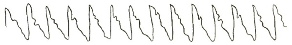
|
| Aortic Regurgitation. |
| FIG. 43. |
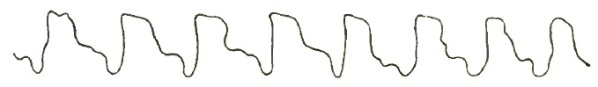
|
| Aortic Obstruction and Regurgitation (from a Patient in Bellevue Hospital). |
This vibrating pulse or pulse of unfilled arteries is usually possessed of fulness of volume, but when obstruction coexists it may be small and flickering unless the arteries are calcified or atheromatous. The pulse of aortic insufficiency taken by the sphygmograph resembles strongly the pouls des vieillards, but the senile pulse gives a rounded instead of a pointed summit. Still, in old age the two tracings may be indistinguishable.10 The peculiar crochet or beak is noticeable in graphic tracings of the pulse of aortic inadequacy.
10 Marey, Phys. Méd. de Circ. du Sang, Paris, 1863.
Stokes has described, under the designation of steel-hammer pulse, a peculiar and characteristic pulsation of the arteries which occurs in cases of acute rheumatic arthritis supervening upon chronic inadequacy of the aortic valves. The pulse is abrupt and energetic, as the rebound of a smith's hammer from the anvil; it is exhibited, however, only in the arteries adjacent to the affected joints.11
11 Continued Fever, 1874, p. 244.
PHYSICAL SIGNS.—Inspection.—There is an increase in the area of the apex-beat, which is plainly more forcible and is visible over a wider area than in aortic obstruction. After compensation has ceased to balance the forces in the heart the apex-beat becomes more and more feeble and diffused. One of the most important points obtained by inspection is pulsation of the carotids and the vessels of the upper extremities. Becker and Quincke have observed pulsation of the retinal vessels in cases of extensive aortic regurgitation.12
12 London Ophth. Hosp. Rep., Feb., 1873.
Palpation.—On placing the hand over the præcordial region a heaving, lifting impulse will be perceived, which is transmitted over a large portion of the thoracic walls. The apex-beat is displaced downward and toward the left, sometimes as far as the eighth rib and two and a half inches to the left of the left nipple. Occasionally a continuous diastolic thrill, equally intense during the whole of the diastole, is felt over the sternum, most distinctly at the site of the aortic valves. In some cases there is a slight pulsation in the scrobiculus cordis.
[p. 663] Percussion.—The area of percussion dulness corresponds to the extent of the cardiac enlargement. Deep dulness is elicited below and to the left of the normal area, and its outline has more of an oval contour than in health. So soon as the cardiac dilatation exceeds the hypertrophy, the area of dulness will extend horizontally rather than vertically, and it may be carried slightly upward, the apex beating in the axillary space. The area of dulness may extend six and a half inches from right to left, and from the upper edge of the third rib to the line of the liver dulness. The superficial area of dulness is likewise increased horizontally and toward the left.
Auscultation.—Aortic regurgitation is characterized by a diastolic murmur, which may take the place of, or immediately follow, the second sound of the heart. It is very distinct at any point over the base of the heart, but usually has its maximum intensity either at the sternal end of the second right costal cartilage, in the second right intercostal space, or at the sternal junction of the third rib on the left side. It is transmitted over the sternum, and sometimes will be loudest at the xiphoid cartilage, and is thence transmitted in the direction of the apex. Its area of diffusion is greater than that of any other cardiac murmur: it is not only conducted down the sternum to the xiphoid cartilage and to the apex, but it may be heard at the sides of the chest along the spinal column, and sometimes faintly in the ascending and transverse portions of the arch, in the carotids, and in rare instances as far as the radial arteries. The murmur of aortic reflux is accompanying rather than substitutive, for the pulmonic second sound is audible at the right base.
Foster13 regards incompetency of the posterior segment of the valve as producing a murmur which is conducted to the apex, whereas inadequacy of either or both of the anterior segments is accompanied by a murmur which is conducted to the ensiform cartilage. This point has a practical bearing on account of the relationship of the anterior segments of the valve to the coronary arteries. If the murmur indicates a lesion of the posterior flap of the valve, the prognosis will be better. When the second sound of the heart is distinct the murmur immediately follows it. Many English writers call the murmur a post-diastolic aortic murmur.
13 Med. Times and Gaz., 1873, vol. ii. p. 658 et seq.
Although having the greatest area of diffusion, aortic reflux has not the loudest murmur; it is soft, blowing, sometimes rough, and frequently musical. It is loudest at the beginning of diastole, gradually decreasing in intensity, although it may preserve its rushing, blowing character during all the diastole.
An aortic regurgitant murmur may temporarily disappear if a plug of fibrin closes the orifice, or if the walls of the left ventricle are the seat of extensive fatty degeneration, the aorta being rigid and inelastic.14 When aortic stenosis coexists there will be a double murmur, audible over a very large space, having its maximum intensity at the right edge of the sternum in the second interspace.
14 Brit. Med. Journ., 30th March, 1882.
Systolic and diastolic murmurs, though sometimes separated by a well-defined pause, may run into each other. If mitral regurgitation occurs with aortic regurgitation, each murmur retains its own location of maximum intensity. In rare instances, when two segments of the valve are healthy, a clear aortic second sound is heard, which is preceded by a faint reflux murmur. Such a murmur is said to be prediastolic in rhythm. Aortic reflux murmurs are often very indistinct, and can only be heard when the patient is in the recumbent posture. There is no necessary connection between the amount of reflux and the loudness of the murmur.
A diastolic murmur heard at or below the level of the aortic valves, chiefly [p. 664] audible in the line of the sternum, indicates considerable aortic incompetence. If a diastolic murmur is inaudible in the carotids, it is usually preceded by a systolic murmur, which has its maximum intensity at the aortic valves or in the so-called aortic area: such a murmur indicates comparatively trifling incompetence with considerable obstruction, probably produced by calcified semi-lunar valves.
If a diastolic murmur is distinctly audible in the carotid arteries, it is invariably preceded by a loud systolic murmur in them, the systolic portion of the murmur not being very plainly audible in the aortic nor in any part of the cardiac area: this indicates very considerable incompetence with comparatively trifling obstruction.
DIFFERENTIAL DIAGNOSIS.—The diagnosis of aortic regurgitation is generally not difficult, as it rests almost exclusively upon the existence or nonexistence of a diastolic murmur. It may, however, be mistaken for aortic stenosis, for mitral obstruction, for pericarditis localized over the aorta, for aneurism of the aorta, for aneurism of the aorta immediately above the valves, patency of the ductus arteriosus, for insufficiency of the pulmonic semi-lunar valves, and, occasionally, for a rough and inelastic condition of the ascending aorta.
1st. Mitral obstruction gives a presystolic murmur, while aortic reflux produces a diastolic murmur. Mitral stenosis is accompanied by no hypertrophy or dilatation of the left ventricle, whereas these conditions are always present with aortic reflux. The quality of a presystolic mitral murmur is harsh and rough, and it has a churning, blubbering, or grinding character, while aortic reflux has a murmur of low pitch and of a soft, blowing, or musical character. Mitral stenosis is accompanied by a purring thrill which is absent in aortic regurgitation. The murmur of mitral stenosis is the longest of all the cardiac murmurs. The murmur of mitral stenosis is never heard behind, whereas that of aortic regurgitation is often heard at the sides of the chest and along the spinal column. Finally, mitral stenosis is attended by well-marked pulmonary symptoms during active physical exertion, which are rarely present in aortic insufficiency.
2d. A pericardial friction sound over the aorta has its maximum intensity over the seat of its production, and is usually audible during both the cardiac systole and diastole. In aortic regurgitation the character of the pulse, the existence of hypertrophy and dilatation of the left ventricle, and the carotid pulsation will establish the diagnosis.
3d. An aneurism at the sinuses of Valsalva is diagnosticated by the history of the case, the presence of the murmur over the pulmonary artery, the evidences of arterial degeneration, the absence of left ventricular dilatation and hypertrophy, and the peculiar jerking pulse. An aneurismal murmur is circumscribed, has a booming quality, and is usually systolic in rhythm and never transmitted to the apex of the heart.
4th. Patency of the ductus arteriosus is a rare condition: in a case where this was diagnosticated15 the murmur was audible at the left of the sternum, was not everywhere continuous with the second sound, was only transmitted very feebly to the left, and had a wavy character, sufficient of itself to distinguish it from an aortic regurgitant murmur.
15 Guy's Hosp. Rep., Ser. 3, vol. xviii., 1872–73.
5th. Insufficiency of the pulmonic semi-lunar valves is the rarest of all valvular lesions: the murmur should be diastolic, having its maximum intensity in the second intercostal space of the left side; it should be transmitted only downward and toward the right apex; and should not be attended by arterial pulsation, a jerking pulse, or by left ventricular hypertrophy and dilatation.
6th. A diastolic murmur in the ascending arch, due to roughening, rigidity, [p. 665] and dilatation of the artery, is also rare, while the condition which some say can produce it is very common.
Two cases are recorded in which the diagnosis rested upon the character of the pulse, throbbing of the arteries, and the absence of hypertrophy and dilatation of the left ventricle.16
16 Bellingham Dis. of Heart, 1857, p. 152; also Trans. Path. Society, vol. iii., March, 1868, p. 3, article by Prof. Law.
Stenosis, or obstruction of the auriculo-ventricular opening of the left heart, is due partially to constriction at the base of the mitral valves, and partially to adhesions of the valve-tips or chordæ tendineæ. It usually occurs as a consequence of rheumatic endocarditis, rarely of atheromatous degeneration, and is most likely to occur in endocarditis affecting young persons.
Mitral disease is present in one-half the cases of valvular diseases of the heart. Usually, insufficiency and stenosis of the mitral orifice occur together, and stenosis probably never occurs without some insufficiency.
MORBID ANATOMY.—As a result of acute exudative or interstitial endocarditis, the valves are rendered shorter and narrower, as well as thicker and more cartilaginous, than normal. These rigid valvular projections not only obstruct the flow of blood from the auricle to the ventricle, but allow of its regurgitation from the ventricle into the auricle. In mitral stenosis there is not only thickening and contraction of the valves, but the valve-tips or the chordæ tendineæ become adherent, and sometimes each papillary muscle gives rise to a corrugated, cylindrical mass pierced with one or more slits, indicating the chordæ of which it was originally made up. The wall of the valve, especially toward its free edge, is greatly thickened, and these thickened portions are so dense that they have a distinctly cartilaginous feel. On the valvular flaps that have undergone this sclerotic change calcareous masses are very frequently developed, and are especially liable to form when a gouty diathesis exists.
When the chordæ tendineæ and papillary muscles have become adherent, the edges of the valves are drawn down toward the apex of the heart; and since the flaps are adherent for a greater or less distance upward from their bases, the valve presents a funnel-shaped appearance with its base looking toward the auricle and its apex toward the ventricle, whose smaller opening, rarely circular, usually resembles a slit with its axis in the line which unites the original segments of the valve. This button-hole slit may scarcely admit the tip of the little finger, while the normal mitral orifice permits the easy introduction of three fingers.
Annular (ring-like) stenosis is far more common at the mitral than at the aortic orifice. Hard, wart-like vegetations frequently develop on the puckered and seamed flaps, which increase the already existing obstruction. Sometimes the funnel-shaped appearance is wanting, and the flaps are stretched horizontally across, with a small opening in the centre, like a diaphragm: looked at it from the auricle, this slit is often crescentic in shape.
In cases of long standing the vegetations may become calcified. If the new tissue in the diseased valves undergoes fatty change and softens, ulcerative processes are set up and the chordæ tendineæ may rupture. On the floor of such ulcers calcareous masses and débris are frequently found.
Hayden thinks that "all funnel-shaped mitral stenosis is the result of primary acute inflammation and thickening of the valve-segments, with cohesion of their adjacent edges." Out of 62 cases of mitral stenosis, 59 assumed the button-hole form, and 3 only the funnel-shape (Fagge and Hayden).
[p. 666] In rare instances the tendons will adhere to the wall of the heart as well as become matted together. Adjacent to the valves the endocardium will usually be found slightly thickened.
The following changes are developed in the heart and vessels as the result of mitral stenosis: The left ventricle becomes smaller, and sometimes its walls are thinner than normal. The aorta is also small and thin-walled. An almost necessary result of mitral stenosis is dilatation, with subsequent hypertrophy of the left auricle. Sometimes the auricular cavity is enormously dilated—so much so that fifty years ago Thurman described it as true aneurism of the left auricle.17 Not infrequently the left auricular walls are from one-eighth to one-fourth of an inch in thickness. Its appendix is elongated, assuming a peculiar curved form, the aperture between it and the auricle becoming wider than normal. Moxon records a case of extensive mitral stenosis where the appendix was two and three-quarter inches long.
17 Med.-Chir. Trans., vol. iii., Ser. 2, p. 244.
As soon as the auricular hypertrophy ceases to be compensatory and dilatation begins, the pulmonary circulation becomes obstructed, causing increased tension in and distension of the pulmonary vessels. The walls of the pulmonary vessels, especially those of the main trunk, are thickened and hypertrophied; in rare cases they have been found twice the thickness of those of the aorta.
Although mitral stenosis is a disease of youth, and atheroma one of old age, yet it not infrequently happens that even before the age of puberty atheromatous degeneration occurs in the pulmonary vessels, especially in the small branches, as a result of the increased blood-tension in the pulmonary system.18
18 Trans. Path. Society, vol. xvii. p. 90.
The passive pulmonary hyperæmia which results from the obstructed pulmonary circulation may lead to those changes which collectively constitute brown induration of the lung. Another occasional occurrence, directly due to extensive mitral stenosis, is nodular hemorrhagic infarction. Hemorrhagic infarction of the lungs is in nearly every case preceded by thrombosis of the right side of the heart.
In some instances the enormously dilated left auricle may, by pressing on a bronchus, reduce its calibre one-half, and thus interfere with the functional activity of the left lung. When the pulmonary hyperæmia is extensive violent physical exertion or violent coughing may cause a rupture of one of the larger pulmonary vessels, and true pulmonary apoplexy result.
Bronchorrhoeal expectoration of large quantities of glairy mucus is a very frequent result of the intense hyperæmia of the mucous membrane of the bronchial tubes which sometimes occurs in mitral stenosis. The secretion is increased with every increase in the passive hyperæmia. The lungs are at all times so liable to congestion and oedema that any sudden or violent exercise may lead to a rapidly fatal result. Again, when the conditions enumerated have existed for some time, mitral stenosis may lead to hypertrophy of the right heart. In some rare cases the tricuspid orifice has become slightly insufficient.
ETIOLOGY.—Mitral disease is especially met with in the young, and in the child it is almost invariably a stenosis. The average age is about thirty-one; it is very rare to find it occurring after the fiftieth year of life. It seems from statistics that it is nearly twice as frequent in females as in males.
It is not infrequently of congenital origin. Acute rheumatic endocarditis is its most frequent cause. The mitral valves are more frequently affected in chorea than the aortic. In some few instances stenosis results from extension of the inflammatory process from the aortic semi-lunar valves, or prolonged aortic regurgitation and stenosis may lead mechanically to mitral disease, but not to stenosis. Niemeyer regards atheroma as an exceptional cause of mitral stenosis. No other authority regards it as a possible cause.
[p. 667] It is a question whether scarlatina or diphtheria tends to produce in children a valvular endocarditis which is followed by mitral stenosis. It seems plausible, since in many young children it is certain that mitral stenosis has not resulted from either rheumatism or chorea. Finally, with the exception of atheroma, all the causes enumerated in the etiology of aortic stenosis may be the cause of mitral stenosis.
SYMPTOMS.—Rational Signs.—The subjective cardiac symptoms of mitral stenosis are few. There may be no such symptoms. Usually, after violent exercise there is more or less cardiac palpitation, but this will cease as soon as the auricle can relieve itself, which is readily accomplished by the patient's assuming a recumbent position on the right side with the head slightly elevated. This class of patients as a rule are pale and anæmic. There is a sharp pain frequently felt in the region of the apex, which is always suggestive of mitral stenosis. The pulse is regular and normal in character so long as the auricular hypertrophy compensates for the auricular dilatation.
When the ventricle is unable to receive and discharge its normal quantity of blood with normal regularity, the pulse becomes small in volume, feeble in force, rapid and irregular in rhythm. The sphygmograph exhibits a tracing, frequently called the mitral pulse; the sphygmograph tracing is the same as when the ventricle throws a greatly diminished blood-current into the aorta (Fig. 44). This is asystolism, and the pulse is a clear indication of the condition.
| FIG. 44. |
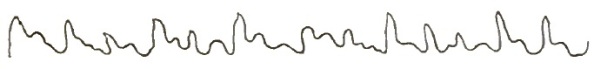
|
| Mitral Obstruction (from Patient in Bellevue Hospital). |
Balfour differs from other authorities in the statement that among the most remarkable subsidiary phenomena of mitral stenosis is irregularity of cardiac rhythm, which, always present in a greater or less degree, is sometimes a diagnostic phenomenon. The auricular systole commences earlier than normal on account of its hypertrophy. This premature contraction of the auricle, stimulating ventricular contraction, is indicated by a second ventricular systole which is much less forcible than the first.
The passive pulmonary hyperæmia attending the advanced stages of this form of cardiac disease causes habitual dyspnoea, which is exaggerated by physical exertion and is attended by a dry, hacking, teasing cough which resembles the so-called nervous cough.
After violent or prolonged exertion there may be bronchorrhoea, a pint of glairy, watery mucus often being expectorated in a few moments. Not infrequently severe exercise induces attacks of profuse, watery, blood-stained expectoration, indicative of pulmonary congestion and oedema. Sometimes the exertion of walking rapidly against a strong wind will induce such intense congestion and oedema of the lungs in one with extensive mitral stenosis as to cause sudden death.
Hæmoptysis is not infrequent, small quantities of pure florid blood being expectorated.
Orthopnoea is not a frequent symptom of mitral stenosis, for even in extensive and long-standing cases the pulmonary congestion is not constant, as the auricle is able ordinarily to empty itself, and only becomes engorged during active physical exertion or great mental excitement.
It should be mentioned here that the old idea, that "mitral stenosis sometimes produces hypertrophy of the left ventricle," is fallacious. In no instance can it be attributable to mitral stenosis.
[p. 668] Physical Signs.—Inspection.—As the left ventricle does not receive its normal quantity of blood, the cardiac impulse is feeble. Sometimes it has a visible undulating movement.
Palpation.—On palpation, although the apex-beat is less forcible than normal, a distinct purring thrill will be communicated to the hand: this thrill is a constant attendant of mitral stenosis, and may be regarded as its diagnostic sign. It should be remembered, however, that a purring thrill does not always indicate mitral stenosis. It is most distinct at the apex-beat, although it may be diffused over the whole præcordial space. It either continues through the entire diastole or is only present just before the systole. It is sometimes called a presystolic thrill. It ceases with the apex-beat. The only conditions besides mitral stenosis which will cause a purring thrill at the cardiac apex are mitral regurgitation, with extensive dilatation of the left ventricle, and left ventricular aneurism; in both instances the thrill will not be presystolic, but systolic.
Percussion.—The increased size of the left auricle may cause an increase in the area of cardiac dulness upward and to the left at the inner part of the second left interspace. This increased area of dulness will only be recognized on careful percussion during expiration.
Auscultation.—Mitral stenosis is characterized by a loud churning, grinding, or blubbering presystolic murmur; this murmur is of longer duration than any other cardiac murmur, on account of the time required for the blood to pass through the narrowed and obstructed orifice. It ends with the commencement of the first sound and the apex-beat, being synchronous with the purring thrill. The murmur is heard with its maximum intensity a little above the apex-beat.
Cryan records a case where the murmur was absent, but the diagnosis of mitral stenosis was made from the other symptoms. At the autopsy the orifice would barely admit the tip of the little finger, and the absence of the murmur was accounted for by the smallness of the aperture.19
19 Trans. Path. Society, Dublin, Part 2, vol. iv., 1870.
As a rule, mitral stenosis is accompanied by the loudest as well as the longest cardiac murmur. The murmur is always louder when the patient is erect than when in the recumbent posture. For a few days before death, and at any time when there is great constitutional debility, the murmur may be held in abeyance. A presystolic murmur is never present when auriculo-ventricular narrowing does not exist. When this lesion does exist it is never permanently, and very seldom temporarily, absent. A prolonged murmur and a sharp first sound indicate a funnel-shaped stenosis. A murmur immediately following the second sound, and running through the apex-beat, indicates great contraction of the orifice—diaphragmatic contraction. The murmur of mitral stenosis is very rarely, if ever, conveyed to the left of the apex-beat, and it is rarely heard more than two inches to the right of the apex. The second sound of the heart is intensified over the pulmonary valves. When mitral reflux and mitral obstruction coexist, the two murmurs run into each other, constituting a single murmur that may be mistaken for a systolic murmur. The harsh character of the presystolic element of the murmur can always be recognized.
A mitral obstructive murmur is never soft or musical, but there is a rare form of presystolic mitral which is so short as to resemble a tone. A mitral stenotic murmur does not often merge into the first sound of the heart, but is usually separated from it by a short interval. Sometimes a stenotic murmur only becomes audible when the patient sits up. In about one-third of all cases of stenosis of the mitral orifice the second sound is reduplicated. It is best heard at the apex and when the heart's action is slow. The reduplication may be temporarily absent. Pulmonary congestion efficiently accounts for [p. 669] this reduplication. Geigel ascribes it to "non-coincidence in the closure of the valves." Guttman regards it as originating at the stenotic orifice itself. Balfour thinks that thrill and reduplication of the second sound are sufficient to make a diagnosis in the absence of murmur. Some regard the length of the pause between the murmur and the first sound as a measure of the stenosis—the shorter the pause, the greater the stenosis.
DIFFERENTIAL DIAGNOSIS.—The diagnosis of mitral stenosis is not difficult; it mainly depends upon the existence of two physical signs—the purring thrill and a loud, long, blubbering presystolic murmur.
Mitral obstruction may be mistaken for the murmur of aortic regurgitation (see page 657), for a pericardial friction located over the apex, for a prolonged systolic murmur replacing the first sound at the apex, and for a prediastolic basic murmur transmitted to the apex.
1. To diagnosticate between local pericarditis and mitral stenosis, the same methods are employed and the same rules are to be observed as were mentioned in the diagnosis between aortic reflux and local pericarditis (p. 664).
2. A prolonged systolic apexial murmur, enduring as it does for the period of the first sound, that of the short pause, and reaching the second sound, is often accompanied by a muffled second sound readily mistaken for the first. The diagnosis of this murmur rests upon its soft and blowing character, the synchronism of the murmur with the systolic impulse and carotid pulsation, and the fact that there is no murmur with the second sound at the base.
| FIG. 45. |
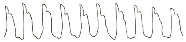
|
| Mitral and Aortic Obstruction and Regurgitation (from a Patient in Bellevue Hospital). |
A prediastolic murmur is distinguished from a mitral stenotic murmur by its progressively diminishing intensity from the base to the apex, by its being accompanied by hypertrophy of the left ventricle, and by a jerking, irregular pulse. The preceding tracings explain themselves.
Regurgitation at the mitral orifice is due to a condition of the mitral valves which allows the blood to flow back from the left ventricle into the left auricle. The backward effects of mitral reflux are more varied than those of any other valvular lesion.
It is a common form of valvular disease, and in the majority of cases is the result of acute exudative or interstitial endocarditis.
MORBID ANATOMY.—The most common lesions which give rise to mitral regurgitation are thickening, induration, and shortening of the mitral valves. In rare instances it may occur independent of valvular disease from displacement of one or more of the segments of the valve, the result of changes in the papillary muscles, chordæ tendineæ, or the ventricular walls. It may also occur in extreme anæmia, or from relaxation of the papillary muscles and dilatation of the left ventricle, without a corresponding elongation of the papillary muscles, and from rupture of the chordæ tendineæ. In most instances, however, the valves are shortened, thickened, and indurated.
In some instances lime salts and large masses of chalky matter are found [p. 670] imbedded in the indurated valves. In such cases the surface and edges of the valves are so rough and jagged that more or less obstruction accompanies the regurgitation.
All these changes, except calcification, may also occur in the chordæ tendineæ and columnæ carneæ. The valves may also become adherent to the walls of the ventricles, or as a result of the shrinking and shortening of the chordæ tendineæ the valve-flaps may not pass back to the plane of the orifice.
Again, the valves or the chordæ tendineæ may be ruptured, so that the valves are pressed during the cardiac systole back into the auricle. If the chordæ tendineæ which are inserted nearest the centre of the valve become lengthened, that part of the flap will be bent upon itself, having evidently yielded to the blood-pressure, and this allows of regurgitation. Sometimes, when the valves appear perfectly healthy, by the application of the water test they will be found to be insufficient.
The first effect of mitral regurgitation is dilatation of the left auricle, due to the pressure of the two blood-currents during its diastole—one from the lungs, and the other from the left ventricle. This dilatation leads to thickening and hypertrophy of the left auricular walls. Following this, the pulmonary circulation is impeded, the pulmonary vessels enlarge, and they may undergo degeneration as a result of the continued regurgitant pressure.
Passive congestion of the lungs with brown or pigment induration is an early pathological sequel of mitral regurgitation. The constant interference with the return circulation from the lungs obstructs more or less the outward current of blood to the lungs from the right ventricle. As the obstruction is a gradual one, the right ventricle becomes so hypertrophied as to overcome it. Consequently, the hypertrophied right ventricle compensates at first for the mitral regurgitation, and as long as the right ventricle is able to fully overcome the abnormal pressure of the blood in the lungs from the mitral regurgitation, so long the patients are comfortable. Sooner or later, however, the compensatory hypertrophy of the right ventricle ceases, and a secondary dilatation occurs which admits of no compensation.
This final dilatation of the right ventricle is favored by the myocardial degeneration, which occurs as a result of defective nutrition of the heart-walls; when this condition is reached the veins throughout the body are placed in a similar condition to those in the lungs.
This general venous congestion is indicated by passive hyperæmia of the abdominal viscera and by cyanosis of the surface during active physical exercise.
The liver is the organ first affected, on account of its great vascularity and from the fact that the hepatic veins do not collapse readily and possess no valves. Thus the liver becomes enlarged and stony (the nutmeg liver) as a result of the obstruction to the emptying of the hepatic vein, and when there is coexistent obstruction of the bile-ducts jaundice will be present.
This portal obstruction induces passive hyperæmia of the intestines and stomach, enlargement of the spleen, and large and painful hemorrhoidal tumors. The impediment to the return of blood from the brain causes cerebral congestion; from the kidney, renal congestion; and, finally, the obstruction to the systemic venous return leads to the accumulation of fluid in the areolar tissue and in the cavities. This dropsy generally begins in the feet and extends upward. In females the obstruction in the vena cava inferior induces derangements of the menstrual functions. Ascites, hydrothorax, hydro-pericardium, and pulmonary oedema may subsequently develop.
In addition to these changes, the dilated and hypertrophied left auricle throws an abnormal quantity of blood with abnormal force into the left ventricle during its diastole, which leads to dilatation of its cavity and necessitates a compensatory hypertrophy of the left ventricular walls. This [p. 671] hypertrophy of the left ventricle increases the force of the reflux current, so that during excitement and active physical exertion pulmonary congestion, oedema, and cerebral apoplexy are liable to occur. In many cases of mitral regurgitation, when the venous engorgement is excessive, general dropsy is favored by the anæmia produced by the obstruction of the thoracic duct.
Friedreich maintains that the augmented tension in the venous system causes an increased resistance in the systemic arteries, which leads to left ventricular hypertrophy.
ETIOLOGY.—Mitral regurgitation may occur at any age; it is especially liable to follow rheumatic endocarditis in the young.
Acute exudative and interstitial endocarditis of rheumatic origin is the primary cause of most of the changes which lead to mitral insufficiency. These changes cause the extensive retractions and thickenings which are present in most cases.
It may occur in conditions of extreme anæmia or where there is degeneration of the walls of the left ventricle.
It is not infrequently secondary to changes at the aortic orifice, produced either by an extension of endocarditis from the aortic to the mitral valves and their appendages, or by the secondary mitral valvulitis excited by regurgitant blood-currents from the aorta.
Mitral insufficiency may also be the result of the enlargement of the left auriculo-ventricular orifice which accompanies excessive dilatation of the left ventricle.
Disease of the columnæ carneæ and chordæ tendineæ, when their structures are so weakened as to allow the flaps of the valve to pass back of the plane of the orifice, will also cause mitral insufficiency.
Ulcerative endocarditis may cause it, either by perforation and rupture of the valves or by rupture of the chordæ tendineæ.
SYMPTOMS.—During the early stage of mitral insufficiency, when the hypertrophy of the right ventricle compensates for the regurgitation, there are no rational symptoms which would lead one to suspect its existence; but when the right ventricle is unable to overcome the obstruction to the pulmonary circulation caused by the regurgitant blood-current, there will be more or less dyspnoea, accompanied by a short, hacking cough, with an abundant expectoration of frothy serum. Sometimes the watery expectoration is blood-stained.
Frequently, the blood-stained expectoration is accompanied by free hæmoptysis, although it should be remembered that profuse hæmoptysis is far more frequent with stenosis than with regurgitation at the mitral orifice. But a cough and watery expectoration with occasional dark blood-stains are usually present as an advanced symptom of mitral regurgitation. Active physical exertion increases the dyspnoea and causes cardiac palpitation.
In advanced cases the extremities, face, and lips become blue, the result of the interference with the capillary circulation, and the liver becomes enlarged and hardened—conditions easily recognized by palpation and percussion.
The patient will complain of a sense of weight and fulness in the right hypochondrium, and there will be anorexia, nausea, and a sense of oppression in the epigastrium. Sometimes the hepatic circulation becomes so obstructed that the biliary secretion is interfered with, and jaundice will be added to the cyanotic discoloration, which gives to the surface a peculiar greenish hue.
Following the hepatic derangement are frequent attacks of gastric and intestinal catarrh and evidences of embarrassed renal circulation.
The urine is diminished in quantity, high-colored, and loaded with lithates. Sometimes albumen and fibrinous or blood casts are found in it.
[p. 672] Headache, dizziness, vertigo, stupor, somnolence, and sometimes a peculiar form of delirium of short duration, result from the passive cerebral hyperæmia induced by obstruction in the superior vena cava.
A late symptom of mitral regurgitation is dropsy, which results both from impaired general nutrition and the abnormal blood-pressure in the venous system, both together causing an exudation of the watery portion of the blood through the walls of the vessels. Dropsy, from mechanical causes having their seat in the heart, first appears in the lower extremities, the ankles becoming oedematous, and thence may extend over the whole body. For this condition to be reached it may require several years or only a few months, depending upon the general condition of the patient and the amount of the reflux. With the general anasarca the dyspnoea becomes extreme; the serous cavities of the body as well as the lungs become oedematous; erythema may occur in the region of the groins, the skin exhibiting a tendency to diffuse gangrene.
Late in the disease pulmonary hemorrhagic infarction may occur as a result of metastasis, and this, in the vast majority of cases, lights up a rapidly fatal pneumonia.
All these changes, however grave and urgent they may be, are gradual in their development, so that the condition of the patient is not so insufferable as its description would lead one to suppose.
The pulse of mitral regurgitation is at first in no respect characteristic. It remains regular in force and rhythm, but later it becomes somewhat diminished in force and volume, irregular in its rhythm, and increased in frequency, but never jerking in character. This tracing illustrates my meaning. While it remains full it is feeble and always compressible. When the heart's action is excited, it has a certain tremulousness: these last-named characteristics are to be regarded more as the result of the failure of the left ventricle than of changes in the valvular insufficiency. If a mitral regurgitant pulse has any distinctive peculiarity, it is its diminution in volume.
| FIG. 46. |

|
| Mitral Regurgitation (from a Patient in Bellevue Hospital). |
Coincident mitral or aortic stenosis may render the pulse regular even in extensive mitral regurgitation.
| FIG. 47. |

|
| Mitral and Aortic Regurgitation (from a Patient in Bellevue Hospital). |
Physical Signs.—Inspection.—The area of visible cardiac impulse extends over an abnormal space, and is more or less distinct according as the right ventricular hypertrophy is moderate or extensive. Sometimes the thoracic wall is seen to rise and fall with each cardiac cycle, and not infrequently the epigastrium exhibits slight pulsation corresponding in rhythm with the heart-beats.
The epigastric pulsation is due to the right ventricular hypertrophy always found with extensive mitral regurgitation.
Skoda, Bamberger, and Leyden record a few instances in which inspection revealed a double impulse accompanying, with more or less regularity, each cardiac systole. This double impulse only occurs in aggravated cases of [p. 673] mitral insufficiency, and arises from non-coincidence of contraction of the two ventricles.
The jugular veins appear swollen, and this is always most conspicuous when the patient is lying down.
Palpation.—The apex-beat is displaced to the left. When hypertrophy predominates over dilatation, the apex-beat is felt lower than normal. When the dilatation exceeds the hypertrophy, the apex-beat is carried outward and often slightly upward. The impulse is diffused and more or less forcible according as the right or left ventricular hypertrophy predominates. This systolic frémissement is most noticeable when the base of the heart lies close to the chest-wall from retraction of the margin of the left lung.
Purring tremor, systolic in rhythm, felt most intensely at the apex and becoming feebler the farther the hand is removed from that part, either to the right or upward, is invariably due to mitral reflux.
Hayden says that it is exceptional to have a purring thrill with simple mitral reflux. I have never found it except in those cases where left ventricular dilatation greatly exceeded the hypertrophy.
Percussion.—Percussion reveals an increase in the area of cardiac dulness, especially laterally; it extends both to the left and right of the normal line, as well as downward. The area of superficial as well as deep-seated dulness will be increased laterally and downward.
Auscultation.—Mitral insufficiency is attended by a systolic murmur which either completely or partially replaces the first sound of the heart. The quality of the murmur is variable, and not in itself as distinctive as that of mitral stenosis. It is usually a soft and blowing bellows murmur; sometimes, toward its end, the murmur will assume a distinctly musical character.
While the first sound of the heart may be heard distinctly in the early stages of mitral reflux, later the murmur in nearly all cases takes the place of the heart-sounds. Hence many English writers rightly denominate this murmur as post-systolic rather than systolic in its nascent stages. It is heard with its maximum intensity at the apex-beat. Its area of diffusion is to the left on a line corresponding to the apex-beat. It is audible at or near the inferior angle of the left scapula. It can be heard between the lower border of the fifth and the upper border of the eighth vertebra, at the left of the spine, with nearly the same intensity as at the apex. The murmur may be absent from the latter situation until cardiac hypertrophy is developed.
The second sound of the heart over the pulmonary valves is accentuated, while below the junction of the third rib with the sternum on the left side both heart-sounds are feeble. Skoda first drew attention to exaggeration of the second pulmonary arterial sound as a positive and unerring indication of mitral regurgitation.
An intensified pulmonary second sound requires a strong right ventricle and an intact tricuspid valve, and is not always present. In general terms, the area of diffusion of a mitral regurgitant murmur is toward the left of the apex-beat. Whatever may be its character, the murmur is generally loudest at its commencement. A loud systolic murmur at the apex, and not heard at the back, is probably not produced by mitral reflux.
As at the aortic orifice, so at the mitral, stenosis and regurgitation are apt to occur in the same individual, giving rise to a combined presystolic and systolic murmur, which is a continuous murmur that begins shortly after the second sound of the heart and often continues until the second sound commences. The two sounds, although mingling to form one murmur, can, in the majority of cases, be readily distinguished from each other, for the point of maximum intensity and the very limited area of diffusion of a presystolic murmur readily distinguish it from a mitral systolic which is audible in the left scapular region. It is important to recognize the existence of both these [p. 674] murmurs in estimating the prognosis in any case. Guttman mentions a case where five distinct murmurs were combined and yet clearly distinguishable.
DIFFERENTIAL DIAGNOSIS.—It is usually not difficult to recognize mitral regurgitation. The seat and rhythm of the murmur and its area of diffusion are sufficient to distinguish it from other cardiac murmurs. The character of the pulse, the symptoms referable to the right heart, and the pulmonary complications will also assist in its diagnosis.
It may, however, be mistaken for aortic obstruction, since both give rise to a systolic murmur, for tricuspid regurgitation, for fibroid disease of the heart, and for roughening of the ventricular surface of the mitral valve or of the ventricular wall near the aortic orifice.
The diagnosis between mitral regurgitation and aortic stenosis has already been given (see page 657).
Mitral and tricuspid insufficiency both produce a systolic murmur, but a mitral regurgitant murmur has its maximum of intensity at the apex, and is conveyed toward the left axillary and scapular regions, while the maximum intensity of a tricuspid regurgitant murmur is to the right of the base of the xiphoid cartilage, and it is transmitted upward and to the right: the area of transmission establishes the diagnosis.
Pulmonary symptoms are prominent in mitral reflux, and absent in tricuspid regurgitation. The pulmonary second sound is markedly enfeebled in tricuspid regurgitation, and markedly intensified in mitral regurgitation.
Fibroid disease of the heart may produce a systolic apex murmur, but it is an exceedingly rare disease, a pathological curiosity.20
20 In the Pathological Transactions (1874, vol. xxv. p. 64) Fagge records a few cases, and mentions that perhaps one positive indication of fibroid disease of the heart, rather than of a valvular lesion, may be found in its resisting treatment with greater obstinacy.
Roughening of the ventricular wall gives rise to a murmur which has its maximum intensity at the base of the heart, and is transmitted along the aortic arch and into the vessels which spring from it in the thorax.
The vibration of an irregular chordæ tendineæ stretched across the aortic orifice, its extremities being inserted into opposite walls of the ventricle, may produce a systolic musical murmur, but the line of its transmission will correspond to that of an aortic obstruction. A systolic mitral murmur due to the sudden rupture of one or a number of the valve-flaps, of the papillary muscles or tendons, is accompanied by a loud systolic blowing murmur, which is immediately accompanied by all the urgent symptoms of acute pulmonary congestion.
On account of the infrequency of disease of the pulmonic valves very little is known of the phenomena to which such diseases may give rise. In fact, they are so rare that there is no written history of their subjective symptoms; their diagnosis is only arrived at by exclusion, and they cannot be recognized except by the physical signs which attend them.
As has been already stated, endocarditis in the right heart is rare, except in intra-uterine life, and the various conditions of the aorta, atheroma, aortitis, etc., which I have mentioned in the etiology of aortic valvular disease have no analogues in the pulmonary vessels.
Usually, valvular disease of the right heart is the sequela of lesions in the left. It must be remembered, however, that the pulmonary artery may become atheromatous. I have already shown (see p. 666) how certain valvular diseases of the left heart may induce such a pathological condition. But even under such conditions disease of the pulmonary valves is rare. Balfour [p. 675] believes that constriction of the pulmonary artery may occur at various periods of intra-uterine life. As a rule, the pulmonary valves are subject to no lesions except congenital malformation.
MORBID ANATOMY.—Bertin records an instance of pulmonary obstruction where the valves, distorted and adherent, formed a horizontal septum across the orifice, it being barely one-fourth of an inch.
A rigid tricuspid valve has been found to be the cause of obstruction at the pulmonary orifice, the pulmonary valves themselves being normal. A few autopsies have revealed obstructions at the pulmonary artery, caused not so much by valvular defect as by aneurisms, tumors of the pericardium or of the anterior mediastinum, enlarged bronchial glands, or pressure of a solidified lung.
The pulmonary artery may be occluded just beyond the valves by a cancerous tumor, and there are examples where a phthisical process in the left lung has induced it.
A murmur indicative of pulmonary obstruction may be produced by a cardiac thrombosis.
I have placed these statements under the head of its morbid anatomy for the reason that they cannot be appreciated and their pathological significance realized during life.
Reasoning from analogy, obstruction at the pulmonary orifice ought to be followed by compensatory hypertrophy of the right ventricle and accompanied by tricuspid regurgitation and dilatation of the right auricle.
Ormerod records 3 cases21 where pulmonary obstruction was diagnosticated during life, and where the post-mortem proved the accuracy of the diagnosis: 2 of these cases occurred in men under twenty-eight, and the other in a woman of twenty-one. In 2 of these cases all the other cardiac valves were healthy. The pulmonic orifice would barely admit the introduction of a goosequill. Warburton Bigbie mentions a case (man æt. eighteen) where reflux and stenosis at the pulmonary orifice coexisted. There were four valves, and these were incompetent. All the other valves were normal.
21 Edin. Med. and Surg. Journ.
Congenital stenosis of the infundibulum of the right ventricle is the probable result of foetal myocarditis or of syphilis.
I have never met but two pulmonic obstructive murmurs where subsequent autopsies were obtained. In both cases it was found that the murmur had been produced by mediastinal tumors pressing on the pulmonic artery so as to diminish the calibre.
ETIOLOGY.—Pulmonary stenosis is rarely the result of endocarditis or of degenerative changes in the pulmonary artery. Bertin states that when abnormal communication between the two sides of the heart has existed, the arterial blood has excited endocarditis in the right heart.
Syphilis has been advanced as a possible cause of degenerations at the pulmonic orifice.
SYMPTOMS.—The only rational symptoms that have been noted in the few recorded cases of pulmonic disease admit of manifold explanations, and no one is either constant or diagnostic. In some cases anæmia existed, in others there were cardiac palpitation, dyspnoea, cyanosis, and dropsy; but none of these belong exclusively to a pulmonic lesion nor do they necessarily depend upon it.
Physical Signs.—Inspection, palpation, and percussion give negative rather than positive results. In a few instances palpation may give a systolic thrill confined to the second left intercostal articulation. Such a frémissement results both from roughness and contraction of the pulmonic orifice.
Auscultation.—A systolic murmur is heard with its maximum intensity directly over the pulmonic valves; it is very superficial, and consequently [p. 676] very distinct, and it is limited in its diffusion. It is never heard at the xiphoid cartilage nor along the course of the aorta. If it has an area of diffusion, it is toward the left shoulder. The murmur is loud and soft in character, sometimes bellows. It is not audible in the vessels of the neck nor is it attended by arterial pulsation.
When phthisical consolidation partially occludes the pulmonary artery, a loud but soft systolic murmur is heard, which is sometimes high-pitched and musical, and often entirely suspended during a full inspiration. In some few instances there is a bruit de diable in the jugular veins.
DIFFERENTIAL DIAGNOSIS.—It is possible to confound a pulmonic obstructive murmur with a mitral regurgitation which is propagated upward into the left auricular appendix. But the area of a mitral regurgitant is also backward, and by this it could be distinguished from a pulmonic obstruction. Besides, in mitral disease the pulse is very different from the pulse of pulmonary stenosis.
Aortic stenosis can hardly be mistaken for pulmonary obstruction, for the arterial pulsation, the peculiar pulse, and the transmission of the murmur into the arteries of the neck will suffice to discriminate between them.
An aneurism at the sinus of Valsalva may produce a systolic pulmonary murmur by the pressure which it produces upon the pulmonary artery. It would be impossible to distinguish it from a pulmonic stenosis.
The diagnosis of pulmonary obstruction is usually reached only by exclusion.
This form of valvular lesion is exceedingly rare; indeed, many doubt its occurrence. The lesion seldom occurs except as the result of injury or congenital defect, and there are but few well-authenticated cases in medical literature.22
22 Path. Trans., vol. xvi. p. 74.
The statement23 that the pulmonary valves exhibit a cribriform condition nearly as often as the aortic is not sustained in this country by the results of post-mortems. In one of the cases to which I have referred (p. 675) as an example of pulmonary stenosis the valves were likewise found insufficient. In Bigbie's case (referred to on p. 675), where there were four flaps to the valve (producing obstruction), there was marked insufficiency coexisting.
23 Dis. of the Heart, Bellingham.
The morbid anatomy, etiology, and rational symptoms do not require a separate consideration. The anatomical appearances are the same as those found in similar conditions of the aortic valves, and the etiology and rational symptoms are the same as those of pulmonic stenosis.
Physical Signs.—Theoretically, pulmonic regurgitation should be accompanied by a diastolic murmur having its maximum intensity over the pulmonic valves, and its area of diffusion should be downward and toward the xiphoid cartilage. It should be soft and blowing in character. This murmur is rarely heard alone: it is usually associated with obstruction at the same orifice or with some murmur whose origin is on the left side of the heart.
Niemeyer states that dyspnoea, hemorrhagic infarction, and consumption of the lungs have followed insufficiency at the pulmonary orifice. No other authority mentions any such symptoms, while the assignment of valvular disease as a cause of phthisis is not based upon clinical facts.
With a pulmonic regurgitant murmur there should be on palpation and percussion physical evidences of hypertrophy and dilatation of the right heart, the rationale of whose production would be identical with that which was considered in aortic regurgitation. I have never heard a regurgitant pulmonic murmur.
[p. 677] DIFFERENTIAL DIAGNOSIS.—The murmur of pulmonary regurgitation may be mistaken for that of aortic regurgitation. The points in connection with their differentiation are fully discussed on p. 664.
The PROGNOSIS and TREATMENT are identical with those of the former lesion.
This valvular lesion is so rare that there are no established rules for its diagnosis.
Its MORBID APPEARANCES and ETIOLOGY are the same as those of pulmonic stenosis.
The SYMPTOMS of tricuspid stenosis would be those due to obstruction to the entire venous circulation. The right auricle would be dilated, and there would be visceral enlargements in the abdomen, cyanosis of the face and extremities, scanty and albuminous urine, hemorrhoidal tumors, headache, dizziness and vertigo due to passive cerebral hyperæmia, and finally general anasarca. The few recorded cases were associated with mitral stenosis with one exception, a case of Bertin's.24
24 Traité des Maladies du Coeur, Obs. 17.
In a case exhibited by Quain the tricuspid flaps, thick and opaque, were united for one-third of their extent. In the other cases the valve-flaps formed a diaphragm whose central opening varied in size, admitting only the point of one finger. In every condition of tricuspid stenosis the heart was enlarged.
Tricuspid stenosis (as in pulmonic stenosis) may be the result of pressure of tumors.
In all well-authenticated cases the chief symptoms seem to be extreme lividity, palpitation, and dyspnoea.
Physical Signs.—Inspection reveals general cyanosis. The jugulars are turgescent and exhibit presystolic pulsation. This pulsation is sometimes the only inconvenience the patient suffers.
Palpation may discover a venous thrill at the base of the neck.
Percussion may show the right auricle to be greatly enlarged, and cardiac dulness will be increased laterally and toward the right.
Auscultation.—Tricuspid stenosis should be attended by a presystolic murmur whose maximum intensity would be at the lower portion of the sternum just above the xiphoid cartilage. This murmur may be propagated faintly toward the base, but never toward the apex of the heart. It is sometimes accompanied by fremitus.
Hayden offers the following diagnostic point: The murmur of mitral stenosis (without which tricuspid stenosis never occurs) is limited to the apex region; a murmur of the same rhythm is produced at the sternum by tricuspid stenosis, "and between these two localities there is a point where no murmur can be heard."
It is unnecessary to consider its differential diagnosis.
The lesion would be diagnosticated (if at all) by exclusion, and prognosis and treatment would depend on the gravity and sequelæ of the accompanying condition—viz. Mitral Stenosis (q. v.), for the rule is, that stenosis of the tricuspid never occurs unless there is extensive mitral obstruction, and the latter condition is always the predominant one.
Regurgitation at the tricuspid orifice is generally secondary to mitral stenosis or regurgitation; primary disease of the tricuspid valves, however, is not infrequent.
[p. 678] MORBID ANATOMY.—The valvular lesions which lead to tricuspid insufficiency are similar to those which produce mitral insufficiency. The valves are thickened, shrunken, and opaque, the papillary muscles are shortened, thickened, and the chordæ tendineæ undergo similar changes and are sometimes adherent.
The valves or the chordæ tendineæ and columnæ carneæ may rupture; in either case acute and extensive insufficiency results, as has been stated. Acute endocarditis of the right heart is rare in adult life, but when it occurs the tricuspid valves are its primary and principal seat.
The reason for this is found in their anatomical structure and in the tension to which they are subject in diseases of the mitral valves. They are rarely the seat of rheumatic endocarditis or calcareous degenerations.
Ulcerative endocarditis is seldom met with in the right heart. In a case recorded by Charcot and Vulpian one of the tricuspid valves was softened and perforated, presenting numerous vegetations. Scattered abscesses in the lungs were found in this case.
Any infection through emboli from the tricuspid flaps will produce secondary effects within the thoracic cavity. The first effect of tricuspid regurgitation is dilatation of the right auricle; following this there will be more or less hypertrophy of its walls. As soon as the valves in the subclavian and jugular veins are no longer able to resist the regurgitant current jugular pulsation follows. But before this occurs the tributaries of the inferior cava and the organs to which they are distributed will become greatly engorged, for they have no valves to resist the regurgitant current, as are found in the veins coming from the upper part of the body. The inferior cava and the hepatic veins sometimes become enormously distended under these circumstances, and the liver will show the peculiar section that has gained for it the name of nutmeg liver.
Following the hepatic changes, the skin assumes a dingy yellow hue. When this is combined with cyanosis it produces a peculiar greenish tint which is only met with in heart disease. The spleen enlarges and hardens; the mucous membrane of the stomach is congested, ecchymotic, and often presents numerous hemorrhagic erosions. Intestinal catarrh is subsequently developed, and the general venous congestion within the abdominal cavity is exhibited by hemorrhoids and ascites. The kidneys become congested and stony, and thrombi may form in the femoral vein and induce subsequent pulmonary infarctions.
The stasis in the veins below the diaphragm is accompanied by transudation of serum—first in the ankles, and thence the dropsy progresses upward until the patient may finally reach a condition of general anasarca. The obstruction to the general systemic circulation which results may subject the left ventricle to so much extra labor that it hypertrophies, and then we have the infrequent occurrence of disease of the left heart following that of the right.
Since tricuspid reflux has mitral disease for its principal cause in abnormal cases, the heart becomes greatly enlarged and a condition of extreme cardiac dilatation and hypertrophy is reached.
ETIOLOGY.—As has been stated, the most frequent cause of tricuspid regurgitation is mitral disease, either stenosis or regurgitation. Any condition of the lungs which will produce hypertrophy and dilatation of the right ventricle will lead to it; it is met with in extensive pulmonary emphysema, in cirrhosis of the lung, and in extensive chronic bronchitis. Balfour regards chronic bronchitis as its most frequent cause after mitral stenosis.
It is possible for any valvular disease in the left heart, when of long duration, to lead to tricuspid regurgitation. From all these causes the rationale is [p. 679] the same: the abnormal amount of blood in the right ventricle presses with undue force against a valve, which physiologists regard as normally slightly insufficient; the stress upon the valve-flaps and the valvular attachments is such that endocardial inflammation is excited at the part subject to the greatest strain, and valvular insufficiency is the result.
It is possible for disease of the tricuspid valves to result from any of the causes which have been enumerated on p. 666 as etiological factors in valvular diseases.
SYMPTOMS.—Tricuspid regurgitation being in the majority of cases secondary to some other valvular disease or some chronic pulmonary affection, its symptoms during the early stages are vague and masked by those of the primary disease. But as soon as the valves become so insufficient that the venous return is markedly impeded, a train of symptoms is developed which has its origin in the visceral derangements already referred to.
In addition to these symptoms there may be, with extensive tricuspid regurgitation, cardiac palpitation, cardiac dyspnoea, and marked irregularity in the force and rhythm of the heart. The liver and spleen are enlarged, the skin becomes dingy, and there is obstinate constipation with hemorrhoids. The liver is likewise rendered very liable under such circumstances to attacks of interstitial hepatitis. Venous stasis is evinced by dyspepsia, nausea, vomiting, and hæmatemesis. The secretion of the kidneys is scanty, dark-colored, of high specific gravity, often containing albumen and casts.
Passive cerebral hyperæmia is marked by headache, dizziness, vertigo, and muscæ volitantes, and there is a peculiar mental disturbance which is not met with in any other form of heart disease.
Late in the disease, if the patient is placed in a horizontal position, the face becomes turgid and blue, and if he remain long in the recumbent position stupor and coma may supervene. Jugular and epigastric pulsation are characteristic physical signs.
A very late symptom is dropsy, which begins at the ankles and extends upward until there is general anasarca. It is a point to be noticed that in the dropsy from tricuspid reflux the genital organs suffer slightly if at all.
| FIG. 48. |
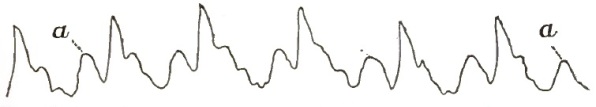
|
| Tricuspid Regurgitation (after Galabin): a, a, anadicrotic wave synchronous with the auricular systole, and caused by reflux into the large veins. |
Physical Signs.—Inspection.—In extensive tricuspid disease the area of the cardiac impulse is increased more than in any other valvular lesion. This area sometimes extends from the nipple to the xiphoid cartilage, and it may reach as high as the second right intercostal space. There is a visible impulse in the jugular veins, more apparent in the right than in the left. Sometimes the veins in the face, arms, and hands, or even the thyroid and mammary veins, are seen to pulsate.
Palpation.—The apex-beat is indistinct, except in cases where there is marked hypertrophy of the left ventricle. Pulsation occurs in the epigastrium, which may be due to reflux into the enlarged hepatic veins or to the fact that the dilated and hypertrophied right ventricle so presses on the liver that the impulse is conveyed through the diaphragm with each cardiac pulsation. Guttman thinks epigastric pulsation is due wholly to reflux into the veins of the liver, and not to right ventricular pulsation.
[p. 680] Early in the disease the impulse in the jugulars is confined to the lower part of the vessels, particularly to the sinus. Beyond this point the vein merely undulates. Later, a systolic pulsation is felt as high up as the angle of the jaw, and may be accompanied by distinct though feeble presystolic pulsation.
The liver may first simply undergo systolic depression, chiefly at the left lobe; secondly, the whole liver may have an impulse coming from an enormously dilated vena cava; and thirdly, the systolic pulsation of the veins within the organ may give to it a palpable expanso-pulsatory movement. The hepatic pulsation is rhythmical with the cardiac impulse. In rare cases it precedes jugular pulsation. Sometimes pulsation is felt in the femoral veins.
Sphygmographic tracings of the jugular pulse show it to be dicrotic.
Percussion shows an increase in the area of cardiac dulness to the right and upward, sometimes as far as the second intercostal space.
Auscultation.—The murmur of tricuspid insufficiency is heard with, or takes the place of, the first sound of the heart; it is superficial, of low pitch, blowing, soft, and faint, and is heard with the greatest intensity over the lower part of the sternum, at its left border, between the fourth and sixth ribs. It is rarely audible above the third rib or to the left of the apex-beat. This murmur is transmitted from the region at the base of the xiphoid cartilage upward and to the right from one to two inches. Sometimes it is heard only over a very limited area, and then it may be overlooked.
DIFFERENTIAL DIAGNOSIS.—A tricuspid regurgitant murmur may be confounded with that due to aortic obstruction, pulmonic obstruction, and mitral regurgitation. A tricuspid regurgitant murmur is never audible above the third rib; is not accompanied by an accentuation of the second sound over the pulmonary artery, but by jugular and epigastric pulsation; and is heard with maximum intensity near the base of the ensiform cartilage. These points are sufficient to differentiate it from an aortic or pulmonary obstructive murmur. The differential diagnosis between it and a mitral regurgitant murmur has been given.
PROGNOSIS IN VALVULAR DISEASES OF THE HEART.—Any statements as to the duration of life in valvular diseases of the heart, and their relative frequency as a cause of death (especially of sudden death), must be based upon personal observation, and necessarily will differ with different observers.
In order to establish, if possible, a basis of comparison for the different valvular lesions, I give a résumé which I have made of 81 cases, in all of which autopsies were made and the diagnosis of valvular disease verified.25
25 Med. Rec. N.Y., April 1, 1870, p. 66 et seq.
In 14 cases of various valvular lesions, each of which was accompanied by cardiac hypertrophy and dilatation, 50 per cent. of the deaths were due directly to the valvular lesion. In 1 of these, where there was stenosis at both auriculo-ventricular orifices, death was sudden.
In 15 cases of valvular disease, in which there was only cardiac hypertrophy, there were 11 deaths from the heart lesion. In 5 of these death occurred suddenly, and these 5 sudden deaths were all directly due to the heart lesion.
In 6 cases of valvular disease accompanied by dilatation alone, 4 deaths resulted directly from the heart lesion, and 2 of these were sudden.
In 15 cases where the aortic valves were involved (either calcified, rigid, or atheromatous) the heart lesion was not the cause of death in any case. Of these 15 cases, sudden death occurred but in 2; in 1 there were firm and long-standing pericardial adhesions, and in the other cerebral apoplexy.
In 12 cases of calcification of the mitral valve, no death occurred as the direct result of the valvular lesion, and there were only 2 sudden deaths, both from cerebral apoplexy.
[p. 681] The aortic and mitral valves were diseased in 14 cases; in 2 of these only did death result from the heart lesion, and the only three sudden deaths in this class were from uræmia, apoplexy, and croupous laryngitis.
The aortic and pulmonic valves were both diseased in 3 cases which died suddenly, and in no instance was death due directly to the heart lesions.
In 2 cases there was disease at the aortic, mitral, and tricuspid orifices, and no sudden death.
Thus it will be seen that of these 81 cases, in 24 only was death due directly to the heart lesion. There were only 8 sudden deaths due directly to the heart lesion.
The results of personal, clinical, and pathological observation lead me to the opinion that the loudness, harshness, and the area of diffusion of any cardiac murmur have little to do with its prognosis.
I deduce from the above-mentioned cases that cardiac murmurs rarely necessitate a bad prognosis unless hypertrophy and dilatation coexist; but so soon as the signs of considerable dilatation and hypertrophy are present a great variety of complications are liable to occur.
In 1870, I had a patient sixty years of age with extensive aortic reflux, who had been under my observation eight years, during which time he had three attacks of pneumonia. There were no appreciable signs of cardiac dilatation in his case.
Walshe says: "The order of relative gravity, as estimated not only by their ultimate lethal tendency, but by the amount of complicated miseries they inflict, is—1, tricuspid regurgitation; 2, mitral obstruction and regurgitation; 3, aortic regurgitation; 4, pulmonic obstruction, 5, aortic obstruction."
The following are conditions which render the prognosis in each valvular lesion more or less unfavorable:
In aortic stenosis the prognosis is less grave than in any other valvular lesion. Life may be prolonged and good health enjoyed for many years. Yet it must be remembered that extensive aortic stenosis rarely exists without attendant regurgitation.
So long as the hypertrophy of the left ventricle compensates for the obstruction, the prognosis is good; but when the hypertrophied walls fail to overcome the obstruction, dilatation begins, and the ventricular systole becomes feeble and intermitting, and the arterial supply to the brain is so much diminished as to lead to cerebral anæmia.
If after sudden exertion or violent muscular effort there is interruption or great irregularity in the heart's action, sudden death may occur from a complete arrest of the ventricular systole.
Evidences of excessive hypertrophy and dilatation, the occurrence of syncope, signs of cerebral anæmia, attacks of vertigo, great muscular prostration, continued and marked paleness of the face, and irregularity of the pulse, render the prognosis exceedingly unfavorable in aortic stenosis.
If the presence of vegetations can be determined, there is danger from cerebral embolism.
When there are no evidences of alterations in the ventricular walls after an aortic obstructive murmur has existed for some time, it may be assumed that no vegetations exist on the valves, and that the murmur is not due to extensive aortic stenosis, and consequently is not dangerous to life.
When the mitral valves become involved, the combined lesions render the prognosis unfavorable.
Death may result from cerebral complications, pulmonary oedema, or cardiac degeneration.
Aortic insufficiency is a much graver form of valvular disease than aortic stenosis. It is difficult to estimate the probable duration of life in aortic [p. 682] insufficiency, for it frequently gives rise to no symptom that would lead to its diagnosis until it is far advanced. Twenty-one days and five years are the extreme limits that have been recorded. It must always be borne in mind in estimating the factors for and against a good prognosis that in no other valvular lesion is sudden death so liable to occur. Yet the record of the cases which I have given (page 680) indicates that mitral stenosis is nearly, if not quite, as frequently a cause of sudden death.
A diseased valve can never be restored to its normal functions, and the shorter and more gushing the murmur the more extensive the regurgitation. The effects of the regurgitation must be carefully estimated before a prognosis can be given in any case. When one aortic flap is puckered and shrunken, the other two may elongate and compensate for the patency. But this occurs only in very young subjects.
Aortic regurgitation is, however, more serious in the very young than in adults. In children the valvular changes are less atrophic and more inflammatory in character.
Where the disease is met with in middle life, in those who daily undergo severe mental or bodily strain, the prognosis is unfavorable. And when in such patients there are the evidences of arterial degeneration or a tendency to it, the dangers are greatly increased, for the hypertrophied ventricle drives out the blood from its dilated cavity with greater than the normal force, and the vessels being weakened there is great danger of their rupture; hence the frequent occurrence of apoplexy and infarctions. In the very old I have seen aortic incompetence last a long time and cause little inconvenience.
Again, the prognosis is bad when cyanosis and dropsy result from the failure of a dilated and hypertrophied left ventricle to empty itself. This weakness is the result of that interference with the coronary circulation which brings about impaired nutrition, and therefore degeneration of the heart-walls.
When mitral insufficiency is secondarily induced, then obstruction to the systemic circulation leads to induration of the liver and kidneys, which interferes with the performance of their functions and hastens the fatal issue.
Sudden rupture of a valve or valvular disease that has developed very rapidly is more dangerous than when the valvular insufficiency is slowly developed. The flap or flaps involved can sometimes be determined during life, and then the prognosis will be more or less favorable according as the anterior or posterior are incompetent. In all cases the prognosis depends more upon the condition of the heart-walls and on the general nutrition than upon any other element.
When aortic regurgitation is complicated by aortic stenosis, mitral regurgitation, or by the vascular and visceral conditions resulting from the derangement of the circulation, the prognosis is exceedingly unfavorable. Death may result from embolism, apoplexy, dropsy, pulmonary oedema, from sudden cardiac insufficiency, or from visceral complications. When the radial impulse is felt a little after the apex-beat, it is always important to determine whether the action of the heart remains regular under mental excitement or violent physical exertion: if it does, the prognosis is far better than when it becomes irregular.
Mitral stenosis admits of but slight compensation; if extensive, it is always a grave disease. The prognosis in any case can be estimated by the severity of the thoracic symptoms. When physical exertion greatly exacerbates the thoracic symptoms, the prognosis is especially bad; for during violent exercise such patients are not only liable to pulmonary congestion and oedema, but to pulmonary infarctions and pulmonary apoplexy with large extravasations.
Where mitral stenosis is extensive it ranks next to aortic regurgitation in its danger of sudden death. The statistics furnished by Bellevue Hospital show sudden death to occur as often in mitral stenosis as in aortic reflux.
[p. 683] Congenital mitral stenosis is not dangerous, and does not cause much embarrassment, for it is invariably associated with hyperplasia of the pulmonary arterial system. The later in life mitral stenosis occurs, the more unfavorable the prognosis.
Mitral regurgitation uncomplicated by any other valvular lesion gives rise to very little disturbance of the systemic or capillary circulation. It is more often fully compensated for than any other valvular lesion. The changes which lead to it are of slow growth and their tendency is to remain stationary. Patients with a moderate regurgitation at the mitral orifice suffer very little except during or after violent physical exercise, and, were it not for the slight dizziness which attends it, it would pass unnoticed. As long as the compensatory hypertrophy of the right ventricle is sufficient to overcome the obstruction to the pulmonary circulation, patients with this form of heart disease may not suffer from dyspnoea even after violent physical exercise. As regards the duration of life, the prognosis in mitral regurgitation is good. When, however, mitral stenosis and regurgitation coexist, the liability to sudden pulmonary complications becomes so great that a very guarded prognosis must be given; and it must be remembered that combined reflux and stenosis at the mitral orifice is a frequent combination.
In very many instances it is unnecessary to tell a patient with mitral reflux that he has an incurable heart disease, for with no other valvular lesion the individual may live to advanced life. But when it is combined with mitral stenosis it must be regarded as a very serious form of valvular lesion. As soon as symptoms occur that show failure of the right heart, the prognosis becomes unfavorable. Oedema of the extremities or fluid in any of the serous cavities, cyanosis, dyspnoea, and hæmoptysis, are indications of such failure.
Death may result from general anasarca, from serous effusions into the pleuræ, peritoneum, or pericardium, from pulmonary oedema and congestion, or from heart-insufficiency.
Extensive obstruction or regurgitation at the pulmonic orifice would necessarily lead to serious results, but there are no reliable data upon which the prognosis can be based.
The prognosis in tricuspid obstruction and regurgitation, when associated with mitral disease, is very grave; but it is not as bad as when it results from chronic bronchitis and pulmonary emphysema.
When in any case jugular and epigastric pulsation are marked, the changes in the various organs of the body already referred to rapidly ensue. Walshe says that "tricuspid regurgitation is the worst of all valvular lesions." Patients with tricuspid reflux are in extreme danger from intercurrent attacks of acute pulmonary hyperæmia.
Tricuspid disease, of all valvular lesions, leads most rapidly to cyanosis and dropsy.
TREATMENT.—The treatment of aortic stenosis and of aortic regurgitation may be summed up under three heads—viz. rest, diet, and regimen.
Rest is most important; it must be mental as well as physical; the appetite, emotions, and passions must be kept under perfect control: these indications are best maintained by a sedentary country life. Straining, especially when the hands are above the head, should be carefully avoided.
The stomach also must have all the rest compatible with the most perfect nutrition; it is frequently a difficult matter to combine both indications, for it should be remembered that the more perfectly the nutritive processes are maintained the longer will the cardiac muscle resist degeneration. Sugar, sweet vegetables, and animal fat must be sparingly indulged in. The food should consist of nitrogenous, albuminoid material, and should be taken in quantities that do not disturb the heart's action.
[p. 684] In aortic incompetence patients in sleeping should assume, as nearly as possible, a horizontal posture. By lying on their backs they lower the height of the distending column of blood, and thus relieve both the cardiac circulation and the tendency to pulmonary congestion. Sometimes, when defective aortic pressure reacts injuriously on both the gastric and hepatic secretions and limits both their supply and their efficiency, moderate alcoholic stimulation may be cautiously employed to tide over a weakly period. The bowels should be gently moved once daily. That the cutaneous circulation may be active the body should be warmly clothed. Any prolonged exposure of the surface to cold is to be avoided. In winter the warm bath may be occasionally used, and in summer the patient is frequently benefited by a warm sea-water bath.
Medicinal agents are not to be resorted to until the cardiac hypertrophy fails to be compensatory. Then relief is demanded for the failing heart-power. In aortic regurgitation with feeble heart-action the tincture of digitalis and the tincture of the perchloride of iron are to be given in ten-minim doses three times a day. The iron is especially indicated whenever anæmia is evidenced. Digitalis is given to produce a sedative action, and therefore should be given in very small doses and regulated according to its effects on each patient. An infusion of the English leaves is the preparation which is most reliable, although the tincture, if fresh and well prepared, is equally good. When rapid and immediate action is demanded, digitalis may be given hypodermically. There is one guide to its use not unimportant to remember: that is, as long as it causes an increase in the flow of the urine it is safe to continue its use. When vertigo and syncope are prominent symptoms quinine and strychnia may be given with the digitalis. When the heart in aortic reflux acts with violence and rapidity, and the arteries are in a state of high tension, aconite will be found of service in quieting the heart's action. In aortic incompetence small doses of arsenic seem to have a stimulating effect, especially when given with digitalis and iron. Iron may disturb the stomach, arsenic seldom if ever does. It is always a safe rule when giving iron to administer at the same time a bitter vegetable infusion, as quassia or columba.
When the hepatic and gastric vessels are engorged, three or four leeches over the liver or epigastrium, followed by a warm fomentation, will afford temporary relief.
At no time should a large quantity of fluid be taken into the stomach. Symptoms of angina pectoris, with local pain and dyspnoea, are evidences of aortitis. This demands the application of leeches over the sternum and continued small doses of mercury.
The treatment of dyspnoea, dropsy, pulmonary oedema, and other late and distressing symptoms will be considered in connection with mitral disease. Sometimes the pain of aortic disease is so severe as to require an anodyne for its relief: opium must not be given by the mouth, but the sulphate or the hydrochlorate of morphine can be safely given hypodermically. The severe angina-like pain of aortic regurgitation can often be promptly relieved by the nitrate of amyl.
Barlowe and Fagge both advise senega and ammonia carbonate for the less severe effects of aortic reflux. They advance no reason for the use of these drugs, but their cases show that they have a markedly beneficial effect. All authorities unite in regarding aortic insufficiency as less amenable to treatment than other valvular lesions.
In all cases the idiosyncrasy of each patient should be carefully considered.
No treatment can restore a diseased valve to its normal condition, or prevent, for any considerable time, cardiac dilatation and hypertrophy when the normal function of the valves is greatly interfered with.
[p. 685] The first step in the treatment of a serious lesion at the mitral valves is to make the patient clearly understand his exact condition, that he may see the reasonableness of the advice given, for his treatment for the most part must be carried on by himself. A patient must be fully persuaded of its necessity before he will regulate his habits and mode of life in accordance with the requirements of his case. The rules as to nutrition are the same as those to be observed in aortic stenosis and reflux. There should be a gentle and regular daily evacuation from the bowels. Straining at stool must be avoided, and any use of alcohol, strong tea, coffee, and tobacco is to be prohibited. If in either form of mitral valvular disease the patient is anæmic, iron should be given. This is given as a food to such patients, and is best administered about half an hour after meal-time. Ten or twenty grains of Vallette's mass may be given with benefit to anæmic patients two or three times a day for a long period.
Patients with mitral reflux should avoid a prolonged use of the voice, especially in speaking or singing. Small doses of quinine and strychnine, alternating with the administration of iron, are often of service. If there is anorexia, infusion of quassia or columba may be given with the iron. The triple phosphates of iron, quinine, and strychnine, or small doses of dilute sulphuric acid, will be found to improve the condition of these patients when they show signs of extreme debility.
In every case of mitral disease there comes a period when the pulmonary hyperæmia shows that the compensation of the right heart has failed. An adjustment of the heart to the circulation is now effected by the judicious administration of digitalis. Digitalis should only be given at those times when the heart-failure is imminent and there is marked pulmonary congestion. Half an ounce of the infusion every two hours for twenty-four or forty-eight hours is often required to overcome the heart-failure. The time will come when digitalis ceases to have its sustaining effect upon the heart-muscle; hence it should always be most sparingly and carefully used, and the patient should never be allowed to use it continually.
When the pulse is rapid, feeble, and irregular, more time is needed for the flow of blood into the ventricle, and greater force and regularity in the ejection of the blood from that ventricle are demanded. Digitalis fulfils all these conditions: the pulse becomes regular, beating about sixty per minute, full and forceful. The urine, before scanty, now becomes abundant and normal. Pulmonary engorgement diminishes, and commencing dropsy gradually but totally disappears.
Hayden advises ten minims of the spirits of chloroform and fifteen minims each of the tincture of digitalis and the tincture of the perchloride of iron in an ounce of water every three hours.
Whenever asystolism is present or suppression of urine is threatened, digitalis should be given whether the other indications are present or not. In most cases of mitral stenosis it is best to avoid the use of digitalis as far as possible.
The dropsy which accompanies advanced mitral regurgitation may be promptly relieved by compound jalap powder, combined with calomel in sufficient quantity to produce prompt and free catharsis. In some cases of cardiac dropsy, squill, juniper, brown cream of tartar, and copaiba act as diuretics. This latter drug is best exhibited in the form of the resin.
In mitral reflux a combination of digitalis and nitrous ether will often be found to act as a diuretic. In all cases when a diuretic is given in heart disease the loins should be cupped or warm poultices applied and the bowels freely purged. In copious hæmoptysis in cardiac disease ergotin may be given in full doses either by the mouth or hypodermically.
The hæmoptysis which accompanies pulmonary apoplexy of heart disease [p. 686] sometimes temporarily relieves the dyspnoea. On this basis Dickenson and Fagge and other English writers recommend venesection for the relief of the pulmonary engorgement or heart-failure. Pain in the præcordial region which accompanies valvular insufficiency may sometimes be relieved by the application of leeches over the præcordial space. Hyoscyamus, hydrochlorate of morphia, nitrate of amyl, chloroform, and a belladonna plaster over the præcordial space have all been employed for the same purpose.
It is to be remembered that such pain is the cry of the heart-muscle for a higher degree of nutrition.
Bleeding in heart disease favors dropsy by thinning the blood and by diminishing the heart-power. It should never be resorted to except in great emergencies. Niemeyer advises arsenic and antimony in mitral valvular disease, but does not say in what cases or for what reason they are to be used. When in the late stages of mitral disease the free use of digitalis fails to regulate the pulse and to relieve the pulmonary engorgement, its prolonged administration does harm rather than good; but in every case of mitral disease where the drug has not been used it may be safely affirmed that its administration will give prompt relief.
If it becomes necessary to use an anodyne or hypnotic at any period in the course of mitral valvular disease, morphia hypodermically is to be preferred to all others.
The rules in regard to hygiene, diet, and exercise which have been given for the management of mitral disease are equally indicated in the management of pulmonary obstruction or regurgitation. Beyond this their treatment is purely symptomatic.
The treatment of tricuspid obstruction depends upon the gravity and sequelæ of the accompanying disease—viz. mitral. Stenosis of the tricuspid orifice never occurs until mitral obstruction is excessive, and the latter condition is always the predominant one.
The same rules of hygiene and diet which have already been given for mitral disease must be followed with the utmost care by those suffering from tricuspid reflux. The patient must lead a life of perfect quiet, and should live in a warm, equable climate. When occurring with mitral disease digitalis should not be omitted; for although the drug, by increasing the action of the heart, would seem to be injurious, yet it promotes ventricular contraction, and thus tends to relieve the tricuspid pressure. In tricuspid insufficiency with pulmonary emphysema this drug should be very cautiously exhibited, and its use or omission must depend upon the effects produced in each case. If the cerebral symptoms are exaggerated, it must be discontinued. The indications for the use of tonics, such as iron, quinine, strychnine, are the same and follow the same demands as in mitral disease. When venous engorgement demands prompt relief, drastic cathartics or the abstraction of a few ounces of blood from the arm will temporarily diminish the high venous tension. The treatment of the dropsy and the local oedema is the same as for similar condition occurring in mitral disease. There are many subsidiary remedies which will have to be employed for the relief of gastric, hepatic, and intestinal symptoms, which are often the most troublesome occurrences of this disease.
The questions involved in the subject of the congenital defects of the heart and its great vessels and their causes are not easy of settlement. In the first place, the seat, the extent, and the consequences of the deficiency or defect are not regular or constant. Secondly, the causes and the mode and date of their origin are involved in great obscurity. Their classification either on a purely topographical or on a purely etiological basis is almost impossible on the one hand, because the changes are so irregular and varying, and, on the other hand, because our knowledge of the primary cause or causes of the alterations is quite defective. The views which at the present time find most favor arrange the various malformations into classes according to the period of development of the foetus at which the arrest or change of tissue occurred—as it were, a chronological classification. The ideas in respect to the pathology or the pathological causes of malformed hearts have undergone great changes—changing in some degree pari passu with the mode of classification, and in great degree inducing and compelling such changes.
In early times deformed hearts were looked upon as monsters, curiosities, lusus naturæ. When a knowledge of foetal development and circulation was acquired the deformed heart was compared with the heart-formation in classes of a lower grade than mammals. Such were the beliefs of comparative anatomy and physiology that it was held that the human foetus was matured by stages from the forms found in the lowest invertebrates through the various ascending scales of the animal kingdom. This classification was, on the basis of comparative anatomy, purely anatomical. The underlying thought of such pathological teaching was that in the original ovum something was left out—an actual deficiency of parts which, when developed in the natural manner, made man different from the lower animals; or else, supposing these parts to have been originally present, there was a defect of plasticity, causing a failure of the proper adhesion of symmetrical portions. Excessive development was looked upon as a surplus of parts in the ovum, and by their growth certain of the openings of the heart were prematurely closed. In this view of the pathological alterations no expression of opinion was made how the excess or deficiency of structure was occasioned: the malformation was merely a failure of the parts to rise and pass through the various grades of development—a too rapid or a too slow growth of one or more of the various parts of the foetal heart. There was no reason assigned why the human ovum had in it deficiencies or excesses of material, and thus came to resemble in one of its parts the conditions found in lower animals.
About 1850, Dittrich of Erlangen, by his studies of inflammation of the heart during intra-uterine life, quite diverted public opinion from the older views of the subject. Peacock's earlier studies preceded this work by a few [p. 688] years, and a few years later came Meyer,1 who greatly extended the scope and influence of the inflammatory theory of Dittrich. Ten years later commenced the clinical recognition of congenital heart defects, and especially the anatomical changes in congenital narrowing of the pulmonary artery, by Von Dusch and by Mannkopff,2 and by Stoelker.3 Friedberg had, however, as early as 1844, published his studies of the stages of development of the circulatory organs in the human embryo, and had in accordance therewith divided the malformation of the heart into three groups, corresponding to the three periods of the heart's growth. This was the classification adopted quite independently by Peacock of London in his first publication in 1857. It was not until after Dittrich's studies4 and Meyer's that any distinctive cause was assigned for the failure to develop.
1 Virch. Arch., Bd. xii., 1857.
2 Ann. des Charité-Krankenh. zu Berl., 1863.
3 Diss., Bern, 1865.
4 See Dorsch's (his student) dissertation, Die Herzmuskelentzundung als Ursache angeborner Herzcyanose, Erlangen, 1855.
Carl Heine,5 and also Halbertsma, proposed a classification based on the quantitative and qualitative differences. Under the first division the former placed such changes as absence of the heart, deficiency of individual parts, abnormal smallness, atresia, and fissures; and, in the other direction, duplication of the heart as a whole or in its individual parts, and abnormal largeness. The qualitative differences were deviations of form, of position, and of the arrangement of the great vessels.
5 Angeborene Atresie d. Ostium arteriosum dextrum, Beitrag z. Lehre v. d. angeborenen Herzanomalium, Tübingen, 1861.
Peacock's classification in his earlier edition (1858) was partly on the basis of the time at which arrest of development occurs, and partly on the degree of impediment to the circulation and the functions of the heart. In his second edition he adheres to the same classes, with slight modifications, thus: 1. Arrest of development early in foetal life (fourth to sixth week; heart with two or three cavities; single or imperfectly divided arterial trunk); 2. Arrests at a later period (sixth to twelfth week; imperfect auricular or ventricular septa; imperfect or misplaced vessels); 3. Those after the third foetal month (closure and patency of foetal passages; irregularities of valves, cavities, etc.).
Kussmaul (1865) published a very important work on malformations due to defects of the pulmonary artery,6 and these malformations he considers under two general groups—viz. those having their origin before the ventricular septum closes, and those occurring after this period. His most valuable contribution to the subject is the importance which attaches to the distinction between primary and secondary defects or arrests of development—i.e. between an original alteration of growth or morbid condition, and those which follow from it as a necessary consequence. Of his classification, and of the importance of pulmonary artery malformations, a further description will be given.
6 Ueber angeborene Enge und Verschluss der Lungenarterienbahn, Freiburg, i. B.
For study, one would wish to arrange the malformations in classes convenient for clinical purposes. For example, separate them into groups of the defects compatible with extra-uterine existence and those incompatible with adult life. Unfortunately, this division is not possible. We find many cases of defects involving originally the same seat: in one the individual lives many years, in another the obstruction immediately induces symptoms, and death soon comes. A classification according to the seat of the disease alone, if it could be made, would give the subject a simplicity equal to that of valvular heart disease in the adult. Here, however, we find such variations in the details of the alteration that if this principle of classification alone is [p. 689] employed the confusion becomes very great. It would seem, therefore, that the principle first made use of by Kussmaul, of classifying the defects by distinguishing the primary malformations from their secondary effects, renders the subject the most simple, and at the same time affords the advantage of more readily understanding the mechanism of their production.
It will be useful to pass over seriatim, following the course of the foetal circulation, the various valves, orifices, and foetal openings to be able to comprehend which are most liable to defects or to see which defects most frequently occur, and also to find which alterations produce the greatest disturbance of the circulation.
1. The Foramen Ovale and Septum of the Auricles.—In markedly deformed hearts the entire septum may be in greater or less degree wanting, as seen in cases of the bilocular or trilocular organ. This defect is comparatively rare, and the foetus has but a short extra-uterine life. In other cases the septum is complete, but the foramen may be unusually large, and remain unclosed wholly or in part; perforations may be present, or the valve may merely fail to adhere. Of the latter cases, the patent foramen is found in conjunction with defects at other parts, while small sieve-like perforations or the mere non-adherence of the membrane—both of very common occurrence—may be owing to a temporary obstruction during the early hours of life or to any unknown cause, or may possibly be due to a reopening of the foramen from an acquired disturbance of the circulation. Opinions vary as to the mechanism of the closure of the foramen. Some consider it a passive process due to increased blood-pressure in the left auricle, coming from the entrance of the current of aërated blood from the lungs; others speak of it as an active process resulting from the excitation to contraction of the muscular fibres in the membranous valve. Whatever may be the mechanism, patency of the foramen ovale of undoubted foetal origin (excepting the minute perforations and oblique slits) must be looked upon in nearly every case as a secondary defect—secondary to an obstruction to the outflow of blood from the ventricles through the great arterial trunks, or it may be from the auricle itself through defect of the auriculo-ventricular orifice. In a vastly preponderating number of cases it results from pulmonary artery obstruction. The foramen may close, however, in such a case if an outlet is provided by the aorta through an open septum ventriculorum, or when this vessel arises from both ventricles. Narrowing or closure of the right auriculo-ventricular orifice, as a primary cause, can prevent the closure of the foramen ovale; primary narrowing of the tricuspid orifice is very rare, single or combined with other defects. In these cases the direction of the blood is from the right auricle to the left. There are, however, cases on record of patency of the foramen ovale in which the blood-current is from the left to right side, the reverse of the foetal course. Here the cause to be looked to is a congenital deficiency of the mitral orifice, or a narrowing, closure, or malposition of the aorta.
2. The Right Auriculo-ventricular Orifice and Tricuspid Valve.—A primary deficiency of this orifice and the valve guarding it very rarely occurs as a primary defect and uncombined with malformation of other parts of the pulmonary circulation. It does come in certain cases in conjunction with great narrowing of the pulmonary orifice or artery, but by no means commonly. If the pulmonary outlet is normal and in the usual position, the right auriculo-ventricular orifice is never found closed, although the leaflets have been seen defective, permitting regurgitation. In certain other cases the orifice and valve, as well as entire right ventricle, show a failure to develop, and all these parts appear shrivelled. This condition is a secondary result, due to a great deficiency of the pulmonary artery and narrowing of the pulmonary conus. The malformation of the pulmonary artery in such cases results from an unequal division of the truncus communis—the narrowing [p. 690] of the conus generally from endo-myocarditis. The aorta is unusually large in diameter. The blood from the right auricle passes through the foramen ovale to the left side of the heart; the ductus arteriosus Botalli remains open, or in very rare cases the mixed venous blood reaches the lung through collateral channels. In rare cases the blood, in addition to the open foramen ovale, has a direct passage from the right auricle into the left ventricle.
3. The Pulmonary Artery and the Right Conus Arteriosus.—This situation presents by far the largest number of cases of congenital heart malformation of primary occurrence. The defects at this part require different interpretations according as they are found within the right ventricle or in the pulmonary artery itself. So frequent are the defects at these seats that Kussmaul bases his classification, for a large proportion of cases, on the malformation of the pulmonary artery track, and describes them as combined with defects secondarily resulting in other parts.
The narrowing or closure may exist either at the limit between the sinus and the conus of the right ventricle, the conus arteriosus may be uniformly narrowed, or the defective development may involve the orifice only or the whole length of the pulmonary artery. Many of these defects, resulting in closure or narrowing, are due, as Rokitansky was the first to show, to inflammatory changes. It is Kussmaul's great merit to have pointed to the fact that a very large proportion of all malformations owe their origin primarily to diseased conditions originating at this seat. The varieties of these defects and their secondary consequents will be described later.
4. Patency of the Septum Ventriculorum.—The degree of deficiency of the septum varies greatly. The entire partition between the ventricles may be wanting or exist in merely a rudimentary condition. Ordinarily, there is found a triangular, rounded, or oval opening in the septum close to the base of the heart, at the portion which in the normal heart consists of only a membrane (pars membranacea). Besides this usual opening, one, or even two, others may present themselves at other points of the septum, thus forming multiple communications between the cavities. In narrowing or closure of the pulmonary passage the septum is more or less deflected toward the left ventricle to allow a freer passage of blood from the right side of the heart through the open septum into the aorta. In other cases the passage of blood may be from the left ventricle into the right—the reverse of the usual direction. The defects of the septum are usually of a secondary character, dependent on primary malformation of other parts, and, as already said, chiefly those of the pulmonary track. They are of congenital origin, commencing early in foetal life, before the third month, when normally the septum closes. Hence patency of this septum furnishes in many cases a valuable means of determining the date of the primary defects with which it is found combined. This malformation, however, does very rarely stand as an isolated defect, and still more rarely it is believed to have been acquired through an ulcerative destruction (myocarditis) of a portion of the septum, either during foetal or extra-uterine life; wasting or atrophy of the membranous part is sometimes thought to have occurred. In these latter cases a misdirection of the blood-current of a marked sort rarely occurs unless the inflammatory or other changes affect the main arterial orifices.
5. The aortic and mitral orifices are very much less frequently found narrowed or obstructed as the result of congenital primary defects than the orifices and their valvular apparatus of the right heart; and, also following the rule which obtains on the right side, the mitral is less frequently affected than the aortic orifice.
6. Of the Malformations of the Great Vessels.—Such changes may come alone, though usually they are combined with simultaneous or consecutive defects in other parts of the central circulatory apparatus. Of the sorts of [p. 691] defects or malformation which these two vessels suffer, there are two chief forms to be described: 1, such as result from an unequal division of the vessels in their formation from the truncus communis; 2, those which result in more or less complete transposition of their origins. Of the transpositions we may find two sorts: in one the vessels maintain nearly their normal relative positions to each other, but each communicates with the improper ventricle; in the other they are transposed relatively to each other and also to the respective ventricles. In the first of these classes, unequal division, one variety may be ascribed to a defective or irregular development of the septum by which the vessels are formed of unequal sizes; the other, originating later in foetal life—i.e. after the third month (for the septum between the vessels is completed nearly simultaneously to the ventricular septum)—results from inflammatory or other morbid change in or about the orifice and trunk of one or other vessel, causing a narrowing or closure, the other vessel showing compensatory enlargement. This form is not a true unequal division of vessels. The apparent origin of one or both vessels from the same ventricle in these cases is not a true example of transposition of the vessel, but is due to a deviation of the septum ventriculorum toward one side or the other from increased blood in the ventricle from which the outflow is more or less completely obstructed. True transpositions of the vessels, both relatively to each other as well as to the ventricles, originate very early in foetal life, and these as well as the unequally-divided vessels are primary defects, and are usually accompanied by many secondary changes. Another malformation occasionally found, involving the beginning portion of the great vessels, is a failure of complete division: the septum truncus communis remains rudimentary, and the blood of the aorta is free to mingle with that in the pulmonary artery. This defect may be accompanied with a rudimentary septum ventriculorum.
7. Ductus Arteriosus Botalli.—This foetal orifice varies greatly in the conditions which are presented; sometimes it is entirely wanting, in others patulous and even in a state of dilatation; in others, again, a short portion is patent (this state is probably comparable to failure to adhere seen in the valve of the foramen ovale or the sieve-like opening in the fossa ovalis; unlike the valve of the foramen, the ductus probably never reopens), or in yet others the ductus is closed in some cases of malformation, and in others of very similar character it remains open. It becomes difficult to explain the varied states of the duct, so dissimilar are they to other defects of development present. In none of the conditions which are presented can the malformations be regarded as of a primary character. Our surprise at certain of its conditions probably must depend on a failure to justly appreciate the primary malformation present, or else on changes in the heart and the circulation coming at a period subsequent to the date of origin of the malformation of the duct itself. When the duct is open at one end and closed at the other, the open part communicates usually with the pulmonary artery, since the closing process commences normally at the aortic extremity: the closure beginning at the pulmonic extremity is occasionally seen in malformations of heart where the blood-current has had a reverse direction through the duct.
The premature closure of the ductus arteriosus Botalli, which is spoken of by some authors, seems to be a rather unfair designation to apply to the condition. In most cases it is in reality an absence of the duct dependent on the defective development of certain of the branchial arches. In other cases the apparent premature closure is due to general uniform narrowing, almost closure, of the pulmonary orifice and vessels; in such cases the lungs are supplied by the enlarged bronchial arteries or other collateral branches. The ductus arteriosus Botalli remains patulous when the pulmonary artery is [p. 692]narrowed or closed; in these cases the blood from the right side of the heart to reach the lungs must pass either through an opening in the septum ventriculorum or through the patent foramen ovale. The duct is generally open in cases of transposition of the main arteries, or even in cases of obstruction of the aortic orifice, or of uniform narrowing of the descending aorta or its main branches. Its usual length and its point of origin from the pulmonary artery or its branches, as well as its junction with the aorta, may vary. Two ducts have been found—one from each pulmonary branch, one of them joining the aorta as usual, the other seeking one of its branches. A distinct duct has been found arising directly from the right ventricle. None of these defects are to be considered as primary malformation, but as the secondary results from alterations of the circulation occasioned by other malformations of the heart or of its great vascular trunks.
Fully bearing in mind the distinction which must be made between primary and secondary defects, and the fact that congenital lesions of the orifices and valves are mostly located on the right side of the heart, let us look at various causes which are capable of producing malformations. In many cases, from the condition of the parts, it is possible to say positively that the alterations are dependent on an inflammatory process commencing in the endo-myocardium at an early period of foetal existence; this is true even after excluding cases in which the inflammatory products present may fairly be considered to be the result of defective development and not its cause. Inflammation was, as has already been shown, long ago pointed out as the cause of these obstructive malformations. Rokitansky (1844) was followed in his views by many, who asserted, probably wrongly, that this condition was the sole cause of the misdirection of development. It was considered that while in very many cases the evidences of the inflammation remained indubitable, in others, through a greater lapse of time, the inflammatory products became less distinct or were wholly removed. Thus, all defects of development may be traced as the results of some obstruction of the pathways of the foetal blood, which, on the one hand, effects the closure of certain vessels or orifices, or on the other hand maintains patent others which normally should be obliterated. It is much easier to trace these causes when they operate during the later periods of development, after the heart and great vessels have assumed the general shape they maintain, than those which operate at the earlier periods of transition. It is plain to us that an obstruction of the pulmonary artery or its branches coming before the end of the third foetal month must, by preventing the flow of blood through it from the right ventricle, maintain an opening of greater or less size in the incomplete septum ventriculorum. It is much less easy—or, in fact, impossible—to be positive about an obstruction or other change which causes the transposition or an unequal division of the great vessels, or which prevents entirely the development of either septum. Nevertheless, we can believe that some obstruction of the foetal circulation causes the former defect as well as the latter, if we may judge of so dark a question by the analogies. In fact, what would present itself as a trifling obstacle in the third or fourth month of foetal life would in the sixth week be an impassable obstruction.
It has been urged against the view that some inflammatory process is the invariable cause of the obstacle, by those who support the development theory, that, as the heart remains in a rudimentary condition, the defects result from a want of formative or plastive activity of the parts. It seems, however, as difficult to account for the want of formative activity which prevents the development of the septum or causes an unequal division of great arterial trunks as to find the traces of an obstruction. Maternal impressions or shocks have doubtless caused many headless [p. 693] foetuses or otherwise misshapen the product of conception during the early months of development. The effect on the foetus from such shocks cannot of course be a direct nervous impression, such as those seen producing local disturbances of nutrition or of formative activity in the adult's own organism, but it is due to disturbances of the placental circulation, by which the blood-current is delayed in the foetal circuit. Such delay may result in a temporary obstruction of the blood in certain foetal vessels. A delay of the blood-current during a few hours in the early period of development of the foetus, when formation is excessively rapid, may result in changes which become permanent. The evidences of such obstruction may fade completely. Osler7 has recently urged that it is difficult to suppose an endocarditis limited to the pulmonary valves in an embryo not more than an inch in length, and whose heart could not be above a few millimeters in size. But is it not possible to suppose an endocardial inflammation which affects at the same time, for example, the vascular orifices and the line of the rudimentary septum? The septum may thus be prevented from further development, and the orifice suffer malformation by subsequent contraction. The evidences of the inflammation would greatly lessen as the size of the heart expanded. Cannot inflammation, syphilis, or other communicable disease, from which we know the foetus suffers, be substituted for the unknown "want of formative activity"? In respect to the extent of surface involved in the foetal heart in inflammatory or other morbid processes, can we not suppose that the area exhibiting evidences of disease in the minute heart would be as restricted as in the adult heart? In rheumatic endocarditis of the adult the cause which leads to the inflammation is a general one; the evidences we find of the morbid process, however, are confined to very narrow limits. The reasons for this restriction may be the same.
7 Montreal Gen. Hos. Reps., vol. ii.
The simple narrowing of a blood-track where direct evidence is wanting may be explained by the occurrence of a specific morbid process as satisfactorily as by an appeal to lack or excess of formative power. The real difficulty arises in the explanation of cases of transposition of the great vessels. The problem is in every way a most difficult one for solution under any supposition. If it were true that the formation of the pulmonary artery and the aorta was from the start by separate blood-channels, and these distinct vessels suffered a genuine transplantation and became attached to the wrong ventricle, the aorta to the right and the pulmonary to the left ventricle, then undoubtedly we should be compelled to accept the developmental theory as usually expressed. But it is not the case that these vessels are developed in distinct trunks: their development results from the division of a common trunk through an infolding of the walls or the gradual formation of a septum proceeding contemporaneously with the septum of the ventricles, the vessels at the same time making a half turn on their axis. A delay in the formation of either septum may result in the malapposition of the vessels to the ventricles. The septum which is probably delayed in formation is the vascular septum, since it is apparently the growth of this septum that applies the force which results in the axis rotation of the vessels. Are we again to explain the abortive formation of the vascular septum or any portion of the branchial arches by the unknown want of formative power? The want of formative power must have a cause; it does not come spontaneously. Are not inflammatory endarteritis and syphilitic lesions of the blood-channels probable causes of the contraction or obliteration of portions of the branchial arches?
Another question, dark and obscure, requires a short comment. It is commonly accepted, if an abnormal communication (speaking of small openings) exists between the two ventricles, that the septum has been prevented from [p. 694] closing by the blood-current being diverted from its usual course through narrowing of an arterial ostium, and compelled to flow into one or the other ventricle. The patency or the closure of the ventricular septum is held as a criterion of the date of origin of the primary malformation. We know that certain ulcerations of the endo-myocardium may result in forming openings between the two ventricles, but is it not possible that a perforation may be made in the ventricular septum after it has closed by a lesion originating at an arterial ostium of the same character as one that prevented the septum from closing? The muscular tissue of the heart from the third to the sixth foetal month, and even later, is of very soft character. A rapidly-coming closure, or even temporary obstruction, of one or the other great arterial trunks would greatly increase the blood-pressure within the corresponding ventricular cavity. The ventricular septum would become stretched and thin, and might readily be perforated, so delicate is the muscular tissue.
If such a possibility is consummated, it must alter the value which has hitherto been placed on the opening in the ventricular septum as a criterion of the date of origin of the primary lesions of the great vessels which ordinarily are the cause of the patent condition of this partition.
It is to be seen from a review of the recorded cases of malformation of the heart that defects of the arterial outlet of the right ventricle are the primary cause of the largest number of cases. It is impossible to state the proportion of these to those at other orifices or the great vessels, so incomplete are the records and so unlike are the opinions of the reporters. It is but natural that this the more active ventricle of foetal life should exhibit more frequently defects of development, since the left ventricle in adults suffers more commonly in its valvular apparatus during its more active period.
The position at which the defects resulting in obstruction of the blood-current through the pulmonary artery may occur have been mentioned. The degree of the narrowing is of much importance—much more than the seat of the obstruction; but of still greater consequence is the date of origin of the defect of development, since on its occurrence early or late in foetal life depend the condition of the septum ventriculorum and the perfection of secondary compensatory alterations which render the heart capable or incapable of a prolonged extra-uterine life.
Narrowing or closure of the course of the blood passing through the pulmonary artery may be divided into two classes: 1, those cases in which the septum ventriculorum is imperfect to a greater or less degree; and 2, those in which it is fully formed, the separation between the ventricles being complete. The date of their origin corresponds to different periods of the development of the foetus. The earlier the obstruction comes in the normal outlet of the ventricle, the more rudimentary is the ventricular septum. The size of the opening of the septum depends on the degree of narrowing of the pulmonary outlet as well as on the date of origin of the obstruction. If the arteries are transposed in relation to the ventricles, and one of them becomes obstructed, the effect on the septum is the same, although the direction of the current through the opening is reversed. Kussmaul and others have pointed to certain exceptions which may lead to errors. In a congenital opening of the ventricular septum, isolated from other defects, an endocarditis involving the pulmonary orifice may occur subsequent to the time of the usual closure of the septum, or even after birth. It would be difficult to distinguish such a case from one of pulmonary narrowing occurring before the third foetal month. The character of the inflammatory changes and the size of the pulmonary artery beyond the point of narrowing would assist in marking the distinction. It must be remembered, however, that the pulmonary artery is [p. 695] recorded as possessing a large size beyond the seat of narrowing in cases of undoubted congenital origin.
The alteration in the form and size of the right ventricle varies greatly according to the time at which the pulmonary obstruction originates. The ventricle seems to maintain its size, and even to become hypertrophied and dilated, when the pulmonary obstruction occurs before the closure of the septum: if the pulmonary artery is obliterated or exceedingly narrowed at a later period, the ventricle shrivels, because no blood is able to pass, and gradually more and more of the foetal current passes through the foramen ovale to the left side; if, however, the pulmonary defect is but slight, the right ventricle continues its function, becomes hypertrophied, and may dilate. In pulmonary obstruction the right ventricle changes its form somewhat in accordance with the seat of obstruction. Thus the primary obstruction may be in the pulmonary artery or its branches; or in other cases the malformation is found within the cavity of the right ventricle. The last group is spoken of as conus stenosis.
The malformations of the conus of the right ventricle may present themselves under three forms: they all act as constrictions, but alter the shape of the ventricle very variously; their effect on the circulation is practically the same, varying only with the closeness of the constriction. If an inflammatory process occur at the seat of the normal muscular constriction between the sinus and the conus, it may result in fibrous thickening and contraction; thus the normal division of the sinus from the conus becomes exaggerated and permanent. The narrowed portion may continue to exhibit evidences of endocarditis, or these may fade away, leaving a smooth surface. These narrowed parts seem to be especially liable to inflammation at a subsequent period as the bulk of the blood and the force of the circulation increase. Peacock describes a condition of narrowing due to muscular hypertrophy alone. It would seem in these cases that the hypertrophy was, in not a few of the instances, an acquired condition, and not congenital.
These cases present a heart having, as it were, a double or subdivided ventricle, comparable to that of the turtle. The condition has been described by some writers as a supernumerary ventricle. The form and size of the communication between the two portions of the ventricles vary very greatly: in some of the cases due to inflammation the passage merely admits of a large probe, and consists of a firm fibrous ring, or there may be two or more such openings. In constriction by muscular bands the opening is usually a large oval with smooth walls. In these cases the size and the condition of the walls of the so-called supernumerary ventricle present different appearances according to the degree of constriction and the size of the pulmonary opening; it is probable also that the condition of the ventricular septum influences the consecutive alteration in the parts. When the constriction is close and but little blood enters the conus, its walls are thin and flaccid, while in cases of less marked narrowing, provided the pulmonary artery remains nearly normal, the walls of the conus become hypertrophied, in conjunction with a similar development of the other parts of the right ventricle.
In other cases the entire conus may be uniformly narrowed: this change is due almost invariably to inflammatory lesions, and in many instances it is difficult to determine whether the condition is of foetal origin or whether it arose during the early months of extra-uterine life or even at a later period. Its occurrence in conjunction with other malformations would point to its origination during the developmental period. The conus may also present a constriction directly at or just beneath the valvular orifice of the pulmonary artery. This condition is almost invariably combined with some narrowing of the artery itself, and there is so constantly present evidence of inflammation of recent date that it is almost impossible to say whether the defect [p. 696]is not due to a myocarditis originating after the developmental period. With this condition the entire conus usually presents more or less shrinkage or collapse, becoming greater as the constriction at the orifice is more marked. This collapse of the conus is to be looked upon as secondary to the primary defect at the orifice.
Closure or narrowing of the pulmonary artery trunk may be traced to many conditions acting at several different points of the course of the blood. Nearly all these conditions are caused by inflammatory lesions which result in contractions of the arterial walls. In fact, pulmonary artery defects not dependent on inflammatory changes are very obscure and difficult of explanation. In adult life we know of only two conditions which lead to obliterations of vessels; first, inflammation of the lining membrane (endarteritis); and second, stoppage of the blood-current, usually through pressure directly applied to the vascular trunk. The clots of blood which occupy the vessels form both in advance and beyond the point of pressure; hence we can look for obstruction, causing closure of the pulmonary artery, at either extremity of the blood-course. Thus, we may think of a primary conus obstruction which may secondarily have the effect of reducing the size of the pulmonary artery, but it is never obliterated through this means; nearly always some blood passes in this direction, and blood also enters the pulmonary artery from the ductus arteriosus Botalli: both conditions necessarily tend to keep the artery from complete collapse; moreover, the artery, even in cases of very narrow conus, may remain of its usual size. The same effect may be produced by narrowing of the tricuspid orifice. This condition is a very rare one, and never could lead to complete closure of the pulmonary artery unless this orifice were entirely obliterated and the septum of the ventricles remained closed. Peacock speaks of premature occlusion of the ductus arteriosus Botalli as one of the causes of narrowing of the pulmonary artery. The obliteration of this portion of the branchial arches, by preventing the blood flowing in its usual course to the descending aorta, he thinks results in narrowing the calibre of the pulmonary artery. May not the condition be equally well interpreted in a different manner? May not it be that the obstruction of the artery was the cause of collapse of the ductus? One would think it possible, if an obstruction arose in the ductus arteriosus Botalli, for the blood-current in the pulmonary artery to maintain another branchial arch patulous for its accommodation, or, failing this, to dilate the pulmonary branches and thence return to the left side of the heart. In rare cases the pulmonary artery has been found deficient in size when the lungs are malformed, either by reduction in their size as a whole or by the absence of one or more lobes. Such a cause has very little opportunity of acting with much force on the pulmonary artery during foetal life. This cause and all the others in this group are to be looked upon as secondary in their effects.
In primary defects of the pulmonary artery trunk the vast majority afford indubitable evidences of an original inflammatory causation; others are due just as positively to a defective evolution of this vessel from the common arterial trunk. Instances are on record of the complete closure of the pulmonary artery and its conversion into a ligamentous cord: these cases are very rare. In a somewhat larger number a pretty uniform narrowing, sometimes to an extreme degree, and often exhibiting thickened walls, is found. It is much more frequent to see the obstruction of the artery, due to inflammatory changes, at its valvular orifice.
Peacock describes the narrowing at the pulmonary orifice in many cases to be due to disease of the pulmonary valves, whereby the number of cusps are reduced in number, or to a membrane stretched across with small openings in its central portion; or the obstruction may consist of a duplicature of the lining of the vessels, or even to bands of muscular fibres surrounding the [p. 697] orifice. Two valves of unequal size may be found at the orifice, giving evidence that the larger one has been formed by the adhesion of two of the normal cusps; the membranous obstruction is probably due to the union more or less complete of the three cusps. The curtains thus formed protrude into the course of the artery and form a deep circular sinus between the valves and the walls of the vessel. The opening between these adherent valves varies from a transverse slit to a tubular or barrel-shaped orifice—a tube within a tube. These diseased valves are thickened, very firm, fibrous, or even calcified. In other cases the obstruction consists of abundant warty elevations, so numerous that they are equally effective in preventing the passage of blood as the united valves. The size of the opening is sometimes extremely reduced, measuring only five millimeters in diameter. The pulmonary artery is most generally less in size than normal, but never becomes reduced to the same extent as its orifice, unless it has likewise suffered from inflammatory disease; otherwise its walls remain thin, resembling the venæ cavæ.
In addition to disease within the calibre of the vessel, Meyer, who strongly advocated the inflammatory cause for all these defects, pointed to pericarditis, occurring at the origin of the pulmonary artery and compressing the vessel, as a rare method of causation.
In a very large majority of the cases of pulmonary narrowing on record the septum ventriculorum is found to be more or less defective. In accordance with the usual principles, this defect of the septum, in conjunction with narrowing of the pulmonary artery, is held to indicate that the obstruction of the artery dates from a period of development anterior to the closure of the septum. This view was advanced by Hunter in 1783. But Peacock gives an account of many cases of pulmonary narrowing, combined with open septum ventriculorum, in which the obstruction was caused by adhesion of the pulmonary valves. It is, however, a fact that the development of the valvular apparatus is not effected until after the septum of the ventricles is completed. How, then, can we suppose valves to adhere so as to obstruct the pulmonary artery and prevent the closure of the septum when in reality the valves themselves have not developed? Does it not seem possible that in some rare cases the opening found in the septum ventriculorum is in reality a reopening? Another case is on record of open septum ventriculorum and narrowing of the pulmonary orifice in a child born of a mother who suffered a prolonged fright during the fifth month of utero-gestation. Strong mental impressions are accounted causes of malformation of the foetus, and in this case the fright, if it was the origin of the defective development of the septum, came more than two months too late.
In cases of pulmonary narrowing with open septum the aorta communicates freely with the right ventricle, or appears to arise from both ventricles, or more rarely from the right cavity alone (the deficient pulmonary artery remaining in its usual position). Many opinions have been held as to which one of the three defects is primary. Hunter's conclusion has most generally prevailed. The obstruction of the course of the pulmonary artery is looked upon as the primary defect. From the obstruction the right ventricle becomes distended, and the opening of the septum is due to the blood-pressure, which prevents the final closure. The blood-pressure also alters the direction of the septum and pushes it farther to the right. Thus the septum comes to stand directly under the aortic orifice, or by a further deviation to the left side brings that orifice wholly within the right cavity. In these simple cases the origin of the aorta from the right ventricle is not a real but merely an apparent transposition or transplantation of this vessel; the aorta has not been moved, but only the septum has been moved under its orifice, and the right ventricle has consequently become more extensive. In other cases the aorta [p. 698]seems to move more toward the right side, usually coming also more to the front, and in other cases there is an actual transposition of these vessels. The method of this transposition will be further described.
Meckel's original theory for open septum and narrowed pulmonary artery was that the defect was primarily in the septum of the ventricles, due to a want of formative energy, and the pulmonary artery closed itself, as do other arteries, from want of use. Meyer showed that a defect of the septum was incapable of causing narrowing of the pulmonary artery, since the exit of blood is easier through the artery, from the form of the right ventricle, than through the open septum; the passage of the blood from right to left is opposed by the blood-mass in the left cavity. Heine also thought the pulmonary-artery narrowing was a secondary defect, but did not think the opening of the septum caused the narrowing. He considered the primary malformation to be a deviation of the septum to the left. The deviation of the partition before its closure brought the aorta within the left cavity, and furnished a free exit for the blood from this chamber shorter and more convenient than through the pulmonary and the ductus arteriosus Botalli to the descending aorta; the pulmonary artery collapsed for want of use, similarly to other foetal blood-courses. Hence, Heine considered that in all cases of open septum and apparent transposition of the aorta which exhibited no evidences of inflammation as a conjectural cause of narrowing or closure of the pulmonary artery the explanation was to be found in a primary deviation of the septum ventriculorum.
The difficulty in Heine's theory lies in showing the mechanism of a deviation of the septum without a primary obstruction of the flow of blood through the pulmonary artery. The hypertrophy of the right ventricle which Heine proposed as an explanation is almost certainly a secondary effect of the obstruction, and therefore cannot be supposed to originate a deviation of the septum; it is doubtful if hypertrophy can be considered as a cause of increased blood-pressure within the cavity of a ventricle under any circumstances, and certainly not as exercising pressure in a direction to cause the supposed deviation of the septum. An open septum without obstruction of the pulmonary orifice, which rarely occurs, does not produce hypertrophy of the right chamber.
The explanation of cases of open septum with obstruction of the pulmonary artery seems entirely satisfactory by Hunter's theory, or by what Kussmaul has named the engorgement theory. But when there is a real transposition of the arteries, the pulmonary placed farther to the left and behind and coming from the left cavity, the aorta in front and to the right and arising from the right or pulmonary chamber, thus changing their relative positions and their orifices exchanging ventricles, the difficulty of explanation becomes great, and the cause of the abnormal relations of the vessels cannot be traced to a simple deviation of the septum ventriculorum.
For the explanation of these cases of complete transposition of the vessels, as well as their transplantation relatively to the ventricles, Rokitansky has traced respectively the development of the two arterial trunks from the common trunk and of the septum ventriculorum. He considers that the partitioning of the arterial trunks is the governing factor in their formation, and that the ventricular septum is arranged in conformity with the septum of arterial trunks. In tracing the development of the circulatory apparatus in man there seems to be no doubt that the heart develops exactly like that of other vertebrates.
The very first rudiment of the heart is a spindle-shaped thickening of the intestinal fibrous layer of the fore part of the alimentary canal. This spindle-shaped formation then becomes a hollow pouch, and separates from the intestinal layer and lies free in the cardiac cavity. The earliest condition [p. 699]yet seen in the human being is that from an embryo of about two weeks (Coste), in which the viscus appeared as a simple tube in the shape of a letter S—the hollow rounded pouch having slightly elongated and bent to this form, and simultaneously turned spirally on an imaginary axis, so that the posterior part of the tube rested on the dorsal surface of the anterior part. The yelk-veins connect at its posterior part, while the arteries form a continuation of its anterior extremity. The spiral turning and curving increase, and simultaneously two shallow indentations appear in the twisted pouch, transversely to its long axis, looking like kinks in a flexible tube. These indentations mark the outline of the three primitive portions of the central organ—viz. the first, with which the veins communicate, represents the future auricles; the next, the ventricles; the third portion, the common arterial trunk (aortic bulb or truncus arteriosus communis). Early in development the first section is the largest, but by the time the S is formed the middle or ventricular portion exceeds in size the auricles and their appendages. So far, the central organ remains a continuous tube, indented transversely in its course at the points which mark its future division; the blood moves through it as through a coiled tube, entering by the veins and passing out by the aortic bulb to the vascular or branchial arches; the venous entrance is posterior, the arterial exit is anterior and is directed toward the future aortic arch. This is the condition at the end of the second week. The future auricles and ventricles now form a common cavity; the indentation between them, called the auricular canal, represents the future auriculo-ventricular orifice. The future fibrous ring forming this orifice is the first to be developed of all the permanent structures of the heart; its infolding to form the two auriculo-ventricular orifices comes early, but at a later date than here spoken of. Its exact method of development is not clearly described.
Between the second and fourth weeks is exhibited an indication of the future most important step in development; this process does not really step forth until the fourth week, although superficial traces of a furrow antedate this time. This step is the division of three sections of the tube into opposite halves, a right or venous, a left or arterial half. This division results in the formation of the future septa between the auricles and between the ventricles, and separates the common arterial trunk (aortic bulb) into the future aorta and pulmonary artery. This partition is spoken of as longitudinal; but it will be seen, if the real lines of growth of the future auricular and ventricular septa are carefully regarded, that the indentations which mark their site are also transverse, as were the primitive ones for division of the auricles from the ventricles. The proximal end of the tube comes in contact with the distal portion by a further bending movement, so that these two ends go to make the left half of the heart; and the middle portion of tube, composed partly of auricle and partly of ventricle, forms the right half of the heart. This secondary indentation, commonly spoken of as longitudinal, is in reality transverse, although, from the more markedly bent condition of the tube which has come about, it does not advance in the same plane as the primitive indentation of the tube. The mechanism of the division of the aortic bulb will be described later.
This secondary indentation, which finally results in the formation of the auricular and ventricular septa, appears earlier in the ventricular cavity, about the fourth week, and later in the auricles, about the eighth week. By about the twelfth week the process of formation for the muscular partitions is completed; the septum ventriculorum normally is gradually built up, and by this time has joined itself, at the base of the heart, to the septum forming itself in the arterial bulb; thus the right and left ventricles are finally separated. The septum in the auricles is also finished in its muscular part, mostly built up from the base and posteriorly toward the roof of the cavity, [p. 700]leaving, however, the foramen to be closed by the membrane some days after birth.
The foetal heart from the fourth week onward becomes more and more rounded in outline, and finally more or less rectangular. The auricular appendages become conspicuous and overhang the ventricles. The future left ventricle appears larger than the right, and the former projects notably leftward and downward. The aortic bulb or common trunk appears to arise wholly from the right ventricle, although the vessel communicates with both cavities, since at this period the cavities are undivided. The furrow which marks the line of the future septum ventriculorum runs to the left of the root of the common trunk; and until at least as late as the sixth week this trunk appears from the exterior to be in connection only with the future right ventricle.
As early as the sixth week, possibly earlier, a distinct furrow is seen on both sides of the common trunk running longitudinally from its root at the ventricle to its first branch (branchial arch). This indentation does not traverse directly to the ventricular furrow; in fact, at this period the ventricular furrow is not conspicuous at the origin of the trunk toward the base of the heart, the septum within not having risen as yet to the base of the ventricles. During the formation of this furrow the common trunk continues its slow partial rotation on its axis; the rotation of the other parts of the cardiac tube has ceased; the segments of the tube have come to a standstill—become, as it were, fixed and adherent to each other, the proximal to the distal end, the anterior surface to the posterior, through the previous bending of the tube on itself.
Within the common trunk Rokitansky has described the changes, as seen in cross-sections, which result in its division into a permanent aorta and pulmonary artery, and also the adaptation of the septum arteriosus trunci to the septum ventriculorum. He says that at an earlier period than here described for the external furrow appearing, on the inner surface of the truncus arteriosus communis (aortic bulb), to its left side and somewhat posteriorly, above the starting-point of the anterior limb of the septum ventriculorum, a little swelling appears, which grows toward the right and slightly forward, so that the common trunk is divided into an anterior rather left-hand, and a posterior right portion, respectively the pulmonary artery and aorta. The growth does not pass in a straight line through the lumen of the common trunk, but so that the forming septum makes a concavity posteriorly toward the aorta, and a convexity anteriorly toward the pulmonary; thus, on cross-section the aorta has the outline of the gibbous moon—the pulmonary, fitting into it, separated by the septum, of a new moon. The septum ventriculorum, as seen starting at the base of the ventricles from the fibrous ring of the auriculo-ventricular orifice (having already been built upward from the future apex of the heart), originates at a point on the posterior wall of the common ventricular cavity in exact correspondence with the starting-point of the little swelling on the inner surface of the common arterial trunk. The two septa are thus formed in apposition. The septum ventriculorum, in advancing forward to meet the other limb of the septum forming on the opposite wall of the ventricular cavity, follows the septum trunci arteriosus communis, surrounds the posterior vessel (the aorta) to its front, then passes around it to its right; the pulmonary is on the other side of the septum; the portion of the septum ventriculorum between the orifices of the vessels is the pars membranacea of the septum. The anterior portion of the septum ventriculorum forms one wall of the arterial conus of the right ventricle. Thus it happens that by the eighth week the common trunk is divided into aorta and pulmonary artery; the structure of the septum ventriculorum is so far advanced that these vascular trunks are connected with the proper ventricles, but the [p. 701] septum ventriculorum does not close completely until about the twelfth week.
In explaining the occurrence of a transposition of the arterial trunks in accordance with the facts of their normal development, Rokitansky says, if the septum trunci, starting from the usual point of the little swelling on the inner surface of the common trunk, turns abnormally with its concavity forward (instead of backward as normally), and thus passes through the trunk, there will be established an anterior left aorta and a posterior right pulmonary, because the septum ventriculorum in its growth conforms to the direction of the septum trunci. Thus, another than the usual portion of the common trunk is partitioned off and placed in communication with the respective ventricles. This furnishes us with examples of transposition of the arterial trunks relatively to each other, but not transposed in relation to the ventricle into which they are implanted. The great majority of specimens of this sort with which we are acquainted—and Rokitansky knew no others—show an open septum. They are usually spoken of, therefore, as instances of "both vessels arising from the same ventricle (the right usually)," or of "aorta communicating with both ventricles, the pulmonary artery normally placed." Rokitansky assigns no reason for this deviation in the line of growth of the septum trunci across the lumen of the common trunk; in fact, he never examined a malformed heart during this stage of development. The deviation of the septum trunci, the primitive factor in this malformation—since to it the septum ventriculorum conforms its development—he accounts for by chance (deviation of formative energy). It seems much more probable, as it is always the pulmonary artery which must be reduced in size when the concavity of the septum trunci presents anteriorly (the aorta occupies the smaller area when the concavity of the septum is posterior), that the deviation of the septum trunci is due to some one of the many conditions (endo-myocarditis) which have already been pointed out as the cause of pulmonary-artery narrowing or closure; hence, another malformation of the heart can be thus traced to pulmonary obstruction, the evident cause of so many other defects.
For examples of transposition of the vessels, both relatively to each other and to the ventricles, with complete closure of the septum ventriculorum, Rokitansky also gives a satisfactory explanation. It is important to note the distinction between cases of closed and open septum. Transposition of the vessels with open septum are, as already shown, doubtful instances of transposition from one ventricle to the other, although the vessels may be transposed in relation to each other; furthermore, the mechanism which explains relative transposition of the vessels does not explain the implantation of the vessels into the improper ventricle. His explanation is that the starting-point of the little swelling from which the septum trunci forms is shifted to a point farther forward on the inner circumference of the common trunk, and at the same time has its concavity anteriorly, and as in the previous case decreasing also the area of the pulmonary artery; and thus the aorta comes more forward and to the right, and the pulmonary artery passes more to the left and backward. The septum ventriculorum, in conforming itself to the abnormal starting-point and direction of the septum trunci, must consequently pass across the common ventricular cavity in such direction that the aorta comes in connection with the pulmonary side of the heart, and the pulmonary artery with the systemic heart. Consequently, Rokitansky traces both the relative and the actual transposition of the arterial trunks to the deviation either of the direction or of the starting-point of the septum trunci. The deviation of the position of the little swelling on the inner surface of the common trunk, which Rokitansky supposes, is probably not an actual transference or misplacement of this point of formative energy, but in reality a failure of the common trunk (aortic bulb) to continue its axis-rotation, as it [p. 702] normally does, after the other portions have become fixed. This premature cessation of the rotation of the common trunk would leave the starting-point of the septum trunci in a more anterior position than normal, since the trunk rotates normally in a direction to bring its left side, on which the starting-point of the septum trunci is situated, more posteriorly. A pericardial inflammatory adhesion, such as Meyer pointed out for certain cases of pulmonary artery obstruction, would fix the common trunk, prevent its proper rotation, and at the same time narrow the pulmonary orifice in certain instances. In other cases, in which the pulmonary artery is found of normal size, the septum trunci may be supposed to divide the vessel in the usual direction (concavity posteriorly as normal), whilst the septum trunci commenced to grow from an abnormal position, more anteriorly and to the left than normal (through failure of rotation); hence, as the septum ventriculorum conforms to its growth, the vessels become connected with the improper ventricle; the pulmonary, however, is not found permanently narrowed, and the septum ventriculorum is completely closed. Here the cause is a failure of the common trunk to rotate on its axis, probably from an external adhesion of its periphery.
In classifying defects in the course of the pulmonary artery we come to—
1. Closure or narrowing of the artery, with perfect ventricular septum.
Congenital obstruction of the pulmonary artery, with closed septum, although more rare than with open septum, is nevertheless a frequent defect. Unfortunately, it is very often impossible to distinguish with certainty whether the stenosis is essentially congenital or is acquired after birth. Complete closure is the least difficult to distinguish, because this defect very soon causes death; the prognosis in a merely narrowed orifice is much more favorable. The duration of life in complete closure never extends beyond a full year, while in undoubted congenital narrowing the age of sixty-five years has been attained.
From this atresia the most striking consequence is a reduction of size of the right ventricle, increasing almost to closure. This result is so common that Peacock thought it was the law that in atresia the right ventricle reduced itself to closure, while in stenosis it dilated and became hypertrophied. This is not the law, but only a rule of very common occurrence. Instances of eccentric and concentric hypertrophy are found among the records of these cases. Great reduction of the right ventricle results probably only when the obstruction comes very soon after the completion of the septum ventriculorum—thus at a time when the ventricle is yet very small. The wasting of the right ventricle can reach a very high degree, and when it becomes very great the tricuspid orifice is also defective. The foramen ovale and the ductus arteriosus Botalli are, in complete closure, usually found open. The obstruction may come in the conus or at the valvular orifice, or the artery is found converted into a cord.
In seven cases the duration of life varied from four days to nine months.
When the stenosis does not reach a high grade, positive clinical signs are often wanting for the determination of its existence, and the difficulty becomes greater as the age of the person advances.
Clinically, we find congenital blueness with palpitation, dyspnoea, together with the physical signs of pulmonary stenosis; these symptoms, however, may make their first appearance only on the advent of some acute disease. Sometimes they come in the first month or the first year of life, or even much later. [p. 703] If abundant congenital compensatory changes are present, the symptoms may be postponed until further compensatory alterations become impossible; or at the narrowed orifice the development of a fresh endocarditis may determine the occurrence of symptoms. The mere increase of the body and of the mass of the blood may alter the relations of the circulation, and this disproportion may show itself with suddenness. Febrile conditions may also suddenly disorder the circulation.
The compensatory alterations which commonly are held to indicate a congenital origin of stenosis of the pulmonary artery are patulousness of the auricular septum and of the ductus arteriosus Botalli. When both of these remain open there cannot be much doubt that the date of origin is from the foetal state or in the first weeks of life. If only one of the foetal passages remains open, the ductus gives a greater surety of a foetal date than the foramen ovale. The closure of both foetal passages does not exclude a congenital origin if the obstruction of the pulmonary orifice is moderate. The patulousness of both foetal passages indicates that the defect arose at least shortly after birth, because these openings close within four or five weeks of this event. The foramen ovale alone open indicates very little with certainty, as it is so often found with one or more small openings without any probable cause. Bézot found it partially unprotected in 44 cases out of 155; Klob, 224 in 500; Wallmann, 130 in 300. Rokitansky has indicated that a strong blood-pressure not unfrequently may press the fibrous valve of the foramen strongly toward one auricle or the other, and thus lead to its atrophy in part, forming larger or smaller openings of communication between the two cavities. In doubtful cases of stenosis of the pulmonary artery such small openings are not signs of much value in determining the congenital origin of the stenosis. Unless there is a marked defect in the septum atriorum, the congenital origin of the pulmonary atresia or stenosis cannot be predicated on this ground.
Patency of the ductus arteriosus Botalli has been very rarely observed as a primary malformation. A coincidence of this as primary defect with post-natal stenosis of the pulmonary artery must necessarily be extremely rare. In persistence of the ductus art. Botalli the current passes from the aorta toward the pulmonary artery; the obstruction of the pulmonary artery conditions a dilatation with hypertrophy of the right ventricle. Reopening of the closed ductus is impossible.
The condition of the pulmonary valves as well as the diameter of the pulmonary artery itself and its branches often afford valuable points for the determination of the congenital origin of stenosis of this orifice. The greater the narrowing, or the more extreme the thinning of the wall, the earlier the coming of the obstruction.
Morgagni reported the first case of stenosis of the pulmonary orifice—in fact, the first case of congenital malformation of the heart—in a girl aged sixteen. He recognized the relationship of the open foramen ovale and the dilatation of the right ventricle as mechanical effects of the pulmonary stenosis.
2. Obstruction of the conus arteriosus dexter, with open ventricular septum.
A. The separation of the conus in the form of a so-called supernumerary third ventricle has been reported by Peacock in ten cases, and ten others have been added by Kussmaul from various sources. The degree of separation varies very greatly in individual cases: in some it is so slight that the designation becomes doubtful, while in others it is so great that the word stricture might with propriety be employed. In two cases a goosequill-sized opening existed between the sinus of the right ventricle and the conus; in others the communication between the two was multiple. The size of the supernumerary ventricle varies greatly; in one case of a girl of twelve years it would only contain a hazelnut.
[p. 704] In most of the cases the partitioning probably commences by hypertrophy of the muscular bands which are more or less marked in normal hearts in this situation: to this, as the result of endo-myocarditis, is added cicatricial contraction of the inflammatory products, whereby the original partitioning becomes greatly increased. The preponderating frequency of the seat of the stenosis directly at the transition of the conus to the sinus increases the likelihood of this explanation of its causation.
Alteration of the valves of the pulmonary artery, probably of foetal inflammatory origin, is of very frequent occurrence with supernumerary ventricle and conus stenosis; sometimes only two cusps are found, though four cusps have been noted; they may be absent or be replaced by a ring mass formed from their union. Normal valves have been observed unaltered by inflammatory changes. The conus appears always to suffer diminution, and the pulmonary artery is found more or less narrowed according to the degree of obstruction. The sinus of the ventricle is dilated and its walls hypertrophied. The aorta, mostly widened, springs in all cases from both the ventricles, unless wholly from the right one. The foramen ovale is generally more or less widely open, although it has been found closed; the ductus art. Botalli is mostly closed. The duration of life may be long; Kussmaul reports the oldest case at thirty-eight years.
B. Of primary uniform narrowing or shrinkage of the right conus art. dext. Kussmaul reports eight cases from various sources. The conus was shortened, and formed a ring-shaped fissure, gradually reduced in size toward the orifice of the pulmonary artery. The pulmonary valves were variously changed, mostly by union of one or more of the cusps, though sometimes remaining normal in shape, though very delicate. The pulmonary artery was generally narrowed to about the width of the calibre of the conus itself, unless further change came to it from its special involvement by endarteritis.
In all cases the aorta arose from both ventricles. The right side of heart was dilated, and the right ventricular wall hypertrophied. The foramen ovale remained open. The ductus arteriosus Botalli has been found absent or closed, and the collateral circulation effected by anomalous communications, oftentimes duplicate or manifold; in most cases the ductus remains open. The oldest patient in which this form of heart has been found was twenty-five years.
C. Ring-formed narrowing of the conus, due to a muscular band. Peacock reports this defect from a girl æt. nineteen, cyanotic from birth; the constriction was situated at the bases of the valves, and was formed by a muscular band covered by fibrous tissue, and the edges of the opening were studded with warty vegetations. The pulmonary valves were two in number, probably resulting from fusion of two of the cusps; were thickened and opaque, but smooth. The index finger could be passed between the valves. The artery was of small size, but much wider than the constriction. The aorta was dilated, and arose from both ventricles through a perforation of the septum ventriculorum. The foramen ovale was closed. The ductus art. Botalli gave free passage to a crowquill. The right side of heart showed dilatation and hypertrophy of both its cavities, and the tricuspid valve was thickened and had vegetations on its auricular aspect.
3. Simple stenosis and atresia of the pulmonary artery, with open septum ventriculorum.
This class includes malformations, with stenosis or atresia of the pulmonary artery, in which the defects occur before the completion of the ventricular septum, as the result of engorgement already described, but in which no other primary congenital defect exists; thus the separation of the pulmonary artery from the truncus arteriosus communis is completed; the auricles and ventricles are marked out by their septa, though not completely divided; [p. 705]the position of the aorta in relation to the pulmonary artery is either normal or more to its right; and there are no primary defects of any consequence in the other orifices of the heart. The simple stenosis or atresia of the pulmonary artery as thus defined is by far the most frequent malformation of the heart. Kussmaul has found about 90 described; among these are 26 cases of atresia.
As a rule, in partial obstruction the entire length of the artery, as far as the bifurcation, shows narrowing, but the greatest narrowing exists at the orifice of the pulmonary artery; only rarely are the orifice and the tube equally narrowed. Exceptionally, the tube has been found narrower just without the orifice, and later resumed its normal circumference. The walls are very often thin, like those of veins, and at times the vessel is shrunken. The valves are variously altered, often to a greater or less degree united, thickened, and opaque.
In complete closure two different conditions are seen; in some the artery itself to its bifurcation changes to a firm cord or thread; in others the tube is more or less narrowed and the orifice alone is closed.
As a rule, in stenosis and atresia of the pulmonary artery the conus is only moderately narrowed and its walls hypertrophied, while the sinus of the right ventricle is dilated and hypertrophied. The right auricle is dilated and hypertrophied. The tricuspid leaflets are clouded and thickened. The left ventricle is commonly small, and the wall not thicker than the dilated right ventricle. Sometimes the aortic and mitral valves suffer alterations of an inflammatory sort, probably of foetal origin. The shape and position of the heart are changed, but the size, as a whole, may not be much altered. The aorta may be widened, often to double the normal size. As to the origin of the aorta, it is often difficult to speak with certainty; its relative position to the pulmonary artery and to the body and axis of the heart is, as a rule, unchanged. Whether it is to be described as arising from one or both ventricles, or from the right one alone, depends on the posture which the septum ventriculorum assumes beneath its orifice. As a matter of fact, this relationship makes little difference to the flow of blood from the right ventricle, whose normal orifice is obstructed; provided the septal opening is sufficient, the flow of blood is secured and the hindrance to the circulation precluded. The opening in the septum ventriculorum may be only at the membranous portion, or it may also involve the adjacent muscular septum; the defect may be round or triangular, with its apex above and with smooth margins.
The foramen ovale has been found open in 39 cases out of 53. Its condition in this respect shows very great proportional variation in the different collections of cases. The open or closed condition of the foramen does not seem to depend on the degree of stenosis of the pulmonary artery itself. It depends, probably, more on the freedom of escape for the blood from both the ventricles through the aorta—probably also on the condition of the ductus arteriosus Botalli. The foramen ovale and ductus art. Bot. have been found closed much more frequently in stenosis than in atresia of the pulmonary artery, and the ductus is deficient or absent oftener in stenosis than in atresia. This absence of the ductus occurs in 13 per cent. of the cases, and tends to support Peacock's theory that narrowing of the pulmonary artery is the consequence of the defective development of that branchial arch out of which the ductus art. Botalli is formed.
It is of great interest to note the collateral circulation by which blood reaches the lungs when the pulmonary artery is closed. When the ductus arteriosus is open, the blood passes from the aorta into the ductus and the branches of the pulmonary artery become branches from it. When the ductus arteriosus is closed or very narrow, the bronchial arteries become [p. 706] the means of supply for the lungs, and through them the blood passes to be aërated. Branches from the coronary arteries have been found supplying a partial channel for the blood to the lungs, as well as the oesophageal, pericardial, internal mammary, and intercostal arteries.
The duration of life is often very considerable. Thirty-seven years have been attained.
4. Combined Stenosis and Atresia of the Pulmonary Artery.—Under this division are arranged other primary defects of the heart, which are found combined with stenosis and atresia of the pulmonary artery. It is very striking how frequently this artery is narrowed or closed in defects of the heart which date from the early period of foetal life, before the division of the truncus art. comm. and of the ventricles has occurred. It is only very rarely that defects from this early period show a normal width in this vessel; in the great majority it is narrowed or closed. The aorta is rarely affected in this manner. Changes in the aorta may come also, but a complete failure or great narrowness of this circulation is so difficult to overcome by a collateral circulation—more difficult than the pulmonary circulation—that life must cease in the foetus, or at least the conditions are incompatible with extra-uterine existence.
A. Combination with partial persistence of the truncus arteriosus communis. The defects coming under this head show usually very great deficiency of the organ and its great vessels, although the heart itself in rare instances shows the proper arrangement of the cavities and their valves. The persistence of the truncus art. comm. may be complete or partial; the defect consists in the total absence or arrest of growth of the septum of the truncus, which partitions it into two portions. Normally, the two septa grow simultaneously and meet at the base of the heart. In cases of persistence of the truncus art. comm. the upper septum fails to develop. In incomplete division of the truncus the pulmonary artery suffers more than the aorta, and the former is always narrower than its fellow-vessel. This difference varies greatly. The valves of the pulmonary artery often fail entirely, and the ductus art. Botalli is many times absent.
B. Combination with cor biloculare.—Here we have a heart consisting of two cavities—one auricle and one ventricle—where no partitioning has taken effect. The defect results from the failure of the septum ventriculorum to grow; and with this, as in the former division, comes also a more or less complete failure of the septum trunci art. comm.
C. Combination with single ventricle and divided auricles (cor triloculare biatriatum).—In the cases of single ventricle with more or less complete division of the auricles the pulmonary artery generally shows narrowing to a greater or less degree; it may still be pervious, although its orifice is closed, or it may be throughout entirely obliterated. The valves may be entirely wanting. The duration of life is very short, though in a very few with effective compensatory changes it may be prolonged very considerably.
D. Combination with divided ventricle and a single auricle (cor triloculare biventriculare).—In strictness, this defect is nothing more than an open foramen ovale with some deficiency of the pulmonary artery; but, in reality, the heart is much more malformed. The whole septum atriorum is wanting; the superior or descending vena cava is doubled—one entering the left part of the common auricle, the other opening more to the right. The ventricular septum shows a greater or less defection, the pulmonary artery is narrowed, and the aorta arises from both ventricles or wholly from the right one.
E. Combination with special anomalies in the position of both the great arterial trunks.—Here come a variety of anomalies in the arrangement of the aorta and the pulmonary artery in relation to their respective ventricles and to themselves.
[p. 707] a. In transposition of the great arteries, the aorta arising from the right ventricle and the pulmonary artery from the left cavity, either there comes a general transposition of all the viscera or the heart alone is reversed. Very rarely in transposition of the vessels the septum ventriculorum is closed, commonly open, and although the size of the vessels may be normal, usually their relation and position continue reversed throughout their course. In cases where the pulmonary artery is narrowed the duration of life is short. b. The pulmonary artery may arise from the left ventricle and the aorta from both ventricles; or, c, the aorta may come wholly from the right ventricle, and the pulmonary artery from both cavities; the latter vessel may be narrowed or show its normal width or even be considerably dilated. d. Both the great vessels may arise from the left ventricle, very much dilated, with the aorta in front of the pulmonary artery and the latter narrowed. e. The relation of the great arteries may be found reversed—i.e. the aorta in front and the pulmonary artery behind, and the aorta spring from both ventricles and the pulmonary from the right alone.
F. Combination with primary defects of other valvular orifices of the heart.
a. The tricuspid valve may be quite rudimentary, producing by the regurgitation thus allowed, especially when combined with pulmonary stenosis, great dilatation of the right auricle. When the pulmonary artery is narrowed the septum ventriculorum remains open; the aorta carries the blood, distributing it to the lungs by an open ductus arteriosus Botalli or a collateral circulation. The collateral circulation is less developed the greater the width of the pulmonary artery. The foramen ovale may close in such a case, but when it remains open the relief to the over-distended right auricle is very great.
b. Many cases of congenital stenosis and atresia of the right auriculo-ventricular orifice are reported in which the condition of the pulmonary artery is not described. In fact, it is a difficult matter to determine if the auriculo-ventricular narrowing is a primary one. Its defective size may be merely, as it were, a rudimentary condition, a failure to enlarge through disuse. When the pulmonary orifice is closed and the right ventricular cavity remains small, the tricuspid orifice is naturally small in size. There are, however, undoubted cases of tricuspid narrowing with or without stenosis of the pulmonary artery; the defect consists in a primary contraction of the fibrous ring or in the union by partial adhesions of the leaflets.
Primary defects of the systemic side of the heart are, for the reasons already given, very much more rarely seen than those of the pulmonary heart. In such cases the aortic conus and its orifice are found more frequently affected than the mitral orifice; both of these orifices, however, may be congenitally altered without foetal malformations at other parts of the heart being present; such cases are on record, though only sparsely scattered through the literature of cardiac diseases. Dilg8 has recently made an important addition to this subject. He proposes a classification on a new basis for all forms of cardiac malformation; to these classes he makes conform the malformations of the left side of the heart. In the first class he places all cases dependent on an inflammatory process occurring in the foetal heart after its normal development is completed; in the second, those cases of malformation in which the deviation from the normal consists in defects of formation; in the third, those which present a combination of endo-myocarditis with defective development.
8 Virch. Arch., Bd. xci., S. 193–259, 1883: "Ein Beitrag zur Kenntniss seltener Herzanomalien in Anschluss an einem Fall von angeborner linksseitiger Conusstenose."
[p. 708] Among the many cases of malformation of the heart which he presents there are reports of 15 cases of stenosis of the conus, which are to be divided into two categories, in accordance with his classification. In the first group, in 7 cases the stenosis is due to an inflammatory process, and is conditioned by the results of the endo-myocarditis localized in the aortic conus. These cases must have originated at a late period of foetal life, and they correspond closely to the conditions arising in the adult organs from similar processes. In all the specimens the mitral orifice was involved, and contributed a share in the production of the conus stenosis; in all the cases the aortic valves also had suffered inflammatory changes. Here Dilg also speaks of a band-like hypertrophy of muscular fibres, marking the outlines of the aortic conus, similar to the condition described by Peacock in the right ventricle; in this condition there was no evidence of endocarditis, and the condition may have been due to cadaveric rigidity. The left ventricle presented varying conditions according to the state of the aortic and mitral valves; in some cases there was concentric hypertrophy, or, more strictly speaking, narrowing or shrivelling of the cavity with hypertrophied walls; in others dilatation existed. The other cavities of the heart were influenced by the competency of the mitral orifice, but almost always showed considerable hypertrophy and dilatation. The valvular apparatus of the right heart was not free from evidences of old inflammation, but this condition was not very marked. The ages of the reported cases reached from thirty to seventy-five years.
In the second group there are eight cases in which the defective condition of the aortic conus caused malformation of other parts of the heart. Here the conus stenosis occurred at an early period of foetal development, before the permanent structures of the heart were fully formed. The conus stenosis is to be considered as primary, the other defects as secondary. As we have already seen, the left side of the heart is much less liable to deforming causes, and when such do occur the secondary defects are less conspicuous. Thus, in only four of these cases were there such malformations of other parts of the heart as openings in the septa of the ventricles or auricles, patency of the ductus arteriosus Botalli. The defects consist more usually in what were formerly called excesses of development, such as the formation of bands below the aortic orifice; or of deficiencies of development, such as only two aortic valves. These conditions are very doubtfully due to formative excesses or deficiencies, but rather to intense inflammatory processes or other morbid conditions which have resulted in the formation of excessive cicatrices or the removal of normal parts.
Another division of cases shows narrowing of the aortic trunk itself. This condition is probably always a true defect of development; so far as these cases, collected by Dilg, show, it is unquestionably so. In narrowing or closure of the pulmonary artery trunk it is found that in some instances this condition was dependent on an endarteritis resulting in a partial occlusion of the lumen of the vessel; here, however, the aortic trunk furnishes no evidences of such a process. It must therefore be due to an unequal division of the truncus arteriosus communis. The cause and the mechanism of this unequal division of the common trunk, resulting in a reduction of the size of the aorta, are probably similar to what Rokitansky indicated for the reduction in the size of the pulmonary artery trunk.
In the specimens of aortic narrowing (no cases of complete closure are reported) from this cause and mechanism the pulmonary artery has been found unusually wide, but this condition of the pulmonary trunk is not very conspicuous, and does not necessarily result from the narrowed state of the aorta. The compensatory or secondary defect of open septum ventriculorum, or even of the auricular septum, is, in these cases, neither invariable nor necessary to a proper maintenance of the foetal or adult circulation. In fact, [p. 709]the open ventricular septum is rare; the condition of the auricular septum is, in the reports, often not stated. The left ventricular walls commonly show a preponderance of hypertrophy over dilatation of this cavity, but in some cases the distension of the cavity is marked.
SYMPTOMS.—The most striking symptom which occurs in malformation of the heart is the cyanosis, but the appearance of this peculiar symptom may be postponed until some time, even a long period, after birth. In the newly-born infant presenting a blue color the diagnosis rests between the not infrequent temporary failure of respiration from many causes and a defective development of the circulatory organs. In most cases the doubt is promptly solved by the voluntary or artificial efforts of breathing, whereby the cyanosis disappears. If the dark hue persists after the respiratory movements have been developed, the cyanosis may be found to depend either on cardiac malformation or an imperfect expansion of the lungs (atelectasis). The distinction between these two conditions can usually be made by a study of the respiratory movements, by the state of the heart's action and of the pulse, aided sometimes by an inspection of the outlines of the chest. In cardiac malformation respiration seems to be well performed and full, though often hurried or labored; in atelectasis this function is often found characteristically altered by being short, high, and imperfect, with imperfect distension; the ribs, instead of moving upward and outward, fall toward the median line, and the chest fails to expand transversely. In malformation the heart's action and the pulse are rapid, and a murmur can often be heard. The thoracic outline may deviate from the usual antero-posterior flattening by the sternum being prominent in cases where the heart, instead of its usual position to the left, is placed more centrally, as comes in certain defects of development. Both of these conditions may be present, and then the symptoms are mixed in character.
Cases of atelectasis, sufficiently marked to give rise to persistent cyanosis, if not relieved too frequently show a pretty rapid increase of color, becoming deeply livid, with convulsive movement, ending shortly in death. The diagnosis in such cases between a cardiac malformation and a non-expanded lung is almost impossible unless the respiration shows characteristic features. It is probable that the treatment proper for the latter would aggravate the condition of the circulation in malformation. In a majority of cases a post-mortem examination is necessary to determine whether the cyanosis is of cardiac or of pulmonary origin. In the atelectatic condition, if death comes within a few days of birth, the ductus arteriosus Botalli and the foramen ovale may both be found open, especially the latter, their time of normal closure not having arrived; in cases dying at a later period, if the foetal openings are still found patulous, the open state must be considered as dependent on the condition of the lung-tissue, since in malformation of the heart the patulous state of these foetal openings is, as has already been shown, rare as a primary defect, and, except in connection with defects of development resulting in obstruction, which operate at other points of the foetal circulation, is almost never found. In other words, an open foramen or ductus is a secondary defect, dependent, on the one hand, on a primary obstruction of the cardiac ostia, or, on the other hand, it may be on a primary atelectasis or malformation of the lungs.
If the child passes beyond the first weeks of life without exhibiting cyanosis, the subsequent occurrence of the condition becomes almost a pathognomonic symptom of cardiac or vascular malformation, unless it can be shown that the coloration is dependent on some acute disease, especially acquired valvular disease: in this connection collapse of the lung (post-natal atelectasis), too, must be remembered.
[p. 710] It is during the first week of life that cyanosis makes its appearance in the great majority of cases of malformation of the heart, in the proportion of more than two to one of the cases. The coloration, once developed, may remain permanent and of equal intensity until death, but as less than 8 per cent. of infants with malformation die within the first week, and only 36 per cent. within the first year, this symptom usually remits. It may wholly disappear, to return on very slight provocation, such as excitement, or on exertion, on the advent of acute disease, or without apparent cause. Probably about one-fourth of those who die in infancy perish in paroxysms of dyspnoea, another quarter of acute disease, and the remaining half of convulsions; and toward death the cyanosis generally becomes very intense.
If the malformation is not of character or degree to develop cyanosis early in life, the child grows and passes through the usual stage of development, usually, however, feeble, poorly nourished, incapable of common exertion, but often without any special phenomena to attract attention, and the vice of formation is undetected unless by a special examination. There are several other symptoms frequently present in connection with malformation, but not of a pathognomonic character. Dyspnoea, though rarely occurring without cyanosis, may attract attention, and, if frequently brought on by active exercise, increases in violence, to be later accompanied with the cyanosis originally absent. Palpitation is not uncommon, especially in cases of great hypertrophy with dilatation, in hearts struggling to overcome an obstruction; in other cases it is absent or only occurs on exertion in connection with dyspnoea and cyanosis. The degree of animal heat varies greatly, judging by the various opinions expressed by writers. The sensation of patients able to express their feeling is often that of chilliness, and in some cases the surfaces of the body feel cold, although the indications of the thermometer show no great variation from the normal temperature. It is obvious that no very great variation from this standard is compatible with the long duration of life, although a depression may exist during or immediately after paroxysms of dyspnoea or cyanosis. Cough is also frequent, but is probably always due to some acquired pulmonary disease.
The physical signs offer increased facilities for the recognition of defects of development. In the early reported cases there are of course no records of these conditions, and there is therefore a lessened number of instances from which to collate the physical signs. In the early days of life it has been shown wherein the presence of a cardiac murmur may lead to the distinction between malformation and atelectasis. In later periods of life the physical signs cannot be regarded as characteristic. There are no signs by which a malformation can be distinguished accurately from an acquired cardiac disease, so that without the clinical history and a grouping of symptoms the diagnosis cannot be made from the physical examination.
Inspection and palpation of the chest often show the heart to be in an unusual position, placed more centrally under the sternum. It must be remembered that transposition of the heart to the right side is not unfrequently unaccompanied with any malformation of its ostia giving rise to symptoms; and this organ may be even more markedly displaced without being malformed, although under both these conditions irregularities of the principal trunks are usually found.
Percussion frequently shows enlargement of the area of cardiac dulness, but, on the other hand, at the post-mortem examination the heart is often found markedly defective without externally showing variation of its size or shape, or of its position within the thorax; hence in such cases no deviation from the normal will be revealed on percussion or inspection of the chest. It is probable that cardiac murmurs are not always to be detected in cases of even marked defects of development, but when present it is recorded most [p. 711]frequently that a single murmur is heard over the base of the heart, blowing in character and systolic in time. Such a sound is probably produced by the passage of the blood through an abnormal opening between the ventricles or through the foramen ovale. Other murmurs may also be present; if the arterial ostia are defective from narrowing, roughness, or insufficiency of their valvular apparatus, abnormal sounds of different characters, diastolic or systolic in time, may be heard. Too few observations as yet exist for a general diagnostic scheme to be formulated. Auscultation of the intra-uterine heart may in the future become sufficiently accurate to enable us to prognosticate a congenital cardiac malformation or disease; there is one case on record in which a correct diagnosis was made in this way.
The ends of the fingers and toes are frequently described as bulbous. This rounding and retraction of the nails, frequently spoken of as clubbing, does undoubtedly exist in many cases, but the condition cannot be regarded as characteristic of malformation of the heart, since it comes with even more frequency in tubercular disease of the lungs, in chronic pleurisy, and in other chronic pulmonary maladies.
Lebert has recently insisted on the connection between stenosis of the pulmonary artery and tuberculosis, not merely as a coincidence, but as the cause of the development of the tubercles in the lungs. Many others have spoken of this connection, and very many are the cases recorded—perhaps nearly one-quarter of the whole number. In some cases large or small single cheesy masses exist; in others cavities form, and in rare cases a miliary tuberculosis exists, still more rarely affecting other organs than the lungs. In view of the recent dogmas of tuberculosis it is doubtful if many of these authors would at present insist on the connection between malformation of the heart and tuberculosis being other than a coincidence, since it is not apparent why such patients are more likely to be invaded by a bacillus of tuberculosis than other persons, and this organism is known to grow so readily wherever the spores chance to fall.
DURATION OF LIFE.—In connection with certain malformations some indications have already been given in respect to the duration of life in such defects. It is, however, apparent that the degree of the obstruction to an orifice or vessel, and still more the completeness of the secondary compensatory alterations, exert a greater influence than the seat of the malformation on the continuance of life. The occasional slight isolated malformations, such as open septa without obstruction of the orifices, in themselves often entail no symptoms, and, unless combined with acquired valvular disease, exercise no influence on the duration of life; here, however, the prognosis merges entirely into the acquired malady.
Of the other conditions of malformation, narrowing of the aorta and of the aortic conus seems to be, on the whole, compatible with a longer duration of life than any other condition, and these defects cause death in the early days or months in fewer cases than similar obstructions on the right side of the heart. This result apparently comes from the fact that the left ventricle seems to possess unlimited capacity for hypertrophy, and hence is able to overcome the obstruction; when the aortic valves allow of regurgitation the compensation fails and death comes sooner. When the main branches of the aorta are defective or when the descending aorta is derived from the pulmonary artery, the duration of life is much shortened.
In cases of pulmonary narrowing in general it may be stated that the greater the obstruction the shorter the life. This rule is subject to many exceptions; so frequent are the exceptions that the rule is almost valueless for determining the life in any given case. Complete closure of the pulmonary trunk has permitted of the continuance of life for sixteen years, [p. 712] and then ended from an intercurrent acute disease. When the septa are maintained open—when, therefore, the communications between the pulmonic and systemic sides of the heart are free—a greater age is attained than when these openings have become closed. This condition of the pulmonary artery in order to permit of a long duration of life must be coincident with a considerable development of the collateral circulation by which the blood freely enters the lungs for aëration; otherwise the compensation fails very soon.
In transposition of the main trunk relatively to the ventricles, with closure of the septum ventriculorum (very rare), life ends not many weeks after birth; if the septa remain open, which is not common, life may be prolonged for a year or two.
There are two views to be found, set in opposition to each other, to account for the peculiar blue coloration of the skin and mucous membranes in cases of malformation of the heart. The first explanation attributes the phenomenon to a general congestion of the venous system, due to the obstruction of the pulmonary artery. This view was proposed by Morgagni in connection with his, the first described, case of malformation of the heart. The other view considers that the intermingling of venous and arterial blood through any channel, but especially by means of abnormal openings in the septa, produces the blue coloration. Numerous writers have defended each of these theories of causation; from most of their observations darkness rather than light has resulted through the attempt to defend one or the other theory exclusively.
Gintrac defended the admixture theory for cyanosis, and his views became so well known that a large majority of persons conformed their belief to his teachings. This author distinguished four varieties of blue coloration: first, that due to some malformation of the heart or great vessels, by which the blood of the right side of the heart enters the systemic arterial circulation; second, likewise due to intermixture of the blood, but produced by conditions developed after birth through the re-establishment of the passages of communication or other changes in the circulation; third, where the coloration appears without direct admixture of the blood, but from organic disease of the heart; fourth, cases without malformation, from a suppression of the menses. Before the time of Gintrac, cyanosis had a very indefinite signification, and the condition was looked upon, and was classed by very many, as one of the cachexiæ, and was often spoken of as a form of icterus. He, however, held that the organic lesions of the heart and great vessels were the necessary conditions of its production, and that the mixture of the red and black blood, and the distribution of the mixed fluid by means of the arteries to all parts of the body, determined its essential character. He showed, too, that all communications between the right and left heart were not followed by cyanosis; the explanation of the absence of the blue color was that from the simultaneous contraction of the auricles and ventricles of the two sides of the heart an equilibrium was produced, and the blood did not deviate from its normal course. This result followed only when the normal exits of the blood were unobstructed. This supposition, as is apparent, is not in accord with the facts. During the filling of the ventricles, before the muscular contraction of the walls occurs, the blood has the opportunity of freely mingling if the opening between the cavities is sufficiently large: that the blood will not thus mingle when the muscular contraction acts remains to be proved. Cases of open septum ventriculorum, as an isolated defect, without obstruction of the great vascular trunks (a rare condition), are not attended with cyanosis: the absence of this symptom, as will be shown later, is readily to [p. 713]be explained on other grounds than those supposed by Gintrac. The normal outlets of the blood are, however, almost always obstructed to a greater or less degree; and here the explanation of the absence of the cyanosis fails. In the delayed appearance of cyanosis Gintrac considered the reason to be that the venous blood differed less from the arterial in the young subject than in those of more advanced age, because, on the one hand, the aëration was more active, and, on the other hand, the deterioration of arterial blood was less marked. In other cases he points to an increase of the obstruction, through inflammatory changes, as the probable reason for the delayed appearance of the blue color; in still other cases it was supposed to be due to a disturbance of the equilibrium of the pulmonary and systemic circulation from an increase in the blood-mass. In cases of unilocular and bilocular hearts, of which the author speaks, his explanation completely fails, for here the admixture of the blood within the heart is very marked; yet such cases have been reported without cyanosis. He further believed that openings in the ventricular septum, as well as between the auricles, were effected after birth as the results of acquired cardiac disease.
Gintrac, in speaking of the causes of cyanosis, says that the condition shows no hereditary tendency; that the pregnancy during which the defective infant is developed is without noticeable phenomena; and that the confinement is normal. It is on some of these points that we are in want of accurate information. It has been pointed out that many congenital defects of the heart result from morbid processes affecting the organ during its developmental stage. These lesions are the same in kind as those which produce cardiac and vascular disease in the adult, and are likewise of a sort capable of communication from the parent to the foetus. Such diseases are found acting oftentimes temporarily in the parent; and if they acted during pregnancy, or even if present only at the time of conception, their results would rationally be expected to be displayed in the foetus. Such diseases as rheumatism and syphilis, which may be regarded as temporarily-acting maladies, would come under this class, and doubtless many others might be added to the list. The work of collecting the histories of pregnancies or the condition of the parents at or before the time of conception would be painfully tedious: such records do not exist at present, and they could be made sufficiently full only in exceptional cases; but their value in determining the causes which operate in the production of defective development of the heart cannot be too highly estimated.
The conclusions stated by Moreton Stillé9 seem to be the first which justly cover the ground from a comparison of large numbers of cases of malformation of the heart. The first conclusion by him is that cyanosis may exist without admixture of the blood; by this was meant that no abnormal communication between the right and left sides of the heart, and no channels between the principal vascular trunks, are present. He mentions five cases of cyanosis occurring in which no means of admixture existed. The second conclusion is that there exists no proportion between cyanosis and the degree in which the blood is mixed; for this he cites four cases, some with the aorta arising from the right ventricle, others of hearts with only two cavities and the common trunk undivided, in which the cyanosis was only partial or transient. The third conclusion, the converse of the first, and reinforcing the preceding one, is that complete admixture of the blood may take place without cyanosis. The fourth, that the variation in the extent, depth, and duration of the discoloration is inexplicable by the doctrine of the mixture of the blood.
9 "Inaug. Thesis." Amer. Journ. Med. Sci., N. S., vol. viii., 1844.
Having shown that commingling of arterial and venous blood cannot be the cause in itself of cyanosis, Stillé proceeds to the study of the other [p. 714] theory—viz. that it is due to congestion of the general venous system resulting from some obstruction in the right side of the heart or in the pulmonary artery, impeding the passage of the blood through the heart. These structural lesions must fulfil the three following indications: 1st, that they shall be sufficient in degree to account for the symptom; 2d, that they be present in every case of cyanosis, or in their place some other cause acting on similar principles; 3d, that they shall never exist without cyanosis or without a satisfactory explanation of the exceptional occurrence. He holds that contraction of the pulmonary artery is to be taken as the type of all the lesions that may produce cyanosis, and that this type fulfils the indications given above.
Most writers since Stillé have coincided with him, or have regarded cyanosis as partly due to venous congestion and partly to commingling of arterial and venous blood. Some writers, however, have pointed to the abnormal communications between the right and left side of the heart, and asked why, if admixture of the venous and arterial blood is not the cause of cyanosis, should the admixture through such openings be found in such a large proportion of cases. Such writers have failed to distinguish between the primary and secondary defects of development. They have failed to see that the pulmonary obstruction which prevents the blood during extra-uterine life from passing to the lungs for aëration, and consequently produced the cyanosis, prevented the closure of the ventricular septum during intra-uterine life, or of the auricular septum within a few days of birth. In reply to the above question it may be pointed out, as Peacock has done, that such communications between the two sides of the heart are all important for the continuance of life, even for the shortest period, when the pulmonary artery is occluded.
It is evident, as Peacock has shown, that if Stillé's first and third conclusions are true, as the cases undoubtedly show, the theory of intermixture of the blood does not account for the condition of cyanosis. It is probable in many of these cases with abnormal openings in the septa that the intermixture of the blood is but slight, since if the pressure on the two sides of the heart is equal—and it may become equal through the establishment of a collateral circulation, although primarily it was unequal—no intermixture takes place through the defective septa. Neither does the admixture theory account for cases of intermittent or delayed cyanosis. Such cases can only be supposed to be due to a varying propulsive power or to a subsequent increase of the pulmonary obstruction. Neither does admixture account for localized cyanosis; for example, in the face or in one extremity: this condition, rare as it is, must be due to other causes. Peacock, while combating the admixture theory, considers that Stillé's conclusions in favor of the congestion theory as dependent on obstruction of the pulmonary artery are too exclusive. He discusses also the relationship of congenital cyanosis due to malformation, to cyanosis acquired through pulmonary and cardiac disease as seen in the adult, and shows why the latter condition is rarely ever as intense as the former, and also why acquired obstruction of the pulmonary artery is not necessarily productive of cyanosis. The reason of the difference he believes consists in the compensatory hypertrophy of the right ventricle, with perhaps a gradual diminution of the blood-mass, as seen in some cases.
On the whole, Peacock subscribes to the congestive theory, but thinks that the intensity of the cyanosis is modified by the capacity of the capillaries, by the period of development or duration of the obstruction, by the natural coloration of the skin, and by the color of the blood itself.
Under these two theories, and the arguments offered in support of them, there seems to be no other explanation possible of the condition of blueness, and yet the whole story of the mechanism of cyanosis does not seem clear. Partly, this is due to the incomplete knowledge of the physiology of the aëration of the blood which obtained during the most active period of the [p. 715] discussion of cyanosis and its causation. Let us consider briefly the simplest case of cyanosis. Every child born has in one sense a temporary malformation of the heart—an open foramen ovale which does not close for several days after birth. Every child is born partially cyanotic, owing to compression of the uterine sinuses or pressure on the umbilical cord; it is completely cyanotic if there occurs premature separation of the placenta. The cyanosis continues until the child breathes. The cause of this cyanosis must be looked for, not in the temporary malformation, but in the imperfect expansion of the lungs. As soon as the respiratory function is assumed—as soon as, in other words, the pulmonary-artery branches carry a full amount of blood which becomes aërated in the lungs—the cyanosis ceases, although the foramen ovale is not yet closed.
The closure of the foramen by a trapdoor valve is, as has already been pointed out, not in accordance with the anatomical facts: turning the newly-born infant on its right side does not favor, as it is commonly supposed, the closure by gravity of a preformed swinging lid, which when it has dropped down for ever partitions the right from the left auricle. The right-sided position may favor the expansion of lungs or in other ways promote the pulmonary circulation, but in itself it does not tend to close the foramen. In fact, cyanosis does not here depend on the defective development, but on want of aëration of the blood.
Again, looking to the skin or mucous membrane, what is the condition of the blood and of the circulation which renders the parts of a blue color, and in what do they differ from the normal? In the normal state of the blood and circulation the capillaries of a given area are filled, one half with arterial blood, and the other half with venous blood; that is to say, the capillaries at the point of their origin from the arterioles contain pure arterial blood: as the blood-current proceeds outward the blood becomes progressively less and less red and more and more blue or black; when the venous radicle is reached the blood-current is of as dark a hue as it ever becomes. In general terms, therefore, it may be said, taking the average, that in a given area half the blood is venous, half arterial. Here, then, we see, with an equal mixture of the red and blue blood, nothing resembling cyanosis. It is evident, therefore, that to produce a cyanotic hue the blood must be wholly venous; the intensity of the blueness will vary with the amount of non-aërated blood present in the capillaries. But let us suppose an equal admixture of right- and left-sided blood to take place—for example, when the aorta arises from both ventricles, the pulmonary artery obstructed. It cannot be supposed that the venous blood would retain its dark hue. The contact of the two bloods within the aorta on their way to the capillaries would result in arterializing the venous blood at least one-half, so that when it arrives at the capillary network the intensely blue color of a marked case of cyanosis would have disappeared.
Besides this, there are other considerations to be taken into account to show that neither of the two exclusive theories accounts for the state of the blood and of the circulation in cyanosis. If the condition of the cyanotic parts, due to acquired valvular heart disease or various morbid states of the pulmonary tissue of an acute character be compared with the same parts in cyanosis from malformation of the heart, striking differences are discernible. If the simple condition of cyanosis of the part due to localized pressure on the veins be examined, the differences are even more perceptible. In the malformation there is an admixture of blood; in the other condition there is no opportunity for the intermingling of the currents. In the latter the cyanotic area becomes swollen, and the intensity of the color may become lessened through the oedematous condition; in the former the skin of the cyanotic infant rarely if ever presents any swelling; the veins of the part show little, [p. 716]if any distension, as is so frequent in the latter; cases of malformation in which subsequent endocarditis with additional obstruction occurs may show oedema and swelling similar to cases of acquired valvular disease. In these cases of cyanosis the condition must be due to a want of aëration of the blood, since it never appears until such alterations of the pulmonary tissue and circulation are reached as to render it certain that the blue coloration is due to a want of aëration of the blood. Fulness of the veins and oedema may be present, but never general cyanosis.
Another important consideration in the production of cyanosis does not seem to have been fully appreciated. It is the fact that in all cases of obstruction of the pulmonary artery the collateral circulation, carried on by very varying channels, the bronchial arteries, the oesophageals, the coronaries in some cases, the internal mammaries and intercostal arteries in rare cases, or by the ductus arteriosus Botalli, which alone must be always inadequate in marked narrowing of the pulmonary trunk,—the collateral circulation must always remain insufficient for carrying sufficient blood to the lungs for aëration. Kussmaul was the first to call particular attention to this fact; and it is to this condition of insufficient channels for the blood reaching the lungs that certain cases of cyanosis must owe their causation.
Hence it must be that, in all the complex conditions found in cases of cyanosis from defective development of the heart, a want of due arterialization or aëration of the blood is at the foundation of the state as seen in the cyanotic area. Whether it results in a given case from excessive admixture of venous blood with the arterial when the current reaches the capillaries, or from venous stasis due to central obstruction, of which pulmonary-artery narrowing or closure is the type, or whether from a failure of sufficient blood to reach the lung, as where the collateral circulation remains imperfect, or as seen in certain cases of defective development of the lungs, is most difficult to ascertain. That sufficient consideration has not been given to the third possible factor in the causation of cyanosis—viz. failure of the blood to reach the lung, as distinguished from general venous congestion alone—is evident. That intermingling of the blood from the two sides of the heart must inevitably reduce the red color is certain—that in very many cases the reduction in color does not cause cyanosis can be readily understood from the consideration already offered. The cases of free admixture in which cyanosis does occur may coincide with a condition of very imperfect collateral circulation to the lungs, and hence with a low aëration of blood of the left ventricle, insufficient, therefore, to bring up the color of the blood from the right side of the heart above the cyanotic point.
Whether non-aëration of the blood from failure to reach the lungs, apart from general venous congestion, is a sufficient explanation of the cyanosis in a large majority of cases or in the whole number, is not apparent from the records of reported cases. Much more accurate post-mortem accounts, made with a view to determine the question, than at present exist will be required. In a number of well-reported cases of defective pulmonary artery with a free admixture of blood the pulmonary collateral circulation is found to be well developed, and no cyanosis had appeared, or had been but trifling and inconstant. In other cases of quite as marked pulmonary obstruction with but slight commingling of the blood through abnormal apertures and but slightly-developed collateral circulation, cyanosis has been found intense and constant. In the two conditions the possibilities for general venous congestion are about the same, though perhaps not equal, while the striking difference, apart from the admixture of the blood-current, consists in the conveniences for the aëration of the blood.
The only variety of malformation of the heart in which intense and constant cyanosis must inevitably be present is that very rare form of [p. 717] transposition of the great trunks, the aorta springing from the right ventricle, the pulmonary artery from the left, with closure of the septum ventriculorum; the pulmonary veins enter the left auricle bearing red blood, and the venæ cavæ the right auricle with blue blood; if the ventricular septum is closed, the aorta necessarily carries blue blood to the systemic circulation, and the pulmonary artery is filled with red, carrying it back to the lungs, whence the fluid has just come. In such relation of the principal trunks, even if the ductus arteriosus Botalli and the foramen ovale remain open, cyanosis is necessarily present. The bulk of the blood in the aorta is blue: the only points in which it comes in contact with red blood are, first, at the foramen ovale: here the intermingling is not sufficient to bring it above the cyanotic color; and, secondly, at the ductus arteriosus, and here the tube is not favorably directed for a copious intermingling of the two bloods, neither can it probably ever be sufficient in itself for this purpose. Hence the aortic blood is almost wholly venous. If these two foetal openings did not persist life could not continue beyond a few hours, or even a few minutes, after birth. In such a case the cyanosis does not depend on general venous congestion, and specimens are reported of this sort in which the great vascular trunks were without obstruction, life having been maintained for a few months; adult existence is probably impossible. If, however, with such transposition of the vessels to the improper ventricle, the septum ventriculorum remains widely open, cyanosis may be absent or inconstant, because, apparently, admixture of the blood and also aëration are sufficiently free. But in cases of transposition of the vessels, or even in the much more frequent specimens without transposition, when the track to the lungs is defective either from want of a collateral pulmonary circulation or directly from impervious pulmonary artery, cyanosis becomes more intense and more constant or comes in more frequent paroxysms, irrespective of the presence or absence of evidences of general venous congestion.
It would seem to result from this grouping of facts, and looking at them from a reverse bearing to Moreton Stillé's point of view, that distal rather than proximal obstruction of the pulmonary artery, taken as a type, was the cause of cyanosis. Admixture of arterial and venous blood must reduce the redness of the arterial stream, just as certainly as red paint mixed with black varnish will render the black less intense: whether admixture alone ever produces a deep cyanotic hue of the surfaces is probably more than doubtful; that admixture will prevent constant cyanosis seems certain, when cases of complete transposition of the vessels with open septum ventriculorum are compared with those with closed septum, the other conditions remaining the same. General venous congestion from pulmonary obstruction or other causes outside the pulmonary tissue produces cyanosis, but of a sort quite unlike the typical cyanotic condition of malformation of the heart. It may therefore be doubted if the cyanosis seen in obstruction of the pulmonary artery is due to general venous congestion; it may be wholly produced by conditions on the other side of the obstruction—viz. want of aëration of the blood, which must ever remain the essential feature of cyanosis. This supposition allows of an easy explanation of the difference between cases of apparently equal obstruction of the artery, in some of which cyanosis is present and in others absent; it also allows of the explanation of inconstant or paroxysmal cyanosis where the obstruction, and consequently the venous congestion, is uniform and permanent.
DEFINITION.—In general, this name is given to every deposit of coagulated blood or fibrin in one or more of the cardiac cavities. By its derivation ([Greek: thrombôsis], coagulation, from [Greek: thrombys], clot) it further implies the manner in which the coagulum is formed and all the morbid alterations connected with it.
SYNONYMS.—Heart-clot. Fr. Thrombose cardiaque; Ger. Blutgerinnungen im herzen; Lat. Thrombosis cordis; It. Trombo; Sp. Trumbo.
The definition offered is not wholly satisfactory, because, although it is accurate as far as it goes, it is not complete. It does not distinguish between concretions of different origin, etiology, mode of formation, and age. No separation is made between fibrinous deposits which increase from the beginning and layer by layer in the cavities of the heart, and those transported there from a distance and forming a nucleus for fresh deposits. To make the definition anything like exhaustive would require many references to the general history of THROMBOSIS AND EMBOLISM; we therefore direct our readers to that article for what relates to the common facts of these morbid processes, retaining for the present only those matters which relate specially to the heart.
Heart-clots may be formed—1, during life, when the patient enjoys, apparently, good health and strength; 2, toward the termination of life, when the general forces are evidently depressed, or at the final stage, when life ebbs low and the agony has appeared; 3, after death. These clots have therefore been divided into cadaveric clots, those of the agony, and ancient clots. To the clots of the agony exception is properly taken, for the reason that agony is a term employed with a somewhat badly-defined signification. At what period does it begin? Is it not frequently of different length? Does it always exist? The answers to these different questions render our objection proper, and show that we had better employ the word terminal for coagula of the second division.
Manifestly, the separate varieties of coagula have not an equal importance. The clots which are post-mortem productions are only interesting for their physical characters, which, fortunately, are well marked, and enable us at once to distinguish them from the two other varieties. The coagula in the first two divisions have an interest both clinical and pathological.
The ancient clots are invariably accompanied with signs and symptoms which should reveal their presence. As much might be affirmed for the terminal clots in the majority of instances and when the patient is not already in extremis. The pathological study of these varieties has great value, and especially in so far as it will the better enable us to distinguish the clots formed some time previous to death—be it of shorter or longer duration—from those which are but the result of the gradual stagnation of the blood-current in a weakened and wellnigh powerless organ. Amongst the clots which are formed in the venous system, some are transported, and pass [p. 719]immediately through the heart, to be arrested finally in some of the larger or smaller arterial trunks, whilst others remain in situ in the heart, and are constantly increased by successive additions or layers of new fibrin or cruor.
The nomenclature to be desired is one which would assign different distinguishing names to each variety of coagulum, so that at once its origin, mode of formation, and perhaps too its age, should be exactly determined. The ancients employed the term polyp for deposits of every description in the heart, but such use of the word was, generally speaking, erroneous, since the true polyp is a very rare disease of the cardiac cavities. Bartholetti and Pissini first made use of it, and considered without doubt that the false polyps or fibrinous deposits in the heart were of analogous nature with the true polypi which are found so frequently in the uterus and nasal cavities. No doubt (as has been inferred) the term polyp in regard to these formations came into habitual use owing to lack of familiarity on the part of the older writers with the varied aspects of clots, as well as their ignorance of the distinct appearance offered by sections from them under lenses of great power. This mistake, therefore, is to-day not to be wondered at, if we duly consider how imperfect and unusual in olden times were pathological researches. Heart-clot was, as will be seen in the historical sketch which follows, the subject of numerous prolonged and animated discussions. As a result of these latter, it was ultimately believed that the great differences of appearance and formation which exist between coagula depend in great measure upon their relative age, and it is for this reason that the basis of distinction between their varieties rests mainly upon the period of time previous to a death at which they are formed. When we speak, however, of polypiform concretion or deposit, we approximate nearer the truth and indicate in a measure the local origin of a coagulum. Many others have employed the terms post-mortem and ante-mortem as being the only suitable terms with which to make a distinction between the coagula formed during active existence and those which are revealed only with the scalpel in the dead-house. In the consideration of this subject the symptoms shall be fully described which indicate the presence of heart-clot found during life, whilst in regard to clots formed in extremis or after death it is desirable particularly to show the pathological characters which shall definitely place them. For all that pertains to embolism of the heart we shall refer the reader, except when it is essential to mention certain details, to other articles in this work. Certain authors have erroneously, it is believed, regarded this subject of heart-clot as one of mere pathological interest, stating that the dead-house is the only place to study its origin and many of its organic effects. This opinion should be combated with vigor. Such a view is far too restricted, and it is here believed that the clinical aspects of cardiac thrombosis are worthy of attentive study, and that something better and further should be attained than merely to watch the downward course of a patient thus affected, and to bear in mind the pathological sequelæ of this disease.
HISTORICAL SKETCH.—The questions which have a present interest in regard to heart-clot are very different from those which formerly engaged medical attention. No longer are we uncertain as to the formation of these coagula during life, nor doubtful as to the various and important effects produced by their transport in different organs through the arterial and venous vessels. Thanks especially to the inaugural thesis of Legroux (1827), to those of Le Marchand and Ball (1862), to that of Bucquoy (1863), but particularly to the experimental researches of Virchow (1846–56) and to the observations of Senhouse Kirkes1 in regard to the formation and transport of emboli into the cerebral vessels, these facts are all matters of ordinary information. There is little doubt that Galen had noted the formation of [p. 720] intra-cardiac thrombi during life, and attributed to them interference with circulation and respiration, and, at times, sudden death. With the exception of Salius, mentioned by Morgagni as having remarked oedema due to this cause, we reach the sixteenth century before again meeting with any detailed mention of a similar pathological condition. Helidé of Padua, according to some,2 Benivenius, according to others,3 were the first authors to give full descriptions of cardiac polyps. This, indeed, was the term affixed for a long period to fibrinous concretions in the heart, beginning with Sebastian Pissini (Milan, 1654), who first employed it. The name took origin, without doubt, on account of their resemblance to polyps of the nasal fossæ, and perhaps to the animal thus named. It was particularly at this period that they acquired their significance, and became the subject of animated discussions between distinguished physicians of the last two centuries. Some, exaggerating their importance, attributed to them the gravest and most important symptoms, although a chronic affection of the heart or lungs present at the time was frequently sufficient to explain them; others, like Kerkring (1670) and Jos. Pasta4 (1737), who contested the possibility of the blood coagulating during life, and believed they were invariably cadaveric formations, took from them even a pathological interest. This latter extreme opposition to reality originated very soon a mixed conviction, which was that held by Senac and Morgagni. These distinguished observers recognized that intra-cardiac thrombi formed both during life and after death, the former being rarely encountered. The later, or anatomical school, confirmed these views, but also added testimony to show that ancient and terminal concretions were not phenomena of such unusual occurrence as had been previously held. Testa (1810) and Kreyssig (1824) connected fibrinous deposits with inflammation of the heart, and the last-named writer described a disease which he named carditis polyposa. This view and that of Laennec, which attributed globular vegetations to an inflammatory cause, are in our day disproved. Amongst those authors who rendered certain the formation of cardiac clots during life, we should mention a few others whose names have a special importance in this connection as having made a special study of diseases of the central organ of circulation. These are Corvisart, Burns, Andral, and Bouillaud. Since this period the field of research has become far less limited, and investigations have been made in regard to similar coagulations in the large vessels of the body.
1 Med.-Chir. Trans., 1844, pp. 281–325.
2 Dict. de Méd. pratique, vol. viii. p. 558.
3 Ziemssen's Cyclopædia, vol. vi. p. 292.
4 Quoted by Grisolle, Pathologie interne, Paris, 1865, p. 464.
In 1856 a new era was established in regard to these formations, and especially with reference to their transport. Virchow at this period showed conclusively, after long-continued and accurate clinical observations and experimental researches, that a clot formed on the one hand in one of the large veins might be carried to the pulmonary artery and block up more or less completely the supply of blood to the lungs; on the other, that a portion of a thrombus formed in the left heart-cavity might become detached and plug completely one of the arteries of some far-removed organ, as the spleen or kidney, and thus give rise to those ultimate effects which we now understand under the name of infarction. Thus was first established the new pathological ideas which have become familiar with the words embolus and embolism. True it is that Virchow was not the first writer who had described the facts relating to the translation of portions of coagulum from one region to another of the circulatory system, and its fixation in a particular arterial branch. Already this subject had been clearly and succinctly narrated by Van Swieten. A passage in which the causes and mechanism of apoplexy are referred to gives lucid explanation of this doctrine: "Whatever causes change the blood, lymph, and the matter which supplies the spirits, so as they cannot pass freely through the arteries of the brain, but are there impacted. Such are [p. 721] frequently—polypous concretions in the carotid and vertebral arteries, whether first formed about the heart or within the cranium itself."5 These ideas of Van Swieten had not, however, produced any very permanent impression, and were almost forgotten, when Legroux (1827) promulgated his view in regard to the possibility of portions of coagulum being carried from the heart into different portions of the arterial system. He published, in fact, two most interesting cases of gangrene of the hand and forearm in which the efficient cause of the disease was found in an obliterating embolus of the brachial artery, which was evidently similar in its nature to the thrombus found in the heart of his patient. It is interesting to remark that Legroux's inaugural thesis, in which these facts were brought to light, was only the forerunner of some very complete articles on the subject of cardiac and vascular concretions, in which he goes over much of the ground which was covered in Germany by the work of Virchow. Legroux published his ulterior researches in the Gazette hébdomadaire, Paris, 1856, pp. 716 et seq. In fact, under the head of correspondence we find in No. 20 of the journal of this year (pp. 349 and 350) an interesting letter from Legroux to the editor, in which he claims for himself the priority of publication (Van Swieten excepted) of the facts pertaining to intra-cardiac thrombi and their effects due to transport of detached fragments into a region more or less removed from their place of development (p. 34). As this claim, according to my researches, appears justified, a part at least of the credit awarded to Kirkes, Virchow, and Schützenberger as discoverers and disseminators properly belongs to Legroux.
5 G. Van Swieten, Commentaries upon the Aphorisms of Boerhaave, Aph. mx., vol. iii. p. 159, ed. London, 1774.
In spite, however, of these investigations, and those of Allibert (1828), Louis (1837), Baron (1838), and Paget,6 who showed how the blood could coagulate in the heart and by transport block up the pulmonary capillaries, we cannot dispute the glory to Virchow of having in some sort created this study. Owing to his great sagacity, he was able to seal his studies and experiments with the stamp of a master-mind. The new words embolus and embolism introduced by him refer to a process which was previously but badly understood, and which now fix, as it were, a domain in modern pathology. The theory of Virchow found many advocates—many who were opposed to it in the beginning. In consequence of this it was the origin of numerous works undertaken in this new direction. Amongst the most important are the communication of Schützenberger,7 the thesis of Lancereaux (1862), the great work of Cohn (Berlin, 1862), the article of Weber in the treatise of Pitta and Billroth—which contains recent theories about coagulation of the blood and the transformation of clots—and the memoir of Polaillon upon cardiac embolism (Paris, 1879).
6 Med.-Chir. Trans., 1844, pp. 162–188.
7 Gazette médicale de Strasbourg, 1857.
ETIOLOGY.—So soon as the blood is withdrawn from the influence of life it no longer remains fluid, but rapidly coagulates. Thus it is we find frequently after death coagula filling the cavities of the heart and extending in long ribbon-like bands into the larger vessels, more particularly in the veins. What occurs here is very similar to what we notice in a bowl which receives the blood of a venesection. Here the blood thickens rapidly, the clot forms, leaves the sides of the bowl, assumes the appearance of jelly more or less colored owing to the corpuscles enclosed in the meshes of fibrin, and is bathed in a quantity of ambient serum. A similar change takes place in the heart: the serum is imbibed by the tissues and the clot remains in its cavities.
Coagulation of the blood is a very complex problem. Many theories seek to explain it. On the one hand, it has been said the fibrin pre-exists in the blood, and by the fact of the slowing of the circulation, the reduction of the [p. 722] temperature, etc. the fibrin separates from the blood and coagulates. Again, it is admitted that the fibrin does not exist formed in the blood, but that a fibrinogenous material is present which is acted upon by the hæmoglobin or globulin contained in the red globules, the leucocytes, and the corpuscles of connective tissue, and sometimes is, sometimes is not, caused to precipitate as fibrin (Virchow). The exact conditions which occasion the activity of the globulin are unknown. The reaction which takes place has been said to resemble that which takes place between amygdalin and emulsin when prussic acid is formed, or between myrosin and myronic acid when the volatile oil of mustard is produced. Further, it is stated, in accordance with accurate chemical investigations, that the plasma of the blood contains a substance called plasmin, which separates itself into fibrin which coagulates and into fibrin which remains dissolved in the blood (metalbumen, Robin). These fibrins are evidently of two kinds. The plasmin divides itself under the influence of slowing of the circulation, the action of acids, of foreign bodies, of oxygen in excess, etc.; it remains intact in a fluid condition when the vascular walls and globules are healthy, the blood circulating with normal rapidity, and in presence of alkaline principles.8 According to Foster,9 "Coagulation is the result of the interaction of two bodies, paraglobulin and fibrinogen, brought about by the agency of a third body, fibrin ferment." Schmidt concludes that when blood is shed a number of white and intermediate corpuscles fall to pieces, by which act a quantity of fibrin ferment and of paraglobulin is discharged into the plasma. These meeting there with the already present fibrogen give rise to fibrin, and coagulation results.
8 Dict. de Méd. et de Chirurgie pratique, vol. viii. p. 569.
9 A Textbook of Physiology, p. 22, New York, 1880.
As regards the formation of clots within the body, it is supposed that injured or diseased spots or foreign bodies first attract, and then, as it were, by irritation cause the death of, a certain number of corpuscles.10 The views of Schmidt of the fibrino-plastic function of paraglobulin are not accepted by all investigators; and some authors believe that the fibrinogen as well as the fibrin ferment arises from the white corpuscles.11
10 Pflüger's Archiv, vi. (1872), p. 413; xi. (1875), pp. 291 and 515; xiii. (1876), pp. 93 and 146; quoted by Foster.
11 Frédericq, L., Recherches sur la Coagulation du Sang, Bruxelles, 1877, quoted by Foster.
According to Bristowe,12 the frequency of sanguineous concretions does not depend upon sex, but is in a certain relation with age. He has remarked, for example, that they are proportionately more often met with at the extremes of life than toward middle age. This might be explained satisfactorily, perhaps, on the supposition that at these periods the circulation is at times very feeble, owing either to congenital feebleness on the one hand or chronic organic affections on the other. At all events, when we seek for the causes which have most influence in determining the formation of cardiac concretions previous to death, we find—I. the mechanical, or those which act specially in slowing the current of blood through the heart. These causes may exist within the heart or may be removed from it. II. The vital or pathological. These causes are of somewhat difficult determination at times, and pertain usually to affections in which there is notable blood-change, in which the quantity of the fibrin has been augmented absolutely or relatively, or to those of infectious type—viz. diphtheria; or to those constitutional in nature—phthisis, cancer, etc. III. The inflammation of the endocardium or endocarditis. This is admitted by Andral, in a note upon the etiology of cardiac concretions in the work of Laennec, as having special importance. Bouillaud also attributed their formation in certain cases to the chemical action of pus which was present in the economy.
12 Pathol. Society's Trans., vol. xiv. p. 71, quoted by Bartholow.
[p. 723] I. Amongst the mechanical causes we should mention all organic lesions of the heart, all obstacles in the pulmonary circulation, and possibly, by analogy, certain badly-defined lesions of the pneumogastric nerves. All the stenoses and dilatations of orifices, all irregularities of the valves or heart-walls, all depressions or roughened parts of the walls,13 may determine the beginning of a concretion. In the same way, a small mass of fibrin deposited on a calcareous valve after transport from one of the veins of the limbs may originate a voluminous heart-clot. Dilatation of the heart, pericarditis, every cardiac change which weakens the contractile power, is a predisposing cause of cardiac thrombosis. Every organic lesion of the heart tending toward that final stage of asystolism so often encountered, and which weakens so greatly cardiac contractility; pouching of different portions of the cardiac wall, or aneurism; pressure upon the right heart by a mediastinal tumor or a sacculated aneurism of the arch of the aorta,14—all these have great power in producing intra-cardiac thrombi. The mechanism of these different lesions was familiar to Kreyssig, Laennec, and Hope. At the same time it must be admitted that these changes in the heart are not of themselves always sufficient to give rise to fibrinous deposits. We encounter stenoses and regurgitations at orifices very frequently, and concretions, on the other hand, are relatively rare. Moreover, we find heart-clot at times when there is no cardiac alteration. We believe, therefore, that the heart lesion is an aiding factor—that in the last moments of life, when the force of the heart's contraction is weakened and the conditions of the blood favor coagulation, they will act with special power.
13 Pathol. Society's Trans., vol. xiv. p. 71, cases by J. W. Ogle.
14 Walshe, Dis. of the Heart, Lond., 1873.
Among the mechanical causes which are removed or distant should be mentioned all those which interfere with the pulmonary circulation. Such are the effects left behind by pneumonia, pleuro-pneumonia, or the compression of the blood-vessels by old congestion of the lungs. In these cases, when the vis a tergo is impaired somewhat, and an obstacle is placed in the pulmonary capillary circulation, even if cardiac thrombosis does not directly result at first, we may have thrombi form in the pulmonary veins. In the same way, the nervous affections which are accompanied with slowing of the circulation tend to produce coagulation of the blood. All lesions, as we have said, of the pneumogastrics act in the same direction. In proof of this we should cite the experiments of Meyer of Bonn, of Longet, and of Blondet, who produced fibrinous concretions in the hearts of animals by tying or cutting the pneumogastric nerves. At the same time, the heart-beats became more rapid, wavering, unequal, and less energetic than in ordinary physiological conditions. After all, however, all these mechanical causes are but predisposing causes, for they do not always produce cardiac concretions. Frequently, as we have said already, the obstruction to the circulation may be present, and yet at the autopsy no fibrinous deposit be found in the heart. In order that the mechanical causes act efficiently to produce coagula, it is essential that they be aided by the conditions of the blood which favor it.
All concretions do not form with the same rapidity nor are they of the same size. At times their production is sudden, and but a few hours elapse before the fatal termination is reached. Again, it is affirmed that weeks, and even months, may pass before the concretion has reached a volume sufficient to cause entire stoppage of the heart's contractions. In the former category are found, of course, the softer, least consistent coagula—usually, however, very voluminous; in the latter are the smaller, more elastic, and resistant concretions, at times even presenting a stratified structure15 and [p. 724]surrounded habitually by a clot formed during the latter moments of life, and having a large proportion of cruor in its composition. The heart affected with fatty degeneration should, if we consider its weakened power and deficient contractility, be a predisposing cause of stagnation first, and finally of the formation of intra-cardiac thrombi. As a matter of observation in the dead-house, however, such hearts are not frequently accompanied with fibrinous deposits in their cavities.
15 According to Legroux, roughening of the walls or valves gives rise to stratified coagula of moderate size, or else to those small clots which deposit on the surface or margin of the valves (Dict. Encycl. des Sci. méd., article "Concrétions sanguines," Paris, 1876).
All diseases which by their nature and duration produce great exhaustion of the vital powers tend strongly to produce fibrinous coagula in the heart. This is eminently true of those which at the same time do not occasion a diminished plasticity of the blood. It is often assumed that mere stasis in the blood-current through the heart is essential to the formation of clots in its cavities, and to lend support to this belief reference is made to the phenomena which take place in bleeding. It is not true, however, that stasis is necessary to coagulation, and the proof is afforded when we take a bundle of twigs and by beating the blood forcibly produce the separation of the fibrin. Besides a slowness of the circulation, there must be, once again, an obstacle in the heart itself, and even then polypoid concretions are not always formed.16
16 Gaz. hébdomadaire, Paris, 1856.
II. The Vital or Pathological Causes.—In this class of conditions leading to cardiac thrombosis are included all diseases in which certain special changes have taken place in the blood itself. Among these we should mention, first, certain sthenic inflammatory affections in which the proportion of the plasmin (fibrin and metalbumen) is notably elevated, and in which, on this account, there is a strong tendency to the separation of fibrin from the blood and to the formation of cardiac concretions. In fibrinous pneumonia and acute rheumatism this is particularly true, and amongst the numerous accidents we have to dread in the course of these diseases none strike us with more dread than the possible production of intra-cardiac thrombi. In fibrinous pneumonia this complication is so frequent that Bouillaud has enunciated the following pathological law: "Fibrinous concretions exist constantly in patients who succumb to a frank, acute pleuro-pneumonia, well characterized, which has reached the second stage."17 According to Raynaud, this is without question a great exaggeration, and results from the confusion this learned author evidently made between terminal clots and those formed some time previous to death. Nevertheless, there is here a proof of the great frequency of coagula occasioned by this disease, and of the strong tendency to their formation which the condition of the blood must afford. What we have said of fibrinous pneumonia and acute articular rheumatism is not true, singular to say, of lobular or broncho-pneumonia. The lesions of this form of pneumonia are those of a catarrhal inflammation of the lung, and the blood does not offer during its course the remarkable tendency to coagulation that is shown in fibrinous pneumonia. Usually, the heart-cavities and the vessels are filled after death with a liquid of a black or violet-brown color, very often sticky.18 The fibrin in the heart-cavities in pneumonia is fibrillar, and does not present those changes which indicate that it has been deposited for a long while. Moreover, these coagula do not present physical characters which show any considerable degree of age. They are usually terminal coagula, or at least formed within a few days of the fatal termination. Do globular vegetations occur in pneumonia? At times they do, but they are at least very rare as compared with the fibrinous conditions just referred to.
17 Gazette méd., 1843, vol. xi. p. 270, quoted by Armand, Thèse de Paris, 1857, p. 41.
18 Damaschino, Thèse de Paris.
There are other general conditions in which there is a marked tendency to [p. 725] the formation of cardiac coagula. In the puerperal state, according to Simpson, it is occasioned by the resorption of many new elements which vitiate its composition and thus occasion this result. In the poisoning following upon glanders or pyohæmia intra-cardiac thrombi are often found. Lancereaux has found in this latter disease fibrinous deposits in the right ventricle and pulmonary artery around small masses composed of pus-cells. In the different cachectic states, such as those caused by chronic Bright's disease,19 advanced phthisis, and cancer, although we have a diminution in the proportion of red globules, there is present at the same time a relative increase of fibrin; and the consequence is that concretions are often formed in the heart. In fact, it is in these cachexiæ that we often encounter those fibrinous cysts which will be described under the title of Morbid Anatomy.
19 Here the retention of the excreta is an important factor in the formation of cardiac thrombosis (Bristowe).
Many well-known authors have declared that diphtheria was very powerful in producing fibrinous concretions in the right heart some time previous to death. Among those who have written specially on this subject we would mention Winkler,20 Richardson,21 Meigs22 and Robinson.23 According to the latter writer, elastic fibrinous clots twisted in the valves and adherent to the cardiac walls are developed frequently in children at a period quite removed from that of the agony, and at a time when they are not as yet in a condition of extreme weakness. Except in exceptional instances this influence of diphtheria to produce cardiac coagula is doubted by Parrot.24 He admits its power, particularly when it is complicated with membranous croup, and in these examples he believes the precocious formation of coagula is determined probably by the asphyxic condition. Whilst denying the influence of diphtheria, Parrot freely acknowledges that measles, especially when complicated with broncho-pneumonia, tends to produce cardiac concretions. The same tendency is recognized by Harley in the early stage of scarlet fever where there is high pyrexia.25
20 Die Bluthlumpen dann der Häutiger Bräune, Wien, 1852.
21 Med. Times, vol. i. p. 23, 1860.
22 Am. Journ. Med. Sci., April, 1864.
23 De la Thrombose cardiaque dans la Diphthérie, Paris, 1872.
24 Dict. Encycl. des Sci. méd., vol. xviii. p. 484.
25 Medico-Chirurg. Trans., vol. lv.
Notwithstanding the diminished proportion of fibrin in typhoid fever, and the impossibility of explaining, in many cases, any increase of the plasticity by local inflammatory disorders, cardiac concretions have been observed by Huss, Virchow, and Hardy.26 Bucquoy also relates, after Huxham, an epidemic which reigned at Plymouth in 1742 amongst sailors who came from a long cruise, characterized by dyspnoea, cardiac palpitations, and intermittences of the pulse. Many of those attacked died, and at the autopsies made polypoid concretions of considerable elasticity and adherent to the walls of the heart were found. Another similar occurrence took place amongst the soldiers of the garrison of Rocroy in 1746. Quite a number succumbed after having shown symptoms similar to those of the sailors of Huxham. Cadaveric sections discovered in the left ventricle several hard, consistent cardiac thrombi.
26 Quoted by Bucquoy, Des Concrétions sanguines, Paris, 1863, p. 36.
III. Endocarditis.—Whatever may be the opinion of different authors in regard to the frequency of endocarditis when intra-cardiac thrombi are present, it is certain that if it does exist the explanation of the presence of these deposits is clear and ample. In endocarditis we have both a local and mechanical cause and also a vital condition of fibrinous deposits in the heart. As a mechanical cause we know that often it is the cause of the stenoses of orifice which are present, and that further, by its effect in producing roughening or fissuring of surface, it offers a strongly predisposing cause of the [p. 726]deposit of fibrin. Ulcerative endocarditis acts still more efficiently in this direction, owing to the fact that it produces its effects as much on the surface of the valve, aortic and mitral, near the adherent portion and in the neighborhood of the cardiac orifice, as between its layers. The result is, that the surface is rough, unequal, presenting often cauliflower excrescences, and showing sometimes, in the midst of a mass of fibrin that has become deposited by degrees, portions of a softened, partially-detached valve which was the nucleus of the outer layers of fibrin. Further, endocarditis of both forms acts as a vital and efficient cause of cardiac thrombosis, in that it belongs to the class of inflammatory diseases which occasions an absolute increase in the proportion of fibrin of the blood (from 25/1000, concrete fibrin 3, and metalbumen 22, to 56/1000, concrete fibrin 17, metalbumen 36); and also, more especially in ulcerous endocarditis, by the transport of infectious materials into the blood, which still further tend to cause coagulation.27
27 At times there is complete deprivation of epithelium over a limited area, and in rare cases slight ulcerations of membrane. These two conditions are efficient factors of the exudation of plastic lymph.
SYMPTOMATOLOGY.—According to Laennec,28 it is equally erroneous to attribute to cardiac thrombosis many symptoms which properly belong to an organic lesion of the heart (notably hypertrophy) as it is to believe that intra-cardiac thrombi never begin to form until the terminal period of life. According to him, Haller, Vinckler, Staneari, and Bonaroli29 have observed obliterations of the internal jugular vein and carotid artery by very firm concrete fibrin, and he himself has seen a similar production in the inferior vena cava for the space of four fingers' breadth. Although these concretions were evidently formed during life, they occasioned no symptoms indicative of their presence, nor were there any obstructions in the course of the circulation which could explain their origin. Reasoning from these facts and from the phenomena which occur in aneurismal tumors, it seems highly probable that the blood should coagulate in the heart also during life. Later writers frankly admitted that coagulations in the veins caused partial dropsies, a usual instance of which is the white swelled leg, or phlegmasia alba dolens, from obliteration of the femoral vein.30 This is not invariable, for I have seen, in patients who have succumbed to diphtheria, both venæ cavæ obstructed by coagula, without having observed during life either local or general oedema.31
28 A Treatise on Diseases of the Chest, p. 183, Philada., 1823.
29 Quoted by Morgagni, Epist. 64.
30 Vide Bouillaud, Archiv. gén. de, Méd., t. ii. et v., quoted by Hope.
31 Thrombose cardiaque dans la Diphthérie, Paris, 1872, p. 43.
Scarcely any contemporary author doubts that cardiac thrombosis gives rise to more or less well-defined symptoms. What these are we shall now consider. Of course we are far less liable to-day, when the diagnosis of organic cardiac disease is so accurate, to attribute to intra-cardiac thrombi the signs, physical or rational, which properly belong to them, and which ancient observers could not differentiate. Nevertheless, there are complex cases in which one is at fault even in regard to this problem.
The symptoms of cardiac thrombosis vary naturally with their size, situation, and rapidity of formation. Certain authors have affirmed, for example, that the concretions formed in an auricle cause a greater amount of interference with the circulation than those elsewhere situated. This they do partly by reason of their size and the less contractile power possessed by the auricle, partly because from the auricle prolongations are sent off which occlude the cardiac orifices. When cardiac concretions form suddenly a few days previous to death, they always aggravate all the symptoms of an obstructed circulation.32 If the case be one of pre-existing disease of the heart, they soon obliterate the cardiac cavities and lead to a rapid fatal [p. 727] termination. According to Grisolle,33 when the concretions are small and form an obstacle neither to the play of the valves nor to the cardiac circulation, they are not revealed by any appreciable functional trouble. The opinion of Grisolle in regard to small coagula is also shared by Legroux, especially when they are fixed at a distance from a cardiac orifice or concealed in a sinus. When, however, the thrombi are larger and interfere more or less with the course of the blood, they occasion very marked symptoms.
32 Hope, On the Heart, p. 486, Philada., 1846.
33 Pathologie interne, p. 467, Paris, 1865.
Even before the days of auscultation there were certain rational signs which were dwelt upon with much force as showing the presence of cardiac concretions. Thus, Senac34 writes that the patients thus afflicted feel a weight or oppression in the præcordial region which sometimes becomes extremely painful. Palpitations and irregularities of the pulse were also noted as symptomatic of these productions. Laennec believes that coagula of any size may be recognized; "when, in a patient who till then had presented regular pulsations of the heart, these suddenly became so anomalous, confused, and obscure that they can no longer be analyzed, we may suspect the formation of a polypous concretion."35 He further adds that if the trouble takes place on one side alone of the heart, the fact is almost certain. When the coagula occupy the cavities of the right heart, the sounds of the left heart may remain normal whilst those of the right side are more or less distant and muffled (Legroux). Several authors, amongst whom we should mention Legroux, Bouillaud, Barth, and Roger, have mentioned amongst the physical characters which show the existence of intra-cardiac thrombi the sudden development of a blowing murmur limited to the præcordial region or propagated into the aorta. Sometimes this bruit was soft, sometimes harsh and rough. These writers have also noticed, in conjunction with grave general symptoms, the doubling of the first sound of the heart, making occasionally a sort of galloping murmur. As regards the recognition of concretions on one side alone, I acknowledge that after auscultating carefully several cases in which the autopsy showed coagula formed during life, I have been unable to note signs sufficient to justify a differential diagnosis.
34 Traité de la Structure du Coeur, de son Action et de ses Maladies, t. ii. p. 470 et suix, quoted by Bucquoy.
35 De l'Auscult., t. ii. p. 597, quoted by Hope.
The distinction appears to me difficult in like cases, for how explain that a trouble so considerable, even though it exists on one side only, should not influence the entire cardiac circulation? Moreover, it should be emphasized that the phenomena dwelt upon do not always manifest themselves when the cardiac contractions are perfectly normal. The heart-beats may be increased in frequency and the rhythm be changed. The passage, therefore, from a state of relative calm merely to that of extreme agitation is appreciated less readily. This is particularly true of the symptoms usually described as pertaining to the presence of terminal coagula. For here, at a period approximating the fatal termination, it is wellnigh impossible to determine accurately special symptoms. For this reason it is not surprising how authors have varied in their descriptions, and at best none of them are completely full and satisfactory. I have myself many times sought to recognize the blowing murmur given by Bouillaud as a physical sign of cardiac concretions, but in not a single instance have I been able to satisfy myself as to its existence. True it is that the cases I have watched with greatest care were those of children affected with toxic diphtheria, and it is possible, on account of the infrequency of valvular diseases during childhood, that more than once there may have been confusion between the signs afforded by newly-formed thrombi and those which belonged exclusively to a pre-existing disease of the endocardium.
Moreover, these murmurs have been heard and described by too many good [p. 728] observers (Walshe, Flint, Richardson) for any small negative evidence to weigh against that which is very positive. Sometimes they have been but the exaggeration of a bruit previously heard and which characterized an organic affection of the heart. Sometimes the presence of the thrombus has caused the diminution or complete disappearance of the pre-existing structural murmur. Again, these murmurs are discovered for the first time when the other signs indicate the existence of intra-cardiac thrombi. When they are heard under these circumstances they prove positively that the coagula have sent prolongations between the cavities of the heart or into the great vessels, so as to prevent the accurate coaptation of the valves or to obstruct the onward current of the blood. In the first case a regurgitant murmur is occasioned, tricuspid or mitral, which is heard at the apex; in the second case a basic murmur is detected, which is pulmonary or aortic. Usually these murmurs are systolic, although they may in rare conditions be diastolic. The murmurs have been heard more frequently on the right side of the heart, and have pointed by their location of greatest intensity to the obstruction of the infundibulum and pulmonary orifice. They are then basic or suprabasic, and carried upward toward the infra-clavicular region on the left side. These murmurs are heard very rarely on the left side—so infrequently, indeed, that Walshe cannot affirm that he has ever observed clinically one in this region. Theoretically, of course, such murmurs may be heard at any spot in the præcordial region, and with the first or second sounds provided their size and position in relation to orifices or valves could sufficiently account for them. Whilst there can be little question that murmurs do take place in the præcordial region wholly due to the presence of heart-clot, it is probable that their frequency and diagnostic importance is less than superficial consideration would cause one to believe. Thus, Flint36 states that "the presence of coagula may occasion an endocardial murmur, but as a rule it is wanting, probably in consequence of the enfeebled action of the heart." Richardson37 holds an analogous opinion, and writes: "There are sometimes abnormal sounds, but it is difficult to distinguish these from murmurs the results of valvular lesions." Walshe38 is at variance with this view, especially in regard to the thrombal de novo murmur, and has "heard such a murmur when the examination post-mortem showed the fibrinous coagulum as the only probable cause of it."
36 Disease of the Heart, Philada., 1870, p. 280.
37 The Coagulation of the Blood, Lond., 1858, p. 428.
38 Op. cit. (foot-note).
Auscultation.—In the heart the single, constant sign that has been observed consists in the modified tonality of the normal sounds. These are rapid, irregular, muffled, obscured, and distant. There is notable inequality also in the strength of successive beats, which is obviously explained by the great difficulty the blood encounters in traversing the heart. Now, as I have seen in frequent autopsies that the valvular mechanism of the left heart is ordinarily free from any fibrinous deposit, it is readily understood that it can produce the two sounds normal as regards situation and time, but greatly modified in transmission.
Percussion.—Percussion, except in particular cases which are rare, and in which the cavities are much distended by their contents, will only furnish us with negative signs. When the ventricles are swollen by large coagula, the percussion dulness will be extended laterally. As the right cavity is usually the seat of the deposit, it will be particularly marked toward the right of the sternum. In those instances where the area of præcordial dulness had been determined before the formation of the fibrinous concretion this extension becomes a physical sign of great value. It is to Piorry and the use of the plessimeter that we owe whatever of exactness belongs in like cases to this method of examination.
[p. 729] Inspection and Palpation.—The cardiac impulse may be unaffected by the presence of the thrombus, and if it has been regular in rhythm previous to its formation it may still remain so. This condition is infrequent, however, and usually the pulsations become irregular, tumultuous, and rapid. The force of successive beats will also be different. These signs can be determined by the sight and touch.
Pulse.—The characters of the pulse are variable. Sometimes it presents manifest inequalities, occasional intermittences, and is extremely frequent. It may be quite feeble in the beginning, and afterward gain in strength. Sometimes, in spite of its weakness and depressibility, it retains its regularity and its rhythm is unchanged. The coagulum existing in the cardiac cavities, especially on the right side, explains the variations of the pulse. Effectively, at each contraction of the ventricle this chamber, containing a less quantity of liquid blood, projects a smaller amount of venous blood to the lungs. Besides, this quantity is insufficient to replace the volume of revivified blood which leaves the lung with each inspiration. Soon the left cardiac cavities contract with but small power upon an amount of blood below the normal, and yet it is with this supply that the left heart must satisfy the needs of all the organs. The arteries during life become nearly empty, and it is to this condition, as well as to the lack of synchronism between the action of the two sides of the heart, should be attributed the signs we recognize in the characters of the pulse.39
39 Robinson, loc. cit.
In some instances of cardiac concretions the sonority of the chest remains normal. In others it is obviously augmented, and even by percussion very lightly performed a sound of raised pitch is produced. According to Richardson, this acute emphysema is the direct result of an insufficient blood-supply in the capillaries which surround the pulmonary alveoli. Whilst such a condition may often be observed amongst children, it is not unknown with adults. The affirmation, therefore, of Walshe, that it can only be observed in young people, and that in adults its place is supplied by considerable pulmonary congestion, is not exact. Since Richardson first called attention to the exaggerated sonority of the lungs in cases of heart-clot, other observers have also referred to it. Lavirotte (1864) particularly has insisted on it as a proof of fibrinous deposition in the right heart, and has demonstrated with pathological specimens that it was occasioned by the exsanguinated state of the lungs and their hyperdistension with air.40 On the other hand, Raynaud41 states that when the left cavities are the seat of the concretion there is considerable stagnation in the lungs, and they show signs of great congestion. Thoracic percussion becomes less resonant, and subcrepitant râles are heard in an extended area. Sometimes, even, a moderate hæmoptysis takes place. These signs of emphysema on one hand, of congestion on the other, are not spoken of by the majority of writers on this subject; yet when they are present they will serve to fix our diagnosis and render it more certain. With respect to emphysema, especially amongst children, we should mention its great frequency, and on this account perhaps proper value has not been given to it when found at the autopsy of a child whose death has been occasioned by cardiac thrombosis. When the cardiac thrombus is present in the right side of the heart, Legroux42 has shown that there will be a more or less turgid condition of the veins of the neck, and perhaps also of the right upper limb. With this distension of the veins we shall remark, according to him, a partial or general infiltration of the subcutaneous tissues. Sometimes the oedematous condition is limited to the face and neck; occasionally it extends below the diaphragm, especially on the right side of the body. The extent of the oedema will depend upon the number and size of the prolongations which are [p. 730]given out by the main coagulum. Occasionally these prolongations have been found not only blocking up the pulmonary artery, but also filling one or both venæ cavæ and branching out as far as the jugular and subclavian veins.
40 Congrès Medico-Chirurgical, Lyon, 1864.
41 Dict. de Méd. et de Chirurgie, vol. viii. p. 573.
42 Gazette hébdomadaire, 1856.
In my own observations I have always found the veins of the neck manifest, without in a single instance reaching any great size, and never have I noticed the prominence of the eyes noted by Walshe. In these cases cyanosis was limited, and was notable in a marked degree only upon the lips, the cheeks, and in the upper extremities. The general or local infiltration of tissue I have never remarked, although closely looked for on several occasions. Some authors, indeed, have described a bluish appearance of the entire surface of the body, together with signs of general infiltration. The explanation given of these phenomena is that there is a general obstruction of the capillary circulation, and that the return of the venous blood to the cavities of the heart is rendered almost impossible. In other words, we have here a well-marked asphyxic condition. If this be true, it is only partially so, and there must be great variation in different instances of fibrinous deposition in the right heart. The rational symptom which was for me one of great value in the diagnosis of these cases was that of excessive pallor, not only of the face, but of the limbs and the entire trunk. This pallor appeared constantly to increase until the last moments of life.
Richardson indeed says the symptoms are those of syncope, not of asphyxia. The different processes of life are arrested on account of a simple absence of arterial blood, not owing to the presence of blood unfitted to reconstitute the tissues. The tendency to fainting is probably due, therefore, to the fact that the right ventricle being more or less completely filled by a fibrinous coagulum, the blood is prevented from passing through its cavity and entering the lungs. As a result, there will be but a relatively small portion of blood which becomes oxygenized after each right cardiac systole. When the clot occupies the left ventricle and auricle, there will be a reflux of blood into the pulmonary tissue, thus causing great congestion of this structure. So intense will this congestion become that occasionally hæmoptysis results and pulmonary apoplexy may be developed, due, doubtless, to rupture of the capillary vessels. This condition occurs before the right heart is much or at all obstructed by coagula. We can appreciate that the physical signs must, if properly noted, show manifestly in which cavity the clot is located. If it be in the right heart, anæmia and emphysema of the lung should follow; hence breathlessness and increased pulmonary resonance. If it be in the left cardiac cavity, the lungs become engorged very rapidly, and we should find dulness on percussion, moist râles, and perhaps an equal or even greater amount of dyspnoea.
Difficulty of breathing appears to belong as well to the symptoms which indicate cardiac thrombus on one side of the heart as to those which characterize its presence upon the other. This symptom was first accurately described by Hope, since his day by Richardson and Meigs.43 It has something special in its features which strikes one particularly, but may deceive unless closely observed. It takes place not because the movements of the thorax are interfered with, not because the entrance of air into the lungs is prevented, for the vesicular respiratory murmur is easily distinguished, but because the amount of blood furnished by the pulmonary artery is diminished. The anguish of the patient is sometimes terrible. The nares dilate, the chest expands spasmodically with each inspiration, and the patient is agitated, moans, and shows that extreme craving for air described by Van Swieten in the summæ anxietates. Under these circumstances, Hayden44 states, the surface is cold, and often humid with perspiration. Pain and great oppression [p. 731]in the præcordial region have occasionally been referred to, as in the patient of Beau, who said, in placing his hand to his chest, "I have there a weight which has suddenly formed and which stifles me." Often the anxiety is extreme, and the painful sensations continue to increase steadily until death occurs. In rare instances the suffering, when it has reached a certain degree of intensity, may remain stationary, or even become considerably less. If such a respite occur, it is only temporary, however, and the anguish soon recommences. In milder cases sometimes, and apparently after dissolution or disintegration of the clot, the severe symptoms may by degrees disappear, and from this period the patient makes a steady, uninterrupted march toward recovery. I have only lately witnessed a similar example in a youth attacked with typhoid fever, which had reached the third week. There are constant and intense effort to breathe, extreme restlessness, and the patient will throw himself from one side of the bed to the other, and scarcely remain quiet for a few moments. These symptoms are usually more developed when there is concomitant cardiac disease of organic nature, and unless this be present may not be so pronounced as to concentrate attention upon them.
43 Am. Journ. Med. Sciences, April, 1864.
44 Cases 106, 107, and 111.
The brain scarcely shows the effect of congestion when the patient dozes for a few moments even in the midst of his great distress. These times of repose are frequent, but very temporary. In a little while the patient goes off in delirium or has a convulsion. Again, he relapses into coma, in which state death may take place. In some instances there has been obstinate vomiting during several days preceding a fatal termination. It is possible that this symptom favored the rapid development of the thrombus.
The preceding signs and symptoms will sometimes declare themselves suddenly in the midst of an inflammatory or cachectic affection, and will then point directly to the presence of a cardiac thrombus of considerable size which has rapidly formed, and which obstructs an orifice or interferes with the normal play of the valves. Again, there are all the physical evidences of an old organic affection of the heart, or those of acute endocarditis or pericarditis, and rapidly all the symptoms referable to the heart become greatly increased, whilst orthopnoea, pallor, and coldness of the extremities take place. If a careful examination of the chest reveals no intercurrent and pulmonary nor superadded cardiac affection, we may then fairly assume the existence of an intra-cardiac concretion. Nevertheless, we should remember that in many of these cases there is a close resemblance of the symptoms with those occasioned by a sudden rupture of one of the chordæ tendineæ in the course of acute endocarditis (Walshe).
We should not lose sight of the fact that at times a clot will form in the heart without giving rise to manifest symptoms unless the attention be specially directed to its formation. This will be true in instances where the coagulum forms slowly, is small, or occupies a place removed from orifices or valves. In a cardiac sinus, for example, a coagulum of inconsiderable size may remain fixed and latent for a long period. Such is not the case, as we already have shown, when the coagulum fills in part one or more of the cardiac cavities, is situated near an orifice, is attached to the walls by a pedicle which allows it to float freely in the ventricle, or is intertwined with the valves or chordæ tendineæ. Under all these circumstances, they give rise to the signs and symptoms we have dwelt upon above, and which ordinarily make known their presence. Occasionally, however, there is such a combination of symptomatic morbid phenomena relating to different organs that we are at a loss to separate them accurately and to determine how this or that symptom is occasioned. This statement is particularly true in regard to the distinguishing symptoms which indicate the presence of terminal coagula. At a period when the fatal termination is not far removed, and when it is extremely difficult both to recognize and interpret special [p. 732] symptoms, it is readily understood why those pertaining directly to cardiac thrombosis have not hitherto been fully and accurately described.
COURSE, DURATION, AND TERMINATIONS.—Cardiac concretions may form more or less rapidly, and in certain situations occasion death instantaneously and surely. This is eminently true of large coagula which fill up the infundibulum and pulmonary artery. Cases of this sort have been mentioned by various authors. Amongst others, we would specially direct attention to those instances in which sudden death has taken place during the puerperal state after severe post-partum hemorrhage. The patient has at times, in assuming an erect sitting posture, been attacked with a syncopal attack resulting in a few instances fatally.45 In the same category we should include those examples in which sudden death has followed severe surgical operations.46 Two cases of this termination, due to coagula in the right heart, are reported by Robert Lawson.47
45 Philada. Medical Examiner, March, 1849, paper by Charles D. Meigs; vide also Spiegelberg, Lerbuch der Geburtshülfe, and Lusk, The Science and Art of Midwifery, p. 597.
46 Med. Times and Gazette, vol. i., 1873, p. 58; also Pathol. Soc. Trans., vol. xxvii. p. 70.
47 Med. Times and Gazette, Feb. 8, 1873.
In cardiac dilatation this mode of death is not infrequently seen. It here seems to depend mainly upon stasis of blood caused by weakened power of contractility in the right heart and "by impairment of respiratory and nutritive attraction arising from feeble respiration and arrested tissue-change" (Hayden). The post-mortem revelation has afterward shown cardiac thrombosis to be the efficient cause of death. In diphtheria48 and pneumonia such examples are not infrequently encountered. As Austin Flint49 remarks, however, these coagula present almost identical physical characters with those formed after death, and consequently to fix precisely the moment of their production will at best be but a matter for conjecture. According to Walshe, it would be difficult to determine whether or not some of these almost instantaneous deaths occurred as a coincidence or as an effect. Besides, it is frequently impossible to determine the length of time they have existed before completely obstructing the circulation through the pulmonary artery into the lungs, and hence causing fatal syncopal or asphyxic phenomena. Bristowe50 goes so far even as to affirm in the great majority of cases that cardiac concretions are unaccompanied with appreciable symptoms. In this statement he includes coagula of large size entirely filling one or more of the cardiac cavities, and doubtless formed within a few hours of the final termination. To quote his own words, "We ought to require very strong testimony indeed to convince us in any case that concretions found in the heart at the time of death have caused death, still more to convince us that those clots which resemble in every point the clots which are the mere result of dying have had this effect." How different does this sound from the opinions of B. W. Richardson,51 who attributes so many well-marked symptoms to the formation of voluminous moulded clots in the heart! And, indeed, is it not at variance with the views of a host of the best medical observers? We believe Bristowe goes too far, and that cardiac concretions are not infrequently the cause of very sudden death both in acute and chronic diseases.
48 Robinson, loc. cit.
49 Diseases of the Heart, p. 276, Philada., 1870.
50 Reynolds's System of Medicine, vol. v. p. 113.
51 "Lectures" in the British Medical Journal, 1860.
There are numerous instances in which the coagulum formed in the heart is of smaller size, does not form so rapidly, and besides occupies a position in which, as it does not interfere greatly with the function of the heart, death does not of necessity immediately take place. Little by little, however, the clot is added to, and before many days have elapsed symptoms of gravest [p. 733] import are pronounced. So usual is it for the phenomena connected with the formation of a large cardiac concretion to be accompanied by those which properly belong to another serious affection which may likewise occasion rapid death (pneumonia, endocarditis, typhoid fever, diphtheria, etc.) that we with the greatest difficulty separate the symptoms, and can assign to the intra-cardiac condition those doubtless occasioned by it.
The cases referred to above are not the only ones. Occasionally we meet with cardiac concretions after death which have evidently existed for a number of years, and sometimes without having ever revealed their presence by notable interference with the circulation or in any way affecting the habitual good health of the individual (Laennec52). This is perhaps not to be wondered at when the coagulum is small and situated near the apex of the heart, in one of the auricular appendages, or in such a position as not to alter the play of the cardiac valves or obstruct the orifices. But when we see a whole cavity, as an auricle, forcibly distended by an old concretion which fills its entire cavity, the absence of all symptoms during life pointing to its existence occasions much surprise. Some of these large coagula have nevertheless, by a sudden change in their position, caused instantaneous death; others again, after giving rise to obscure symptoms affecting both the pulmonary and cardiac circulation, have likewise brought about a rapidly fatal termination.53 Sometimes, in consequence of the condensation or atrophy of the clot, the phenomena which took place suddenly with great intensity and indicated its presence became gradually modified, and we have known one remarkable instance in a youth during the third week of an attack of typhoid fever where the accidents thus occasioned completely disappeared, and the patient left the hospital apparently cured.54
52 Dict. Encycl. des Sci. méd., article "Concrétions sanguines."
53 Edin. Med. Journal, April, 1868, v.—case by H. Douglas.
54 What occurred in this case I am of course unable to state in a positive manner. All I know is, that the heart became suddenly obstructed, followed by weak, irregular pulse and dusky countenance, and that in twenty-four hours, under treatment with frequently-repeated doses of digitalis and carbonate of ammonia, the accidents subsided. Was there a solution and disintegration of an incompletely formed heart-clot? It seems to me probable.
COMPLICATIONS AND SEQUELÆ.—One, if not the gravest, complication which can arise during the formation and duration of heart-clot is the production of an embolus of the pulmonary artery, completely filling up its cavity, arresting respiration, and causing sudden death by asphyxia. More frequently smaller portions of heart-clot become detached and are transported farther along by the blood-current. Finally, they become arrested in vessels of smaller calibre. In these they may remain for a short time, and then become dissolved and resorbed, leaving the calibre of the vessel free after their disappearance, or else they form permanent plugs and give rise to inflammation, coagulation, or hemorrhage. According to the investigations of Lefeuvre,55 which are both clinical and experimental, it would appear that the obstruction of the arterial distribution to any given part is almost immediately followed by engorgement of tissue and hyperæmia of the capillaries of the affected region. Feltz56 has further shown that this condition is brought about by reflux from the veins and paralysis of the capillaries. It is not infrequent, moreover, to find hemorrhage into the tissues as a direct sequela of this changed condition of circulation.
55 Brit. and Foreign Med.-Chir. Review, Oct., 1871.
56 Traité clinique et expérimentale des Embolies capillaires, Strasburg, 1870.
These are, in fact, the conditions described under the name of infarctions. Small detached particles may be detached from the cardiac clot, if it be found in the left cavity, and transported after a similar manner by the blood-current of the aorta and its divisions until finally arrested in the different [p. 734]viscera of the economy (spleen, kidney, liver) or in the arteries of the extremities.57 In these different situations they give rise, when finally arrested, to results which differ considerably according to the structure of the organs or tissues where they become impacted. In certain instances, carefully studied by Senhouse Kirkes, the disintegrated and puriform contents of old fibrinous coagula are carried throughout the vascular system and determine marked typhoid phenomena. The patient is attacked with irregular paroxysms of fever of intermittent type, diarrhoea, vomiting, and extreme feebleness. Kirkes explains these symptoms partly by the obstructions occasioned by small emboli, partly by a sort of poisoning due to the transformation of the fibrin. The accidents thus occasioned at times very closely resemble those which characterize pyæmia.58 The fluid contained in the interior of the old clots, which give rise to these accidents by reason of their transformation, is thick, grumous, and puriform. It is surrounded by a sort of pseudo-cyst, and is composed mainly of altered fibrin and red and white blood-corpuscles.59
57 Gazette hébdomadaire, 1856. Legroux reports a case of acute rheumatism accompanied by endocarditis and followed by concretions in the left cavities of the heart, and obliterations of the arteries of the limbs by emboli without gangrene ensuing.
58 Dict. Encycl. des Sci. méd., loc. cit.
59 Pathol. Soc. Transact., vol. xiv. p. 65, cases by J. W. Ogle.
Pulmonary apoplexy and hæmoptysis often take place in connection with the presence of a fibrinous clot of the right heart. This connection, however, is not absolute, and many cases of right cardiac coagulum have been observed in which neither of these complications became manifest. When there has been pre-existent valvular disease, especially of the mitral, these sequelæ more surely follow than when there has not been this organic disease. The connection between the pulmonary apoplexy and the valvular affection is even more intimate than that of the hæmoptysis, and the same statement is also true of its relationship with cardiac thrombosis.
Upon this subject Hayden60 writes as follows: "Pulmonary apoplexy seeming to require it as a necessary antecedent condition, while hæmoptysis, though generally associated with thrombosis in the last moments of life, frequently does occur independently of it."
60 Dis. of the Heart, vol. i. p. 529.
The doctrine of Ludwig, as supported by Niemeyer,61 that the pulmonary apoplexy is directly due to stasis and deposit in the capillaries of blood-corpuscles, does not appear possible if we accord faith to the researches of Waters,62 who has shown an intercommunication between the bronchial vessels and pulmonary veins; and reasoning upon this basis we have a strong confirmation of Virchow's theory of hemorrhagic infarction (Hayden) consequent upon embolism.
61 A Textbook of Practical Medicine, 1869, vol. i. p. 156.
62 The Human Lung, 1860, p. 201.
PATHOLOGY AND MORBID ANATOMY.—In the great majority of cases clots presenting different physical characters are found in one or more of the cavities of the heart after death. According to the supposed time of their formation, they have been very properly divided into—1, cadaveric (post-mortem); 2, terminal (in actu mortis); 3, ancient (ante-mortem). It is important at the very beginning of the considerations which I shall make in regard to these formations to determine, if possible, the physical characters of cadaveric and terminal clots, so as to be able afterward to more clearly separate from them the true cardiac concretions or those formed at a time more or less removed from the period of death. Without much question, it is owing to the indifference or neglect of later writers in making these necessary distinctions that uncertainty has arisen in the minds of many with respect of the age of many heart-clots. The cadaveric and terminal clots would indeed have but slight pathological interest attached to them were it not that occasionally during [p. 735]life, in a spontaneous manner, cardiac thrombosis suddenly takes place, and is always the cause of symptoms of considerable gravity and which often occasion a fatal termination.
I. Cadaveric Clots.—These present the characters of blood drawn from the arm by venesection and which is allowed to coagulate in a vase. 1. Sometimes they are large, soft, homogeneous, friable masses, distending one or more of the cardiac cavities, and having an appearance very similar to badly-cooked currant-jelly, and there is no apparent separation of the fibrin and the globules. Such an aspect is found particularly when the relative quantity of fibrin is below the normal or the blood is deficient in plasticity. In alkaline poisoning and many adynamic forms of disease this is notably the case.63 It may likewise occur in forms of death in which there has been considerable obstruction to the circulation. 2. In a somewhat similar manner, when the blood is removed from the influences which give it life and stagnates, or is arrested within the heart, coagulation takes place and the blood separates into two layers. The upper is fibrinous, and resembles the buffy coat covering a clot after bloodletting; the under layer is mainly cruoric, and encloses within its meshes by far the larger proportion of the red globules. This latter mass always forms the lowest stratum by relation with the position of the body after death. Between these two layers, and from the fact of their smaller density, we find more of the leucocytes. This formation of blood-clot in distinct strata has been accomplished experimentally by Pasta,64 who poured some blood of an animal into the heart of an ox and allowed it to deposit. The cruoric mass is always soft, and may be readily washed from the fibrin by a stream of water. Frequently these clots distend the cardiac cavities to such an extent that when they are opened at the autopsy a portion will fall upon the table and the rest is readily detached from them. The microscope shows the same condition of globules and fibrin in these coagula as it does in those of a venesection. According to Walshe, these cadaveric coagula are usually voluminous, jelly-like masses of fibrin of a pale straw-color, semi-transparent, and containing a quantity of serum in their meshes. Never do they show the slightest signs of stratification, and are not really adherent to the cardiac walls. Occasionally their prolongations may be intertwined amongst the papillary muscles and fleshy columns. According to Legroux, it appears difficult to understand how these large masses of fibrin become separated from the blood and deposited in the heart during life, and yet he is indisposed to regard them as a strictly post-mortem production. They are for him simply the result of the agony.65 After death the serum is expelled from the clot in larger or smaller quantity, and for a longer or shorter time according to its own spontaneous retractility.
63 Magendie, "Lectures on the Blood," Lancet, 1839.
64 Dict. de Médecine, t. viii. p. 560, Paris, 1868.
65 Gaz. hébdomadaire, 1856.
There are instances in which death has taken place very suddenly (chloroform, lightning, blow on epigastrium), and the blood remains liquid in the cardiac cavities and shows no tendency to coagulation (Walshe). The intimate cause of this condition is difficult to state, although the sudden shock to the nervous system is doubtless the main explanation. Under these circumstances the lining membrane of the heart is apt to become stained with the coloring matter of the blood.66 At times the ventricles of the heart contain no blood at the autopsy. This is more frequently true of the left than of the right ventricle. Even then the auricles are more or less full.
66 Bristowe, in Reynolds's System of Medicine, vol. v. p. 106.
II. Terminal Clots.—These clots are found at a period more or less removed from the time of death. It may be that they have been present in the heart many days before the fatal termination is reached, or indeed that the act of dying, when the whole organism is overcome by the [p. 736] numerous conditions which inevitably tend in this direction, is mainly instrumental in their rapid production. Of course their outward aspect as well as their intimate structure will vary greatly with their age and with the disease which has been present. Never are they formed entirely of cruor; frequently they are composed of a large quantity of fibrin. Their coloration varies with the quantity of red globules, leucocytes, and serum shut up in the meshes of the latter. In the cruoric as well as the fibrinous clots time also works changes of coloration. In the latter by the mere expression of the fibrin the coagula become less shiny and take on a darker tint, and when deeply colored by red globules they may go through many changes of tint from a violet or red-brick color to a pink. Usually, however, these latter changes require a much longer time to be effected than is properly understood in the term terminal clot. The latter is white, with a yellow or green tint, or again of a fleshy color with spots of deeper hue upon their surface. These are nothing more than small masses of blood, although to superficial inspection they may appear vascular. In structure they may be homogeneous throughout, but this is extremely rare, for in the same clot we habitually find different parts which are evidently of different ages; and not only is this true, but what leads more to confusion in regard to the precise age of a given clot is the fact that a relatively old one is at times juxtaposed or intimately annexed to a purely cadaveric one. To make the distinction of what portion of clot has been formed some time, and what part in the agony, is occasionally almost impossible. Owing to the manner of death or to certain rapid chemical changes which may take place, the interior of terminal clots is at times softened and filled with a puriform material which is probably only softened fibrin.67 These clots are more or less firm and elastic. They adhere quite intimately by a number of roots to the walls of the heart, and are twined around the chordæ tendineæ, the musculi pectinati, and are closely attached after this manner in the depressions between the columnæ carneæ. Sometimes they send off long projections into the large vessels which proceed from the base of the heart. These latter may be cylindrical in form and fill up the vascular calibre, or appear like so many flattened and ribbon-like strips. The elasticity of these clots is made especially evident when we attempt to tear them away from the cavities in which they are adherent. They come away in small pieces, and show a rough, irregular surface where they have been torn asunder. Upon pressure the terminal clots allow a smaller or larger amount of serum to exude from their surface, according to their age and the site of their formation. If the quantity be large, the clot is much reduced in size and changes considerably its physical characters. It must be evident, therefore, that if a clot be contained in the ventricle, and be submitted for any notable length of time to active and forcible contractions, it cannot contain any large amount of serum. In the auricles near the appendages the clot does not bear any very strong outward pressure—not much more, in fact, than it would in an aneurismal sac. Clots in this situation may have existed, therefore, for quite a time before all or even a great part of their serum has exuded (Legroux). Rarely, terminal clots are somewhat stratified. The form of these clots is variable; usually flat, they may also be globular, ovoid, or thick. As they pass through the cardiac orifices they are narrowed. At a level with the sigmoid valves the full margin of the cusps is marked upon their surface, and discoid masses, formed usually almost exclusively of fibrin, fill the cavities of the cusps and are moulded to their surface. To this condition great importance has been attached as indicating the formation of the coagula prior to death. In fact, Poullet68 has endeavored to prove irrecusably by experiments upon animals [p. 737]that in all cases where these masses were present the clot had been formed quite a length of time during life. Raynaud,69 although admitting the ante-mortem foundation of these imprints, nevertheless holds that they are produced in the act of dying. The author,70 owing to the fact that he has found more than once the amount of fibrin and globules about equally proportioned in the deposits of the sigmoid sacs, considers that they may be formed after death. In this opinion he is upheld by Walshe, who goes even farther, and states that he has seen coagula filling the right ventricle, the infundibulum, pulmonary artery, and its branches, and tightly grasped by all these parts in which this mark was apparent,71 and yet the coagulum was certainly formed post-mortem. This opinion was further sustained by more than one case observed during life, and in which the final symptoms were not at all those usually assigned to cardiac thrombosis. According to Richardson,72 the fact that the clot is grooved upon its surface or contains a canicula through its interior is a positive proof of the passage of the blood-current, and hence of its formation during life.
67 Cycl. of Anatomy and Physiology, p. 114, 1848.
68 Thèse de Montpellier, 1866. In this sign Poullet also endeavored to show a distinguishing feature between clots formed within the heart and those transported from one of the large veins of the extremities and arrested in the heart. Before Poullet, these sigmoid prolongations had been mentioned by Gallard and studied by Chauveau of Lyons and Gardner of Glasgow.
69 Dict. de Méd. et de Chirurgie, vol. viii. pp. 562 and 565.
70 De la Thrombose cardiaque dans la Diphthérie, Paris, 1872.
71 V. (after Walshe) such a specimen, No. 3636 Univ. College Museum, London.
72 On Fibrinous Deposits of the Heart, 1860.
Whilst attaching a certain amount of importance to the signs just mentioned as indicating the age of a clot, Parrot73 is disposed to consider the color, consistence, intimate attachments, and histological structure of far greater importance in determining their formation some time prior to death. Usually speaking, the terminal coagula have gone through no retrogressive changes as regards their primary elements. The red globules are perhaps paler than normal, but the fibrillæ of fibrin are still distinct and the leucocytes show well-defined nuclei and do not contain any fat-granules. These coagula, both terminal and cadaveric, are found more frequently in the right than the left side of the heart (Bouillaud). For the terminal especially the right auricle is a frequent location (Parrot). This does not coincide with the following table, taken from Legroux, and which shows the relative frequency of the products in the different cardiac cavities: In 48 cases concretions were found "in all the cavities at the same time, 8 times; the right cavities and the left ventricle, 2; the left cavities and the right ventricle, 2; the two ventricles, 4; the two right cavities, 5; the two left cavities, 3; the right auricle, 1; the right ventricle, 7; the left auricle 8; the left ventricle, 8 = 48 times."
73 Dict. Encyc. des Sci. méd., Paris, 1876.
III. Ancient Clots.—There are several varieties which differ considerably in their outward conformation and appearances, and are formed at a period more or less removed from the time of death: (a) Stratified coagula, which are attached intimately to the cardiac walls, and present frequently an aspect which has been confounded with that of true vascularization. So intimate is their adherence at times that to effect their separation the scalpel has to be used, and in the attempt the endocardium is detached. This membrane is frequently affected at the level of their attachments with an alteration of atheromatous nature. The volume of these coagula differs considerably. According to the old writers, they may have become large enough to fill the cavities entirely of one side of the heart and weigh at least a pound.74 This is evidently an exaggeration, and coagula of this size could only be formed after death. Still, very large clots, formed some time previous to death, have been carefully described by Bouillaud.75 These should be considered very [p. 738] exceptional cases, and according to Raynaud76 such masses would inevitably cause immediate death. Notwithstanding this affirmation, an ancient clot so voluminous as to fill an entire cavity has occasionally been found. Such an instance is the one referred to by Parrot,77 where the left auricle was found distended by a stratified coagulum, whilst the other cavities were relatively empty. Generally, the volume of these clots varies from that of a walnut to that of a grain of millet. Sometimes they are flattened out, cover a large surface, and extend from one cavity into another. It is extremely infrequent to encounter a coagulum which fills more than the one-third or one-half of the cavity which contains it. These coagula have different shapes. They are ovoid, globular, sessile, pedunculated. Their number is usually in inverse proportion with their volume. When they have a certain mass and occupy the cardiac cavities they are often unique.
74 Cited by Bucquoy, Des Concrétions sanguines, Paris, 1863, p. 62.
75 Traité des Maladies du Coeur.
76 Dict. de Médecine et de Chirurgie, vol. viii. p. 565.
77 Dict. Encyc. des Sci. méd., 1 Série, vol. xviii. p. 481.
(b) Warty excrescences, which deposit generally upon the surfaces or margins of the aortic or mitral valves, although they may be found adherent to other portions of the endocardium. These warty growths or vegetations are only so in appearance, for their real structure is mainly that of fibrin. Rarely do we find them in the right heart. They have a jagged mulberry or cauliflower aspect, and adhere to an otherwise healthy endocardial lining or to points where an alteration or fissure already exists. Sometimes they are in the form of rounded pedunculated masses, as described by Laennec,78 and have given rise to no obvious symptoms during life. These deposits of fibrin should be distinguished from morbid growths and exuded lymph. The latter may be augmented in size by layers of fibrin, and may require close inspection to clearly differentiate them. The two preceding varieties of clot are often apparently due to some constitutional dyscrasia.
78 "Végétations globuleuses," Traité d'Auscultation médiate, t. ii. p. 630.
(c) Globular concretions or fibrinous cysts, the latter term being adopted on account of the well-known contents, which have a grumous or purulent appearance79 and are of fluid consistence. They are limited by a cyst-wall, and are firmly attached to the walls of the heart either by a single pedicle or by a series of roots intertwined with the columnæ carneæ or musculi pectinati. Usually they occupy situations in the cardiac cavities somewhat removed from the direct current of the blood. The favorite situations for them are at the apex of the left ventricle or in the appendix of the right auricle. According to Rokitansky,80 they almost invariably occupy the left ventricle, but the observations of later writers show conclusively that this is an error (Bristowe). Thus, Hayden states that in his belief the right chambers are much more frequently the seat of thrombosis than the left chambers. This difference is explained by the greater tendency to stasis in the right heart, where also there is less considerable muscular development. Of 44 fatal cases of valvular lesion, he cites 24 instances of cardiac thrombosis on the right side of the heart, and 12 instances on the left side. No case is reported by him in which the coagulum existed solely on the left side.81 They have been found inserting upon the cardiac valves, and in this situation, owing to their pedunculated formation and varying position, have sometimes occasioned curious physical phenomena. A rare instance of this kind is cited by Walshe,82 where, the mitral valve being perforated, the concretion caused at one time a systolic, at another a diastolic, murmur. They vary in size from a pullet's egg to that of a hazelnut, and exist singly in a cardiac cavity or are in considerable [p. 739] numbers. When we attempt to detach them from the cardiac parietes, we frequently tear through some of their roots and leave small masses behind. When quite numerous they are also small in size, and may then be wholly lodged in the interspaces between the fleshy columns. Under these circumstances they are usually continuous with one another and extend their processes underneath the muscular bands, which are only attached by their extremities to the walls of the heart.83 These clots have been found in the heart free of all attachments. In one such instance reported by Pitres84 they were very numerous and were contained in all the cavities of the heart. This was a rare example. Their surface is usually smooth and the cyst-wall occasionally very thin. The cyst itself may be unilocular or divided into a number of smaller intercommunicating cavities. Occasionally, through rupture of the sac-wall, the contents have been emptied into the cardiac cavity outside. The color of these globular or ovoid concretions is buff or brick-red, and corresponds very nearly with the fluid contained in their interior. The different coloration of the contents is due mainly to the larger or smaller proportion of the coloring matter of the blood mingled with them. Sometimes these ancient concretions are covered by coagula of later formation, and it is only after close inspection that we can determine their real character. The endocardium is usually intact at their level, and rarely shows signs even of irritative inflammation. Hence we conclude that in an analogous manner with preceding forms of coagula they owe their existence to a constitutional alteration of the blood. Whilst the rule is that on section these globular concretions offer an interior consistence which is more or less softened, yet occasionally we encounter one in which the structure is homogeneous throughout, and presents very closely the appearance everywhere revealed by its external aspect. The elements, under these circumstances, of the sac-wall and the interior part of the concretion are almost identical. Under the microscope these are recognized as being mainly compound granular bodies, oil-globules, some imperfect cells, or altered blood-corpuscles surrounded by a network of fibrin. After a brief period, and in consequence of disintegration, the contents of these cysts may resemble pus and show certain differences in their constituents according to their appearance. "When white or buff-colored they consist almost solely, if not solely, of molecular matter, oil, and broken-down corpuscles, with which are frequently mixed compound granular cells and colorless acicular crystals. When presenting a brick-red or chocolate hue they exhibit, in addition to the elements just mentioned, numerous blood-corpuscles more or less altered, and consequently more or less indistinct, and occasionally also ruby-colored, rhomboidal, hæmatoid crystals."85
79 Pathol. Society's Trans., vol. xiv. p. 65 et seq.
80 Path. Anat. (Syd. Soc. trans.), vol. iv. p. 217.
81 Dis. of the Heart and Aorta, Part ii. p. 1020.
82 Dis. of the Heart, 4th ed., p. 106 (b).
83 Bristowe, in Reynolds's System of Medicine, vol. v. p. 107.
84 Bull. Soc. anatomique, Feb. 5, 1875.
85 Bristowe, on "Softening Clots in the Heart," Path. Society's Trans., vol. xiv.
It is to the rupture of cysts of similar characters with those just detailed that may be properly ascribed pyæmic symptoms occasioned by the diffusion of their contents in the circulation.86
86 Ogle, loc. cit.
Coloration.—The color of ancient coagula varies from a dull white to that of a grayish, slightly yellowish, or slate tint. These extremes of color and all intermediary shades depend upon the age of the clot, the manner of its formation, the larger or smaller number of red corpuscles shut up in its fibrinous texture, and the chemical transformations it has undergone. In order that the opinion at first formed of the age of a clot by its coloration may be of some value, it is essential that this ocular examination may be further aided by the results of microscopic investigation. Occasionally, as already stated, the ancient coagula are covered by clots of late formation, but these may ordinarily be distinguished by even slight inspection.
[p. 740] Consistence.—Usually the ancient coagula are firm, friable, and without elasticity. They are then readily detached from their insertions by traction, and always come away in small masses. On other occasions they offer considerable cohesion, and preserve their form when we attempt to tear through or break them. The degree of friability is in proportion with the regressive alteration of their substance. Sometimes the clinical history apparently indicates that a heart-clot has remained soft during several years (Walshe). Coagula, however, which have evidently been formed for a considerable period are frequently fibrinous or cartilaginous in their structure, and a deposit of calcareous material in their interior or upon their surface is occasionally found.
Organization.—The question as to whether the coagula formed within the cavities of the heart can become organized has been variously determined. Amongst those authors who speak of the progressive evolution of the clot, some admit the possibility, others absolutely deny it. That these cardiac clots are frequently coherent, firm, fibrous, or lamellated is no proof that they may become organized, since the same features prevail in the old coagula contained in an aneurismal sac. These latter, as we are aware, are readily separated from the membranous walls which surround them, and never take on a similar structure to theirs or give evidence of a new vascular formation in their interior (Legroux). Cruveilhier, Monneret, and Robin consider these coagula to be dead structures incapable of organization. Those who believe in the possibility of the clot becoming organized support their convictions by referring to certain rude resemblances with organized tissues; yet even these (Hunter, Laennec, Bouillaud) have never established their statements by any unquestioned examples. Moreover, we should remember that formerly investigations were made in a very imperfect manner. The instruments employed were insufficient and poorly adapted to accurate research of this kind. Whenever the organization of a clot was admitted, it was in connection with a preceding inflammation of the endocardium, which itself occasioned a plastic exudation. This exudation, becoming organized, was the means, according to them, of introducing a new vascular formation into the clot. According to the later researches of Virchow, Billroth, Feltz, etc., there can be no doubt as to the vascularization at times of ancient coagula contained in the vessels. In regard to cardiac coagula, we should urge the facts of their greater size and different situation as rendering their organization very improbable. Moreover, hitherto no experimenter has injected any vascular twigs in a cardiac thrombus. To sum up: whilst it appears possible that a cardiac clot may become organized in view of what has been shown to take place in vessels, still the facts thus far closely observed do not corroborate strongly this opinion, and we cannot pronounce ourselves in an absolute manner (Raynaud). Amongst the coagula least likely to become organized are the very large ones and those connected with the heart-walls by a narrow pedicle.
DIAGNOSIS.—From the preceding signs and symptoms can an accurate diagnosis be established of the presence within the cardiac cavities of fibrinous coagula? Evidently not if these formations be of small size and be situated where they do not interfere notably with the circulation. This is eminently true of those which are formed slowly in the auricular appendix or at the apex of the ventricle. In order that even a probable diagnosis of cardiac thrombosis should be made, it is essential that the coagulum should occupy a certain space, that it should be fixed near or at one of the orifices, or interfere in a perceptible degree with the valvular play. Due consideration is always to be had for etiological conditions when these can be wholly or in part known. If, for example, there be present an acute or chronic affection of the heart, and in a sudden manner, without apparent or sufficient [p. 741] cause, the symptoms and physical signs pointing to greater disturbance of the function of this organ become developed, we naturally suspect the formation of a cardiac coagulum. And this is true, although the signs of this production are not dissimilar to those indicating structural heart disease. Thus, the rapid development of præcordial dyspnoea, of rapid, tumultuous action of the heart, of feeble, depressible, irregular pulse, and of extreme pallor or lividity of surface, combined with coldness of the extremities and extreme anxiety, gasping for breath, and jactitation, indicate under like circumstances the formation of heart-clot. This diagnosis is further confirmed when upon listening to the respiration we find that the air enters and goes out of the lungs freely, and that there is no evidence in the lungs of any sudden obstruction or inflammatory condition. Of course it is very important for the physician to be familiar with the patient's previous condition and antecedents. If the accidents just referred to become developed without these facts being known, it would be far more difficult to make a diagnosis of cardiac clot than when the accidents take place whilst the patient is being constantly watched and when the physical state never varies without being observed and noted. If there be a venous obstruction in one of the large veins of the limbs, either at the time or prior to the formation of the cardiac thrombus, the symptoms occasioned by it will give even more significance to those which show heart trouble. The same information is also afforded by sudden obstructions in different portions of the arterial channels; and whenever these embolic transports take place they show, with tolerable certainty, the pre-existence of an intra-cardiac thrombus. As we can readily understand, it is far less practicable to make the diagnosis of a clot which develops slowly, and therefore gives rise to symptoms gradually, than of one which has manifested itself more or less suddenly.
The physical signs of cardiac thrombosis as a complication of cardiac disease are not necessarily very significative. This is true, first, because there may not be an abnormal murmur owing to the weakness of the cardiac contractions; second, because (even if it be present) the murmur may be readily confounded with one already existing which is occasioned by organic heart disease. Theoretically, the first sound of the heart should be muffled by the presence of a coagulum of any notable size which interferes with the play of the valves, but this might be also occasioned by the presence of chronic cardiac valvulitis. Still, if an abnormal murmur, harsh or soft in character, become suddenly developed over the pulmonary or aortic orifice, where it was known not to have previously existed, it is a physical sign which points with much certainty to the presence of a heart-clot. Whenever the signs and symptoms given above which show disturbance in the heart's action occur in a similar sudden manner in the course of an inflammatory or cachectic disease, such as pneumonia, cancer, or phthisis, we should properly suspect the formation of an autochthonous or embolic clot in the heart. These formations arise also, not infrequently, as an instantaneous complication in the duration of acute articular rheumatism, certain of the eruptive or acute fevers87—i.e. measles, scarlatina, etc.—and the puerperal state, as we have already pointed out in another portion of this article. In pneumonia, as in the other affections just mentioned, if no fresh inflammatory area either in the lungs or in another viscus can be discovered which is sufficient to explain the occurrence of new alarming symptoms of obstructed circulation, the difficulties of a correct diagnosis are much less than if organic heart disease be present. And this is particularly true because another solution of the cause of the patient's condition is less available (Flint). Besides, if it be sure that suddenly an endocardial murmur is developed where none existed previously, this sign, [p. 742] taken with the striking rational and other symptoms referable to the heart, is one of great corroborative value as regards diagnosis. Not only does cardiac thrombosis occur under the circumstances mentioned already when we have a certain right to expect it by reason of its relative frequency, but occasionally it will become evident by its symptoms under conditions where we have no right to look for its development. In these instances it is only by a diagnosis of exclusion that we can discover the correct interpretation of the phenomena presented. In the obstruction caused by a heart-clot developed in the right cardiac cavities there is of course stasis in the systemic venous circulation in consequence of the small quantity of blood which can pass through the heart on its way to the lungs. This condition, moreover, develops a peculiar dyspnoea which has been very striking at times, and which has been particularly considered by Richardson,88 so as to differentiate it with an analogous but dissimilar state which prevails when the obstruction exists in the lungs or other portions of the respiratory tract. In the former case if we listen carefully to the breathing the vesicular murmur is normal in quality and pitch, although of exaggerated intensity, and the dyspnoea is evidently due to the fact that the air lacks, so to speak, a sufficient quantity of blood to arterialize it. Consequently, the surface of the body is pale rather than cyanosed, and the heart-sounds and pulse are feeble, tumultuous, or notably irregular. In the latter case the lungs are congested or there is some other evident obstruction of the larynx, trachea, or bronchial tubes which prevents the entrance into the alveoli of a sufficient quantity of blood for the purposes of hæmatosis. Hence a rapidly generalized cyanosis becomes developed, the superficial veins are generally turgescent over the surface of the body, and what with the irregular, feeble action of the heart, although its normal sounds are distinctly defined, the violent convulsive movements of the voluntary muscles, the abolition of the intelligence of the patient toward the fatal termination, we have a sufficient number of signs which point distinctly to an asphyxic state. Finally, at the end of life in the former case it is the heart which first comes to a stop, whereas in the latter the lungs are the organs which are primarily arrested in their movements. These differential signs have great practical importance. Unfortunately, there are instances in which it is extremely difficult to assign in proper degree the symptoms occasioned by the heart-clot on the one hand or obstructed respiration on the other.
87 Keating, Am. Journ. Med. Science, Jan., 1885, p. 122, v.—an able article, entitled "Heart-Clot as a Fatal Complication in the Acute Fevers of Childhood."
88 Medical Times, vol. i. p. 330, 1856.
We have in another place pointed out this fact where at the same time there was present a membranous deposit of diphtheritic membrane blocking up the calibre of the larynx and a cardiac coagulum distending the right cardiac cavities.89 In like manner, there may be an inflammatory complication in the lungs themselves—i.e. broncho-pneumonia—which by its sudden beginning and the rapid rise in the number of the respirations and the pulse should awaken a suspicion as to the cause of these symptoms. An error in regard to the modifying influence of this accident would be possible were it not that broncho-pneumonia, even of limited extent, reveals itself by stethoscopic signs, and, moreover, would not explain all the phenomena which arise. These are: the excessive pallor, the special kind of anxiety, the weakness and inequality of the pulse, the muffled heart-sounds, and the very rapid death. In exceptional instances, when the lungs are merely affected with hyperæmia, the characteristic signs of cardiac thrombosis are more readily recognized.
89 Robinson, loc. cit., p. 48.
That form of uræmia known as the dyspnoeic or respiratory, which has been well described by Fournier, is sometimes confounded with heart-clot. Its commencement is often sudden. Soon labored respiratory action is very marked, and approximates true orthopnoea, although there is absence of [p. 743] pulmonary lesion. From the cardiac disturbance it can be differentiated by the pulse, the cardiac rhythm, bodily pallor, and the usual evidences of kidney disease.
The distinguishing features between pulmonary embolism or thrombosis and the deposit of fibrinous coagula in the heart are extremely difficult to delineate. At times the cardiac coagula manifest their existence quite as suddenly as does pulmonary embolism. Nothing, moreover, prevents the formation90 at a simultaneous moment of a coagula in the veins as well as in the heart. The puerperal condition, which is a predisposing cause of an excessive relative amount of fibrin, is likewise an efficient cause of both these formations. Besides, we should add, there is no reason why the fibrinous coagulum of the heart in changing position should not throw off a plug which will block up the pulmonary artery completely. To separate these conditions or to make a diagnosis between them other than one based upon probabilities is not possible.91
90 Ball, Des Embolies pulmonaires, Paris, 1862.
91 Vernay, Gaz. médicale de Lyon, Nos. des 13 Mars et 22 Mars, 1868.
We do not consider it essential in this place to go farther and make known the signs by which we shall be able to distinguish cardiac thrombosis from certain affections of the larynx, such as laryngitis stridulosa, oedema glottidis, and membranous laryngitis, or indeed from asthma or functional disturbance of the heart. It is easy, indeed, to confound this affection with organic cardiac disease, but what we have already said should enable us to make the distinction with facility. In certain infectious diseases, and more particularly diphtheria, death by cardiac paralysis has been described. In these instances there would seem to be a real impairment, functional or organic, of the structure of the pneumogastric nerves, which is accompanied by an irregular action of the pharyngeal muscles, by vomiting,92 extreme slowness of the pulse,93 a remittent form of syncopal attacks, and powerless action of the heart. No such combined symptoms appear in our description of cardiac thrombosis, and they are therefore sufficient, in our opinion, to substantiate the opinion of a morbid entity which can be satisfactorily explained by recognizing solely a lesion of nerve.
92 Jenner, Diphtheria, its Symptoms and Treatment, London, 1861, p. 42 et seq.
93 Maingault, Actes de la Société méd. des Hôpitaux, 5ème Fascicule, 1861, Obs. 40.
In many examples of death by heart-clot the aspect of the patient is very much that of one who dies in the period of a collapse from cholera (Flint), the great difference between the two states consisting in the fact that in the latter there is no notable degree of dyspnoea.
The diagnosis between coagulum of the right and left side of the heart can be determined with some accuracy if strict attention be paid to the effect of the presence of the clot on the normal cardiac murmurs. If, for example, the clot is situated in the right ventricle, it is probable that by interference with the tricuspid play it will render the valvular sound occasioned by closure less distinct, and for this reason the first sound of the heart will not be heard as well to the right as to the left of the sternum. In a similar way, the diminution of sound at the pulmonary orifice in the left second intercostal space may be explained, for the extension of the concretion into the origin of the pulmonary artery will almost certainly prevent the perfect closure of its cusps (Richardson). In deposits of fibrin in the left cavities of the heart we naturally distinguish less well the cardiac sounds along the left border of the sternum than toward its right margin. We also have congestion of the lungs, owing to the fact that a smaller quantity of blood is able to pass through the partially-filled left heart. To this is added a tumultuous, irregular action of the heart and a feeble pulse. It is proper to add, however, that excepting cases of chronic organic heart disease with dilatation or [p. 744] degeneration of the walls deposits of fibrin in the left heart are relatively very infrequent.
In cases of acute endocarditis we have no means usually to distinguish between the general symptoms of nervous shock and the physical signs occasioned by cardiac thrombosis on the one hand, and rupture of a valve or tendinous cord on the other. According to Walshe, this could scarcely be otherwise, as clotting to a greater or less extent must necessarily deposit around the spot where the tear takes place. In view of a case reported by Hammer94 of sudden cardiac failure in which the symptoms prior to death pointed to possible intra-cardiac thrombosis, and where at the autopsy thrombotic occlusion of one of the coronary arteries was found, it is well to bear in mind the possibility of this rare occurrence. The principal features of this case were the suddenness of the collapse, pallor, slight dyspnoea, and extremely slow pulse, ranging from 23 to 8 to the minute!
94 Abstract of Med. Science, 1878, p. 208; Lond. Med. Rec., March 15th.
PROGNOSIS.—The prognosis of fibrinous coagula in the cavities of the heart is always extremely serious. The gravity of the situation is, however, in some degree proportionate to their size, their situation, and the rapidity of their formation. Thus, for example, those which are spread out like a membrane over the interior surface of the heart, as has been noted after endocarditis, are of less serious a nature than those which are polypiform. As regards the polypiform concretions which we encounter singly, which are small and formed slowly, they will be so much more dangerous as the lobe held by the pedicle can become engaged in the orifices of the heart or the vessels which take origin from it.95 Certain well-known observers, it is true, such as Bouillaud, Barth, Roger, Racle, Meigs, and Armand, have stated their belief that in rare instances these coagula may become dissolved and disappear. Indeed, we ourselves have become convinced in more than one exceptional case that the morbid phenomena manifested, both local and general, were but the evident proofs of the beginning of fibrinous deposit in the right ventricle of the heart, and yet we have seen these evidences change their characters and finally disappear under proper treatment, leaving the patients ultimately in as good health as they were previous to their formation.96
95 Armand, Des Concrétions fibrineuses polypiformes du Coeur, Paris, 1857, p. 49.
96 We are more assured in regard to this possibility than we were ten years ago (v. Thesis).
Legroux does not believe cardiac concretions can be reabsorbed, and with Cruveilhier he admits them to be dead formations. Nevertheless, he admits that fibrinous cysts may entirely disappear by a process of progressive liquefaction. Moreover, a case reported in his exhaustive article which he observed makes him acknowledge that a fibrinous coagulum may diminish, retract, atrophy, form adhesions with the cardiac walls, and thus not interfere notably with the cardiac functions.97 The fact, however, that there may be no present suffering does not shield such a patient surely from future accidents of a serious nature brought on by his intra-cardiac condition. About the diagnosis, however, of intra-cardiac thrombi, especially when a perfect cure has been established, there always will remain an element of justifiable doubt, and particularly in those conditions where an underlying constitutional dyscrasia of grave import was present. This latter state of itself often becomes either rapidly or eventually mortal. Apart from the gravity of cardiac thrombosis in view of its evidently pernicious influence upon the heart, it is likewise a very serious affection on account of the possibility of its giving rise to embolic transports into different viscera (brain, lungs, etc.), which themselves may bring about a direct and speedy fatal termination. Even when the embolic plugs do not occasion such considerable obstructions [p. 745] of important vascular channels as to cause rapid death, they may fill up numerous capillaries of the economy with material of a kind which shall be followed, sooner or later, by septic symptoms or those of pyæmic poisoning.
97 Gazette hébdomadaire, 1856.
TREATMENT.—According to certain well-known authors, all curative treatment of heart-clot is useless (Bucquoy). Others, more sanguine, repose confidence in the internal use of alkalies, even when a fibrinous deposit in the cavities of the heart has commenced to form. A third class of observers, whilst they doubt the efficacy of any treatment under these circumstances in causing the disaggregation or absorption of an intra-cardiac coagulum, nevertheless believe we can limit the rapidity and size of its formation, and also retard the fatal termination, by giving time sufficient for adhesions to form with the cardiac walls. Richardson has proposed the administration of minim x doses of liquor ammoniæ at short intervals in an ounce of water, in order to dissolve existing coagula, and reports favorably upon its use. Gerhardt98 counts upon better results from the use of a saline spray of bicarbonate of sodium of the strength of ½° to 1½°. This spray should be frequently inhaled, and in this manner, he believes, the heart is reached more directly and effectually. Successes are claimed by the use of this method of treatment. According to Flint,99 the idea of giving any remedies with a view to dissolve solidified fibrin is absurd, whereas as a preventive treatment it is legitimate in circumstances where this state is likely to occur, and may even become an important therapeutic object.
98 Deutsches Archiv für klinische Medicin, vol. v. p. 207, summarized in the Dublin Quarterly Journal of Medical Sciences, May, 1869, p. 421, quoted by Walshe.
99 Diseases of the Heart, p. 285.
Alkaline remedies are said to have the power of holding the fibrin of the blood in solution. If this be true, they are certainly indicated to prevent coagulation. Moreover, if the fibrin in normal blood be held in solution owing to the presence of ammonia, it must be evident that this remedy is specially indicated in carrying out a secondary object of the prophylactic treatment. Bartholow100 still maintains, however, that frequent small doses of ammonium carbonate afford the best chances of relief even when the coagulum is already formed. The latter distinguished author advises in cases which are most imminent intravenous injections of ammonia. The proportions should be one part of ammonia to three of water. The vein selected must be the jugular, and special precautions taken to avoid the entrance of air or a foreign body into the circulation. With attention to this formal indication there is little or no danger from these injections, as has been many times proven experimentally. Walshe101 regards the use of carbonate of ammonium, combined with bicarbonate of potassium, in five-grain doses, repeated three times daily, as a mere prophylactic, but as the best, after all, we possess, and recognizes from its use the only practical outcome from the enormous sacrifices of canine life made by Magendie in his experiments to illustrate his lectures on the blood. In spite of the numerous attempts to fluidify the blood, these efforts have always remained unsuccessful (Raynaud), and Legroux, who first proposed it, in his later writings abandoned the alkaline treatment as useless. The most he affirms that can be done is to combat with energy cardiac inflammations.
100 Practice of Medicine, New York, 1880, p. 285.
101 Diseases of the Heart, 4th ed., London, 1873.
There is, however, a palliative medication which is indicated by the presence of the obstacles to the circulation within the heart. The general condition must be kept in view in carrying out treatment rather than the local signs. A properly combined therapeutic method in which the derivatives and counter-irritants play an important rôle offers, in Legroux's estimation, the best solacing means to oppose to the developed accidents. We must, however, maintain the patient in a quiet attitude and administer drugs which [p. 746]shall tranquillize pain and diminish anxiety. The counsel to keep the patient absolutely at rest is of primary importance in view of the sudden fatal accidents which have frequently occurred either in getting into bed after descending from it, or in sitting up and reaching for something the patient needs. The patient should be placed in bed in a semi-recumbent position, properly supported, and arrangements must be made so that all fatigue of eating and drinking or attending to his excrementitial functions are provided against. Of course we should treat a case of cardiac thrombosis complicating a frank inflammatory condition, such as acute endocarditis, certain forms of pleurisy or pneumonia, very differently from a case in which the state is one of relative feebleness or adynamia, as in the advanced stages of diphtheria, or after profuse uterine hemorrhage during or after confinement. In the first category of cases it may be in a few rare instances that local depletion of the blood by means of leeches or venesection is still indicated, especially if the patient be one of more than usual vigorous frame. In any example of this sort it is obvious that the internal use of the alkalies, the employment of revulsives (i.e. dry cups), and counter-irritants over the chest (as previously mentioned), adjoined, perhaps, to the action on the emunctories by diluent drinks, are the means which offer us the best guarantee of success. But how shall we act with our second class of cases? Certainly, we ought not for one moment, with our actual physiological knowledge, to consider the propriety of taking blood from a patient thus affected. May we use the alkaline treatment with reasonable hopes of benefit in a curative way? Yes, if we employ certain of the stimulating salts, like carbonate of ammonium, or even this salt combined with moderate doses of bicarbonate of potassium. We should remember, however, that these drugs are intended particularly to combat the pathological condition of the blood which apparently underlies the formation of fibrinous concretions in the heart.
Against the possible fatty degeneration of the cardiac muscular fibre, or the functional or organic affection of the pneumogastrics, which predispose to or accompany the production of cardiac coagula, we must make use of digitalis in small, repeated doses, and nux vomica or some other preparation containing strychnine. I have on more than one occasion seen these agents do evident good,102 and on this account am encouraged to urge their exhibition. With Hertz, we are not disposed to believe that digitalis, when given with a little precaution, and especially in urgent cases, is contraindicated by the danger feared by Gerhardt and Penzoldt, that it favors thrombosis of the right side of the heart and gives rise to new emboli.103
102 Loc. cit., p. 68.
103 Ziemssen's Cyclopædia, vol. v. p. 326.
It is almost needless to add that under like circumstances we should insist upon the frequent use of stimulants, like alcohol, chloroform, and ether, in the form of brandy, whiskey, spiritus chloroformi, or spiritus ætheris, or repeated doses of strong black coffee with one of the preceding preparations added to it. In regard to the prophylactic use of alkaline treatment continued during several days and in large or frequently-repeated doses, we advise against it for the reasons, first, that we do not know, in advance, the precise conditions in which fibrinous intra-cardiac coagula will form; and second, because though the alkalies have a well-known antiplastic action, they act as depressants to the general economy when employed in the manner mentioned, which is the sole method in which their internal use would be of some practical advantage.
Whenever we have in diphtheria a case in which there is at the same time obstruction of the glottis by a false membrane and clogging of the heart by a fibrinous coagulum, we should abstain from performing tracheotomy on account of its evident uselessness.104
104 Medical Times, vol. ii. p. 617.
The neuroses of the heart are those affections relating to this organ which do not necessarily involve either inflammation or structural lesion of any of its component parts. The larger proportion of these affections may be grouped under the name functional disorders of the heart's action. The affection called angina pectoris is characterized by pain more or less intense. It is generally associated with disordered action of the heart, and also with cardiac lesions. It may, however, exist without either disordered action or lesion, and hence it is with propriety included among the neuroses of the heart. Exophthalmic goitre is invariably associated with disordered action of the heart, but it has other very marked symptomatic traits which give to it a distinctive character. The name of the affection refers to these. The cardiac disorder is, however, the most constant, and, pathologically, the most important, and therefore the affection may be considered as one of the neuroses of the heart. In this article the functional disorders of the heart's action, irrespective of angina pectoris and exophthalmic goitre, will be first considered, and afterward these two affections will receive separate consideration.
The disorders of the heart's action which agree in respect of their functional character present marked variations as regards the manner in which the action is disordered. An account of these will be given under the name Varieties, together with the symptomatology.
VARIETIES AND SYMPTOMATOLOGY.—The term palpitation denotes a violent or tumultuous action of the heart. A type of this variety of disorder is afforded when the heart is much excited by fear or some other intense mental emotion. The fact that emotional excitement will produce in some persons notable palpitation, and in others little or no disturbance of the heart's action, illustrates differences inherent in the organ itself as regards susceptibility to disorder. These innate differences are exemplified in cases of disease. In certain persons the heart readily takes on a morbid functional disorder from causes which in other persons do not produce this effect. A peculiar susceptibility to disorder is expressed by the term irritable heart, a term introduced by DaCosta. Instead of the violence which characterizes palpitation, there may be irregularity, with notable feebleness of the heart's action. The patient often describes this variety of disorder as a fluttering of the heart. The consciousness of the disorder is less distinct than when the [p. 748] disordered action is violent. With irregularity are generally associated increased frequency of the heart's action and præcordial distress. The degree of disorder as respects either violence or feebleness and irregularity of action differs in different cases within wide limits. Intermittence is another variety of disorder. The intermission may extend over a period of one, two, three, or more beats. It is sometimes preceded or followed by increased frequency of action, and it sometimes occurs without any other rhythmical disturbance. The patient is usually conscious of the intermittence, and it is apt to occasion great alarm, especially before the mind has become accustomed to it. The intermissions occur more or less frequently in different cases and at different periods in the same case. In the cases of palpitation in which the heart acts with violence it is not probable that the power of the heart's action is increased. The systolic ventricular movements are quick and have a spasmodic violence, without actual increase of force. The first sound of the heart over the apex under these circumstances is short and its quality valvular. The valvular element of this sound is predominant and intensified in consequence of the quickness of the systolic movements and the small quantity of blood in the ventricles when the ventricular systole takes place. Owing to the latter physical condition the range of movement of the auriculo-ventricular valves is greater and the valvular sound proportionately increased. The systolic movements of the apex against the chest-wall sometimes give rise to a ringing or metallic sound (cliquetis métallique).
A rare variety of functional disorder which has received but little attention is notable infrequency of the heart's action. The revolutions of the organ were reduced to sixteen per minute in a case reported by Thornton.1 In 1876, I reported 5 cases, the reduction in frequency varying from 26 to 40 per minute.2 In one of these cases there was marked intermittency, and in another case the action of the heart was irregular. With these exceptions the rhythm was regular. I have met with a few additional instances since these cases were reported. In this variety the disorder continues for several successive days, and it may be for a much longer period. A persistent infrequency sometimes remains as a sequel, recovery in other respects being complete. In one of my reported cases the revolutions were 36 for several weeks after recovery. In these instances the infrequency of the heart's action, which is sometimes a congenital peculiarity, is acquired. Hewan has reported his own case as an illustration of this fact. His normal frequency had been 72, but after a period of intense study the frequency gradually decreased, and finally remained at from 28 to 32 per minute.3 This variety of disorder will claim distinct consideration with reference to diagnosis and etiology. It may or may not be accompanied by præcordial distress.
1 Trans. Clinical Society of London, vol. viii., 1875.
2 American Practitioner, January, 1876.
3 London Med. Times and Gazette, March, 1875.
The more frequent varieties of disorder of the heart's action occur in most instances in paroxysms. The paroxysms differ widely in duration as well as in their intensity. They may last for an instant only or for many continuous days. Exceptionally the duration is much longer. I have known a persistent and very great increase of frequency of the heart's action with irregularity, and such a degree of weakness that the pulse could with difficulty be counted, to continue for several weeks, leading to oedema of the lower limbs, prostration, and pallor, so that the patient's appearance was that of one moribund. In this case before the attack and after recovery there was no evidence of any other affection than functional disorder of the heart, and to this the patient had long been subject. In another case an extremely irregular action of the heart continued unceasingly for more than two months, there [p. 749] being no signs of either an inflammatory or a structural affection of the organ, and the functional disorder at length ceasing. As a rule, an attack of functional disorder of the heart implies a liability thereto; other attacks occur after variable intervals. This fact involves a peculiar susceptibility, or, in other words, an irritable heart.
The symptoms referable to the heart may be combined with those of coexisting affections. Disturbances of digestion are frequently associated. Paroxysms of disordered action of the heart are often accompanied by gastric flatulence, and gaseous eructations afford relief. Patients are apt to endeavor to eructate by voluntary efforts. Other evidences of indigestion are not infrequent. The mind is much disturbed, especially if previous paroxysms have not occurred. The facial expression shows anxiety. The apprehension is of organic disease of the heart and of sudden death. This apprehension is excited in a marked degree by intermittence of the heart's action. It is often extremely difficult to convince patients of the absence of immediate danger. They require to be assured of this fact over and over again, and whenever a paroxysm occurs. This statement applies even to medical men who suffer from functional disorders of the heart's action. The surface is usually cool or cold. It is sometimes bathed in perspiration—a symptom probably due, in a great measure, to the condition of mind. Exclusive of angina pectoris, paroxysms of functional disorder are not attended by præcordial pain. The paroxysms may cease either suddenly or after a gradual improvement. The cessation is abrupt in the instances in which the paroxysms last but an instant or but a few moments, and not infrequently when the paroxysms are of much longer duration the normal rhythmical action is at once resumed.
The variety of disorder characterized by diminished frequency of the heart's action is often associated with cerebral disturbance. In 2 cases cited in my paper there were severe epileptiform seizures, together with frequent epileptoid attacks; in 2 cases there was mental excitability amounting to delirium; and in 1 case there was great mental and physical prostration with gastric irritability, the latter due apparently to cerebral disturbance. In 1 case only there was no evidence of disorder of the brain. Of 3 cases which have fallen under observation since the publication of my paper, in 1 there was notable mental disturbance, the mind remaining intact in the other 2 cases.
DIAGNOSIS.—Certain facts pertaining to functional disorders of the heart's action in their ordinary paroxysmal forms render the diagnosis probable. One of these is the occurrence in paroxysms, the action of the heart being normal in the intervals. Another fact is the occurrence of the paroxysms at night oftener than in the daytime. The ability of the patient to take active exercise without exciting a paroxysm and without discomfort is evidence that the paroxysmal affection is functional. A diagnostic feature of a purely functional disorder is great apprehension connected with the disordered action of the heart. The patient is apt to feel that there is imminent danger of sudden death. So strong is this apprehension that it is sometimes difficult to overcome it by positive assertions of the absence of danger. On the other hand, disordered action of the heart, when incident to structural affections, occasions comparatively little mental disturbance; the patient suffers chiefly or exclusively from the physical ailments. In a purely functional affection the patient generally is vividly conscious of the disordered action, whereas the action in structural affections may be greatly disordered and the patient take no cognizance of it. The existence of certain causes to be mentioned under the head of the Etiology bears upon the diagnosis. The liability to functional disorders, as evidenced by previous attacks, is also to be taken into account. These facts, however, are not fully adequate for the exclusion of [p. 750] structural affections of the heart. Moreover, the persistence in some cases of notable disorder for days, weeks, or even months, would seem to render highly probable the existence of some structural affection. The basis of a positive diagnosis is the exclusion, by the absence of their physical signs, of inflammatory affections and lesions of structure.
The physician who undertakes to diagnosticate functional disorders of the heart by symptoms alone—that is, without physical exploration—must often be in doubt, and if not prudently distrustful of his ability as a diagnostician, he is liable to commit errors which are sometimes extremely unfortunate. I was requested to see a young woman who was represented as suffering from a disease of the heart from which she might die at any moment. It was stated to me that her situation was perfectly understood by herself and her family, and that the object of my visit was simply to satisfy some of her friends. I found her in a dark room, with every arrangement to prevent the least mental excitement and physical exertion. Fearing that my questions and the examination of the chest might occasion disturbance which would prove fatal, it was proposed that one of her family be made the medium of the former, and that the latter be dispensed with. This was of course objected to on my part. My questions she answered in a feeble whisper. The examination of the chest showed the absence of all physical signs of disease. The affection was purely functional and wholly devoid of danger. I could cite from cases which have come under my observation not a few in which the error of imputing functional disorders to organic lesions has occasioned the loss of years as regards the duties and pleasures of life, together with the unhappiness incident to living in daily expectation of sudden death. With a degree of practical knowledge of auscultation and percussion sufficient to recognize the signs of inflammatory and structural diseases, and self-confidence sufficient to decide upon the absence of these signs, there is but little liability to error in the diagnosis of functional disorders.
If the apex-beat be in its normal situation, and the areas of the superficial and deep cardiac regions be not extended, the heart is not enlarged; and if there be no endocardial murmur it may be inferred that the valves and orifices are normal. The exclusion of structural lesions under these circumstances is almost positive. It is open only to the exception that certain occult lesions may exist, such as fatty degeneration and obstruction of the coronary arteries. Aside from the infrequency of these, the history and symptoms may render their existence extremely improbable. A hæmic murmur at the aortic or the pulmonic orifices or at both orifices is not uncommon. That the murmur is inorganic may generally be determined by other evidences of anæmia, by an arterial murmur in the neck, and by the venous hum. With the results of physical exploration as just stated, whatever may be the form of disorder, whatever may be its intensity, whatever may be its duration, and whatever may be the associated symptoms, it may be declared to be purely functional.
The diagnosis is less simple and easy when functional disorders occur in connection with structural lesions, but without any relation of cause and effect. Lesions affecting the valves or orifices of the heart often exist without giving rise to any appreciable disturbance. They are either innocuous or their effects do not occasion any inconvenience of which notice is taken. How often is it that an examination of the chest reveals the signs of cardiac lesions which had not been suspected by either the patient or the physician! How often are applicants for life insurance astonished when told that they are not insurable on account of the signs of a cardiac affection! Now these persons are liable to functional disorders of the heart from the causes which produce them in those with perfectly sound organs, the cardiac lesions having no part in the etiology, but perhaps contributing to render the disorders more [p. 751] intense. The problem of diagnosis in these cases is to determine that the functional disorders are not dependent on the lesions. Were they thereon dependent they might denote grave disease, but if not thus dependent they have little or no gravity. This diagnostic problem is to be solved, in the first place, by attention to the inquiry whether the lesions are in proportion to the disturbance of the heart's action. Valvular lesions, if the heart be but little or not at all enlarged, are either innocuous or occasion small inconvenience. This fact will often suffice for the solution of the problem. Moreover, the physical signs may show that the lesions involve neither valvular insufficiency nor obstruction, or, at all events, not in a degree adequate to account for the disturbed action; in the second place, the symptoms are to be considered with reference to the inquiry whether they belong to the clinical history of structural affections or of functional disorders; and, in the third place, the existence of any of the well-known causes of functional disorders is to be taken into consideration. The error is not uncommon of attributing functional disorders to coexisting lesions when the connection is one of mere coincidence. This error may be as unfortunate as that of supposing that functional disorders denote structural affections when the latter are entirely wanting.
Certain considerations, aside from the exclusion of organic affections of the heart, apply particularly to the diagnosis of that variety of functional disorder characterized by infrequency of the heart's action. It is to be ascertained that the infrequency is not a normal peculiarity, either congenital or acquired. Napoleon the Great was a well-known instance of normal infrequency, the number of beats being 40 per minute. As a rule, if an intelligent adult person has habitually a notably infrequent pulse he becomes acquainted with the fact, and therefore if he be ignorant of such a peculiarity it may be inferred that it is not normal.
There is a curious form of functional disorder which would lead to the error of inferring infrequency of the heart's action from the pulse. The disorder is characterized by the regular alternation of a ventricular systole giving rise to a radial pulse, with one too feeble to be appreciated at the wrist. Assuming the number of ventricular systoles to be 70 per minute, in such a case the radial pulse would be 35 per minute. I have met with several examples of this form of disorder in which, as may be said, there is a regular irregularity of the heart's action. The carotid pulse in these cases represents each ventricular systole, and on auscultation of the heart's sounds there will be found to be four sounds to each radial pulse. This form of disorder is liable to lead to the error of supposing reduplication of both the first and the second sound of the heart. It is hardly necessary to add that in cases of obstructive and regurgitant lesions with feebleness of the heart's action the diminished quantity of blood expelled from the left ventricle, with some of its contractions, may be too small to produce an appreciable radial pulse. The existence of these cardiac lesions is easily ascertained by auscultation.
Infrequency of the heart's action is a well-known symptom in cases of injury of the skull and in certain intra-cranial affections. Cerebral hemorrhage, embolism, and thrombosis are easily excluded by the absence of paralysis, but the exclusion of subacute or chronic meningitis is not so easy. But infrequency of the heart's action, when a symptom of the latter affection, is accompanied by cerebral symptoms denoting compression of the brain—symptoms which are wanting when the infrequency is the characteristic of a functional disorder of the heart's action. Moreover, the absence of fever, of increased sensibility to light and sounds, and of the symptoms embraced in the clinical history of cerebral meningitis, will render the exclusion of that affection positive. The heart's action is abnormally infrequent in some cases [p. 752] of cholæmia and of uræmia, but these affections are easily excluded. Certain drugs—namely, aconite, digitalis, and veratrum viride—diminish the frequency of the heart's action. These drugs, given to a person in health, produce, in fact, a transient effect which is equivalent to the functional disorder of the heart thereby characterized.
PATHOLOGY AND ETIOLOGY.—The neuroses of the heart are functional disorders involving the relations of this organ to the nervous system. The functional disorders of the heart's action affect the frequency, the rhythm, and the force of the cardiac movements. The pathology of these disorders would be more fully understood were our knowledge of the physiology of the heart's movements more complete. We know that contractions of the heart continue when it is separated from all its nervous connections and after removal from the body, especially in cold-blooded animals. The rhythm, frequency, and force of its normal movements are evidently dependent on influences derived through the sympathetic and pneumogastric nerves. Experiments show that the movements continue, but with increased frequency and with irregularity, after division of the pneumogastrics; hence this nerve is regarded as exercising an inhibitory and regulating influence over the action of the heart. Disorders of the heart's action from causes which pertain to the brain doubtless involve especially this nerve. Other causes act through the relations with the different organs of the body by means of the sympathetic system of nerves.
Impoverishment of the blood occasions disorder, probably by affecting the nutrition of the heart. Toxical agents in the blood enter into the pathology in certain cases.
The etiology of functional disorders of the heart's action involves, as an important factor, a predisposition inherent either in the organ or in its nervous connections. A peculiar susceptibility to the causes which induce disorder is an idiosyncrasy. Causes which produce disorder in those who have this idiosyncrasy are inoperative upon others. Some persons are liable to functional disorders of this organ all their lives, whereas some appear to be exempt from any liability thereto. In this respect the cardiac muscular fibres are analogous to those of the pulmonary bronchi. A peculiar susceptibility of the latter is requisite for the capability of having bronchial asthma. The susceptibility of the heart-muscle varies in different persons, and a reasonable supposition is that in proportion to the degree of this susceptibility will the causes of functional disorder be more readily and actively operative.
Clinical observation furnishes evidence of various causes giving rise to functional disorders of the heart. The more prominent are—over-exertion of the faculties of the mind, prolonged mental anxiety, the use of tobacco, tea and coffee taken in excess, too great indulgence in venery, the unnatural abuse of the sexual system, dyspeptic ailments, uricæmia, and anæmia. These causes are often combined in individual cases. With reference to effective treatment, inquiries should be directed in every case to facts relating to these several causes.
Long-continued violent muscular exertions are supposed to lead to functional disorders of the heart. DaCosta has described cases occurring among soldiers during the late Civil War in which the cardiac disorder seemed to him referable to severe marches. He applied the name irritable heart to the condition in these cases.4 It is probable that mental excitement had more or less to do with the causation. Albutt, Seitz, and other observers have attributed functional disorders to over-straining of the heart by occupations which call for severe exercise of the muscles.
4 Medical Memoirs of the United States Sanitary Commission, 1867. See Address before the Philadelphia Medical Society, by A. Stillé, 1883, p. 18. See also Diseases of the Heart among Soldiers, by A. B. R. Myers, London, 1870.
[p. 753] Paroxysmal disorder of the heart belongs among the multifarious symptoms referable to the nervous system in cases of hysteria. It is among the toxical manifestations embraced in the clinical history of gout, being referable, when it occurs in this pathological connection, to uricæmia. It may have this causation in cases in which the ordinary gouty manifestations do not occur.
In the variety of disorder characterized by infrequency of the heart's action it may be assumed that the causative agency is exerted through the pneumogastrics. The inhibitory function of this nerve is affected in the same way as by the galvanic current in the experimental observations on animals in illustration of this function. This view is corroborated by the frequent association of this variety of disorder with notable cerebral disturbance.
PROGNOSIS.—A purely functional disorder of the heart's action may be said to be devoid of danger to life. This is a remarkable fact, taking into view the importance of the organ, together with the degree and the duration of disordered action in some cases. Of many thousand cases which have come under my observation, I am not aware of having met with a single instance in which death was fairly attributable to an uncomplicated functional disorder. It is readily understood that functional disorders superadded to, albeit not dependent upon, organic affections of the heart may contribute to a fatal termination. But the tolerance of functional disorders under these circumstances is often very remarkable.
The assurance of the absence of all danger frequently lifts from the minds of patients a heavy load of anxiety and apprehension. To be able to give such an assurance is one of the delights of medical practice. Patients often find it difficult to believe that the disorder from which they suffer can take place while the heart is organically sound. Many require very positive and repeated assurances in order to secure their belief. The question is many times asked, "How is it possible that I should suffer so much, and yet the heart be free from disease?" Another question which is apt to be asked is, "How can you ascertain so quickly that there is no disease?" In anticipation of the latter question, in order to ensure the desirable moral effect, it is sometimes good policy to prolong the examination, inasmuch as for the exclusion of all the physical signs of organic disease a few moments only are required. Another question, still, is, "Will not organic disease be likely to be produced by the functional disorders?" The physician is fully warranted in giving a negative answer. Exclusive of the cases of exophthalmic goitre, functional disorders of the heart do not involve liability to either inflammatory or structural affections.
Recurrences of functional disorders of the heart constitute the rule rather than the exception. Their frequency will depend much on the degree of the predisposition, but of course more or less on the causes therewith associated. The mental anxiety and apprehensions which they at first occasion after a time wear away, and they are at length reckoned as belonging among those annoyances of life to which may be applied the common expression, "What cannot be cured must be endured."
TREATMENT.—Prompt relief or palliation of suffering is often the immediate object of treatment when cases first come under observation. The medicinal remedies for this object are the ethereal or alcoholic stimulants, the different antispasmodics, and opium. Chloric ether and the compound spirit of ether (Hoffman's anodyne) often act efficiently. An eligible prescription is the combination of one of these with an equal part of the compound tincture of lavender, of which a teaspoonful, properly diluted, may be given after short intervals. Brandy, whiskey, or some other form of spirit in many cases will afford prompt relief. It should be given not much diluted. These remedies [p. 754] are especially indicated in paroxysms of irregular or intermittent and enfeebled action of the heart. They are less adapted to cases in which the heart's action is violent. Of antispasmodics, valerian, the valerianate of ammonia, camphor, and asafoetida are appropriate. Some one of the preparations of opium is to be employed if the disorder be not relieved by other remedies. Of the different forms of opiate, codeia is the least objectionable, and perhaps as efficient as any other. With a view to promptness of relief in certain cases of severity, morphia may be administered hypodermically. Other palliative measures are a sinapism to the præcordia, and, if the extremities be cold, a mustard pediluvium. Of the efficacy of the ice-bag applied over the heart, which is recommended by German writers, I cannot speak from personal observation. The testimony in behalf of its usefulness is, to say the least, sufficient for resorting to it without apprehension of doing harm. In some cases of obstinate persistence of disorder the opportunity is afforded for trying in succession the various remedies which have been named. Digitalis is sometimes useful. Concomitant disorders which may have originated or which tend to keep up the disordered action of the heart are to be appropriately treated. Flatulence and other ailments referable to indigestion and constipation not infrequently are in this category. Paroxysms may be sometimes arrested by certain mechanical means, such as pressure upon the abdomen, holding the breath after a deep inspiration, and compression of the vagus and sympathetic nerves in the neck.
In some cases of functional disorder there is a persistent increase of the frequency of the heart's action without irregularity in rhythm. The action of the heart in these cases is the same as in cases of exophthalmic goitre, the enlargement of the thyroid body and the prominence of the eyeballs which characterize the latter affection being wanting. In these cases aconite in small doses is to be recommended. From one minim to three minims of the tincture of the root may be given, repeated after intervals of four or six hours and continued indefinitely. In cases the opposite to the foregoing—namely, those in which the disorder is characterized by infrequency of the heart's action—a rational indication is to give remedies with a view to excite the heart. In the cases which have come under my observation alcoholics have had but little effect upon the heart, although apparently useful as regards the nervous symptoms which are apt to accompany this variety of cardiac disorder. As this disorder does not, as a rule, occasion distress, the patient perhaps not being conscious of any disturbances of the heart's action, and as the infrequency does not appear to involve danger, the treatment may be directed to fulfilling other symptomatic indications.
Positive assurances of the absence of danger have often a potential influence in relieving paroxysms of functional disorder. The disorder is not infrequently increased and kept up by mental apprehension, and these assurances therefore do away with an active causative agency. They are also useful in the way of preventing the recurrence of paroxysms. It is evident that in order to exert this moral influence the physician must be competent to decide that the disorder is purely functional. He can so decide only if he have confidence in his ability to exclude inflammatory and structural affections or to determine that the disorder is not dependent on lesions which may coexist. If he have not sufficient confidence in his opinion, he will naturally and properly not give positive assurances, and a lack of positiveness will be likely to lead the patient to infer that the disorder is not devoid of danger. The good effect of certain measures of treatment is in part attributable to a mental influence. This is legitimately a therapeutic object here as in other affections.
The more important part of the treatment in the majority of the cases of functional disorders of the heart's action is that which relates to prevention. [p. 755] The preventive treatment, in addition to the moral influences already referred to, consists chiefly in removing as far as practicable the causes of the disorder. The predisposition cannot be removed, but the causes which are auxiliary thereto in producing disorder are, to a greater or less extent, controllable.
Prolonged mental anxiety is often inseparable from the events of life. "Therein the patient must minister unto himself" The voluntary exercise of the mental faculties, however, can be restrained within physiological limits. Tobacco can be abstained from, and, as a rule, total abstinence is easier than moderate indulgence. Tea and coffee can be used moderately if at all. Dyspeptic ailments are amenable to appropriate dietetic and medicinal treatment. On no account should the diet be reduced below the requirements for ample nutrition. Anæmia, which exists in a large proportion of cases, especially in women, calls for chalybeate tonics, to be continued persistently as long as the blood remains impoverished. It is needless to add that in these cases the causes of the anæmia are, if possible, to be removed, and that chalybeates are to be supplemented by proper dietetic and regiminal treatment. Sexual excess and abuses are to receive adequate attention. There can be no question as to unnatural sexual excitation. But a question often arises in individual cases concerning the physiological limitations of natural indulgence. These limitations probably differ widely in different persons. They are, however, always exceeded if the indulgence exceed the instinctive demand—that is, if its increase be made an object for voluntary efforts. Long-continued and violent muscular exertions should be interdicted. Uricæmia or the gouty diathesis claims appropriate remedies and hygienic regulations.
Several of the various causes just recapitulated are frequently combined, so that the preventive treatment is by no means always limited to the removal of a single cause. The treatment will prove successful in proportion as the efforts to remove the causes are effectual.
The name angina pectoris was introduced by Heberden in 1768 to designate a group of symptoms which from that date has been regarded as constituting an individual affection. The word angina, signifying strangulation, has but little pertinency in this application of it, and various other names have been proposed in its stead. For the most part these are based on pathological views which are either erroneous or hypothetical, and at the present time the name angina pectoris is generally adopted in all countries.
The affection may be defined as a paroxysmal neuralgia, the pain of which is seated within or near the præcordia, shooting thence in most cases into the left shoulder, and extending downward to a greater or less extent into the left upper extremity, the right upper extremity being sometimes similarly affected. In some instances the pain extends to the lower limbs; the paroxysms often accompanied by a feeling of anguish and of impending death, the affection in the great majority of cases being incident to organic disease of either the heart or the aorta and involving liability to sudden death.
SYMPTOMATOLOGY.—The foregoing definition embraces the prominent traits of a severe paroxysm. The pain may extend into situations other than those mentioned—namely, in different directions throughout the chest, into the neck, the jaws, and the temples, the abdomen, and the groin. In describing the pain patients use such terms as constricting, tearing, burning, etc. Perhaps in its most severe form there is no disease attended with more intense suffering. It is related that the description of the affection by Heberden led to a communication to him from an unknown correspondent who gave an account of his own case, and bequeathed to Heberden his body to be [p. 756]examined after death. The examination was made by John Hunter, who himself fell a victim to the affection. An analogous instance occurred in my own experience. A patient was led by the intensity of his sufferings to request that I should make a post-mortem examination in his case, with the hope that something might be thereby ascertained which would prove useful to others. This request was complied with. Associated with the pain in severe paroxysms is what has been called a breast-pang, giving rise to a sensation as if death were at hand. A choking sensation, which is implied in the name angina, is an occasional symptom, resembling the globus hystericus. Respiration is not obstructed, but the patient may voluntarily restrain the respiratory movements lest they increase the suffering. Dyspnoea, if present, is thereby produced. During the continuance of the paroxysm the patient refrains from movements of the body or limbs, keeping a fixed position and grasping some firm support in order better to remain motionless while the pain lasts. A sensation of numbness in the affected limbs accompanies the pain. The circulation is usually more or less disturbed. There is sometimes increased and sometimes diminished frequency of the pulse. The action of the heart is often intermittent and otherwise irregular. It may be strong, but oftener it is weak. At the beginning the arterial tension has been found to be increased, but later is diminished. The face is generally pallid, but sometimes livid. The disturbances of the circulation are often modified by coexisting organic disease of the heart, but superadded are those of functional disorder incident to the paroxysm. The countenance is haggard and anxious. The surface of the body is cold, and may be bathed in perspiration. The mind remains unaffected. The paroxysms usually commence suddenly, and, as a rule, so end. Eructations of gas are apt to follow their cessation, together with a free discharge of limpid urine. The duration of a paroxysm may be but a few seconds; it is rarely longer than a few minutes. When it appears to be protracted for a considerable period, there is generally a series of attacks occurring in quick succession, instead of one continuous paroxysm.
There is much variation in different cases as regards the severity of the paroxysms, and the mildest offer a striking contrast to the severest, the essential symptomatic characters of the affection, however, being preserved. In mild paroxysms the pain is comparatively slight, the anguish or heart-pang is less, and the heart's action may be but little or not at all disturbed. Such paroxysms occasion annoyance without great suffering. Different cases, and the same case at different times, exemplify varying degrees of severity.
Recurrences of angina take place as a rule, to which there are but few exceptions. The intervals between the paroxysms vary in different cases, and often in the same case. Their recurrence is not governed by any law of periodicity. Generally, they are at first infrequent, and their frequency increases slowly. With increase in frequency their severity is apt to be increased. At first, and for a certain length of time, they are occasioned by some apparent exciting cause. A common cause is the exertion of walking, especially against a current of wind. Often for a considerable period patients are exempt whenever they are at rest. Sooner or later, in most cases, attacks are produced by other causes, such as a fit of anger or other mental emotion, and finally without any appreciable existing cause. I have known attacks to be caused by the act of swallowing solid food, so that eating became a source of terror to the patient. They occur in some cases during sleep. Occurring after intervals of a few moments, the affection in this respect resembling certain cases of tic douloureux, it doubtless would be difficult by any description to convey an adequate idea of the lamentable condition of the patient.
On account of the wide range of the gradations as regards the degree of severity or mildness, of the diversity of symptoms referable to the different [p. 757] forms of disease of the heart with which the affection may be associated, and of the varied disorders which may be accidentally connected, the clinical picture of angina is by no means uniform. There is, however, no practical advantage in making formal varieties of the affection. Eulenberg makes four different types, their differential characters being based on the different nerves supposed to be especially affected, as follows: 1st, excito-motor cardiac angina; 2d, regulator angina; 3d, excito-motor sympathetic angina; and 4th, vasomotor angina. Assuming that there is ground for these pathological distinctions (which, to say the least, admits of doubt), in a practical point of view they involve difficulties not compensated for by important bearings on diagnosis and treatment. One point of distinction, however, has important bearings—namely, the existence of angina with or without organic disease of the heart. It cannot be doubted that in the vast majority of cases angina is incident to some form of cardiac lesion. That it may exist without any appreciable lesion is admitted. The propriety of recognizing it as a functional disorder rests on the latter fact.5 Practically, the coexistence of organic disease of the heart or otherwise, and, if organic disease exist, its nature and extent, are points which it is important to take into account in the diagnosis with reference to prognosis and treatment.
5 Of 71 cases analyzed by Gauthier, in 3 only was the affection to be regarded as purely functional. Vide Eichhorst.
DIAGNOSIS.—The diagnostic points in cases of angina are the præcordial seat of the pain, its radiations thence into the shoulder and upper extremity, generally of the left side, the character of the pain, the accompanying anguish and sense of impending death, the coexisting disorder of the heart (which occurs as the rule), and the voluntary immobility of the body. These are positive criteria which, if marked, render the diagnosis easy and certain. The diagnosis is further substantiated by finding the signs of organic disease of the heart, especially if there be lesions at the aortic orifice or within the aorta. Well-marked angina is in itself strong presumptive evidence of organic disease of the heart. Not infrequently the existence of the latter is for the first time discovered by an examination suggested by the occurrence of an attack of angina. The cases in which the diagnosis involves difficulty are those in which certain of the above-mentioned diagnostic points are either wanting or not well marked.
The affections which may be mistaken for angina are gastralgia and intercostal neuralgia. In gastralgia the pain is seated below the præcordia. It may radiate in different directions, but does not extend to the upper extremities, and is not accompanied by irregularity of the heart's action. The patient writhes and changes the position of the body in the effort to obtain relief. There is not a sense of impending death. The paroxysms are of much longer duration than those of angina. These differential points should suffice for the discrimination.
An acute attack of intercostal neuralgia does not differ so widely from angina, but the differential points are generally distinctive enough for a positive diagnosis. The pain in intercostal neuralgia is not seated in the præcordia. It does not shoot into the upper extremities; it is increased by the act of inspiration; the peculiar anguish of angina is wanting; the action of the heart is likely to be regular; and the diagnosis is confirmed by finding tenderness over circumscribed areas in the intercostal spaces anteriorly, laterally, and posteriorly.
Cardiac lesions in cases of angina are to be excluded by finding no physical signs of their existence. But it is to be remembered that angina is not infrequently associated with lesions not readily recognized by signs—to wit, obstruction of the coronary arteries and fatty degeneration of the heart. Persistent feebleness of the heart's action and symptoms other than angina [p. 758]incidental thereto render it probable that one or the other or both of these lesions exist. It is probable that these lesions have been overlooked in examinations after death in some of the cases in which angina has been reported as not connected with any organic affection of the heart.
PATHOLOGY AND ETIOLOGY.—The paroxysms of angina have the distinctive traits of neuralgic affections as regards the character of the pain, its extension in the course of sensory nerves, the occurrence of intermissions, the absence of fever, the functions of digestion and assimilation remaining often unaffected, and the attacks not always being referable to any exciting cause. The association of the affection, as a rule, with organic disease of the heart is evidence of course of some pathological connection. What is this connection? A difficulty in answering this question arises from the fact that the affection is associated not with any one lesion, but with different lesions. It may be associated with obstruction (usually from calcification) of the coronary arteries, with insufficiency of the aortic valves, with rigidity from calcareous degeneration of the aorta, with aortic aneurism, and with fatty degeneration of the heart, these different morbid changes existing either singly or more or less of them in combination. The question then resolves itself into another—namely, What is the pathological condition common to these different lesions which stands in a special etiological relation to angina? It is a logical conclusion that the affection must depend upon some condition which is common to these lesions. The association with the lesions is too frequent to be explained by mere coincidence. The etiological relation involves evidently a condition which exists only in a small proportion of the cases of these lesions. This statement is a logical deduction from the great infrequency of angina and the frequency of these varieties of organic disease of the heart. I submit, as the most rational theory, that the pathological condition on which the angina depends is ischæmia of the heart. This theory is supported by the frequency of the instances in which in cases of angina the coronary arteries are obstructed; by the fact that not very infrequently this is the only lesion found after death (two instances having fallen under my own observation within the past year); by the association with aortic insufficiency and rigidity of the aorta, lesions which interfere materially with the supply of blood to the heart if it be admitted that the blood is driven into the coronary arteries, not during the ventricular systole, but by the recoil of the arterial coats in the ventricular diastole; and by the association with fatty degeneration of the heart when, owing to the weakness of the heart's action, the supply of blood to the muscular structure of the heart must be diminished. That the sudden withdrawal of a supply of blood to a part may occasion neuralgia is shown by the intense pain in the limb which directly follows embolism of the femoral artery. Moreover, general anæmia, as is well known, favors the recurrence of neuralgia in various situations.
The cardiac nerves in which the pain is seated are doubtless sensory fibres of the pneumogastrics. Their anatomical connections with the brachial plexus will explain the extension of pain to the left upper extremities. To account for the pain in parts which have no direct connection with the cardiac nerves, it may be assumed that in angina, as in other neuralgic affections, a centripetal influence conveyed to the nervous centres may occasion pain referable to different situations. This explains the shifting of pains which is one of the diagnostic traits of neuralgia. The explanation of the disturbed rhythm of the heart's action so often coexisting with the neuralgic pain is not more difficult than in cases of functional disorder disconnected from angina. For what is to be said of the rationale the reader is referred to that portion of this article which treats of Functional Disorders of the Heart.
[p. 759] Angina, as a purely functional affection—that is, not symptomatic of any organic lesion of the heart, and not due to any structural change in, nor mechanical pressure upon, nerves—is obscure as regards its pathology and etiology, but not more so than many other neuralgic affections. As already stated, cases in which it is thus purely functional are few in number—fewer even than has been supposed, because there is reason to believe that lesions have been overlooked. Moreover, cases which have been reported render it probable that in some instances in which the heart has been found free from appreciable lesions nerves entering into the cardiac plexuses may be the seat of structural changes or may be subject to pressure from a morbid growth. But there are cases in which no lesions are discernible during life, and in which the existence of lesions is disproved by complete recovery. The affection under these circumstances must be regarded as purely functional. There is no positive knowledge of the etiology in these cases. The affection has been attributed to gout, to hysteria, to the action of cold, to the use of tobacco, and to other causes. These causes may have a certain amount of agency, but there is an unknown intervening link in their etiological connection concerning which, in the present state of our knowledge, it is useless to speculate.
Age and sex have an undoubted influence in the etiology. The affection very rarely occurs under middle life, and it occurs in men much oftener than in women.
PROGNOSIS.—As a very rare exception to the rule, a single paroxysm only may occur, the patient living for many years without any recurrence. Recurring paroxysms sometimes are separated by long intervals—weeks, months, and years. In the majority of cases, however, paroxysms recur with more and more frequency and with increasing severity. Under these circumstances death may take place after a long period of suffering.
The liability to sudden death is an important point in the prognosis. This may occur in the first paroxysm. An instance has fallen under my observation within a few months, there having been no signs previously indicative of disease of the heart. Calcareous obstruction of the coronary arteries was the lesion found after death. A person subject to paroxysms of angina must be considered as in more or less danger of sudden death with the recurrence of each paroxysm. The physician should be sufficiently impressed with the importance of this fact. While it is doubtful whether it be the physician's duty to apprise the patient of the fact, the danger should always be communicated to some discreet relative or friend. To do this is a duty which the physician owes to himself as well as to the patient. If he omit it, he exposes himself to censure should sudden death unexpectedly take place. The mildness of the paroxysms which have already occurred does not afford a positive security against the liability to a severe and fatal paroxysm. But it is a hopeful consideration that paroxysms may recur more or less frequently for an indefinite period without proving fatal. At this time I am cognizant of three cases in which paroxysms have recurred frequently for several years, the patients, with that exception, having had fair health. Let not the physician, therefore, predict with positiveness that a patient with angina will die sooner or later in a paroxysm. The uncertainty is a ground of encouragement as well as for apprehension.
The coexistence of organic disease of the heart and the nature of the cardiac lesions have a very important bearing on the prognosis. The danger is in proportion to the importance of these. Recovery is never to be expected when the affection is associated with well-marked cardiac lesions, and there is always great danger in the recurrence of paroxysms when the associated lesions are in themselves dangerous. Lesions which give rise to free aortic regurgitation and to fatty degeneration of the heart involve more or less [p. 760]danger of sudden death, irrespective of angina. It is evidence of greatly increased danger if paroxysm of angina be superadded.
During a paroxysm of angina the immediate danger is to be estimated by the symptoms denoting disturbance of the heart's action. The danger is great in proportion as the action of the heart is feeble, irregular, or intermitting. Per contra, the danger is less in proportion as the deviation from the normal force and rhythm is small. It may be said that there is no danger so long as the heart's action remains unaffected, but the disturbance may be slight or wanting at the outset of a paroxysm and afterward become fatally great.
A favorable prognosis may be entertained when there are no signs of cardiac lesion, and when there is little or no disturbance of the heart's action during the paroxysms. Let it be borne in mind that such cases are exceptional and extremely rare. Let it also be borne in mind that lesions especially apt to be associated with fatal paroxysms may be latent—namely, obstruction of the coronary arteries and fatty degeneration. The latter fact renders it proper that a favorable prognosis should always be formed with a reservation, while the fact that recovery takes place in a few well-marked cases of angina renders it improper to withhold encouragement whenever lesions are not discoverable and the paroxysms are not accompanied by alarming symptoms referable to the heart's action. The long tolerance of the affection in some cases is not to be lost sight of with reference to the encouragement which may be fairly derived therefrom.
The immediate cause of sudden death in a paroxysm is probably an arrest of the heart's action in diastole, or such a degree of diminution of the force of its action that the accumulation of blood within its cavities induces paralysis from distension.
TREATMENT.—It is important that a paroxysm of angina be treated as soon as possible, not alone with a view to the relief of pain, but to remove immediate danger. If the physician be present, an opiate in a form to act promptly should be given either by the mouth or hypodermically; the latter mode is to be preferred. Laudanum or a solution of a salt of morphia is the most eligible form if given by the mouth. If the heart's action be weak and irregular, a diffusible stimulant is indicated. If at once available, chloric ether, Hoffman's anodyne, and the compound tincture of lavender act efficiently. If these be not at hand, an alcoholic stimulant should be given, diluted but little, and the doses repeated at short intervals until the paroxysm ends and the disturbed action of the heart has ceased. The duration of paroxysms is generally so short that a physician is rarely present unless they recur after brief intervals. A patient, therefore, subject to angina should be provided with remedies and instructions as to their use at the instant a paroxysm occurs. The amyl nitrite, first recommended in this affection by Brunton, is a remedy of signal benefit in some cases. From two to five minims may be inhaled at the commencement of the paroxysm. It is especially indicated when the characters of the pulse denote arterial tension. Caution is to be exercised in its use if there be notable weakness of the heart's action. Sinapisms, stimulating embrocations, and fomentations applied to the chest have a certain measure of utility, but they should not take the place nor delay the use of remedies which are more efficient.
A still more important object of treatment than relief in the paroxysms is their prevention. During the intervals this object claims assiduous attention. First in importance is the avoidance of all exciting causes. Bodily exercise is to be kept within the limits required in order to incur no risk of a paroxysm being produced. The same precaution applies to mental excitement. Unhappily, this is not as easy as the avoidance of muscular exertion. John Hunter's saying, that his life was at the mercy of any scoundrel who chose to [p. 761]insult him, proved a prediction. He fell dead on receiving an insult from one of his colleagues at St. George's Hospital. Sexual intercourse I have known to prove an exciting cause. Excesses in eating and drinking are in this category. The diet, however, is not to be reduced below the full requirements for nutrition, and wine or spirits, as conducive to digestion, are in some cases serviceable. The use of tobacco is to be interdicted.
Coexisting affections which have no special pathological connection with the angina may act as auxiliary causes, and therefore claim attention. Gout is to receive appropriate treatment. Anæmia especially is to be removed. This condition strongly conduces to the development and the continuance of neuralgic affections. Chalybeate remedies and the dietetic treatment are called for if this condition coexist. It is a rational indication to supply the heart with good blood if it be true that angina depends on an ischæmic condition of this organ.
Associated cardiac lesions are to be treated according to symptomatic indications, as in cases in which angina does not occur. Digitalis may be used under the proper restrictions. I have known this remedy to prove highly useful in preventing the recurrence of paroxysms. Nux vomica is sometimes useful as a cardiac tonic.
Various drugs have had repute as empirical remedies. Of these may be mentioned the preparations of zinc, arsenic, the nitrate of silver, phosphorus, the bromine salts, the iodide of potassium, and quinine. There is no proof that these remedies have any special therapeutical effect in this affection, but that they are sometimes useful there is abundant testimony. Trial should be made of them, with proper care in their administration. Electricity in the form of the induced and of the constant current has been advocated as not only serviceable, but as effecting in some instances a permanent cure.6 Beard and Rockwell have found general faradization useful in a few cases.7
6 Vide Eulenburg in Ziemssen's Cyclopædia, vol. xlv. p. 54.
7 Vide Medical and Surgical Electricity.
This affection is characterized by three striking symptomatic events—namely, persistent increase of the frequency of the heart's action, enlargement of the thyroid body, and protuberance of the eyeballs. The name exophthalmic goitre relates to the last two of these three events. It is defective, inasmuch as it does not include the increased frequency of the heart's action, which is the primary one of the three events, and the only one which is never wanting. As an individual affection it was first described by Graves in 1835, although cases in which these events were associated had been previously reported. Parry collected 7 cases in which the affection of the heart was associated with thyroid enlargement, and in 1 of these cases exophthalmia existed. An account of these cases was published in 1825. The name Graves' disease, proposed by Trousseau, has been adopted by French, English, and American writers. Basedow's disease is the name given to the affection by German writers. The affection was described by Basedow in 1840 under the name Glotz augenkrankheit.
There are cases in which one of the events in this symptomatic triad is wanting, the cases in other respects corresponding to the affection. The exophthalmia is the event oftenest wanting, the goitre, the functional disorder of the heart, and the associated phenomena being the same as if protuberance of the eyeballs coexisted. In some instances the goitre alone is wanting. The name exophthalmic goitre is not strictly applicable to these cases, but that the affection is essentially the same as when the three events [p. 762]are present cannot be doubted. It is a chronic affection, being in the great majority of cases of long duration. Exceptionally, it is developed suddenly and disappears after a few days. In these cases the affection has been distinguished as acute, but its claim to be so called rests exclusively on the shortness of its duration.
SYMPTOMATOLOGY.—Of the three cardinal events, the increased frequency of the heart's action is the first in the order of time. This precedes the other events usually for several weeks or even months. The frequency varies in different cases within wide limits—namely, from 90 or 100 to 150 beats, and even more, per minute. There is notable variation at different times in the same case. Generally, the frequency is greatly increased by exercise and mental emotions. In other words, irritability of the heart is in most cases a marked feature. As a rule, there are none of the disturbances of action, in other respects than frequency, which are found in cases of functional disorder not associated with exophthalmic goitre. The action may be intermittent or in other respects irregular, but in most cases the rhythm is not disturbed. The patient is conscious of the heart's action, and is annoyed by it, especially under any excitement; but there is not that distressing sense of the disorder which is felt in the paroxysms of palpitation with irregularity of action considered in the first division of this article. At the outset and for a considerable period there are no signs of any organic disease of the heart, or if the latter be present the association is accidental; the disordered action, as far as it relates to the affection under consideration, is purely functional. At a later period there may be enlargement of the heart as a result of long-continued increased activity of function. From the first cardiac murmurs are generally present at the base and over the body of the heart. These are blood-murmurs due to coexisting anæmia.
Following the increased frequency of the heart's action, after a variable period enlargement of the thyroid body occurs. The enlargement may be rapid, but in most cases it takes place slowly, and ceases when it has reached a moderate degree. Cases are exceptional in which the degree of enlargement is such as to occasion any obstruction to respiration. Almost invariably both lobes of the thyroid body are enlarged, but the enlargement is generally not equal on the two sides, and, as a rule, it is greater on the right side. The enlarged lobes are soft at first, afterward becoming hard. The subcutaneous veins over them are often distended. Pulsation of their arteries is apparent to the hand and to the eye. A systolic arterial blowing murmur and a continuous hum are heard when the thyroid region is auscultated. In some instances the murmur is like that of an aneurismal varix. As a rule, murmurs are heard over the carotid artery and the jugular vein. A thrill or fremitus is often felt by the hand placed upon the thyroid body. The thyroid enlargement is due at first chiefly to dilatation of the arteries and veins. Hyperplasia of the fibroid tissue occurs afterward, and then the enlarged gland becomes hard to the touch. The size of the enlarged thyroid body is often found to vary considerably at different times—a fact attributable to varying degrees of the dilatation of the vessels and of the consequent hyperæmia.
A notable protuberance of the eyeballs has sometimes been observed to take place suddenly, but, as a rule, it is at first slight and increases slowly. The degree of protuberance varies considerably in different cases. When marked, the patient has a remarkable staring expression. Both eyeballs are alike protuberant with very rare exceptions.8 The pupils are unaffected and [p. 763]vision is not impaired. The protuberance is sometimes so great that the globes cannot be covered by the eyelids. Under these circumstances inflammation of the conjunctiva ensues, and perforation of the cornea has been known to occur. The eyeballs can be pressed backward into the sockets without a degree of force which occasions pain, but the protuberance returns directly the pressure is discontinued. In most, but not in all, cases the consensual movements of the upper eyelid and the globe, when the latter is moved upward or downward, are impaired; that is, the movements of the lids do not follow those of the globes. That this symptom is not to be accounted for by the exophthalmia is shown by the fact that it is not a symptom when the protuberance of the eyeball is caused by an intra-orbital tumor. The symptom therefore has diagnostic significance. The ophthalmoscope shows the veins of the retina to be dilated and tortuous, with, in some instances, visible pulsation of the retinal arteries. Anatomical conditions to which the exophthalmia is, in a measure at least, referable, are enlargement of the intra-orbital vessels by hyperæmia and an increased amount of post-ocular fat. Paresis of the straight muscles, induced by stretching, is probably an important factor when the protuberance is great. These muscles have in some instances been found to have undergone fatty degeneration.
8 Allan McLane Hamilton, in his work on Nervous Diseases, cites a case reported by Yeo, in which the exophthalmia effected only the left eye, and the goitre was limited to the right thyroid body. Cases of unilateral goitre with bilateral exophthalmia have been observed.
Anæmia is usually associated with the foregoing cardinal symptoms. It is sometimes wanting. This was true of a case recently under my observation. If anæmia does not exist, the blood-murmurs referable to the heart and vascular system may be absent. If anæmia exist in a marked degree, there are present certain symptomatic phenomena referable thereto—namely, neuralgic pains in different situations, want of physical and mental endurance, hysterical manifestations, depression of spirits, etc. Mental irritability is apt to be a prominent trait of the affection. This may in a great measure be referred to sensitiveness occasioned by the exophthalmia. Owing to this, patients often avoid observation as much as possible. They naturally, women especially, are led to brood over the calamity of such a singular and conspicuous deformity. Breathlessness on exercise is a symptom more or less marked according to the increase in the frequency of the heart's action and the impoverishment of the blood. The appetite and digestion may or may not be impaired, and hence there may or may not be emaciation. It cannot be said that the affection is accompanied by fever, although in a certain proportion of cases the temperature of the body is half a degree or a degree above the normal range. Reports of cases embrace a considerable number of concurrent symptoms which are occasionally present, such as cephalalgia, insomnia, vertigo, amenorrhoea, neuralgia, unilateral sweating, etc. These have no special connection with the affection, but are incident to associated pathological conditions.
DIAGNOSIS.—The three phenomena which distinguish this affection are so obvious as well as characteristic that a diagnosis cannot well be avoided, after a description derived from books or lectures, when the first case presents itself in practice. The wonder is that the affection had not been clearly pointed out prior to the writings of Graves and Parry. Any difficulty in diagnosis relates to cases in which either the exophthalmia or the enlargement of the thyroid body is wanting, or to the incipiency of the affection when its characteristics are not fully developed. The bilateral protuberance of the eyeballs, the absence of local symptoms other than those caused by the exposure of the conjunctiva when the eyelids fail to cover the globes, the mobility and normal size of the pupils, the want of the normal consensus in the movements of the eyelids and the globes, and the replacement of the latter by moderate pressure, are the diagnostic points which distinguish the exophthalmia in this affection from that incident to intra-orbital tumor. The moderate increase of the thyroid body, its softness to the touch, its notable variations in volume at [p. 764] different times, its pulsation and the auscultatory murmurs which it generally furnishes, are diagnostic points distinguishing the enlargement in this affection from that of bronchocele. The persistent frequency of the heart's action is not less marked when either of the two phenomena just referred to is wanting than when both are present. The degree of frequency varies, but more or less increase is a constant symptom; and it is a symptom not likely to be present in either exophthalmia or in goitre unassociated with Graves' disease.
Aside from the symptomatic triad, the clinical history offers in different cases considerable diversity. The diverse inconstant symptoms as they occur in other pathological conditions are without diagnostic significance. A large proportion are incident to the anæmia so often associated with the affection under consideration.
PATHOLOGY AND ETIOLOGY.—Inasmuch as the persistent frequency of the heart's action is the first event in the order of time, the thyroid enlargement and the protuberance of the eyeballs being epiphenomena, it seemed a rational supposition that the latter events were dependent on the cardiac disorder. This view was held by Graves and his colleague, Stokes. A supposition much more rational is that the three events are united by a common causation. Anæmia has been supposed to be the causative condition. This supposition is disproved by the fact that anæmia does not exist in all cases. Moreover, anæmia is a pathological condition of frequent occurrence, whereas the affection under consideration is extremely rare. It is, however, very probable that anæmia may play an important auxiliary part in the causation, as it does in all the neuroses. With the knowledge of the sympathetic and vaso-motor nerves which has been acquired since the date of Graves' discovery, the pathology seems clearly to involve these components of the nervous system. This pathological view is perhaps generally held at the present time. But to interpret all the phenomena satisfactorily by reference to the known functions of these nerves is not easy. Vaso-motor paresis will account for the dilatation of the vessels, which is an important anatomical element in the enlargement of the thyroid body and the exophthalmia. On the other hand, acceleration of the heart's action is not an effect of paresis, but of excitation. To account for this incongruity there have been different hypotheses, which it does not fall within the scope of this article to discuss. Some autopsies have shown anatomical changes in the cervical sympathetic and its ganglia, but in others no morbid appearances have been found. Whether the pathology involves peripheral nerves alone or a central morbid condition in the spinal cord or the medulla oblongata is an undecided question. For facts and arguments bearing on the different points of inquiry relating to the pathological seat and character of the affection the reader is referred to other works.9 I will only add that in view of the fact of the exophthalmia and the goitre being, in the vast majority of cases, bilateral, it seems rational to suppose the pathological nervous condition to be central rather than peripheral. This is assuming that the three cardinal events involve a common causative condition, and not that the exophthalmia and goitre are dependent on the cardiac disorder. The termination in a certain proportion of cases in recovery goes to show that the affection does not necessarily involve structural lesions, and hence that it is properly included among the neuroses. The constancy and prominence of the disordered action of the heart render it proper to consider the affection in connection with the neuroses of that organ.
9 For a résumé, vide article by Eulenburg in Ziemssen's Cyclopædia, vol. xiv.
In the etiology of Graves' disease sex and age have a decided influence. In very much the larger proportion of cases the patients are women. The proportion of 2 to 1, which is stated by some writers, is not sufficiently large. Out of 20 or more cases which have fallen under my observation, in 1 [p. 765] only was the patient of the male sex. The disease is extremely rare under puberty and after middle age. Between these extremes of age there is no special predilection of the disease for any particular period of life.
Of causes which are independent of sex and age we have no positive knowledge. In particular cases the disease has been attributed to traumatic causes, to fright or other kinds of mental excitement, to sexual excess, etc. The evidence of a causative relation in these cases is simply a post-hoc connection which obtains in but a single instance or at most in a few instances. Etiological speculations, in the absence of ascertained facts, are, to say the least, useless, and it is the most politic as well as the fairest statement to say that in the present state of our knowledge we have no adequate data for determining the causation of the affection.
PROGNOSIS.—Graves' disease has no direct fatal tendency. It may not interfere with fair health for a long period. It diminishes the ability to tolerate other diseases, and in this way indirectly it threatens life. If it supervene upon organic disease of the heart, the gravity of the latter is thereby increased and its progress hastened. It induces, as a result of long-persistent increased activity of the action of the heart, enlargement of this organ. Sooner or later, if the disease continue, dilatation predominates over hypertrophy of the heart, and then occur the evils incidental to the inability of this organ to carry on the circulation adequately. Want of breath on exercise, and at length constant dyspnoea, become sources of suffering. Generally, dropsy finally ensues, and thus, indirectly, the affection leads to a fatal result. In most cases, however, death is caused by some intercurrent malady before the effect upon the heart is sufficient to occasion grave symptoms. Aside from the effect upon the heart, the affection does not seem to involve an intrinsic tendency to any particular complication.
The affection tends to long continuance. I have not met with an instance of its rapid development and its disappearance after a brief duration. Instances of complete recovery are rare; that is, the exophthalmia and the goitre do not disappear entirely, and the action of the heart does not become perfectly normal. A close approximation to complete recovery is not very infrequent, and in some instances all traces of the affection disappear.
The cases offering most in the way of a favorable prognosis are those in which there is not great acceleration of the action of the heart, this organ being free from organic disease, and those in which, exclusive of the affection under consideration, there are no marked unhealthful conditions. Impaired appetite, lack of digestive power, defective nutrition, and persistent anæmia are unfavorable prognostics. Any important antecedent disease affects of course the prognosis unfavorably.
TREATMENT.—From what has been stated in relation to the etiology of Graves' disease, it follows that there are no known special causative indications in the treatment. It is, however, a rational consideration that anything in the habits and surroundings of patients which is prejudicial to health has perhaps some agency either in causing or in maintaining the affection. It is therefore an important part of the treatment to remove all causes of ill-health which can be ascertained. The treatment, in this point of view, will embrace injunctions respecting mental occupations and excitement, a proper proportion of time devoted to out-of-door life, an adequate diet, avoidance of dietetic excesses, moderation in the use of alcohol, the disuse of tobacco, the regulation of sexual indulgence, etc. Without going farther into details, the object, in general terms, is to place the patient under the best attainable hygienic conditions.
Any disorders which coexist may possibly be involved, if not in the causation, in the persistence of the affection. They claim, therefore, appropriate treatment. Diminution of appetite and difficulties relating to digestion are [p. 766] to be treated by measures which must vary according to the circumstances in each case, and which need not be here considered. Uterine troubles are to be removed. These have been supposed to stand in a special causative relation to the affection. The anæmic condition which is so frequently associated (in addition to the removal of its causes, if these be ascertained and if they be removable) calls for the long-continued use of chalybeate preparations in conjunction with dietetic and regiminal treatment. In a case under my observation in which recovery took place the patient took two grains of reduced iron three times daily for three years. It is generally advisable to change from time to time the preparation of iron, partly for the moral effect of giving a new remedy in order to secure perseverance on the part of the patient, and in part because, irrespective of this effect, changes seem to be of use. The prevalent idea that iron is not well tolerated is to be overcome by assurances, argument, and, if necessary, by stratagem. It is certain that in most, if not all, instances this idea is a delusion. The anæmia in this affection, as in other pathological connections, is only to be overcome by the long-continued, uninterrupted employment of chalybeates conjoined with the other measures of treatment. This should be clearly stated to patients in order to forestall discouragement and neglect of the treatment advised.
Hydropathic packing and the needle-bath have been highly recommended. A patient of mine who has recovered apparently derived benefit from daily sea-bathing. The propriety of these measures is to be determined by the glow and feeling of invigoration to which they give rise if they be useful. Should these effects not follow, daily sponging of the body with cold or tepid water, to which may be added sea-salt or alcohol, may be substituted. Mental diversion is an important hygienic measure. The patient should be urged to conquer the feeling of mortification which prevents social enjoyments and disposes to brooding over the malady.
The enlargement of the thyroid body naturally suggests the employment of iodine. This local affection, however, is very different from bronchocele or goitre occurring independently of Graves' disease. Experience shows that iodine employed either topically or for its constitutional effect is useless if not injurious. Many years ago a case was related to me by a non-medical friend in which thyroid enlargement had been treated by the application of iodine. Remarkable prominence of the eyes soon followed, which was attributed to the iodine, and the physician fell under censure which, as I suspect, he was not prepared to meet by an acquaintance with Graves' disease. If the thyroid enlargement be sufficient to occasion tracheal obstruction or give rise to great deformity, the injection into the gland of a solution of ergotin may be resorted to. William Pepper has effected a complete reduction of the thyroid enlargement by this measure, in addition to ergot given internally. He employed a solution of ninety-six grains of ergotin to an ounce of distilled water, of which from six to ten minims were injected weekly by means of a needle introduced from half an inch to an inch in depth.
For the relief of the exophthalmia, gentle compression upon the eyes by a compress and bandage during sleep has been recommended. Aside from this, the indications for local treatment relate to the inflammation which is liable to be produced by insufficient covering of the eyeballs by the eyelids and by the impaired consensual movements of the latter with the former. The patient should, as far as practicable, abstain from reading, writing, and other uses of the eyes which involve strain.
Insomnia and general nervous irritability may call for palliative treatment. Opiates should if possible be withheld, owing to their effect upon appetite and digestion, and also on account of the risk of forming the opium habit. Other hypnotics and nervines are to be preferred, but it is best to be [p. 767]chary in the use of these. The bromides are perhaps the least objectionable of the remedies given to tranquillize the nervous system and promote sleep, but their prolonged use is detrimental.
The most important part of the remedial treatment relates to the accelerated action of the heart. Cardiac sedatives are rationally indicated, and experience confirms their usefulness. All writers recommend digitalis in order to diminish the frequency of the heart's action. A difficulty pertaining to this drug is its liability to disturb the stomach, and the consequent necessity for discontinuing its use. It is proper to give it a fair trial. In my experience aconite has proved more satisfactory. In a case already referred to two grains of reduced iron and one minim of the tincture of aconite constituted the medicinal treatment. These remedies, without any increase of dose, were continued for three years. At the end of this period the patient was in excellent health and had gained in weight forty pounds; slight exophthalmia and goitre only remained. In another case the treatment consisted exclusively of the tincture of aconite in doses gradually increased to seven minims three times daily. Chalybeates were not given in this case, because the patient was not anæmic. The remedy was continued most of the time for two years. The recovery is complete except that the heart is irritable and moderate prominence of the eyeballs remains. The treatment has been discontinued in this case for the past two years. Of veratrum and gelsemium as cardiac sedatives, which have been recommended in this affection, I have no practical knowledge. In paroxysms of unusual violence of the heart's action German writers recommend the application of cold to the præcordia by means of the ice-bag.
Galvanization of the sympathetic is strongly advocated by German writers—namely, Eulenburg, Dusch, Guttmann, Von Chvostok, Meyer, Leube—and in this country by Bartholow and others, as not only useful, but sometimes effecting a cure. The following extract from a treatise by Bartholow embraces rules for the employment of this therapeutic agent: "Recent cases treated efficiently by galvanism are relieved permanently or their course and progress much modified. During exacerbations, which constitute a prominent feature of the clinical history, the passage of a sufficient galvanic current through the pneumogastric immediately lessens the cardiac excitement. In the treatment for curative results a mild current is held to be most efficient (Chvostok). An electrode—the anode—is placed in the angle behind the jaw, and the cathode on the epigastrium, and a stabile current is allowed to flow for three to five minutes. The cervical spine should also be galvanized. It may be included in a circuit by placing the anode over the vertebræ in turn whilst the cathode rests on the epigastrium. Stabile may be varied by labile applications. The faradic current may be used successfully. An instance of this kind has come under my notice. The first published cases illustrating the curative value of galvanism were those of Chvostok (1871), who followed with a series of examples the next year, when Meyer also reported several cases. In 1874, I read a paper before the medical section of the American Medical Association advocating this plan of treatment, and illustrated its advantages by the details of five cases. In 1878, Vizioli, in a paper on electropathy, amongst others narrated several cases of Basedow's disease cured. In making the claim for the curative power for electricity the reader should understand that uncomplicated cases only are referred to."10 Rosenthal gives the following directions: "The ascending stabile galvanic current, from one to ten elements, is passed through the cervical sympathetic (the anode in the mastoid fossa and the cathode upon the upper cervical ganglion) for eight to ten minutes at a time. The current is also directed transversely across the thyroid tumor, or an ascending current may [p. 768] be applied to the cervical and upper dorsal vertebræ."11 Guttmann states that temporary reduction of the frequency of the heart's action is first produced, but by persisting in the electrical treatment the reduction becomes permanent, together with progressive improvement as regards the exophthalmia and the thyroid enlargement.12
10 Medical Electricity, by Roberts Bartholow, M.D., LL.D., etc., Philadelphia, 1881.
11 Clinical Treatise on the Diseases of the Nervous System, by M. Rosenthal, translated by L. Putzel, M.D., New York, 1878.
12 Vide article entitled "Basedowsche Krankheit," in Real-Encyclopedie, Wien and Leipzig, 1880.
The diseases of the pericardium, with a few exceptions, belong to the inflammatory variety, and, as a rule, are the consequences or accompaniments of other inflammatory diseases of the circulatory system or of parts near the heart. The most common of the pericardial affections is pericarditis, which may be simple or secondary, and acute or chronic.
Pericarditis may occur upon either the visceral or the parietal layer of the membrane, and may attack any portion or several or all parts at the same time, being thus circumscribed or general. Usually, the whole or a large part of the pericardium is affected. Pericarditis is further characterized by effusions or exudations, which may be either fluid or semi-solid, and in consequence of the varied character of these exudations subdivisions are often made, such as the serous, fibrinous, sero-fibrinous, purulent, sero-purulent, and hemorrhagic forms. Pericarditis is generally marked by an effusion of fluid, the exception being designated as dry pericarditis, in which serum or other thin exuded material is almost or entirely absent.
Simple acute or idiopathic pericarditis is comparatively rare, and some authorities doubt its existence, believing that the pericardial inflammation is always secondary, plausibly supposing that the primary affection has escaped detection. Bamberger and Hayden, for instance, are of this opinion. I am, however, certain that I have met with several instances of true acute idiopathic pericarditis. Cases of so-called simple pericarditis are really often due to injury. It may not be easy in many cases to determine the traumatic or other condition in which the apparent simple acute pericarditis originated. The weight of evidence is so much in favor of traumatism as a preceding and efficient cause of simple acute pericarditis that a diligent search should always be made for the same. But even these doubtful examples are comparatively rare; and pericarditis is in the vast majority of instances secondary, and not difficult to identify as such. By some, traumatic pericarditis is classed with simple pericarditis as a variety, although not idiopathic.
Inflammation of the pericardium is governed by all the laws which control inflammatory processes elsewhere, being either acute, subacute, or chronic. The subacute form probably exists frequently, but escapes detection on account of the latency of the symptoms. The acute form is the most readily recognized. If not relieved, it passes into the chronic disease, which may be of long duration. The passage from one kind to the other is so gradual as to make it almost impossible to determine when one stops and the other begins, though it may be stated that after an acute attack has continued for from two to three weeks the chronic form is established. The chronic affection may begin, however, insidiously, or develop out of the subacute variety.
CAUSES.—The causes of pericarditis are numerous, and range from simple [p. 770] cold and injuries to the thorax to those diseases of which it becomes a companion, whether the seat be remote from, or in immediate juxtaposition to, the pericardium. Simple cold as a cause of pericarditis is, as has already been indicated, very much questioned. Though a very rare, I believe it a possible, cause. Other causes of simple pericarditis may be blows upon the breast, as with the fist; crushing or compression, as in railway accidents; penetrating wounds, as from gunshot or knife; and injury from foreign bodies in the oesophagus, such as pins, false teeth, etc. Buist1 records a case of a man who swallowed a plate with artificial teeth attached. The plate, becoming lodged in the oesophagus, finally penetrated the pericardium posteriorly and produced fatal pericarditis. A similar case is recorded by Flint.2
1 Charlestown Medical Journal and Review, Jan., 1858.
2 Diseases of the Heart.
By far the most common form of pericarditis may be termed secondary, which, like simple pericarditis, may be divided into the acute and chronic forms. It is termed secondary or consecutive, because it follows as a result either of impoverishment of the system or a pre-existing disease, constitutional or local. There are, however, exceptions to this rule; for we meet with cases of secondary pericarditis in which pericarditis preceded the onset of, and then continued associated with, the other manifestations of the disease which determined it. We see this sometimes in the history of acute rheumatism.
The disease of the pericardium is often the result of contiguity, but is much oftener determined by constitutional causes. Why the pericardium should be the particular membrane selected to take on inflammation as a complication to other affections has baffled the best endeavors of the most careful inquirers to determine. The diseases affecting the pericardium by continuity or contiguity of texture are chiefly myocarditis, tubercle of the lung and mediastinal glands, cancer of the same structures, pleurisy, pneumonia, and cancer of the oesophagus. On fibroid disease of the heart pericarditis is a frequent attendant.3 The diseases affecting the pericardium by a special election, and which are remote from the membrane, are, principally, acute articular rheumatism, Bright's disease, inflammation and other diseases of the liver, phlebitis, typhus, typhoid and eruptive fevers, scurvy, and acute alcoholism. Without doubt, by far the most frequent cause of pericarditis is acute articular rheumatism. Pericarditis does not occur in chronic rheumatism, and it is doubtful whether it may be occasioned by gout, notwithstanding the decided and weighty opinion of Hayden that this is an efficient cause.
3 It was found in more than half the cases published by Fagge in Transactions of the Path. Soc. of London, vol. xxv.
Acute pericarditis resulting from acute articular rheumatism has some peculiarities which it is well to bear in mind. It comes on early in the disease. We also know of its great frequency as a result of rheumatism, although the rheumatism be mild; for the intensity of the rheumatic inflammation is no measure of the extent or severity of the pericarditis. Nor does the number of joints involved nor their location give any idea of the greater or lesser liability of the pericardium to participate in the inflammatory action. Neither does the frequency of the rheumatic attacks bear any direct relation to the pericardial involvement; although experience has shown that the first attack usually is the one most likely to be the cause of pericarditis, while succeeding ones may or may not produce fresh seizures of pericarditis, or an aggravation of the disease where it has remained as the result of previous attacks of rheumatism.
Clinical literature is notably deficient in the reports of pericarditis ending in recovery, while the recorded cases of death from the disease as verified by autopsies are most numerous. Yet, although pericarditis is a serious malady, it is not commonly fatal; and this is especially true of the pericarditis of acute rheumatism. But it is a frequent disorder. Sibson,4 with large experience [p. 771]and patient observation, has collected and tabulated facts from many sources. In that particular variety of pericarditis which is the accompaniment of acute articular rheumatism he found that in 326 cases of acute rheumatism admitted into St. Mary's Hospital, about one-fifth of the cases (63) had pericarditis, which was accompanied in 54 cases by endocarditis; and only in one-fourth of the whole number (79) was there neither pericarditis nor endocarditis. One-third of the whole number of cases (108) had endocarditis, and a fourth (76) had threatened endocarditis, the signs being transient or imperfect. It is notable that the majority of the cases, regardless of sex and occupation, occurred prior to the twenty-fifth year of age; and what is equally notable is that the severity both of the joint and the heart affections was greatest at or before the same year. Of the 63 cases of pericarditis in rheumatism, there were 35 males and 18 females; of these, 11 males and 14 females were from sixteen to twenty years of age, and the fatal cases were all under the twentieth year.
4 Reynolds's System of Medicine, vol. iv.
Pericarditis happens most frequently between the first and second weeks of acute rheumatism, although there are instances in which it occurs later, and occasionally it follows a sudden subsidence of the disease. It may be observed coincident with the onset of the rheumatic attack, and even preceding it by several hours. Latham has pointed out how acute pericarditis is more to be looked for when acute rheumatism is shifting and inconstant in its seat than when it is fixed and abiding.
Having now looked at rheumatic pericarditis, we may examine the pericarditis of some other disorders. In that class of affections known as Bright's disease of the kidney the serous membranes are liable to take on inflammatory action. A particular preference for the pericardium seems to exist, and the affections are the cause of pericarditis next in frequency to acute rheumatism. The tendency varies, however, with the particular kind of disease of the kidney which may be present. Pericarditis is common in the contracted kidney; in amyloid degeneration it is rare.5 Where uræmia happens, it is apt to be developed. In warm climates it is less usual as an accompaniment than it is in cold and damp. But whether this be the full explanation of the varying frequency of pericarditis as an attendant upon Bright's disease in different countries is doubtful. There is, however, certainly, as we learn from the elaborate inquiry of Sibson, a varying ratio. The complication is, he proves, more frequent in Germany than in England, least frequent in France.
5 Ziemssen's Cyclopædia, vol. xv. p. 629.
Let us now take into consideration other diseases which in their course have strong, although less-marked, tendencies to involve the pericardium. As a class, the eruptive fevers, especially scarlet fever, may present a pericardial lesion. This is owing to the fact that the serous membranes generally are liable to become inflamed in these conditions; but another element in the production of acute pericarditis may probably be found in the congestion of the kidneys which is apt to occur. Pericarditis is not commonly present early in these diseases, but rather in their later stages, when the body is enfeebled by the specific poison and the skin is susceptible to the slightest variation of temperature. It is then that the weakest and most vulnerable part will be attacked, and the pericardium may prove to be the most vulnerable part.
Other diseases which will cause pericarditis are those dependent upon dyscrasia of the blood, as in the diatheses, injuries attended by shock, and those conditions in which there is a great drain from the system. Perhaps the diathesis most apt to induce pericardial inflammation is the scorbutic, in which the impoverished and relaxed state of the system frequently manifests itself by inflammatory lesions of a low grade. In injuries or diseases [p. 772]where there is excessive suppuration the system is so weakened that a low form of pericarditis is prone to develop itself. Diseases of the respiratory organs, as phthisis, pneumonia, or pleurisy, also enteric inflammations, will sometimes produce pericarditis. Indeed, any disease dependent upon or attended by a greatly deteriorated condition of the blood may cause pericarditis; for the health of the heart itself is determined by the quality of the vital fluid from which it draws its own sustenance in common with all other structures of the body, and any vitiated state of the blood seems to make a special impression upon the heart itself, its membranes as well as its structure.
MORBID ANATOMY.—In acute pericarditis the serous membrane first becomes injected with blood, and the injection, starting at a single or at several points, may become diffuse. If the engorged vessels do not relieve themselves, infiltration of lymph into the transparent serous layer follows, producing thickening and opacity as well as slight roughness. Consequent upon this there is further congestion, the membrane becomes red, with possibly here and there points of inflammation of greater intensity than that surrounding the original lesion; and at these places the vessels may give way and cause a hemorrhage into the sac or there are little spots of ecchymosis in the membrane. Usually there is a drying up or a partial suspension of the serous secretion from the turgid membrane, but before long the secretion generally recurs, and is even increased in quantity. Upon the surface of the serous membrane patches of coagulable lymph, more or less extended, are at the same time exuded. Under the microscope the bundle of fibres of connective tissue of the membrane appear swollen and broken up, and the proliferation starts which, as it progresses, determines the new growth and the villosities. Portions of the exuded lymph may be washed off and be found as shreds in the serum. The appearance of the lymphous deposit, as just indicated, is not always that of a plain smooth layer, but may be velvety and villous, like the lining of the small intestine, or it may be more roughened, or it may be honeycombed, as the interior of the stomach of the calf, or be in ragged shreds of varying sizes, either single or in bunches. Again, it may assume a lace-like texture, as of fibres coarsely woven together, or it may appear as if the threads were attached at one end to the pericardium and at the other floating free. All of these various forms are largely due to the heart, which in its action presses and rubs the lymph-covered surfaces together and keeps the softish exudation in constant agitation. One layer of lymph may be superimposed upon another until the deposit becomes very thick. It is this lymph which, existing before fluid is effused to any extent, determines what clinicians recognize as the dry or plastic stage of pericarditis.
Generally, however, there is effusion of considerable liquid, occasioning what is termed the stage of effusion. The fluid poured out is serous, alkaline, and albuminous, of a pale-yellow color, and transparent, but it may be opaque and milky. It may have flocculi floating in it, be stained any shade of color from red to brown by the coloring matter of the blood or by exuded blood-corpuscles, and may also contain pus. The quantity of fluid varies from a few ounces to several pints, but the latter amount is rare. The fluid is usually composed of the watery and saline elements of the blood, with a small quantity of albumen and a trace of fibrin. If the amount of fluid be small, the opposing surfaces of the pericardium come together, and the lymphous layer, becoming more or less organized by the presence of blood-vessels in it, makes attachments to the opposite wall; in this manner adherent pericardium is produced. The adhesion may vary in extent from the slightest filamentous attachment to complete obliteration of the pericardial sac; and it may be readily peeled off, or it may be so closely united as to become a part of the tissue upon which it lies. As the disease progresses the serum and, in exceptional cases, the fibrinous deposits may be entirely reabsorbed and leave [p. 773] but little evidence of the previous inflammation. The white milky-looking spots often found in autopsies are regarded by many as the remains of cured pericarditis, but they are more likely the result of nutritive changes and consequent tissue-alteration. Fibrinous deposits are not always entirely removed. In complete adhesion of the pericardium they may be considerably reduced, but the sac never regains its normal appearance, and when the adhesions are partial they remain permanently.
The formation, density, and organization of the lymph depends largely upon the cause of the pericarditis. The more acute the attack and the greater the constitutional disturbance, the more likelihood there is of rapid effusion of lymph and of its speedy organization, whether it form adhesions or not. Where the fibrin is exuded under the influence of a subacute or chronic disease, the formation will be slow, paler, less highly organized, softer, and if adhesions form they will be less strong.
The heart participates in the inflammation of the pericardium, and if it be for any time subjected to the presence of the fluid effusion its walls degenerate and a granular atrophy occurs. Besides this, in extensive and firm adhesions there is likely to be primary hypertrophy followed by dilatation, the walls being enfeebled by degeneration, and, it may be, becoming thinner. At first, the effort to overcome the pressure of the pericardial effusion produces the hypertrophy; then the more or less complete binding down of the walls of the heart, preventing complete systole and weakening their inherent elasticity, and the pressure upon the coronary vessels, depriving the heart of the blood necessary for its healthful existence, are the causes of the degeneration and wasting of the walls and of the dilatation of the cavities.
Pus in the pericardium, as a result of pericarditis, may appear very early in the inflammatory attack, or it may occur after the effusion of lymph and serum. It may happen but in small amounts smeared over the surface of the membrane, or be profuse in quantity. Pus may also arise from small abscesses in the tissue of the heart bursting through the pericardium. It may be the result of injuries to the pericardium or to the inflamed membrane, or it may originate in the migration and proliferation of the leucocytes of the blood. The microscope in doubtful cases gives us the best idea of their prevalence and quantity, as well as of the amount of blood-corpuscles present. Where pus alone exists it is yellow and creamy; but with an excess of serum or fibrin it may be thinner or thicker in consistence, the entire heart being bathed in the fluid.
The lesions of chronic pericarditis differ but little from the acute, except as to their inception or the initial stage. The change from the acute to the chronic form may occur in a very few days, or even in less time, and an autopsy would not reveal anything to determine the fact. Pericarditis in any form is apt to be associated with pleurisy, and adhesions between the pericardium and adjacent pleura are common. In some instances the distended sac is adherent to the back of the chest. By its pressure on the lung and the oesophagus it may produce secondary lesions in them as well as in the phrenic nerves.
SYMPTOMS.—The symptoms of pericarditis may be so slight as not to attract attention. Where they are noticeable we find pain or a sense of uneasiness or of pressure, with or without tenderness in the pericardial region. The pain or uneasiness is not infrequently accompanied by pain or tenderness in the epigastric region when pressure is made upon it. This arises from the contiguity of the part and the pressure of the diaphragm against the inflamed and tender pericardium. The pain is sometimes preceded by a chill of varying severity, and is followed by febrile symptoms of greater or less intensity; but these may be so slight as to escape observation altogether except by taking notice of the markings of the thermometer.
[p. 774] Yet the thermometric record, although indicative of fever, has nothing characteristic. It is, I think, more influenced by the conditions under which pericarditis happens than by the pericardial inflammation itself. Often the fever-curve is marked by decided remissions, and as the result of the pericarditis alone does not attain a high degree. In the aged, Charcot has pointed out that the temperature of the body is lowered in some instances of acute pericarditis. The setting in of pericarditis in acute rheumatism was observed by Lorain to depress the thermometric marking, and Brouardel has noted the same effect at the onset of pericarditis in typhoid fever.6
6 Constantin Paul, Maladies du Coeur, Paris, 1883, p. 130.
The action of the heart is increased in frequency and force, as indicated by observing the impulse and the pulse at the wrist. There may be present, in different degrees, difficulty of breathing or a sense of suffocation; difficulty in swallowing; also cerebral disturbance, as headache, dizziness, sleeplessness, mental depression, fear of impending death. Besides these we may meet with hiccough and nausea and vomiting. But any or all these symptoms may also occur in myocarditis and in endocarditis, and are therefore not of themselves diagnostic; they only serve as indicators of the direction in which to seek the cause of disturbance. Some of the latter symptoms may be so aggravated, particularly those manifested by the nervous system, that attention is absolutely diverted from the seat of the disease. Indeed, they are often very misleading; and I cannot even agree to Hayden's statement7 that with few exceptions the symptoms of pericarditis take precedence of the physical signs, though they cannot be regarded as sufficiently distinctive to warrant a positive diagnosis. Doubtless these symptoms, however suggestive of pericarditis, may be found to depend upon other causes. With so little, then, of a positive nature to assist us in our search, we should be always at great loss were it not for the physical signs.
7 Diseases of the Heart and Aorta.
PHYSICAL SIGNS.—The chief of these are determined by inspection of the chest, by palpation, by auscultation, and by percussion.
Inspection.—In inspection of the chest the age of the patient is to be regarded in the interpretation of the appearances. In pericarditis with effusion we are apt to find a change in the shape of the chest—a bulging in the region of the heart, even though the effusion be somewhat small in quantity. This change is more apparent when it occurs in young persons, where the chest-walls are very elastic. In those advanced in years, in whom the costal cartilages are more or less ossified and the elasticity of the rib materially altered, or where the chest-walls are bound down by pleuritic adhesions, the shape of the chest may be materially altered and yet not be very apparent. The intercostal distension is in any case a matter for investigation. The chest shows a bulging in the pericardial region, slightly diminished by a dorsal decubitus and but little influenced by the acts of respiration.
Palpation.—This gives us an idea of the amount and outline of the tenderness, which is often found to correspond with that of the inflamed pericardium. It also enables us to determine to some extent the limit of distension of the pericardium, the location of the heart, and the shape of the sac. We also ascertain the impulse of the heart. Now, at first this is somewhat increased, although it is apt to be irregular. As effusion of liquid takes place, the heart is displaced generally backward and upward, and the impulse becomes indistinct or imperceptible. A slight wavy, irregular motion diffused over considerable part of the cardiac region may take its place.
Percussion.—During the dry stage, unless a very considerable amount of lymph be extravasated, the natural percussion dulness in the cardiac region is not appreciably altered. When the pericardium becomes distended with fluid the cardiac dulness increases markedly, particularly in a transverse [p. 775] manner; and as the pericardium is conoidal in shape, but its position the reverse of that of the heart, its base resting upon the diaphragm, with its distension a roughly pyramidal outline of dulness is found, the apex being near the root of the vessels, the base upon the diaphragm. A great deal of stress has been laid on this shape of the percussion dulness—much more, I think, than in point of fact is warranted, for it is not always to be distinctly made out. Rotch8 has called attention to the dulness being early manifest in the fifth intercostal space of the right side, and in all large effusions it is sure to extend across the sternum. It may, when the sac is much distended, reach as high as the first rib, as low as the seventh rib, and below the ensiform cartilage, and the line of the lower dulness may become continuous with that of the displaced liver. The dulness may extend on the left side backward almost to the spinal column and across the sternum to the right nipple. The dulness is somewhat influenced by position; changing from side to side alters the line of the fluid.
8 Boston Medical and Surgical Journal, 1878, vol. xcix.
Auscultation.—Pericarditis is not discoverable without the signs by auscultation, and it is the interpretation of these signs which enables us to distinguish the various stages. We must bear in mind that, roughly speaking, there is first a stage of suspension of the serous secretion, and consequent dryness of the pericardium; secondly, effusion of lymph or fibrin; thirdly, effusion of serum or sero-pus. Now, the question arises whether we can distinguish the first effect of the inflammation on the serous membrane, which, indeed, may be exceedingly short in duration, limited to a few hours. From the fact of there being a suspension of secretion and absorption of that which has been normally secreted, it becomes evident that, the parietal pericardium coming into direct contact with the visceral layer, certain sounds will be caused by the friction of the heart in its action. Can we discern them? Great differences of opinion have been expressed with reference to this; indeed, it has even been questioned whether sounds would be or would not be produced. Stokes doubted the competency of simple dryness of the pericardium to generate friction phenomena. Collin, on the contrary, held that this is actually the condition of the pericardium indicated by the new-leather sound. To this Walshe makes assent. Hayden9 says: "I have never met with a case which would warrant me in asserting that a state of simple dryness and vascularity of surface may give rise in the pericardium to veritable friction sound. I do not, however, deny the possibility of an occurrence which, theoretically, would seem not improbable. In every instance, without exception, in which I have had the advantage of determining by post-mortem examination of the body the condition of the serous surface of the pericardium, where friction sound of indubitable pericardial origin had existed during the patient's last illness, I have found lymph in greater or less quantity effused upon the surface." My own experience is entirely in accord with this. Theoretically, I grant the possibility. Practically, I have never seen it; and in the suspected cases lymph has always been found, with the single exception of a case in which the friction sound had disappeared nearly a week before death, which resulted from kidney lesion, and where it was reasonable to infer that the lymph had been absorbed.
9 Diseases of the Heart and Aorta, Philada., 1875, vol. i. p. 327.
The friction sound, then, is the sign of exudation. Since it was originally described by Stokes in 1833 it has been likened by different observers to familiar objects, such as the crackling of parchment and the new-leather sound. It is generally most evident at the base of the heart, is considerably influenced by pressure, is more often double than single, frequently resembles a double cardiac murmur, and justifies the name of a to-and-fro sound given to it by Watson. The friction sounds change from time to time according to [p. 776]the character, quantity, and stage of the exudation, ceasing altogether when adhesions have taken place or fluid has been effused, to return again as the fluid is absorbed, and to cease when recovery has taken place. They exhibit an inspiratory rhythm very much intensified by full inspiration. Although, as the place of election of the inflammation is at the base of the heart, we are apt to find the friction there earliest as well as longest, this is not invariable; for, as above stated, the morbid process may begin anywhere in the continuity of the pericardium.
Next to the friction sound, the most valuable signs in pericarditis are derived from the muffling of the cardiac sounds. This is particularly valuable in the stage of effusion, for prior, notwithstanding the friction phenomena are somewhat obscure, they do not render the sounds of the heart fainter to any material degree. The cardiac sounds become less and less distinct as the fluid increases. The heart sounds cease to be audible, just as is the case with the friction sound, from below upward, beginning to be indistinct at the apex of the heart. Gradually and lastly, the sounds of the aorta and pulmonary valves are lost, but not entirely, unless there be a large amount of fluid pushing up the pericardium at its attachment around the roots of the great vessels, and the second sound at these valves is scarcely ever wholly gone. Sudden effusions of large quantities of fluid are so rare that the progressive extinction of the cardiac sounds becomes an important element in diagnosis and prognosis. It has already been noted that the friction sounds linger around the base of the heart; this may happen with even considerable effusion. As regards the character of the fluid influencing the distinctness of the cardiac sounds, I think it may in general terms be stated that if the effusion be dense, sero-purulent, or purulent, the sounds of the heart are, in proportion to the size of the effusion, relatively more obscured than when this is thin.
DIAGNOSIS.—The diagnosis of pericarditis, as before remarked, cannot be determined by any but physical signs, and even these signs may not be sufficient for us to come at once to a positive conclusion: the refinement of perception necessary to detect and properly interpret the delicate changes which occur in some cases is still lacking to us. In reviewing the general diagnosis of pericarditis we must bear certain facts in mind. The acute malady has a very dissimilar origin. It usually sets in with a fever, ordinarily not of high grade, which may be preceded by a chill of differing intensity; the pulse is decidedly accelerated and of varying regularity, not uncommonly strikingly irregular; on the other hand, the nervous phenomena may be the most prominent. Craigie10 observed long ago in a case of pericardial inflammation in a girl of fourteen that the only prominent symptom besides the symptom of fever was constant tossing of the extremities and person, jactitation similar to the motions of the dance of St. Vitus. Roeser of Bartenstein observed the same symptom in a child of nine years. There is at times early delirium, very frequently considerable restlessness, with more or less of an anxious expression of countenance. Quickened rather laborious breathing is often early observed, and so is pain in the præcordial region directly under or near the sternum, perhaps extending to the left shoulder, acute, severe, and shooting, increased by pressure and motion, and, as Peter11 has pointed out, associated with pain in the phrenic nerve, elicited by pressure between the two insertions of the sterno-mastoid and also found on each side of the xiphoid appendix. But the pulse may be regular, the breathing not perceptibly accelerated or laborious, and even the important symptom, pain, may be wanting from the beginning to the end of the disease. This occurs in the so-called latent cases.
10 Elements of the Practice of Physic, Edinburgh, 1837, vol. ii. p. 151.
11 Clinique Médicale.
Since pericarditis is frequently attendant upon certain classes of diseases, [p. 777] as acute articular rheumatism, Bright's disease of the kidneys, the eruptive fevers, it behooves the physician to be on the alert and examine the heart, even though nothing point to its involvement. Reminded of this fact, we must seek for those signs which will enable us to diagnosticate early the cardiac disease. And in any case the first sign of importance detected will be, in all probability, the friction sound, generally, but not invariably, first heard at the base, and prone to mask the natural sounds of the heart. At all events, this is the case when the friction sound is localized at the apex of the heart, as it occasionally is, before there is very marked development of the lymph-deposit; it is then, too, that from its softness the friction may be mistaken for a regurgitant mitral murmur. The friction may at times be felt by applying the hand to the region of the heart. This friction fremitus is, however, far from constant, and can hardly be considered of much diagnostic value, notwithstanding the high authority of Stokes, who looked upon it as distinguishing pericarditis from valvular disease. Prior to the existence of the friction sound we may suspect pericarditis by the sense of general distress and the dropping of pulse-beats or the otherwise altered cardiac rhythm. But the diagnosis is presumptive; the friction phenomena make it positive. Until the quantity of fluid is sufficient to separate the two walls of the sac the rubbing sound will be apparent. The friction sound never disappears suddenly, and this gradual disappearance points to the formation of fluid and may be regarded as a truly diagnostic sign. The fluid, following the laws of gravitation, seeks the most dependent portion of the sac, which it more or less fully distends; in consequence, the disappearance of the friction begins at the bottom of the sac and at the apex of the heart and gradually extends upward to the base. Adhesions of the pericardium will modify and may entirely prevent the formation of the friction sounds. If the adhesions be local, and if no lymph-deposit be present between them, there can be no friction; so also where the adhesion is general the friction sound is destroyed. Where local adhesions and portions of free surface more or less covered by the lymph exist, the heart, being allowed sufficient motion, produces friction sounds which may be found anywhere over its surface except at the points of adhesion. From the character of these sounds the location and the extent of the adhesions and of the cardiac movements may be determined, for "the rhythm of the pericardial friction sound is as the natural movement of the portion of the heart engaged and the mobility of the opposed surfaces," says Hayden12 very truly.
12 Diseases of the Heart and Aorta.
In weighing the value of friction sounds in diagnosis, especially in determining whether they are produced in the pericardium and not in the adjacent pleura, we have the simple, though not infallible, method of discrimination of letting the patient cease breathing for a moment and then ausculting the heart: they persist if pericardial. This test will fail, however, in case a portion of the pleura adjacent to the pericardium also be covered with lymph: then the heart's motion, transmitted through the pericardium, may produce pleuritic friction even while the lung is at rest. In such a case if a friction fremitus be felt it will pass beyond the cardiac area, while in pericarditis without associated pleurisy it will not be likely to extend farther than the normal limit of cardiac dulness. The pericardial friction sound may be sometimes noticed more or less extensively over the whole chest in children, and also in adults with hypertrophy of the heart, but this is far from being usual. There may be a friction sound produced by the action of the normal heart in an inflamed roughened pleura. This is very difficult to distinguish except by the attending symptoms. The sound is perceived near the apex of the heart. It is not apt to occur with each beat of the heart, and may be absent in held expiration.
[p. 778] In the diagnosis of pericardial effusion, when at all extensive, we have, in judging of the amount of fluid in the pericardium, to take into account the increasing dyspnoea with a decided suffocative tendency, the dizziness, the pallor or lividity of the countenance, the swollen cervical veins, the bluish nails, the heart flutterings, the weak, rapid, and irregular pulse, the drowsiness or tendency to mental wandering. But the physical signs of the effusion above detailed are of the greatest value, although they give us but little information as to the character of the fluid. Even in large effusions the friction sound may not disappear from the base. Indeed, Balfour13 records as the result of his observation that "however large may be the effusion, basic friction, if it have once existed, is never effaced." It is stated that when the amount of fluid does not entirely fill the pericardium there may be a splashing sound, and the location of the sound, as well as that of the percussion dulness, will be changed by changing the position of the patient's body. I have never observed this splashing sound. The extent of percussion dulness is no absolute sign of the extent of effusion. The area of cardiac dulness may be materially influenced by the following circumstances: the anterior margins of the lungs which overlap the front of the heart may, from emphysema, give rise to percussion resonance over the heart, even though considerable effusion have taken place; the anterior margin of the lungs, becoming solidified and having strong pleuritic attachments to the pericardium and anterior chest-wall, may increase the dulness over the heart and prevent the recognition of the effusion in the pericardium; effusion in the pleural cavity of one or both sides may produce similar results. Balfour14 in fact mentions a case of his own in which the pericardial dulness was merged in the pleuritic dulness, and careful auscultation failed at any time to detect friction sound; the coexistence of pericarditis was surmised, but could not be detected. After death the pericardium was found to be distended with reddish serum, and both its surfaces were coated with shaggy, blood-stained lymph. Such cases are unusual, yet I have met with a similar instance. Lastly, a growth in the anterior mediastinum may be the means of masking or being mistaken for pericardial effusion by changing the dulness in the cardiac region and altering the cardiac sounds, or it may, by obstructing the circulation, cause effusion. When an effusion of fluid takes place into a partially adherent pericardium, the area of cardiac dulness may be irregular or restricted, or both, the shape and size depending on the length and strength of the adhesions.
13 Diseases of the Heart.
14 Ibid.
Some of the results of large effusions show themselves on other organs. The backward pressure of the fluid upon the bronchi, trachea, aorta, and oesophagus interferes with their functions and actions. There may be bronchial or blowing respiration heard over the lung, due to compression of the parenchyma. The fluid around the heart prevents free motion of the organ, although not to so great an extent as in adherent pericardium; complete diastole does not occur; the auricles and ventricles are not completely filled; the systemic and pulmonary circulations become engorged, and pressure is exerted upon the coronary arteries, thus disturbing the nutrition of the heart. The irregular action of the heart occasions at times a vibration which is more or less apparent to the touch. Percussion of the liver shows enlargement of the viscus; this is due to the obstruction of the ascending vena cava, which prevents a free emptying of its blood into the right auricle, and consequently causes a backing up of the blood in the gland. If the pericardial effusion press upon the anterior portion of the chest, it may produce pain and aggravate all the other symptoms, such as the pulmonary oppression, the dizziness, the hurried respiration, the increase of pulse. Water, blood, or pus in the pericardial sac gives rise to the same physical signs as serous effusion, and [p. 779] cannot be distinguished from it with any degree of certainty, although a careful consideration of the general symptoms presented may enable us to make a guess which can only be proved or disproved by an autopsy.
Having endeavored to show the most prominent features characterizing pericarditis in its various stages and bearing in a general way on its diagnosis, we shall examine some of the special maladies which are liable to be confounded with it. The diseases most likely to be mistaken for the acute inflammatory stage of pericarditis are inflammation of the pleura and of the endocardium. They are liable to occur from the same causes, and may be—indeed, often are—concurrent. Pleurisy gives rise to many of the symptoms of pericarditis. The chief difference is in the physical signs, some of which, however, are alike in kind, although different in locality; for in pericarditis they are confined to the region of the heart: in pleurisy they are spread over the whole side of the chest and are most perceptible at the back. This is true of the dulness, and for the most part of the friction sound, which when of pericardial origin is very rarely heard posteriorly. Then stopping the act of breathing if the sound be pleural suspends it. At times, however, as above described, we meet with cases in which a friction sound discovered over the heart may in reality be produced in the adjoining pleura. To confound the dulness on percussion caused by liquid in the pericardium with that due to liquid in the pleura is, from the different site of the liquid, not likely to happen unless the effusion be extremely large; for ordinarily a pericarditis uncomplicated with pleurisy or with pleuro-pneumonia does not change the clear sound at the back of the chest nor enfeeble or abolish there the breath sounds and the vocal fremitus. Besides, effusion into the pleura, if it give rise to a flat sound anteriorly, does not occasion the special præcordial bulging, and shows the sounds of the heart unaltered unless the pericardium contain fluid also.
Acute pericarditis is likely to be confounded with acute endocarditis. The chief difference consists in the physical signs—the friction sounds and signs of effusion in pericarditis, the blowing sounds, the slight alteration of percussion dulness in endocarditis, and the fact that in this disease the abnormal murmurs are often transmitted beyond the cardiac region and heard in the carotids and subclavian, and are far less changeable in character and in pitch.
There are other affections with which pericarditis is likely to be confounded, such as gastric irritation and acute inflammation of the brain. When pericarditis resembles gastric disorder the thoracic symptoms may be latent, but the disease produce the manifestations of extreme gastric irritation or inflammation. There are nausea and vomiting, and tenderness on pressure in the epigastric region, yet no disease of the stomach may be present. An examination of the cardiac region for the physical signs of pericarditis should be made in every case of persistent vomiting or of hiccough.
Where the symptoms are chiefly cerebral, the cardiac disease may be overlooked; indeed, in both endocarditis and pericarditis the insomnia and the active delirium may throw all the other symptoms into the shade. The violent disturbance of the brain may have its origin, in part at least, in the contaminated state of the blood which occurs in the affections, as rheumatism or Bright's disease, with which pericarditis is often associated. But it is possible also that it may be due to a coexisting endocarditis of which the products are washed into the brain. In ulcerative endocarditis cerebral manifestations are especially common, and there may be acute mania of the most violent type, as in the case reported by Sioli.15 Sibson in his exhaustive analysis points out what I have known to happen in more than one instance, that the desponding and taciturn—or, as he calls it, sombre—delirium of pericarditis lasts from two to three weeks to as many months. Indeed, it may terminate [p. 780]in confirmed insanity. Any form of nervous disturbance having its centre of disorder in the cerebro-spinal axis and of any degree of intensity may be seen in cases of pericarditis, whether produced as a consequence of rheumatism, of albuminuria, or by other causes. The cases with marked nervous symptoms are apt to present high temperature, 105° or more.
15 Archiv für Psychiatrie, Bd. x.
The diagnosis of pericarditis from hypertrophy of the heart is made by remembering that in pericarditis we find friction sound, præcordial bulging, peculiar enlargement of percussion area, enfeebled impulse and heart sounds, besides the presence of pain, of fever, of dyspnoea. In hypertrophy the area of percussion dulness is enlarged, but the shape is normal; the impulse and heart sounds are strong; no pain or fever, no friction sounds exist. The chance of mistaking dilatation of the heart for pericarditis is much greater. In the early stage of pericarditis the area of percussion dulness is generally similar in size and shape to the dulness in dilatation. But soon the difference both in size and shape of the cardiac area becomes marked, the shape being pyramidal or pyriform in pericardial effusion, while in dilatation the increase is lateral and does not extend beyond the point of impulse. There is no friction sound in dilatation; and if the first sound be weakened, though it may be also sharp and short, the second sound is everywhere distinct, unlike the muffling of the cardiac sounds, except at the base, in pericardial effusion.
Tumor of the anterior mediastinum, whether solid or fluid, may become a source of perplexity in determining the diagnosis of pericarditis; for by the interposition of the morbid mass between the chest-wall and the heart the cardiac dulness is increased and the heart sounds are lessened in distinctness and perhaps in force; though if the tumor be solid and very dense the sounds may be intensified. Pericarditis may also be associated with a tumor, and a diagnosis under such circumstances is attended with great difficulty. A tumor of the anterior mediastinum is comparatively rare, and seems to be more frequent in females than in males, although the statistics are meagre and not conclusive. There may be displacement of the heart in any direction as the result of pressure from the growth. Should this be equable in front of the heart, the diagnosis becomes one of doubt, for the same alteration of the shape of the chest may be present as in pericarditis with effusion. If the tumor be malignant or scrofulous, tumors of a similar character may be found in the neck, axilla, or elsewhere, and aid us in arriving at a correct conclusion.
The differential diagnosis of pericarditis from inflammation of the anterior mediastinum will cause at times no slight difficulty. However, inflammation of the anterior mediastinum is infrequent. It may come on without assignable cause or as the result of injuries. It may be produced by extension of inflammation from adjacent parts, as in pericarditis; it does not appear in association with, or as a consequence of, other diseases, such as rheumatism, renal diseases, scurvy, or the exanthemata, as is so largely the case with pericarditis. The symptoms resemble those of pericarditis, and there is likely to be chill, followed by fever, substernal pain and weight, pain on pressure over the sternum, accelerated action of the heart. Respiration is more or less difficult and painful, on account of the movements of the cartilages and intercostal muscles. The disorder in respiration becomes the more decided when the inflammation has extended to the pleura; there is also pain on pressure in the epigastrium. The physical signs of mediastinitis may be precisely similar to those of pericarditis. The extension of the inflammation to the adjacent parts produces the characteristics of uncomplicated inflammation of these parts, and under such circumstances the distinction is far from being easily made; the pleuritic and pericardial friction sounds which are developed will naturally be ascribed to affections of the pleura and pericardium alone. In accumulation of pus in the mediastinum no little uncertainty [p. 781] will exist in determining the difference between this and pericardial effusion. The percussion dulness may extend beyond the area of the heart, and take the form of the area in effusion into the pericardial sac. It is true, however, that in purulent collections in the mediastinum the shape of the percussion dulness is often more elongated, extending upward to the sterno-clavicular articulation. Should the accumulation be large, we meet with difficulty of respiration and of deglutition from pressure, as in pericarditis with effusion or in hydropericardium; and there may be elevation of the sternum and intercostal bulging. Abscess of the mediastinum tends to point at an intercostal space; it may also do so in the scrobiculus cordis: the impulse of the heart is weakened or entirely lost and the heart sounds are distant and obscured. There is apt to be hectic, with headache, delirium, and syncope. In fact, there is no symptom of pericarditis or of hydropericardium which may not also be found in acute mediastinitis or in the accumulation of pus in the mediastinum. Where the inflammation can be traced to an injury, as a blow upon the sternum, or where there exists caries or necrosis of the sternum, the diagnosis is greatly facilitated. The inflammatory symptoms, while of all grades of intensity, are, as a rule, more intense in the forms of mediastinitis than in any of the acute stages of pericarditis. In cases of fibrinous mediastinitis associated with fibrinous or fibro-purulent pericarditis, Kussmaul has called attention to the diagnostic value of a pulse intermitting at regular intervals simultaneously with inspiration, the pulsus paradoxicus.
PROGNOSIS.—The prognosis of pericarditis is exceedingly variable, depending largely upon the primary cause, the intensity, the stage and duration of the attack, the prior condition of the individual, and his surroundings. The general prognosis is favorable to life: though some of the older writers were disposed to look upon it as a highly dangerous disease, it is clearly one from which recovery is frequent. In many autopsies of individuals who have subsequently died of other disease the evidences of cured pericarditis have been found. By cured it is not wished to convey the idea that the pericardium was restored to the condition it was in prior to the inflammatory attack, but that the inflammation had ceased without injurious consequences. There may be recurrent attacks, and they are frequently of a subacute character; even when fibrinous deposit and attachments continue to exist, it often happens that the movement and functions of the heart are not interfered with. Unless the disease be exceedingly severe in the acute stage, the prognosis is decidedly favorable. When the attack is very severe there are strong reasons for believing that the structure of the heart is also involved, and death ensues chiefly from the latter complication.
Should adhesions take place, the prognosis is unfavorable in proportion to their extent, though to this rule there are decided exceptions. If effusion rapidly develop, the prognosis becomes at once unfavorable, death resulting in a short time from sudden pressure upon the heart and its palsy. If, however, the effusion accumulate slowly, the parts become tolerant, and a large amount of fluid may be thrown out without fatal consequences. Where death occurs it usually comes on slowly, and the immediate cause is from the pressure of the large effusion upon the heart, preventing its free diastole. The lungs become engorged with venous blood, and asphyxia of the heart ensues. There is apt to be general dropsy in such cases, particularly oedema of the lower limbs and accumulation of fluid in the serous cavities, as in the pleuræ, and the patient becomes gradually exhausted. If effusion of serum be accompanied by pus or by blood, or if there be pericarditis with pus or blood alone, the prognosis is unfavorable. Balfour,16 however, states that recovery is not impossible in purulent pericarditis, "for the elements of pus are more or less present in every pericarditis, and pus may be only a transitional [p. 782] stage, and may result in the breaking down of cell-elements, the formation of a pathological cream, and its complete absorption, and the perfect cure of the disease." The caseous formation, or even the pathological cream, is rarely met with, and cannot be detected prior to death. Burrows17 records a case in which there was a layer of concrete pus over a small space in a pericarditis of seven days' duration. Pericarditis with large amount of membrane, whether this be coated with pus or not, and even without liquid effusion into the sac, is always of grave prognosis; so are cases with high temperature, cases complicated with pneumonia, cases in which the dyspnoea is of intensity disproportionate to the local symptoms, and in which the pulse is not in unison with the impulse of the heart.
16 Diseases of the Heart, 1876, p. 299.
17 Disorders of the Cerebral Circulation, London, 1846, p. 187.
The pathological changes in pericarditis are such that it is quite impossible to determine by the special signs or symptoms of the affection between simple pericarditis and a pericarditis the result of transmission from diseases in adjacent organs, as pleurisy or pneumonia, or as a complication of rheumatism or Bright's disease, except by the history and the general features of the case. Yet the prognosis is vastly different. The prognosis of simple pericarditis without carditis is good. Pericarditis in acute articular rheumatism is generally favorable as to life, and is nearly as favorable as simple pericarditis. Balfour18 states that he has records of 1968 cases of disease treated in the Royal Infirmary, 70 of which were cases of acute rheumatism, with but 1 fatal case of rheumatic pericarditis. My general experience of the favorable character of rheumatic pericarditis without marked involvement of the deeper structures of the heart corresponds with this. I except, however, the comparatively rare cases with high temperature. A temperature of 105° is always grave. The prognosis of pericarditis in Bright's disease is, speaking in general terms, as unfavorable as that of the pericarditis of acute rheumatism is favorable. The pericarditis of poisons, of pyæmia, or of scurvy is, as a rule, a very serious malady. In the exanthemata recovery is the rule, unless there be extensive pleurisy or pneumonia as a complication.
18 Op. cit., p. 288.
In injuries, such as in rupture or puncture, the prognosis must depend upon the extent and the character of the injury, the condition of the patient, and whether or not the puncturing body has been removed from the wound. Generally, these must be regarded as unfavorable cases, although paracentesis of the pericardium is now accepted as a proper operation and is attended with comparatively little risk. The cerebral symptoms occurring in pericarditis can hardly in themselves be regarded as unfavorable to life, but they are unfavorable when associated with high temperature and when considered in connection with full recovery of the mental powers. Relapses and recurrences of pericardial attacks have strongly fatal tendencies. Age and sex contribute materially to the prognosis. The very young and the aged are unpromising subjects; and Sibson19 has shown that while females are somewhat more liable than males to acute articular rheumatism, males are more often attacked with rheumatic pericarditis; also that endocarditis accompanies pericarditis more frequently in males than in females, while simple endocarditis is more frequent in the female than in the male. He also shows that while pericarditis affects the two sexes below the age of twenty-one in nearly equal proportions, after the twenty-fifth year males are three times oftener subject to it than females. The disease is greatly modified by occupation as well as by age. Thus, Sibson has pointed out that female domestic servants under twenty-one years of age are extremely prone to acute rheumatic pericarditis, endocarditis, and carditis, as they are often unequal to labor and fatigue, and are easily affected by draughts and by exposure to wet and cold.
19 A System of Medicine, by Reynolds.
[p. 783] The causes of death in pericarditis are various. Death may occur in a few hours after the attack by the rapid effusion of a large quantity of fluid, compressing and causing mechanical paralysis of the heart; or it may happen from syncope due to the patient making sudden exertion, as in getting out of bed, more especially if there be a large amount of fluid in the pericardium; or, again, it may be owing to paralysis of the heart from disturbance of the cardiac centres, or to fatty degeneration of the cardiac walls largely induced by the inflammatory condition. Again, a fatal termination may be caused by pneumonia or extensive congestion of the lungs, or by a large quantity of fluid in the pleura, having its origin really in the pressure exerted on the veins and the other structures by the pericardial effusion; or death may result from non-aëration of the blood and from general exhaustion.
TREATMENT.—In the treatment of acute pericarditis the first thing to insist upon is absolute rest—rest of body, rest of mind; all effort, all fatiguing conversation, is to be avoided. The diet should be of an easily-digested kind, nourishing, but given in small quantities at a time, so as not to distend the stomach. Milk, eggs, animal broths, with occasionally just enough solid food to gratify the wish of the patient, constitute the best diet. Further, from the very outset the cause of the malady should be clearly kept in view and the treatment directed in accordance. As so many cases have their origin in rheumatism, an antirheumatic treatment has usually to be carried out. But here let me at once record the more than uselessness of the salicylates. They have no influence when pericarditis has arisen, and if salicylic acid or its compounds are being given, they should at once be stopped. The alkalies have a far better action. Again, speaking in general terms, opium in moderate doses, to keep the nervous system quiet and to moderate the general discomfort, is of wide applicability and signal use; few are the cases which its steady, judicious employment will not benefit. Especially is this witnessed in the earlier stages and before marked effusion occurs.
The treatment of acute pericarditis is much influenced by the stages of the malady—whether it is seen in the stage with plastic exudation; whether this exudation markedly persist and but little liquid effusion takes place; whether the effusion is copious. Now, in the earlier stages and before decided effusion bloodletting was at one time much in vogue, but it has been by general consent abandoned, at least general bloodletting has. Local bloodletting is still employed by some, and I am sure I have known a few cups to the præcordial region or leeches there applied relieve the pain and make the action of the heart more regular. It is, I think, in robust subjects and in the early stages decidedly to be recommended. Mercurials, like general bloodletting, have fallen into disuse. Cases of pericarditis have been seen to originate in those whose gums were touched by mercury, and it does not prevent effusion. Certainly, in pericarditis with Bright's disease the remedy must not be thought of; but under other circumstances, in lingering cases with extensive plastic deposits, or in effusions that remain uninfluenced, it is worth a trial.
The application of cold to the cardiac region, either in the shape of cold compresses frequently changed or of a bladder of ice, is very much lauded by some of the French and German physicians. Gendrin's method consists in keeping a bladder of ice over the heart for from one to three hours until the pulse and temperature come down to about a normal condition. As these rise it is from time to time reapplied, although for a shorter period; and it is thought to influence both the pain and the inflammation. I have not seen the latter effects from it; and for the pain it is less trustworthy than the more commonly employed hot-water applications and poultices. Digitalis is in the earlier stages an admirable remedy. Its use in small, frequently-repeated doses will render the action of the heart more regular and reduce [p. 784]its frequency. Friedreich20 and Bauer21 both recommend its employment in large doses, to be suspended when the pulse becomes slower or irregular. Notwithstanding it might be thought particularly valuable in marked effusions alike from its tonic action on the heart and its diuretic powers, my clinical experience is against it under such circumstances. It is far inferior to the free use of stimulants.
20 Die Krankheiten des Herzen.
21 "Diseases of the Pericardium," Ziemssen's Cyclop.
When there is decided effusion diuretics are our main dependence, and squills and tartrate or acetate of potassium are most employed. The acetate of potassium is very serviceable—half an ounce or more in twenty-four hours in broken doses. Nor need we wait for the occurrence of the effusion to begin with this remedy. An occasional hydragogue cathartic is also indicated where the strength of the patient permits; but care must be enjoined not to let him rise to go to stool. In lingering effusions iodide of potassium, not less than forty grains daily, and repeated blisters are employed. The latter remedy may also be used early in the case where the friction sound is extensive, and a large blister then is better than a small one.
A state of things is at times met with in which the pulse is weak, the extremities cool, the effusion large, the impulse of the heart very faint, the heart evidently struggling. There is but one remedy for this—the free use of stimulus, whiskey or brandy or wine, whichever is best taken. Nor do cerebral symptoms contraindicate—on the contrary, they more decidedly indicate—stimulants. Tonic doses of quinine and hypodermics of brandy aid in this stage. Should the symptoms still prove unyielding and the effusion large, the question of puncturing the pericardium will arise; and as a means at least of gaining time the operation is strongly indicated. Its manner of performance and its general results have been carefully studied by John B. Roberts, and to his remarks in this volume the reader is referred. We cannot be too careful to be on the lookout for the pulmonary complications, pleurisy or pneumonia, which are so apt to be found in acute pericarditis. They require prompt treatment, but they ill bear depressants. They demand, among other means, often quinine, and the greatest attention in sustaining the action of the heart and in keeping the kidneys actively at work. When the dyspnoea is very great, and there is considerable pleural as well as pericardial effusion, it is best to tap the pleura. I have several times given this advice in cases in which it was under discussion to tap the pericardium, and after the relief afforded to the lung the pericardial affection has yielded to remedies.
Chronic pericarditis, as such, requires but little consideration here, since its main features have been discussed in this article under other heads.
Chronic pericarditis is divided from the acute by a very shadowy line: a few hours of the acute disease may terminate in the chronic form, as in acute inflammatory affections elsewhere, or the malady may follow an attack of acute pericarditis of several weeks' duration, or it may be chronic from the beginning. In the first case the pericardium is the organ primarily affected, generally from cold, or the lesion is dependent upon some acute inflammatory disease adjacent or remote, as carditis, pleurisy, mediastinitis, or upon rheumatism. In pericarditis the result of the exanthemata, of Bright's disease, of scurvy, of tuberculosis of the lungs or elsewhere, of profuse drainage from abscesses or injuries—of, in fact, any wasting disease or fault in the economy associated with malnutrition—the pericarditis may be subacute at first, and is then apt to become chronic.
[p. 785] The symptoms are slow of development, and are not usually rapidly productive of discomfort. They are in the main the same as those of the acute affection, although less decided, and the thermometer may mark a normal degree or but little above the normal. The physical signs of effusion of fluid, the presence of pus and blood or of adhesions, have all been discussed under their proper heads. The prognosis is, generally speaking, not as favorable as in the acute form; it depends very much upon the cause, the duration of the case, and the character of the fluid. In the treatment great attention must be paid to the cause as well as to getting rid of the effusion and relieving any direct oppression of the heart the result of the pressure of the fluid. If this cannot be done by medical means, or if there be reason to believe that the collection is purulent, paracentesis is indicated. Adhesions are not, or are but very seldom, removed by any special treatment directed to them. Indeed, it is by adhesions that most of the cases of pericarditis with lymphous effusion get well. When adhesions have disappeared after these attacks of inflammation, it has been through the efforts of nature, and nothing is left but the milk spots to testify to the previous condition of the membrane. But these, it must be remembered, are also the result of altered nutrition in the membrane, and do not in themselves bespeak a chronic pericarditis.
Early in this article adhesions were mentioned as one of the results of pericardial inflammation, and it was stated that the exudation may appear in spots or extend over the visceral or parietal layers of the pericardium or over both, and become organized tissue filled with blood-vessels, gluing the walls together, and completely obliterating the sac. Limited adhesions are much more common than those which are extensive or complete. The intensity of the inflammation offers no indication of the probability of the formation of adhesions. The position of the body will materially assist in the adhesion of one point in preference to another, more especially if the body should retain a certain posture for any length of time; for the heart naturally gravitates to the most dependent part, and these portions coming into apposition will form attachments. If these are not too large and firm they may become broken, their torn ends being absorbed or remaining as pendent shreds or patches.
When the adhesions are long and flexible, the motion of the heart is not interfered with; but when they are short, firm, and extensive, the heart labors to perform its duties, without hope of relief. If the adhesions do not contract, the heart retains its shape, and diastole is easy; but in its systole the difficulty is marked, for besides the effort to expel the blood there is restraint of motion, with great loss of energy in drawing to itself the unyielding pericardium. If the pericardium be adherent to the pleura and other surrounding parts, the obstacle is increased and the sternum and costal cartilages are drawn inward and the diaphragm upward. It is to this effort of the heart in systole that the hypertrophy which is often found with pericardial adhesions has been attributed; and in the main I believe this view to be correct. But a number of distinguished observers have denied that the pericardial adhesion is the cause, and think that the cardiac hypertrophy is more probably accidental or dependent upon valvular disease the result of endocarditis, or upon a condition of myocarditis which, however slight, may coexist and lead to inflammatory deposit in the walls, and consequent hypertrophy. It is not difficult to understand how with altered walls dilatation, another consequence of pericardial adhesion, may be caused. Adhesions to the more resisting chest-wall and diaphragm prevent the approximation of [p. 786]the cardiac walls and also the complete closure of the valves. The weakened cardiac walls begin to yield: this will be assisted by the traction of the adhesions on the walls and by the persistent engorgement of the cavities of the heart resulting from inability to empty themselves as completely as when in the normal condition. Another element will be that of shrinkage of the heart-walls, which comes on when the adhesions become so firm and produce so much pressure by contraction that the nutrition of the organ is materially interfered with. But the problem is by no means an easy one to solve, and it seems to me that there is more than one factor influencing it, and that in cases with predominant dilatation the altered heart-walls play, most likely, the prominent part.
Now, even as to the fact of hypertrophy occurring there is far from unanimity. To cite, by way of illustration, the opinion of a few observers. This condition has been asserted by Chevers22 and by Barlow23 to be the usual and normal result of complete adhesion of the pericardium to the heart and consequent obliteration of the sac. Hope24 very emphatically states: "I have never examined, after death, a case of complete adhesion of the pericardium without finding enlargement of the heart, generally hypertrophy with dilatation." Stokes,25 on the other hand, writes: "Without denying that generally adhesion may induce hypertrophy and dilatation, experience leads me to doubt that such an effect necessarily or even commonly follows the condition indicated. I have often found the heart in a perfectly normal condition with the exception of an obliterated pericardium." He adds: "It has been stated to me by Smith that he has found general adhesion of the pericardium coinciding with atrophy or with hypertrophy of the heart in nearly equal frequency. In some of the cases of atrophy the change was simple, consisting essentially in a diminished volume, with perhaps a paler color of the heart, while in others a true fatty degeneration had commenced." Bauer26 records that "as a rule the heart is found in a more or less marked condition of degeneration and atrophy. The bundles of muscular fibres show evidences of fatty degeneration, or even of hyalin and pigment degeneration, or the appearances are those of an interstitial myocarditis, with its results." To my mind, I repeat, the state of the muscular walls seems of great importance, and it may explain the varying condition of hypertrophy and dilatation found in association with the pericardial adhesions in such a differing manner.
22 Guy's Hospital Reports, vol. vii. p. 421.
23 "Gulstonian Lectures," London Med. Gazette, 1844, pp. 755, 756.
24 Diseases of the Heart.
25 Diseases of the Heart and Aorta.
26 Ziemssen's Cyclopædia, vol. vi. p. 634.
It is strongly held by some that hypertrophy is occasioned more by the valvular disease that may coexist than it is by adherent pericardium. Sibson27 tells us that "when pericardial adhesions are associated with valvular disease the heart is always enlarged. It was so in twenty-five out of twenty-six cases, and in the remaining instance, a case of mitral constriction, the heart was rather large." Undoubtedly, this combination is not unusual, but there may be the most marked hypertrophy with adherent pericardium without valve affections. I have met with several such instances, and Blache28 has recorded three of striking character.
27 A System of Medicine, by Reynolds, London, 1877, vol. iv. p. 440.
28 Maladies du Coeur chez les Enfants, Thèse de Paris, 1869.
Adherent pericardium may occur at any age. It has been found by Behier as the result of chronic pericarditis in an infant of eleven months.29
29 Constantin Paul, Maladies du Coeur, Paris, 1883.
The SYMPTOMS of adherent pericardium are uncertain; the physical signs are the only means we have of determining its existence, and even these signs are far from invariable or well defined. In marked cases, on inspection of the præcordial region, it will be noticed that there is more or less complete [p. 787] absence of the heart's impulse against the chest-wall. This is due to the fixed or restrained condition of the heart, particularly of its apex, and to the interposition of a layer of plastic lymph, and possibly of some fluid. There is sometimes a prominence of the costal cartilages over the heart, and the organ itself may be abrupt and jogging in its motion. The intercostal spaces to the left of the sternum are indented, and there is a drawing in of the lower portion of the sternum and attached cartilages with each systole of the heart, giving rise to a wavy movement in the epigastrium.
The application of the hand over the heart detects the impulse, but this is diminished in force and extends over a larger area than in health. The pulse is usually accelerated and irregular in its rhythm. When palpitation of the heart occurs—and this is far from a constant sign—it is dependent upon pressure at the origin of the great vessels. In some cases there is pulsation in the liver, also pulsation in the epigastrium, and venous pulsation in the vessels of the neck. The regularity of form of the chest in its rise and fall during the acts of respiration will be interfered with if the adhesions be extensive.
The position of the heart is but little changed from the normal, though of necessity the organ is more or less fixed in its position by the adhesions. No matter what posture the patient may assume, the apex-beat of the heart remains unchanged where bound by the adhesions; this is especially the case if the adhesions have extended to the pleura. The apex-beat may be entirely masked; but if it be in its normal site, a depression of the intercostal space during the systole of the heart occurs, caused by traction upon the intercostal muscle at that point. If the pleura be implicated, greater expansion of the upper and outside portion of the left side of the chest in inspiration takes place. In a certain proportion of cases the position of the heart is more oblique than normal.
On auscultation the sounds of the heart are found to be more distant and muffled, though generally less so than in effusions of fluid into the pericardium. They may be very faint; at least the first sound may be, on account of the degeneration of the walls of the heart, and murmurs may exist from attending valvular lesions. The sounds of the heart may be reduplicated. Skoda and Friedreich laid great stress on this. But reduplicated heart sounds are not pathognomonic of any affection.
It has been stated that partial adhesions may exist in such form as not to prevent the free surfaces of the pericardium from rubbing against each other, and friction sounds will result, but as the adhesions become general these sounds will disappear.
The cardiac percussion dulness is but slightly increased unless there be also hypertrophy or dilatation. The area of cardiac dulness is lessened during inspiration, because the anterior margins of the lungs extend nearly to the middle line over the front of the heart. This is so even in pericarditis with adhesions, unless the adherent pericardium be attached to the front of the chest and the pleura be also adherent; then the area of absolute dulness remains unchanged during the respiratory acts.
The cardiac impulse will be found at times to be increased by the traction of the adhesions in the pericardium and adjacent parts; at others the impulse is diminished. A disproportion between the marked beating of the body of the heart against the chest-walls and the feeble impulse of the apex has a diagnostic significance—one much greater than a double impulse. The point of cardiac impulse mostly remains unchanged. A depression at and near that point, noticeable during the systolic action of the heart, is among the more certain of the signs of adherent pericardium. When the adhesions extend to the pleura, this systolic dimpling is greater, and becomes often very marked; and it is questionable whether it occurs to any extent without pleural adhesions also existing. Often the apex-beat of the heart does not change [p. 788] with the change of position of the patient. The chest remains normal in shape unless altered by extensive and strong adhesions to the adjacent parts. Under such circumstances there is depression of the fifth and sixth intercostal spaces, the epigastrium is sunken, and the sternum and cartilages are flattened or drawn in; this becomes most apparent during the systole of the heart. The inspiratory bulging is greatest on the right side in consequence of the fixation of the diaphragm.
Hypertrophy or dilatation and valvular disease, if associated with adherent pericardium, modify of necessity both the signs on percussion and auscultation. The aortic and mitral valves are the ones particularly affected. It is when these complications exist, rather than merely from the pericardial adhesion, that we find more or less dyspnoea or orthopnoea and a sense of faintness and dizziness, an anxious expression of countenance, imperfect aëration of the blood, lividity of the lips, dropsy, and difficulty of swallowing.
There is much uncertainty in the DIAGNOSIS of partially adherent pericardium; for the friction sound may be present, the impulse normal, the heart's action unrestrained, there may be no impeded respiration, and the patient may present none of the physical signs of adhesions. Indeed, under any circumstances the diagnosis of adherent pericardium is not a very trustworthy one. More than one of the physical signs mentioned must exist to warrant anything like a positive opinion, and the disease may be latent.
William H. Webb30 has recorded a case of complete obliteration of the pericardial sac by inflammatory adhesions, associated with enormous hypertrophy of the heart and valvular disease, in which there were no symptoms nor physical signs to lead to a suspicion of the true state of things.31
30 I take this opportunity of acknowledging the valuable aid I have received from Dr. Webb in preparing this essay on affections of the pericardium.
31 Philadelphia Medical Times, vol. ii.
The PROGNOSIS of adherent pericardium depends rather upon the secondary consequences, upon the condition of the muscular walls, the hypertrophy, the dilatation, the coexistence of valvular disease, than upon the adherent pericardium itself. Yet there is a tendency to sudden death caused by it. In 115 instances of sudden death, Aran has recorded 9 of complete pericardial adhesion.
The TREATMENT must be that of the consequences with careful attention to the state of the muscular walls. Digitalis is indicated in cases with dilatation and flabby walls. Early in the case repeated small blisters and a course of iodide of potassium may be tried. But it is doubtful whether any useful result will be accomplished.
Hæmopericardium, or blood or blood and serum in the pericardial cavity, is rarely met with except as a result of rupture of the heart, injury to the pericardium by perforation or crushing, aneurisms, and in pericarditis occurring in diseases of a low type with degeneration of the blood, as in scurvy and purpura hæmorrhagica.
In rupture of the heart the effusion of blood into the sac is rarely rapid, and death is not immediate unless the rupture be large. Rapid distension of the pericardium with blood speedily causes death by embarrassing the action of the heart and by producing anæmia of the brain. Thus the rupture of an aneurism into the pericardial sac is of necessity quickly fatal. Penetrating wounds may be the cause of a bloody accumulation in the pericardium and give rise to serious symptoms. But the injury is not always fatal, since large vessels are not likely to be cut; the [p. 789] hemorrhage is slow, thus permitting the pericardium to accommodate itself to the fluid; and if the amount of blood be not very large, it may be ultimately absorbed. Crushing injuries to the chest may produce effusion of blood into the pericardium by lacerating small vessels, and may burst the coronary arteries if they be diseased. The foregoing are traumatic causes; the true hæmopericardium is due to the effusion of blood or blood and serum in diseases of malnutrition and in dyscrasias which have special tendencies toward the serous membrane, particularly to the pericardium. This does not take note of the bloody effusions or of a certain amount of blood in the serum which may occur in the course of acute pericarditis; but rather of those diseases, such as scurvy, purpura, and chronic alcoholism, in which the blood is broken down, the tissues weakened, the degenerated vessels rupture or are no longer able to contain their contents, and in which the blood or bloody serum accumulates speedily in the pericardium, without or with but slight previous inflammation.
The physical signs of hæmopericardium are the same as in other effusions into the sac, with this difference—that in the traumatic kind the area of cardiac dulness is rapidly increased, while at the same time the fluid never reaches the bulk of other effusions, for before this can happen death occurs. Friction phenomena are not perceived. There are as symptoms dizziness and faintness, drowsiness, difficulty of breathing, sense of præcordial oppression, weak pulse, and, when myocarditis exists, pain in the heart. The prognosis generally is unfavorable. Death, if not the direct result of the causes producing hæmopericardium, is due to the hemorrhage or to failure of the heart.
The TREATMENT consists in absolute rest, in giving readily-digested food, and in supporting the action of the heart; for this purpose stimulants may be required, unless something in the history of the case forbid. Of course it will also be important to keep the emunctories, especially the kidneys, freely at work, and to modify the condition of the blood in the cases associated with dyscrasias. The mineral acids and ergot are remedies to be borne in mind.
Hydropericardium is the presence of serous fluid in the pericardium of greater quantity than the normal, not dependent upon inflammation—a pericardial dropsy. To constitute this it must be more than an ounce or two; it must be sufficient to be recognizable during life.
The fluid in hydropericardium very rarely reaches the extreme quantity effused in pericarditis. It is alkaline in its reaction and of a pale straw color, or it may be of a deeper yellow and opaque, the color and opacity depending upon the presence of hæmatin, biliary coloring matter, and epithelium. It is chiefly water. According to the analysis of Gorup-Besanez, there are of water, 95.51; albumen, 2.46; fibrin, 0.08; organic matter, 1.27; inorganic salts, 0.95.
Hydropericardium is apt to occur in conjunction with dropsies in other parts, particularly with hydrothorax. It may be the result of local stasis in the veins and lymphatics of the heart and pericardium or of neighboring parts; or it is more usually the sequela, forming part of a diffused dropsy, of certain general diseases, as of the exanthemata, particularly scarlet fever; or is the accompaniment of Bright's disease of the kidneys; or of obstructive diseases of the liver; or of affections of the thoracic viscera which impede the circulation of blood through the heart and lungs. The walls of the heart become soft and flabby, and are consequently weakened; the circulation in the coronary arteries and veins is sluggish.
It is almost invariably a chronic affection, coming on insidiously, and its [p. 790] existence may not be suspected until the disorder is well advanced, when some symptom, suddenly developed, directs attention to the heart.
After death the serous pericardium is found to be opaque, somewhat thickened, and to have an anæmic appearance. The opacity is due either to interstitial deposit or to the swelling of the epithelium.
The DIAGNOSIS of hydropericardium is surrounded by similar difficulties to that of pericardial effusion. It presents the same physical signs as this disease, except the friction at the base, and can only be distinguished by the history of the case and the attending general features.
The PROGNOSIS depends upon the extent of the dropsy and the cause producing it; in point of fact, more upon the latter. The prognosis is apt to be unfavorable when the disease is occasioned by any of the exanthemata or by Bright's disease.
The TREATMENT is that of the disease occasioning it and of the dropsy of which it forms part.
Pus may accumulate in the pericardium as a result of pericarditis, and this has been already described. Further, metastatic or pyæmic abscesses occur occasionally in the tissue of the heart, and may be sufficiently superficial to burst into the pericardium, provided the patient survive the constitutional disturbance long enough. Morgagni observed numerous small abscesses form in the pericardium in consequence of inflammation. Abscesses in the lung and pleura may rupture and discharge their contents into the pericardial sac, and the communication may heal. Thus, Balfour32 records a case of a boy aged thirteen who had evidences of effusion into the pericardium. Paracentesis of the pericardium was performed, and thirty ounces of pus were drawn off. While there was no evidence of communication with an abscess external to the pericardium, yet an abscess was found at the base of the right lung which was partially adherent to the sac. A communication which was closed up by the subsequent pericarditis was believed to have existed. The quantity of pus does not often reach the amount just mentioned. The fact is, a small quantity may be attended by fatal consequence. There may be pus in the pericardium when death is occasioned by diseases involving the general system, as in scurvy, erysipelas, pyæmia.
32 Diseases of the Heart.
The SYMPTOMS of pyopericardium are those of acute or chronic pericarditis, with marked depression. The physical signs are the same. Indeed, there is no certainty in the diagnosis. Where there is, the operation of paracentesis is strongly indicated. Free incision of the pericardium has been recently practised by Rosenstein and by Samuel West33 for purulent pericarditis.
33 The Lancet, Dec., 1883.
Pneumopericardium, or accumulation of air in the pericardial sac, is a very rare affection. Yet Laennec34 has stated that in his opinion air as well as fluid accumulates in the pericardium in all diseases just prior to death. Pneumopericardium may be associated with fluid, and may or may not be attended with inflammation of the pericardium. As the pericardium is a closed sac, air does not readily gain entrance. But it may do so through perforations of the walls by stabs or gunshot wounds, or by openings communicating with the oesophagus, lung, or stomach. Air is then drawn into [p. 791] the sac during the contractions of the heart. Cases are on record of perforation of the sac with a knife,35 and through the oesophagus by means of a sword swallowed by a juggler.36 Sometimes the perforations communicate with organs that contain gas, as the stomach or intestine or the oesophagus. Graves has recorded a case in connection with abscess of the liver. When the pericardial sac is intact, the distending gas may arise from decomposing fluid in the pericardium: it is supposed that it may even be secreted by the blood of the coronary vessels. Pure air, such as we breathe, is never developed in the sac.
34 Traité de l'Auscultation mediate, chap. xxiii.
35 Flint, Diseases of the Heart.
36 Walshe, Diseases of the Heart.
The accumulation of air in the pericardium which is sometimes noticed after death has been declared by many to be the result of the death-struggle. But it most likely occurs shortly before life ceases. In such cases the source of the air or gas must be the blood, for it is well known that blood contains several gases which may leave the corpuscles and fluid in which they are held mechanically.
The DIAGNOSIS of the condition under consideration is difficult, since we must chiefly depend upon the signs elicited by percussion. The general indications are a feeling of oppression in the præcordia, a sense of suffocation, fluttering of the heart; these, however, would only point to some functional disturbance. Percussion shows a preternatural resonance over the heart, the area of cardiac dulness being restricted and indistinct in proportion to the amount of air or gas contained in the sac. Emphysema of the margins of the lungs which overlap the front of the heart may give the same resonant sound, but it is not likely that emphysema of the lungs would be confined to their margins only.
Uncomplicated pneumopericardium is not frequently met with, for the affection is usually associated with fluid accumulations, and with the percussion resonance there will be other phenomena presently to be noted. On auscultation the heart sounds have a ringing character.
This, too, is a disorder of great rarity, and may be considered one of the curiosities of clinical experience. It is indeed an unsolved problem whether pneumo-hydropericardium ever exists except as a result of the ingress of air from without the body or from an adjacent organ through an opening made into the pericardium. Nearly all the cases that have been reported have upon careful investigation exhibited the evidence of perforation either by mechanical means or by ulcerative action.
The SYMPTOMS of the accumulation of gas or air in the pericardium associated with fluid are largely, if not entirely, the same as in pericarditis with effusion. There is the same sense of oppression in the chest, irregular rapid action of the heart, pain in the præcordial region, difficulty of breathing, and there may be febrile excitement.
These symptoms are thus not of much diagnostic value unless accompanied by the physical signs indicative of the disease. They are præcordial bulging, diminished cardiac impulse, and the sounds elicited by percussion and auscultation which show the presence of air and fluid. On percussion we have clear or tympanitic resonance in the cardiac region, somewhat modified, especially at the lower parts, by the dulness from the fluid, and very changeable with the altering postures of the patient. On auscultation the signs are variable. Laennec placed great reliance on fluctuation audible with the action of the heart and on deep inspiration, the heart sounds being heard at a distance. We may also find what has been called a splashing or a [p. 792]churning splash, or the sounds of the heart may be extremely ringing, and even metallic; there may be a combination of sounds, as in the case recorded by Stokes,37 where "they were not the rasping sounds of indurated lymph or the leather creak of Collin, nor those proceeding from pericarditis with valvular murmurs, but a mixture of various attrition murmurs with a large crepitating and gurgling sound, while to all these phenomena was added a distinct metallic character." In the case recorded by John F. Meigs38 loud splashing or churning sounds were audible three or four feet distant from the heart; while Reynier39 directs particular attention to an intermittent sound, at first metallic, and resembling a water-wheel.
37 Diseases of the Heart and Aorta.
38 Amer. Journ. Med. Sci., Jan., 1875.
39 Arch. génér. de Méd., Mai, 1880.
In point of DIAGNOSIS we must be very careful not to confound the resonance transmitted from a distended stomach to the cardiac region with pneumo-hydropericardium. The rapid action of the heart and shortness of breath due to the gastric distension may further mislead, and the heart sounds may become sharply defined—the second more ringing. I have several times been called upon for an opinion in cases of the kind which were supposed to be pneumo-hydropericardium. Cavities situated near the heart may also present transmitted cardiac sounds of metallic timbre.40
40 Bauer, Diseases of the Pericardium.
The PROGNOSIS is always very grave, yet cases of recovery have been reported in instances of traumatic origin.
The TREATMENT is that of pericarditis, with great attention to sustaining the action of the heart. This is chiefly effected by stimulants. Opium for its quieting effect is also indicated. In cases of marked cardiac pressure paracentesis has been recommended.
Cancer of the pericardium is one of the rarest of all cancerous affections, never occurring as a primary disease, but consequent on cancer in some other part of the body, generally on cancer adjacent to the heart. It may be the result of direct extension of cancer or of secondary formations. In cancer of the pericardium the parietal layer of the sac is the one always attacked. The extension of the disease from the bronchi and mediastinal glands, from the lungs, pleura, oesophagus, and stomach, is the common cause. Cancer will under certain conditions produce lymphous exudation and adhesions and serous, hemorrhagic, and even purulent effusions. When lymph is thrown out friction sound exists and adhesions may follow. Serous effusion with little or no inflammation is generally present in cancer of the pericardium, and results from the obstruction in the vessels caused by pressure or by direct extension of the disease to the vessels. If the effusion be hemorrhagic, it can be attributed to the same cause. Pus is generally the result of erosion of vessels and membrane.
The DIAGNOSIS of cancer of the pericardium is practically impossible, for the physical signs are essentially the same as in pericarditis from other causes, the darting, lancinating pain excepted; yet even the pain may not be sufficiently typical to lead us to a correct conclusion. Therefore, as a rule, the existence of the disease can only be suspected, or regarded as very probable in consequence of the general features of the malady.
The rarity of this affection is seen in the summary given in Ziemssen's Cyclopædia. Köhler noted 6 cases of cancer of the pericardium in 9118 autopsies; Günsburg found 1 case of cancer in 1700 autopsies; and Willigk, 7 cases in 477 autopsies of persons dying of cancer.
Death, which is the result in all cases, is generally by exhaustion. Other [p. 793] diseases of a nature allied to cancer also attack or involve the pericardium, such as lymphadenoma or lymphosarcoma in the mediastinum; the pathology is practically the same as that of cancer, and the general symptoms and the termination are alike.
Hydatids41 give rise to growths which occasion a surmise of cancer; so do those white calcified bodies formed in concentric layers known as cardiliths. Neither has any diagnostic signs by which it can be distinguished.
41 See Rokitansky's Pathological Anatomy, and Klob, "Zeitschrift der K. K.," Gesellschaft der Aertze zu Wien, 1860.
Tubercular pericarditis is an exceedingly uncommon affection. Laennec only met with 2 instances of it, Louis with but 1 case. It is never primary, being always associated with tubercle in some other part of the body. Among the earlier records we find the case of Baillie,42 who mentions "a case of two or three scrofulous tumors growing within the cavity of the pericardium." The case had tubercles in the lungs, and died with all the symptoms of phthisis, nothing indicating the presence of tumors in the pericardium prior to death.
42 Morbid Anatomy, 5th ed., London, 1818, pp. 11, 17.
Tubercle in the pericardium may remain latent or excite inflammation which gives rise to the same physical signs and local phenomena as when the pericarditis is of idiopathic origin. The tubercle is mostly found beneath the serous layer of the pericardium, either cardiac or parietal, and sometimes in the adhesions, and bears a close resemblance to tubercular disease of the meninges, the peritoneum, and pleura. It must be understood, however, that pericarditis may happen in a tubercular person without being due to a deposit of tubercle in the pericardium; and a deposit may occur in the adhesions in a case of pericarditis in a tubercular person brought on by other causes than a tubercular development in the pericardium, as the instances reported by Burrows show.43 Tubercular disease of the pericardium may be due, as Weigert has proved, to infection by contiguity from the lymphatic glands of the thorax. The pericardium may be free from tubercle, yet the purulent fluid in it be filled with tubercle bacilli.44 Vaillard45 declares the pericarditis to be dry in the majority of cases. The disease generally happens under forty years of age, but in Mickle's46 case the patient died at the age of fifty-four.
43 Med.-Chir. Trans., vol. xxx. p. 77.
44 Kast, Virchow's Archiv, June, 1884; see also Medical News, Aug., 1884.
45 Journ. de Méd. de Bordeaux, 1880, l. x.
46 London Lancet, May 26, 1883.
The differential DIAGNOSIS of tubercular pericarditis cannot be made, as there is no positive physical sign distinguishing this form from any other. If pericarditis either in its acute or advanced stage occur in a tubercular person, and if there be neither rheumatism, Bright's disease, nor pleuro-pneumonia, and if the person have not been subjected to any injury in the præcordial region, the pericardial affection may be presumed to be due to tubercle, but only an autopsy would afford certain proof.
The PROGNOSIS is always unfavorable.
The TREATMENT is that of chronic pericarditis, sustaining the failing nutrition as well as we can by cod-liver oil and other nutrients.
The operative treatment of pericardial exudations and transudations has received a new impetus within the last fifteen or twenty years from the investigations of Trousseau,1 Roberts,2 Hindenlang,3 Fiedler,4 West,5 and others. Reference to the works of these writers will furnish the reader with the history and statistics of such operations, and with those details that I have not deemed necessary to incorporate in the present article.
1 Clinical Medicine.
2 New York Med. Journ., Dec., 1876, with analysis of 41 cases; also Paracentesis of the Pericardium, Philada., 1880; Trans. Am. Med. Ass'n, 1880; and elsewhere.
3 Deutsches Archiv für klinische Medicin, 1879.
4 Samml. klin. Vortr., No. 215, Leipzig, 1882.
5 Medico-Chirurgical Transactions, 1883.
In all cases of bloody, serous, purulent, or aërial effusions into the pericardium, that present dangerous symptoms of heart failure, operative interference should be undertaken as soon as it is evident that medication is not lessening the embarrassment of the central organ of circulation. It is bad practice to delay the operation, which will generally be aspiration, until exhaustion, pulmonary engorgement, pericardial changes, and degeneration of the cardiac muscle render permanent relief impossible. The tendency is to wait, instead of affording immediate relief of the distressing symptoms by prompt resort to pericardicentesis. Clinical experience has abundantly shown that when the pericardial fluid is evacuated, dyspnoea, cyanosis, irregularity of the pulse, and the other threatening symptoms are lessened; and usually at once.
The time for aspiration depends less on the amount of fluid than would at first be supposed, because the sudden effusion of a moderate amount of serum will exert more pressure upon the heart than a much larger quantity poured out in so gradual a manner as to allow the pericardium to become stretched. Aspiration should therefore be performed in all cases of pericardial effusion, in which dangerous symptoms of heart embarrassment occur, as soon as medication fails, and without regard to the supposed quantity of fluid. This should be the practice without regard to any other visceral lesion that may be present as a complication, except in the case of pleural effusion.
When pleural effusion of considerable amount coexists, the pleural sac should be aspirated first, because of the difficulty of discriminating between respiratory distress due to pulmonary pressure and that resulting secondarily from interference with cardiac action, and because the evacuation of the pleural effusion seems at times to lead to absorption of the fluid in the [p. 795]pericardium without resort to operation. This rule applies to pleurisy of the right side as well as of the left.
In dropsy of the pericardium from renal disease I admit that the transudation is at times absorbed with great rapidity, and that aspiration does not directly affect the primary disease; but still, tapping should be done if the failure of circulation and respiration seems to be dependent on the effusion. Pepper's case6 of recovery after pericardicentesis affords corroborative evidence of the propriety of this advice. Before operation the urine was albuminous and contained tube-casts, but these symptoms entirely disappeared in the course of a few weeks.
6 Medical News and Library, Philada., March, 1878; and Am. Journ. Med. Sciences, April, 1879.
When the amelioration of symptoms following the operation is not permanent because reaccumulation takes place, repetition of the operative procedure is demanded. It is better, in my opinion, to vary somewhat the point of puncture, lest the heart be wounded at the second tapping because of adhesion of the parietal to the visceral pericardium at the original point of puncture. Should repeated tapping be required in serous effusions, I should at the time of the third operation inject into the sac, after removing the serum, a solution containing tincture of iodine, alcohol, or carbolic acid, with the purpose of modifying the secreting surface and producing pericardial adhesion. Universal pericardial adhesion has been found by examination subsequent to cure by aspiration; and in a number of cases intra-pericardial injections have been made without preventing, or apparently interfering with, recovery.
The fluid injected ought probably to be concentrated, as the object to be obtained is pericarditis of a grade that will furnish plastic exudation instead of serum. Undiluted but liquefied carbolic acid, such as is used in treating hydrocele of the vaginal tunic of the testicle, would be the proper agent were it not for the possibility that its contact with the heart-walls might induce dangerous cardiac spasm. The strength of the fluid to be injected, as well as its utility, will have to be determined by future observation. Aran used fifteen grammes of tincture of iodine (French), one gramme of iodide of potassium, and fifty grammes of distilled water, and his patient recovered. Malle injected a solution of tincture of iodine "five times weaker than that recommended for hydrocele operations," but suspended the operation quickly because of the excessive pain in the cardiac region produced by the injection. Violent inflammatory symptoms arose. The patient died of diarrhoea before the exact result of the injection could be determined, though the indications were that cure by pericardial adhesion was about to take place. The autopsy seemed to confirm this belief.7 It must be remembered also that his operation was done by trephining the sternum, which may have had something to do with the inflammatory reaction, though the injection was not made until the sixteenth day after the original operation.
7 De la Paracentèse du Péricarde, par Michel Labrousse, Thèse No. 107, 1871, pp. 22, 27.
When aspiration has shown the pericarditis to be purulent, a free incision should be made, an antiseptic drainage-tube of good size introduced, and the cavity washed out daily with antiseptic solutions of carbolic acid (1 to 40) or corrosive sublimate (1 to 2000). In fact, pericardial effusions should be managed exactly as pleural effusions by tapping, injection, or drainage, according to the character of the contents of the sac. I have advocated this course since 1876, and it has been justified by the cases of Villeneuve, Jürgensen, Viry, Rosenstein, West, Partzevsky,8 and Savory. Although these operators did not all practise free incision, yet the study of their cases shows the absence from danger and the propriety of such incision. As far as I [p. 796] know, no cases of purulent pericarditis have recovered after simple aspiration. The case of Rosenstein and that of West, however, did recover after incision and drainage; and in that of Villeneuve, which was originally serous, there remained a fistulous track discharging pus for nearly six months, when spontaneous closure and cure resulted. Gussenbauer has successfully treated pyopericardium following acute osteo-myelitis at the shoulder by resection of five ribs and washing out the sac with a thymol solution.9
8 See Lond. Med. Rec., Feb. 15, 1883.
9 Wien. med. Wochenschr., Nov. 21, 1884, quoted in Medical News, Philada., Jan. 17, 1885.
Pericardial fistules, due to spontaneous or operative evacuation, should be managed by dilatation, with compressed sponge, and irrigation of the cavity with astringent or disinfectant solutions. Some supposed pericardial fistules may be pleural fistules, or sinuses opening into small pockets between the parietal and visceral layers of an adherent pericardium, or entirely external to the pericardium in new tissue occupying the mediastinum. Such sinuses should be laid open with the scalpel, and compelled to granulate from the bottom. Sinuses dependent upon diseased rib, sternum, or cartilage should be laid open, and the necrotic or carious structure removed by burr or chisel.
Incision of the pericardium under antiseptic precautions may be useful, and is justifiable as a diagnostic procedure in grave cases when doubt exists between a large pericardial effusion and a dilated heart. The wound will scarcely increase the danger if the pathological condition be cardiac dilatation, and may save life if effusion be the cause of the threatening symptoms. The case of Vigla upon which Roux operated shows the value of such procedures.10
10 Trousseau's Clinical Medicine.
Aspiration is the method to be employed at first in all instances of pericardicentesis. Incision is to be reserved for the second step in purulent pericarditis, for diagnostic purposes, and for the extraction of foreign bodies, and similar operative designs. The best point for aspiration is usually in the fifth interspace, just above the sixth rib, and about five or six centimeters (2–2¼ inches) to the left of the median line of the sternum. In a child it should be a little nearer the sternum. The point advised is outside of the line of the internal mammary artery, is in a wide portion of the intercostal space, corresponds with the notch in the border of the left lung, is low enough to preclude wounding the auricle, high enough to avoid the diaphragm, and does not approach the point where a cartilaginous band often joins the fifth and sixth costal cartilages. Both layers of the pleura will probably be pierced by the aspirating-needle at this point, but this is not an important complication, and can only be avoided with anything like certainty by going close to the sternum, which is objectionable on other grounds.
The aspiration may be performed by using the pump and the ordinary needle or trocar which is furnished by instrument-makers in the aspirator-case. In cases of emergency or for mere exploratory puncture the common hypodermic syringe and needle will answer the purpose. The puncturing instrument should be clean and anointed with carbolized oil, and in all cases the vacuum-chamber should be attached to the needle or trocar as soon as its point is buried beneath the skin, in order that a flow of fluid may indicate the moment at which the pericardial sac is entered. Abrasion of the heart, which may occur from contact with the needle-point when the fluid is almost entirely evacuated, is not very important, but should be avoided if possible by deflecting or partially withdrawing the needle, or by using Roberts's improved pericardial trocar or that suggested by Pepper. The instrument figured in my monograph on Paracentesis of the Pericardium was too large [p. 797]for use. The improved instrument here figured is no larger than a moderate-size aspirating-needle. It consists of such a needle, flattened at its upper extremity to give the surgeon a firm hold, within which slides a canula. The distal end of the canula, made flexible by a spiral, when thrust beyond the point of the needle curves downward, and thus prevents the point of the puncturing instrument injuring the heart when the sac is nearly emptied. During penetration of the thoracic wall the canula is retracted, so that the flexible end is contained within the needle, and the perforation at the end of the canula allows the fluid to escape as soon as the sac itself is punctured. The canula is then thrust forward until the sharp point of the needle is guarded. This movement brings a lateral fenestra in the canula opposite a similar opening in the needle, and thus provides a second orifice for the escape of fluid in case the terminal one becomes occluded. The external end of the canula has a square shoulder to prevent rotation within the needle, and should be tight enough at that point to preclude entrance of air. The canula finally terminates in a ground end for attachment to the aspirator-tube. The needle—or outer canula as it may be called—is marked on the surface to show the number of centimeters concealed in the tissues. If the inner canula is suspected to be clogged with shreds of lymph or with thick pus, it can be withdrawn without disturbing the needle. The attachment may then be made to the latter as if it were an ordinary aspirating-needle, or the inner tube being cleaned may be reinserted. This is an important element, gained by using a double aspirating-trocar; for plugging is not uncommon in pericardicentesis done for chronic inflammation of the sac.
| FIG. 49. |
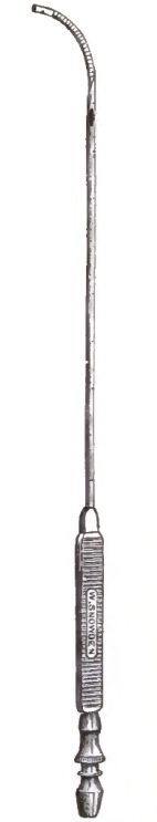
|
|
Roberts's Pericardial Aspirating Trocar. |
Beverley Robinson of New York has still further modified11 my trocar. His additions may have improved the instrument if they do not unduly complicate it. Pepper, after operating upon his case, had made a delicate double canula, the inner tube of which was furnished with a fine needle-point. After introduction the inner tube was withdrawn until its point was sheathed.12
11 New York Med. Record, March 29, 1884.
12 Medical News and Library, Philada., March, 1878.
It is said that at the meeting of the Italian Medical Association at Pisa in 1878, Baccelli proposed a new method of puncture; but the account given by Severi13 in speaking of Baccelli's cases indicates that his proposal referred not to a method of operating, but to a method of selecting the point of puncture.
13 Lo Sperimentale, Aprile, 1881, p. 392.
It must also be remembered that failure to obtain fluid when pericardial effusion existed has occurred because the needle had been passed through a costal cartilage, and was thus plugged by a disk of cartilage. The manner in which the intercostal spaces are narrowed and changed in direction by the curving upward of the anterior portion of the ribs and by the curvature of the cartilages should be impressed upon the operator.
If failure to obtain fluid occurs, and the diagnosis remains quite certain from the symptoms, withdrawal of the needle and puncture in another position should be done or an incision of an exploratory kind made.
[p. 798] In pericardicentesis care must be taken not to thrust the needle or trocar into the heart. This may happen even in quite careful hands. If the right ventricle is entered, venous blood will escape through the canula; if the needle is buried in the cardiac muscle, no fluid or blood can escape. The violent movements communicated to the needle will usually indicate that the needle is either in contact with the heart or thrust into its tissue. Of course such movements will occur from cardiac contact when most of the fluid has been withdrawn; but are not to be expected immediately after the introduction of the puncturing instrument unless the fluid is very small in amount, the needle deeply inserted, the pericardium adherent at the point of puncture, or the diagnosis of fluid an error.
Puncture of the heart has occurred accidentally during pericardial tapping without doing any harm, and has been suggested as a proper surgical procedure in certain cardiac conditions. Still, it is an accident to be avoided by the use of proper trocars and pumps, by the selection of a proper site of operation, by the adaptation of the suction power as soon as the point of the trocar or needle is buried beneath the skin, and by other precautions that will suggest themselves. In thick, oedematous, or fatty chest-walls no fluid will be reached perhaps until a depth of four or five centimeters (about two inches) has been attained by the point of the puncturing apparatus.
I must call attention to the fact that West14 records a case of pericardial tapping occurring at St. Bartholomew's Hospital in 1874 where a trocar and a canula were introduced through the fourth left space near the edge of the sternum, and caused death in five minutes from hemorrhage into the pericardium, due to tearing of the right ventricle. The position chosen and the form of instrument may have had to do with this unfortunate result, of which the details are not given.
14 Med.-Chir. Trans., 1883, pp. 259, 275.
A few words on cardicentesis, or intentional heart-puncture, may here be appropriate. It has been suggested as a means for rapid abstraction of blood from the right heart in intense pulmonary and cardiac engorgement, and for the abstraction of air after air-embolism has occurred from wounds of the large venous trunks. It has been known for years that aspiration and similar punctures of the heart are comparatively harmless. Roger accidentally withdrew 200 grammes of blood from the right ventricle of a boy of five years without doing harm. Hulke seemed to benefit a case of pleuro-pneumonia by accidentally aspirating the right heart. Cloquet, Bouchut, Steiner, and Legros and Onimus have made similar observations on the absence of danger from such wounds. Westbrook of Brooklyn, Corwin,15 Dana,16 and apparently Janeway of New York, have performed intentional cardiac aspiration in moribund patients without causing any noticeable harm. The contributions of Westbrook,17 Roberts,18 and Leuf19 on this topic, as well as that of Senn20 on air-embolism and its treatment, will interest those who wish further information.
15 N.Y. Med. Record, March 10, 1883, p. 263.
16 Ibid., Feb. 3, 1883, p. 140.
17 Ibid., Dec. 23, 1882.
18 Philada. Med. News, Jan. 13, 1883.
19 Amer. Journ. Med. Sci., Jan., 1885, p. 79.
20 Trans. Amer. Surg. Ass., 1885, and Annals of Surgery, St. Louis, 1885.
The results of operations for pericardial aspiration or incision are exceedingly good when the frequent postponement of the operation till the patient is almost moribund is recollected. Elaborate statistical tables would be out of place in this volume; and, besides, it seems almost impossible to get a complete collection of the cases. Hindenlang, West, and I have published [p. 799]and analyzed long lists of cases collected from various sources, and I have now references to more, but this tabulation seems unnecessary, as the practical points to be derived from their study are well proved by the previous work done. In addition to the bibliographical notes already given, I add for the use of inquirers in this field two recent monographs—one by a German,21 the other by a French writer.22
21 Ueber Paracentese des Herzbeutels, Gerhard Beck, Würzb., 1882, p. 33 (Thesis).
22 Contribution à l'Étude de la Paracentèse du Péricarde, H. Ferraud, Bordeaux, 1883.
The existence of inflammation of the membranes of the aorta was mentioned by Galen and other early writers, but it was not until 1824 that a systematic treatise on this subject was published. Since that time the subject has received more attention, but the results obtained are unsatisfactory. There is grave doubt, according to many writers, as to the existence of acute aortitis independent of other lesions, although it is recognized that the aorta may participate in inflammation of the neighboring organs. Even then, as Powell says, "the aorta is very slow to share in such processes, and when it does so the inflammation is very chronic and limited, giving rise to no special symptoms." Peter treats the subject at length, and after enumerating certain so-called symptoms of acute aortitis, confesses that these symptoms are merely the ordinary phenomena of angina pectoris, and these two affections cannot be distinguished from each other. It must be concluded for the present, therefore, that acute aortitis is rare, and that we know of no symptoms which are characteristic of it.
Atheroma of the aorta is the result of chronic endarteritis, and is always of slow development. The process may be limited to the intima or it may extend to the middle and outer tunics. Beginning with a thickening and softening of the wall, it finally develops plates of calcareous deposit. These plates are most numerous in the region of the aortic valves, and diminish in number as the artery proceeds from that point. The descending portion of the aorta is relatively free from these patches, but they reappear again near or at the bifurcation.
ETIOLOGY.—Atheroma is one of the ordinary products of old age, and is therefore one expression of senility. Heredity probably exerts some influence, and certain cachexias predispose to an early occurrence of the process. Gout and syphilis render one especially prone to it. High pressure and strain are also important factors. Continuous hard toil is more productive of atheroma, according to Allbutt, than intermittent work. The pre-albuminuric stage of Bright's disease, which is characterized by high arterial pressure, is frequently productive of atheroma.
SYMPTOMS.—When the inner coat alone is affected, there are no symptoms of this disease. According as the degeneration extends deeper and involves the middle and outer tunics, the aorta begins to dilate, and the symptoms may vary from the slightest feelings of discomfort upon exertion to the most violent attacks of palpitation and pain.
[p. 801] Usually, at the beginning the symptoms are very obscure. A slight dyspnoea on exertion, or palpitation, or dyspeptic troubles are the chief complaint. The presence of these troubles in a man of fifty years or over, whose heart and kidneys present nothing abnormal, and in whom the smaller arteries of the extremities feel hard and calcareous, may excite the suspicion of atheroma of the aorta. There are no distinctive physical signs. Some writers speak of a short post-systolic murmur over the aorta beyond the valves, which may be audible only when the heart is acting strongly.
The aorta is almost invariably dilated, and Peter says that this dilatation may be traced by percussion. According to him, the normal aortic dull area measures from two to five centimeters transversely in the male, and from two to four centimeters in the female. He says that he has seen cases of atheroma where he was able to determine a dull aortic area of eight centimeters in diameter. If the inflammation extend from the aorta to the neighboring nerves, the patient may suffer from the ordinary symptoms of angina pectoris.
TREATMENT.—This disease cannot be cured by drugs. The physician's task is to regulate the habits of the patient, to remove so far as possible all conditions which tend to aggravate and increase the trouble, and to alleviate incidental symptoms of distress.
DEFINITION.—The origin of the term aneurism is buried in obscurity, and the theories which have been advanced regarding it are not very satisfactory. Montanus thought it was derived from a privative, and neuron, a nerve. Oetius declares it is from aneurisma, an enlargement, from eurumo, I dilate. Coale thinks a ready origin is offered in the words aneu, without, rusmos, a series, course, or succession, from ruo, I flow.
Aneurism of the aorta is a local dilatation of that vessel. When all the arterial tunics persist unruptured in the tumor, it is a true aneurism. When one or more of the tunics are torn in the process of expansion, it becomes a false aneurism. When all the tunics of the artery rupture and the blood escapes into the neighboring cellular tissue, it becomes a diffuse false aneurism. The internal and middle coats of an artery may burst, and the blood escape into and coagulate in the space between the middle and external tunics, and this is termed a dissecting aneurism. In rare instances of this type of aneurism the blood finds a second opening, and returns into the artery again, thus forming a double tube for a short distance.
In former times great stress was laid upon the distinction of aneurism according to the number and combination of persistent tunics, and we read of the mixed internal and the mixed external type. These points have less clinical importance, however, than a proper appreciation of the size and shape of a tumor, because all aneurisms are false after they exceed a certain size. When an aneurism involves the entire periphery of the aorta, it may be cylindrical, fusiform, or globular in shape, and receive names accordingly. When it is a mere bulging on one side of the artery it is saccular in shape. Obviously, the opening into the fusiform aneurism is quite or nearly the entire length of the tumor, whereas in the false saccular type the orifice may be reduced to a mere puncture of the arterial wall. The size of the orifice is a matter of great importance, particularly in connection with the question of operative interference, and therefore it will be referred to later. The sacciform and fusiform aneurisms are often combined together, or, in other words, it is quite common to find a lateral bulging superimposed upon a local dilatation of the artery; but such grouping is not necessary, as either form appears [p. 802]without the other. It is not uncommon also to find one bulbous aneurism superimposed upon another, the dependent aneurism in this case being of the false or diffuse type. The second aneurism often lies outside the chest-wall, and it is connected with the mother aneurism by a narrow opening or channel.
Varicose aneurism is a false aneurism formed by communication between the aorta and the vena cava, the pulmonary artery, the right auricle, or the right ventricle. It is almost without exception rapidly fatal and not amenable to treatment.
Occasionally the aorta will present alternate bulgings upon one side and the other, so that the vessel appears to wind in its course. This condition is called cirsoid aneurism, but it has nothing in common with external aneurism of the same name.
The size of an aneurism is variable, like its shape, but in general the true aneurism rarely exceeds the size of an egg (Jaccoud). Beyond this size one or more of the coats give way, and the aneurism becomes false, in which condition it may grow as large as an adult's head if the patient lives long enough to allow such development. Balfour refers to two rare forms of aneurism—the intravalvular, which is situated within the aortic valves and above the ventricle, and the intervalvular, which is still more rare, and is situated between the valves themselves. The symptoms of these aneurisms are merely those of valvular lesion, and therefore present no differential points for diagnosis.
ETIOLOGY.—Local weakness of the aorta submitted to sudden strain is unquestionably the most frequent cause of aneurism. It is rare to find an aneurism in an otherwise healthy aorta, and some authors go so far as to assert that aneurism never occurs without preceding degenerative changes in the arterial wall. Naturally, strain is the physiological burden of the aorta, and this strain tends sooner or later to degeneration of the arterial tunics. Then, given a weakened spot, the ordinary occurrences of every-day life are sufficient to precipitate disaster. A sneeze, a cough, some sudden exertion of the body in lifting or moving, have been the starting-points of aneurism. All accumulated testimony indicates that sudden strain is more dangerous than prolonged uniform strain, and therefore some occupations are more productive of aneurism than others. Inasmuch as age, sex, occupation, and personal habits influence the development and nutrition of the aorta, it is obvious that they must exert an important influence upon the occurrence of aneurism.
All records agree that aneurism is pre-eminently a casualty of middle life, and a glance at the accompanying table, which I have prepared from an analysis of 69 reported cases, shows that the disease is most common between thirty and fifty years of age:
| From | 20 | to | 30 | years | of age, | 4 | cases. |
| " | 30 | " | 40 | " | " | 21 | " |
| " | 40 | " | 50 | " | " | 29 | " |
| " | 50 | " | 60 | " | " | 14 | " |
| " | 60 | " | 70 | " | " | 1 | case. |
| Youngest case, 20 years of age. | |||||||
| Oldest case, 72 years of age. | |||||||
Crisp analyzed 551 cases, and reports 398 between the ages of thirty and fifty.
Beneke has found in his records of arterial measurements that the pulmonary artery greatly exceeds the aorta in circumference up to the age of thirty. After that period the aorta begins to increase with relatively greater rapidity, until in the forties it exceeds the pulmonary artery in size, and it maintains its superiority from that time forward. The aorta continues to increase in circumference throughout life, but after the age of fifty this increase is considered a senile dilatation rather than an actual growth. It is interesting to [p. 803] note that the era of greatest liability to aneurism coincides with that of most rapid aortic development.
Sex furnishes a distinction in the frequency of aneurism. In 82 cases I found that only a seventh were females; Crisp registers less than an eighth. The radically different occupations and habits of women may contribute somewhat to their relative immunity from aneurism, and their physiological development also seems in their favor. Beneke states that the blood-pressure during childhood is about the same for both sexes. From puberty onward it is greater in the male. This is due to the fact that after puberty the volume of the heart relative to the length of the body is less in the female than in the male, and at the same time the main arteries of the body relative to the length of the body are only a trifle narrower in women than in men. The pulmonary artery, indeed, is relatively a trifle wider in women than in men. It follows from this that the blood-tension in both the large vessels emerging from the heart is less in the female than in the male.
In general terms, it may be said that those people who are exposed to heavy labor, as mechanics, laborers, soldiers, porters, cabmen, etc., are more liable to aneurism than those who are less exposed to such straining efforts. Fixture of the chest during effort brings greater strain upon the heart and aorta, and therefore men who wield heavy hammers and sledges are especially liable to aortic disease. Constriction of the neck or forcible extension of the same during exertion is dangerous, because it thus happens that the arteries are stretched in their long diameter at the same instant that the blood-wave is expanding them laterally, and they are thereby subjected to double strain. I knew of a trotting horse which was killed by this very combination of strain upon the aorta. At the end of a trial of speed the animal refused to stop; whereupon a groom sprang forward, seized him by the bit, and threw his head strongly upward and backward. His carotids and aorta were thus stretched to full length at the moment when his heart was acting with great force. The horse dropped dead, and the autopsy revealed a rupture of the aorta.
The frequency of aneurism among the soldiers of the English army was long the subject of anxiety and thought to English surgeons. Finally, some bright man recognized one cause in the dress of the soldiers. They were obliged to wear a high stock, which constricted the neck and kept it stretched, and their trappings were adjusted so as to keep the body in a stiff and unnatural position. These objectionable details of the dress have been removed, and it is now claimed that aneurism is much less common in the army.
Syphilis and gout undoubtedly contribute to the formation of aneurism, because they both dispose to degenerative processes in the arterial tunics. Some writers, however, have laid too much stress upon syphilis. It was claimed that this disease was the cause of the great frequency of aneurism in the English army. Barwell, however, calls attention to the fact that aneurism has been 13½ times more frequent per 1000 men in the army than in the navy, and yet no one maintains that syphilis is more common in the army than in the navy.
SYMPTOMS.—The diagnosis of aneurism of the aorta may be one of the easiest problems of clinical medicine, or it may present difficulties which defeat the most skilful diagnosticians. A large number of aneurisms utter no sign of their existence, and are only revealed by the manner of death or by an autopsy. Again, the so-called signs of aneurism are so indefinite in character, and so associated with other pathological conditions, that the greatest confusion often befogs their interpretation. Mistakes therefore arise in two ways: either aneurism is diagnosed as present when it is absent, or it is declared absent when present. Robin reports the case of a vigorous young man upon whom several of the most eminent clinicians of [p. 804]Paris diagnosed aneurism of the aorta, and yet a rest of a few days sufficed to remove all symptoms of that disease. Three candidates for the diploma of the Royal College of Physicians and Surgeons in England recently declared a case of loculated pleurisy to be aneurism of the aorta, and B. W. Richardson says he has "seen at least seven persons suffering severe mental anxiety from the belief that they were fatally struck with aneurism," and yet they were free of such disease. Balfour says: "There is only one phenomenon positively characteristic of thoracic aneurism, and that is the existence in some part of the thorax of a pulsating tumor other than the heart, which beats isochronously with it, and at least as forcibly, and which at each pulsation expands in every direction." And yet simple dilatation of the aorta, combined with mental excitement, will so increase the thoracic pulsations as to simulate aneurism. It is necessary, therefore, that a patient during an examination should be as quiet as possible, both in mind and body, and if any doubt exist regarding the significance of the symptoms present, the patient should be kept in bed for a few days in order to allay the arterial excitement.
The phenomena produced by an aneurism are naturally divided into two groups: 1. The direct symptoms, which are confined to the limits of the tumor itself, and which are termed the physical signs. 2. The indirect symptoms, which are due to the influence of the tumor upon neighboring organs, and which present themselves often at remote points as signals of distress within. This influence of the tumor upon its environment is purely mechanical and due to pressure, and the resulting symptoms vary according to the particular organ or function involved. These symptoms are therefore classified as the physiological signs.
Pain is one of the earliest and most troublesome of the pressure symptoms of aneurism. It is due to a stretching of the nerve-filaments in the aortic wall and to the pressure of the tumor upon neighboring organs, especially the vertebral column and sternum. When due to nerve-stretching, the pain is neuralgic in character, and is not necessarily confined to the chest. It may appear in the back, and is intensified by coughing or sneezing. It may be rheumatic in type, and affect the arm and shoulders for several months before other aneurismal signs develop. In such cases the right arm and shoulder appear to be most often affected. Sometimes the pain cannot be located, but is referred indefinitely to the chest, or it may accompany acts of deglutition. As a rule—and this point is important—this form of pain from an aneurism exhibits wide variations of intensity and is usually intermittent. Exercise, coughing and sneezing, mental excitement, or anything which increases the activity of the circulation or raises the blood-tension, increases the pain. It may resemble angina pectoris in location and radiation, but it differs essentially otherwise. It is more continuous, and is associated with less anxiety, which is such a conspicuous element of angina.
When the pain is due to erosion of the vertebræ or sternum, it is more steady and gnawing. It is still liable to violent exacerbations, and excitement of all kinds increases it. Oftentimes the pain is so excessive that the sufferer cannot lie down or obtain relief in any position. This is especially the case with aneurism of the abdominal aorta. Bennet reports the case of a patient who poisoned himself to be free from the terrible pain, and deaths by exhaustion from pain and distress are not uncommon.
Numerous other accidents besides pain arise from pressure upon the neighboring veins. Balfour says that severe dyspnoea, vomiting, and flatulency are frequently caused by pressure of an aneurism upon the pneumogastric nerves, and that these symptoms may be relieved by gently rubbing the tumor. Hiccough and paralysis of one-half the diaphragm are caused by pressure upon the phrenic nerve. Occasionally destructive inflammations of [p. 805] the lung and pleura occur with aneurism, and these have been attributed to pressure upon the pneumogastric nerves and the pulmonary plexus. Palpitation of the heart is likewise often produced in a similar manner. Sometimes the patient is conscious of a pulsation in the tumor itself. Pressure upon the intercostal nerves will produce herpes zoster, and cicatricial records of such attacks are found upon patients with aneurism. Implication of the sympathetic nerves produces modifications of the pupils according as the nerves are merely irritated or paralyzed. In the first case the radial muscles of the iris become permanently contracted and the pupil is dilated. In the second case the radial muscle becomes paralyzed and the pupil is contracted. Jaccoud says that this succession of changes is not rare, and he has watched cases progress through both pupillary stages. The nerves affected are those which emerge from the cilio-spinal region, which extends, according to Budge and Waller, from the sixth cervical to the sixth dorsal, or, according to Brown-Séquard, as low as the tenth dorsal vertebra. From the anterior roots of this region nerve-filaments pass through the cervical sympathetic to the iris. The difference in the pupils is often so slight that it requires very careful measurement to detect it. The application of atropia will assist in the examination, because that drug has very incomplete influence upon the affected pupil. The pupil is also much less sensitive to light, but it contracts more strongly than the normal eyes in its accommodation for near objects. Robertson cautions against conclusions based upon mere casual observance of the eyes, because 1 person in every 14 has one pupil naturally smaller than the other.
Myosis is not pathognomonic of aneurism. It denotes merely some trouble with the cilio-spinal nerves. The nature of that trouble must of course be determined by the other associated symptoms of the case. The contraction of the pupil is sometimes accompanied by paleness of the corresponding side of the face and neck, while at other times the same region may be swollen, oedematous, and perspiring. These symptoms are due to local vascular changes from interference with, and disorganization of, the vaso-motor nerves which govern these regions. Remote local paralysis sometimes utters the first warning of aneurism, and such cases are usually very striking. Paralysis of the recurrent laryngeal is the most frequent of this group of signs. Urquhart reports a case where for some months the chief symptom was a falling of the head on the breast, as if it had been forcibly drawn down by the sterno-cleido-mastoids. Another patient was supposed to have rheumatism, but he soon became paralyzed on the right side and lost his speech. He recovered somewhat, but died subsequently from bursting of the tumor into a pulmonary cavity. Tufnell says if an amaurosis occur suddenly look for valvular disease of the heart or for aneurism of the aorta.
Dyspnoea.—The dyspnoea produced by an aneurism may vary from a slight difficulty of breathing on exertion to the most marked orthopnoea. It is produced by—a, direct pressure upon the trachea or bronchi; b, pressure upon the recurrent laryngeal or the vagus. The two forms of trouble are easily discriminated by physical examination. In cases of pressure upon the respiratory tubes auscultation reveals very characteristic signs. The constriction of the tube causes a peculiar harsh sound, which, heard only in inspiration at first, becomes audible later in expiration as well. If the pressure is upon the trachea, the sounds will be heard equally in both lungs; whereas if only one bronchus is involved, the sounds will be confined to the corresponding side. If a bronchus be completely occluded by pressure, then the peculiar breath-signs will disappear, and complete respiratory silence reign instead. The dyspnoea of this origin is greatly relieved by motion and by certain positions of the body. In capillary bronchitis, pneumonia, asthma, etc. the patient sits with the head thrown back and the shoulders raised, whereas a patient with tracheal compression finds greater relief in leaning across the [p. 806]back of a chair, with his head resting upon his arms folded on a table, and the nights are passed in this position. Again, the pressure dyspnoea is subject to sudden and excessive variations. Any excitement which increases the cardiac activity and the blood-tension will excite dyspnoea, whereas rest and repose diminish it. This form of dyspnoea is likewise accompanied by loud stridulous breathing, and by harshness and a metallic quality of the voice. The stridor and dyspnoea bear no direct relation to the size of the tumor, because a small tumor pressing upon the side of the trachea, where the cartilaginous rings are thinner and less resistant, will produce more discomfort than a larger tumor directly in front. Where the compression of an air-tube is considerable, it usually provokes inflammation of the mucous membrane, and the secretions thereby engendered are liable to collect behind the obstruction and increase the distress for breath. Cases are reported where, tracheotomy having been performed, a catheter was pushed by the obstruction and the backed-up secretion allowed to escape, to the great relief of the sufferer. One case is recorded where the examining physician was able to see by the aid of a laryngoscope an inward projection of the wall of the trachea, which pulsated with each heart-beat.
The dyspnoea arising from pressure upon the recurrent laryngeal and vagus may begin in two ways—either by a sudden paralysis of both vocal cords, or by a preliminary spasm of the cords due to nerve-irritation. When both cords are paralyzed, which is very rare, the voice is entirely obliterated and the dyspnoea is intense and continuous. The complete paralysis may be associated with choking at meals. When only one cord is paralyzed, the breathing is not materially affected, though the voice is altered in a characteristic manner.
If the compression of the nerves mentioned simply irritates them, then the phenomenon of laryngeal spasm occurs. The voice becomes high, squeaking, and false or whispering, with a muffled falsetto. Jaccoud describes a condition where the nerves of the two sides are not uniformly affected, and therefore the cords are not equally tense in their spasm. The result of this difference of tension and vibration is a peculiar commingling of high and low tones, which produces a very discordant and unpleasant sound to the ear. Jaccoud terms this la voix bitonale. The dyspnoea from spasm persists through both inspiration and expiration, whereas with paralysis of the cords the inspiration is alone or mainly affected. The cough in these cases is phenomenal in its character, being very loud and metallic, often barking, and it is very distressing to the patient and to all who hear it.
When a bronchus is compressed the percussion note on the corresponding lung is higher in pitch and tympanitic. The inspiratory murmur is ordinarily diminished, but bronchial breathing may (rarely) occur. The coincidence of bronchial breathing with tympanitic resonance is an eccentric combination of a very paradoxical character. The cough is almost pathognomonic, with a loud barking, distressing metallic clang. Such a cough is still more suggestive when combined with the high, shrill, whistling vox anserina. The amount of expectoration is at first small, consisting of glairy, frothy mucus. Later it becomes more copious and muco-purulent, and may even be rusty and red. The presence of bloody sputa with an aneurism is always grave, because it raises suspicion of a so-called weeping aneurism which is approaching rupture.
Dysphagia.—This is a common symptom with aneurism, but it is not so constant in appearance as it is with other mediastinal tumors. It appears more often when the aneurism is situated upon the transverse portion of the aorta. It is frequently painful, but always variable in severity, and may disappear for long intervals at a time. Lying upon the face usually relieves the difficulty, while it is aggravated by reclining upon the back. Fluids are usually [p. 807]swallowed more easily than solids. Hayden says that a feeling of sharp pain in a particular part of the gullet in swallowing when aneurism is present indicates erosion of the mucous membrane and early perforation.
Pressure upon Veins.—Localized oedema and cyanosis are two common symptoms of aneurism of the aorta. The sudden eruption, the limited distribution, and the terrifying effect of these symptoms render them especially interesting. They are due to pressure of the tumor upon the veins near the heart, and particularly upon the superior vena cava. Dujardin-Beaumetz says that, thanks to the vena azygos, compression of the superior vena cava produces simply a varicosity of the neck and upper part of trunk. Should the vena azygos be simultaneously blocked, then the oedema and cyanosis will spread over the entire head, neck, arms, shoulders, and upper trunk—i.e. over all parts drained by the superior vena cava. Only two such cases have been reported, however. One case was seen by Piorry and one by Dujardin-Beaumetz. In the latter case the oedema and cyanosis of parts named above came on suddenly without apparent cause. The face was swollen, blue, and covered with red patches, and the eyes were injected. The ears were cold; the abdomen and lower limbs retained their normal color. The contrast between the upper and lower portions of the body under these conditions is very striking.
Balfour says that "a thick oedematous collar covered with large veins surrounding the root of the neck" is indicative of compression of the superior vena cava.
Pressure upon the brachio-cephalic veins produces oedema and cyanosis of the head and upper extremities; oedema of the glottis has occurred under such conditions. Sudden swelling of one arm, unaccompanied by inflammation, is suspicious of aneurismal compression of the corresponding vein, especially if it comes on suddenly after exertion. Compression of the descending vena cava or right auricle may give rise to congestion and dropsy of the lower part of the body, but these are later symptoms.
Pressure upon the thoracic duct is relatively rare. It may cause emaciation, but loss of flesh with aneurism is more often due to obstruction of the oesophagus or to dyspepsia and the exhaustion from pain and sleeplessness.
Pressure upon Bones.—Pressure of a tumor on neighboring bones causes absorption and dislocation of the same. The clavicles, sternum, and ribs are rapidly eroded by the aneurism, and are pushed forward and disarticulated. Pressure upon the spinal column causes absorption of the vertebræ and of the cartilages, until oftentimes the cord is laid bare and even subjected to direct pressure.
Inspection.—Inspecting a person suspected of aneurism, one should examine the pupils, the color of the skin, the condition of the veins of the head, neck, and arms, all movements of the neck and chest, and especially the contour of the front part of the chest.
The conditions of the pupils, skin, and veins have all been described, but the movements of the neck and chest require notice here. Any area of pulsation apart from the normal apex-impulse should be critically marked and examined. Fulness or beating in the episternal notch is significant. Cheesman reports a case where a curious pulsation was occasionally communicated to the larynx and the tongue by an aneurism situated beneath the manubrium. Every now and then the thyroid cartilage would rise and fall, and the tongue would pulsate backward and forward with each beat of the aneurism.
Inspection of the larynx quickly determines the presence or absence of paralysis of the cord, and may sometimes reveal pulsating tumors pressing upon the trachea. While inspecting the shape of the chest it is best to stand upon one side of the patient and look across the surface of the thorax. In [p. 808] this way slight deviations from the symmetrical become most readily apparent. If any abnormal point seems to pulsate, the fact can be rendered more obvious by pasting bits of paper upon the suspected spot and around its immediate neighborhood. Viewed thus in an oblique light, the relative movements of these pieces may be easily discerned. If a tumor be present and the diagnosis established, one should carefully note the color and condition of the skin over the prominence. As the tumor develops pressure the skin becomes tense and glossy. Then it turns red, and may be covered with livid spots and even ecchymoses. In later stages a black dried scale of flesh may be all that seems to restrain the heaving blood. Weeping of blood may take place for some time before the final break.
Palpation.—Given a prominence of the chest-wall or a localized pulsation in the abdomen, the next step is to examine the suspected part with the hands. Any tumor lying across an artery will move forward and backward with each pulsation of the artery, and conditions of this kind have been repeatedly diagnosed as aneurism. An aneurismal tumor, however, is distensile as well as pulsatile. Every tumor, therefore, should be grasped as far as possible between the two hands, to determine if it distends with each beat.
When one cannot reach the sides of the tumors in front, one can resort to Stokes's plan. Place the flat of one hand upon the front of the chest, and the other hand upon the back. By this means the expansile character of the pulsation may sometimes be determined.
Many intra-thoracic aneurisms present a double impulse or two distinct blows to the hand during the cardiac systole; and when these blows are too faint to be felt, they may still be registered by the sphygmograph. This double impulse is not characteristic of aneurism of the aorta, because it may also be felt in aneurisms of the large branches of the arch. Bellingham thought that the second blow was due to a reflex wave from the aortic valves, and was therefore diastolic in rhythm. Jaccoud, however, showed that it occurs even with great insufficiency of the aortic valves, thus excluding reflex waves. François Frank also proved that both blows were systolic in rhythm. He thinks they are due to the fact that the blood enters the aneurism en deux temps. The blood, rushing in at the beginning of the systole, gives a sudden distension of the partially relaxed sac-walls, and thus causes the first impulse. Then the bulk of the blood-waves, following more slowly on account of greater resistance, produces a second elevation more or less pronounced.
Balfour states that aneurismal pulsations are usually more forcible than those of the heart, and that this point has not received the attention which it merits. If the sac contains much fibrin the impulse is feebler than that of the heart.
W. S. Oliver describes a new sign of aneurism and the method for detecting it. Place the patient in the erect position and direct him to close his mouth and elevate his chin to the fullest extent. Grasp the cricoid cartilage between the fingers and the thumb, and push it gently upward. If an aneurism of the arch of the aorta be present, its pulsation will be plainly transmitted up the trachea to the hand. The act of examining will also increase the laryngeal distress if such be present.
The frémissement cataire, or thrill imparted to the hand by an aneurism, has been frequently described. It is very characteristic when felt, but Powers says it is not of frequent occurrence. He has felt it in eight cases of aneurism, but four of them were complicated by regurgitant disease of rheumatic origin, and all were probably of the fusiform kind.
Pulse.—Partial or total obliteration of a large vessel, dilatation of the aorta, compression of an artery by a tumor, may produce a radial pulse [p. 809] similar to that of aneurism. Moreover, we may find the radials differing from each other in persons who are perfectly healthy. It follows, therefore, that, taken by itself, the pulse does not contribute very decisive evidence of an aneurism. When the diagnosis of an aneurism is established or confirmed by other signs, then the added evidence of the pulse does possess some value. The finger will often detect the following characteristics of an aneurismal pulse:
1. Delay.—The pulse at the wrist is normally from 11/100 to 14/100 of a second later than the cardiac impulse. With aneurism this interval may be prolonged in one or both radials, and the additional delay may amount to 4/100 of a second. This sign of delay is of most value when the pulse in one wrist loiters behind its mate. The relative delay of the impulse of the aneurism itself and of the carotid artery may give useful information. If the beat of the tumor precedes that of the left carotid, then the tumor is nearer the heart, whereas the aneurism is evidently beyond the left carotid when the beat of the latter precedes.
2. Diminution in Volume.—The pulse in one radial may be much smaller than in the other or altogether absent.
3. Diminution in Force.—The pulse of one side may convey a less sudden and less forcible blow to the finger. This diminished suddenness of the sensation imparted to the finger corresponds to the sloping up-stroke of the sphygmographic tracing.
4. Thrill.—Under certain rare and not very clearly defined circumstances the pulse imparts a sensation of thrill to the finger. Mahomed says this probably occurs when the entrance to the aneurismal sac is very narrow and the aneurism is directly in the course of the vessel. It may also be occasionally produced by the rigidity of the wall of the vessel or by a partially-dilated clot vibrating in the blood-stream.
Under the enthusiastic and elaborate study of Mahomed the sphygmograph has attained a certain degree of usefulness. Though difficult in its application and limited in its results, yet many of the points demonstrated by it are of sufficient importance to justify their consideration. The sphygmographic tracing of the normal pulse is shown in Fig. 50.
| FIG. 50. |

|
| AB. The Up-stroke. ABC. Percussion Wave. E. Aortic Notch. D. Dicrotic Wave. |
Now, the points which distinguish an aneurismal tracing from the normal are—1, a sloping up-stroke; 2, impairment or loss of the percussion wave; 3, obliteration of the secondary waves; 4, diminished volume of the curve; 5, vibratile waves; 6, a different blood-tension.
| FIG. 51. |
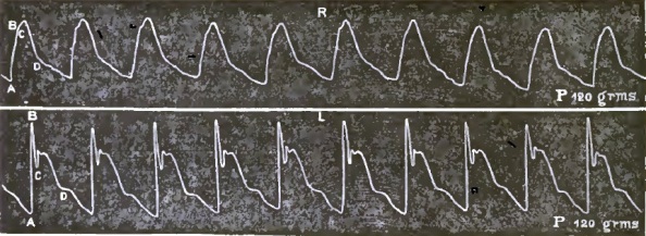
|
| Right and Left Radial Pulse in Aneurism of Aorta. |
In comparing the curves shown in Fig. 51, taken from Powell's article upon aneurism, it will be noticed that the up-stroke AB is more sloping in the curve of the right wrist than in that of the left. The percussion and dicrotic waves are entirely smoothed out into an almost uniform wavy slope. As one writer has expressed it, an aneurism acts like an air-chamber in an engine, and tends to break up the intermittent pulse into a steady stream. The relative difference of the blood-tension of two arteries is determined by the relative amount of pressure required of the instrument to develop the tracing. This amount of pressure is sometimes greater and sometimes less on the affected side.
In comparing the tracings from the radials the following points are to be noted: 1. Is there any difference in the percussion waves?—i.e. is the up-stroke more sloping or the apex less pointed in the one than in the other? 2. Is the tidal wave equally high and sustained in both? 3. Is the dicrotic wave equally developed?
[p. 810] If a difference exist in the tidal wave alone, it need not, and probably will not, be due to aneurism. It is the loss of the percussion wave and of the dicrotism which characterizes aneurism.
It must be conceded here that the use of the sphygmograph and the interpretation of its tracings are beset by the greatest difficulties. Mahomed, to whom I am chiefly indebted for these sphygmographic details, declares that the use of the instrument requires great care and skill, and it may easily lead to error. "No one should attempt to use it who cannot readily obtain similar tracings from the two radials of a healthy person." Great care in the application of the instrument should be exerted, and we must guard against all causes of transient excitement. It is well to let the patient see the instrument applied to others before attempting it on him, in order that he may not fear it. The patient must be placed in a comfortable position, with both arms alike, and the points of application of the instrument must be alike on the two sides. The amount of pressure on the two sides must be equal, or the difference carefully noted. Moreover, one should never be contented with one tracing, but a number on each wrist should be taken. If, then, the two radials appear to differ, the precautions must be redoubled, and the pulse tested again on another day. Inequalities of the tracings may be produced by abnormal distribution of the radials, and an old fracture or other injury of one arm may affect the flow of blood in the arm.
Paralysis of the arm, by interfering with the vaso-motor nerves, and thereby with the venous return of the blood, may alter the character of the pulse. A tumor external to the artery, either intra-thoracic or extra-thoracic, will produce aneurismal pulse and endarteritis, or congenital contraction of the aorta may so block the artery as to produce diminished pulse-waves. It may be said that the sphygmograph is incapable of distinguishing between an endarteritis and an aneurism.
On the other hand, the instrument is very useful in distinguishing between an aneurism and a tumor compressing an artery, because in the latter case the up-stroke and percussion wave remain normal, whereas in the former they are strongly modified, as described above. With aneurism of the ascending aorta both radials must be similarly affected, if at all, and in these cases the sphygmograph teaches very little. If the right radial is alone or mainly affected, then the aneurism involves the innominate and arch together. When an aneurism of the innominate includes the aorta, then the whole sac forms virtually a dilated aorta, and no difference in the radials will appear. Hence it follows as a corollary: Given an innominate aneurism, if the radials remain equal the aorta is certainly involved. When the left radial pulse is alone affected, the aneurism lies beyond the brachio-cephalic branch, and may or may not involve the left subclavian.
[p. 811] The sphygmograph is of less avail in aneurisms of the descending portion of the thoracic aorta or of the abdominal aorta. It may be of service in affording information regarding the condition of the aorta itself with reference to an operation, and it may also be of service in determining the upper limits of an aneurism under the following conditions: A case is reported which presented all the physical signs of aneurism of the descending aorta, but the sphygmograph showed that the left radial was affected, and thereby proved that the aneurism extended as high as the left subclavian at least.
While the foregoing facts prove that the sphygmograph by itself affords very inconclusive and untrustworthy evidence, yet when the presence of a tumor and other physical signs prove the existence of an aneurism, the written pulse-record will often be the guide to the accurate placement of the tumor, and thereby will often furnish decisive indications in the selection of the method of treatment.
Auscultation.—The typical aneurismal bruit is not an ordinary souffle, but it is an accentuated booming sound of a very peculiar character. Many writers describe it as a systolic jog or shock. Occasionally this bruit de battement is double—i.e. one hears two shocks, so to speak, just as one feels a double impulse. No satisfactory explanation for this reduplication of murmur has yet been given. The aneurismal murmur is almost invariably systolic. Balfour reports two cases of a diastolic murmur heard with abdominal aneurism. One of these cases was observed by himself and the other by Wickham Legg.
When this peculiar booming sound is heard over a circumscribed dull patch, it is very distinctive of aneurism, but its absence possesses no eliminative value. Many aneurismal tumors are absolutely quiet, and some of them give only a soft murmur like an ordinary cardiac souffle.
Associated with the aneurismal sound one also hears the normal heart sounds much intensified. This is peculiarly noticeable of the second cardiac sound, which acquires a ringing, booming, accentuated character when heard over an aneurism. Johnson thinks that this intensification of the heart sounds is due to the sudden tension of the walls of the sac. Balfour in referring to the same phenomenon considers it of greatest diagnostic value, and thinks that proper emphasis is not ordinarily given it.
A fundamental rule in the examination of a suspected case of aneurism is to auscult over every inch of the thorax, front and back. Not only the intrinsic signs of the tumor itself are important, but all testimony from the neighboring organs must be collected and weighed. The modification of the respiratory sounds have already been mentioned. Stokes attaches great importance to this fact, that "over one lung, more rarely over both, the breath sound has often communicated to it a peculiar sonorous vibrating quality, probably by conduction from the laryngeal stridor present."
Valvular complications of the heart are not necessarily associated with aneurism. Cases are reported, however, where a tumor is situated so near the aortic orifice as to interfere with its closure, and thus induce the ordinary phenomena of aortic insufficiency.
Of course when valvular disease is coincident with aneurism the customary signs will be added to those of the tumor, and must be carefully distinguished.
Drummond of England has recently contributed a new sign of aneurism. It is a familiar fact that after sudden exertion, and with the heart acting violently, one can hear in the mouth during expiration a well-marked whiff proceeding from the glottis. Under normal conditions of the chest this whiff is only heard after exertion, and never during perfect repose. Now, Drummond has noticed that this oral whiff, as he terms it, occurs regularly in many cases of aneurism of the aorta. When the sign is well marked the [p. 812] whiff is audible in the trachea with the mouth shut, but disappears on compressing the nostrils with the fingers. The whiff may be double, synchronous with both the expansion and contraction of the tumor. The sign does not exist in cases of valvular lesions of the heart without aneurism. As indicated above, this sign possesses a diagnostic value only when it is observed under conditions of absolute bodily and cardiac composure. One should make a patient lie quietly for a while before examining him for this sign.
Percussion.—Circumscribed dulness is always present when the tumor reaches the chest-wall. Owing to the globular shape of the tumor, its size is usually larger than the area of dulness would seem to indicate. There is no abrupt line of demarcation, but the dulness shades off gradually into the surrounding pulmonary resonance. The dull patch is most frequently situated to the right of the sternum and on a level with the second and third ribs. More rarely it may be found on the sternum or to the left of the same. If the neighboring lungs are solidified from any cause, the percussion signs of the aneurism will of course be obscured.
Localization.—When the signs of aortic aneurism are all conclusive, the next point in the diagnosis is to determine the probable seat and extent of the tumor.
In a general way, it may be stated that the physical signs of an aneurism of the ascending aorta are grouped about the upper two right intercostal spaces. Tumor of the transverse portion presents itself at the manubrium, and aneurism of the descending aorta may be detected in the upper interscapular region to the left of the spinal column. Balfour says that the aneurism is probably about the middle of the transverse portion when the point of greatest pulsation is situated at the middle of the manubrium or from that to the fourchette above, and the veins of the root of the neck are congested.
An aneurism of the left extremity of the transverse portion usually points below the left clavicle. There are many startling exceptions to these rules. One case is reported where an aneurism of the ascending aorta pointed at the left of the sternum and pressed upon the left bronchus. Another case of aneurism of the descending aorta passed behind the oesophagus and compressed the right bronchus. An innominate aneurism occupies the episternal notch, and usually appears first along the tracheal edge of the sterno-mastoid muscle. As it increases in size it will extend across the episternal notch and push out the inner end of the right clavicle. It may appear first under the end of the clavicle, but then it is at the cardiac end of the vessel and involves the aorta.
An innominate aneurism must be distinguished from a low carotid aneurism. The latter usually appears between the sternal and clavicular portion of the sterno-mastoid muscle, and its pulsations can be felt by pushing the finger into this space when the muscle is relaxed. Cockle said that he knew of no instance of a carotid aneurism distending the episternal notch. Barwell also mentions the fact that the ear on the affected side will gain color more slowly than its mate after pinching when the aneurism is situated upon the carotid.
It is always serviceable, and often essential, to determine whether an aneurism of the innominate also involves the aorta. If the tumor appears first under the sterno-costal articulation, the aneurism probably extends on to the aorta. Again, if the radials are both equal, the tumor undoubtedly includes the aorta, for reasons already explained in connection with the pulse-curves. If the right pulse alone is affected, we can eliminate aortic complication. Barwell also states that innominate aneurism involving the aorta presents the following symptoms: The pulsation, dulness, and abnormally loud heart sounds are on and to the right of the middle line. The various congestions [p. 813] are on the left side, and do not encroach upon the right side until later. This venous symptom is especially marked on the left pectoral.
A subclavian aneurism may cause confusion when it occupies the first third of the vessel. Such an aneurism, however, is an elongated oval in shape, and is partly covered by the clavicle, and this bone will move up and down in front of it with movements of the shoulders.
I have emphasized the unreliable character of the pulse as a diagnostic sign of aneurism, but when other signs of this lesion are well marked the pulse furnishes some evidence regarding the locality of the tumor. The following summary of the pulse-signs serves as a useful guide, therefore, in examining the pulse.
1. Both radials affected alike, the aneurism is limited to the ascending aorta.
2. Right pulse more altered than the left, the aneurism involves both the aorta and the innominate artery.
3. Right pulse alone affected, the left remaining normal, the aneurism is confined to the innominate artery.
4. Left pulse not affected, the aneurism is situated beyond the innominate.
5. Both pulses aneurismal. This occurs sometimes with aneurisms of the arch which involve the large vessels.
Varicose aneurism can only be suspected by exclusion. Thurman emphasizes one symptom which is significant when heard, but it is rare. This sign is an intense superficial souffle, accompanied by a frémissement cataire, and situated over the opening of the aneurism. It is continuous in time, though louder during systole; and this element of continuity serves to distinguish it from the ordinary bruits of aortic aneurism or valvular lesions. When there is a varicose communication between the aorta and the vena cava superior or the right auricle, the souffle will be extended along the right border of the sternum, with its maximum at the level of the second intercostal space. If the aneurism opens into the pulmonary artery or the upper part of the right ventricle, the souffle will be heard along the left border of the sternum. When the signs are manifested as the result of some excessive effort, and are accompanied by præcordial pain, Thurman thinks them almost conclusive of varicose aneurism of the ascending aorta. He adds a few other symptoms likely to be present, but less characteristic of this particular lesion. These are anasarca, venous congestion, dilatation of cutaneous veins, dyspnoea even to orthopnoea, cough with sanguinolent sputa, a bounding pulse, and less frequently general feebleness, with diminution of the animal heat. These signs have a general significance, however, except when the vena cava superior is involved, and there the venous congestion and oedema occupy the upper half of the body. We have, however, previously seen such phenomena limited to the upper part of the body, resulting from pressure upon the vena cava.
T. Gallard has related a very interesting case of an arterio-venous aneurism of the arch of the aorta communicating with the vena cava superior. This case furnished all the ordinary signs of a tumor of the mediastinum with compression of the vena cava superior. It emitted a souffle which began with the first cardiac sound and persisted through the short interval of silence and to the end of the second sound. This souffle was especially pronounced at the base of the heart, and Gallard diagnosed a communication with the vein above mentioned. The autopsy revealed the accuracy of the diagnosis.
Hayden says that aneurisms opening into the heart, the pulmonary artery, or the vena cava have, so far as he knows, without exception, arisen from the ascending aorta. The simple projection of an aneurism into one or more of the chambers of the heart is attended only by symptoms of obstruction to the blood-current, and he knows of no symptom characteristic of a communication between an aneurism and the heart. When the sac opens into the [p. 814]pulmonary artery there occur sudden and most urgent dyspnoea and blood-expectoration, without spasm or stridor. If aneurism of the ascending aorta has been primarily determined, then the sudden eruption of such symptoms would be almost pathognomonic of this accident.
DIFFERENTIAL DIAGNOSIS.—We have enumerated a large number of symptoms, direct and indirect, which are grouped about aortic aneurism. It is an unknown thing, however, for any one aneurism to present the entire group in one tableau. A few only appear in a given case, and the possible kaleidoscopic combinations of the whole number are almost infinite. There are also numerous other conditions of the thoracic organs which produce groups of phenomena closely resembling those of aneurism, and requiring critical analysis.
An aneurism is a tumor, and the majority of its symptoms are simply signs of a tumor. It is necessary, therefore, to determine whether the tumor at hand is a solid growth or an expanded vessel. This is always difficult when the tumor is beyond reach. It may be pulsatile from lying upon the aorta. The following points, therefore, should be carefully noted and tested:
1. A solid tumor may be pulsatile, but it is never distensile.
2. The shock of a solid tumor is not markedly stronger than that of the heart (Balfour).
3. There is no accentuation of the second heart sound (Walshe), nor bruit of a booming character (Hayden).
4. In the sphygmographic tracing of a tumor-pulse the up-stroke is never sloping, and the percussion wave remains well marked.
5. Variations in the position and size of a tumor, and also in the pressure phenomena, are important. An aneurism varies constantly in its size and in its mural tension; hence all its signs vary correspondingly; whereas with a solid tumor in the mediastinum the phenomena are more constantly progressive. An aneurism which is visible and palpable upon the external chest-walls will sometimes recede within the thorax, whereas solid or cancerous tumors never act thus.
Abscess of a gland in the episternal notch may closely simulate aneurism of the innominate. Mahomed and Golding-Bird report such a case. The imitation was so close in this case as to balk a number of very careful observers, and no absolute diagnosis was reached until the sudden rapid increase of the growth and of acute superficial inflammatory symptoms revealed the probability of pus. The abscess was supposed to result from the pressure of a collar-button. A companion case was reported by the same author where an actual aneurism of the innominate presented such neutral signs that no diagnosis was reached until the patient was etherized and an exploratory incision was made down to the sac. It is well to remember that an aneurism may rise and fall with deglutition and with coughing and straining when it is adherent to the trachea. A case is reported of a very vascular sarcoma attached to the manubrium sterni and projecting into the episternal notch, which presented the double murmur, pulsation, and pressure symptoms of an aneurism, and was diagnosed as such, the mistake being discovered only at the autopsy. In such very obscure cases I know of no reliable or distinctive signs on which a diagnosis may be established: the only resort seems to be to await developments. In process of time the appearance of cancerous growth in other parts of the body will often throw light upon a thoracic tumor. Occasionally aneurism of the aorta may simulate insufficiency of the aortic valves. Guttmann reports a case which presented all the classical symptoms of aortic regurgitation and none of aneurism. The autopsy revealed a large aneurism of the ascending aorta and the aortic valves intact. The aorta itself was notably dilated throughout, and it is probable that the change in the arterial walls affected the proper systolic [p. 815] contraction of the aortic orifice, so that insufficiency resulted. Chronic endarteritis of the aorta may produce aneurismal signs. Dujardin-Beaumetz reports a case where there were contraction of the left pupil, sudden reddening of the left side of the face, transient aphonia, intermittent dyspnoea, suppression of the left radial pulse, and a double souffle along the track of the aorta; and yet the autopsy revealed simply endarteritis of the transverse portion of that vessel, without the least dilatation. Many of the symptoms of this case could be explained by the extension of the inflammation to the sympathetic nerves.
Many aneurismal signs connected with the voice, eye, and vascular supply of the heart may be produced by the implication of either vagus in neighboring inflammation. Chronic empyema of the left side will sometimes pulsate synchronously with the heart and simulate aneurism. The following points are important:
1. Such pulsations occur only on the left side.
2. There is always a disproportion between the pulsations, which are feeble, and the extent of dulness, which is large.
3. There is absence of expansile pulsations.
4. There is usually ample evidence of the presence of a pleuritic effusion, displacement of the heart, etc.
5. Aneurism may be coexistent, however, and therefore it may sometimes be advisable to make an exploratory capillary puncture before opening the chest freely.
Berard reports a case of empyema which formed a tumor on the left side of the sternum, which pulsated and looked like an aneurism. Finally, the tumor burst and discharged pus.
DURATION.—The progress of aneurism of the aorta is very rapid, and in the majority of cases the fatal termination is not delayed many months. In 40 cases where the duration of the disease was well defined, I found that 20 of them died within one year, 9 lived for two years, and 3 lingered five years. About 75 per cent., therefore, died within two years.
TERMINATION.—Rupture of the sac is a frequent cause of death. In 106 cases analyzed by me, 39 terminated in this manner. The seat of the rupture and the organs into which the blood escapes vary according to the location of the sac.
Aneurisms of the ascending aorta burst most frequently into the pericardium, right auricle, right ventricle, right pulmonary artery, and rarely externally. Tumors of the transverse portion burst into the trachea, left lung and left bronchi, left pleural cavity, oesophagus, and externally. Those of the descending aorta empty into the oesophagus, left pleural cavity, and spinal cord.
The most frequent point of rupture appears to be into the pericardium, as 13 out of 39 ruptures emptied into that cavity. It will also be noticed that the right side of the heart and the left pleura and lung are the chosen seats of hemorrhage. I found no case of rupture into the left side of the heart.
The bursting of an aneurism is not always an immediately fatal accident. The so-called weeping aneurism may pour forth small amounts of blood for weeks and months. Neligan reports a case of external rupture near the second rib on the right side which discharged blood at intervals for more than a year. At times the bleeding was with difficulty arrested, and yet the aneurism finally solidified and the patient left the hospital calling himself well. Another man with an external aneurism thought it was a blood-boil, and squeezed it with his chin to favor the flow until he fainted. The bleeding then ceased, and never occurred again. He died one year later of typhoid fever. Such cases, however, are very rare, and usually when an aneurism bursts externally the death is sudden and tragic.
[p. 816] Rupture of a sac into the pericardium or pleural cavity may not prove fatal for several hours, and the patient will exhibit the ordinary symptoms of internal hemorrhage. Rupture into the heart or pulmonary artery causes great dyspnoea and distress, and death follows rapidly.
Aneurism may cause death indirectly by starvation from pressure on the oesophagus, or by suffocation from occlusion of the trachea. The pain and distress occasioned by the tumor may cause death from exhaustion. Pain at times is so great that the sufferers can neither lie down nor stand, and, deprived of rest and food, they wear out. A few patients die from intercurrent accidental diseases or complications, but it may justly be said that the death of a patient with aneurism is usually directly referable to the tumor itself.
TREATMENT.—Aneurisms of the aorta occasionally solidify by the formation of a clot, and thus a spontaneous cure is established. Unfortunately, however, such a result is a rare exception to the rule of steady progress to death.
A number of methods of treatment have been advocated, and some of them present here and there gleams of hope for some cases. The aim of all these methods is to produce coagulation of the blood in the sac, either by mechanical means or by the chemical action of drugs.
The introduction of fine wire has been attempted. A canula is plunged into the aneurism, and then either short pieces of wire are dropped into the sac or one long wire is pushed in. Murchison introduced twenty-six yards of steel spring into an aneurism of the ascending aorta. This method is attended with great danger, and has not been successful, and is therefore abandoned at present.
The hypodermic injection of ergotin into the sac was also recommended by Langenbeck, but it has not met with success.
Pressure upon the aorta can only be applied to cases of abdominal aneurism, and here it has been successful. The pressure must be applied under ether, and great care must be exercised not to injure the other abdominal organs.
The starvation method was first advocated by Hippocrates, and was espoused later by Valsalva. The idea of this treatment was to render the blood more coagulable by making it less watery and richer in fibrin. Valsalva commenced by bleeding a patient freely, and then reduced his meat and drink until only half a pound of pudding was allowed morning and evening. The bleedings were repeated at intervals until the patient was too weak to lift his hand from the bed on which he lay. The vital objection to this treatment is that starving renders the blood less coagulable, though it may lower tension. Copland has seen aneurisms previously quiet begin to grow and end fatally on the starving and bleeding method.
A few years ago Valsalva's method was resurrected by Tufnell, but was modified somewhat in detail. The bleeding was omitted and the starving was less vigorous. Tufnell's three rules are—rest, restricted diet, and medicine. The rest must be absolute repose in bed, and must continue two months or ten weeks at least, without the patient sitting once erect. By this means Tufnell reduces the frequency and force of the heart-beats, and thereby lessens the number of distending blows upon the interior of the aneurism. This is of course a very tedious treatment, and many patients will be unwilling to submit to it. Others who are unable to appreciate the gravity of their disease, and seek merely relief from their subjective suffering, will refuse to continue the treatment as soon as they obtain such relief. Hence the ingenuity of the physician will often be taxed to the utmost in devising means and measures for controlling refractory patients and lessening the tedium as much as possible for all.
The room of confinement should be light, cheerful, and airy, and should [p. 817] command a view of outdoor life if possible. Tufnell urges the choice of a south room, because the presence of sunlight is very restful to the spirits, while absence of the same is depressing. The bed should be made as comfortable as possible, and with mechanical contrivances to obviate the necessity of raising the patient. It should not be too narrow, and should be of a height most convenient for the nurse attending. Tufnell recommends a large water-cushion, not over full, under the hips. The sheets and protectives should be drawn taut and pinned to the sides of the bed to prevent wrinkling. No movement should be allowed the patient except to turn upon his side now and then, and occasionally upon his face in case such movement relieves dorsal pain. A urinal and bed-pan should be at hand, and a pleasant, agreeable nurse who will be willing to read to, converse with, and amuse the patient as desired.
The diet recommended by Tufnell is as follows: Breakfast: Two ounces of white bread and butter; two ounces of cocoa or milk. Dinner: Three ounces of boiled or broiled meat; three ounces of potatoes or bread; four ounces of water or light claret. Supper: Two ounces of bread and butter; two ounces of milk or tea. This makes an aggregate of ten ounces of solid and eight ounces of fluid food in the twenty-four hours, and no more. Thirst is liable to be present at first, especially in the summer months; and this may be relieved by holding a pebble in the mouth or by occasionally sucking a piece of ice. Tufnell thinks that the diminished amount of fluids reduces the duty of the heart and renders the blood thicker and more fitted for deposit. If the patients are very intolerant and restless, it is better oftentimes to indulge them in a little more liberal diet, but only enough to appease them and keep them in control.
Medicinal Agents.—As rest is the great refrain of his method, Tufnell recommends anodynes and soperfacients at night. For mere restlessness he prescribes the following combination: Lactucarium, 20 grains; extract of hyoscyamus, 10 grams—made into six pills, two to be taken at bedtime. The bowels will naturally be constipated, owing to rest in bed, and for this he recommends compound jalap powder. Too much purgation should be avoided, as irritation of the bowels will hasten the circulation. Obstinate constipation, however, must not be allowed, or anything which can produce straining. The instant such a condition manifests itself, enemata by tepid water should be administered.
The principal symptom to contend with is pain, and for this purpose opiates should be used freely according to the exigencies of the case. In one case it was found that smoking twenty grains of stramonium at bedtime would produce a quiet night. This was discovered accidentally by the patient, who began to smoke the stramonium under the false impression that he was suffering from asthma.
Maclean recommends the use of eucalyptus globulus for the relief of the distress due to irritation of the pneumogastric nerve.
Issues and blisters upon the back are not advisable, as they interfere with the recumbent position. Relief to dorsal pain will often be obtained by change of position, by turning upon the side or upon the face. Sometimes the application of a heated flat-iron, with the protection of brown paper, over the tender portion of the spine will relieve the boring pain. Iron may be used in anæmic cases.
We have been explicit in giving the details of the Tufnell method for two reasons. In the first place, the Tufnell method means to many people simply putting a man to bed, but it also means keeping him there for a prolonged interval of time; and this is a difficult task, and one that requires great ingenuity and patience in its execution. In the second place, when any method is attempted it should be carried out conscientiously and literally in [p. 818]every detail, and then the results obtained can be legitimately scored to the credit or discredit of the method. But it is neither fair nor honorable to pretend to follow a method, and, neglecting important details, accredit the method with the failures which follow. Tufnell claims to have cured many cases, and he declares that absolute recumbency is the price paid. With regard to the prospects in individual cases, he says that with a strong pulse at the wrist and an excessively strong action of the heart, and a healthy state of the cardiac valves and of the aorta in general, the aneurism is difficult to cure. On the contrary, when the aorta in some part of its course is dilated into a cavity, with its walls so plated with atheroma as to be passively recipient of the blood, and not capable of transmitting it with force, the cure is comparatively easy. If this be true, it would appear that the Tufnell method is best adapted to just these cases which are least amenable to the surgical methods of treatment.
The use of iodide of potash for aortic aneurism was first advised by Nélaton and Bouillaud in 1859, and this treatment has found its warmest advocate in Balfour. The points in favor of this treatment are its simplicity, the ease with which it can be carried out, and the frequent happy results which have followed its employment. The drug may be given with an infusion of cinchona in doses of 20 grains three times daily. It almost invariably lessens the amount of pulsation in an aneurism, and rapidly diminishes the subjective discomforts of the patient.
Balfour rejects entirely the starvation diet, and even bodily repose. He allows his patients to keep about their ordinary employments while under treatment. Kämmerer has shown that iodide of potash destroys the albuminates in the blood, and therefore Balfour is inclined to feed more freely than he formerly did. He avoids any unnecessary amount of fluids in the food, but as the iodide of potash produces free diuresis, this point does not require special attention. Balfour's theory is that iodide of potash lowers the blood-tension of the artery, and also brings about a thickening and contraction of the aneurismal sac. He says: "Post-mortem examinations teach us that under the influence of iodide of potassium coagula are only occasional and concomitant, and that the essential relief is obtained by thickening and contraction of the wall of the sac."
Barwell's Operation.—During the latter part of the last century a French surgeon named Brasdor conceived the idea of placing a ligature beyond an aneurism in cases where it is impossible to tie between the tumor and the heart. A few years later Wardrop carried this idea one step farther, and suggested tying the branches of an aneurismal artery when the main vessel cannot be reached, and Cockle recommended tying the left carotid for aneurism of the aorta. In this way the idea of distal ligature for aortic aneurism was worked up. The operation was attempted a number of times, but was not attended with great success at first. Recently, Barwell of England has revived the operation and elaborated its details, so that now it is attended by encouraging success. Barwell says that one should try the milder measures first, but when a case has resisted the effects of rest, diet, and medicine, then it is time to consider the practicability of surgical interference.
Barwell's operation consists in ligating the carotid and subclavian arteries, and he performs it for aneurisms of the innominate and of the aorta also. Contrary to the ordinary teaching that the inner coat of a vessel must be ruptured in order to ensure the coagulation of the blood after a ligature, Barwell declares that such a rupture of the inner coat is a positive detriment to the operation, and more likely to lead to secondary hemorrhage. He simply endeavors in his tying to bring the inner surface of the artery into contact, and hold it thus; and in order to accomplish this without cutting the arterial tunics, he discards the round ligature in favor of a flat one. [p. 819]Catgut is unsafe, because it is liable to decompose, even in a preservative fluid, and it is also too readily absorbable in a wound. After considerable experimenting, Barwell has adopted the aorta of an ox as the best material for a ligature. The aorta should be obtained perfectly fresh from the butcher. Peel away the outer cellular coat, and then with a pair of scissors cut the middle and inner coats spirally round and round, taking care to keep the breadth equable. The ribbon thus obtained is very elastic, and must be suspended with weights (two to four pounds) attached to it. In this way, the ribbon dries in about six hours into a horny or vellum-like substance. Any irregularities of surface can be easily scraped off, and the cord stored in antiseptic gauze. About fifteen or twenty minutes before it is needed a piece of ribbon can be picked out and soaked in a 3 per cent. solution of carbolic acid, when it will be ready for use. Care should be taken not to bend these ribbons when in the dry state or fibres in them will crack and render them fragile. In view of such chances a piece should be soaked and tested by pulling. (For details regarding the surgical work of this operation one should consult the ordinary authorities upon surgery.)
The manner of the action of the distal ligature is not clear. Brasdor and Wardrop supposed that it reduces the force and velocity of the blood in the aneurism. But the tension and blood-momentum are still transmitted to the sac. Holmes thinks that a clot forms on the proximal side of the ligature and extends down the artery into the sac.
Bennet May, in a recent discussion of this operation, says that 35 cases of double distal ligature for aneurism at the root of the neck have been recorded up to the present time. In 29 operations the two vessels were tied simultaneously. In 6 cases the subclavian artery was tied at varying intervals after the carotid. 23 of these cases died outright or were hastened to a fatal termination by the operation. In 6 cases the progress of the disease was apparently not affected by the operation. A practical cure is claimed for the remaining 6 cases. One patient lived four and a half years, another three and a half years, and the remainder are living from two years downward.
It is a noticeable fact that all the recoveries except one follow operations performed since 1877, and the betterment in result is due to improvements in the method of operating. Barwell acknowledges, however, that "success in great measure depends upon a judicious selection of cases, while want of judgment or insufficient care in examination will most certainly bring a valuable operation into disrepute." He submits the following conclusions from his own experience—
I. An aneurism commencing suddenly, especially if traceable to some traumatism or over-exertion, is more likely to be benefited by operation than one arising gradually and without assignable mechanical cause.
II. Distinct sacculation is a most desirable condition; fusiform dilatation of the innominate indicates almost certainly a similar condition of the aorta and widespread arterial disease.
III. The absence of other aneurisms of the aorta should be determined if possible.
IV. Absence of rasp-sound along the aorta or any other indication of extensive atheroma should be verified.
V. Aortic incompetence (obstruction, regurgitation, or both), unless very slight, is a decided objection, as is also mitral disease or considerable hypertrophy of the heart.
VI. Patency of the vessels leading to the brain should be investigated by making a few seconds' pressure on the carotids alternately and then simultaneously.
VII. Absence of visceral disease must be ascertained.
Electrolysis.—Like all other methods of treating aneurism, electrolysis has [p. 820] had enthusiastic advocates and bitter opponents. Cuisselli began employing it in 1846, and was able to report 4 successful cases in 1869. He says that success may be looked for when one can diagnosticate that the aneurism is slightly developed, is lateral, and communicates with the artery by a limited opening. The heart and vessels otherwise must be in good condition. Balfour recommends electrolysis as a dernier ressort in cases where an external rupture is imminent. He says that four cells of a Bunsen's battery are sufficient, as more than four cells cause pain and require the use of chloroform. Balfour inserts both electrodes. Robin, however, strongly insists that the use of both poles produces greater pain, is more destructive to the neighboring tissues, and gives unsatisfactory results in the aneurism. He advises one to place the negative electrode upon the skin outside, and introduce the positive needle. This invariably determines the formation of a coagulum which is more firm and more resistant to the finger than the ordinary clot of stagnant blood. This clot is always small, whatever the strength of the electric current, but it forms a nucleus for further coagulation in the sac. The negative pole should not be introduced into the sac, according to Robin, because it forms only a soft diffluent clot which readily breaks up and floats away. The negative pole also is much more destructive to the surrounding tissues than the positive pole, and its withdrawal is almost invariably followed by hemorrhage. The coagulation is more rapid and more energetic when the needles are oxidizable, as iron or steel.
Robin lays down the following rules for operating: The patient should lie comfortably in bed, with his shoulders elevated by pillows, and he should be cautioned not to jump or move during the operation. Three or four needles should be inserted about one centimeter and a half from each other, and about thirty millimeters in depth. One will recognize that the needles are well in the aneurism when they exhibit movements synchronous with the sac itself. One of the needles is then attached to the positive pole of the battery, while the negative pole is attached to a sponge and pressed upon the outside of the chest. The galvanic current is allowed to pass for ten or twenty minutes, when it is gradually reduced to nothing. Then the positive pole is transferred to the second needle, which is similarly treated, and so on until the three or four needles have each been used in turn. After stopping the current leave the needles quiet for some moments; then withdraw them gently, so as not to disturb the clots, cover the punctures with charpie in collodion, and apply ice or cold-water compresses if any inflammation occurs. Sometimes morphine may be required on account of pain, but the crises of pain, dyspnoea, and other painful phenomena of the aneurism are calmed almost immediately.
The cure of an aneurism by electrolysis must not be expected from one session. More often several sessions are required, but the repetitions should be separated by four to five weeks, so that time may be allowed to develop the full benefit of the preceding operation, and to heal any secondary inflammation which may have been produced.
Acupuncture.—Constantine Paul conceived the idea of applying simple acupuncture to aneurism. He treated one case as follows: Four needles were introduced into the sac, and allowed to remain there fifteen minutes. Little or no pain was experienced. In three days there was a notable diminution of anxiety and dysphagia. A second introduction was made four days later, which was followed by still greater improvement. The patient felt so much better that he insisted on leaving the hospital. Paul thinks that electrolysis and acupuncture produce an endarteritis which thickens and strengthens the pouch-wall.
This lesion is much more rare than aneurism of the thoracic aorta. Among 551 cases of aortic aneurism accumulated by Crisp, only 59 were abdominal. I find no one particular point of the abdominal aorta which is especially liable to aneurism, but in general terms the upper part is more often affected than the lower. Of 103 cases noted by Lebert, only 3 occurred at or near the bifurcation. Abdominal aneurisms are twelve times more frequent in men than in women, and they are more common between the ages of twenty to forty than after that period.
They form adhesions with all the neighboring organs and tissues, and thus develop a certain number of pressure symptoms. These symptoms, however, are by no means so diversified or numerous as in the cases of thoracic aneurism.
Abdominal aneurism is invariably false after it has attained cognizable size, and it causes death in various ways. Oftentimes it kills from exhaustion by reason of intense pain, which prevents sleeping or eating. Again, by blocking up the arterial supply to neighboring organs, as in the lower aorta itself, it will cause secondary diseases which produce death. The most common termination, however, is by rupture. The sac may rupture into the peritoneum, retro-peritoneal tissue, bowels, bladder, pleural cavity, vena cava, or into the spinal column. Lebert says he has never found a case of external rupture through the skin, but Bramwell reports a case of rupture into the retro-peritoneal tissues and subsequent escape of blood through a bedsore.
SYMPTOMS.—In a large majority of cases pain in the back is the first symptom which heralds abdominal aneurism. This pain may precede the appearance of a tumor for weeks and months. At first the pain is usually due to a stretching of the nerve-plexus which surrounds the dilating vessels, and hence it is of a neuralgic character. It is intensely severe and shooting. Beginning in the lumbar region, it shoots down into the hips and knees, or through the abdomen to the epigastric and umbilical region. It is usually more or less continuous, but subject to great exacerbations. Motion, change from reclining to upright posture, acts of coughing and sneezing, increase it. One peculiarity of this pain is that it is increased by eating and drinking. This is explained by the fact that the taking of food and drink increases the amount of blood and thereby stretches still more the sensitive wall of the aneurism. The pain often obliges patients to keep in bed, and even there the relief is very slight, so that death may result from the exhaustion of sleepless days and nights.
When the aneurism encroaches upon the vertebræ there is added a gnawing, grinding pain which is constant, and is relieved but little by change of posture. Pressure upon the stomach and bowels and upon the nerve-plexuses which supply these organs produces dyspepsia, vomiting, constipation, and a tendency to accumulation of gas in the bowels. This interference with the nutrition of the body invariably causes marked cachexia, so that a patient who has suffered some time from abdominal aneurism will look as if he were affected with cancer.
Pressure upon the renal vessels causes atrophy of the kidneys and hemorrhagic impactions. Patients may die with uræmic symptoms, such as convulsions, dropsy, and stertor.
Pressure on the bladder causes painful micturition, which is a not uncommon symptom of this complaint. Pressure upon the aorta itself below the seat of the tumor will produce symptoms of obliteration of that artery, and will be treated of under that head. Rupture of an abdominal aneurism into the vena cava produces orthopnoea, pallor, and dropsy. Smith reports such [p. 822] a case in which gangrene of the right leg followed a puncture to relieve the dropsical tension.
Physical Signs.—The aneurismal tumor often appears suddenly after a preceding interval of pain or after some sudden strain. It may show itself in the epigastrium, iliac regions, or about the umbilicus. It presents the classical symptoms of expansile pulsation and souffle. But these are often wanting. Every case should be auscultated both front and back, because the murmurs are sometimes more audible behind than in front. François Frank calls attention to the fact that manual pressure upon an abdominal aneurism will produce an increase of tension in the vessels of the lower extremities. This rise of tension is caused by the forcing of the blood in the aneurism out into the lower vessels.
If the pressure be now suddenly removed, the general pulse will almost entirely disappear for one to two pulsations. This is due to the aspiration of the elastic wall of the tumor, which goes back to its original size. The reverse of these phenomena is true in case the tumor is solid and lies across the artery.
Scheele of Dantzig draws attention to a new diagnostic sign, which he considers pathognomonic. This is a suddenly-heightened pressure in the region of the aneurism when both femorals are compressed. This test is not without danger, however, as Sandsby found in one case which he compressed for ten to fifteen seconds. There was a momentary retardation, and then increase of impulse in the tumor, with an increased loudness of the systolic murmur. Directly after, the patient complained of a sharp attack of pain which continued during the day, and that night death followed from rupture of the tumor.
DIFFERENTIAL DIAGNOSIS.—A few diseases of the chest and abdomen may simulate this affection, and require to be eliminated in the diagnosis. A gravitating empyema may present symptoms of abdominal aneurism. The distinguishing points are the signs of an effusion in the left chest, the reducibility of the tumor by pressure, and the absence of a thrill or bruit.
A case is reported of a vast aneurism of the thoracic aorta which grew downward until it pointed in the right iliac fossa. It was considered an abscess with pulsations from the iliac arteries. It would seem as if the only safeguards against mistake in such cases were great skill in examining the whole breadth and depth of every doubtful case and a knowledge of the fact that eccentric developments may occur. Aneurism of the abdominal aorta may be simulated by excessive pulsation of that vessel. This condition appears usually in nervous, weak people, and is often the occasion of great alarm. It occurs frequently in anæmia, and may follow hæmatemesis from gastric ulcer, and thus lead to a fear of a ruptured aneurism.
The diagnosis is easy if the abdominal wall is thin enough, so that the aorta can be reached and felt. If the abdomen is distended by gas, the diagnosis may be more difficult. Duckworth reports a case where it was necessary to give ether and entirely relax the muscles of the abdomen before a satisfactory examination could be made.
Finally, in examining the abdominal aorta by auscultation, one should be careful about any murmur which may be heard. It may be due simply to pressure of the stethoscope upon the vessel. Constriction at a low point of the oesophagus, which causes an accumulation of food above and a dilatation of the tube, may closely resemble aneurism. Hayden refers to a case which exhibited dysphagia, epigastric pulsation with tenderness and percussion dulness, pain in the back and shoulder, and a tearing or raking sensation at the epigastrium on attempting to swallow.
No opinion regarding an abdominal aneurism should be formed until it is certain that the bowels are not loaded with fecal accumulations. Evacuation [p. 823] of the bowels, therefore, is a proper preliminary to an examination for abdominal aneurism. The condition of the bladder and uterus must also be carefully noted, and the bladder should be emptied.
TREATMENT.—Excellent results have been obtained by the Tufnell method. Compression of the aorta above the tumor has been recommended, and has been followed by good results. One case is reported in which the tourniquet was applied four inches above the umbilicus on three occasions, the patient being under an anæsthetic. The first session lasted half an hour, the second three-quarters of an hour, and the third for one and a half hours. The tumor was as large as a cricket-ball, and it became solid in forty-eight hours after the last application. Three weeks later there was no evidence of an aneurism to be found. Another case is reported of one compression of five hours, and another of ten and a half hours. One case in England required fifty-two hours of pressure under chloroform.
These results encourage one to persevere in repeated sessions in case of failure at first. But a word of caution must be given to avoid injury to the abdominal organs during pressure.
Although very frequent in connection with aneurism, rupture of the aorta is otherwise relatively rare. It almost never happens in a normal aorta, but a few cases are reported where the arterial wall is described as merely thin. Usually the rupture occurs at a spot weakened by atheromatous disease, and is produced by sudden strains, falls, or blows upon the chest, or by rapid exercise of the arms. Congenital narrowing of any part of the aorta will produce so much strain behind the obstruction as to cause rupture. Fernand reports such a case in a boy fifteen years old. The ascending and transverse portions were dilated, and the inner surface was covered with small red vascular plaques. The remainder of the aorta was contracted to the size of the iliac vessels.
Men and women are both liable to rupture, but the former more than the latter. One would suppose that women during the terrible strain of childbirth would be especially liable to such an accident, but I have found only one such case reported. This woman, thirty-eight years of age, died suddenly during the first stage of labor, and a living child was extracted five minutes later by forceps. The rupture was seated one and a half centimeters above the aortic valves, and reached nearly round the entire circumference of the artery. Heinricius reports the case,1 and says that he has been unable to find any similar case recorded. I have found one case of rupture of the aorta during the sixth month of pregnancy, but not associated with any sign of labor.
1 Cent. f. Gynäkol, No. 1, 1883.
The majority of the ruptures occur in the immediate neighborhood of the valves or within two inches of the same. It is a very rare thing to find a rupture of the transverse or descending portion of the arch. One case is reported of a girl twelve years of age who was trampled upon by a pony and never rallied. The descending aorta was found ruptured, and the tear was apparently produced by the nipping of the vessel between the vertebral column and the heads of three left ribs, which projected forward and could be protruded still farther by pressure upon the sternum.
When the inner coat of the aorta ruptures and the blood escapes, it immediately forms a pocket between the arterial tissues, and then one of two things may occur: the escaped blood may coagulate solid, and so fill up the opening and prevent further leakage. This occasionally happens; more often, [p. 824] however, the escaped blood pushes along, dissecting apart the tissues of the artery, and advancing until it finds some point of escape. Sometimes the blood bursts back into the aorta and rejoins the main current. In such cases the separation of the tissues continues transversely until the entire circumference of the aorta is included, and then the vessel forms a double tube. When the blood does not re-enter the aorta, it may push ahead until it reaches the iliac arteries, which is not at all uncommon. While advancing in this direction the blood also dissects backward toward the heart, and finally bursts into the pericardium. Almost invariably in these cases the pericardium is found more or less full, and the pressure of a large amount of blood in the pericardium upon the heart no doubt contributes largely to the fatal result by obstructing the action of that organ.
There may be two pints of blood in the pericardium. Death by rupture is by no means instantaneous. As a rule, the victims continue to live several hours, and even days, after the initial accident.
If the escaped blood coagulates and plugs, several months may elapse before death, as in a case examined by myself. A washwoman while shaking out a heavy piece of wet cloth in November was suddenly seized with severe pain in the chest. This pain continued with other distressing symptoms which disabled her for work, but she did not die until the latter part of the following January. The autopsy revealed a rupture, plugged by a clot, two inches above the aortic valves.
Rupture is usually announced by sharp pain coming on during exertion. There may also be a sense of choking, but this is not invariable.
Generally, the head is clear, and there is no paralysis, but occasionally the patient will swoon and appear collapsed. This of course depends upon the size of the rent and the freedom of the escape of blood. The heart is excited and rapid. The pain is located in the front of the chest or in the epigastrium, and the victims are a prey to great anxiety. Excessive trembling and inability to restrain muscular movements have been noticed. Profuse sweating, together with vomiting and evacuations of the bowels, may occur. Often the only record is, "Obscure symptoms, referable to the heart." There are no characteristics or pathognomonic symptoms of rupture of the aorta. Death is the invariable result, sooner or later, and no treatment has yet been devised to remedy the evil.
This accident causes death very rapidly, but not always instantly. Instances are reported where patients, after the piercing of all the arterial coats, have lived from one hour to three days. A case is reported of a boy sixteen years old who swallowed a needle. It passed through the wall of the oesophagus into the descending aorta, where it remained impacted. Blood poured out into the connective tissue and acted as a plug. Food escaped from the oesophagus, and putrefaction, hemorrhage, and death occurred in ten days.
Occlusion of the aorta is produced by the formation of a clot. Such clot may occur in any part of the aorta. It may extend out from the heart or from the ductus Botalli. Such localization of the clot, however, is comparatively rare, and the most common seat of occlusion is in the abdominal aorta. The clot is usually associated with an aneurism, but it may sometimes be occasioned by an atheromatous patch. The attack is always abrupt and [p. 825] unheralded by any prodromata. The effect of the clot is to cut off the blood-supply to all organs below the obstruction and disturb the nutrition and function of the same.
SYMPTOMS.—The attack is sudden, and begins with a shooting pain in the abdomen or sometimes under the sternum. Almost immediately the patient loses power over his legs and falls completely paraplegic. At the same time there is an intense desire to stool, which rapidly increases to involuntary evacuations. This lesion may be accompanied by intense pain at the anus. The abdomen may be very tender to pressure. The head is always clear, and the inability to stand is not associated with giddiness. There is no anxiety of the face, and often no sign of distress there.
In a few moments the legs become cold and numb, and patients complain of a sense of deadness in them. The reflexes are entirely abolished. If the renal arteries are occluded the urine is suppressed at first, but reappears as soon as collateral circulation is established through the capsule. The urine rapidly becomes albuminous and foul smelling from the cystitis which develops. In the course of forty-eight hours bullæ appear upon the legs and thighs, bedsores appear over the sacrum; violent cystitis and inflammation of the rectum follow. Some patients live long enough for gangrene of the lower extremities to form.
Great thirst is present, and vomiting with hiccough may aggravate the suffering. The bodily temperature rises above 100° F., while the temperature of the legs falls. It may reach 94° F. There is usually no pulsation perceptible in the abdomen or legs, except in rare cases, when the occlusion is incomplete.
DURATION.—Death results from exhaustion, and occurs in a few days. Two weeks is a long time for life to continue under such circumstances. One case is reported, however, where the occlusion was evidently imperfect and the man survived seven months. Collateral circulation was developed, and the epigastric was mentioned as very much enlarged.
TREATMENT.—The treatment is wholly symptomatic. Pack the extremities for warmth and protect from bedsores if possible.
PATHOLOGY.—In 1789 attention was first called to a peculiar constriction of the thoracic aorta at the insertion of the ductus arteriosus Botalli. Careful search for this lesion since that date has discovered a series of cases, so that in 1878, Kriegk was able to report 55 instances of it. This constriction is a definite, locally circumscribed lesion, always limited to the same region, and is entirely independent of all other affections of the aorta, although it may itself be the cause of atheroma and aneurism. Beyond the locality specified stenosis of the aorta is an extremely rare affection, except as the result of outside pressure or of local arteritis. Kriegk says he found only two cases of stenosis of other parts of the aorta, although he searched through forty years of medical literature. A few instances of complete obliteration of the aorta have been recorded, and some instances of universal narrowing of the aorta from congenital obstruction in the heart are given.
The constriction at the ductus Botalli is a congenital lesion, and consists of a sinking in of the superior wall of the aorta just at the insertion of the ductus arteriosus or a little above or a little below the same. This sinking may extend to and involve the origin of the left subclavian artery, but this is not usual. The lower wall of the aorta rarely exhibits any depression.
The ascending and transverse portions of the aorta, together with the main branches, become very much enlarged. As the aorta approaches the [p. 826] constriction, its dilatation does not terminate abruptly, but the vessel tapers down to the stenosed section in a funnel shape. Beyond the stricture the descending aorta may recover its normal size or may remain smaller than natural.
In many cases the aorta, barring the stenosis, is perfectly healthy, but the increased pressure behind the obstruction tends to develop atheroma, aneurism, hypertrophy of the heart, and rupture.
Naturally, the lower part of the body must be deprived of a portion of its quota of blood except for the compensatory circulation which develops. This collateral supply may be so complete that the person affected is unconscious of any circulatory deficiency, and may live an active life to old age. An Austrian officer born with this lesion was able to serve in all the campaigns from 1790 to 1815, and then died one day sitting at a card-table. Another man lived ninety-two years with his aorta constricted. The collateral communication between the upper and lower segments of the aorta is established by means of the deep arteries of the neck, the transversus colli, the dorsalis scapulæ, the subscapularis, the intercostals, and the lumbar arteries. The internal mammary also communicates directly with the epigastric artery. These vessels become enormously dilated, so that the superior intercostal, for instance, may equal the femoral in size.
| FIG. 52. |
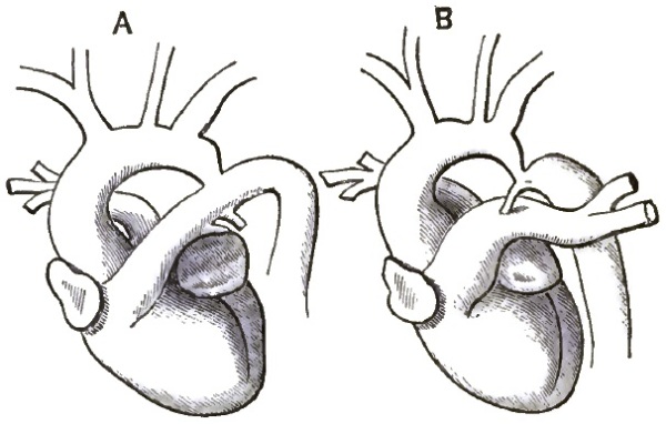
|
| A, Appearance of Aortic Arch in Early Foetal Life.—B, Stenosis of the Aorta. |
ETIOLOGY.—The lesion is a congenital one, and results from a defective development of the aorta. In early foetal life the descending aorta is a continuation of the ductus Botalli, and the aortic arch looks like an independent communicating vessel. (See fig. 52, A.) As the arch develops, however, it gradually forms a more direct union with the descending portion, until finally the longitudinal axes of the two parts form one uniform curve and the ductus Botalli becomes a side branch. At birth there is physiologically a slight nicking of the upper wall of the aorta at the point where the two sections are joined, and the stricture we are studying seems to be merely an exaggeration of this physiological mark. Just how the depression becomes established is not clear and the explanations given are not satisfactory.
SYMPTOMS.—Indications of this lesion are usually very obscure or absent, and it is only discovered at the autopsy. Severe headache is sometimes complained of, and dyspnoea, cough, hæmoptysis, and vertigo may occur if the stenosis is excessive.
Physical Signs.—One of the most marked signs is the conspicuous beating of the dilated arteries around the shoulders and ribs. These arteries may be seen and felt. If the patient is very fleshy, however, they may be [p. 827] concealed. There is usually a marked contrast between the arteries of the upper and lower extremities. The former are full and strong, while the latter are weak and barely perceptible. In many cases it is almost impossible to feel any pulse in the abdominal aorta or in the crural arteries. A loud murmur is also described as occurring over the aorta. This murmur is post-systolic, and does not correspond to any of the ordinary aortic murmurs.
DIAGNOSIS.—This lesion has rarely been suspected, much less diagnosed, during life, but a better knowledge of its peculiarities may lead to more frequent recognition of it hereafter. When the collateral circulation is fully established, stenosis of the aorta could hardly be mistaken for anything else. The resulting excessive dilatation of the great vessels at the root of the neck may simulate aneurism, and it should be borne in mind that aneurism is liable to follow stenosis.
PROGNOSIS.—The death of most of the victims of stenosis of the aorta is directly referable to the lesion itself, although the existence of the trouble is compatible with long life and active occupation. The duration of life and the amount of suffering caused by stenosis both depend upon the amount of obstruction in the aorta and the efficiency of the collateral circulation.
In 49 cases death occurred in the following manner:
| Rupture of the aorta | 10 | times. |
| Rupture of the heart | 3 | " |
| Sudden pulmonary oedema | 4 | " |
| Cardiac failure | 8 | " |
| Apoplexy | 4 | " |
| Pneumonia | 8 | " |
| Capillary bronchitis | 4 | " |
| Paralysis | 2 | " |
| Pleurisy | 1 | time. |
| No cause assigned | 5 | times. |
| 49 | times. |
TREATMENT.—Obviously, no treatment for the lesion itself is possible. If recognized, the existence of the sufferer may be prolonged by adopting moderation in all things as the maxim of his life. Subjective symptoms of discomfort must be combated on general principles as they arise.
This is the most important inflammatory disease of the coronary artery which has been observed. It resembles chronic endarteritis elsewhere, and frequently accompanies the same affection of the aorta, though it may occur alone. The disease may be general, affecting both coronary arteries equally, or one may be more involved than the other, or the disease may be confined to one vessel or to even a small branch.
ETIOLOGY.—Chronic endarteritis of the coronary arteries is especially a disease of middle and advanced life. It occurs most frequently in the male sex. The coronary artery stands fifth in the order of frequency in which the vessels are attacked. The disease is attributed to the misuse of alcoholic drinks, syphilis, chronic lead-poisoning, gout, and chronic kidney disease, by encouraging an early senescence of the tissues, and hence favoring the occurrence of the arterial change.
SYMPTOMS.—There are no symptoms which are peculiar to the disease, those which exist being due to the consecutive changes in the substance of the heart. We may divide cases for convenience of description into those with an acute course and rapid death; those pursuing a subacute course; and, finally, those having a chronic one. In the first instance, sudden death either occurs in a person apparently in perfect health after the manner of a syncope, as in one getting out of bed or standing on the street, while straining at stool, or under sudden emotional excitement. Death may not follow on the instant, but occurs in the course of a longer or shorter time. The attack begins with pressure in the cardiac region, anxiety, restlessness, streaming pain. The complaints and anxiety increase; the breath becomes short and troublesome, the pulse small, frequent, and intermittent; finally, collapse occurs, with oedema of the lung. Death takes place with either a clear mind or slight delirium. Such a fatal ending may cover a day or two or only a few hours. Almost always careful subsequent inquiry elicits the fact that for some time past respiratory or cardiac difficulties have existed, which appeared and disappeared and were not regarded as serious or suspicious. Sudden death may also occur in cases of protracted chronic heart disease following arterio-sclerosis, with an old history of the symptoms of angina pectoris, under the appearance of a fainting fit or of a severe attack of angina or oedema of the lung lasting several days. In such a case rupture of the heart may be found, with bloody infiltration of the cardiac [p. 829]muscle and effusion of blood into the pericardium. In other cases there may be small hemorrhages, often with pronounced infarct formation and softening. In still other cases neither hemorrhage nor infarction is found, but fatty degeneration of the muscle or beginning softening. The sclerosis in such cases is usually very distinct, and affects the trunk and anterior descending branches of the left coronary artery. Sometimes it is hard to find the diseased spot, as it may be circumscribed or on a side branch. In the last-mentioned cases, where sudden death occurs in a chronic process, no post-mortem signs of acute disease are usually found. A chronic fibroid process, with atrophy, exists, which has run a tolerably latent course and leads to death under the appearance of sudden cardiac weakness.
PATHOLOGY.—There are two stages of chronic endarteritis: 1. The stage of simple thickening of the intima; 2. The stage of ulceration and the accompanying further changes.
At first, the normal smooth, shining inner surface of the intima is interrupted here and there or in long stretches by flat rounded elevations, which gradually merge into the healthy surrounding tissues, and are characterized by a paler, more transparent character, and at the same time softer but elastic consistence. The surface of these thickenings, which are frequently located at the point where branches are given off, is either perfectly smooth or slightly wrinkled. Besides these translucent spots there are similar ones which are opaque, whitish or yellowish in color, and have a somewhat rougher surface. Lastly, there are very pronounced thickenings with a yellow color. In the slighter degrees these spots occur singly. In the more pronounced cases they may take up the greater part of the surface; the wall of the vessel is thickened, the inner surface is uneven, and the vessel itself more or less dilated. In the beginning the intima retains its shining surface: after the disease has lasted a long time this is changed, and the second stage appears. Roughnesses, erosions, and ulcerations appear, or more commonly calcification of the wall. This latter appears at first as little thin layers, and finally in large shield-like plates of lime salts, which may occupy the whole circumference of the artery and change it into a stiff, bony tube. It is found where ulceration has occurred, and often without the appearance of the latter. Together with the rigidity of the wall there occurs a slight tortuousness of the vessel. At first the superficial layers of the intima are soft; next they become more sclerosed, and their tissue denser and finally striated; or disintegration, commencing deep in, may reach as far as the surface and lead to an atheromatous ulcer. A more or less abundant deposit of lime salts follows in the sclerosed layers of the intima, leading to the formation of homogeneous plates as hard as bone.
The result of the process at first is diminution of the calibre of the vessel, next diminution of the elasticity and contractility of the artery: it loses its resistance and suffers dilatation in consequence of the blood-pressure, and may attain aneurism. Or if calcification occurs early the diminution of the lumen remains, or perhaps even increases, and may reach an almost complete occlusion of the vessel.
The effects on the heart which follow this form of disease of the coronary artery, though described in another place, had best be enumerated here: 1. The flow of blood not being sufficiently interfered with to cause disease, the heart may remain unchanged. 2. Hemorrhagic infarction may result, accompanied by simple fatty degeneration or softening, which is the most frequent cause of rupture of the heart. 3. Fibrous degeneration or myocarditis may occur, leading perhaps to aneurism of the heart. 4. There may be a combination of these two—a greater or less marked fibrous degeneration, to which a fresh hemorrhagic softening is added.
DIAGNOSIS.—There are no pathognomonic symptoms of this disease, and [p. 830] it is doubtful if a diagnosis can be arrived at. When the conditions spoken of under Etiology pertain, and certain of the symptoms mentioned in connection with the disease are present, a suspicion of chronic endarteritis of the coronary artery may be entertained with some degree of probability.
PROGNOSIS.—This must necessarily be unfavorable where the suspicion of the disease is entertained.
TREATMENT.—Little is to be expected in the way of treatment beyond mere palliation. In the rapid cases death occurs so soon that the medical attendant barely has time to reach the patient. In those cases which last longer the treatment must bear special reference to the symptoms. Pain and spasm may be allayed by opiates or by the inhalation of some anæsthetic cautiously administered, as ether or nitrate of amyl, or by the cautious use of nitro-glycerin and the application of counter-irritants, as mustard, over the cardiac region. Digitalis is to be used with the greatest caution, if at all, as its action may be positively harmful. The same is true of the bromides.
Besides the preceding, another form of endarteritis has been met with in the coronary artery—namely, the obliterating endarteritis, more especially found in cases of syphilis and occurring in the smallest branches. It is characterized by a gradually increasing thickening of the intima through the formation of a connective tissue rich in cells, and which leads to a narrowing, or even complete closure, of the lumen of the artery. This thickening may involve one side of the artery or its whole circumference. The inner surface of the intima on microscopic examination is found to be covered by a layer of intact endothelium where occlusion is not complete. There is deposit of neither fat nor lime salts in the thickened intima. The outer coats of the artery show little change.
The disease is accompanied by indurating myocarditis. Its symptoms are those seen in this disease—namely, weakened cardiac activity, cardiac dilatation and irregularity, possibly cardiac murmurs, an accentuated pulmonary second sound, a pulse of moderate frequency, weak and non-rhythmical, dyspnoea, cough.
DIAGNOSIS.—Impossible.
TREATMENT.—Purely symptomatic.
Aneurism of the coronary artery is of rare occurrence. There is no place of election for the disease, all parts and each artery being alike liable to be affected.
ETIOLOGY.—The most common cause of the affection is chronic endarteritis, where, through disease of the intima, the resistance to the blood-pressure is diminished. Embolism is another though far less frequent cause of the disease, several such cases having been reported; and other highly suggestive cases are on record in which embolism of the artery had occurred, with the production of considerable dilatation for a short distance above the obstruction.
PATHOLOGY.—This does not differ from aneurism in other vessels. The number may be from one to many, usually not more than two or three. The size is generally that of a pea, often it is smaller, and sometimes it is as large as a large nut. The termination is usually rupture with fatal hemorrhage, and in far the majority of cases this occurs into the pericardium.
[p. 831] SYMPTOMS.—In most all of the cases I have found recorded there were no symptoms till rupture of the sac occurred, giving rise to death from hemorrhage. Then those symptoms which might be expected occurred—namely, great præcordial pain, dyspnoea, suffocation, tumultuous heart, irregular and intermittent pulse, and sudden death.
DIAGNOSIS, PROGNOSIS, and TREATMENT need not be considered, as the disease is not recognizable.
Occlusion, more or less complete, of one or both of the orifices of the coronary artery has been met with in connection with chronic endarteritis of the root of the aorta. The accompanying sclerosis may draw the orifices up like the strings of a purse, or a calcific plate may extend from one side, or perhaps, detached, may lie simply applied to the orifice. In rare cases the chief disease may be in the artery itself, one of the main trunks or a branch being affected.
The PATHOLOGY is the same as that already described under Endarteritis, stenosis being an early consequence of the process, and persisting, or even increasing, to the last.
The SYMPTOMS observed in such cases are neither peculiar nor diagnostic. They consist of those depending on the concurrent affections, as of the cardiac valves, muscular tissue, or aortic arterio-sclerosis. Prominent among them are dyspnoea, palpitation, sudden cardiac distress, painful pressure in the region of the heart, great anxiety; at last pallor of the skin, feeble cardiac impulse, indistinctness of the cardiac sounds, the right ventricle continuing to contract forcibly till the end. There is oedema of the lungs at last, and on post-mortem examination fatty degeneration of the heart-walls is found as a secondary consequence of the occlusion.
Although these conditions are rarely found, yet a sufficient number of cases is already on record to enable us to form a tolerably good idea of the symptoms which accompany them. These latter in embolism remarkably resemble those observed in the lower animals on ligation of the coronary arteries. In the animals experimented on a rapid enfeeblement of the heart's action ensued. The phenomena occurred in the following order: First, there was retardation of the rhythmical cardiac contractions, the left ventricle being primarily affected. At first, the right ventricle beat faster, and then gradually became slow. The beats became slower and slower till they ceased, the left ventricle ceasing to contract a little before the right. The second result was a gradual loss of power of the cardiac contraction. The third result was the gradual distension of the left auricle when the left coronary artery was compressed. The auricle swelled up more and more, became bright red, and the rhythmical contractions changed to oscillatory movements, which ultimately ceased entirely. The right ventricle and auricle continued to contract powerfully, and the left ventricle feebly.
ETIOLOGY.—Rheumatism with its attendant complications—that is, disease of the valves, and especially of the aortic valve, atheroma of the coronary artery and possibly cardiac or other thrombosis—forms the chief cause of [p. 832] embolism, a small fragment of tissue being borne away by the current of blood.
SYMPTOMS.—These are acute paralysis of the heart's movements, pain, feeling of impending annihilation, retained consciousness, and regular respiration. Nausea and vomiting have been observed. The lips are livid, extremities cold and covered with a clammy sweat. In one case there was inability to lie down. No pulse could be felt in any of the accessible arteries, and neither apex-beat nor heart-sounds could be detected. The ear applied to the cardiac region could hear only a kind of cardiac tremor, which was very like the sound of a vibrating steel plate. There was no loss of consciousness. The respiration was regular and rhythmical, not exceeding eighteen or twenty in the minute. The patient died twenty hours after the first symptoms.
DIAGNOSIS.—Although a positive diagnosis is impossible, the negative pulmonary physical signs, the regular and rhythmic character of the respiration, and the enfeeblement of the heart's action may lead to a very strong suspicion of embolism of the coronary artery.
PATHOLOGY.—A small coagulum may stop up the main branch, usually the left anterior, of one artery, or both arteries may be occluded by a larger coagulum. In one instance an atheromatous softened patch ruptured into the anterior portion of the left coronary artery, and filled up the lumen with a soft putty-like mass (the sculptor Thorwaldsen). A fatal issue is likely to occur in a very short time, as the anastomosis cannot be sufficient for the sudden demand.
PROGNOSIS and TREATMENT need hardly be considered, as the affection is necessarily fatal in cases which can be made out.
The same causes which give rise to thrombosis elsewhere are operative in this case. They are chiefly arterio-sclerosis and rheumatism.
SYMPTOMS.—There have been observed slight tightness in the cardiac region, lasting a few days, or a sense of oppression or constraint at the back of the sternum. The pulse has been quickened, but is usually very much slowed and very feeble; it has been observed as low as eight beats in the minute. There is a sense of great lassitude and feebleness of all the limbs. The respiration is normal in rhythm and frequency. Auscultation reveals nothing but ordinary respiration till near a fatal issue, when moist râles indicative of oedema of the lungs are heard. Percussion gives at all times a normal resonance. There is no dyspnoea. The heart-tones are clear, though weak, if occlusion is not complete or anastomosis is perfect. (West was able to inject the arterial system of the heart completely from one coronary artery, the other having been tied.) If there is complete obstruction, we may expect to hear a fremitus such as is produced by muscular spasm instead of normal heart-sounds (observed in two cases). The skin of the body and face is cool, pallid, and covered with sweat. The visible mucous membranes are anæmic and pale. The mind is clear.
DIAGNOSIS.—The cardiac feebleness and progressive slowness, together with the absence of symptoms connected with the lungs, might lead one to suspect the presence of thrombus.
PROGNOSIS.—If a large branch of the artery is affected a fatal termination is probable. If, on the other hand, the affection occurs in a small branch, there is reason to believe that the circulation is sometimes re-established through anastomosis.
TREATMENT must be purely symptomatic.
This may occur independent of aneurism. There are no premonitory symptoms in some cases, death taking place suddenly. In other cases vague and irregular symptoms lead the patient to understand that he is not in perfect health. The symptoms of the disease are not characteristic. Those which have been recorded are a difficulty of breathing, a sense of constriction across the chest, or a pain and feeling of anxiety in the præcordia; a frequent, feeble, and perhaps very irregular pulse; epigastric pain and tenderness. The extremities are cold. The mind remains clear. The physical signs are increased area of flatness in the cardiac region, due to the escape of blood into the pericardium, and scarcely audible cardiac sounds. The symptoms may extend over a period varying from a few moments to several days. Usually, some of the changes indicative of arterio-sclerosis are found in the artery.
Acute inflammation of the coats of the pulmonary artery has only been found associated with the pyæmic process as circumscribed abscesses of the wall.
Endarteritis of the pulmonary artery, though quite rare, is occasionally met with in persons the subject of rheumatism, gout, syphilis, or alcoholism. It is seen only when the pressure is abnormally increased in the pulmonary vessels, especially in diseases of the mitral valve. It is usually accompanied by a more pronounced disease of the aorta, but is occasionally seen alone. The extent of disease is hardly ever so great as that found in the other large vessels, and at most amounts to the presence of prominent hard yellow or gray patches in the intima, with perhaps ulcerated surfaces, and rarely containing a deposit of lime salts. Complete rigidity has been observed extending far into the lung. The wall of the vessel may be irregularly dilated and its elasticity diminished. Usually, the disease is in a much milder form, presenting perhaps a small amount of fatty degeneration of the intima, and is not infrequently associated with mitral stenosis or insufficiency (notably the former), pulmonary fibrosis or emphysema, with accompanying hypertrophy of the right ventricle. No symptoms have thus far been found to be distinctly referable to atheroma of this artery.
Dilatation of the pulmonary artery from primary disease of its walls is of so rare occurrence that it may be merely mentioned. It depends on chronic endarteritis, just spoken of. Where, on the other hand, there is great pressure in the pulmonary circulation, as in marked mitral stenosis, or insufficiency, collapse, or emphysema of the lung, with great hypertrophy of the right ventricle, general dilatation of the pulmonary artery may take place. The artery has been found to be six and a half inches in circumference in a case of emphysema, the normal average being three and a half inches; the semilunar valves were insufficient, and the walls of the artery very much diminished in thickness. From this as a maximum all degrees of dilatation have [p. 834] been recorded, with sometimes thickening and degeneration of the coats, at others thinning with or without degeneration.
A systolic murmur has been observed over the artery when the dilatation was considerable. The second pulmonary sound is usually strengthened (unless the elasticity of the pulmonary artery is very much diminished or the blood-pressure lowered in the right ventricle by changes of its walls, or the pulsation is very quick and irregular). A circumscribed dulness on percussion has been found in a few cases at the left edge of the sternum, when the position of the heart was normal, between the second and third cartilages. Sometimes there is a double impulse, a systolic thrill, or more often a systolic pulsation, felt in this position without any perceptible dulness, the edge of the lung being retracted and the dilated artery taking its place. It is to be borne in mind, however, that this sign (impulse, thrill, or pulsation) may be present without any dilatation of the pulmonary artery or hypertrophy of the ventricle, when inflammatory contraction of the lung has occurred or the respiration is superficial, as may happen in phthisical subjects, women, feeble and anæmic individuals, pregnant women, convalescents, and persons afflicted with acute rheumatism. In such persons the pulmonary second sound frequently seems to be unusually loud when compared with the aortic second sound, without any evidence of hypertrophy of the right ventricle being present. The determination of the position of the lung establishes the diagnosis in such cases. On the other hand, a lung dilated by emphysema may interpose and completely cover the heart and pulmonary artery, which, though dilated, may thus be masked.
Aneurism of the trunk or primary branches of the pulmonary artery, on the other hand, is an exceedingly rare disease. But few cases are on record. Aneurisms may be spindle-shaped or sacculated, of moderate size, and are usually situated on the trunk. Lividity of the face, dyspnoea, cough, dysphagia, headache, pain in the chest and epigastrium, are the principal symptoms; and a systolic pulsation (sometimes also diastolic) between the second and third left ribs near the sternum, more or less prominence here, a superficial rough systolic murmur propagated to the left and upward, a purring thrill, and flatness on percussion in the same region and a little above it, are the principal physical signs which have been recorded.
But the physical signs and symptoms above enumerated are not all present in each case, nor are they when present distinctive of pulmonary aneurism. Dysphagia is mentioned in but a single case, dyspnoea is not constant, and cyanosis was at times absent. Also, the physical signs were not constant. Even if all were present they might be produced, as has been the case, by aneurism of the left wall of the aorta, infiltrated lung-tissue, or by a solid tumor lying over the vessels. The locality of the cardiac hypertrophy and dilatation aids in establishing the diagnosis. If it is on the left side of the heart, aneurism of the aorta is indicated; if it is on the right side, pulmonary aneurism. These aneurisms tend to rupture into the pericardium sooner or later.
Dissecting aneurism of the pulmonary artery has been observed once. It was of small extent.1
1 Bul. de la Soc. Anat. de Paris, 1881, pp. 589–591.
Narrowing of the trunk or of one of the main branches of the pulmonary artery is of very rare occurrence. It may follow compression by an aneurism of the ascending or transverse portion of the aorta, compression by tumors in [p. 835] the mediastinum, as from new growths or enlarged glands; it may be caused by cicatricial contraction following mediastinitis, inflammation of a portion of lung or of the bronchial glands, or it may follow disease of the coats of the artery (endarteritis).
The phenomena produced by stenosis of the trunk of the pulmonary artery are similar to those found in stenosis at the orifice, which are treated of in another place. They are anæmia of both lungs, accompanied by persistent dyspnoea with occasional exacerbations (the patient assumes a horizontal position either habitually or during the paroxysm—a fact of true diagnostic importance [Chevers], as in all other forms of disease of the heart and great vessels the patient breathes easier when the shoulders are raised. But in this the dyspnoea results from insufficiency of the supply of blood to the lungs and system generally, and hence the recumbent posture affords relief by removing the impediment of gravity, and thus promotes the supply of blood to the brain), congestion, dilatation and hypertrophy of the right side of the heart, cardiac palpitation, and finally general venous congestion. Hypertrophy of the right ventricle is shown by increase in the transverse measurement of the cardiac area of flatness and increase in the force of the cardiac impulse. The artery up to the point of constriction is dilated; the second sound is abnormally loud and accentuated. Pulsation may be felt and a systolic murmur heard in the second left intercostal space (observed in the right once), propagated upward to the neck at the left of the sternum, or heard in the interscapular space close to the spinal column.
PROGNOSIS is unfavorable.
There is nothing to be gained by treatment.
Violent effort and great excitement have been followed by rupture of the trunk or a main branch of the pulmonary artery. In the majority of cases the coats were degenerated, though this was not always the case (Chevers). Death is often instantaneous, but sometimes is delayed some hours. In one case observed by Ollivier the duration was twenty-seven hours.
The pulmonary artery, from its position, is especially prone to become plugged, either by substances coming from other parts of the body or by coagula originating in the vessel itself. Pieces of disintegrated coagula from the systemic veins, the contents of echinococcus cysts ruptured into the venous current, fragments of new growths, are carried to the heart and pass into the pulmonary artery, or large thrombi may be detached from their position in a vein and lodge in the trunk or main branches of the pulmonary artery.
Primary thrombosis of the pulmonary artery is very uncommon. In certain septic conditions, in parturient women, in typhoid fever, and in extreme anæmia thrombosis of the pulmonary artery may occur. It commences perhaps in the right ventricle or at the pulmonary valves, though it is also seen farther up.
SYMPTOMS.—The severity of the symptoms depends on the completeness of the obstruction. There is dyspnoea, more or less marked according to the size of the thrombus or embolus, pain in the præcordia, great distress, anxiety, faintness, sense of suffocation, tightness in the chest, palpitation, lividity and extreme pallor, cold sweats, an almost imperceptible pulse, great restlessness, [p. 836]and occasionally convulsions. The mind remains clear. The symptoms develop gradually or rapidly—in the former case depending on the slow increase of a small thrombus—and remissions are often seen; in the latter case depending on the sudden lodgment of an embolus of large size. Sometimes the symptoms are extremely marked, and death takes place in a few minutes. The appearances are not those of asphyxia, and death is usually attributed to want of arterial blood-supply to the brain and medulla oblongata, and not to suffocation or paralysis of the heart.
Percussion shows a normally resonant chest. Auscultation gives normal breath sounds with free inspiration and expiration. There is very likely a basic systolic murmur conducted along the course of the pulmonary artery, but this is not constant. The cardiac second sound and impulse are increased. At the post-mortem examination the heart is found in diastole, the left cavities and pulmonary veins empty, the right cavities filled with blood, and the cardiac veins strongly distended.
DIAGNOSIS.—The diagnosis is often uncertain. When not developing with extreme rapidity the symptoms are very similar to those caused by stenosis of the pulmonary artery, and in the suddenly fatal cases they are almost identical with rupture of the heart or rupture of a thoracic aneurism, or even angina pectoris. The history of an antecedent thrombus or of a disease of the heart which is likely to be accompanied by thrombus, together with the absence of physical signs, render a diagnosis many times probable.
PROGNOSIS.—To be regarded as of the gravest character.
TREATMENT.—In the rapid cases death occurs before anything can be attempted. In the less severe cases absolute rest must be enjoined, and free stimulation with brandy, ammonia, and ether attempted. It might be worth while to place the patient with the head lower than the body, to favor the flow of blood to the brain.
Aneurism of both the superior and the inferior mesenteric arteries occurs. The former is the more frequent, though still a rare disease.
The symptoms are pain in the epigastric and lumbar regions, a globular pulsating tumor in the median line, the pulsation being accompanied by a bellows murmur. The tumor has been seen in at least one instance to be so large as to press on the renal arteries. Rupture is apt to take place with the signs of internal hemorrhage. The cause of the disease is the same as of aneurism elsewhere. Embolism is said to be a not infrequent precedent. The aneurism is seldom larger than a hen's egg, and is usually globular.
A positive DIAGNOSIS of the locality of the aneurism is not possible.
The TREATMENT must follow individual indications. Compression has been successful in a few instances.
Several cases where the superior mesenteric artery was found at autopsy to be completely occluded by coagulated fibrin were mentioned by Tiedemann in a work published in 1843. Virchow first described the characteristic [p. 837] post-mortem appearances which follow this lesion in his Gesammelte Abhandlungen, and since then records of cases have been numerous.
CLINICAL HISTORY.—In by far the majority of cases there is an evident source for an embolus. Pain in the abdomen is the first symptom, and usually remains one of the most prominent throughout. At first it may be a dull aching just below the borders of the ribs, but soon there is superadded paroxysmal pain resembling colic, and which may at times even be relieved by pressure. The occurrence of this colic in cases where embolism might happen ought to put the physician on his guard for other symptoms; for, though insufficient in itself to establish a diagnosis of embolism, the presence of a colic resisting treatment in the course of cardiac disease justifies the suspicion that this may be the case. The pain is usually located near or above the umbilicus.
Intestinal hemorrhage occurs in nearly every case; death may take place before any change in color of the stools is observed or any blood appears at the anus, but on post-mortem examination blood is found in the intestine. The cause of this hemorrhage is the infarction of the intestine analogous to that which takes place in other organs supplied by end arteries, the superior mesenteric having been proved experimentally to be functionally such an artery, owing to its great length, the extent of tissue supplied by it, and the comparative smallness of the vessels with which it anastomoses on the borders of its territory. The collateral circulation is thus so long in being established that ample time is allowed for those disturbances of nutrition in the walls of the vessel which render them permeable and allow the blood to escape. In view of the hemorrhage certain other symptoms are readily accounted for, as, for example, pallor of the face and surface of the body, the considerable and rapid fall of the temperature, syncope, hæmatemesis, diarrhoea, and melæna. These two latter symptoms are important though inconstant. There is reason to believe that the first effect of the embolism is to paralyze the bowel and prevent peristaltic action. Diarrhoea is of frequent occurrence, and may be profuse, the stools remaining of their natural color; or fresh blood may be passed at first from the rectum, followed by the continuous passage of tar-like masses; or the stools may be of pulpy consistence, mixed with blood, or consisting of tarry blood. Lastly, profuse hemorrhage may take place in which the stools resemble tar-water. The character of the blood does not give any kind of clue to the locality of the lesion.
Vomiting is a frequent symptom, and may consist of altered blood of variable consistency. A fall in temperature can often be determined by the thermometer, especially after severe hemorrhage. Not rarely the temperature is normal or may be even increased, especially if secondary inflammation has set in.
Tension and tympanitic swelling of the abdomen may occur or fluid may be detected late in the case, these being evidence of peritonitis.
PATHOLOGY.—Before proceeding to consider the pathological changes occurring in embolism, a few words on the blood-supply of the intestine might perhaps render what follows clearer. The superior mesenteric artery supplies the whole of the small intestine except the first part of the duodenum; it also supplies the cæcum and the ascending and transverse colon. The inferior mesenteric supplies the descending and sigmoid flexure of the colon and the greater part of the rectum. The anastomoses are as follows: The pancreatico-duodenalis, a very small artery and a branch of the hepatic, anastomoses with the first branch of the superior mesenteric, also a very small artery and given off under cover of the pancreas. The middle colic artery anastomoses with a branch of the inferior mesenteric. Both these arteries are given off from the main trunks of the arteries.
The experiments of Litten in 1875 show that the superior mesenteric artery, [p. 838] though not so anatomically, is functionally a terminal artery, the anastomosis not being developed with sufficient rapidity in case of extensive embolism to ensure the integrity of the circulation.
1. The result of sudden total closure by embolism of the trunk of this artery, therefore, is precisely like that of ligature of this artery in animals, and is first to produce sudden abdominal pain, attacks of colic, vomiting, uncontrollable intestinal hemorrhage, death. The intestine from the lower transverse portion of the duodenum to the middle of the transverse colon is found to be suffused, brown-red, blackish, or grayish. All the layers are swollen; innumerable capillary extravasations of small and great extent are seen, with venous hyperæmia and oedematous infiltration. In other words, there occurs necrosis with oedema and hemorrhage in all those portions of the intestines which are supplied by this artery.
2. Closure of large branches by embolism gives rise to infarction of the portion of intestine concerned, followed by death. The symptoms differ only in intensity, if at all, from the preceding. A case has been seen where there was every reason to believe that embolism had occurred, and yet the patient recovered. (The patient, suffering from acute rheumatism complicated with peri- and endocarditis, suddenly developed profuse intestinal hemorrhage of tar-like color, which was repeated twice. Colic pains, tympanites, depression of the temperature of the body, followed. At the same time symptoms of embolism of various other arteries were present. Recovery took place after eight weeks.) This result of course depended on the subsequent perfection of the collateral circulation.
3. Closure of the smallest branches may produce the same kind of symptoms as the above, though less in degree. Limited portions of intestine have been found to be in a gangrenous condition from embolism of very minute branches, more especially when the embolus extended well into the artery. In place of gangrene of the intestine ulcers of the mucous membrane have been seen independent of typhoid fever or tuberculosis. Considerable stenosis has followed such ulcers.
The affected portion of intestine in embolism is found to contain a variable amount of blood mixed with the other contents of the gut. Peritonitis, dry and limited or general and accompanied by effusion, is the rule. The mesenteric glands are found enlarged and succulent, with perhaps here and there necrosed spots. Thrombosis of the corresponding veins is not uncommon. Large collections of blood under the peritoneum and in the mesentery have been observed. The color of the mucous membrane has been slaty, and a diphtheritic appearance has been observed.
DIAGNOSIS.—The following are the most important points in forming a diagnosis: 1. A source exists from which an embolus might be derived. 2. Profuse and even exhaustive intestinal hemorrhage sets in, which can neither be explained by primary disease of the intestinal walls nor by hindrance to the portal circulation. 3. There is a rapid and considerable fall of the temperature. 4. Pain in the abdomen comes on, which may resemble colic and be very severe. 5. Finally, tension and tympanitic swelling of the abdomen occur, and there may be fluid in the abdominal cavity. 6. Evidence of embolism of other arteries may have been obtained before the symptoms of embolism of the superior mesenteric artery come on, or such evidence may appear at the same time as the latter. 7. Palpation may reveal the presence of collections of blood between the folds of the mesentery.
PROGNOSIS.—The prognosis in embolism of the superior mesenteric artery, though not absolutely bad, is exceedingly grave. It must be borne in mind that the symptoms of occlusion of one of the large branches are similar to those where the main stem is involved, while the probabilities of recovery in the former are much greater, as already explained, from the shorter extent of [p. 839]the anastomosis. There is evidence that recovery from the immediate effects of embolism may take place even where subsequent ulceration has been so great as to cause complete closure of the intestine through cicatrization. (A case is related by Parenski where the patient was operated on for stricture of the bowel, and only at the autopsy was it discovered that the stricture was due to cicatrization from ulceration caused by embolism of one of the branches of the superior mesenteric.) There are at least three cases of recovery on record where occlusion of the main stem was supposed to have taken place; but inasmuch as the situation of the embolus cannot be determined with certainty if the patient recovers, these cases are open to the suspicion that one or more of the larger branches only were occluded. The profuseness of the hemorrhage, though it may imperil the life of the patient from exhaustion, bears no constant relation to the gravity of the case. Copious and repeated hemorrhages per anum took place in cases of recovery, while in other fatal cases this symptom was entirely absent. Extreme fetor of the stools must be regarded as of evil omen, as it may be the evidence that gangrene of the bowel has taken place.
TREATMENT.—One of the first symptoms calling for relief is the colic, which is best met by morphia given subcutaneously or by suppository. For the hemorrhage ergot by the mouth and alum enemata have proved serviceable, or the application of ice to the abdomen. The lowering of the heart's action by sedatives is to be avoided when we remember that their use would lower the blood-pressure, and thus tend to retard the establishment of the collateral circulation.
The symptoms of thrombosis have not been determined apart from embolism, and it is doubtful if the affection proves fatal unless the extent of artery involved is very considerable or the formation of the thrombus is very rapid, for the anastomosis is gradually made compensatory. In either of the latter cases the symptoms are identical with embolism, and the pathological appearances are the same. With regard to treatment, general indications must be pursued.
This disease is met with, but it is usually slight and unaccompanied by symptoms.
Aneurism of this artery has been seen after death. The diagnosis could not be made, in all probability, during life. Pain might be a prominent symptom, though not necessarily, as many of the aneurisms of the abdomen are unattended by any symptoms. Rupture is not unlikely as a termination.
Embolism has been observed. Sudden pain in the abdomen comes on, followed by vomiting and diarrhoea. The patient looks miserably; the [p. 840] belly is drawn in and painful on pressure almost exclusively in the left iliac region. Severe spontaneous colic-like pains continue, with occasional vomiting and diarrhoea. At first the stools are feculent and pap-like; then they begin to smell bad, and even stink. Red blood is passed. Soon there is a mixture of blood and slimy masses. Finally, the stools are slimy, blackish, almost tar-like, and have a terrible odor, and are passed with griping and tenesmus. Occasional vomiting still continues. The pulse becomes smaller and more frequent, and gradually irregular and intermittent. Soon collapse and death follow.
The predisposing and exciting causes are the same as in embolism of the superior mesenteric artery.
The duration is usually short, lasting from a few hours to three or four days. The termination is ordinarily fatal, though doubtless cases of recovery have occurred, as stated under Embolism of the Superior Mesenteric Artery, the size and position of the embolus not precluding the possibility of the establishment of collateral circulation.
Complications are varying degrees of peritonitis, evinced by tympanites, pain, and tenderness, either localized or diffused, and later by the occurrence of effusion. Sequelæ, when the disease is not immediately or rapidly fatal, are ulceration of the colon with subsequent cicatrization and contraction.
PATHOLOGY.—The mucous membrane of the descending colon, sigmoid flexure, and rectum is somewhat swollen, strongly reddened, and contains ecchymoses and extensive suffusions of blood; or the color may be blackish or slaty and the surface sloughy.
DIAGNOSIS.—The diagnosis can only be made by exclusion. The same points are to be carefully verified as in embolism of the superior mesenteric artery, only the pain and symptoms are in a different place, and the secondary peritonitis also begins on the left.
PROGNOSIS.—The prognosis is very grave, but recovery may take place, contractions or constrictions being left behind.
TREATMENT.—The treatment combines perfect rest, the exhibition of wine, opium, vegetable astringents, and the subcutaneous injection of morphia.
The tumor varies in size from a hazelnut to a child's head, and is egg-shaped. Pain in the epigastrium and right hypochondrium or upper abdominal region is a characteristic symptom. At first the pain is not severe, and is occasional, recurring after a pause of several months' duration; later it becomes very severe and lasting. The abdomen is not tender to the touch or on pressure during the remissions from the attacks of pain, but after rupture of the aneurism, whether temporary or lasting, it is very severe. The abdomen is sometimes distended, at others not. The tumor, owing to its position, cannot be felt, nor can pulsation be detected, as the wall of the aneurism consists of connective tissue and blood-clot, and the stream of blood coming from a small artery is slow. In but a single case has increase in size of the spleen and liver been observed. The functions of the stomach and intestines remain normal in spite of the pain. The locality of aneurism of the hepatic artery is such as to readily cause temporary or lasting icterus—a phenomenon which occurs in perhaps two-thirds of the cases. Rupture, with the ordinary signs of internal hemorrhage, seems to be the usual termination. Inflammatory processes or fever does not follow hemorrhage into the abdomen. [p. 841] If perforation occurs into the gall-bladder, a gall-duct, or the intestine, the hemorrhage may appear to be moderate. In such instances repeated discharges of blood may occur from the intestine, or at the same time may be thrown off from the stomach.
There is no means of determining how long aneurism of the hepatic artery may exist without giving any kind of sign of its presence. Judging from analogy, it is very probable that a considerable time may elapse before the disease is observed. Since pain in the abdomen is the first pathological indication, and rupture the last, we may measure the probable duration of the disease by these phenomena and also by the clinical course. This was not over ten days in two cases, and in three cases it was three to four months. Since aneurisms of the hepatic artery, even when they have reached their greatest dimensions, are not palpable, the pains which appear with them have in themselves no diagnostic worth. The same is true of the icterus which appears sooner or later. It is only after rupture has occurred that all the chances are so placed that a comprehensive estimate of them may be made and a diagnosis arrived at by exclusion. The fact that the function of the stomach remains unchanged in spite of rupture (hemorrhage), and the totally unchanged character of the blood-clots vomited, enable us to locate the situation of the hemorrhage as outside the stomach. If at the same time there is an alternate relation between the occurrence and disappearance of the icterus and the hemorrhage, the inference is admissible that the latter is located in the immediate vicinity of the gall-ducts. Other peculiarities of the blood-clots passed at stool are perhaps the imprints of the valvulæ conniventes of the jejunum.
The DIAGNOSIS of aneurism of the hepatic artery is usually impossible.
Aneurisms of the splenic, renal, and other abdominal arteries are recorded, but not in sufficient numbers to warrant a detailed description of them.
Aneurism of the coeliac axis, when the tumor is large, is accompanied by very much the same symptoms as aneurism of the abdominal aorta. The disease is rather uncommon.
ETIOLOGY.—Syphilis, rheumatism, and advanced age play important parts in the etiology of this disease as predisposing causes of arterial degeneration. Many persons affected have been immoderate spirit-drinkers, which of itself does not directly tend to the disease, but does so indirectly, in that it encourages an early senescence of the tissues. In the same way any debilitating conditions may act as predisposing causes. Chronic endarteritis is most frequently found at the seat of the aneurism. Secondary or exciting causes are peculiarities of occupation, as those which are laborious and require much physical exertion and entail exposure to inclemencies of the weather.
SYMPTOMS.—Pulsation is usually the first symptom observed. It is felt in the epigastrium about two and a half inches below the ensiform cartilage, or even higher, and a little to the left of the median line; or it may be midway between the ensiform cartilage and the umbilicus, on the left. It is not unfrequently of a distensile character, and is unaffected by changes in the position of the patient. It is not synchronous with the cardiac systole, but follows in rapid succession to, and terminates with, the ventricular [p. 842]diastole. A tumor, usually globular, is felt in the region of the pulsation. It is of variable size, from that of a hen's egg to a cricket-ball, or in case of false aneurism even much larger. The tumor is slightly tender; it moves with the diaphragm, and sometimes when it presses upon the pancreas ptyalism has been observed, which in one instance was increased by external pressure on the aneurism with the hands.
Another constant symptom is pain in the left side, extending from well up in the chest to the region of the hip, or located in the lower part of the chest alone, or perhaps in the epigastrium. This pain is either constant or excited by exertion, and paroxysmal in character.
Flatness on percussion over the tumor of varying extent is observed in many cases, and a systolic bruit, perhaps of a whistling character, is heard.
The usual termination of aneurism of the coeliac axis is rupture with internal hemorrhage. The symptoms of this accident do not differ from those of the same occurrence in abdominal and thoracic aneurism, and are likewise usually fatal.
PATHOLOGY.—Strain doubtless forms an important factor in the production of this aneurism in an artery previously weakened by disease of its coats. The tumor is frequently a false aneurism, and has for walls connective tissue and the neighboring organs. When it is of large size, on account of its position it sometimes presses upon the pancreas or vertebræ, and produces absorption with consecutive symptoms. In the former case ptyalism has been observed, which perhaps may have been due to reflex action through the coeliac plexus and pneumogastric nerve, the reflex centre being the medulla oblongata with the facial origin. The wall of the aneurism is usually thin, and in some cases it has given way, leading to the formation of so-called false aneurism. Not infrequently the wall is atheromatous. The size of the aneurism varies greatly, though it is never larger than the two fists.
DIAGNOSIS.—This aneurism is apt to be confounded with aortic aneurism, and can only at times be distinguished from it by its locality and small size.
PROGNOSIS.—This must be grave if a diagnosis is made, for the ultimate result is usually rupture and hemorrhage.
TREATMENT.—The general principles recommended in treating abdominal aneurism should be followed out. It is but rarely the case that compression is admissible, and then the distal pressure is to be used. Rest and diet form the most reliable means of treatment at our command.
The principal affections to which the veins are liable are the following: Inflammation (phlebitis), acute and chronic; Dilatation; Narrowing or obliteration; Degeneration; Concretions.
Idiopathic phlebitis occurs for the most part under one of three conditions: First, as a simple primary inflammation of the tissues composing the walls of the vessel; second, as a participation in an inflamed or diseased condition of surrounding structures; third, as the result of the absorption of poisonous material into the blood.
Like any other structure of the body, the veins are liable to inflammation as a purely local affection. It is nevertheless true that, in the acute form, this inflammation is most likely to occur in connection with certain conditions of the system which seem to act as predisposing causes, although the connection between them and the local phlebitis is not apparent. Thus it occurs (perhaps associated with more or less of lymphangitis) in the puerperal state, in phthisis, in heart disease, and in other conditions of general depression. I have met with it, for example, during recovery from pneumonia after typhoid fever and after suffocative laryngitis. Under these circumstances it constitutes the chief element in the affection known as phlegmasia dolens. Now, none of the above conditions implies, so far as is known, any source of irritation to the venous structures, much less to a limited portion of the venous system; and the only explanation of their association with phlebitis seems to be in the assumption that these conditions favor coagulation of the blood, and that, in these cases, the formation of a clot precedes the local inflammatory process. The location of this clot is probably determined by anatomical conditions.
In other cases, however, the process evidently begins in the wall of the vessel, and the formation of the thrombus is secondary. Any change which interferes with the smoothness of the inner coat, whether by loss of endothelium or by producing inequalities of the surface, will very certainly determine the deposition of fibrin and the formation of a coagulum. The glossy smoothness of the intima seems to require the most perfect nutrition of the subjacent tissues for its maintenance, and its loss produces an immediate slowing and ultimate stoppage of the blood-current. This is admirably shown by the experiments of Nicasse,1 which demonstrate that simply denuding a portion of a vein, and thus cutting off its vascular and nervous supply, induces almost immediately the formation of a thrombus coextensive with the denuded portion.
1 Des Plaies et de la Ligature des Veinse, Thèse, Paris, 1872.
[p. 844] Inflammation affecting the inner coat of a vein and extending along its surface, as in the case of a serous membrane, probably never occurs. The picture of phlebitis formerly drawn, and which embraced the exudation of false membrane or the formation of pus upon the inner surface of a vein, the pus in the latter case floating off with the blood and constituting pyæmia, the formation of a clot being a later and unimportant event, has little or no resemblance to what actually occurs.
The observations upon which these assumptions were based were erroneous, as shown by Virchow, in that the staining of the intima by absorption of coloring matter from the blood was mistaken for inflammatory redness, and changes in the clot itself were confounded with exudation and suppuration. Indeed, when we reflect that the intima is not vascular, we should scarcely expect from it anything analogous to serous inflammation. The only acute process to which it appears liable is an erosion or crumbling away under the same conditions which determine, in the middle or outer coats, increased vascularity, exudation, and the formation of pus.
Thus, from some general condition favoring the coagulation of the blood we may have a thrombus formed, followed by secondary inflammation of the wall of the vessel, or, without such general condition, we may have inflammatory changes, commencing in the outer or middle coat and causing the secondary formation of a thrombus. In either case the clot shuts off the affected portion of the vein from the general circulation. Changes take place in the clot which are more properly considered under the head of thrombosis, and by which it is ultimately removed. Exudation takes place into and between the tunics which form the venous wall, the latter becoming thickened and comparatively rigid, so that when the vein is cut across its lumen remains open like that of an artery.
Sometimes pus is formed between the different coats, constituting small mural abscesses; sometimes the intima crumbles away and exposes the middle coat, which suppurates on its inner surface, and the pus mingles with the débris of the clot. In this way a larger abscess is formed, bounded by the wall of the vein and by a partly-organized coagulum on either side. These coagula sometimes break down, and fragments from them, infected by the pus and its contained micrococci, are swept on in the current of the blood until they find a lodgment, where the process begins anew, and whence it may be propagated in like manner to other and more distant parts.2 It is only to the condition above described that the term suppurative phlebitis can properly be applied.
2 Ziegler, Path. Anatomie, Jena, 1881, p. 429.
But, instead of a suppurative process taking place, the endothelium may be thrown off and replaced by minute vegetations of the character of granulation-tissue, which, penetrating into and blending with the clot, may temporarily or permanently occlude the vein, and the contraction which follows may ultimately leave only a fibrous cord to represent the vessel.3
3 Leroux, Gaz. méd. de Paris, 28 Juin, 1879.
This process is designated adhesive phlebitis, and is one of frequent occurrence and very important in its results. It takes place in connection with suppurative phlebitis, and by closing the vessel on either side of the suppurating portion serves to prevent the pus from mingling with the general circulation.4 By its action the largest veins, including the venæ cavæ, are occluded, and extensive and important changes in the circulation are brought about.
4 While this is true of a pus-cavity forming within a vein, an abscess originating outside of a vein or between the layers of the venous wall may open into the vessel at a point not protected by a clot, and the pus mingling with the blood will constitute veritable pyæmia.
The second condition under which phlebitis occurs is that in which a vein, [p. 845] coursing through an inflamed or diseased structure, becomes itself inflamed. This takes place most frequently in phlegmonous erysipelas and in diffused inflammation of the cellular tissue, but it may be the result of any inflammation in the neighborhood of a vein. Under these circumstances the external layer of the venous wall is first affected, and the others subsequently. Only a portion of the circumference of the vessel may be involved, and the wall may bulge inward considerably without necessitating the formation of a thrombus (Virchow). But if the nutrition of the walls is seriously impaired, the intima becomes roughened by the loss of its endothelium, the blood-current is slowed by the increased friction thus caused, and, the uneven surface favoring at the same time the adhesion of fibrin, a clot is formed, and the course thereafter is the same as when the vessel is primarily affected.
Suppuration may also take place between the vein and its sheath, and extend for a considerable distance along the vessel. The walls participate secondarily, and the vein becomes occluded as before described.
In the third class of cases, those depending upon toxic infection, the inflammation is caused by the irritation of some poisonous material circulating in the blood. The phlebitis is therefore secondary, and is to a great degree overshadowed by the general condition which accompanies it. Aside from instances in which there is a direct inoculation of a poisonous material—as, for example, the venom of a serpent—the conditions merge into those which come under the designations pyæmia and septicæmia—diseases which were formerly confounded with phlebitis, but which are now recognized as distinct from, though often coexisting with, it.
If in acute phlebitis the inflammation does not result in the formation of pus, the vein may recover its normal condition, or the walls may remain thickened and the lumen contracted, but still pervious, or it may be entirely occluded. Suppuration, however, always results in complete and permanent closure of the vein.
The symptoms of acute phlebitis are chiefly such as indicate obstruction of the vein. When a large vein, situated in one of the extremities, is the seat of the affection, there are usually severe pain of a tensive character and decided tenderness on pressure. The limb swells, sometimes to a very considerable extent, and becomes stiff and unwieldy. If a superficial vein, such as the long saphena, is affected, there will be subcutaneous oedema and pitting; but when the vessel lies beneath a firm, tense fascia, this will limit the swelling, and the limb will be hard and brawny, while the tension will greatly aggravate the pain.
When the vein is sufficiently near the surface it may be felt at the affected part as a hard cord, usually more or less knotted. The skin over it may be discolored, presenting a red or somewhat coppery hue and a streaked or mottled appearance, or the pressure from the effused serum may empty the capillaries of blood and render the skin pale and shining.
The temperature of the limb may be elevated, normal, or subnormal. In the outset, under the influence of the inflammation, there is usually increased heat, but as the tension from the oedema interferes more and more with the circulation, the temperature falls, and the limb may become colder than its fellow.
Inflammation of a limited portion of a vein may not be attended by any notable symptoms, the collateral circulation being quickly established, and the effects of the obstruction thus obviated, while, at the same time, the local symptoms are masked by the morbid conditions in the surrounding tissues which give rise to the phlebitis.
The constitutional symptoms accompanying phlebitis are those of inflammatory fever, the grade of which will depend upon the extent and severity of the inflammation. When a considerable length of vein is involved, as [p. 846] may be the case in the form of phlebitis already referred to, which progresses along the sheath of the vessel, the irritation of the general system may be great, especially if pus is formed, when hectic or even typhoid symptoms are not uncommon.
The differential diagnosis of phlebitis in its local appearances requires only its distinction from lymphangitis. The latter disease is more abrupt in its invasion, depends almost always upon some wound or injury with which the local symptoms are directly connected, is more diffuse, affecting a network of vessels rather than a single one, and is invariably accompanied by engorgement of the lymphatic glands to which the affected vessels lead, as, in the case of the extremities, the axillary or inguinal glands.
In complicated cases the occurrence of phlebitis may not be marked by any distinctive symptoms. It may be suspected if, in the course of erysipelas, diffuse cellulitis, etc. in the neighborhood of an important vein there is a somewhat sudden increase of pain and swelling, and if an enlargement of the tributary cutaneous veins is soon observed.
The treatment of phlebitis consists in complete rest, in the use of such constitutional means as may be necessary to allay the irritation of the system, and locally in the application of leeches and warm fomentations. If, on the other hand, the local temperature is very high, the use of ice may be indicated.
Nonat, in cases of commencing phlebitis from venesection, tried the use of flying blisters over the part affected. Obtaining good results, he extended the treatment to phlebitis following typhoid fever, etc., and the morbid phenomena were at once arrested.5
5 Gaz. des Hôp., No. 86 (Med. Times and Gaz., Aug. 7, 1858).
Much disturbance of the parts, either in examining them or in the use of frictions, etc., is to be avoided, as there are not a few instances on record in which portions of thrombi have been detached in this way, and, floating off in the current of the blood, have resulted in pulmonary and even cardiac embolism, the latter causing immediate death.6 The tendency to oedema will be lessened by placing the affected part in a position that will favor the return of the blood by the collateral circulation.
6 Lyon Médicale, June 18, 1876 (N.Y. Med. Rec., Sept. 2, 1876).
As an internal remedy the calcium sulphide is worthy of trial.7 The administration of ammonia is thought to lessen the tendency to the formation of coagula and to promote their absorption if already formed. Abscesses occurring in superficial localities should be promptly opened, antiseptic precautions being observed. The strength of the patient should be maintained by every possible means, the danger of an extension of the mischief being proportioned to the lowering of the vital forces.
7 "Report of N.Y. Therapeutical Society," N.Y. Med. Journ., June, 1882.
As already stated, acute phlebitis plays a very important part in the affection known as phlegmasia alba dolens or white leg. Indeed, many writers consider that it is the only essential factor in the affection. This view is strongly insisted upon by Hervieux, but the researches of Mackenzie,8 Simpson,9 Barker,10 and others have shown that something more than phlebitis is embraced in the disease. Tilbury Fox claims that there is an association of lymphangitis with the phlebitis. At all events, whatever may be the exact pathology of the affection, it appears to be certain that an abnormal condition of the blood, favoring the formation of coagula in the veins, is an essential prerequisite.
8 Pathol. and Treat. of Phleg. Dolens, London, 1862; Med. Times and Gazette, Aug. 22, 1866.
9 Med. Times and Gazette, Jan. 14 and 18, 1859.
10 The Puerperal Diseases, New York, 1876.
Phlegmasia dolens occurs chiefly in the puerperal state, and affects chiefly [p. 847] the lower extremities; but it may affect males and non-puerperal females, and may be seated in the arms as well as the legs. Outside of the puerperal state it is met with in conditions of depressed vitality, as during convalescence from acute disease, and in those suffering from phthisis, cancer, and other cachexiæ. When one of these conditions is present a degree of venous obstruction—from pressure, for example—which would ordinarily cause merely a slight amount of oedema may result in an adhesive or even suppurative phlebitis, and the associated phenomena which form the disease in question.11
11 Murchison, Med. Times and Gaz., May 23, 1863, reports the case of a man recovering from typhus in whom phlegmasia dolens resulted from the pressure of a diverticulum from the bladder upon the right iliac vein.
The preponderance of cases, however, occurring from the second to the fourth week after delivery indicates a special condition present at that time tending to produce the disease. Some cases, doubtless, are due to the cause suggested by Lee—viz. the formation of clots in the uterine veins, and the growing out of these thrombi through the hypogastric and into the iliac and femoral veins. But that this is not the only or the usual cause is proved by numerous autopsies in which no evidence of thrombosis has been found in the uterine veins. Still, the puerperal period is very generally one of vital depression, in which hyperinosis and inopexia are presumably present. To this is added another source of irritation, in the loading of the blood with the material absorbed from the uterus in the rapid reduction of its bulk which is taking place.
It is not improbable also that small amounts of decomposing blood, and even clots, may be retained in the uterine sinuses, and ultimately be forced suddenly on into the venous circulation by the pressure resulting from the shrinking of the tissues by which they are surrounded. This would explain the suddenness with which symptoms of toxæmia or embolism often occur.
The principal difference between phlegmasia dolens and simple obstructive crural phlebitis is in the degree rather than the character of the symptoms.
When, in a healthy animal, phlebitis of the crural vein is set up artificially, causing complete obstruction, there is but little pain, and only a comparatively slight effusion into the cellular tissue, and the limb pits readily. In phlegmasia dolens, on the other hand, the pain may be very severe and the oedema very great, and the limb is stiff, hard, tense, and shining, and pits only with firm and continued pressure (Barker). Moreover, crural phlebitis may occur and prove fatal without causing phlegmasia dolens.
These facts have perhaps been allowed undue weight in the argument for non-identity. It would seem that we have only to admit a depraved condition of the blood favoring thrombosis and secondary phlebitis, and disposing to more abundant effusion of a more plastic character as the result of the obstruction, and all the distinctive phenomena of phlegmasia dolens are covered. The experiment has never been tried of producing phlebitis artificially in a subject, with the blood-condition predisposing to white leg, in order to determine whether this condition would follow; but clinically it has more than once been demonstrated that in such a subject phlegmasia dolens may result from simple pressure upon the iliac vein.
The fact, too, that the disease occurs three times in four on the left side, where the iliac vein is pressed upon by the rectum and by the iliac artery, is not to be forgotten in this connection. If lymphangitis were a necessary factor in the disease, pressure upon the vein would not have such a marked causative influence.
The symptoms of phlegmasia dolens may be gathered from the preceding remarks, together with the description of the symptoms of acute phlebitis. [p. 848] It is to be noted, however, in addition, that the majority of cases are ushered in by one or more chills, and that the progress of the case is usually marked by a tendency to profuse perspirations. In the puerperal woman lactation is generally very much interfered with or entirely suspended. "The lochial discharges seem, in very many cases, to be very little influenced by the onset and progress of this disease, but in others they have been observed to become very fetid and offensive" (Barker).
The tendency of this affection is to terminate by resolution. The hardness diminishes before the size of the leg becomes less, and with this diminution of tension the muscles regain their power. Gradually the oedema subsides, and the knotted cords which indicated the course of the affected veins disappear. If all goes well, the limb is restored in the course of three or four weeks apparently to its normal condition. Yet even in these cases the affected vein probably remains entirely obliterated, the circulation being carried on by the subsidiary vessels.
But in many cases the recovery is only partial, and for months or years the limb remains larger than its fellow, the superficial veins are enlarged, and the skin congested and of a dusky hue. Long standing or walking causes increased oedema, and there is a disposition to eczema and ulceration above the ankle.
What was said in regard to the treatment of phlebitis is applicable to that of phlegmasia dolens. As the tension subsides the application of a roller bandage will hasten the return of the limb to its normal size. But care must be taken that it is not tight enough to still further impede the already obstructed circulation. At a later period the support of an elastic stocking may be required. Constipation is to be avoided, especially in those cases in which the left lower extremity is affected, as the pressure of the loaded rectum interferes with the return circulation.
Chronic phlebitis is usually the sequel of an acute attack or else is developed in a vein already varicose. The coats of the vessel become thickened and hardened by interlamellar development of nucleated fibrous tissue, so that the walls become more or less rigid. This thickening may be partly at the expense of the lumen of the vein, thus reducing its calibre, or it may be entirely excentric. The vasa vasorum are sometimes developed in chronically-inflamed veins to a remarkable extent. Quincke states that they may attain the size of cuticular veins.12
12 Ziemssen's Cyclopædia, art. "Dis. of the Veins."
Except in the case of superficial veins, in which the vessel may be felt as a hard cord, the affection cannot be recognized during life. It may be assumed to exist when the symptoms of acute phlebitis continue in a less degree, or when tenderness, without other active symptoms, is found along the course of a vein. Under these circumstances there are apt to be acute attacks of pain and swelling from the operation of slight causes, the attacks subsiding, but the chronic condition remaining through the intervals.
The treatment looks to the avoidance or removal of the causes which tend to produce acute exacerbations. Rest is of the first importance. In chronic inflammation of a superficial vein the local use of iodine or of the ointment of iodide of lead will be of service. A succession of flying blisters along the course of the vein may be employed with advantage. When there is chronic enlargement of the limb the persistent administration of potassium iodide may be useful in promoting the absorption of effused material. After the subsidence of all inflammatory action massage may be resorted to.
This condition results either from undue pressure of the blood within the veins or from impaired resistance of their walls. The former condition is found in certain forms of heart disease affecting the right chambers; on the distal side of an obstruction in a vein; when collateral veins are required to carry on the circulation, the natural channel being narrowed or obliterated; and in the veins of a limb when the position is such, a great portion of the time, that the blood is forced to mount against gravity.
The second condition, that of diminished resistance of the walls, is found in enfeebled constitutions and in the degeneracy of tissue incident to advancing age. A familiar example is furnished by the enlargement of the veins on the back of the hand in old persons.
Excessive dilatation of the veins which go to make up the superior cava often results from insufficiency of the tricuspid valve. When this insufficiency exists a proportionate part of the systolic energy is expended in driving the blood back into the systemic venous circulation, and the superior cava, from the nearer correspondence of the axis of its opening with the axis of the auriculo-ventricular opening, receives the larger share. Hence with every contraction of the ventricle a direct distending force is exerted upon this vessel and its branches which they are not fitted anatomically to resist. In such cases the distended veins may reach an enormous size, and are seen to pulsate synchronously with the arteries. The distension is greatest in the neck, but affects also the veins of the chest and of the upper extremities.
Whenever a vein is obstructed, either by some process taking place within it or by pressure from without, the distal portion is more or less dilated. Examples of this are seen in the closure of veins from phlebitis and by the pressure of abdominal tumors or the gravid uterus.
Under like conditions the tributary veins also, being forced to carry more than the normal amount of blood, become enlarged. This we see constantly in the dilated veins of the abdomen when the internal vessels are pressed upon by large dropsical effusions.
The term caput Medusæ is applied to a collection of enlarged veins radiating from a common centre or arranged in the form of a corona. Such collections often occur on a small scale above the ankles, but under some conditions they assume vast proportions. When there is obstruction of the inferior cava a great mat or pad of dilated, convoluted veins may form on the abdomen or thighs. Some of these veins may be as large as the little finger.
In the erect posture the veins of the lower extremities are subject to a distending force proportioned to the height of the column of blood which they have to sustain. For short periods at a time the resistance of the walls is ordinarily sufficient to bear this pressure without yielding, but in persons whose occupation requires them to stand a considerable portion of each day, and especially in those past middle life, there is a gradual giving way, which results in increasing not only the diameter but the length of the vein.
The dilatation takes place irregularly, being greater at one point than at another, and in one place affecting the entire circumference of the vessel, while in another it produces a bulging on one side or even a pouch or diverticulum. Especially just above the valves in the veins of the lower extremities, where the diameter is naturally a little greater, the larger area gives rise to greater pressure, and more marked dilatation results. Their breadth remaining the same, the valves are no longer able to reach across the vein, and the circulation is deprived of the aid which it is their office to give. Instead of the column of blood being divided into a number of portions, each resting upon the valve beneath it, there is now a continuous column which exerts its full static pressure. Dilatation is thenceforth doubly rapid, and at the same [p. 850] time the vein is stretched longitudinally and becomes tortuous, thus adding another impediment to the circulation. The nutrient vessels ramifying in the venous walls are pressed upon, and the nutrition of the several tunica is impaired. From this arises fatty or calcareous degeneration. Under these combined influences the walls often become so thinned that rupture takes place. But it is rare that the blood is effused into the tissues surrounding the vein, for the overlying integument or mucous membrane, atrophied from the pressure of the vein beneath, usually gives way at the same time, affording a means of escape. Even bone is not capable of resisting the continuous pressure of an enlarged vein, but may be absorbed in the same way as in the case of arterial aneurism (Bristowe).
Sometimes the dilated vein becomes thicker instead of thinner by addition to the outer tunic; probably the result of a slow inflammatory process, to which, as already stated, varicose veins are peculiarly liable.
The slow circulation, especially in pouched and tortuous veins, favors the formation of coagula which frequently close up the vein entirely, thus bringing about a spontaneous cure. Independently of this, there is a disposition to recovery when the cause is removed, and the vessel may, under favorable circumstances, regain its normal condition. If, however, the valves have atrophied, as they are apt to do after their efficiency has ceased, entire recovery is impossible.
Oedema is apt to occur in connection with dilated veins if the impediment to the circulation is considerable. Chronic ulcers of the legs, accompanied by eczema, are a very common result of a varicose condition of the superficial veins of the lower limbs; and a permanent cure can seldom be effected unless the varicose condition is first removed.
Dilatation of the hemorrhoidal veins is an important factor in hemorrhoids. But it is far from constituting the disease, as was formerly supposed, the tumors being largely made up of dilated capillaries and hypertrophied connective tissue. Indeed, in some of the worst forms of piles it is not possible to find any evidence of varicose veins in the extruded mass.
In most cases, however, these varices are present, and may be distinguished as smooth blue or purple nodules. When a hypodermic needle is thrust into one of these, the point is felt to be in a free cavity, which immediately becomes filled with a solid coagulum when a few drops of a weak solution of carbolic acid are injected—an operation which usually effects a cure.
Obstruction of the portal circulation predisposes to hemorrhoids; hence they are a frequent attendant upon diseases of the liver. The habitual presence of fecal accumulations in the rectum, pressing upon the veins, operates directly to impede the return circulation, while the straining at stool which accompanies this condition greatly aggravates the difficulty.
The TREATMENT of external varicose veins belongs properly to the province of surgery. When the dilatation can be traced to changes occurring in any of the internal organs, treatment should be directed to removing the cause or mitigating its effects. A constipated habit should be corrected and the hepatic circulation be promoted.
The presence of ascites will call for the use of diuretics or purgatives or of the aspirator. In cases having a cardiac origin much good may often be accomplished, for a time, by the judicious use of digitalis.
In all cases advantage is to be taken of position to aid the circulation as far as possible.
In the case of superficial veins the application of moderate and evenly-distributed pressure is of much service.
This condition may occur as the result of inflammation which has stopped short of occlusion.
Under the name of hypovenosity has been described a condition of the saphenous system in which there is a deficiency in the number and size of the veins. The outlines of the limb (bone, muscle, etc.) are effaced, the skin is dusky, the limb brawny, and there are no veins visible. The motion of the limb is painful and difficult. There is degeneration of the superficial veins, collateral dilatation of the deep veins, and ultimately atrophy of the muscles.
Exercise, frictions, and hot applications are to be employed. Rest and bandaging as a mode of treatment aggravate the disease.13 The affection is of rare occurrence.
13 J. Gay, Lancet, Nov., 1871.
Venous occlusion results very frequently from adhesive phlebitis. It is also brought about by the presence of cancerous or other tumors. The complete arrest of the current of blood through a vein rarely produces the serious results which may occur from a like obstruction of an artery. The aggregate diameter of the venous system is much greater than that of the arterial, and the venous walls are much thinner and more distensible. Hence an adequate collateral circulation is more readily established. In a healthy individual and in a healthy condition of the part simple occlusion of a vein produces only a moderate oedema of the tissues on the distal side of the obstruction. In unhealthy conditions, however, as already pointed out in discussing phlegmasia dolens, very serious results may follow.
Occlusion of either the superior or the inferior vena cava is of not very rare occurrence. It may be the result of pressure from a cancerous or other growth,14 which is the most frequent cause, or in the case of the inferior cava it may be brought about by a thrombus gradually extending upward in one of the iliac veins until it reaches the bifurcation, when a thrombus in the other iliac is occasioned by the partial obstruction of its entrance into the cava. These united thrombi then extend upward into the cava, producing complete occlusion. This is an occasional event in phlegmasia dolens.
14 Watson describes a case arising from pressure from hydatids of the liver.
Occlusion of the superior cava is less frequent than that of the ascending. It is nearly always the result of pressure from an intra-thoracic tumor, and its symptoms are more or less masked by those directly referable to the growth. There are, however, great dilatation of the veins and oedema of the tissues of the head and neck and of the upper part of the thorax. These symptoms in a case in which there are physical signs of a substernal growth would afford a strong presumption of obstruction of the cava.15
15 Stocks, Med. Times and Gaz., April 22, 1876; Williams, Tr. Dublin Path. Soc'y, July, 1878.
The glandulæ concatenatæ of the neck are apt to be enlarged from the chronic engorgement. Watson mentions a case in which this added so much to the volume of the neck as to give a superficial resemblance to goitre.
Occlusion of the inferior vena cava produces, if life is continued, an immense dilatation of the veins of the abdomen and of the thighs. By compressing the abdominal veins it can be seen that the blood-current is reversed, flowing upward through vessels anastomosing with the intercostal and internal mammary veins. Internally, the circulation is carried on chiefly by the azygos, which may become as large as the normal cava.
[p. 852] There is usually, but not always,16 an extreme degree of ascites, together with anasarca of the lower half of the body. After a time, however, as the tributary circulation becomes established, the effusion will be reabsorbed.
16 Le Progrès Médical, May 26, 1877; Med. Record, July 28, 1877.
If the obstruction involves the portal vein, the ascites will be still more marked. In this case there is also enlargement of the spleen. When the cava is occluded above the point at which it receives the renal veins, congestion of the kidneys results, which in time produces interstitial change. Yet even here the establishment of the collateral circulation may be sufficiently prompt to avert the danger.
Anomalies of the cava are occasionally observed. Osler has reported a case in which the inferior cava was represented only by a fibrous cord. The condition was probably congenital.17 Greenfield mentions a case in which the descending cava was absent, both brachio-cephalic trunks passing into the heart by the coronary sinus.18
17 Journal of Anatomy and Physiology, April, 1879.
18 Med. Times and Gazette, April 22, 1876.
If the cause of the occlusion of either cava be not such as of itself to destroy life, the patient may get on with some degree of comfort for many years. The establishment of the collateral circulation sometimes keeps pace with the increasing obstruction, so that little or no ascites or oedema occurs.19
19 Turpin, "Obliteration Inf. Vena Cava," N. O. Med. and Surg. Journal, 1881, p. 575.
The TREATMENT of obstruction of either of the venæ cavæ can, as a rule, be only palliative. In the great majority of cases the cause is entirely beyond our reach. All violent muscular exertion, making an excessive demand upon the circulation, should be avoided. While the blood should not be impoverished, as that would favor dropsical effusions, the patient, on the other hand, should not be allowed to become plethoric through the influence of his enforced sedentary habits. The diet should therefore be light and digestible, and over-feeding should be carefully avoided. The occasional use of saline purgatives may be required. Dropsical accumulations may call for the administration of diuretics or drastic cathartics, and perhaps for tapping.
Occlusion of the vena portæ, by obstructing the return of the blood from the intestines, gives rise to rapid and abundant effusion into the abdominal cavity. As the gastric vein cannot empty itself, there is congestion of the stomach, often ending in hemorrhage, the blood being both vomited and passed by stool. The spleen also is enlarged by passive engorgement, its vein depending upon the portal for an outlet. This assemblage of symptoms renders the diagnosis almost positive.20 There is no enlargement of the liver unless the hepatic vein is also involved.
20 An interesting case is reported by A. A. Smith in the N.Y. Med. Journal, January, 1880.
Paget maintains that the occlusion of the principal vein of a limb may result in an increased growth of some of the tissues, especially of the muscles.
Fatty degeneration is rarely observed in the veins, but it occasionally occurs in those which have long been subjected to excessive strain, which by compressing the nutrient vessels affects the nutrition of the walls.
Calcification is less rare. It results in the formation of plates or rings which closely resemble bone in their structure. Such plates may not unfrequently be felt in old superficial varicose veins. Sometimes these formations project as spines into the lumen of the vessel, and, coagula forming about them, a thrombus is the result.21
21 See preceding reference.
[p. 853] Cancer of the veins is rare as a primary affection, but it is not uncommon when the vessel traverses a cancerous mass. The morbid process readily penetrates the thin wall of the vessel, and cancerous nodules form on the inside and become the starting-point of thrombi which are soon permeated and supplanted by the heterologous growth. This is sometimes moulded to the shape of the vein, and fills it for some distance in the form of a cylindrical plug. Fragments may be swept away in the blood-current and give rise to secondary cancer at the point of arrest in the liver or lungs. Virchow has described a case of primary sarcoma of the inferior cava.
The existence of syphilitic lesions in the veins has not been satisfactorily demonstrated. It is positively denied by some authorities, while certain appearances found in the veins, especially of new-born children, are attributed by other writers to syphilitic inheritance.
Vein-stones are roundish, oval, or cylindrical bodies found in the veins or in pouches connected with the veins, or sometimes in the connective tissue adjacent to a vein. Their size varies from that of a hempseed to that of a nutmeg. Externally they are white, but when divided they are found of a yellowish color at the centre. There is generally a central cavity, around which are disposed concentric laminæ such as are observed in vesical calculi. Chemically, these bodies are composed of an animal substance in which are deposited phosphate and carbonate of lime, and sometimes magnesia. The inner part is hard and brittle, the outer softer and more earthy.
Usually, phlebolithes are found loose in the vein, but if large they may be firmly impacted in the vessel, causing complete obstruction. Sometimes the outer portion is of a gelatinous texture, from which a delicate mesh extends to the wall of the vein and becomes incorporated with it.
Frequently these concretions occupy sacs or diverticula connected with the vein. Occasionally these sacs become detached from the vessel and are absorbed and removed, and the stone, then entirely outside of the vein, becomes enveloped in a fibrous cyst formed from the surrounding connective tissue.
Some doubt exists as to the manner in which these concretions are formed, but the probability is that a small clot first forms in the vessel, and that around this, as a nucleus, successive layers are deposited from the plasma of the blood. These layers then undergo chalky transformation by the deposit within them of salts of lime and magnesia. These formations seem to be conditioned by a slow current in a dilated vein. Hence they are most frequently found in the enlarged pelvic veins of old people, and especially about the neck of the bladder in those suffering from prostatic enlargement. They are also found in the varicose veins of the extremities.
Except in superficial situations they are usually not recognized during life. They seldom produce discomfort, and therefore rarely call for treatment. When accessible they may be excised if requisite, the vein being secured above and below if not already permanently occluded.22
22 Rokitansky, Path. Anat., Philadelphia, 1858.
1 This article is mostly drawn from a report by the writer on The Effects of High Atmospheric Pressure, including the Caisson Disease, published in 1873 by the New York and Brooklyn Bridge Company.
Persons exposed for a considerable time to a greatly increased atmospheric pressure are liable, after the pressure is removed, to certain morbid effects which comprise what is known as the caisson disease. It is observed principally in those employed in submarine operations by the aid of compressed air, and who labor for hours together in what is termed by engineers a caisson. The pressure varies with the depth at which the work is carried on, and reaches sometimes fifty or more pounds to the square inch. The disease rarely if ever occurs when the pressure is less than fifteen pounds, and its severity is, other things being equal, in direct ratio to the increase in the density of the atmosphere.
SYMPTOMS.—These are, in the order of their frequency, intense neuralgic pain in one or more of the extremities, and sometimes in the trunk; epigastric pain; nausea and vomiting; more or less complete paralysis, which may be local or general; headache; vertigo; and coma.
The pain, which is often very severe, is usually paroxysmal, exacerbations and remissions occurring at short intervals. It may come on suddenly in its full severity, or it may be slight at first and rapidly increase until it becomes absolutely intolerable, "as if the flesh were being torn from the bones." The pain begins most frequently in the knees, extending rapidly to the legs and thighs, but the upper extremities may be first attacked. Sometimes the most severe pain is felt in the spine, and especially in the lumbar region. There is usually some tenderness with the pain, and a stiffness of the muscles of the affected limbs.
Epigastric pain occurs in a considerable proportion of the cases. It is often very severe, and if not relieved by treatment is liable to be followed by nausea and vomiting. The vomiting is usually limited to the ejection of the contents of the stomach, but it may persist, sometimes even after the pain has ceased. Vomiting accompanied by giddiness may occur without epigastric pain, and is then probably of cerebral origin. Paralysis, to a greater or less degree, occurs with considerable frequency, the percentage of cases increasing in proportion to the pressure of the atmosphere to which the patients have been exposed and the duration of the exposure. It affects most frequently the lower half of the body, but it may include the trunk or one or both arms. In rare cases an arm alone is affected.
The paralysis is of sensation as well as motion. It comes on soon after the invasion of the pains, but affords no relief from them. Thus, while pinching or pricking occasions no pain, the part may still be the seat of exquisite suffering. Paralysis may, however, occur in cases in which the pain is very slight or entirely absent. The paralysis varies in degree from a transient [p. 855] weakness of the limbs and slightly impaired sensation to complete loss of motion and sensation in the affected part. Even the minor degrees generally affect the bladder.
Symptoms of a transient character are often observed depending upon changes in the brain. They consist of headache, dizziness, double vision, incoherence of speech, and sometimes syncope. They usually pass off in a few hours. In fatal cases, however, coma is the usual forerunner of death.
The duration of the caisson disease is from three or four hours to six or eight days. When paralysis occurs it may continue for weeks, or it may pass off within twelve hours. The cases marked only by neuralgic pains do not generally last more than six to twelve hours, though some continue five or six days. Death occurs only in cases which are severe from the first and show symptoms of cerebral or spinal effusion.
MORBID ANATOMY.—The constant lesion in fatal cases of caisson disease is congestion of the brain or spinal cord. This congestion may be pretty evenly distributed or it may vary in intensity in different localities. This is especially true as regards the cord. It affects both the meninges and the substance of the brain or cord. In most cases there is more or less of serous effusion into the arachnoid. The tissues of the scalp and those surrounding the spinal column are sometimes engorged.
When sufficient time elapses before death the brain may be softened in spots. This is probably due to the occlusion of vessels by coagula formed during the primary congestion.
Congestions also occur in other localities, and especially in the solid abdominal viscera. The liver and spleen have been found engorged in nearly every case. Jaminet has found clots of blood in the kidneys.2 The mucous membrane of the stomach, intestines, and bladder is often injected and marked with patches of ecchymosis. The lungs in cases of true caisson disease, though occasionally found in a state resembling red hepatization, seldom present any other change than simple hypostatic congestion.
2 Physical Effects of Compressed Air, p. 20.
PATHOLOGY.—It is probable that the pathology of this disease is not entirely uniform in all cases. Doubtless the chief element in it is the congestions already described, and especially of the brain and spinal cord. The mechanism, therefore, of these congestions becomes a subject of paramount importance.
It was suggested by François3 that the morbid phenomena might be due to the liberation in the vessels of air which had been absorbed by the blood while under pressure, but which was set free again when the pressure was removed. This theory has been reasserted by Paul Bert,4 with this difference: that he claims that bubbles of nitrogen instead of air are the cause of the interruption of the circulation. These bubbles he has discovered after death in the vessels of the brain and cord. But he states that when the pressure does not exceed five atmospheres three minutes allowed for the restoration of the normal pressure will be found to prevent the formation of these globules of nitrogen. Now, we find the caisson disease occurring when the pressure does not exceed two atmospheres and when six to eight minutes are allowed for locking out.5 It would seem that under these conditions the gas should escape through the lungs as rapidly as it is disengaged from the blood. Moreover, we find that the attack often comes on several minutes or even hours after leaving the caisson. During this time any free nitrogen in the blood should be constantly becoming less by diffusion through the [p. 856] pulmonary membrane, and if enough were not present at first to cause obstruction, such an effect could scarcely take place at a later period.6
3 Annales d'Hygien publique et de Méd. legale, t. xiv., 1860.
4 Comptes Rendus, August, 1872, and February and March, 1873.
5 I.e. passing from the caisson into the open air through the lock, or antechamber, where the pressure is gradually reduced.
6 In a private letter to the writer, T. Lauder Brunton suggests that a bubble of air might pass from a larger vessel, which it had only partially obstructed, into a smaller branch, which would be entirely occluded by it, or that additional nitrogen might be disengaged when the pressure was lessened by relaxation of vascular tension.
It is also very difficult to reconcile with Bert's theory the fact of the comparative immunity from danger which results from repeated exposures to the effects of compressed air. If the action were that of purely physical causes, habit could make no difference. The obstruction of the vessels, as described by Bert, is a condition of which the system could never become tolerant by frequency of repetition.
In the writer's view, the explanation is to be found in the changed conditions of the circulation, which result first from the increased pressure upon the surface, and then from the sudden removal of the pressure. While the subject is in the caisson the blood is driven from the peripheral vessels toward the interior of the body, where the pressure is less than at the surface.7 It is also forced from the more compressible tissues into the solid and resisting organs, such as the liver and kidneys; and lastly, it flows toward bony cavities, for the reason that their walls resist the effect of direct pressure, and equilibrium of pressure can be restored within them only by an afflux of blood. Thus the distribution of the blood is everywhere changed, and the size of the vessels is no longer determined by the muscular action of their walls, but by the amount of blood forced into them, the vital action which should regulate the circulation being entirely overpowered and set at naught by an overwhelming physical force operating from without. The vessels become merely passive tubes, distended in some places where they are protected from pressure, and compressed in others where the tissues about them are compressible. By this transfer of blood from one part to another the equilibrium of pressure is restored and the circulation goes on, though without any regard to the physiological demands of the different organs. There is no stasis anywhere so long as pressure and counter-pressure are equal, thus allowing fair play for the action of the heart.
7 This is shown by the marked pallor of the skin and the shrunken and wrinkled appearance of the hands.
If, now, the external pressure is suddenly removed, what will be the result? Vessels which have been compressed and almost emptied of blood will now offer new channels through which the blood can rush, and vessels overcrowded with blood, with their walls paralyzed by over-distension, will have the current within them slowed almost or quite to the point of stopping. The vessels of the brain and spinal cord, being within bony walls, where the direct pressure of the condensed air could not affect them, will be found the most distended and the most helpless to relieve themselves. They will get little aid from the vis a tergo of the circulation, for the blood will find easier courses by other ways, vascular tension being almost nil and the vaso-motor system out of use.
The longer the sojourn in the caisson has been, the more entirely passive the vessels will have become, and the longer will be the time they will require to resume their normal condition. At some points the circulation will be greatly slowed or entirely interrupted, and nerve-elements lying beyond and deprived of their blood-supply will express their want by pain or paralysis. Areas of stasis once formed will be likely to extend, and may thus affect nerve-elements which at first escaped. This would explain those cases in which the attack is deferred until some time after leaving the caisson.
It is readily conceivable that in persons beginning work when the pressure is slight and continuing day by day, as the pressure slowly increases the [p. 857] vessels should acquire the power of adaptation to the variations in the amount of their contents, since this is only an extension of the physiological principle which we see exemplified in all organs having an intermittent function.
The influence of the trophic system of nerves also, as the connecting link between central nerve-lesions and peripheral vascular disturbances, must not be forgotten in this connection. Suspension of function in trophic cells, either in the cerebral cortex or in the anterior horns of the cord, could easily be brought about by the action of the mechanical causes already described, and would result in areas of vaso-motor paralysis and consequent congestion at the termination of the corresponding nerve-fibres. The proneness of the large joints, and especially the knees, to be attacked is suggestive, in view of the like circumstance in chronic degeneration of the cord.
CAUSES.—The one essential cause without which the disease can never be developed is transition to the normal atmospheric pressure after a prolonged sojourn in a highly-condensed atmosphere. Hence we have to consider two elements, pressure and time, the danger in these cases being as the degree of pressure to which the person has been exposed multiplied by the duration of the exposure.
But inasmuch as a prolonged sojourn in the caisson does not in every case produce the disease (many of the men employed escaping it entirely), it follows that there must be concurrent causes which determine its development.
The first of these is a special predisposition. This is occasionally strongly marked, some persons being affected by a short exposure to a low pressure from which there would generally be experienced no inconvenience whatever.
Perhaps the most frequent exciting cause of the caisson disease is too rapid locking out. Indeed, it is altogether probable that if sufficient time were allowed for passing through the lock the disease would never occur. But what is sufficient time for one is too short for another; and all that can be done is to fix upon a duration for the process which shall be proportioned to the pressure, and as great as is consistent with the circumstances, and then to see that the rule is rigidly observed. At least five minutes should always be allowed for each additional atmosphere of pressure.
Newness to the Work.—Unquestionably, the liability to the caisson disease is greatest in those exposed for the first time to the influence of the compressed air. New hands are very apt indeed to suffer more or less during the first week. Those least affected are such as begin work when the pressure is comparatively slight, and continue without intermission as the pressure increases. It seems that the system after a time becomes adapted to the changed conditions, and is protected in a measure from their effects. Nevertheless, some serious cases occur among old hands, especially when for any reason their stay in the caisson is prolonged beyond the usual time, thus showing that their immunity is merely relative. A sudden increase of pressure also, even though very slight, is certain to develop new cases, men thoroughly inured to the work often being affected under such circumstances.
Fulness of Habit.—During the progress of the work on the East River Bridge in 1872 the writer, who had medical charge of the men, observed that among those taken sick there was a very marked preponderance of men of heavy build and with a tendency to corpulency. Of 39 men of this build, only 3 escaped illness, while of 53 lank and spare men 25 escaped. Of the 39 stout men, 8 were more or less paralyzed; of the 53 slender men, only 2 were paralyzed. The deaths, 3 in number, were all of heavy men.
These figures show unmistakably that a tendency to fulness of habit renders work in a compressed atmosphere much more hazardous. Persons of this build have more fluids in the body, the distribution of which is changed by the pressure in the manner before stated, and it is therefore not surprising [p. 858] that the effect upon them should be greater than upon lean and sinewy persons, whose bodies contain a minimum of fluid.
Severe Exertion immediately after Leaving the Caisson.—As at the moment of going out of the compressed air the system undergoes a violent reaction, it is manifestly unfitted to bear in addition a severe tax upon the muscular strength. Hence the ascent of a long flight of stairs immediately after leaving the air-lock is as wrong in theory as it has proved bad in practice. Triger, whose apparatus at Chalonnes was so arranged that the ascent of the ladder took place in the compressed air, the lock being placed at the top instead of the bottom of the shaft, found that the men ascended a distance of seventy feet without becoming in the least out of breath—making the ascent, in fact, more easily than if it had been in the open air.8
8 Comptes Rendus, t. xiii., 1841.
The Abuse of Alcohol.—Several writers have remarked that habitual drinkers are more likely to be affected than those who used spirits moderately or not at all. It is stated by the director of the work at Douchy9 that the attacks from which the men suffered were "almost always coincident with some excess committed in the interval of the shifts." It is easy to perceive that, as the disease is characterized by cerebral congestion, the abuse of alcohol, which has a tendency to produce the same result, would act as a predisposing cause.
9 Annales d'Hyg. pub. et de Méd. legale, 1854.
Entering the Caisson Fasting.—Jaminet insists very strongly upon the influence of this cause, and cites instances to prove his position. Several cases corroborative of his views occurred under the observation of the writer. One of the rules for the men working in the New York caisson prohibited entering the compressed air without having taken food, and in addition to this each new hand was especially cautioned as to the danger of disregarding this precaution, and the foremen were directed to use every effort to secure its observance. Yet, notwithstanding all this, a number of very severe attacks were found to be coincident with, if not dependent upon, violations of this rule. In these cases epigastric pain and retching were prominent symptoms.
TREATMENT.—The treatment of this disease will depend upon the severity of the case and the presence or absence of gastric symptoms or of paralysis. If we have to deal with the neuralgic pains only, the chief reliance must be upon anodynes administered with a liberal hand. Fortunately, the pain, though very severe while it lasts, is in most cases of short duration, the attack passing off usually in a few hours. It is therefore quite practicable to keep the patient under the influence of morphine during the whole time, and thus enable him to escape entirely all extreme suffering. But large doses will be required, the intense pain inducing a remarkable tolerance of the drug. Half a grain may be given at the outset, and a quarter of a grain every half hour afterward until relief is obtained. When employed hypodermically somewhat smaller doses may be used.
In some instances the very best results are obtained from hypodermic injections of atropine at the seat of pain, but in other cases they fail to procure relief, and, upon the whole, atropine is inferior to morphine.
Jaminet, regarding the affection as wholly the result of exhaustion, relies entirely upon stimulants and concentrated nourishment, ignoring the aid of anodynes altogether. It is difficult to see the reason for this, even admitting to the fullest extent his theory of the disease, for nothing can be more exhausting than the intolerable pain which characterizes this affection, and nothing could act more promptly as a restorative than an efficient anodyne.
Starting from the theory already given as to the mode in which the disease is produced, the writer was led to the idea that benefit would be derived from [p. 859] the use of an agent that would induce contraction of the capillaries, and thus correct the want of tone which was considered to lie at the foundation of the difficulty. For this purpose ergot was employed, with the belief that it would be useful, first, by contracting the vessels of the brain and spinal cord and relieving their congested state; and, secondly, by restoring tone to the superficial vessels, and thus imparting vigor to the circulation.
An extended trial warrants him in saying that the results justified the theory. In his hands, though not always successful, ergot was certainly very useful in a considerable number of cases. He has seen very severe pain completely relieved within half an hour after the administration of a drachm of the fluid extract. In other instances unsteadiness of the limbs, which seemed about to usher in paralysis, yielded promptly to one or two doses. A teaspoonful of the fluid extract may be given, and the dose repeated in half or three-quarters of an hour, unless the pain is relieved.
Frictions, with or without stimulating liniments, are very generally resorted to, and seem sometimes to give momentary relief, but it appears to be rather by occupying the attention of the patient than by any action occasioned in the part. In some instances, when the pain is confined to a particular locality, having the part immersed in hot water will afford temporary relief. But the use of the general hot bath is not advised, as it is unsafe to increase the already existing relaxation of the vessels. In several of Jaminet's cases paralysis came on while in the hot bath. In two of the writer's cases cold was applied to the spine, with apparent benefit in each.
Epigastric pain is almost always relieved at once by the use of an alcoholic stimulant with ginger, as employed by Jaminet.
Vomiting is best treated with sinapisms to the epigastrium and swallowing small bits of ice.
When paralysis occurs it is to be treated on general principles. Cups or leeches, with douches and frictions to the spine, may be useful; and, if the case be protracted, the use of strychnine may be called for. Electricity may be of service in preserving the nutrition of the muscles. The bladder will almost certainly be involved, requiring the constant use of the catheter.
The cerebral symptoms which occasionally occur are, with the exception of coma, so transient in their nature as to call for no special treatment. Coma, when it takes place, is to be managed according to the circumstances of the case, as when proceeding from other causes. If accompanied by a full, strong pulse, venesection may be expedient.
There remains to be considered a plan of treatment originally suggested by Pol, and carried out to some extent by Foley—viz. returning the patient at once into the compressed air. Foley says, as the result of his experience, "A true specific is returning to the caisson, through which means all such accidents (pains, vertigo, etc.) speedily disappear. It is to be resorted to unhesitatingly in all threatening cases, and the pressure should be admitted rapidly." But the means of access to the caisson are usually such that it would be difficult to remove a patient into it, even if he could be comfortably cared for while there or if his presence would not interfere with the work. It would therefore be desirable to have facilities for employing compressed air at some point above ground which would be easily accessible.
Of course the secondary effects which arise in protracted cases would not be capable of direct relief by simply reproducing the physical conditions existing in the caisson. The most that might be hoped for in such cases would be that the pressure might result in giving a new impulse to the circulation in the congested part, and thus favor resolution.
Reasoning from his view of the pathology of the disease, Bert has proposed the inhalation of oxygen in order to displace the free nitrogen from the blood by diffusion. Experiments upon animals demonstrated that the sounds [p. 860] produced in the heart by the presence of free nitrogen speedily disappeared when the animal was made to inhale oxygen, the nitrogen diffusing into this gas much more readily than into common air. But, though immediate death was averted by this expedient, paralysis nevertheless occurred, and the post-mortem examination showed the presence of bubbles of nitrogen in the vessels of the cord.
SYNONYMS.—Mediastinitis. Fr. Médiastinite; Ger. Mediastinitis.
Lesions caused by inflammatory processes in the mediastinum may, theoretically, occur in the duplicatures of the pleura, separating the pleural from the mediastinal cavity. This condition may terminate in resolution or in effusion of plastic lymph, as in a case reported by Wildemann, in which the anterior mediastinum was filled with layers of solid exudation, the pericardium inflamed, and its cavity distended by six ounces of pus. The effusion appeared to have been occasioned by long-continued pressure on the sternal region. The process is practically unrecognizable during life, or at least possesses no described clinical features.
Galen has alluded to trephining of the sternum for caries or necrosis inducing the formation of pus; and Petit1 has furnished many instances of mediastinal abscess from the warfare of preceding centuries.
1 Traité des Maladies chirurgicales, tome i. p. 143.
ETIOLOGY.—I. Predisposing Influences.—Mediastinal abscess is very rare, at least of such dimensions as to simulate tumor. The condition is sometimes idiopathic, possibly due to sudden exposure to cold,2 or is associated with the rheumatic diathesis, but in these cases some forgotten injury may have been received.
2 Gunther, Oesterreich Zeitschrift f. Prak. Heilk., 1859; Gross, System Surgery.
Symptomatic or secondary purulent collections may occur in connection with operations upon the neck, as tracheotomy, also from softening gummata or glanders, or they may be due to a constitutional cause, the so-called metastatic inflammation of the mediastinal connective tissue in the course of pyæmia.
Scrofulous suppuration of the lymphatic glands may result in secondary abscess.3
3 Bristowe, Path. Soc. Trans., London, vol. ix. p. 46.
II. Exciting Causes.—The mediastinum has been penetrated by balls and sabres, and in one case the shaft of a carriage passed through the anterior space, yet without damage to the contained viscera. Gunshot fracture of the sternum, recorded in the history of the Civil War in America, seems to have been very rarely followed by suppuration, even though the tissues have been exposed to such a degree as to render the arch of the aorta distinctly visible.
The anterior mediastinum may be threatened with inflammation, which may sometimes terminate in abscess, as in cases of caries, necrosis, or fracture of the sternum.
[p. 862] Warner4 reports a case in a boy aged thirteen in which two weeks after fracture of the sternal bone a separation of the edges of the fracture was observed, the interval being occupied by a tumor of considerable size, which contracted and dilated with as much regularity as the heart. It receded on palpation, and on removal of the pressure the tumor immediately resumed its former size. It subsequently ruptured, discharged the contents of an abscess, and the patient recovered.
4 Amer. Journ. Med. Sci., Apr., 1873.
Goodhart5 records a case of acute mediastinal abscess resulting apparently from injury produced by the sticking of a piece of meat in the oesophagus. A case illustrating the possibilities of direct injury to this region by a blow or fall has been recorded by Bennett. In a middle-aged lady, previously in good health, an abscess slowly formed and presented a prominence over the upper part of the sternum. Two months before the lady had fallen in going up stairs, and struck the sternum against the stone edge of the stairs. These examples have been selected because they seem to cover the possibilities of directly determining causes.
5 Path. Trans., London, vol. xxvii.
SYMPTOMS.—There are three separate groupings under which the symptoms may be classified: (a) The latent symptoms, which include chiefly manifestations of intra-thoracic irritation or pressure; (b) the fulminating phenomena; (c) the physical signs.
As a rule, mediastinal abscess is accompanied from first to last by deep-seated and gradually increasing pain and tenderness on pressure over the sternum; but it may be a sense of constriction and oppression with boring or throbbing sensations. Sometimes there is merely a sense of uneasiness about the chest, with pains of a rheumatic or neuralgic character in the shoulders or neck, brought about by irritation of the intercostal and humeral nerves. The general health may be impaired, and irritation of the pneumogastrics may be manifested by dyspepsia, nausea, vertigo, syncope, headache, dyspnoea, and inability to lie down. Laryngeal irritation is shown by cough, or spasm, with dryness of the throat; a frothy mucus may be expectorated, with occasional rigors, sweatings, and irregular febrile movement. When abscess follows severe injuries, such as fracture or wounds, distinct evidences of phlegmon appear, possibly within a week, accompanied by intermittent fever with rigors, and a sense of weight and oppression in the front of the chest, with pain in coughing and drinking, or breathlessness, "as if one had been running" (Petit).
The pressure symptoms of mediastinal abscess are never so grave as in other forms of mediastinal tumor, since the diffluent contents of an abscess occasion less compression of the mediastinal viscera, or when the intra-thoracic tension is excessive it seeks a channel by which the pus is evacuated. The pressure symptoms are least marked when the abscess is located in the anterior mediastinum.
There may be, on inspection, a distinct prominence over the upper part of the sternum, with or without redness or oedema. Palpation may enable one to recognize fluctuation on the borders of the sternum with tenderness. The tumor may pulsate, but the pulsation never acquires the expansile character of aneurism. Dulness on percussion may be marked, and, according to Daudé, the dulness under the sternum may undergo a change by alteration of the position of the patient. The heart sounds may be heard distantly and indistinctly. The respiratory murmur may be whistling over the region of the trachea, and in the chest a few moist râles may indicate venous congestion, with exudation into the bronchial passages; otherwise the condition of the lungs will probably be normal. The entire series of pressure symptoms common to intra-thoracic growths may be present, especially if the [p. 863]posterior mediastinum is invaded, and may correspond with those of mediastinal tumors in general.
DURATION AND PROGNOSIS.—The causal relations of abscess in the mediastinum are so various that it is only possible to decide the question of duration after weighing the possibilities of treatment. The persistence of the abscess is also decidedly governed by the thoroughness of the drainage after opening has been affected.
The PROGNOSIS depends upon the etiology and the fulfilment of the indications for treatment by drainage. Pressure on the heart and the great vessels which proceed from its base, the descending aorta, oesophagus, the pneumogastrics, and the internal thoracic circulation, must be considered as complications adverse to a favorable prognosis unless speedy relief is possible. Prominent pressure symptoms indicate an implication of the intra-thoracic glandular system.
COMPLICATIONS, TERMINATION.—The abscess may open into any of the internal viscera—the trachea, bronchi, or oesophagus. A favorable case terminating by rupture into the latter passage is reported by Bennett. At first a teaspoonful of bright fluid blood was coughed up, and the day following from two to three ounces of purulent matter followed. The discharge of pus continued five weeks, the sternal swelling subsiding pari passu.
The pleura and pericardium have both been recorded as points of outlet. The pus can even sink down into the inguinal or lumbar region. Spontaneous external opening is said to occur most frequently on a level with the second rib to the left of the sternum.
DIAGNOSIS.—The differential diagnosis between abscess and other mediastinal growths will be considered in the section on Mediastinal Tumors.
TREATMENT.—The exploratory puncture is to be recommended if a fluctuating tumor appear presenting the general symptoms of abscess. Rest, local sedative applications, and the relief of pain are positive indications. Petit, Agnew, and others have applied the trephine to the sternum in search of pus, with a satisfactory result. It is, however, generally conceded that it is better to wait until pointing occurs, as the area of the sternum is so limited that in all probability matter forming behind it would speedily make its way to the surface in an intercostal space at one of the margins of the bone. If the abscess be deeper or due to scrofulous or syphilitic caries of the sternum, the matter which forms may escape into the neck or through perforations of the bone. The latter may be congenitally present or due to disease. Caries, necrosis, or fracture of the bone may make trephining obligatory, or the same indication may prevail if a dependent flow of pus sufficient to drain the cavity is not otherwise obtainable.6
6 Chassaignac, Traité de la Suppuration, tome ii. p. 330.
A similar line of treatment would be indicated if there was no tendency to external pointing, and evacuation into the viscera seemed threatened.
Excision of the whole or part of the sternum for abscess, cancer, or other causes seems to have been fairly successful. Heyfelder7 had collected, in 1863, 18 established cases, in which there were 17 recoveries and 1 death.
7 Traité des Resections, traduit de l'Allemand avec Additions et Notes, par le Docteur Boekels, Strasburg et Paris, 1863.
Adhesions usually prevent a double pneumothorax, even when the sternum and ribs have been resected. Unilateral pneumothorax is not necessarily fatal.
ANATOMY.—The mediastinum is the space which the two pleural sacs leave between them in the antero-posterior plane of the chest, and which [p. 864] contains all the thoracic viscera except the lungs. It is subdivided into three parts—the anterior, middle, and posterior mediastinum. A superior mediastinum has also been described. The space between the pleural sacs occupied by the heart enclosed in the pericardium, the vena cava superior, the ascending aorta, the pulmonary arteries and veins, the phrenic nerves with their accompanying arteries, and the bifurcation of the trachea and roots of the lungs with some bronchial glands, takes the name of the middle mediastinum.
The anterior mediastinum is narrow in the middle, where the edges of the lungs nearly meet, wider above, where the lungs diverge, and widest of all below, for the same reason. It is very shallow from before backward, and it is limited posteriorly by the anterior layer of the pericardium, in front by the sternum, with the fifth, sixth, and a small portion of the seventh costal cartilages, and by the triangularis sterni muscle. The region is occupied simply by connective tissue, save in its upper part, where lies, when it still persists, the shrivelled remnants of the thymus body. It also contains a few lymphatic glands and the left internal mammary artery and vein.
The superior mediastinum is bounded by a plane passing through the lower part of the body of the dorsal vertebra behind and the junction of the manubrium and the gladiolus in front. Its upper limit corresponds to the superior aperture of the thorax. The contents of this space are the transverse portion of the arch of the aorta and its three large branches, the trachea and oesophagus, the thoracic duct, the innominate veins, upper part of the superior vena cava, left recurrent laryngeal nerve, phrenic, pneumogastric, and cardiac nerves, with lymphatic glands and remains of the thymus body.
The posterior mediastinum is triangular in shape, placed in front of the lower border of the fourth dorsal vertebra downward, and bounded anteriorly by the pericardium and roots of the lungs. The lateral boundaries are formed by the pleuræ. The space contains the descending thoracic aorta: in front of the aorta the oesophagus with the pneumogastric nerves, the left in front, the right behind. On the right of the aorta is the vena azygos major; between this vein and the aorta is the thoracic duct; superiorly is the trachea; inferiorly are the splanchnic nerves and the posterior mediastinal lymphatic glands.
DEFINITION.—There are three principal forms of morbid growths in the mediastina—sarcoma, lymphoma or lymphadenoma, and carcinoma. Hyperplasia of the mediastinal glands also may arise, intertwined with various diseases, such as phthisis (especially the form known as pneumonic), pertussis, aneurism, rachitis, and syphilis. Enlargement of the lymphatic glands may occur in connection with the scrofulous diathesis, or similar enlargement associated with primary subacute or chronic bronchitis and the varieties of catarrhal fever and influenza.
Allusion in this place will only be made to the rare instances in which uncomplicated enlargement of the thoracic glands occurs in the mediastinal spaces. Aneurism, abscess, and pericardial effusions will be referred to only in so far as they affect differential diagnosis.
Mediastinal tumors, however, include certain forms which have the interest of pathological curiosities rather than possessing a clinical importance. Cysts in this region are rare, mostly of embryonic origin (dermoid), and contain epithelial structure, such as hair, sebaceous and sweat-glands, teeth, and occasionally bone, cartilage, and other tissues. These cysts often develop rapidly and may attain great size. Lipomata8 occur as the result of an undue increase of the mediastinal fat, and are associated with accumulation of the same in the pericardium and in the system at large. Such tumors are rare and of very gradual development. Kronlein9 has described a congenital lipoma of the [p. 865] anterior mediastinum in a child aged one year, which found its way through an intercostal space and then rapidly increased in size. Fibromata, osteomata, and enchondroma are also possible mediastinal and pulmonary tumors, but are seldom met with. Exostoses may form upon the internal surface, and gummata upon the anterior and posterior surfaces of the sternum.
8 Reigel, Virchow's Arch., vol. xlix.
9 Langenbeck, Klinic, p. 157.
PATHOLOGY AND MORBID ANATOMY.—Pulmonary processes associated with bronchial catarrh frequently lead to enlargement of the bronchial glands, because, owing to the impervious character of the basement membrane of the bronchial passages, the mucous and epithelial portion of the exudation is expectorated, and that portion of the exudate which occurs from the bronchial blood-vessels is absorbed and carried by means of the pulmonary lymphatics to the bronchial glands. Tubercular deposits frequently occur in the glands of the posterior, and much less frequently in those of the anterior, mediastinum.
Independently of the above conditions, caseating bronchial glands have been found as complications of scarlatina with nephritis or tubercular meningitis. An interesting case of this condition has been reported as following an abscess in the glands at the root of the neck as a sequel to measles nine months before.10 Riegel also mentions an instance in which some of the mediastinal glands were enlarged to the size of hen's eggs. The trachea was compressed at the point of bifurcation, so that its calibre was reduced to one-third its natural size. This case was free from other glandular enlargements. Coupland has described a case in a boy four years of age, in whom the cervical glands were enlarged and idiopathic hyperplasia of the bronchial glands was suspected. Autopsy: On raising the sternum a collection of indurated glands was found in the anterior mediastinum, and over the root of the right lung one of these glands had broken down into a cheesy mass. A chain of enlarged lymphatics accompanied the right bronchus. The largest caseous mass had ulcerated through the trachea just above the origin of the right bronchus by an aperture measuring half an inch along the axis of the tube, while for half an inch above its lumen was compressed. In this case the right lung was solidified and contained cheesy matter, with a cavity at the apex. The father of the child had also suffered from increase in the glandular tissues.
10 See Path. Soc. London, 1884.
The historical literature of intra-thoracic morbid growths has been exhaustively reviewed in a monograph by Cockle, but until within the last fifteen years little attempt was made to separate mediastinal tumors into definite groups.
Our present knowledge on this subject was first shaped by Virchow,11 since which period numerous cases have been recorded.
11 Virchow's Archiv, Bd. xciii. Heft 3.
Sarcoma of the Mediastinum.—Primary sarcomatous growths are relatively uncommon. In 7566 cadavers examined at the Marine Hospital at Kronstadt there were found 158 malignant tumors, 127 being carcinomatous, the other 31 being sarcomatous. In 24 cases reported by Kahnlich, 13 occurred in the anterior mediastinal region, and a similar location was found in a case reported by the writer,12 also in one instance reported by West.13
12 Philada. Med. News, March 15, 1884.
13 Path. Soc. London, 1883.
The anterior mediastinal space is a favorite location for the origin of the purely sarcomatous form of tumor. Sarcoma may arise from a persistent thymus (as in cases reported by Gee, Church, and Powell), from the parietal or visceral layers of the pericardium or pleura, from the periosteum of the sternum, or from the mediastinal connective tissue.14
14 Kahnlich, loc. cit., describes 13 as originating in the connective tissue of the anterior mediastinum, 5 in the periosteum of the sternum, and 1 in the pericardial substance.[p. 866]
| FIG. 53. |
|
| 1, tumor; 2, aorta; 3, right ventricle of heart. |
In a disease of this rare nature we can best formulate an idea of the character of the growths by the recital of a few typical cases. In an autopsy made by the writer, on removing the sternum and cartilages they were found to be adherent on the right side to a mass which occupied the anterior mediastinum (see Fig. 53). The growth was seven inches long, measuring from the sternal notch, and terminated in a somewhat diffused thickening of the visceral pleura, which covered the anterior margin of the upper and middle lobe of the right lung. The growth was two and a half inches broad. It overlaid the aorta, pulmonary artery, and the vessels of the neck. The calibre of the trachea was slightly diminished. The glands of the neck were unaffected on either side. The posterior mediastinal glands were very slightly enlarged along the sides of the trachea and upper bronchi. Laterally, at the lower portion of the growth, the pulmonary pleura was thickened at the line of contact with the tumor, but the lungs were free from any traces of disease. The new formation was of fibrous consistence, of a gray-white color, and through its centre a softened tissue was found. Microscopic examination showed the growth to be composed of medium-sized lymphoid cells mixed with spindle-shaped cells, and imbedded in a homogeneous stroma or a stroma which consisted of reticulated fibres and wavy fibrous tissue. Other portions of the body were normal.
In West's case the tumor also occupied the anterior mediastinum, extending toward the second left intercostal space. The mass was about the size of a boy's head, soft, cellular, and adherent to the upper lobe of the left lung; it also rose into the episternal notch and left supra-clavicular fossa. The brachial plexus and vessels of the left side, subclavian and carotid arteries, the jugular and innominate veins, were imbedded in the tumor. The left bronchus and a portion of the trachea were flattened. The left phrenic and left pneumogastric nerves passed through the mass, and on dissection were found much thickened as they ran through the tumor. The tenth nerve measured [p. 867] three times its normal diameter, and was pushed out of its course nearly an inch from the carotid. The recurrent laryngeal was also thickened; the right pneumogastric and phrenic nerves were not involved. The heart lay beneath the tumor; nodules of the new growth were found upon the anterior surface of the heart and along the vessels issuing from it. No secondary deposit was found in the lungs except at the margin of the left upper lobe, into which the tumor spread directly. The spleen, liver, kidneys, and lumbar glands were normal.
Microscopic examination determined the growth to be a round-celled sarcoma, the thickening of the nerves being due to infiltration by similar small-celled growth.
In primary sarcoma of the mediastinum—and the same is true of lymphadenoma—the invasion of the various intra-thoracic organs is chiefly by continuity or direct spreading of the growth. The lymphatics of the neck are very rarely implicated in this form of malignant disease; and while in lympho-sarcoma the glands may be involved, they are not so frequently as in cancerous processes. Sarcomata of the mediastinum with implication of the lungs and pleura are more frequently secondary processes; indeed, the lungs would seem never to be the seat of primary sarcoma. The pleural tissues, however, may be primarily involved. Lepine, Birch-Hirschfeld, Böhme, Eppinger, Schultz, Greenish, and others have reported cases in which the growths were abundantly distributed in the pleural tissues as primary formations. The point of origin is believed to be either directly from the ordinary connective-tissue cells or from the endothelium of the lymphatics.
Secondary sarcomata may form in the mediastinum or in the lungs within a month or so long as a year after the removal of tumors from other parts of the body, probably by metastasis prior to the removal. In some of these cases the seat of original growth and the neighboring glands may be entirely healthy.
In a typical case of multiple osteoid sarcoma of the lung reported by West fleshy vegetations were found on the visceral pleura: upon the parietal pleura, over the seventh rib, two inches from the spine and growing from it, was a lobular spongy mass as large as an orange, but perfectly disconnected with the parts beneath. The right lung was irregular in shape, owing to the presence of masses of new growth in its different parts. The middle lobe seemed almost completely converted into the new growth. Between the lower lobe and the diaphragm, but attached to the lung, was a mass the size of a cricket-ball, covered with a dark, laminated, but easily separated coagulum. The tumor occupied the upper lobe of the left lung, forming an irregular oval mass six by four and a half inches. It was white in color, and adherent to its upper border was compressed lung-tissue. There were also four or five independent nodules situated near the surface, and of a white color. The lower lobe contained one medium-sized growth and four or five small ones. The bronchial glands were not involved. The tumors appeared soft and spongy, but on incision they were found so hard that a knife could scarcely divide them.
Frequently, the lungs are found infiltrated with sarcomatous nodules of a soft consistency, varying in size from a walnut to an orange. To sum up: primary sarcomata may be the round- or spindle-celled variety; but myeloid sarcomata also occur, chiefly as secondary growths. (See Fig. 54.)
| FIG. 54. |

|
| From photograph of a case of Professor Osler's, showing secondary myeloid sarcoma of mediastinum—appearing six months after removal of myeloid sarcoma of radius. The tumor figured in the plate occupied the front of the thorax lying beneath the sternum and the cartilages and ribs of the left side, pushing back and completely covering the pericardium. It was loosely adherent to the ribs and sternum, and appeared to grow from the pulmonary pleura, to which it was attached in a large part of its extent, and only had penetrated the lung at one spot on the anterior border of upper lobe. Right lung contained secondary masses, chiefly in the pleura. |
Lympho-sarcoma of the Mediastinum.—Lympho-sarcoma, lymphoma, or lymphadenoma is the form of malignant process which probably includes the majority of cases of primary mediastinal growth. It is sometimes, however, a part of a more general disease, affecting more or less the whole glandular system.
[p. 868] Murchison15 classified the first case of this disease involving the intestines, liver, mesentery, and heart. The same observer the following year described a case in which the glands of the neck, mediastinum, axillæ, and spleen were involved. Wunderlich has recorded a case of malignant mediastinal disease which commenced in the glands of the neck; but the cervical glands may be enormously enlarged without implication of the bronchial.
15 Path. Soc. Trans., London, vols. xx. and xxi., together with a summary of the literature of the subject.
The general disease dates back to the time of Hodgkin, Bright, and Wilkes, and was then known as anæmia lymphatica. It has been specifically described by Virchow,16 Cornil, and Ranvier as independent of leukæmia, and was designated lymphadénie. It was noticed by Trousseau under the title of adénie, and Ogle and numerous clinical observers since have also recorded cases.
16 Die Krankhaften Geschwülste, Band ii. p. 376.
As a mediastinal growth the characteristics of lympho-sarcoma can be made more vivid by the reproduction of one of the first cases recorded of this disease. On removing the sternum and cartilages they were found adherent to a mass occupying the anterior mediastinum. The morbid growth reached backward to the trachea, surrounding it with a thickness posteriorly of a quarter of an inch; it extended downward to the bifurcation of the trachea, and, involving the superior prolongation of the pericardium, invaded and greatly thickened the parietal part of that membrane, covering the heart at [p. 869]its upper half. The diseased structure reached upward to the root of the neck, involving the anterior mediastinal glands, and surrounded the trachea by a thin layer as high as the thyroid cartilage. Some of the glands on each side of the neck were affected as high as the angle of the jaw. Laterally, the morbid growth extended on each side to the line of junction of the cartilages with their ribs, displacing the anterior margin of the lung. The pulmonary pleura was involved and thickened at the line of contact, and the right lung at the upper part of its anterior margin was invaded from the pleura by white, fibrous-looking branched bands. At the lower part of the anterior lobe the lung was also invaded from the pericardium. Some of the glands at the root of the lung were involved by extension, but they were not generally affected, nor was the lung invaded except to the limited extent above mentioned. The heart and pericardium were free from disease.17
17 Powell, Path. Trans., vol. xxi., London.
The malignant growths of the mediastinal region implicate the surrounding structures so rapidly that it is, as a rule, quite impossible to determine, even after death, the starting-point of the disease; and while lymphadenoma can originate in the same tissues as the other forms of sarcoma already alluded to, yet it in most cases probably originates in the lymphatics of the anterior or posterior mediastinum.
In reference to the location of this form of morbid growth, we find, on consulting a series of cases reported by Fenwick, Eve, Payne, Peacock, Powell, Murchison, Bennett, Dickinson, that the region for principal development seems to be the anterior mediastinal space, although in several instances the posterior mediastinal region was also involved.
The characteristics of the growth of lymphadenoma are the involvement by continuity of all adjacent tissues, thus affording a contrast to secondary sarcomata. The glands of the neck are sometimes invaded, but are unaffected in a considerable proportion of cases. The lungs may be involved slowly, the growth following the lymphatic paths along the bronchial or vascular sheaths. The malignancy of lympho-sarcoma is unquestionable, but as a local growth it is less so than when the process is general; it is less malignant than cancer or certain forms of sarcoma.
Carcinoma of the Mediastinum.—Primary carcinoma of the mediastinum, as separated from the foregoing groups, is relatively rare; even as a secondary growth the same is true, unless it directly penetrates the chest-wall from a cancerous breast. The cancerous growths present a special peculiarity in the fact that they incorporate all the tissues with which they come in contact, and are followed by contraction. Carcinoma often originates in the lymph-tissue at the root of the lung, and may form a mass which may involve the bronchial glands, lower part of the trachea, the right and left bronchi, and surround the aorta and oesophagus. Scirrhous cancer frequently originates in the tissues at the root of the lung surrounding the bronchi and vessels, compressing them, and extending by branching rays through the lung-substance toward the periphery, following the course of the large bronchi, the lymph or arterial vessels. Carcinomatous formation may also originate in the follicles of the mucous glands of the bronchial tubes, and the mucous membrane of the same is frequently ulcerated by extension of the morbid process. The mucous membrane of the bronchi may be covered with villous-like formations springing from the surrounding growth.18 Obstruction of the bronchial lumen by carcinomatous growth may prevent the expectoration of the bronchial secretions, and dilatation of the bronchial tubes may be consecutive. These dilated tubes may become filled with pus from associated bronchitis or forms of catarrhal pneumonia.
18 See cases by Bennett and Williams, Lond. Path. Trans., vols. xix. and xxiv.; also Burrows, Med.-Chir. Trans., vol. xxvii.
[p. 870] The special pathological characteristics of cancerous growths are that they exist most frequently in the posterior mediastinum, and therefore exert special pressure on the respiratory passages. Again, they are subject to contraction, by which the various pulmonary structures are fused together. Hard, nodulated, cervical glands usually appear in the supra-clavicular spaces, affording special contrast in this respect with the pure sarcomata. Since, in general, the same tissues may be affected as in lympho-sarcoma or other processes affecting the bronchial glands, a positive diagnosis can usually only be made by a microscopic study of the growth. Only one lung is usually implicated, while the sarcomata spread by extension in all directions and may involve both lungs.
The effect upon the lungs of mediastinal pressure on the bronchial tubes may be very serious. Collapse of the bronchial tubes and oedema of the lungs may ensue, or subacute catarrhal inflammation with consolidation—a process which has been described by Fuchs as a form of pneumonia under the title of apneumatosis. The affected tissues not uncommonly break down by necrotic disintegration, which may lead to the formation of cavities sometimes erroneously described as resulting from softening of cancerous nodules.
Pleural effusions are prominent in the clinical history of malignant intra-thoracic disease, and especially in mediastinal processes. These effusions are consequent on pressure on the intra-thoracic circulation, or may be traceable to inflammation, either developed by irritation of the contiguous morbid process or extension of the same upon the serous membrane. Purulent pleural collections have been noted in certain cases, and they may be hemorrhagic. In 31 cases in which the character of the effusion was mentioned, 6 only were tinged with blood. This characteristic is therefore simply of relative importance. Pericardial effusion is also possible from causes similar to those operating upon the pleural tissues. Pressure may occasion dilatation or thrombosis in the vena cava. The vessels of the neck suffer, either directly from pressure inducing dilatation, or by being converted into rigid tubes, allowing of no adaptation to the amount of blood passing through them. There may be corresponding collateral swelling of the azygos or hemi-azygos veins, and at the same time collateral circulation is established between the jugular and the subclavian on the one side and the azygos and hemi-azygos on the other through the superior intercostal veins. The external thoracic veins may, in some cases, become enlarged, and infrequently compression of the inferior cava may occasion effusion into the abdominal cavity and cause oedema of the lower extremities. Morbid growths have occasionally invaded the spinal canal and excited sufficient pressure to occasion paralysis.19
19 Bennett, loc. cit.
There are certain forms of mediastinal and pulmonary tumors very seldom met with; for example, fibromata and osteomata,20 the latter sometimes occurring as an exostoses springing from the posterior surface of the sternum. Dermoid cysts of this region, as in the lungs, are also most unique. Mohr records the case of a woman æt. twenty-eight who had spat up hair since her sixteenth year. In the left lung was found a cyst which communicated with the bronchus. Inside of it was found several rounded knobs, here and there pedunculated, varying in size from a nut to a hen's egg, consisting of fibrous tissue provided with sebaceous and sweat-glands, and from which sprang numerous long hairs. The remaining contents consisted of fat and balls of hair. Teeth, bone, and cartilage can sometimes be recognized in these cysts.21
20 Die Krankhaften Geschwülste, ii. p. 102; Förster, loc. cit., p. 105; Wagner, Arch. für Physiol. Heilk., 1859, p. 411; Luschka, Virchow's Arch., Bd. x. p. 500; Förster, Ibid., Bd. xiii. p. 105; Didardier, L'Union méd., 1867, No. 83.
21 Nederland Weekblat. vor Geneesk., 1851, p. 44.
[p. 871] Enchondroma may occur in the mediastinum or lungs; it is rare as a primary process, but is more often found as secondary to enchondromata of the bones.22
22 Lebert, Physiol. Pathol., ii. p. 213; also, Förster, Virchow's Arch., xiii. p. 106.
ETIOLOGY.—Predisposing and Exciting Causes.—The etiology of morbid growths in the mediastinum, as elsewhere, is subject to debate and conjecture. The most practical query relates to location—viz. that sarcomatous growths originate in the anterior mediastinum, and carcinoma more frequently in the posterior.
The trade of shoemaker was followed by several subjects of sarcoma observed by the writer. These men were accustomed to press the last against the sternum. With a pure family history free from taint of malignant disease the etiology of sarcomata may be more readily linked with some cause of irritation than is the etiology of cancerous tumors. This irritation may be a blow or other direct injury or some local irritation, as antecedent inflammatory process in the lungs, bronchial mucous membrane, or pleura. Intemperance, insufficient food, and over-exercise have been noticed in rare instances as antecedents. In reference to lympho-sarcoma, preceding causal irritation may have existed, but in two-thirds of the cases the etiology is obscure. Hereditary transmission has not been distinctly proven in regard to any of the forms of sarcomata. The etiology of cancerous tumors is still more vague, though possibly the previously-named conditions may have preceded the growth. Louis, speaking generally upon intra-thoracic cancer, places it fourth in the scale of comparative frequency of organs affected—viz. uterus, stomach, liver, and lungs. The history of the removal of a morbid growth may attest the secondary character of some growths apparently primary.
In the question of age and sex the autopsies at Kronstadt already referred to show that in 158 malignant growths 127 were carcinomatous; 81 occurred in men of an average age of fifty-three, and 46 in women of an average age of fifty-six. So we may conclude that carcinomatous growths occur after the middle period of life. In 31 cases of sarcomatous tumors, 20 occurred in men of an average age of thirty-eight, and 11 in women of an average age of forty-eight. Powell gives 24.8 as the mean age for the occurrence of mediastinal growths in general. As a rule, a mediastinal tumor recognized at a relatively early period of life, before the thirtieth year, is most likely to be one of the forms of sarcomata. Sarcomatous tumors, however, sometimes occur in the aged; for example, in a woman æt. seventy-six (Laboriou23) and in a woman over sixty reported by Wilson.24 The question of liability through sex is somewhat uncertain, but while these growths may occur in either, a slight preponderance exists in favor of the male sex, especially if the growth be a lympho-sarcoma.
23 Virchow's Arch., loc. cit.
24 Trans. Path. Soc. Philada., Jan., 1884.
SYMPTOMS.—In studying the semeiotic characteristics of mediastinal growths an accurate history of the case is a prerequisite of paramount importance to a correct understanding of the essential features of the malady. It should be borne in mind that no single fact determined by the methods of physical diagnosis has special pathological significance, but simply indicates certain definite physical conditions in the region under examination. The purely objective physical signs are so closely intertwined with the general symptoms of morbid process that any study is partial which does not recognize this combination. Both physical signs and general symptoms must be in turn considered in connection with a thoughtful analysis of the processes of morbid anatomy, because symptomatology is the study of the expression of pathological changes. The general nutrition of patients suffering from primary sarcoma or lympho-sarcoma is often good in the early stages of the disease unless the oesophagus is pressed upon or implicated, and at the last [p. 872] patients may even die in a well-nourished condition. Indeed, the special import of the peculiar respiratory disturbance with pain seems set at naught by the appearance of fair health. In some cases of sarcoma or lympho-sarcoma, however, emaciation is progressive, though slower than in cancerous growths.
When cancer itself is primary, the ordinary characteristic train of disturbances of nutrition, with cachexia, follows, and then emaciation is rapid and decided. The loss of nutrition with anæmia is more marked in secondary sarcoma, and in cases of secondary cancer cachexia is the rule.
In reviewing the clinical history attention is specially directed to the development of the mediastinal growth by the gradual increment of subjective sensations of shortness of breath, with a sense of discomfort or tightness in the chest, with or without radiating pains. The respiratory phenomena present great diversity, yet the neurotic character of the dyspnoea is characteristic. Rest or change of posture may remove all oppression, or on the least exertion dyspnoea may be at once manifested. With limited physical signs there may be great distress of breathing or orthopnoea, while in many cases with unquestioned evidence of tumor there may be only a little quickening of respiration. As a rule, tumors of the anterior mediastinum are less characterized by dyspnoea than those involving the posterior space. The dyspnoea depends upon the size and seat of the tumor, and increases day by day with its growth; but in certain cases the tumor is so placed that pressure on the trachea, bronchus, or direct pneumogastric irritation may induce severe paroxysmal attacks of dyspnoea, with laryngeal symptoms resembling the condition so common in aneurismal tumors. Pressure symptoms, traceable to irritation of the pneumogastrics, are, however, as a rule, less marked than in aneurism. Pressure on the trachea without implication of the laryngeal nerves can occasion many of the symptoms usually assigned to the latter cause.25 Even when the tenth nerves have been surrounded or involved by the growth, special symptoms may be absent, although in other cases serious phenomena follow, such as vomiting or other gastric disturbance, or even inability to swallow; sometimes palpitation, angina, irregular action, or tendency to faintness may follow implication of the cardiac plexus.
25 Bristow, St. Thomas's Hosp. Rep., vol. lxxi.: "Influence of Pressure on Trachea without Implication of the Recurrent Laryngeal Nerves."
The symptom of pain is usually far less than in cases of aneurism, since it is only in rare instances that the chest-walls become eroded by the outward pressure of the tumor, as so frequently occurs in aneurism. Moreover, the growth more readily adapts itself to the contour of the chest, and tends to envelop rather than compress organs or nerves. From the time that pressure commences, either on the trachea, bronchi, or intra-thoracic nerves, cough is more or less constant. It may, however, be due to pulmonary changes occasioned by the pressure or actual involvement of the lung by the growth. Cough is an earlier symptom when the growth is situated in the posterior mediastinum than when it is located anteriorly. It is usually laryngeal and ringing in timbre, and may occur paroxysmally, as in pertussis. It is ineffectual, dry, or attended with only scanty mucous or frothy expectoration. The sputa may be tinged with blood, or profuse hæmoptysis is a possible symptom. A microscopic examination of the sputa in a case of intra-thoracic tumor is always important, because portions of the morbid growth may be found, or by perforation of the trachea or oesophagus the pus from a mediastinal abscess may be mixed with the sputa. Mediastinal tumors are not, as a rule, characterized by febrile symptoms. Inflammatory complications of the lungs or pleura may account for the exceptional thermometric variations. Cases have been reported by Bennett and Church in which there was persistent elevation of temperature, with daily fluctuations and rapidity of pulse and respiration. In one instance of lympho-sarcoma the paroxysms of fever corresponded with the periods of [p. 873]growth in the enlarged glands, but in this case the lymphatics of the general system were also implicated. From the fact that the growths are strictly mesial, dysphagia is a far more common and persistent symptom than in aneurism, especially in growths of the posterior mediastinum. When the growth is situated in the anterior mediastinum the dysphagia is less frequent; but it must be borne in mind that prolongations of the tumor may occasion lateral oesophageal pressure, or narrowing of the lumen of the oesophagus can occur from pressure upon the trachea by the growth. Exceptionally, dysphagia may be due to implication of the oesophagus in the new growth. (See Cancer of the Lung.) Neural influences may increase the dysphagia, in which case it is doubtless a reflex phenomenon and is associated with hiccough or vomiting. The passage of a bougie can be readily effected in such cases.
In reference to the foregoing pressure symptoms one fact deserves recognition—viz. that in aneurismal tumors the pressure symptoms are subject to variations in intensity due to changes in the intra-aneurismal tension, while in morbid growths in the mediastinum the pressure symptoms exhibit a progressive tendency, advance upon the same lines, and are more constant than in aneurism. With this principle in mind, the additional pressure symptoms in doubtful cases of mediastinal growth must all be considered; for instance, in some histories recorded by Rossbach the pupils could be dilated by firm pressure on the tumor above the clavicle. The pulses in the brachials or radials may be unequal, and variations of rhythm, volume, and rate may be noted as evidences of pressure, which may occasion thrombosis by retarding the circulation in the innominate, subclavian, or azygos vein. The blood may reach the heart by the collateral circulation elsewhere described or by the dilated mammary superior and inferior epigastrics and the inferior vena cava. Pressure may therefore give rise to cyanosis, oedema of the upper or lower portions of the body, with enlargement of the superficial veins, or dropsy may be traceable to hydræmia.
PHYSICAL SIGNS.—When mediastinal tumors are of small size, physical signs may afford no help in making a diagnosis, and they will always vary according to the location of the growth.
Inspection may reveal venous repletion of the veins of the face and neck, with distension of the superficial veins of the chest; the latter symptom is more frequently obvious than in aneurism. In the case of sarcoma represented by Fig. 53 the foreign growth was so limited to the mesial line as not to involve the vessels or create pressure symptoms upon them. If the anterior mediastinum is implicated, there may be circumscribed alterations in the contour of the chest. Prominence of the upper piece of the sternum and of the sternal attachment of one or more ribs may be recognized. The sternum itself may appear thickened upon palpation of the notch. One side of the chest may be larger than the other above the nipple-line; the affected side, however, may be smaller, since vicarious respiratory function may create distension. The usual changes in the contour of the chest-walls will indicate pleural effusions. (See Cancer of the Lungs.) Since tumors of the anterior mediastinum overlie the aorta, transmitted pulsation may be detected in rare instances; this pulsation can be differentiated from aneurismal vibrations by the absence of the sense of expansile pulsation characteristic of dilated aorta or aneurism, but it sometimes closely resembles that yielded by an aneurismal sac thickly lined by coagulum.
Lympho-sarcoma and cancer are often accompanied by painless, movable glandular enlargements, recognizable by palpation in the supra-clavicular spaces; but the absence of the glandular implication in sarcomata is conspicuous; swelling occasionally manifests itself in the suprasternal notch. Tumors of the posterior mediastinum must attain considerable size before [p. 874] they can be recognizable by the foregoing methods. Growths in the latter space are those especially liable to complication by pleural effusion in one or both sides. Mediastinal growths may occasion collapse of the lung, or cirrhotic processes or pleural adhesions may diminish the circumferential measurements of the chest. The heart may be displaced backward, downward, to the left or to the right side; and since in aneurism, uncombined with valvular disease, little cardiac displacement occurs, this sign is of importance. Rarely, as in Cotton's case in Brompton Hospital, the heart may be fixed in situ by the extension of the growth on both sides of it.
Percussion.—It has been observed that a very small tumor may, from its particular site, at a very early stage give rise to symptoms both of pressure and disordered innervation of great severity, whilst another may attain considerable magnitude before the patient experiences any distress or any decided evidence of pressure is manifested. It is equally true that percussion and auscultation may be most valuable, or, on the other hand, indefinite. The degree of dulness occasioned by a morbid growth in the anterior mediastinum is dependent on its size, large growths yielding flatness; but when the tumors are small the osteal resonance of the sternum is simply hardened. Respiratory percussion is available if the growth lies anteriorly. The full, clear resonance of full-held inspiration contrasts with the increased dulness developed when the lungs are stripped from the mediastinal space by forced expiration. The boundaries of the growth on either side of the sternum may be defined by percussion, and it is possible that the greater part of one side may be occupied by the new formation. The adjacent tissues are involved by direct invasion, or indirectly by extension along the bronchus from behind forward, thus involving the middle tier of the lung. Mediastinal tumors therefore in their mode of growth yield a contrast with pleural effusions, because the latter usually advance steadily from below upward. When the growth is located in the posterior mediastinum, percussion should be practised after the manner recommended by Mussey to facilitate recognition of enlarged bronchial glands. (See Pulmonary Syphilis.) The apices, humeral, scapular, basic, or marginal regions often yield a tympanitic type of resonance, since they are often in a condition of vesicular emphysema. The pericardial sac may be distended with effusion or implicated in the growth, and an area of pyramidal dulness with the base above may be recognizable.
Auscultation.—In growths situated anteriorly, in the mesial line, one of the most forcible lessons may be impressed by the distance and obscurity of the second sounds of the heart over the aortic and pulmonary artery, cartilages, or the upper piece of the sternum. The cardiac sounds may be transmitted downward, and can be heard distinctly in some abnormal position. Even a murmur can occur due to compression of the aorta, or pericardial friction. The respiratory murmur will probably seem feeble and distant over one or both apices, and whistling near the trachea.
If the posterior mediastinal space be involved, the respiratory murmur may represent some type of bronchial breathing, or if the lumen of the trachea or one of the bronchi be decidedly lessened, the respiratory murmur may be whistling, feeble, or suppressed over the affected side. Over the other bronchus the respiratory murmur may be more high-pitched than in health, and slightly exaggerated. The rhythm is often jerky and paroxysmal; the paroxysms are more or less constant, but are liable at times to increase. Auscultation should be especially practised over the roots of the lungs or in the neighborhood of the second dorsal vertebra. Frequently it can be demonstrated, both by auscultation and percussion, that there is diminished air-supply to one or other of the lungs, while the respiratory murmur is not sufficiently changed for classification. The respiration may acquire a stridulous or sibilant character, most marked on expiration, but [p. 875]less often than in aneurism, because there is a greater tendency to occlusion of the bronchi. It should always be remembered that the lung undergoes very various and opposite changes as the result of pressure on the bronchi, interrupting the entrance and egress of air from the lobules, and the physical signs of emphysema, infarction, congestion, or consolidation may exist in one or the other side. The ordinary methods of physical examination indicate the existence of pleural effusions, but large growths extending from the mediastinum or originating in the lung may so closely simulate such effusions that a positive diagnosis can be arrived at only by paracentesis.
When tumors exist in the form of very small nodules as diffused sarcomata, no changes in the character of the respiration may be noted. Friction râles and pleuro-pericardial frictions may be heard in some cases. Distension of the bronchial tubes from pressure may occasion the dilatation of the distal bronchial passages and pulmonary lobules with retained muco-purulent secretions. The cross-sections of the bronchi have been described as multiple abscesses. Areas of collapse or slow inflammation with softening of the secondary inflammatory product can follow. The bronchial pressure may prevent the sufficient transit of air through the bronchi to create râles, or moist râles indicative of tracheo-bronchitis or oedema may abound.
The study of the vocal resonance and fremitus presents nothing novel, but corresponds with the generally-understood principles.
DURATION.—It is very difficult to determine accurately the duration of malignant diseases of the mediastinum, since for a long time the patient may be quite free from any local subjective symptom, even though a growth may have attained to a considerable size. Moreover, intra-thoracic malignant disease, especially in the non-cancerous varieties and if the digestive tract be normal, may be unattended by any of those symptoms commonly associated with malignant process, such as a peculiar tint of skin, progressive and great emaciation, or the aspect of suffering. Sarcomatous tumors usually grow rapidly, as in a case related by Jaccoud, in which death occurred within eight days after admission to the hospital. Prior to this time the patient had suffered from no objective symptoms whatever, although when admitted there was physical evidence of a large growth extending from the clavicle to the nipple.26 West records a fatal case at two and a half months; Horstman, one in which the disease originated on the right of the sternum, as evidenced by a very small area of dulness; the entire right side of the thorax was invaded within five weeks.27 Berevidge reports a case of sudden death from hæmoptysis in a man aged sixty-four years, who up to that time had appeared healthy, and only a few days before had complained of a slight cough and a feeling of oppression in the chest. At the autopsy two cancerous masses the size of a hazelnut were found, one of which overlaid a bronchus which was ulcerated to a considerable extent. The bronchi were filled with blood. Virchow mentions a case the duration of which was only two months. Walsh, speaking of malignant growths in general, assigns three and a half months as the minimum duration of these cases.
26 Leçon de Clin. méd., p. 636, Paris, 1867.
27 Trans. Path. Soc. London, 1883.
Undoubtedly, the duration will depend on the freedom from pressure upon the oesophagus, or from interference with digestion due to pneumogastric irritation, or from malignant processes in the stomach or intestines. Pain, and consequent loss of sleep, will also accelerate the termination of any case. Lebert assumes an average duration of thirteen months, and Walsh states the maximum duration in intra-thoracic malignant processes at twenty-seven months. The soft secondary malignant sarcomata or carcinomata grow more quickly, and have a relatively shorter course, than the harder forms of the same species. Lymphadenoma may persist a long time, and appear for a while to be stationary and unattended by any serious impairment of the general [p. 876] health, but the cases are exceptional. The persistence of fibrous, fatty, or cystic tumors depends chiefly on the mechanical inconvenience occasioned by them. All forms of malignant intra-thoracic disease, however, are steadily progressive to a fatal termination. Death commonly arises from the gradual increase in seriousness of the pressure symptoms. Inability to lie down, harassing cough, want of sleep, all tend to induce fatigue which may prove fatal. Deficient aëration of the blood may occasion stupor, or sudden simultaneous pleural and pericardial effusion or general pulmonary oedema may terminate the scene. In exceptional instances death has resulted from laryngeal spasm or from acute hypertrophy of the thyroid gland with tracheal occlusion. In a remarkable case reported by Bennett paroxysmal dyspnoea had been the only symptom of intra-thoracic disease for a few months, when suddenly a severe seizure occurred which persisted uninterruptedly for three days, till weakness and exhaustion terminated in death by asphyxia. In this case the thyroid gland was found enlarged to the size of a double fist, but the enlargement was mainly below the sternum and along the sides of the trachea, which was literally surrounded by the greatly-enlarged and firm lateral lobes of the thyroid, so as to be completely flattened laterally. The structure of the thyroid appeared healthy, but very firm, and the enlargement was due solely to hypertrophy, and not to cystic or other disease, nor was there any exophthalmos.28 Death is possible from sudden asthmatic attack, or, more rarely of all, by hæmoptysis.
28 See "Cancerous and Other Intra-thoracic Growths," Bennett, The Lumleian Lect., 1872, p. 169.
PROGNOSIS.—The prognosis is invariably unfavorable, and must continue so unless the more recent attempts for removal of primary growths in the anterior thoracic regions yield grounds for a more hopeful outlook. We may also hope that some remedy may influence or control the development of lymphoma. Considerable relief may be obtained by rest, suitable feeding, careful regulation of the digestive system, and such hygienic measures as may seem most available.
DIAGNOSIS.—From Aneurism.—When we consider that in the diagnosis of aneurism of the aorta every sign and symptom has in turn been found fallacious in the ever-varying conditions under which aneurisms appear, and that one is forced to say that aneurism has no pathognomonic signs or symptoms, the difficulties in the way of the diagnosis of intra-thoracic morbid growths may be recognized. Moreover, the diversity in the peculiarities of each case, the multifarious character of the pressure symptoms and physical signs, and the absence of a precise order of phenomena peculiar to tumors in this situation, may render a positive diagnosis in the early stages very difficult.
Aneurism in the absence of unequivocal signs of its existence may be excluded on the following grounds: the absence of conditions which predispose to disease of the coats of the arteries—i.e. syphilis, alcoholism, Bright's disease, rheumatism, laborious avocations, violent exercise. Aneurism may occur at any age, but it is rare before the age of thirty years, and most prevalent between the ages of forty and fifty years. Aneurism is also less frequent in the female sex. The distal pressure symptoms of aneurism are more variable than in other morbid growths of the mediastinum, and especially dysphagia is less constant. Great emaciation without intense pain is adverse to the diagnosis of aneurism, while severe pain with occasional exacerbations is favorable to this diagnosis. However, instances of morbid growths are recorded in which intercosto-humeral neuralgia was an initial symptom.
"An extensive area of dulness must in aneurism mean a large sac, and with such a large tumor we should almost invariably get marked expansive pulsation. Again, aneurismal sacs, before they produce extensive dulness in [p. 877] any portion of the parietes of the chest, point, as it were, in some particular direction, becoming distinctly prominent and producing an eccentric motion around them in consequence of the thoracic parietes being absorbed or yielding at the point of greatest pressure" (Graves). Hæmoptysis may occur not only from aneurismal leakage, but from the effects of pressure of morbid growths upon a bronchus or the invasion of the same by the malignant process. Blood-spitting cannot therefore be regarded as an important differential symptom. Unless valvular disease be associated with aneurism, the displacement of the heart is less frequent in aneurism than in morbid growths.
From Abscess.—The etiological relation in this process is traumatic, or mediastinal abscess occurs in connection with caries or fracture or after an operation in the neighborhood of the throat or neck, or of suppurative disease elsewhere in the thorax, as abscess of the lung or empyema. The pain in cases of abscess is deep-seated, constant, slowly increasing, rather than the paroxysmal pain of aneurism or solid tumor. The febrile movement may afford decided aid in the diagnosis, but it is also true that high temperature may mark the progress of lymphadenomata, as in Bennett and Sutton's case, in which from Jan. 11th to Feb. 28th the thermometrical wave vibrated between 103.5° maximum, with a pulse of 148 per minute, to 100.5° minimum, with a pulse of 108. In this remarkable case sweating was also a prominent feature; and a somewhat similar example has been recorded by Murchison. In corresponding circumstances the existence of secondary processes in the lungs or elsewhere, with enlarged glands in the neck, may prevent error. In mediastinal abscess there will probably be a tendency to point, with the appearance of a fluctuating, circumscribed, superficial tumor at the sternal border or adjacent to this bone. There may also be tenderness on pressure associated with the pain, and an oedematous condition of the tissues of that portion of the sternal region covering the tumor, although this symptom sometimes attends malignant new formations. Pulsation may accompany abscess, but will be of the transmitted variety. In suspicious cases the sternal bone can be drilled and an exploratory needle introduced into the tumor.
The general diagnosis of mediastinal tumor can be more easily made upon the basis of regional invasion. But in any suspicious case an elaborate and thorough clinical history is an essential prerequisite. In proportion as one completes the natural history of a case of obscure intra-thoracic disease the more likely one is to approach by exclusion a correct interpretation of the existing physical signs and symptoms.
Growths in the Anterior Mediastinum.—Tumors located in the anterior mediastinal space overlie the heart and aorta, and consequently the heart-sounds, especially the second, may be indistinct or muffled; or the second sounds may be audible in some new situation, owing to displacement of the heart. The sternal region may be distinctly prominent or bulged, and at the notch the bone may appear thickened. The resonance in the interscapular regions remains unimpaired, but over the sternum percussion should yield a very dull sound if the growth be large, but when a comparatively small tumor exists the sternal resonance will be hardened and high-pitched. An additional explanation of this modification exists in instances where the growth is not adherent to the sternum and the bone is arched over the tumor.
The respiration may be whistling or stridulous if the stethoscope is placed over the trachea, and over one or other apex anteriorly the respiratory murmur may be feeble or blowing, in proportion to the volume of air which is permitted to enter the chest. Posteriorly, the respiratory murmur may be unaffected at first, although as the growth advances evidence of pressure on the bronchial tubes may be detected over the interscapular region. The superficial veins of the chest may be enlarged, especially those below the level of the upper segment of the sternum. Dysphagia is usually slight in [p. 878]proportion to the other pressure symptoms or entirely absent. It may be simply a symptom of irritation of the intra-thoracic nerves or due to enlargement of the glands of the mediastinum.
Mediastinal growths usually develop in the middle line; they spread in all directions, especially laterally, but avoid at first the roots of the lungs. Pressure is rather exercised upon the parts in the mesial line. They reach a large size and grow with great rapidity, producing symptoms rather as a consequence of their size than by virtue of contractile properties.
From Pericarditis.—A possible pericarditis may be mistaken for a tumor of the anterior mediastinum. The diagnosis of pericarditis must be sustained by evidence showing the dependence of this process upon rheumatism, syphilis, nephritis, or propagated inflammation. The distension of the pericardial sac due to pericarditis exhibits a definite outline. The dulness of a tumor is irregular, with a tier of dulness upon a higher level than in effusion. The absence of various pressure signs is marked in pericarditis, while disturbance of the heart's rhythm is more frequent. Kussmaul states that there are two signs characteristic of chronic pericardial inflammation with thickening and adhesion—viz.: a "complete or almost complete failure of the radial pulse during inspiration, and simultaneously visible swelling of the great veins of the neck, instead of the collapse that usually takes place during this portion of the expiratory act. Adhesion of the great vessels to the sternum, either directly or through the medium of the pericardium, is supposed to account for these phenomena."
Febrile movement is usually present in pericarditis, and, while a possible temporary feature in new growths, is not persistent unless complicated by inflammation in the pulmonary tissues. Finally, the progress of the case will often decide the question.
Growths in the Posterior Mediastinum.—In growths located in the posterior mediastinum one or the other bronchus is one of the earliest structures implicated by the pressure, because in these cases the chief mass of tumor is found at the root of the lung. Secondary lesions in the lungs directly traceable to pressure are frequent, but unilateral, although secondary cancer from malignant lesions elsewhere than in the lungs may be bilateral. Pressure symptoms as a class occur early, are grave, constant, and progressive. Percussion according to directions of Guéneau de Mussy may be made available. Abolition or great impairment of breath and voice sounds over one or other posterior aspect of the chest is the rule, since these tumors are prone to contraction. Sometimes the respiratory murmur is whistling or blowing if the bronchial pressure is less decided. Progressive emaciation and cachexia are commonly present, not only from the inherent tendencies of the disease, but also depending upon the disturbance of the functions of many important organs which have been encroached upon by the tumor. The exclusion of a malignant disease of the oesophagus is very difficult. The passage of a bougie might determine the seat of obstruction, and thus assist in the diagnosis, but great caution must be observed lest penetration of the softened tissues occur. (See Cancer of Oesophagus.)
From Pleural Effusion.—The greatest difficulty may be experienced in deciding between uncomplicated pleurisy and effusion complicated by morbid growth.
Aside from the history of the case and state of nutrition, paracentesis may aid the diagnosis, since, if the fluid is turbid, highly albuminous, with a large proportion of coagulable fibrin, it is an evidence of inflammatory origin; but if it is clear, limpid, and on standing gives but a delicate veil of pseudo-fibrin, it indicates a passive or mechanical cause. Hemorrhagic exudation is only of relative importance. The recognition of pleural friction râles over parts flat on percussion will be an evidence of tumor. Hæmoptysis in this [p. 879] association would negative the idea of simple effusion. The presence of signs of pressure on central parts is indicative of tumor (Walsh), but Powell has recorded an instance of simple pleural effusion accompanied by husky voice and laryngeal cough; and also an instance in which, from a similar cause, there was increased size, tortuosity, and throbbing of the radial and brachial arteries on the affected side without oedema of the limb, yet probably attributable to obstruction of the return circulation.
Enlarged glands in the neck, or enlarged veins with evidence of thrombosis of the descending vena cava, would indicate tumor. Dulness from a tumor itself might resemble sacculated effusion, yet there might be retraction in place of distension of the chest, and particularly characteristic dulness in the mediastinal region as compared with the circumferential regions, or peripheral patches of resonance may be suggestive and lead to critical revision of the symptoms.
From Chronic Pneumonia.—Mediastinal growth invading the lung from its root has been mistaken for chronic pneumonia. Walsh lays stress on the following signs as distinguishing tumor: 1. A tendency to increase instead of diminution of bulk of the affected side. 2. Implication of the mediastinum, with dyspnoea out of proportion to the extent of consolidation. 3. Different characters of respiration in the two diseases. To these may be added pressure symptoms in general in cases of tumor, with displacement of the heart toward the side unaffected by the pulmonary process. Hæmoptysis is very often a concomitant of bronchial pressure, but occurs so frequently in basic pneumonia, especially in the syphilitic, that it is devoid of importance except from the standpoint of relative investigation. With reference to symptoms of bronchial irritation without assignable cause, we should always do well to remember the observation of Stokes, that they may be characteristic of disseminated morbid process.
Differentiation of Malignant Growths.—The younger the patient the more probable the existence of lymphoma or sarcoma. The majority of primary tumors of the mediastinum are lymphomatous, and when the growths originate in the anterior space they are almost certainly lympho-sarcoma or sarcoma. Widespread enlargement of the lymphatic glands, with or without enlargement of the spleen, indicates a lymphadenoma.
Finally, primary lympho-sarcoma or sarcoma tends to spread by extension of the process by continuity of structure, although secondary forms of the process present lesions distributed through the lungs.
The evidence in favor of sarcoma may be drawn from exclusion of the other forms of morbid process, from the rapidity of the growth, and from the history of previous operative interference for the removal of foreign growth, especially if the previous disease were sarcomatous.
Carcinomata may be suspected in cases in which there has been an hereditary predisposition to carcinomatous disease or the previous or concomitant existence of cancerous disease in the mammæ or elsewhere, particularly if the period of life is relatively advanced. The development of the tumor may be more slow than other forms of growth, and is associated with tendency to progressive emaciation in the absence of evidences of direct pressure on the oesophagus and the existence of cachexia. Carcinomatous disease is more commonly coincident with the presence of hard, nodular, immovable masses in the neck.
Cystic tumors present signs of fluctuation. Syphilitic gummata must be diagnosticated by exclusion and the existence of the syphilitic history. The possibility of substernal thickening due to syphilis, with reflex disturbances, particularly oesophageal spasm, must be borne in mind.
Those rare forms of disease due to hyperplasia or caseous deposit in the thoracic glands, independent of pulmonary disease, must be recognized by [p. 880] exclusion. The fact must be remembered that with great enlargement of glands in the neck and elsewhere the bronchial glands may remain constantly unaffected.
TREATMENT.—From the inaccessible location of these growths but little assistance can be rendered by surgery. The progress of this branch of science has of recent years included resection or excision of the sternum or some of the ribs for the removal of growths involving the mediastinum or pleura. Küster29 has successfully made partial resections of the sternum for the removal of mediastinal tumors, and the entire bone has been excised by König30 in a case of sarcoma. The pericardial and both pleural cavities were opened in the course of the dissection; the wound became gangrenous, and the heart was afterward surrounded with pus: notwithstanding this, the wound slowly healed and the patient ultimately recovered. In cases treated by this method pleural adhesions usually prevent double pneumothorax; portions of the ribs have been resected with the sternum, and have been succeeded by unilateral pneumothorax, and recovery has ensued. (See Fig. 55.)
29 Berliner klinische Wochenschrift, No. 20, 1883, pp. 127, 136, 274.
30 Centralblatt f. Chir., No. 42, 1882.
| FIG. 55. |
|
| From a case of Kolaczek's, in which the resection of the third to the sixth ribs, with a portion of the sternum, was practised for the removal of an enchondroma. Diagram exhibits the aperture in the thoracic wall which permitted the exposure of the pericardium. Pneumothorax occurred, but patient recovered (Deutsches Archiv für klinische Medicin, Bd. xxx. 1882). |
Paracentesis must sometimes be practised to relieve accumulation of fluid in the pleural sacs in instances in which dyspnoea is serious, and life may be prolonged by repeatedly practising this operation. Reflex laryngeal irritation, or paroxysmal dyspnoea with stridulous breathing, requires the use of inhalations or atomization of antispasmodics, and among the most useful of these are ether and chloroform. This group of neural symptoms can sometimes be markedly palliated by hypodermic use of morphia with atropia. But too often the symptoms are caused by actual pressure, and not by nerve-irritation, and this mode of treatment is futile, and therefore these measures should be employed with caution.
[p. 881] Sleeplessness, cough, bronchial or other pulmonary complications, must be managed upon general principles. The local pains may be met by local treatment, such as mustard sinapisms or soothing lotions; even blisters may secure temporary relief. The digestive system should be carefully studied, and assimilable and appropriate food should be selected. In lymphadenoma combinations of iodine with arsenic, as in Donovan's solution, may be tried, but, unfortunately, the utmost aid from present resources consists in a palliative and expectant policy.
The blood is a fluid tissue composed of cells floating in an albuminous plasma, and it differs from other tissues not less in the arrangement of its elements than in the activity of the changes which go on in it. It is the mart into which is poured from the alimentary canal the commodities needed in nutrition, and the elements of the body select from it the various materials which they require, giving in exchange those chemical combinations which result from the metabolism of the tissues. In spite of ceaseless changes, a uniformity of composition is one of the most striking features of the blood in health. This is maintained, as regards the constituents of the plasma, by the activity of the organs which regulate income and expenditure—the alimentary canal and liver on the one hand, and the kidneys, lungs, and skin on the other; while histological uniformity is maintained by the adenoid or cytogenous tissue throughout the body, the function of which is to replace the wornout blood-corpuscles.
The corpuscles form rather less than one-half by weight of the blood. The plasma contains about 90 per cent. of water, which holds in solution proteids in the form of serum, albumen, and the fibrin-forming factors; sugar in traces; creatin, hypoxanthin, and urea; various fatty bodies in small amount; salts, chiefly sodium; and gases. The corpuscles (red) consist of hæmoglobin (90 per cent.), proteid bodies, and traces of lecithin and cholesterin.
So far as we know at present of the function of these two portions of the blood, the plasma ministers to the general nutrition of the tissues, while the corpuscles (red) are chiefly concerned with respiratory processes, acting as the carriers of oxygen and carbonic oxide.
We shall first give a brief account of the histological characters of the blood, and of the relation of the groups of adenoid or cytogenous tissue to the corpuscles.
Two forms of corpuscles are usually described, but we can recognize four varieties of blood-corpuscles in the body: (1) red, (2) white, (3) nucleated red, and (4) the hæmatoblasts (Hayem), or blood-plates of Bizzozero.
(1) Red Corpuscles.—In each cubic millimeter of plasma there are about 5,000,000 red cells. The percentage may vary within health limits from 90 to 110. The corpuscles are circular, non-nucleated, biconcave disks, homogeneous, to ordinary inspection structureless, and consist of a colorless stroma which is possibly reticulated, and a red coloring matter, the hæmoglobin. In [p. 883] health they are tolerably uniform in size, about 7.9 µ1 in diameter, or in English measurement 1/3200 of an inch (Gulliver). Even in normal blood there may be slight variations in size between 6.5 µ and 8.5 µ, the average, according to Hayem, being 7.5 µ.
1 µ is used to signify a micro-millimeter or 1/1000 part of a millimeter.
(2) Colorless or white corpuscles, nucleated masses of protoplasm, with an average diameter of 10 µ, or about 1/2500 of an inch. The majority have a finely granular protoplasm, but in a few the granules are coarse and do not completely fill the clear protoplasm. The ultimate structure is reticular (Heitzman). Erhlich2 has shown by their varying reaction to eosin that there are chemical differences among the colorless cells quite unrecognizable by other means. In healthy blood they display active amoeboid changes at ordinary temperatures. Their protoplasm does not, as is commonly stated, rapidly disintegrate, but if kept at a medium temperature retains its vitality, as shown by movements, for hours. The number per cubic millimeter is from 8 to 15 millions, and the ratio to the red is variously computed as 1 to 300 or 1 to 500.
2 Frerichs find Leyden's Archiv, Bd. i.
(3) Nucleated red corpuscles, which occur in the blood of the foetus and the infant, gradually diminishing until at the third or fourth year they disappear. In the adult they do not occur in the blood in health, but are normal constituents of the red marrow of the short bones. They measure from 1/1500 to 1/2000 of an inch, and are of somewhat variable intensity of color, often quite as deep as the ordinary red forms. There may be two or even three nuclei, not colored, grouped together, often eccentric, and in some instances protruding from the cell.
(4) The hæmatoblasts of Hayem, the blood-plates of Bizzozero, the elementary or intermediate corpuscles, are small discoid colorless corpuscles about 3 µ in diameter, and are normal constituents of healthy blood. When the blood is withdrawn, they aggregate together into irregular clumps or masses, which have long been known as Schultze's granule-masses. It can be readily demonstrated in new-born rats or kittens, in which these masses abound, that the corpuscles composing them are isolated in the vessels, and only run together when the blood is drawn. The statement is commonly made that the granule-masses of Schultze result from the disintegration of the white corpuscle (of the red, Erhlich), but half an hour's study of the question in a new-born rat will convince any competent histologist that we have here to do with a separate blood-element.3 It appears to have important relations with the production of fibrin.
3 Consult Proceedings Royal Society, 1874; Centralblatt f. d. Med. Wissenschaften; Medical News, 1882, 2; Bizzozero, Virchow's Archiv, Bd. xi.; Hayem, Recherches sur l'Anatomie normal et pathologique du Sang, Paris, 1878.
Of the origin and life-history of the red corpuscles during post-embryonic life we have still much to learn. They are stated to develop—
(1) From colorless corpuscles, the lymph-cells or leucocytes. In the lymph-glands, the Malpighian bodies of the spleen, in the thymus, or the adenoid tissue of the tonsil, of the lymph-elements in the intestines and other regions, colorless cells are constantly being manufactured, and the general belief has been since Hewson's time that the red corpuscles develop in some way or other from these leucocytes. How or where has not yet been settled. It does not apparently go on in the blood, or we should surely catch, in the many observations and with the excellent powers now in use, a glimpse of the birth of one of them. Some observers (Johnstone4) maintain that they develop from the granular protoplasm of the adenoid reticulum by a process of budding. This may be so, but we should expect to find the lymph in the efferent [p. 884] vessels and of the thoracic duct much more rich in red cells than is usually the case, and in specimens of healthy glands we should find young-looking elements such as he describes.
4 Seguin's Archiv, vol. vi.
(2) From the nucleated red corpuscles. In the embryo this undoubtedly takes place, and as the weeks of development proceed, the ordinary red forms gradually predominate. In the child the red nucleated cells disappear early, and are then found only in the red marrow. So far as my observations go,5 they apparently originate from colorless marrow-cells, which gradually become more homogeneous, and hæmoglobin develops in the protoplasm. The nucleus degenerates and disappears, when the cell has the appearance of an ordinary red disk. Rindfleisch thinks that the nucleus of the nucleated red is extruded in the development. It is possible that from these nucleated red corpuscles cells may originate in another way—viz. by budding. This I have seen and sketched in the marrow-cells,6 and Malassez has studied the same process.7 The gemmæ are small, and sprout from the protoplasm, not the nuclei, and when they break off they resemble the microcytes which occur so abundantly in certain anæmic states. Bizzozero8 holds that these nucleated red corpuscles are independent elements which do not develop from the colorless marrow-cells. They multiply by fission, and develop into the ordinary red forms with the disappearance of the nuclei. Several recent investigations support this view.9
5 Centralblatt f. d. Med. Wissensch., 1878.
6 Trans. Am. Ass. Ad. Science, 1882.
7 Archives de Physiologie, 1882.
8 Centralblatt f. d. Med. Wissenschaften, Bd. xix.
9 Fortschritte der Medicin, 1885, No. 1.
(3) Hayem believes that the red corpuscles develop from the small hæmatoblasts, but, so far as I know, his observations have never been confirmed. He states that in normal blood they occur in the proportion of about 1 to 20 red. In all states of blood reparation they increase greatly. He describes a hæmatoblastic crisis as occurring after hemorrhage, fevers, etc., when the number of these elements rapidly augments, and is succeeded by the addition of many small pale-red corpuscles, which he looks upon as intermediate between the hæmatoblasts and the ordinary red forms.
The colorless corpuscles are regarded as the direct offspring of the cells of the follicular cords in the lymph-glands and adenoid tissue, but whether by process of division of existing leucocytes or by sprouting from the endothelial places, or from the protoplasm in the fibres of the reticulum, remains to be settled.
The nucleated red corpuscles are in the healthy adult confined to red marrow, in which they probably develop from colorless cells, and may be regarded, as Neumann originally suggested, as transitional or intermediate forms between white and red cells. In anæmic states they may occur in the spleen and in the lymph-glands.
Of the origin of the hæmatoblasts or blood-plates we know absolutely nothing. They occur most abundantly under two most opposite conditions—in the young growing animal just entering upon life, and in the diseased, cachectic, wornout animal just preparing to abandon it.
Our knowledge of the relation of the cytogenetic organs to blood-formation may be thus briefly stated: The spleen certainly takes part in the development of colorless corpuscles, but its participation in red blood-formation is more doubtful. The nucleated red or embryonal forms do not occur, at least in any numbers, in health, though some observers have noted that after a repeated bleeding the organ was swollen and contained many such cells, as if it was the seat of an active development. Though the opinion prevails widely that the spleen is one of the important organs in the formation of red corpuscles, the evidence for this belief is of an exceedingly scanty nature.
The lymphatic glands and the adenoid tissue in other regions are the seats [p. 885] of constant production of colorless corpuscles, but of their relation to the red corpuscles there is the same lack of information as in the spleen. I do not know of any corroboration of the observation of Johnstone above mentioned, and in any case the number of red cells in the efferent vessels of a lymph-gland is so small—and indeed in the thoracic duct itself—that we cannot believe they are produced as red corpuscles in large numbers within the lymphatic system.
The red bone-marrow, as pointed out by Neumann10 and Bizzozero,11 appears to be the seat of blood-formation, and in the adult body is the only region in which the embryonic or nucleated red cells are found. It is a tissue similar in many respects to the spleen, and, though confined to the short and flat bones, the total amount in the body is very considerable. In the young it also fills the long bones. The evidence of the development of red corpuscles in the marrow rests upon the constant presence of nucleated cells infiltrated with hæmoglobin, and of their fission. Forms undergoing the process of karyokinesis can be seen without difficulty. In excessive hemorrhage, natural or induced, it appears to undergo an active proliferation, and in the long bones a red marrow may replace the fatty tissue.
10 Centralblatt f. d. Med. Wissenschaften, 1868.
11 Ibid., 1868.
The liver is doubtless the seat of blood-destruction, for the bile-pigments and leucocytes with red corpuscles in their interior have been found in its tissue. Nicolaides12 has shown that in the blood of the hepatic vein there may be a reduction of from one million to one million and a half of red corpuscles per c.m. In the embryo Neumann13 has shown that it may be the seat of the production of corpuscles, but there is no satisfactory evidence that in the adult this ever takes place.
12 Archives de Physiologie, 1882.
13 Archiv der Heilkunde, xv.
The remarkable rapidity with which, after a profuse bleeding, the normal proportion of red corpuscles is reached shows with what activity the development may proceed, and how favorable the conditions must be for their production. After the loss of a large quantity of blood the manufacture of new corpuscles may proceed at the rate of 30,000, 40,000, or even 50,000 a day.
What becomes of the red corpuscles? Here, again, is a question not satisfactorily settled. We do not know the average length of life of corpuscles. They are supposed to be short-lived—three weeks, according to Quincke. The need for their dissolution is assumed to provide pigment for the various secretions and tissues, and we occasionally see a few cells in the blood with a pallor which may be regarded as an indication of senility.
Positive evidence, however, of their destruction is afforded by the occurrence of the so-called corpuscles containing red corpuscles, which occur normally in red marrow and in the spleen, and under some circumstances in the lymphatic glands and liver. The red cells undergo gradual transformation into a yellow granular, and finally black, pigment. In normal spleen and marrow the numbers found are very variable; in fevers and cachectic states they may be in extraordinary numbers. Quincke and his pupil Peters14 have studied with great care this process of transformation of the red corpuscles and accumulation of the pigment in the cells of the marrow, spleen, liver, and lymph-glands, to which the term siderosis is applied. These pigment-granules are in the form of an iron albuminate, and are used in the development of new corpuscles. Thus, after repeated bleedings in animals, they may disappear completely in the restoration of the blood, while in animals into whose vessels blood has been transfused or injected subcutaneously the iron-containing cells in the various organs are very numerous, and even the cortical cells of the kidney contain numerous granules.
14 Deutsches Archiv f. klin. Med., Bds. xxv., xxvii., xxxii., xxxiii.
The amount of hæmoglobin in 100 grammes of healthy blood is [p. 886]13.45 grm. (Preyer). Malassez estimated the quantity in a cubic millimeter of blood at between 0.125 and 0.134 milligramme, and, taking the corpuscular richness at from 4,000,000 to 4,600,000, he has estimated approximately the amount of hæmoglobin in each corpuscle.
General and persistent polyæmia or plethora has scarcely a place in recent pathology. Formerly it was thought that either from over-production or lowered expenditure the total amount of blood accumulated and filled the blood-vessels to an abnormal extent. The amount of blood undergoes, within limits, constant daily alterations, and after a full meal the vessels are in a state of plethora compared with their condition at the end of a ten hours' fast. If a plethysmograph could be devised to record graphically the variations in the total quantity of blood, each ingestion of food or drink into the vessels would be followed by a rise, and each interval by a gradual decline. So long as the organs of secretion and excretion are active the quantitative and qualitative condition of the blood is maintained at a tolerably uniform standard in each individual. At different periods of life the relation of blood-weight to body-weight varies. In the new-born the blood amounts to one-eighteenth part by weight of the body, while in the adult the average is from one-twelfth to one-fourteenth; so that in the infant there is a condition of comparative plethora. There are no reliable observations on the proportion of blood- to body-weight at respective ages, but there appears to be a reduction in old age. In women it is stated that just before each menstrual period there is a state of polyæmia.15
15 Mary Putnam Jacobi, The Question of Rest for Women, New York.
Worm-Müller16 and Cohnheim17 have made some very interesting experiments on this question of plethora. By transfusion in dogs a state of artificial plethora is readily established, and the animals stand the injection of as much as 10 or 12 per cent. of the body-weight of blood, above which quantity a fatal result ensues. After an injection of 20 or 30 per cent. of the total amount of blood, the superfluous plasma and corpuscles are got rid of in a few days, while with a larger injection of 60 to 80 per cent. it takes two or three weeks before the normal state is again reached. The albuminous and nitrogenous materials are largely got rid of by the urine, which increases rapidly in quantity and also in the amount of urea. The excess of corpuscles gradually disappears, and the hæmoglobin becomes deposited, as Quincke has shown, in the form of small granules in the cells of the liver, spleen, and bone-marrow.
16 Transfusion and Plethora, Christiania, 1875.
17 Allgemeine Pathologie, 2te Auflage.
In a similar way, it is reasonable to think that the body is quite capable of disposing of surplus albuminous materials in over-fed, lazy individuals with active digestion, whose red faces, full vessels, and bounding pulses give the impression of a distended circulatory system, and whom we term plethoric. Their appearance is the result rather of blood-distribution than of actual increase in the total volume, and there is no evidence that under any circumstances a rich and abundant diet without much exercise can permanently increase the amount of blood. It was formerly held that the healing of an old sore or the cessation of an accustomed discharge or loss of blood, by diminishing expenditure while the blood-making power was maintained, could induce plethora if no local disorder was excited "before the vessels [p. 887]in general reached a state of plethoric tension." Of such a condition, and of the plethora apocoptica that was thought to occur after the amputation of a limb, we do not hear much now, and the prevalent opinion of pathologists is expressed by Cohnheim, when he says "that, except as a transitory state, polyæmia does not occur under any circumstances." What, then, is the meaning of the full-blooded, rubicund condition which we see in some men, not necessarily large feeders, but often with vigorous active constitutions and perfect types of health? The appearance of plethora is caused chiefly by the distension of the superficial vessels; the circulation of the skin is remarkably active, particularly in the face, and it is probable that we have here to deal with local peculiarities of the vessels or of their innervation, and not with any general augmentation of the total blood-mass. It may be, however, that in such persons there is a plethora of certain of the constituents of the blood—viz. the red corpuscles—and there may be a state of polycythæmia rubra, as it has been called, in which the percentage of red cells is increased. In several such cases I have found, as has been previously noted, the number of red corpuscles considerably over the average. A relative increase in the number of red corpuscles also occurs in those sudden and excessive losses of fluid, as in cholera, in which the blood may become thick and sticky from the great reduction in the plasma, particularly of the water and salts—anhydræmia—or in cases in which the income of fluid is greatly restricted. Henry (F. P.) has recorded a case18 of stenosis of the cardia, in which, with great emaciation, the corpuscles per c.m. were 5,525,000.
18 Archives of Medicine, New York, vol. vii.
The condition known as hydræmic plethora develops whenever there is a great reduction in the number of corpuscles, as after a hemorrhage, or when the blood has been impoverished by long-standing suppuration, albuminuria, or in the growth of large tumors. So also when the secretion of urine is diminished, as in some cases of Bright's disease, and at the same time charged with albumen, the blood may become very watery; but in these states there is not an absolute increase in the entire blood, but only a relative excess of the water. Occasionally, this great excess can be noticed in the blood-drop as it comes from the finger-tip; the corpuscles do not fill the entire drop, and consequently leave irregular areas unoccupied by the red disks.
A reduction in the amount of the blood or of its corpuscles occurs under a great variety of circumstances. Broadly speaking, we can recognize two great clinical and pathological groups of cases: I. Those induced by causes acting upon the blood itself; and, II. Those induced by disturbance in the functions of the blood-making organs.
I. Of causes acting directly upon the blood, we shall consider—
1st. Hemorrhage, traumatic or spontaneous. A high grade of anæmia may be quickly produced by loss of a large quantity of blood, and the reduction is in all the constituents; there is a true oligæmia. If the amount lost be excessive, death results from the diminution in the total volume of blood and general lowering of the arterial pressure. If the hemorrhage is sudden and profuse, as from a large vessel, the loss of four or five pounds of blood, or even less, may be sufficient to induce fatal syncope. In hemorrhage into the pleura or peritoneum from rupture of aneurisms, etc., it is rare to meet with more than three or four pounds of clot and serum; seven and a half [p. 888]pounds is the largest amount I know of shed into one of the cavities (pleura) by rupture of an aneurism. When the bleeding extends over several days, the amount lost may be very much greater. In cases of hæmophilia extraordinary accounts are given of the amount collected in the course of a few days. In a case of hæmoptysis a patient lost over ten pounds by measurement in one week, and then recovered from the immediate effects. After the most severe hemorrhages the reduction in the number of red corpuscles is not nearly so great as in forms of idiopathic anæmia. Thus in the case just mentioned at the termination of the week of bleeding there were 1,390,000 red corpuscles to the cubic millimeter. In any single bleeding a fatal result follows the loss of one-third or one-half of the total blood-volume. The process of regeneration of the blood goes on with astonishing rapidity, and in some bleeders a week or ten days will suffice to re-establish the normal amount. The restitution begins even during a hemorrhage by the absorption of lymph from the tissues under the lowered pressure in the vessels. The dryness and stickiness of the serous membranes after death from a profuse hemorrhage is usually very marked. The water and saline constituents of the blood are readily restored by absorption from the gastro-intestinal tract. The albuminous elements are also quickly renewed, but it may take weeks or months before the number of corpuscles reaches the normal standard. Indeed, this condition of oligocythæmia, as it is called, may persist, grow worse, and ultimately prove fatal. The microscopical characters of the blood after severe hemorrhage are not much changed, except as regards the white corpuscles, which are relatively increased, and the fibrin network, which is much less marked than in health. The white corpuscles may be very slightly reduced in number per cubic millimeter—a fact to be accounted for either by a relatively diminished loss during the bleeding, owing to their adhesiveness and wall-loving properties, or to a quick restitution from the lymph which is poured into the blood-stream. It has been observed both in dogs and men by Lyon19 that after a severe hemorrhage the number of red per cubic millimeter diminished for several days after the bleeding had been checked. How and where does the regeneration of red corpuscles take place after a severe hemorrhage? One would think that under these circumstances, if any, we should be able to get information which might be of service in determining the problem of blood-development; but, in spite of the numerous experiments on the subject, we are still far from a knowledge of full details. The observations of Neumann,20 Litten and Orth,21 Bizzozero,22 Lepine,23 and others appear to prove conclusively that the bone-marrow plays an important part in the formation of the new red disks, becoming lymphoid, losing its fat, and the nucleated red cells increase enormously. The same process has been observed in many cases in man. In a case of profuse metrorrhagia with profound anæmia Neumann24 found the marrow in all the bones of a rich raspberry red, full of the nucleated forms, which were also very abundant in the blood, and more in the vena azygos than in the aorta. The evidence in favor of the active participation of the spleen is not so conclusive. Neumann25 concludes that the spleen takes no share in the process, and holds that the nucleated red cells found in it are probably derived from the bone-marrow. Bizzozero, on the other hand, has found the spleen swollen and showing signs of lively blood-formation. He states that after removal of the spleen the restitution of the red corpuscles takes place much more slowly. Pouchet, on the contrary, says the regeneration goes on just as rapidly without the spleen. [p. 889]Of the action of the lymph-glands there is even less evidence. They have been found swollen, but in traumatic anæmia I do not know of any observations on their swelling and conversion into a red spleen-like tissue, such as have been found in some cases of idiopathic anæmia.
19 Of Norwich, Conn.: Virchow's Archiv, lxxxiv.
20 Archiv der Heilkunde, Bd. x.; Frerichs and Leyden's Archiv, Bd. iii.
21 Berliner klin. Wochenschrift, 1877, li.
22 Centralblatt f. d. Med. Wissenschaften, 1879, xvi.
23 Revue Mensuelle de Méd. et de Chirurg., 1877.
24 Loc. cit.
25 Loc. cit.
The microcytes which occur in numbers in blood in some cases of traumatic anæmia have been regarded as young developmental forms, but there is a great diversity of opinion as to their real nature, and their connection with productive blood-processes is somewhat doubtful.
Cohnheim suggests26 that after a profuse hemorrhage the rapid consumption of red corpuscles may be reduced, in which case we need not suppose such an active development; but the fact noted by Lyon27 and others of the increased reduction after a bleeding is against the view. In any case, if Quincke is right in assuming that the average life of a red corpuscle is only three or four weeks, what is the restitution of a couple of millions of corpuscles per cubic millimeter in comparison with the monthly renovation of the entire mass?
26 Loc. cit.
27 Loc. cit.
In the regeneration of the blood the development of the hæmoglobin does not keep pace with that of the corpuscles, so that they may, even when normal in amount, have a lowered hæmoglobin percentage, indicated under the microscope by a paleness in the cells.
2d. There is a large group of cases in which the anæmia is induced by a long-continued drain on the albuminous material of the blood—pus in a chronic suppuration, albumen in Bright's disease, prolonged lactation, etc. Rapidly-growing tumors act in the same way.
3d. The anæmia of inanition, brought about by defective food-supply or by conditions of the digestive organs which interfere with the proper reception and preparation of nourishment, as cancer of the gullet, chronic dyspepsia, etc. The reduction in the blood-mass may be extreme, but the plasma suffers proportionately more than the corpuscles, which even in the extreme wasting of cancer of the oesophagus may not be reduced more than one-half or three-fourths.
4th. Toxic anæmia, induced by the action of certain poisons in the blood, such as lead, mercury, and arsenic among inorganic substances, and the virus of syphilis and malaria among organic poisons. They act by increasing the rate of consumption of the red corpuscles, and the reduction may be considerable. The gradual impoverishment of the blood in pyrexia may be in part due to the toxic action of the fever-producing agent on the blood itself; but in this there is probably also disturbance of function in the blood-making organs.
The last three groups comprise what are known as secondary anæmias, and the condition of the blood is characterized by an increase in the water and diminution in the albuminous elements; the fibrin is often increased, and the network which separates, as seen under the microscope, is unusually dense; the white corpuscles are not much increased; there is rarely microcytosis or poikilocytosis; the reduction in the number of red corpuscles is not so great; hemorrhages do not often occur; when fever is present it is due to the disease or some complication, and is not the pyrexia of anæmia; and, lastly, they are more or less amenable to the action of iron and other remedies.
II. A consideration of the anæmias induced by disturbance in the blood-making organs themselves presents difficulties proportionate to our ignorance of the details of hæmatogenesis. We may regard, as above stated, the spleen, the general lymphatic tissue, and the marrow as the sites of production of corpuscles which are passed into the circulation fully formed. Certain of these organs—the spleen and marrow particularly—are also concerned with blood-destruction as well as blood-elaboration; but there is evidence to show28 that [p. 890]they, to use an ordinary simile, consume their own smoke, using the waste products for the purpose of further manufacture. Looking now upon the hæmatogenetic tissues as a single organ scattered through the body, let us consider what general disturbances of function it may suffer comparable to those met with in other structures. We can evidently suppose the physiological activity to be diminished or increased, and we should expect to find corresponding to these changes equivalent alterations in the character of the blood. Unfortunately, our knowledge of the normal processes as they go on in these tissues is so scanty that it amounts to a discussion upon the disturbances of a function itself imperfectly understood.
28 Quincke, quoted above.
With diminished functional activity in an organ we commonly meet with reduction in volume, the one depending on the other: now, the only instance in the blood-making organs in which a decrease in size and diminished functional activity go together is in the senile atrophy in which the spleen becomes small, the marrow more fatty, and the lymph-glands sclerotic, and in consequence the blood also is reduced in amount; but this is only a part of the general failure of nutrition in old age. Pathologically, there is no such well-recognized condition of uniform atrophy of spleen, lymph-gland, and bone-marrow, with a corresponding general reduction in the elements of the blood. Certain cases of idiopathic anæmia come close to it, in which these parts are wasted, but there are other differences which make the two conditions scarcely comparable. In fact, as we shall see, diminished activity in blood-making is usually associated with an increase in what we call hæmatogenetic tissues. Of increased functional activity in these parts we know very little, apart from the changes met with in cases of traumatic anæmia, in which the hyperplasia of the spleen and bone-marrow may be regarded as intimately connected with the rapid development of red corpuscles.
One fact is evident: that a progressive increase in the cytogenic tissues, local or general, is associated with disturbance in the process of blood-formation, and sooner or later induces anæmia. Thus, progressive enlargement of the spleen or of the lymph-glands or marked hyperplasia of the marrow, either singly or combined, is invariably accompanied with alteration in the characters of the blood. Even in those rare instances in which the lymphoid elements of the tonsils and fauces or of the gastro-intestinal canal are chiefly involved the same change may take place.
The nature of the process in the organs is of a hyperplastic character. In the spleen the pulp at first increases and the Malpighian bodies enlarge, but ultimately there is such a development of the fibrous reticulum that the consistence is greatly augmented and the organ becomes indurated. Histologically, there is very little distinction to be made between forms of chronic enlargement of this organ. In the lymph-glands there is increase in the cells; the tissue becomes more succulent, and is in a state of hyperplasia which may terminate in a great development of the fibrous elements, with induration. So also with the bone-marrow: in the short and flat bones, where in the adult a reddish or slightly fatty tissue exists, the fat disappears entirely, and the long bones, normally filled with yellow marrow, become occupied with a red-gray or greenish-gray cytogenous tissue not unlike spleen-pulp, and in many instances more consistent than the red marrow of early life.
A reduction in the number of red corpuscles is the chief and most constant change in the blood; anæmia seems to be the invariable result, whether the spleen, marrow, or lymph-glands are affected singly or together, and is the central feature in the entire group of cases. This diminution in the red cells may or may not be accompanied by an increase in the white corpuscles, which in some cases may be so striking as to be regarded as the special blood-change, and is, as a rule, permanent, though it may be a variable or even a transitory state.
[p. 891] The general and histological differences between forms of hypertrophy of these blood-making organs are exceedingly slight, and in their clinical features they present a large number of symptoms in common; indeed, we may say that all the important symptoms are present, whether the spleen is affected alone or with the lymph-glands and bone-marrow, or whether these parts are independently involved, and whether there is simple reduction in the red or with it an increase in the white corpuscles. Such common features are—the progressive anæmia with its group of circulatory symptoms; the irregular febrile reaction, essential fever of anæmia; the absence of marked emaciation; the tendency to effusions of serum; the progressive debility; the occurrence of hemorrhages; gastric and intestinal disturbances; and resistance to treatment.
The affections characterized pathologically and clinically by so many similar features are known and recognized as distinct diseases under the names leukæmia, Hodgkin's disease or pseudo-leukæmia, splenic anæmia, and idiopathic anæmia (some cases); and we shall now consider these a little more closely.
First, of the hyperplasias of the cytogenic tissues associated with simple anæmia. The various groups, spleen, lymph-glands, and marrow, may be involved singly or together; usually one is first affected, and the others, if at all, subsequently. Progressive enlargement of the spleen induces sooner or later anæmia, the anæmia splenica of Griesinger. These cases are by no means rare: certain of them represent the final stage of a malarial intoxication, but there are others in which the enlargement seems causeless. There may also be hyperplasia of the bone-marrow, less often of the lymph-glands. The anæmia may be profound, and the clinical picture is that mentioned above. Two cases of it under my care died of hæmatemesis. The diagnosis of this affection from splenic leukæmia rests solely on the microscopical examination of the blood. It is also classed as the splenic form of Hodgkin's disease or pseudo-leukæmia.
Primary enlargement of the lymph-glands with anæmia constitutes Hodgkin's disease or pseudo-leukæmia, in which there may be general hyperplasia of the lymphatic elements throughout the body, with nodular growths of adenoid tissue in other organs. The spleen and marrow are not often affected. Here, too, the diagnosis from lymphatic leukæmia rests with the microscope.
Is there a form of anæmia dependent upon hyperplasia of the bone-marrow—an anæmia medullaris? In 1875, Pepper and Tyson29 found affection of the marrow in idiopathic anæmia, and Pepper suggested that this might be the starting-point of the disease, which could thus be regarded as a medullary form of pseudo-leukæmia. Cohnheim in 187630 described the same condition, and I had an opportunity of examining several cases.31 Granting that the marrow is a tissue which shares in the blood-making functions, it seemed reasonable to suppose that a general hyperplasia of its elements might disturb the processes of hæmatosis and produce anæmia, just as in hyperplasia of the spleen and lymph-glands. Two facts soon came to light which seem opposed to this explanation of the pathology of idiopathic anæmia. A hyperplasia of the marrow was found in cases of chronic disease with wasting, and cases of idiopathic anæmia were described in which the marrow was normal. The numerous observations of the past five or six years have not brought us nearer to a solution of the problem. The observations of Neumann,32 and those of Litten and Orth,33 on the changes in the marrow in chronic diseases have been abundantly confirmed, and a red lymphoid marrow may be met with in various cachectic states. This, too, I have frequently seen, [p. 892] yet it is in my experience rare to find such marked, rich hyperplasia of the marrow, such an entire absence of fat, as in some cases of idiopathic anæmia. In 9 autopsies in typical cases at Montreal, not parturition cases, the marrow of the long bones was lymphoid and red in 6; in 1 it was not examined; in 1, which I did not see, the marrow was stated to be normal; and in 1, an old woman over sixty years of age, the marrow of the short bones was rich in lymphoid cells and nucleated red corpuscles, and the long bones contained a grayish gelatinoid—atrophic—marrow. It does not appear possible with our present knowledge to arrive at a satisfactory conclusion on this question. Some regard the marrow-change as the consequence, others as the cause, of the anæmia. Both Cohnheim34 and Pye-Smith35 regard those cases of idiopathic anæmia in which the marrow-changes are pronounced as cases of anæmia medullaris.
29 American Journal Med. Sciences, 1875, ii.
30 Virchow's Archiv, Bd. lxviii.
31 Centralblatt f. d. Med. Wissenschaften, 1877, Nos. 15 and 28; 1878, No. 26.
32 Berl. klin. Wochenschrift, 1877, xlvii.
33 Ibid., 1877, li.
34 Loc. cit., Bd. i. S. 467.
35 Loc cit.
Next of the parallel series of hyperplasias of the blood-forming organs with anæmia, plus an increase of the colorless corpuscles—leukæmia. Here, too, we have the three forms—splenic, lymphatic, and medullary.
The splenic leukæmia is the most common, and in its general features is identical with splenic anæmia, the excess of white corpuscles being the only distinguishing feature. It is almost invariably associated with changes in the marrow.
The lymphatic leukæmia may arise in connection with hyperplasia of the lymph-glands or of the adenoid elements in the alimentary tract—tonsils and Peyer's glands. It is much less common than lymphatic anæmia or Hodgkin's disease, and there are not many uncomplicated cases on record. Apparently, a very limited bunch of glands—cervical—may induce the change in the blood.36 Medullary changes are almost invariably associated with a great increase of colorless corpuscles in the blood, and a myelogenous form of leukæmia is now, owing chiefly to the investigations of Neumann, well established. Indeed, he would regard the change in this tissue as the primary and important, and those in the lymph-glands and spleen as secondary.
36 Gowers, Reynolds's System of Medicine, art. "Leucocythæmia."
The hyperplasia, either lymphadenoid in character or pyoid, may result in the expansion and softening of the bones, with the production of irregular tumor-like masses.
We have, then, the following group of anæmias induced by a primary disturbance of function in the blood-making organs:
| PRIMARY OR CYTOGENIC ANÆMIA. |
Leucocytic | Splenic, Lymphatic, Medullary, |
Leukæmia. |
| Non-leucocytic | Splenic, Anæmia splenica. Lymphatic, Hodgkin's disease. Medullary, Idiopathic anæmia (certain cases). |
||
There remain for consideration the relation of the tissue-change to the anæmia and the nature of the leucocytosis; but until the chief facts in the development of the corpuscles are thoroughly known we cannot expect a satisfactory solution of these problems.
The anæmia may be explained on the view of diminished production (anæmatosis) or increased consumption of the red corpuscles (hæmophthisis). We know nothing of the intimate processes connected with lessened production, but as anæmia so constantly accompanies the hyperplasia, we assume they are intimately connected with each other, and the diminution in the number of corpuscles in some way the result of disturbed functional activity in the blood-making organs. An increased consumption of corpuscles in anæmia is [p. 893]indicated by the presence in large numbers of cells containing red blood-corpuscles in the spleen and marrow, and occasionally in the lymph-glands; by the increased amount of iron which has been found in the liver; and in some cases by the deep color of the muscles and an intensification of the color of the urine. Either a failing production with normal rate of consumption, or a normal output with heightened destruction, would produce anæmia. Possibly, in some instances, both factors may prevail. Quincke's interesting observations37 may enable us to determine the cases in which one or other has been dominant. Where there is great destruction we shall expect to find the granules of iron albuminate in the spleen, bone marrow, and liver-cells, possibly in the cells of the cortex of the kidneys, and the iron reaction should be present.
37 Loc. cit.
The relation of the hyperplasia of the cytogenic tissues to the increase in the colorless corpuscles is even more obscure. A prime difficulty is the circumstance that apparently identical tissue-changes may be associated with either a leucocytic or non-leucocytic anæmia. The splenic hyperplasia of leukæmia and of anæmia splenica are histologically identical. The excess of white corpuscles may be due either to over-production or to failure in their transformation into red. That they develop in the hyperplastic spleen, marrow, and lymph-glands is not to be doubted, and it seems reasonable to attribute the excess to the hyperplasia. Their variable size, as spleen or lymph-glands are chiefly affected, was early observed by Virchow, and when the marrow is involved there may be many large leucocytes similar to the larger marrow-cells. Virchow's original explanation, that the excess of colorless cells was due to a failure in their transformation into red corpuscles, rests upon the presumption that such a transformation is the normal process—a view not fully established. If this is the case, we should expect to find some relation between the increase of the white and the decrease in the red, but this is not always constant; as a general rule, with a diminution of the white there is an increase in the red, but the red and the white cells may increase or diminish in numbers simultaneously, or, again, the leucocytes may be greatly reduced while the red corpuscles remain about stationary. Griesinger,38 Biesiadecki,39 and others regard the increase in leucocytes as a primary blood-change. Several recent French writers support this view, as Renant,40 who believes that the unequal size of the leucocytes indicates their division in the blood, and Variot.41 One of the most interesting features in connection with an increase in the colorless cells is that it may be only transitory, and a case which clinically and pathologically may present the features of idiopathic anæmia to-day may to-morrow present the characters of leukæmia; a case of splenic anæmia may become one of splenic leukæmia, or vice versâ. Thus, in Litten's oft-quoted case—about which there can be no doubt42—of acute anæmia of three weeks' duration, an enormous increase of colorless corpuscles took place, and finally a ratio of one white to four red was reached. Quite as interesting is the case of Fleischer and Penzoldt,43 in which for eight months the patient presented the ordinary symptoms of anæmia lymphatica or Hodgkin's disease, and then, before death, the blood became intensely leukæmic, the ratio 1:9. Still more so as the case of Goodhart's,44 in which, with an enlarged spleen and lymphoid growths in liver and kidneys, there were variations in the number of corpuscles every few days—at one time great excess of white, at another no increase whatever. Again, a case may early come under observation as one of leukæmia, with a ratio of 1:20 or 1:30, and in the course of a few months, with persistence or even aggravation of [p. 894]the general symptoms, the normal ratio of white to red may be reached. This was the history in one of the Montreal cases.45
38 Virch. Archiv, Bd. v.
39 Wien. Med. Jahrbuch., 1876.
40 Archives de Physiologie, 1881.
41 Thèse de Paris, 1882.
42 Berl. klin. Wochenschrift, 1877.
43 Deutsches Archiv f. klin. Medicin, Bd. xxvi.
44 Clin. Society's Transactions, London, 1877.
45 Howard, Montreal General Hospital Reports, vol. i. p. 39.
It seems questionable whether such a variable feature as increase in the colorless corpuscles should be permitted to separate diseases which have all essential characters in common. We shall probably, however, continue for a long time to speak of these conditions as separate and distinct, but it is evident that as time goes on, and our knowledge of the diseases and of blood-development increases, the identity of many of them will be acknowledged, and we shall find that here, as so often the case in natural history, the multiplication of species has been the result of imperfect information, and that as points of resemblance in essential characters and development are studied minor differences disappear.
With reference to the general tissue-changes in anæmia there are two points of interest: The metabolism of the proteids is increased, as shown by the increased excretion of urea, and owing to defective exudation the decomposition of the fats is lessened; hence the retention of fat, or even increase, in anæmic persons. The influence of repeated small bleedings in hastening the fattening of cattle has been known since the time of Aristotle, and horse-dealers still affirm that there is nothing like bloodletting to put an animal into good condition.
is a special form of anæmia distinguished by certain etiological and anatomical peculiarities. In the first place, it is a disease of the female sex; cases in the male are of extreme rarity. In the majority of instances it is associated with disturbed menstrual function or with the evolution of the reproductive organs at the period of puberty. Occasionally it occurs in pregnant women and in children. It is a common disease among the ill-fed, overworked young girls in large towns who are confined all day in close, badly-lighted rooms or who have to do much stair-climbing. Girls of the better classes are by no means exempt; indeed, some writers speak of it as specially prone to affect the higher ranks of life. Lack of proper exercise, good food, and fresh air, the mental stimulation of unhealthy literature, and masturbation, are important factors. Emotional and nervous symptoms may be prominent—so much so that the disease is regarded by some as a neurosis.
The anatomical peculiarities relate to the blood and circulatory system. There is anæmia, but the impoverishment is less in the number than in the corpuscular richness in hæmoglobin. This fact, first pointed out by Duncan,46 has been abundantly confirmed. Thus, for example, in one case, with a globular richness of 85 per cent., the hæmoglobin was only 52 per cent., and in another, with 92 per cent. of red, the hæmoglobin percentage was as low as 64. The numerous investigations of the past few years47 have, among other points, fully established this as perhaps one of the most striking features in chlorosis. The color-value of the individual corpuscle is very much reduced. Of 22 observations of Hayem, the average number of red corpuscles was [p. 895] 3,740,000, and the hæmoglobin reduced to about 50 per cent. In Laache's 13 cases the average percentage of corpuscles was 72, and of hæmoglobin 45. This author has pointed out that in certain cases with all the clinical symptoms of chlorosis well marked there may be very slight reduction in the corpuscles or hæmoglobin; and such he terms pseudo-chlorosis. The red corpuscles in chlorosis vary much in size. Very large forms—giant red cells—are common, and microcytes are sometimes to be seen; but there is not the extreme irregularity in size and outline of the blood in idiopathic anæmia. The presence of a large number of young, imperfectly-formed corpuscles, especially as regards the hæmoglobin, is the distinguishing feature of chlorotic blood. Hayem and Willcocks both regard the average corpuscular diameter to be lower than normal, though many large forms occur. The color of the red corpuscles is noticeably pale, and the marked deficiency in hæmoglobin can be observed in individual corpuscles as well as in the blood-mixture prepared for counting. Quinquaud found the serum normal in quality, but the solids were slightly reduced in amount. Hunt48 has shown that there are peculiar inter-menstrual oscillations in the blood in chlorotics. There is usually a fall in numbers just before the flow, but the individual value remains good; subsequently the number rises, but the color-value is not maintained (Willcocks). Virchow49 pointed out that in many cases of chlorosis there was a defective development of the circulatory system, either congenital or resulting in failure of the normal rate of growth; the parts remained infantile. The heart and arteries were small, the walls of the latter thin, and the calibre of the aorta narrowed. In some instances there was found a compensatory hypertrophy of the heart. Defective development of the uterus and ovaries has also been noted, but these changes on the part of the circulatory and generative organs are not constant features in chlorosis.
46 Sitzungsbericht d. Kais. Akad. d. Wissenschaften zu Wien, 1867.
47 Leichtenstern, Hæmoglobingehalt des Blutes, Leipzig, 1878; Hayem, Recherches sur l'Anatomie, etc. du Sang, 1878; Malassez, Archives de Physiologie, 1877; Moriez, La Chlorose, Paris, 1880; Laache, Die Anämie, Christiania, 1883; Willcocks, Practitioner, 1883.
48 Lancet, ii., 1880.
49 Ueber die Chlorose, etc., Berlin, 1872.
The SYMPTOMS of chlorosis are those of anæmia of moderate grade. As in idiopathic anæmia, the subcutaneous fat is in full, or even extra, amount. The complexion is most peculiar, neither the blanched aspect of hemorrhage nor the muddy pallor of grave anæmia; but there is a curious yellow-green tinge in marked cases which has given the name to the disease ([Greek: chlôros]), and also its popular designation, the green sickness. Breathlessness, palpitation, and tendency to fainting are due to the anæmia. Digestive troubles are also common, and the appetite is often depraved. There are venous and cardiac murmurs. The menstrual functions are almost always deranged, and there may be hysterical and nervous manifestations. Relapses are not uncommon. The intimate pathology of the disease is unknown. In its insidious onset, sometimes causeless, and in certain features of the blood-state, it resembles pernicious anæmia, but it differs from it in many essential particulars. The association with menstrual disorders, the hypoplasia of the circulatory and generative organs in some cases, the favorable course and response to suitable treatment, as well as the sex and period of life, are features peculiar to chlorosis. Then, again, the anæmia is not so intense, and the relation of the hæmoglobin is just the reverse; in chlorosis the individual corpuscles are deficient in hæmoglobin, while in idiopathic anæmia the reverse appears to be the case.
Some regard the blood circulatory and uterine condition as the expression of a congenital defect leading to the formation of a diathesis—and in certain cases this may be so—but some of the most marked cases I have seen have been in girls of healthy families, who after a healthy childhood developed chlorosis at puberty, from which, under suitable treatment, they recovered to become robust and vigorous women. The almost specific action of iron suggests failure of the digestion or assimilation of the minute traces of this substance which are contained in our ordinary foods, and from which [p. 896]the iron of the corpuscles must be derived. Zander50 holds that it is largely due to a defect in the hydrochloric acid of the gastric juice, by which the iron-holding compounds are dissolved, and claims that in chlorosis the administration of this remedy after eating fulfils every indication and enables the iron in the foods to be converted into an absorbable compound.
50 Virchow's Archiv, lxxxiv.
The condition of the blood-making organs themselves throw no light on the PATHOLOGY of the disease.
The TREATMENT of chlorosis requires special mention. Iron may be regarded as a specific when given in sufficient doses. I have found Blaud's formula, as given in Niemeyer's textbook (ferri sulph. potass. carb. et tart. aa ounce ss; tragacanth q. s. Make ninety-six pills. Two or three pills to be taken three times a day), the most satisfactory method of administering the drug. Under their use I have repeatedly seen the number of the red corpuscles per cubic millimeter double in a fortnight; and it is one of the most interesting therapeutic phenomena to watch with the hæmacytometer the progressive development and increase of red corpuscles under the influence of fifteen or twenty grains of iron daily. Other forms may be used—reduced iron, dialyzed, the lactate, the tinct. of the perchloride—and it does not really make much difference which form is employed so long as enough is administered. Dilute hydrochloric acids or the vegetable acids may be given, and special attention should be devoted to dietetic and hygienic regulations.
is a condition characterized by an accumulation of granular pigment in the blood and various organs, particularly the spleen, liver, marrow, and brain. It is almost invariably associated with prolonged malarial infection, and the pigment results from the transformation of the hæmoglobin of the corpuscles, many of which undergo destruction as a direct consequence of the influence of marsh miasm. Very exceptionally, however, the dark particles are extraneous, and result from the passage of carbon-granules into the circulation in cases of intense anthracosis. Soyka51 met with a case of this kind in which the coal particles were distributed throughout the spleen, liver, and kidneys. In blood the pigment occurs either free in the form of fine granules or in cloud-like collections of various sizes and shapes, often surrounded by a hyaline margin, or it occurs enclosed in cells. The free pigment, not often met with, is either molecular or in the form of irregular particles which may equal a red corpuscle in size. Aggregations of the granules are not uncommon, forming various-sized masses which may be imbedded in a hyaline substance. More commonly the pigment is contained in cells, ordinary leucocytes or large flattened—endothelial—cells derived from the spleen or liver. The color varies from yellowish-brown to a deep black. Except during periods of intense malarial infection and in the most severe and chronic cases melanæmia is rarely observed. In most ordinary cases of intermittent one may seek in vain for the pigment-granules, and I have examined many chronic cases with well-marked ague-cake with negative results. In other instances the pigment is found during or after a paroxysm; and this is the period when an examination of the blood should be made. The greatest care and cleanliness should be exercised in obtaining the blood-drop; and it should be remembered that in some of the glass slips used for microscopic purposes [p. 897] irregular brownish flakes may occur which I have known to be mistaken for pigment.
51 Quoted by Hindenlang, Virchow's Archiv, lxxix.
The melanæmia is but the expression of extensive destruction of corpuscles and accumulation of pigment in the spleen, liver, and bone-marrow; and these organs in cases of fatal intermittent or remittent fevers may present important changes. In the spleen, which is usually enlarged and indurated, the pigment is chiefly in the vicinity of the arteries and veins, the tissues about which may be absolutely black, and in both stroma and pulp innumerable cells are found filled with blood-corpuscles and blood-pigment in all stages of transformation to melanin. The color of the organ may be of a deep reddish-brown, or in very chronic states gray or even a dark olive. In the liver the dark granules are chiefly at the periphery of the lobules, fixed within the connective-tissue elements and leucocytes, not in the liver-cells themselves. It may be abundant about the portal branches, staining the connective tissue of Glisson's sheath, and it is also met with in the vicinity of the hepatic veins. When much affected the liver may have a deep bronze tint. As Arnstein has shown,52 the bone-marrow may present similar changes and have a grayish-brown color. There may be deep pigmentation of peritoneum and omentum. The deposition of the granules in and about the vessels of the cortex cerebri may give a slate-gray color to the brain, or even a graphite tint in very severe cases. The capillaries have been found occluded with cells filled with the pigment-granules. The kidneys—particularly the Malpighian tufts—the mucous surfaces, and the skin may also be the seat of pigmentary deposition. These coarse changes in the organs in chronic malaria were known to the older writers, and in Bright's Medical Observations a beautiful representation is given of the condition of the brain. To American physicians, with their extensive experience of malarial fevers, these changes were well known, and Stewardson of the Pennsylvania Hospital gave an admirable description of them in 1841;53 and from the same institution in 1868 came another important contribution to the subject by Meigs, Pepper, and Rhoads.54 Meckel55 and Virchow56 gave the first satisfactory explanation of the discoloration, showing that it was due to pigment, which might also be free in the blood. Frerichs in his well-known work on the liver gave an exhaustive account of the coarse and microscopical appearances.
52 Virchow's Archiv, lxi.
53 Am. Journal Medical Sciences.
54 "On the Morphological Changes of the Blood in Malarial Fever," Penn. Hospital Reports, 1868.
55 Deutsche Klinik, 1850.
56 Virchow's Archiv, Bd. i.
There is still some difference of opinion as to the mode of origin of the pigment. Most writers hold that it results from the destruction of the red corpuscles in the spleen and liver, and from these situations the pigment gets into the blood; but more recently Arnstein57 and Kelsch58 have urged the view that the melanæmia is the primary process, the destruction of corpuscles going on in the blood itself, and the particles and coloring material taken up by the leucocytes are transformed into melanin, and then the cells collect in the spleen, liver, and bone-marrow, producing the condition of melanosis. It is probable that the older view is the true one, and we may regard the process as an exaggeration or intensification, under the stimulus of the malarial poison, of the normal process of blood-destruction which goes on in the spleen and bone-marrow, and under some circumstances in the liver and lymph-glands. We can often trace in the cells of these organs the stages of transformation from red corpuscles to melanin-granules, just as can be done in the tissues in the neighborhood of an extravasation, where also the process is chiefly intracellular (Langhans). On the other hand, in those very states in which the red corpuscles are destroyed in the blood and the hæmoglobin set free, we do [p. 898] not find melanæmia. It happens occasionally in fevers that we meet with colorless cells in the blood containing red blood-corpuscles, which in time would be transformed into pigment, but, so far as we know, such a condition has not been observed in the blood in malaria. The connection between the fever paroxysm and the appearance of the pigment in the blood depends, most likely, on changes in the volume of the organs under the influence of the fever, whereby cells containing the pigment are dislodged and get into the circulation. This explains, too, their rapid appearance in some cases with the onset of a paroxysm. No doubt, as Virchow originally taught and as well shown in Gussenbauer's59 observations, the pigment may result from the diffusion of the coloring matter and gradual precipitation of it in the granular form within the protoplasm of colorless cells; but of the occurrence of such a process in the circulating blood in malaria we have no satisfactory evidence, and we incline to the belief that the melanosis of the organs is the primary condition, while the melanæmia is secondary and inconstant.
57 Loc. cit., and ibid., lxxi.
58 Archiv de Physiologie, 1875.
59 Virchow's Archiv, lxiii.
Occasionally, in cases of extensive melano-sarcoma, pigment-granules may be found in the blood in large numbers, and even appear in the urine and be deposited in the organs and skin. In a few instances also free pigment has been observed in the blood in Addison's disease.
DEFINITION.—Extreme and progressive anæmia developing without evident or apparently adequate cause.
SYNONYMS.—Idiopathic anæmia (Addison); Essential anæmia (Lebert); Anæmatosis (Pepper).
HISTORY.—During the first two or three decades of this century cases of severe and fatal anæmia were noted by Andral and others, but the credit of having given the first accurate series of cases belongs to Walter Channing of Harvard, who in the New England Quarterly Journal of Medicine for 1842 published a paper entitled "Notes on Anhæmia, particularly in connection with the Puerperal State and with Functional Disease of the Uterus, with Cases."60 Any one who reads this communication will be convinced that Channing's description, particularly of the seven cases occurring in the puerperal state, is that of the disease to which Gusserow and Biermer have more recently directed attention.
60 My attention was accidentally called to Channing's observations in the Periscope of Hall's British-American Journal for 1845. Since then Musser, in the Med. News, Oct. 7, 1882, has given a valuable abstract of the paper.
In Addison's monograph on the suprarenal capsules (1855) there is a brief but clear account of the disease, which he speaks of as follows: "For a long period I had from time to time met with a very remarkable form of general anæmia occurring without any discoverable cause whatever—cases in which there had been no previous loss of blood, no exhausting diarrhoea, no chlorosis, no purpura, no renal, splenic, miasmatic, glandular, strumous, or malignant disease. Accordingly, in speaking of this form of anæmia in clinical lectures, I, perhaps with little propriety, applied to it the term idiopathic, to distinguish it from cases in which there existed more or less evidence of some of the usual causes or concomitants of the anæmic state." As early as 1843 this acute observer had spoken in his clinics of this condition.61
61 McKenzie, S., Lancet, 1879, ii.
[p. 899] The physicians at Guy's appear to have been well acquainted with the disease, and in 1857 Wilks described cases under the heading "Idiopathic Fatty Degeneration." To the labors of Zurich professors we are indebted for much of our knowledge. That versatile clinicist Lebert, then at Zurich, published in 1853 cases of puerperal chlorosis, and we owe to him the excellent designation of essential as applied to these cases of anæmia (1858). It was in 1871–72 that the communications of Gusserow62 and Biermer63 aroused a very general interest in the disease. Gusserow's cases, like some of Channing's, were in connection with pregnancy. Biermer, thinking he was dealing with a previously unknown affection, gave it the name of progressive pernicious anæmia. In the past ten years the literature of this form of anæmia has enormously increased. In Germany, in addition to the articles in the encyclopædias (Ziemssen's, Eulenberg's) and innumerable contributions and dissertations, two important monographs have appeared by Müller (Zurich, 1877) and Eichorst (Leipzig, 1878). In France, Hayem, Lepine, and others have published important observations. In England, the Guy's Hospital physicians, Taylor and Pye-Smith, in the Hospital Reports (1878–83) have fully established Addison's claim to having given a clear account of the disease. Important contributions have been made by Stephen Mackenzie, Coupland, Bramwell, Bradbury, and others. In this country Pepper in 1875 brought the disease to the notice of the profession and suggested the name anæmatosis. Howard (R. P.) of Montreal at the Centennial Medical Congress (1876) gave a full account of the affection, the existence of which he had long recognized and taught. Musser64 has reviewed the American literature, and has given a tabular synopsis of 39 cases which have been recorded in this country.
62 Archiv f. Gynäkologie, ii.
63 Correspondenzblatt für Schweizerische Artze, 1872.
64 Proceedings of Philadelphia County Med. Society, 1885.
ETIOLOGY.—The disease is widely distributed, and there are no special geographical influences. In Germany and certain of the Swiss cantons—Zurich particularly—the cases seem to occur more frequently than in England or America. In this country it can scarcely be called one of the rare diseases, although up to January, 1885, Musser65 could collect only 39 cases. During ten years in Canada I saw 16 cases, most of them with colleagues at Montreal.
65 Loc. cit.
That bad hygienic conditions have much to do with the induction of the disease is shown by the records of Zurich and Berne, where the cases have been very numerous among the lower classes, who are hard worked, ill fed, and poorly housed. Possibly here other unknown causes may be at work, as the conditions which prevail in the Zurich canton are not unknown in other countries. In Ireland, where the peasants have poor food and wretched houses, the disease does not appear to be common. In the Montreal cases the subjects were chiefly of the upper or of the higher mechanic classes.
The age most subject to the disease is the adult period; cases are rare under twenty and over fifty. In Pye-Smith's table of 103 selected cases there were only 6 under fifteen years of age; 4 between fifteen and twenty; 29 between the twenty-first and thirtieth years; 26 cases between the thirty-first and fortieth years; 21 between the forty-first and fiftieth years; 13 between the fifty-first and sixtieth; and only 4 above sixty. The youngest case I have seen was in a girl of twenty, and oldest in a woman over sixty. The youngest case on record was at the fifth year.66
66 Quoted in Am. Journ. Med. Sci., Jan., 1885.
Sex.—If we exclude all cases in women directly connected with the puerperal state, primary idiopathic anæmia is more frequent in men than in women. Of the 16 Montreal cases, 4 were dependent upon parturition, and of the remainder, 9 were in men and only 3 in women. But most of the [p. 900] collected figures include the parturition cases, and the women are in excess; thus, of 93 cases from the Swiss clinics at Zurich and Berne, 67 were females. Eichorst's figures are 65 women and 30 men. Of 110 cases collected by Coupland, 56 were men and 54 women. In Pye-Smith's careful tabulation of 103 selected cases, 48 were men and 59 women.
As observed by Channing, Lebert, and Gusserow, pregnancy and parturition are important factors in the production of a grave form of anæmia. In the majority of cases the symptoms develop post-partum, often, but not necessarily, in consequence of loss of blood during delivery. Obstinate vomiting during pregnancy and prolonged lactation may bring about the same condition. Of 29 cases of this sort in Eichorst's table, in 19 the symptoms developed during pregnancy and in 10 after delivery.
Gastric and intestinal disturbance, dyspepsia, vomiting, and diarrhoea have occurred in a number of cases prior to the development of the anæmia.
In some instances loss of blood, chronic discharges, ulcers, or other sources of drain have been present.
In not a few cases there has been mental worry, grief, or fright. This has been specially noted by Wilks and Howard, and more recently by Curtin.67 It does not seem probable that malaria has any predisposing influence.
67 "Nervous Shock as a Cause of Pernicious Anæmia," Med. Times, Philada., April 4, 1885.
It is by no means always the ailing or delicate who are attacked; many of the cases have occurred in men previously strong and robust.
After excluding all these factors, which prevail in a considerable proportion, there still remain cases without, as Addison says, any discoverable cause whatever—cases to which in our present knowledge we may apply the term idiopathic. These may be primary, and the others, in which some one or other of the above-mentioned causes appears to have prevailed, secondary anæmias, the latter to be distinguished from a host of other sequential anæmias only by the fact of a progressive and pernicious course.
Of 91 observations collected by Eichorst, in 24 cases the disease appeared to have come on spontaneously, and 67 as the result of various causes: pregnancy and parturition, 29; digestive troubles, 24; loss of blood, etc., 7; bad hygienic conditions, 7.
SYMPTOMS.—The classical description of Addison must ever be quoted in this connection: "It makes its approach in so slow and insidious a manner that the patient can hardly fix a date to the earliest feeling of that languor which is shortly to become so extreme. The countenance gets pale, the whites of the eyes become pearly, the general frame flabby rather than wasted, the pulse perhaps large, but remarkably soft and compressible, and occasionally with a slight jerk, especially under the slightest excitement. There is an increasing indisposition to exertion, with an uncomfortable feeling of faintness or breathlessness in attempting it; the heart is readily made to palpitate; the whole surface of the body presents a blanched, smooth, and waxy appearance; the lips, gums, and tongue seem bloodless; the flabbiness of the solids increases; the appetite fails; extreme languor and faintness supervene; breathlessness and palpitations are produced by the most trifling exertion or emotion; some slight oedema is probably perceived about the ankles; the debility becomes extreme—the patient can no longer rise from his bed; the mind occasionally wanders; he falls into a prostrate and half-torpid state, and at length expires: nevertheless, to the very last, and after a sickness of several months' duration, the bulkiness of the general frame and the amount of obesity often present a most striking contrast to the failure and exhaustion observable in every other respect."68
68 Monograph on Disease of Suprarenal Capsules, p. 3.
The mode of onset is variable: in many cases there are etiological [p. 901] conditions, such as pregnancy, loss of blood, etc., which for weeks or months precede, and perhaps determine, the development of the anæmia. There may have been mental worry or shock, and after a prolonged period of ill-health the anæmic symptoms become marked. Failure of strength, lassitude and disinclination for exertion, with shortness of breath and palpitation and an increasing pallor, accompanied by headache, giddiness, and dyspepsia, are the symptoms for which the patient seeks advice.
The condition of the skin is remarkable in pronounced cases: the color is rarely a deep white or ashen, as in the pallor of fear or fainting or the bloodlessness from hemorrhage, but there is a peculiar lemon tint, a light straw-yellow or grayish-yellow color, which may be mistaken for a mild icterus. This is one of the most characteristic features of the disease.
The subcutaneous fat does not waste; on the contrary, the fatty panniculus may increase, and, as Addison remarked, there may be a bulkiness of the frame. Actual emaciation is very uncommon.
Slight oedema is present in the feet, particularly toward the close, and it may extend up the legs, but rarely reaches a high grade. Occasionally it may appear in the face and hands.
Cutaneous hemorrhages in the form of small petechiæ are liable to appear on the legs and arms, not often on the trunk.
The sweat secretion is not affected, but in several instances, as in other chronic affections where death is protracted, a cadaverous odor has been perceived from the skin or breath.
The blood, as expressed, often with difficulty, from the finger-tip, has not the rich color of health, but is pale, like a light claret. The corpuscles usually fill the drop, and we do not see, as in certain cachectic states, an extreme degree of hydræmia in which the red corpuscles do not entirely occupy the plasma. It is sometimes difficult to get a drop of blood from the finger-tip, and to do so the arm should hang by the side and may be squeezed from the shoulder downward to press the blood into the hand. The microscopical characters of the blood are as follows:
(1) The red cells present a great variation in size, and there can usually be seen (a) large giant forms, the megalocytes measuring 8, 11, or even 15 mm.: these are not often very numerous, and may show irregular foldings at the edges; (b) medium-sized disks, such as are usually found in the blood: these always predominate; (c) small round cells, microcytes, 6, 4, or even 2 mm. in diameter, and of a deep color. They are rarely absent in typical cases, though varying in number at different periods. The color of the large and medium-sized corpuscles may be much less intense than normal.
(2) In addition to the variation in size, the corpuscles show a remarkable irregularity in form—an irregularity which, so far as my observation goes, is never met with to the same extent in other conditions. They may be elongated and rod-like, scarcely recognizable as blood-disks. Balloon and kidney shapes are common. One end of a corpuscle may retain its shape, while the other is extended as a pointed or blunt process. The normal concavity may be lost on one side and deepened on the other. Many of the large forms are longer than broad, often quite ovoid, and with sinuous margins. The microcytes are either globular or present a pit-like depression on one surface. To this condition of irregularity of the corpuscles in size and form Quincke has given the name poikilocytosis ([Greek: poikilos], variously shaped). It possibly depends on an altered state of the serum; I have failed to produce it with dilution. The corpuscles in the blood of idiopathic anæmia do not form such well-defined rouleaux as in health.
(3) The colorless corpuscles may be relatively increased, but are usually diminished to some extent. They present no very special changes in form or stricture. Larger forms may occasionally occur, but I have not noted their [p. 902] presence, specially the cases in which the marrow was found red and lymphoid after death. In two cases the majority of the corpuscles at several observations were smaller than normal. The amoeboid movements are active.
(4) In only two instances, in the cases I have examined, were nucleated red corpuscles present, and these very scanty. They have been noted by several observers. Ehrlich states69 that they are present in all cases.
69 Berl. klin. Wochenschrift, 1880.
(5) Schultze's granule masses, composed of the hæmatoblasts or blood-plates, are either absent or very scanty. In some cases not a trace of them could be found, and in others they are less abundant than in health. In this respect the blood offers a marked contrast to that of various cachectic states, and also to leukemia, in which the masses are sometimes very numerous. Leube,70 however, has recorded a case in which they were abundant.
70 Ibid., 1879.
(6) The fibrinous network between the corpuscles is thin and indistinct. The Cercomonas globulus and Cercomonas navicala, described by Klebs71 as occurring in pernicious anæmia, are possibly peculiar to Prague.
71 Real Encyklopädie, art. "Flagellata."
The reduction in the number of the red corpuscles is the special feature of the disease, the diminution reaching far below that met with after the most severe hemorrhage. Instead of a corpuscular richness of 5,000,000 per cubic millimeter, the number may be reduced to one-quarter, or even one-tenth. In the more extreme anæmia from hemorrhage, in cancer or in phthisis, the reduction rarely reaches as low as 1,500,000, while this figure is common in pernicious anæmia, and in advanced cases may sink below 1,000,000, or even to 500,000. This latter figure is exceptional. In only 2 cases have I counted the number so low as this. In a case of Quincke's the red were reduced to 143,000 per c.m., and, strange to say, the man recovered. Great variations may occur from month to month in the course of the disease. An increase in the number is not always associated with an improvement in the patient's condition.
The hæmoglobin is also greatly reduced, but not in proportion to the reduction in the red corpuscles. The relative coloration of the corpuscles is increased, and this seems as marked a feature in pernicious anæmia as the relative reduction is in chlorosis.72 Owing to the fact that the hæmoglobin value of individual corpuscles is increased, the anæmia is never quite so intense as the number of corpuscles would appear to indicate.
72 Laache, Die Anämie, Christiania, 1883; Deutsche Medicin Wochenschrift, 1884, No. 43.
The circulatory system presents many symptoms of importance. When the patient is recumbent and at rest, the heart's action is quiet, but on exertion or excitement the action becomes rapid, and there are palpitation, fluttering, and sometimes painful sensations in the cardiac region. Stairs are particularly trying to these patients. There may be slight enlargement of the heart, indicated by an increased area of visible pulsation, and an impulse in the third or fourth left intercostal space, near the sternum, is frequently seen. The hæmic or functional murmurs are usually present, variable in intensity and site, most often heard at the base and in both aortic and pulmonary areas, but also at the apex. Indeed, their variability is often puzzling; sometimes it would seem that there might be a murmur at each orifice, at another limited only to one; and for the bruit to be present at one examination and absent at the next is not uncommon. In several of Eichorst's cases there was a variable diastolic murmur at apex or base.
The larger arteries pulsate visibly—so much so that at times it suggests the water-hammer pulsation of aortic insufficiency. The carotid pulsation may be most evident, and still more so in the abdominal aorta, the throbbing of which may be very distressing to the patient. A systolic arterial murmur may be heard in all the arteries. The pulse is soft, compressible, usually [p. 903]rapid (80 to 100 or over), depending a good deal on the position and state of excitement. One is sometimes surprised in these cases to find a full and at the same time very soft pulse. It may be dicrotic.
The venous hum is well marked, and is rarely absent except after prolonged rest in bed, when both cardiac and venous murmurs may disappear, to return at once on making the patient stand up. Channing, in the paper already referred to, speaks of the thin, scanty state of the blood, and yet notes the prominent appearance of the veins beneath the skin, particularly about the hands and wrists.
Hemorrhages occur very frequently. Epistaxis is most common, and may have preceded for years the onset of the grave anæmia. It may recur repeatedly and be a source of constant drain, or ultimately be the cause of death. From other mucous surfaces bleeding is not so common. I have seen one case in which there were for months repeated small hemorrhages from the bowels, and bleeding from the gums has been observed in several cases. The petechiæ on the skin have already been referred to. Retinal hemorrhages, as first noticed by Biermer, are very common. They are numerous and small, scattered around the disk. They are not peculiar to any special form, but are liable to occur in severe anæmia from any cause.
Respiratory symptoms are not prominent: a short cough may be present, but the only special feature is the shortness of breath, which is often early and troublesome, and depends on the condition of the blood, not of the lungs. There may be a very distressing and persistent sense of insufficient aëration (Pepper). Toward the close hydrothorax may develop.
The gastro-intestinal system is in the majority of cases more or less deranged. Dyspepsia may precede for years the anæmia, and may persist throughout the illness. There is loss of appetite, amounting sometimes to a positive repulsion toward all forms of nutriment. Nausea and vomiting are rarely absent throughout the illness, and there are some cases in which the gastric symptoms are so marked as to suggest a primary stomach lesion as the atrophy to which Fenwick73 has called attention, or even arouse a suspicion of cancer.
73 Lancet, 1877, ii.
Diarrhoea is also a frequent symptom, and in some cases hastens the fatal result. There may be melæna, and in Müller's monograph a case is given in which leucin and tyrosin were found in the stools.
The urine is pale, acid, and of low specific gravity. Occasionally it becomes darker in color. The urea may be diminished, but it has been found increased in some cases by Quincke, Eichorst, and Laache. The uric acid is more commonly increased, and the phosphoric acid. The percentage of iron has been found larger than normal. Albumen is rarely found. Peptones, leucin, and tyrosin may be present (Laache). Blood does not often occur.
Fever is not a constant symptom; some cases run their course without any elevation, but there is usually slight febrile reaction of an irregular, remittent type, an evening elevation of two or three degrees, and a morning remission to the normal standard. There may be a week or ten days of fever, and then a long spell without any. Toward the close there is commonly an elevation, occasionally depression, of temperature, as in one case reported by Müller in which it sank to 24.8° C.
COURSE.—In the majority of cases the disease runs a steadily downward course, well indicated by the terms progressive and pernicious. In almost every case periods of temporary improvement occur. Recovery is possible, and Pye-Smith74 gives a summary of 20 undoubted cases which got well. The lactation and parturition cases stand a better chance of recovery than others. [p. 904] The average course of the affection is from six to twelve months; there are rapid cases in which a fatal termination may be reached in a few months, and there are others which drag on for two, or even three, years, periods of improvement alternating with relapses. Death is usually by asthenia. It may be hastened by hemorrhage from the nose or bowels or by persistent vomiting or diarrhoea.
74 Guy's Hospital Reports, 1883.
MORBID ANATOMY.—The body is not often emaciated; usually, indeed, the panniculus adiposus is well developed. The peculiar lemon tint of the skin is present in the majority of cases, and there may be petechiæ. The voluntary muscles may appear normal, but are often of an intense flesh color, more like horse muscle. In six cases the words "rich red color" and "remarkably deep red color" occur in my notes. In other instances they are pale. When the cavities are opened the general pallor of all the organs is most striking. The serous surfaces are smooth and glistening, and occasionally present ecchymoses. The amount of fluid may be increased. The mucous membranes are pale; minute hemorrhages are not uncommon.
The heart is in many cases large and flabby, in others normal, and in a few undersized. The pericardial fluid may be in excess, and the subpericardial fat is often increased. The flaccid relaxed state of the walls is very noticeable, and on opening the chambers the amount of blood is always very slight. In one case I could only obtain two drachms from the right heart, and between three and four from the left. There may be small clots entangled with the chordæ tendineæ of the valves. The muscle-substance is pale, of a faded-leaf, light-yellow color, and beneath the endocardium, particularly of the left side and of the papillæ, there are flaky spots (tabby mottling) of fatty degeneration. The peculiarities of general fatty degeneration of the heart are nowhere better seen than in these cases. The valves and orifices are usually normal. The intima of the aorta may show fatty changes. The smaller arteries and veins contain most of the blood.
The lungs are crepitant, pale, with a slight bloody oedema at the bases. The fluid expressed has often a yellowish tinge. Exudation into the pleural cavities is common. The air-passages do not offer any special changes.
The liver is of normal size, pale and generally fatty, not invariably; in none of Eichorst's cases was this a marked feature. Quincke and others have found the amount of iron increased.
The mucous membrane of the gastro-intestinal tract is pale, covered with a thin mucus, and may present ecchymoses. Post-mortem solution of the gastric mucosa is common, and I have seen oedema of it. Fatty degeneration of the cells of the peptic tubules is common, and they may be in an atrophic state, as well described by Fenwick.75 Ecchymoses of the small and large bowel are common; ulceration is rare. In a few instances the lymphatic elements of the mucosa have been found swollen. Extensive atrophy of the mucosa has been found associated with degeneration of the nerve-elements, but these changes, as shown by the observations of Nothnagel76 and Schleimpflug,77 are not uncommon in many other conditions.78
75 Loc. cit.
76 Beiträge zur Phy. u. Path. des Darms, Berlin, 1884.
77 Zeitsch. f. klin. Med., ix., 1885.
78 Sasaki, Virchow's Archiv, 96.
The blood vascular organs have naturally received special attention. The spleen offers, as a rule, no important changes; the size is variable, rarely enlarged, occasionally reduced in size, but for the most part normal. The smallest I have seen was in one of Howard's cases, in which the organ weighed only one ounce and five drachms. In the 51 autopsies noted in Howard's paper the spleen was stated to be normal in 36 and enlarged in 13. Ten ounces is the heaviest I have seen. The spleen-tissue is moderately firm, of a light brown-red color. I have never noticed either the extreme softening of an acute splenic swelling or the hardness of chronic induration. [p. 905]The histological characters present nothing special. Cells containing red corpuscles occur, but not in such numbers as in cases of acute splenic swelling from fever. I have seen the nucleated red corpuscles in several instances.
The lymph-glands are, as a rule, normal in size and appearance. In three instances I found them decidedly smaller than normal, and in two they had a rich deep-red color, and on section looked more like spleen-tissue than lymph-gland. Weigert has noted the same appearance.79 In one of the cases there were nucleated red corpuscles in the glands, as has been observed by Rindfleisch in a case of rickets,80 and more recently in tuberculosis.81
79 Virchow's Archiv, Bd. lxxix.
80 Archiv f. Mikros. anatomie, Bd. xxiii.
81 Med. News, xiv. No. 23.
The fatty tissue of the long bones is in many instances replaced by a red marrow resembling that of the short bones of the adult and the entire osseous system of the infant. This was first noticed by Pepper in 1875,82 and has since been frequently observed. The color is usually of a reddish-purple when fresh, becoming a bright red on exposure. Sometimes there is a grayish-red appearance. It may not be universally distributed in the long bones, and the change would appear to proceed from the trunk toward the periphery—a direction the reverse to that in which the red marrow of the child becomes fatty. In many cases the marrow has been found normal; in others, the change known as gelatinoid has been observed. In five Montreal cases I found the marrow of the long bones lymphoid, in one gelatinoid, and in two the long bones could not be examined. It must be borne in mind that the short and flat bones of the adult contain a red lymphoid marrow mixed with a variable amount of fat, in which nucleated red corpuscles can always be found.
82 American Journal of Medical Sciences, lxx.
The brain and cord present an intensely anæmic appearance; the membranes are relaxed and oedematous, and petechiæ may exist. The convolutions are often wasted, and the amount of cerebro-spinal fluid increased. No important changes have been found in the substance.
The ganglia of the sympathetic system have been examined by Queckett in one of Addison's cases and found fatty. Wilks and others have found them normal. Brigidi83 has described an increase in the interstitial tissue and pigmentation of the cells. In two instances I found nothing abnormal. Sasaki84 has described marked degenerative changes in Auerbach's and Meissner's plexuses in two cases of pernicious anæmia.
83 London Med. Record, 1878.
84 Loc. cit.
The kidneys are usually pale and without special change beyond the fatty degeneration. Quincke has found the amount of iron increased. The suprarenals have in several instances been found very small. The sexual organs show no constant changes.
PATHOLOGY.—Under the general subject of Anæmia the pathology has been discussed at sufficient length. After excluding pregnancy, parturition, lactation, and inanition cases, as partaking more of the characters of secondary anæmia, we can recognize three groups of cases: First, those in which the bone-marrow has been found extensively affected—cases of anæmia medullaris; second, cases in which a primary atrophic change in the mucous membrane of the stomach appears to have been the starting-point of the trouble; and, third, cases in which after death no special changes have been found sufficient to explain the anæmia. To the latter the term idiopathic is applicable, and possibly they may be instances of hæmophthisis due to increased destruction of the corpuscles from causes unknown at present.
DIAGNOSIS.—A case in which anæmia comes on without obvious cause and without enlargement of the spleen, and progressively increases in spite of remedies, diet, change of air, etc., may be regarded as one of an idiopathic or essential character. If the case goes on to a fatal termination, the designation of pernicious is appropriate. I would place some reliance on the [p. 906] microscopical examination of the blood, and would consider the presence of microcytes with great irregularity in the ordinary red corpuscles strong confirmatory evidence. The absence of wasting, the peculiar lemon tint of the skin, the occurrence of epistaxis and retinal hemorrhages, would render a diagnosis certain.
In that class of cases so well described by Fenwick,85 Nothnagel,86 and Nolen,87 in which there has been an interstitial inflammation of the gastric mucosa and atrophy of the glands, the question has not yet been decided how far this condition is to be considered causal, and how far a part of the general disturbance of nutrition. The clinical picture may be identical with that of idiopathic anæmia, and in some of the cases the gastric symptoms have been so marked that the relation of the atrophy and the anæmia has evidently been that of cause and effect. And yet in these cases there does not appear to be the pronounced emaciation of inanition anæmia. In other instances the diarrhoea and chronic intestinal trouble may, with or without gastric participation, bring about a similar condition.
85 Loc. cit.
86 Deutsches Archiv f. klin. Med., xxiv.
87 Centralblatt f. d. Med. Wissenschaft., xx.
Profound anæmia may arise during or after pregnancy, and a considerable proportion of the cases on record have been in this connection.
From ordinary cases of Hodgkin's disease, anæmia lymphatica, there could be no difficulty in making a diagnosis if the superficial glands were enlarged. In splenic anæmia, if the enlargement was not great, there might for a time be uncertainty, which the progressive increase of the organ would remove. Neither in anæmia splenica nor lymphatica are we so likely to meet with the microcytes or irregular corpuscles.
Chlorosis occurs chiefly in young girls, and is amenable to treatment.
From the various cachexias—malarial, syphilitic, metallic—the history will commonly afford grounds for a diagnosis, and in these states, as in latent cancer, the wasting is apt to be more pronounced than in essential anæmia. Cases of gastric cancer are occasionally met with which simulate closely pernicious anæmia, and the diagnosis may be doubtful for months.88
88 Richard Neale, Practitioner, 1883.
The enteritis and hemorrhages caused by the presence of Anchylostoma duodenale in the intestines may produce an aggravated form of anæmia resembling closely the form under consideration. It prevails among the workers in mines and tunnels, hence the name miner's anæmia or anchylostomiasis. The diarrhoea and the detection of the ova or worms in the discharges would afford grounds for a diagnosis.89
89 Trans. of the International Med. Congress, London, 1881, vol. i. 437.
The PROGNOSIS is unfavorable, particularly in those cases which have arisen without any cause or previous ill-health. In the cases arising from defective food, etc.—inanition anæmias—pregnancy, or lactation, the outlook is less grave. Of the 64 Zurich cases in Müller's monograph, 7 recovered, and of Quincke's 31 cases, 11 recovered. Pye-Smith gives a table of 20 recorded cases of recovery.90 Great improvement may occur, or even recovery for a period of several years, after which the disease may recur and prove fatal. This was the history in a case under the care of Wilkins at the General Hospital, Montreal.
90 Loc. cit.
TREATMENT.—The designation pernicious applied by Biermer indicates the hopeless character of the disease in perhaps a majority of the cases; of late the records happily show a considerable percentage of recoveries. Thus, Pye-Smith has collected 20 cases in which convalescence was established after severe and profound anæmia, belonging undoubtedly to the class here considered. The intractable nature of a case and the resistance to ordinary treatment are points which may first suggest to the practitioner the fact that he is dealing with a something more than simple anæmia.
[p. 907] Hygienic and dietetic regulations are of the first importance. Cases appear to have got well with change of air and a better diet after resisting all ordinary means. In other instances no benefit whatever has been derived from residence at the sea or in the mountains. As a rule, the cases are best treated at home. The greatest care must be exercised in the regulation of the diet, which should be light and nutritious. So long as the digestion keeps tolerably active there is hope: anorexia, vomiting, and other dyspeptic symptoms are among the most troublesome and serious features. The bitter tonics, hydrochloric acid, and pepsin may be administered. But the stomach may fail absolutely and reject even the smallest amount of liquid food, and rectal alimentation must be employed. The gastric symptoms have been specially marked in cases in which there has been found post-mortem atrophy of the peptic glands. In certain of these cases the problem of feeding will tax to the uttermost the resources of the physician. Rectal injections of blood (fresh or dried), as recommended by A. H. Smith, I have found beneficial in several cases. Intestinal symptoms—diarrhoea, flatulence, and in some cases melæna—call for treatment.
Of medicines, arsenic is the most important, and in the form of Fowler's solution should be employed in small and increasing doses. We are indebted to Bramwell91 for pointing out the great value of this medicine, and in certain cases it acts almost as a specific. In 8 of the 20 cases of recovery noted by Pye-Smith the improvement seemed due to the arsenic. Padley92 has collected in the literature 48 cases treated without arsenic, of which 42 were fatal, while of 22 cases treated by arsenic 16 recovered, 2 improved, and only 4 proved fatal. The testimony of recent observers is very strongly in favor of this drug as the most efficacious we possess in this grave disease. The use should be continued long after the convalescence is apparently established; indeed, it should be given at intervals for many months after recovery, as there are dangers of relapse. There are cases which are not benefited by arsenic, even when well borne. Finlay93 has recently reported a case which was cured by iron after the failure of arsenic.
91 Edinburgh Med. Journal, 1877.
92 Lancet, 1883, ii.
93 Lancet, 1885, i.
Iron, as a rule, seems quite useless in the majority of these cases. I have frequently seen the percentage of red corpuscles gradually sink under its administration, and then rise in a remarkable way when the arsenic was employed. This is in curious contrast to the effect of this drug in the various secondary anæmias and chlorosis in which it is rightly regarded as a specific. The cases which are benefited may have a different etiology, and where the arsenic does not succeed some form of iron should be given, as Finlay's case, just mentioned, shows that there are instances where it cures after the failure of the arsenic.
Broadbent advises the use of manganese when the anæmia is associated with uterine or menstrual trouble. Phosphorus has been extensively employed, and occasionally with benefit.
When all remedies have been tried in vain the question of transfusion of blood arises. As a substitute for the intravenous transfusion the blood has been injected into the peritoneum: this has been practised in Italy with success.94 The subcutaneous injection has also been used, and lately the inhalation of a spray of blood has been recommended.95 In four or five instances intravenous injection has succeeded, but in the majority of cases it has proved useless. Von Ott's96 interesting researches show that the injected blood-corpuscles and albuminous materials always undergo destruction in the blood, and a 6/10 per cent. solution of common salt seems to answer just as well, and is much more available and less dangerous.
94 Practitioner, vol. xxxi.; Ponfick, Berl. klin. Wochenschrift, 1879.
95 Med. News, 1885, i.
96 Virchow's Archiv, Bd. xciii.
[p. 908] The injection of milk, as first practised in cholera by my preceptors, Bovell and Hodder97 of Toronto, has also been employed in anæmia (Pepper, Wulfberg).
97 Canadian Journal of Science, 1854.
DEFINITION.—A disease characterized by a great and persistent increase of the colorless corpuscles of the blood, associated with enlargement of the spleen, lymphatic elements, and bone-marrow.
SYNONYM.—Leucocythemia (Bennett).
HISTORY.—Our knowledge of this affection dates from the description of two cases by Craigie and Bennett in the October number of the Edinburgh Medical Journal for 1845. The altered state of the blood was thought to be due to the presence of pus—a suppuration of the blood. In the November number of Froriep's Notizen for 1845, Virchow described a case in which the proportion between the red and white corpuscles seemed reversed, and the blood had in consequence a grayish-white appearance. He attributed the condition to an increase in the colorless corpuscles. A case of Rokitansky's is referred to in this article. In 1846, Fuller described a case before the Medico-Chirurgical Society of London, in which the increase in the colorless corpuscles was noted during life and after death.
In the August and September numbers of Medicinische Zeitung des Vereins für Heilkunde (1846) Virchow reviewed these four cases, and insisted upon the fact that the colorless cells in the blood were not pus, and vindicated a place in pathology for the white blood-corpuscle. In the January number (1847) of the same journal he gives further cases of white blood which he had collected in the literature—cases of Bichat (1801), Velpeau (1827), Caventon (1828), Andral (1839), Barth (1834),98 and several others—and discussed the conditions under which the colorless elements might increase and the relation of the spleen to the white cells. In the same year (1847), in the first volume of his Archiv, Virchow proposed the name leukæmia. Vogel in 1849 diagnosed a case during life.99
98 Donne (Cours de Microscope, 1844), who examined Barth's case, seems to have been the first to recognize that the colorless cells were blood- and not pus-corpuscles. See note by Gowers in Lancet, i., 1878.
99 Virchow's Archiv, Bd. iii.
Bennett in 1851 collected additional cases, and gave the name of leucocythemia to the disease, and in 1852 published a monograph entitled Leucocythemia; or, White-celled Blood, in Relation to the Physiology and Pathology of the Lymph-gland System. He claimed priority in the discovery of the condition, and for several years a lively paper war raged between the Edinburgh and the Berlin professors.
At this distance of time and place we can, now that the clouds of controversy have blown away, see the truth. Bennett certainly described cases before Virchow, but only in a manner similar to that in which Bichat, Velpeau, and others had previously done, and he distinctly stated his belief that the grayish-white color of the blood was due to pus. Virchow from the first grasped the idea that the altered state of the blood was due to an increase in the colorless cells, and he first suggested the relation between their increase and the condition of the spleen and lymph-glands, and he first gave a satisfactory name to the disease; so that, while acknowledging the great and valuable services of Bennett, we must, perforce, recognize the greater merit [p. 909]of Virchow, and recognize his priority in the scientific description of the disease and in giving to it a suitable name. The further investigations of Virchow enabled a splenic and a lymphatic form to be recognized, and many years later Neumann100 described the myelogenous variety.
100 Archiv der Heilkunde, Bd. xi.
FORMS OF THE DISEASE.—According as the pathological changes are located in the spleen, lymph-glands, or marrow we speak of splenic, lymphatic, and medullary or myelogenous forms; but it is very exceptional for pure unmixed varieties to occur. More commonly, the spleen and marrow, or these with the lymph-glands, are involved. The disease may begin and make great progress in one of these regions, or be confined to it for months, before appearing elsewhere. The spleen is most often affected, and with it the marrow. According to many recent writers, the myelogenous form is the most general, and certainly the marrow is rarely found unchanged. The unmixed lymphatic variety is not of frequent occurrence. An intestinal form, characterized by swelling of the solitary and agminated lymph-glands and the general adenoid tissue of the bowel, has been described by Behier.101 In a few instances the tonsillar and pharyngeal lymph-elements have been early, perhaps primarily affected, and Kaposi has recently recorded a case102 under the name lymphoderma perniciosa, in which the lymphatic elements of the skin were first involved.
101 L'Union médicale, 1869.
102 Wiener Med. Jahrbücher, 1885.
ETIOLOGY.—We know scarcely anything of the causes of the disease, but it is usual to take into account certain factors which may possibly influence its production, such as climate and country, age, sex, etc.
Climate and Country.—The disease appears to be more common in temperate regions; not many instances are reported from the tropics. It does not appear to be often met with in India. It is, I think, more common in Europe than in this country. The determination of its prevalence is rendered difficult by the fact that many cases reported simply as enlarged spleen, without any examination of the blood, have possibly been leukæmia. It does not seem to be more common in the southern part of this continent.
Age.—No age appears exempt. Cases are recorded in infants of eight or ten weeks and in men of sixty-nine and seventy years of age. The youngest case I have seen was in a suckling of eight months. The majority of cases occur at the middle period of life, from thirty to forty. After the fiftieth year the cases diminish very much in number.
Sex.—Males are more prone to the affection than females, in the proportion of at least 2 to 1. Of 11 cases which I saw in Montreal, only 3 were in females; of 200 cases collected in the literature, 135 were in males and 65 in females (Birch-Hirschfeld103).
103 Lehrbuch der Path. Anatomie, 2te Auf., 1883.
Social and sanitary conditions do not appear to have much influence, though the lower and middle classes furnish the majority of the cases. Mental worry and depression are specially mentioned as predisposing causes in some cases.
Previous Disease.—In women it has frequently been noticed that disturbance in the menstrual and sexual functions has preceded the onset of the disease. The climacteric period has the greatest number of cases, and in a few instances the disease had developed during pregnancy.
The hemorrhagic diathesis has been noted in many cases, and the patient may have been the subject of slight hemorrhages for years. In one case of Howard's104 the lad had been subject to nose-bleeding as a child, and his mother and one sister had been much troubled in the same way.
104 Montreal Gen. Hosp. Reports, vol. i., 1880.
Malaria.—On account of the frequency of chronic splenic tumor in malarial infection, inquiries are always carefully made in any suspected case as to the occurrence of intermittent fever. An intimate connection is believed by [p. 910] certain writers to exist between the affections, and a few cases seem to have followed directly upon chronic malaria. In Mosler's statistics of 112 cases there were only 4 in which the sequence was well marked.105 In Gowers' 150 cases there was a history of malaria in 30.106 In the Montreal cases there was an account of malaria in 3 certainly—possibly in a fourth. In the reports of 33 American cases there were only 6 with a history of malarial attacks within twelve years from the date of the onset of leukæmia. Guiteras of Key West (now of Charleston, S.C.) states that it is a rare affection in the South. Schmidt of New Orleans writes me that it is not uncommon in Louisiana, but there are very few cases reported in Southern journals.
105 Die Leukæmie, Berlin, 1872.
106 Reynolds's System, art. "Leucocythæmia."
Syphilis appears to have been in a few cases closely connected with the onset of the disease.
Injury.—Many patients give an account of a blow or strain in lifting. In 3 cases which I have seen the patients laid great stress on this. One had received a kick in the side from a horse, and the two others had strained themselves in lifting. De Chapelle107 has dealt specially with this feature in the etiology of the disease.
107 De la Leukémie dans ses Rapports avec la Traumatisme, Paris, 1881.
Previously-existing splenic enlargement does not seem, as we might expect, to predispose to leukæmia. It is rare for a case of simple chronic hyperplasia of the spleen—from malaria, for instance—to terminate in leukæmia.
The disease occurs in the lower animals, and cases have been described in horses, dogs, oxen, cats, swine, dogs, and mice. The majority of cases have been in dogs.108 A study of the comparative pathology of the disease has not thrown any light on the etiology.
108 Siedamgrotzky, Ueber die Leukæmie bei den Hausthieren, Leipzig, 1878; Bollinger, Virchow's Archiv, lix.; London Med. Record, vol. ii., 1874.
SYMPTOMS.—A division of the disease into two or three stages has been made by some writers, but as no special regularity is observed in the sequence of events, we need only recognize a period of development, in which the disease gradually becomes established, and a final period of cachexia, when there are symptoms of profound blood-change and the viscera are involved.
The mode of onset is insidious. In the majority of cases there is failure in health and strength, and the patient seeks advice for progressive enlargement of the abdomen with dragging pain in the side, or for the shortness of breath, the enlarged lymph-glands, the pallor, or the various symptoms of anæmia, as headache, palpitation, and dizziness. Bleeding at the nose is common. Vomiting and diarrhoea may be early symptoms, and in a few cases oedema of the face and feet has been noted early in the disease. Occasionally the first symptoms to attract the attention of friends or physician are of a serious nature—a sudden hemorrhage, for example. In one of Howard's cases the lad had played lacrosse two days before the onset of the fatal hæmatemesis, and in another case, a girl, there was early and fatal hemorrhage from the stomach before the condition of splenic enlargement was suspected.
Digestive System.—Gastric symptoms are rarely absent in the form of oppression after eating, nausea, and vomiting; the latter may be an early and troublesome feature. The appetite is variable, and when the spleen is greatly enlarged the mechanical pressure is apt to cause uneasy feelings after eating.
Diarrhoea is common, and may come on very early in the disease (Case I.109), and it is a frequent cause of death. The stools are usually thin and catarrhal, not often dysenteric, but melæna occurs in many cases. The diarrhoea may be due to a dysenteric process in the colon (Case II.), and tenesmus may [p. 911] be present. It has not been noted that the diarrhoea is more frequent when the lymph-glands of the intestines are involved.
109 These figures refer to Montreal cases, some of which I saw with my colleague, Palmer Howard, others with John Bell, Lachapelle, and G. T. Ross.
The liver shows no alterations in the early stage, but as the disease advances it is almost invariably enlarged. Jaundice is not often present, but there may be recurrent attacks (Case II.), due either to catarrh of the ducts or to pressure of glands in the hilus. Ascites is a variable feature; a slight degree is not uncommon as part of a general dropsy dependent upon the blood-condition, but in some cases it is a prominent symptom and calls for frequent tappings (Case IX.). In some cases it is due to pressure of leukæmic growths in the branches of the portal vein or the liver, or of enlarged glands upon the trunk at the hilus. The ascites is not always hepatic; like the hæmatemesis and melæna, it may be of splenic origin and occur without any disturbance in the portal vein or liver. Leukæmic peritonitis has been met with.110
110 Willcocks, Proceedings of Conn. Med. Soc., 1874.
Nervous System.—Headache, dizziness, and attacks of fainting are common, and due chiefly to the anæmia. In some cases the headache is severe and persistent. The intelligence is well preserved; only toward the close is there rambling delirium. Mental disturbance may occur, and in one case the patient committed suicide. Some writers speak of special sadness and moroseness. This I have never noticed; on the contrary, in most of the cases I have seen the patients seemed specially tranquil and resigned. Coma may come on suddenly from cerebral hemorrhage (Case X.).
Sleep is not usually disturbed; some patients doze incessantly.
Special Senses.—Weak vision is often complained of, due to the anæmia, sometimes to the leukæmic retinitis. Blindness rarely supervenes, even when the retinal changes are extensive. Marked intolerance of light may be present.
The condition of the retina is variable. There may be simply turbidity and swelling of the retina, with large and tortuous veins, or more commonly with the opacity there are hemorrhages, such as occur in profound anæmia from any cause. A peculiarity, however, of the extravasations in some cases of leukæmic retinitis is the appearance produced by the aggregation of colorless corpuscles, often in the centre of the hemorrhage, so that there is a yellow or white nucleus and a zone of red. The collections of colorless corpuscles may indeed have the characters of small leukæmic growths. In one case (XI.) throughout the retinæ were numerous small raised opaque white bodies one to two millimeters in diameter, some of them surrounded by rings of extravasation. There was no swelling of the disk.
Deafness has been frequently observed, and may appear early. It was specially noted in the cases of Edes,111 Morrill,112 Seguin,113 and Pepper,114 and DaCosta.115 No satisfactory explanation has been given, and the suggestion that it is due to hemorrhage has not, so far as I know, been confirmed by autopsy. Noises in the ears may be very troublesome, and even be so bad as to disturb hearing and necessitate the writing of questions (Case XI.).
111 Boston Med. and Surg. Journal, 1871.
112 Loc. cit.
113 Archives of Scientific and Practical Medicine, New York, 1873.
114 Med. and Surg. Reporter, 1883, 48.
115 Ibid., 1874.
Blood-vascular System.—In a well-marked case the blood-drop squeezed from the finger-tip is more or less turbid, of a reddish-brown or in extreme cases chocolate-brown color.
The blood should be examined in a thin layer, and for this purpose it is better to take a small than a large drop. A rough estimation of the proportion of white corpuscles can be much better obtained when a uniform thin layer is beneath the top cover. The red corpuscles, as a rule, present no striking changes, no special alterations in size or shape. Microcytes are [p. 912] occasionally seen, and now and then larger forms, but the extreme variations of pernicious anæmia are rarely met with. They are reduced in number, but not often to a great extent. A reduction below 2,000,000 to the cubic millimeter has been exceptional in cases which I have examined. In only one did the number sink to 1,500,000 per c.m. Laache116 has noted a case in which, with enlargement of the spleen and a ratio of white to red of 1:17, the number of red corpuscles was little if at all reduced.
116 Die Anämie, Christiania, 1883.
The colorless corpuscles are enormously increased. Instead of eight to ten millions per c.m., as in normal blood, they may reach 500,000 per c.m. or even 700,000 per c.m. The ratio of white to red cells may be 1:20, 1:10, 1:4, or they may even equal or exceed the red. Without a proper apparatus (Gowers, Malassez, or Zeiss) an accurate estimate is impossible, and it is chiefly upon the rough-and-ready method that the statements are made of the white exceeding the red in numbers. It is very seldom indeed that this is the case, and even in extreme leukæmia the ratio does not often reach 1:3 or 1:2. In none of my observations did the ratio rise to 1:1; the highest was 1:2. Cases are on record in which the white have exceeded the red: Sörensen's,117 where the red per c.m. were 470,000 and the white 680,000, and in an interesting observation of Fleischer and Penzoldt,118 as a mean of 57 accurate counts, the ratio of white to red was 115:100. The corpuscles have the natural grayish-white appearance of leucocytes, but differ in certain points from normal white blood-cells. The variations in size are greater: in normal blood only a few may be seen less than 1/2800 or 1/3000 of an inch, but in leukæmia on a single slide there may be colorless cells with the extreme measurements of 1/2000 and 1/3500. In ordinary cases we meet with—(1) cells of the average size, about 1/2800 of an inch in diameter, like normal corpuscles, with two or three nuclei and fine granular protoplasm; (2) smaller forms, 1/3000 of an inch and under, with single nuclei, resembling rather lymph-cells, and they were believed by Virchow to indicate special involvement of the lymph-glands, but they are present in all forms, though possibly more prevalent in the lymphatic variety; (3) large forms, 1/2000 to 1/1500 of an inch, with bold nuclei and bearing a close resemblance to the marrow-cells. They are not always present, and are believed to originate in the marrow.
117 Virchow-Hirsch's Jahresber., 1874.
118 Deutsches Archiv f. klin. Med., Bd. xxvi.
Cafavy states119 that the colorless corpuscles of leukæmia do not display active amoeboid changes, indicating thereby a diseased and enfeebled condition of protoplasm. I find a note made in Sanderson's laboratory in 1873 on the very sluggish and imperfect movements of the colorless corpuscles in a case of leukæmia in University College Hospital. In Case V. the note on one day is, "Active amoeboid changes," and in two other cases since Cafavy's paper I have seen the protoplasmic movements tolerably active, but not in all equally. Possibly the leucocytes from the marrow do not move so freely as the others; normal marrow-cells have very feeble amoeboid powers. Ehrlich120 has observed that the number of leucocytes in leukæmic blood which contain granules reacting with eosin is very great, whereas in normal blood very few occur.
119 Lancet, ii., 1880.
120 Zeitschrift f. klin. Med., Bd. i.
Nucleated red blood-corpuscles, such as occur in the blood of the foetus and in the red marrow of the adult, have been found in leukæmic blood by Klebs, Mosler, and others. I have observed them in four cases. They are scanty, usually isolated, rarely more than one or two in a field; often, indeed, many fields must be searched before finding one. On two occasions (Case IX.) they might be called numerous—three or four in each field of the No. 9 immersion lens.
Schultze's granule-masses, the aggregations of the discoid hæmatoblasts, are [p. 913] present in variable numbers, sometimes very numerous. I have examined slides in which they were absent. A curious mistake was made by a writer in the Lancet (1878, ii.) when he described these as a hitherto unnoticed feature of the blood in leukæmia.
The fibrin network which separates between the corpuscles is usually very thick and dense.
Peculiar crystals, elongated octahedra or spindles, of variable size and bright-white appearance, separate very commonly on a slide of leukæmic blood, particularly if kept surrounded with oil or paraffin for twenty-four hours. They are known as Charcot's crystals, and are identical with those which occur in the bone-marrow, in semen, and in sputum in some cases of bronchitic asthma. White of Boston described them well in 1859,121 and believed they were produced by the separation of a neutral principle which he named leukosin. I can confirm Zenker's observation,122 that they form sometimes in the colorless cells.
121 Boston Medical and Surg. Journal.
122 Deutsches Archiv f. klin. Med., xviii.
Leukæmic differs from ordinary blood, and from that of other anæmic or cachectic states, by the readiness with which the hæmoglobin crystallizes. Often if a slide is kept and prevented from evaporating by a rim of paraffin, beautiful plates of hæmoglobin will crystallize.
The pulse is always quickened—80 to 100, and in the final stages 110–130—usually soft and compressible, and not always small in volume. The heart's action is readily excited. A systolic murmur is not infrequent at the apex; basic hæmic murmurs are not so often heard as in anæmia, but a venous hum in the neck is generally present. The apex-beat of the heart may be pushed up an interspace by the enlarged spleen. Oedema of the ankles and feet from the feeble circulation is constantly met with, particularly toward the close, and there may be general anasarca. Effusion into the pleural cavities is not common.
Hemorrhages are among the most constant features of the disease, and may occur at any time, early or late, in the course. The tendency to bleeding is greater in this than in any of the allied affections. Epistaxis is the most frequent form, and may precede the development of the disease for months or years. Hæmatemesis may carry off a patient early (Case II.), or even before the nature of the trouble is suspected (Case VI.). Hemorrhage from the bowel is common. Hæmoptysis and hæmaturia are rare. Bleeding from the gums may be present (Cases V. and VII.). In women there may be profuse menstruation. Petechiæ on the skin are frequent; occasionally there are large extravasations beneath the skin or between the muscles. Hemorrhage into the brain may prove quickly fatal (Case X.), and the extravasations into the retinæ may impair vision.
The respiratory system offers few special symptoms. The shortness of breath on exertion is due in great part to the anæmia, and progressively increases with the advance of the disease. The free action of the diaphragm is hampered by the enlarged spleen. There may be cough from bronchitis, and toward the end signs of oedema at the bases of the lungs. Pneumonia is not uncommon as a final complication.
The temperature in the early stages presents very slight variations, but when the disease is advanced there is always fever of the remittent or of the continuous type. There is usually a morning remission, and an evening exacerbation which reaches 101° or 103°. Periods of pyrexia may alternate with prolonged intervals of freedom from fever. In some cases the febrile movement is very slight throughout the entire course.
Genito-urinary System.—The urine is usually normal in amount, pale, strongly acid, and its specific gravity above the normal. Considerable variations occur in individual cases. Sediments of lithates are very common. [p. 914] The urea presents no constant changes; sometimes it is increased, at others diminished, the quantity depending probably on the food and the presence or absence of fever. The quantity of uric acid excreted seems always to be increased, due either to a lessening of the oxidation processes in consequence of the reduced number of red corpuscles, or, as Salkowski suggests, it stands in relation to the existing splenic tumor; but observers have not found the amount proportionately increased in other forms of splenic enlargement, and the cause of the constant increase is still doubtful. Hypoxanthine, lactic, formic, acetic, and hippuric acids have been found, but their presence is neither constant nor apparently of special import. Albumen may be present. Sugar is rare. Hæmaturia, as before observed, very seldom occurs. Cystitis may arise and be troublesome (Case XI.).
A curious symptom in connection with the generative system is priapism, of which a number of cases have been recorded. Edes123 narrates the case of a boy of fifteen in whom obstinate priapism was the first symptom. Longuet124 reports a case of six weeks' duration. Saltzer125 mentions five cases, in one of which the condition persisted for seven weeks, and Peabody126 gives a case in which it lasted six weeks. It is not definitely settled whether the priapism is due to thrombosis in the corpora cavernosa or to irritation of the nervi errigentes.
123 Boston Med. and Surg. Journ., 1871.
124 Progrès méd., 1875.
125 Berliner klin. Wochenschrift, 1879.
126 New York Med. Journ., 1880, xxi.
In women there are the usual menstrual irregularities consequent upon a grave constitutional disease. Occasionally the flow is excessive; more commonly it is interrupted altogether.
Blood-glandular System.—Slow increase in the volume of the spleen, causing a sense of weight in the left hypochondrium, is an early symptom in many cases. Patients do not usually come under observation until the enlargement is established and the organ can be felt below the costal border. Pain and tenderness over the organ are very common, though sometimes it is painless throughout. Palpation often elicits a creaking fremitus due to the rubbing together of the adhesions. The gradual enlargement causes an evident increase of girth in the lower thoracic and upper abdominal zones, and marked prominence of the left hypochondrium. The tumor extends to the right and downward, and may occupy a large portion of the abdomen, extending even to the pelvis. When there is no ascites the edge can be easily felt with the anterior notch or notches. The pressure of a large spleen causes distress after a full meal, and by its mechanical effect may even compress the bowels and produce fatal obstruction.127 The effect upon the heart and respiration has already been noticed. The adhesions may interfere with the depression of the organ during a deep inspiration. The size varies in an inexplicable way, considering the indurated fibroid nature of the enlargement. It may be perceptibly larger after a meal.128 A hemorrhage or free diarrhoea may reduce the size very much, as in Morrill's case.129 A murmur may occasionally be heard, and an enlarged spleen has been known to pulsate.130
127 Collins, Brit. Med. Journ., 1882, i.
128 Johnson, Lancet, 1870, Jan.
129 Bost. Med. Journ., 1877.
130 Gerhardt, Zeitschrift f. klin. Medicin, Berlin, Bd. iv.
Lymphatic Glands.—In the great majority of cases the lymph-glands are but slightly if at all involved. Even when they are affected it is rare to see such large bunches as in Hodgkin's disease. When they are growing there may be pain and tenderness, and if large they may be a source of inconvenience, but severe pressure symptoms are not often witnessed. Enlargement of the glands in the superficial groups is readily detected, but the deep-seated collections in the mesentery and retro-peritoneum can rarely be palpated unless of considerable size. Mediastinal lymph-tumors in leukæmia are exceptional. In none of the cases I have seen were the lymph-glands [p. 915]greatly enlarged. It is stated that in children the lymphatic variety is more common than in adults.
There may be tenderness over the bones, and in rare instances swelling, but unless the tenderness is marked and accompanied by some local expansion or softening, we cannot determine positively the existence of the myelogenous variety. The sternum, ribs, and flat bones are most often affected, and there may be great irregularity and deformities, as in a case I saw with Riess of Berlin. It is well to bear in mind that in perhaps the majority of persons there is a tender spot upon the sternum which may cause marked wincing when touched firmly. No reliance should be placed upon tenderness without swelling or softening. Such tenderness may exist, and post-mortem the marrow be found normal;131 and, on the other hand, there may be extensive changes in the bone-marrow without any tenderness (Litten).
131 Deutsches Archiv f. klin. Med., xxvi.
MORBID ANATOMY.—There may be extreme wasting. Dropsy of the feet is common, and ascites may be present.
A noteworthy feature is the full amount of blood in the heart and blood-vessels, usually in the form of large coagula. In one case (XI.) the weight of clots alone in the heart-chambers, not including what came from the veins, was 620 grammes. The portal, cerebral, pulmonary, and subcutaneous vessels were also greatly distended with clots. The portal vein just above the union of the branches measured eleven centimeters in circumference.
The blood is usually clotted in the heart and vessels, and the aggregation of the colorless corpuscles densely infiltrating the fibrinous clots and the serum gives a pus-like appearance, so that it has not infrequently happened, as in Virchow's memorable case, that the observer on opening the right auricle believed for the moment that he had cut into an abscess. The leukæmic clots often have a peculiar greenish color, and resemble somewhat the fat of the turtle. Similar coagula may fill the veins of the brain and abdominal viscera. The tendency of the white corpuscles to aggregate together, and the subsidence of the red to the lower part of the heart-chambers and vessels, may give an appearance of more intense leukæmia than actually exists. The reaction of the blood is usually acid. The chemical constitution has been carefully studied, but with no very satisfactory results. Hypoxanthine, lactic acid, leucin, tyrosin, a mucin-like body, and a gelatinous substance have been described, but none of them may be regarded as characteristic of the disease. The octahedral crystals are thought by some to be tyrosin,132 but Schreiner133 says they consist of the phosphate of an organic base, the composition of which is not yet settled.
132 Huber, Archiv der Heilkunde, Bd. xviii.
133 Liebig's Annalen, cxciv.
The specific gravity of the blood is lowered, 1036 to 1049. The water is increased. The fibrin in many observations has also been found increased; 4.8 per 1000 was the average of ten observations by Bennett. The albumen and the salts have not often been estimated. The former is stated to be diminished. The fatty bodies have been found in excess of the normal quantities.
The heart is often pushed up by the large spleen; the pericardium, more rarely the endocardium, may present ecchymoses, and the fluid may be in excess. In a few instances leukæmic growths have been met with. The chambers are usually distended, the walls soft, and a moderate grade of fatty change is very common. No special alteration has been met with in the blood-vessels. I have seen extensive fatty degeneration of the intima and small arteries.
In the great majority of cases the spleen is increased in size, but the shape is retained. It is usually of a deep violet-red color, and strong adhesions may unite it to the abdominal wall, diaphragm, or stomach. The capsule [p. 916] may be greatly thickened, forming a firm fibro-cartilaginous investment. The vessels are enlarged, particularly the veins at the hilus. The weight may vary from two to eighteen pounds. The largest on record is given by Langley Browne134—18½ pounds. Six or seven pounds is an average weight. The length may vary from seven to twenty inches, and the breadth eight to twelve. The organ is in a condition of chronic hyperplasia—hard, firm, cuts with resistance, and displays a uniform reddish-brown surface on which the trabeculæ are more or less prominent. There may be hemorrhages or infarcts, and it is not uncommon to see regions of yellow or rusty-brown staining, indicating where an extravasation had occurred. As a rule, no trace of the Malpighian bodies can be seen. Grayish-white, circumscribed lymphoid tumors may occur throughout the organ, contrasting strongly with the reddish-brown matrix. The process of gradual enlargement is a simple hyperplasia. In the early stage, not often seen, there is swelling of the pulp, increase in the cell-elements, without the firmness and induration of the fully-developed leukæmic organ. Rupture may occur at this period from the intense hyperæmia. The Malpighian bodies are enlarged and prominent by their grayish-white color. A gradual and progressive induration results from the increase in the adenoid network and the fibrous trabeculæ. A section shows the enormous development of the fibrous elements. The cells may be scanty, only two or three in meshes, or, indeed, the reticulum may be so close that only a single cell is enclosed. As a rule, the hyperplasia extends over the whole organ, and the Malpighian bodies become involved and lose their distinctness. In only one of the cases which I have examined were they at all prominent. Leukæmic new growths in the spleen are rare.
134 Lancet, 1877, ii.
Uncomplicated cases of the lymphatic form are very uncommon: usually they enlarge with the spleen, and in the majority of instances the hypertrophy is not extensive, scarcely ever reaching the high grade seen in Hodgkin's disease. The groups of cervical, axillary, mesenteric, and inguinal are most frequently affected; the bronchial and mediastinal but rarely. The bunches of glands are not usually larger than walnuts, moderately soft, isolated, movable; large matted groups do not often occur. They may vary a good deal in size during the course of the disease, often diminishing notably before death. In chronic cases they may become very indurated. The leukæmic lymphadenitis is a simple hyperplasia, and the soft glands may look, on section, of a normal gray color or may have a deep gray-red appearance. Hemorrhages may occur, and twice I have seen the enlarged glands deeply hyperæmic. Histologically, the appearance is very like a normal gland, only the lymph-spaces are more closely packed. In the harder glands the fibrous reticulum is much increased, the capsule thickened, and the section more grayish in color. Caseation or suppuration rarely occurs, and invasion of contiguous parts is most exceptional.
The tonsils and the lymph-follicles of the tongue, pharynx, and mouth have been found much enlarged.
The bone-marrow is usually the seat of important changes, which in some cases appear early and persist as very prominent features of the disease. The most constant alteration is a uniform substitution of a grayish-red or gray-green puriform-looking tissue for the normal red and fatty marrow of the long and short bones. The entire medulla may resemble the consistent matter which forms the core of an abscess, and the term pyoid applied to this condition by German authors well expresses the general characters. More rarely the marrow has a reddish-brown hue. The difference depends largely on the number of colorless corpuscles, which in the pyoid form are enormously increased, and there are but few red cells. Ponfick has met with dark-red, dense hemorrhagic infarctions in leukæmic marrow. The condition of the [p. 917] bones is variable; usually, the compact and cancellated tissues appear normal, but the hard shell may be much thinned and expanded, the cancellæ widened, and the whole substance rendered spongy. In marked cases there may be localized swellings which are tender, and even yield, on firm pressure. The sternum and ribs are most frequently affected in this way. There are instances in which the bone-marrow has not been involved, and in one case there was osteo-sclerosis.135 Histologically, the chief change is hyperplasia of the colorless marrow-cells, which in the pyoid variety compose the chief part of the tissue. They vary much in size and appearance. Three forms can usually be recognized: large granular cells with distinct nuclei; medium-sized cells, like colorless blood-corpuscles; and smaller forms, like lymph-cells, with large nuclei and a narrow zone of investing protoplasm. The red corpuscles and microcytes are in variable numbers. In one case the latter were very abundant. Nucleated red corpuscles are very constant elements. Corpuscles containing red blood-corpuscles are not so numerous as in ordinary red marrow, nor, as a rule, are the myeloplaques abundant. Charcot's crystals are always to be found—if not at first, when the marrow is quite fresh, certainly later, when decomposition has begun.
135 Heuck, Virchow's Archiv, lxxviii.
The thymus is rarely affected, and even in children is not often swollen. A few cases of enlargement have been recorded.
The thyroid is even less frequently involved.
In one case the suprarenal capsules were large and swollen,136 and in addition to the leukæmia there was bronzed skin. Hemorrhage, caseous degeneration, and in one instance rupture,137 have been noted.
136 Barclay, Lancet, 1863, i.
137 Fleischer and Penzoldt, loc. cit.
In the digestive system the stomach rarely presents any changes other than catarrhal. Even when death has occurred from hæmatemesis the mucous membrane may be pale, without erosion, hemorrhage, or ulceration (Cases II. and VI.). In a few instances lymphatic growths have been described. In many cases the intestines have been the seat of leukæmic tumors which have originated in the solitary and agminated glands of Peyer. Occasionally the lymphoid infiltration is diffuse in the mucosa and not confined to the follicles. Ulceration may occur in the patches, and in a few cases the bowel lesions have been so pronounced that the term intestinal leukæmia seemed justifiable.138 The cæcum and colon may also present these new growths, and in a few cases dysenteric processes have been observed (Case II.). The peritoneum has been found covered with small lymphoid growths. In Willcocks' case of lymphatic leukæmia139 there were growths on the surface of the stomach and gastro-splenic omentum. Blood may be found in the cavity from rupture of the spleen. Ascitic fluid is common. Fibroid thickening, induration, and adhesions are very often met with, particularly in the neighborhood of the spleen.
138 Behier, loc. cit.
139 Loc. cit.
The liver is very commonly enlarged, pale, smooth, and retains the normal shape. It may be greatly increased in size, as in case of Walshe's, where it weighed 13½ pounds. The substance is usually firm, of a grayish-brown color, or even marbled. Two chief changes have been met with—a diffuse leukæmic infiltration and numerous small leukæmic tumors. The infiltration may be very slight, and not noticeable with the naked eye, or it may be in the form of irregular scattered areas of a yellowish-white appearance, not distinctly isolated, but merging into the hepatic tissue. When moderate, a section shows the columns of liver-cells to be separated by wide spaces occupied by leucocytes, which are partly within and partly outside of the capillaries. The accumulation of these elements produces atrophy of the liver-cells, and their aggregation and increase in certain regions produce the grayish-white areas, in the midst of which traces of liver-tissue may be found. [p. 918] The defined leukæmic growths are small, not often attaining a large size, and may resemble tubercles. They are usually situated in the interlobular tissue, and consist of lymphoid cells in a well-defined reticulum, and they possibly have a different origin from the diffuse infiltrations.
Fatty degeneration of the liver-cells is a very common change.
The respiratory system is not often the seat of important lesions. Lymphoid growths have been found in the mucous membrane of the trachea and bronchi, and occasionally in the lungs, in which situation they may closely resemble tubercles, but differ from them in not tending to caseate or soften. Oedema of the bases of the lung is almost always found. Many patients are carried off by a low pneumonia. The greenish leukæmic clots projecting from the cut ends of the vessels may give a very curious appearance to the section of the lung. The pleural surfaces may be the seat of lymphoid growths.
The kidneys are usually pale, often enlarged, and show signs of parenchymatous swelling. The capillaries, like those of the liver, may be distended with leucocytes, and leukæmic tumors may occur, generally situated in the cortex and ranging in size from a pea to a cherry. In none of the cases I have examined were there any special changes in these organs beyond slight enlargement and filling of the capillaries with leucocytes.
The generative organs are usually normal. No changes have been found to account for the persistent priapism met with in certain cases.
The meninges of the brain, the veins, and sinuses, are often filled with grayish clots. Occasionally meningitis has been found, with exudation of lymph. The small vessels of the brain may be plugged with leucocytes, forming thrombi, from which softening results. Cerebral hemorrhage may prove rapidly fatal. In Case X. of the Montreal series the patient died suddenly, and without any premonition, with a huge apoplexy of the ventricles and posterior part of the hemispheres.
Leukæmic growths in the skin have been described.
The leukæmic tumors demand further consideration. They are not common. In 10 of the 11 Montreal cases careful post-mortem examinations were made, and in not one were there definite new growths. In Case I. there was diffuse leukæmic infiltration of the liver, the histological characters of which were carefully studied. In the 157 cases collected by Gowers140 there were only 13 instances of leukæmic nodules in the liver, and 10 in the kidneys. They are still more uncommon in the lungs. In the spleen—unlike this organ in Hodgkin's disease—they are very rarely seen. The nodules consist of leucocytes in a meshwork of delicate reticular tissue. Their mode of origin has been much discussed. There can be no doubt, I think, that they are new growths of lymphoid tissue of local origin. Possibly they start from accumulations of colorless corpuscles which pass out of the capillaries. In the infiltration of the liver one sees diffuse collections which resemble new growths, but which have evidently resulted from the aggregation in and outside of the capillaries of enormous numbers of leucocytes, which cause the atrophy of the cells of the organ. Doubtless, they multiply in loco by a process of fission, and these aggregations may themselves be foci for the origin and development of colorless cells which pass into the blood-current and augment the number.141 Quite recently Bizzozero has studied the development of these leukæmic new growths, and has shown clearly that the cells which compose them are in process of active fission.
140 Loc. cit.
141 "On the Histology of Leucocythæmia," Canada Medical and Surgical Journal, 1876.
The COURSE OF THE DISEASE is slow and chronic, a matter of months and years. There are exceptional instances in which the disease has proved fatal in a few weeks; this occurs sometimes in children,142 but acute leukæmia is [p. 919] very rare. In a table of 63 cases collected by Gowers, in which the date of the first symptoms was fixed with tolerable accuracy, the duration was less than one year in 13 cases; from one to two years in 16; from two to three years in 19; from three to four years in 9; from 4 to 5 years in 3; and five years and upward in 3 cases. The course is rarely uniform, but periods of improvement occur in which the fever subsides, the painful sensations in the abdomen diminish, the appetite improves, and the spleen reduces in volume. Such intervals, corresponding to the administration of certain drugs, are apt to lead to therapeutic errors. A patient may sometimes get about for months, and even attend to a light business, with an enormous spleen and a ratio of white to red corpuscles of 1 to 6 (Case VIII.). Hemorrhages, high fever, profuse diarrhoea, and the occurrence of dropsy shorten the course. Toward the close there is great muscular debility, and usually a wandering delirium.
142 Golitzinsky, Jahrb. f. Kinderheilkunde, 1860–61.
In the majority of cases death is by asthenia—a gradually progressive weakness and ultimate failure of the heart. Diarrhoea and hemorrhage hasten the fatal result. A profuse hemorrhage may cut off a patient early or after the disease is well established. Cerebral hemorrhage was noticed in 6 of 60 cases in which Gowers was able to ascertain accurately the cause of death. A few are carried off by pleurisy or pneumonia or peritonitis after tapping.
Pyæmia and rupture of the spleen are mentioned as causes of death in some cases.
The DIAGNOSIS of leukæmia rests upon the determination of a great and persistent increase in the colorless elements of the blood. Cases of Hodgkin's disease and of splenic anæmia, almost identical in general features, can only be distinguished by an examination of the blood. I should say that in any case we can speak of the blood as leukæmia when the ratio of white to red cells falls below 1 to 50. Some writers hold that to determine leukæmia the ratio should be at least 1 to 20, but when the study of the variations in the proportion of the corpuscles in any case extends over weeks or months, we not uncommonly find that the ratio, which, at one observation may be 1 to 8, or 1 to 10, a week later may be 1 to 60 or 80, or even 150. Indeed, the state of the blood is a variable factor, and too close attention to it has diverted our minds from the broad features which this disease has in common with others. For practical clinical purposes we have to distinguish ordinary lieno-lymphatic leukæmia from—(1st) chronic malarial infection with splenic hypertrophy; (2d) from cases of non-malarial splenic enlargements with anæmia; (3d) from general lymphadenoma or Hodgkin's disease. The history in malarial cachexia, the absence of lymphatic enlargement, and the blood-condition will usually be sufficient for purposes of a diagnosis. Great increase in the white blood-corpuscles is not often seen in the chronic splenic tumor of malaria; indeed they may be much diminished in number. Toward the end in very chronic cases the clinical picture may be very similar: the large abdomen, possibly ascites, dropsy of the feet, and irregular fever may resemble closely splenic leukæmia, and the absence of an increase in the colorless corpuscles may be the only marked difference. From anæmia splenica there is still greater difficulty, and I have seen instances in which the absence of an excess of the colorless corpuscles in the blood formed the sole criterion: the hemorrhages, the dropsy of feet and abdomen, retinal extravasations, the general cachexia, and the fever were identical with those of leukæmia. Still greater may be the difficulty of separating certain cases of lymphatic leukæmia from general lymphadenoma or Hodgkin's disease; but in the latter affection the glandular enlargement is usually greater and altogether a more prominent feature, and the spleen is not so often increased in size. There may, however, be a considerable increase in the number of the white corpuscles, 1 to 150 or 1 to 100 red, and cases do occur which appear intermediate [p. 920] or transitional in character, and upon which judgment must be reserved until the progress of the case decides the question.
Pure cases of myelogenous leukæmia are almost unknown; if the osseous symptoms are not marked the course is very like that of pernicious anæmia. Indeed, there are two interesting cases on record in which the progressive anæmia seemed to pass into leukæmia. In Litten's case143 the patient presented the symptoms of profound anæmia, and five days before death the blood became markedly leukæmic. There was no enlargement of spleen or lymph-glands, but the bone-marrow was intensely leukæmic—i.e. of the pyoid form. In the case reported by Leube and Fleischer144 the patient, aged thirty, four months after her confinement became anæmic and the left leg was swollen. Though at first only anæmic, subsequently the ratio of white to red corpuscles rose to 1 in 10. Gangrene of the leg supervened, necessitating amputation, from the effects of which she died. There was no affection of spleen or lymph-glands, but the marrow was of the red lymphoid variety. A gastric ulcer was also present. This was no doubt a case of post-partum anæmia aggravated by the presence of ulcer of the stomach, and the great interest of the case lies in the transition of the anæmia into leukæmia.
143 Berliner klin. Wochenschrift, 1877.
144 Virchow's Archiv, lxxxiii.
There are certain general conditions, accompanied by an increase in the colorless corpuscles, which must be distinguished from leukæmia. In suppuration there may be marked leucocytosis; so also in cancer and protracted cachectic states, as phthisis. In cases with large cancerous masses about the stomach and omentum, or where, as occasionally happens in chronic phthisis, there is a greatly enlarged amyloid spleen, if the white blood-corpuscles are much increased, care may be necessary to escape a mistake in diagnosis. In diphtheria the colorless elements may be much increased. Bouchat says that in some instances there may be an acute leukæmia.145 In puerperal fever also the condition of leucocytosis is not uncommon.
145 Gazette des Hôpitaux, 1877.
The PROGNOSIS is in the highest degree unfavorable, and in those cases, few, indeed, in number, in which there were symptoms like leukæmia and which disappeared under treatment, the doubt remains whether they were true examples of the disease. When once established, the spleen and glands enlarged, the hemorrhages and dropsies present, and the blood condition marked, death is the only termination to be expected. Specially unfavorable signs are a tendency to hemorrhage, persistent diarrhoea, early dropsy, rapid increase in the splenic tumor, great excess of colorless corpuscles, and high fever. Temporary improvement may occur for weeks or even months, and the white blood-corpuscles reduce in number, but such breaks are usually transient.
TREATMENT.—If, as some writers hold, chronic malarial poisoning is an important factor in the induction of leukæmia, we should take special pains with patients so affected, and endeavor by the use of quinine and arsenic to free the system and reduce the volume of the spleen. There certainly may be danger of the development of leukæmia in any case of chronic splenic tumor, though my own experience has been that in these cases the production of anæmia of high grade, without increase in the white blood-corpuscles, is more common. It is a mistake to suppose that anæmia always accompanies chronic splenic enlargement: it may persist for years with a percentage of red corpuscles little if at all below normal, but grave anæmia or leukæmia are probabilities to be dreaded.
In an early stage, when the spleen is moderately enlarged, the lymph-glands scarcely swollen, and the leucocytosis not intense, there is a hope that by the persistent use of quinine, iron, and arsenic a cure may be effected; but when the disease is fully established and the leukæmia marked, a recovery [p. 921] is rarely if ever witnessed, and the treatment is largely palliative and symptomatic. To reduce the volume of the spleen various remedies are recommended, and so long as the organ is only moderately enlarged and hardened some of them may be beneficial. Quinine should be given a full and prolonged trial, as undoubtedly under its use the organ may reduce in size. As anæmia is almost always present, iron may be administered at the same time. That the quinine has any special influence over the production of the white corpuscles, as some think, I have not been able to satisfy myself. To be of use, it must be employed early and in large doses. Ergotin internally and by injection into the spleen has been recommended. I have not seen any permanent benefit from its use. Local measures, such as inunction of biniodide of mercury ointment over the spleen, the interrupted voltaic current, the application of cold, either ice or the cold douche, may be employed. Moderate reduction in the volume may be effected by these means—most effectually by the electricity and mercurial inunction.
Arsenic should always be given a trial, and pushed for several months in increasing doses. Several cases are reported in which the improvement lasted for many months. Direct injections into the spleen are also of service. Phosphorus, from which much was expected after the favorable reports of Broadbent and Wilson Fox, has not proved of much value. There are very curious remissions in the course of the disease which render therapeutical deductions somewhat fallacious. I have seen the most marked improvement occur without any special treatment: ascites and dyspnoea disappear, the white corpuscles decrease in number, and the patient from a bed-ridden, wretched condition get up, attend to light duties, and walk half a mile to hospital (Case IX.). In Case VIII. there were also during eighteen months remarkable variations, depending more on the state of the gastro-intestinal canal than the blood condition.
Transfusion has proved useless. Leukæmic blood to the amount of several ounces has been withdrawn and other healthy blood substituted.
Excision of the spleen has been frequently practised in leukæmia. Collier146 gives a résumé of 16 cases, and concludes that it is a useless and unjustifiable operation, as all of them proved fatal. A successful case, however, is reported from Italy. If performed early, there is a possibility of success, but when the organ is enormously enlarged and the blood intensely leukæmic, the conditions are most unfavorable.
146 Lancet, 1882, i.
Gastric symptoms and diarrhoea call for careful treatment, as the comfort of the patient depends largely on the condition of the primæ viæ. Hemorrhage is frequent, and is a dangerous symptom, particularly when it depends upon engorgement of the portal system, and calls for appropriate remedies. Purgatives are to be employed with caution. The dragging pain in the left hypochondrium, and the sense of weight and distension after eating, are very distressing, and the splenic pain may require sedatives. Inhalations of oxygen relieve the dyspnoea and have been found to check the progress of the disease.
DEFINITION.—A disease characterized by progressive hyperplasia of the lymph-glands, sometimes also of the spleen, with anæmia and the development of secondary lymphatic growths in various parts of the body.
SYNONYMS.—Pseudo-leukæmia; General lymphadenoma; Malignant [p. 922] lymphoma (Billroth); Lympho-sarcoma (Virchow); Adénie (Trousseau); Desmoid carcinoma (Wagner); Anæmia lymphatica (Wilks); Lymphatic cachexia (Mursick); Adenoid disease (Southey).
HISTORY.—Morgagni and other writers mention cases of enlargement of the lymph-glands proving fatal, but Hodgkin of Guy's Hospital first called special attention to the subject in a paper before the Medico-chirurgical Society of London,147 entitled "On Some Morbid Appearances of the Absorbent Glands and Spleen." Some of the cases then described were undoubtedly examples of scrofulous glands, but four at least were instances of the disease which now bears his name; and at the meeting of the London Pathological Society in 1878, when a discussion on lymphatic disease took place, Wilks exhibited the original specimens collected by Hodgkin. Other cases were recorded in England by several observers, and in 1856, Wilks148 reported several examples of enlarged lymph-glands with growths in the spleen associated with anæmia, but without any leukæmia; and again in 1865 this observer published additional cases,149 and gave the name of Hodgkin's disease to the affection characterized by enlargement of the lymph-glands, growths in the spleen and other organs, and anæmia. The cases and discussions contained in the Transactions of the Pathological Society of London and Gowers' exhaustive article in Reynolds's System of Medicine embrace the most valuable of the English contributions. In Germany, Virchow described the cases under the term lympho-sarcoma, and in his work on tumors gave a full account of the histology. Billroth gave the term malignant lymphoma to these growths to distinguish them from local non-infective lympho-sarcomas. Cohnheim and Wunderlich used the term pseudo-leukæmia to express the distinction between these cases and leukæmic enlargements.
147 Transactions, vol. xvii., 1832.
148 Guy's Hospital Reports, 3d Series, vol. ii.
149 Ibid., vol. ix.
In France, Trousseau described it under the term adénie, and Ranvier used the term lymph-adénie. In America many cases have been described, and one of the first and fullest analyses of recorded observations is by J. H. Hutchinson in the Transactions of the College of Physicians of Philadelphia, Series 3, vol. i.
ETIOLOGY.—No satisfactory etiological relations have been determined in the disease.
Age has an important predisposing influence. The majority of the cases are young persons. In Gowers' table of 100 cases, 30 were under twenty years, 34 between twenty and forty, and 36 above forty. Most of the cases I have seen have been in young adults.
Sex has a still more marked influence; at least three-fourths of all cases are in males, the proportion being considerably higher than in leukæmia—75 per cent. in Gowers' tables, and 40 out of 58 in Hutchinson's tables.150
150 Loc. cit.
Heredity has in a few instances been adduced as a possible cause, but not, I think, on very reliable grounds. In two cases (II. and III.151) the patients were each a twin. It might be supposed that members of tuberculous families, or those who had suffered from scrofulous enlargements when young, would be more liable to the disease, but the cases in which such connection can be traced are very few in number.
151 These figures refer to cases of which I have notes.
Antecedent syphilis has been noted in a few instances.
Exposure, intemperance, bad food, etc. are possible predisposing causes.
Local irritation, which so often produces lymphatic swellings, appears to stand occasionally in causal connection with the development of general lymphadenoma. Trousseau lays particular stress upon this, and gives instances in which chronic irritation of the skin, otorrhoea, chronic nasal or pharyngeal catarrh, irritation of a decayed tooth, gave rise to local gland swelling which preceded the general development of the disease. But this [p. 923]is a comparatively rare affection, and think of the hundreds of instances met with of local lymphatic irritation!
SYMPTOMS.—Enlargement of the lymphatic glands in the neck, axillæ, or groins is the earliest symptom noticeable in the great majority of cases. This may be quite painless at first, and the patient seeks advice on account of the disfigurement or the inconvenience felt in adjusting the collar. Occasionally the anæmic and constitutional symptoms first attract attention. When the trouble begins in the deeper groups—bronchial, mesenteric, or retro-peritoneal—pressure effects are the first complaint, and there may be great obscurity and uncertainty about the nature of the case. Thus, the first symptom may be dyspnoea, with pain in the chest, or pain in the abdomen with swelling of the legs and shooting pains in the course of the nerves; or in rare cases symptoms of a totally different nature may be among the first to attract attention. Thus in J. H. Hutchinson's case there was paraplegia from pressure of a secondary growth, and the same was observed in a case which I dissected at the Montreal General Hospital (Case VI.). But such are very exceptional, and in the great majority swelling of the superficial glands is the earliest phenomenon. In rare instances the tonsils and pharyngeal adenoid tissue have been first affected.
Hemorrhage is not an early symptom. Epistaxis has been noted, but not with the frequency with which it occurs in leukæmia.
With the progressive enlargement of the glands the patient becomes anæmic, and finally cachexia is developed.
The Lymphatic System.—In an early stage it is difficult or impossible to distinguish the affection from syphilitic or scrofulous adenitis. The gradual increase in the size and the involvement of other groups, and the oncoming anæmia, will alone in certain cases render a decision possible. In the cervical group, in which the trouble usually begins, the chain of glands on one side becomes enlarged—perhaps only those just above the clavicle, or in some instances the posterior ones are also affected. They are isolated, movable, and not, as a rule, tender. Months, or even years (three years, Case VII.), may elapse before the enlargement becomes general or affects the other side. With their increase in size and number the separation between the glands, at first evident, disappears, and they form distinct groups or bunches. Thus the submaxillary set, those of the anterior triangle, and those of the posterior may form irregular aggregations of various sizes. Ultimately, huge tumors may develop which obliterate the neck, extending upon the shoulders and over the clavicles and sternum. When these grow inward, toward the trachea, great dyspnoea may be produced, and the pressure may be so extreme that tracheotomy must be performed.
The skin becomes involved, and ulcerates. Usually it is freely movable over the masses. The pharynx and oesophagus may be compressed, and occasionally the carotids. The submaxillary tumors may limit the movement of the jaws.
Next to the cervical, the axillary glands are most frequently involved. If small, no inconvenience is felt, but when large bunches occur there is great pain in moving the arms, and pressure upon the brachial or axillary veins may cause swelling of the limbs. The tumors may pass far out, almost to the nipple.
The inguinal glands are not so often involved. In only one of the ten cases which I have seen were they affected, but they may form large and even pendulous tumors, as well shown in the cases of Surgeon-Major Porter.152
152 Figured in Path. Soc. Trans., xxix.
Of the internal glands, those of the thoracic cavity are most often attacked. The chain in the posterior mediastinum may be involved and surround the aorta or compress the gullet; or they may pass up the trachea to the [p. 924] neck, and involve the thyroid (Case V.). When the bronchial group is enlarged there are signs of pressure on the tubes, dyspnoeal attacks, and serious implication of the lung (Case VI.) In the mediastinum there may be large masses covering the aorta, extending over the pericardium, and producing bulging of the sternum and ribs, perhaps pulsation, and ultimately erosion of the bones and outward projection of the tumors (Cases II. and III.). There may be considerable pressure upon the veins and obstruction to the flow in the superior cava and jugulars.
In the abdomen the mesenteric glands are often affected, and if the belly-walls are thin can be readily felt. The continuous chain of retro-peritoneal glands may be greatly enlarged, and extend from the diaphragm into the pelvis, surrounding the aorta, cava, and nerves. When the patient is thin there may be no difficulty in detecting these, but when there is an enormously thick panniculus the diagnosis may be impossible, as in Case I., in which intense lumbar and sacral pain and swelling of the legs were the only symptoms. The matting of organs in the pelvis caused by these growths may be a source of great difficulty in the diagnosis, as in a case in which I saw an eminent and careful surgeon open the abdomen to extirpate a uterus for fibroids, and found general lympho-sarcoma of the retro-peritoneal and pelvic glands.
It is probably in connection with affection of the abdominal glands that the bronzing of the skin occurs which is mentioned in a few instances. It was well marked in Case IV. of my series.
The glands present great variations in the rate of growth and there may be fluctuations from month to month. They may diminish rapidly, and almost disappear from a region to develop again in a few weeks. The enlargements may diminish very much before death.
The spleen does not present the almost constant enlargement of leukæmia, and in the majority of cases cannot be felt below the ribs. Moderate hyperplasia is common, but I have never seen the large splenic tumor. In some instances it has been found extending into the umbilical region, and if there are secondary lymphoid growths the surface may be very irregular.
The thyroid may be enlarged; it was so in Cases II. and IV., and in Case V. the growth in the glands of the neck involved the right lobe.
The thymus has also been found affected; indeed, the disease may, according to Virchow, sometimes begin in the gland.
Blood and Circulation.—The blood presents the characters of anæmia, and as a rule the more advanced the glandular trouble the greater the impoverishment. The red corpuscles are reduced in numbers one-half or even three-fourths, but never, in my experience, to the extent in pernicious anæmia. The lowest number per cubic millimeter which I have counted was in Case II., when on one occasion the numbers sank to 2,100,000 per c.m. There may be most advanced disease without great anæmia. In one case (IV.) with enormous enlargement of the cervical and axillary gland there were 4,250,000 to the c.m., and during his three weeks' stay in the hospital the numbers were never much reduced. So also in Case III. there was not profound anæmia to within two months of the patient's death.
The red corpuscles are usually uniform in size. I have never seen extreme poikilocytosis, though occasionally the microcytes have been numerous. The colorless corpuscles are not greatly increased, although there may be moderate leucocytosis, as in Case IV., in which the ratio of white to red kept about 1:150. A condition of actual leukæmia may be induced. The corpuscles may be smaller than usual, and present the characters of the blood in lymphatic leukæmia. I have not met with nucleated red corpuscles in any of the cases which I have examined.
The granule-masses of Schultze are in variable numbers.
[p. 925] Cardiac weakness and palpitation are common, due chiefly to the anæmia. The mediastinal growths in some cases cause great embarrassment from pressure. Fatty heart-muscle is an almost constant sequence of the anæmia. The pulse is quickened—80–110, or, if much fever, 120–130. Hæmic murmurs may be heard at the base of the heart, and the venous hum at the root of the neck is often very distinct. Pressure of the tumors upon the nerves may influence the heart's action, and in one case in which sudden death took place it may have been due to interference with the innervation of the heart by pressure on the nerve-trunks.
Respiratory System.—Shortness of breath from the anæmia is common, particularly on exertion. When the tracheal and bronchial glands are affected urgent attacks of dyspnoea may occur and suffocation be induced. Pressure on the pneumogastric or recurrent laryngeal may cause hoarseness or aphonia. The gland-tumors may invade the lung, or there may be secondary growths. These are not usually large enough to induce symptoms. The shortness of breath may be caused by pleuritic effusion, which may be an early symptom and the one for which the patient is sent to hospital (Case X.). It is due to pressure on the azygos and intercostal veins.
Fever is observed in nearly all cases; even in the early stages slight elevation of temperature may be noted. When the disease is firmly established the fever is a marked feature. It may be of an irregular hectic type, with morning remissions—this is, I think, the most common—or it may be continuous, with an evening exacerbation. More rarely there are ague-like paroxysms, with rigor, hot and sweating stage (Case I.), and during these the fever may rise to 104° and glands may become more swollen. The range is never very great, rarely exceeding 103°.
Digestive System.—Difficulty in swallowing may result from the enlargement of the lymph-follicles at the base of the lungs and of the tonsils and pharyngeal adenoid tissue. This may be so great as to necessitate feeding with a tube. There may be early gastric trouble when the mesenteric and abdominal glands are first affected—dyspepsia, nausea, and vomiting. Secondary tumors of the stomach are not common. The loss of appetite and feeble digestion, prominent symptoms in so many cases, are largely due to the anæmia.
Diarrhoea is not met with so frequently as in leukæmia; it may come on toward the close and carry off the patient. New growths in the intestine may produce severe attacks and sometimes hemorrhage. Obstinate constipation may be the result of pressure.
The liver is rarely enlarged, and there are not often hepatic symptoms. The new growths do not produce irregularity in the enlargement. Pressure of enlarged glands at the hilus may cause jaundice and ascites.
Genito-urinary System.—The urine is usually clear and presents no striking changes. Reaction acid; albumen may be present. The testicles may be the seat of secondary growths.
Nervous System.—Headache, giddiness, and noises in the ear are common, and are dependent upon the anæmic state. Southey153 has noticed delirium and coma in some cases.
153 Barth. Hospital Reports, vol. ix.
Special Senses.—Deafness is not uncommon, caused by pressure of the large glands in the neck or by the growth of adenoid tissue about the pharynx, closing the Eustachian tube. Inequality of the pupils has been noted, from pressure of a gland on the sympathetic. Retinal hemorrhages are uncommon.
Skin.—There may be definite secondary lymphatic tumors apart from direct infiltration by continuity.154 Bronzing may occur (Case IV.). Papular rashes may be very troublesome. Subcutaneous oedema of feet and eyelids may occur when the anæmia is very profound.
154 Greenfield, Path. Soc., xxvii.
[p. 926] MORBID ANATOMY.—The Lymph-glands.—Virchow made the division into the hard and soft varieties, the difference depending on the proportion between the cells and the adenoid reticulum. Where the cells predominate the growth is soft—may be semi-fluctuating—but when the stroma is much hypertrophied the glands are hard, firm, and feel like organs in a state of chronic induration. The great majority of the cases are of the soft variety. When first affected the glands may be hard, and as the development proceeds become less consistent; but there are cases in which they maintain their firmness and solidity throughout.
When examined in the early stage the individual glands are more or less isolated, perhaps not larger than almonds or walnuts, adherent by their capsules, but readily separated and movable. Even when death has been caused, some groups may generally be found in this state, as it is rare for all to be equally developed. When advanced, the glands fuse together, distinction is lost between them, and the bunch may form a large tumor the size of an orange or even a cocoanut. When of moderate size the section may show normal-looking gland-substance, and the distinction between cortical and medullary portions may be well preserved. When much enlarged the section has usually a grayish-white appearance, smooth, and of variable consistence, either firm and dry or soft and juicy. The vascularity is not often marked, and extravasation and areas of congestion are not seen so frequently as in some actively-growing neoplasms of the lymph-glands.
The capsules are thinned, and may disappear in the fusion of contiguous glands, traces being seen on the section as strands of connective tissue. About large groups the capsular tissues may be much condensed, forming a very firm investment. The growth may perforate the capsule and invade contiguous parts—muscle, skin, or the solid organs.
The chief changes which the tumors may undergo are fibroid induration, suppuration, and caseation. The gradual increase of the stroma may give a high degree of density, and the gland on section may present a smooth, glistening appearance. Suppuration is most frequently seen when the growth reaches the skin; it may point and an abscess discharge. In the deep glands the formation of pus is not often met with. Caseation is extremely rare. Hemorrhages may take place from rupture of the thin-walled vessels.
The chief characters of the lesions in the different groups have been dealt with in the section on Symptoms. The superficial glands are most often attacked, and the cervical or axillary may form huge masses before there are any signs of internal trouble. The superficial and deep cervical groups may be uniformly affected, the muscles lifted and wasted, and vessels and trachea surrounded by a solid mass. Sometimes all distinction between the tissues is lost, and the carotids run in the midst of the new growth, which may extend far out beneath the trapezius and down into the chest or over the clavicle on to the outside. When the neck is not primarily affected the groups are more isolated, and can be traced as chains of enlarged glands along the trachea and the carotids continuous with those of the axillæ and mediastinum.
The axillary group is next involved in the order of frequency, and the masses when large grow out under the pectorals and back beneath the scapulæ and high into the fossa, compressing the axillary vessels and causing great swelling of the arm. In Case VII. the growth infiltrated the neighboring muscles and eroded the humerus and neck of the scapula, perforated the blade, and exuded on its outer surface. Though an enormous mass, the vessels were not infiltrated, and only moderately compressed. The inguinal glands when very large may obstruct the femoral artery and vein, and seriously interfere with the circulation in the legs.
[p. 927] Of the internal groups, those of the thorax are most often affected, and we may have the chain in the posterior mediastinum along the aorta and the sides of the trachea and gullet, and along them pass into the neck (Case V.), or the bronchial group may be primarily attacked, with the formation of a great bunch at the fork and numerous small masses along each bronchus at the root of the lung, which may be extensively involved (Case VI.); or those of the anterior mediastinum beneath the sternum may be affected, with the production of large masses extending over the pericardium and passing even to the diaphragm. In these cases bulging of the sternum and ribs, with erosion and perforation, may occur. In Case II. the sternum was completely destroyed to a level with the fourth rib. The heart may be pushed aside and the aorta and its branches completely surrounded by growths (Cases II. and VI.). It is remarkable in these cases that great vessels do not suffer more from compression. When the abdominal glands are involved, the retro-peritoneal are most frequently enlarged, and form a continuous chain from the diaphragm to the internal rings on either side of the aorta and its branches, extending into the pelvis. Pressure effects are not common, but they may compress the ureter, causing hydronephrosis, the sacral and lumbar nerves, the iliac veins, and, as in the case I mentioned, may adhere to the broad ligaments and uterus in such a way as to deceive the most skilled gynæcologist. The mesenteric glands may present slight enlargement, but in my experience they are but little affected, even when the retro-peritoneal are of large size. When the glands at the portal fissure are involved they may compress the vein and duct. Phelps of Chateaugay, N.Y., sent me a specimen in which the glands of this region formed two huge masses the size of cocoanuts, and, so far as I could ascertain, they were primary lympho-adenomatous growths. The possibility of ovarian disease had been discussed by several consultants.
The chief change is an increase of the cells with or without thickening of the reticulum. The cells correspond to ordinary lymph-corpuscles; some may be a little larger, with darker granules and more pronounced nuclei. Giant cells are frequently met with, more often in the small glands. I have not seen them in the large soft tumors. In the early stage there may be simple hyperplasia and the relations of the lymph-paths are maintained, but when the glands are much developed the normal arrangement is disturbed and they cannot be injected. The reticulum varies much; in the very soft form it is expanded and can scarcely be found; the substance may be semi-diffluent. The firmer the structure the more evident is it, and in the hard forms the network of fibres in whose meshes the cells are enclosed can be distinctly seen and by pencilling very clearly brought out. It is not merely a thickening of pre-existing fibres, but probably there is a new development of adenoid tissue. In some cases of advanced fibroid change very few cells can be seen. The vessels passing to the glands are sometimes dilated.
Spleen.—In about 75 per cent. this organ is hypertrophied or presents lymphoid growths (Gowers). The enlargement is not often great, rarely approximating the colossal size of the leukæmic organ. It is due to either simple hyperplasia or to the presence of the new growths, sometimes to both. In the 75 cases of enlarged spleen new growths occurred in 56 (Gowers). Of the 38 cases in Hutchinson's table, 27 presented the splenic tumors. These are grayish-white bodies, ranging in size from a small pea to a walnut or larger, scattered irregularly through the substance, usually rounded in outline, but in some instances irregularly shaped. They contrast by color strongly with the red spleen-pulp. The numbers may vary from one or two to many dozens, the spleen-substance being a mere remnant between them. These masses often resemble the lymph-glands in appearance and consistence. [p. 928] They are not encapsulated, but in immediate contact with the spleen-tissue. They originate from the Malpighian corpuscles, and may be regarded as the enlarged and developed lymph-elements in the spleen. The larger ones probably arise from the fusion of several small ones. When uniform in size and scattered throughout the organ, they may resemble coarse tubercles, but the absence of any caseation may serve to distinguish them. Their histological characters are those of the glands, lymph-corpuscles in a fibrous reticulum; the consistence depends on the preponderating element.
Amyloid degeneration was found by Gowers in two cases in the growths.
The thymus has been found involved in the mediastinal growths, and is occasionally affected primarily. The thyroid may be attacked by the cervical tumors.
The suprarenals may contain secondary growths. In Case VII. both were extensively involved.
The medulla of the long bones has been found converted into red lymphoid marrow, and in a few instances into the pyoid variety met with in leukæmia. It has been found normal in other cases.
Digestive System.—In the mouth and pharynx the lymphatic elements are very commonly affected when the cervical glands are enlarged, sometimes independently. The tonsils may form large masses, and with the follicles at the root of the tongue and at the pharynx produce great obstruction. Sloughing may occur. In the gullet and stomach secondary tumors have occasionally been seen. In Case VII. there was a flat elevated mass at the cardia beginning to ulcerate.
The small intestines may be extensively involved; the glands of Peyer enlarged and even ulcerated. In Case VII. there were over twenty ulcers in the jejunum and ileum, ranging in size from a split pea to a bean, edges elevated and indurated and the bases sloughing. The large intestines may be secondarily affected, the intertubular adenoid tissue be greatly developed and compress the crypts of Lieberkühn, and lead to thickening of the mucosa.
The liver is often enlarged, and presents scattered lymphoid tumors, rarely larger than a pea, of a white or yellow-white color, and may be readily mistaken for tubercles. They are most common beneath the capsule and in the interlobular tissue. A diffuse interacinous growth may also occur. Cirrhosis has been observed in the vicinity of the growths, and fatty degeneration.
The pancreas may be the seat of secondary masses.
Genito-urinary System.—The kidneys are very often the seat of new growths, usually small and of a character similar to those in the spleen and liver. When the disease is very rapid the tumors may be large and very vascular. The texture of the kidney is usually soft, and parenchymatous change is common. The testicles may also be the seat of adenoid growths; this was the case in one of Hodgkin's patients.
The Respiratory System.—Growths in the trachea are rare. The lungs are frequently affected, either by the direct invasion at the root from the bronchial glands (Case V.), or by numerous scattered nodules through the substance. They develop about the bronchi, and may reach the size of marbles. Intense bronchitis, oedema, and congestion may be secondary changes induced by pressure on the bronchi or trachea.
The serous membranes occasionally present lymphoid growths. Pleural effusion is not uncommon.
The heart presents no very constant changes. When the anæmia is profound it may be very fatty. It may be compressed by mediastinal growths, and has been found much atrophied. Lymphoid growths may occur in it.
The Nervous System.—The brain itself is rarely affected, but growths have been found in the dura mater. In Case VI. a secondary mass compressed the spinal cord, as in Hutchinson's case, producing paraplegia.
[p. 929] The skin may be the seat of adenoid growths, as in Greenfield's case.155 The growing tumors may involve it (Case IV.), and ulceration may occur.
155 Loc. cit.
COURSE, DURATION, AND TERMINATION.—Trousseau and other French writers have divided the disease into different stages—the latent and period of early development, the period of generalization, and the cachectic state; but the course of the disease is very variable, and depends much upon the position of the glandular enlargements, the rapidity of development of secondary growths, and also the constitutional peculiarities of the patient. Early and rapid growth in the mediastinal groups may produce pressure effects, and cause death before any marked anæmia—much less cachexia or the development of secondary masses in important organs, as the cord, may prove quickly fatal. In some cases the glandular enlargement rapidly spreads, and group after group is involved in the space of a few months; in others there may be hyperplasia of a single set, as the cervical on one side, for months, or even years, before the glands on the other side or in other regions become involved. The most acute cases may run a course in three or four months, the most chronic in as many years. Periods of quiescence are not uncommon, and the tumors may not only cease to grow, but actually diminish, or even disappear in a region, and this without any special treatment.
The mode of death is commonly by asthenia; cachexia is gradually developed, the anæmia becomes more profound, and finally, with local or even general dropsy, the end comes from heart failure. Very frequently the patient is cut off before grave constitutional disturbance is established, particularly by asphyxia from the pressure of enlarged glands on the trachea and bronchi or occlusion of the pharynx. Hemorrhage and diarrhoea, such common symptoms in leukæmia, are rarely seen. Coma has been the cause of death in a few cases. Oedema of the lungs, pneumonia, extensive pleuritic effusions, may hasten, and in some instances cause, the fatal result.
The DIAGNOSIS is in most cases easy; in others time alone will decide the true nature of the glandular enlargement. Of the chronic forms of adenitis which are liable to be confounded, the scrofulous is the most common. The points to be attended to in the diagnosis are—the age; scrofulous glands affecting chiefly the young and individuals presenting other signs of the so-called scrofulous habit, or there may be a well-marked family history of phthisis. In the question of age, however, it is to be remembered that there is a condition known as adult or senile scrofula, in which there may be general enlargement of the glands. Of all groups the cervical are most frequently involved in scrofula, and the submaxillary set more often than those of the anterior and posterior triangles, while in Hodgkin's disease the latter are usually affected first. The enlargement in scrofula is rapid at first, and may last for years in a group without extending; the bunches are often, even when small, welded together, and, most important of all, they tend to suppurate—a feature scarcely ever seen in true lymphadenosis. Size is an uncertain criterion. I have seen masses of scrofulous glands in the neck as large as two fists and without suppuration. A single large bunch in the neck, particularly if submaxillary, persisting for over a year or eighteen months without involvement, however slight, of the glands in the same or the opposite side or in the axillæ, is almost certainly not malignant lymphoma. On the other hand, a group of slowly-enlarging glands in the anterior cervical triangle, with gradual affection of those of the opposite side of the axillæ, particularly if in a person between twenty and thirty and becoming anæmic, would render the suspicion of Hodgkin's disease strongly probable.
In connection with this it may be mentioned that occasionally in acute [p. 930] phthisis there may be great swelling of the glands, from a growth of miliary tubercles in them. A case of the kind was admitted into my wards in the General Hospital, Montreal: a man aged twenty-four, with great swelling of the cervical glands in both sides, tonsillitis, and sloughing pharyngitis, irregular fever, and diarrhoea, and for a time the case was believed to be one of Hodgkin's disease.
PATHOLOGY.—Local benign lymphomata occur, identical in histological characters with the tumors of Hodgkin's disease, and differing only in the absence of any tendency to extend in the neighborhood or to generalize. They are not uncommon about the neck, may grow slowly, and last for years.
The lymphatic growths of leukæmia are not in any essential particular different from those of Hodgkin's disease, and the diagnosis rests upon the examination of the blood. There are, however, certain broad differences when any considerable number of cases of the two diseases are compared. Thus the lymphatic element in leukæmia is less pronounced, the splenic and medullary forms predominate; in Hodgkin's disease exactly the reverse prevails. It is rare in leukæmia for the internal glands to be much involved, and patients do not often die from the pressure effects of the tumors. The hemorrhages so common in leukæmia, and the diarrhoea, are rare symptoms. The bone-marrow is more generally affected, and, lastly, the tendency to generalize seems greater in the growths of Hodgkin's disease.
From other forms of malignant growths in the lymph-glands there may be difficulty in the diagnosis, and even a microscopical examination may not serve to make the distinction.
Thus there is a true lympho-sarcoma, a small-celled growth of the lymph-glands, which must be distinguished, though it is hard in some cases, from the general lymphadenoma. The distinctions laid down by some writers, such as a special tendency to attack contiguous parts, and a more general distribution of the metastatic growths, will not hold, as we have seen that cases of lymphadenosis or Hodgkin's disease may attack neighboring structures, and the secondary tumors, though preferably in lymphatic textures, may occur in every organ. In the retro-peritoneum, for example, true lympho-sarcoma is not uncommon, forming large tumors which may press forward the viscera and produce a very prominent mass in the abdomen. They are not uncommon in children, and with renal sarcomas make up three-fourths of the abdominal growths of early life. But they may occur in adults and attain large size, involving adjacent organs, such as the kidneys, or, as in a case I saw a short time since, grow into the colon and cause death by gradual hemorrhage. These are local growths as regards the lymphatic system, not involving distant glands, and not often, indeed, producing metastasis.
We may recognize in the lymphatic glands—1st, the local benign growth which seems nothing more than hypertrophy, lymphadenoma, and which may persist for years; 2d, a local malignant growth, lympho-sarcoma, which invades contiguous structures and may be followed by metastasis, but there is not general involvement of the lymphatic tissues; and 3d, there is a generalized lymphoma involving groups of glands in succession, and the adenoid tissue throughout the body, usually accompanied by anæmia alone, in which case we term it Hodgkin's disease—sometimes by an excess of colorless corpuscles as well, when we call the affection lymphatic leukæmia.
PROGNOSIS.—When established sufficiently to make a sure diagnosis, the prognosis is in the great majority of cases bad; true examples of the disease rarely if ever recover. A hopeful prognosis may be given in those cases in which only a few glands are involved, and where there is any suspicion of a scrofulous habit or where the enlargement has persisted for years without [p. 931] extending. The presence of profound anæmia, the existence of swelling in distant groups and in internal glands, are grave indications. High, irregular fever, rapid growth, and the development of cachexia are symptoms of the full establishment of the disease. The physician must not be deceived by intervals of improvement, with perhaps subsidence of the glandular swelling in places. Such breaks in the onward progress are not uncommon.
TREATMENT.—When small and localized, the question of the removal of the glands may be raised. If they persist after appropriate remedies, and if there is not grave anæmia, and other groups and the spleen are not affected, excision should certainly be performed. Circumscribed lymphadenoma, particularly of the neck, may exist for years before the glands in other regions become involved; and in such cases removal affords the best guarantee that the disease will not extend.
Local applications are of doubtful benefit. I have never seen any permanent improvement follow the persistent use of iodine, biniodide of mercury ointment, or friction with oil. Galvano-puncture has not been successful, and the same may be said of the various substances injected into the glands—iodine, arsenic, chromic acid, etc.
Internally, iodine and iodide of potassium have been extensively used, but without much benefit. Quinine, iron, and cod-liver oil are useful as tonics, but have no influence on the size of the tumors. Arsenic is the only medicine which has seemed to me of positive value, and under its use I have seen the gland-tumors decrease greatly in size. It should be given in increasing doses until some of the unpleasant effects of the drug are manifested, when a return should be made to a small dose, and again gradually increase. When well borne, large doses, 20 or 25 minims, of the liquor arsenicalis should be taken three times a day for many weeks. In two cases with moderate enlargement of the cervical and axillary glands the progress of the disease seemed arrested, and the glands certainly became smaller and softer. In the history of these cases the patients will often speak of changes in the volume of the gland quite uninfluenced by any treatment; and these fluctuations must be taken into account in estimating the value of a drug; but, making due allowance for this, the beneficial effects of the arsenic are unquestionable when given early in large doses and the administration kept up for months. Many recent writers have borne testimony to this, among them Karewski,156 who reports three recoveries.
156 Berl. klin. Wochenschrift, 1884, 17 and 18.
Phosphorus has been of service in the hands of Gowers and Broadbent, and when arsenic is not well borne it should be tried.
Change of air and scene has benefited some cases. The patient's strength must be supported by every possible means; fortunately, gastro-intestinal disturbance is not so marked as in leukæmia, and even with most extensive and progressive enlargement of many groups of glands the appetite may be good and the digestion excellent.
When the glands of the neck compress the trachea, or when the lymphoid elements of the tonsils and pharynx obstruct the orifice of the glottis, tracheotomy may be necessary.
DEFINITION.—An hereditary or congenital fault of constitution, characterized by a tendency to bleeding, spontaneous or traumatic, and often associated with swelling of the joints.
[p. 932] SYNONYMS.—Hæmatophilia; Hereditary hæmorrhage; Hæmorrhagic diathesis; Idiosyncrasia hæmorrhagica. Ger. Bluterkrankheit, Blutsucht; Fr. Hémophilie. The term bleeder is applied to a patient.
CLASSIFICATION.—In this article the congenital or hereditary disease will alone be considered, to the exclusion of cases of transient hemorrhagic diathesis, the hemorrhages of scurvy, fevers, anæmia, purpura simplex, and purpura hæmorrhagica.
HISTORY.—So far as is known, the classical writers make no mention of the disease, though in the Pharsalia of Lucan there is a passage, quoted by Legg,157 which well describes the hemorrhagic diathesis. The first positive reference is in the writings of Alzaharvi, a physician of Cordova who died in 1107 A.D. A doubtful case is mentioned by Benedictus in 1539, who relates the history of a barber who bled to death from slight wounds of the nose caused by clipping the hairs. Hochstetter described a case in 1674 to which Virchow has called attention.158 Legg159 found a well-recorded case by Banyer in the Philosophical Transactions (1743). Fordyce in 1784 described a Northamptonshire family the members of which suffered from hemorrhages.160 With brief references to the disease by two German writers in 1793 and 1798, these scanty materials comprise the facts known at the beginning of this century.
157 Hæmophilia, London, 1872.
158 Virchow's Archiv, Bd. xxviii.
159 Loc. cit.
160 Fragmenta Chirurgica et Medica, London, 1784.
To American physicians belongs the credit of the full recognition and description of the disease and the discovery of its remarkable hereditary nature.
Otto161 gave an account of a New England family members of which had been bleeders for several generations. He also referred to a Maryland family observed by Rush. Otto appears to have been the first to note the immunity of females in bleeder families, and their tendency to transmit the disposition. In the Philadelphia Medical Museum, vol. i., 1805, a letter of E. H. Smith is published, written in 1794, in which he gives an account of a boy affected with the disease. Hay162 reported the Appleton-Swain families of Reading—one of the most remarkable histories ever published of the disease. In 1817 the Buel Brothers described the Collins family,163 and Coates164 a family in Delaware county, Pa. Hughes165 and Gould166 also described notable examples. Holton, Harris, and Dunn have studied other American bleeder families, and a brief record of the local literature of the subject will be found at the end of this article.
161 Medical Repository, New York, 1803, vol. vi.
162 New England Medical Journal, 1813, vol. ii.
163 Transactions of the Medical and Physical Society of New York, 1817.
164 North American Medical and Surgical Journal, Philada., vol. vi., 1828.
165 Transylvania Journal, 1831, vol. iv., and American Journal Med. Sciences, 1833, vol. xxi.
166 Boston Medical and Surgical Journal, 1857.
In Germany, Nasse (1820), Rieken (1829), Schönlein, Canstatt, Wachsmuth, Lange, Virchow, and others added greatly to our knowledge of the disease. Grandidier published a monograph in 1855, a new edition of which in 1877167 contains a most exhaustive account of the disease and a statistical résumé of all cases to date. In England the disease has not attracted much attention. Legg published an important monograph in 1872, and many papers of value are scattered through the Transactions and journals.
167 Die Hämophilie, Leipzig, Zweite Auflage, 1877.
In France the articles in the encyclopedias and a few theses—of which Gavoy's (1861) and Simon's (1874) are the most important—comprise the chief literature.
ETIOLOGY.—The disposition is, in the majority of cases, hereditary, but there may be a spontaneous origin, the disease appearing in the child of a family in which no previous cases had occurred. Nothing is known of the [p. 933] conditions under which the disease may thus arise in a healthy stock. Many of such cases die early, but others live and may become the starting-points of new bleeder families. In the history of sixty families Grandidier168 found statements of this mode of origin of the affection.
168 Op. cit., p. 136.
The two most interesting features in the etiology relate to sex and heredity. The disease is much more common in males than females, the proportion being variously estimated at 11 to 1, or even 13 to 1. In 64 bleeder families, in 5 were sons and daughters alike affected; in 27 all the sons were bleeders; and in 6 of these there were no daughters.
There is no disease with so marked a tendency to transmission, and it may appear in four or five generations in succession. In the Appleton-Swain family of Reading, Mass., there have been cases since the early part of the last century, and F. F. Brown of that town writes me that cases still occur in the descendants.169 Legg gives a chart of the Clitherow family, in which it has existed for the past two hundred years.170
169 The last case Brown has been able to ascertain was in a lad, Warren Coburn, aged seventeen, who died about twelve years ago. His mother's brother was a bleeder, and died of hemorrhage from a slight scalp wound after having been brought to death's door on three or four other occasions by trivial wounds. Mrs. Coburn was a daughter of Daniel Hart, whose wife was a Norton. Her mother was a Bacheller and a granddaughter of Oliver Appleton's daughter. This lad is an instance of the transmission of the disposition to the seventh generation within a period of two hundred years. Brown further states that there do not appear to be in the vicinity of Reading any Appleton or Swain families in which bleeders exist. As the tendency is chiefly transmitted through the female members of a family, who lose the patronymic by marriage, it is often difficult to trace the relationship. I think if we had fuller genealogical details we should find that several of the bleeder families now thought to be distinct belonged to the same stock.
170 St. Barth. Hospital Reports, 1881.
|
In the celebrated bleeder families of Tenna, Switzerland, five generations have been affected. The modes of transmission are as follows: (1) Father [p. 934] to son, grandson, etc. This is rare, but instances are on record. (2) Father not a bleeder, but of bleeder stock, transmits the tendency to son—very uncommon. (3) Father to daughter, granddaughter, etc.—not common. The daughters of a bleeder are usually free, though their brothers may be affected. (4) Mother a bleeder, transmits to sons and daughters. (5) Mother not a bleeder, but daughter of one, transmits to her sons, the daughters remaining free, but their sons affected. This is the most common mode of inheritance. Atavism by transmission through the female line is almost the rule, and the daughters of a bleeder, though healthy and free from any tendency, are almost certain to transmit the disposition to their male offspring. The 657 cases analyzed by Grandidier occurred in two hundred families. The chief facts of heredity are well illustrated by the preceding chart of the Yeaton family, given by Gould in the Boston Medical and Surgical Journal, 1857.
The Anglo-Germanic nations appear especially prone to the disease. Of 194 families in Grandidier's table, 154 were of the Teutonic stock. Records of the disease among the Latin races are rare. Jews are probably not more liable than other people, but the rite of circumcision gives an unusual opportunity for its manifestation at an early age.
The age at which the bleeding tendency first appears was determined by Grandidier in 113 cases as follows: in 63 during the first year; in 17 during the second, and up to the end of the second year in 93. It is rare for the first manifestation to occur after the twelfth year, and there was only one case in which the first bleeding appeared after the fifteenth year.
The constitution and temperament of bleeders, about which the older writers had much to say, probably present no peculiar characteristics. Some persons claim to be able to recognize bleeders even before they have manifested any tendency to hemorrhage. They are usually fresh, healthy-looking persons, with fine, soft skins, through which the superficial veins may show with more than usual distinctness. A division of cases into erethetic and atonic forms has been made by Wachsmuth and Grandidier. The mental activity of bleeders has been noted to be above the average, due, doubtless, to the fact that the liability to bleed from slight blows and cuts has made sedentary and studious habits preferred to out-of-door employments and amusements.
Families in all conditions of life are affected. Much interest was excited in the disease in England from the fact that the late Prince Leopold was a sufferer.
Climate appears to have an influence in determining attacks. Cold, damp, changeable weather is favorable, while a residence in a warm, equable climate diminishes the tendency in a very marked manner. Some patients have an extraordinary susceptibility to changes in the weather.
All observers have noted the great fertility of bleeder families. Those first born seem less liable to bleed than subsequent ones.
SYMPTOMS.—The existence of the defect of constitution may not be suspected until an uncontrollable hemorrhage follows some trivial injury or operation, or a spontaneous bleeding may occur and present great or insuperable difficulties in its arrest. The symptoms usually occur in the first years of life, and in the great majority of cases, as mentioned above, the first bleeding occurs before the fifth year. The symptoms may be grouped under three divisions (Legg, Grandidier): external bleedings, spontaneous and traumatic; interstitial bleedings, petechiæ, and ecchymoses; and the joint affections. Legg recognizes three grades of the disease. The first and most severe is characterized by bleedings of every kind, external and internal, and by troublesome joint affections: this form is most often seen in men. The second grade is less severe; there are spontaneous hemorrhages from the mucous surfaces, but no traumatic bleeding or ecchymoses and no joint [p. 935] troubles: this form is most often seen in women. The third and lowest degree is when there is a tendency simply to ecchymoses; no dangerous bleedings occur: this form is often seen in members of bleeder families, and if in women the menstruation may be early and profuse.
External Bleedings.—The spontaneous bleedings may occur from the skin, the mucous, and in rare instances the serous, membranes. There are frequently preliminary symptoms—prodromata—such as flushing, fulness of the head, and throbbing of the arteries—signs of so-called plethora; often there is irritability of temper, but sometimes, in children, extra cheerfulness has been observed. The localities affected and the frequency are shown by the following analysis of 334 cases by Grandidier: Epistaxis, 169 times; from the mouth, 43; stomach, 15; bowels, 36; urethra, 16; lungs, 17; cerebral hemorrhage, 2; skin of head, 4; tongue and finger-tips, 4 each; tear-papilla, 3; eyelids, 2; external ear, 5; female generative organs, 10; ulcer of skin, 2; navel (long healed), 2. An odd situation for spontaneous bleeding is mentioned by Townsend,171 in which a child bled to death from the scrotum. In many cases these spontaneous hemorrhages prove fatal—most frequently the epistaxis. The traumatic bleeding may result from blows, cuts, scratches, etc., and the blood may be effused into the tissues or discharged externally. Fatal hemorrhages have occurred from the following wounds: blow on head, 11 times; slight scratches on skin or abrasion of dermis; laceration of the frænum of the lip, slight cut (two lines deep) in a duel wound; bite of the tongue (7 cases); fall on the mouth; blow on the nose; blow of a stone on the finger; cut in paring the nail; fall on the head with meningeal hemorrhage (2 cases, brothers); and rupture of the hymen on the wedding-night.
171 Boston Med. and Surg. Journal, lv.
After operations, trivial and severe, many fatal cases have occurred, and the statistics of the same author give the following: cutting of the frænum linguæ, 1; leeching, 5; venesection, 4; blister, 2; extraction of tooth, 12; circumcision, 8; cutting umbilical cord, 4; vaccination, 2; fistula, stone, ligation of carotid, of radial, of ulnar, of femoral arteries, amputation of arm and of thigh, 1 each; phimosis, 2. Leeching, extraction of teeth, and circumcision are most dangerous operations in bleeders.
The bleeding is always a capillary oozing, and the vessels are not seen. It may last for hours, or even many days and weeks, and the amount of blood lost may be enormous. Epistaxis may be fatal in twenty-four hours. In Coates' case a medical student lost half a gallon of blood in twenty-four hours, and in the ten days which the bleeding continued it was estimated that he lost about three gallons.
The healing of a wound in a bleeder may take place rapidly, either with or without suppuration. When the hemorrhage is large or prolonged, severe anæmia follows, from which, as a rule, the patients recover with remarkable rapidity.
The interstitial hemorrhages—petechiæ, ecchymoses, hæmatoma—may be spontaneous or the result of injuries. The petechiæ occur most frequently in the skin, particularly of parts distant from the heart—the legs and arms, less often the face. On the mucous and serous surfaces they are less common. They resemble ordinary purpuric spots, and crops may come out with symptoms of swelling and pain in the joints. Large extravasations—hæmatoma—are most frequently of traumatic origin and may follow the slightest blow, as in a case of Sir Wm. Jenner's, in which from the fall of a rubber ball on the thigh an enormous extravasation took place between the knee and trochanter.172 They are blue, black, or reddish-black at first, and in their absorption go through the various changes in color which we notice in a bruise. These blood-tumors may occasionally arise spontaneously.
172 Legg, etc., p. 68.
The arthritic affections in hæmophilia are very remarkable, and so [p. 936] common as to form prominent features in the disease. There may be simple pain in and about the joints, or swelling with redness and signs of intense inflammation. The attacks may come on suddenly with fever, resembling closely acute rheumatism. The large joints are usually affected, the knees most often, then the elbows, ankles, and shoulders. There may be repeated attacks, and at last great crippling and deformity. The small joints are rarely affected. In cold, damp weather the attacks are most common; occasionally they follow traumatism. In addition to the joint troubles, bleeders suffer much with irregular pains in the limbs, particularly during change of weather, or these pains with arthritis may usher in an attack of hemorrhage.
Many other irregular symptoms are described in the monographs, some of which have no intimate relation with the disease. The anæmia has, of course, all the features of the traumatic form. Digestive troubles, after the bleeding, are common, and are due to the anæmia. The Buel Brothers173 mention that in two of their cases the patients showed a marked inclination to eat sand and earth. Children with the hemorrhagic tendency pass through the ordinary diseases of infancy like others. Whooping cough is very liable to cause epistaxis. Rheumatism and scrofula are said to be common in bleeder families.
173 Op. cit.
The blood in bleeder cases is, as a rule, normal, so far as our present means of investigation enable us to decide. When a hemorrhage has continued for some time, it is thin and watery, but at the beginning of the bleeding the blood is usually rich in corpuscles and fibrin and coagulates firmly. The salts have been found increased in quantity. No change has been noted in the corpuscles, the number of which is stated by several observers to be increased. Prior to a hemorrhage there may be, according to some writers, a state of plethora or increase in the total quantity of blood, and the tolerance of the loss, so much greater in bleeders than in ordinary persons, is adduced in support of this view.
MORBID ANATOMY.—Not many changes other than those of profound anæmia have been found in the bodies of bleeders. An unusual thinness of the walls of the vessels, first noted by Bladgen in 1817,174 has been met with in a number of cases; in a few instances hypertrophy of the heart; in others a rounded foetal shape of the organ. Within the past few years careful microscopical examination has been made of the tissues and blood-vessels of bleeders. Kidd175 found degeneration of the muscle-fibres of the middle coat of the arteries, and the endothelium of the small arteries, veins, and capillaries was swollen, proliferated, and some of the small veins were blocked with the products. Legg176 reports a case in which Klein made a most careful examination with negative results, and he stated that of six such examinations which had heretofore been made, in only one case (Kidd's) were important changes found. At the same meeting of the London Pathological Society, Theodore Ackland also reported a case with negative results as regards histological changes.
174 Medico-Chirur. Transactions.
175 Medico-Chirurgical Society's Transactions, vol. lxi.
176 Lancet, Oct. 27, 1884.
The joint changes have been studied in a number of cases. Hemorrhage has been found in and about the capsule, and the acute swelling may be due largely to it, as was shown in Hutchinson's case,177 in which he aspirated the joint. When it lasts any time, there is great staining of the cartilages and discoloration. There may be inflammation of the synovial fringes and erosion and destruction of the articular surfaces (Legg).
177 Trans. State Med. Soc. N.Y., 1877.
The PATHOLOGY of the disease is unknown. No doubt two circumstances combine in hæmophilia—congenital fragibility of the vessels and a defect in coagulability of the blood—but whereon these depend we are as yet entirely ignorant. There is no evidence of the nature of the anatomical changes in [p. 937] the vessels which permits of their ready laceration, and none on the nature of the alteration of the blood which prevents the normal thrombus formation in a wound; and in the absence of information on these points theories must necessarily be unsatisfactory, and their discussion, in a work of this practical nature, profitless.
The DIAGNOSIS presents no difficulty in members of a bleeder family, in whom slight joint trouble and petechiæ are as much manifestations of the disease as the more severe hemorrhages. In a large majority of cases the tendency becomes manifest at an early date. The spontaneous umbilical hemorrhages of infants are, as a rule, to be excluded, being dependent upon, or associated with, jaundice or syphilis or a mycosis (Weigert178). The hemorrhagic diathesis may develop in children or members of a healthy family and prove fatal, and the question in such cases always comes up, Are they instances of hæmophilia? There seems to be a desire to limit this term to cases of an hereditary nature only; but when a child shows a marked tendency to multiple hemorrhages, spontaneous or traumatic, which tendency persists and is not merely transitory, and particularly if there are joint troubles, I think that under these circumstances we have a genuine case of hæmophilia; and such a child, if he—it is more likely to be a male—survives and marries, may be the founder of a bleeder family. These are the congenital in contradistinction to the hereditary cases. In the histories of the bleeder families we frequently come back to the origin in a person born of a healthy stock in which there have been no hemorrhagic tendencies. On the other hand, single severe uncontrollable hemorrhages in children or adults are not to be ranked as hæmophilia unless there have been other features pointing to the existence of the diathesis. The literature abounds in cases of this kind, many of which are described as hæmophilia. In doubtful cases it is very difficult to decide, as in a case of Forscheimer brought before the Academy of Medicine of Cincinnati.179 In the review of American literature we have excluded all cases in which the hereditary or congenital characters were not well marked.
178 Cohnheim's Pathologie, i. 382.
179 Cincinnati Lancet and Clinic, 1884.
It may be useful to put down here for the guidance of the practitioner the varieties of bleeding commonly met with, and which must not be confounded with hæmophilia:
(1) The umbilical hemorrhages of infants, due to jaundice or to syphilis hæmorrhagica neonatorum, etc.
(2) Purpura simplex, seen often in debilitated, rarely in healthy, children, usually confined to the legs, and in some cases I have seen it associated with rheumatic pains or swellings in the knees and ankles.
(3) Peliosis rheumatica, an affection which in the large interstitial hemorrhages and the joint swellings touches hæmophilia in a curious way. It too may show itself in several members of the same family.
(4) Purpura hæmorrhagica, Morbus maculosus Werlhöfii, a grave disease, characterized by extensive cutaneous ecchymoses, mucous hemorrhages, but not dependent on any local disease, or, so far as is known, on any specific poison. The bleedings in scurvy may be mentioned here, but there could be little difficulty in determining their nature.
(5) Infective purpura, due to the action of some specific poison—small-pox, measles, scarlet fever, cerebro-spinal fever, etc. The hemorrhages may be cutaneous and trivial, or may be in the most aggravated form of interstitial and mucous bleedings, as seen, for example, in black small-pox.
(6) Toxic purpura, as in snake-bites and many poisons, such as phosphorus.
(7) Simple hemorrhagic diathesis, under which may be included those cases in which, without any hereditary disposition or previous hemorrhagic history, there is a tendency to uncontrollable hemorrhage from a slight wound.
[p. 938] (8) Hæmatidrosis, bloody sweats, which occur usually in hysterical or epileptic females, and are in rare instances accompanied with mucous hemorrhages.
In considering the PROGNOSIS it is well to remember that the patients rarely die in the first bleeding. The younger the individual the worse is the outlook. As above stated, the attacks are most frequent under five years of age, and of 152 boys the subject of the disease, 81 died before the termination of the seventh year (Grandidier). Legg, however, states that it is rarely fatal in the first year. The longer a bleeder lives, the greater the chance of his outlasting the tendency; but that it may persist to the end of a long life, and then prove fatal, is shown by the case of old Oliver Appleton, the first recorded American bleeder, who died at an advanced age of hemorrhage from a bedsore and from the urethra. A bleeder may have years of existence, in which the tendency seems lessened or even absent. The prognosis is always worse in a boy than in a girl. In the latter menstruation is sometimes early and excessive, but, happily, in the female members of hæmophilic families neither this function nor the act of parturition bring with them special dangers.
TREATMENT.—The prophylaxis is all-important. The members of a bleeder family, particularly the boys, must be guarded from injury as much as possible, and operations of all kinds must be avoided, except when life itself is in jeopardy. The extraction of a tooth should be absolutely prohibited. Occupations must be sought which will give the least possible risk of injury. Daughters of bleeder families should not be permitted to marry, as it is through them that the tendency is chiefly propagated, and, even if not bleeders themselves, some of their male children are certain to be affected. The question of the marriage of male bleeders is more difficult to decide, but in any case where the tendency is marked it should be prohibited.
When an injury or wound has occurred, absolute rest, cleansing the wound, and compression should first be tried. If in a favorable locality pressure on the artery may be employed, failing in this, the various styptics may be used. In epistaxis, ice, tannin, and gallic acid may be tried before plugging.
Internally, ergot seems to have been of use in several cases. Otto180 speaks of the value of sulphate of soda in purgative doses. The perchloride of iron, 30–40 minim doses, every two hours, is advised by Legg, with a purge of sulphate of soda if there is no bleeding from the bowels. Venesection has been resorted to in several instances. Transfusion has been employed, but without benefit. The diet should be light and supporting. After the attacks the patients should take iron and cod-liver oil until the health seems restored. When possible, a residence in the South during the winter is advisable, as most cases are aggravated by the cold weather, and in any case care must be taken to protect patients against cold and wet.
180 Loc. cit.
The joint troubles must be treated on general principles.
Buel: Transact. of the Med.-Physic. Society of New York, vol. i., 1817; 1 F., 4 cases.
Coates: North Am. Med. and Surgical Journal, Philada., 1828, vi. p. 37; 1 F., 5 cases.
Dunn: Am. Journal Med. Sciences, 1883, vol. lxxxv. p. 68; 4 F.
Felt: History of Ipswich, 1834—Appleton family referred to.
Gould: Boston Med. and Surgical Journal, 1857, p. 500; 1 F., 11 cases.
Harris: Philada. Med. Times, 1872; 2 F., 7 cases.
Hay: New England Journal of Med. and Surgery, 1813, vol. ii. p. 221; 1 F., 15 or 16 cases (accurate number rather difficult to make out).
[p. 939] Holton: Am. Journal of Med. Sciences, April, 1874; 1 F., 7 cases.
Hughes: Transylvania Med. Journal, 1831, vol. iv. p. 518, and Am. Journal Med. Sciences, 1833, vol. xxi. p. 543; 1 F., many cases.
Hutchinson: Trans. State Med. Society of New York, 1877, p. 208.
Otto: Medical Repository, 1803, vol. vi.; 3 F., 8 cases.
Pepper: Philada. Med. Times, 1881, vol. xii. p. 109; 1 case—Lancaster county family described by Dunn.
Sewell: Med. Chronicle, Montreal, 1857, vol. iv.; 2 F., 4 or 5 cases.
Smith: Philadelphia Medical Museum, 1801, vol. i. p. 284.
Traneus: St. Louis Med. and Surgical Journal, 1870, p. 535; 1 F., 4 cases.
Townsend: Boston Med. and Surgical Journal, vol. lv. p. 447; 1 F., 3 cases.
DEFINITION.—A constitutional affection characterized by asthenia without emaciation, a depressed circulation, gastric irritability, and usually pigmentation of the skin. In the majority of cases it is associated with a fibro-caseous degeneration of the suprarenal capsules, and in many there are changes in the abdominal sympathetic system.
SYNONYMS.—Morbus Addisonii; Bronzed-skin disease. Fr. Maladie d'Addison, Maladie bronzée; Ger. Addisonische Krankheit.
HISTORY.—In the Halle Hospital Reports for 1823 Schötte describes a case, and one is also given by Bright in vol. ii. of his Medical Reports, 1831. A few other instances are also on record before 1855, when Addison published his monograph On the Constitutional and Local Effects of Disease of the Suprarenal Capsules, from which we may date our knowledge of the affection. Following close upon the work of Addison numerous observations were made in England, where the disease appears to be more common than elsewhere. Wilks of Guy's Hospital,181 and Greenhow of the Middlesex Hospital, may be mentioned among those who in England have specially studied the disease, and the latter published an important monograph in 1875.182 In France, besides numerous minor contributions, the exhaustive articles in the encyclopædias have been the most important publications. In Germany the monograph of Averbeck (1869) and the abstracts of Meissner in Schmidt's Jahrbücher may be specially mentioned. Virchow, Griesinger, Oppolzer, Bamberger, and others have made valuable contributions. Recently Burger183 has published a small monograph. In other European centres contributions have been made, among which may be mentioned that of Schmidt of Amsterdam, who brought forward cases in support of the view that the disease was an affection of the sympathetic ganglion. In America the first cases were reported by Ranking184 and Taylor.185
181 In numerous communications in Guy's Hospital Reports and Trans. Path. Society.
182 "On Addison's Disease," Croonian Lectures.
183 Die Nebeunieren und der Morbus Addison, Berlin, 1883.
184 Am. Journ. Med. Sci., 1856.
185 New York Med. Journal, 1856.
ETIOLOGY.—The causation of the disease is unknown. Cases are more frequent in hospital than in private practice. Males are more often attacked than females; the proportion in Jaccoud's table186 is 79 to 48, and in Greenhow's187 analysis of 183 good cases, 119 were males and 64 females. Under ten and over fifty years of age the disease is very uncommon; the majority of the cases occur between the twentieth and the fortieth year. Greenhow [p. 940] has called attention to the fact that in a number of instances the disease appears to have followed an injury, such as a blow upon the abdomen or back, and in several cases caries of the spine has preceded the attack. He refers also to the greater frequency of the disease in the laboring classes and those exposed to injury from over-exertion. The disease does not seem to be more prevalent among members of phthisical families, although the morbid process in the glands has been regarded as of a tuberculous nature, and it is common for other tuberculous lesions to occur in the course of the disease.
186 Dictionnaire de Médecine.
187 Op. cit.
The disease is rare in America—apparently much more so than in England.
SYMPTOMS.—In the words of Addison, the leading and characteristic symptoms are: "Anæmia, general languor and debility, remarkable feebleness of the heart's action, irritability of the stomach, and a peculiar change of color in the skin occurring in connection with a diseased condition of the suprarenal capsules."
Although, perhaps, not the most essential, the symptoms pertaining to the skin are in the majority of cases the most prominent, and have given rise to the names bronzed skin, melasma suprarenale, etc. which have been applied to the disease. A gradual increase in the pigment of the rete mucosum, either patchy or diffuse, causes a gradual discoloration, which may ultimately reach such a degree that a previously blonde individual may have the aspect of a Malay or a mulatto. The grades of coloration may range from a light yellow to a deep brown, or even black. In some instances there is a greenish-brown tinge, to which the term bronzed is peculiarly applicable. In typical cases it is diffuse over the whole surface, but as a rule deeper on exposed parts, face, neck, and hands, and also in those regions where the normal pigmentation is most intense, nipples, scrotum, and penis, or in the vicinity of cicatrices or regions of chronic irritation. It is usually first noticed on the face, either diffuse or in spots, and the extension may be rapid or gradual, in many instances not reaching a high grade and not becoming universal. It may be absent, and is not to be regarded—as was formerly the case—as an essential feature of the disease. Patches of leucoderma may occur in connection with the pigmentation, as beautifully delineated in Pl. xi. of Addison's monograph. The pigmentation is not confined to the skin, but may extend to the mucous surfaces—mouth, conjunctivæ, vagina. In the mouth the patches may be as dark as in the dog; they are usually scattered, often on the margins of the lips and on the edges and under surface of the tongue and on the cheeks. The conjunctivæ are less often affected. The vagina may be very deeply pigmented. An intensification of the choroidal pigment has been observed. In some cases a patchy pigmentation of the serous membranes has been found, and is figured in one of Addison's original plates, and pigmentation of the nails, hair, and teeth may also occur. A variation has been observed in the intensity of the coloration with the general health of the patient. The discoloration rarely precedes the general symptoms, but more usually follows the asthenia.
Some observers have noted a peculiar odor of the breath and from the skin, particularly during the last few days of life.
Anæmia of a moderate degree may exist, but it is not, as often stated, a constant symptom. Greenhow states that "there is no real anæmia, the blood being often rich in red corpuscles, even in excess, and there is no increase in the white." No special alterations in the appearance of the corpuscles have been noted. In some instances free pigment has been found.188 In a case recently at the hospital of the University of Pennsylvania, Hughes found the number of red corpuscles over five millions per cubic millimeter, and there were free pigment-granules in the blood.
188 Corput, Gazette hébdomadaire, 1863.
Hemorrhages are rare; extravasations into the retinæ have not, so far as I [p. 941] can ascertain, been observed, nor are there often the other common features of anæmia.
The pulse is frequent and small, the heart's action weak—sounds clear; a venous murmur may sometimes be heard. In some cases there appears to be a special enfeeblement of the heart and a liability to fainting attacks, and without any warning a fatal syncope may occur. Cold feet and hands result from the weak circulation, and may be a most annoying symptom.
Symptoms in connection with the respiratory system are not common. There may be dyspnoea, and the complication of phthisis may give rise to all the features of that disease. There may, however, be extensive lung trouble with but few symptoms. The temperature is rarely elevated, more often it is subnormal.
Gastric disturbances are very common—anorexia, nausea, vomiting—particularly toward the close, but they may be early and prominent features, persisting in spite of all remedies and proving the most formidable symptoms of the malady. They appear to be of nervous origin, and not referable to changes in the organ itself. It is doubtful if the case reported by Gilliam,189 in which there was degeneration of the gastric mucosa, was Addison's disease. The state of the bowels is variable; constipation is more frequent than the normal condition. Diarrhoea is common, and may come on suddenly without obvious cause, and is a not infrequent cause of death.
189 Phil. Med. and Surg. Reporter, xxiv.
The urine is usually pale, free from albumen, not often increased in amount. It is interesting to note, in connection with the involvement of the abdominal sympathetic, that in a few cases there has been polyuria. The nitrogenous elements may be greatly reduced, the urea to 13–20 grammes daily, and the amount of indican may be increased as much as 64–75 milligrammes in 1000 c.c. (Samuel). In one case Thudichum found the urinary pigments greatly reduced in amount, the uromelanin not amounting to more than one-twelfth the normal quantity. A recent observation of Nothnagel is of interest.190 In a patient aged twenty, who had the typical symptoms of the disease for two years, death took place by coma and the condition of acetonuria was determined.
190 Zeitschrift für klin. Med., Bd. ix.
The symptoms connected with the nervous system are the most prominent in the disease, and are more constant than the anæmia or the bronzing. The most marked is a depression and enfeeblement of the nervous forces, a profound asthenia out of all proportion to the general condition. The patients complain of a lack of energy, mental and bodily; the least exertion is an effort, and there is a feeling of tire and weariness with which the facial expression is quite in keeping. The fainting fits, giddiness, noises in the ears, may also be due to faulty innervation, as they occur in cases in which the anæmia is by no means advanced. Headache, lumbar and abdominal pains are frequent, and in a considerable proportion of cases there is tenderness on pressure in the lumbar region. With the advance of the disease the prostration becomes more marked, the patient remains in the recumbent posture, the voice gets weak and small, the intelligence dulled, and occasionally there is delirium. Head symptoms may suddenly supervene, and death by coma or convulsions cut short the progress even early in the disease (Pye-Smith). In Jaccoud's series convulsions were noted in 19 cases.
The disease may be said to be invariably fatal, but the course presents many variations. The majority of cases die within eighteen months of the first onset of the symptoms. B. Fenwick, in an analysis of 30 recent cases,191 calls attention to the fact that when bronzing does not occur the course is more rapid. Thus the average duration of the non-bronzed cases was only 4.8 months, while for bronzed ones it was 23.6 months. There are acute cases [p. 942]in which, with great weakness, vomiting, and diarrhoea, the fatal end may occur in a few weeks. Some of these rapid cases resemble typhus. Syncopal attacks, coma, or convulsions cut short not a few cases. In a few instances it is much prolonged—six years (Niemeyer) or even ten years (Greenhow). Whether recovery ever takes place is doubtful. Finney192 has reported an apparently genuine case which got well. Some French observers (Potain) think that recovery takes place more often than is supposed. Sir Wm. Gull mentioned a case of recovery.193 Periods of improvement lasting many months may occur.
191 Path. Soc. Trans., vol. xxxiii., London.
192 Dublin Med. Journ., April, 1882.
193 Int. Med. Congress (London) Transactions, vol. ii.
MORBID ANATOMY.—The panniculus adiposus and subperitoneal fat may be in normal quantity. There is rarely great emaciation, nor are the organs blanched and bloodless. The most constant lesion is in the suprarenal organs, which present a caseo-fibrous change, more rarely simple atrophy or other alterations. So frequent is the caseo-fibrous condition that some writers (Wilks) hold that it is the specific lesion of the disease. The organs are enlarged—may weigh several ounces each. The capsules are thickened, and may present caseous or even calcareous masses. The normal shape of the gland is lost, and it forms an irregular nodular mass closely adherent to contiguous parts—liver, kidney, and cava on the right side, kidney, spleen, and often pancreas, on the left. There is usually a good deal of fibrous thickening and matting in the vicinity, and the adhesions to adjacent structures may be very strong. The peritoneum often shows patches of fibroid induration. On section the diseased organ cuts with great resistance, and to the touch has an almost cartilaginous hardness. The exposed surface shows caseous masses of a yellow or grayish-white color, varying in size from a pea to a walnut, imbedded in a grayish semi-translucent fibrous tissue, pale when first cut, becoming reddish on exposure. These caseous masses may undergo softening or calcification, and pockets of pus are not uncommon. Definite small miliary granulations are not often seen, though nodular grayish-yellow bodies the size of small peas may occur. The strands of fibrous tissue which separate and enclose the caseous masses have often a very peculiar translucent, infiltrated appearance. When the cheesy lumps are small, the amount of this tissue is considerable and gives a remarkable character to the section. Wilks has described a case in which this tissue made up the entire mass. The substance of the gland is usually destroyed. The vessels and nerves can be traced to the organs where they become imbedded in the fibrous tissue.
Histologically, the soft translucent tissue consists chiefly of spindle-shaped fibre-cells, and in firmer older parts of ordinary fibrous stroma. In the immediate neighborhood of the cheesy masses there are round corpuscles—about the size of or a little larger than white blood-cells—imbedded in a fine reticulum of fibres. Giant-cells are sometimes found, but they have not been common elements in the specimens which I have examined. The caseous substance consists of granular débris in which the remains of cells and fibres can be seen. In coarse and microscopical features the lesion resembles closely local tubercular affections. The extension is by a small-celled growth, which gradually invades the adjacent parts, extending peripherally as the central portions caseate. Distinct miliary granulations are not often met with. The relation of this local growth to tuberculosis is a very interesting question. It is usually regarded as a scrofulous or tuberculous process, to which in its general features it quite conforms. I have been interested in ascertaining whether the bacillus tuberculosis existed or not in the local lesion. In Cohnheim's laboratory Karl Hüber kindly gave me an opportunity of examining the adrenals in two cases, in only one of which were the [p. 943]bacilli evident. Since then I have re-examined the fibro-caseous tissue in Ross's case,194 which was a most typical one, the suprarenals alone involved, and in the recent case reported by Pepper,195 and in neither have I been able to demonstrate bacilli. Future examinations must decide whether the local affection is inflammatory or whether it belongs to the infective granulomata.
194 Can. Med. Assoc. Trans., vol. i., 1877.
195 Phila. Med. Times, 1885.
That other alterations may occur in cases of Addison's disease appears well established, though some still regard the caseo-fibrous change essential and specific. Atrophy of one or both glands has been frequently seen. Jaccoud gives 7 cases in his tables. Good recent cases have been described by W. B. Hadden,196 Hebb,197 and Goodhart.198 The atrophy is due to a chronic interstitial process similar to cirrhosis of the liver. Hadden199 states that the lesion is identical with that in the thyroid gland in myxoedema.
196 British Medical Journal, 1885.
197 Lancet, 1883, i.
198 Path. Soc. Trans., 1882.
199 Loc. cit.
Absence of one or both the capsules has been noted by Legg, Spender, Borland200 and Hubbard.201
200 Boston Med. and Surg. Journal, 1867.
201 Proceedings of Conn. Med. Society, 1868–71.
Cancer of the adrenals, by no means uncommon as a secondary process, rarely produces any special symptoms; but there are cases which are difficult to exclude from the category of Addison's disease. Jaccoud gives several, and in the case of Edes, often quoted,202 the asthenia and discoloration may have been due to the capsular affection, but there was also extensive peritoneal cancer.
202 Boston Med. and Surg. Journal, 1878.
By far the most constant morbid change after that in the adrenals is a more or less widely distributed tuberculosis, particularly of the lungs. A very considerable proportion of the cases are complicated with chronic phthisis. Regarding the disease of the suprarenals as primary, the general tuberculous affection may be secondary; and it is just in these organs, as Weigert has shown, that the veins are apt to be perforated by tubercles and systemic infection induced. The retro-peritoneal and mesenteric lymph-glands may also be tuberculous. Ulcers of the ileum may occur, and swelling of Peyer's glands and the solitary elements in the bowels is very common. In Ross's case there were numerous lymphoid infiltrations of the mucosa of the stomach, chiefly about the pylorus and cardia. The changes in the skin are confined to an increase of the pigment in the cells of the rete mucosum, most pronounced in the deeper layers and in the deeper parts in the connective-tissue cells of the papillæ and subcutaneous tissues. The condition is not to be distinguished from a deeply-pigmented scrotum or from the dark skin of the negro. The pigment resembles the ordinary coloring matter of the skin, but is possibly different from it in containing no iron.203 Nothnagel has made204 an exceedingly interesting study of the pigmentation in Addison's disease, and concludes that it is identical in distribution with that in the skin of the dark races; that it does not originate in the cells of the rete mucosum, but is elaborated in deeper cells, about the vessels of the cornium, and transported by them to the more superficial layers—a mode which recent observations seem to show is the normal one; and, lastly, that it is a process induced through the nervous system in some way as yet unknown.
203 Arnold, Virchow's Archiv, xxxv.
204 Loc. cit., Bd. ix.
The spleen has been found enlarged. The thymus gland may also persist or be much larger than normal. In Ross's case it weighed six ounces. The heart and blood-vessels do not present any constant changes: the heart has been found small in some cases. Venous engorgement of the abdominal viscera has been noted in a few cases, but it is not a common feature. It was not present in two typical cases which I have examined.
[p. 944] In the nervous system the condition of the abdominal sympathetic has received special attention, and in a number of cases definite changes have been met with, chiefly of a sclerotic or chronic nature and intimately associated with the fibroid induration about the capsules. The nerve-cells of the semilunar ganglia are described as degenerated, deeply pigmented, and often present a new growth of connective tissue about and between the cells. There are at least thirty or more cases in which such alterations have been found. In some instances the medulla of the nerves passing from the ganglia has been found wasted and the fibres in a state of fatty degeneration. In some cases these parts have been found normal (Foa,205 Huber,206 Hebb,207 and Hadden208). In a most typical case under Ross at the Montreal General Hospital, I could find no differences in the cells and nerves, comparing them with those of a woman dead on the same day of heart disease. More recently, I have examined a case for Pepper in which the right semilunar ganglion was imbedded in the sclerotic tissue of the right adrenal; the nerve-cells were undergoing atrophy from compression; and there were fatty changes and degenerations in the nerves connected with this ganglion. The left was uninvolved, and the cells and fibres appeared normal.
205 Virchow-Hirsch, 1879.
206 Virchow's Archiv, 86.
207 Lancet, 1883, i.
208 Loc. cit.
Jacquet has described pigmentary changes in the ganglia of the cord as well as in those of the abdomen, and Guermonprez209 alterations in the brain similar to those of senile dementia. At the Congress in 1881 at London, Semmola of Naples showed a figure illustrative of degeneration of the ganglia of the abdomen, and also an infiltration of leucocytes in the neighborhood of the central canal of the cord, from a case of Addison's disease without affection of the adrenals.
209 Quoted by Burger, loc. cit.
PATHOLOGY.—The suprarenal organs are usually grouped with the blood-vascular organs. From the number of nerve-fibres—sympathetic, pneumogastric, and even phrenic—passing to the medullary part, and from the presence of cells resembling nerve-corpuscles, Leydig and others have thought that this portion belonged to the nervous system. We know absolutely nothing of their functions. They do not appear to be essential to life, but may be removed, crushed, or destroyed with impunity, though the operation is not without danger from their close proximity to important structures. They are sometimes congenitally absent. They are proportionately larger during foetal life, but they do not appear to atrophy as age advances; indeed, it would appear from the observation of Mattei (Jaccoud) that they augment in volume with increasing years. Their chemistry has attracted much attention. Vulpian has described a material which gives a green, blue, or black color with perchloride of iron, and with oxidizing substances a rose-red; and the same observer found also hippuric and taurocholic acids. Leucin, margarin, myeline (Segilsohn), and a special coloring matter (Arnold), have been described. Henle has pointed out that the central part in the horse became of a rich brown with bichromate of potash from the reduction of the brown oxide of chromium. MacMunn's210 observations on the spectroscopic appearance of the pigment of the suprarenals point to these glands as in some way concerned with the transformation of the effete coloring matters of the body.
210 Paper read before the Physiological Society of London, Journal of Am. Med. Assoc., 1885, March 21.
An immense number of experiments have been made with a view of ascertaining the function of these bodies, and extirpations, crushings, etc. have been made—among others by Brown-Séquard,211 Gratiolet,212 Phillipeaux,213 Harley,214 Nothnagel,215 the general result of which appears to be that they are not [p. 945]important organs and that they have no influence in the production of pigment. Recently, Tizzoni216 has stated—as Brown-Séquard had done—that pigmentation followed extirpation in the rabbit; but there is a large amount of negative evidence by most careful observers; as, for example, Nothnagel, who found no changes in 153 animals in which he had destroyed the suprarenals.
211 Archives générale, 1858.
212 Ibid., 1856, ii.
213 Ibid., 1858.
214 Med.-Chir. Review, vol. xxi.
215 Zeitsch. f. klin. Med., Bd. i., 1879.
216 Lancet, 1884, ii.
Various attempts have been made to explain the phenomena of the disease, to two or three of which we shall refer:
1st. That the disease is directly dependent upon destruction of the capsules and consequent abnegation of their functions. This was the view of Addison, and it appeared to be supported by the experiments of Brown-Séquard (performed shortly after the publication of Addison's memoir), who held that after extirpation of the glands pigment accumulated in the blood; which he explained on the supposition that their function was the disposal of a material in the blood readily converted into pigment. Subsequent experiments appear to have demonstrated conclusively that, like the spleen, the adrenals are not necessary to life, and that no important changes occur after their removal, or even after the induction of caseous and fibroid induration (Nothnagel). A much stronger argument against this view is found in the fact that cases have been reported in which the capsules presented little or no change.217 Taylor218 held that the pigmentation was induced by destruction of the cortical part of the organs, and the general nervous phenomena by involvement of the central part, which has such close relation with the nerve-structures. This view has again been advanced by B. Fenwick.219
217 Care must be exercised in the examination of apparently normal capsules. There may be extensive small-celled infiltration and destruction of the gland-elements without either reduction or increase in size.
218 Loc. cit.
219 Path. Soc. Trans., xxxiii., 1882.
2d. That it is an affection of the abdominal sympathetic system, induced, most commonly, by capsular disease, but also by other chronic affections which implicate the solar plexus and its ganglia. Addison hinted at this explanation, and had the ganglia examined in one of his cases, but Schmidt of Amsterdam (1859) was the first to point out the possible connection and to record a case. Many corroborative observations have since been made, and this view has the support of the leading authorities. The changes which have been met with are very varied—fibroid thickening of the sheaths with atrophy of the nerve-tubes, fatty degeneration and wasting, excessive pigmentation of the cells, myxomatous degeneration of the stroma of the semilunar ganglia, and in a few instances there have been changes in the spinal cord. The chronic caseo-fibrous process in the capsules seems specially prone to involve contiguous tissues, and the close proximity of the semilunar ganglia renders them more liable to be attacked by the sclerotic process than in other affections in the vicinity, such as aneurism or tumors. According to this view, the symptoms of Addison's disease are to be regarded as the expression of a severe nutrition disturbance caused by a morbid state of the sympathetic ganglia, or, as Semmola puts it, the entire affection, beginning with disturbance of digestion and running its course with asthenia, low temperature, and marked debility in the oxidation and nutritive processes, is a pathological demonstration of the physiological functions of the sympathetic ganglia. The pigmentation may have its origin in changes in the trophic nerves, and the pronounced debility is the outcome of the disturbed chemical activity in the tissue-elements. It is, in short, a disease of the nervous system of organic life. Greenhow, who is a strong advocate for this view, also thinks that the circulatory, respiratory, and digestive symptoms may in part be due to implication of the pneumogastrics, the peripheral branches of which are frequently [p. 946] involved in the thickened tissues about the capsules. The feeble action of the heart, small pulse, the nausea, vomiting, and the gasping respiration, may arise reflexly from irritation of these branches.
There are about thirty cases on record in which changes have been found in the sympathetic system. Riesel220 compares the symptoms of Addison's disease with those which follow extirpation of the semilunar ganglia in animals. There is a paralysis of the vaso-motor nerves of the abdominal viscera, induced either by degeneration of the ganglia or reflexly by irritation, and consequently the blood accumulates in these parts, and there is a corresponding spanæmia of other organs, which explains the weak circulation, anæmia and the heart symptoms, fainting, and loss of energy. Recently this theory has been advocated by F. P. Henry.221
220 Deutsches Archiv f. klin. Med., Bd. vii.
221 Philada. Med. Times, 1885, No. 452.
The occasional occurrence of pigmentation of the skin in abdominal tuberculosis, retro-peritoneal tumors, cancer of pancreas, and in uterine irritation lends support to this view.
The weak points of this view are—the doubtful nature of the changes in the ganglia and the nerves in many cases. Mere increase of the normal pigment, slight fatty degeneration or swelling, so often recorded, should not be regarded as important, for they occur under a variety of conditions. Of positive swelling and redness of the ganglia, fibroid atrophy with destruction of nerve-cells and degeneration of the nerve-fibres, there can be no doubt, but about less marked alterations opinions will differ whether they are truly morbid or not. The fact that in certain well-observed cases the ganglia and nerves were found normal is hard to reconcile with a theory that the disease is an affection of the abdominal sympathetic. Burger states222 that there are nine cases in which changes could not be found, and there are the recent cases of Huber,223 Hebb,224 Foa,225 and Hadden.226
222 Loc. cit.
223 Virchow's Archiv, Bd. lxxxviii.
224 Lancet, 1883.
225 Virchow-Hirsch, 1879.
226 Brit. Med. Journ., 1885, i.
Hale White's recent observations,227 as well as those of Saundby,228 on the histological changes in the sympathetic clearly show that many of the changes which have been described in cases of Addison's disease are common in other affections, and have probably no direct association with the characteristic symptoms of the malady.
227 Ibid.
228 Ibid., 1883, i.
Then, again, the absence of the characteristic symptoms of Addison's disease in so many cases in which the matting and implication of the nerves seems quite as great as in capsular disease. In aneurism of the abdominal aorta in the neighborhood of the coeliac axis the tissues in the vicinity may be indurated and cicatricial, the semilunar ganglia compressed, and the nerve-fibres atrophied, without bronzing and without the constitutional symptoms. Cases, too, of retro-peritoneal cancer rarely induce pigmentation, though in some instances—as in a case of Paget's (Geo.),229 in which there was extensive lymphadenosis with involvement of the abdominal sympathetic—the bronzing may be intense. Induration about the pancreas and stomach in cancer has induced the same change, and recently Jürgens has recorded a case of aneurism230 of the abdominal aorta with symptoms of Addison's disease and degeneration of the sympathetic nerves.
229 Lancet, 1879, i.
230 Berliner klin. Woch., March, 1885.
3d. That the essence of the disease is to be sought in some injurious agent—a poison introduced from without or possibly arising within the body as a result of faulty metabolism. There is not the slightest evidence for the existence of any such specific poison, which Averbeck, in his monograph, brings forward to account for the anæmia and the local disease in the capsules.
A more plausible theory, one closely related to the first one mentioned, is [p. 947] that the blood is gradually poisoned by the retention of some material the destruction or alteration of which it is the function of the adrenals to effect. The disease is in this view analogous to chronic uræmia.
The relation of affections of the thyroid gland to myxoedema and cretinism, and the experimental production of these conditions by the removal of the thyroid, have widened our view of the importance of the ductless glands. It is interesting to note the analogy between myxoedema and Addison's disease. In both there are distinct histological changes in the tissues—in one an increase in the mucin, in the other an increase in the pigment—and in both marked nervous phenomena: mental dulness, a progressive dementia in myxoedema, a profound asthenia in Addison's disease. We regarded the thyroid as unimportant to life until the experience of surgeons and extirpation in monkeys by Horsley demonstrated that abolition of its function was followed by a serious train of symptoms; and perhaps the experimental removal of the suprarenals in monkeys—so much more closely allied to man than the animals hitherto experimented upon—may demonstrate that these little bodies are also not without their influence upon health.
Although the view of disturbed innervation consequent upon involvement of the abdominal sympathetic meets the case, theoretically, better than any other, and is at present widely held, yet there are signs of a return to the old view of Addison, which has been so consistently advocated by Wilks.231 The data are not yet forthcoming for a final decision of the question, but it is possible that future investigations may establish the truth of Addison's view, that suspension of the function of the glands is the essential factor in the causation of the disease. That the sympathetic may be normal in genuine cases, and again that all the symptoms of the disease may occur without affection of the adrenals, are, however, facts difficult to harmonize with either theory.
231 Discussions at Pathological Society of London, session 1884–85.
DIAGNOSIS.—It is of the first importance to remember that an increase in the pigment of the skin is by no means confined to Addison's disease, and, on the other hand, that the constitutional symptoms may be present without a trace of bronzing; and in their absence a positive diagnosis cannot be made. The conditions which give rise to a deepening of the color of the skin are—(1) Abdominal growths, tubercle, cancer, lymphoma. The patches of pigmentation in such cases are usually scattered, most often about the face and forehead. Occasionally the pigmentation may be deep and extensive, as in one case I saw of abdominal tuberculosis believed to be Addison's disease. Guéneau de Mussey232 has called special attention to the frequency of this complication in chronic tuberculous peritonitis. Pigmentation may also be on the mucous surfaces in these cases. (2) Pregnancy, in which the discoloration is usually limited to the face, the so-called masque des femmes enceinte, and which, it is to be remembered, does not always disappear with the pregnancy. Chronic uterine disease, especially fibro-myoma, is a very common cause of patchy melasma. (3) Hepatic disease, which may induce definite pigmentation as well as the yellow-brown color of jaundice. Overworked persons of constipated habit and sluggish livers may present a patchy staining about the face and forehead. (4) The vagabond's discoloration, caused by the irritation of lice and dirt, may reach a high grade, and has been mistaken in several instances for the pigmentation of Addison's disease. (5) In rare instances there may be deep discoloration of the skin in connection with melanotic cancer—so deep and general that it has been confounded with melasma suprarenale. Wagner,233 Wickham Legg,234 and Falls235 have [p. 948]described remarkable cases of the kind. The occurrence of melano-sarcoma of the choroid or skin should render the diagnosis in these cases easy enough, but if deep seated a difficulty might readily occur.
232 Étude sur la Pigmentation de la Face dans la Tuberculose abdominaire, Paris, 1879.
233 Archiv der Heilkunde, Bd. v.
234 Path. Soc. Trans., London, vol. xxxv., 1884.
235 Philada. Med. Times, 1883.
It must be borne in mind that there are cases without bronzing, in which the profound asthenia and gastric symptoms are the prominent features, and, as mentioned above, these cases seem to run a very acute course. Indeed, they have been mistaken for typhus.
TREATMENT.—As cure is out of the question, the treatment is symptomatic and directed to the avoidance of certain perils associated with the disease. We have no means of checking the progress of the capsular affection. Pepper advises counter-irritation, and in the early stages the cautery may be used. Rest of mind and body must be enjoined, and the dangers of exertion and exhaustion set before the patient. Even in the early stage fatal syncope may occur.236 The sense of weakness and tire at times becomes greatly aggravated, and may deepen into attacks of the most profound asthenia, during which the patient should be strictly confined to bed. It is in these paroxysms that special dangers occur. General tonic measures must be employed for the support of the strength. When there is anæmia, iron may be given, and Greenhow speaks of the good effects of the citrate or perchloride given with glycerin. Arsenic, strychnia, phosphorus, have been found useful in individual cases. Galvanism has been used, but without much benefit. The paroxysms of profound asthenia call for stimulants—wine, brandy, and ammonia. The gastro-intestinal symptoms require the most careful treatment. Bismuth, hydrocyanic acid, creasote, soda-water, ice, and champagne will be found useful in allaying the vomiting and irritability of stomach, but in some cases these symptoms prove most intractable. Purgative medicines must be given with very great caution on account of the liability to profuse diarrhoea and serious collapse. The constipation, which may be obstinate, is best treated by mild enemata. The greatest care should be exercised in the diet, which should be plain and easily digested. Though the vomiting is not directly dependent upon the state of the stomach, yet indigestible food and irregularities in eating may induce the gastric attacks. When there is much irritability of the stomach the patients seem to do best on a strict milk diet.
236 Quite recently an active professional man consulted me for bronzing of the face and hands, and he had had one fainting spell. With the concurrence of Pepper he was advised to give up business for a year and live quietly abroad. His general condition was so good and the pigmentation so limited that there seemed just a possibility that it was not Addison's disease. He went home and prepared to follow out our advice, but a second sudden attack of syncope proved fatal.
ANOMALIES.—There may be four glands, two on each side. More commonly, there are small supplementary organs—glandulæ succenturiatæ—situated in the neighborhood, seldom reaching the size of a pea. Grawitz has recently shown that many of the small adenomas of the kidney are in reality minute portions of suprarenal tissue which have become included in the course of the development of these organs. Fusion of the two glands has been observed (Klebs). They may be absent.237
237 Defect of adrenals is very rare. There are not a few observations in which it is stated that the right gland was absent. Now, if the examination is not made with care, and particularly if the liver is removed first, the right gland may be taken away with it closely lodged in the fossa suprarenalis, and so escape observation. Time and again have I directed the attention of the student making the autopsy to the right adrenal on the under surface of the liver.
It is curious how liable the suprarenals are to anomalies in position or form in connection with defective development of the brain and cord. In anencephalous monsters the glands may be absent or very small.238 In one instance I found them normal in size, but they were below, not above, the kidneys.
238 Lomer, Virchow's Archiv, Bd. xc.; Weigert, ibid., Bd. c.
ATROPHY.—Extreme wasting may be met with as an accidental circumstance: there may be only a trace of gland-tissue left. Several such specimens have been found in association with Addison's disease. There may be an interstitial growth of fibrous tissue, cirrhosis, with shrinking of the organ. More often the glands are larger and harder in connection with the cyanotic induration of heart disease. It is stated by some writers that the adrenals of the negro are larger than those of the European races—a statement which I have not been able to confirm in several observations.
APOPLEXY.—In the new-born and young children congestion is not infrequent. Hemorrhage into the central medullary substance is by no means uncommon, either on one side or bilateral. The amount may be considerable, and the glands greatly distended, forming large tumors.
INFLAMMATION.—Suppuration is rare except in connection with the caseo-fibrous change already described as specially associated with Addison's disease. Abscesses in the vicinity, as from caries of the spine, may involve one or both capsules.
DEGENERATIONS.—Fatty changes are very common, particularly in the cortical layer, which then has a light-yellow color, instead of the normal dark gray-red. Yellow oil-drops appear to be normal constituents of the cells of the cortex.
Amyloid degeneration may occur, but only in connection with similar changes in other organs. The glands are enlarged, very firm, and the medullary part translucent. The iodine reaction shows it to be limited to the fibrous septa and blood-vessels.
The brown pigment of the intermediate zone, zona reticularis, may be greatly increased. Normally in man, the amount is very variable, and the deeper color may be due to congestion of the blood-vessels.
CYSTS with serous or hemorrhagic contents are occasionally found, chiefly in the cortical part. They may be multiple. Hydatid cysts have been met with.
TUMORS are not very uncommon. Cancer may attack them primarily, but more often they are involved in secondary growths after carcinoma of stomach or other organs. They are not infrequently affected in cancer of the kidneys [p. 950] by direct extension of the growth. Sarcomas are also not uncommon, and may form large masses the size of the foetal head. They may be melanotic.
These varied pathological conditions are not usually associated with any special or distinctive symptoms, and in the great majority of cases have been unsuspected during life. The organs may be totally destroyed without inducing any of the phenomena of Addison's disease. In a few cases, however, bronzing of the skin has been met with.
Morbid processes affecting the spleen have been and remain involved in great obscurity. Older writers, who were accustomed to reach their conclusions in great measure through the observation of symptoms alone, were obliged in the absence of anything like correct knowledge of anatomy, physiology, and pathology to supply from the imagination most of their theories of disease. Untrammelled by the bonds of accurate investigation and ignorant of pathological anatomy, they found no difficulty in ascribing to various parts and organs peculiar groups of symptoms, both physical and moral; and for a number of these the spleen was held responsible. We now know that many of the symptoms thus supposed to indicate splenic disease depend upon alterations in other parts of the body, and may be observed in persons possessing perfectly healthy spleens. But while we have learned that symptoms formerly supposed to depend upon splenic disorder may, in reality, have nothing to do with this organ, we still remain ignorant of many of the real symptoms of splenic disease, as well as of many of the morbid conditions that induce them. Such knowledge as we have, however, is based upon comparisons of symptomatology with dead-house revelations and the experience of the laboratory, and, while as yet imperfect, cannot fail to increase under modern methods of research.
In order to begin the study of diseases of the spleen in an intelligent manner it is manifestly necessary to have some settled ideas regarding its anatomy and physiology. No apology is needed, therefore, for the brief anatomical and physiological descriptions that follow.
The spleen is the largest of the ductless glands, and is situated in the left hypochondriac region. It is of a dark slate or bluish-gray color, and often of wrinkled appearance. It is of soft, friable structure. It rests between the stomach, diaphragm, and left kidney, and in form resembles a flattened oval. It extends from the level of the eleventh rib, beginning one or two centimeters distant from the vertebral column, downward and forward to a position about four centimeters from the point of the eleventh rib (Lushka). It is separated from the ninth, tenth, and eleventh ribs by the diaphragm. It presents two surfaces—one external and convex, facing the diaphragm; the other internal and concave, applied to the cardiac end of the stomach. The hilum divides the internal portion into two parts by a deep fissure, which marks the line of attachment of the gastro-splenic omentum. The larger and anterior part is bound to the fundus of the stomach by delicate areolar tissue, and the posterior and smaller portion to the left pillar of the diaphragm and the left suprarenal capsule. The upper portion is connected with the diaphragm by peritoneum forming a suspensory ligament. The bottom of the hilum is perforated by a number of openings for the transmission of blood-vessels, nerves, and lymphatics. The anterior border of the organ is notched and thinner than the posterior border. The pointed lower end touches the splenic flexure of the transverse colon and rests upon the costo-colic ligament. [p. 952] The spleen varies in size and weight within wide limits. Its average weight in adults is 250 grams, its length from 11 to 13 centimeters, and its thickness from 4 to 6 centimeters (Orth). Its volume is from 150 to 180 cubic centimeters. According to Gray, the proportionate weight of this organ to that of the whole body varies from 1:320 to 1:400, gradually diminishing until old age, when the proportion becomes as 1:700.
In the vicinity of the spleen are often found a number of small bodies similar to it in structure. These are known as accessory spleens, and are usually situated in the gastro-splenic or in the greater omentum. The attachments of the viscus are not very close, and much variation in size and position is possible.
Except at the hilum the peritoneum forms everywhere one of the coverings of the spleen. Its peculiar sheath or capsule is composed of fibro-elastic tissue of a whitish color, prolongations of which extend into the substance of the organ and form the trabeculæ that constitute its supporting framework and sheaths for blood-vessels and nerves. A close meshwork is thus created in which are contained the splenic vessels and pulp. This fibrous coat and these trabeculæ contain involuntary muscular fibres. These, with the elastic fibres, provide for the changes in size that the organ undergoes. When incised, the normal spleen presents a reddish-brown color, and its substance may be readily broken down with the finger into a pulp. This pulp consists of a mass of branched intercommunicating connective-tissue corpuscles of different sizes, within the substance of which remains of red blood-corpuscles may often be detected. The interstices of these cells are filled with blood. The very large splenic artery enters the spleen by numerous branches, ramifying within the trabecular sheaths and terminating in pencils of minute size.
The external coats of the smaller arteries are converted into lymphoid tissue, which, suddenly expanding here and there, forms the bodies known as the Malpighian follicles, which are supplied with capillary vessels, and which may often be distinguished by the naked eye as points of whitish color, sometimes attaining the size of pinheads. These small arteries end in capillaries, which, according to Müller, gradually lose their cylindrical character and emerge into a system of connective-tissue corpuscles, inosculating with the corpuscles of the splenic pulp in such a manner that the blood passes into the pulp-tissue freely, and is gradually brought to the veins by the transition of this tissue into that of the blood-vascular system. The splenic lymphatics originate in the arterial sheaths and in the trabeculæ. In the former case they accompany the blood-vessels; in the latter, they communicate with a superficial network in the corpuscle. All join at the hilum and enter the neighboring lymphatic glands. The splenic nerves are from the right and left semi-lunar ganglions and right pneumogastric nerve. They accompany the branches of the splenic artery, and have been traced deeply into the tissue of the organ.
It is perfectly established that under normal conditions the volume of the spleen may vary considerably, and especially during the act of digestion, and that this does not occur through simple engorgement of the vessels. The very important experiments of Roy show that, in cats and dogs at least, the splenic circulation does not depend upon the ordinary blood-pressure, but is carried on "chiefly, if not exclusively, by a rhythmic contraction of the muscles contained in the capsule and trabeculæ of the organ."1 This rhythmic contraction and expansion Roy observed to occur with great regularity at the rate of about sixty contractions an hour, with extremes of rapidity of rhythm of forty-six seconds for the most rapid and two minutes three seconds for the slowest. He also observed that stimulation of the central end [p. 953] of a cut sensory nerve, or of the medulla oblongata, or of the peripheral ends of both splanchnics and both vagi, causes a rapid contraction of the spleen. Unsatisfactory as is our knowledge of splenic physiology and of its exact relations to the maintenance of life (for that the spleen is not the seat of a peculiar and exclusive function has been demonstrated by the survival of individuals after extirpation of the organ), at present certain theories of its nature find pretty general acceptance. Thus, it is considered that in the lymphoid tissue of the blood-vessels and Malpighian corpuscles leucocytes are produced—that the cells of the splenic pulp appear to take red blood-corpuscles into their interior, where their disintegration takes place. There are not sufficient grounds for believing that in the spleen red blood-corpuscles are formed. Recent observations of Tizzoni, Crédé, and Zesas have led them to the conclusion that they are made in the spleen; but Bizzozero and others deny that this occurs except after serious hemorrhage.
1 Journal of Physiology, vol. iii., 3 and 4, p. 203.
It is impossible to detect by palpation any part of a healthy spleen. Its area may be approximately defined by percussion alone, though even by this method it is not always easy to determine its position and size. Loomis advises that the patient be placed upon his right side in order to facilitate the examination. The anterior border of the spleen is then "readily determined by the tympanitic resonance of the stomach and intestines. Inferiorly, where the organ comes into contact with the kidney, it is difficult, and often impossible, to determine its boundary. Its superior border corresponds to the line which marks the change from flatness to pulmonary resonance." The vagueness of these directions is necessitated by the difficulties of the subject, the splenic outlines being liable to frequent variations. Schuster and Mosler give excellent reasons for prosecuting the investigation with the patient in the right semi-supine position.
Except within the physiological limits already referred to, acute congestion of the spleen never occurs as a primary process. Under pathological conditions it is known to take place under a great variety of circumstances, principally, however, in connection with those states of the system in which disease is supposed to depend upon some specific principle or germ. To a minor extent it is probable that splenic congestion accompanies nearly all febrile conditions, and from the border-lands of health to that highest and most intense degree of hyperæmia by which the organ acquires a volume and prominence that have caused it to be designated as acute splenic tumor, all gradations may be observed, though in many instances these may be so slight as to be incapable of recognition clinically, and are only brought to our knowledge through necroscopic examination. The congestion becomes most marked in the course of the acute specific fevers. In typhus and typhoid fevers, in small-pox, scarlatina, diphtheria, in epidemic cerebro-spinal meningitis, in acute tuberculosis, in erysipelas, puerperal fever, in conditions of blood-poisoning and in malarial fevers, more especially those of more severe type, it reaches its highest development. According to Friedreich, a form of pneumonia (differing from ordinary croupous pneumonia in its serpiginous course), acute coryza, and acute pharyngitis and tonsillitis are accompanied by enlargement of the spleen in consequence of the septic nature of these disorders. During the fever of secondary syphilis a splenic enlargement purely hyperæmic in character may sometimes be detected. Similar conditions are occasionally observed in a number of other affections. This tendency of the spleen to active congestion is to be accounted for by its peculiar anatomical structure, whereby unusual facilities for hyperæmia are [p. 954] afforded, more especially in the infective fevers, in the course of which the organic germs which are supposed to constitute their essential principles collect in the pulp, and by their accumulation and multiplication serve to excite a more or less intense determination of blood to the part, the organisms themselves being taken up by the leucocytes and connective-tissue corpuscles composing the pulp. We can thus account for the multitudes of these organisms to be found in the splenic pulp after various infective disorders, as in relapsing fever as observed by Ponfick, in pyæmia by Birch-Hirschfeld, and in splenic fever of animals by various observers. The less intense degrees of congestion occurring during the various specific fevers and in many simple febrile disturbances are usually so slight as not to attract attention. When the hyperæmia has been unduly prolonged, as more especially occurs as a result of chronic malarial poisoning, leucocythæmia, pseudo-leucocythæmia, or Hodgkin's disease, there is a well-pronounced tendency toward permanent structural changes and the development of hypertrophy.
SYMPTOMATOLOGY.—Milder degrees of congestion do not, generally, reveal their existence by symptoms, and those of more pronounced character give for the most part signs that are vague and nearly obscured by the more prominent features of the pathological processes that occasion or accompany the splenic changes. It may happen that acute splenic tumor of considerable size may be quite painless. It has been objected, indeed, that when pain accompanies splenic enlargements it is not attributable to any sensibility of the spleen itself, but to the participation of the investing peritoneum in the morbid action or to the dragging of the enlarged organ upon the parts with which it is connected (Mosler). Patients, however, will often complain of a dull, aching pain and a sensation of weight in the left hypochondrium. Occasionally, this pain may be severe and lancinating or may extend to the shoulder. Headache and various digestive disorders—anorexia, vomiting, flatulence, and diarrhoea—may prove distressing accompaniments. Other symptoms, such as melæna, voracious appetite, vertigo, extreme anæmia with its various concomitants, etc., belong rather to conditions of protracted congestion where new formation and true hypertrophy have been developed.
It is evident that it will often be extremely difficult, and sometimes even impossible, to determine the extent to which symptoms are occasioned by the splenic congestion or by the general affection to which it owes its origin. Mosler declares that he is nearly always able to detect during the cold stage of intermittent fever a peculiar murmur over the splenic region and upward and downward in the abdominal region, which he attributes to the contraction of the splenic artery. This murmur he has not been able to perceive in chronic splenic tumors.2
2 Ziemssen's Cyclop., vol. viii. p. 468.
The normal splenic area can only be defined by percussion, and congestion to a not insignificant extent may occur without revealing itself by other symptoms than increase of the extent of percussion dulness. When the organ projects beyond the margin of the ribs and can be felt by the fingers of the examiner, it is enlarged, unless the patient is the subject of displaced or of wandering spleen. But whether the enlargement be due to hyperæmia simply or to hypertrophy can only be determined by a consideration of all the concomitant circumstances. Unless under the influence of chronic irritation or as a result of mechanical hyperæmia, congestions of the spleen are commonly of sudden development and of transitory duration. In ordinary inflammations, such as pleurisy, etc., the degree of congestion is so slight as to be unnoticeable; but as an epiphenomenon of the various specific fevers the enlargement occurs rapidly and acquires a prominent interest in many cases. Acute splenic tumor, for example, is almost of constant occurrence during the course of typhoid fever, and, according to Friedreich, its presence may [p. 955] be ascertained some days before the specific symptoms of the disease have declared themselves. A similar early development has been claimed for it in diphtheria and other affections. The congested spleen of typhoid fever and of relapsing fever, however, differs from that of most other acute disorders in returning to its normal dimensions much more slowly; and it is important to remember that until the splenic tumor has disappeared there is reason to believe the danger of relapse still imminent. In most cases the enlargement disappears pari passu with the disorder that occasioned it. In malarial fevers and in septic diseases the splenic tumor may acquire excessive dimensions. Acute splenic tumor, however, never attains the dimensions often encountered in chronic congestion and hypertrophy.
PATHOLOGY AND PATHOLOGICAL ANATOMY.—Simple splenic congestion presents at first no anatomical features differing from purely physiological hyperæmia. There is simply more blood in the dilated vessels and vascular spaces, and consequently in the viscus, than is usual. Very soon, however, there is hyperplasia of the cells of the pulp. Enlargement, tension of the capsule, and diminished consistency of the spleen appear. The color will depend upon the condition of the capsule, being most dark and blue when this is thinnest. In high grades of congestion the parenchyma upon section will be found distended and semi-diffluent, and after acute malarial fever (pernicious remittent fever), the organ may resemble a bag of half-liquid pulp. Softening in varying degree may be found after acute congestion from whatever cause. In the congestions due to some infective processes at least additional factors are introduced, although as yet definite knowledge of their exact pathogenetic influence has not been attained. The observations connecting minute organisms with the origin of these affections have been so elaborate, so carefully and conscientiously reported, extend over such wide and varied fields, that it is difficult to refuse to place reliance in them. It seems that in a number of affections the presence of these microscopic organisms is constant and essential, and that the splenic congestion that accompanies them is a direct result of their presence in the spleen itself. The micro-organisms will be found infesting the cells of the pulp, and, so far as we have definite knowledge, they show peculiar characteristics according to the particular infectious disease to which the patient succumbed. While the conditions in acute splenic tumor are identical with those of inflammation, and in the affections properly designated as septic, should the life of the patient have been sufficiently prolonged, may be found to have led to the formation of embolic centres with hemorrhagic infarctions and abscess, in infectious diseases not septic they do not prove equal to the production of suppuration. Where the action is acute, resolution will speedily follow the subsidence of the febrile process. But in prolonged hyperæmia new formation will be developed, and the enormous collection of leucocytes will give a reddish-gray color to the organ. This change will also be sometimes observed in the spleens of those in whom the infectious diseases have run a more protracted course.
DIAGNOSIS.—Acute splenic tumor, if at all pronounced, may usually be diagnosticated without much difficulty. The development of an enlargement in the splenic region, with pain and tenderness to pressure, during the course of any acute febrile disease will nearly always indicate splenic hyperæmia. It may sometimes be difficult to determine whether the tumor may not have existed prior to the invasion of the present malady. In such cases one must have recourse to the previous history of the patient, or, failing in this, must observe the behavior of the tumor upon the subsidence of the general affection.
PROGNOSIS.—The prognosis of acute congestion and acute splenic tumor will depend rather upon the exciting cause. When of simple origin it is of but insignificant importance. Even in specific fevers the spleen will in most [p. 956] instances return to its normal volume upon the establishment of convalescence. Rupture of the spleen has been known to occur in congestion from severe malarial fever, but this is a most rare accident in the absence of traumatic influences. The congestion may become chronic, and frequently does become so, in cases where the stimulus continues to exert an influence upon the spleen, as is done in chronic malarial poisoning.
TREATMENT.—The transitory hyperæmia of a brief malarial attack or of any ordinary febrile seizure will disappear with its exciting cause, and will require no special treatment. For the acute congestions of most specific fevers but little is to be done except through attention to the general condition: it is only when pain and discomfort in the splenic region are sufficient to attract the attention of the patient that measures for the relief of the congestion will be necessary. In that most common exciting cause of it, malarial fever, patients will often complain bitterly of the pain in the left hypochondrium for some time after the febrile attack has been overcome. In such cases it may be pretty safely concluded that the poisonous influence of the malaria has not been entirely overcome, and the proper employment of quinine and other derivatives of Peruvian bark, and bitter tonics, will undoubtedly prove most serviceable. In very many cases benefit may be derived from local applications. Experiment has clearly shown that the stimulation of the splenic nerves is capable of effecting a notable reduction in the bulk of the organ. Clinical experience gives similar proofs, and cold effusions, evaporating lotions, etc. will sometimes secure prompt unloading of the spleen; indeed, Mosler considers that there is danger in treating the acute splenic tumor of typhus fever of inducing unfavorable changes by the too sudden reduction of its bulk by local applications. The use of stimulating applications to the splenic region will also prove beneficial in many cases. Among the most valuable of these will be found the tincture of iodine.
Within narrow limits there may be simple increase in the size of the spleen from hyperæmia, without alteration of the relations between its structural parts. The common results, however, of hyperæmia of long standing are overgrowth of the elements of the reticulum, with new formation of connective tissue and hyperplasia of the pulp-tissue. This condition of chronic enlargement or hypertrophy of the spleen may develop as a result of chronic active hyperæmia or through passive or mechanical engorgement of the portal system. Chronic active hyperæmia of the spleen is in much the greater number of instances caused by chronic malarial poisoning. It also occurs as a cause or a result of leucocythæmia and of pseudo-leucocythæmia or Hodgkin's disease, and is always associated with more or less true hypertrophy of the structural elements of the organ. Enlargement from the above-mentioned causes constitutes the vast majority of those abnormalities generally designated as chronic splenic tumor. In persons living in malarious countries, and subjected for prolonged periods to the intoxicating influence, the peculiar splenic enlargement tends to become chronic. After the earlier attacks the spleen returns more or less promptly to its normal dimensions. Usually it is only after repeated attacks of intermittent or remittent fevers, and often only after exposure to the malarious influence for years, that the splenic tumor becomes established as a permanent disorder and assumes the characteristics that have secured for it the popular denomination ague-cake. Persons living in the localities referred to may develop this enlargement without ever having had unequivocal attacks of malarial fever. They will betray, however, the effects of the poisoning by malarial neuralgias and [p. 957] neuroses or by a well-marked periodicity in the course of simple maladies, or they will exhibit its effects by the peculiar facies and by general paludal cachexia. Under these conditions the splenic enlargement sometimes attains enormous proportions.
Splenic enlargement of considerable extent may result from mechanical hyperæmia of the portal circulation from cirrhosis of the liver. It is, however, certainly not a necessary consequence of cirrhosis, since this may exist to a pronounced degree and yet the spleen remain normal—a condition that is probably favored by extensive distribution of muscular and elastic fibres to the viscus, that enable it to a great extent to regulate its own circulation. On the other hand, the spleen may be atrophied by a fibrotic contraction of its trabeculæ, the result of long-standing hyperplasia. Chronic engorgement and enlargement of the spleen may also result from mechanical obstruction to the systemic venous circulation, especially that due to insufficiency of the mitral valve, whereby obstruction to the portal circulation arises secondarily. (The ulcerative endocarditis of septic origin is associated with splenic congestion, which is, however, always of the acute active variety, and complicated for the most part with embolic abscess and hemorrhagic infarction.)
SYMPTOMATOLOGY.—Long-continued or frequently-recurring attacks of splenic hyperæmia, occurring under the stimulus of chronic malarial poisoning or of leucocythæmia or pseudo-leucocythæmia, will ultimately induce those structural changes that result in new formation. Enlargements from the two latter diseases will be more appropriately considered elsewhere. After repeated attacks of remittent or intermittent fever or other forms of malarial intoxication the symptoms of acute will gradually merge into those of chronic congestion. They will usually prevail to a more intense degree. The dragging weight of the tumor will excite pain, and may render rest upon the right side too uncomfortable to be indulged in. Hemorrhage from the stomach and bowels may occur, and at times will be excessive. The patient may be reduced to an extreme degree by the profuse and repeated losses of blood. In the intervals of the malarial attacks the temperature will be unelevated, and the pulse may be slow and irregular, though oftener feeble and rapid. All the symptoms will be, however, commingled with those from other causes. Those of malarial cachexia will sometimes be very pronounced. The pale, sallow complexion, the pallid lips, the extreme anæmia and generally unhealthy aspect, and the general symptoms accompanying such states, the history of miasmatic fevers, of characteristic neuralgias, etc., will generally be present. Oedema may be observed, but will usually be hydræmic in origin. Anomalous symptoms due to the systemic condition will be often developed when the enlargement arises from other than malarial causes.
Under the influence of the latter cause the spleen may acquire many times its normal dimensions, and may easily be felt below the border of the ribs, where its irregularly curving and notched border will serve to identify it. The tumor sometimes becomes so large that it reveals its presence by causing a bulging and asymmetry appreciable by the patient. Here, however, congestion will have been supplanted by hypertrophy. The tumor may vary greatly in size. It may fill the left part of the abdominal cavity, reaching to the pubes and distending the belly-wall with its dense enlargement, dull upon percussion, and perhaps moving within narrow limits under the hand of the examiner. This tumor may attain a size and weight many times greater than the normal. Hypertrophy once established, it may remain more or less pronounced for years, directly occasioning unimportant symptoms. It is difficult to determine the exact influence exerted by these tumors upon the duration of life.
PATHOLOGICAL ANATOMY.—In simple hypertrophy there is both hyperplasia of the pulp and of the trabecular connective tissue. The spleen is [p. 958] enlarged, sometimes to an extreme degree, equalling fifteen or sixteen times its normal weight.3 Such enlargement is not observed in any other form of splenic disorder, excepting in some rare cases of leucocythæmia and tumor. Its density is also increased. The capsule is thickened, and adhesions to the surrounding parts may be quite intimate. The color of the surface is darker than normal. Upon section the structure appears dense, smooth, of a dark color (from deposit of pigment), and showing to the naked eye great increase of the trabecular tissue. The pigmentation more especially observed in malarial intoxication occurs in the intervascular cords of the pulp (Rindfleisch), where it can be seen as black, flaky masses of hæmatin (the origin of melanæmia). According to Friedreich,4 there may be a circumscribed splenic hypertrophy, consisting of little points of granulation imbedded in the pulp. In ordinary diffuse hypertrophy all the elements are involved, though the trabeculæ show the greatest increase and encroach more or less upon the pulp. The Malpighian corpuscles may show little or no enlargement. The processes are indistinguishable from those of chronic inflammation. In hypertrophy from obstructed portal circulation the organ will be dark red and very full of blood. It sometimes happens that obstructive hypertrophy terminates in fibrotic contraction, when the connective tissue will be found to have almost completely crowded out the pulp.
3 Hertz, Ziemssen's Cyc., vol. ii.
4 Virchow's Archiv, xxiii., 1865; Ziemssen's Cyc., viii., Mosler.
DIAGNOSIS.—Decided enlargement will usually be recognized with but little difficulty. A tumor in the left hypochondrium, occupying and transgressing the normal splenic boundaries, will probably be of splenic origin. Occasionally enlargement may be simulated by a spleen of normal size displaced downward by intra-thoracic growths or effusions or by that remarkable abnormality known as wandering spleen. The course of the primary affection in the one case, and the free movability of the organ in the other, will suffice generally to guard against error. Rarely, the tumor may be due to cancer of the stomach, enlargement of the left kidney or of the left lobe of the liver, omental tumors, fecal accumulations in the colon, or ovarian tumors. The concomitant symptoms will suffice to distinguish cancer of the cardiac end of the stomach. Percussion will reveal the presence of subjacent gases, and palpation will detect the greater hardness of the gastric tumor. Enlargement of the left kidney may be due to cancer, abscess, or other causes, and may simulate splenic hypertrophy. The renal tumor may be traced farther backward, and will not present the characteristic outline of the spleen. The clinical history and symptoms will here, again, prevent error. Omental tumor is usually separated from the splenic region by an area of resonance. Enlarged liver may be traced toward the right side of the body, becoming more noticeable as the spleen is receded from. Fecal accumulation may closely resemble splenic tumor, as it does other abdominal enlargements. The doughy consistency of enlarged spleen may be like that of the fecal mass, but one may often permanently alter the shape of the latter by the pressure of the fingers, and in any case doubt may be dispelled by the use of purgatives. Ovarian tumors may be traced into the pelvis, as may also, for the most part, fibro-cystic and fibroid tumors of the uterus and its appendages.
On the other hand, recognition of splenic tumors may be prevented by gaseous distension of the stomach and bowels, by abdominal dropsy, diffuse or encysted, by fecal distension of the colon, and may, indeed, be impossible until these conditions have been remedied. Enlargement of the spleen from simple hyperplasia must also be distinguished from other forms of splenic enlargement—from splenitis, from lardaceous degeneration, from tumors, from leukæmia and pseudo-leukæmia, from syphilitic and tuberculous [p. 959] spleen, etc. In such cases the diagnosis will rather depend upon concomitant symptoms than upon the physical characters of the enlarged organ. Percussion and palpation will not seldom enable one to determine the presence of tumor (cancer), hydatids, etc. Pressure will often serve to elicit expressions of great tenderness in splenitis; enlargements with fluid contents will be revealed by fluctuation. In the greater number of cases where the enlargement is evident, but is without distinguishing characteristics, the general condition of the patient and the history of his illness will disclose its true nature. Lardaceous degeneration will have been anteceded by prolonged suppuration, by tubercle, by scrofula, or by syphilis, and will generally be associated with the same processes in other parts. Syphilitic disease may be indicated by the history of the patient, though in this case, of course, lardaceous degeneration could only with difficulty be excluded. Tubercle, rarely giving rise to an appreciable tumor, can only be conjecturally diagnosticated from the history and general condition of the patient. The condition of the blood and of the lymphatic system in leukæmia and pseudo-leukæmia will suffice to determine the nature of the splenic enlargement. The ague-cake of chronic malarial poisoning is usually accompanied by a degree of cachexia, as is shown in the earthy pallor of the complexion. This is often sufficient to enable one to discriminate between several forms of enlargement, for it differs from the intense pallor of leukæmia by its sallow hue, and is not at all like the hue of the complexion in lardaceous disease. The cancerous cachexia, it is true, may closely resemble it, but here the history and symptoms assist in avoiding mistakes.
PROGNOSIS.—When the hyperplastic processes have amounted to true connective-tissue formation, a complete return to normal conditions will not occur after the removal of the stimulus. The permanence of the enlargement will be proportionate to the extent of organization of the hyperplastic elements. In ague-cake some reduction in size will follow the exhaustion of the malarial influence, though the spleen probably never ceases to be appreciable as a distinct enlargement. At the same time, the enlarged organ may not, of itself, exert any specially unfavorable effect upon its bearer. Not a few persons will live for years with it, and eventually die from other causes. It may be assumed, however, that the presence of ague-cake is indicative of profound malarial cachexia, by which the powers of life are much less resistant to unfavorable influences. It may be said, in a general way, that the larger the spleen the less favorable is the prognosis. It should be remembered that a considerable proportion of persons suffering from leucocythæmia have also suffered from chronic malarial poisoning, and that the enlarged spleen of this affection may possibly have begun its morbid course under the influence of malaria.
TREATMENT.—In passive congestion relief is often secured through the use of remedies that diminish portal engorgement or enable the heart to find compensation for a damaged mitral valve—conditions in which the splenic disorder is really an unimportant concomitant. In the enlargement that has for its cause chronic malarial intoxication cinchona and its alkaloids are preferable to all other remedies, not only in arresting the new growth otherwise progressive under the stimulus of the poison, but by neutralizing the latter and facilitating the absorption of the hyperplastic elements that have not already become converted into more highly-developed tissue. To effect these objects the agents must be given in fair doses (twenty grains of sulphate of quinia daily) until the malarial cachexia shall have been overcome—until the bulk of the enlarged spleen shall have been reduced to the smallest possible proportions. To bring about the desired result the treatment may have to be continued during several months, occasionally suspended upon the supervention of symptoms of cinchonism. A drug of deserved repute [p. 960] (probably through its anti-malarial influence) is arsenic. This should be given for protracted periods. Many remedies possessing anti-malarial properties have also been recommended and employed in these conditions. Eucalyptus and eucalyptol have recently been used with promising results, though the sanguine expectations of some will hardly be realized. Iron, preferably as a sulphate or as the tincture of the chloride, is invaluable in correcting the profound anæmia always present in these cases, though its influence in reducing the splenic bulk immediately is, at best, doubtful. Remedies competent to reduce hepatic and portal engorgement will often prove beneficial. Salines and vegetable cathartics may more especially be employed, but the use of mercurials, except for occasional administration, is almost universally condemned as productive of evil consequences.
Local Treatment.—The systematic application of cold by effusion or by ice-bags will at times undoubtedly reduce the size of an enlarged spleen. Alleviation will often be afforded by solutions of nitric acid to the splenic region, and counter-irritants are of occasional service, either by means of the tincture of iodine persistently employed or by blistering fluids or plasters. These, however, should be used with great caution in debilitated subjects, as gangrene has been known to follow their application. Mosler thinks that the practice of injecting tincture of iodine, carbolic acid, etc. into the substance of the spleen is sufficiently promising to justify further experiment. The continuous electric current and electrolysis have also been recently recommended as of advantage in reducing the splenic bulk. In cases of excessive enlargement, where accompanying or consequent cachexia threatens to end in death, extirpation of the spleen has been advised and practised. While the removal of the leucocythæmic spleen is so constantly followed by death that the operation cannot be considered justifiable, it seems that the spleen enlarged from other causes may sometimes be removed with safety. In the Lancet of Feb. 11, 1882, Herbert Collier tabulates all (until then) known cases of removal of the spleen for disease, 29 in number: 16 of these operations were upon leukæmic subjects, and had a fatal termination; 8 of the remaining cases recovered. Crédé5 concludes from an analysis of 30 cases of extirpation of splenic tumor that the adult spleen may be removed without detriment; that its removal causes temporary derangement of the blood-making function; and that this is compensated for by activity of the thyroid body and red marrow of the bones. As bearing further upon the question of the practicability of splenectomy, should surgical art succeed in reducing the dangers immediately dependent upon the operation, are the highly interesting experiments of Tizzoni6 and Griffini,7 wherein extirpation of the spleen in dogs was followed by reproduction of true splenic tissue.
5 Centralbl. f. d. med. Wissensch., June 23, 1883.
6 Arch. Ital. de Biologie, 1883, iii. 2, and i. 1.
7 Ibid., 1883, iii. 2.
In chronic congestion and enlargement of the spleen from malarial poisoning the removal of the patient to a non-malarious locality will always materially assist in the recovery of health.
The investigations of Virchow, and more recently of Cohnheim, into the pathogenesis and pathology of hemorrhagic infarction have afforded an easily intelligible explanation of the causes of the frequency of this morbid process in the spleen. A moment's reference to the anatomy of the splenic blood-vessels will show that the conditions most favorable to the production of hemorrhagic infarction in the presence of an exciting cause are here afforded. Instead of terminating in a capillary network with free and abundant [p. 961]anastomoses, the splenic arteries end in fine pencils, opening, not into capillaries leading to venous radicles, but into vascular spaces in which traces of both afferent and efferent blood-vessels gradually become effaced. These arterioles have no other vascular communications than with the small arteries, the terminal extremities of which they are. This arrangement may be very perfectly demonstrated in injected spleens where the material has been imperfectly driven through the vascular system of the organ, so that wedge-shaped areas of successful injection become sharply defined. This distribution of the blood-vessels renders the area supplied by each almost completely dependent upon it for efficient nutrition, and almost certain to become structurally altered if its lumen should become in any manner obstructed. To this arrangement of the arteries and arterioles upon one side is added, upon the other, a valveless condition of the splenic veins, whereby regurgitation may readily occur. We have here, therefore, evidently conditions most favorable to the development of hemorrhagic infarction.
The process through which hemorrhagic infarction occurs has been definitely observed by Cohnheim. The area of the distribution of the obstructed artery or arteriole, receiving no blood-supply from anastomosing branches, undergoes disintegration. The walls of the blood-vessels as far as the nearest communicating branch participate in the process of disorganization. After a while a backward movement of the blood-current begins in the nearest still pervious vessels, and is continued into the obstructed vessels, through whose disintegrating walls the blood escapes and the hemorrhagic infarction is established. The future course of the infarct depends almost entirely upon the nature of the causes that brought it about.
In the spleen, as in other organs, the causes of hemorrhagic infarction may be widely different, though an essential condition of each is that it be competent to produce plugging of the blood-vessel. The most important cause is probably ulcerative endocarditis, in the course of which minute fragments of the endocardium or of the vegetations that have formed upon it, especially of the neighborhood of the valves, in consequence of inflammation, constitute the emboli. The plugs largely consist of fibrinous matter enveloping colonies of micrococci, or they may be derived from detached portions of thrombi, or from solid particles that may have in any way gained access to the circulatory current, as from endarteritis, from atheroma, primary emboli in the pulmonary circulation, etc.
Hemorrhagic infarction has also been described by Ponfick8 as occurring in relapsing fever and originating in the veins, and not to be referred to any of the already-mentioned causes. It is thrombotic in origin, and due to some peculiarities of the morbid processes of the affection.
8 Virchow's Archiv, Bd. lx.; Mosler, Ziemssen's Cyclop., viii. p. 443.
SYMPTOMATOLOGY.—Hemorrhagic infarction of the spleen, as such, reveals its presence by no signs during life. Its importance depends almost entirely upon the nature of its exciting cause. When this is of simple origin there is hardly ever any deleterious influence exerted upon the health of the individual. That hemorrhagic infarction was present in any given case can only be ascertained by the evidences of it discoverable after death. Rarely by its presence localized inflammation and abscess may be excited. Far different, however, is the result where the embolic material has been derived from an ulcerative endocarditis or other septic centre. In this event the infarction serves surely as the starting-point for metastatic abscess.
PATHOLOGICAL ANATOMY.—Hemorrhagic infarctions of the spleen may vary in number from one to many, and in size from that of a large shot to a bulk nearly equal to that of the spleen itself. They are usually situated at the surface of the organ in a wedge-shaped distribution, the base looking toward the capsule and causing a slight projection, the apex pointing toward [p. 962] the deeper portions. Infarctions, however, may also occasionally occur in the central parts of the spleen. A definite wedge-shape may be destroyed by the coalescence of several neighboring infarcts.
The appearance and density of the infarction will depend very much upon its age. When recent it is of a dark-red color, of firm consistency, and of homogeneous aspect, and is surrounded by a zone of hyperæmia. As it grows older the dark color gradually fades to a paler hue, in consequence of the absorption of the color-elements of the hemorrhage, and a yellowish shade appears, from fatty degeneration of the cellular constituents. With the fading in color the infarction decreases in size; contractions and scar-formations are developed, later to become converted into bands of dense fibrous tissue. Occasionally complete fatty metamorphosis of the cellular elements may ensue and caseation of the infarct take place. The caseous mass may soften and form a cavity, or may ultimately undergo calcareous degeneration. Not very infrequently one may detect at necroscopic examinations of spleens these calcareous nodules, equal to shot or peas in size, witnesses of the bygone metamorphoses of which we are speaking. When, instead of being of simple origin, the infarct is the result of septic changes, the course is different. Coincident with or immediately preceding the hemorrhagic infarction inflammatory symptoms will develop around the embolic masses (consisting principally of fibrinous material imprisoning multitudes of micrococci), and metastatic abscess rapidly becomes established. Pus will then be commingled with the softening mass, and the microscope will reveal the swarming organisms. In the latter event the changes of the hemorrhagic infarction are much more rapid than in simple infarction, when they may be very protracted.
Although it is impossible to separate acute splenic tumor and chronic splenic enlargement from the processes of inflammation, for practical purposes it is convenient to consider as splenitis only those morbid conditions in which the tendency is toward suppuration. Simple idiopathic splenitis is undoubtedly very rare, and, although formerly its symptoms were described with great detail, most recent writers are content to acknowledge an almost complete ignorance of them. Indeed, splenic abscess is often detected after death when it had not even been suspected during life.
Diffuse Splenic Abscess.—In the rare cases of idiopathic splenic inflammation the exciting cause will commonly have been a fall or blow or other violence by which the spleen has been injured, or it may have followed chronic malarial poisoning or an extension of inflammation from the capsule or neighboring parts.
SYMPTOMATOLOGY.—The rarity of this affection makes accurate description of its symptoms almost impossible. Mosler has never seen it. The descriptions of it are based, at best, upon observation of but few cases. Its onset may be sudden; more commonly it is insidious, the patient complaining of weight and dull pain in the left hypochondrium, irradiating to the left shoulder. The presence of pain depends upon the participation of the capsule in the process. Cough and dyspnoea may be present, and febrile phenomena are constantly to be observed. Vomiting, want of appetite, furred tongue, etc. will be noticeable. After a time a tumor will be detected that will, at first, almost certainly show no sign of fluctuation. Coincidently with the development of the tumor a small degree of ascites and of anasarca of the lower extremities may appear.
Up to this point the presence of abscess may only be conjectured, and indeed throughout its entire course it usually escapes positive identification. [p. 963] Fluctuation may, however, be detected, and from its location and concomitant symptoms may reasonably be ascribed to a splenic abscess. The fluctuating tumor has been known to fill the whole abdominal cavity from epigastrium to pubes. Grisolle reports such a case where the tumor presented the appearance of ascites. In this form of splenic abscess the progress is generally insidious. Under symptoms of hectic fever, wasting, etc. the physical signs of splenic enlargement are gradually manifested until the presence of fluid may be determined. After months of suffering the patient will expire from exhaustion or from the consequences of rupture of the abscess into the abdominal or pleural cavity or into the lungs, lighting up rapidly-fatal inflammation; or, discharging into the bowel or stomach or through the abdominal wall, the abscess may temporarily improve and allow a short prolongation of a wretched life. In the event, however, of an escape of pus from the body, as through the abdominal wall, bowel, etc., recovery is possible in a very small proportion of cases. Wardell has seen such a case discharging through the abdominal parietes. Zweifel has met a similar condition. Nasse has known of recovery after the pus had been expectorated, and Webb, after discharge into the intestine. Occasionally, splenic abscess may become encapsulated and undergo caseous metamorphosis, when it may become inactive, ultimately cicatrize, or become calcareous.
PATHOLOGICAL ANATOMY.—In abscess of the spleen, when of small size, a non-metastatic origin may be recognized by the absence of micrococci from its purulent contents and of concomitant signs of blood-poisoning. When not spontaneously arrested, these abscesses attain a size not equalled by metastatic splenic abscess. The splenic substance will then be reduced to a semi-fluid or fluid mass of reddish pus enclosed within a pyogenic membrane. In extreme cases all traces of true splenic pulp and trabeculæ will be obliterated; but when the inflammatory action is less intense the trabeculæ will extend in all directions through the abscess cavity. The capsule, thickened and indurated, will have formed adhesions or will have entirely disappeared before the advancing wall of the abscess.
It has been shown that hemorrhagic infarction in the spleen is the result of an embolic obstruction of the splenic blood-vessels. If the embolus be simply a detached portion of an aseptic clot or fibrinous vegetation or of atheromatous degeneration, the subsequent changes will be those characteristic of the involution of hemorrhagic infarction. Under exceptional circumstances and from not understood causes inflammation and abscess may follow. These, however, are to be reckoned among the rare results of simple hemorrhagic infarction of the spleen. Altogether more frequently the embolus is derived from the ulcerative endocarditis of septic origin or from other septic centre, and consists of congeries of micro-organisms, themselves the infecting agents or the vehicles of the poison that lights up the characteristic morbid processes. A colony of these micro-organisms, lodged in and occluding splenic arteries, by the irritation of their presence and by their multiplication excite the inflammatory processes that accompany and follow the hemorrhagic infarction. Embolic splenic abscess is, then, nearly constantly a secondary result of conditions of blood-poisoning, and as such can only play a subservient part in the train of pathological events in which all parts reached by the blood-supply may be engaged. The vascular distribution in the spleen is such as to afford exceptionally favorable opportunities for the development of metastatic abscesses, and in a large proportion of spleens of those who have died from blood-poisoning they will be detected. They are rarely [p. 964] present without the appearance of similar changes in other organs, and there is, therefore, but little difficulty in attributing them to their true cause.
SYMPTOMATOLOGY.—Unless inflammation of the splenic capsule be excited, these abscesses give rise to no pain, neither do they (except rarely) produce discoverable splenic enlargement as distinct from the general splenic enlargement always present in septic fever. Their course is usually brief, in consequence of the usually acute course of the disease that occasions them. When, in chronic pyæmia, splenic embolic abscess may develop more slowly, exceptionally palpable fluctuating tumor becomes manifest.
Fever, with all the accompanying phenomena of blood-poisoning, is present in these cases, and commonly masks any splenic alteration that might otherwise become apparent. Embolic abscess should always be suspected in blood-poisoning, though in most cases its detection could have but little influence in determining treatment.
PATHOLOGICAL ANATOMY.—Embolic abscess may develop from a hemorrhagic infarction, in which case the necrotic central mass is surrounded by a zone of inflammation which rapidly converts the whole area into a broken-down, reddish, purulent, semi-fluid matter. If the abscess supervene without the occurrence of hemorrhagic infarction, its situation is still nearly always peripheral, the wedge-shaped embolic area pointing toward the centre. It varies in size from that of a pinhead to that of a pea and larger. It consists of a necrotic centre composed of pus-cells and detritus, a surrounding mass of exudation, and a circumscribing border of hyperæmia. Microscopic examination will usually reveal swarms of micrococci. In the progress of the abscess the whole mass becomes converted into a grumous brownish fluid. The peritoneum rarely participates in the activity of the inflammation, but may form deposits of lymph over the seats of the abscesses.
Mosler9 summarizes Ponfick's description of a peculiar splenic inflammatory process resulting from relapsing fever. It differs from ordinary embolic abscess in being limited to the splenic venous system. It may equal two-thirds of the entire spleen in bulk. It resembles in appearance embolic abscess, but the arteries remain pervious. These abscesses may heal or may enlarge and peritonitis may be excited. The possibility of their originating in a venous thrombosis is entertained by Ponfick. A peculiar inflammatory condition of the follicular tissue of the spleen has also been described by Ponfick as a result of relapsing fever.
9 Ziemssen's Cyclop., vol. iii.
DIAGNOSIS.—The diagnosis of splenic abscess presents very often great difficulties, and is frequently quite impossible. In ordinary embolic abscess a diagnosis cannot be made with certainty. The existence of pyæmia with enlargement and pain would make it probable that splenic abscess had formed. In the larger abscesses of malarial, traumatic, or unknown origin the detection of a fluctuating tumor in the region of the spleen will suggest its true cause, but examination of the contents will alone clear up the diagnosis between the real disease and hydatid tumors, nephritic and perinephritic accumulations of fluids, etc. Even where the contents of the cavity are purulent, it will often be impossible to decide upon their splenic origin unless in the event of portions of the splenic tissue escaping at the orifice of the abscess. In cases of constant and increasing pain and tenderness in the splenic region, with enlargement, associated with general failure of health, splenic abscess may be suspected, and an exploratory puncture with the aspirating-needle may not only be justifiable, but imperatively called for. In all cases it must be remembered that splenic abscess of this character is a most rare disease.
PROGNOSIS.—Splenic abscess usually terminates fatally. The life-destroying influence, however, is not exerted through the spleen itself, for this may [p. 965] be converted into a simple bag of semi-fluid contents, with complete destruction of all its tissue, and yet danger is to be apprehended only from the effects of suppuration or of rupture into the closed cavities or from peritonitis, etc. Of itself, embolic abscess rarely excites alarming symptoms, because, being usually of septic origin, the stress of the general condition is thrown more upon the whole body, or upon a number of its parts, of which the spleen is not the most important.
TREATMENT.—Treatment should be directed more toward prophylaxis than toward cure. In those congestions and hyperplasias that may result in abscess the remedies indicated for these conditions should be actively employed. The application of ice to the splenic region, of counter-irritants, the use of local bloodletting, the unloading of the intestinal circulation by saline purgatives, the proper employment of quinine, etc. in chronic malarial poisoning, seasonably adapted, may prevent the formation of abscess. In the event of fluctuation declaring itself, evacuation under antiseptic precautions should be practised; ordinarily, the most effective general treatment is that directed against the primary disease.
Inflammation of the splenic capsule is a more common affection than clinical observation would lead one to suppose. It consists of a more or less localized splenic peritonitis, and its lesions are often found at the necropsy when its existence had not been suspected during life.
ETIOLOGY.—Its commonest cause is the extension of inflammation from neighboring parts. Chronic ulcer of the stomach may be the origin of chronic perisplenitis, leading to the formation of dense inflammatory deposits. Persons who have long suffered from miasmatic poisoning frequently develop strong adhesions between the spleen and diaphragm. And from the same cause the spleen may become closely adherent to the neighboring viscera. Chronic enteritis, perinephritic inflammation, and the like may excite it. It has been shown that the pain in splenic affections is nearly always due to the capsulitis present; and it is probable that much pain in the splenic region, stitches in the side, etc. are really the results of this inflammation. It can only be conjectured that in given cases one has to do with perisplenitis. Almost all that is known about it comes from the dead-house.
PATHOLOGICAL ANATOMY.—The simplest post-mortem signs of bygone perisplenitis are the unusually dense fibrous adhesions between the spleen and surrounding parts. These may vary within wide limits. Exceptionally, the spleen will be found intimately adherent to surrounding parts throughout, and can only be separated from them by tearing it away. Under these circumstances, mostly in chronic malarial subjects, the capsule will be uniformly much thickened and sac-like. The splenic tissue may be reduced to a tarry, semi-fluid pulp that oozes through the lacerated walls. Sometimes the capsule of the spleen will show localized thickenings of dense cartilage-like consistency, usually on the convex surface. According to Wilks and Moxon (p. 487), "section shows them to be laminated parallel to the surface, and the microscope reveals a fibrinous structure, the fibres being arranged in dense areolated lamellæ." The same authors consider these to be among the most decisive evidences of chronic alcoholism. They may become calcified (Orth). It is not unlikely that they may often be the effects of syphilis. They undoubtedly often occur in syphilitic subjects. The interest attaching to them is entirely a pathological one, as the affection is never detected during life, and as they probably exert no influence whatever upon the duration of life or even upon the well-being of their bearer.
The spleen is more liable to lardaceous or amyloid disease than any other organ of the body. And, although in the further course of the degeneration other organs and tissues inevitably become implicated (unless the patient die of some intercurrent affection), the spleen may in the earlier stages be alone involved. In 58 cases of lardaceous disease compiled from the records of the London Hospital, the spleen was the only organ in which the degeneration was detected in 28 cases, while it remained unaffected in only 10 cases.10
10 Turner, Transactions Patholog. Soc. Lond., vol. xxx.
The tendency of lardaceous disease toward generalization shows that it is under systemic and not local influence, though whether this influence is exerted in depositing preformed albuminoid material in the affected parts (infiltration), or in bringing about a special alteration in situ (degeneration), is even yet not definitely decided. Upon the one hand, the infiltration theory is upheld by Rindfleisch, Billroth, and others, while Fehr, Kyber, Cohnheim, and others consider it to be a result of tissue-metamorphosis. Cohnheim concludes that the infiltration theory could only be accepted upon the presumption that the lardaceous material is not a soluble but a corpuscular substance, or that it is only deposited in consequence of some acquired predisposition of the part. He regards the process as a local degeneration due to general causes in which the lardaceous material is derived from the pre-existing albumen of the tissues. According to Virchow and Kyber, there is brought to the tissue whose nutrition is somehow lowered a substance, between which and a malarial substance formed in loco an intimate combination occurs, the result being lardaceous material (Ziegler).
This form of degeneration involves the spleen in one or both of two ways. It may appear as scattered points throughout the splenic substance, corresponding to the Malpighian bodies and presenting a resemblance to grains of boiled sago, or in a diffused manner, constituting true lardaceous spleen, in which the entire organ appears to be involved.
ETIOLOGY.—As in lardaceous disease of other parts, by far the most common causes of its development in the spleen are prolonged suppuration, especially of bone, the suppurative processes of phthisis pulmonalis and of scrofulosis. The next most frequent causative influence is syphilis, whether accompanied by prolonged suppuration or by the cachexia so often observed in this disorder. Chronic malarial poisoning, chronic diarrhoea, chronic alcoholism, and occasionally the less-rapidly fatal malignant new growths, may induce the degeneration. Exceptionally, it has been observed where no other general disturbance of nutrition had existed. The various causes of lardaceous degeneration have in common one feature, chronicity, though Mosler quotes from Cohnheim instances where lardaceous spleen was discovered in one case five months after joint injuries had been received, and in another four months after a compound fracture of the right leg.
SYMPTOMATOLOGY.—Lardaceous disease of the spleen is usually associated with similar disease of other organs—the liver, kidneys, stomach, intestines, heart, etc.—and its symptoms are so frequently accompanied by those of the affection that has given origin to it that it must always be difficult to distinguish them as attributable to the condition of the spleen itself. Profound anæmia with an appearance of cachexia is always present in advanced cases. Milder cases may reveal themselves by no signs. The symptoms arising from other parts implicated in the degeneration may completely mask those depending upon the spleen. When the stomach is involved, vomiting and hæmatemesis even to a fatal termination may occur, or uncontrollable diarrhoea from intestinal changes may supervene. Splenic enlargement is not [p. 967] unusually accompanied by enlargement of the liver. Ascites, however, is always a rare accompaniment.
Rarely, the spleen attains enormous size, and may then occasion sensations of weight and tension, and occasionally acute pain from implication of the capsule in inflammatory action. When the organ can be felt through the abdominal walls it will generally be hardened, painless, and with its boundaries much thicker and rounder than normal.
PATHOLOGICAL ANATOMY.—As has been already remarked, lardaceous disease of the spleen is observed in two forms. In both the spleen is enlarged and hardened. Its structure presents a tough, waxy consistence, and the organ has entirely lost its friability. In sago spleen, light-brown or grayish waxy bodies are scattered throughout the splenic structure. The pulp may remain quite healthy, or it may also be involved. These sago-like bodies correspond to the enlarged and lardaceous Malpighian corpuscles, and stand out with some prominence from the general surface. They may vary in size from that of a pinhead to that of a small pea. The color of the spleen may shade from a pale fawn color to a reddish-brown. In many cases where the parenchyma is involved there will be exhibited scattered areas of semi-transparent, wax-like material.
In the diffusely lardaceous spleen the organ is enlarged throughout, pitting to pressure, and upon section presenting a waxy, semi-translucent appearance, usually of a reddish-gray, but sometimes of a deep-red, color. Instead of a pulpy, easily broken-down condition of the splenic parenchyma, there will be found a dense tissue that can be cut into tough, glistening slices. Minor degrees of the change cannot be readily detected by the unaided eye, and even in advanced cases the judgment will often be at fault. Under these, and in fact under all circumstances a correct conclusion as to the nature of a given change can only be reached after the employment of reagents that exert peculiar influences over the lardaceous material. The action of iodine upon this material is quite characteristic. If a watery solution of iodine with iodide of potassium be applied to the cut surface of the suspected organ, the normal portions will be stained a yellowish color, while those parts that have undergone lardaceous degeneration will assume a rich mahogany red or brown, which will become violet or purple upon addition of sulphuric acid. This latter reaction is not constant, and may usually be omitted. Cornil has recently proposed as a test a solution of methyl-aniline-violet, which possesses the property of staining lardaceous matter red, while ordinary tissues will be stained a deep, bright blue. This reaction possesses the advantage of being permanent and very delicate, and on that account preferable for microscopic examination of specimens. According to Cohnheim, this reagent enables one to distinguish commencing lardaceous change.
In lardaceous disease of the Malpighian corpuscles the alteration will be found to begin in the arterial twig to which the corpuscle is attached, soon extending to the entire tissue of the corpuscle, which it causes to enlarge considerably. When the splenic pulp is attacked it is said to be the vessels of the pulp that are first involved. It is held by most pathologists (Virchow, Kyber, etc.) that the change is chiefly seated in the muscular coat of the small arteries, but that the intima is also very frequently affected, and that occasionally all the coats are involved. Thence the degeneration spreads to the cells and nuclei of the splenic tissue. Later investigations, however, seem to make it probable that the lardaceous degeneration is mostly limited to the connective-tissue trabeculæ and walls of the venous sinuses; that the pulp-cells are for the most part not implicated, but that they disappear in consequence of the pressure of the ever-increasing lardaceous material and the consequent anæmia (Cohnheim, Ziegler).
DIAGNOSIS.—This will depend more upon the history and concomitant [p. 968] symptoms and general condition of the patient than upon any positive evidence to be gained by special reference to the spleen. In a patient predisposed to lardaceous degeneration by any of the influences enumerated above the presumption in favor of splenic lardaceous disease is strong if, in addition to splenic enlargement, there is evidence of hypertrophy of the liver and albuminuria, indications of the participation of other organs in the process, and an anæmic and cachectic appearance of the individual always observed in advanced degrees of the degeneration.
PROGNOSIS.—The prognosis is almost always unfavorable, not so much on account of the splenic condition as from the general depreciation of the powers of life. The disorder being progressive, the tendency is toward death by complications resulting from degenerations of other organs. And yet it seems quite probable that mild grades of lardaceous degeneration may be entirely recovered from occasionally; but this will be almost invariably in cases where the spleen alone is implicated. At all events, when not advanced it may be long held in abeyance. The duration of the disease generally is indefinite and may cover a space of years.
TREATMENT.—The treatment of lardaceous degeneration of the spleen will consist rather in combating its exciting causes than in efforts directed toward the condition of the spleen itself. It may, however, be possible to effect some good by resorting to remedies supposed to be useful in subduing ordinary splenic enlargement.
Echinococci invade the human body in the United States far less frequently than in many other countries, where the canine race occupies much closer relations with man (as in Iceland). The echinococci are the larval forms of Tænia echinococcus, a tape-worm of minute size inhabiting the intestinal tract of the genus Canis, more especially that of the dog. The ova of the tæniæ are voided in countless numbers in the feces of their hosts. Still unhatched or in an embryonic form, they are thence conveyed through the medium of water or otherwise to the stomach of man, whence the embryos (scolices) escape into the tissues and develop into ordinary hydatid cysts. Rare as is this affection in the human body, it is relatively extremely uncommon as implicating the spleen, and recorded instances of its occurrence are not numerous. Hydatids of the spleen may coexist with those of other parts, and in occasional instances are said to be secondary to these. They are commonly encountered about the middle period of life, and appear to affect the sexes in equal proportions.
In cases of multiple hydatid cysts in different parts of the body it has been asserted, upon the one hand, that a single older cyst serves as the parent cyst, germs from which become transplanted in other localities through the blood. This view receives some support from the fact that one cyst, usually seated in the liver, is commonly much larger than the others. An objection to its universal acceptance, however, as pointed out by Budd, is that it is very difficult to imagine that a germ from a larger cyst can travel through the portal vein, against the current, toward the spleen, mesentery, etc., to form a secondary cyst. On the other hand, it seems likely that an individual exposed to infection by the echinococcus would be liable to ingest many scolices at one time or on repeated occasions, and that the differences in development depend upon varying degrees of assimilative power on the part of the parasite and of the conditions of its environment.
SYMPTOMATOLOGY.—Whether echinococcus of the spleen will betray symptoms of its presence depends upon varied circumstances. Small cysts, [p. 969] certainly, may occasion no signs, subjective or objective. Cysts may even attain very large dimensions without exciting discomfort to their bearer, and may consequently escape detection. Pain may precede the appearance of a tumor, but will be irregular and paroxysmal, increasing in severity with the growth of the cyst. The most constant annoyance, however, is that occasioned by the size and weight of the enlargement. The patient may detect its presence accidentally, or his attention may first be directed to it by his medical attendant. He may give a history of its growth during a number of years without its having occasioned more than passing uneasiness.
The tumor may exceptionally attain a large size, nearly filling the left side of the abdominal cavity. It may encroach upon the area of the thoracic cavity. Upon examination, the tumor, when of sufficient size, will be rounded, not resembling the appearance of a simply enlarged spleen. Fluctuation will be detected, and occasionally the peculiar hydatid thrill, upon the diagnostic importance of which great stress has been placed. This, however, is a very inconstant sign, and in the majority of cases is not to be discovered. Frerichs only found it where the sac was not tense and contained other vesicles. A peritoneal friction sound may sometimes be detected by the ear placed over the region of the tumor. These cysts differ from other fluctuating tumors in being of very slow growth, remaining almost without change for years, and in exciting no constitutional reaction, unless, as is quite possible, inflammation of the sac is developed, when rigors, hectic, and other symptoms indicative of suppurative inflammation will be observed. Pressure of the tumor upon the stomach may excite anorexia, vomiting, epigastric uneasiness, and gastric catarrh. If the pressure is exerted upon the portal vein or vena cava, dropsy may result; if upon the bowel, constipation may be produced.
It is possible for the development of the cyst to be arrested through the death of the echinococcus. This may occur if it is of small size. Its walls may then become calcareous, and the mass will cease to exert any injurious influence upon the host. In other cases, as a result of inflammation, rupture will take place, and the contents of the cyst, with the characteristic formations, will escape into the peritoneal, pericardial, or pleural cavities, or into the alimentary tract, the urinary passages, or even into the vena cava; or they may be discharged through the body-wall. In any of these events a fatal termination is almost inevitable. Rupture may also occur in an unaltered cyst from any sudden or excessive violence. Death will usually speedily ensue from collapse or as a result of inflammation of the peritoneum. Finally, complete recovery will sometimes be secured through treatment.
DIAGNOSIS.—Echinococcus of the spleen presents no characteristic symptoms. When the tumor is small and escapes observation, or when the fluid nature of its contents cannot be recognized, its existence cannot be determined. In larger tumors the hydatid thrill will, when present, assist the observer, and the presence of fluctuation will of course serve to exclude all solid enlargements of the spleen from consideration. Abscess will differ in its shorter course, its rapid increase in size, and its inflammatory symptoms, the general condition contrasting with the excellent condition of health usually observed in simple hydatid tumor. The diagnosis will become greatly obscured in the event of inflammation of the cyst. Certainty can only be attained through an exploratory puncture and examination of the contents of the cysts. These will consist of a clear, non-albuminous fluid, rich in sodium chloride, and revealing the echinococcus scolices and hooks and membranous shreds when examined under the microscope. Doubt may arise where inflammatory changes have made the fluid albuminous and where the scolices and hooklets have been destroyed or do not accompany the escaped fluid.
[p. 970] MORBID ANATOMY.—The spleen may be almost destroyed by the hydatid cysts, which, usually single, may exist in large numbers. According to Wardell,11 "they are seldom found in the pulp, usually in the gastro-splenic epiploon or in the cysts constituted of the serous investment." The cysts consist of a thick fibrous investment and an inner parenchymatous layer, from which the little heads develop in tiny vesicles. Compound systems, one enclosed within the other, are thus formed, varying from the size of a pea to that of a marble, and even very much larger. The cysts may undergo atheromatous or calcareous degeneration. In these cases the echinococci are destroyed, and the mass becomes encapsulated in a calcareous envelope and remains quiescent. The microscope will reveal the remains of the echinococci, even after long periods. Where rupture has taken place the rent in the cyst will have allowed characteristic matters to escape into the communicating parts, where they may be detected.
11 Reynolds's System of Medicine, vol. v.
The PROGNOSIS of echinococcus of the spleen is always serious, usually most unfavorable. The best results are observed in those cases where, the cyst being small, spontaneous arrest of development has occurred. Puncture of the cyst and partial evacuation of its contents, when practicable, increase what would otherwise be almost hopeless chances of ultimate recovery in cysts of moderate and large size.
TREATMENT.—The only treatment that promises good results is the evacuation of the cyst fluid. Murchison recommends the removal of the fluid with a very small trocar, whereby the admission of air into the cavity is avoided. The withdrawal of the fluid is sufficient to destroy the life of the parasite, and in favorable cases to secure the degenerative changes of which mention has been made. The adoption of antiseptic precautions will undoubtedly increase the chances of recovery. Unfortunately, a certain number of cases will run into suppuration, when all the dangers of suppurating cavities have to be encountered, and must be treated in the usual way. Various injections into the cyst-cavity have been recommended, but they do not seem to afford better results than simple evacuation. These will probably most successfully be employed in cases where the cyst has formed inflammatory adhesions to the skin, which may be effected through the external application of caustic agents capable of exciting inflammatory changes in the deeper parts (Vienna paste, etc.). Injections may be then made through incisions carried into the cyst, without danger of exposing the peritoneal cavity. Internal medication, except for general purposes, has no efficacy in the treatment of these tumors.
The spleen is not very frequently affected by syphilis. Nevertheless, this viscus may become the seat of syphilitic disorder during either its early or late phases. It has even been asserted by Weil that the spleen may become enlarged in the interval between the appearance of the primary sore and the first cutaneous eruption. Whatever changes the spleen may undergo during the course of early syphilis are essentially of the simple congestive type, and are comparable to the acute splenic enlargements of the ordinary specific fevers; certainly, no essentially syphilitic changes can be detected at this stage. In fact, throughout the whole secondary period the splenic derangement is of the nature of simple hyperplasia. In the later stages of syphilis there is a more permanent enlargement of the spleen, due to a chronic interstitial inflammation that should be distinguished from that very much more common result of old syphilis, lardaceous degeneration. The histological characteristics of these enlargements are not known to differ essentially from the simple chronic enlargements of the spleen already considered.
[p. 971] It is only toward the end of the secondary period, and during tertiary syphilis and in inherited syphilis, that products essentially syphilitic can be recognized. Gummy infiltrations and tumors of the spleen have been observed by a few writers—not, however, clinically, but for the most part in the dead-house. These tumors are found scattered throughout the substance of the organ, but most commonly near its surface. They vary in number within not very wide limits, and in size from that of a pinhead to that of a pea or larger. They may be sharply circumscribed (but not encapsulated) or more diffused. The portions of the spleen affected become changed by the syphilitic material into grayish-red, homogeneous masses in recent cases. At a later stage they are "gray or grayish-yellow, homogeneous, somewhat dry, tough, almost cheesy."12 The spleen under these circumstances is, as a whole, somewhat enlarged.13 Gummy tumors of the spleen may be confounded with tubercle and old hemorrhagic infarction.
12 Wagner, Mosler, Ziemssen's Cyclop., vol. viii. p. 485, Am. ed.
13 Gold, Viertelj. f. Derm. und Syph., 1880, p. 463.
There is a form of circumscribed enlargement from new growth that is sometimes observed in the spleens of syphilitics, and which is probably of syphilitic origin, producing changes similar to certain forms already described as a variety of perisplenitis. It is situated at the surface of the spleen, and consists of hard whitish or pale-yellow plates but slightly elevated above the normal level, but of considerable superficial extent. When incised, these plates remind one of cartilage.
Splenic enlargements are common in the subjects of inherited syphilis. According to Cornil, infants syphilitic by inheritance have very frequently enlarged spleens, the capsule being inflamed and thickened and the splenic tissue abnormally hard. The organ may thus become sufficiently enlarged to be detected by palpation. Sée considers that enlargement of the spleen is present in one-fourth of all cases of inherited syphilis, and Haslund reports splenic enlargement in 58 of 154 necropsies of such infants.
The clinical signs of syphilitic spleen are almost beyond recognition, if indeed they can be said to exist. Circumstances of growth, etc. may excite the suspicion that a given splenic tumor may be syphilitic. Jullien, it is true, describes symptoms of splenic syphilis, but his views do not seem to be well founded.
TREATMENT.—In recent enlargements therapeutics may effect much in reducing the tumor, and the facility with which its reduction is effected will afford a valuable indication of the success of treatment. Gummy tumors are probably within the reach of antisyphilitic treatment, and it is not unlikely that some of the shrunken, indurated areas often detected post-mortem, and usually ascribed to infarctions, are in reality due to the cicatricial remnants of old gummata. Chronic diffuse splenic enlargements of syphilitic origin are but little influenced by treatment.
The peculiar texture of the spleen renders it especially liable to rupture—more so than any of the other abdominal viscera. By far the most common cause of splenic rupture is external violence from blows, kicks, falls, squeezing force, and wounds incised or punctured. It may be the direct result of the injury, or the rent may be made by the penetration of broken ribs or of foreign bodies. The rupture may even occur spontaneously from causes located within the organ itself. It has been previously observed that in the enlargement accompanying the acute infectious fevers, malarial fever, etc., while the distension of the capsule renders the spleen tense and elastic, [p. 972]section through its substance will often reveal a semi-diffluent condition, the exact nature of which is not well understood, but which undoubtedly originates in excessive vascularity. This occurs especially in malarial fever and typhus. Rupture may here take place spontaneously, or, as is commonly the case, a very slight degree of violence is sufficient to produce it: a wrench, the effort to preserve a disturbed equilibrium, an otherwise insignificant blow, may determine the lesion. Pregnancy and the puerperal state may be the predisposing causes to the accident, and vomiting has been known to produce it. It has also been known to follow the softening and breaking down of a hemorrhagic infarction or the rupture of varices and aneurism. The normal spleen is only with the greatest rarity subjected to a degree of violence sufficient to rupture it, while in countries where enlargement of the spleen is of common occurrence, as from malaria, the accident occurs more frequently.
SYMPTOMATOLOGY AND COURSE.—When the rupture is of traumatic origin there may be no marks of external violence: the symptoms usually are those that follow sudden and great losses of blood, faintness, pallor following intense pain in the splenic region, frequency and weakness of pulse, sighing, coldness of the extremities, and the rapidly developing signs of profound prostration. A fatal termination usually quickly follows the rupture. Where the hemorrhage is not immediately great the patient may not succumb at once, but may live for hours, even days—nay, may even recover, as has occurred in the experience of some observers. Wilks and Moxon saw a case of splenic laceration where five ounces of laminated clot in process of absorption were found lying upon the spleen, death having occurred eighteen days after the accident in consequence of abscess of the brain. In cases where rupture has taken place, perhaps from very slight violence, in a spleen enlarged and softened from disease, the above-mentioned symptoms may have been preceded by pain and a sense of weariness in the splenic region, and by distinguishable enlargement of the organ.
PATHOLOGICAL ANATOMY.—Except in injuries caused by the penetration of foreign bodies or fractured ribs the rupture will usually be linear, and either straight, curved, angular, or stellate. If the rupture have occurred spontaneously it will probably be single, but in the event of its following violence it will most often have resulted at several places. In cases of traumatic splenic rupture in persons suffering from chronic malarial poisoning, Konaraloff14 invariably found the rents in the lower portion of the organ, the greater ones on the outer surface, the smaller ones mostly on the inner surface near the hilum. They were widely gaping and deep. In ruptures consequent upon disease alone or slight violence to a diseased organ the spleen will usually be found enlarged, sometimes to three or four times its normal volume, with its substance softened and of a cherry-red color. In such cases signs of bruising or injury to the integument will not usually be discoverable. Splenic hemorrhage has been known to occur from the rupture of varices and aneurism, in which case characteristic appearances have been found after death. After death from rupture of the spleen the abdominal cavity will be more or less filled with blood, dark and coagulated. Though the contrary has been held, it is doubtful if multiple rupture of the spleen can be regarded as certainly indicative of a traumatic origin.
14 Lond. Med. Rec., No. 97, 1883, p. 259.
Tubercle not unfrequently attacks the spleen, but only as secondary to general tuberculosis. Wilks and Moxon indeed think the larger nodules of tubercle may be primary, but there seems to be no evidence in support [p. 973] of this opinion. As a symptom of general tuberculosis, splenic enlargement from congestion, simply and quite without any specific deposit, is observed as a form of acute splenic tumor. It is at the later stages of general tuberculosis that distinct deposits of tubercle are formed in the spleen, and these are consequently almost always crude. They are generally scattered throughout the pulp, and, according to Billroth, they but rarely affect the Malpighian corpuscles. They are of very small size, and may be present in great numbers; their color is grayish and they are translucent, and only the largest show the yellow tinge of commencing fatty degeneration. According to Orth, they are not always easily distinguishable from the Malpighian bodies. Occasionally, and especially in scrofulous children, larger nodules are formed by the confluence of several tubercles that may equal a pea in size and present numerous yellow points of caseation.
It is usually impossible to diagnosticate the existence of splenic tubercle during life. After death the general splenic tissue will be darkened from hyperæmia and the tubercles surrounded by a vascular halo. When incised the tubercles will stand out from the tissue in which they are imbedded, unlike the Malpighian bodies, and when exposed to a stream of water the latter will disappear, while the tubercles will remain unaffected.
The spleen is very rarely invaded by new growths other than those already mentioned, and then almost exclusively either from direct extension from other parts or by metastasis. In pseudo-leukæmia or Hodgkin's disease the spleen is usually enlarged by a hyperplastic process quite like that of leukæmia. In that variety of this disease that has been called lympho-sarcoma, in which the spleen is invaded subsequently to the implication of the lymphatic glands, especially those of the cervical region, the Malpighian follicles may become enlarged, and even attain the size of walnuts. They contain spindle-cells and connective tissue. The trabeculæ likewise participate in the enlargement. Apart from the hyperplastic follicles thus occurring and also seen in leukæmia, small-pox, scarlatina, etc., lymphoma has been observed by Virchow, Weichselbaum, and others. The tumors consist of bright grayish-red or reddish, not sharply defined, nodules projecting from the dark-red mass of the spleen. Primary sarcoma is said to have been observed in the spleen, but malignant tumors of this organ are usually secondary growths, and even thus occurring are exceedingly rare. They are soft and very rapidly-growing sarcomata and carcinomata. As a rule, they depend upon malignant disease of the liver or abdominal glands through metastasis or by extension of growth. They sometimes grow with almost incredible rapidity. The symptoms are very obscure, and the presence of the malignant infiltration cannot be detected unless hard nodulated masses are formed, which become perceptible through the abdominal wall, as in hepatic cancer. The prognosis is always bad, and depends generally upon the existence of splenic cancer only in so far as this indicates the dissemination of the primary affection and becomes the forerunner of the cancerous cachexia. Fibroma and angioma have also been encountered in the spleen: they are both exceedingly rare. The latter has been known as a pulsating tumor (Langhans).
The thyroid body occupies a very important position in the neck, being closely related to the larynx, the trachea, the carotid blood-vessels, the pneumogastric, sympathetic, and recurrent laryngeal nerves. These relations render quite intelligible the phenomena which are so frequently present when the gland becomes the subject of disease. It is richly supplied with blood-vessels from the external carotids and the subclavian arteries.
Notwithstanding the obscurity which enshrouds the physiological function of the gland, there are good reasons for supposing that its office in the animal economy is not an unimportant one: indeed, its presence, not in the vertebrata alone, but also in birds, reptiles, and fishes, tends to strengthen this conclusion. The experiments of Zesas appear to show that the thyroid body plays an important rôle in regulating the supply of blood to the brain, and also of supplementing the work of the spleen. The place, therefore, of the gland in the body as an appendage to the vascular system appears to be well chosen.
Congenital absence of the thyroid body is uncommon, though it has been noted by a few writers. Curtin1 met with one case in which the gland was replaced by a mass of fat. Possibly in this instance the fat was the result of a morbid change in the thyroid, and not an evidence of the latter having never been present. Beach2 furnishes another case where on dissection no trace of the gland could be found. Hyrtl speaks of the isthmus being frequently absent—a fact observed by other anatomists.
1 Lancet, 1850, vol. ii. p. 25.
2 Medical Times and Gazette, May 30, 1884, p. 603.
Various names have been employed by different writers to designate enlargements of the thyroid body. Among these may be named bronchocele, tracheocele, thyrophraxia, Derbyshire neck, struma, and goitre. Among English-speaking people the disease is generally spoken of as goitre or Derbyshire neck.
Hypertrophy of the gland may be either general or partial; when general—that is to say, involving the entire body—the term symmetrical or bilateral is employed to designate the enlargement; when confined to a single lobe, it is said to be asymmetrical or unilateral. Not unfrequently limited portions or small areas of one lobe only are affected, causing irregularities or nodosities which may be readily detected by the eye or the touch.
SYMPTOMS.—The earliest evidence of bilateral goitre is the appearance of an unusual fulness and breadth of the lower part of the neck or that part between the sternum and the larynx. This fulness extends laterally under the sterno-cleido-mastoid muscles, partially effacing the suprasternal fossa, [p. 975] and is entirely unattended by pain, heat, redness, or other sign of inflammation. When the disease is unilateral, the swelling is seen to extend from the side of the trachea and larynx outward under the sterno-cleido muscle. The tumor, in consequence of its attachment to the trachea, follows the movements of the latter, and hence will be seen to rise and fall during the act of swallowing or of deglutition.
The progress of the enlargement varies greatly in different cases. After its first appearance it may remain quiescent for years, scarcely causing any change in the appearance of the neck which could be deemed a deformity; in other instances the growth will be progressive, attaining to the size of a goose egg, when it may again remain stationary. It is not common in the United States to meet with those excessive hypertrophies of the thyroid so common in Switzerland, where the gland extends up behind the ears, outward to the margins of the trapezii muscles, and hangs down in front of the sternum a large pendulous mass and imparting a most hideous appearance to the patient.
Pressure Symptoms and the Attendant Phenomena.—It is very remarkable to what a degree hypertrophy of the thyroid may reach without giving rise to any marked functional disturbances. This is due, no doubt, to the character of the enlargement, the cystic and vascular causing less inconvenience than the fibrous or more solid varieties. The pressure symptoms which may ensue are—first, difficulty of respiration. This is likely to follow when the central portion of the gland enlarges in common with the lateral masses, thereby causing pressure directly upon the trachea. This pressure may result in softening, and even complete absorption, of one or more of the rings of the trachea. An irritative cough may appear in the course of the hypertrophy, which is to be referred to the encroachment by the gland on the pneumogastric nerve. Hoarseness and a peculiar croaking voice are also sometimes witnessed, indicating the contact of the tumor with one or both recurrent laryngeal nerves.
Redness of the skin and elevation of temperature on one side of the neck are occasionally present, and sometimes accompanied by dilatation of the pupil of the eye corresponding to the affected side. These symptoms result from pressure upon the sympathetic nerve, and may exist in either unilateral or bilateral goitre. When associated with the latter form of the disease, the sides of the tumor will be found asymmetrical.
GEOGRAPHICAL DISTRIBUTION.—Goitre is met with in all parts of the world. There are, however, localities in which it prevails to a remarkable extent, assuming, indeed, the importance of an epidemic disease. In some portions of Switzerland, as in Savoy and in the Tyrol, there are villages in which scarcely a single inhabitant escapes. The disease is very common in Piedmont and in all deep valleys of the Alps, the Pyrenees, the Apennines, and about the foot-hills of the Cordilleras. In the valley of the Maurienne, Larrey states, nearly all the residents were subjects of goitre. According to the government reports in Piedmont and Savoy, there are 22,371 persons afflicted with bronchocele. There is a notable prevalence of the disease at Schlettstadt on the Rhine. In France the districts where the largest number of cases of goitre is observed are St. Aubin and Rosieux. These places, with others less noteworthy in the same country, it is estimated, furnish not less than 500,000 cases of the disease. In the government of Irkoutsh, which is drained by the sources of the Lena and its tributaries, there were in 1870, according to Hachine, as many as 3400 persons laboring under goitre. Among the inhabitants of Siberia antecedent to the conquest by Russia the disease was scarcely known. Its prevalence after this event was attributed to the habit adopted by the Russians of living in heated and uncleanly rooms, altogether unlike the Siberians, who spend most of their time in the open air. [p. 976] Humboldt speaks of goitre being so common in Honda and Moussa, towns contiguous to the Magdalena River, that very few of their inhabitants escaped the disease.
In England the counties of Derbyshire, Surrey, Nottingham, and Norfolk furnish a large number of cases. In this country New Hampshire, Connecticut, Vermont, and New York are the States which supply the most examples of goitre. In Lower Canada goitre is also quite common.
In Switzerland the disease is frequently associated with mental imbecility (cretinism), though it is not at all established that between the two there exists any necessary connection, as cretinism is often met with in persons free from goitre, and the latter in those whose intellectual powers are unimpaired. Indeed, it has been observed by Burns, that in some countries where goitre is very prevalent cretinism is exceptionally rare; nevertheless, the observations of Lemon and the experiments of Horsley are of a character to leave the relation between the two still an open question.
CAUSES.—The causes of goitre are quite obscure. The disease is in some way associated with countries the topographical features of which consist in high mountains and deep valleys. In illustration of this fact we have only to cite the great prevalence of the disease in Switzerland, in the central mountainous parts of Asia, on the Himalayas and the Andes, as also in the mountains of Brazil. In Europe it may be said that goitre is much more common in the south and south-west countries than in the north and north-west. Sea-coasts are generally exempt from the disease. Bardeleben during the many years in which he acted as chief of the surgical clinic at Greifswald saw only two cases of goitre.
The use of glacier- or snow-water has been charged with the production of this evil, containing as it does large quantities of carbonic acid and other matters not generally found in pure potable water. In opposition to this view we are able to present the testimony of Captain Gerard that in those portions of the Himalayas where the inhabitants for a number of months in each year drink snow-water goitre is really less frequently observed than among those who live at the foot-hills of the same region. This coincides with what Lebert states, that if water from the regions of ice and snow constitutes a cause of goitre, then we should expect to find the disease increasing more and more as the glaciers are approached, when, really, just the reverse is the case, the subjects of such enlargements being seen in greater numbers at the bottom of valleys than in the more elevated regions. The Polar expeditions of Lenstake and Kolleweg, undertaken in the years 1868 and 1870, also contradict the supposed connection between goitre and ice-water, as not a case of the disease was reported, notwithstanding the men drank nothing else; and in Sumatra, where snow is never seen, goitre is quite common. Nor is there any satisfactory evidence that lime- or magnesia-water, also charged with exerting a determining influence in the causation of goitre, has anything to do with its existence. The testimony of Humboldt as to the rareness of the affection at Mariquita, where the water is strongly impregnated with lime salts, and my own observation that throughout the Pequea and Conestoga valleys, both limestone districts, goitre seldom occurs, are inimical to such a theory. From St. Maurice to Martiny in Wallis, Lebert speaks of goitre being very common, notwithstanding the absence of lime formation.
That water, however, does become the medium for certain materials which, taken into the system, produce enlargements of the thyroid, is unquestionably true. In corroboration of this statement we have two notable facts recorded by Frank, who says that at Rheims, where goitre was very common, quite one-half of the tumors disappeared after the source of the old water-supply was abandoned and the town supplied by a branch from the river Verle. [p. 977] And again at Stenseifen, near Schmideberg, where goitre prevailed as an endemic, the disease disappeared on the closing of a fountain which furnished water to the inhabitants of the place.
Atmospheric causes have also been invoked in order to shed light on the production of goitre. Thus it is said that the common occurrence of the latter in very deep valleys, so overshadowed by the dense foliage of timber as to prevent a proper interchange or circulation of air, is favorable to this theory; yet as against this view we have the statement of Humboldt, who says that on the plateaus of Bogota, which are swept by constant currents of air and are quite sterile in vegetation, goitre is common.
That local or geological conditions do exist which are directly concerned in the development of endemic goitre cannot be gainsaid, and these of so active a nature that persons coming from remote districts into such goitrous centres and entirely free from all enlargements of the gland, are liable to suffer in common with the native born. Not only so, but, as has been observed by Virchow, even domestic animals in such localities become subjects of the disease.
The very careful study of this subject by Labour of Newcastle, England, furnishes strong evidence that water passing through calcareous soils alone had little if anything to do with goitre, but when such soils were impregnated with ferruginous and earthy salts the geological conditions were present for developing the disease.
Enlargement of the thyroid body is occasionally seen as one of the late manifestations of syphilis, usually bilateral and attaining in some instances a great size.
Gestation is another and not an uncommon cause of goitre, the tumor appearing in the last months of pregnancy or immediately after parturition. Three cases clearly traceable to the above cause are under the writer's care while penning this article. It is in such cases that the tumors sometimes grow with frightful rapidity. Roberts reports three cases in primiparæ, all of which ran an acute course and terminated fatally by asphyxia.
In Graves' or Basedow's disease goitre forms one of the elements in the morbid circle, and when thus associated may be regarded as a neurosis.
VARIETIES.—Goitre appears under different forms, and not unfrequently one variety is transformed into another. The following classification, resting on a pathological basis, will be adopted, namely—Follicular; Gelatinous; Cystic; Fibrous; Vascular.
In follicular goitre there is a proliferation, both in the cell-elements of the follicles and in the connective tissue constituting their walls. This general hyperplasia of the normal histological components of the gland constitutes a tumor which, for a time at least, remains quite soft and compressible, even communicating to the touch the sensation of fluctuation. The tendency, however, of the growth is not to remain long in this condition, but to become more firm and even hard to the feel.
The fibrous is often a transformation from the follicular goitre, an advanced stage in the life-history of the latter. There occurs a new formation of interstitial connective tissue, which by its accumulation and encroachment upon the follicles lessens, and finally obliterates, them to a degree which converts the gland into a fibroma. It is rare, however, to find this metamorphosis general. Generally the change is limited to portions of the thyroid, and accordingly the tumor in this variety of the disease is found hard, knotty, and incompressible at different points corresponding to the sclerosed portions. The vascularity of the fibrous variety is quite insignificant in those portions of the gland which have been the subject of this morbid change, though in other parts there is a liberal supply of blood-vessels.
Vascular goitre may also be a transformation from the follicular variety, in [p. 978] which, with an increased hyperplasia of the follicular elements of the gland, there is a new formation of blood-vessels taking the place of the connective tissue present in the fibrous form of goitre. When the arterial element predominates, the vessels will be found to be very much dilated and anastamosing freely. These goitres are compressible, have a soft, spongy feel, sometimes pulsate, and on auscultation disclose a distinct bruit, hence the term aneurismal goitre often applied to such. In other instances the venous element predominates, when the swelling will, as in the arterial variety, be compressible and communicate to the ear a well-marked blowing sound or murmur. As the superficial veins, in common with the deep ones, are enlarged and tortuous, the surface of the tumor will often exhibit at different points a bluish appearance. In two instances, and in females of a highly-wrought nervous temperament, I have known the vascular goitre to enlarge in a few minutes to wellnigh twice its usual size, threatening the patient with suffocation for the time, and almost as quickly subside after a free emesis.
In gelatinoid goitre the follicles of the gland are distended so as to form large cavities filled with a gelatinoid- or colloid-looking substance, the product of the enclosed cells. As the distension of the follicles progresses the vascularity of the gland becomes notably less, the vessels being obliterated by the pressure. This tumor may attain a very great size, is much firmer than the vascular goitre, and to the touch has a doughy feel.
Cystic goitre is rarely such in the beginning of its history, being often an advanced stage of the follicular variety. In the transition the compartments of the latter undergo enlargement, their normal cell-contents being replaced by an albuminoid transudation from the vessels of the follicles. This process continuing, the interfollicular connective tissue disappears—a mechanical result caused by pressure. Still later, and from this cause, the walls of the adjoining follicles suffer a similar fate, and as these melt away larger cavities are formed, until at length the whole interior of the gland is converted into a number of loculi, and in some rare instances into one great sac. The gelatinoid or colloid goitre may undergo a similar transformation, and much in the same way. The fluid contents of cystic goitre vary in their physical properties as also in their chemical constitution. Generally the substance contained in the cysts is rich in albumen, has a ropy appearance resembling somewhat the white of an egg, and to the feel is viscous or unctuous, similar to that of the synovial secretion. Sometimes it is dark, resembling coffee-grounds—a condition due to the decomposition of extravasated blood derived from ruptured blood-vessels belonging to the gland. Crystals of cholesterin are also present, formed by fatty degeneration of the cells of the follicles, and mingled with a variable amount of sodium chloride. The cystic goitre is soft and fluctuating, and often grows to a large size.
The blood-vessels of goitre are not exempt from pathological changes, but frequently become the subjects of atheromatous and amyloid changes.
Malignant growths of the thyroid body are comparatively rare, and when present are accompanied by symptoms sufficiently significant to differentiate them from those which are benign. In both carcinoma and sarcoma the increase of the tumor is rapid; the surface veins become very distinct, and the enlargement is general, affecting the entire gland. In addition to the above phenomena, the evil effect resulting from pressure is sooner realized and more pronounced than in goitre, and in a short time the generalization of the disease becomes apparent in the loss of flesh and strength. Should [p. 979]the tumor be a carcinoma, there will likely follow the infection of those lymph-glands in nearest relation to the neoplasm.
EVIL EFFECTS OF GOITRE.—In this country, though goitre may grow to a large size, it is not common for patients to suffer any inconvenience other than that which results from the unsightly appearance of the tumor; hence life is not seriously imperilled by the disease. Occasionally, however, there are exceptional instances in which unpleasant and even troublesome symptoms are developed. Among these may be mentioned alteration of voice or a slight aphonia in consequence of pressure by the tumor on the recurrent laryngeal nerve. An irritating cough may also exist, and when no evidence of pulmonary trouble is present it must be referred to pressure upon the pneumogastric nerve. Dyspnoea when present results usually from pressure upon the trachea. It has been observed that when this pressure is long continued, particularly in cases of vascular goitre, some of the rings of the trachea gradually disappear, leaving only a membranous tube, which may collapse and cause the sudden death of the patient.
Hiccough and diaphragmatic spasms have also occurred when the enlargement of the gland extended laterally, in consequence of pressure on the phrenic nerve. In addition to the above phenomena there is often experienced in goitre severe neuralgic pains on the side of the neck, in the ear, and over the back of the head, and indeed in the course of any of the branches of the cervical plexus of nerves.
Occasionally I have seen a red blush of the integument on the side of the neck, answering to the largest portion of tumor, accompanied by increased heat, doubtless from the growth encroaching on the sympathetic nerve.
TREATMENT.—The treatment of goitre may be divided into constitutional and local. Too often the management of the disease is conducted in an empirical manner, every variety being subjected to the same routine of remedies. No greater mistake can be made. To attain any satisfactory success it is absolutely necessary that a correct diagnosis of the composition of the tumor shall be known. In follicular and in fibrous goitre much may be anticipated from constitutional and local measures. Those remedies which possess the property of inducing retrograde changes of structure and their subsequent absorption are the ones to be selected for administration, and among these iodine and its combinations rank highest. The compound solution of iodine, the iodide of potash, and the iodide of iron, all have their therapeutical adaptiveness. The earlier treatment is commenced the more hopeful will be the prognosis. If the patient is in all other respects in good health, and especially is somewhat fleshy or given to obesity, the compound solution of iodine should of preference be selected. At first the dose should be small, in order to test the tolerance of the stomach, not exceeding five or six drops three times a day, taken in some sweetened water, orange syrup, or curaçoa, and always about one hour after meals. Every two or three days the dose may be increased one or two drops until eighteen or twenty are taken, beyond which it is not desirable to go. It is in these forms of goitre that the burned sponge (spongia usta) was at one time very generally used as an internal remedy, half a drachm to a drachm being taken twice or thrice daily. As the virtue of the article was due to the iodine it contained, it must necessarily be inferior to the solution of the same substance.
In addition to the constitutional treatment the local use of alterative ointments will be indicated, the best of these being iodoform, iodide of lead, and iodide of mercury. Iodoform will be found most efficient employed in the following formula:
| Rx. | Iodoform, | drachm iss; |
| Benzoated lard, | ounce j. |
This ointment is to be rubbed into the goitre for fifteen or twenty minutes [p. 980] morning and evening, after which a piece of lint smeared with the same should be laid over the tumor, covered with oiled silk, and retained in position by a strip of muslin. If the officinal iodide-of-lead ointment be used, it will be desirable to lessen its strength by the addition of a little simple cerate, as it is liable to cause severe irritation of the skin when thoroughly applied, thus neutralizing in a great measure the value of the application. Whatever unguent is selected, the application will be best made before an open fire.
There are several natural waters which can at the same time with the other treatment be taken with advantage, their efficiency being due to the iodine which they contain. The most valuable of these are Adelheid's Quelle and Wildegger. A glass of either can be drank morning and evening. If after two or three months' continuous treatment under the plan described no impression is made on the disease, or in the event of the iodine acting unpleasantly by causing symptoms of iodism, the iodide of potash should be substituted, administering three times a day from five to twenty grains of the salt dissolved in water and syrup, and well diluted.
Boinet has proposed the mixture of iodine with the food as a very satisfactory mode of introducing the drug into the system; and I suppose that it was on the strength of this suggestion that Michaud, with a view to protect the garrison of Étiennes against the prevailing goitre, ordered iodine to be baked in the soldiers' bread. In cases of goitre associated with a pale, anæmic state of the system it will often be found necessary to alternate, for a time, the remedies already directed with iodide of iron and cod-liver oil.
In follicular and fibrous goitres which prove rebellious to the plan of treatment detailed a resort may be had to injections. From eight to twenty drops of the tincture of iodine should be introduced deep into the substance of the gland by the hypodermic syringe. This procedure can be repeated every three or four days, selecting at each operation a different section of the gland, at the same time carefully watching the effect produced. Any marked elevation of temperature, local or general, accompanied by pain or stiffness of the neck, is the signal for suspending temporarily this form of medication. The favorable signs following injections are the shrinking and increasing hardness of the tumor; and so long as these processes continue progressive no repetition of injections will be necessary.
Electrolysis constitutes another therapeutic resource, applicable not only to the treatment of follicular and fibrous, but also to the vascular, goitre. This agent has been favorably employed by Chvostek of Vienna, and to some extent in this country by Baird and others. The current used by Chvostek was one from a Siemens battery of thirteen elements and strong enough to cause a moderate degree of burning. The time consumed at each sitting is not to exceed five minutes, during which the points of application must be frequently changed.
In vascular goitre, iodine, either internally or locally, effects little good. Ergot is to be preferred. From ten to twenty drops of the fluid extract should be given internally three times a day, with injections of the same amount and used in the same general manner as has been directed for the iodine.
Recently I have been using injections of carbolic acid in vascular goitre, and thus far with the most promising results. Four or five drops of a solution of the crystals of the acid dissolved in glycerin, using no more of the latter than will be barely sufficient to liquefy the crystals, should be deposited by means of the hypodermic syringe deep into different portions of the gland at intervals of four or five days. On the withdrawal of the instrument the puncture can be covered with a strip of rubber adhesive plaster. The acid when thus employed causes the tumor to shrink and become hard.
[p. 981] Gelatinous and cystic goitres are quite intractable to constitutional remedies. They require to be attacked locally. Bonnet has tried caustic potash and chloride of zinc. The applications were made over the front wall of the tumor, and in some instances to the inner surface of the sac. The results were not of a kind to make the method a popular one. Iodine and alcohol have also been thrown into the parenchyma of the gland, and with a like unsatisfactory effect. Setons have had numerous trials. The method is an old one, having been used by Celsus, and revived from time to time by Quadi of Naples, Hutchinson, Kennedy, and Stanton. The object in using the seton is to develop in the tumor a destructive inflammation and suppuration. Any one who has witnessed a case of acute suppuration in the thyroid gland will not be anxious to repeat the experience. The purulent products are profuse, highly offensive, and tax severely the powers of the general system; and to these disadvantages may be added the risks of sloughing, hemorrhage, and septic poisoning.
The treatment which promises most in gelatinous and cystic goitre is either that practised by Gosselin or that by Morrell Mackenzie of London. The plan of Gosselin is to make a very small incision in the skin over the front of the tumor, in order to lessen the resistance to the introduction of a small trocar and canula, through the latter of which, after evacuating the cyst and washing it out with tepid water, he injects the tincture of iodine (fluidrachm j). This injection is allowed to remain about five minutes. Should its discharge be followed by a flow of blood, a second and even a third injection is made before withdrawing the canula. This operation does not materially differ from that practised by V. Dumreicher, except that this practitioner emptied the cyst with an aspirator previous to washing out the sac and injecting the iodine. The method of Mackenzie, however, has succeeded so well in practice that it is to be preferred both for efficiency and safety. In this plan perchloride of iron is substituted for iodine. One part of the iron is mixed with four parts of water, and after partially emptying the cyst with a trocar and canula at its most dependent part the fluid (fluidrachm j–fluidrachm ij) is thrown into the sac by a syringe. The canula is now plugged with a stopper that the injection may be retained. After three days the stopper is removed and the contents of the sac are allowed to flow out. In case the discharge proves to be bloody or serous, the injection is repeated; if, on the contrary, it exhibits signs of pus the iron solution is withheld, but the canula is permitted to remain, that free drainage may be maintained until the goitre has been destroyed by suppuration. During the presence of the instrument the affected part of the neck is covered by a flaxseed-meal poultice.
In gelatinous goitre, when the tumor is multilocular, after tapping and before injecting, I introduce through the canula a plunger, and by pushing it in different directions through the interior of the tumor break up the separating walls of the cysts, and thus open a way for the better diffusion of the injecting material. The plan of Mackenzie, destroying as it does the tumor by a slow chronic form of suppuration, and at the same time draining away the pus as it forms, greatly lessens the risk which might otherwise arise from diffused suppuration, bleeding, or sloughing. At the Copenhagen Congress, Mackenzie stated that he had by the method described treated 193 cases of goitre with only 2 fatal cases, the latter being those of a fibro-cystic form.
In making a comparison between the relative safety of perchloride of iron and of iodine as injections in goitre, the great superiority of the former over the latter is well brought out in the cases collected and analyzed by Schwalbe, 106 in number, for the cure of which iodine was used, death following in 5 cases and diffused suppuration in 22 cases.
When all measures fail to control the growth of a goitre, and the life of the patient is jeopardized from the effects of pressure, the case ceases to be [p. 982] a medical one and must be relegated to the domain of surgery. Fortunately, the necessity for operation rarely occurs. It may be remarked, in closing this article, that the excision of the gland has been followed in several instances by evils greater than those for which the operation was performed. The experiments of Zesas and Horsley on lower animals, and the observations of Kocher after the removal of the thyroid in man, place the question of extirpation of goitre among the unsettled problems of surgery.
ANATOMY AND PHYSIOLOGY.—The pathological relations of the absorbent system are important, because of their direct connection with the morbid processes and structural changes taking place in a variety of diseases; therefore, before discussing the subject of lymphangitis, a brief reference to the anatomy and physiology of the lymphatic system is necessary.
The lymphatic system consists of large and capillary vessels, interstitial spaces or juice-tracks, lacteals, follicles, and glands. The serous cavities are also considered lymph-chambers, and the loose cellular tissue is a vast chambered lymphatic sac communicating with lymphatic vessels. The larger vessels are divided into two classes—the superficial, which in the subcutaneous tissue accompany the veins, while in the solid viscera they lie under the capsule, and in the tubular viscera under the serous membrane; and the deep-seated vessels, which accompany the deep-seated blood-vessels, ramify through the interior of the organ in the solid viscera, and emerge at the hilus; while in the tubular viscera they lie in the submucosa, and by free anastomosis form plexuses. There is no communication between these two sets of vessels, except in the solid viscera and in the glands which may be common to both sets. Between the vessels of each set there is, however, a free anastomosis, by which large-meshed plexuses are formed. In consequence of these peculiar arrangements each set may be separately diseased, and inflammation may spread rapidly from vessel to vessel of the same set.
The lymph-capillaries are arranged in networks which lie in the meshes of the plexuses of the blood-capillaries, from which they are separated by intervening tissue-elements. Their walls consist of a single layer of endothelium resting upon elastic tissue. In their continuity they are sinuous, and are provided with an incomplete valvular arrangement.
The large vessels have three coats, not unlike the coats of the veins, and are provided with numerous valves. These valves are the more abundant in the superficial vessels, and the intervals between them grow gradually less as they approach the glands.
The whole lymph vascular system terminates either in the right or left thoracic duct.
The origin of the lymphatics has not been definitely settled. It has been demonstrated that lymph circulates in the connective-tissue interstices, and it seems to have been established that these spaces are lymph reservoirs, discharging through lymph-capillaries. It is admitted that the capillaries commence either in closely-meshed networks or lacunar spaces. Plexuses of lymphatic capillaries, corresponding with the distribution of the blood-capillaries, lie under the endothelium of the serous membranes, and are in open communication with the serous cavities through the stomata. The stomata vera are either the openings of lymph-channels communicating directly with lymph-capillaries, or discontinuities between the cells of the surface, leading [p. 984] into superficial lymph-sinuses. The pseudo-stomata are the interstitial or intercellular cement substance, and represent the communication of the lymph canalicular system with the free surface of serous membranes.
Lymph-follicles consist of a reticulum of connective tissue, the meshes of which are crowded with cells, thus forming patches in the submucous or subserous tissue. Around these patches there is a plexus of lymph-capillaries.
Lymphatic glands are round or oval bodies situated in the course of the lymphatic and lacteal vessels. They are composed of follicular tissue, trabeculæ, and lymph-tracts, all enclosed in a capsule. No doubt exists in regard to a channel of communication between the afferent and efferent vessels through a complex system of lymph-paths which communicate more freely with the afferent than with the efferent vessels. They are very vascular.
Every lymphatic vessel passes through one or more glands before reaching the trunks. Before penetrating the peripheric fascia of a gland these vessels divide into a number of smaller ones, which are distributed upon the surface of the cortical portion, and empty directly into the superficial lymph-sinuses. A number of vessels emerge from each gland, but they are less numerous and larger than the afferent vessels. The lymph is poured through the afferent vessels into the lymph-spaces of the cortical alveoli, and thence into the channels of the medullary substance, from which it escapes, enriched in corpuscular elements, into the efferent tubes. The current of fluid passing through such a complex structure must necessarily be retarded. This relation of the glands to the lymph-current is, moreover, especially interesting in its pathological significance. Whatever enters the lymph may, if small enough, pass through the glands and be swept along with the current, but the structure of the gland is, in a mechanical sense, a filtering apparatus, interrupting the free current of the fluid and retaining the coarser particles. The lymph in passing through the glands derives constituents not previously possessed, but, nevertheless, the retention of elements which for a time might arrest the dissemination of hurtful material may eventually convert the gland into a new source of infection. This fact is illustrated in the history of malignant growths.
Perhaps the most interesting consideration connected with this relation of the lymph-glands to the fluid passing through them is presented by the anatomical arrangement of the chyle-vessels and the mesenteric glands. The lacteals, commencing as the central efferent vessels of the intestinal villi, pass between the folds of the mesentery, through several tiers of mesenteric glands, and, uniting into one or more trunks, terminate in the receptaculum chyli. During digestion these vessels are full of chyle, and during the intervals of digestion they convey lymph.
The lymphatic system may be considered an appendage of the blood vascular system. By the blood the tissues are supplied with nutriment and oxygen. By both the blood and lymph the surplus and waste are conveyed away. The current of the lymph is in a reverse direction to that of the blood-supply. The lymphatic vascular system receives through its rootlets, which are distributed through the tissues, the surplus transudation from the arterial capillaries, the products of tissue-waste and transformation, and the chyle, and empties its contents into the great venous trunks near their termini. It therefore performs the double function of absorption from without and absorption from within. In other words, it introduces into the blood the material from both the food and the air which is required for the sustenance and repair of the tissues, and conveys away the unassimilated surplus, waste, and effete material.
The forces concerned in the locomotion of the lymph are numerous. Recklinghausen believes the movement of the lymph to be mainly due to the difference between the arterial and venous blood-pressure. The greater this [p. 985] difference the more rapid its current. The lymph canalicular system is not in vascular continuity with the blood-capillaries, and consequently the force of blood-pressure can only be communicated to the column of lymph by the passage of the plasmatic fluid into the lymphatic system by peripheral transudation and endosmosis. These are favored by the single homogeneous walls of the lymphatic plexuses and the enormous absorbing surface. These forces are essentially vis a tergo, for the difference between the arterial and venous blood-pressure is the excess of the former over the latter. To these must be added other factors, not less important or necessary, derived from the contractility of the walls of the lymphatic vessels, from the compression of the surrounding and contiguous parts, from the movements of respiration, and from the absorption of chyle. Besides these, the slowness of the movement of the lymph as compared with the rapidity of the arterial and venous blood-currents; the varying amount of pressure in the lymph vascular system, and the absence of distension in a normal condition; the entrance of the lymphatic trunks into the veins near the confluence of large branches, where the venous blood-pressure is almost inappreciable and the current is most rapid; the marked effect of active muscular movement in accelerating the flow of lymph; and the contractility of the vascular walls,—are all conditions which cannot be omitted from a consideration of the forces concerned in the locomotion of the lymph.
The supply of valves is very abundant, and they are always more numerous where pressure from surrounding and contiguous parts is most effective, though not infrequently most irregular in its operation, and consequently where isolation of small sections of the column of the fluid is most needed. The valves prevent regurgitation only so far as the superimposed column of fluid is insufficient to impair their integrity, or where there is no solution of the continuity of the vascular walls, and distension is within the limits of ordinary and normal extensibility. In cases of lymphangiectasis it is not usually necessary to look beyond the nearest neighboring and connected gland for the cause of such distension. Nature has increased the number of valves in the afferent vessels as they approach the glands, as well to modify and direct the flow as to prevent regurgitation; but if from any cause the passage of the lymph through the glands is obstructed or prevented, dilatation of the afferent vessels will ensue as a consequence. Valvular insufficiency and dilatation may exist in opposite relations to each other, either as cause or effect. The dilatation of a vessel may result from thinning or loss of contractility of its walls, caused by increased resistance to the onward movement of the fluid, and by the lesser extensibility of the intima than of the adventitia.
Lymphatic varices usually have their beginning in the vessels and extend to the plexuses, but the plexuses may be alone affected. Varicosities always extend backward from the point where the flow of the contained lymph is arrested, and may result from a repletion of each proximal intervalvular section with valvular incompetency.
The propulsive power of the heart diminishes with increased distance, due to increased friction and increasing resistance from flexures, bendings, and anastomoses, but chiefly from the increased carrying capacity of the vascular subdivisions. Hence, as the current of the lymph is in reverse relation to the capacity of the vessels, flowing, as does the venous blood, from subdivisions into trunks of diminished aggregate capacity, the velocity of the current of the lymph should be faster in the trunkal vessels than in the subdivisions. Such is the fact, though farther removed from the left heart and peripheral plasmatic circulation; and yet it is much slower in the thoracic duct than the blood-current in the venæ cavæ, which are not supplied with valves. The movement of the venous blood is in a measure due to cardiac and arterial contraction, but that force is least where the current is most rapid—in the [p. 986] venæ cavæ. The increased rapidity of the venous blood-current as it approaches the heart must, therefore, be derived from some other source; and it is equally manifest that the velocity of the venous blood in the terminal trunks is transmitted to the column of lymph and chyle flowing from the thoracic duct into the blood-channels.
The foregoing reference to the anatomy and physiology of the absorbent system shows very conclusively the importance of its pathological relations. It is certainly concerned in the morbid processes of a variety of diseases. But not less important is the fact that it is the main channel for the diffusion of infections throughout the body. Disease may be conveyed by the lymph from a single focus to many and distant parts, whilst the intervening channel of communication may remain free from injury. Along the course of the current every gland may become an additional focus, intensifying the infectiveness of the noxious material and increasing the area of its diffusion. This is alike true of poisons introduced from without and of those originating in the system.
SYNONYM.—Angioleucitis.
DEFINITION.—Lymphangitis may be either simple or septic. As a rule, the disease is localized, but may, especially when induced by some septic poison, be widely diffused, implicating extensive areas of lymphatic tissue and extending to contiguous structures.
Simple lymphangitis may be either reticular or tubular. In the former the fine capillary network or plexus is involved; in the latter the trunkal vessels are inflamed. Very frequently both forms exist at the same time.
ETIOLOGY.—Simple lymphangitis may be either idiopathic or traumatic. It is, however, rarely spontaneous. External irritation, such as solar rays, pressure, and friction, may set up a superficial inflammation, though usually there is some form of injury—a wound, scratch, sprain, contusion, abrasion, prick, or sting of an insect. The graver forms are caused by neighboring inflammation, suppuration, and ulceration. The products of these morbid changes are absorbed and conveyed along the vessels. The inflammation may be continuous along the course of the vessel, or separated from the origin of the morbid product by an area of intervening healthy tissue. Absorption of the secretions and parenchymatous fluids of inflamed parts is an active and frequent agency in the causation of lymphangitis. It may also be caused by contiguous inflamed tissue and by obstruction of the current of the lymph. Lymph-thrombosis, from whatever cause produced, may excite inflammation at the locality of formation, which is usually in the immediate vicinity of a valve, or the thrombi may disintegrate or undergo puriform liquefaction, and thus extend and diffuse the inflammation.
Slight pricks, scratches, and abrasions, which in themselves are so trifling as not to attract attention, may admit irritating substances from without. This is a frequent cause among medical men, whose hands and fingers are constantly exposed to irritating and ichorous discharges.
Localized lymphangitis is frequently set up by specific kinds of irritation. The adenitis and periglandular inflammations in cases of scarlet fever and diphtheria are familiar illustrations. The indurated glands in syphilis and suppurating buboes in chancroid exhibit the different effects of the virus of these forms of disease. The lymphatics of the solid viscera are often inflamed when the organ is the seat of disease. Pelvic cellulitis, if not in itself a lymphangitis, may be the starting-point of a severe and extensive inflammation of the absorbents, occasionally involving both the superficial and deep-seated vessels along one or both thighs.
Age and constitution are recognized factors. Lymphangitis is more frequent in the young, and is much more easily excited in the strumous and persons in a low state of health. Unhygienic conditions predispose to its development.
[p. 987] Lymphangitis may also find its cause in excessive exercise of function, paralysis of vessels, mechanical obstruction to the lymph-stream, lodgment of particles of cancerous or tuberculous matter in the vessels, compression from cicatrices, indurated connective tissue, tumors, diseased glands, stasis in large veins, and regurgitant heart affections.
SYMPTOMATOLOGY.—Reticular lymphangitis is characterized by rapidly-increasing localized redness, attended with a burning, throbbing pain, and usually quickly implicates the skin and its capillaries. Oedema to a greater or less extent may soon ensue, which, when present, increases the pain. Fever may or may not be present, depending in some measure upon the extent, intensity, and cause of the inflammation and upon individual peculiarities. Erythema usually represents a reticular lymphangitis with hyperæmia of the skin and its capillaries, and erythema nodosum is the same associated with lymphatic oedema. Any trivial injury may induce this form of inflammation, such as a prick or the sting of an insect, which in extent, duration, and intensity will vary with the cause, nature of the poison introduced, location, and susceptibility of the sufferer.
Tubular lymphangitis is usually a much more serious form of the disease. When the vessels of the superficial set are involved, wavy or straight irregularly reddened lines are seen along the course of the vessels, extending from the point of beginning to a single gland or ganglion, which is usually tender and enlarged. These lines feel like hard, knotted cords. The inflammation may be limited by the first tier of glands, or it may extend to one or more distant ganglia. From the inflamed gland the disease may be conveyed along the connecting branches of the deeper set of vessels, and both sets may become involved. The inflammation may also extend through the intervening tissues from the superficial to the deeper-seated vessels. When both sets are involved, the disease assumes a graver form and the symptoms are aggravated. The pain becomes more acute, and the swelling is greatly increased and more diffused. Fever may or may not be present, and is usually moderate when the inflammation is confined to the superficial vessels, but when the deeper set is implicated it often commences with a rigor and is usually considerable. When the deeper set is alone affected the red wavy, knotted lines cannot be seen, but may, unless the oedema is great, be felt. The parts are swelled, indurated, and stiffened, due in the acute stage to increased saturation of the tissues, and in the chronic stage to hypertrophy of the connective tissue. When the oedema is great the covering integument presents a glossy, shining appearance.
PATHOLOGY AND MORBID ANATOMY.—In lymphangitis the adventitia of the vessels and surrounding connective tissue are chiefly affected. The external coat is thickened, injected, and infiltrated with cells. The intima becomes opaque and is stripped of its endothelium. The lymph coagulates in the interior of inflamed vessels and blocks up the channel. These thrombi may become organized and permanently obliterate the lumen of the vessel, or they may liquefy or suppurate. Their products may enter the circulation and cause septicæmia or pyæmia. In a few instances the clots have undergone calcareous degeneration. In some instances coagula are found independently of any disease of the coats of the vessels. In such cases the coagulation has been caused by the entrance of some foreign material into the lymph-stream. The thickening and relaxation of the coats of the vessels lead to dilatation, and consequently to slowing of the current and stasis of lymph. From this may result the serious consequences of an extensive lymphangiectasia, which may involve either or both the superficial and deeper vessels of a large area or an entire extremity. In such cases enormous development of the adipose tissue usually takes place, not infrequently associated with rupture of the dilated radicals and exhaustive periodic discharges of lymph. In most of the [p. 988] cases of lymphangiectasia and lymphorrhagia the fluid, which either accumulates in the affected area or is discharged through the ruptured orifices, presents the physical characteristics and appearance of chyle, due to the quantity of fat it contains. In some cases the fluid at first discharged is serous, and gradually changes, as the flow continues, to a chylous or milk-like fluid. In these cases there is also a tendency to frequently-recurring attacks of an erysipelatous or elephantoid inflammation. This predisposition is traceable to the structural changes produced by the previous inflammation, traumatism, or thrombosis. Inflammation and lymph-thrombosis are the pathological processes which usually cause circumscribed narrowing or complete occlusion of lymph-channels; and within the area from which the narrowed or occluded vessels originate there is lymph-stasis, dilatation of trunkal vessels, and oedema of the tissues. Lymphangitis may also cause adhesion of the internal surfaces of the vessel, fibroid transformation or calcification of their coats, and suppuration.
The alterations which take place in the lymph consist chiefly of an increased proportion of fibrin, the addition of numerous cell-elements, not unlike endothelial cells, white and occasionally red blood-corpuscles, lymphoid cells, granular matter, and a varying quantity of albumen and fat, which in a measure must owe their presence to pathological processes affecting the intima and to transformation of the inflammatory products.
All forms of inflammation of the lymphatic vessels exhibit a tendency to extend to the connective tissue. Cellulitis is almost a constant accompaniment of lymphangitis. In other cases the inflammation and consequent thrombosis and obstruction of the lymph-stream produce oedema and saturation of the tissues. Hyperplasia and sclerosis of the connective tissue follow.
Adenitis is characterized by swelling, congestion, and hardness. If resolution takes place, as is usual in all forms of simple lymphangitis, the gland or ganglion will be restored to its normal condition, though not infrequently some enlargement and firmness will remain for a considerable time, which favor recurrences from very trivial causes. It often happens, however, that structural changes occur. Exudation and suppuration may take place. Suppuration begins in the centre, and sooner or later the whole gland-structure is converted into a pus-cavity. Buboes are usually associated with periglandular abscesses. In fact, the latter are very frequently present when the glands do not suppurate, but have assumed a condition of chronic or subacute inflammation, which subsides very slowly and is subject to recurring acute exacerbations from some continuous or repeated irritation. Glands may be devastated or rendered wholly or partially impermeable, thus forming permanent and irremediable obstacles to the lymph-stream. Inflamed and swollen glands are not necessarily impermeable, but the flow of the lymph through them is undoubtedly impeded. The subacute or chronically inflamed glands may become adherent to and imbedded in a mass of indurated connective tissue, and may finally undergo calcareous or caseous degeneration.
Lymphangitis sometimes extends by contiguity of tissue to the synovial membrane of joints, most frequently the knee-joint. So likewise may synovitis and other joint affections set up a lymphangitis. In either case the tendency to suppuration is imminent.
DIAGNOSIS.—The diagnosis of the forms of simple lymphangitis is very easy. The red, wavy, corded, and irregularly-knotted lines following the course of lymphatic vessels readily distinguish it from phlebitis. These lines lead to a gland, which soon also becomes tender and swollen. Oedema soon takes place. An inflamed lymphatic vessel is much smaller and more tender to the touch than an inflamed vein, and usually lies between the injured locality and an inflamed gland. Fever is more constantly present and higher than in phlebitis.
[p. 989] Reticular lymphangitis is usually a circumscribed inflammation, with more or less oedema, located in the region of a lymphatic network. It invades the integumental structures. It is not necessary to distinguish it from an erythema, for the latter can scarcely ever be present without implication of the lymphatic radicles. Tubular lymphangitis and lymphangiectasia, which are so frequently associated conditions and attended with oedema, present objective appearances very similar to those present in oedema from phlebectasis. Phlebectasis is excluded by the absence of pain, of dilatation of the superficial vein, and of changed color, and of a single hard cord along the course of the varicose vein; by the non-appearance of oedema in the neighborhood of the ankle and on the dorsum of the foot during the earlier stages of the disease, and its gradual extension upward. The infiltration in phlebectasis results from increased transudation in consequence of increased blood-pressure in the venous radicles, and their dilatation, or from interrupted venous circulation. The accumulated fluid is consequently watery, poor in solid constituents, and the resulting swelling presents all the characteristics of ordinary oedema. Absorption may be normal or perhaps increased, and with rest of the limb the intumescence will probably diminish or disappear. In consequence of the poverty of the transuded fluid the changes in nutrition are very slow, and the enlargement partakes more of the nature of an anasarca than of an hypertrophy; and, finally, phlebectasis is usually connected with some constitutional affection or distant local disease, and attacks the most distal parts, where the circulation is least supported by the muscles. Lymphangiectasis is most often found in circumscribed localities where the networks of lymph-capillaries are most numerously distributed. The swelling is more diffused, and is not in the form of single hard cords. It is more resistant, and the color of the surface is unchanged. It usually extends downward, and is not so much influenced by continued rest and posture. The accumulated fluid results from diminished absorption or interrupted lymph-circulation, and consists of the normal pre-existing parenchymatous fluids, the nutritive juices continually conveyed thither, and the fluids consumed by the functions of the parts saturated with organic débris. It is, however, more abundantly supplied with organic elements from both progressive and retrogressive metamorphosis. It also contains more albumen and fibrinous substances than the accumulated fluid in phlebectasis and ordinary oedema. The swelling or enlargement is formed of more consistent, coagulable, and partly organizable material, possesses greater consistence, and is nearly compact to the touch, which increases as the fluid undergoes the changes due to its retention in the parts. The development is peculiar, and not altogether unlike phlegmasia. The pus-formations which sometimes ensue partake of the nature of cold abscesses, and are located in the connective tissue. The pus-formations in phlebectasis usually begin in the venous thrombi within the dilated and enlarged veins, are associated with acute symptoms, and result, usually, in purulent absorption.
In view of later anatomical and pathological researches, it must be admitted that phlegmasia dolens is occasionally a lymphangitis, having its origin in inflammation of the vessels or areolar tissue. Some pathologists have advanced the theory that, as seen in lying-in women, it is a parametritis commencing in the cellular tissue in the immediate vicinity of the womb and extending to remoter parts. The writer saw recently, in consultation with J. Taber Johnson, a case of puerperal pelvic cellulitis associated with a firm, resistant, diffuse, painful, and tender swelling involving the inner aspect of both thighs, and extending from the groin on each side downward below the middle third of the thigh. The pelvic inflammation appeared first in the left iliac fossa, and was associated with the swelling before described on the thigh. This subsided, and was immediately followed by a similar condition [p. 990] in the right iliac fossa, accompanied by a precisely similar intumescence on the right thigh. At no time could any enlarged, hard, or corded veins be discovered. The swellings presented the usual objective and tactile characteristics of those inflammatory affections so frequently supervening within areas abundantly supplied with lymph networks, in communication with the original lymphangitis and lymph-thrombosis. In this case the swellings were located in a region specially rich in lymph capillary networks. With the subsidence of the pelvic cellulitis the thigh intumescence on either side gradually disappeared.
Tubular lymphangitis is readily distinguished from erysipelas by the presence of the knotted and corded lymphatic vessels. Reticular lymphangitis is characterized by fine, closely-arranged red lines limited to a circumscribed area, and is usually associated with and starts from some injury. In erysipelas the redness is uniform. It does not follow the course of the lymphatic vessels, nor extend from a wound in the direction of and to a gland or ganglion of glands. The fever is usually higher and of longer duration. The inflamed surface is marked by the appearance of blebs.
PROGNOSIS.—Simple lymphangitis is usually unattended with danger unless complicated with suppurating arthritis. The disease, as a rule, runs a rapid course to recovery. It is more favorable the nearer the inflammation lies to the surface.
TREATMENT.—The treatment is both constitutional and local. The first indication is to remove the cause. The wound should be cleansed and disinfected. For this purpose solutions of carbolic or acetic acid may be employed, or it may be cauterized with caustic potash or chloride of zinc. The fever should be controlled by the employment of antipyretics. One or more full doses of the sulphate or hydrochlorate of quinia, administered at shorter or longer intervals according to the quantity given at each dose and the intensity of the fever, may be sufficient. Antipyrin is a very valuable remedy. It will reduce the fever more speedily and decidedly than the salts of quinia. If the fever is reduced and kept under control by the judicious administration of this remedy in moderate doses, the tendency of the inflammation to extend is very greatly diminished, and may be arrested. Its antipyretic effect is, however, less durable than that of the salts of quinia, but is unattended with the cerebral disturbances usually associated with the employment of quinia salts. The bowels should be kept solvent by the use of saline cathartics. The diet should be restricted during the pyrexial stage. After the acute stage has passed, tonics and improved diet may become necessary; especially will this be the case in those previously debilitated. In healthy, robust subjects it is not probable, under proper and prompt treatment, that the disease will continue long enough to endanger convalescence by serious exhaustion. When needed, iron, cod-liver oil, and the salts of quinia may be resorted to. But, after all, a good appetite and a sufficient supply of nutritious and easily-digested foods constitute the best and most available tonics. Rest of the affected part is very important, and the posture should be such as to remove pressure and relieve tension.
In the beginning of the acute stage cold applications may be employed, but, as a rule, the local treatment should be confined to the assiduous application of hot soothing and emollient fomentations, to which opium or belladonna may be added. By these means the tension of the swollen and inflamed parts, and consequently the pain, are assuaged. It is rarely necessary to employ internally any anodyne to relieve the pain; but in occasional cases, occurring in persons keenly susceptible to pain, an opiate or some less powerful anodyne may be administered. Some advise the local abstraction of blood by leeching, but it is admissible only when the pain is very acute and confined to a limited and defined area. After the subsidence [p. 991] of the fever and acute inflammatory stage the remaining oedema and indurations may be treated with the local application of the tincture of iodine, inunction with mercurial ointment, bandaging, massage, and rest.
For the oedematous condition, which is sometimes very persistent, pressure is the most available and potential remedy. This should be secured by systematic bandaging either with a flannel or an elastic bandage. In such cases passive movement and massage or kneading of the part constitute an important and valuable auxiliary to pressure.
To allay itching, which is sometimes almost intolerable even after the acute inflammation has subsided, the part may be painted with a solution of nitrate of silver or collodion. If these fail, an alcoholic solution of benzoic acid, twenty grains to the ounce, may be employed.
If suppuration takes place, the abscess should be promptly and effectually incised. It should be thoroughly evacuated and dressed antiseptically. When this occurs a more or less tonic and supporting treatment is necessary. Iron, cod-liver oil, quinia, and stimulants may be, according to circumstances, administered. The devastating effects of suppurating cavities should be controlled by the liberal use of the appropriate remedies to arrest exhaustion and to rebuild waste.
In occasional instances the initial stage, consentaneous with the receipt of the injury, such as the sting of an insect, is marked by violent shock and threatening collapse. The writer has witnessed two such cases occurring in robust, healthy men stung by honey-bees on the forearm, where great exhaustion and alarming collapse, with violent retching, profuse diarrhoea, and agonizing pain, were accompanied by rapidly-developed inflammation and swelling at the locality of the puncture. In such cases the free administration of alcoholic stimulants seems imperatively demanded.
The general plan of treatment of acute simple lymphangitis is antiphlogistic, by the employment of remedies to reduce inflammation and promote resolution. The danger of suppuration should not be overlooked or underestimated. A single suppurating focus may widely diffuse disease and impair the entire organism. A single and apparently trivial inflammation of lymphatic tissue may be the initial stage of a fatal pyæmia or septicæmia.
[p. 992]