Transcriber's Note:
The twenty-five engraved illustrations of adult woodlice shown in Plates I to XXV at the end of the book have been prepared with thumbnails which preserve the same scale as the original book. Clicking on one of these thumbnail images, or on most other figures in the text, will display a larger image to the same scale.
Obvious typographical errors have been corrected. Inconsistent accents, punctuation, and hyphenation are as in the original text.
The "æ" ligature is used interchangeably with "ae" throughout.
Some taxonomic names may have changed since 1906.
For a complete list of corrections, please see the end of this document.
THE BRITISH WOODLICE.
This monograph first appeared in the "Essex Naturalist"
(Volume XIV., 1905-6) and has been republished by special
arrangement with the Council of the Essex Field Club.
BEING
A MONOGRAPH OF THE TERRESTRIAL ISOPOD CRUSTACEA
OCCURRING IN THE BRITISH ISLANDS.
BY
WILFRED MARK WEBB, F.L.S.,
Lecturer on Biology and Nature Study to the Surrey County Council,
Honorary Secretary of the Selborne Society,
Sometime, Senior Assistant Lecturer on Biology to the Essex County Council,
and Editor of the Journal of Malacology,
Joint Author of "Eton Nature Study and Observational Lessons."
AND
CHARLES SILLEM.
With Twenty-Five Plates and Fifty-Nine Figures in the Text.
LONDON:
DUCKWORTH & CO.,
3, HENRIETTA STREET, COVENT GARDEN.
1906.
In Professor Sars' "Crustacea of Norway," quite a number of the British species of woodlice are figured in detail and described in English, but few copies of this fine work are to be met with in our country. The Rev. Canon Norman has from time to time published notes on the British species in "The Annals and Magazine of Natural History;" these are, however, scattered, and contain but few figures, while other literature that exists is out of date. Under these circumstances, we have thought that the following account and figures of all the British species would be useful to those anxious to work at the woodlice, and might also encourage others to pay attention to the distribution and habits of the interesting tribe to which they belong.
The writers would welcome any corrections or additions in view of a second edition.
W.M.W.
C.S.
Odstock, Hanwell, December, 1905.
| PAGE | |
| Introduction | 1 |
| Geological history | 1 |
| External structure and appendages | 2 |
| Alimentary canal | 6 |
| Circulatory system | 7 |
| Excretory system | 7 |
| Nervous system | 8 |
| Reproductive organs | 8 |
| Development | 9 |
| Habits and Economic considerations | 12 |
| Local names | 15 |
| Methods of collections and preservation | 16 |
| Classification | 17 |
| Scheme of classification and synopsis of generic characters | 18 |
| British Species | 19 |
| Section—Ligiæ | 19 |
| Family—Ligiidæ | 19 |
| Genus—Ligia Fabricius | 19 |
| Ligia oceanica Linzé | 19 |
| Genus—Ligidium Brandt | 21 |
| Ligidium hypnorum Cuvier | 21 |
| Family—Trichoniscidæ | 22 |
| Genus—Trichoniscus Brandt | 22 |
| Trichoniscus pusillus Brandt | 22 |
| Trichoniscus vividus Koch | 23 |
| Trichoniscus roseus Koch | 24 |
| Genus—Trichoniscoides, Sars | 25 |
| Trichoniscoides albidus Budde-Lund | 25 |
| Genus—Haplophthalmus Schobl | 26 |
| Haplophthalmus mengii Zaddach | 26 |
| Haplophthalmus danicus Budde-Lund | 27 |
| Family—Oniscidæ | 27 |
| Genus—Oniscus Linné | 27 |
| Oniscus asellus Linné | 27 |
| Genus—Philoscia Latreille | 29 |
| Philoscia muscorum Scopoli | 29 |
| Philoscia couchii Kinahan | 30 |
| Genus—Platyarthrus Brandt | 30 |
| Platyarthrus hoffmannseggii Brandt | 30 |
| Genus—Porcellio Latreille | 32 |
| Porcellio scaber Latreille | 32 |
| Porcellio pictus Brandt and Ratzeburg | 33 |
| Porcellio dilatatus Brandt | 33 |
| Porcellio rathkei Brandt | 34 |
| Porcellio laevis Latreille | 35 |
| Porcellio ratzeburgii Brandt | 36 |
| Genus—Metoponorthus Budde-Lund | 37 |
| Metoponorthus pruinosus Brandt | 37 |
| Metoponorthus cingendus Kinahan | 38 |
| Genus—Cylisticus Schnitzler | 38 |
| Cylisticus convexus De Geer | 39 |
| Family—Armadillidiidæ | 40 |
| Genus—Armadillidium Brandt | 40 |
| Armadillidium nasatum Budde-Lund | 40 |
| Armadillidium vulgare Latreille | 41 |
| Armadillidium pulchellum Zencker | 42 |
| Armadillidium depressum Brandt | 43 |
| Distribution of species | 43 |
| Conclusion | 43 |
| Bibliography | 45 |
PLATES I.-XXV. with a List, will be found at the end of the Book.
| FIGURE. | PAGE. |
| 1. | Parts of the body (Oniscus asellus) | 2 |
| 2. | The first antenna (Oniscus asellus) | 2 |
| 3. | The second antenna (Oniscus asellus) | 3 |
| 4. | The underside of the head (Oniscus asellus) | 3 |
| 5. | The mandibles (Oniscus asellus) | 4 |
| 6. | The first maxillae (Oniscus asellus) | 4 |
| 7. | The second maxillae (Oniscus asellus) | 4 |
| 8. | The fused maxillipeds (Oniscus asellus) | 4 |
| 9. | The "upper lip" (Oniscus asellus) | 5 |
| 10. | The "lower lip" (Oniscus asellus) | 5 |
| 11. | A typical thoracic segment (Oniscus asellus) | 5 |
| 12. | The fifth thoracic segment of a female (Oniscus asellus) | 5 |
| 13. | The underside of the abdomen of a female (Oniscus asellus) | 6 |
| 14. | A typical abdominal appendage (Oniscus asellus) | 6 |
| 15. | The first abdominal appendage of the male (Oniscus asellus) | 6 |
| 16. | The second abdominal appendage of the male (Oniscus asellus) | 6 |
| 17. | The alimentary canal (Oniscus asellus) | 7 |
| 18. | The circulatory system (Oniscus asellus) | 7 |
| 19. | The nervous system (Oniscus asellus) | 8 |
| 20. | Female reproductory organs (Oniscus asellus) | 8 |
| 21. | The male reproductory organs (Oniscus asellus) | 9 |
| 22. | The fertilized egg (Porcellio scaber) after Roule | 10 |
| 23. | The fertilized egg seen in section (Porcellio scaber) after Roule | 10 |
| 24 to 31. | The development of a woodlouse (Porcellio scaber) after Roule | 10 |
| 32. | Embryo of the woodlouse showing the three divisions of the intestine separately developed (Porcellio scaber) after Roule | 11 |
| 33. | Embryo of the woodlouse showing traces of the segments (Porcellio scaber) after Roule | 11 |
| 34. | An embryo woodlouse ready to be hatched (Porcellio scaber) after Roule | 11 |
| 35. | Flagellum and last peduncular joint of the antenna of Ligia oceanica | 12 |
| 36. | Flagellum and last peduncular joint of the antenna of Ligidium hypnorum | 13 |
| 37. | Flagellum and last peduncular joint of the antenna of Trichoniscus pusillus | 23 |
| 38. | Flagellum and last peduncular joint of the antenna of Trichoniscus vividus | 24 |
| 39. | Flagellum and last peduncular joint of the antenna of Trichoniscus roseus | 24 |
| 40. | Flagellum and last peduncular joint of the antenna of Trischoniscoides albidus | 25 |
| 41. | Flagellum and last peduncular joint of the antenna of Haplophthalmus mengii | 26 |
| 42. | Flagellum and last peduncular joint of the antenna of Haplophthalmus danicus | 27 |
| 43. | Flagellum and last peduncular joint of the antenna of Oniscus asellus | 28 |
| 44. | Flagellum and last peduncular joint of the antenna of Philoscia muscorum | 29 |
| 45. | Flagellum and last peduncular joint of the antenna of Philoscia couchii | 30 |
| 46. | Flagellum and last peduncular joint of the antenna of Platyarthrus hoffmannseggii | 31 |
| 47. | Flagellum and last peduncular joint of the antenna of Porcellio scaber | 32 |
| 48. | Flagellum and last peduncular joint of the antenna of Porcellio pictus | 33 |
| 49. | Flagellum and last peduncular joint of the antenna of Porcellio dilatatus | 34 |
| 50. | Flagellum and last peduncular joint of the antenna of Porcellio rathkei | 35 |
| 51. | Flagellum and last peduncular joint of the antenna of Porcellio laevis | 35 |
| 52. | Flagellum and last peduncular joint of the antenna of Porcellio ratzeburgii | 36 |
| 53. | Flagellum and last peduncular joint of the antenna of Metoponorthus pruinosus | 37 |
| 54. | Flagellum and last peduncular joint of the antenna of Metoponorthus cingendus | 38 |
| 55. | Flagellum and last peduncular joint of the antenna of Cylisticus convexus | 39 |
| 56. | Flagellum and last peduncular joint of the antenna of Armadillidium nasatum | 40 |
| 57. | Flagellum and last peduncular joint of the antenna of Armadillidium vulgare | 41 |
| 58. | Flagellum and last peduncular joint of the antenna of Armadillidium pulchellum | 42 |
| 59. | Flagellum and last peduncular joint of the antenna of Armadillidium depressum | 43 |
Introduction.—Having finished a somewhat exhaustive list of the land and fresh-water molluscs of Essex, [1] one of the present writers felt that if he were to make any further contributions of importance to a knowledge of the fauna of that interesting county, he must turn his attention to some other group of animals. It seemed most fitting that some creatures should be chosen which are commonly met with during the search for molluscs. Centipedes, millepedes, and woodlice fulfilled these conditions, and all were collected, but as only seventeen species of woodlice had at the time been found in England, it was deemed advisable to study these in detail to begin with. The present contribution is the result of the undertaking, and we have thought that a general consideration of the British Woodlice, with careful drawings from nature of all the species now known from this country, ought to lead to a more general study of these interesting creatures and their habits.
Position in the scheme of classification.—The Woodlice belong to an immense group of invertebrate animals known as the Arthropoda, the bodies of which are segmented and provided with jointed appendages for purposes of walking, swimming, and feeding. Of this group, two large divisions are recognized. The first contains the forms which breathe by means of air-tubes, such as the Insects; and the second has been constituted for Crustacea, which breathe by means of gills. The latter are, of course, adapted more especially for a life in water, but here and there we come across examples so modified that they can exist in air. The land-crabs are a case in point, and so are the Woodlice. These belong to an order which contains many fresh-water and marine species, known as the Isopoda.
Geological history.—The known history of the order is a long one, for remains occur in the Old Red Sandstone (Devonian) of Herefordshire, and in the Coal Measures. (79)[2]. A form[Pg 2] which has been named Archæoniscus brodiei, and is said to be referable to the recent family Aegidae which is found in some numbers in the Purbeck Beds (Upper Jurassic), of this country (47). Fossil Isopods have also been recorded from the Oolite and from the Oligocene (Isle of Wight).
 FIG. 1.—PARTS OF THE BODY.
Turning to the Woodlice proper, we find that they first make their appearance in the Miocene (of Oenigen and Baden), and occur also in amber (79); while examples of genera, such as Oniscus and Porcellio, have been discovered in late Tertiary deposits (47).
External structure and appendages.—Woodlice agree in being of a somewhat oval form, and their bodies are arched, the curve varying in different genera and species. A head is to be distinguished; behind this comes the thorax of seven segments which are often considerably broader than the six succeeding ones which form the abdomen (see fig. 1.)
The head carries two large antennae (fig. 3) which are very evident, and a careful search with a lens will reveal a second and minute pair (the smaller antennae) situated between the base of the others, and really anterior to them. (figs. 2 and 4.)
The larger antennae are customarily bent at certain points, and we can distinguish a[Pg 3] terminal part, or flagellum, and a basal part, the peduncle (fig. 3). The number of joints in these structures, which varies in different genera and species, forms a useful classificatory character, and the relative length of the component parts is of considerable value in distinguishing species.
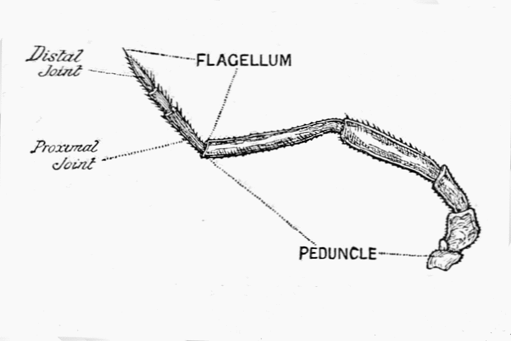 FIG. 3.—THE SECOND ANTENNA.
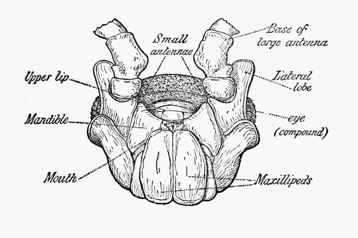 FIG. 4.—THE UNDERSIDE OF THE HEAD.
There are four pairs of mouth appendages—namely the jaws or mandibles (fig. 5), the first maxillae (fig. 6), the second maxilla (fig. 7), and the maxillipeds (fig. 8). When the head is examined from the underside the last of these organs will be seen first, covering in the others.
[Pg 4] A small median plate attached to the front of the head has been called "the upper lip" (fig. 9), while inside the mouth appendages is a little bilobed structure "the lower lip" (fig. 10).
Before leaving the external features of the head, we must allude to the pair of eyes which are usually present, though never raised on stalks. In the Common Woodlouse (Oniscus asellus, from which all our figures to illustrate structure have been made), as in many other species, the eyes are compound (fig. 4), but in some forms these are simple.
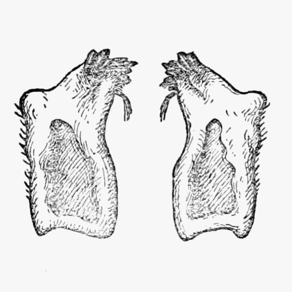 FIG. 5.—THE MANDIBLES.
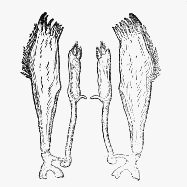 FIG. 6.—THE FIRST
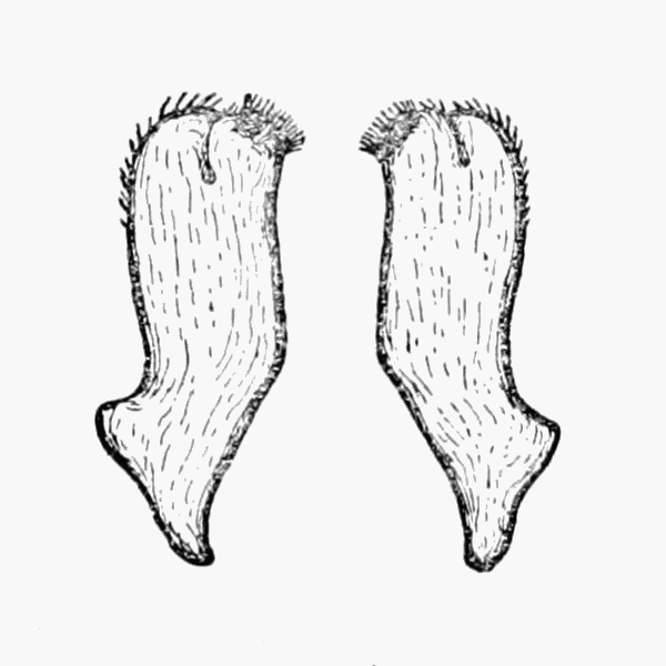 FIG. 7.—THE SECOND
 FIG. 8.—THE FUSED
Each of the seven joints of the thorax bears a pair of walking legs (fig. 11), and in the female at the time when the eggs are laid, a pair of plates (fig. 12) arises on segments II. to V. These plates together form a brood pouch, in which the eggs are carried (fig. 12) until they are hatched, and in which the young ones remain for some time afterwards.
 FIG. 9.—THE "UPPER LIP.
 FIG. 10.—THE "LOWER LIP.
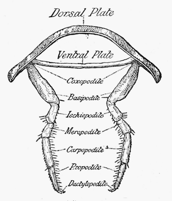 FIG. 11.—A TYPICAL THORACIC SEGMENT.
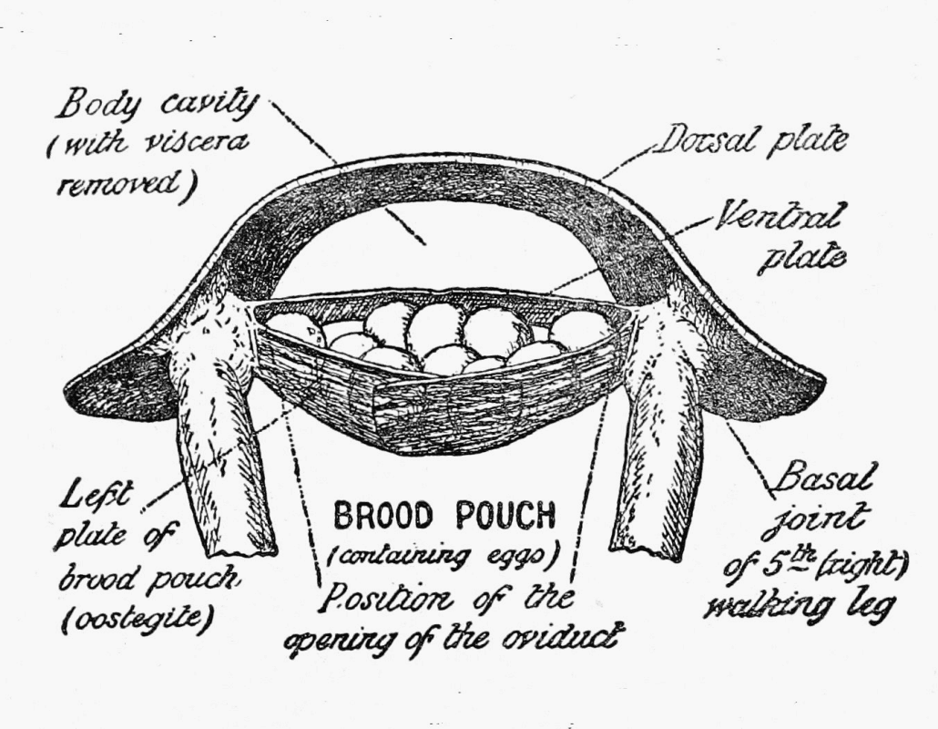 FIG. 12.—THE FIFTH THORACIC SEGMENT OF A FEMALE.
When we examine the abdomen, we find that the appendages are plate-like, with the exception of the last pair (fig. 13), and they all agree in having two divisions, an arrangement which would prove awkward in limbs used for walking or feeling.
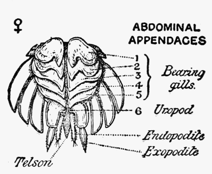 FIG. 13.—THE UNDERSIDE OF THE ABDOMEN OF A
FEMALE.
FIG. 13.—THE UNDERSIDE OF THE ABDOMEN OF A
FEMALE.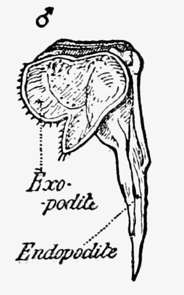 FIG. 15.—THE FIRST ABDOMINAL APPENDAGE OF THE MALE.
The inner plate (or endopodite) is in structure a gill, but the blood that passes through it, is enabled to take up oxygen[Pg 6] from moist air, while the outer division (or exopodite) acts as a protecting cover (fig. 14). In Porcellio, air-tubes (tracheae) may be present (see below).
In the male, the first two pairs of abdominal appendages are specially modified, the inner divisions (endopodites) being long and pointed (figs. 15 and 16). The last pair, or tail appendages, in the male are often considerably larger than in the female, and the form of these structures is sometimes of value in classification.
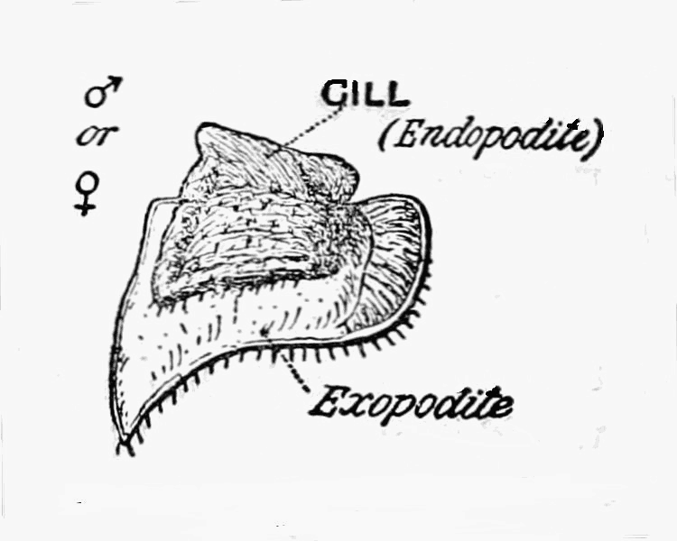 FIG. 14.—A TYPICAL ABDOMINAL
APPENDAGE.
FIG. 14.—A TYPICAL ABDOMINAL
APPENDAGE.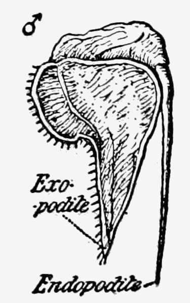 FIG. 16.—THE SECOND ABDOMINAL APPENDAGE OF THE MALE.
Alimentary canal.—The main portion of the alimentary system is, practically speaking, a straight tube (fig. 17). Its first part (not shown in the figure) is a narrow gullet, which after passing through the nerve collar dilates to form a sort of stomach. Into this the secretion of four digestive glands is poured by two ducts. These glands have a somewhat striking appearance, being yellow tubes spirally coiled, and they end blindly. From the stomach the intestine runs to the hinder end of the body and passes under the heart.
Circulatory system.—The blood being aërated in the abdominal appendages, we find that the heart is situated towards the hinder end of the body (fig. 18). Three main arteries supply the thorax and head, while the blood is brought from the gills to the heart.
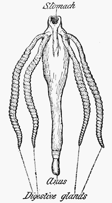 Fig. 17.—THE ALIMENTARY CANAL.
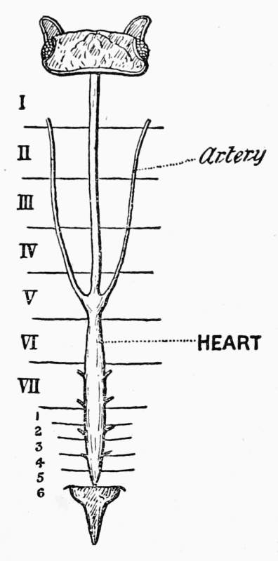 Fig. 18.—THE CIRCULATORY SYSTEM.
Excretory system.—The excretory organs consist of a (a) pair of so-called "shell glands," which are considered to be the equivalents of the excretory tubes or nephridia of annelid worms. In the woodlouse these excretory organs open on the second pair of maxillae. They are composed of a tube (sacculus) closed at one end and more or less bent upon itself (5, p. 261) which communicates with a labyrinth that is provided with an excretory orifice. Matters are eliminated by the epithelial cells [the histology has been described and figured in Ligidium hypnorum (66)], which are very large in Ligia oceanica.
(b) Masses of cellules in the head, very greatly developed in Ligia oceanica (but numbering scarcely more than ten in Oniscus asellus), which have no external opening. They also function as excretory organs (5, p. 263), and have been called "cephalic nephrocytes."
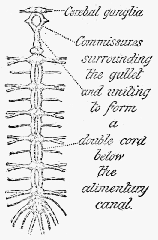 FIG. 19.—THE NERVOUS SYSTEM.
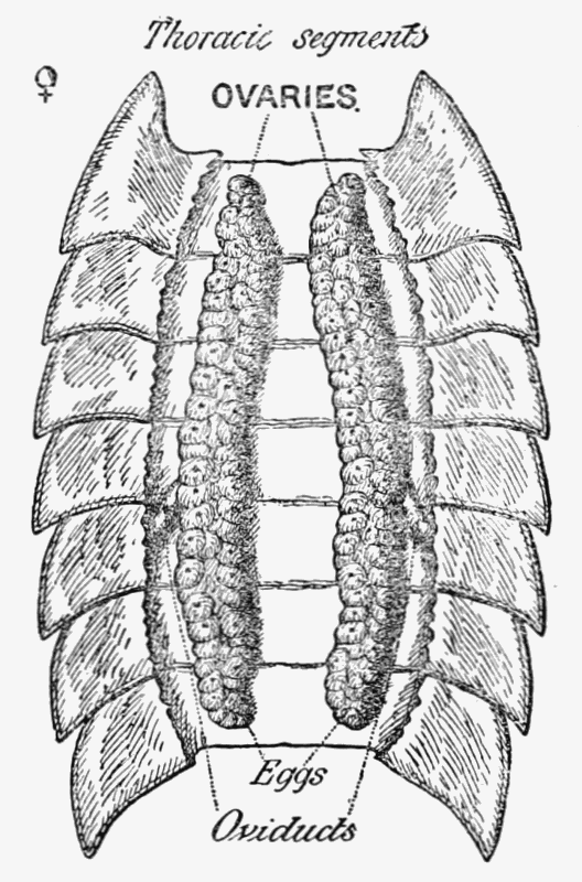 FIG. 20.—FEMALE REPRODUCTORY ORGANS.
(c) Other "branchial nephrocytes" are situated on the dorsal surface between the last thoracic and the first abdominal segments, as well as between those that follow, with the exception of the last two; they are in distinct patches, one on each of the middle line in Ligia, but more or less continuous in Oniscus (5, p. 265).
(d) The digestive glands have also been shown to be excretory (5, p. 270).
Nervous system.—The nervous system consists of paired ganglia in the head, above the alimentary canal which send off nerves (commissures) that meet below, to form a double nerve cord with ganglia at intervals (see fig. 19).
Reproductive organs.—In the female there are a pair of ovaries in the positions shewn in fig. 20; and ducts run to the underside of the fifth thoracic segment.
The openings are very difficult to identify, and Lereboullet (39, p. 113) was unable to find them. It is obvious that the openings must be underneath the plates that form the egg pouch, and as a change of skin is required to set these free, it would appear that at ordinary seasons the ducts from the ovaries are closed.
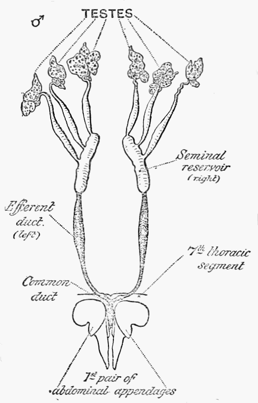 FIG. 21.—THE MALE REPRODUCTORY ORGANS.
The writers have been able to determine from external examination of specimens which had moulted and were about to lay eggs, that the oviducts at such time open to the inside of the base of each walking leg on the fifth segment. In similar specimens the oviducts were also followed to the opening from within. The brood pouch has already been described.
The male organs consist of six testes arranged in two pairs, each of which is provided with a reservoir (see fig. 21). The efferent ducts from the two reservoirs unite at the base of the thorax to form a common duct (or "penis").
Development.—The eggs, in the common species of woodlice, at least, are laid at the beginning of summer, and are retained in the brood pouch, where they undergo their development. The process has been recently traced with great care by Professor Louis Roule (58) in Porcellio scaber and the description which follows is based upon his researches.
As, practically speaking, the larval stages are passed within the egg, and there is no free embryo differing in form from the[Pg 10] parent, it is necessary for the young creatures to be well supplied with nutritive material. In fact, the bulk of the large egg is made up of food-yolk, on the outside of which the formative protoplasm is disposed in irregular patches. In the fertilized ovum, one of the latter, which lies in a particular position at the end, is found to be larger than the others (see fig. 22). It contains the nucleus of the egg-cell (see fig. 23) and is called the cicatricula. This is the only portion of the egg which divides and produces nucleated cells. It is these which gradually spread all over the surface of the food-yolk, forming a layer known as the blastoderm, which is at first but one cell thick (see figs. 24, 26, and 28).
Before, however, the food-yolk is quite closed in, a differentiation into two layers—the pro-ectoderm and pro-endoderm—takes place (see fig. 25) and rudiments of the first two pairs of appendages appear (see fig. 26). Moreover, the cells of the ectoderm change their shape and begin to multiply at two points to form the beginnings of the cerebral ganglia and the nerve cord respectively.
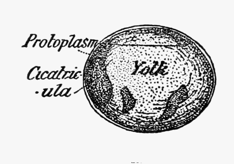 FIG. 22.—THE FERTILIZED EGG
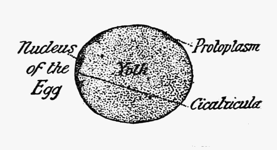 FIG. 23.—THE FERTILIZED EGG SEEN IN SECTION
As the blastoderm closes over the food-yolk, two more appendages arise and these are soon followed by others (see fig. 28). A depression appears at the point where the blastoderm closed and internally the pro-endoderm or inner layer is differentiated into two—the endoderm proper and the mesoderm (see fig. 29). The former begins to grow so that its edges unite to form the middle part of the intestine (see fig. 29) seen from the outside in fig. 30. The depression already mentioned grows deeper, forming a tube which is the hind portion of the intestine, while at the anterior end of the embryo the front part of the intestine is similarly formed (see fig. 30). By this time also all the nineteen appendages have made their appearance and the mesoderm, (which has grown considerably, to form the beginnings of the muscles) has sent prolongations into each of them. About this time, spaces (see fig. 31) are formed in the muscular mesoderm which are all that remain of the true body cavity characteristic of animals above the level of the jelly fish, and in these spaces the blood ultimately circulates.
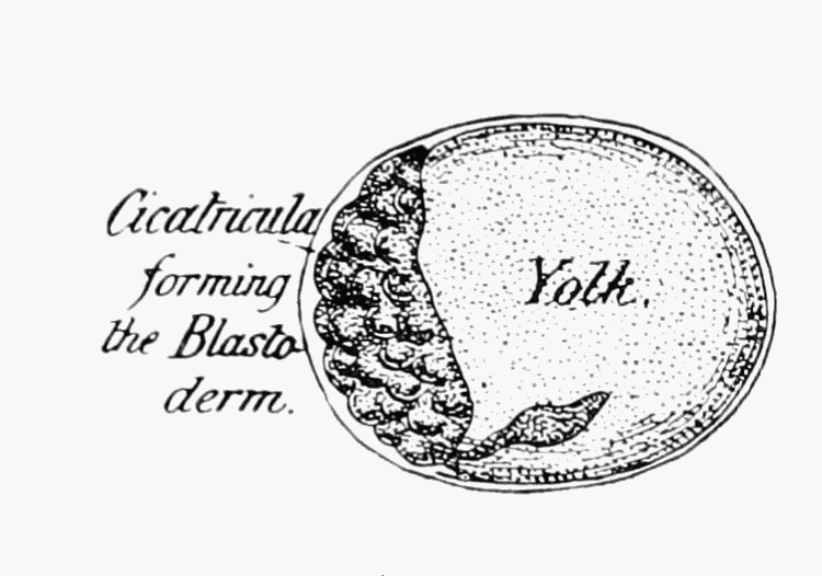 FIG. 24.
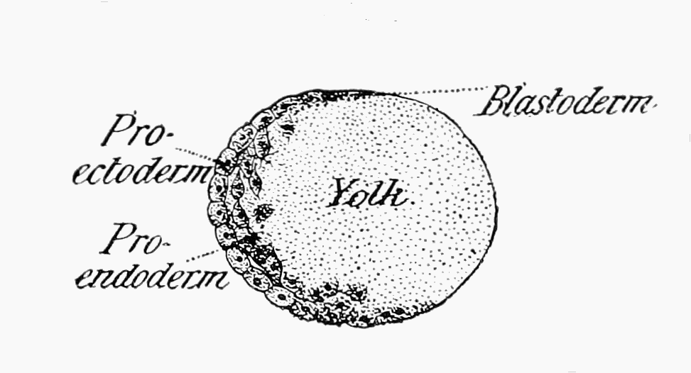 FIG. 25.
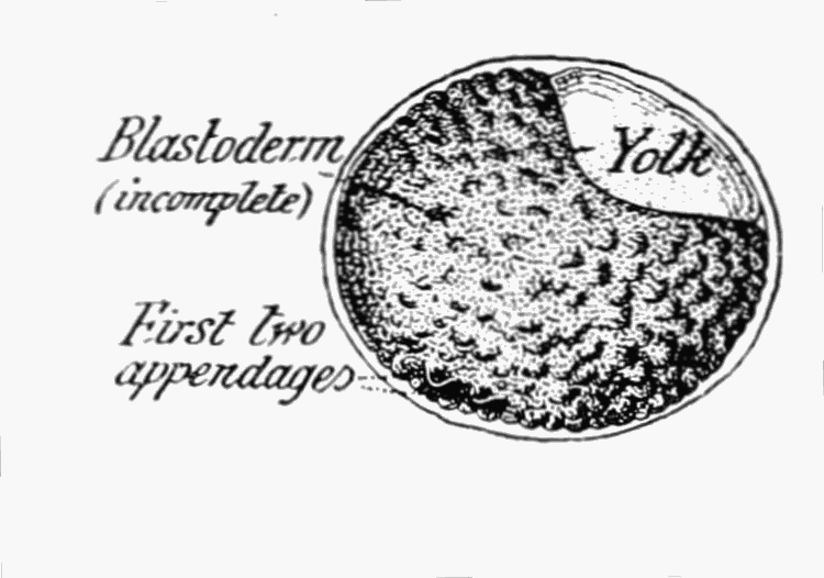 FIG. 26.
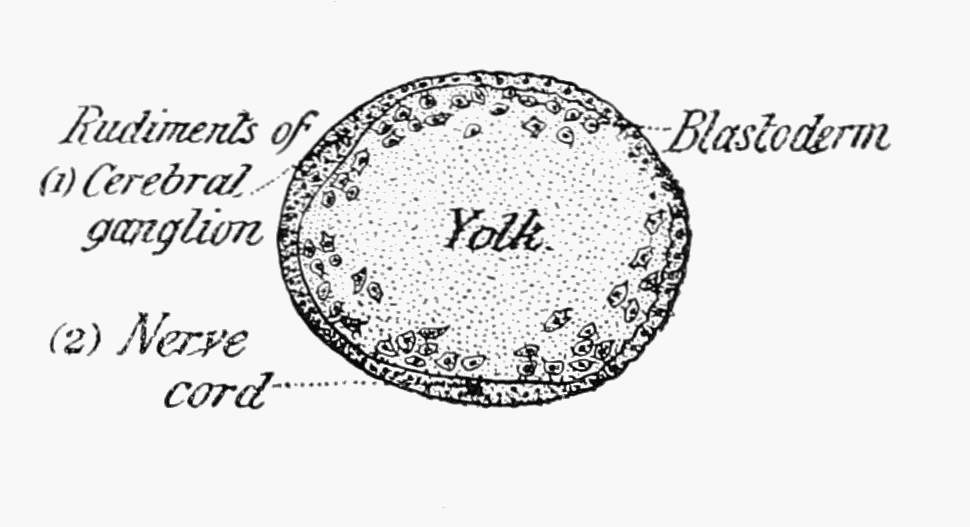 FIG. 27.
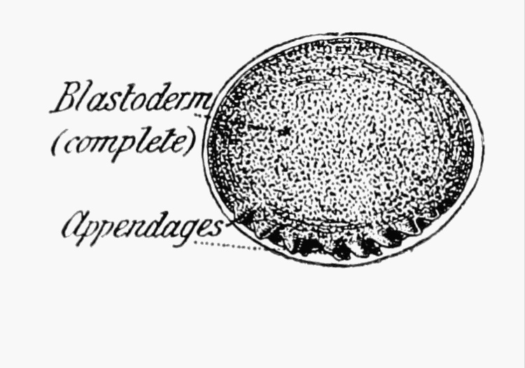 FIG. 28.
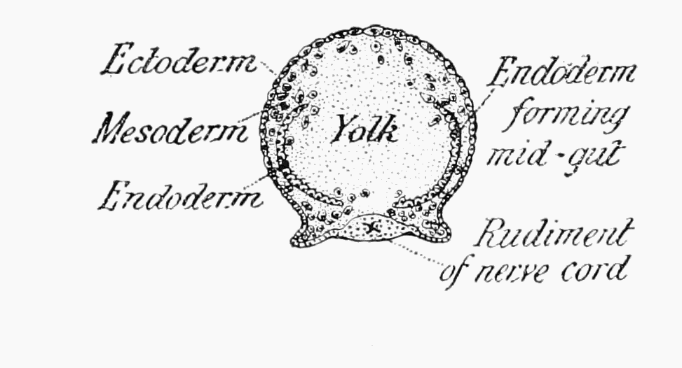 FIG. 29.
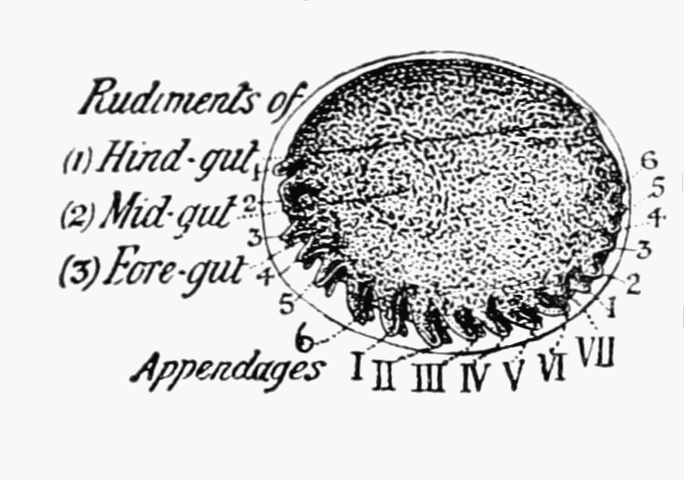 FIG. 30.
 FIG. 31.
THE DEVELOPMENT OF A WOODLOUSE (Porcellio scaber), AFTER ROULE.
Figs. 24, 26, 28, 30, are Surface Views, and figs. 25, 27, 29, 31, which indicate slightly later stages respectively than the others, are of egg seen in Optical Section.
The body next alters somewhat in shape and the three divisions of the intestine approach one another (see fig. 32) previous to their junction. As may be imagined during these processes the food-yolk has gradually been used up and the space which it occupied taken by the internal organs, which we have mentioned.
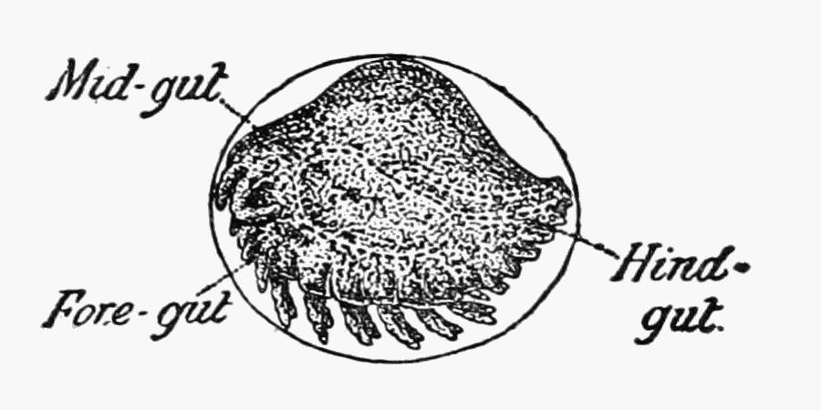 FIG. 32.—EMBRYO OF THE WOODLOUSE SHOWING THE
THREE DIVISIONS OF THE INTESTINE SEPARATELY
DEVELOPED (Porcellio scaber), AFTER ROULE.
FIG. 32.—EMBRYO OF THE WOODLOUSE SHOWING THE
THREE DIVISIONS OF THE INTESTINE SEPARATELY
DEVELOPED (Porcellio scaber), AFTER ROULE.
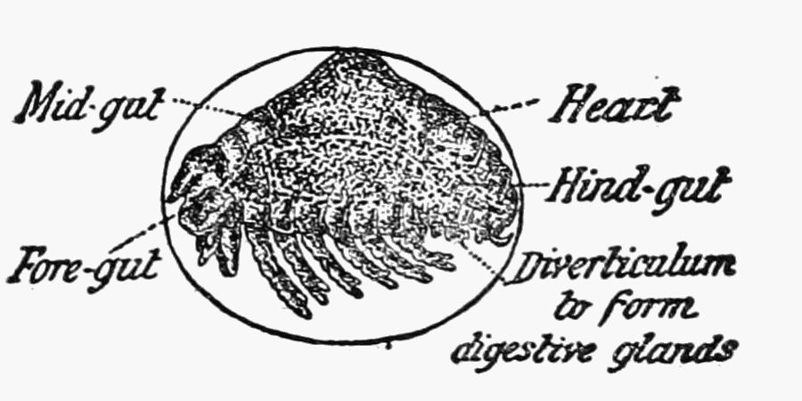 FIG. 33.—EMBRYO OF THE WOODLOUSE SHOWING TRACES
OF THE SEGMENTS (Porcellio scaber), AFTER ROULE.
FIG. 33.—EMBRYO OF THE WOODLOUSE SHOWING TRACES
OF THE SEGMENTS (Porcellio scaber), AFTER ROULE.
In the last stages of the development the appendages become larger still, the heart makes its appearance, segmentation of the body is completed, and except that the seventh pair of walking legs are as yet rudimentary the woodlouse is completed. It is only after hatching that the pair of legs mentioned, attain to their normal length.
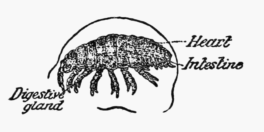 FIG. 34.—EMBRYO OF THE WOODLOUSE SHOWING TRACES
OF THE SEGMENTS (Porcellio scaber), AFTER ROULE.
FIG. 34.—EMBRYO OF THE WOODLOUSE SHOWING TRACES
OF THE SEGMENTS (Porcellio scaber), AFTER ROULE.
The process of segmentation of the egg and the formation of its layers lasts about a fortnight, while the completion of the development proceeds much more rapidly, for another three weeks bring it to an end.
After the first moult or change of skin the last pair of[Pg 12] walking legs makes its appearance, and Mr. James B. Casserley [whose work one of us (75) has described elsewhere] found when keeping a number of the common pill-woodlouse (Armadillidium vulgare) in captivity that his specimens did not subsequently change their skins more than once in the six months during which he had them under observation. He also noted that the crustaceans go on growing after they are sexually mature. As his specimens grew older, Mr. Casserley noticed that their colour became darker, and a curious point recorded by him is that two examples of the same age may change their skins at the same time, and while one may have afterwards nearly twice as many markings, on the other very few at all will be seen. The time required for the growth of a woodlouse from the size of a pin's head to that of an adult example—say three-quarters-of-an-inch long—must be fairly considerable, taking into account the fact that any appreciable increase in size can only occur at a moult and Mr. Casserley's observations as to the infrequency of the process in Armadillidium vulgare. (See p. 13.)
Habits and Economic Considerations.—The construction of the breathing organs of woodlice, and the necessity which exists for these to be kept moist, restricts the habitats of the animals considerably. Woodlice are found under stones and logs, beneath the bark of dead and rotten trees, among decaying vegetable matter as well as living grass and moss in damp or wet situations. When looking for some of the common species under the bark of fallen trees it is surprising to notice that the crustaceans may be entirely absent from many trunks, while when another is examined which seems to differ very slightly, if at all, in condition or situation, they are found in swarms. There is no doubt but that the habits of woodlice would well repay the attention of naturalists, who are now recognizing that besides anatomy as such, and the classification which a knowledge of structure permits, there is the equally important consideration of the creatures as they live their own life and affect that of others. It is not our object to give a detailed account of the ecology of British woodlice, but rather to provide a basis from which it may be approached. Nevertheless a few general remarks may not come amiss. Many points in the life-history of woodlice may no doubt be learned by keeping them in captivity and there is[Pg 13] just sufficient difficulty in doing this successfully to give an interest to the matter.
Apart from a supply of proper food, we take it that the chief object to be attained is the provision of the amount of moisture required by the particular species under examination, together with a sufficient supply of air.
A great many interesting observations can be thus carried out, such as those of Mr. Casserley, to which allusion has already been made. The process of moulting for instance is well worth watching, and although specimens with half their coat changed may be found in remote corners, yet the whole course of the moult can be seen much better in the case of captive woodlice. The following account is taken from Mr. Casserley's description (75) of what happens in the case of Armadillidium vulgare:—The approach of the moult is indicated by the appearance of a white border on each segment of the body, which becomes gradually more marked, while at the same time the animal is seen to be less active and often makes a small burrow in which to hide. Sometimes a sheltered corner against a stone is looked upon as affording sufficient protection, but in either case each woodlouse keeps to the place originally chosen. About ten days after the white lines have become visible the animal appears to be divided into two. Its skin is becoming loose and little movement can take place at the joints of its body with the exception of that between the fourth and fifth thoracic segments where the skin will ultimately break. The woodlouse spends a day or two in this condition and then, by suddenly walking forward, frees itself from the covering of the hinder portion of its body. The three last pairs of walking legs are carefully pulled out from the old skin, which now appears perfectly white, and at the same time the lining of the hind portion of the alimentary canal (hind gut) is also shed. After putting the tender half of his body well into his corner or burrow the woodlouse proceeds to eat the part of his skin that he has cast. The creature has now a very odd appearance. His front half with the exception of the white edges is as it was before, the rest of him instead of a light slaty blue, and is very soft as well as proportionately a little larger.
In three days or so the tail end becomes hard and attains the normal colour. Then the old skin from the front half is pushed off and the creature becomes practically defenceless, so much[Pg 14] so in fact, that any of his species that happen to find him will attack him and eat all his front half, rejecting, however, his now hardened tail-end.
Provided that the moulting woodlouse has survived (and in captivity, to ensure this, he must be isolated), after three days his jaws will be sufficiently hardened to allow of his eating, and usually he first of all devours the second half of his cast skin. The operation of moulting does not occupy quite so long a time in the case of young examples. Specimens half-an-inch long do not moult more than once in six months and show but little increase in size after the process.
Woodlice do not appear to live on either animal or vegetable food alone, but adopt a mixed diet. It is, however, owing to their attacks upon cultivated plants that the creatures are looked upon as pests by the horticulturalist. The animals feed either in the night or in the very early morning, on seedlings, orchid tubers, mushrooms, or anything that comes to hand. Few of the accounts, however, of their ravages, mention that the crustaceans have been caught absolutely in the act of doing the damage ascribed to them. Some careful inquiries have nevertheless enabled us to discover several observers who have watched woodlice feeding. Mr. F. V. Theobald, of Wye College, and one of the students at Swanley Horticultural College are among the number. The former has also given us an account of the methods, out of many tried, which he has found most successful for getting rid of the crustaceans. Out of doors trapping with moss, sacking or horse-dung is best. In glass houses, fumigation with hydro-cyanic acid gas has cleared them out, and poison baits, especially potatoes cut and soaked in white arsenic, have done some good. Stable manure is especially favourable to these creatures, particularly when it is used "long": in this condition it should therefore be avoided.
It is interesting to note how the woodlice in winter simply remain where they happen to be so long as there is sufficient moisture, though they are ready to run about as rapidly, for a time, as in summer, should they happen to be disturbed.
No doubt many points of inter-relation between woodlice and other animals remain to be discovered. Mr. John W. Odell tells us that on Exmoor, in the open, he found no Armadillidia, though other forms occurred under nine out of every ten stones[Pg 15] that he turned over, and here the smaller species of ants also abounded. Close to stone walls Armadillidia were to be seen to the exclusion of all other genera, and this state of affairs was ascribed by Mr. Odell to the presence of swarms of the large wood-ants which he considers would make short work of any woodlice that could not protect themselves by rolling up.
We ought not to conclude this account without mentioning the fact that woodlice once played an important part in medicine.
Doctor Fernie (28) gives some interesting extracts with regard to the hog-louse and the woodlouse. The latter he seems to have identified quite correctly as Oniscus asellus. He calls the former, however, indiscriminately, "the common armadillo" (which is the old name for the pill-woodlice now known as Armadillidium), "the pill millipede" and "Glomeris marginata." The last two names are those of another creature, not a crustacean, which when it is rolled up can be very easily mistaken for an Armadillidium, though, when it uncurls, it will be seen to have many more than seven pairs of legs. The local appellations applied to the hog-louse by Doctor Fernie, and his remarks with regard to its commonness, tend to show that it is Armadillidium vulgare, to which he really refers, and the use of which in medicine was commonly general.
Hog-lice were prescribed for scrofulous diseases and obstructions of the liver and digestive organs, among other things, and the London College of Physicians directed that the creatures should be prepared by suspending them in a thin canvas bag placed within a covered vessel over the steam of hot spirit or wine, so that being killed by the spirit they might become friable. Hog-lice and Woodlice were also administered alive, while the former were also put down the throats of cows "to promote the restoration" of their cud, hence their name of "cud-worm." There seems to be considerable evidence that even in modern times Woodlice have had considerable remedial effect which depends upon "an alkalescent fluid" contained in them.
Local Names.—Among the local names by which these creatures are known are those of "sow bug," "lucre pig" (Berkshire), "carpenter" and "chiselhog" (Berkshire). Doctor Fernie (28) gives a number of others:—"thrush-louse," "tiggyhog," "cheslip," "kitchenball," "chiselbob," "lugdor,"[Pg 16] "palmer," and "cudworm." In the eastern counties the same writer notes that they are known as "old-sows" or "St. Anthony's hogs" while the Welsh call them "little grey-hogs," "the little old women of the wood" or "grammar-sows," grammar signifying a shrivelled up old dame. Oniscus asellus was sometimes called "socchetre," "church louse," and "chinch."
Methods of Collection and Preservation.—Woodlice should be collected straightway into tubes or bottles half filled with 30 per cent. methylated spirit.[3] Woodlice dropped into this weak spirit become gradually narcotised and die, and they remain limp enough for purposes of examination or to allow, of their legs and antennæ being set out during the process of mounting. Specimens to be kept permanently should be placed in 70 per cent. alcohol. For storage purposes the specimens of each species from a given locality should be put together into a small flat bottomed tube such as is used for pillules by apothecaries or specially made for natural history purposes. A paper label on which the name, locality, date of capture and any other necessary particulars have been written with dark lead pencil, is not affected by the spirit. The tubes may be corked, though if not frequently examined all the spirit may evaporate, and cause the specimens to be spoilt. A safer method is to plug the tubes with cotton wool and keep all those containing a given species or specimens from a particular locality beneath the surface of spirit in a large wide-mouthed bottle, into which first of all some cotton wool has been put to prevent the tubes from coming into sudden contact with the glass at the bottom. For show purposes in museums, specimens taken direct from 30 per cent. spirit should be mounted on slips of opal glass by means of gum-tragacanth which has been powdered and shaken up in spirit before having water added to it. The slips can be exhibited in glass tubes, six inches high by one across, or in narrow stoppered museum jars. A variation of the method is to mount the animals on clear glass and to place behind them another strip of any colour that may be preferred.
[Pg 17] Classification.—The various genera of woodlice are connected together so closely, by intermediate forms, that their division into families is, to a very great extent, arbitrary. Bate and Westwood described but a single family Oniscidæ (I), though they distinguished two sub-families:—Ligiinæ, which included the forms with many joints to the flagellum of the antenna, and Oniscinæ, which contained the rest.
Since then the pill-woodlice have been thought by some to be sufficiently different from the other genera to warrant their separation, and three families namely, Ligiidæ, Oniscidæ, and Armadillidæ have been recognized, as for instance by Dr. Scharff (63).
A fourth family—Trichoniscidæ—has been added by Professor G. O. Sars, who in his Crustacea of Norway (59) alludes to the division of the tribe into the sections Ligiæ and Onisci and has adopted the following classification:—
Order—ISOPODA.
Tribe—ONISCOIDA.
| Family I.— | Ligiidae. | Family III.— | Oniscidæ. |
| Ligia. | Oniscus. | ||
| Ligidium. | Philoscia. | ||
| Platyarthrus. | |||
| Porcellio. | |||
| Metoponorthus. | |||
| Cylisticus. | |||
| Family II.— | Trichoniscidæ. | Family IV.— | Armadillidiiæ. |
| Trichoniscus. | Armadillidium. | ||
| Trichoniscoides. | |||
| Haplophthalmus. |
All the genera described by Professor Sars are represented in the British Islands.
Below is a scheme of classification and synopsis of the characters of British genera of woodlice which we have compiled in order to render easy the determination of the genus to which any particular specimen may belong.
SCHEME OF CLASSIFICATION AND SYNOPSIS OF GENERIC CHARACTERS.
[Pg 18]
Order—ISOPODA.
Tribe—ONISCOIDA.
Section I.—LIGIÆ.
The Two Divisions of the Tail Appendages alike in Shape.
|
(A.)—Flagellum with 10 or more joints; tail appendages wholly visible; head without lateral lobes |
Ligiidae. |
|
(1.)—Abdomen broad; body large; habitat, the sea-shore |
Ligia. |
| (2.)—Abdomen narrow; habitat, wet moss | Ligidium. |
|
(B.)—Flagellum with less than 10 joints; head with small lateral lobes, tail appendages partly covered |
Trichoniscidæ. |
|
(3.)—Abdomen narrow; eyes compound; flagellum usually with more than 3 joints |
Trichoniscus. |
|
(4.)—Abdomen narrow; eyes simple or wanting; flagellum with 4 joints |
Trichoniscoides. |
|
(5.)—Abdomen broad (comparatively); eyes simple; back with longitudinal ridges; flagellum with 3 joints |
Haplophthalmus. |
Section II.—ONISCI.
The Outer Divisions of the Tail Appendages Broader than the Inner.
|
(A.)—Tail appendages projecting when the animal is walking |
Oniscidæ. |
|
(a.)—Unable to roll up into a complete ball. (6.)—Flagellum with 3 joints; abdomen broad; head, with lateral lobes |
Oniscus. |
|
(7.)—Flagellum with 3 joints; abdomen narrow; head without lateral lobes |
Philoscia. |
|
(8.)—Flagellum with 1 joint; eyes wanting; abdomen broad; habitat, ant's nests |
Platyarthrus. |
|
(9.)—Flagellum with 2 joints; abdomen broad; frontal lobe projecting |
Porcellio |
|
(10.)—Flagellum with 2 joints; abdomen narrow |
Metoponorthus. |
|
(b.)—Able to roll up into a complete ball. (11.)—Flagellum with 2 joints; antennae folded together over the thorax when the animal is rolled up into a ball |
Cylisticus. |
|
(B.)—Tail appendages not projecting when the animal walking |
Armadillidiidæ. |
|
(12.)—Flagellum with 2 joints; antennae hidden or carried at the sides of the head when the animal is rolled up into a ball |
Armadillidium. |
[Pg 19] British Species.—Naturalists in this country paid little attention to the recognition or description of Woodlice, until the latter half of the nineteenth century.
In 1857 Kinahan read a paper before the British Association (32) in which he described fourteen species of woodlice from the British Islands, and eleven years later when Bate and Westwood published their book (1), the number had risen to seventeen. One of the species (Oniscus fossor), however, was doubtful, and although Dr. Scharff in 1894 (63) rejected it, his list contained also seventeen species, for in the meantime the Rev. T. R. R. Stebbing had found Ligidium hypnorum in Surrey (70).
Since then the Rev. Canon Norman, Dr. Scharff, the Rev. T. R. R. Stebbing, and one of the present writers, have added other species, as will be seen from the following pages, in which all those found, up to the present time in the British Islands are described and figured.
We shall now consider in detail the British genera and species of woodlice and give their synonymy and distribution.
Order—ISOPODA.
Tribe—ONISCOIDA.
Section—LIGIÆ.
THE TWO DIVISIONS OF THE TAIL APPENDAGES ALIKE IN SHAPE.
Family—LIGIIDÆ.
Flagellum with ten or more joints; tail appendages
wholly visible; head without lateral lobes.
Genus—LIGIA Fabricius, 1798 (27), p. 301.
Abdomen broad; body large; habitat, the sea-shore.
The genus Ligia agrees with Ligidium alone, in that the flagellum of the larger antennæ has more than ten joints. In both genera, there are no lateral lobes to the head, and the tail appendages are wholly visible from the upper surface of the body. The latter in Ligia is, however, very many times bigger than in Ligidium and shows no abrupt decrease in the width of its segments when the abdomen is reached.
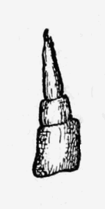
Ligia oceanica Linné (The Quay-louse). Plate I.
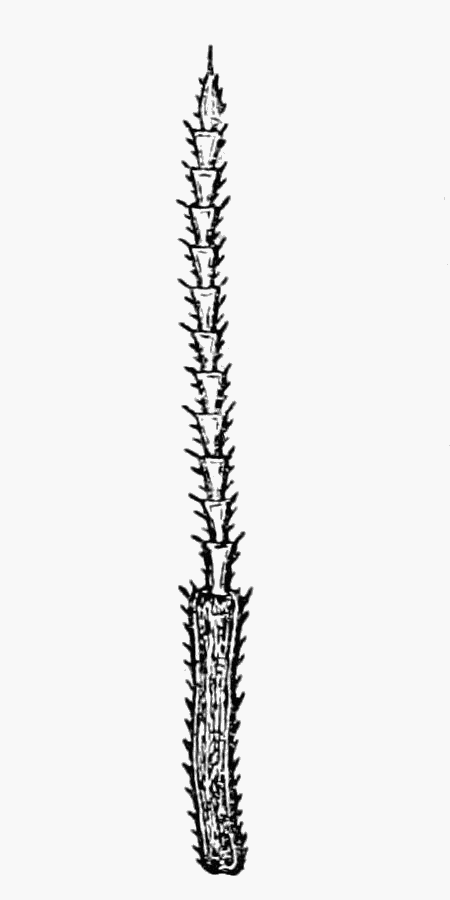 FIG. 35.—FLAGELLUM
There is but one British species of Ligia, and this, the largest member of the whole tribe to be met with in these Islands,usually attains a length of two centimetres, while adult males may be nearly half as long again. It is the Oniscus oceanicus of Linnæus and lives on the sea shore, where it may be found at low tide beneath stones and rubbish in the crevices of timber. Ligia forms a connecting link between the woodlice proper and the many Isopods which actually live in the sea.
The colour of the animals is a greenish grey, and the compound eyes are almost black, so that they are very conspicuous; there are from eleven to fourteen joints to the flagellum of the outer antennae and this feature, taken in conjunction with the large size and habitat, is sufficient to identify the species in question.
On the coast of Essex the name "quay-lowders" is given to these crustaceans, "lowder" being apparently an old plural of louse.
It is worthy of mention that Mr. Webb, when in charge of the Marine Biological Station at Brightlingsea, examined a very large male specimen of Ligia oceanica, in which the maxillæ were duplicated and consisted of four pairs instead of two.
BRITISH LOCALITIES:—
England: Brightlingsea; (W.M.W.): Maldon; (W.M.W. from R.M.): Southend; (J.A.M.): Whitstable; (W.M.W.): Herne Bay; Margate; Dover; Folkestone; (J.A.M.)
Scotland: Shetland to Cornwall; (Norman, 49).
Ireland: East Coast; West Glengariff; Castletown; Berehaven; Bundoran; (Scharff, 63).
FOREIGN DISTRIBUTION:—
Europe: France; (25): Spain; (12): Denmark; Prussia; Norway; Faroe Islands; Belgium; (59).
Africa: Morocco; (16).
Genus—LIGIDIUM Brandt, 1833 (3), p. 173. Zia, Koch (34).
Abdomen narrow; habitat, wet moss.
In Ligidium there are numerous joints to the flagellum, lateral lobes are absent from the head, and the tail appendages are completely to be seen. All the segments of the abdomen are distinctly narrower than those of the thorax and in this it agrees with Trichoniscus, Trichoniscoides, Philoscia, and Metoponorthus. In these, however, the flagellum has never more than seven joints, the tail appendages (as in all genera but Ligia and Ligidium) are partially hidden by the last segment, and in all the four but Philoscia there are lobes to the head.
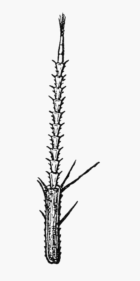 FIG. 36.—FLAGELLUM
AND LAST PEDUNCULAR
FIG. 36.—FLAGELLUM
AND LAST PEDUNCULARLigidium hypnorum, Cuvier. Plate II.
This species, which like the last, is the only British representative of its genus, was added to our fauna in 1873 by the Rev. Thomas R. R. Stebbing (70) who found specimens in the neighbourhood of Copthorne Common, Surrey. Up to the present time, when we are pleased to announce that we discovered it in the spring of 1902 at Warley in Essex, Ligidium hypnorum has not been recorded from any other place in the British Islands.
As the name of the species implies, it lives in wet situations and in its turn connects Ligia with the forms which inhabit drier places. Ligidium hypnorum might be mistaken for Philoscia muscorum, but as already pointed out in the generic description, the latter has but a few (three) joints to the flagellum, instead of from ten to thirteen. From Ligia, the species under consideration is distinguished by its small size, narrow abdomen, and habitat.
BRITISH LOCALITIES:—
England: Warley, Essex; (W.M.W.): Copthorne Common, Surrey; (Stebbing, 70).
FOREIGN DISTRIBUTION:—
Europe: France; (25): Sweden; Denmark; Germany; (59): Turkey; (8).
Family—TRICHONISCIDÆ.
Flagellum with less than ten joints; head with lateral
lobes; tail appendages partly hidden.
Genus—TRICHONISCUS Brandt, 1833 (3), p. 174.
Abdomen narrow; eyes compound; flagellum, usually with more than
three joints.
In Trichoniscus the flagellum may have from seven to four (rarely three) joints. As in Trichoniscoides and Haplophthalmus there are lateral lobes to the head, though these are not very pronounced; the body is also of small size, the abdomen narrow with both divisions of the tail appendages equally so, and almost of the same length though slightly covered by the last segment. The compound eyes distinguish Trichoniscus from the two genera named, and from Platyarthrus, while its small size and the character of its tail-parts mark it out from all others.
Trichoniscus pusillus Brandt. Plate III.
This tiny species is found commonly amongst the roots of the herbage in very moist places. It presents a horny translucent appearance and is of a reddish brown colour. It runs with considerable speed, and when it is moving, the white irregular lines with which it is beset are not evident. Trichoniscus pusillus is very much like Trichoniscus vividus in colour but the latter species is nearly twice as big and has from five to seven joints to the flagellum, while the former has never more than four. Trichoniscus roseus is also much larger and its bright red colour (which it loses, however, when preserved in alcohol) is another means of distinguishing it from the species under consideration.
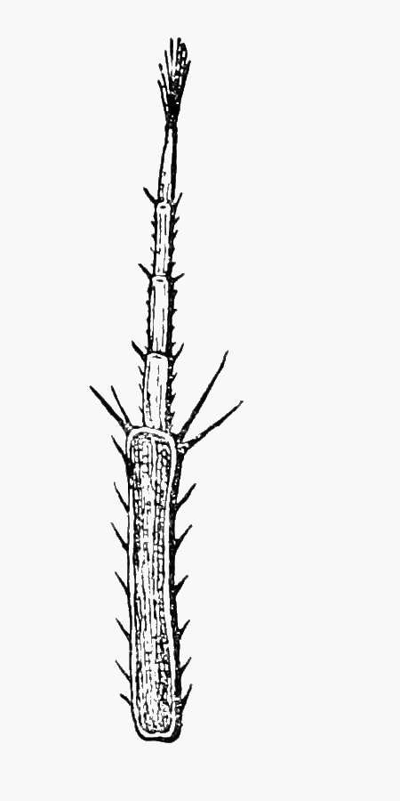 FIG. 37.—FLAGELLUM
Professor Sars in his Crustacea of Norway (p. 162) describes from Christiania, under the name of Trichoniscus pygmæus, a still smaller species. As this may possibly be discovered in this country a brief comparison between it and Trichoniscus pusillus may be of value. The former reaches a length of but two millimetres; it is "whitish, semi-pellucid with a few light brown pigmentary ramifications across the segments and a double row of irregular opaque patches along the middle of its back" (p. 163). Its body is covered with minute tubercles and there are only three joints to the flagellum; its movements are by no means rapid.
The body of Trichoniscus pusillus is smooth and polished. It has four joints to the flagellum—Dr. Scharff (63) says three or four—and it moves quickly.
BRITISH LOCALITIES:—
England: Brightlingsea; Warley; (W.M.W.): Epping Forest; (Bate and Westwood, 1): Hanwell; Southall; Kew Gardens; Langley; Burnham Beeches; Dropmore; Skirmett; Bluebell Hill, Maidstone; (W.M.W.): Chislehurst; Plymouth; Polperro; Looe; (Bate and Westwood, 1): Hertfordshire; Northumberland; Durham; (Norman, 49): Exeter; (Parfitt, 53).
Scotland: Edinburgh; (Scott, 68): Cumbrae; (Robertson, 57).
Ireland: Connemara; (Norman, 49): Dublin; Wexford; Cork and Kerry; (Percival Wright teste Bate and Westwood, 1): Tyrone; Waterford; Portlaw; Kilkenny; Wicklow; (Kinahan, 33).
FOREIGN DISTRIBUTION:—
Europe: France; (25): Spain; (15): Italy; (19): Norway; Sweden; Denmark; Germany; (59).
Africa: Algeria; Tunis; Azores; (24).
America: Niagara; North America; (59).
Trichoniscus vividus, Koch. Plate IV. (from a spirit specimen).
This species is claret-brown in colour and under a lens it is seen to be marbled with white, indeed in appearance it is much like Trichoniscus pusillus though twice the size. There are important differences between the two species as regards the number of joints to the flagellum. These vary from five to seven in Trichoniscus vividus while in the other, as already pointed[Pg 24] out, there are not more than four.
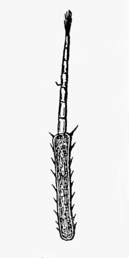 FIG. 38.—FLAGELLUM
The body is practically speaking smooth for it bears only very small tubercles, widely separated. In Trichoniscus vividus the antennæ lack the bristles which characterise those of the other species in the genus. The species under consideration was discovered by Dr. Kinahan in March, 1858, at Portlaw, Co. Waterford and is active even amongst the snow.
BRITISH LOCALITIES:—
Ireland: Portlaw, Co. Waterford; (Kinahan, 33): Cappagh, Co. Waterford; (Scharff, Irish Nat., Vol. IX., p. 158): Borris, Co. Carlow; (Scharff, 64.)
FOREIGN DISTRIBUTION:—
Europe: Spain; (12).
Trichoniscus roseus Koch. Plate V.
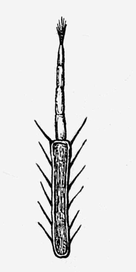 FIG. 39.—FLAGELLUM
The third British species of Trichoniscus is of a deep pink colour and has a light yellow stripe down the back (in some habitats the animals are said to be quite white). Arranged in transverse rows upon the body are large tubercles, each of which under strong magnification will be found to end in a tiny hair. It is distinguished from Trichoniscus pusillus by the larger size of its body, which is also comparatively broader, and from Trichoniscus vividus by the four joints of the flagellum of its antennæ which latter have strong bristles upon them. In the former species there are five or more joints to the flagellum and the antennæ, though hairy, lack the bristles. Trichoniscus roseus is to be looked for in old gardens.
BRITISH LOCALITIES:—
England: Warley; (W.M.W.): Maldon; (W.M.W. from R.M.): Stanmore; Hanwell; Ealing; Wimbledon; (W.M.W.): Berkhamsted; Torquay; (Norman, 49): Plymouth; (Bate and Westwood, 1 and B.M.,): Grassendale, near Liverpool; (R.W.): Newtownards; (R.W., Irish Nat, 1904, p. 260.)
Scotland: Tarbert; (Scot, 68).
Ireland: Dublin; Ballyfinder, Co. Down; (Scharff, 63): Templeogue; Dundrum; Blackrock; Rathgar, Co. Dublin; Bray, Co. Wicklow; (R.F.S.): Oakleigh; Kerry; (R.W.): Belfast; (Welch, Irish Nat., 1896, p. 213.): At the grave of Josiah Welch (grandson of John Knox), Castle Upton; Richhill, Co. Armagh; Castleconnell Ferry; (R.W.): Glenade House, Co. Antrim; (R.W. from R. Ll. Praeger).
FOREIGN DISTRIBUTION:—
Europe: France; (25): Spain; (12): Italy; (59): Denmark; Germany; Holland; (39): Dalmatia; (18).
Africa: Algeria; Tunis; (24).
Genus—TRICHONISCOIDES, Sars, 1898 (59), p. 164.
Abdomen narrow; eyes simple; (or wanting); flagellum, with four joints.
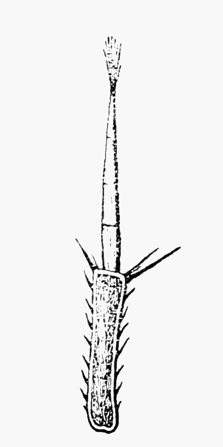 FIG. 40.—FLAGELLUM
The members of this genus are very much like those of Trichoniscus. In the latter, however, the hinder legs are longer in proportion and the eyes are compound.
Trichoniscoides albidus Budde-Lund. Plate VI.
We are able to include this species, as a specimen was found by Mr. Webb at Eton Wick in the summer of 1899. It is one of a number of species which the Rev. Canon Norman (49, p. 18) suggested as likely to be British. It is the only representative of its genus, which does not differ in any very important characters from the others in the family. The narrow elongated body will serve to separate it from Trichoniscus vividus and Trichoniscus roseus, but on account of its size, which is much the same as that of Trichoniscus pusillus and the two British species of Haplophthalmus, it will be advisable to give some further points of distinction. From the first its white colour will serve to differentiate it; the other two lack the narrow abdomen seen in Trichoniscoides albidus. Moreover, not one of the three shows the serrations on the side plates which characterise the species under[Pg 26] consideration. Platyarthrus hoffmannseggii is small and white and the edges of its side plates are toothed, but it is oval in shape, possesses no eyes, and its stout antennæ have but a single joint to the flagellum instead of four. On the Continent this species has been found in rich soil.
BRITISH LOCALITIES:—
England: Eton; (Stebbing, 71a): Sunderland; (Brady, 50a).
FOREIGN DISTRIBUTION:—
Europe: France; Wimereux and Lyons, Forêt (25): Norway; Denmark; (59).
Genus—HAPLOPHTHALMUS Schöbl, 1850 (66), p. 449.
Abdomen broad (comparatively); eyes simple; flagellum with three joints; back with longitudinal ridges.
The body of Haplophthalmus is long in proportion to its width, but there is no abrupt decrease in the breadth of the abdomen as seen in Trichoniscus and Trichoniscoides. The eyes are simple as in the latter genus and the lateral lobes of the head are rather large, while the side plates of the body are well separated.
Haplophthalmus mengii Zaddach. Plate VII.
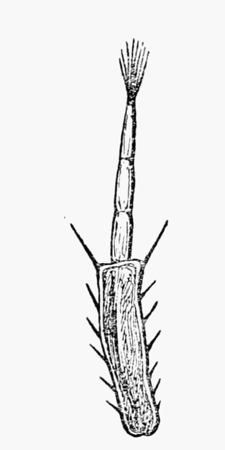 FIG. 41.—FLAGELLUM
The Rev. Canon Norman discovered two specimens of this species in Ireland in June, 1900 (50); in the previous year one of us (Mr. Webb) found a single example at Eton Wick.
The main differences between the members of this genus and their allies are set forth in the generic description and incidentally elsewhere, so we shall content ourselves with giving the distinctive points of the two British species. Haplophthalmus mengii has a number of raised longitudinal ribs on each segment of the thorax, the outer ridges being somewhat broken. There are also two prominent ribs upon the third segment of the abdomen.
BRITISH LOCALITIES:—
England: Eton; (Stebbing, 71a): Sunderland; (Brady, 50a).
Ireland: Corcumroe Abbey; Co. Clare (Norman, 50).
FOREIGN DISTRIBUTION:—
Europe: France; (25): Norway; Prussia; Germany; Bohemia; (59).
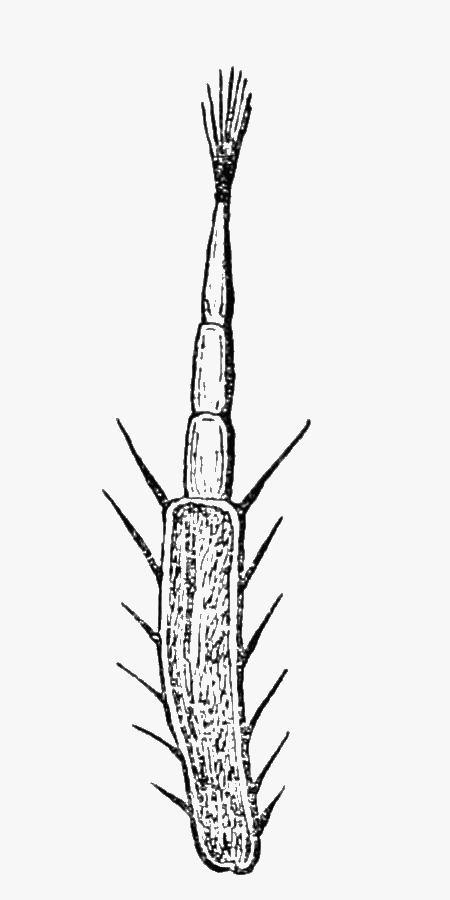 FIG. 42.—FLAGELLUM
Haplophthalmus danicus Budde-Lund. Plate VIII.
This species was added to the British list by the Rev. Canon Norman (49), who found a colony in his garden at Berkhamsted. It has rows of tubercles on its thorax instead of ridges, and there are no ribs at all upon the abdomen. The front of the head projects further comparatively and forms a more acute point than in Haplophthalmus mengii and it is not so purely white in colour as the latter species.
BRITISH LOCALITIES:—
England: Warley Place; (W.M.W. from Miss Willmott): Queen's Cottage, Kew Gardens; Stanmore; Hanwell, garden at Odstock, Bennett's Nurseries; (W.M.W.): Berkhamsted; (Norman, 49): Sunderland; (Brady, 50a).
FOREIGN DISTRIBUTION:—
Europe: France; (25): Denmark; Holland; Germany;
(Dollfus, Feu de Jeun, Nat., April, 1896): Norway;
(Sars, 59).
Section—ONISCI.
THE OUTER DIVISIONS OF THE TAIL APPENDAGES BROADER THAN
THE INNER ONES.
Family—ONISCIDÆ.
Tail appendages projecting when the animal is walking.
(1.) Unable to roll up into a complete ball.
Genus—ONISCUS Linné 1746 (41), p. 360.
Flagellum, with three joints; abdomen broad; head with lateral lobes.
The characters given above taken in conjunction with the size of the animals will serve to distinguish the members of this genus.
Oniscus asellus Linné (The "Common Slater.") Plate IX.
Oniscus asellus is one of the largest of our woodlice and it is also probably the commonest, though Porcellio scaber is in many places quite as abundant. The body of Oniscus is broad and expanded and the colour is usually a slate grey with yellowish markings more or less regularly arranged.
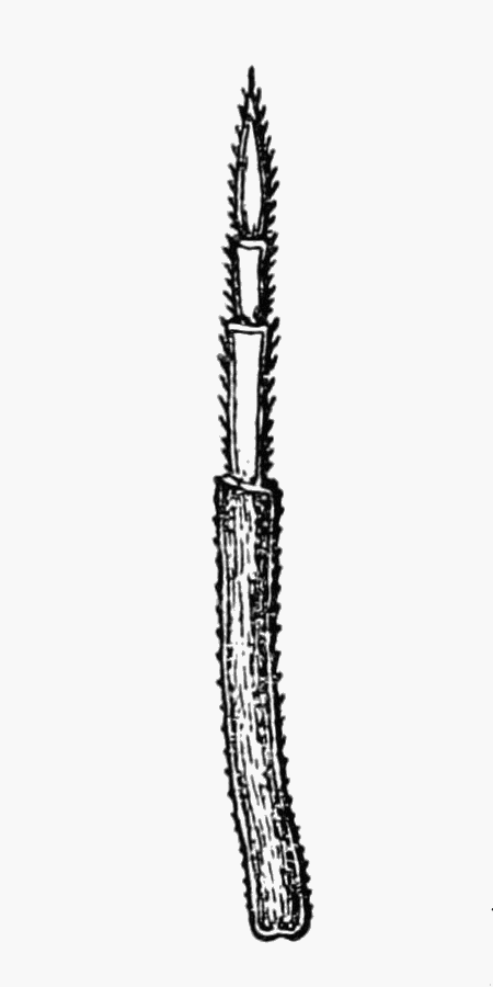 FIG. 43.—FLAGELLUM
From the genus Porcellio the species with which we are concerned is at once distinguished by the three jointed flagellum. Porcellio has but two joints and has, besides, a prominent lobe projecting from the middle of the head, which is not seen in Oniscus. Philoscia, although it has three joints to the flagellum, has a narrow abdomen and lacks entirely the lateral lobes which are a feature of the other genera of Oniscidæ.
Oniscus fossor of Koch (34) was recognized by Kinahan and by Bate and Westwood as a species. Dr. Scharff submitted specimens to Professor Budde-Lund who found no differences between them and Oniscus asellus. The former (63) mentions, however, that the characteristics of the supposed species are those of young examples of Oniscus asellus, and Professor Sars (59, p. 173) seems to be of the same opinion. Many young examples of Oniscus asellus that we have examined have a curious whitish transverse band owing to the light colour of the dorsal plates of the first abdominal segments. The flagellum also does not seem to shew in young animals a distinct division into three joints.
BRITISH LOCALITIES:—
England: High Beach, Epping, including an albino; Maldon; Brightlingsea; Iver; Hanwell; Eton; Kew; Pamber Forest; Kingston-on-Soar; Bluebell Hill, Maidstone; (W.M.W.): Lynmouth; (W.M.W. from J.T.C.).
Scotland: (Scharff, 63). Dinnet, Aberdeenshire; (W.M.W. from Madame Christen).
Ireland: (Scharff, 63). Yellow form with black spots, Donegal (R.W.)
FOREIGN DISTRIBUTION:—
Europe: Almost throughout; (12): France; (25): Spain; (12): Sweden Norway; Denmark; Germany; Holland; Italy; Iceland; (59): Faroe Islands; Thorsharn; (R.F.S.)
Africa: Azores; (24).
America: Greenland; (59): North America; (Budde-Lund).
Genus—PHILOSCIA Latreille, 1804 (37), p. 43.
Flagellum with three joints; abdomen narrow; head without
lateral lobes.
If any further differences of an obvious kind be required to distinguish Philoscia from Oniscus, one at least will be found in the much greater development of the hinder legs in the former genus.
Philoscia muscorum Scopoli. Plate X.
[Not of Lereboullet, which is an Oniscus, see Bate and Westwood (1).]
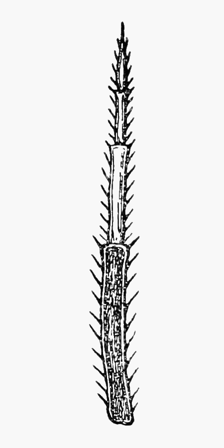 FIG. 44.—FLAGELLUM
This species lives chiefly at the roots of grass and under the stones or sticks that lie among it. Philoscia muscorum has a very smooth and shining body, and its long legs enable it to move very rapidly. The ground colour of its dorsal surface varies from light yellow to deep brown. There are characteristic dark markings down the middle of the thorax and on the sides, between which are lighter patches. In dark coloured specimens the markings are by no means so evident.
BRITISH LOCALITIES:—
England: High Beach, Epping; Warley; (W.M.W.): Maldon; (W.M.W. from R.M.): Kew; Langley; Hanwell, yellow variation; Bluebell Hill, Maidstone; (W.M.W.): Liphook; (C.S.): Pamber Forest; Kingston-on-Soar; (W.M.W.)
Scotland: (Scott, 68).
Ireland: Almost throughout; (Scharff, 63).
FOREIGN DISTRIBUTION:—
Europe: France; (25): Spain; (12): Sicily; (19): Hertsogovinia; (22): Sweden; (21): Norway; Denmark; Prussia; Germany; Holland; Poland; Austria; Italy; (59): Sardinia; (21).
Africa: Algeria; Tunis; (24).
Philoscia couchii Kinahan. Plate XI.
 FIG. 45.—FLAGELLUM
Philoscia couchii is an inhabitant of the sea-side; it is smaller than the last species, its colour to the naked eye is a uniform lead-grey, and its antennæ are very large (compared with its size) and hairy.
This species was discovered by Professor Kinahan when in the company of Messrs. Bate and Westwood near Polperro in Cornwall in the year 1858, and dried specimens presented by him are in the British Museum (Natural History).
BRITISH LOCALITIES:—
England: Talland Cove; Polperro; (Bate and Westwood, 1): Salcombe, Devon; (Norman, 49): Meadefoot, Torquay; (Stebbing in 49).
FOREIGN DISTRIBUTION:—
Europe: France; (25): Spain; (12): Sicily; (19): Sebastopol; (Norman, 49).
Africa: Azores; Canaries; Morocco; Algiers; Tunis: Egypt Senegal; (24).
Atlantic Isles: Canaries; Azores; (21).
Asia: Syracuse; Bazone (18).
Genus—PLATYARTHRUS Brandt, 1833 (3), p. 174.
[Typhloniscus Schöbl (66), p. 279.]
Flagellum with one joint; eyes wanting; abdomen broad; habitat, ants' nests.
The broad body, which is much flattened, and the very thick antennæ distinguish Platyarthrus from the other small woodlice (Trichoniscidæ).
Platyarthrus hoffmannseggii Brandt. Plate XII.
Up to the present this is the only woodlouse which has been found in the nests of British ants. It is small and oval, its[Pg 31] colour is white, and its body is covered with tubercles. The edges of its side plates are toothed, its flagellum has but a single joint and it has no eyes.
Miss Kate Hall tells us that, if very hungry, ants in captivity will kill and eat Platyarthrus. With regard to its own food, Lord Avebury has favoured us with the opinion that it lives on the spores of the lower plants, such as would be found in the ants' nest.
 FIG. 46.—FLAGELLUM
BRITISH LOCALITIES:—
England: Warley; Hanwell; West Drayton; Langley; Kingston-on-Soar; Bluebell Hill, Maidstone; (W.M.W.): Berkhamsted; Salcombe; Devon; Cheddar Cliffs, Somerset; (Norman, 49): Ide, near Exeter; (Parfitt, 53): Torquay; (Stebbing in 49); Lulworth Cove; (Rev. A. R. Hogan teste Bate and Westwood, 1): Hammersmith; Oxford; Berry Head, Torquay; Plymouth; (Bate and Westwood, 1): In the nest of Myrmica rubra, Newton Ferrers (E. E. Lowe).
Scotland: Banff; (Thomas Edward in 49).
Ireland: Leixlip, Co. Dublin; Lismore, Co. Waterford; Glengariff, Co. Cork; (Scharff, 63): Bagenalstown, Co. Carlow; (64).
FOREIGN DISTRIBUTION:—
Europe: France; (28): Spain; (12): Denmark; Germany; Holland; Bohemia; Austria; Tyrol; Helvetia; (59).
NOTE.—In the genera which follow, air-tubes or air-cavities
(tracheæ) are present in the outer plates of the abdominal
appendages, 1 and 2, or 1 to 5. The appendages in question
have in consequence a milk-white appearance in the living
animal owing to the fact that the enclosed air reflects white
light. Considerable interest attaches to the study of these
tracheæ, which have the same function as those of insects,
but which have been independently developed. To emphasise
the latter fact the structures are often termed "pseudotracheæ."
Genus—PORCELLIO Latreille, 1804 (37), p. 45.
Flagellum, with two joints; abdomen, broad; frontal lobe projecting.
Porcellio is easily separated from the previous genera—Oniscus, Philoscia, and Platyarthrus—by its two-jointed flagellum. The fact that the abdomen is not abruptly narrowed separates it from Metoponorthus, which also lacks the prominent frontal lobe so characteristic of Porcellio. The species of this genera might be confused with Cyclisticus which has two joints to the flagellum and a broad abdomen, but the latter genus has the power of rolling itself into a ball, while its frontal lobe is very small, and the first segment of its thorax is comparatively larger than in any species of Porcellio.
Porcellio scaber Latreille. Plate XIII.
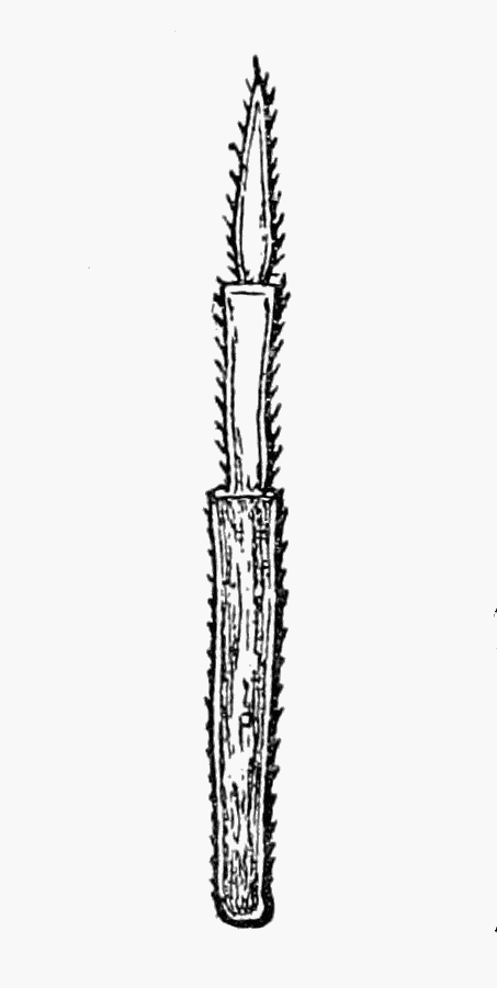 FIG. 47.—FLAGELLUM
The body of Porcellio scaber is densely covered with tubercles. Its colour is usually of a very dark grey, but at times it is quite red or variegated with yellow. Albino specimens have been recorded. The two joints of the flagellum are of the same length and together equal that of the last joint of the peduncle. Air-tubes are present in the outer plates of the first two abdominal appendages.
BRITISH LOCALITIES:—
England: High Beach, Epping; Warley; Brightlingsea; (W.M.W.): Maldon; (W.M.W. from R.M.): Langley; Kew; Skirmett; Pamber Forest; (W.M.W.): Liphook; (C.S.): Stoke-on-Trent; Kingston-on-Soar; (W.M.W.)
Scotland: Dinnet (W.M.W. from Madame Christen).
Ireland: Common everywhere; (Scharff, 63.)
FOREIGN DISTRIBUTION:—
Europe: Throughout; (59): France; (28): Spain; (15): Iceland; (59): Faroe Isles—Thorsharn and Naalsoe—(R.F.S. and B.M., N. Annadale).
America: Greenland; North America; Sandwich Isles; (B.M.); Mexico; (59): St. Paul; St. Croix; (59); Ascension; Tristan d'Acunha; (23)
Asia: Ceylon; Kamtschatka: (23).
Australia: Melbourne; Sydney; Tasmania; New Zealand; (B.M., Chilton).
Africa: Azores; Canaries; Cape of Good Hope; (24).
Porcellio pictus Brandt and Ratzeburg. Plate XIV.
There are tubercles on the body of Porcellio pictus, which is a striking looking animal. Its head is black with the lateral lobes curved outwards; there is a dark band down the middle of the back and commonly two others on each side, with more or less conspicuous yellow markings between.
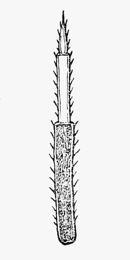 FIG. 48.—FLAGELLUM
The distal (terminal) joint of the flagellum is but half the length of the proximal one and the last peduncular joint is longer than the two combined.
The abdominal appendages—1 and 2—are provided with air-tubes.
BRITISH LOCALITIES:—
England: Maldon; (W.M.W. from R.M.): Chislehurst; (Bate and Westwood, 1): Cooper's Hill, near Cheltenham; (Norman, 49): Exeter; (Parfitt, 53): Kent; (Bate and Westwood, 1.)
Scotland: Between Leith and Portobello; (Scott, 68): Cumbrae (Scott, 68a): Ayrshire; (Boyd in Norman, 49): Banff; (T. Edwards in Norman, 49).
Ireland: Dublin; Belfast; (Bate and Westwood, 1): Galway; Maryborough; Queen's Co., Castel; and Caher Co. Tipperary; (R.F.S.)
FOREIGN DISTRIBUTION:—
Europe: France; (25): North, West-Central, and East Europe; (8): Sweden; Norway; Denmark; Germany; Hungary; Russia; (59).
North America: (8).
Porcellio dilatatus Brandt. Plate XV.
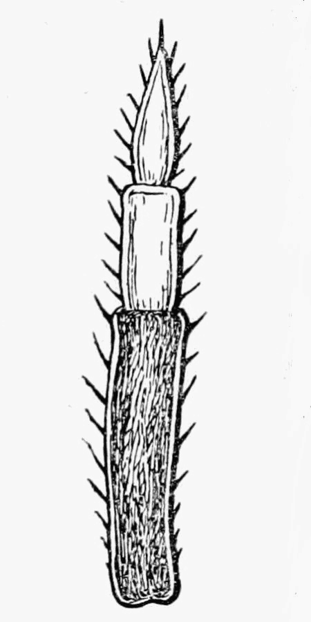 FIG. 49.—FLAGELLUM
The fact that Porcellio dilatatus is more than half as broad as it is long, at once distinguishes it from the other species of Porcellio. It is tuberculated and of somewhat a lighter grey than Porcellio scaber usually is. The two species agree in having the two joints of the flagellum equal, but the last peduncular joint, as in Porcellio pictus, is longer than the flagellum.
As in the two preceding species, air-tubes are found in the outer plates of the appendages on the first two abdominal segments. Porcellio dilatatus is to be looked for near houses.
BRITISH LOCALITIES:—
England: Maldon; (W.M.W. from R.M.): Eton; (Stebbing from W.M.W., 71a): Berkhamsted; (Norman, 50): Headley, Surrey; Ventnor; (Stebbing in Norman, 49).
Ireland: Dublin; (Scharff, 63): Dundrum; (Scharff in Norman, 50): Galway; Roundstone; (R.F.S.): Belfast; (C. W. Buckle, Irish Nat., Vol. XI. (1902), p. 43).
FOREIGN DISTRIBUTION:—
Europe: France; (25): Spain; (12 ): Denmark; Norway; Germany; Poland; Holland; (59).
Africa: Madeira; Azores; (24).
Australia: New Guinea; (59).
Porcellio rathkei Brandt. Plate XVI.
There is often a light band down the back and one on either side of it near the margin in Porcellio rathkei (especially in the males), with other more irregularly arranged light patches between. Unlike the three species previously considered, the present one has a smooth body. The distal joint of the flagellum is the longer, and the flagellum itself is equal in length to the last joint of the peduncle.
Some specimens found by Mr. Webb in 1899 at Eton were submitted to Mr. Stebbing, and since then the former has found Porcellio rathkei to be pretty generally distributed in West Middlesex, where the species appears to frequent the open fields.
 FIG. 50.—FLAGELLUM
Air-tubes occur in abdominal appendages 1 to 5 and the white appearance of all of these at once serves to distinguish the living animal from Porcellio scaber in which the first two pairs of abdominal appendages alone are white.
BRITISH LOCALITIES:—
England: Eton; (Stebbing, 71a): Lane End; (Stebbing, from the Misses Johnston, 71a): Acton; Ealing; Hanwell; Southall; Northolt; Greenford; West Drayton; Mortlake; (W.M.W.); Sunderland; (Brady, 50a).
FOREIGN DISTRIBUTION:—
Europe: France; (25): Bosnia; Servia; (22): Hertzogovania (B.M.); Norway; Northern, Western, and Middle Europe, everywhere; (59): Corfu (B.M.)
Asia: Transcaucasia; (59).
North America: (59).
Porcellio laevis Latreille. Plate XVII.
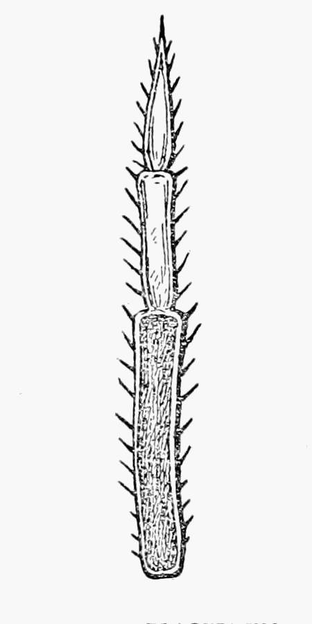 FIG. 51.—FLAGELLUM
Another smooth species is Porcellio laevis. The colour of its body is light grey with irregular white markings. The large size of this species and the very long tail-appendages of the males are features which will help to identify it. The distal joint of the flagellum is slightly the longer and as in the last species (P. rathkei) the flagellum is equal in length to the last joint of the peduncle. The chief habitats for this species are among vegetable rubbish near human dwellings.
Only the first two abdominal appendages contain air-tubes.
BRITISH LOCALITIES:—
England: Maldon; (W.M.W. from R.M.): Ipswich (1892); Hanwell; Wimbledon; (W.M.W.): Kent; (Kinahan, 32).
Ireland: Dublin; (Bate and Westwood, 1): Blackrock Dundrum; Co. Dublin; Galway; (R.F.S.)
FOREIGN DISTRIBUTION:—
Europe: France; (25): Spain; (15): Sicily; (19): Hertzogovania; (22): Sweden; Denmark; Germany; Belgium; Austria; Italy; Dalmatia; Greece; Turkey; (59); Corfu; (B.M.): Inca, Majorca (23); (B.M.—Pocock and Thomas.)
Asia: Syria; Turkestan; (21).
Africa: Morocco; Algeria; Tunis; Tripoli; Senegal; Egypt; (23).
Atlantic Isles: Bermudas; Azores; Canaries; Cape Vera; Madeira; (24).
America: North America; Mexico; Peru; Brazil; Chili; West Indies; Pacific Islands; (59); Sandwich Isles; (B.M.)
Porcellio ratzeburgii Brandt. Plate XVIII.
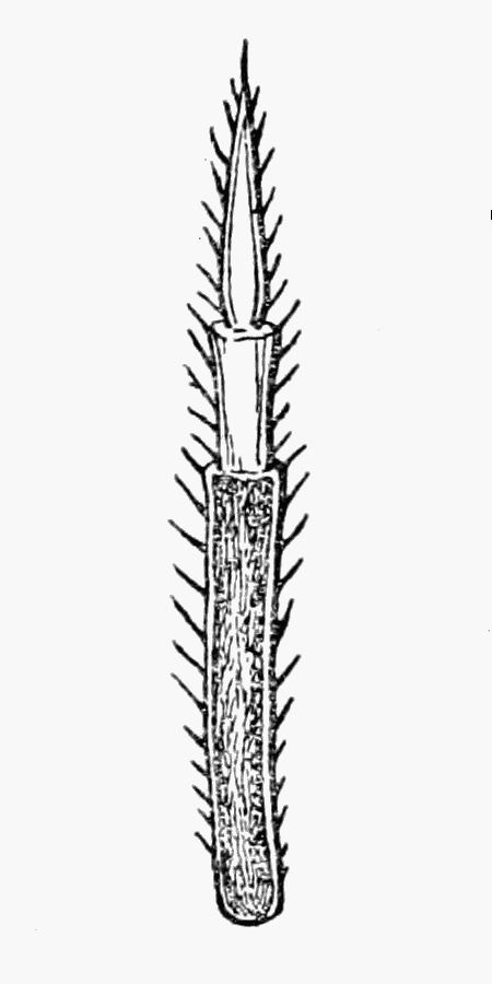 FIG. 52.—FLAGELLUM
There are granulations on the middle of the segments in Porcellio ratzeburgii and the sides of its body are more nearly parallel than in the other species of Porcellio; the frontal lobe is, practically speaking, semicircular and the lateral plates of the thorax flank the head to a considerable extent. As in Porcellio pictus, the dark band is in the middle of the back. The distal joint of the flagellum is nearly twice as long as the proximal, and the flagellum is shorter than the last joint of the peduncle. This species was added to the British list by Mr. Webb (74) in 1898.
Porcellio ratzeburgii agrees with Porcellio rathkei in having air-tubes in the first five abdominal appendages.
BRITISH LOCALITIES:—
England: Warley; Brightlingsea; young examples (W.M.W.): Maldon; young examples (W.M.W. from R.M.)
FOREIGN DISTRIBUTION:—
Europe: Trafoi St. Martini, and Capitello, in the Tyrol; (Norman, 50); East Alps, very common; Val-de-Joux; Massif de la Chartreuse Vaulnaveys (25): Bosnia; (22): Norway; Central Europe; Upper Pfaltz; Bohemia; Saxony; Rhaetia; (59).
Genus—METOPONORTHUS Budde-Lund, 1879 (7), p. 4.
Porcellionides Miers, 1876 (44), p. 98.
Flagellum, with two joints; abdomen, narrow; frontal lobe not developed.
The hinder legs of Metoponorthus are proportionately longer than in any other Oniscidæ save Philoscia. Both genera have a narrow abdomen, but Philoscia has an extra joint to the flagellum, and shows no sign of lateral lobes to the head.
Metoponorthus pruinosus Brandt. Plate XIX.
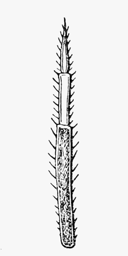 FIG. 53.—FLAGELLUM
Undamaged specimens of Metoponorthus pruinosus are of a beautiful bluish-grey colour, owing to a "bloom" which is easily brushed off, revealing a dark reddish-brown tint beneath it. The antennæ are long and have white markings upon them.
Air-tubes occur in the first two abdominal appendages.
BRITISH LOCALITIES:—
England: Maldon; (W.M.W. from R.M.): Hanwell; Eton Wick; Kew; Ipswich; Stoke-on-Trent; (W.M.W.): Chiselhurst; Oxford; (Bate and Westwood): Berkhamsted; Burnmoor; Durham; (Norman, 49): Exeter; (Parfitt, 53); Torquay; (B.M.—T.R.R.S.)
Scotland: Banff; (Thomas Edwards in Norman, 49)
Ireland: Dublin; (Kinahan, 32): Foyle District; Donegal; Galway; Clonbrock, Co. Galway; Mornington, Co. Meath; Santry; Gleeson Park; Dundrum, Co. Dublin; Bray; (R.F.S.)
FOREIGN DISTRIBUTION:—
Europe: Practically all the Countries of Europe are given in Dollfus' list; (23).
Asia: Japan; China; Syria; Ceylon; Sumatra; Celebes; Phillipines; Caucasus; Himalayas; (23): Christmas Island; (B.M.)
Africa: Generally distributed; Madagascar; Seychelles; (23).
Atlantic Isles; (23).
America: North and South, almost everywhere, to judge from M. Dollfus' list; (23).
Australia: New Caledonia; (23).
Metoponorthus cingendus Kinahan. Plate XX.
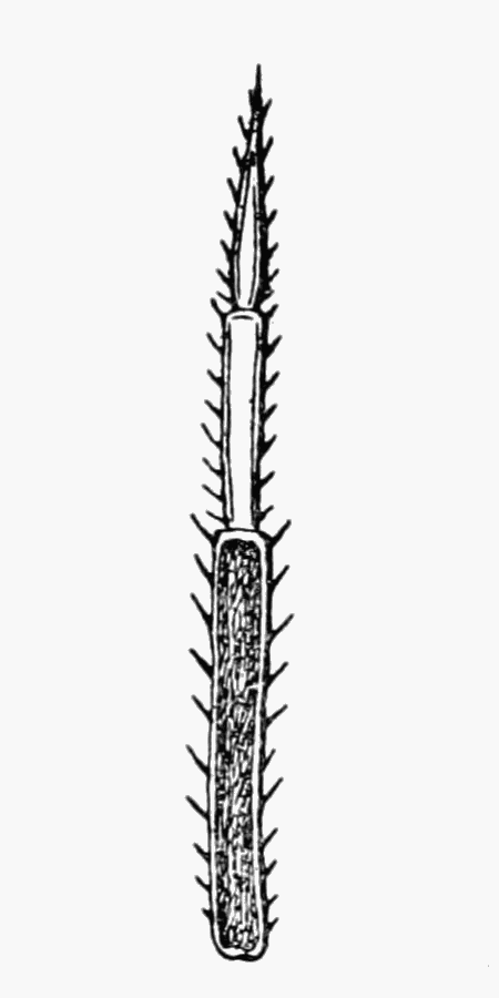 FIG. 54.—FLAGELLUM
The colour of Metoponorthus cingendus is steel blue with red or yellowish spots. It has a raised line across each thoracic segment and its abdomen is narrower than in Metoponorthus pruinosus.
BRITISH LOCALITIES:—
England: Salcombe, Devon; (Norman, 49): South Devon; (Stebbing in 49).
Ireland: Dublin; (B.M. from Kinahan); Mountain Districts of Dublin, Wicklow, and Cork; Coast of Kerry; Arran Islands; Achill, Co. Mayo; Roundstone, Co. Galway; Mallow, Caef Island; Glandore; Brock Haven, Co. Cork; Killoughrim Forest, Co. Wexford; Kenmare, Co. Kerry; (R.F.S.).
FOREIGN DISTRIBUTION:—
Europe: France; (25): Spain; (12).
(2.) Able to roll up into a ball.
Genus—CYLISTICUS Schnitzler, 1853 (65), p. 24.
Flagellum, with two joints; abdomen broad; frontal lobe, very small.
The characters given immediately above are almost those of Porcellio with which Cylisticus might, perhaps, be confounded. The latter has the power, however, of rolling itself into a ball, and the first segment of the thorax is comparatively larger than in any species of Porcellio, indeed the side plates of the segment in question entirely flank the head. These features, as well as the straight sides of the body and the arched back, connect Cylisticus with Armadillidium, from which the former is, however, at once separated by its long pointed tail appendages.
Cylisticus convexus De Geer. Plate XXI.
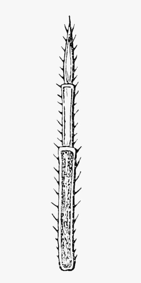 FIG. 55.—FLAGELLUM
There is but a single species of Cylisticus found in this country, so that it is not necessary for us to go into much further detail with regard to it. Cylisticus convexus has the two joints of the flagellum about equal, and they together in turn closely approximate in length to the last joint of the peduncle. Mr. Stebbing says, in a letter, that British examples do not appear to have the "white tail-piece" seen in Continental ones. It is not noticeable in the preserved specimens which we have seen from Berkhamsted and Leixlip, but it is very evident in the living ones found at Hanwell and Maidstone.
The abdominal appendages 1 to 5 are provided with air-tubes.
BRITISH LOCALITIES:—
England: Maldon; (W.M.W. from R.M.): Hanwell; Bluebell Hill, Maidstone; Eton; (W.M.W.): Berkhamsted; Portland; (Norman, 49).
Scotland: Salisbury Crags; Edinburgh; Lanarkshire; Rothesay; (Scott, 68): Killwinning; (John Smith fide Robertson, 57): Highgate; (Bate and Westwood, 1).
Ireland: Leixlip, Co. Dublin; Tempo, Co. Fermanagh; Goresbridge, Co. Kilkenny; (R.F.S.)
FOREIGN DISTRIBUTION:—
Europe: France; (25): Sweden; Norway; Denmark; Germany; Bohemia; Holland; Belgium; Turkey; Caucasus; (59).
North America; (59).
Family—ARMADILLIDIIDÆ.
Tail appendages not projecting when the animal is walking.
Genus—ARMADILLIDIUM Brandt, 1833 (3), p. 184.
Flagellum, with two joints; outer division of the tail appendages expanded and broader at the hinder end.
The members of the genus Armadillidium are more likely to be confounded, by the uninitiated, with the "Pill-millipedes" than with other Woodlice. Excepting Cylisticus (which has long pointed tail appendages) no other British forms have the power of rolling themselves up into a complete ball. The very arched body is characteristic of Armadillidium, and so is the groove into which the basal joints of the antennæ fit when the creatures curl up.
The first two abdominal appendages only are provided with air-tubes.
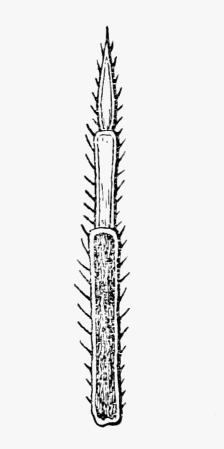 FIG. 56.—FLAGELLUM
Armadillidium nasatum Budde-Lund. Plate XXII.
Armadillidium nasatum has a narrow but very prominent frontal lobe, which is almost square and curves somewhat upwards and backwards. The joints of the flagellum are approximately equal, and are together of the same length as the last peduncular joint.
The telson is as long as it is broad at the base, and tapers to a roundish point, while its sides are slightly incurved.
The outer divisions of the tail appendages are considerably longer than broad, and are more or less paddle-shaped.
It will be noticed that the slope from thorax to telson is more gentle than in the common species, Armadillidium vulgare, and the first thoracic segment is not so greatly developed. Consequently the species which we are considering does not produce a perfect sphere, and the antennæ are not hidden when[Pg 41] it rolls up. It is interesting to compare this species with Cylisticus convexus. The surface of the body is smooth, and its colour is a delicate brownish grey with more or less distinct rows of darker markings.
BRITISH LOCALITIES:—
England: Maldon; (W.M.W. from R.M.): Bluebell Hill, Maidstone (W.M.W.); Clifton, banks of the Avon; (W.M.W. from J.T.C. 1900): Leigh Woods, Clifton; Tunbridge Wells; South Devon; (Stebbing in 49); Cheddar Cliffs, Somerset; (Norman, 49).
FOREIGN DISTRIBUTION:—
Europe: France; (28): Spain; (12): Italy; (23).
Armadillidium vulgare Latreille. Plate XXIII.
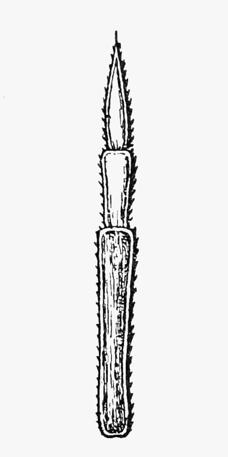 FIG. 57.—FLAGELLUM
The common pill woodlouse is Armadillidium vulgare. Its frontal lobe is not large, though it is broad, while its margin where it joins the head is rounded and slightly recurved. The proximal joint of the flagellum is somewhat the shorter and the two together, as in Armadillidium nasatum, are of about the same length as the last joint of the peduncle.
The telson has the form of a triangle with the angles truncated and is about as long as it is broad at the base. The outer divisions of the tail appendages are considerably broader than they are long.
The species can roll itself up into a very perfect sphere, and when it assumes this form its antennæ are hidden beneath the much expanded lateral plates of the first thoracic segment.
The body is smooth, shiny, and strongly arched. Its colour varies very considerably, generally it is of a slaty-grey, but yellow markings are often present to a greater or less extent. In a specimen before us (from Bluebell Hill, Maidstone) the head is of a[Pg 42] uniform dark grey, the sides of the thorax are yellow, while the back is mottled with the same colour. The abdomen, including the telson, is also yellow with the exception only of the tail appendages, which are dark grey.
BRITISH LOCALITIES:—
England: Warley; (W.M.W): Maldon; (W.M.W. from R.M.): Brightlingsea; Hanwell; Mortlake; Bluebell Hill, Maidstone; Langley; Skirmett; Pamber Forest; Kingston-on-Soar; Ipswich; (W.M.W.); Lynmouth; (W.M.W. from J.T.C.)
Scotland: (Scott, 68.)
Ireland: Ardrahan; (Norman, 50): Borris, Co. Carlow; Glandare, Terneay, Co. Cork; Courtstown, Co. Wexford; Cappagh, Co. Waterford; Castel, Co. Tipperary; (R.F.S.)
FOREIGN DISTRIBUTION:—
Europe: Throughout; (23).
Asia: Damascus; (23).
Africa: Algeria; (23).
Atlantic Isles: (23).
America: North and South; (23).
Australia: Melbourne (64 quoting Budde-Lund); New Zealand; (23).
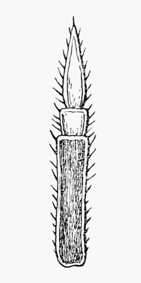 FIG. 58.—FLAGELLUM
Armadillidium pulchellum Zencker. Plate XXIV.
The smallest British species is Armadillidium pulchellum. The frontal lobe projects so as to make the head somewhat triangular. The antennæ are very short and the distal joint of the flagellum is three times the length of the other, while the two together are not as long as the last peduncular joint.
The telson is truncated at the end so that it is by no means as long as it is broad at the base, and the outer divisions of the tail appendages are in similar proportion.
The colour of the body (which is smooth) is dark brown with four important series of light patches running down the back and less marked variegations between them.
BRITISH LOCALITIES:—
England: Matlock; (T.R.R.S.) Arnside; Westmorland (Brady, 50a).
Ireland: Ballymote, (Irish Nat., May, 1901), Sligo; (Scharff).
FOREIGN DISTRIBUTION:—
Europe: Vosges; Switzerland; Pyrenees; (23): Forest de Soignes; Belgium; (14).
Armadillidium depressum Brandt. Plate XXV.
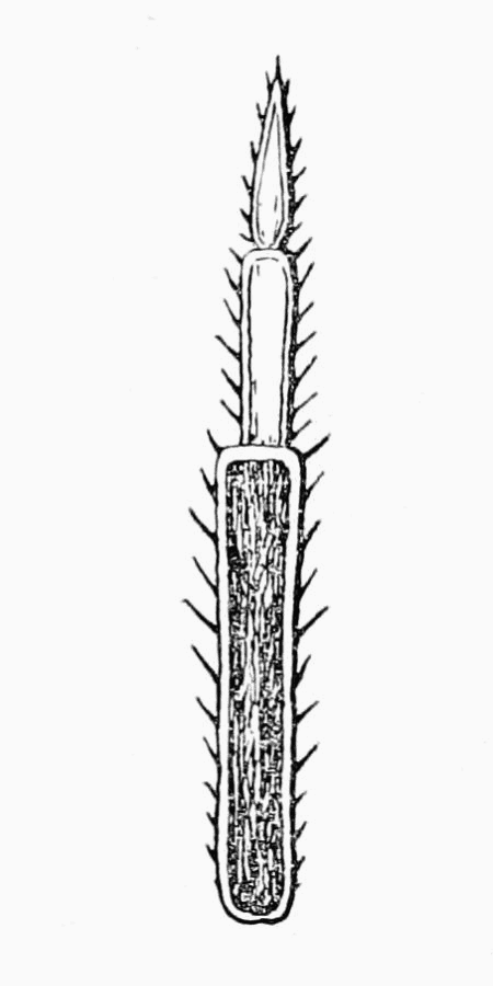 FIG. 59.—FLAGELLUM
The frontal lobe in Armadillidium depressum is very prominent and much recurved. The antennæ are fairly long and while the two joints of the flagellum are nearly equal they are together not so long as the last peduncular joint.
The telson is slightly longer than it is broad at the base, and its sides are incurved. As in Armadillidium vulgare and Armadillidium pulchellum the outer divisions of the tail appendages are broader than they are long.
The body is flatter than in the other species and bears tubercles; its colour is a slate-grey with yellowish markings. The first thoracic segment is well developed and the head appears as if almost completely imbedded in it.
BRITISH LOCALITIES:—
England: Clifton, banks of the Avon; (W.M.W. from J.T.C., 1900): Shirehampton, near Bristol; (Stebbing in 49): Clifton; (Dollfus from Miers 14).
FOREIGN DISTRIBUTION:—
Europe: France; Italy; (25): Asia Minor; (14, quoting Brandt).
Distribution of Species. There are not sufficient records at present to enable us to draw any conclusions as to the general distribution of Woodlice in the British Isles, but it is hoped that more attention will be given to these creatures, and that before long there may be other material available.
Conclusion. At the beginning it was mentioned that the present work grew out of an investigation into the fauna of Essex, and in order to show what may be expected when places[Pg 44] are explored in which no collecting has been done, we may briefly indicate the results which we obtained in the county in question.
It was not long before a species new to Britain—to wit, Porcellio ratzeburgii—was found (74). This discovery was mentioned by Mr. Stebbing in the Victoria County History of Essex (p. 71), and he prophesied that most of the British species then would be met with in the county. We may safely claim to have shown that his prediction was true, for we have been able to record in the preceding pages no less than sixteen other species, as will be seen from the following lists:—
WOODLICE RECORDED FROM ESSEX.
| 1. | Ligia oceanica | 10. | Porcellio pictus |
| 2. | Ligidium hypnorum | 11. | Porcellio dilatatus |
| 3. | Trichoniscus pusillus | 12. | Porcellio laevis |
| 4. | Trichoniscus roseus | 13. | Porcellio ratzeburgii |
| 5. | Haplophthalmus danicus | 14. | Metoponorthus pruinosus |
| 6. | Oniscus asellus | 15. | Cylisticus convexus |
| 7. | Philoscia muscorum | 16. | Armadillidium nasatum |
| 8. | Platyarthrus hoffmannseggii | 17. | Armadillidium vulgare |
| 9. | Porcellio scaber |
Of these Ligidium hypnorum calls for special mention, as it had not been found in this country since Mr. Stebbing discovered it in Surrey in 1873. Several of the Porcellios and Cylisticus convexus have been met with in but few places, and the same may be said of Armadillidium nasatum.
Of the British species not as yet found in Essex Trichoniscus vividus has at present only been recorded from Ireland; Philoscia couchii and Armadillidium depressum have not been collected except in the extreme south west of England, while Metoponorthus cingendus has hitherto only been noticed in Devonshire and Ireland. The other four species, with the exception of Porcellio rathkei, which is well distributed in west Middlesex (and might have been expected to occur in Essex), are still rare. In fact, for Trichoniscoides albidus but two British localities are known; for Haplophthalmus mengii three (two in England and one in Ireland); while Armadillidium pulchellum has only been recorded from two or three places.
In other counties quite as satisfactory results were obtained as in Essex—a systematic search in Buckinghamshire brought to[Pg 45] light at Eton three species which at the time had not been recorded from the British Isles, while in Middlesex, no less than a dozen species were found at Hanwell.
Since part of this contribution was printed our attention has been drawn to some notes by the Rev. Canon Norman and Professor G. S. Brady (50a). These bear out the remarks which have already been made, for among the species found by Professor Brady in the north of England were Trichoniscoides albidus, Haplophthalmus mengii, Porcellio rathkei, and Armadillidium pulchellum. In one of Canon Norman's previous papers (50—1903) he claims to have added the second species to the British list, and in the notes in question a similar claim is made with regard to the first and third. It should, however, be pointed out that all three of them were found in Buckinghamshire in 1899 by Mr. Webb, and that they were exhibited at the Nature Study Exhibition held in London in August, 1902. A specimen of Armadillidium pulchellum from Matlock was sent to us by the Rev. T. R. R. Stebbing in January, 1904, and was found, we understand, some considerable time previously.
It only remains for us to express our hearty thanks to the numerous friends and correspondents who have given us their ready help. The names of these have been printed in the text, but we would like to mention more particularly Dr. Calman, of the British Museum (Natural History), Monsieur Adrian Dollfus, Mr. Roland Matthams, the Rev. Canon Norman, Dr. Scharff, the Rev. T. R. R. Stebbing, and Miss Willmott.
FOOTNOTES:
[1] "The Non-Marine Molluscs of Essex," by Wilfred Mark Webb; Essex Naturalist, Vol. x. (1897), pp. 27-48 and 65-81.
[2] The numbers in brackets refer to papers mentioned in the Bibliography at the end.
[3] It should be pointed out that the methylated spirit now sold in the shops contains mineral naphtha and goes milky on the addition of water. Permission can be obtained from Somerset House to buy what is still called "ordinary methylated spirit," but at present five gallons has to be purchased at one time.
Explanation of the initials given in the text:—
| B.M.—Specimens in the British Museum (Natural History). | C.S.—Charles Sillem. |
| J.T.C.—John Thomas Carrington. | T.R.R.S.—The Rev. Thomas R. R. Stebbing. |
| J.A.M.—James A. Murie. | W.M.W.—Wilfred Mark Webb. |
| R.M.—Roland Matthams. | R.W.—R. Welch. |
| R.F.S.—R. F. Scharff. |
(1) BATE, C. SPENCE, and WESTWOOD, J.C.: A history of the British Sessile-eyed Crustacea, London, 1868.
(2) BOSC, L. A. G.: Manuel de l'histoire Naturelle des Crustacés, Vol. II. Paris, 1830.
(3) BRANDT, J. F.: "Conspectus Monographiae Crustaceorum Oniscodorum Latreillei," Bull. Soc. Nat., Moscow. Vol. VI. (1833), pp. 171-193.
(4) BRANDT, J. F., and RATZEBURG, J. T. C.: Medizinische Zoologie. Vol. II., Berlin, 1830-1834.
(5) BRUNTZ, L.: "Contribution à l'Etude de l'Excretion chez les Arthropodes." Archives de Biologie. Vol. XXII. (Nov., 1903), pp. 215-422, pls. I.-VIII.
(6) BUDDE-LUND, G.: "Danmarks Isopode Landkrebsdyr," Naturhistorisk Tidsskrift. Part 3, vol. VII. (1870, pp. 217-245).
(7) BUDDE-LUND, G.: Prospectus generum specierumque Crustaceorum Isopodum terrestrium. Copenhagen, 1879.
(8) BUDDE-LUND, G.: Crustacea Isopoda Terrestria. Hauniae, 1885.
(9) CUVIER, G.: "Memoires sur les Cloportes terrestres." Journ. d'hist. nat. Vol. II. 1792.
(10) DE GEER, C.: Memoires pour servir à l'histoire des insectes. Vol. VII., Stockholm, 1778.
(11) DESMAREST, A.G.: Considérations générales sur la classe des Crustacés. Paris, 1825.
(12) DOLLFUS, A.: "Catalogue raisonné des Isopodes terrestres de l'Espagne." Ann. Soc. Espan. Hist. Nat. Vol. XXI. (1892), pp. 161-190.
(13) DOLLFUS, A.: "Sur la Distribution Geographique des Isopodes terrestres dans la region des Basses-Pyrenees." Assoc. Francaise pour l'avancement des Sciences. Rep. Congress de Pau., 1892.
(14) DOLLFUS, A.: "Le Genre Armadillidium." Feu. des Jeunes Naturalistes, Ser. 3. May 1892.
(15) DOLLFUS, A.: "Catalogue raisonné des Isopodes terrestres de l'Espagne." Premier supplement, Ann. Soc. Espan. Hist. Nat. Vol. XXII. (1893), pp. 47-51.
(16) DOLLFUS, A.: "Isopodes Terrestres in 'Voyage de M. Ch. Allmand aux Iles Canaries.'" Mem. Soc. Zool. de France. Vol. VI. (1893), p. 46.
(17) DOLLFUS, A.: "Sur la Distribution Geographique des Armadilliens en Europe." Compte-Rendu troisieme Congres. Internal. de Zool. Leyden, Sept. 1895 (1896), pp. 356-358.
(18) DOLLFUS, A.: "Les Isopodes terrestres du Nord de l'Afrique du Cap. Blanc a Tripoli." Mem. Soc. Zool. Trans. Vol. IX. (1896), pp. 523-553.
(19) DOLLFUS, A.: Crustacés Isopodes de la Sicile. 1896.
(20) DOLLFUS, A.: "On West Indian Isopod Crustaceans." Proc. Zool. Soc. Lond. 1896, pp. 388-400.
(21) DOLLFUS, A.: "Tableau Iconographique des Philoscia d'Europe." Feu. des Jeun. Natural. Ser. 3, 1897, pp. 70-73 and 91-95.
(22) DOLLFUS, A.: "Land Isopods der Balkan region." Wissenschaft Mittheilung aus Bosnien und der Hercegovina. Vol. IV. (1896).
(23) DOLLFUS, A.: "Les Crustacés Isopodes Terrestres à grande dispersion," Feu des Jeun. Natural. Ser. 3 (Oct. 1897).
(24) DOLLFUS, A.: "Sur la Distribution Geographique des Isopodes Terrestres dans l'Afrique Septentrional du Senegal à Obock." Proc. Inter. Congres. Zool. Cambridge, 1898, pp. 249-259.
(25) DOLLFUS, A.: "Catalogue des Crustacés Isopodes Terrestres de France." Feu. des Jeun. Natural. Ser. 3 (October 1899).
(26) FABRICIUS, J.C.: Entomologia systematica. Vol. II., Hauniae, 1793.
(27) FABRICIUS, J. C.: Supplement to the last, 1798.
(28) FERNIE, Dr.: Animal Simples, 1899.
(29) FITCH, Asa.: Noxious Insects of New York. Albany, 1856.
(30) GUERIN, F.E.: "Sur Porcellio Poeyi." Ann. Sci. Soc. Entom. de France. Vol. VI. (Paris, 1837, in Bull. ent., page 6).
(31) HELLER, C.: Reise der Novara; Crustacea. 1865.
(32) KINAHAN, J.R.: "Analysis of certain genera of terrestrial Isopoda." Nat. Hist. Rev. Vol. IV. (1857).
(33) KINAHAN, J.R.: "On the genera Philoscia, Itea and Philougria." Nat. Hist. Rev. Vol. V. 1858.
(34) KOCH, C.L.: Deutschlands Crustaceen, Myriapoden and
Arachniden. Regensburg, 1835-1844.
Koch's descriptions and figures were published in Deutschlands Crustaceen and appeared also in Panzer's
Faunæ Insectorum Germanicæ continued by Herrich-Schäffer (which see). The number of the parts of the latter
which correspond to those of the former are placed in square brackets after them in the list here given. Part 6 (1836)
[139]; Part 22 (1838) [162]; Part 28 (1839) [178]; Part 34 (1840) [180]; Part 36 (1844) [186]. Some of the dates
quoted will be found to differ from those usually given (in Budde-Lund (8, p. 7) for instance), and printed on
the parts of the copy in the Zoological Society's Library. Our authority for this change is Mr. C. D. Sherborne,
who bases his dates upon reviews which he has discovered.
(35) KOCH, C. L.: System der Myriapoden mit den Verzeichnissen und Berichtigungen zu Deutschlands Crustaceen, Myriapoden und Arachniden. Regensburg, 1847.
(36) LAMARCK, J. B.: Histoire naturelle des animaux sans vertébres. Vol. V., 1818.
(37) LATREILLE, P. A.: Histoire Naturelle des Crustacés et des Insectes. Vol. VII., Paris, 1804.
(38) LEACH, W. E.: "Tabular view of the external characters of four classes of animals, which Linné arranged under Insectes." Trans. Linn. Soc. Lond. Vol. XI., 1815.
(39) LEREBOULLET, A.: "Memoire sur les Crustacés de la famille des Cloportides qui habitent les environs de Strasbourg." Mem. Soc. Mus. Hist. Nat. Strasbourg. Vol. IV., 1853, pp. 130, pl. X.
(40) LEUCKART, R.: Verzeichnisz der zur Fauna Helgolands gehörenden wirbellosen Seethiere. Braunsweig, 1847.
(41) LINNÉ, C.: Fauna Suecica. First edition, Holmiæ, 1746.
(42) LINNÉ, C.: Fauna Suecica. Second edition, Holmiæ, 1761.
(43) LINNÉ, C.: Systema Naturae, ed. 12, 1767.
(44) MIERS, E. J.: "Catalogue of New Zealand Crustacea." Ann. Mag. Nat. Hist., Ser. 4., Vol. XVII., 1876.
(45) MIERS, E. J.: "On a collection of Crustacea (Decapoda and Isopoda), chiefly from South America." Proc. Zool. Soc., 1877, p. 653-678.
(46) MILNE-EDWARDS, H.: Histoire naturelle des Crustacés. Vol. III. Paris, 1840.
(47) NICHOLSON and LYDDEKER: Manual of Palæontology, 1889, Vol. II., p. 559.
(48) NORMAN, A. M.: "Note on the discovery of Ligidium agile, Persoon (Zia saundersii Stebbing)." Ann. Mag. Nat. Hist. Ser. 4, Vol. XI., p. 419.
(49) NORMAN, A. M.: "British Land Isopoda." Ann. Mag. Nat. Hist. (Ser. 7), Vol. III. (1899), pp. 71-78, pl. VI.
(50) NORMAN, A. M.: Continuation of the last. Vol. XI., 1903, pp. 309-372.
(50a) NORMAN, A.M.: and BRADY C.S.: "British Land Isopoda." Second Supplement Ann. Mag. Nat. Hist. (Ser. 7), Vol. XIV. (1904), pp. 449-450.
(51) PANZER, G. W. F.: Faunæ Insectorum Germanicæ initia, oder Deutschlands Insecten. Parts 1-110, Nurnberg, 1793-1813. (See Koch.)
(52) PANZER, G. W. F.: Continuation by Herrich-Schäffer, Parts 111-190, 1829-1844. (See Koch.)
(53) PARFITT, E.: "The Fauna of Devon." Sessile-Eyed Crustacea. Trans. Devon Assoc. Sci., Sept. 1873.
(54) PERSOON: No paper upon Woodlice by this author is known, but he is quoted in Panzer's Deutschlands Insecten (51), which see.
(55) PLATEAU, F.: "Crustacés Isopodes Terrestres." Bull. Acad. Roy. de Belgique, Ser. 2., Vol. XXIX., No. 2 (1870), p. 112.
(56) RISSO, A.: Histoire Naturelle des Crustacés des environs de Nice. Paris, 1816.
(57) ROBERTSON, D.: "Catalogue of the Amphipoda and Isopoda of the Firth of Clyde." Trans. Nat. Hist. Soc. Glasgow, Vol. II. (1888), pp. 9-99.
(58) ROULE, LOUIS: "Etudes sur le Development de Crustacés." Ann. Sci. Nat., Vol. XVIII. (1895), pp. 1-156, pls. I.-X.
(59) SARS, G. O.: An account of the Crustacea of Norway. Vol. II., 1896-1899.
(60) SAUSSURE, H. de: "Diagnoses de quelques crustacés nouveaux des Antilles et du Mexique." Rev. et Mag. de Zool. (Ser. 2), Vol. IX. (1857), pp. 304-308.
(61) SAVIGNY, J. C., and AUDOUIN, V.: Description de l'Egypte. Vol. XXII., 1827.
(62) SAY, T.: "An account of the Crustacea of the United States." J. Acad. Nat. Sci. Philadelphia, Vol. I., pt. II., 1818.
(63) SCHARFF, R. F.: "The Irish Woodlice." Irish Naturalist, Vol. III., 1894, pp. 4-7 and 25-29, pl. II.
(64) SCHARFF, R. F.: "The Woodlice of Co. Carlow." Irish Naturalist, 1895, p. 319.
(65) SCHNITZLER, H. J.: De Oniscineis agri Bonnensis. (Thesis.), Cologne, 1853.
(66) SCHOBL, J.: "Typhloniscus, eine neue blinde Gattung der Crustacea Isopoda." Sitzungsberichte der math. naturw. Acad. Wiss. Wien. Vol. XL. (1860), pp. 279-330.
(67) SCOPOLI, J. A.: Entomologia Carniolica. Vindibonæ, 1763.
(68) SCOTT, T.: "The Land and Freshwater Crustacea of the District around Edinburgh." Proc. R. Phys. S. Edin., Vol. XI. (1890-91), p. 75.
(68a) SCOTT, T.: British Association Hand Book on the Natural History of Glasgow. 1901; Isopoda, pp. 335 and 336.
(69) SILL, VICTOR: "Beitrag zur Kentniss der Crustaceen, Arachniden and Myriapoden Siebenburgens." Verhandl. u. Mittheil. des Siebenburghischen Ver. für Naturwiss. zu Hermannstadt, Vol. XII. (1861), p. 1-11.
(70) STEBBING, T. R. R.: "On a Crustacean of the Genus Zia." Ann. Mag. Nat. Hist. Ser. 4., Vol. XI. (1873).
(71) STEBBING, T. R. R.: The Victoria County History of Essex (1903); Crustacea, pp. 27-28.
(71a) STEBBING, T. R. R.: The Victoria County History of Buckinghamshire (1905); Crustacea.
(72) VEJDOVSKYF: "Zur Morphologie der Antennen und Schalendruse der Crustaceen." Zeit. Wiss. Zool., Vol. LXIX., p. 378.
(73) VERHOEF, K. W.: "Ueber Palæarktische Isopoden." Zool. Anz., Vol. XXIV.
(74) WEBB, WILFRED MARK: "The occurrence in Essex of a species of Woodlouse (Isopoda) new to Britain (Porcellio ratzeburgii, Brandt)." Essex Naturalist, Vol. XI. (1899), p. 127.
(75) WEBB, WILFRED MARK: "Notes on Woodlice" (Including extracts from a paper read before the North London Natural History Society by James B. Casserley). Science Gossip, Vol. VI., New Series (1900), pp. 295-296.
(76) WEBER, MAX.: "Uber einige neue Isopoden der Niederlandischen Fauna." Tijdschr der Niederland Dierk veren, Vol. V. (1881), pp. 167-196, pl. V.
(77) ZADDACH, E. G.: Synopses crustaceorum Prussicorum prodromus Regiomonti, 1844.
(78) ZENKER, C. D.: No paper upon woodlice by this author is known, but he is quoted in Panzer's Deutschlands Insecten (51), which see.
(79) ZITTEL, K. A. von: Textbook of Palæontology. English Translation 1900, p. 668.
| PLATE | |
| Ligia oceanica Linné | I. |
| Ligidium hypnorum Cuvier | II. |
| Trichoniscus pusillus Brandt | III. |
| Trichoniscus vividus Koch | IV. |
| Trichoniscus roseus Koch | V. |
| Trichoniscoides albidus Budde-Lund | VI. |
| Haplophthalmus mengii Zaddach | VII. |
| Haplophthalmus danicus Budde-Lund | VIII. |
| Oniscus asellus Linné | IX. |
| Philoscia muscorum Scopoli | X. |
| Philoscia couchii Kinahan | XI. |
| Platyarthrus hoffmannseggii Brandt | XII. |
| Porcellio scabier Latreille | XIII. |
| Porcellio pictus Brandt and Ratzeburg | XIV. |
| Porcellio dilatatus Brandt | XV. |
| Porcellio rathkei Brandt | XVI. |
| Porcellio laevis Latreille | XVII. |
| Porcellio ratzeburgii Brandt | XVIII. |
| Metoponorthus pruinosus Brandt | XIX. |
| Metoponorthus cingendus Kinahan | XX. |
| Cylisticus convexus De Geer | XXI. |
| Armadillidium nasatum Budde-Lund | XXII. |
| Armadillidium vulgare Latreille | XXIII. |
| Armadillidium pulchellum Zencker | XXIV. |
| Armadillidium depressum Brandt | XXV. |
BRITISH WOODLICE.
PLATE I.
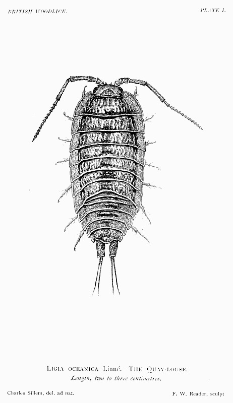 Ligia oceanica Linné. The Quay-louse.
BRITISH WOODLICE.
PLATE II.
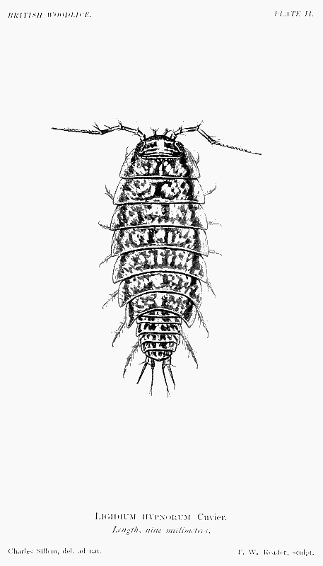 Ligidium hypnorum Cuvier.
BRITISH WOODLICE.
PLATE III.
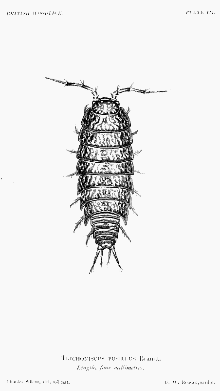 Trichoniscus pusillus Brandt.
BRITISH WOODLICE.
PLATE IV.
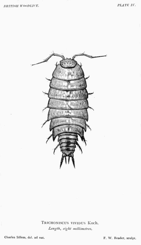 Trichoniscus vividus Koch.
BRITISH WOODLICE.
PLATE V.
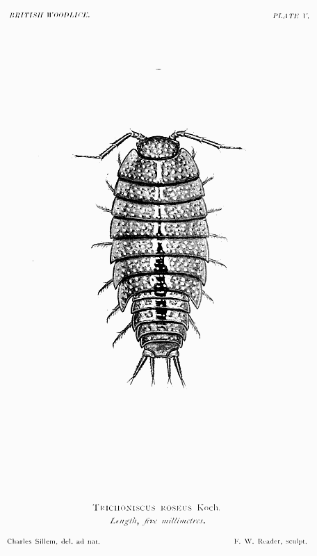 Trichoniscus roseus Koch.
BRITISH WOODLICE.
PLATE VI.
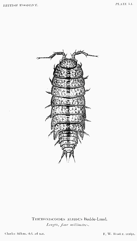 Trichoniscoides albidus Budde-Lund.
BRITISH WOODLICE.
PLATE VII.
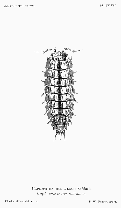 Haplophthalmus mengii Zaddach.
BRITISH WOODLICE.
PLATE VIII.
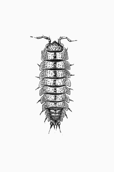 Haplophthalmus danicus Budde-Lund.
BRITISH WOODLICE.
PLATE IX.
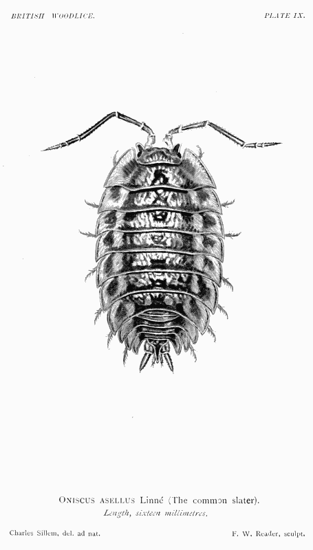 Oniscus asellus Linné (The common slater).
BRITISH WOODLICE.
PLATE X.
 Philoscia muscorum Scopoli.
BRITISH WOODLICE.
PLATE XI.
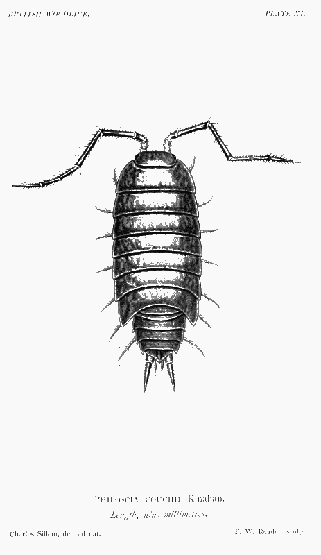 Philoscia couchii Kinahan.
BRITISH WOODLICE.
PLATE XII.
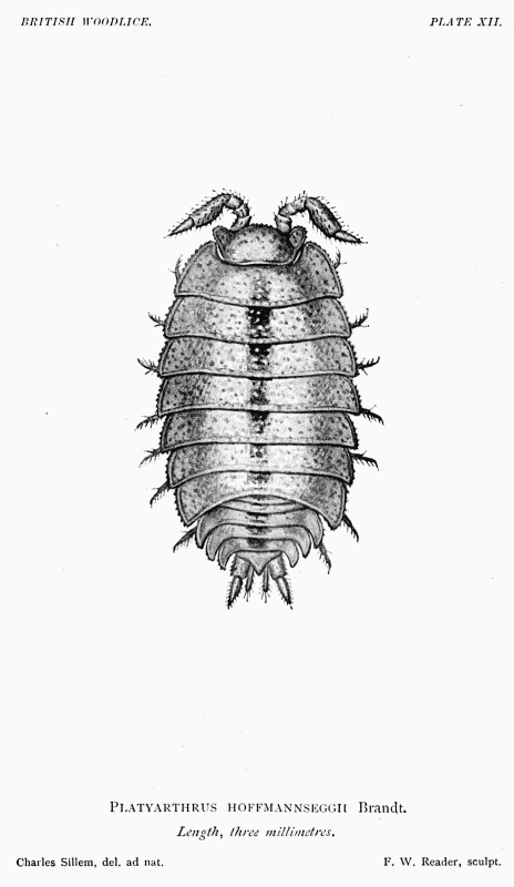 Platyarthrus hoffmannseggii Brandt.
BRITISH WOODLICE.
PLATE XIII.
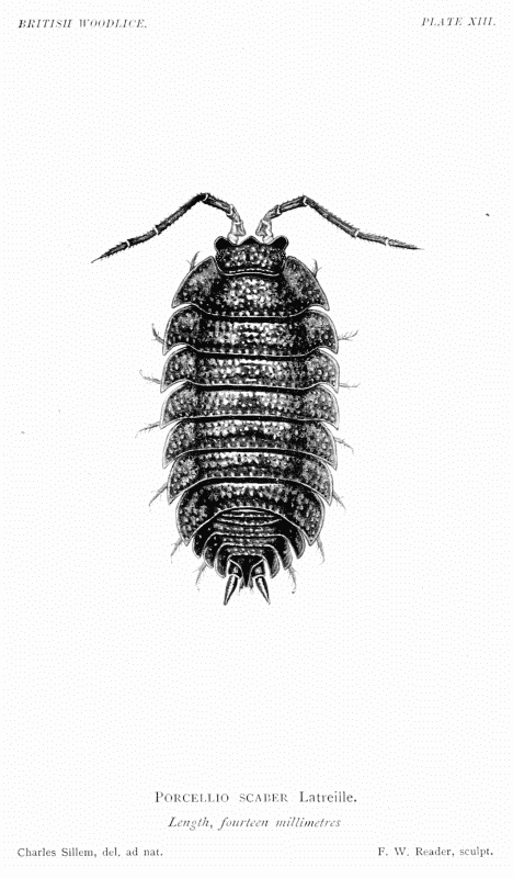 Porcellio scaber Latreille.
BRITISH WOODLICE.
PLATE XIV.
 Porcellio pictus Brandt and Ratzeburg.
BRITISH WOODLICE.
PLATE XV.
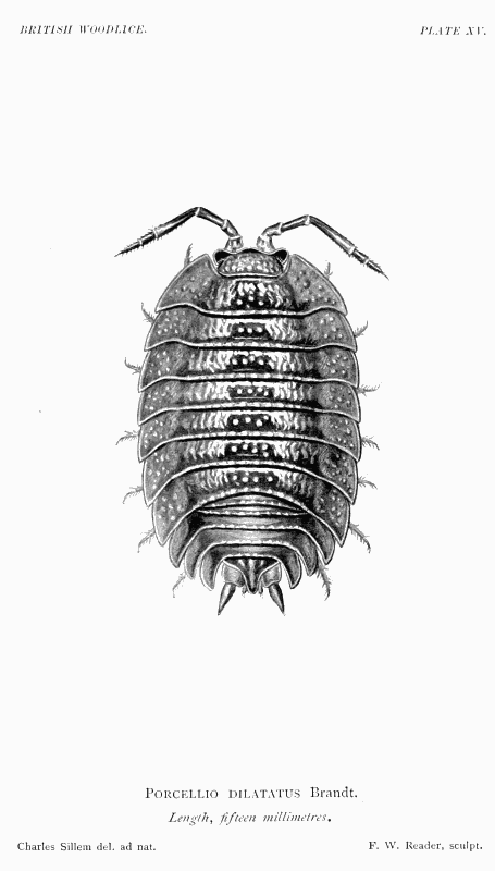 Porcellio dilatatus Brandt.
BRITISH WOODLICE.
PLATE XVI.
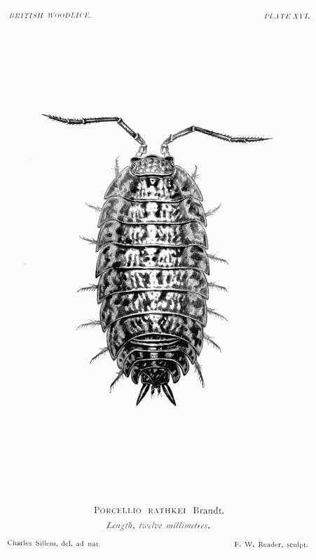 Porcellio rathkei Brandt.
BRITISH WOODLICE.
PLATE XVII.
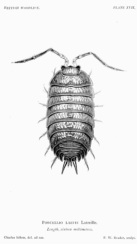 Porcellio laevis Latreille.
BRITISH WOODLICE.
PLATE XVIII.
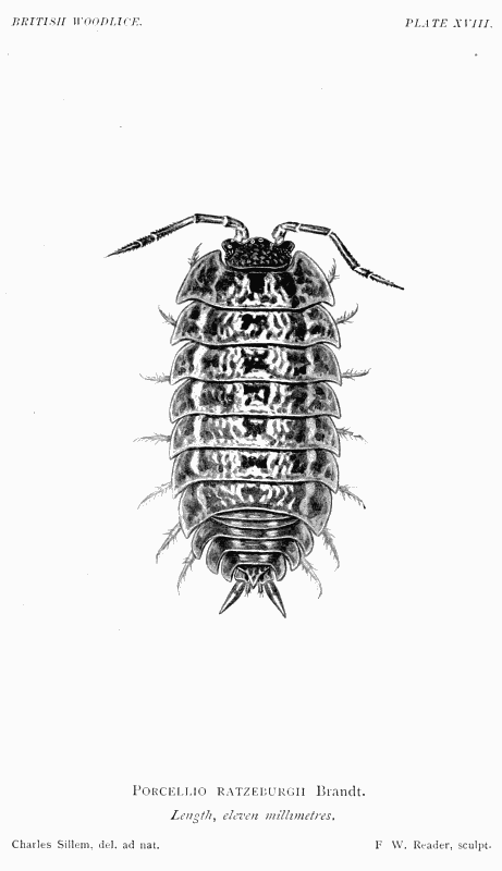 Porcellio ratzeburgii Brandt.
BRITISH WOODLICE.
PLATE XIX.
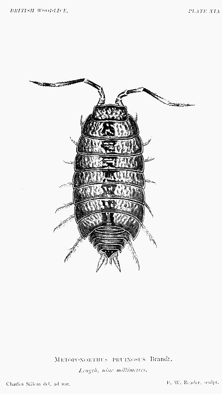 Metoponorthus pruinosus Brandt.
BRITISH WOODLICE.
PLATE XX.
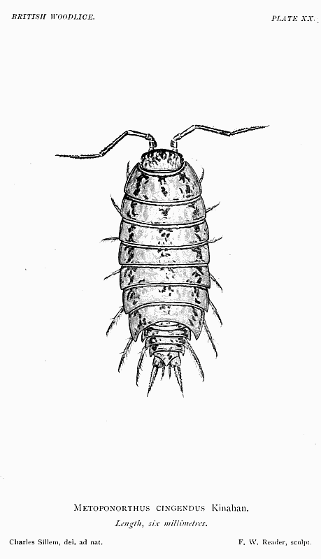 Metoponorthus cingendus Kinahan.
BRITISH WOODLICE.
PLATE XXI.
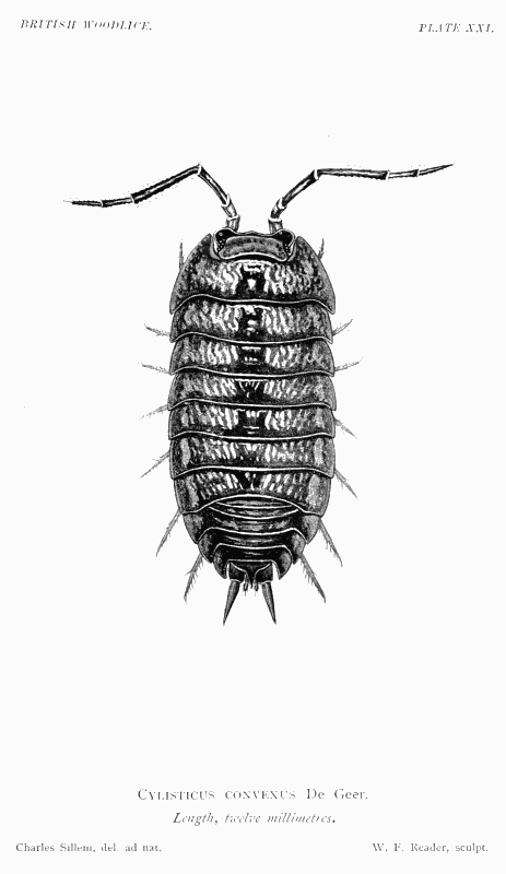 Cylisticus convexus De Geer.
BRITISH WOODLICE.
PLATE XXII.
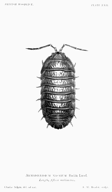 Armadillidium nasatum Budde-Lund.
BRITISH WOODLICE.
PLATE XXIII.
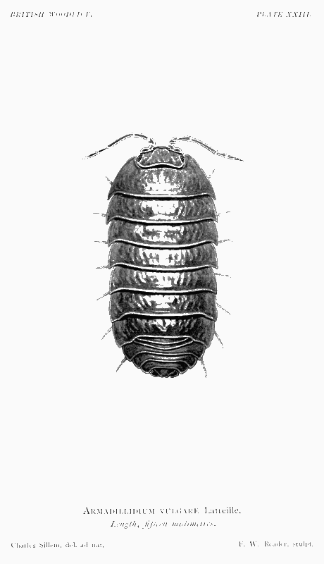 Armadillidium vulgare Latreille.
BRITISH WOODLICE.
PLATE XXIV.
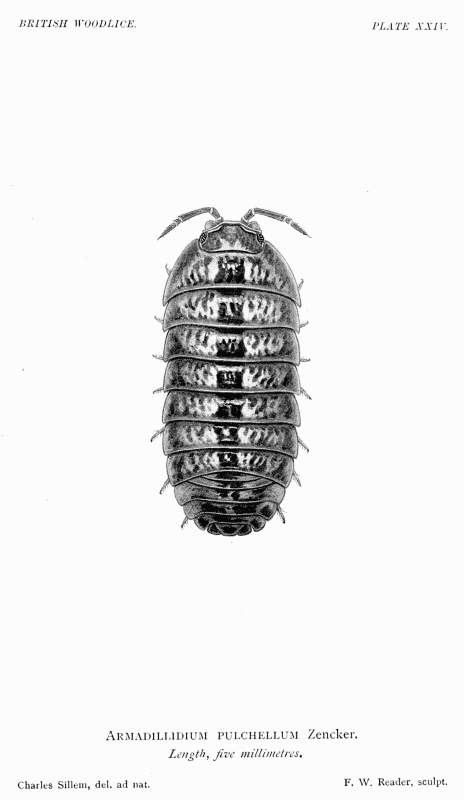 Armadillidium pulchellum Zencker.
BRITISH WOODLICE.
PLATE XXV.
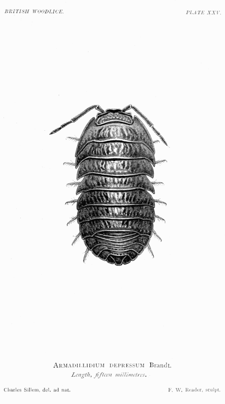 Armadillidium depressum Brandt.
Misprints and misspellings noted or corrected in the text:
Page vii [CONTENTS]: "Family--Ligidæ " changed to "Family--Ligiidæ".
Page vii [CONTENTS]: "Haplopthalmus mengii" changed to "Haplophthalmus mengii".
Page viii [CONTENTS]: "Armydillidium vulgare" changed to "Armadillidium vulgare".
Page 2: "Aegidae is found" changed to "Aegidae which is found".
Page 16: "naptha" changed to "naphtha" in footnote.
Page 28: "Faroe Islands; Thornsharn;" changed to "Faroe Islands; Thorsharn;".
Page 29: "1833 Philoscia marmorala ...", changed to "1833 Philoscia marmorata ..."
Page 30: Missing page ref. at end of "1868 Philoscia couchii Bate and Westwood (1), p."
Page 33: Missing page ref. at end of "1868 Porcellio pictus Bate and Westwood (1), p."
Page 33: Missing page ref. at end of "1868 Porcellio dilatatus Bate and Westwood (1), p."
Page 51 [INDEX]: "Ligidae, characters of" changed to "Ligiidae, characters of".