There is a single footnote, which has been rendered using the original asterisk. The footnote itself has been placed after the paragraph where it is referenced. Illustrations have been re-positioned slightly.
Please see the detailed notes at the end of this text for details about the few corrections that were made during it’s preparation.
For the reader’s convenience, links have been added to the text for references to figures and pages not in the immediate vicinity.
The cover image has been fabricated and is placed in the public domain.
THE OUT-DOOR WORLD SERIES.
THE OUT-DOOR WORLD; or, the Young Collector’s Handbook.
By W. S. Furneaux. With 18 Plates (16 of which are Coloured), and 549
Illustrations in the Text. Crown 8vo, 6s. 6d. net.
FIELD AND WOODLAND PLANTS.
By W. S. Furneaux. With 8 Plates in Colour, and numerous other
Illustrations by Patten Wilson, and from Photographs. Crown 8vo, 6s. 6d.
net.
BRITISH BUTTERFLIES AND MOTHS.
By W. S. Furneaux. With 12 Coloured Plates and 241 Illustrations in the
Text. Crown 8vo, 6s. 6d. net.
LIFE IN PONDS AND STREAMS.
By W. S. Furneaux. With 8 Coloured Plates and 331 Illustrations in the
Text. Crown 8vo, 6s. 6d. net.
THE SEA SHORE. By W. S. Furneaux.
With 8 Coloured Plates and over 300 Illustrations in the Text. Crown
8vo, 6s. 6d. net.
BRITISH BIRDS. By W. H. Hudson.
With a Chapter on Structure and Classification by Frank E. Beddard,
F.R.S. With 16 Plates (8 of which are Coloured), and 103 Illustrations
in the Text. Crown 8vo, 6s. 6d. net.
LONGMANS, GREEN & CO., 39 Paternoster Row, London, E.C.4 New York, Toronto, Bombay, Calcutta and Madras.
THE SEA SHORE
BY
W. S. FURNEAUX
AUTHOR OF
‘THE OUTDOOR WORLD’ ‘BRITISH BUTTERFLIES AND MOTHS’
‘LIFE IN PONDS AND STREAMS’ ETC.

WITH EIGHT PLATES IN COLOUR
AND OVER THREE HUNDRED ILLUSTRATIONS IN THE
TEXT
NEW IMPRESSION
LONGMANS, GREEN AND CO.
39 PATERNOSTER ROW, LONDON, E.C.4
NEW YORK, TORONTO
BOMBAY, CALCUTTA AND MADRAS
1922
All rights reserved
BIBLIOGRAPHICAL NOTE.
First published in September, 1903.
Re-issue at Cheaper Price, July, 1911.
New Impression, November, 1922.
Made in Great Britain
To sea-side naturalists it must be a matter of great surprise that of the inhabitants of our coast towns and villages, and of the pleasure-seekers that swarm on various parts of the coast during the holiday season, so few take a real interest in the natural history of the shore. The tide flows and ebbs and the restless waves incessantly roll on the beach without arousing a thought as to the nature and cause of their movements. The beach itself teems with peculiar forms of life that are scarcely noticed except when they disturb the peace of the resting visitor. The charming vegetation of the tranquil rock-pool receives but a passing glance, and the little world of busy creatures that people it are scarcely observed; while the wonderful forms of life that inhabit the sheltered nooks of the rugged rocks between the tide-marks are almost entirely unknown except to the comparatively few students of Nature. So general is this apparent lack of interest in the things of the shore that he who delights in the study of littoral life and scenes but seldom meets with a kindred spirit while following his pursuits, even though the crowded beach of a popular resort be situated in the immediate neighbourhood of his hunting ground. The sea-side cottager is too accustomed to the shore to suppose that he has anything to learn concerning it, and this familiarity leads, if not to contempt, most certainly to a disinclination to observe closely; and the visitor from town often considers himself to be too much in need of his hard-earned rest to undertake anything that may seem to require energy of either mind or body.
Let both, however, cast aside any predisposition to look upon the naturalist’s employment as arduous and toilsome, and make up their minds to look enquiringly into the living world around them, and they will soon find that they are led onward from the study of one object to another, the employment becoming more and more fascinating as they proceed.
Our aim in writing the following pages is to encourage the observation of the nature and life of the sea shore; to give such assistance to the beginner as will show him where the most interesting objects are to be found, and how he should set to work to obtain them. Practical hints are also furnished to enable the reader to successfully establish and maintain a salt-water aquarium for the observation of marine life at home, and to preserve various marine objects for the purpose of forming a study-collection of the common objects of the shore.
To have given a detailed description of all such objects would have been impossible in a work of this size, but a large number have been described and figured, and the broad principles of the classification of marine animals and plants have been given such prominence that, it is hoped, even the younger readers will find but little difficulty in determining the approximate positions, in the scale of life, of the various living things that come within their reach.
Of the many illustrations, which must necessarily greatly assist the reader in understanding the structure of the selected types and in the identification of the different species, a large number have been prepared especially for this work.
| CHAPTER | PAGE | |
| I. | THE GENERAL CHARACTERISTICS OF THE SEA SHORE | 1 |
| II. | THE SEA-SIDE NATURALIST | 21 |
| III. | SEA ANGLING | 34 |
| IV. | THE MARINE AQUARIUM | 51 |
| V. | THE PRESERVATION OF MARINE OBJECTS | 71 |
| VI. | EXAMINATION OF MARINE OBJECTS—DISSECTION | 91 |
| VII. | THE PROTOZOA OF THE SEA SHORE | 102 |
| VIII. | BRITISH SPONGES | 115 |
| IX. | THE CŒLENTERATES—JELLY-FISHES, ANEMONES, AND THEIR ALLIES | 127 |
| X. | STARFISHES, SEA URCHINS, ETC. | 157 |
| XI. | MARINE WORMS | 172 |
| XII. | MARINE MOLLUSCS | 190 |
| XIII. | MARINE ARTHROPODS | 256 |
| XIV. | MARINE VERTEBRATES | 306 |
| XV. | SEA WEEDS | 343 |
| XVI. | THE FLOWERING PLANTS OF THE SEA-SIDE | 391 |
| INDEX | 425 |
Drawn by Mr. Robert Lillie and reproduced by Messrs. André & Sleigh, Ltd., Bushey.
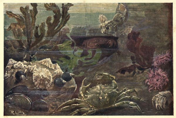
| Plate I—A ROCK-POOL | Frontispiece |
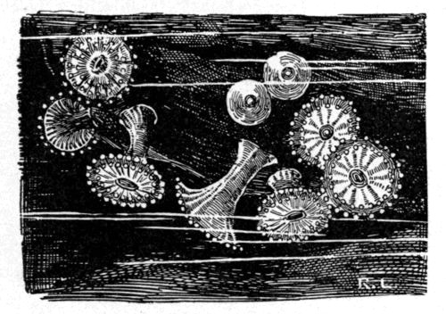
| Plate II—SEA ANEMONES | To face p. 142 |
| 1, 2, 3. Actinia mesembryanthemum. 4. Caryophyllia Smithii. 5. Tealia crassicornis. |
6. Sagartia bellis. 7. Balanophyllia regia. 8. Actinoloba dianthus. |
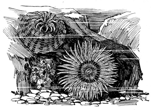
| Plate III—SEA ANEMONES | To face p. 150 |
| 1. Sagartia troglodytes. 2. ” venusta. 3. Actinia glauca. 4. ” chiococca. |
5. Bunodes Ballii. 6. ” gemmacea. 7. Anthea cereus. 8. Sagartia rosea. |
| Plate IV—ECHINODERMS | To face p. 168 |
| 1. Asterias rubens. 2. Goniaster equestris. 3. Ophiothrix fragilis. |
4. Echinocardium cordatum. 5. Echinus miliaris. 6. ” esculentus. |
| Plate V—MOLLUSCS | To face p. 222 |
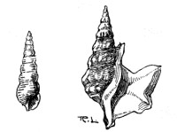
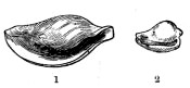
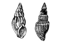
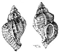
| 1. Solen ensis. 2. Trivia europæa. 3. Trochus umbilicatus. 4. ” magnus. 5. Littorina littorea. 6. ” rudis. 7. Haminea (Bulla) hydatis. 8. Tellina. |
9. Capulus (Pileopsis) hungaricus. 10. Chrysodomus (Fusus) antiquus. 11. Buccinum undatum. 12, 13. Scalaria communis. 14. Pecten opercularis. 15. ” varius. 16. ” maximus. |
| Plate VI—CRUSTACEA | To face p. 290 |
| 1. Gonoplax angulata. 2. Xantho florida. 3. Portunus puber. |
4. Polybius Henslowii. 5. Porcellana platycheles. |
| Plate VII—SEAWEEDS | To face p. 354 |
| 1. Fucus nodosus. 2. Nitophyllum laceratum. 3. Codium tomentosum. |
4. Padina pavonia. 5. Porphyra laciniata (vulgaris). |
| Plate VIII—SEAWEEDS | To face p. 384 |
| 1. Chorda filum. 2. Fucus vesiculosus. 3. ” canaliculatus. 4. Delesseria (Maugeria) sanguinea. |
5. Rhodymenia palmata. 6. Chondrus crispus. 7. Ulva lactuca. |
| FIG. | PAGE | |
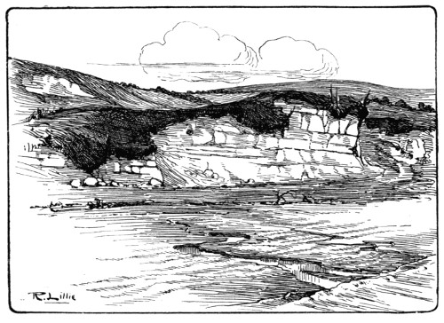
| 1. | Chalk Cliff | 3 |
| 2. | Whitecliff (Chalk), Dorset | 4 |
| 3. | Penlee Point, Cornwall | 5 |
| 4. | Balanus Shells | 6 |
| 5. | A Cluster of Mussels | 7 |
| 6. | Breakers | 8 |
| 7. | Illustrating the Tide-producing Influence of the Moon | 10 |
| 8. | Illustrating the tides | 11 |
| 9. | Spring Tides at Full Moon | 12 |
| 10. | Spring Tides at New Moon | 12 |
| 11. | Neap Tides | 13 |
| 12. | Chart showing the relative Times of High Tide on different parts of the British Coast | 16 |
| 13. | The Vasculum | 22 |
| 14. | Wire Ring for Net | 24 |
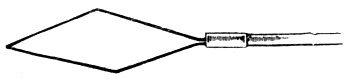
| 15. | Net Frame with Curved Point | 24 |
| 16. | Rhomboidal Frame for Net | 24 |
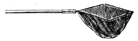
| 17. | Rhomboidal Net | 25 |
| 18. | Semicircular Net | 25 |
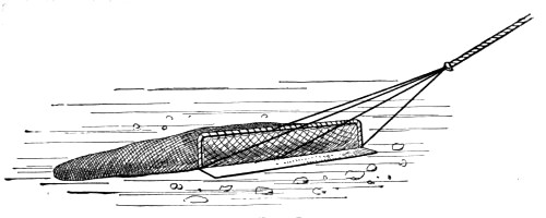
| 19. | The Dredge | 25 |
| 20. | The Crab-pot | 26 |
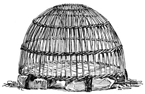
| 21. | An old Bird-cage used as a Crab-pot | 27 |
| 22. | A Young Naturalist at Work | 32 |
| 23. | A good Hunting-ground on the Cornish Coast | 33 |
| 24. | Round Bend Hook with Flattened End | 37 |
| 25. | Limerick Hook, eyed | 37 |
| 26. | Method of Attaching Snood to Flattened Hook | 38 |
| 27. | Method of Attaching Snood to Eyed Hook | 38 |
| 28. | The Lugworm | 39 |
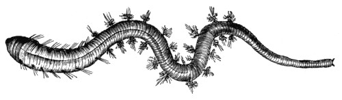
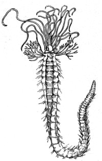
| 29. | The Ragworm | 40 |
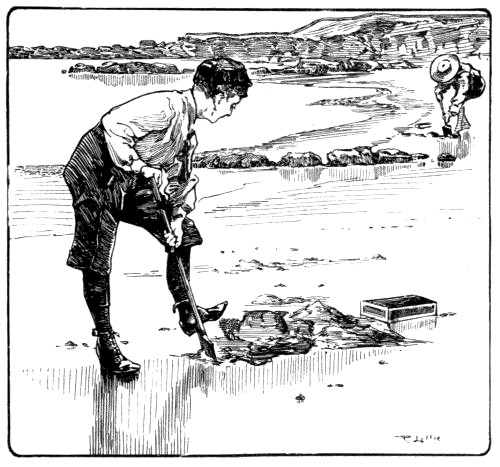
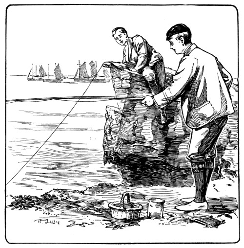
| 30. | Digging for Bait | 41 |
| 31. | Method of Opening a Mussel | 42 |
| 32. | Fishing from the Rocks | 46 |
| 33. | The Paternoster | 48 |
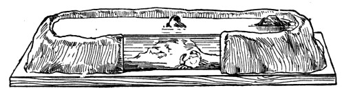
| 34. | Section of an Aquarium constructed with a Mixture of Cement and Sand | 54 |
| 35. | Cement Aquarium with a Glass Plate in Front | 55 |
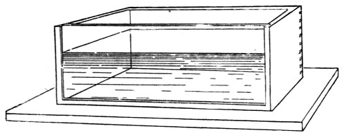
| 36. | Aquarium of Wood with Glass Front | 56 |
| 37. | Hexagonal Aquarium constructed of Angle Zinc, with Glass Sides | 57 |
| 38. | Method of Aerating the Water of an Aquarium | 65 |
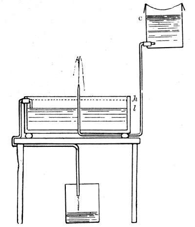
| 39. | Aquarium fitted with Apparatus for Periodic Outflow | 67 |
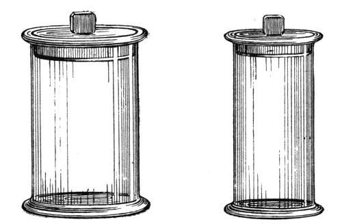
| 40. | Jars for preserving Anatomical and Biological Specimens | 76 |
| 41. | Showing the different stages in the making of a small Specimen Tube | 77 |
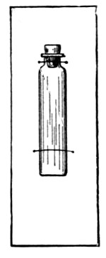
| 42. | Small Specimen Tube mounted on a Card | 78 |
| 43. | Small Crab mounted on a Card | 82 |
| 44. | Spring for holding together small Bivalve Shells | 84 |
| 45. | The Triplet Magnifier | 92 |
| 46. | A small Dissecting Trough | 93 |
| 47. | Cell for small Living Objects | 95 |
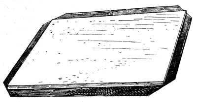
| 48. | Sheet of Cork on thin Sheet Lead | 99 |
| 49. | Weighted Cork for Dissecting Trough | 99 |
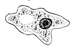
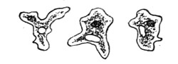
| 50. | The Amœba, highly magnified | 102 |
| 51. | ” ” showing changes of form | 103 |
| 52. | ” ” feeding | 103 |
| 53. | ” ” dividing | 104 |
| 54. | A Group of Foraminifers, magnified | 105 |
| 55. | A Spiral Foraminifer Shell | 106 |
| 56. | A Foraminifer out of its Shell | 106 |
| 57. | The same Foraminifer (fig. 56) as seen when alive | 107 |
| 58. | Section of the Shell of a Compound Foraminifer | 107 |
| 59. | Section of a Nummulite Shell | 108 |
| 60. | Globigerina bulloides, as seen when alive, magnified | 108 |
| 61. | Section of a piece of Nummulitic Limestone | 109 |
| 62. | A Group of Radiolarian Shells, magnified | 111 |
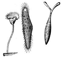
| 63. | Three Infusorians, magnified | 113 |
| 64. | A Phosphorescent Marine Infusorian (Noctiluca), magnified | 114 |
| 65. | Section of a Simple Sponge | 116 |
| 66. | Diagrammatic section of a portion of a Complex Sponge | 117 |
| 67. | Horny Network of a Sponge, magnified | 118 |
| 68. | Grantia compressa | 120 |
| 69. | Spicules of Grantia, magnified | 120 |
| 70. | Sycon ciliatum | 121 |
| 71. | Leucosolenia botryoides, with portion magnified | 121 |
| 72. | Chalina oculata | 122 |
| 73. | Halichondria panicea | 123 |
| 74. | Spicules of Halichondria, magnified | 124 |
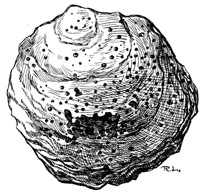
| 75. | An Oyster Shell, bored by Cliona | 124 |
| 76. | Spicules of Cliona | 125 |
| 77. | Thread Cells of a Cœlenterate, magnified | 127 |
| 78. | The Squirrel’s-tail Sea Fir (Sertularia argentea), with a portion enlarged | 128 |
| 79. | Sertularia filicula | 129 |
| 80. | ” cupressina | 130 |
| 81. | The Herring-bone Polype (Halecium halecinum | 131 |
| 82. | Tubularia indivisa | 132 |
| 83. | The Bottle Brush (Thuiaria thuja) | 132 |
| 84. | Antennularia antennia | 133 |
| 85. | Aurelia aurita | 135 |
| 86. | The Early Stages of Aurelia | 136 |
| 87. | Rhizostoma | 136 |
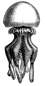
| 88. | Chrysaora | 136 |
| 89. | Cydippe pileus | 137 |
| 90. | Section of an Anemone | 139 |
| 91. | Stinging Cells of Anemone, highly magnified | 140 |
| 92. | Diagrammatic transverse section of an Anemone | 140 |
| 93. | Larva of Anemone | 140 |
| 94. | The Trumpet Anemone (Aiptasia Couchii), Cornwall; deep water | 144 |
| 95. | Peachia hastata, S. Devon | 145 |
| 96. | Sagartia pallida, Devon and Cornwall | 146 |
| 97. | Sagartia nivea, Devon and Cornwall | 147 |
| 98. | Corynactus viridis, Devon and Cornwall | 148 |
| 99. | Bunodes thallia, West Coast | 150 |
| 100. | Bunodes gemmacea, with tentacles retracted | 151 |
| 101. | Caryophyllia cyathus | 152 |
| 102. | Sagartia parasitica | 153 |
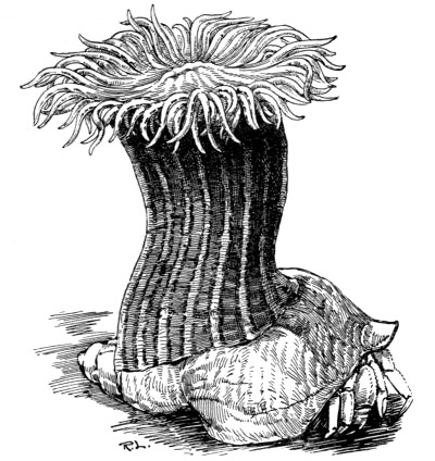
| 103. | The Cloak Anemone (Adamsia palliata) on a Whelk Shell, with Hermit Crab | 154 |
| 104. | Larva of the Brittle Starfish | 158 |
| 105. | Larva of the Feather Star | 160 |
| 106. | The Rosy Feather Star | 160 |
| 107. | The Common Brittle Star | 162 |
| 108. | Section of the Spine of a Sea Urchin | 165 |
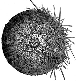
| 109. | Sea Urchin with Spines removed on one Side | 166 |
| 110. | Apex of Shell of Sea Urchin | 166 |
| 111. | Shell of Sea Urchin with Teeth protruding | 167 |
| 112. | Interior of Shell of Sea Urchin | 167 |
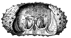
| 113. | Masticatory Apparatus of Sea Urchin | 167 |
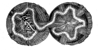
| 114. | Sea Urchin Dissected, showing the Digestive Tube | 168 |
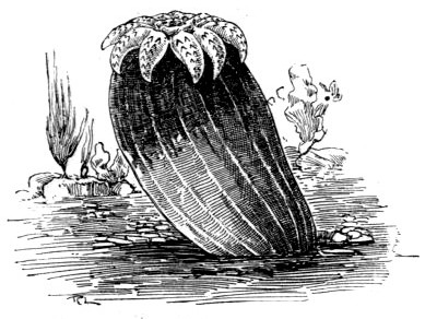
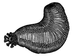
| 115. | The Sea Cucumber | 170 |
| 116. | A Turbellarian, magnified | 175 |
| 117. | Arenicola piscatorum | 178 |
| 118. | The Sea Mouse | 179 |
| 119. | Tube-building Worms: Terebella, Serpula, Sabella | 182 |
| 120. | Terebella removed from its Tube | 183 |
| 121. | A tube of Serpula attached to a Shell | 185 |
| 122. | Serpula removed from its Tube | 186 |
| 123. | The Sea Mat (Flustra) | 187 |
| 124. | Flustra in its Cell, magnified | 188 |
| 125. | Sea Squirt | 189 |
| 126. | Larvæ of Molluscs | 191 |
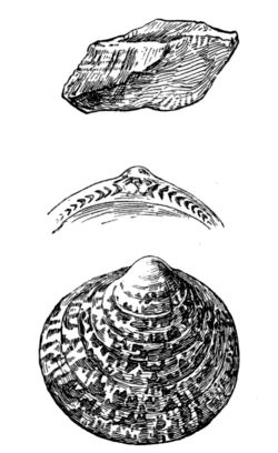
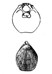
| 127. | Shell of the Prickly Cockle (Cardium aculeatum) showing Umbo and Hinge; also the interior showing the Teeth | 192 |
| 128. | Interior of Bivalve Shell, showing Muscular Scars and Pallial Line | 193 |
| 129. | Diagram of the Anatomy of a Lamellibranch | 194 |
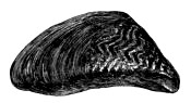
| 130. | Mytilus edulis, with Byssus | 195 |
| 131. | A Bivalve Shell (Tapes virgineana) | 196 |
| 132. | Pholas dactylus | 199 |
| 133. | ” ” interior of Valve; and Pholadidea with Animal | 201 |
| 134. | The Ship Worm | 202 |
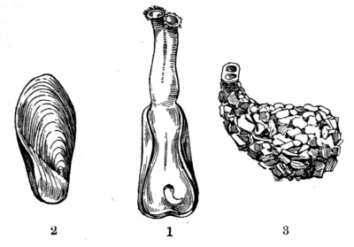
| 135. | 1. Teredo navalis. 2. Teredo norvegica | 202 |
| 136. | Gastrochæna modiolina | 203 |
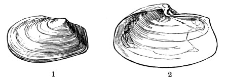
| 137. | 1. Thracia phaseolina. 2. Thracia pubescens, showing Pallial Line | 204 |
| 138. | 1. Mya truncata. 2. Interior of Shell. 3. Mya arenaria. 4. Corbula nucleus | 205 |
| 139. | Solen siliqua | 206 |
| 140. | 1. Solen ensis. 2. Cerati-solen legumen. 3. Solecurtus candidus | 207 |
| 141. | Tellinidæ | 208 |
| 142. | 1. Lutraria elliptica. 2. Part of the Hinge of Lutraria, showing the Cartilage Pit. 3. Macra stultorum. 4. Interior of same showing Pallial Line | 210 |
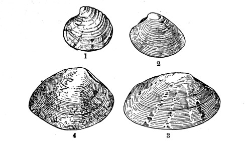
| 143. | Veneridæ | 211 |
| 144. | Cyprinidæ | 213 |
| 145. | Galeomma Turtoni | 214 |
| 146. | 1. Cardium pygmæum. 2. Cardium fasciatum. 3. Cardium rusticum | 215 |
| 147. | Cardium aculeatum | 215 |
| 148. | Pectunculus glycimeris, with portion of Valve showing Teeth, and Arca tetragona | 216 |
| 149. | Mytilus edulis | 217 |
| 150. | 1. Modiola modiolus. 2. Modiola tulipa. 3. Crenella discors | 218 |
| 151. | Dreissena polymorpha | 219 |
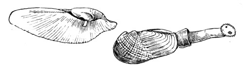
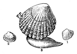
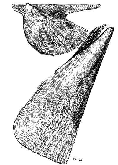
| 152. | Avicula, and Pinna pectinata | 220 |
| 153. | 1. Anomia ephippium. 2. Pecten tigris. 3. Pecten, animal in shell | 222 |
| 154. | Terebratulina. The upper figure represents the interior of the Dorsal Valve | 224 |
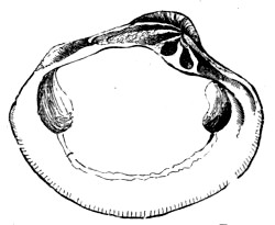
| 155. | Under side of the Shell of Natica catena, showing the Umbilicus; and outline of the Shell, showing the Right-handed Spiral | 225 |
| 156. | Section of the Shell of the Whelk, showing the Columella | 226 |
| 157. | Diagram of the Anatomy of the Whelk, the Shell being removed | 228 |
| 158. | A portion of the Lingual Ribbon of the Whelk, magnified; and a single row of Teeth on a much larger scale | 229 |
| 159. | Egg Cases of the Whelk | 230 |
| 160. | Pteropods | 231 |
| 161. | Nudibranchs | 234 |
| 162. | ” | 235 |
| 163. | Shells of Tectibranchs | 236 |
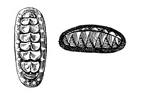
| 164. | Chiton Shells | 238 |
| 165. | Shells of Dentalium | 238 |
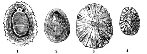
| 166. | Patellidæ | 239 |
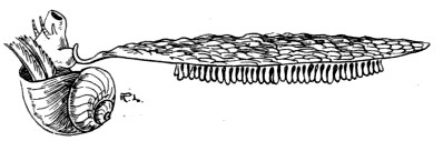
| 167. | Calyptræa sinensis | 241 |
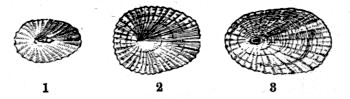
| 168. | Fissurellidæ | 241 |
| 169. | Haliotis | 242 |
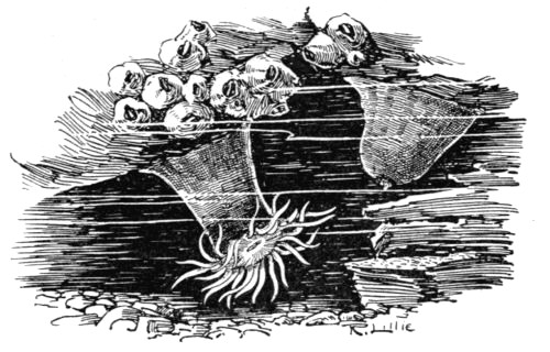
| 170. | Ianthina fragilis | 242 |
| 171. | Trochus zizyphinus. 2. Under Side of Shell. 3. Trochus magnus. 4. Adeorbis subcarinatus | 244 |
| 172. | Rissoa labiosa and Lacuna pallidula | 244 |
| 173. | Section of Shell of Turritella | 245 |
| 174. | Turritella communis and Cæcum trachea | 245 |
| 175. | Cerithium reticulatum and Aporrhais pes-pelicani | 245 |
| 176. | Aporrhais pes-pelicani, showing both Shell and Animal | 246 |
| 177. | 1. Odostomia plicata. 2. Eulima polita. 3. Aclis supranitida | 246 |
| 178. | Cypræa (Trivia) europæa | 247 |
| 179. | 1. Ovulum patulum. 2. Erato lævis | 248 |
| 180. | Mangelia septangularis and Mangelia turricula | 248 |
| 181. | 1. Purpura lapillus. 2. Egg Cases of Purpura. 3. Nassa reticulata | 249 |
| 182. | Murex erinaceus | 249 |
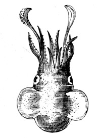
| 183. | Octopus | 251 |
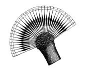
| 184. | Loligo vulgaris and its Pen | 252 |
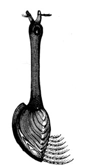
| 185. | Sepiola atlantica | 252 |
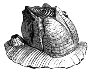
| 186. | Sepia officinalis and its ‘Bone’ | 253 |
| 187. | Eggs of Sepia | 254 |
| 188. | The Nerve-chain of an Arthropod (Lobster) | 257 |
| 189. | Section through the Compound Eye of an Arthropod | 260 |
| 190. | Four Stages in the Development of the Common Shore Crab | 261 |
| 191. | The Barnacle | 261 |
| 192. | Four Stages in the Development of the Acorn Barnacle | 262 |
| 193. | A Cluster of Acorn Shells | 263 |
| 194. | Shell of Acorn Barnacle (Balanus) | 263 |
| 195. | The Acorn Barnacle (Balanus porcatus) with Appendages protruded | 264 |
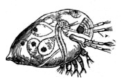
| 196. | A Group of Marine Copepods, magnified | 265 |
| 197. | A Group of Ostracode Shells | 265 |
| 198. | Evadne | 266 |
| 199. | Marine Isopods | 267 |
| 200. | Marine Amphipods | 268 |
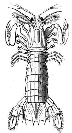
| 201. | The Mantis Shrimp (Squilla Mantis) | 270 |
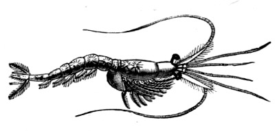
| 202. | The Opossum Shrimp (Mysis chamæleon) | 271 |
| 203. | Parts of Lobster’s Shell, separated, and viewed from above | 272 |
| 204. | A Segment of the Abdomen of a Lobster | 272 |
| 205. | Appendages of a Lobster | 273 |
| 206. | Longitudinal Section of the Lobster | 274 |
| 207. | The Spiny Lobster (Palinurus vulgaris) | 275 |
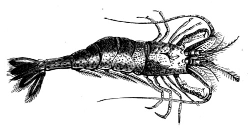
| 208. | The Norway Lobster (Nephrops norvegicus) | 276 |
| 209. | 1. The Mud-borer (Gebia stellata). 2. The Mud-borrower (Callianassa subterranea) | 277 |
| 210. | The Common Shrimp (Crangon vulgaris) | 278 |
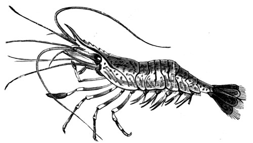
| 211. | The Prawn (Palæmon serratus) | 279 |
| 212. | Dromia vulgaris | 282 |
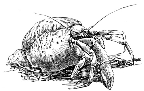
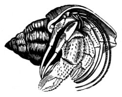
| 213. | The Hermit Crab in a Whelk Shell | 282 |
| 214. | The Long-armed Crab (Corystes Cassivelaunus) | 287 |
| 215. | Spider Crabs at Home | 288 |
| 216. | The Thornback Crab (Maia Squinado) | 290 |
| 217. | The Pea Crab (Pinnotheres pisum) | 290 |
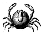
| 218. | The Common Shore Crab (Carcinus mænas) | 291 |
| 219. | The Shore Spider | 294 |
| 220. | The Leg of an Insect | 295 |
| 221. | Trachea of an Insect, magnified | 296 |
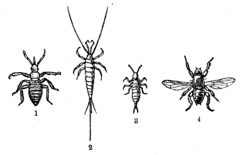
| 222. | Sea-Shore Insects | 298 |
| 223. | Marine Beetles of the genus (Bembidium) | 302 |
| 224. | Marine Beetles | 303 |
| 225. | Transverse section through the Bony Framework of a Typical Vertebrate Animal | 306 |
| 226. | The Sea Lamprey | 309 |
| 227. | The Pilchard | 310 |
| 228. | The Skeleton of a Fish (Perch) | 315 |
| 229. | The Internal Organs of the Herring | 316 |
| 230. | The Egg-case of the Dogfish | 319 |
| 231. | The Smooth Hound | 320 |
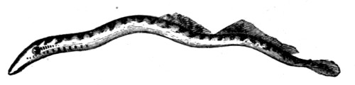
| 232. | The Common Eel | 323 |
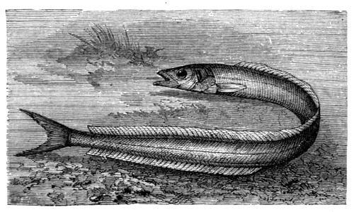
| 233. | The Lesser Sand Eel | 326 |
| 234. | The Three-bearded Rockling | 327 |
| 235. | The Snake Pipe-fish | 328 |
| 236. | The Rainbow Wrass (Labrus julis) | 330 |
| 237. | The Cornish Sucker | 330 |
| 238. | The Fifteen-spined Stickleback and Nest | 331 |
| 239. | The Smooth Blenny | 333 |
| 240. | The Butterfish | 334 |
| 241. | The Black Goby | 335 |
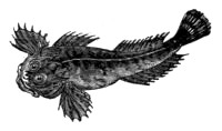
| 242. | The Father Lasher | 335 |
| 243. | The Lesser Weaver | 337 |
| 244. | The Common Porpoise | 341 |
| 245. | Callithamnion roseum | 359 |
| 246. | Callithamnion tetricum | 359 |
| 247. | Griffithsia corallina | 361 |
| 248. | Halurus equisetifolius | 361 |
| 249. | Pilota plumosa | 361 |
| 250. | Ceramium diaphanum | 363 |
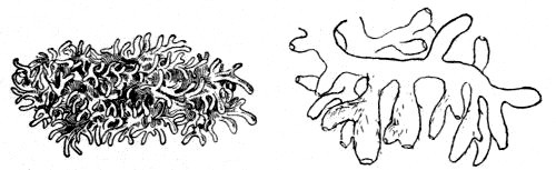
| 251. | Plocamium | 366 |
| 252. | Delesseria alata | 368 |
| 253. | Delesseria hypoglossum | 368 |
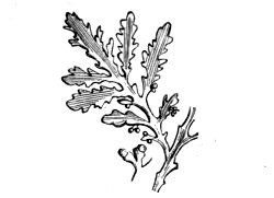
| 254. | Laurencia pinnatifida | 371 |
| 255. | Laurencia obtusa | 371 |
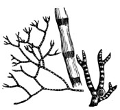
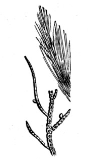
| 256. | Polysiphonia fastigiata | 373 |
| 257. | Polysiphonia parasitica | 374 |
| 258. | Polysiphonia Brodiæi | 374 |
| 259. | Polysiphonia nigrescens | 374 |
| 260. | Ectocarpus granulosus | 378 |
| 261. | Ectocarpus siliculosus | 378 |
| 262. | Ectocarpus Mertensii | 378 |
| 263. | Sphacelaria cirrhosa | 379 |
| 264. | Sphacelaria plumosa | 379 |
| 265. | Sphacelaria radicans | 380 |
| 266. | Cladostephus spongiosus | 380 |
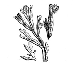
| 267. | Chordaria flagelliformis | 380 |
| 268. | Laminaria bulbosa | 384 |
| 269. | Laminaria saccharina | 384 |
| 270. | Alaria esculenta | 385 |
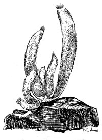
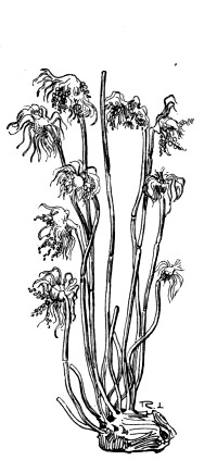
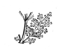
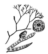
| 271. | Sporochnus pedunculatus | 385 |
| 272. | Desmarestia ligulata | 386 |
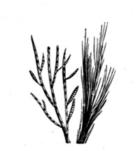
| 273. | Himanthalia lorea | 387 |
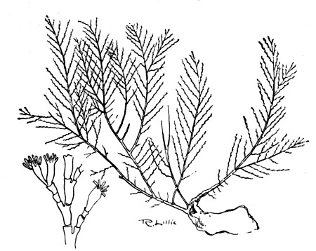
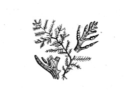
| 274. | Cystoseira ericoides | 388 |
| 275. | Transverse Section of the Stem of a Monocotyledon | 391 |
| 276. | Leaf of a Monocotyledon | 392 |
| 277. | Expanded Spikelet of the Oat | 393 |
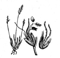
| 278. | The Sea Lyme Grass | 395 |
| 279. | Knappia agrostidea | 397 |
| 280. | The Dog’s-tooth Grass | 397 |
| 281. | The Reed Canary Grass | 397 |
| 282. | Male and Female Flowers of Carex, magnified | 399 |
| 283. | The Sea Sedge | 400 |
| 284. | The Curved Sedge | 400 |
| 285. | The Great Sea Rush | 400 |
| 286. | The Broad-leaved Grass Wrack | 401 |
| 287. | The Sea-side Arrow Grass | 401 |
| 288. | The Common Asparagus | 401 |
| 289. | The Sea Spurge | 403 |
| 290. | The Purple Spurge | 404 |
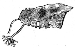
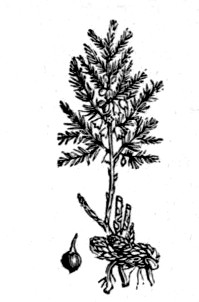
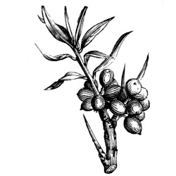
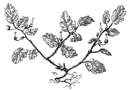
| 291. | The Sea Buckthorn | 404 |
| 292. | Chenopodium botryoides | 405 |
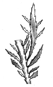
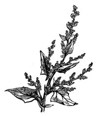
| 293. | The Frosted Sea Orache | 406 |
| 294. | The Prickly Salt Wort | 406 |
| 295. | The Creeping Glass Wort | 407 |
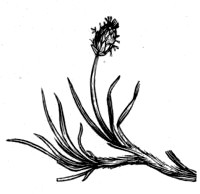
| 296. | The Sea-side Plantain | 408 |
| 297. | The Sea Lavender | 408 |
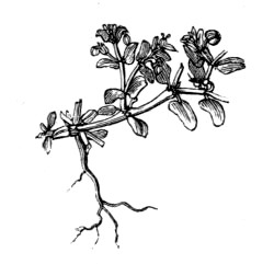
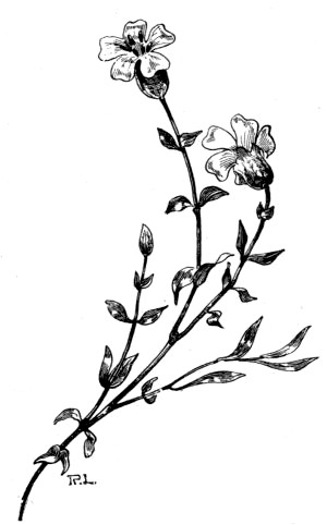
| 298. | The Dwarf Centaury | 410 |
| 299. | The Sea Samphire | 412 |
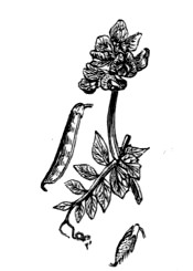
| 300. | The Sea-side Everlasting Pea | 413 |
| 301. | The Sea Stork’s-bill | 414 |
| 302. | The Sea Campion | 416 |
| 303. | The Sea Pearl Wort | 417 |
| 304. | The Shrubby Mignonette | 417 |
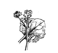
| 305. | The Wild Cabbage | 418 |
| 306. | The Isle of Man Cabbage | 418 |
| 307. | The Great Sea Stock | 419 |
| 308. | The Hoary Shrubby Stock | 419 |
| 309. | The Scurvy Grass | 419 |
| 310. | The Sea Radish | 419 |
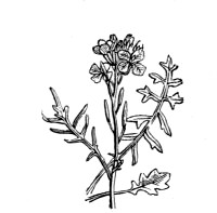
| 311. | The Sea Rocket | 420 |
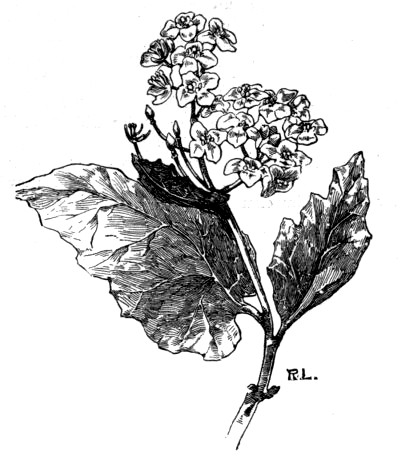
| 312. | The Sea Kale | 421 |
| 313. | The Horned Poppy | 422 |
THE SEA SHORE
What are the attractions which so often entice us to the sea shore, which give such charm to a ramble along the cliffs or the beach, and which will so frequently constrain the most active wanderer to rest and admire the scene before him? The chief of these attractions is undoubtedly the incessant motion of the water and the constant change of scene presented to his view. As we ramble along a beaten track at the edge of the cliff, new and varied features of the coast are constantly opening up before us. Each little headland passed reveals a sheltered picturesque cove or a gentle bay with its line of yellow sands backed by the cliffs and washed by the foaming waves; while now and again our path slopes down to a peaceful valley with its cluster of pretty cottages, and the rippling stream winding its way towards the sea. On the one hand is the blue sea, full of life and motion as far as the eye can reach, and on the other the cultivated fields or the wild and rugged downs.
The variety of these scenes is further increased by the frequent changes in the character of the cliffs themselves. Where they are composed of soft material we find the coast-line washed into gentle curves, and the beach formed of a continuous stretch of fine sand; but where harder rocks exist the scenery is wild and varied, and the beach usually strewn with irregular masses of all sizes.
Then, when we approach the water’s edge, we find a delight in watching the approaching waves as they roll over the sandy or pebbly beach, or embrace an outlying rock, gently raising its olive covering of dangling weeds.
Such attractions will allure the ordinary lover of Nature—the mere seeker after the picturesque—but to the true naturalist there are many others. The latter loves to read in the cliffs their past history, to observe to what extent the general scenery of the coast is due to the nature of the rocks, and to learn the action of the waves from the character of the cliffs and beach, and from the changes which are known to have taken place in the contour of the land in past years. He also delights to study those plants and flowers which are peculiar to the coast, and to observe how the influences of the sea have produced interesting modifications in certain of our flowering plants, as may be seen by comparing them with the same species from inland districts. The sea birds, too, differing so much as they do from our other feathered friends in structure and habit, provide a new field for study; while the remarkably varied character of the forms of life met with on the beach and in the shallow waters fringing the land is in itself sufficient to supply the most active naturalist with material for prolonged and constant work.
Let us first observe some of the general features of the coast itself, and see how far we can account for the great diversity of character presented to us, and for the continual changes and incessant motions that add such a charm to the sea-side ramble.
Here we stand on the top of a cliff composed of a soft calcareous rock—on the exposed edge of a bed of chalk that extends far inland. All the country round is gently undulating, and devoid of any of the features that make up a wild and romantic scene. The coast-line, too, is wrought into a series of gentle bays, separated by inconspicuous promontories where the rock, being slightly harder, has better withstood the eroding action of the sea; or where a current, washing the neighbouring shore, has been by some force deflected seaward. The cliff, though not high, rises almost perpendicularly from the beach, and presents to the sea a face which is but little broken, and which in itself shows no strong evidence of the action of raging, tempestuous seas; its chief diversity being its gradual rise and fall with each successive undulation of the land. The same soft and gentle nature characterises the beach below. Beyond a few small blocks of freshly-loosened chalk, with here and there a liberated nodule of flint, we find nothing but a continuous, fine, siliceous sand, the surface of which is but seldom broken by the protrusion of masses from below. Such cliffs and beaches do not in themselves suggest any violent action on the part of the sea, and yet it is here that the ocean is enabled to make its destructive efforts with the greatest effect. The soft rock is gradually but surely reduced, partly by the mechanical action of the waves and partly by the chemical action of the sea-water. The rock being almost uniformly soft, it is uniformly worn away, thus presenting a comparatively unbroken face. Its material is gradually dissolved in the sea; and the calcareous matter being thus removed, we have a beach composed of the remains of the flints which have been pulverised by the action of the waves. Thus slowly but surely the sea gains upon the land. Thus it is that many a famous landmark, once hundreds of yards from the coast, now stands so near the edge of the cliff as to be threatened by every storm; or some ancient castle, once miles from the shore, lies entirely buried by the encroaching sea.
The coast we have described is most certainly not the one with the fullest attractions for the naturalist, for the cliffs lack those nooks that provide so much shelter for bird and beast, and the rugged coves and rock pools in which we find such a wonderful variety of marine life are nowhere to be seen. But, although it represents a typical shore for a chalky district, yet we may find others of a very different nature even where the same rock exists. Thus, at Flamborough in Yorkshire, and St. Alban’s Head in Dorset, we find the hardened, exposed edge of the chalk formation terminating in bold and majestic promontories, while the inner edge surrounding the Weald gives rise to the famous cliffs of Dover and the dizzy heights of Beachy Head. The hard chalk of the Isle of Wight, too, which has so well withstood the repeated attacks of the Atlantic waves, presents a bold barrier to the sea on the south and east coasts, and terminates in the west with the majestic stacks of the Needles.
Where this harder chalk exists the coast is rugged and irregular. Sea birds find a home in the sheltered ledges and in the protected nooks of its serrated edge; and the countless wave-resisting blocks of weathered chalk that have been hurled from the heights above, together with the many remnants of former cliffs that have at last succumbed to the attacks of the boisterous sea, all form abundant shelter for a variety of marine plants and animals.
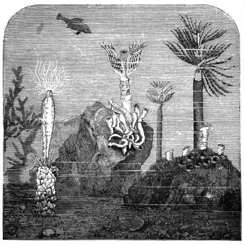
But it is in the west and south-west of our island that we find both the most furious waves and the rocks that are best able to resist their attacks. Here we are exposed to the full force of the frontal attacks of the Atlantic, and it is here that the dashing breakers seek out the weaker portions of the upturned and contorted strata, eating out deep inlets, and often loosening enormous blocks of the hardest material, hurling them on the rugged beach, where they are eventually to be reduced to small fragments by the continual clashing and grinding action of the smaller masses as they are thrown up by the angry sea. Here it is that we find the most rugged and precipitous cliffs, bordering a more or less wild and desolate country, now broken by a deep and narrow chasm where the resonant roar of the sea ascends to the dizzy heights above, and anon stretching seaward into a rocky headland, whose former greatness is marked by a continuation of fantastic outliers and smaller wave-worn masses of the harder strata. Here, too, we find that the unyielding rocks give a permanent attachment to the red and olive weeds which clothe them, and which provide a home for so many inhabitants of our shallow waters. It is here, also, that we see those picturesque rock pools of all sizes, formed by the removal of the softer material of the rocks, and converted into so many miniature seas by the receding of the tide.
A more lovely sight than the typical rock pool of the West coast one can hardly imagine. Around lies the rugged but sea-worn rock, partly hidden by dense patches of the conical shells of the Balanus, with here and there a snug cluster of young mussels held together by their intertwining silken byssi. The surface is further relieved by the clinging limpet, the beautifully banded shells of the variable dog-periwinkle, the pretty top shells, and a variety of other common but interesting molluscs. Clusters of the common bladdery weeds are also suspended from the dry rock, and hang gracefully into the still water below, where the mantled cowry may be seen slowly gliding over the olive fronds. Submerged in the peaceful pool are beautiful tufts of white and pink corallines, among which a number of small and slender starfishes may climb unnoticed by the casual observer; while the scene is brightened by the numerous patches of slender green and red algæ, the thread-like fronds of which are occasionally disturbed as the lively little blenny darts among them to evade the intruder’s glance. Dotted here and there are the beautiful anemones—the variously-hued animal flowers of the sea, with expanded tentacles gently and gracefully swaying, ready to grasp and paralyse any small living being that may wander within their reach. Here, under a projecting ledge of the rock, partly hidden by pale green threads, are the glaring eyes of the voracious bullhead, eager to pounce on almost any moving object; while above it the five-fingered starfish slowly climbs among the dangling weeds by means of its innumerable suckers. In yonder shady corner, where the overhanging rock cuts off all direct rays of the sun from the deeper water of the pool, are the pink and yellow incrustations of little sponges, some of the latter colour resembling a group of miniature inverted volcanic cones, while on the sandy floor of the pool itself may be seen the transparent phantom-like prawn, with its rapidly moving spinnerets and gently-waving antennæ, suddenly darting backward when disturbed by the incautious approach of the observer; and the spotted sand-crab, entirely buried with the exception of its upper surface, and so closely imitating its surroundings as to be quite invisible except on the closest inspection. Finally, the scene is greatly enlivened by the active movements of the hermit-crab, that appropriates to its own use the shell which once covered the body of a mollusc, and by the erratic excursions of its cousin crabs as they climb over the weedy banks of the pool in search of food.
Thus we may find much to admire and study on the sea shore at all times, but there are attractions of quite another nature that call for notice on a stormy day, especially on the wilder and more desolate western coasts. At such times we delight to watch the distant waves as they approach the shore, to see how they become gradually converted into the foaming breakers that dash against the standing rocks and wash the rattling pebbles high on the beach. The powerful effects of the sea in wearing away the cliffs are now apparent, and we can well understand that even the most obdurate of rocks must sooner or later break away beneath its mighty waves.
The extreme mobility of the sea is displayed not only by the storm waves, and by the soft ripples of the calm day, but is seen in the gentle currents that almost imperceptibly wash our shores, and more manifestly in the perpetual motions of the tides.
This last-named phenomenon is one of extreme interest to the sea-side rambler, and also one of such great importance to the naturalist that we cannot do better than spend a few moments in trying to understand how the swaying of the waters of the ocean is brought about, and to see what determines the period and intensity of its pulsations, as well as some of the variations in the daily motions which are to be observed on our own shores.
In doing this we shall, of course, not enter fully into the technical theories of the tides, for which the reader should refer to authoritative works on the subject, but merely endeavour to briefly explain the observed oscillations of the sea and the general laws which govern them.
The most casual observer must have noticed the close connection between the movements of the ocean and the position of the moon, while those who have given closer attention to the subject will have seen that the relative heights of the tides vary regularly with the relative positions of the sun, moon, and earth.
In the first place, then, we notice that the time of high tide in any given place is always the same at the same period of the cycle of the moon; that is, it is always the same at the time of new moon, full moon, &c. Hence it becomes evident that the moon is the prime mover in the formation of tides. Now, it is a fact that the sun, though about ninety-three millions of miles from the earth, has a much greater attractive influence on the earth and its oceans than the moon has, although the distance of the latter is only about a quarter of a million miles: but this is due to the vastly superior mass of the sun, which is about twenty-six million times the mass of the moon. How is it, then, that we find the tides apparently regulated by the moon rather than by the sun? The reason is that the tide-producing influence is due not to the actual attractive force exerted on the earth as a whole, but to the difference between the attraction for one side of the globe and that for the opposite side. Now, it will be seen that the diameter of the earth—about eight thousand miles—is an appreciable fraction of the moon’s distance, and thus the attractive influence of the moon for the side of the earth nearest to it will be appreciably greater than that for the opposite side; while in the case of the sun, the earth’s diameter is such a small fraction of the distance from the sun that the difference in the attractive force for the two opposite sides of the earth is comparatively small.
Omitting, then, for the present the minor tide-producing influence of the sun, let us see how the incessant rising and falling of the water of the ocean are brought about; and, to simplify our explanation, we will imagine the earth to be a globe entirely covered with water of uniform depth.
The moon attracts the water on the side nearest to it with a greater force than that exerted on the earth itself; hence the water is caused to bulge out slightly on that side. Again, since the attractive force of the moon for the earth as a whole is greater than that for the water on the opposite side, the earth is pulled away, as it were, from the water on that side, causing it to bulge out there also. Hence high tides are produced on two opposite sides of the earth at the same time, while the level of the water is correspondingly reduced at two other parts at right angles with these sides.
This being the case, how are we to account for the observed changes in the level of the sea that occur every day on our shores?
Let us first see the exact nature of these changes:—At a certain time we find the water high on the beach; and, soon after reaching its highest limit, a gradual descent takes place, generally extending over a period of a little more than six hours. This is then followed by another rise, occupying about the same time, and the oscillations are repeated indefinitely with remarkable regularity as to time.
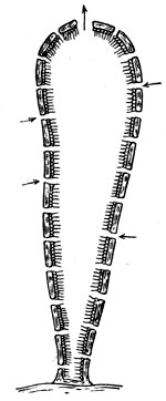
Now, from what has been previously said with regard to the tidal influence of the moon, we see that the tide must necessarily be high under the moon, as well as on the side of the earth directly opposite this body, and that the high tides must follow the moon in its regular motion. But we must not forget that the earth itself is continually turning on its axis, making a complete rotation in about twenty-four hours; while the moon, which revolves round the earth in about twenty-eight days, describes only a small portion of its orbit in the same time; thus, while the tidal wave slowly follows the moon as it travels in its orbit, the earth slips round, as it were, under the tidal wave, causing four changes of tide in approximately the period of one rotation. Suppose, for example, the earth to be performing its daily rotation in the direction indicated by the arrow (fig. 8), and the tide high at the place markedÛuccessively, where the tide is high and low respectively. Hence the daily changes are to a great extent determined by the rotation of the earth.
But we have already observed that each change of tide occupies a little more than six hours, the average time being nearly six hours and a quarter, and so we find that the high and low tides occur nearly an hour later every day. This is due to the fact that, owing to the revolution of the moon round the earth in the same direction as that of the rotation of the earth itself, the day as measured by the moon is nearly an hour longer than the average solar day as given by the clock.
There is yet another point worth noting with regard to the relation between the moon and the tidal movements of the water, which is that the high tides are never exactly under the moon, but always occur some time after the moon has passed the meridian. This is due to the inertia of the ocean, and to the resistance offered by the land to its movements.
Now, in addition to these diurnal changes of the tide, there are others, extending over longer periods, and which must be more or less familiar to everyone who has spent some time on the coast. On a certain day, for instance, we observe that the high tide flows very far up the beach, and that this is followed, a few hours later, by an unusually low ebb, exposing rocks or sand-banks that are not frequently visible. Careful observations of the motions of the water for some days after will show that this great difference between the levels of high and low-water gradually decreases until, about a week later, it is considerably reduced, the high tide not flowing so far inland and the low-water mark not extending so far seaward. Then, from this time, the difference increases again, till, after about two weeks from the commencement of our observations, we find it at the maximum again.
Here again we find that the changes exactly coincide with changes in the position of the moon with regard to the sun and the earth. Thus, the spring tides—those which rise very high and fall very low—always occur when the moon is full or new; while the less vigorous neap tides occur when the moon is in her quarters and presents only one-half of her illuminated disc to the earth. And, as the moon passes through a complete cycle of changes from new to first-quarter, full, last-quarter, and then to new again in about twenty-nine days, so the tides run through four changes from spring to neap, spring, neap, and then to spring again in the same period.
The reason for this is not far to seek, for we have already seen that both sun and moon exert a tide-producing influence on the earth, though that of the moon is considerably greater than that of the sun; hence, if the sun, earth, and moon are in a straight line, as they are when the moon is full, at which time she and the sun are on opposite sides of the earth, and also when new, at which time she is between the earth and sun, the sun’s tide is added to the moon’s tide, thus producing the well-marked spring tides; while, when the moon is in her quarters, occupying a position at right angles from the sun as viewed from the earth, the two bodies tend to produce high tides on different parts of the earth at the same time, and thus we have the moon’s greater tides reduced by the amount of the lesser tides of the sun, with the result that the difference between high and low tides is much lessened.
Again, the difference between high and low water marks is not always exactly the same for the same kind of tide—the spring tide for a certain period, for example, not having the same limits as the same tide of another time. This is due to the fact that the moon revolves round the sun in an elliptical orbit, while the earth, at the same time, revolves round the sun in a similar path, so that the distances of both moon and sun from the earth vary at different times. And, since the tide-producing influences of both these bodies must increase as their distance from the earth diminishes, it follows that there must be occasional appreciable variations in the vigour of the tidal movements of the ocean.
As the earth rotates on its axis, while at the same time the tidal wave must necessarily keep its position under the moon, this wave appears to sweep round the earth with considerable velocity. The differences in the level of the ocean thus produced would hardly be appreciable if the earth were entirely covered with water; but, owing to the very irregular distribution of the land, the movements of the tidal wave become exceedingly complex; and, when it breaks an entrance into a gradually narrowing channel, the water is compressed laterally, and correspondingly increased in height. It is thus that we find a much greater difference between the levels of high and low tides in continental seas than are to be observed on the shores of oceanic islands.
We have occupied so much of our time and space in explanation of the movements of the tides not only because we think it desirable that all who delight in sea-side rambles should understand something of the varied motions which help to give such a charm to the sea, but also because, as we shall observe later, these motions are a matter of great importance to those who are interested in the observation and study of marine life. And, seeing that we are writing more particularly for the young naturalists of our own island, we must devote a little space to the study of the movements of the tidal wave round Great Britain, in order that we may understand the great diversity in the time of high tide on any one day on different parts of the coast, and see how the time of high tide for one part may be calculated from that of any other locality.
Were it not for the inertia of the ocean and the resistance offered by the irregular continents, high tide would always exist exactly under the moon, and we should have high water at any place just at the time when the moon is in the south and crossing the meridian of that place. But while the inertia of the water tends to make all tides late, the irregular distribution of the land breaks up the tidal wave into so many wave-crests and greatly retards their progress.
Thus, the tidal wave entering the Atlantic round the Cape of Good Hope mingles with another wave that flows round Cape Horn, and the combined wave travels northward at the rate of several hundred miles an hour. On reaching the British Isles it is broken up, one wave-crest travelling up the English Channel, while another flows round Scotland and then southwards into the North Sea.
The former branch, taking the shorter course, determines the time of high tide along the Channel coast. Passing the Land’s End, it reaches Plymouth in about an hour, Torquay in about an hour and a half, the Isle of Portland in two hours and a half, Brighton in about seven hours, and London in about nine hours and a half. The other branch, taking a much longer course, makes its arrival in the southern part of the North Sea about twelve hours later, thus mingling at that point with the Channel wave of the next tide. It takes about twenty hours to travel from the south-west coast of Ireland, round Scotland, and then to the mouth of the Thames. Where the two waves meet, the height of the tides is considerably increased; and it will be understood that, at certain points, where the rising of one tide coincides with the falling of another, the two may partially or entirely neutralise each other. Further, the flow and the ebb of the tide are subject to numerous variations and complications in places where two distinct tidal wave-crests arrive at different times. Thus, the ebbing of the tide may be retarded by the approach of a second crest a few hours after the first, so that the ebb and the flow do not occupy equal times. At Eastbourne, for example, the water flows for about five hours, and ebbs for about seven and a half. Or, the approach of the second wave may even arrest the ebbing waters, and produce a second high tide during the course of six hours, as is the case at some places along the Hampshire and Dorset coasts.
Those who visit various places on our own coasts will probably be interested in tracing the course of the tidal crests by the aid of the accompanying map of the British Isles, on which the time of high tide at several ports for the same time of day is marked. It will be seen from this that the main tidal wave from the Atlantic approaches our islands from the south-west, and divides into lesser waves, one of which passes up the Channel, and another round Scotland and into the North Sea, as previously mentioned, while minor wave-crests flow northward into the Irish Sea and the Bristol Channel. The chart thus supplies the data by means of which we can calculate the approximate time of high tide for any one port from that of another.
Although the time of high water varies so greatly on the same day over such a small area of country, yet that time for any one place is always approximately the same during the same relative positions of the sun, earth, and moon; that is, for the same ‘age’ of the moon; so that it is possible to determine the time of high water at any port from the moon’s age.
The time of high tide is generally given for the current year in the local calendars of our principal seaports, and many guide-books supply a table from which the time may be calculated from the age of the moon.
At every port the observed high water follows the meridional passage of the moon by a fixed interval of time, which, as we have seen, varies considerably in places within a small area of the globe. This interval is known as the establishment of the port, and provides a means by which the time of high water may be calculated.
Before closing this short chapter on the general characteristics of the sea shore we ought to make a few observations on the nature of the water of the sea. Almost everyone is acquainted with the saltness while many bathers have noticed the superior buoyancy of salt water as compared with the fresh water of our rivers and lakes. The dissolved salts contained in sea water give it a greater density than that of pure water; and, since all floating bodies displace their own weight of the liquid in which they float, it is clear that they will not sink so far in the denser water of the sea as they would in fresh water.
If we evaporate a known weight of sea water to dryness and weigh the solid residue of sea salt that remains, we find that this residue forms about three and a half per cent. of the original weight. Then, supposing that the evaporation has been conducted very slowly, the residue is crystalline in structure, and a careful examination with the aid of a lens will reveal crystals of various shapes, but by far the larger number of them cubical in form. These cubical crystals consist of common salt (sodium chloride), which constitutes about three-fourths of the entire residue, while the remainder of the three and a half per cent. consists principally of various salts of magnesium, calcium, and sodium.
Sea salt may be obtained ready prepared in any quantity, as it is manufactured for the convenience of those who desire a sea bath at home; and it will be seen from what has been said that the artificial sea-water may be prepared, to correspond almost exactly with that of the sea, by the addition of three and a half pounds of sea salt to about ninety-six and a half pounds of water.
This is often a matter of no little importance to the sea-side naturalist, who may require to keep marine animals alive for some time at considerable distance from the sea shore, while their growth and habits are observed. Hence we shall refer to this subject again when dealing with the management of the salt-water aquarium.
The attractions of the sea coast are undoubtedly greater by day than at night, especially in the summer season, when the excessive heat of the land is tempered by the cool sea breezes, and when life, both on the cliffs and among the rocks, is at its maximum. But the sea is grand at night, when its gentle ripples flicker in the silvery light of the full moon. No phenomenon of the sea, however, is more interesting than the beautiful phosphorescence to be observed on a dark summer’s night. At times the breaking ripples flash with a soft bluish light, and the water in the wake of a boat is illuminated by what appears to be liquid fire. The advancing ripples, as they embrace a standing rock, surround it with a ring of flame; while streaks and flashes alternately appear and disappear in the open water where there is apparently no disturbance of any kind.
These effects are all produced by the agency of certain marine animals, some of which display a phosphorescent light over the whole surface of their bodies, while in others the light-giving power is restricted to certain organs or to certain well-defined areas of the body; and in some cases it even appears as if the creatures concerned have the power of ejecting from their bodies a phosphorescent fluid.
It was once supposed that the phosphorescence of the sea was produced by only a few of the lower forms of life, but it is well known now that quite a large number of animals, belonging to widely different classes, play a part in this phenomenon. Many of these are minute creatures, hardly to be seen without the aid of some magnifying power, while others are of considerable size.
Among the peculiar features of the phosphorescence of the sea are the suddenness with which it sometimes appears and disappears, and its very irregular variations both at different seasons and at different hours of the same night. On certain nights the sea is apparently full of living fire when, almost suddenly the light vanishes and hardly a trace of phosphorescence remains; while, on other occasions, the phenomenon is observed only on certain patches of water, the areas of which are so well defined that one passes suddenly from or into a luminous sea.
The actual nature of the light and the manner in which it is produced are but ill understood, but the variations and fitfulness of its appearances can be to a certain extent conjectured from our knowledge of some of the animals that produce it.
In our own seas the luminosity is undoubtedly caused principally by the presence of myriads of minute floating or free-swimming organisms that inhabit the surface waters. Of these each one has its own season, in which it appears in vast numbers. Some appear to live entirely at or near the surface, but others apparently remain near the surface only during the night, or only while certain conditions favourable to their mode of life prevail. And further, it is possible that these minute creatures, produced as they generally are in vast numbers at about the same time, and being more or less local, are greatly influenced by changes of temperature, changes in the nature of the wind, and the periodic changes in the tides; and it is probable that we are to look to these circumstances for the explanations of the sudden changes so frequently observed.
In warmer seas the phenomenon of phosphorescence is much more striking than in our own, the brilliancy of the light being much stronger, and also produced by a greater variety of living beings, some of which are of great size, and embrace species belonging to the vertebrates and the higher invertebrate animals.
Those interested in the investigation of this subject should make it a rule to collect the forms of life that inhabit the water at a time when the sea is unusually luminous. A sample of the water may be taken away for the purpose of examination, and this should be viewed in a good light, both with and without a magnifying lens. It is probable, too, that a very productive haul may be obtained by drawing a fine muslin net very slowly through the water. After some time the net should be emptied and gently washed in a small quantity of sea water to remove the smaller forms of life contained, and the water then examined at leisure.
Of course it must not be assumed that all the species so obtained are concerned in any way with the phosphorescence of the sea, but any one form turning up in abundance when collected under the conditions named will probably have some connection with the phenomenon.
One may well ask ‘What is the use of this light-emitting power to the animals who possess it?’ but this question is not easily answered. The light produced by the glow-worm and other luminous insects is evidently a signal by means of which they call their mates, and this may be the case with many of the marine luminous animals, but it is evidently not so with those which live in such immense numbers that they are simply crowded together; nor can it be so with the many luminous creatures that are hermaphrodite. It is a fact, however, that numbers of deep-sea species possess the power of emitting light to a striking extent; and the use of this power is in such cases obvious, for since the rays of the sun do not penetrate to great depths in the ocean, these luminous species are enabled to illuminate their own surroundings while in search of food, and, in many cases at least, to quench their lights suddenly at such times as they themselves are in danger.
Assuming that the reader is one who desires to become intimately acquainted with the wonderful and varied forms of life to be met with on the sea shore, or, hoping that he may be lured into the interesting and profitable pastimes of the sea-side naturalist, we shall now devote a chapter to the consideration of the appliances required for the collection and examination of marine life, and to general instructions as to the methods by which we may best search out the principal and most interesting objects of the shore.
First, then, we shall describe the equipment of an enthusiastic and all-round admirer of Nature—he who is interested in plant forms from the flowering species down to the ‘meanest weed that grows,’ and is always ready to learn something of any member of the animal world that may happen to come within his reach. And this, not because we hope, or even desire, that every reader may develop into an all-round naturalist, but so that each may be able to select from the various appliances named just those which would be useful for the collection and observation of the objects which are to form his pet study.
The most generally useful of all these appliances is undoubtedly some kind of case of the ‘hold-all’ type, a case into which specimens in general may be placed for transmission from the hunting-ground in order that they may be studied at leisure, and we know of nothing more satisfactory than the botanist’s ‘vasculum.’ This is an oblong box of japanned tin, fitted with a hinged front, and having both handle and strap, so that it can be either carried in the hand or slung over the shoulder. Of course almost any kind of non-collapsible box or basket will answer the purpose, but we know of no utensils so convenient as the one we have named. It is perfectly satisfactory for the temporary storage of the wild flowers gathered on the cliffs, as it will keep them moist and fresh for some considerable time; and for the reception of sea weeds of all kinds it is all that could be desired, for it will preserve them in splendid condition, and is so constructed that there is no possibility of the inconvenience arising from the dripping of salt water on the lower garments. Then, as regards marine animal-life in general—starfishes, urchins, anemones, molluscs, crustaceans, fishes, &c.—these may be conveyed away in it with a liberal packing of moist weeds not only without injury, but in such a satisfactory condition that nearly all may be turned out alive at the end of a day’s work; and this must be looked upon as a very important matter to him who aims at becoming a naturalist rather than a mere collector, for while the latter is content with a museum of empty shells and dried specimens, the former will endeavour to keep many of the creatures alive for a time in some kind of artificial rock pool in order that he may have the opportunity of studying their development and their habits at times when he has not the chance of visiting the sea shore for the purpose.
But although the vasculum is so generally useful for the temporary storage and the transmission of the objects collected, yet it is not in itself sufficient for all purposes. There are many marine animals so small—but none the less interesting because they are small—that they would probably be lost in a case containing a mass of sea weeds with various larger creatures. These should be placed in small well-corked bottles, and temporarily preserved in a little sea-water, or, preferably, a tuft of one of the delicate weeds so common in our rock pools. Others, again, though they may be larger, are of so fragile a nature that they should be isolated from the general stock on that account alone. Instead of bottles or tubes, small tin boxes may be used, and these have the advantage of being unbreakable, though, of course, they will not hold water. This, however, is of no consequence, as most marine animals may be kept alive for some time in moist sea-weed quite as well as in water.
When small animals are required for structural examination only, they may be put into methylated spirit as they are taken, and when stored in this way a much larger number may be put into the same receptacle; hence the collector will often find it convenient to have a small supply of this liquid while at his work.
A strong pocket-knife is essential for sea-side work. It serves to remove those molluscs that adhere firmly to the rocks by suction, and also others that fix themselves by means of a byssus of silken fibres, as is the case with mussels. It will also be employed in the removal of acorn barnacles, anemones, and small tufts of algæ, and may be useful in cutting through the stouter weeds. Small sponges and other low forms of life often form incrustations on the solid rock, and may be peeled off with the aid of a knife. In the case of the last-named, however, as well as with the anemones and other fixed animals, it is often far more satisfactory to remove a small portion of the rock itself with the animal attached, and for this purpose a small hammer will be of great service.
A strong net of some kind is necessary in searching the rock pools, and as suitable nets are, we believe, not to be obtained of the dealers in naturalists’ appliances, it devolves on one to manufacture a net according to his requirements.
The simplest form of net may be made by bending a piece of stout galvanised iron wire into the form here shown (fig. 14), and firmly wedging the two straight ends in a short piece of strong metal tube which will also serve as a ferrule for the attachment of a tough handle. Such a circular frame although satisfactory for a net to be used in fresh-water ponds and streams, is not nearly so suitable for the irregular rocky pools to be met with on the sea coast, for it will not enable one to search the numerous corners and crevices into which many marine creatures will retire on being disturbed, but it may be greatly improved by bending the side opposite the ferrule into a moderately sharp angle and then turning the angle slightly upward, as shown in fig. 15.
Another very convenient net frame may be made by bending the wire into a rhomboidal form (fig. 16), the ferrule being attached by means of two short, straight ends at one of the angles. The opposite angle will serve the purpose of searching into the crannies of the rocks, while the straight sides will prove very useful in removing the objects that lie on the sandy bottoms so commonly seen in rock pools. The semicircular net shown in fig. 18 will also prove useful for working on sands or for scraping the flatter surfaces of weed-covered rocks.
The material of the net should be some kind of strong gauze, or a loosely-woven canvas. Leno answers very well, but is somewhat easily torn, and will have to be frequently renewed. This, however, may be avoided to a great extent if, instead of sewing the gauze directly round the wire, a strip of strong calico be first attached to the frame, and the gauze then sewn to the calico; for it will be understood that any fragile material placed round the wire will soon be worn through by friction against the rugged surfaces of the rocks and stones. The net itself should not be very deep, and should have no corners; and as to the length of the handle, that will be determined by the fancy of the collector, or by the character of the ponds to be searched, but a tough walking-stick with a crook handle will generally answer all purposes, the crook being itself frequently useful for removing the larger weeds and other obstructions.
Although the net, as above described, will answer the requirements of nearly all young collectors, yet there may be some, who, not satisfied with the exploration of the rocks and pools exposed when the tide is out, desire to know something of the creatures that live entirely beyond low-water mark, where the water is generally too deep for work with a hand net. To such we recommend a small dredge that may be lowered from a boat and then drawn along the bottom. A good form of dredge is shown in fig. 19, and a little skill and ingenuity will enable anyone to construct one with the help of our illustration; but, seeing that the best work is to be done on rough bottoms, it is absolutely necessary that both frame and net should be made of the stoutest materials that can be conveniently employed.
Those who have ever accompanied a fisherman while taking a pull round to examine the contents of his crab or lobster pots will probably have noticed what strange creatures, in addition to the edible crabs and lobsters, sometimes find their way into the trap. These creatures are often of great interest to a young naturalist, and it will repay him to take an occasional trip with a fisherman in order to obtain them; or, still better, to have a crab-pot of his own. The writer has obtained many good specimens by means of an inexpensive trap, on the same principle as the ordinary crab-pot, made from an old metal bird-cage of rather small size. The bottom was removed, and a very shallow bag of thick canvas fixed in its place; and some of the wires were cut, and bent inwards so as to allow the easy entrance of moderately large crustaceans and other creatures, while at the same time they served as a barrier to their escape. Such a trap, baited with pieces of fish, and let down to a rocky bottom, will enable the young naturalist to secure specimens that are seldom seen between the tide-marks; and the animals thus obtained will include not only those larger ones for which the opening was made, but also a variety of smaller creatures that may enter between the wires of the cage. Some of the latter may, of course, escape by the same way as the trap is being hauled up for examination, but this is not so likely to occur if the canvas bottom is of a material so loosely woven that water can pass through it very freely. It will, of course, occur to the reader that the insertion of a stone or other weight will assist in sinking the trap; also that the ordinary door of the cage forms a ready means by which the captives may be removed.
One thing more: make it a rule never to go out collecting natural objects of any kind without a note-book and pencil. This, to the beginner who is anxious to get to his work, with the idea only too prevalent with the amateur that the success of his labours is to be measured only by the number of specimens obtained, may seem quite an unnecessary part of the equipment. But it must be remembered that there is much to observe as well as much to collect on a well-selected coast; and that without the aid of the book and pencil a great many of the observations made will be forgotten, and thus much interest that would otherwise be attached to the objects permanently preserved will be lacking.
The above appliances include the only necessary equipment of the sea-side naturalist, with the exception of a few required for occasional use in connection with the species of a somewhat restricted habitat, and the outfit of the sea angler. The former will be dealt with in the chapters where the species concerned are described, while the subject of sea angling is of such general interest that we propose to devote a short chapter exclusively to it.
It may seem hardly necessary to discourse on the nature of the attire most suitable for sea-side work, since the majority will readily form their own opinions on this matter, but perhaps a few words of advice to the inexperienced may not be altogether out of place. First, then, make it a rule to wear no clothing of any value. The work will lead the enthusiast over slippery weeds, on treacherous boulders, over rocks covered with sharp acorn shells, and among slimy and muddy stones, and many a slip may occur in the course of a day’s work. Large pockets specially but simply made by sewing square pieces of lining on the inside of an old jacket are a great convenience; a cap rather than a brimmed hat should be worn unless the latter be considered essential for protection from a burning summer’s sun; and a pair of old shoes, preferably with rubber soles, are just the thing for both rough and slippery rocks, as well as for wading through shallow waters. Other details we can safely leave to the fancy of the reader himself.
Now comes the most important question ‘Where shall we go?’ Fortunately we are favoured with a great extent of coast-line considering the area of our country, but the character of the coast is so diversified, both with regard to its scenery and its life, that the naturalist will do well to carefully select his locality according to the objects he desires to study. The east coast of England is not generally noted either for variety or abundance of marine life, and the same is true both of the south-east and a large portion of the south coast. In some places the beach is formed of an unbroken stretch of sand on which one may walk for miles without seeing any sign of life, with the exception of an occasional empty shell and a few fragments of dried sea-weed washed in by the breakers during a recent storm; while at the same time the cliffs, if such exist at all, are not very generous in their production of the fauna and flora that are characteristic of the shore. But even on the coasts referred to there are, here and there, isolated spots where the uplands jut into the sea, giving rise to bold promontories, at the foot of which are the fallen masses of rock that afford protection to a moderate variety of truly marine life, while the rough bottoms beyond yield numerous interesting forms that may be secured by means of the dredge or suitable traps. Such spots are to be found where the chalk hills abut on the sea, as at Flamborough and Beachy Head, but it is in the neighbourhood of Weymouth that the English coast really begins to be of great interest to the naturalist. From here to the Land’s End almost every part of the shore will yield a great variety of life in abundance, and the same is true of the rocky coasts of the west, and also of the more rugged shores of the Isle of Wight. As an ideal hunting-ground one cannot do better than to select one of the small fishing towns or villages on the rocky coasts of Devon and Cornwall. With such a spot as his headquarters the most enthusiastic sea-side naturalist will find ample employment. The exposed rocks and rock pools yield abundance of life; and if these be searched when the tide is out, there will remain plenty of sea angling and other employments to occupy him at other times.
We will now describe the actual work of the sea-side naturalist, giving the necessary instructions for the observation and collection of the various living things he will meet with.
First, then, with regard to work on the cliffs, a very few words will suffice; for, seeing that the objects of interest to be met with here will consist principally of the various flowers that are peculiar to or characteristic of the sea shore, and certain insects and other creatures more or less partial to a life on the cliffs, we may regard these as coming within the range of the general work of the botanist, entomologist, &c.; and since instructions for the collection and preservation of such objects have already been given in former works of this series, we may pass them over at once in order to deal with those objects which are essentially marine.
It has already been hinted that the right time for collecting on the shore is when the tide is at its lowest; and in order that the best work may be done the collector should consult the local tide-tables, or calculate, if necessary, the time of high tide from the establishment of the port; and, of course, the period of spring tides should be selected if possible. The time during which work should continue must be regulated according to the enthusiasm of the collector or the time at his disposal, but, as a rule, it is advisable to be on the scene of action about three hours before the time of low tide, with a determination to work continuously until the lowest ebb of the water.
On reaching the beach it is always advisable to start by examining the line of miscellaneous material at high-water mark, along which may be found quite a variety of objects, more or less interesting, which have been washed in by the breakers, especially just after a storm, together with numerous scavengers of the shore that perform a most useful work in devouring the decomposing organic matter that would otherwise tend to pollute the air.
Here we may find many useful and interesting objects of both the animal and vegetable worlds. Among the former are the empty shells of both univalve and bivalve molluscs, some of which are more or less worn by the action of the waves, while others are in splendid condition for examination and study. Here, too, are various species of sea firs and the skeletons of sponges; the shell of the cuttle-fish, and occasionally a cluster of the eggs of this creature—the sea-grapes of the fishermen; also the egg-cases of the skate and the dog-fish—usually empty, but sometimes enclosing the young animal still alive; and, lastly, we frequently meet with portions of the skeletons of fishes in a perfect state of preservation, the animal matter having been cleared away by the combined action of the scavengers previously referred to. Then, as regards the vegetable world, we often find beautiful specimens of sea-weeds along the high-water mark, some of which are rarely met with in the rock pools, since they are species that have been detached from beyond the line of low water, and washed up by the breakers.
On turning over the debris thus thrown on the beach we intrude on the privacy of numerous living creatures which immediately scamper away to find a new hiding-place. These consist principally of sand-hoppers, but occasionally we find members of the insect world engaged in the same useful work in addition to the numerous flies that perform their office of scavengers in the bright sunshine on the top of the matter that supplies them with food.
It will be interesting to capture a few of these scavengers, and to compare them with others of the same order obtained from different localities. Thus, the flies may be compared with the more familiar house fly, and the sand-hoppers of high-water mark with similar crustaceans to be afterwards obtained lower on the beach.
Attention should now be given to the rocks left exposed by the retreating tide, and it is here that the real work begins. Examine each rock pool as soon as possible after it is no longer disturbed by the waves. Remove any tufts of corallines or other weeds required for study or preservation, and simply place them, pro tem., in the vasculum or other receptacle provided for the purpose. These will form a useful protective packing for other objects that are to be carried away, so that it will be advisable to secure a moderate amount rather early, even though they may not be required for any other purpose. Live molluscs, crabs, small fishes, &c., may all be put in the receptacle with this weed, and all will probably be still alive after the collecting and the homeward journey have been completed. Probe the corners of the pool with the point of the net, and also sweep the net upward among the weeds to remove any creatures that seek shelter among the fronds. Tufts of corallines and other weeds should be searched for the small and delicate starfishes that live among them, and any stones that may cover the bottom of the pool should be lifted. Anemones may be removed from the rocks by means of a rather blunt knife; but, if possible, it will be better to chip off a small piece of the rock with the anemone attached to it, and wrap it lightly round with a tuft of soft weed previous to placing it in the collecting case.
A number of rock pools should be searched in this manner, but those chosen should vary as much as possible in general character. All very small and delicate objects should be isolated from the general stock, and placed, with the usual packing material, either in tin boxes or small wide-mouthed bottles; and if any animals taken are not required alive, but only for preservation, they should be preferably killed at once and then stored in a separate case. Some creatures are easily killed by simply dropping them into a bottle of fresh water, but others should be covered with methylated spirit. It should be mentioned, however, that the natural appearance of some of the crustaceans is quite destroyed by strong spirit, which soon makes them look as if they had been boiled. Some species are changed in this way much more readily than others; and, until sufficient experience has been gained to enable the young collector to distinguish between them, it will be advisable to kill and temporarily preserve crustaceans in spirit that has been considerably diluted with water—about two parts of water to one of spirit, for example. Further, there are certain fragile starfishes that have a way of breaking themselves into pieces when dropped into spirit, or even when suddenly disturbed in almost any other manner. These must always be handled gently, and if it is required to kill them for preservation, the best way will be to put them in a little salt water, and then gradually add fresh water until the desired result is obtained.
Perhaps the most productive of all sea-shore work is the turning over of the stones of various sizes near the low-tide mark, and the examination of the chinks and sheltered hollows of the rocks that are left uncovered for but a short period. This work should be carried on as near the water’s edge as possible, closely following the receding tide; and the collector must now be prepared with a number of small bottles or tins for the isolation of small and delicate specimens. He must also be on the alert for numerous examples of protective resemblance, in which the animals concerned so closely resemble their surroundings in colour and general character of surface that they are detected only by careful observation, while the difficulty of identification is still further increased in instances where the creatures remain perfectly still even when disturbed.
Under the stones all manner of animals—fishes, crustaceans, worms, molluscs, starfishes, anemones, &c.—will be hiding until covered by the next tide. Some of these will be found on the ground beneath the stones, and others attached to the under surfaces of the stones themselves; therefore both should be carefully examined, attention being given at first to the more active species that hurry away with all speed towards a new shelter as soon as they find themselves exposed to the light; the less active creatures may then be secured at leisure.
The tide will not allow the collector a great deal of time in which to turn over the most productive stones—those close to the low-water mark, so there is but little opportunity of observing the movements and other interesting habits of many of the animals found; hence it is advisable to secure a good variety of living specimens, especially of the less familiar species, in order that they may be placed in some kind of aquarium, temporary or otherwise, for observation at home.
One thing more remains to be done while the tide is well out, and that is to examine the weed-covered rocks near the water’s edge. Lift the dangling weeds and carefully search the rocks for those low forms of animal life that form incrustations on the surface, as well as for new species of anemones, sea firs, &c. Lastly, look well into the dark and narrow chinks of the rocks, for here several species of lowly animals that are hardly met with elsewhere may be found, and also certain crustaceans that delight to squeeze their bodies into the remotest corner of a sheltered niche.
We do not propose dealing with this subject from the point of view of the angler, but rather that of the naturalist. The former is actuated principally, if not entirely, by the mere love of sport; or, it may be, to a great extent by the desire to obtain a supply of fish for food; and he generally estimates the success of his expeditions not by the number of species captured, but by the total weight of his catch, no regard being paid, as a rule, to the inedible specimens. The naturalist, however, does not desire weight, or sweetness of flesh. He works the greatest possible variety of habitats, with the object of determining the number of species inhabiting the locality and of learning as much as possible of their general form, habits, and adaptations of structure to habits. His success is measured by the number and variety of species caught, and he pays but little attention to superiority of size or weight, or to the estimated market value of his haul. The element of sport may enter more or less largely into the pleasure of his occupation, but the main end in view is to learn as much as possible of all the species obtainable.
Further, our remarks will not include the subject of the different kinds of fishing usually resorted to by sea anglers, but will be confined almost exclusively to the simple means of catching the common species that frequent the immediate neighbourhood of the shore.
If the reader will follow the general instructions given in Chapter II. on the outdoor work of the marine naturalist, he will undoubtedly make the acquaintance of a considerable variety of interesting species which may be captured in the rock pools, found under stones at low tide, or obtained by means of a small dredge; but his knowledge of our littoral fishes may be appreciably extended by the occasional employment of rod and line from rocks and piers, or from a small boat in close proximity to the shore.
The appliances required are of a very simple nature, and not at all costly. The long, heavy rod and strong tackle of the sea angler and professional fisherman are not at all essential to our purpose, for our work will be confined almost exclusively to shallow water, and the fish to be caught will be chiefly of small size. True it is that one may occasionally find his light tackle snapped and carried away by the unexpected run of a large fish, for cod and other large species often approach close to the shore, and bite at baits intended for the smaller fish that make their home among the partly submerged rocks of the coast; but such surprises will not frequently occur, and the young naturalist may learn all he wants to know of the fishes of our shallow waters with the aid of a light rod of about nine or ten feet and one or two light lines of no great length.
It must not be understood, however, that we assume the reader’s disinclination to know anything of the inhabitants of deep water, but rather that we consider the whole subject of deep-sea fishing quite beyond the scope of this work. It is a fact that quite a large number of species, the forms and habits of which are extremely interesting, live exclusively on deep bottoms. These should undoubtedly be studied by all who are interested in the various phases of marine life; but unless the reader is prepared to practise sea fishing in all its branches—to put his trust in the restless sea, supplied with all the necessary heavy gear, and to risk those internal qualms that arise from the incessant swaying of the boat on open waters, he should make arrangements with the professional deep-sea fisher—preferably a trawler—for the supply of those disreputable species that invariably form part of the haul, while the better-known food fishes can always be obtained from dealers for purposes of study.
On one occasion we had a rather unique and very successful interview with a friendly trawler. She was sailing slowly towards her station in a south-western fishing port, while two of her crew were clearing her nets, and throwing all refuse into the sea. We rowed behind her in order to see the nature of the rejected portion of the haul, and finding that it included specimens of interesting fishes of ill repute, dead but perfectly fresh, we followed her track, and collected a few for future examination. Presently our movements were watched from aboard, and we were invited to pull up to larboard, where a short explanation as to our wants led to the acquisition of quite a variety of deep-sea life, including several species of fishes not often seen on land, crabs, shelled and shell-less molluscs, worms, star-fishes, and various lowly organised beings, many alive and in good condition, together with several good food fishes thrown in by way of sympathy. There is no doubt that a naturalist can obtain much more deep-sea life with the aid of a friendly trawler than by any amount of ‘fishing’ with ordinary tackle from a boat; and this without the necessity of going to sea at all, if he will only take the opportunity of examining the nets as the boats are stranded on their return.
But now to return to our angling:—We have to provide a light rod, about ten feet long, with a winch, and a line of twisted silk or other thin but strong material; also a light hand line, and a supply of gut, leads, shot, and hooks, together with one or two small floats, and a few bait boxes.
We do not, as a rule, recommend the amateur angler to use both rod and hand line at the same time, for the attempt to do this leads to the neglect of both. In the end it is not likely to lead to any gain, so many fish being lost through the inability to strike at the moment a bite is given, and so much time having to be devoted to the baiting of hooks rather than to the direct management of the lines. In most cases the rod is much more convenient than the hand line. The young collector will meet with the greatest variety of species in rocky and weedy places, where abundant shelter exists for those fishes that prefer to keep well under cover, and any attempt with a hand line in such spots will certainly lead to frequent loss of hooks, and often of lead, line, and temper. Such a line must be reserved for fishing on sandy bottoms, while the ten-foot rod recommended will enable the angler to do good work in the rockiest parts without much danger of fouling; and, in fact, to fish anywhere along the coast.
The arrangement of hooks and lead must necessarily depend on the character of the place to be worked, but in all cases we strongly recommend no such multiplicity of hooks as is made use of by fishermen and others who fish for food. In their case the use of so many hooks often pays them well; but, as we have previously hinted, the naturalist does not desire quantity of fish so much as variety of species. Further, there is no necessity to make his work heavy and arduous. His desire is not to spend an undue proportion of his time in baiting hooks, but to have his line so under control that he is ready to strike at any moment, and to be able to alter the conditions of his work as often as his ideas or the conditions change.
In rugged and weedy places the hooks must be kept free from rocks and weeds. This may be done by letting down the rod line with a lead at the bottom, and one or two hooks fastened to gut at such a level as to keep quite clear of weeds. A much better arrangement, and one which we ourselves almost invariably employ, consists of a light lead, as a rule not exceeding an ounce in weight, fastened at the end of the line, and below it a few feet of gut terminating in a single hook. With such tackle it is of course necessary to determine previously the depth of the water, in order to adjust the line to such a length that the hook keeps clear of rocks and weeds, and a float may be used if desired.
We do not recommend a float for the general work of the marine collector, for it is a decided advantage to be prepared to bring the bait to any level from bottom to surface, especially when the water is so clear that the fish may be seen swimming, in which case one is often impressed with the desire to capture a specimen in order to establish its identity, and for such work as this a float is superfluous. If, however, a float is used, it should be a sliding one, so that it may be adapted to the rising and falling of the tide.
Of hooks there is a great variety to choose from, differing in the form both of the curve and of the end of the shank. As to the curve, those with a decided twist are best adapted to our purpose, chiefly on account of the fact that sea fishes generally have larger mouths than fresh-water species of the same size, and are consequently better held with a twisted hook. The shanks of sea hooks are either flattened or eyed, and each is as good as the other providing the snood is firmly attached; but some amateurs find a greater difficulty in attaching the snood to the former than to the latter.
Gut snoods are recommended for our purpose, and fig. 26 shows one method by which they may be fastened to a flattened shank, while fig. 27 illustrates the figure-of-eight knot by means of which the eyed shank may be firmly secured. The gut should be soaked for some hours in cold water previous to tying, and it may be kept soft for some considerable time by giving it a few hours’ immersion in a solution of glycerine—about one part of glycerine to four or five parts of water.
Small hooks will be most suitable for our purpose; and if the reader finds any difficulty in attaching the snood firmly, he may purchase suitable hooks ready mounted on gut, though, of course, these are more expensive than the flattened or eyed hooks generally used for sea-fishing. Such small and fragile hooks may be occasionally snapped off by the run of a vigorous fish of moderate size, therefore it is advisable to have a supply of larger hooks, ready fixed on strong snoods, to be used when it is found that the shore is frequented by larger fishes than those generally caught close to land.
When fishing with a rod and line from rocks, or from piers, the foundations of which are covered with large weeds, the bait will frequently be carried by currents among the weeds and snapped off when endeavours are made to release the hook. This will especially be the case when the hook is a few feet below the lead, as we have already suggested it should be. To reduce the frequency of such mishaps, it will be a good plan to weight the gut below the lead by means of a few split shot. In fact, in sheltered places, where the water is not disturbed, these shot may take the place of the lead, but little weight being necessary for rod fishing in such localities.
The amateur sea angler is often in great doubt as to the best bait to use; and, believing that a certain kind of bait is absolutely necessary for his work in some particular spot, is often at a loss to obtain it. This bait difficulty is evidently a prevailing one among amateur sea fishers, if one may judge from the frequent questions asked as to the best or proper bait to use, and from the very common ‘Can you oblige me with a little bait?’ This latter question, we believe, is frequently the outcome of carelessness or laziness on the part of the asker. He has not the forethought, born of enthusiasm, that would lead him to procure a suitable bait, at a convenient time, previous to starting off on his angling expedition, but rather depends on the possibility of being able to beg or otherwise secure sufficient for his purpose at the time; yet there are so many good baits that are easily secured at the proper time and place that the enthusiastic angler need never be at a loss. Some of these may be collected by himself at low tide, others may be obtained from local fishermen, or from the tradesmen of the town or village.
Some anglers seldom collect their own bait, either purchasing it or employing some one to collect it for them; but we are of opinion that the pleasure of a day’s fishing begins here, and especially so when the angler is of the naturalist type, for he will frequently learn more of the nature and habits of living creatures during one hour’s bait-collecting than during three or four hours’ angling. It is true that the work in question is often a bit laborious, particularly on a warm day, and that it may be frequently described as dirty and odorous; but what is that to one who is interested in his employment, and who derives pleasure in doing his own work? Fishermen often use lugworms for bait, and although these constitute one of the best baits for their own fishing, they are not so suitable for the purposes of the amateur angler, fishing with small hooks close to shore. They may be dug out of the sand when the tide is out, and are most abundant where the sand is mixed with mud. A spade should be used, and this should be thrust deep into the sand, selecting those spots where the holes or burrows of the worms most abound. Lugworms should be used whole; and being of large size, are suitable for baiting large hooks only. They may be kept alive in wet sand or sea-weed, preferably the latter for convenience, and stored till required in a wooden box.
Ragworms also afford good bait, and are particularly adapted for shore angling with small hooks. Almost all the fishes that frequent our shores take them readily, but they are not to be found in all localities. They are to be taken, though not usually in large numbers, on rocky shores where numerous stones lie among the somewhat muddy deposits of the more sheltered nooks, where they may be seen on turning over the stones. The best situation for ragworms, however, is the more or less odoriferous mud so frequently deposited in the estuaries of rivers and in landlocked harbours. Here they maybe dug out in enormous numbers with a spade, attention being directed to those spots where their burrows are most numerous. They are best stored with a little of the mud in a shallow wooden box provided with a sliding, perforated lid.
Failing a supply of the marine worms just mentioned, the common earthworm may be used as a substitute, but it is decidedly less attractive to the fishes; and the same may be said of gentles—the larvæ or grubs of flies. The latter may be bred in large numbers by simply placing a piece of liver in the soil with only a small portion exposed. If this is done in the summer time, hundreds of eggs will soon be deposited on it, and in about a week or so it will be found to be a living mass of fat white grubs, perhaps more useful to the fresh-water angler than to his marine counterpart.
Among the so-called shell fish of the class mollusca, mussels, limpets, cockles, and whelks are all largely used for bait. The last of these are too large for our purpose, but form a splendid bait for deep-sea fishing, while the other three, and especially the mussels, are well suited for shore work. Mussels, in fact, provide one of the best possible baits for almost all kinds of shore fishing, the only drawback being the excessive softness of their bodies, which enables them to be easily torn from the hook. When small hooks are used, mussels of a small size may be used whole, or the larger ones may be divided into portions of suitable size; and in any case it will be found a good plan to tie the bait to the hook with a short piece of cotton thread.
Mussels are not easily opened without injury, and consequently some anglers give them a short immersion in hot water, to kill the animal and thus cause the shell to gape. As far as our own experience goes, the value of the bait is not deteriorated by this treatment, though some are of opinion that it is not so attractive after scalding. Mussels are opened, when alive, much in the same way as oysters, but the valves of the shell fit together so closely that it seems at first almost impossible to insert a knife between them. This, however, can be done with ease if one valve is first made to slide a little way over the other by pressing it with the thumb. This being accomplished, the two valves should not be separated by the mere force of the knife, for this would tear the animal within, and render it more or less unfit for its purpose; but first direct the edge of the knife towards the adductor muscle, by means of which the animal pulls its valves so firmly together, and then cut through this close to the inner surface of the upper valve. This valve can then be lifted without injury to the soft parts, and the whole animal removed from the other valve by cutting through the same muscle close to it.
Between the two lobes of the mantle—the soft covering on both sides of the animal that previously lined the shell—will be seen a brown, fleshy, tongue-like body. This is the ‘foot’ of the mussel. The point of the hook should first be run through this, and then from side to side through the mantle, and finally through the adductor muscle previously described. If this is carefully done, there will be little fear of the bait becoming detached unless it is subjected to rough usage, and still less if it is tied round the shank of the hook by means of a short piece of cotton thread.
It is probably superfluous to mention to the reader the fact that mussels are to be found on almost every rocky coast, where they may be seen attached to the rocks by means of a bunch of silky fibres called the byssus; and that, failing this, they are to be obtained from almost every fisherman and fish-dealer; if, however, these molluscs are not to be obtained, cockles may be used as a substitute, though it will probably be found that they are appreciably inferior, except when fishing for dabs and plaice on sandy shores, in which case they are highly satisfactory. Cockles abound on most sandy coasts, where they live a little below the surface; and are usually obtained by means of an ordinary garden rake. Sometimes we meet with them in large numbers in the estuaries of rivers, where they lie buried in the banks of mixed sand and mud that are left exposed at low tide.
Limpets are extensively used for bait in some places, especially by amateur anglers; and often with good results. They should always be removed from the rocks without injury, and this is no easy matter to those who do not know how to deal with them. If taken completely by surprise, one sharp, but light tap on the side of the conical shell will successfully detach them from their hold; or they may be raised by means of the blade of a strong pocket-knife that has been thrust beneath the cone.
For our work small limpets will prove far more satisfactory than large ones, and these may be used whole; but if the limpets are too large for the hooks employed, the soft, upper part of the body only need be used.
It is not an easy matter to remove fresh limpets from their shells without destroying this soft portion of the animal, but if placed for a minute or so in hot water they come out quite easily, and are apparently none the less attractive as bait. Some fishermen on the Cornish coast always collect the largest limpets for bait, remove them from their shells by means of hot water, and arrange them on the rocks to become partly dry. When required for bait, the soft parts only are used, but these, having been more or less hardened by the drying process, hold much better on the hook than when fresh.
And now, after mentioning the fact that land snails are occasionally used, though, we believe, with no very considerable success, for sea fishing, we will note a few baits derived from the higher head-footed molluscs—the squid, cuttle-fish, &c. There are several species of these peculiar molluscs, but the common squid and the common cuttle of our seas, and especially the former, is highly prized as bait. It may be obtained from fishermen, who frequently haul it in their nets; but if supplied alive and fresh from the sea it must be handled very cautiously, otherwise it may discharge the contents of its ink-bag over one with the most unpleasant results. It is certainly best used while fresh, though some suspend it until dry, and then store it for future use, in which case it will require soaking in water when required. The thin tentacles or arms are very convenient for baiting small hooks, though other parts of the body, cut into narrow strips, will serve the purpose of the angler equally well.
Of the crustaceans, shrimps and prawns, and various species of crabs are used as bait. Shrimps and prawns are used whole for catching flat-fish, but small pieces are better when fishing for smelt and other small species of fish that swim close to shore. Little pieces of the flesh of the crab are also well adapted for baiting hooks of small size, and will prove very attractive to almost all kinds of fish. Small crabs, however, may be used whole, but are of little use except when soft—that is, just after the shedding of their shells, and before the new skin has had time to harden. Such crabs may be found under stones and in other hiding-places at low tide, for at such times they keep well secluded from their numerous enemies by whom they are greedily devoured while in this helpless and unprotected condition.
The hermit-crab, which selects the empty shell of a whelk or winkle for its home, is probably well known to our readers. The protection afforded by such a home is absolutely necessary to its existence, since its abdomen has no other covering than a soft, membranous skin. This soft abdomen is frequently used as a bait with great success, as well as the flesh of the larger claws.
If the shell from which the hermit-crab is taken be broken, a worm, something of the nature of the common ragworm, will almost always be found, and this also is very serviceable as bait.
In addition to all the baits previously named there are several other good ones, many of which are to be obtained almost everywhere. Among these may be mentioned strips cut from the mackerel, herring, or pilchard, preferably with a portion of the silvery skin attached; also thin strips of tripe. Sand-eels, which may be dug out of the sand near the water’s edge, are very useful, and may be cut into pieces for baiting small hooks. Further, a large number of artificial baits are employed in various kinds of sea fishing, but as these are not essential for the work we have in hand we do not propose describing them in detail.
Now let us suppose that we are about to try our luck at sea angling, on some rocky coast, such as that of Devon and Cornwall, our object being to determine, as far as possible, what species of fishes frequent the immediate neighbourhood of the shore. And this is not all; for, when fishing with rod and line on such a coast, it frequently happens that we land some species of crab that has been attracted to our bait. The ordinary angler would regard such crab as an intruder, and, we are sorry to say, would often consider it his duty to crush the unfortunate crustacean beneath his foot. But it is far different with the naturalist. He favourably regards all creatures from which something may be learnt, and is as anxious, as a rule, to gather information concerning the habitats of one class as of another. In fact, we may go still further, and combine crab fishing with ordinary angling, both in one and the same expedition, by letting a small crab-pot down into deep water among the rocks, and allowing it to remain while the angling is proceeding.
We select a spot where there are several feet of water close to a perpendicular rock, varied and broken by numerous holes and crevices, in which various species of fishes and crustaceans habitually hide.
Such a situation is an ideal one for a young naturalist, for not only does he obtain the greatest variety of species here, but the takings will surely include some of those remarkably interesting rock-dwelling fishes that differ from our ordinary food fishes in so many points of structure, all of which, however, display some interesting adaptation to the habits and habitats of the species concerned.
Our apparatus consists of nothing more than rod and line, one or two small leads, a supply of hooks on gut snoods, a box of bait, and a waterproof bag in which to pack the specimens we desire to preserve.
We first determine the depth of the water by means of a lead on the end of the line, and then tie the hook on the end with a small lead a few feet above it, and fish in such a manner that the hook is just on the bottom, or, if the bottom is covered with weeds, the hook should be kept just clear of fouling them.
The peculiar rock fishes so common on such a coast as this on which we are engaged need special treatment at the hands of the angler. They hide in their holes, watching for the unwary creatures on which they feed, and, pouncing upon them suddenly, rush back to their snug little nooks in which they can secure themselves firmly by means of the sharp, hard spines with which their bodies are furnished. When these fishes seize the bait offered them—and they are not at all fastidious in the choice of their viands—they should be hooked and pulled up with one vigorous sweep of the rod, or they will dart into their homes, from which it is almost impossible to dislodge them.
In addition to these, there will be various other species that require gentler treatment, and may be hooked and landed much in the same manner as fresh-water fishes, since they are free swimmers, usually keeping well clear of the rocks and weeds.
If the day is calm, and the water clear, the sea angler will often be able to watch various fishes as they swim, and to bring the bait gently within their reach; and here we find the advantage of the rod as compared with the hand line. Sometimes quite a shoal of small fishes may be seen sporting near the surface, and, as a rule, there will be no difficulty in obtaining one for identification and study. These are generally best secured by means of small hooks, with but very little bait, and will often bite freely at the tiniest fragment of worm on an almost naked hook.
After the water has been searched at all depths, it will be well to allow the bait to rest quite on the bottom, even at the risk of losing a hook or two in the weeds and rocks. This may enable one to take some fresh species of fish or to secure a crustacean or other creature that is not often found between the tide-marks. Care should be always taken, however, to keep the hook well clear of the weeds that grow on the sides of the rock, and sway to and fro with every movement of the restless waters.
Angling from piers may be pursued much in the same manner as described above in those places where the bottom is rocky, but since the chances of hooking large fish are greater here than close to shore, it is necessary to be provided with stronger tackle and larger hooks. If, however, the bottom is sandy, the rod tackle may be modified by placing the lead at the bottom, and arranging two or three hooks above it, about one or two feet apart, the lowest one being near the lead. With such an arrangement the line may be cast some distance out, but for angling close to the pier itself there is, perhaps, nothing better than the single-hook arrangement suggested above, for with this one may fish on the bottom and at all depths without any alteration in the tackle being necessary.
If, however, the rod line is to be cast as suggested above, or if a hand line is to be similarly used, the following hints may be useful as regards the arrangement of hooks and lead.
The line itself may be of twisted silk or hemp, terminated with about a yard of strong gut. The lead, preferably of a conical or pear-shaped form, should be placed at the extreme end, and its weight regulated according to the necessities of the occasion. A few ounces of lead are quite sufficient where there are no strong currents, but it is well to be supplied with larger sizes, to be substituted if circumstances require it. Two hooks will be ample. One of these should be only a few inches from the lead, and the other about eighteen or twenty inches higher. The whole arrangement, known as a Paternoster, is represented in fig. 33, in which the method of fixing the lead and the hook links is also illustrated.

Fig. 33.—The Paternoster
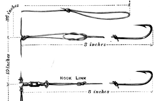
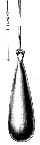
Fig. 33.—The Paternoster
It will be seen that a swivel has been introduced in connection with the bottom hook, the object being to show the manner in which this useful piece of tackle is fitted. It must not be supposed, however, that swivels are always necessary. It is often useful to insert a swivel on the line itself, above the Paternoster, when it is of twisted material, in order to prevent it from kinking; but its use is more frequently serviceable on the hook links, especially when fishing where the currents are strong. When the bait used is one that presents two flat surfaces to the water, as would be the case with a strip of mackerel, a strong current will set it spinning round and round, thus causing the hook link to kink if it has not been fitted with a swivel, and the same effect is often produced by the spinning of a fish on the hook.
The employment of a suitable ground bait will often make a wonderful difference in the angler’s haul. It frequently attracts large numbers, keeping them near at hand for some considerable time, and apparently sharpens their appetite. It may be often observed, too, that a fish will bite freely at the angler’s bait when in the neighbourhood of the ground bait, while the former is viewed with suspicion in the absence of the latter.
When fishing on the bottom only, the ground bait should be weighted if it is of such a nature that it does not sink readily or if it is liable to be carried away by currents; but it will often be found more convenient to secure it on the end of a string, tied up in a muslin bag if necessary, so that it may be adjusted to any desired depth.
Among the attractive viands suitable for this purpose we may mention mussels, crushed crabs, pounded liver, the guts of any oily fish, and the offal of almost any animal.
Along the east coast, and in some of the sandy bays of Devon and Cornwall, fishing from the beach is practised, but we can hardly recommend this as of much value to the amateur whose object is to obtain as great a variety as possible of fishes for study. Some good food fishes are often caught by this means, but the methods employed are often very primitive, and would lack all interest to those who love good sport.
On the east coast a long line, fitted with many hooks, is slung out as far as possible by means of a pole, and the home end either held in the hand of the fisher or fastened to the top of a flexible stick driven into the sand. The latter plan becomes necessary when more than one line is owned by the same individual, and he is made aware of the bite of a large fish—and a large fish only, since the hooks are placed beyond a heavy lead—by the bending of the stick.
The naturalist, however, is as much interested in the small fish as the large ones, and, even for beach fishing, a rod and line, fitted with one or two hooks only, and a lead no heavier than is absolutely essential, will be preferable. A little practice will of course be necessary in order that one may become expert in the casting of the rod line, but with large rings on the rod, and a reel without a check, or a check that can be thrown off when desired, the necessary proficiency in casting ought to be acquired without much difficulty.
In some of the sandy bays of the south-west, long lines with a heavy lead at both ends and baited hooks at short intervals throughout the whole length, are placed on the sand at low tide close to the water’s edge, and left unwatched until the next tide is out. As far as our observations go this primitive mode of fishing is usually anything but successful, the receding of the tide generally revealing a long row of clean hooks, with, perhaps, one or two dead or half-dead fish; and it is probable that most of the bait is devoured by crabs and other crustaceans before the water becomes sufficiently deep to allow the desired fishes to reach it.
There is one other method of fishing on which we may make a few remarks, although it hardly comes under the heading of shore fishing. We refer to a method of catching surface fishes from a moving boat, which method is known as whiffing. The line is weighted with a lead which must be regulated according to the speed of the boat. If the boat is an ordinary rowing-boat, kept going at only a moderate speed, a few ounces of lead will be sufficient, but a whiffing line trailing behind a sailing boat travelling in a good breeze will require a pound or two of lead to keep the bait only a little below the surface.
Beyond the lead we have three or four yards of gimp or strong gut, at the end of which is a single hook fitted with a spinner, or baited with some attractive natural or artificial bait. Whatever be the bait used, there will certainly be more or less spinning caused by the resistance offered by the water, hence it will be necessary to have a swivel beyond the lead.
When whiffing near the shore, care must be taken to avoid outlying rocks that approach the surface of the water, or a sudden snapping of the line will give you an unwelcome warning of their existence. Further, we should note that the fishes which are to be caught when whiffing do not always swim at the same depth, thus it will be advisable to fish at different distances from the surface by varying either the weight of the lead or the speed of the boat.
We have already advised our readers to take home their specimens alive for the purpose of studying their growth and habits. Now, although there may be some difficulties in the way of keeping marine animals and plants alive for any considerable time, yet we are inclined to emphasise the importance of this matter, knowing that the pleasure and instruction that may be obtained from even a moderately successful attempt to carry this out will far more than compensate for the amount of trouble entailed. There are very many marine objects that are exceedingly pretty and also very instructive, even when studied apart from the life with which they were associated in the sea. Thus, a well-preserved sea-weed may retain much of its original beauty of form and colour, the shells of numerous molluscs and crustaceans exhibit a most interesting variety of features well worthy of study, and a number of the soft-bodied animals may be preserved in such a manner as to closely resemble their living forms. This being the case, we can hardly say anything to discourage those who gather sea-side objects merely for the purpose of making a collection of pretty and interesting things to be observed and admired. Such objects must necessarily afford much pleasure and instruction, and the time spent in the collection and preparation will certainly cause the collector to stray to the haunts of the living things, where he is certain to acquire, though it may be to a great extent unconsciously, a certain amount of knowledge concerning their habits and mode of life. Moreover, sea-side collecting is one of the most healthy and invigorating of all out-door occupations, and for this reason alone should be encouraged.
Yet it must be observed that he whose sea-side occupation is merely that of a collector, and whose work at home is simply the mounting and arranging of the objects obtained, can hardly be considered a naturalist. Natural history is a living study, and its devotee is one who delights in observing the growth and development of living things, watching their habits, and noting their wonderful adaptation to their environments; and it is to encourage such observation that we so strongly recommend the young collector to keep his creatures alive as far as it is possible to do so.
The first thing to settle, then, is the nature of the vessel or vessels that are to serve the purpose of aquaria for the work of the young naturalist.
As long as the outdoor work is in progress temporary aquaria will be very useful as a means by which the objects collected may be sorted and stored until a final selection is made for the permanent tank. These temporary aquaria may consist of jars or earthenware pans of any kind, each containing a few small tufts of weed, preferably attached to pieces of rock, and a layer of sand or gravel from the beach.
As such temporary aquaria will, as a rule, be within a convenient distance from the sea-side where the collecting is being done, there will be, we presume, no great difficulty in the way of obtaining the frequent changes of water necessary to keep the animals and plants in a healthy condition, so that we need do no more now than urge the importance of avoiding overcrowding, and of renewing the water frequently for the purpose of supplying the air required for the respiration of the inmates.
When it is desired to isolate small species in such a manner that their movements may be conveniently observed, glass jars answer well; but whatever be the form or size of the vessels used, care must be taken to avoid excess of both light and heat. They should be kept in a cool place, quite out of the way of direct sunshine, and the glass vessels used should be provided with a movable casing of brown paper to exclude all light except that which penetrates from above.
Even temporary aquaria, used merely for the purpose suggested above, should be carefully watched, for a single day’s neglect will sometimes result in the loss of several valuable captives. A dead animal should be removed as soon as it is discovered to avoid the unpleasant results arising from the putrefaction of its body. The appearance of a scum or film on the surface of the water should always be regarded with suspicion. Such a scum should be removed with the aid of absorbent paper, since it tends to prevent the absorption of oxygen from the air; and, should the water be tainted in the slightest degree, it should be changed at once, or, if this is not practicable, air should be driven into it for some time by means of a syringe with a very fine nozzle. Such precautions, however, are not so urgently needed when the aquarium contains crustaceans only, for the majority of these creatures suffer less than others in the tainted sea water, some even being apparently quite as comfortable in this as in a supply fresh from the sea. Sea-weeds exhibiting the slightest tendency to decay must be removed at once; and, as regards the feeding of the animals, one must be careful to introduce only as much food as is required for immediate use, so that there be no excess of dead organic matter left to putrefy. Some of the marine animals obtained from our shores feed entirely on the minute and invisible organisms that are always present in the sea water, and others subsist principally on certain of the weeds. Many, however, of a more predaceous disposition, capture and devour living prey, while some, and more especially the crustaceans, are partial to carrion. If, therefore, the observer desires to study the ways in which the various creatures secure and devour their food, he should introduce into his aquaria live marine worms and other small animals, and also small pieces of fish or flesh.
We will now pass on to the more serious undertaking of the construction and management of a permanent salt-water aquarium.
The first point to decide is, perhaps, the size of the proposed vessel, and this will in many cases be determined partly by a consideration of the space at one’s disposal, and of the apartment it is intended to occupy. If it is to be placed in a drawing-room or other ordinary apartment of a dwelling-house, preference should be given to a window facing the north in order to avoid the direct rays of the sun, but perhaps no situation is more suitable than a cool conservatory in the shady part of a garden; and in either case a strong table or other support should be provided, of a form and size adapted to those of the aquarium to be constructed.
Various materials may be used in the construction of such an indoor aquarium, and we shall deal with two or three different types, so that the reader may make his selection according to his fancy, or to his mechanical ability, if he intends that it shall be of his own construction.
We will begin with an aquarium constructed entirely of a mixture of cement and fine sand, this being the most inexpensive and certainly the easiest to make; and although it may not be regarded as the most ornamental—but opinions will differ on this point—yet it has the decided advantage of being the nearest approach to the natural rock pool. Though somewhat heavy and cumbersome, even when empty, the amount of material used in its construction may be varied according to the taste and convenience of the maker. Further, this form of aquarium is one that will readily admit of structural alterations at any future period. It may be deepened at any time; lateral additions or extensions may be made, or a portion may at any time be shut off for the purpose of isolating certain of the animals procured.
The first thing to do is to prepare a flat, strong slab of hard wood or stone, the exact shape and size of the desired artificial pool, and then cover this, if of wood, with a mixture of fine sand and cement, mixed to a convenient consistency with water, to the depth of about one inch. The banks or walls of the pool must then be built up on all sides, and this is best done by the gradual addition of soft pellets of cement, applied in such a manner as to produce an irregular surface. Unless the walls of the aquarium be very thick and massive the cement will soon show a tendency to fall from its place as the height increases, but this may be avoided by doing the work in instalments, allowing each portion to set before further additions are made to the structure.
Since some marine animals like to occupy snug and shady niches in deep water while others prefer full exposure to the light in shallows, arrangements should be made for all by varying the depth of the bed, and providing several little tunnels and caverns. This may be accomplished either by working the cement itself into suitable form, or by means of piled stones obtained from the sea beach; and if the latter plan is adopted, the stones should not be obtained until the aquarium is quite ready for its living contents; for then a selection of stones and rock fragments with weeds, anemones, sponges, and other fixed forms of life attached to them, may be made. The natural appearance of a rock pool is thus more nearly approached, and in a shorter time than if the sedentary life were required to develop on an artificial ground.
Objection may be raised to the form of aquarium just described on the ground that no life within it is visible except when viewed from above. But is not this also the case with a rock pool on the sea shore? And has any admirer of nature ever been heard to complain of the beauties of such a pool because he was unable to look at them through the sides? Further, it may be urged that the inmates of our aquarium will be living under more natural conditions than those of the more popular glass-sided aquaria, because they receive light from above only.
However, should the reader require a glass front to his cement tank, the matter is easily accomplished. Three sides are built up as before described. A sheet of thick glass—plate glass by preference—is then cut to the size and shape of the remaining space, and this is fixed by means of cement pressed well against its edges, both inside and outside.
Water should not be put into the tank until it is quite dry; and, if one side is made of glass, not until the cement surrounding the edge of the glass has been liberally painted with marine glue, hot pitch, or some other suitable waterproof material.
If any pipes are required in connection with the water supply of the aquarium, according to either of the suggestions in a later portion of this chapter, such pipes may be fixed in their proper places as the cement sides are being built up.
The next type of aquarium we have to describe is of low cost as far as the materials are concerned, and one that may be made by any one who has had a little experience in woodwork; and although the tank itself is of a simple rectangular form, yet it may be made to look very pretty with a suitable adjustment of rocks and weeds.
It consists of a rectangular box, the bottom, ends, and back of which are of hard wood, firmly dovetailed together, and the front of plate glass let into grooves in the bottom and ends. All the joints and grooves are caulked with marine glue, but no paint should be used in the interior.
This form of tank may be vastly improved by the substitution of slabs of slate for the wood, though, of course, this change entails a much greater expenditure of both time and cash; but supposing the work to be well done, the result is everything that could be desired as far as strength and durability are concerned.
In either of the rectangular tanks just described glass may be used for two sides instead of one only; and since this is not a matter of very great importance, the choice may well be left to the fancy of the one who constructs it.
Some prefer an aquarium with glass on all sides, and where this is the case the framework may be made of angle zinc with all the joints strongly soldered. Such an aquarium may be made in the form of any regular polygon, for it is no more difficult to construct one of six or eight sides than of four. It is more difficult, however, to make such an aquarium perfectly watertight, for the glass, instead of being in grooves, has to be securely fastened to the metal frame by means of a cement on one side only, and this cement has to serve the double purpose of holding the glass and keeping in the water.
Various mixtures have been suggested for this purpose, and among them the following are perfectly satisfactory:—
| 1. | Litharge | 2 parts |
| Fine sand | 2 ” | |
| Plaster of Paris | 2 ” | |
| Powdered resin | 1 part |
Mix into a very thick paste with boiled linseed oil and a little driers.
| 2. | Red lead | 3 parts |
| Fine sand | 3 ” | |
| Powdered resin | 1 part |
Mix with boiled linseed oil as above.
Both these cements should be applied very liberally, and the aquarium then allowed to remain quite undisturbed for at least two weeks before any water is introduced.
When ready for the water, the bottom of the aquarium should be covered with a moderately thick layer of fine sand from the sea shore, and stones then piled in such a manner as to form little tunnels and caves to serve as hiding-places for those creatures that prefer to be under cover. As to the selection of stones, we have already suggested that some may have weeds rooted to them, and that pieces of rock with anemones, sponges, and other forms of life attached may be chipped off. Further, on many of our rocky coasts we may find, near low-water mark, a number of stones covered with a layer of vegetable growth, amongst which many small animals live, often more or less concealed by their protective colouring. Some of these stones placed on the bed of the salt-water aquarium would add greatly to the natural appearance, as well as give greater variety to the living objects. Shells bearing the calcareous, snakelike tubes of the common serpula (p. 121), preferably with the living animals enclosed, will also enhance the general appearance and interest of the aquarium.
In making preparations previous to the introduction of animal life, due regard should be paid to the peculiar requirements of the creatures it is intended to obtain. We have already referred to the advisability of arranging the bed of the tank in such a manner that the water may vary considerably in depth, so that both deep and shallow water may be found by the animals as required, and to the provision of dark holes for crustaceans and other creatures that shun the light. Very fine sand should be provided for shrimps, prawns, and other animals that like to lie on it; and this sand must be deep in places if it is intended to introduce any of the burrowing molluscs and marine worms.
The water used may be taken from the sea or be artificially prepared. The former is certainly to be preferred whenever it can be conveniently obtained, and at the present time few will find much difficulty in securing a supply, for not only are we favoured with the means of obtaining any desired quantity by rail at a cheap rate from almost any seaport, but there are companies in various ports who undertake the supply of sea water to any part of the kingdom. If the water is to be conveyed from the coast without the aid of the regular dealers in this commodity, great care must be taken to see that the barrel or other receptacle used for the purpose is perfectly clean. Nothing is more convenient than an ordinary beer or wine barrel, but it should be previously cleansed by filling it several times with water—not necessarily sea water—and allowing each refill to remain in it some time before emptying. This must be repeated as long as the water shows the slightest colouration after standing for some time in the barrel.
Should any difficulty arise in the way of getting the salt water direct from the sea, it may be made artificially by dissolving ‘sea salt’ in the proper proportion of fresh water, or even by purchasing the different salts contained in the sea separately, and then adding them to fresh water in proportionate quantities.
The composition of sea water is as follows:—
| Water | 96·47 | per cent. |
| Sodium chloride | 2·70 | ” |
| Magnesium chloride | ·36 | ” |
| Magnesium sulphate (Epsom salts) | ·23 | ” |
| Calcium sulphate | ·14 | ” |
| Potassium chloride | ·07 | ” |
| Traces of other substances | ·03 | ” |
| 100·00 |
and it will be seen from this table that artificial sea water may be made by adding about three and a half pounds of sea salt, obtained from the sea by the simple process of evaporation, to every ninety-six and a half pounds of fresh water used. In making it there may be some difficulty in determining the weight of the large volume of water required to fill an aquarium of moderate dimensions, but this will probably disappear if it be remembered that one gallon of water weighs just ten pounds, and, therefore, one pint weighs twenty ounces.
If the sea salt cannot be readily obtained, the following mixture may be made, the different salts being purchased separately:—
| Water | 96½ | lbs. |
| Sodium chloride (common salt) | 43¼ | ozs. |
| Magnesium chloride | 5¾ | ” |
| Epsom salts | 3¾ | ” |
| Powdered gypsum (calcium sulphate) | 2¼ | ” |
Although in this mixture the substances contained in the sea in very small quantities have been entirely omitted, yet it will answer its purpose apparently as well as the artificial sea water prepared from the true sea salt, and may therefore be used whenever neither sea salt nor the natural sea water is procurable.
Assuming, now, that the aquarium has been filled with sea water, it remains to introduce the animal and vegetable life for which it is intended; and here it will be necessary to say something with regard to the amount of life that may be safely installed, and the main conditions that determine the proportion in which the animal and vegetable life should be present in order to insure the greatest success.
Concerning the first of these we must caution the reader against the common error of overcrowding the aquarium with animals. It must be remembered that almost all marine animals obtain the oxygen gas required for purposes of respiration from the air dissolved in the water. Now, atmospheric air is only very slightly soluble in water, and hence we can never have an abundant supply in the water of an aquarium at any one time. If a number of animals be placed in any ordinary indoor aquarium, they very soon use up the dissolved oxygen; and, if no means have been taken to replace the loss, the animals die, and their dead bodies soon begin to putrefy and saturate the water with the poisonous products of decomposition.
It is probably well known to the reader that a large proportion of the oxygen absorbed by the respiratory organs of animals is converted by combination of carbon into carbonic acid gas within their bodies, and that this gas is given back into the water where it dissolves, thus taking the place of the oxygen used in its formation.
If, then, an aquarium of any kind is to be a success, some means must be taken to keep the water constantly supplied with fresh oxygen quite as rapidly as it is consumed, and this can be done satisfactorily by the introduction of a proportionate quantity of suitable living weeds, providing there is not too much animal life present.
The majority of living plants require carbonic acid gas as a food, and, under the influence of light, decompose this gas, liberating the oxygen it contained. This is true of many of our common sea-weeds, and thus it is possible to establish in a salt-water aquarium such a balance of animal and vegetable life that the water is maintained in its normal condition, the carbonic acid gas being absorbed by the plants as fast as it is excreted by animals, and oxygen supplied by the plants as rapidly as it is consumed by the animals.
This condition, however, is more difficult to obtain in a salt-water aquarium than in one containing fresh-water life, partly because, generally speaking, the sea-weeds do not supply oxygen to the water as rapidly as do the plants of our ponds and streams, and partly because of the difficulties attending the successful growth of sea-weeds in artificial aquaria. Thus it is usually necessary to adopt some means of mechanically aërating the water; but, for the present, we shall consider the sea-weeds only, leaving the mechanical methods of aërating the water for a later portion of this chapter.
In the first place, let us advise the amateur to confine his attention to the smaller species of weeds that are commonly found in small and shallow rock pools, for the successful growth of the larger purple and olive weeds will probably be beyond his power, even though his tank be one of considerable capacity. The best plan is that we have already suggested—namely, to chip off small pieces of rock with tufts of weed attached, and to fix them amongst the rockery of the aquarium, being careful to place those that grew in shallow water with full exposure to the light, and those which occupied sheltered and shady places in the rock pool, respectively, in similar situations in the artificial pool.
For the purposes of aëration we have to rely principally on the bright green weeds, and preference should be given to any of these that exhibit, in their natural habitat, a multitude of minute air-bubbles on the surface of their fronds, for the bubbles consist principally of oxygen that is being liberated by the plant, and denote that the species in question are those that are most valuable for maintaining the desired condition of the water in an aquarium.
Any small sea-weed may be tried at first, but experience will soon show that some are much more easily kept alive than others. In this experimental stage, however, a constant watch should be maintained for the purpose of detecting signs of decay in the marine garden. A plant should always be removed as soon as it presents any change from the natural colour, or exhibits the smallest amount of slimy growths on the surface, for decomposing plants, as well as decaying animals, will soon convert an aquarium into a vessel of putrid and poisonous water.
It seems almost unnecessary to name a selection of sea-weeds for small aquaria, seeing that our rock pools produce so many extremely beautiful species, most of which may be successfully kept alive in a well-managed tank; but the common Sea Grass (Enteromorpha compressa), and the Sea Lettuce (Ulva latissima), also known locally as the Green Laver or Sloke, are particularly useful for the aëration of the water; while the Common Coralline (Corallina officinalis), the Dulse (Schizymenia edulis), the Peacock’s tail (Padina pavonia), the Irish or Carrageen Moss (Chondrus crispus), Callithamnion, Griffithsia setacea, Plocamium plumosium, Rhodymenia palmata, Rhodophyllis bifida, and Ceramium rubrum are all beautiful plants that ought to give no trouble to the aquarium-keeper.
It is not advisable to introduce animal life into the aquarium immediately it is filled, on account of the possibility of the water being contaminated by contact with the cement that has been used to make it water-tight. It is safer to allow the first water to stand for a few weeks, the weeds and all other objects being in situ, and the necessary means employed for perfect aëration during this interval, and then, immediately before the animals are placed in their new home, to syphon off the whole of the water, and refill with a fresh supply.
In the selection of animals due regard should be paid to two important points—first, the danger of overcrowding, and, secondly, the destructive habits of some of the more predaceous species.
No more than two or three animals should, as a rule, be reckoned for each gallon of water; and the proportion of animals should be even less than this when any of them are of considerable size.
As regards the destructive species, these are intended to include both those that are voracious vegetable feeders and also those whose habit it is to kill and prey on other creatures.
It must be understood that the weeds are to serve two distinct purposes:—They are to supply at least some of the oxygen required for the respiration of the animal inmates, and also to serve as food for them. Some marine fishes and molluscs feed on the fronds of the weeds, and among these the common periwinkle may be mentioned as one of the most voracious. If many such animals are housed in the aquarium, it will be necessary to replace at intervals those species of weeds that suffer most from their ravages. The zoospores thrown off by the weeds, particularly in the autumn, are also valuable as food for some of the animals.
Notwithstanding the destructive character of the periwinkle just referred to, it has one redeeming feature, for it is certainly useful in the aquarium as a scavenger, as it greedily devours the low forms of vegetable life that cover the glass and rocks, thus helping to keep them clean; and the same is true of the common limpet and other creeping molluscs. Some of these are even more to be valued on account of their partiality for decaying vegetable matter, by devouring which they reduce the amount of the products of decomposition passing into the water.
Other details concerning the selection of animal and vegetable life for the indoor aquarium must be left to the discretion and experience of the keeper, for it is impossible by written instructions and advice to cover all the various sources of loss and trouble that may from time to time arise. If, however, the general hints for the management of the marine aquarium here given be faithfully followed, there ought to be no further losses than must accrue from the injudicious selection of animal species, and these will decrease as experience has been acquired respecting the habits of the creatures introduced.
We must now pass on to matters pertaining to the maintenance of the healthy condition of an aquarium which, we will suppose, has been established with due regard to scientific principles. Under this head we shall consider, (1) the aëration of the water, (2) the repair of loss due to evaporation, and (3) the regulation of light and temperature.
It has already been shown that the marine aquarium can hardly be maintained in a satisfactory condition as regards its air supply by leaving the aëration of the water entirely to the action of plant life; and herein this form of aquarium differs from that employed for the animal and vegetable life derived from ponds and streams. Fresh-water weeds develop and multiply with such rapidity, and are such ready generators of oxygen gas that it is a very easy matter to establish a fresh-water aquarium that will remain in good condition for years with but little attention; it is therefore important that we should point out the difference in treatment necessary to those of our readers who are already acquainted with the comparative ease with which the fresh-water aquarium may be kept in good order, lest they expect the same self-aërating condition in the marine tank.
It is never a good plan to leave the renovation of the water of the aquarium until there are visible signs within that something is going wrong. It is true that an unsatisfactory condition of the water, revealed by a slight taint in the odour, or a general turbidity, or the formation of a slight scum on the surface, may sometimes be rectified by the prompt application of some method of artificial aëration, but the aim of the aquarium-keeper should be not the rectification of unsatisfactory conditions, but the establishment of such a method of aëration that the unsatisfactory condition becomes an impossibility. We do not wish to discourage anyone who has the slightest desire to start a marine aquarium. Our aim is to point out any difficulties that lie in the way in order that the aquarium may be a success; and thus, having stated that the difficulties attending it are somewhat greater than those connected with the management of a fresh-water aquarium, we should like to add that these practically disappear when one is prepared to devote a short time at regular intervals in order to see that the process of aëration is properly carried out.
Some recommend the occasional injection of air by a syringe as one means of aërating the water; but, although this may be all very well as a temporary purifier of the slightly tainted aquarium, it is hardly suitable as a means of maintaining a good, healthy condition. It must be remembered that oxygen gas—the gas of the atmosphere so essential to animal life—is only very slightly soluble in water. By this we mean not only that water dissolves oxygen very slowly, but also that it can never hold a large supply of the gas at any one time. This being the case, it is clear that the use of a syringe for a short time, though it discharges an enormous total volume of air into the water, will result in the actual solution of only a small quantity. No method of aëration is perfect that allows the admission of air for a short time only at comparatively long intervals; the most perfect system is that in which air is slowly but continuously passed into solution.
Since air is slightly soluble in water, it is clear that it must be continuously passing into any body of water that has its surface freely exposed to it; hence a wide and shallow aquarium is much more likely to keep in good order than one that is narrower and deeper. But, with marine aquaria, the simple absorption from the air at the surface is not in itself sufficient, as a rule, to maintain a healthy condition. Yet it will be advisable to remember this matter when constructing a tank for marine life.
One of the prettiest, and certainly one of the most effectual, methods of supplying air to an aquarium is by means of a small fountain with a very fine spray. The water need seldom be changed, but the fountain may be fed by water from the aquarium, and as the fine spray passes through the air it will absorb oxygen and carry it in solution to the tank.
The accompanying diagram illustrates the manner in which this can be accomplished. The aquarium (a) is supplied with an outlet (o) about an inch from the top by means of which the water is prevented from overflowing, and the outlet pipe leads to a vessel (v) of considerable capacity which, for the sake of convenience and appearance, may be concealed beneath the table on which the aquarium stands. Some feet above the level of the aquarium is another vessel (c), supported on a shelf, having about the same capacity as v, and supplied with a small compo pipe that passes down into the aquarium, and then, hidden as much as possible by the rockery, terminates in a very fine jet just above the level of the water in the centre. The upper vessel should also be provided at the top with a loose covering of muslin to serve as a strainer, and this should be replaced at intervals as it becomes clogged with sedimentary matter.
In order that this arrangement may give perfect satisfaction the two vessels (c and v) must each be of at least half the capacity of the aquarium itself, and the total quantity of salt water sufficient to fill the aquarium together with one of them. It should also be remembered that since the pressure of water depends not on its quantity, but on its height measured perpendicularly, it follows that the height to which the fountain will play is determined by the height of the vessel c above the level of the jet.
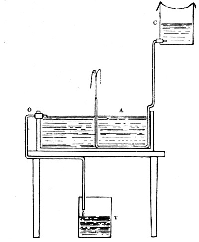
Fig. 38.—Method of aërating the Water of an Aquarium
a, aquarium with fountain; c, cistern to supply the fountain; o, pipe for overflow; v, vessel for overflow
Let us now suppose that the aquarium and the upper vessel have both been filled with sea water. The fine jet from the pipe plays into the air and returns with a supply of oxygen to the aquarium, while the excess above the level of o passes into the concealed vessel below the table. If the two vessels are as large as we recommend, and the jet a very fine one, the fountain may continue to play for hours before c is empty, the animals of the tank being favoured all this time with a continuous supply of air. And when the supply from above is exhausted, the contents of the bottom vessel are transferred to the top one, and at the same time so effectually strained by the layer of muslin that no sedimentary matter passes down to choke the fine jet of the fountain. One great advantage this method possesses is that the living creatures derive the benefit of a much larger quantity of water than the aquarium alone could contain; and thus, apart from the aërating effects of the fountain, the result is the same as if a much larger tank were employed.
In our next illustration (fig. 39) we give a modified arrangement based on the same principle which may commend itself by preference to some of our readers. Here the supply pipe to the fountain passes through a hole in the bottom of the aquarium instead of into the top, and the outlet pipe is bent downward within so as to form a syphon.
Those who are acquainted with the principle of the syphon will understand at once the working of such an arrangement as this. Let us suppose the vessel c to be full of water, and the fountain started, while the water in the aquarium stands no higher than the level l. The water slowly rises until the level h of the bend of the outlet tube has been reached, and during the whole of this time no water escapes through the exit. As soon, however, as the latter level has been attained, the water flows away into the lower vessel, into which it continues to run until the lower level is reached, and then the outflow ceases, not to commence again until the fountain causes the water to rise to the upper level.
From what has been said the reader will see that the total quantity of water required in this instance need not exceed the capacity of the aquarium; also that each of the vessels connected with water supply and waste should have a capacity equivalent to the volume of water contained in the aquarium between the two levels h and l.
The alternate rising and falling of the water produced in the manner just described represents in miniature the flow and ebb of the tides, but perhaps this is in itself of no great advantage in the aquarium except from the fact that it allows those creatures that prefer to be occasionally out of the water for a time a better opportunity of indulging in such a habit. And further, with regard to both the arrangements for aëration above described, it should be noted that earthenware vessels are much to be preferred to those made of metal for the holding of sea water, since the dissolved salts corrode metallic substances rather rapidly, and often produce, by their chemical action, soluble products that render the water more or less poisonous.
Other methods of aërating the water of aquaria are practised, but these, as a rule, are only practicable in the case of the large tanks of public aquaria and biological laboratories, as the mechanical appliances necessary to carry them out successfully are beyond the means of an ordinary amateur.
In such large tanks as those referred to it is common to force a fine jet of air into the water by machinery. Sometimes this air is driven downward from a jet just below the surface, and with such force that a multitude of minute bubbles penetrate to a considerable depth before they commence to rise, but in others the air is made to enter at the bottom and must therefore pass right through the water.
Of course the amateur aquarium-keeper may carry out this method of aëration with every hope of success providing he has some self-acting apparatus for the purpose, or can depend on being able himself to attend to a non-automatic arrangement at fairly regular intervals, always remembering that a single day’s neglect, especially in the case of a small tank with a proportionately large amount of animal life, may lead to a loss of valuable specimens.
We have already mentioned the use of a syringe as a means by which an aquarium may be temporarily restored to a satisfactory condition providing it has not been neglected too long, and some recommend forcing air, or, still better, pure oxygen gas, from an india-rubber bag into the water. We have used, for the same purpose, a stream of oxygen from a steel cylinder of the compressed gas with very satisfactory results; and since oxygen may be now obtained, ready compressed, at a very low price—about twopence a cubic foot—there is much to be said in favour of this method as an auxiliary in the hands of the owner of a small tank, though we hardly recommend it as a prime means of aëration to take the place of the fountain.
In any case, where a stream of air or oxygen is employed, an exceedingly fine jet should be used, in order that the expelled gas may take the form of a stream of minute bubbles; for, as previously stated, the water can absorb the gas only very slowly, so that there must necessarily be a considerable waste when the gas issues rapidly. Further, the smaller the bubbles passing through the water, the greater is the total surface of gas in contact with the liquid, the volume of the supply being the same, and hence the more effectually will the solution of the gas proceed. Again, another advantage of the fine stream of minute bubbles lies in the fact that the smaller these bubbles are the more slowly they rise to the surface of the water, and thus the longer is the time in which the gas may be absorbed during its ascent.
A fine jet, well suited to the purpose here defined, may be made very easily by holding the middle of a piece of glass tubing in a gas flame until it is very soft, and then, immediately on removing it, pulling it out rather quickly. A slight cut made with a small triangular file will then enable the operator to sever the tube at any desired point.
Yet another method of maintaining the air supply of aquaria is adopted in the case of some of the large tanks of public aquaria and biological laboratories situated close to the sea, and this consists in renewing the water at every high tide by means of pumps.
It must not be supposed that an indoor aquarium, even when well established, and supplied with the best possible system of aëration, requires no further care and attention. In the first place there is a continual loss of water by evaporation, especially in warm and dry weather, and this must be rectified occasionally. Now, when water containing salts in solution evaporates, the water passing away into the air is perfectly free from the saline matter, and thus the percentage of salt in the residue becomes higher than before. It is evident, therefore, that the loss by evaporation in a marine aquarium must be replaced by the addition of fresh water, which should either be distilled, or from the domestic supply, providing it is soft and moderately free from dissolved material.
But the question may be asked, ‘Do not the marine animals and plants utilise a certain amount of the saline matter contained in the salt water?’ The answer to this is certainly in the affirmative, for all sea-weeds require and abstract small proportions of certain salts, the nature of which varies considerably in the case of different species; and, further, all the shelled crustaceans and molluscs require the salts of lime for the development of their external coverings, and fishes for the growth of their bony skeletons. Hence the above suggestion as to the replenishment of loss by evaporation with pure water is not perfectly satisfactory. It will answer quite satisfactorily, however, providing the sea water is occasionally changed for an entirely new supply. Again, since carbonate of lime is removed from sea water more than any other salt, being such an essential constituent of both the external and internal skeletons of so many marine animals, as well as of the calcareous framework of the coralline weeds, we suggest that the aquarium may always contain a clean piece of some variety of carbonate of lime, such as chalk, limestone, or marble, which will slowly dissolve and replace that which has been absorbed.
Water is rendered denser, and consequently more buoyant, by the presence of dissolved salts; and, since the density increases with the proportion of dissolved material, we are enabled to determine the degree of salinity by finding the density of the solution. We can apply this principle to the aquarium, as a means of determining whether the water contains the correct amount of sea salt, also for testing any artificial salt water that has been prepared for the aquarium.
Probably some of our readers are acquainted with some form of hydrometer—an instrument used for finding the density of any liquid; but we will describe a simple substitute that may be of use to the owner of a marine aquarium, especially if the salt water for the same is artificially prepared. Melt a little bees-wax, and mix it with fine, clean sand. Then, remembering that the wax is lighter than water, and consequently floats, while sand is considerably heavier, and sinks, adjust the above mixture until a solid ball of it is just heavy enough to sink very slowly in sea water. Now make two such balls, and then cover one of them with a light coating of pure wax. We have now two balls, one of which will float in sea water, and the other sink, and these may be used at any time to test the density of the water in, or prepared for, the aquarium. If the water is only a little too salt, both balls will float; while, if not sufficiently rich in saline matter, both will sink.
We must conclude this chapter by making a few remarks on the important matter of the regulation of light and temperature. Direct sunlight should always be avoided, except for short and occasional intervals, not only because it is liable to raise the temperature to a higher degree than is suitable for the inmates of the aquarium, but also because an excess of light and heat tends to produce a rapid decomposition of organic matter, and a consequent putrid condition of the water, and this dangerous state is most likely to occur when both light and temperature are high at the same time.
The water should always be cold; and as it is not always easy to estimate the temperature, even approximately, by the sensation produced on immersing the fingers, it is a good plan to have a small thermometer always at hand, or placed permanently in the aquarium. In the summer time the water should be kept down to fifty-five degrees or lower, and in winter should never be allowed to cool much below forty. There may be some difficulty in maintaining a temperature sufficiently low in summer, but a small piece of ice thrown in occasionally to replace the loss due to evaporation, especially on very hot days, will help to keep it down.
The sea-side naturalist, in the course of his ramblings and searchings on the coast, will certainly come across many objects, dead or alive, that he will desire to set aside for future study or identification in his leisure moments at home. Some of these will be required for temporary purposes only, while, most probably, a large proportion will be retained permanently for the establishment of a private museum, that shall serve not only as a pleasant reminder of the many enjoyable hours spent on the shore, but also as a means of reference for the study of the classification of natural objects and of their distribution and habitats.
We will first deal with those specimens that are required for temporary purposes only—those of which the collector desires to study the general characters, as well as, perhaps, something of the internal structure; but before doing so we cannot refrain from impressing on the reader the advisability of learning as much as possible of the external features and mode of growth of the different living creatures while still alive, for it must be remembered that it is impossible to preserve many of them without more or less destruction of their natural colouring and distortion of their characteristic forms.
In those cases where it is possible to keep the creatures alive for a short time only, it is a good plan to make notes of their movements and all observed changes in form, and their methods of feeding, and also to illustrate these notes by sketches drawn from life. This may seem quite an unnecessary procedure to many beginners in the study of natural objects, and may even, as far as the sketches are concerned, present difficulties that at first appear to be insurmountable; but the power to sketch from nature will surely be acquired to a greater or less degree by constant practice, and illustrated notes prepared for the purpose we suggest will undoubtedly be of great value to the student. Further, though it may often be necessary to set specimens aside in a preservative fluid until one has the leisure to examine their structure, it should always be remembered that they never improve by keeping, also that they are rarely in such good condition for dissection after saturation with the preservative as when perfectly fresh.
One of the most convenient preservatives for general use is undoubtedly methylated spirit. This is alcohol that has been adulterated in order to render it undrinkable, so that it may be sold free from duty for use in the various arts and manufactures without any danger of its being employed for the concoction of beverages. It may be used just as purchased—that is, in its strongest condition—for many purposes, but in this state it has a powerful affinity for water, and will rapidly abstract water from animal and vegetable objects, causing the softer ones to become hard, shrunken, and shrivelled, often to such an extent that they are almost beyond recognition.
By diluting the spirit, however, we satisfy to a great extent its affinity for water, and thus prevent, or, at least, reduce the action just mentioned. A mixture of equal quantities of spirit and water is quite strong enough. Unfortunately the common methylated spirit of the shops produces a fine white precipitate, that gives the whole mass a milky appearance, when it is diluted. This is due to the presence of mineral naphtha, which is added in a certain fixed proportion in accordance with the Government regulations. But it is possible, by special application, to obtain the ‘non-mineralised’ or ‘ordinary’ methylated spirit of former years, though not in small quantities, and this liquid dissolves in water without the formation of a precipitate. It should be noted, however, that the use of the spirit as a preservative is in no way interfered with by the presence of the mineral naphtha, the only disadvantage of this impurity lying in the fact that the milkiness consequent on dilution prevents the objects in a specimen jar from being observed without removal.
We have just referred to the hardening action of strong spirit as a disadvantage, and so it is when it is required to preserve soft structures with as little as possible of change in general form and appearance; but there are times when it becomes necessary to harden these soft structures in order that sections may be made for the purpose of examining internal structure with or without the aid of the microscope, and for such purposes strong spirit is one of the best hardening agents that can be employed.
Formaldehyde is another very good preservative. It is a colourless liquid, and should be considerably diluted for use, a two per cent. solution being quite strong enough for all ordinary purposes. It possesses some distinct advantages as compared with spirit. In the first place, it does not destroy the natural colours of objects to the extent that spirit does; and, although a hardening agent as well as a preservative, it does not harden soft structures by the extraction of the water they contain, and therefore does not cause them to become shrivelled or otherwise distorted. It will also occur to the reader that, since a small bulk of formaline represents a large volume of the diluted preservative, it is very conveniently stored, and a very small bottle of it taken for outdoor work may, on dilution with water, be made to yield all that is required for the preservation of the takings of a successful day, or even of a longer period. Formaldehyde is usually sold in solution of about forty per cent. strength, and for the preparation of a two per cent. solution it will be found convenient to provide a glass measure graduated either into cubic centimetres or fluid ounces and drams. One hundred volumes of the original solution contain forty of pure formaldehyde, and if water be added to make this up to two thousand volumes, a two per cent. solution is obtained. Thus, one hundred cubic centimetres of the original solution is sufficient to prepare two litres (three and a half pints) of suitable preservative.
A very good preservative liquid may be made by dissolving two ounces of common salt, one ounce of alum, and two or three grains of corrosive sublimate (a deadly poison) in one quart of water, and then, after allowing all sedimentary matter to settle to the bottom, decanting off the clear solution. This mixture is known as Goadby’s fluid, and is well adapted for the preservation of both animal and vegetable structures. It does not cause any undue contraction of soft tissues, and, as a rule, does not destroy the natural colours of the objects kept in it.
Glycerine is valuable as a preservative for both animal and vegetable objects, and especially for the soft-bodied marine animals that form such a large percentage of the fauna of our shores. It maintains the tissues in a soft condition, and preserves the natural tints as well as any liquid.
An inexpensive preservative may also be made by dissolving chloride of zinc—about one ounce to the pint of water. This is considered by some to be one of the best fluids for keeping animal structures in good condition.
Now, although the different fluids here mentioned are described in connection with the temporary preservation of natural objects, it must be remembered that they are equally adapted for the permanent preservation of the animals and plants that are to figure in the museum of the sea-side naturalist; and, although some marine objects may be preserved in a dry state in a manner to be hereafter described, yet there are many species of animals, and also some plants, that can be satisfactorily preserved only by immersion in a suitable fluid.
This method may be applied to all soft-bodied animals, such as anemones, jelly-fishes, marine worms, shell-less molluscs (sea slugs, cephalopods, &c.), the soft parts of shelled molluscs, fishes, &c.; and most sponges retain their natural appearance much better in a preservative fluid than in a dry condition. Many sea-weeds also, which are practically destroyed by the most careful drying process, are most perfectly preserved in fluid.
But the puzzled amateur will probably be inclined to ask: ‘Which is the best preservative liquid for this or that specimen?’ No satisfactory general rule can be given in answer to such a question, and a great deal will have to be determined by his own experiments and observations. Whenever he has two or three specimens of the same object, as many different fluids should be employed, and the results compared and noted. In this way a very great deal of useful information will be obtained and by the best possible means. However, it may be mentioned that all the fluids alluded to above may be safely used for almost every animal or vegetable specimen with the following reservations: strong spirit should not be employed for any very soft animal, nor should it be used for delicate green plants, since it will dissolve out the green colouring matter (chlorophyll), leaving them white or almost colourless. Further, the greatest care should be exercised in dealing with sea anemones and jelly-fishes. If spirit is used for preserving these creatures, it should be very dilute, at least at first, but may with advantage be increased in strength afterwards, though this should be done gradually.
Whatever be the preservative used, it is sure to be more or less charged with sedimentary and coloured matter extracted from the object immersed in it; hence, if the specimen concerned is to form part of a museum collection, it will be necessary to transfer it to a fresh solution after a time, and a second, and even further changes may be necessary before the object ceases to discolour the fluid or render it turbid.
Considerable difficulty will sometimes be found in the attempts to preserve a soft-bodied animal in its natural attitude. Thus, when a sea anemone is removed from its native element, it generally withdraws its tentacles, and, contracting the upper part of its cylindrical body, entirely conceals these appendages, together with the mouth they surround; and a mollusc similarly treated will generally pull itself together within its shell, leaving little or no trace of the living body inhabiting the lifeless case. Then, if these animals are transferred to any fluid other than sea water, or placed anywhere under unnatural conditions, they usually remain in their closed or unexpanded form. Thus, almost every attempt to kill them for preservation deprives them of just the characteristics they should retain as museum specimens.
Some such animals may be dealt with satisfactorily as follows: Transfer them to a vessel of fresh sea water, and leave them perfectly undisturbed until they assume the desired form or attitude. Then add a solution of corrosive sublimate very gradually—a drop or two at intervals of some minutes. In this way the bodies of anemones may be obtained ready for preservation with expanded tentacles, tube-secreting worms with their heads and slender processes protruding from their limy or sandy cases, molluscs with their ‘feet’ or their mantles and gills protruding from their shells, and barnacles with their plume-like appendages projecting beyond the opening of their conical shells.
The specimens thus prepared may be placed at first in very dilute spirit, and then, after a time, finally stored in a stronger solution of spirit in water; or they may be transferred to one of the other preservative solutions previously mentioned.
All specimens permanently preserved in fluid for a museum should be placed in jars, bottles, or tubes of suitable size, each vessel containing, as a rule, only one. Where expense is no object, stoppered jars made expressly for biological and anatomical specimens may be used for all but the smallest objects; or, failing this, ordinary wide-mouthed bottles of white glass, fitted with good corks or glass stoppers.
For very small specimens nothing is more suitable than glass tubes, but it must be remembered that wherever corks are used, even if they are of the best quality procurable, it will be necessary to look over the specimens occasionally to see if the preserving fluid has disappeared to any extent either by leakage or evaporation; for such loss is always liable to occur, although it may be very slow, and especially when methylated spirit is the liquid employed.
The writer has preserved many hundreds of small marine and other objects in glass tubes of dilute spirit that have been hermetically sealed, thus rendering the slightest loss absolutely impossible, while the perfect exclusion of air prevents the development of fungoid growths that sometimes make their appearance in imperfectly preserved specimens. The making and closing of such tubes, though a more or less difficult operation at first to those who have had no previous experience in glass-working, become exceedingly simple after a little practice; and believing it probable that many of our readers would like to try their hand at this most perfect method of preserving and protecting small objects, we will give a description of the manner in which it is done.
The apparatus and materials required for this work are:—Lengths of ‘soft’ glass tubing, varying from about one quarter to a little over half an inch in internal diameter; a supply of diluted spirit—about half spirit and half water; a Herapath blowpipe, preferably with foot-bellows; and a small triangular file.
The glass tubing may be cut into convenient lengths by giving a single sharp stroke with the file, and then pulling it apart with, at the same time, a slight bending from the cut made.
Cut a piece of tubing about eight or nine inches long, heat it in the blowpipe flame, turning it round and round all the time, until it is quite soft, then remove it from the flame and immediately pull it out slowly until the diameter in the middle is reduced to about a sixteenth of an inch (fig. 41, 2). Make a slight scratch with the file at the narrowest part, and divide the tube at this point (fig. 41, 3). Now heat one of these pieces of tubing as before just at the point where the diameter of the drawn part begins to decrease; and, when very soft, pull it out rather quickly while it is still in the flame. The part pulled now becomes completely separated, and the tube is closed, but pointed. Continue to heat the closed end, directing the flame to the point rather than to the sides, until the melted glass forms a rather thick and flattened end; and then, immediately on removing it from the flame, blow gently into the open end until the melted glass is nicely rounded like the bottom of a test-tube (fig. 41, 4). When the tube is cold, the specimen that it is to contain, and which has already been stored for a time in dilute spirit, is dropped into it. The tube is now heated about an inch above the top of the specimen, drawn out as shown in fig. 41, 5, and again allowed to cool. When cold, the fresh spirit is poured into the open end of the tube, but the middle part is so narrow that the spirit will not run down freely. If, however, suction be applied to the open end, air from the bottom will bubble through the spirit, and then, on the cessation of the suction, the spirit will pass down to take the place of the air that was withdrawn. This may be repeated if necessary to entirely cover the specimen with the fluid. Any excess of spirit is then thrown from the upper part of the tube, and the latter cut off. Nothing is now left but to close the tube hermetically. This is done by heating the lower part of the narrow neck, and then drawing it out in the flame, taking great care that the tube is withdrawn from the flame the moment it is closed. The tube must also be kept in an upright position until it has cooled. The appearance of the finished tube is shown in fig. 41, 6.
All preserved specimens should have a label attached on which is written the name of the specimen, the class and order to which it belongs, the locality in which it was found, together with any brief remarks that the owner desires to remember concerning its habits &c.
The bottles or tubes that are too small to have a label attached to them in the ordinary way may be mounted on a card, as represented in fig. 42, and the desired particulars then written on the card.
When soft or delicate specimens are preserved in a bottle of fluid they frequently require some kind of support to keep them in proper form and to display them better for observation. Perhaps the best way to support them is to fasten them to a very thin plate of mica of suitable size by means of a needle and very fine thread. The mica is so transparent that it is invisible in the fluid, and the few stitches are also hardly perceptible, thus making it appear as if the specimen floats freely in the fluid.
We will now pass on to consider those objects of the shore that are usually preserved in a dry condition, commencing with
Starfishes are commonly preserved by simply allowing them to dry in an airy place, with or without direct exposure to the sun’s rays, and this method is fairly satisfactory when the drying proceeds rapidly; but care should be taken to maintain the natural roughness of the exterior as well as to have the numerous suckers of the under surface as prominent as possible. If the starfish is simply laid out on some surface to dry, the side on which it rests is often more or less flattened by the weight of the specimen itself, which therefore becomes adapted for the future examination of one surface only; but a better result, as regards both the rapidity of drying and the after appearance of the specimen, may be obtained by suspending it on a piece of fine net or by threads. A still better plan is to put the dead starfish into strong spirit, which will rapidly extract the greater part of the moisture that its body contained. After allowing it to remain in this for a day or two to harden it, put it out to dry as before mentioned. The spirit, being very volatile, will soon evaporate, so that the specimen will shortly be ready for storing away.
It is most important to observe that dried specimens—not starfishes only, but all animal and vegetable objects—should never be placed in the cabinet or other store-case until perfectly dry, for a very small amount of moisture left in them will often encourage the development of moulds, not only on themselves, but on other specimens stored with them.
Very small and delicate starfishes, when preserved in a dry condition, may be protected from injury by fastening them on a card by means of a little gum, or by keeping them permanently stored on cotton wool in glass-topped boxes.
Sea urchins, or sea eggs, as they are commonly called, may be preserved exactly in the same way as starfishes, though it is more essential in the case of these to soak them in strong spirit previous to drying, otherwise the soft animal matter within the shell will decompose before the drying is complete. Here, however, it is possible to remove the whole interior with the aid of a piece of bent wire, and to thoroughly clean the inner surface of the shell before drying it.
Some of the shells should be preserved with the spines all intact, and others with these removed in order to show the arrangement of the plates which compose the shell, as well as the perforations, and the rounded processes to which the spines are articulated.
The majority of sea urchins are provided with a most complicated and beautiful arrangement of teeth which are well worthy of study. These should be removed from a moderately large specimen, the soft surrounding structures carefully dissected away, and then cleaned by means of an old tooth-brush without disarranging them.
It will be found that dried sea urchins will require care when preserved with spines attached, for these appendages are usually very brittle and are easily dislocated at their bases where they are united to the shell by ball-and-socket joints.
It may be mentioned here that corrosive sublimate is very valuable for preventing the development of mould on the surfaces of starfishes, sea urchins, and museum specimens generally. It is best supplied in the form of an alcoholic solution made by dissolving a few grains in about half a pint of methylated spirit; the advantage of this over an aqueous solution being the rapidity with which it dries. In most cases it is simply necessary to apply the solution to the object by means of a soft brush, but, as regards starfishes and urchins it is far better to dissolve a few grains of the corrosive sublimate in the spirit in which the objects are placed previous to drying.
The preservation of crustaceans by the dry method often requires some care and demands a certain amount of time; but the process is never really difficult, and the satisfaction of having produced a good specimen for a permanent collection well repays one for the trouble taken and time spent.
Some of our crustaceans are only partially protected by a firm outer covering, and almost every attempt to preserve these as dry objects results in such a shrivelling of the soft tissues that the natural appearance is quite destroyed. This is the case with some of the barnacles, and the abdominal portion of the bodies of hermit crabs, which are, therefore, far better preserved in fluid. Dilute spirit is quite satisfactory for most of these as far as the preservation of the soft structures is concerned, but it has the disadvantage that it turns the shells of some crustaceans red, making them appear as if they had been boiled.
Other crustaceans are so small, or are hardened externally to such a slight extent, that they also are not adapted for the dry method of preservation. Speaking generally, such crustaceans as shrimps and sand-hoppers are best preserved in fluid, while the different species of crabs and lobsters are more conveniently preserved dry unless it is desired to study any of their soft structures.
It is quite impossible to remove the soft parts from small crabs and lobsters previous to drying them, hence the drying should be conducted as rapidly as possible, so that no decomposition may set in. Where the process goes on very slowly, as is the case when the air is damp, or when the specimens are not set out in an airy spot, a decay of the soft structures soon proceeds, and the products of this decay will generally saturate the whole specimen, giving rise to most objectionable odours, and destroying the natural colour of the shell.
If it has been found that the species in question are not reddened by the action of methylated spirit, they should be allowed to remain in this fluid, with a few grains of dissolved corrosive sublimate, for at least a few hours, and then they will dry rapidly without any signs of putrefaction; and even those species that are reddened by spirit may be treated to a shorter immersion in this fluid with advantage.
The specimens should always be set out in some natural attitude to dry, unless it is desired to spread out the various appendages in some manner that is more convenient for the study of their structure. A sheet of blotting-paper may be placed on cork or soft wood, the specimens placed on this, and the appendages kept in the desired positions when necessary by means of pins placed beside, but not thrust through them. When more than one specimen of the same species has been collected, one should be set in such a manner as to exhibit the under side; and, further, in instances where the male and female of the same crustacean differ in structure, as is commonly the case, two of each should be preserved, one displaying the upper, and the other the under surface.
When perfectly dry, all small crustaceans should be mounted on cards with the aid of a little gum, and the name and other particulars to be remembered then written on the card.
The question may well be asked: ‘Which is the best gum to use?’ In answer to this we may say that gum tragacanth is certainly as good as any. It holds well, and leaves no visible stain on a white card. A small quantity of the solid gum should be put into a bottle with water in which a grain or so of corrosive sublimate has been dissolved. It absorbs much water, becoming a very soft, jelly-like mass. Any excess of water may be poured off, and the gum is then ready for use.
The larger crabs and lobsters contain such an amount of soft tissue within that it becomes absolutely necessary to clear them in order to avoid the unpleasant and destructive effects of decomposition.
In the case of lobsters the abdomen should be removed from the large cephalo-thorax by cutting through the connecting membrane with a sharp knife. The soft portions of both halves of the body are then raked out by means of a piece of wire flattened and bent at one end, and the interior cleaned with the aid of a rather stiff bottle-brush. The large claws are then removed by cutting through the membrane that unites them with the legs, and these are cleared in a similar manner. The different parts are next laid out to dry on blotting-paper, with the various appendages attached to the body arranged just as in life; and, finally, when all parts are quite dry, both within and without, the separated parts are reattached by means of some kind of cement. For this purpose a solution of gelatine in acetic acid is much better than gum tragacanth, as it has a far greater holding power, and this is necessary when we require to unite rather large structures with but small surfaces in contact.
Large crabs are to be dealt with much in the same manner, but, instead of removing the abdomen only, which, in the crab, is usually very small and doubled under the thorax, the whole carapace—the large shell that covers the entire upper surface of the body—should be lifted off, and replaced again after the specimen has been cleaned and dried.
We have previously dealt with the preservation of the shell-less molluscs, and the soft bodies of the shelled species when such are required, so we will now see what should be done with the shells.
Numerous shells are often to be found on the sea beach—shells that have been washed in by the breakers, and from which the animal contents have disappeared, either by the natural process of decay, aided by the action of the waves, or by the ravages of the voracious or carrion-eating denizens of the sea; and although these shells are rarely perfect, having been tossed about among the other material of the beach, yet we occasionally find here the most perfect specimens of both univalve and bivalve shells in such a condition that they are ready for the cabinet, and these often include species that are seldom found between the tide-marks, or that are otherwise difficult to obtain.
However, the shell-collector must not rely on such specimens as these for the purpose of making up his stock, but must search out the living molluscs in their habitats and prepare the shells as required.
The molluscs collected for this purpose are immersed in boiling water for a short time, and the animal then removed from the shell. In the case of bivalves it will generally be found that the hot water has caused the muscles of the animal to separate from the valves to which they were attached, or, if not, they have been so far softened that they are easily detached, while it does not destroy the ligament by means of which the valves are held together at the hinge; but the univalve molluscs must be removed from their shells by means of a bent pin or wire. In the latter instance care must be taken to extract the whole of the body of the animal, otherwise the remaining portion will decompose within the shell, giving rise to the noxious products of natural decay.
The univalves have now simply to be placed mouth downwards on blotting-paper to drain and dry, when they are ready for the cabinet. If, however, they include those species, like the periwinkles and whelks, that close their shells by means of a horny lid (operculum) when they draw in their bodies, these lids should be removed from the animal and attached to their proper places in the mouth of the shell. The best way to accomplish this is to pack the dry shells with cotton wool, and then fasten the opercula to the wool by means of a little gum tragacanth or acetic glue.
Bivalve shells should, as a rule, be closed while the ligament is still supple, and kept closed until it is quite dry, when the valves will remain together just in the position they assume when pulled together by the living animal. The shells of the larger species may be conveniently kept closed during the drying of the ligament by means of thread tied round them, but the very small ones are best held together by means of a delicate spring made by bending fine brass wire into the form shown in fig. 44.
There are many features connected with the internal structure and surface of the shells of molluscs that are quite as interesting and instructive as those exhibited externally; hence a collection of the shells intended for future study should display internal as well as external characteristics. Thus, some of the spiral univalve shells may be ground down on an ordinary grindstone in order to display the central pillar (the columella) and the winding cavity that surrounds it, while others, such as the cowries, may be ground transversely to show the widely different character of the interior. Bivalve shells, too, may be arranged with the valves wide open for the study of the pearly layer, the lines of growth, the scars which mark the positions of the muscles that were attached to the shell, and the teeth which are so wonderfully formed in some species.
Some collectors make it a rule to thoroughly clean all the shells in their collection, but this, we think, is a great mistake; for when this is done many of the specimens display an aspect that is but seldom observed in nature. Many shells, and especially those usually obtained in deep water, are almost always covered with various forms of both animal and vegetable growth, and it is advisable to display these in a collection, not only because they determine the general natural appearance, but also because these growths are in themselves very interesting objects. Further, it is a most interesting study to inquire into the possible advantages of these external growths to the inhabitants of the shells, and vice versâ—a study to which we shall refer again in certain chapters devoted to the description of the animals concerned.
But there is no reason whatever why some of the duplicate specimens should not be cleaned by means of a suitable brush, with or without the use of dilute hydrochloric acid (spirits of salt), or even polished, in some few cases, to show the beautiful colours so often exhibited when the surface layer has been removed. This, however, should be done somewhat sparingly, thus giving the greater prominence to the exhibition of those appearances most commonly displayed by the shells as we find them on the beach or dredge them from the sea.
Very small and delicate shells may be mounted on cards, as suggested for other objects; but, as a rule, the specimens are best displayed by simply placing them on a layer of cotton wool in shallow boxes of convenient size. The number of insects that may be described as truly marine is so small that their preservation is not likely to form an important part of the work of the sea-side naturalist; and even though a considerable number of species exhibit a decided partiality for the coast, living either on the beach or the cliffs, the study of these is more generally the work of the entomologist. For this reason, and partly because we have already given full instructions for the setting and mounting of insects in a former work of this series, we consider a repetition inadmissible here.
The subject of the preservation of fishes, also, will require but few words. There is no satisfactory method of preserving these in a dry state, though we often meet with certain thin-bodied species, such as the pipe-fish, that have been preserved by simply drying them in the sun. Fishes should be placed in dilute spirit, or in one of the other liquids recommended, but a change of fluid will always be necessary after a time, and also frequently the gentle application of a brush to remove coagulated slime from the surface of the scales.
The great drawbacks in the way of preserving a collection of fishes are the expense of the specimen jars, and the large amount of space required for storing the specimens. Of course the former difficulty can be overcome by substituting ordinary wide-mouthed bottles in the place of the anatomical jars, while the latter can be avoided to a considerable extent by limiting the collection to small species, and to small specimens of the larger species. If this is done, it is surprising what a large number of fishes can be satisfactorily stored in bottles of only a few ounces’ capacity.
The apparatus required for the preservation of the wild flowers of our cliffs, and the sea weeds, consists of a quantity of blotting paper or other thick absorbent paper cut to a convenient size, a few thin boards and a few pieces of calico of the same size, some heavy weights, and several sheets of drawing paper.
The wild flowers are arranged on the sheets of absorbent paper while still fresh, care being taken to display the principal parts to the best advantage. They are then placed in a single pile, with a few extra sheets of absorbent paper between each two specimens to facilitate the drying, boards at the bottom and top as well as at equal distances in the midst of the pile, and the weights on the top of the whole.
The natural colours of leaves and flowers are not very often preserved satisfactorily, but the best results are obtained when the drying process proceeds most rapidly. Hence, if the press contains any specimens of a succulent or sappy nature, they should be taken out after the first day or two, and then replaced with a fresh supply of dry paper.
The flowers must be left in the press until quite dry, and they may then be mounted on sheets of drawing paper, by fixing them with a little gum tragacanth, or by narrow strips of gummed paper passing over their stems.
Some collectors prefer simply placing their botanical specimens inside double sheets of drawing paper, not fastening them at all, and there is much to be said in favour of this, especially as it allows the specimens to be examined on both sides; and even when they are fastened to the paper double sheets are much to be preferred, for the specimens are not then so liable to be damaged by friction when being turned over, especially when the names are written on the outside of each sheet.
The larger sea-weeds may be dried in the same manner, though it is a good plan to absorb the greater part of the moisture they contain by pressing them between pieces of calico previous to placing them in the ordinary press. It should be observed, however, that many sea-weeds exude a certain amount of glutinous substance that makes them adhere to the paper between which they are dried, while they do not so freely adhere to calico. These should be partially dried in the calico press, and then laid on the paper on which they are to be finally mounted, and re-pressed with a piece of dry calico on the top of each specimen.
Many of the smaller weeds may be treated in the manner just described, but the more delicate species require to be dealt with as follows:—Place each in a large, shallow vessel of water, and move it about, if necessary, to cause its delicate fronds to assume that graceful form so characteristic of the algæ of our rock pools. Then immerse the sheet of paper on which the weed is to be finally mounted, and slowly raise the specimen out of the water, on the paper, without disturbing the arrangement of the fronds. If it is found necessary to rearrange any of the fronds, it may be done by means of a wet camel-hair brush. Now lay the specimen on calico or absorbent paper, placed on a sloping board, to drain; and, after the greater part of the moisture has disappeared by draining and evaporation, transfer the specimen to the press with a piece of dry calico immediately over it. All are dealt with in turn in the manner described, and allowed to remain in the press until perfectly dry, when it will be found that the majority of them have become firmly attached to the mount, and require nothing but the label to fit them for the herbarium.
Sea-weed collectors often make the great mistake of pressing tufts that are far too dense to admit of the structural characters being satisfactorily examined. To avoid this fault, it will often be necessary to divide the clusters collected so that the forms of their fronds may be more readily observed.
The calcareous corallines may be pressed in the same way as the other algæ, but very pretty tufts of these, having much the appearance of the living plant, may be obtained by simply suspending them until thoroughly dry; though, of course, specimens so prepared must not be submitted to pressure after they are dry, being then so brittle that they are easily broken to pieces.
The hard framework of these interesting corallines is composed principally of carbonate of lime, a mineral substance that dissolves freely in hydrochloric acid (spirits of salt). Thus, if we place a tuft of coralline in this acid, which should be considerably diluted with water, the calcareous skeleton immediately begins to dissolve, with the evolution of minute bubbles of carbonic acid gas; and after a short time, the end of which is denoted by the absence of any further bubbling, nothing remains but the vegetable matter, now rendered soft and pliant. A decalcified specimen of coralline may be pressed and dried, and then mounted beside the plant in its natural condition for comparison; and the true appearance of the vegetable structure may also be retained, and in a far more satisfactory manner, by preserving a portion of the specimen in dilute spirit.
Finally, it may be observed that many sea-weeds, like wild flowers, do not retain their natural forms and colours when preserved dry. They are spoilt by the pressure applied, or become so shrivelled and discoloured in the drying as to be but sorry representatives of the beautifully tinted and graceful clothing of the rocks of the coast. But many of those that suffer most in appearance when dried may be made to retain all their natural beauty by preserving them in a fluid; and it is most important that this should be remembered by all who desire to study the weeds at home, and particularly by those who possess a microscope, and wish to search into the minute structure of marine algæ. Our own plan is to keep not only the dried specimens for the purpose of studying the general characters and classification of the algæ, but also to keep a few large bottles—stock bottles—filled with weeds of all kinds in a preservative fluid. These latter are exceedingly useful at times, and are frequently brought into requisition for close inspection, with or without the microscope. Small pieces may be detached for microscopic examination when required, and sections may be cut either for temporary or permanent mounting just as well as from living specimens, such sections showing all the details of structure exhibited by the living plant.
One of the greatest difficulties besetting the young collector lies in the choice and construction of the cabinet or other store-house for the accommodation of the specimens that accumulate as time advances.
Of course, when expense is a matter of no great consideration, a visit to the nearest public or private museum to see the manner in which the specimens are housed, followed by an order to a cabinet-maker, will set the matter right in a short time; but it is probable that the majority of our readers are unable to fit up their museum in this luxurious style, and will either have to construct their own cabinets and store-boxes or to purchase cheap substitutes for them.
Where one has the mechanical ability, and the time to spare, the construction of a cabinet with the required number of drawers may be undertaken, and there is no better form of store than this. The whole should be made of well-seasoned wood, and the drawers should vary in depth according to the size of the specimens they are to contain. Some of these drawers may be lined with sheet cork, and the cork covered with white paper or a thin layer of cotton wool. This will enable some of the specimens to be fixed in their places by means of pins. As a rule, however, no pins will be required, and the specimens will be most conveniently arranged in shallow cardboard boxes, placed in rows in the drawer, a little cotton wool covering the bottom of each.
Failing the usual cabinet, the specimens may be stored in shallow trays or boxes, or even in the little cardboard cabinets so often sold for storing stationery &c. The best and cheapest things of this kind we have ever met with are the little cabinets, each containing either six or twelve drawers, made by Macdonald & Co., of Temple Row, Birmingham. By the use of such as these the specimens may be neatly stored away, and additions to match may always be made as the collection increases in magnitude.
The specimens should all be classified according to their positions in the animal or vegetable world, and accompanied by labels giving the name of species and genus, together with localities, habitats, &c. The outlines of classification may be studied from the later chapters of this work, in which the common objects of the sea shore are described in their scientific order, beginning with the lowest sub-kingdoms and classes; and further, it will be observed that the sub-kingdoms are divided into classes, the classes into orders, orders into families, families into genera, and that the genera contain a smaller or larger number of closely allied species.
The collection must be kept in a perfectly dry place, otherwise many of the specimens will be liable to develop moulds, and this will, of course, quite spoil their appearance. It is almost sure to be attacked by mites and other animal pests unless some means be taken to prevent their intrusion.
As regards the latter, it is well to know that it is far easier to prevent the intrusion of small animal pests than it is to exterminate them after they have once found an entrance; and so, from the very commencement of the formation of the collection, all drawers and boxes should be charged with some substance that is objectionable, if not fatal, to them. Small lumps of naphthaline (albo-carbon) put into the various compartments, and renewed occasionally as they disappear by evaporation, will generally suffice to prevent the entrance of all pests, but this substance is not effectual as an insecticide for the purpose of killing them after they are in.
Perhaps the best of all insecticides is the corrosive sublimate already mentioned, and this may be applied to any animal or vegetable object that is capable of providing food for museum pests, and it is difficult to find such an object on which they will not feed.
Many of the specimens that find a place in a museum have been temporarily preserved in spirit previous to being dried, and if a little corrosive sublimate was dissolved in this spirit, the specimens will have been rendered perfectly free from all attacks of marauders, since the spirit will have saturated the whole object, carrying with it the dissolved poison.
Most of the specimens that have not been treated by the above method would not suffer from a short immersion in spirit containing the corrosive sublimate; but in cases where it is considered inexpedient to do this, the same liquid may be applied to them by means of a soft brush. In this way even the dried botanical specimens may be rendered perfectly secure from attacks.
An enthusiastic observer of nature will learn much concerning the structure of natural objects with the unaided eye, but there are times when he will desire some kind of magnifier to reveal more perfectly the structure of minute parts, or to enable him to observe the small creatures that are invisible to the naked eye. Further, one may learn many interesting and instructive facts relating to animal and plant life by cutting sections for close examination, or by making such simple dissections as will enable one to observe the more salient features of internal structure; we therefore propose in the present chapter to make a few remarks and suggestions regarding work of this kind.
A pocket magnifier is of great value to the young naturalist, both for the inspection of natural objects while engaged in out-door work, and for the subsequent examination of the specimens collected for study. It is often necessary to enable one to identify and classify small animals and plants, and will be in constant demand for the purpose of studying the less conspicuous external features. Such an instrument should be regarded as an essential companion of the naturalist, and should accompany him on every ramble.
There are several different forms of pocket lenses, but for general work there is, perhaps, nothing more convenient and serviceable than the ‘triplet’ magnifier. It is a combination of three lenses, enclosed in a pocket case, and so arranged that they may be used separately or in combination, thus supplying a variety of powers. The three lenses of the triplet are themselves of different magnifying powers, and these powers may be increased by combining two or all of them.
For work at home a ‘dissecting microscope’ is very useful. This consists of a magnifying lens, mounted on a support over a surface on which small objects may be examined and dissected, the height of the lens being, of course, adjusted according to its focal distance. Lenses ready mounted on adjustable stands may be purchased for this purpose, but no one ought to experience much difficulty in designing and constructing some simple stand that will give every satisfaction.
The arrangement just described is, of course, suitable for the dissection of only small objects, and these are placed on a material adapted to the nature of the work to be done. Thus it is sometimes convenient to place the object to be examined on a small sheet of cork, in order that it may be secured by means of pins while the dissection proceeds, while at other times it is essential that it be laid on a hard and unyielding surface, such as that of a slip of glass. But whatever be the nature of the substance on which the dissection is made, its colour may be regulated according to that of the object. If, for example, we are dissecting a small white flower on a piece of cork, we should naturally blacken the cork, or cover it with a piece of dead black paper; or, if we are to dissect a small, light-coloured object on a glass surface, we lay the glass on black paper.
The advantage of dissecting objects under water does not seem to be generally appreciated by beginners, who often allow their specimens to become dry and shrivelled, almost beyond recognition, during the progress of their examination. This mode of dissection is certainly not necessary with all objects, but may be generally recommended for soft and succulent vegetable structures, as well as for almost all animal dissections.
This being the case, arrangements should certainly be made to provide a miniature dissecting trough as an accessory to the dissecting microscope, and the following instructions will enable the reader to construct a highly satisfactory and inexpensive one:—
Procure the flat lid of a cylindrical tin box, or the lid of a glass or porcelain pomade pot, such lid to be about two inches in diameter and about half an inch in depth. Cement the flat side of this lid to a small slab of hard wood, or to a square piece of sheet lead, by means of acetic glue—ordinary glue or gelatine dissolved in glacial acetic acid—to give it the necessary steadiness during the dissection. When the cement is quite hard, pour into the lid some melted paraffin (paraffin wax) which has been blackened by the admixture of a small quantity of lamp-black in the form of a fine powder. The paraffin should be melted by putting it into a beaker or wide-mouthed bottle, and standing it in hot water, and the lamp-black should be added, with stirring, as soon as it is entirely liquefied. The quantity of the mixture used must be sufficient to half fill the lid, thus leaving a space to contain water to the depth of about a quarter of an inch. The blackened wax provides a good background on which to work, and provides a hold for pins when these are necessary in order to fix the object under examination.
The complete trough is represented in fig. 46; and will be found to answer its purpose admirably, except that it occasionally displays one fault, but one that is easily remedied. The wax contracts on cooling, and may, therefore, detach itself from the trough; and, being lighter than water, will float instead of remaining submerged. This may be prevented by securing the disc of wax in its place by means of a ring of brass wire, or by weighting the wax with two or three small pieces of lead pushed down into it while it is yet soft.
With such a dissecting microscope and trough as we have described one may do a great deal of exceedingly useful work, both hands being quite free to manipulate the object under examination.
The dissection may be conducted with the aid of a small scalpel or other very sharp knife, the parts being arranged or adjusted by means of a needle, mounted in a handle, and held in the left hand. Sometimes, however, the object to be dissected is so minute that even a small scalpel is too large for the purpose, and in such cases nothing is better than little dissecting instruments made by mounting large sewing needles in suitable handles, and then grinding down the points of the needles on two opposite sides, on a hone, so as to produce little pointed, two-edged blades. Bent needles are often useful, too, and these may be prepared by heating the points to redness in a gas-flame, bending them as desired while hot, and then hardening them by suddenly thrusting them, at a red heat, into cold water.
The compound microscope will often prove useful for the examination of very minute objects, as well as for the study of the structure of the principal tissues of the larger species; but since detailed instructions for the management of the microscope, and for the preparation of objects for microscopic examination would occupy much more space than we can spare, we shall content ourselves with nothing more than a few general hints on this portion of the young naturalist’s work, dealing more particularly with those points which commonly present difficulties to the amateur.
If it is desired to examine some minute living object, such as a protozoon, place the object in a drop of the water in which it lived just in the middle of a clean glass slip, and cover it with a cover-glass. The quantity of water should be just sufficient to fill the space between the two glasses. If less than this has been used, a little more applied to the edge of the cover by means of a glass rod will immediately run in between the glasses; while if an excessive amount was employed, the surplus may be removed by the application of a strip of blotting paper. Place the glass slip on the stage of the microscope, and reflect light through it from the mirror below.
Examine it first with a low power; and, after having observed as much as possible of the creature’s movements and structure with this aid, repeat with a higher power. This rule applies not only to such small objects as we have now under consideration, but to all objects, and parts of them, in which minute details are to be observed.
Beginners with the microscope often find prolonged examination very tiring to the eyes, but this, we believe, would seldom be the case if right methods were followed. Both eyes should always be open, and the microscopist should train himself to use both eyes equally for the actual observation.
The higher the magnifying power used, the nearer must the objective (the lower combination of lenses) be brought to the object itself, and it is no uncommon thing for the amateur, in his attempts to focus his object, to lower the body of the microscope beyond its proper position, causing the objective to crush the object, break the thin cover-glass, and become wetted with the liquid, if any, in which the object was being examined. All this may be avoided by lowering the body of the microscope until it nearly touches the cover-glass before attempting to view the object through it, and then, with the eye above the object-glass, to gradually raise the body until the object is in focus.
The top of the cover-glass should always be perfectly dry; and if by any chance the objective becomes wet it should be wiped perfectly dry with a piece of old silk or with chamois leather. Also, if permanent mounting is attempted, and the preservative liquid is allowed to come in contact with the objective, such liquid must, of course, be washed off with some suitable solvent before any attempt is made to wipe the lens dry.
If the object under examination is of such dimensions that the cover-glass has a tendency to rock on it, or if it is a living object of such a size that it is unable to move freely in the exceedingly thin film of water between the cover and the slip, it should be placed in a cell. The cell may be made by cementing a ring of glass or vulcanite to the middle of a slip, or it may be a little circular cavity prepared in the slip itself. In either case the cell must be quite full of water before the cover-glass is applied, so that no air-bubbles are included.
Hitherto we have spoken only of mounting small objects in water, and this is advisable when the object is moist, whether it be animal or vegetable, alive or dead. But dry objects may be examined in the dry state, in which case they need not be covered. If they are composed of transparent material they are to be dealt with in the manner recommended before, as far as the management of the light is considered; that is, a moderately strong light is sent through them by the reflector below the stage; but opaque objects are best examined on a dead black ground, the light being directed on to them by means of a condensing lens placed between them and the source of light.
A collector who has done only a few days’ work on the sea shore will probably find himself the possessor of a host of interesting objects that will afford much pleasure and instruction when placed under the microscope—objects, many of which have been somewhat hastily deposited in a bottle of spirit or other preservative for study in his future leisure moments. These objects, if small, may be examined as above described, simply placing them under a cover-glass, or in a cell, with a clear drop of the same liquid in which they have been kept.
The general characters of the larger objects may also be observed by means of some kind of hand lens, but even these are generally best examined under water or other suitable liquid.
A great deal may be learnt of natural objects by preparing very thin sections for microscopic examination; and although special works should be consulted if one desires to become proficient in the different methods of cutting and preparing such sections, yet a great amount of good work may be done with the aid of a sharp razor, manipulated with nothing more than ordinary skill.
Some objects, especially certain of those of the vegetable world, are of such a nature that suitable sections may be cut, either from the fresh or preserved specimen, without any preliminary preparation. All that is required is to hold the object firmly between the finger and thumb of the left hand, previously securing it in some kind of holder if necessary, and pare off the thinnest possible slices with a horizontal movement of the razor, both razor and object being kept very wet during the process. As the sections are cut they may be allowed to drop into a shallow vessel of water; and, the thinnest then selected for examination in water as previously described.
Other objects are so soft that the cutting of sections becomes impossible without previously hardening them. Methylated spirit is a good hardening reagent, and many of the soft structures that have been preserved in this fluid, especially if it has been used undiluted, will be found sufficiently hard for cutting thin sections. Among the other hardening reagents used by microscopists may be mentioned a solution of chromic acid—one part by weight of the solid acid dissolved in from one hundred to two hundred parts of water, and a solution of bichromate of potash—one part of the bichromate to about forty parts of water. In either case the hardening of the object takes place slowly, and it should be examined from day to day until the necessary consistence has been obtained.
The structures of many soft animals can never be satisfactorily hardened for section-cutting by either of the above reagents, and thus it becomes necessary either to freeze or to imbed them. In the former case the object is first soaked in gum water—a thin solution of gum arabic—and then frozen by an ether spray or by a mixture of ice and salt. The sections should be cut with a razor just as the object is beginning to thaw, and they may then be examined under a cover-glass, in a drop of the gum water.
The other method is conducted as follows:—The soft object is first soaked in absolute alcohol to extract all the water it contains, and is then transferred to paraffin that has been heated just to its melting-point by standing it in warm water. After the object is thoroughly permeated with the paraffin, the whole is cooled quickly by immersion in cold water. Sections are now cut, the paraffin being sliced away with the substance it contains. These sections are placed in warm turpentine, where they are allowed to remain until the whole of the wax has dissolved, and they may then be mounted in a drop of turpentine, and covered with a cover-glass.
We have given brief instructions for temporary mounting only, but most amateur microscopists would undoubtedly prefer mounting their objects permanently, so that they may be set aside for study at any future period. Hence we append a few directions to this end, advising the reader, however, to consult a work dealing especially with this subject if he desires to become proficient in the preparation of microscopic slides.
Moist objects, including those which have been preserved in dilute spirit, may be soaked in water, then transferred direct to the glass slip, and covered with a drop of glycerine. Any excess of the glycerine should then be absorbed from around the cover-glass by means of a strip of blotting-paper, and the edge of the cover cemented by gold size applied with a small camel-hair brush.
Glycerine jelly is also a valuable mountant for permanent work. When this is used the object should first be soaked in glycerine, and then in the melted jelly. It is then transferred to a drop of melted jelly which has been placed on a warm slide, and covered as before. The jelly soon solidifies, so that a ring of cement is not absolutely necessary, though it is advisable, as a rule, to cement the cover-glass all round with gold size or black varnish.
Sections cut while frozen are best mounted in glycerine, to which they may be transferred direct.
Canada balsam is one of the best media for permanent mounting; and, as it becomes very hard after a time, it serves the purposes of both preservative and cement. When this is used the object must be entirely freed from water by soaking it in absolute alcohol. It is then put into turpentine for a minute or two, transferred to a warm slide, and covered with a drop of the prepared balsam. Sections that have been imbedded in paraffin may be mounted in this way, the turpentine acting as a solvent for the paraffin in which it was cut.
Although the compound microscope is absolutely necessary for the study of the minutest forms of life and of the minute structure of the various tissues of larger beings, yet the young naturalist will find that a vast amount of good work may be done without its aid. Thus the general structure of the larger species may be made out by means of simple dissections requiring no extraordinary skill on the part of the worker, and with appliances that may be obtained at a low cost. Certain of the marine animals, however, require special treatment that can hardly be described in a short chapter devoted to general instructions only, but hints with regards to these will be given in future chapters in which the animals referred to are described.
The appliances referred to above include nothing more than a simple form of dissecting trough, a few dissecting instruments, and one or two minor accessories that may always be found at hand as required.
The dissection of animals is always best performed under water, for by this method the object examined may not only be kept clean as the work proceeds, but the parts, having a tendency to float, readily separate from one another and therefore become more distinctly visible when submerged.
A very convenient form of trough may be made by taking any kind of rectangular, flat-bottomed dish, one made of zinc being, perhaps, the best of all, and covering the bottom with a slab of good cork carpet which has been weighted with sufficient lead to prevent it from floating. Or, instead of cork carpet, a sheet of cork may be used. In either case, a piece of thin sheet lead, a little larger than the slab, should be cut, the corners of which are then snipped off as shown in fig. 48, and the edges finally turned over as represented in the next illustration. The size of the trough must be regulated according to the nature of the work to be done, but one measuring ten inches long, seven wide, and two inches deep will answer most purposes.
The object to be dissected is placed in the trough, secured in position by means of a few ordinary pins, and then completely covered with water.
We need hardly impress upon the reader the great importance of thoroughly examining all external characters—all those structures that are visible without actual dissection—before attempting to remove anything; and we have already insisted on the importance of carefully examining all creatures while alive before anything else is done. The value of this latter stipulation can hardly be overestimated, for in many instances it is almost impossible to detect the use of an organ unless it has been observed in action; and the enthusiastic student will go even further than this, for he will make it an invariable rule to sketch everything he sees, and to make full notes on all his observations.
When pins are used to fix the object under examination—and it is generally essential that the object be fixed—their heads should be turned outwards; for then the object will not slip from its position, nor will the pins tend to get in the way of the work.
Some objects are of such a nature that they are not easily secured by means of pins, and yet require to be fixed in some way or other. Thus, one may desire to examine the structure and appendages of a prawn or small crab, or to investigate the nature of a chiton. In such instances as these it is a good plan to make a cake of paraffin wax of suitable size by pouring the melted substance into a mould, then secure the object in proper position in the wax while still fluid, and pin the latter to the cork of the dissecting trough.
It is often necessary to trace the courses of internal passages that open on the surface of the body, or of tubes that are revealed during the progress of dissection, and this may be done by means of a little instrument called a seeker. It is simply a blunted needle, bent into a large angle, and mounted in a handle; or, it may consist of nothing but a moderately long and stiff bristle, rendered blunt at one end by tipping it with melted sealing wax. This is not always sufficient, however, for it frequently happens that certain tubes and passages in animal forms are disposed in such a complicated manner that it is impossible to send even the most flexible seeker through them. For instance, suppose one desires to trace the course of the digestive tube of some large bivalve mollusc with its many reflections, the seeker is useless except that it will penetrate to the first sharp bend. The arrangement of such a tube must be traced by dissecting along its course, but this may be aided considerably by first filling it with some coloured substance to enable its direction to be more easily followed. In fact, the injection of some brightly coloured fluid, forced through the tube by means of a fine-nozzled glass syringe will often enable the course of such a tube to be seen without any dissection at all, the colour of the fluid used being detected through the semi-transparent tissues surrounding it. A mixture of Berlin blue and water, or a mixture of plaster of Paris and water coloured with carmine is well adapted to this purpose; and if the latter is employed it may be allowed to set, and thus produce a permanent cast from the tube that is being dissected. Perhaps it should be mentioned that if either of the injection mixtures be used for this purpose it must be previously strained through muslin, and that, in the case of the plaster, the mixing and straining should occupy as little time as possible, or it may begin to set before the injection has been completed.
A very considerable insight into the structure of animals may be frequently obtained by cutting sections through the body with all its organs in situ, but, generally speaking, they are too soft to allow of this without danger of the displacement of those very parts, the relations of which we desire to determine. To avoid this the body should be previously hardened by a somewhat prolonged soaking in methylated spirit, or in a solution of chromic acid prepared as before directed. Then, with the aid of a good razor, very interesting sections may be prepared with the greatest of ease, and the true relations of the various organs throughout the body may be exactly determined by cutting a succession of slices, not necessarily very thin, from end to end, or, transversely, from side to side.
Even those crustaceans that are protected by a hard, calcareous exo-skeleton, and the molluscs that cannot be removed from their stony shells without injury to their soft structures, may be studied in the manner just described, and this may be done by first soaking them in dilute hydrochloric acid, renewed as often as may be necessary, until all the mineral matter has been dissolved completely, and then hardening the softer tissues in one of the reagents mentioned above. Hydrochloric acid may also be used to dissolve the calcareous shells of foraminifers, the vegetable corallines, and other small forms of life, previous to microscopic examination of the soft parts.
We shall now study the principal forms of animal life to be found on the sea shore; and, in order that the reader may thoroughly understand the broader principles of classification, so as to be able to locate each creature observed in its approximate position in the scale of life, we shall consider each group in its zoological order, commencing with the lowest forms, and noting, as we proceed, the distinguishing characteristics of each division.
The present chapter will be devoted to the Protozoa—the sub-kingdom that includes the simplest of all animal beings.
Each animal in this division consists of a minute mass of a jelly-like substance called protoplasm, exhibiting little or no differentiation in structure. There is no true body-cavity, no special organs for the performance of distinct functions, and no nervous system.
Perhaps we can best understand the nature of a protozoon by selecting and examining a typical example:
Remove a small quantity of the green thread-like algous weed so commonly seen attached to the banks of both fresh and salt water pools, or surrounding floating objects, and place it in a glass with a little of the water in which it grew. This weed probably shelters numerous protozoons, among which we are almost sure to find some amœbæ if we examine a drop of the water under the high power of a microscope.
The amœba is observed to be a minute mass of protoplasm with an average diameter of about one-hundredth of an inch, endowed with a power of motion and locomotion. Its body is not uniformly clear, for the interior portion is seen to contain a number of minute granules, representing the undigested portions of the animal’s food. There is a small mass of denser protoplasm near the centre, termed the nucleus, and also a clear space filled with fluid. This latter is called the vacuole, and is probably connected with the processes of respiration and excretion, for it may be seen to contract at irregular intervals, and occasionally to collapse and expel its contents.
As we watch the amœba we see that it is continually changing its shape, sending out temporary prolongations (pseudopodia) of its gelatinous substance from any part, and sometimes using these extended portions for the purpose of dragging itself along.
Its method of feeding is as remarkable as it is simple. On coming in contact with any desired morsel, it sends out two pseudopods, one on each side of the food. These two pseudopods gradually extend round the food, till, at last, they meet and coalesce on the opposite side of it, thus completely enclosing it within the body. Any part of the body of the amœba may thus be converted into a temporary mouth; and, there being no special cavity to serve the purpose of a stomach, the process of digestion will proceed equally well in any part of the body except in the superficial layer, where the protoplasm is of a slightly firmer consistence than that of the interior. Further, the process of digestion being over, any portion of the superficial layer may be converted into a temporary opening to admit of the discharge of indigestible matter.
The amœba is an omnivorous feeder, but subsists mainly on vegetable organisms, especially on diatoms and other minute algæ; and the siliceous skeletons of the former may often be seen within the body of the animal, under the high power of a microscope.
The multiplication of the amœba is brought about by a process of fission or division. At first the nucleus divides into two, and then the softer protoplasm contracts in the middle, and finally divides into two portions, each of which contains one of the nuclei. The two distinct animals thus produced both grow until they reach the dimensions of their common progenitor.
All the protozoons resemble the amœba in general structure and function; but while some are even simpler in organisation, others are more highly specialised. Some, like the amœba, are unicellular animals; that is, they consist of a single, simple speck of protoplasm; but others live in colonies, each newly formed cell remaining attached to its parent cell, until at last a comparatively large compound protozoon is formed.
The sub-kingdom is divided into several classes, the principal of which, together with their leading characteristics, are shown in the following table:—
1. Rhizopods:—Body uniform in consistence. Pseudopods protruded from any point.
2. Protoplasta:—Outer protoplasm slightly firmer in consistence. Pseudopods protruded from any point. (Often grouped with the Rhizopods.)
3. Radiolaria:—Possessing a central membranous capsule. Usually supported by a flinty skeleton.
4. Infusoria:—Outer protoplasm firmer and denser; therefore
of more definite shape.
Possess permanent threadlike extensions of protoplasm
instead of pseudopods.
We shall now observe the principal marine members of the protozoa, commencing with the lowest forms, and dealing with each in its proper zoological order as expressed in the above table.
When we stand on a beach of fine sand on a very calm day watching the progress of the ripples over the sand as the tide recedes we frequently observe whitish lines marking the limits reached by the successive ripples as they advance toward the shore. If, now, we scrape up a little of the surface sand, following the exact course of one of these whitish streaks, and examine the material obtained by the aid of a good lens, we shall in all probability discover a number of minute shells among the grains of sand.
These shells are of various shapes—little spheres, discs, rods, spirals, &c.; but all resemble each other in that they are perforated with a number of minute holes or foramina. They are the skeletons of protozoons, belonging to the class Rhizopoda, and they exist in enormous quantities on the beds of certain seas.
We will first examine the shells, and then study the nature of the little animals that inhabit them.
The shells vary very much in general appearance as well as in shape. Some are of an opaque, dead white, the surface somewhat resembling that of a piece of unglazed porcelain; others more nearly resemble glazed porcelain, while some present quite a vitreous appearance, much after the nature of opal. In all cases, however, the material is the same, all the shells consisting of carbonate of lime, having thus the same chemical composition as chalk, limestones, and marble.
If hydrochloric acid be added to some of these shells, they are immediately attacked by the acid and are dissolved in a very short time, the solution being accompanied by an effervescence due to the escape of carbonic acid gas.
The shells vary in size from about one-twelfth to one three-hundredth of an inch, and consist either of a single chamber, or of many chambers separated from each other by perforated partitions of the same material. Sometimes these chambers are arranged in a straight line, but more frequently in the form of a single or double spiral. In some cases, however, the arrangement of chambers is very complex.
We have already referred to the fact that the shells present a number of perforations on the exterior, in addition to those which pierce the partitions within, and it is this characteristic which has led to the application of the name Foraminifera (hole-bearing) to the little beings we are considering.
The animal inhabiting the shell is exceedingly simple in structure, even more so than the amœba. It is merely a speck of protoplasm, exhibiting hardly any differentiation—nothing, in fact, save a contractile cavity (the vacuole), and numerous granules that probably represent the indigestible fragments of its food.
The protoplasm fills the shell, and also forms a complete gelatinous covering on the outside, when the animal is alive; and the vacuole and granules circulate somewhat freely within the semi-solid mass. Further, the protoplasm itself is highly contractile, as may be proved by witnessing the rapidity with which the animal can change its form.
When the foraminifer is alive, it floats freely in the sea, with a comparatively long and slender thread of its substance protruded through each hole in the shell. These threads correspond exactly in function with the blunt pseudopodia of the amœba. Should they come in contact with a particle of suitable food-material, they immediately surround it, and rapidly retracting, draw the particle to the surface of the body. The threads then completely envelop the food, coalescing as soon as they touch, thus bringing it within the animal.
The foraminifer multiplies by fission, or by a process of budding. In some species the division of the protoplasm is complete, as in the case of amœbæ, so that each animal has its own shell which encloses a single chamber, but in most cases the ‘bud’ remains attached to a parent cell, and develops a shell that is also fixed to the shell of its progenitor. The younger animal thus produced from the bud gives rise to another, which develops in the same manner; and this process continues, the new bud being always produced on the newest end, till, at last, a kind of colony of protozoons is formed, their shells remaining attached to one another, thus producing a compound shell, composed of several chambers, arranged in the form of a line or spiral, and communicating by means of their perforated partitions. It will now be seen that each ‘cell’ of the compound protozoon feeds not only for itself, but for all the members of its colony, since the nourishment imbibed by any one is capable of diffusion into the surrounding chambers, the protoplasm of the whole forming one continuous mass by means of the perforated partitions of the complex skeleton.
Some of the simplest foraminifers possess only one hole in the shell, and, consequently, are enabled to throw off pseudopods from one side of the body only. In others, of a much more complex nature, the new chambers form a spiral in such a manner that they overlap and entirely conceal those previously built; and the development may proceed until a comparatively large discoid shell is the result. This is the case with Nummulites, so called on account of the fancied resemblance to coins. Further, some species of foraminifera produce a skeleton that is horny in character, instead of being calcareous, while others are protected merely by grains of sand or particles of other solid matter that adhere to the surface of their glutinous bodies.
We have spoken of foraminifera as floating freely about in the sea water, but while it is certain that many of them live at or near the surface, some are known to thrive at considerable depths; and those who desire to study the various forms of these interesting creatures should search among dredgings whenever an opportunity occurs. Living specimens, whenever obtained, should be examined in sea water, in order that the motions of their pseudopods may be seen.
If we brush off fragments from the surface of a freshly broken piece of chalk, and allow them to fall into a vessel of water, and then examine the sediment under the microscope, we shall observe that this sediment consists of minute shells, and fragments of shells, of foraminifers. In fact, our chalk beds, as well as the beds of certain limestones, consist mainly of vast deposits of the shells of extinct foraminifera that at one time covered the floor of the sea. Such deposits are still being formed, notably that which now covers a vast area of the bed of the Atlantic Ocean at a depth varying from about 300 to 3,000 fathoms. This deposit consists mainly of the shells of a foraminifer called Globigerina bulloides, a figure of which is given on the opposite page.
The structure of chalk may be beautifully revealed by soaking a small piece of the rock for some time in a solution of Canada balsam, allowing it to become thoroughly dry, and then grinding it down till a very thin section is obtained. Such a section, when viewed under the low power of a compound microscope, will be seen to consist very largely of minute shells; though, of course, the shells themselves will be seen in section only.
The extensive beds of nummulitic limestones found in various parts of South Europe and North Africa are also composed largely of foraminifer shells, the most conspicuous of which are those already referred to as nummulites—disc-shaped shells of a spiral form, in which the older chambers overlap and hide those that enclose the earlier portion of the colony.
Before concluding our brief account of these interesting marine protozoons, it may be well to point out that, although the foraminifera belong to the lowest class of the lowest sub-kingdom of animals, yet there are some rhizopods—the Monera, which are even simpler in structure. These are mere specks of undifferentiated protoplasm, not protected by any shell, and not even possessing a nucleus, and are the simplest of all animal beings.
The second division of the Protozoa—the class Protoplasta—has already received a small share of attention, inasmuch as the amœba, which was briefly described as a type of the whole sub-kingdom, belongs to it.
The study of the amœba is usually pursued by means of specimens obtained from fresh-water pools, and reference has been made to it in a former work dealing particularly with the life of ponds and streams; but it should be observed that the amœba inhabits salt water also, and will be frequently met with by those who search for the microscopic life of the sea, especially when the water examined has been taken from those sheltered nooks of a rocky coast that are protected from the direct action of the waves, or from the little pools that are so far from the reach of the tides as to be only occasionally disturbed. Here the amœba may be seen creeping slowly over the slender green threads of the confervæ that surround the margin of the pool.
The third class—Radiolaria—is of great interest to the student of marine life, on account of the great beauty of the shells; but, as with the other members of this sub-kingdom, a compound microscope is necessary for the study of them.
The animals of this group resemble the foraminifers in that they throw out fine thread-like pseudopods, but they are distinguished from them by the possession of a membranous capsule in the centre of the body, surrounding the nucleus, and perforated in order to preserve the continuity of the deeper with the surrounding protoplasm. They have often a central contractile cavity, and further show their claim to a higher position in the animal scale than the preceding classes by the possession of little masses of cells and a certain amount of fatty and colouring matter.
Some of the radiolarians live at or near the surface of the ocean, while others thrive only at the bottom. The former, in some cases, appear to avoid the light, rising to the surface after sunset; and it is supposed that the phosphorescence of the sea is due in part to the presence of these animals. The latter may be obtained from all depths, down to several thousand fathoms.
The beauty of the radiolarians as a class lies in the wonderful shells that protect the great majority of them. These shells are composed not of carbonate of lime, as is the case with foraminifers, but of silex or silica, a substance that is not acted on by the strongest mineral acids. They are of the most exquisite shapes, and exhibit a great variety of forms. Some resemble beautifully sculptured spheres, boxes, bells, cups, &c.; while others may be likened to baskets of various ornamental design. In every case the siliceous framework consists of a number of clusters of radiating rods, all united by slender intertwining threads.
It is not all the radiolarians, however, that produce these beautiful siliceous shells. A few have no skeleton of any kind, while others are supported by a framework composed of a horny material, but yet transparent and glassy in appearance.
The sizes of the shells vary from about one five-hundredth to one half of an inch; but, of course, the larger shells are those of colonies of radiolarians, and not of single individuals, just as we observed was the case with the foraminifers.
Those in search of radiolaria for examination and study should, whenever possible, obtain small quantities of the dredgings from deep water. Material brought up by the trawl will often afford specimens; but, failing these sources of supply, the muddy deposit from deep niches between the rocks at low-water mark will often provide a very interesting variety.
Place the mud in a glass vessel, and pour on it some nitric acid (aqua-fortis). This will soon dissolve all calcareous matter present, and also destroy any organic material. A process of very careful washing is now necessary. Fill up the vessel with water, and allow some time for sedimentary matter to settle. Now decant off the greater part of the water, and repeat the process several times. By this means we get rid of the greater part of the organic material, as well as of the mineral matter that has been attacked by the acid; and if we examine the final sediment under the microscope, preferably in a drop of water, and covered with a cover-glass, any radiolarians present will soon reveal themselves.
It is often possible to obtain radiolarian shells, as well as other siliceous skeletons, through the agency of certain marine animals. The bivalve molluscs, for example, feed almost entirely on microscopic organisms; and, by removing such animals from their shells, and then destroying their bodies with aqua-fortis, we may frequently obtain a sediment composed partly of the skeletons referred to.
There remains one other class of protozoons to be considered, viz. the Infusorians—the highest class of the sub-kingdom. In this group we observe a distinct advance in organisation; for, in the first place, the infusorians are enclosed in a firm cuticle or skin, which forms an almost complete protective layer. Within this is a layer of moderately firm protoplasm, containing one or more cavities that contract at intervals like a heart. Then, in the interior, there is a mass of softer material with cavities filled with fluid, two solid bodies, and numerous granules.
In these creatures we find, too, a distinct and permanent mouth, usually funnel-shaped, leading to the soft, interior substance, in which the food material becomes embedded while the process of digestion proceeds. Here, then, for the first time, we meet with a special portion of the body set apart for the performance of the work of a stomach; and, further, the process of digestion being over, the indigestible matter is ejected through a second permanent opening in the exterior cuticle.
Again, the infusorian does not move by means of temporary pseudopods, as is the case with the lower protozoons, but by means of minute hair-like processes which permanently cover either the whole of the body, or are restricted to certain portions only. These little processes, which are called cilia, move to and fro with such rapidity that they are hardly visible; and, by means of them the little infusorian is enabled to move about in its watery home with considerable speed.
In some species a few of the cilia are much larger than the others, and formed of a firmer material. These often serve the purpose of feet, and are also used as a means by which the little animal can anchor itself to solid substances.
As with the lower protozoons, the infusoria multiply by division; but, in addition to this, the nucleus may sometimes be seen to divide up into a number of minute egg-like bodies, each of which, when set free, is capable of developing into a new animal. Should the water in which infusorians have been living evaporate to dryness, the little bodies just mentioned become so many dust particles that may be carried away by air currents; but, although dry, they retain their vitality, and develop almost immediately on being carried into a suitable environment.
Infusorians are so called because they develop rapidly in infusions of various vegetable substances; and those who desire to study their structure and movements with the aid of a microscope cannot do much better than make an infusion by pouring boiling water on fragments of dried grass, and leaving it exposed for a few days to the warm summer atmosphere. The numerous germs floating in the air will soon give rise to abundance of life, including several different species of infusoria, varying from 1/30 to 1/2000 of an inch in length.
Fresh-water pools and marshes provide such an abundance of infusoria that the animals are generally obtained for study from these sources, and a few of the common and most interesting species inhabiting fresh water have already been described in a former work. Nevertheless, the sea is abundantly supplied with representatives of the class, and it is certain that the beautiful phosphorescence sometimes observed in the sea at night is in part due to the presence of luminous infusoria, some of which appear to have an aversion to sunlight, retiring to a depth during the day, but rising to the surface again after sunset.
It seems to be the popular opinion that sponges are essentially natives of the warmer seas, and it will probably be a surprise to many young amateur naturalists to learn that there are about three hundred species of this sub-kingdom of the animal world to be found on our own shores. It must not be thought, however, that they are all comparable with the well-known toilet sponges in regard to either size or general form and structure, for some of them are very small objects, no larger than about one-twentieth of an inch in diameter, and some form mere incrustations of various dimensions on the surfaces of rocks and weeds, often of such general appearance that they would hardly be regarded as animal structures by those who have not studied the peculiarities of the group.
Sponges are known collectively as the Porifera or Polystomata, and constitute a separate sub-kingdom of animals of such distinct features that they are not readily confused with the creatures of any other group. Their principal characteristic is expressed by both the group names just given, the former of which signifies ‘hole-bearing,’ and the latter ‘many openings’; for in all the members of the sub-kingdom there are a number of holes or pores providing a means of communication between the body cavity or cavities and the surrounding water. Most of these holes are very small, but there is always at least one opening of a larger size at the anterior end.
It will be seen from what we have just stated that sponges exhibit a distinctly higher organisation than the protozoa described in the last chapter, inasmuch as they possess a permanent body-cavity that communicates with the exterior; but in addition to this there are many points of differentiation of structure that denote a superior position in the scale of life.
In order to ascertain the general features of a sponge we cannot do better than select one of the simplest forms from our own shores. If we place the live animal in a glass vessel of sea water, and examine it with a suitable magnifying power, we observe a number of minute pores scattered over its whole surface; and a much larger opening at the free end. The animal is motionless, and exhibits no signs of life except that it may contract slightly when touched. The water surrounding the sponge also appears to be perfectly still, but if we introduce some fine insoluble powder, such as precipitated chalk, or a drop of a soluble dye, the motion of the suspended or soluble material will show that the water is passing into the sponge through all the small pores, and that it is ejected through the larger opening.
On touching the sponge we observe that it is of a soft, gelatinous consistence throughout, or if, as is often the case, the body is supported by a skeleton of greater or less firmness, a gentle application of the finger will still show that this framework is surrounded by material of a jelly-like nature. This gelatinous substance is the animal itself, and a microscopic examination will show that its body-wall is made up of two distinct layers, the inner consisting of cells, many of which possess a cilium or whip-like filament that protrudes from a kind of collar, its free extremity extending into the body-cavity.
These minute cilia are the means by which the water currents just described are set up. By a constant lashing movement they urge the fluid contained in the body-cavity towards the larger hole, thus causing the water to flow in through the numerous small pores. This circulation of sea water through the body-cavity of the sponge is the means by which the animal is supplied with air and food. Air is, of course, absorbed from the water by the soft material of the external layer of the body, but the constant flow of fresh water through the body-cavity enables this process of respiration to go on with equal freedom in the interior. The mode of feeding of the sponge is very similar to that of the protozoa. Organic particles that are carried into the body-cavity, on coming in contact with the cells of the internal layer, are absorbed into their protoplasm by which they are digested. Thus the sponge may be compared to a mass of protozoon cells, all united into a common colony by a more or less perfect coalescing of the cell-substance, some of the units being modified in structure for the performance of definite functions. The air and food absorbed by any one cell may pass readily into the surrounding cells, and thus each one may be said to work for the common weal.
The description just given applies only to the simplest of the sponges, and we have now to learn that in the higher members of the group the structure is much more complicated. In these the surface-pores are the extremities of very narrow tubes which perforate both layers of the body-wall and then communicate with wider tubes or spaces within, some of which are lined with the ciliated cells above described. These spaces, which are sometimes nearly globular in form, and often arranged in groups with a common cavity, communicate with wider tubes which join together until, finally, they terminate in a large opening seen on the exterior of the sponge. Hence it will be seen that the water entering the minute pores of the surface has to circulate through a complicated system of channels and spaces, some of which are lined with the ciliated cells that urge the current onwards before it is expelled through the large hole. Further, imagine a number of such structures as we have described growing side by side, their masses coalescing into one whole, their inner tubes and spaces united into one complex system by numerous inter-communications, and having several large holes for the exit of the circulating water, and you then have some idea of the general nature of many of the more complex sponges to be found on our shores (see fig. 66).
But even this is not all, for as yet we have been regarding the sponges as consisting of animal matter only, whereas nearly all of them possess some kind of internal skeleton for the support of the soft, gelatinous animal substance. The skeleton consists of matter secreted by certain cells from material in the water and food, and is either horny, calcareous, or siliceous. The horny skeleton is formed of a network of fibres of a somewhat silky character, and often, as in the case of the toilet sponges, highly elastic; but it is sometimes so brittle that the sponge mass is easily broken when bent. The fibres of this framework support not only the outer wall of the sponge, but also the walls of all the internal tubes and spaces, which are often of so soft a nature that they would collapse without its aid.
The other forms of skeletons consist of minute bodies of carbonate of lime or of silica, respectively, which assume certain definite shapes, resembling stars, anchors, hooks, pins, spindles, &c., and are known as spicules. Such spicules are usually present in those sponges that have horny skeletons, but in others they form the entire skeleton.
Sponges sometimes increase by division, a part being separated from the parent mass and then developing into a complete colony; and they may be reproduced artificially to almost any extent by this method, each piece cut off, however small, producing a new sponge. They also increase by a process of ‘budding,’ the buds produced sometimes remaining attached to the original colony, thus increasing its size, but on other occasions becoming detached for the formation of new colonies on a different site. In addition to these methods of reproduction there are special cells in a sponge that possess the function of producing eggs which are ejected through the larger holes. The eggs are usually developed in the autumn, and, after being ejected, swim about freely for a time, after which they become fixed to rocks or weeds, and produce sponges in the following year. The eggs may often be seen towards the end of the summer by cutting through a sponge, or by carefully pulling it asunder. They are little rounded or oval bodies, of a yellowish or brownish colour, distinctly visible to the naked eye, occupying cavities in the interior.
Sponges are classified according to the composition of the skeleton and the forms of the spicules, the chief divisions being:—
1. The Calcareous Sponges (Calcarea). Skeleton consisting of spicules of carbonate of lime in the form of needles and three-or four-rayed stars.
2. The Six-Rayed Sponges (Hexactinellida). Skeleton of six-rayed glassy spicules.
3. Common Sponges (Demospongia). Skeleton horny, flinty, or entirely absent.
The first of these divisions contains about a dozen known British species, which are to be found on the rockiest shores, attached to stones, weeds, or shells, generally hidden in very secluded holes or crevices, or sheltered from the light by the pendulous weeds. They should be searched for at the lowest spring tide, particular attention being given to the under surfaces of large stones, narrow, dark crevices, and the roofs of small, sheltered caves. They may be readily recognised as sponges by the numerous pores on the surface, though these are often hardly visible without a lens, and the calcareous nature of the skeleton may be proved by dropping a specimen into dilute hydrochloric acid, when the carbonate of lime will speedily dissolve, the action being accompanied by the evolution of bubbles of carbonic acid gas.
If calcareous sponges are to be preserved for future reference, they may be placed in diluted spirit, in which case the animal matter, as well as the mineral substance, will be preserved with but little alteration in the natural appearance and structure. A specimen which has been decalcified by means of acid, as above described, may also be preserved in the same manner; and small portions of this will serve for the microscopic study of the animal portion of the sponge. If the skeleton only is required, the sponge is simply allowed to dry, when the soft animal substance, on losing its contained water, will leave hardly any residue; or, better, allow the calcareous sponge to macerate in water for some days for the animal substance to decompose, and then, after a few minutes in running water, set it aside to dry.
Small portions of the skeleton, examined under the microscope, will show the nature of the calcareous spicules of which it is composed. These consist of minute needles and stars, the latter having generally either three or four rays.
We give figures of three of the calcareous sponges of our shores, the first of which (Grantia compressa) resembles little oval, flattened bags, which hang pendulous from rocks and weeds, sometimes solitary, but often in clusters. The smaller openings are thickly scattered over the flat sides of the bag, and the larger ones, through which the water is expelled, around the margin. When the sponge is out of the water and inactive, the two opposite sides of the bag are practically in contact, but, when active, the cavity is filled with water by means of the whip-cells that line it, and the sides of the sponge are then more or less convex.
The ciliated sycon (Sycon ciliatum), fig. 70, though of a very different appearance externally, is similar in structure to Grantia. It is also found in similar situations, and is not uncommon on many parts of the South Coast, from Weymouth westwards. The other example, Leucosolenia botryoides, shown in fig. 71, is a branching calcareous sponge, consisting of a number of tubes, all united to form one common cavity which is lined throughout with whip-cells. It is usually found attached to weeds.
Nearly all our British sponges belong to the group Demospongia—common sponges; but the members of this group present a great variety of form and structure. Most of them have a skeleton consisting of siliceous spicules, but some have a horny skeleton, somewhat after the nature of that of the toilet sponges; and others, again, have fleshy bodies entirely, or almost entirely, unsupported by harder structures. They are sometimes known collectively as the Silicia, for the greater number of them have skeletons consisting exclusively of siliceous matter, while the so-called horny sponges usually have spicules of silica intermingled with the horny substance, and even those which are described as having no skeleton at all sometimes contain scattered spicules of silex.
As the spicules of sponges are in themselves beautiful objects, and are important to the naturalist, inasmuch as they form a basis for the classification of sponges, it is well to know by what means they may be separated from the animal for microscopic examination. The separation is based on the fact that nitric acid (aqua-fortis) will destroy organic matter while it has not the slightest action on silica. In some of our common horny sponges the fibres are so transparent that, when teased out and placed under the microscope, the siliceous spicules may be seen embedded within them, but the spicules, both in these and the fleshy sponges, may be separated completely from the animal matter by putting a fragment of the sponge in a test-tube, covering it with nitric acid, and boiling it for a short time. The tube should then be filled up with water and allowed to stand undisturbed for a time, after which the liquid is poured off gently from the sediment. If the sediment is then put under the microscope on a slip of glass, it will be seen to consist of grains of sand, of which there is always a considerable amount in the pores and cavities of a sponge, and the siliceous spicules.
Among the common objects of the sea shore is the horny skeleton of the sponge Chalina oculata, which is frequently washed on the beach by the waves, especially after storms. This sponge is not likely to be seen between the tide-marks except at the lowest spring tide, when it may be found suspended in a sheltered crevice or cave. The skeleton consists of a fine network of horny fibres, in the centre of which lie the spicules, imbedded in the horny material. The spicules are short and straight, tapering at both ends.
The Bread-crumb sponge (Halichondria panicea) is even more common, for it is to be found on every rocky coast, encrusting weeds and rocks, often considerably above low-water mark. It is of a yellowish or pale greenish colour, and forms an incrustation varying in thickness from one-twentieth of an inch to half an inch or more; and, like most sponges, should be looked for in narrow crevices, under heavy growths of weeds, or in other situations where it is protected from the light. Sometimes its free surface is unbroken, except, of course, by the minute pores, and, here and there, the larger openings that serve for the outgoing currents; but when it is found encrusting a rock in patches of considerable size, the larger holes all occupy the summit of a little cone resembling a miniature volcano with its crater. This sponge is easily removed from the rock with the aid of a blunt broad-bladed knife, and retains its natural appearance to perfection if preserved in methylated spirit. Its horny skeleton is of a very compact nature, and the spicules are minute siliceous needles pointed at both ends.
Rambling on the sea beach we frequently meet with old oyster and other shells perforated by a number of circular holes about the size of a pin’s head or less, and chalk and limestone rocks also are seen similarly bored. On breaking into or grinding down the substance we find that the openings are the ends of channels that form a network of canals and chambers, some of which are so near the surface that they are covered by an exceedingly thin layer of the calcareous substance. These canals and chambers form the home of the Boring Sponge (Cliona), which, although a very soft-bodied animal, has itself excavated them.
The manner in which the Cliona excavates such a complicated system of passages in so hard a material has naturally raised a considerable amount of curiosity, and those who have studied the matter are divided in opinion as to whether the work is done by chemical or by mechanical action.
Some of those who advocate the chemical theory suppose that an acid fluid is secreted by the sponge, and that the carbonate of lime forming the shell or stone is thereby dissolved; but such advocates have, as yet, failed to detect the presence of any acid substance in the body of the animal. Others ascribe the action to the solvent power of carbonic acid gas. This gas certainly has the power of dissolving carbonate of lime, as may be proved by a very simple experiment: Pour a little lime water into a glass, and blow into it through a glass tube. The lime water speedily becomes milky in appearance, the lime having been converted into particles of chalk or carbonate of lime by union with the carbonic acid gas from the lungs. Continue to blow into the liquid for some time, and the carbonate of lime will slowly disappear, being gradually dissolved by the excess of the gas—the gas over and above that required for the formation of the carbonate. Thus, it has been said, the carbonic acid gas evolved as a product of the respiration of the sponge is the agent by which the channels are excavated. Whatever be the acid to which this power is ascribed, whether it be the carbonic acid or a special acid fluid secreted for the purpose, there is still this difficulty in the way of accepting the theory, namely, that an acid, though it has the power of dissolving the mineral matter of a shell—the carbonate of lime—has no action on the laminæ of animal substance that form part of the structure. If we put the shell of a mollusc in hydrochloric or dilute nitric acid, we obtain, after the complete solution of the carbonate of lime, a substantial residue of animal matter which the acid does not touch, but in the case of Cliona both animal and mineral substances yield to its power.
Those who favour the mechanical theory assert that the material is worn away by siliceous particles developed by the sponge, and kept in constant motion as long as the animal lives; and the theory is supported by the statement that, in addition to the spicules of silica, which are pin-shaped, and occupy the interior of the animal, there are little siliceous granules scattered on the surface of the sponge which are kept in constant motion resembling that of cilia; and the minute particles of carbonate of lime that form a dusty deposit within the galleries are supposed to be the product of the rasping or drilling action of these granules.
The pin-shaped spicules of Cliona may be obtained for microscopic examination by breaking any old oyster shell that has formed its home, and brushing out the dust from the galleries; or, a part of the shell may be dissolved in acid, and the sediment examined for spicules on a slip of glass.
One of the most interesting groups of marine life is that including jelly-fishes and anemones. In it are the pretty little sea firs, so often mistaken for sea-weeds by the youthful admirers of these plants, who almost always include them in their collection of marine algæ; the transparent, bell-shaped jelly-fishes, which may often be seen in thousands during the summer, carried by the tides, and swimming gently by graceful contractions of their bells; and, most beautiful of all, the lovely anemones—the ‘sea flowers’ of the older naturalists, by whom they were regarded as forms of vegetable life.
The simplest animals of this group are minute jelly-like creatures, of a more or less cylindrical form, usually fixed at one end, and having a mouth at the other. The body is a simple hollow cylinder, the wall of which is made up of two distinct layers, while the cavity within serves the purpose of a stomach. The mouth is surrounded by a circle of arms or tentacles by means of which the creature is enabled to capture its prey. These arms are capable of free movement in every direction, and can be readily retracted when the animal is disturbed. They are also armed with minute oval, hollow cells, each of which has a slender filament coiled up into a spiral within its cavity. Each filament is capable of being suddenly protruded, thus becoming a free whip-like appendage, and these are so numerous as to be very effectual in seizing and holding the living beings on which the animal feeds. This would undoubtedly be the case even if they were capable of mechanical action only, but, in many instances at least, they seem to be aided by the presence of some violent irritant, judging from the rapidity with which the struggling prey is paralysed when seized, especially in the case of some of the larger members of the group.
The simple forms referred to increase by a process of budding, the buds appearing first as simple swellings on the side of the parent creature, and afterwards developing a mouth and tentacles, thus becoming exactly like the adult form. Clusters of eggs also are developed in the outer layer of the body-wall, and these are set free at intervals, and produce new individuals. These animals possess no blood system of any kind, and have no special organs for respiration, but the nutrient matter absorbed from the body-cavity permeates the soft structures of the flower-like body, and the oxygen required for respiratory purposes is readily absorbed from the surrounding water.
The higher cœlenterates differ in certain particulars from the lower forms just referred to. Thus, they frequently have a large number of tentacles around the mouth, often arranged in several distinct whorls. They have also a stomach separate from the general body-cavity, but communicating with the latter below; and the body-cavity is divided into compartments by a number of radiating partitions. Some, also, develop a hard, stony skeleton by secreting carbonate of lime obtained from the water in which they live.
We often see, when collecting on the beaches of rocky coasts, and especially after storms, a number of vegetable-like growths, of a greyish or brownish colour, each consisting of one or more main stalks bearing a number of delicate branches. Some of them, by their peculiar mode of growth, have suggested the name of sea firs, and a few of these, together with other animals of the same group, may readily be recognised by the accompanying illustrations. They are the objects already referred to as being commonly included in collections of sea-weeds by young naturalists, but they are in reality the horny skeletons of colonies of cœlenterates of the simplest type, belonging to the division Hydrozoa.
If we examine them with a lens we find that there are little cup-like bodies projecting from each portion or branch of the stem-like structure, and that the stem itself is hollow, with a communicating pore at the base of each cup. This constitutes the skeleton only of the colony—the dead matter, so to speak, which persists after the living creatures have perished; but if the specimens collected have been obtained fresh from the sea, placed in a glass of sea water, and then examined with the aid of a lens, little jelly-like hydroids or polypites will be seen to protrude from the cups, and extend their short arms in search of food.
Each of the little creatures has a tubular stalk which passes through the hole at the base of the cup, and is continuous with a tube of gelatinous material in the interior of the horny stem, and thus each member of the colony is directly connected with all the others, so that any nutrient matter collected and digested by one member may be absorbed into the central tube for the nourishment of the entire company of little socialists, the activity of the one being thus made to compensate for the laziness or incompetency of others. And this provision seems to be absolutely necessary for the well-being of the colony as a whole, for a close examination will often show that a kind of division of labour has been established, since it includes two or three distinct kinds of polypites, each adapted for the performance of a certain function. Thus, in addition to the feeding or nutritive members of the community, there are some mouthless individuals whose sole function seems to be the production of eggs for the propagation of the species, while others, also mouthless, develop an enormous number of stinging cells, probably for the protection of the whole community against its enemies, and these must therefore be provided, as we have seen they are, with a means by which they may derive nourishment through the agency of the feeding polypites.
When the eggs are liberated from what we may call the reproductive members, they are carried away by the currents or tides, and soon develop into little larvæ which are very unlike the parent, since they are covered with minute vibratile cilia by means of which they can swim freely. This they do for a period, and then settle down, lose their cilia, become stalked, and thus constitute the foundation of a new colony. A tubular stalk grows upward from its root, new members are added as outgrowths or buds from their progenitor, and so the growth proceeds until an extensive colony of hundreds of individuals has been formed.
We have spoken of the hydroid communities as being washed up on the beaches of our rocky coasts, but the collector of these interesting objects should not depend on such specimens for purposes of study. It is undoubtedly true that splendid examples of the sea firs and their allies are frequently washed up by the waves, including some species that inhabit deep water, and which are, consequently, not to be found by the ordinary collector in their proper habitat, and that these may often be secured with the polypites still alive; but several species are to be obtained between the tide-marks, especially at extreme low water, growing on rocks, weeds, and shells; and we have often met with good specimens, still alive, attached to the shells of whelks, scallops, &c., in fishmongers’ stores, even in inland towns.
Sometimes individual polypites become detached from a colony, and develop into little umbrella-shaped jelly-fishes, about a fifth of an inch in diameter; and these float about freely, keeping themselves near the surface by rhythmic contractions of their ‘bells,’ the margins of which are fringed by numerous fine tentacles. The mouth is situated centrally on the under side, and is surrounded by a circular canal from which proceed radiating tubes; and pigmented spots, supposed to be rudimentary eyes, are formed round the edge. These little bodies are called Medusoids, and may frequently be seen floating round our coasts towards the end of the summer. In the water they are almost invisible on account of the extreme transparency of their bodies; but if a muslin net be drawn through the water from the stern of a boat, and the net then gently turned inside out in a vessel of sea water, a number of medusoids may be obtained for examination. These creatures produce eggs which yield small ciliated larvæ that swim about freely for a time, and then settle down and establish stalked colonies as previously described.
The larger jelly-fishes or Medusæ so frequently seen floating in enormous numbers near the surface of the sea during the summer months are allied to the medusoids. Their bodies are so soft that it is a difficult matter to remove them from the water without injury, and when removed their graceful forms are completely destroyed by the pressure of their own weight. When left stranded on the beach, as is often the case, they seem to dissolve almost completely away, so readily does the soft animal tissue disintegrate in the large proportion of water, which forms about 95 per cent. of the weight of the whole body.
Those who desire to examine the nature and movements of the medusæ will find it necessary to observe them in water. The creatures may be lifted out of the sea in a vessel placed below them, and then transferred to a glass tank or a still rock pool by submerging the vessel and allowing them to float out. It will then be observed that the mouth is situated at the summit of a tube that projects from the middle of the under side of the ‘bell,’ and is surrounded by lobed or frilled lips. Marginal tentacles also generally fringe the edge of the bell, projecting downwards into the water. Round the circumference of the body may be seen a circular canal, from which several tubes converge towards, and communicate with, the cavity of the stomach.
When a medusa is inactive, its body gradually sinks to the bottom, being usually slightly heavier than the water in which it lives; but it is enabled to keep afloat by those rhythmic contractions of the bell with which we are so familiar. It seems that the medusæ are very sensitive to various external conditions, for they frequently disappear simultaneously from the surface water, and as suddenly reappear in shoals when the conditions are more favourable; but it is difficult to understand the causes which give rise to these remarkable movements.
The medusæ are often termed the Acalephæ—a word which signifies ‘nettles,’ and they are popularly known as sea nettles. They all possess stinging cells, which are distributed most thickly in the tentacles, and some of the larger species are undoubtedly able to produce an impression on the bodies of unwary bathers, while almost all have the power of paralysing the living prey on which they feed.
By far the commonest of the jelly-fishes of our seas is the beautiful blue medusa—Aurelia aurita. This species appears in enormous shoals during the summer, and large numbers are washed upon flat, sandy beaches. They vary in size from two or three inches to nearly a foot in diameter, and may be recognised from our illustration. The ‘bell’ is umbrella-shaped, and is so transparent that the stomach with its radiating canals may be seen through its substance. Around the margin there are little pigment spots which are supposed to be rudimentary eyes, and little cavities, containing a clear fluid, that are thought to serve the purpose of ears.
On the under surface may be seen the square mouth, furnished with four long and graceful frilled lips, which are richly supplied with stinging cells; also the four ovaries or egg-producing organs, rendered conspicuous by their violet colouring.
The life history of Aurelia is most interesting. The eggs are produced in pouches that communicate directly with the stomach-cavity, and these give rise to little ciliated larvæ that are ejected through the mouth, and then swim about freely in the water for a time. After this they settle at the bottom, lose their cilia, and become little cylindrical jelly-fishes, fixed by a short stalk-like foot to rocks or weeds Numerous tentacles develop as the creatures increase in size, and a number of transverse furrows appear at the surface. The furrows gradually increase in depth until, at last, the body is broken up into several star-like discs, each of which floats away and develops into a new medusa.
Other jelly-fishes, some of which are considerably larger than Aurelia, frequent our seas, and are often to be seen stranded on the beach. Two of these—Rhizostoma and Chrysaora—are figured. Although they differ considerably in form from the blue aurelia, they closely resemble it in general structure and habits.
When strolling on flat, sandy beaches, especially in the spring and early summer, we commonly see what appear to be little balls of exceedingly transparent and glassy jelly, no larger than an ordinary marble. If picked up and examined, we observe that they are not quite spherical, but oval in form, with a little tubercle at one end, and eight equidistant bands running from this to the opposite end, like the meridians on a globe.
This extremely beautiful little creature is one of the cœlenterates, belonging to the division Ctenophora, or comb-bearing jelly-fishes, so called because they possess comb-like ciliated plates, and is called the Globular Beroe (Cydippe pileus).
The ctenophores are very active creatures, swimming freely in the open seas by means of their numerous cilia; and, although of such delicate structure, are very predaceous, devouring small crustaceans and other marine animals. They are usually globular in form, but some are like long ribbons, and almost all are remarkable for their wonderful transparency, which renders them nearly invisible when floating in water. They have not the power of stinging or paralysing their prey, as the medusæ have, but their fringed arms are provided with adhesive cells by which they hold their prey tenaciously.
In order to observe the form and habits of the Beroe we transfer it to a vessel of sea water, when it immediately displays its regular spheroid form, and its eight rows of comb-like plates which form the meridians before alluded to. Its mouth is situated on the little tubercle at what we may call the lower pole, for it is the habit of the Beroe to swim in an inverted position, and the digestive cavity may be seen through its glassy body.
At first no appendages of any kind are visible, but soon the animal protrudes two long and exceedingly slender arms, fringed with slender gelatinous threads, from two cavities, at opposite sides of the body, into which they can be withdrawn. A close examination will also reveal the rapid movements of the cilia of its combs, and it is remarkable that these do not always work together, the animal being able to move any of its plates independently, and to reverse their motion when occasion requires. It has no tentacles corresponding with those of jelly-fishes and anemones, but is assisted in the capture of its prey by its two long arms, the chief use of which, however, seems to be that of a rudder for steering.
If the Beroe is left out of water for some time, the water which forms such a large proportion of its body evaporates, leaving an almost imperceptible residue of solid matter; and if left in water after it is dead, its substance rapidly dissolves away, leaving not the slightest trace of its presence. There seems to be no satisfactory way of preserving this beautiful form of animal life. If placed in strong spirit the water is rapidly extracted from its body, and its animal substance shrivelled to a minute, shapeless mass; while in weak spirit and in other fluid preservatives it becomes more or less distorted, and deprived of its beautiful transparency, or else it disappears altogether.
We now come to the great favourites among the cœlenterates—the beautiful anemones-the animated flowers of the ocean, remarkable not only for their lovely flower-like forms, but also for the great variety of colour and of habits which they display. These, together with the corals, form the division of the cœlenterates known as the Zoantharia, characterised by the possession of simple tentacles, the number of which is a multiple of either five or six. The latter differ from the former mainly in the power of secreting a calcareous skeleton which remains attached by its base after the animal substance has decayed.
The expanded anemone exhibits a more or less cylindrical body, attached by a suctorial base to a rock or some other object, and a broad circular disc above. In the centre of this disc is the mouth, surrounded by the tentacles, often very numerous, and arranged in one or more whorls. When the animal is inactive the tentacles are usually completely withdrawn, and the body contracted into a semiglobular or pear-shaped mass which is very firm to the touch.
The general internal structure of an anemone may be made out by simple dissections, and the examination conducted with the specimen submerged in water. A longitudinal section will show that the body is a double tube, the outer being formed by the body-wall, and the inner by the wall of the stomach. Thus there is a body-cavity distinct from that of the stomach, but the two will be seen to communicate below, since the stomach-wall does not extend as far down as the base. It will be seen, too, that the body-wall is made up of two distinct layers—an outer one, that is continued inward at the mouth to form the inner wall of the stomach, and an inner one that lines the whole of the body-cavity. The latter contains the muscular elements that enable the anemone to contract its body.
When the animal is expanded, the whole interior is filled with sea water, as are also the tentacles, which are hollow tubes, really extensions of the body-cavity, and formed by prolongations of the same two layers that constitute the body-wall. As it contracts this water is expelled, partly through the mouth, and partly through small openings that exist at the tips of the tentacles.

Fig. 90.—Section of an Anemone
t, tentacles; m, mouth; s, stomach; b c, body-cavity p, mesentery; o, egg-producing organ
The outer layer of the body-wall is provided with stinging cells which serve not only to protect the anemone from its enemies, but also to aid it in the capture of its prey, for which latter purpose they are distributed in much greater abundance in the tentacles.
The body-cavity is divided into a number of communicating compartments by means of vertical partitions running from the body-wall and converging towards the centre of the cavity. These are called mesenteries, and are extensions of the inner layer of the body-wall. Five or six of these are larger than the others, extending from disc to base, and are called primary mesenteries. Between these are an equal number of smaller secondary mesenteries; and, sometimes, a third set of still smaller tertiary mesenteries.
These internal partitions are best displayed in a transverse section of the body, which shows the double tube formed by the walls of the body and the stomach, together with the wheel-like arrangement of the mesenteries. At one time all animals that had a radial symmetry—the regular arrangement of parts round a common centre—were grouped together under the title of Radiata; but it has since been recognised that the creatures of this group exhibited such a great diversity of structure that they have been re-classified into two main divisions, one of which constitutes the cœlenterates which we are at present considering, and the other containing such creatures as star fishes and sea urchins.
On the surface of the mesenteries of the anemone may be seen the ovaries or egg-producing organs. These discharge the ova into the general body-cavity, after which they are ejected through the mouth. The embryos are minute jelly-like creatures that have an active existence, swimming about freely in the ocean by means of vibrating cilia, but after this period of activity they settle down and fix themselves, gradually assuming the adult form common to the species.
The habits of sea anemones are particularly interesting, and it will well repay anyone to make a study of these animals in their natural haunts as well as in the aquarium. The gentle swinging of the tentacles when searching for food, the capture and disposal of the prey, the peculiar modes of locomotion, and the development of the young, are among the chief points of interest. As regards locomotion, the usual method of moving from place to place is by an exceedingly slow gliding of the base or ‘foot’; and while some anemones are almost constantly on the move, others hardly ever stir from the secluded niche in which they have taken up their abode.
Sometimes an anemone will detach itself from the rock, and drag itself along, but very slowly, by means of its tentacles, sometimes inverting its body and walking on its head, as it were, and though one may never have the opportunity of witnessing this manœuvre on the shore, we have found it far from an uncommon occurrence in the aquarium.
The natural food of anemones consists of small crustaceans, such as shrimps, and crabs, molluscs, small fishes, and in fact almost every kind of animal diet, and there need never be any difficulty in finding suitable viands for species kept in captivity. It is really astonishing to see what large morsels they can dispose of with the assistance of their extensile mouths and stomachs. It is not even necessary, indeed, that the morsel be so small as to be entirely enclosed by the walls of its digestive cavity, for the anemone will digest one portion while the other remains projecting beyond its mouth. Further, it will even attack bodies which it cannot swallow at all, by protruding its stomach so as to partially envelope them, and then digesting the portion enclosed. Indigestible portions of its food, such as the shells of small molluscs, are ejected through the mouth after the process of digestion has been completed.
We have already referred to the reproduction of sea anemones by means of eggs, but it is interesting to note that they may also increase by a division of the body into two or more parts, and that this division may be either natural or artificial.
If an anemone be cut into halves longitudinally, each half will develop into a complete animal. If cut transversely, the upper portion will almost always develop a new suctorial disc, and produce a new individual complete in every respect; and it has been stated that the basal portion of the divided animal will also, occasionally, produce a new disc and tentacles.
The natural division of the anemone has frequently been spoken of as by no means an uncommon occurrence, but, as far as our experience of captive anemones go, this mode of multiplication does not seem to take place except as the result of some mechanical force applied, or as a means by which the animal may relieve itself of a solid body that it is unable to eject. Thus, on one occasion, when a stone had slipped so that its narrow edge rested across the middle of the disc of a large Mesembryanthemum, the animal, apparently unable to free itself from the burden, simply withdrew its tentacles and awaited results. In a few days two individuals were to be seen, one on either side of the stone, both undoubtedly produced as the result of the pressure applied. This instance seems to be exactly akin to artificial division, for it is far more likely that the animal was severed by the simple pressure of the stone than that it divided itself to be relieved of its burden.
On another occasion an anemone that had almost entirely surrounded a mussel on which it had been feeding, gradually released itself of the shell by a longitudinal division of its body; but here, again, it is probable that the fission was the result of pressure applied rather than of any power on the part of the animal.
A few of the British sea anemones are shown on Plates II. and III., and although the coloured illustrations will probably suffice for purposes of identification, yet a short description of each one represented may be acceptable.
The most common and most widely distributed species is undoubtedly the familiar Beadlet (Actinia mesembryanthemum—Plate II., figs. 1, 2, 3), which is to be found on every bit of rocky coast around the British Isles, and even on some stony beaches where there are no standing rocks between the tide-marks.
The colour of this species is exceedingly variable, but the most abundant variety is of a liver-brown colour, with crimson disc and tentacles, brilliant blue spots round the margin of the disc, and a line of bright blue around the base. In others the prevailing colour is deep crimson, orange, yellowish brown, or green. Fig. 1 represents a variety commonly known as the Strawberry Beadlet (Fragacea), which is distinguished by its superior size, and in which the dark-red ground is often conspicuously spotted with green.
Two members of the same genus are also shown on Plate III. One of these—A. glauca (fig. 3)—is of a bluish-green colour; while the other—A. chiococca (fig. 4)—is bright scarlet, with deep crimson disc and white spots round the disc.
Plate II

SEA ANEMONES
| 1, 2, 3, Actinia mesembryanthemum. | 6. Sagartia bellis. |
| 4. Caryophyllia Smithii. | 7. Balanophyllia regia. |
| 5. Tealia crassicornis. | 8. Actinoloba dianthus. |
The general form of this genus is that of an expanded flower on a short column; the name Beadlet is applied on account of the little bead-like projections on the margin of the disc. The tentacles number nearly two hundred in a fully grown individual, and are arranged in several rows; but when the animal is disturbed and the tentacles retracted, its form is almost hemispherical.
It is interesting to note that A. mesembryanthemum not only exists in varieties distinguished by distinct colours, but that the same individual will sometimes change its tint, as may be observed when it is kept in the aquarium; and it may be mentioned, by the way, that it is very easily reared in captivity, either in the natural or the artificial salt water, for not only may the same individuals be kept alive for years with only a moderate amount of attention, but their offspring may be reared without difficulty.
On Plate II. (fig. 8) are two illustrations of the beautiful Actinoloba dianthus, which grows to a length of five or six inches, and is easily distinguished by its expanded and frilled disc, its very numerous short and slender tentacles, and its tall, pillar-like body. Its colour is somewhat variable, being either salmon, flesh-colour, cream, white, red, orange, or brownish; but whatever be the tint of the body and tentacles, the margin of the mouth is always red or orange. When young it may easily be mistaken for another species, as its disc is not then frilled, and the tentacles are much fewer in number.
This pretty anemone usually inhabits deep water, and is frequently brought in, attached to shells and stones, by trawlers, but it may be commonly observed in the dark crevices of rocks, a little above low-water mark, where it is usually seen contracted into a ball, or even so much flattened that it looks like a mere pulpy incrustation of the rock. It is very common on the rocky coasts of Dorset, Devon, and Cornwall, as well as in many parts of Scotland and Ireland.
Like the Beadlet, it is easily kept alive in the aquarium, where it commonly multiplies by natural division; but as it does not generally expand in full daylight, its beauty is often better observed at night by artificial light.
On Plate II. (fig. 5) we have an illustration of the beautiful Dahlia Wartlet (Tealia crassicornis), which may be readily recognised by its thick, banded, horn-like tentacles, and the numerous little adhesive warts that almost cover the surface of its body.
This species is as abundant as it is beautiful, for it is to be found in plenty on almost every rocky coast, where it may be seen in the rock pools and in the crevices of rocks near low-water mark. The diameter of its cylindrical body often reaches two or three inches, while the expanded tentacles embrace a circle of four or five inches. Specimens even much larger than this are sometimes obtained by dredging in deep water.
The ‘Dahlia’ is not so frequently seen by sea-side collectors as its abundance would lead one to expect, and this is principally due to the fact that it not only conceals itself in narrow and out-of-the-way crevices and angles of rocks, but also that, on the retreat of the tide, it generally covers itself with small stones, fragments of shells, &c., held fast to its body by means of its numerous suckers. In this manner it conceals its beauty so well that the sense of of sight, is necessary in determining its whereabouts. As a rule, however, it does not resort to this method of concealment when it inhabits deep water, or even a permanent rock pool between the tide-marks, and thus it is in the latter home where one may expect to see this sea flower in all its glory, for when permanently covered with water it will seldom hide its crown, except when alarmed, or when in the act of swallowing its food.
It should be noted, too, that the rock pool is the right place in which to study the habits of this anemone, for it is not nearly so easy to rear in the artificial aquarium as the species previously described, and, moreover, it requires a great deal of food. We have found it live longest in running water, kept cool, and frequently renewed by supplies fresh from the sea. It may be fed on almost any, if not every, form of animal life inhabiting a rock pool. A small fish or a prawn is perfectly helpless when once it is seized by the creature’s tentacles. Mussels, winkles, limpets, &c., are eagerly swallowed, and the indigestible shells disgorged after the animal substance has been dissolved by the digestive fluid. Even the active shore crab, armed as it is with a coat of mail and powerful pincers, is no match for its powerfully adhesive tentacles; nor do the sharp spines of the prickly urchin preserve it from so voracious a creature.
The rocky coasts of Devon and Cornwall are the chief haunts of the pretty ‘Daisy Anemone’ (Sagartia bellis), and here it is very abundant in places. This species lives in holes and crevices of the rocks, its body usually entirely hidden from view, but its dark brown disc, intersected by bright red radiating lines, and fringed with numerous small tentacles, fully exposed to view as long as it is submerged. The length of its body is always adapted to the depth of the hole or crevice in which the animal lives, and may vary from half an inch to two or three inches, the diameter of the columns being greatest where the length is least.
Sometimes the ‘Daisy’ may be seen living a solitary life, having settled down in a hole just large enough to accommodate it, but more commonly it is seen in company with several others of its species, occupying a crevice in a rock pool, and often so closely packed together that the tentacles of each individual are intermingled with those of its neighbours, thus exhibiting a more or less continuous cluster or line of ‘flowers,’ each disc being from one to two or three inches in diameter when fully expanded.
On account of the peculiar positions selected by this species, it is not easily removed without injury, and hammer and chisel are almost always necessary for its removal; but if it is obtained without injury, and transferred to the indoor aquarium, but little difficulty will be found in keeping it alive and in health. It is also very prolific, and a single specimen placed in the indoor tank will frequently produce a large number of young.
The colour of S. bellis, like that of many of our anemones, is very variable, but the species may easily be recognised by the radiating lines of the disc, and the numerous small tentacles. One variety, however, deviates considerably in form, colour, and habit from the normal. It (Plate II., fig. 6) is of a dull yellow colour, and has a much less graceful form; and, instead of living in the holes and crevices of rocky coasts, where it would be washed by fresh sea water at every tide, it inhabits the muddy and fœtid waters of narrow inlets of the sea in the neighbourhood of Weymouth.
Three other species of the same genus are represented on Plate III. The first of these—Sagartia troglodytes, sometimes called the Cave-dweller (fig. 1)—though very variable in colour, may be known by its barred tentacles, each with a black B-like mark near its base. It lives in sheltered, sandy, or muddy hollows between the rocks on most rugged coasts, often with its body entirely buried beneath the sediment; or, if only partially buried, the projecting portion of the column concealed by particles that adhere to its suckers.
The column is usually of an olive colour, striped longitudinally with a paler tint, and sometimes reaches a length of two inches, while the diameter of the expanded ‘flower’ may even exceed this length.
This anemone is not a very conspicuous object of the shore, since the exposed portion of its column is usually more or less covered by sedimentary matter, and the tentacles are generally of a tint closely resembling that of the surrounding surface. Thus the anemone is protected from its enemies by its peculiar habit and colouring, while at the same time the spreading tentacles constitute an unseen but deadly snare for the unwary victims that come within their range.
This species is often difficult to secure without injury on account of its preference for narrow chinks in awkward situations, but we have found that it is sometimes easily removed by first clearing away the surrounding débris, and then gently pushing it from its hold by means of the finger-nail. It seems, in fact, that its base is occasionally quite free from the underlying rock, being simply imbedded in sand or mud. In other cases hammer and chisel are necessary to remove it from its snug hole.
If placed in the aquarium it should be allowed to get a foot-hold in a suitable hole or crevice, which should be afterwards partially filled with sand. It is not difficult to keep, and although not a showy species, and having a decided preference for shady places, yet its habits will be found interesting.
The Orange-disked Anemone (Sagartia venusta) is represented in fig. 2 of the same plate. It may be easily distinguished by its brilliant orange-coloured disc, surrounded by white tentacles, which, when fully expanded, commands a circle of from one to one and a half inches. South-west Wales is said to be the headquarters of this pretty sea flower, but we have found it abundant on parts of the north Devon coast, especially in places between Ilfracombe and Lynton. Like the last species, it may be termed a cave-dweller, for it delights to hide in corners and crevices that are so overhung with rocks and weeds that the light is never strong.
Yet another species of this genus (S. rosea) is depicted in Plate III., fig. 8. It has been termed the Rosy Anemone, from the brilliant rosy tint of its numerous tentacles. The column is generally of a dull brown colour, with suckers scattered over the upper portion, and the flower reaches a diameter of an inch or more. This anemone may be seen at rest on overhanging rocks near low-water mark when the tide is out, its disc only partially hidden, and the tips of its bright tentacles just exposed. It may be seen on many parts of the Devon coast, and is, or, at least, was, abundant in localities near Brixham and Shaldon.
On the same plate is an illustration (fig. 7) of one of the most abundant and most interesting of our anemones. It is commonly known as the Opelet, and its scientific name is Anthea cereus. Almost everyone who has done a little collecting on the rocky shores of the south-west of England, or on the shores of Scotland or Ireland, must have seen this species, easily distinguished by its long, slender, smooth tentacles, all of about equal length, and presenting a waxy appearance. These appendages are usually green and tipped with pink, but sometimes pale yellow or red, and are of such a length that they cover a circle of five or six inches.
This species is decidedly of social disposition, for a number may generally be seen in a cluster, crowded closely together; and when we see them, as we often do, occupying a little tide pool that contains scarcely sufficient water to enable them to give free play to their tentacles, and exposed for hours to the full blaze of the summer sun, we naturally form the opinion that they ought to require no special care in the indoor aquarium. And this is actually the case, for they thrive well with but little trouble.
Perhaps the chief interest attached to this anemone is the deadly nature of its grip. The numerous long tentacles have considerable clinging power throughout their length, and their paralysing power is very considerable compared with that of many other species of the same size. Even the human skin is more or less affected by the irritating influence of this species, a sensation approaching to a sting being sometimes produced, and the skin showing visible signs of the injury done. The grip, too, is so tenacious that tentacles are sometimes torn off when the hand is quickly withdrawn from their hold.
Our next example is the Red-specked Pimplet (Bunodes Ballii), shown in fig. 5 of Plate III., which has received its popular name on account of the numerous longitudinal rows of red-specked warts that run down its short yellow column, and other red spots on the column itself, between the rows. Its tentacles are usually pale yellow or white, but sometimes grey or greenish, and often tinged with pink.
This anemone is common on some parts of the coasts of Hampshire, Dorset, Devon, and Cornwall, as well as on the south coast of the Isle of Wight, and may be found in secluded crevices of the rocks, or under the large stones that are scattered on the beach.
Plate III

SEA ANEMONES
| 1. Sagartia troglodytes | 5. Bunodes Ballii |
| 2. Sagartia venusta | 6. Bunodes gemmacea |
| 3. Actinia glauca | 7. Anthea cereus |
| 4. Actinia chiococca | 8. Sagartia rosea |
The Gem Pimplet (Bunodes gemmacea) is shown on the same plate (fig. 6). It is easily distinguished by the six conspicuous longitudinal rows of large white warts, between which are several other rows of smaller ones. The column is pink or brownish, and the thick tentacles are conspicuously marked by light-coloured roundish spots. It is not uncommon on the south-west coast of England, where it may be seen in the rock pools and on the surfaces of rocks between the tide-marks. Both of the species of Bunodes above mentioned may be kept in the aquarium without much trouble.
All the anemones so far briefly described are quite devoid of any kind of skeleton, the whole body being of a pulpy or leathery consistence, but some of our British species develop an internal calcareous skeleton, consisting of a hollow cylinder of carbonate of lime secreted by the body-wall, and attached to the rock by means of a similar deposit formed in the base, and also, within the cylinder, of a number of thin plates attached to the skeleton of the body-wall and projecting inwards towards the axis, thus resembling, in fact, the skeletons of a number of the tropical corals with which we are familiar. The animals in question are often collectively spoken of as British corals.
One of the finest of these corals is the Devon Cup-Coral (Caryophyllia Smithii), figured on Plate II. It may be found in many parts of Devon and Cornwall, attached to the rocks between the tide-marks, often in very exposed places, but is much more abundant in deep water.
Its skeleton is white or pale pink, and very hard, and is in itself a beautiful object. The animal surrounding this stony structure is of a pale fawn colour, with a white disc relieved by a deep brown circle round the mouth. The tentacles are conical, almost colourless and transparent, with the exception of the deep-brown warts scattered irregularly over them, and are tipped by rounded white heads.
Of course a hammer and chisel are necessary for the removal of these corals, but they are hardy creatures, and may be kept for a considerable time in captivity. Their habits, too, are particularly interesting, and two or more may sometimes be found with skeletons attached, suggesting that branched arrangement so common in many of the corals from warmer seas.
Another of these stony corals (Balanophyllia regia) is shown on the same plate. It is much smaller than the last species, but exceedingly pretty. It is also much less abundant, being confined almost exclusively to the coast of North Devon, and is seldom seen far above the lowest ebb of the tide.
Our few brief descriptions of British anemones and corals have been confined to those species which appear in our coloured plates, but we have interspersed here and there between the text a few illustrations which will assist in the identification of other species and also help to show what a rich variety of form is exhibited by these beautiful creatures. Some of these inhabit deep water only and are consequently beyond the reach of most sea-side observers during the ordinary course of their work; yet they may often be seen in fishing villages, especially in the south-west, where they are frequently brought in among the haul of the trawlers, attached either to shells or stones; and live specimens of these deep-sea anemones may even be seen on the shells of whelks and bivalve molluscs in the fishdealers’ shops of London and other large towns.
One of the species in question—the Parasitic Anemone (Sagartia parasitica) is generally found on the shell of the whelk or some other univalve; and, if removed from its chosen spot, it will again transfer itself to a similar shell when an opportunity occurs. This interesting anemone is usually seen among the dredgings of the trawler, but may be occasionally met with on the rocky coasts of the south-west, at extreme low-water mark. Though sometimes seen attached to stones, shells may undoubtedly be regarded as constituting the natural home of the species, and many regard the former position as accidental or merely temporary, and denoting that the animal had been disturbed and removed from its favourite spot, or that circumstances had recently rendered a change of lodgings necessary or desirable. Further, the shell selected by this anemone is almost always one that is inhabited by a hermit crab; and this is so generally the case that the occasional exceptions to the rule probably point to instances in which the occupant of the shell had been roughly ejected during the dredging operations.
The peculiar habit of the anemone just referred to makes it an interesting pet for the aquarium, for if removed from its natural home, and placed in the aquarium with a hermit crab, it will, sooner or later, as the opportunity occurs, glide from its hole on the stone or rock, and transfer itself to its favourite moving home.
It may be difficult at first to see what advantage can accrue to the anemone by the selection of such a situation; and, moreover, it becomes an interesting question as to whether the advantage is a mutual one. Close observations may, and already have, thrown some light on this matter, though it is probable that there still remains something to be learnt concerning the relations which exist between the inside and outside occupants of the portable house.
It may be noticed that the anemone almost invariably takes up a position on the same portion of the shell, and that, when fully expanded, its mouth is usually turned towards that of the crab. This seems to be a very favourable position for the anemone, since it is one that will enable it to catch the waste morsels from the crab’s jaws by its expanded tentacles. But it is, perhaps, not so easy to suggest a means by which the anemone can make an adequate return for free board thus obtained. It is well to remember, however, that crabs are regarded as such delicate morsels by fishes that we have already spoken of the value of these crustaceans as bait; while the fact that sea anemones remain perfectly unmolested in rock pools inhabited by most voracious fishes, coupled with the fisherman’s experience as to the absolute worthlessness of anemones as bait, is sufficient in itself to justify the conclusion that these creatures are very distasteful to fishes. This being the case, it is possible that the hermit crab is amply repaid by the anemone for its liberal board not only by partially hiding the crab from the view of its enemies, and thereby rendering it less conspicuous, but also by associating its own distasteful substance with that which would otherwise be eagerly devoured.
When the hermit grows too large to live comfortably in its shell, a change of home becomes necessary, and it is interesting to observe that the anemone living on the outside of the shell transfers itself at the same time; and this is a matter of vital importance to the crab, since it usually changes its lodging at the moulting period, at which time its body is covered by a soft skin, and is then even more acceptable as prey to the fishes. Thus the anemone accompanies its host, affording it continued protection during the period of its greatest danger.
Before leaving the cœlenterates we must refer to one other form which, though not often having its habitat between the tide-marks, is nevertheless a very common object in the neighbourhood of fishing villages, where the refuse from the nets used in deep water has been thrown on the beach. We refer to the peculiar animal known to fishermen as ‘Dead Men’s Fingers,’ and to the naturalist as the Alcyonium.
When seen out of water it is not by any means an inviting object, but is apparently a mass of gristly matter, of a dirty yellowish or brownish colour, sometimes flattened and shapeless, and sometimes lobed in such a manner as to suggest the popular name so commonly applied. It is always attached to some hard object, such as a stone or a shell, and is so frequently associated with oyster shells that it is by no means an uncommon object in the fishmonger’s shop, from which we have often obtained live specimens for the aquarium.
When placed in sea water it gradually imbibes the fluid surrounding it, becoming much swollen. Then little star-like openings appear, the circumference of each of which protrudes so as to form a little projecting tube. Finally, a crown of eight little tentacles is protruded, and the mass, so uninteresting at first sight, reveals itself as a colony of pretty polyps.
In general structure the Alcyonium resembles the sea anemone, but the firm body-wall of the colony is supported and protected to some extent by the presence of minute spicules of carbonate of lime; and it is interesting to note that while the tentacles of anemones and corals make up a number that is a multiple of either five or six, those of the Alcyonaria and the allied ‘Sea pens’ are always in multiples of four.
Still passing up the scale of animal life, we now come to the Echinodermata—the other sub-kingdom which we have already referred to as forming, with the Cœlenterates, the old division of Radiata. The term Echinoderm signifies ‘hedgehog skin,’ and is applied to the group on account of the fact that the majority of its species possess a skin that is either distinctly spiny, or exhibits numerous more or less defined prominences. This skin is also supported and hardened by the deposit of little plates or spicules of carbonate of lime, all joined together so as to form a kind of scaffolding or ‘test’ for the protection of the animal; and this secretion of carbonate of lime is not always confined to the outer skin, for, in some cases, it occurs in the walls of the internal organs as well.
Most of the animals of this sub-kingdom display a regular radiate symmetry; that is, the parts of their bodies are arranged regularly round a common axis, and the arrangement is usually a five-fold one, as may be observed in the case of the common Five-fingered Starfish of our coasts (see Plate IV.), and it is worthy of note that this radiate disposition of parts is not merely external, but that, as in the case of anemones and jelly-fishes, it also obtains within, and determines the arrangement of the internal organs. Further, although this radiate symmetry characterises the adult animals of the group we are considering, yet some show a tendency towards bilateral symmetry (parts arranged equally on two opposite sides of a common axis), while this is the rule, rather than the exception, with the early stages or larvæ of these creatures. Observe, for instance, the larva of the common Brittle Starfish, the adult of which species exhibits an almost perfect radiate symmetry, and we see something more than a mere trace of a two-sided disposition.
We have not to look far into the structure of any typical echinoderm to see that it is a distinct advance on the anemones in the matter of organisation. To begin with its digestive system—this consists of a tube having no communication with the general body-cavity, but remaining quite distinct throughout its length, with both ends communicating directly with the exterior. Its nervous system also is more highly developed, for it has a well-formed ring of nerve matter round the mouth, from which pass two or three systems of nerve fibres, each system having its own special function to perform. The sense organs, however, do not appear to be well developed, though there exist certain ‘pigment spots,’ in which nerve fibres terminate, and which are supposed to serve the purpose of eyes.
One of the most interesting features in connection with the echinoderms is undoubtedly the structure and function of the apparatus for locomotion. Examine a live sea urchin, or the common five-rayed starfish, in a rock pool or aquarium, and it will be seen to possess a large number of soft, flexible, and protrusible processes, each of which terminates in a little sucking-disc that enables the animal to obtain a good ‘foot-hold;’ and, having fixed itself on one side by means of a number of these little ‘feet,’ it is enabled, by the contraction of certain muscles, to pull itself along.
The little feet we are examining are really tubes filled with water, and capable of being inflated by the injection of water into them from within the body of the animal. Each one communicates with a water tube, several of which (usually five) radiate from a circular canal of water that surrounds the mouth. This circular canal does not communicate with the mouth, but with a tube, known as the ‘stone canal’ because of the carbonate of lime deposited within its walls, that opens at the surface of the body on the opposite side, and is guarded at the orifice by one or more perforated plates through which water gains admission. Thus the animal can fill its ‘water system’ direct from the sea, and, by the contraction of muscles that surround the main canals, force this water into the little ‘tube-feet,’ causing them to protrude and present their sucking-discs to any solid object over which it desires to creep. We may observe, however, that some of the little protrusible tubes have no sucking-discs, and probably serve the purpose of feelers only; also, that while these tube-feet are the principal means of locomotion in certain species, in others the movements of the body are performed almost exclusively by the five or more rays that extend from the centre of the animal, and which are readily curved into any desired position by the action of well-developed muscles.
All the echinoderms come within the domain of the marine naturalist, for no members of the sub-kingdom are inhabitants of fresh water; and it is interesting to observe that, unlike the animals previously described, none of them live in colonies.
A general examination of the various starfishes to be found in our seas will show that they may be divided into three distinct groups. One of these contains the pretty Feather Stars, which are distinguished by their long and slender ‘arms,’ usually ten or more in number, each of which bears a number of pinnules that give it quite a feathered appearance. The second includes the Brittle Stars, possessing five slender arms that are jointed to the small, flattened, central disc, and which are so named on account of the readiness with which the animal falls to pieces when alarmed or disturbed; and the third is formed by the remaining five-rayed stars, the arms of which, instead of being jointed to, are continuous with, the centre of the body.
All these starfishes have a leathery skin, supported and hardened by a framework of calcareous plates, and presenting a number of hard ridges or spines. In addition to the system of water tubes already mentioned as characteristic of the echinoderms, they also possess a second circular vessel round the mouth, from which a number of vessels are distributed to the walls of the digestive tube. These, however, are bloodvessels, and are directly concerned with the nutrition of the body. Some, also, have imperfectly developed eyes at the ends of the arms or rays.
Contrary to what one would expect after watching the somewhat sluggish movements of starfishes, they are really very voracious creatures, attacking and devouring molluscs and small crustaceans, sometimes even protruding their stomachs to surround their prey when too large to be passed completely through the mouth; and they are also valuable as scavengers, since they greedily devour dead fishes and other decomposible animal matter.
Feather Stars differ from other starfishes in that they are stalked or rooted during one portion of their early life. At first they are little free-swimming creatures, feeding on foraminifers and other minute organisms that float about in the sea. Then they settle down and become rooted to the bottom, usually in deep water, at which stage they are like little stalked flowers, and closely resemble the fossil encrinites or stone lilies so common in some of our rock beds, and to which they are, indeed, very closely allied. After a period of this sedentary existence, during which they have to subsist on whatever food happens to come within their reach, they become free again, lose their stalks, and creep about by means of their arms to hunt for their prey.
The commonest British species of these starfishes is the Rosy Feather Star (Antedon rosaceus); and as this creature may be kept alive in an aquarium for some considerable time without much difficulty, it will repay one to secure a specimen for the observation of its habits. It is not often, however, that the Feather Star is to be found above low-water mark, its home being the rugged bottom under a considerable depth of water, where a number usually live in company; but there is no difficulty in obtaining this and many other species of interesting starfishes in fishing towns and villages where trawlers are stationed, for they are being continually found among the contents of the net.
Although the Feather Star can hardly be described as an active creature, yet it will cover a considerable amount of ground in the course of a day, creeping over rocks and weeds by means of its arms, which are raised, extended, and again depressed in succession, each one thus in turn serving the purpose of a foot. These arms are capable of being moved freely in any direction, as are also the little more or less rigid pinnules appended to them. The latter are bent backwards on an extended arm that is being used to pull the animal along, so that they form so many grappling hooks that hold on the bottom; and then the arm in question is bent into a curve by the contraction of its muscles, thus dragging the body forward. The arms on the opposite side of the body are also used to assist the movement by pushing it in the same direction, and this is accomplished by first bending the arms, and then, after curving the pinnules in a direction from the body, again extending them. Other movements of the feather star are equally interesting. Thus, the manner in which it will suddenly extend its arms and apply its pinnules to the surface on which it rests in order to obtain a good hold when alarmed, and the way in which it apparently resents interference when one of the arms is touched, are worthy of observation. The arms themselves are readily broken, and will continue to move for some time after being severed from the body, but the loss to the animal is only temporary, for a new arm grows in the place of each one that has been broken off.
This tendency to break into pieces is much greater in the Brittle Stars, as might be expected from their popular name; and is, in fact, such a marked characteristic of the group that it is not by any means an easy matter to obtain a collection of perfect specimens. They will often snap off all their arms, as if by their own power of will, when disturbed or alarmed, and even when removed from their hold without injury, they will frequently break themselves into pieces if dropped into spirit or in any way subjected to a sudden change of conditions.
The tube-feet of Brittle Stars are very small and are not provided with suckers, but are very sensitive, serving the purpose of feelers; also, having thin, permeable walls, they probably play a large part in the process of respiration. Both arms and disc are hardened by a dense scaffolding of calcareous plates; and not only are the former attached to the latter by a well-formed joint, but the arms themselves are constructed of a number of segments that are held together by a kind of ‘tongue and groove’ joint. Round the mouth are a number of tentacles that are kept in constant motion with the object of carrying the food towards it, and of holding the larger morsels while the act of swallowing is progressing.
The various species of Brittle Stars live among the rocks and weeds, chiefly in deep water, where they move about by means of the muscular contraction of their arms, the disc being raised on the curved arms as the animal proceeds. Some species are to be found between the tide-marks, and especially abundant on the south-west coast are two small species that live among the tufts of coralline weeds, sometimes so crowded together that dozens may be taken from a little patch of coralline only two or three inches square. These have such small discs, and such slender arms, and are, moreover, so well concealed by their colouring, which closely resembles that of the weed-tuft they inhabit, that they are only to be detected by close inspection.
The remaining division of the starfishes, sometimes distinguished by the name of Common Stars, possess five arms or rays, which may be either long or short, and which are not jointed with the central disc, but continuous with it; that is, there is no sharp line of demarcation between arm and disc. One or two species are well known to all frequenters of the sea-side, but the majority of them inhabit deep water, where they creep about over the rocks and weeds, obtaining their food from the bed below them.
If we examine the common five-finger star that is so often stranded on the beach, and so frequently found in rock pools between the tide-marks, we see that each arm has a large and conspicuous groove running along its centre on the under side, and on each side of these are the rows of tube-feet, arranged in such a manner that they have suggested the appearance of an avenue of trees on each side of a garden walk, and have consequently earned the name of ambulacrum. These tube-feet may be protruded for some distance; and, being provided with suckers that possess considerable clinging power, they form the principal means of locomotion.
Put the starfish in the aquarium, or in a tidepool by the sea, and you will find it very interesting to observe how the animal progresses, while some idea of the clinging power of the tube-feet may be ascertained by allowing the animal to creep over the submerged hand.
The movements of the tube-feet may also be seen to advantage when the starfish is laid upside down in a pool, and, what is still more interesting, the manner in which the animal turns itself over. To do this it will first bend the tips of one or two of its arms so as to bring the suckers against the ground; and then, aided by the pulling action of these, it will gradually bring other suckers into a similar position till, at last, the whole body has been turned over. Some of our common starfishes have rays so short that they may be termed angles rather than arms, and these are unable to turn their inverted bodies by the gradual method just described. They generally raise their bodies on the tips of three or four of the rays, assuming somewhat the form of a three-or four-legged stool, and then, bending the remaining one or two arms over the body, they alter the position of the centre of gravity till eventually the body topples over to the desired position.
Some of the common five-rayed stars have no suckers on their tube-feet, and consequently have to creep by means of the muscular contractions of their arms; and several of them are like the brittle stars in breaking up their bodies when irritated or seized. This latter peculiarity will account for the frequency with which we come across animals with one or more rays smaller than the others, the smaller rays being new ones that have been produced in the place of those lost. Again, we sometimes meet with such monstrosities as a five-rayed star with six or more rays, some smaller than others, the smaller ones representing two or more that have grown in the place of one that has been lost; or a starfish with branched or forked arm, illustrating the tendency to produce a new arm even when the original one has been only partially severed.
A close observation of a starfish in water may enable us to detect a number of little transparent processes standing out between the prominences of the rough skin of the upper surface. These are little bags filled with fluid, formed of such thin walls that gases can readily pass through them, and are undoubtedly connected with the process of respiration. Also, on the upturned extremity of each arm a red spot may be seen; and this from the nature of its structure, and from its association with the nervous system, has been regarded as a rudimentary eye.
On the upper side of the disc one may also observe a more or less conspicuous spot of variable colour, on one side of the centre. It is a plate, finely perforated, covering the outer extremity of a short canal which communicates with the system of water tubes that were described in the earlier part of this chapter. It is, in fact, the entrance through which water is admitted into the central ring round the mouth, and from this into the radial water tubes that run through each arm of the starfish to supply the tube-feet. The short tube referred to is always filled with sand, and thus the water that enters into the water-vascular system is filtered before it reaches the circular vessel. It is interesting to note, in this connection, that here is one respect in which the radiate symmetry of the starfish is broken, there being only one entrance, and that not a central one, by which water is distributed into the five rays.
Of course, when the ray of a starfish has been broken off the water vessel or vessels that it contained are destroyed, as is also the prolongation of the stomach, in the form of a long, blind tube, that extended into it. But no inconvenience attaches itself to this loss, for the starfish has the power of reproducing even its lost viscera, as well as any of the five rays of the body that may be broken off.
We must briefly refer to one other feature of the common star, viz. the possession of small prehensile organs around the mouth. These are little spines, the extremities of which are movable, and take the form of little pincers by means of which the animal can hold its prey.
When it is desired to preserve starfishes for future study, immersion in diluted spirit or a solution of formaldehyde will answer all purposes, the soft parts being thus preserved as well as the harder structures; but it is usual to preserve them in a dry state when they are required merely for purposes of identification, as is usually the case with the specimens in an ordinary museum collection. In the latter case it is advisable to put the starfishes in strong spirit for a few days, changing the spirit if several specimens are put together, and then drying them as quickly as possible in the open air.
We have now to consider the Sea Urchins or Sea Eggs, which are readily known by the hedgehog-like covering of hard spines. Externally they appear as globular or heart-shaped bodies, the surface entirely hidden by spines except, perhaps, the mouth on the under side, which is provided with an apparatus for mastication. If alive, and in the water, one may notice that the animal creeps along the bottom, mouth downwards, moving itself either by means of its moveable spines, or by soft tube-feet resembling those of starfishes, that are protruded between the spines, or by both combined; and the movements of its masticating organ may be seen by observing the animal through the side or bottom of a glass vessel of sea water. The last-named organ is surrounded by an area of soft skin, and is not present in all species.
A closer examination of the common globular urchin will show that it is wonderfully constructed. Even the spines, which are in themselves uninteresting objects to the naked eye, are most beautifully formed, a transverse section revealing a radiate or reticulated structure when viewed through the microscope. Each spine has a concave base which fits on a little tubercle of the calcareous shell or test that covers the body of the animal, forming a perfect ball-and-socket joint, and is capable of being moved in any direction by means of small muscular bands.
On removing the spines the shell is seen to completely enclose the animal with the exception of the mouth, with its masticatory apparatus, and the small area around it which is covered by the uncalcified skin just referred to.
At the very top of the shell, exactly opposite the mouth, there is a small plate perforated by the extremity of the digestive tube. Round this are five angular plates, each perforated by the ducts of the ovaries or egg-producing glands, but one of these is enlarged and further perforated, that it may serve the second purpose of allowing water to enter the system of water tubes that supply the tube-feet, and thus corresponds exactly with the plate already noticed on the upper surface of the starfish. Between these are five smaller plates, each with a rudimentary eye that receives a fine nerve-thread.
The remaining and greater portion of the shell of the urchin is composed of ten radiating segments, each of which is made up of two rows of flat angular plates united at their edges. Five of these segments, arranged alternately with the others, are perforated by numerous holes, through which the tube-feet of the urchin are protruded, while the remainder are imperforate; and all ten plates bear the little hemispherical processes to which the spines are jointed.
One of the most interesting features of this urchin is undoubtedly its complex and wonderful masticating system. There are five teeth, symmetrically arranged, and all pointing towards the centre of the mouth. Each is attached to a wedge-shape jaw, made up of several pieces, and the whole apparatus is attached by ligaments to loops in the interior of the shell, and is moved by no less than thirty distinct muscles. The complete system may be readily dissected out, and is well worthy of study and preservation. (The harder portions of the system may often be found in the interior of the empty shell of an urchin after the softer structures of the body have decayed away.)
An interesting dissection of the globular urchin may also be made by cutting completely round the shell with a pair of sharp-pointed scissors midway between the mouth and the apex, and then separating the upper and lower halves, as shown in fig. 114. In this way the whole of the digestive tube, with its numerous curves, may be traced from the mouth to the anus at the opposite pole. The water-vessels that supply the tube-feet in the regions of the five perforated plates may also be seen, as well as the ovaries or egg-producing organs and the bases of the five jaws with their complicated system of muscles.
A little acquaintance with the commonest of the British sea urchins will show that they may be divided into two well-defined groups, one containing the globular or subglobular forms, of which the common sea urchin or sea egg (Echinus sphæra) above described, is a type, as well as the pretty little Green Pea Urchin (Echinocyamus pusillus), and the little Purple-tipped Urchin (Echinus miliaris), which is found principally on the west coast of Scotland; while the second group is formed by the less symmetrical Heart Urchins, which differ from the others in several interesting particulars of structure and habit.
These heart urchins (Plate IV., fig. 4) are covered with short, delicate spines which are not much used for purposes of locomotion, the animals moving from place to place almost entirely by means of their tube-feet, while the globular urchins travel principally by their spines, which are stouter and more freely moved on well-formed ball-and-socket joints. Also, while in the globular species the perforated plates that admit of the protrusion of the feet are arranged with a perfect radiate symmetry, those of the heart urchins are confined to one side of the shell; and the digestive tube, which in the former terminates in the pole opposite the mouth, in the latter ends close to the mouth itself. Further, the heart urchins do not possess any kind of dental apparatus.
Plate IV
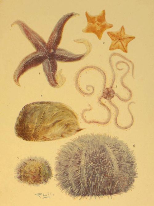
ECHINODERMS
| 1. Asterias rubens | 4. Echinocardium cordatum |
| 2. Goniaster equestris | 5. Echinus miliaris |
| 3. Ophiothrix fragilis | 6. Echinus esculentus |
The habits of sea urchins are interesting, and may be watched in the aquarium, where the movements of the spines and of the tube-feet may be seen perfectly. Some species are very inactive, living in holes and crevices, or under stones, and seldom move from their hiding-places, while others travel considerable distances. The former have generally no eyes, and, instead of seeking their food, simply depend for their subsistence on the material carried to them by the movements of the water; while the latter possess visual organs similar to those observed in certain starfishes. Some species also protect themselves from their enemies when in the open by covering their bodies with sand, small stones, shells, or weeds, and thus so perfectly imitate their surroundings that they are not easily detected. The feet that are used for purposes of locomotion terminate in suckers resembling those of the common five-fingered starfish, and have considerable clinging power, but some have either very imperfectly developed suckers or none at all, and are probably used as feelers only.
Sea urchins, like their allies the starfishes, generally inhabit deep water beyond low-water mark, where they often exist in enormous numbers, feeding on both animal and vegetable substances; but some species are often to be met with between the tide-marks, where they may be seen under stones, and frequently half hidden in mud. The globular species occur principally on rocky coasts, but the heart urchins are more commonly dredged from banks of sand or mud that are always submerged.
The life-history of urchins closely resembles that of starfishes, for the young are free-swimming creatures of an easel-like form, and during this early larval existence their bodies are supported by a calcareous skeleton.
We will conclude our short account of the British echinoderms with a description of the peculiar Sea Cucumbers, which belong to the division Holothuroidea. These creatures are so unlike starfishes and urchins in general appearance that the uninitiated would hardly regard them as close relatives. The body is, as the popular name implies, cucumber-shaped, with the mouth at one end, and the general aspect is wormlike. There is, however, a radiate symmetry—a five-fold arrangement of parts, though not so regular as in most echinoderms. Running lengthwise along the body are five rows of tube-feet, but only two of these are well developed and terminate in functional suckers; and, as might be expected, the animal crawls with these two rows beneath it. The feet are outgrowths of a system of water tubes similar to that of the urchin, there being a circular tube round the mouth, from which branch five radial tubes, one for each row.
The mouth of the sea cucumber is surrounded by plumed tentacles which can be retracted at will, and which are used in capturing the smaller living things that form its food. Like the earthworm, it will often swallow large quantities of sand, from which it digests the organic matter contained.
The body-wall of the Holothuroidea is strong and muscular, and is strengthened by the presence of numerous spicules of carbonate of lime, often in the form of little anchors, wheels, and crosses, while the outer surface is rough and slimy, and often of a colour so closely resembling the surroundings of these animals that they are not easily observed. This feature is one of great value to the creatures, since they have no means of defence from their enemies, and seem to owe their safety entirely to their protective colouring.
There are several species of sea cucumbers on our coasts, but all inhabit deep water and are seldom to be seen above low-water level. They are, as a rule, easily obtained from fishermen, who will bring them in when requested to do so. Live specimens may be kept for a considerable time in the indoor aquarium, and seem to prefer a rocky bottom on which they can hide under stones at times, and a bed of sand on which they will occasionally crawl. They will readily devour small molluscs and crustaceans, and will partake of dead organic matter in a partially decomposed state.
The following tabular summary of the classification of Echinoderms may possibly be of use for reference:—
SUB-KINGDOM ECHINODERMATA
| Body star-shaped |
Body globular, subglobular, or heart-shaped, and covered with a continuous shell. Class: Echinoidea (Sea Urchins). |
Body elongated, and covered with a soft skin containing calcareous spicules. Class: Holothuroidea (Sea Cucumbers). |
||
|
Body stalked, at least during early stage. Feet not used for locomotion. Class: Crinoidea (Feather Star). |
Body not stalked. Tube-feet used for locomotion. Class: Stelleridæ. |
|||
|
Arms jointed to disc, and not containing prolongations of the internal organs. Order: Ophiuroidea (Brittle Stars). |
Arms continuous with disc, and containing processes of the viscera. Order: Asteroidea (Common Stars). |
|||
Some groups of animals are so well defined that the individual species contained in them can be assigned their proper place without any difficulty, the main characteristics by which the group is distinguished running with more or less precision throughout the series; but, unfortunately this is not the case with the ‘worms,’ which constitute the sub-kingdom Vermes. Here we have a most heterogeneous assemblage of animals, collectively exhibiting exceedingly wide variations in both form and structure.
We have already referred to the sea cucumber as wormlike in form, and this creature is only one of a large number of wormlike animals that are not worms; and it is also a fact that a considerable number of the worms are not wormlike. It appears as if the sub-kingdom Vermes were a kind of receptacle into which we may throw almost any invertebrate animal that does not readily fall in line with the general characteristics of the other important groups; for in it we have such a varied assemblage of creatures that, speaking of them collectively as worms, it becomes most difficult, if not absolutely impossible, to say exactly what a worm is; and it is a question whether the sub-kingdom ought not to be divided into at least two or three groups of the same standing.
This being the case we can hardly give a satisfactory summary of the characteristics of the group, and therefore it must be understood that in our attempt to do so we unavoidably exclude some forms that belong to it according to our present system of classification. This being remembered, we will define worms as soft-bodied and elongated animals, exhibiting a bilateral symmetry (that is, having appendages and organs arranged symmetrically on each side of a plane extending from the dorsal to the ventral surface through the centre of the body), and with the body usually divided into a succession of segments, each of which resembles the one preceding and following it. Though many of the worms are generally looked upon as uninteresting creatures, of such an unattractive appearance and with such depraved habits that they are beneath respect, yet a study of the sub-kingdom will prove that not only does it include a number of wonderful forms with the most marvellous life histories, but that some of them are very beautiful objects; and this last remark refers more particularly to many of the marine worms, which come directly within the scope of our work.
Before passing on to the special study and classification of the marine species, however, we must say a few words concerning the worms in general, reminding the reader that all our statements regarding the anatomy of the creatures may be readily verified by simple dissections of one or two typical species, such as the common earthworm, the fisherman’s lugworm, the sea mouse, or the common horse-leech of our fresh-water ponds. With this object in view, the animal may be killed by immersion in spirit, then pinned out in the dissecting tray under water, and the body-wall opened by means of a pair of sharp-pointed scissors.
The digestive tube of a worm runs completely through the length of the body, and though there is no distinct head, there is always a mouth, and this is often provided with horny jaws, and sometimes also with horny teeth, with which the animal is enabled to inflict wounds on its prey.
Like the preceding sub-kingdom—the Echinodermata—worms possess a system of water tubes; this system, however, is not in any way connected with the function of locomotion, but is, in many cases at least, if not in all, intimately associated with the process of respiration. It consists of a series of tubes, arranged in pairs in the successive segments, communicating with the body-cavity internally, and opening at the exterior by means of pores in the cuticle. In some there is a highly organised system of bloodvessels, containing blood that is usually either colourless, red, or green, but the colour of the blood is never due to the presence of corpuscles, as is the case with higher animals, the tint being due to the plasma or fluid portion of the blood; and though worms cannot be said to possess a true heart, yet they often have one or more contractile bloodvessels which serve the purpose of propelling the blood.
Most worms possess a nervous system, and, where this is present, it consists of a chain of ganglia, placed along the ventral side of the body, beneath the digestive tube, all united by means of a nerve cord, and distributing nerves in pairs to various parts of the body; and it may be well to note here one very important point of distinction between the general arrangement of the central portion of the nervous system in the worms and higher invertebrates, as compared with that of the corresponding structure in the vertebrates:—In the former the main axis of the system, consisting, as we have seen, of a chain of ganglia connected by a nerve cord, is invariably placed along the ventral portion of the body-cavity—the surface on which the animal crawls; while in the vertebrates the axis of the nervous system lies along the upper or dorsal part of the body; and, instead of lying in the general body-cavity, in company with the organs of digestion and circulation, is enclosed in the bony canal formed by the vertebral column. It will be seen from this that when it is desired to examine the nervous system of the invertebrate animal, the body-wall should be opened along the middle of the ventral surface, while, in the vertebrate, the central axis should be exposed from above.
Many of the vermes are parasitic, either attaching themselves to the exterior of other animals, and deriving nourishment by sucking their blood, or they are internal parasites, living in the digestive canal of their hosts and partaking of the digested food with which they are almost perpetually surrounded, or burrowing into the tissues and imbibing the nutritive fluids which they contain; and it is interesting to study even these degraded members of the group, if only to observe how their physical organisation degenerates in accordance with their depraved mode of living. In them we find no digestive system with the exception of the simplest sac from which the fluids they swallow may be absorbed, for their food is taken in a condition ready for direct assimilation; and the food so obtained being readily absorbed into all parts of their soft bodies, and being sufficiently charged with oxygen gas by the respiration of their hosts, they require no special organs for circulation or respiration, nor, indeed, do we find any. Further, we find that the nervous system is often undeveloped; for since the parasites, and especially the internal ones, are so plentifully surrounded with all the necessaries of existence, their bodies are so simple in construction that no complex nervous system is required to promote or control either locomotion or internal functions. Even the general body-cavity often disappears in these degraded creatures, the internal organisation being of such a low type that there is no necessity for it; and all the abundant nourishment absorbed over and above that required for the sustenance of their simple bodies is utilised in the reproduction of the species; consequently we find, as a rule, the reproductive organs well represented, and the species concerned very prolific.
It is an interesting fact, too, that these parasites, in their earliest stage, possess organs which are present in the higher worms, but which degenerate as they approach the adult form, thus indicating that they have descended from more respectable members of the animal world, and that the low physical development which they ultimately attain is the natural result of their base mode of living.
The young marine naturalist, working on our coasts, will not be brought into intimate contact with parasitic worms to any large extent, yet we have said this little on parasitism to show that these degenerate creatures are not really devoid of interest, and that they will repay study whenever they are found. They will be more frequently met with during the examination of the animals—usually higher types—that become their hosts, and thus they hardly come within the scope of this work.

Fig. 116.—A Turbellarian, magnified
a, mouth; b, cavity of mouth; c, gullet; d, stomach; e, branches of stomach; f, nerve ganglion; g to m, reproductive organs.
The simplest of the worms are those forming the class Turbellaria, so designated on account of the commotion they produce in the water surrounding them by means of the vibratile cilia that fringe their bodies—a characteristic that is also expressed by their popular name of Whirl Worms. They are usually small creatures, with soft, flattened, unsegmented bodies, though some of the larger species are really wormlike in form, and are more or less distinctly divided into a chain of segments. Many of them are marine, and may be seen gliding over stones left uncovered by the receding tide with a smooth slug-like motion, and when disturbed in a rock pool, occasionally swimming with a similar smooth motion by the aid of their cilia. They avoid bright light, and are consequently generally found on the under surfaces of stones, especially in rather muddy situations, and where the stones are covered with a slimy deposit of low forms of life. In these turbellarians the mouth is situated on the under surface, thus enabling the animal to obtain its nourishment from the slimy surface over which it moves, and it is also provided with an extensile proboscis that aids it in the collection of its food. The digestive tube is generally very complex in form, extending its branches into every part of the soft body; and, there being no special organs of respiration, the animal derives all the oxygen required by direct absorption from the water through the soft integument.
When searching for turbellarians on the sea shore one must be prepared to meet with interesting examples of protective colouring that will render a close examination of rocks and stones absolutely necessary. Some of these worms are of a dull greyish or brownish colour, so closely resembling that of the surface over which they glide that they are not easily distinguished; and the thin bodies of others are so transparent that the colour of the stone beneath is visible through them, thus preventing them from being clearly observed.
Overturned stones should be examined for their flattened bodies gliding along rapidly in close contact with the surface. They may be removed without injury by placing a wet frond of a sea weed close to the stone, in front of one end of the body, and then urging them to glide on to it by gently touching the opposite end. Sometimes, however, the turbellarians remain perfectly still when exposed to the light, in which case they are even more difficult to detect, but a little practice will soon enable one to distinguish them with readiness.
Allied to the turbellarians are the Spoon Worms or Squirt Worms, some species of which inhabit deep water round our shores, where they burrow into the sand or mud of the bed of the sea. These form the class Gephyrea, and consist of creatures with sac-like or cylindrical and elongated bodies, and a protrusible proboscis, which is often of great length. Their bodies are not distinctly segmented, nor do they bear any appendages. The skin is tough and horny, and the body-wall, which is very thick and muscular, is often contracted when the animal is disturbed, thus causing a jet of water to be forcibly ejected.
All the most interesting of the marine worms belong to the Annelida or Chætopoda, popularly known as the Bristle-footed worms, because their locomotion is aided more or less by the presence of stiff bristles that project beyond the surface of the skin. These are all highly organised worms, mostly with very elongated bodies that are distinctly segmented exteriorly by a number of transverse grooves, while the interior is correspondingly divided into a number of compartments by means of a series of septa.
In addition to the bristles already mentioned, there are often numerous appendages, but these must be distinguished from the more perfect appendages of the arthropods, to be hereafter described; for while the latter are distinctly jointed to the body, and are themselves made up of parts that are jointed together, the former are mere outgrowths of the body-wall. The digestive and circulatory systems are well developed, as is also the system of water tubes that connect the body-cavity with the exterior, while the body-cavity itself is full of fluid.
This group of worms is subdivided into two divisions, the many bristled (Polychæta) and the sparsely bristled (Oligochæta) worms. The latter contain the common earthworms and some less known species, while the former include a number of interesting and even beautiful worms, all of which are marine, and many of them among the commonest objects of the sea shore.
These Polychætes exhibit a great variety of habit as well as of appearance. Some live in crevices of the rocks or under stones and weeds, or make burrows in the sand or mud of the bed of the sea, and roam about freely at times in search of food. They are continually coming within the ken of the sea-side collector, being revealed by almost every overturned stone near the low-water mark, and are often seen crawling over the wet rocks just left uncovered by the receding tide; while their burrows are often so numerous that hundreds may be counted in every few square feet. But many are sedentary species, and these are not so generally known to young sea-side naturalists, who frequently observe, and even preserve, the interesting homes they construct, while less attention is given to the architects that build them.
It is very interesting to observe some of the general differences between the roving and the sedentary species—differences which illustrate the principle of adaptation of structure to habit. The roving species are provided with a lobe that overhangs the mouth, bearing feelers and eyes, and are thus enabled to seek out any desired path and to search for their food. They are provided with bristles and other appendages by means of which they can travel freely over the surfaces of solid objects, and are able to swim well either by undulations of the body, or by fringed appendages, or both. The carnivorous species, too, are provided with strong, horny jaws, and sharp, curved teeth, by means of which they can capture and hold their prey. The sedentary species, on the other hand, unable to move about in search of food, are supplied with a number of appendages by means of which they can set up water currents towards their mouths, and which also serve the purpose of special breathing organs, and, having no means of pursuing and devouring animals of any size, they do not possess the horny jaws and curved teeth so common in the rovers. Their eyes, too, are less perfectly developed, and the tactile proboscis of their free-moving relatives is absent.
Of the roving worms, perhaps, the Lugworm or Sandworm (Arenicola piscatorum) is the best known. Its burrows may be seen on almost every low sandy or muddy shore, and, being so highly valued as a bait, its general appearance is well known to all professional and amateur sea fishers. It reaches a length of eight inches or more, and varies in colour according to the sand or mud in which it lives. The segments of this worm are very different in structure in different parts of the body. Those in the front of the body have a few tufts of bristles arranged in pairs, while the middle portion of the body has large brush-like tufts of filamentous gills placed rather close together; and the hindmost part has no bristles or appendages of any kind, and is so well filled with the sand or mud that it is quite hard and firm to the touch. As is the case with our common earthworms, the sand or mud is swallowed in enormous quantities, and this is not only the means by which the lugworm derives its food, but also assists it considerably in making its burrows; the extent to which this creature carries on its work of excavation may be estimated by the thousands of little contorted, worm-like heaps of sand that lie on the surface at every period of low water. These little heaps are known as ‘castings,’ and consist of the sand that passed through the worm’s body as the burrowing proceeded.
The Ragworm is another species that is highly valued as bait. It burrows into the odorous mud that is so commonly deposited in harbours and the mouths of sluggish rivers. In this species the segments are similar throughout the length of the body, and the numerous flattened appendages give it the ragged appearance that has suggested its popular name. Quite a number of marine worms closely allied to the common ragworm, and resembling it in general form, are to be found on our shores. Many of these may be seen by turning over stones that are left exposed at low tide, while others hide themselves in snug little crevices of the rock, or in the empty shells of the acorn barnacle and various molluscs; and some species, including one of a bright-green colour, creep freely over the wet rocks in search of food or home, often exposing themselves to the rays of a fierce summer sun.
The Sea Mouse (Aphrodita aculeata) is certainly one of the most interesting of the roving marine worms, and, though seldom seen above low-water line, may often be obtained by the sea-side collector with the aid of friendly fishermen, who sometimes find it plentifully among the contents of their trawl nets. Failing such aid, it may be looked for among the encrusted stones that are exposed only at the lowest spring tides, especially in places where a certain amount of mud has been deposited under the shelter of outlying rocks; and the chances of success are much greater if the search is made immediately after a storm, for at such times much of the life that exists in deep water will have been driven shoreward by the force of the waves.
At first sight the sea mouse would hardly be associated with the worms; for, instead of having the elongated and cylindrical form that is usually regarded as characteristic of these creatures, it is broad and slug-like in shape, the under surface, on which it crawls, being flat, while the upper side is convex. The segmentation of the body, too, is not readily seen in the upper surface on account of the thick felt-like covering of hairs, but is at once apparent when the creature has been turned over to expose the ventral side.
When seen for the first time in its natural haunt one naturally wonders what the moving mass may be. Crawling sluggishly over incrusted stones, or remaining perfectly still in a muddy puddle that has been exposed by overturning a stone, it looks like a little mound of mud itself, about four or five inches long, and its general colour and surface so closely resembles that of its surroundings that an inexperienced collector may never even suspect that the mass is a living animal form. But take the creature and wash it in the nearest rock pool, and it will be recognised as a broad segmental worm, thickly covered with fine hairs above, and its sides adorned by bristles that display a most beautiful iridescence. It is not easy to see the value of this gorgeous colouring to the animal, and it is doubtful whether, on account of the muddy nature of the creature’s home, such colouring is often displayed to the view of other inhabitants of the sea; but it is well known, on the other hand, that sea mice are readily devoured by fishes, even though they possess an armature of stiff and sharp spines, and that they must therefore be often preserved from destruction by the close resemblance of the general colour to that of their surroundings.
The gills of the sea mouse are not prominent appendages, as with most marine worms, but are soft fleshy structures situated beneath the overlapping scales that lie hidden below the thick hair of the upper surface.
As it is most probable that the reader may desire to preserve a sea mouse at some time or other, a few words concerning the best methods of doing this may be of value. If it is to be preserved in fluid, it should be thoroughly washed to remove all the mud that normally covers its body, and then placed in spirit or formaldehyde, both of which fluids have no destructive effects on the iridescent colouring of the bristles. If, however, it is desired to keep the specimen in a dry state, it should first be put into strong spirit containing a few grains of corrosive sublimate, for a few days. It should then be put under considerable pressure between several thicknesses of absorbent paper to expel the fluid it contains, as well as all the softer internal structures. By this means it will have been squeezed quite flat, so that it presents anything but a natural appearance; but the skin may be blown out to the normal shape by means of a glass tube inserted into the mouth, and then set aside to dry. As the water it originally contained has been extracted by the strong spirit, the drying takes place very quickly; and the small amount of corrosive sublimate that has penetrated into its substance will be sufficient to protect it from the invasion of those pests that commonly attack our museum specimens.
Passing now to the sedentary or fixed worms, we meet with some that are very interesting and beautiful creatures, even when considered apart from the wonderful homes they construct. The several species of the genus Terebella form a soft and flexible tube by binding together particles of sand, shells, or mud with a sticky substance that exudes from their own bodies. These tubes are to be found in abundance between the tide-marks on almost every low, sandy shore, the nature of the tubes varying, of course, with the character of the materials at the disposal of the builder.
In some cases the tubes are exposed throughout the greater part of their length, but very frequently they are more or less buried in the sand or other material of the beach, so that one has to dig to a moderate depth in order to extricate them. In either case, however, the tube of Terebella may be known by the free tufts of sandy threads that form a deep fringe around its mouth.
These worms almost invariably select a sheltered situation for their abode, and should be searched for at the foot of rocks, or under stones, and it is no easy matter to move the buried tube with its occupant intact.
When turning over the stones of a sandy or muddy beach one frequently discovers the slender, thread-like tentacles of the Terebella, together with the sandy filaments that surround the mouth of the tube, the remainder of the tube and its occupant being beneath the surface, and the ground is often so hard and stony that a strong tool is necessary to dig it out; but the work entailed will be amply repaid if a perfect specimen be obtained and placed for observation in the aquarium.
The reader may possibly be acquainted with the tubes or cases that are constructed by the larvæ of caddis flies in fresh-water ponds and streams, and perhaps has noticed the ease with which these creatures may be made to construct new homes after having been turned out of doors. Similar experiments may be performed with Terebella; for when the worm has been extricated from its tube without injury—a work that requires great care on account of the soft and slender nature of the creature’s body—and placed in the aquarium with a bed of suitable material, it will build itself a new dwelling. As with the caddis larvæ, the different species may be known by the materials they select to construct their tubes, but in captivity they may be compelled to employ other than their favourite substance for this purpose. It is unfortunate, however, that Terebella is a nocturnal builder, and thus its movements are not so easily observed.
When removed from its tube its first movements suggest a resentment at the untimely ejection. This being over, it seeks a sheltered situation beneath the edge of a stone, and, at nightfall, commences the slow process of the construction of a fresh home. The particles of material at hand are seized by the tentacles, placed in position round the body, where they are held together by the sticky secretion already mentioned.
The tentacles are employed in two distinct ways:—They may be flattened into slender ribbon-like structures, which, by being folded longitudinally at any point, may be made to grasp a particle of sand; and, in addition to this, the tip of the tentacle may be converted into a minute cup-shaped sucker by the withdrawal of the fluid it contains into the body.
Some species of Terebella build their tubes of ordinary sand, while others select fragments of shells. Some employ mud only, and occasionally we meet with tubes constructed of the silky secretion of the body with hardly any foreign matter.
We sometimes see edges of rocks, on low, sandy shores, covered with what appears to be large masses of consolidated sand, full of holes a little more than an eighth of an inch in diameter; and these masses are often so extensive and so firm that they seem to form the greater part of the rock itself. Such masses are particularly abundant on the south coasts of Devon and Cornwall, but are more or less plentiful on most sandy shores of Great Britain. They consist of the tubes of a species of the marine worm Sabella, which have been built up much in the same manner as those of Terebella, but usually exist in such numbers in the same spot that, together with the sand that has been washed between them, they form the dense masses just described.
A cluster of some dozens of these tubes may be detached with the aid of a hammer and chisel; or, in some instances, where the mass of tubes is not held so firmly together, by the mere pressure of the hand; and it will then be observed that each tube consists of a flexible membrane, of a somewhat leathery nature, formed by a sticky secretion from the body of the worm, with its outer surface covered with grains of sand. The tubes may be easily opened, and the occupants extracted for examination, when it will be observed that the front or upper portion of the worm is short and thick, while the hindmost portion is much thinner, and is doubled forwards in the tube. The body is also provided with numerous bristles, by means of which the worm is enabled to grasp the membranous lining of the tube, and thus secure a firm hold within its home.
A cluster of these tubes should be placed in a rock pool, or in the marine aquarium, when the worms may be seen to protrude gradually, and expose a large number of feathered tentacles, which, by their incessant motion, keep up the constant circulation of the water for the purpose of respiration as well as to bring food particles towards the mouths of the worms.
It is possible to keep these worms alive for some time in the aquarium, but special care is required for the reason that it is a very difficult matter to secure a cluster of tubes without injury to a certain number which are sure to be broken or otherwise damaged; and these, dying and decomposing within their homes, speedily pollute the water. Hence it is necessary to keep a sharp watch for dead specimens, which should, of course, be removed at once. The presence of a putrefying worm may often be detected by the appearance of a whitish fungoid growth round the mouth of what appears to be an empty tube; and if, through neglect, the water of the aquarium has been allowed to become contaminated by the products of decomposition, it will often happen that some of the living worms will come entirely out from their tubes, as if to seek a more sanitary situation. Thus, the exit of worms from their homes may always be looked upon as pointing to a suspicious condition of the water which, if not corrected immediately, may lead to the death of all.
The species we have briefly described is by far the commonest of the genus Sabella, but there are several others to be found on our shores. Some are of a solitary nature, and construct a sandy tube so much like that of a certain species of Terebella that they may be mistaken for that genus. Another solitary species builds a hard stony tube of carbonate of lime that has been extracted from the sea water; and although it is hardly possible to take the live worm from this calcareous tube without injury, the animal may be obtained intact for examination or preservation by dissolving away the tube in dilute hydrochloric acid.
While engaged in collecting specimens on the sea shore we are continually meeting with stones and shells that are more or less covered with white, limy tubes twisted into all manner of serpentine forms. These are the tubes of other marine worms known as the Serpulæ, which, like the species previously mentioned, are interesting objects for the aquarium.
The tubes themselves are worthy of study and preservation, more especially as they vary in form, and may, to some extent, provide a means by which the different species may be identified. They are composed of fine layers of calcareous matter secreted by the body of the worm within, and lined by a thin leathery membrane which may be easily exposed by dissolving away the mineral matter as just described. Some are triangular in section, and often distinctly keeled, while others are cylindrical, and flattened more or less on the lower side. The triangular tubes are attached to stones or shells throughout their length, but the cylindrical ones are often elevated above the surface in the wider and newer part.
If a cluster of these tubes, freshly gathered from between the tide-marks, be placed in the aquarium, the worms will soon protrude the foremost portion of their bodies, exposing beautiful fan-like gills, often brilliantly coloured in shades of scarlet, blue, or purple, which are kept in motion in such a manner as to convey water, and consequently also food, towards the mouth. The gills are of course, richly supplied with blood, for their main function is to aërate that liquid by exposing it to the water in order to absorb oxygen gas. The body of the worm is provided also with little cilia, which, by their constant vibratory motion, keep up a circulation of water through the tube; and this not only keeps the tubular home free from excrement and other sedimentary matter, but also probably assists in the function of respiration by bringing fresh supplies of water in contact with the animal’s soft and absorbent skin.
When the worms are disturbed they immediately withdraw themselves within the tubes, this being done by the aid of the numerous minute hooklets on the surface of the body that enable the worms to cling firmly to the membranous linings of their homes; and it will then be observed that the mouth of each tube is closed by a lid (operculum), which hangs as by a hinge when not in use. These operculi vary much in character, and supply another aid in the identification of the various species. They differ much in shape, and may be either membranous, horny, or calcareous.
Little calcareous tubes, somewhat similar to those of the Serpulæ, but always in the form of a spiral, may often be seen on stones and shells, and the fronds of sea weeds, sometimes so closely packed together as to almost entirely cover the surface. The average diameter of these spirals is only about a sixteenth of an inch, and many are so small that a lens is necessary to discern their shape. In general form they closely resemble some of the small species of Planorbis shells that are so common in our ponds and streams, but these latter are the shells of freely moving molluscs, and are generally of a brownish colour.
The minute worms that live within the tubes in question belong to the genus Spirorbis, and are very similar to those of the Serpulæ, and their pretty plumed gills may be seen with a lens when a cluster of the tubes is placed in a shallow vessel of sea water. A sharp tap on the table on which the vessel rests will cause the little creatures to suddenly retire into their homes, the entrances to which may then be seen to be closed by an operculum.
There is an interesting group of animals known collectively as the Bryozoa or Polyzoa, or, popularly, as the Moss Polyps, that are often classed with the worms, though they are not, according to the general idea, wormlike in appearance. They live in pretty colonies, many of which are certainly familiar objects to all who ramble along the sea shore. Some form pretty lacelike patches on the fronds of sea weeds, while others are built up into flat, frond-like, branching objects that are often mistaken for sea weeds by young collectors. Among the latter is the Sea Mat (Flustra), that is so commonly washed up on the shore in great abundance. An examination with a lens will show that, in both instances, the mass consists of very many minute cells, with horny or calcareous walls, the mouth of each cell being close by an operculum.
On placing the colony in sea water, however, we find that each little cell is the home of a small animal, that protrudes from the cell, exposing a mouth that is surrounded by a crown of tentacles. A moderately high magnifying power will also show that the tentacles are covered with minute vibratile cilia, by means of which currents of water are set in motion towards the mouth to supply the animal with food. Some, too, have a lip by means of which the mouth may be closed.
In addition to the colonies just briefly described, there are other moss polyps that build up little, branching, tree-like clusters which closely resemble some of the sea firs, and many of these are to be found in the sheltered crevices of rocks, or attached to the under sides of stones between the tide-marks.
While searching the surfaces of rocks and weeds at low tide, one’s fingers will be constantly coming in contact with fixed, soft-bodied animals that suddenly eject a fine stream of water as they are touched. These are the Sea Squirts, sometimes spoken of as the Tunicate Worms. They are semi-transparent creatures of oval or elongated form, and usually of a pale yellow, brown, or pink colour; and derive their popular name from the fact that they are covered externally by a continuous tunic or wall of tough structure.
Although the tunicates resemble worms in many points of structure, it is interesting to note that in their young or larval state the body consists of two cavities, one of which contains the internal organs, while in the other the central portion of the nervous system is developed, in which respects they resemble the vertebrate or back-boned animals—fishes, amphibians, reptiles, birds, and mammals. At this stage, too, the creatures possess a tail that is supported by a rod of gristle similar to that which gives place to the backbone in the developing vertebrate. These features, though only transitory, are regarded as a mark of relationship to the higher forms of animal life, and thus the tunicates have been separated from the sub-kingdom Vermes by some zoologists, and given an exalted place at the top of the invertebrate scale, where they form a sub-kingdom of their own, and are looked upon as a link connecting the invertebrates with the vertebrates.
Before passing on to the next sub-kingdom, we should observe that the interesting Rotifers or Wheel Animals also belong to the Vermes; but although many of these minute creatures are to be found in sea water, their principal home is the stagnant water of fresh-water ponds and ditches, and thus we may be excused for neglecting them here.
The sub-kingdom Mollusca includes a great variety of soft-bodied animals which differ from the members of the last division in the fact that they are never segmented, and in the possession of a thick outer covering, of a leathery nature, which completely envelops the body, and which usually secretes a calcareous shell of one or more parts. A general idea of the extent of the group may be formed when we state that it contains the Octopus and the Cuttlefish; all Snails and Slugs, and animals of a similar nature; and all those numerous ‘bivalves’ which are represented by the well-known Oysters, Mussels, Scallops, &c.
By far the greater number of the molluscs are aquatic in habit; and of these such a large proportion are marine that the group provides plenty of occupation for the sea-side naturalist. This being the case, we shall devote the present chapter to a description of the general characteristics of these animals, and to the principles of their classification, illustrating our remarks by a few selections from all the chief divisions.
Although, as we have already hinted, the body of a mollusc generally bears but little resemblance to that of the typical elongated and segmented worm, yet the study of the earliest stages of the former shows that a certain relationship exists between the two sub-kingdoms, the newly hatched mollusc being often a minute free-swimming creature with expanded lobes fringed with cilia, and bearing a resemblance to certain of the Rotifers, Moss Polyps, and other animals that are included among the Vermes. But in the adult molluscs this resemblance is lost, these creatures being generally easily distinguished from all others by certain well-marked external features, as well as by internal characters that are peculiar to them and fairly constant throughout the group.
The external shell, where it exists, is usually composed of one or of two parts, and therefore we speak of univalve and bivalve molluscs; and no internal skeleton of any kind is to be found except in the division containing the Cuttlefishes, the ‘bone’ of which is one of the common objects washed up on our shores by the breakers.
In all the molluscs there is a well-formed digestive tube, and often a complex arrangement of small teeth which sever the food by a rasp-like action. There is also a well-formed heart, consisting of two or more cavities, by means of which the blood is forced through the body; but, as a rule, blood vessels are either few or absent, the blood being driven through spaces between the tissues that serve the same purpose.
The nervous system consists of a few masses of nerve substance (ganglia), connected by nerve cords, and sending off fibres to various parts of the body, the principal ganglion being one situated close to the mouth, and often surrounding the first portion of the digestive tube.
The animals of this sub-kingdom are grouped into three principal and well-marked divisions—the Lamellibranchs, or Plate-gilled molluscs, the gills of which are composed of plate-like layers, and the headless bodies enclosed in a bivalve shell; the Cephalophora, or head-bearing molluscs, protected by a univalve shell; and the Cephalopoda, or Head-footed molluscs, so called because the mouth is surrounded by tentacles or arms by which the animal can cling to objects or seize its prey.
We shall deal with these three divisions in the above order, taking first the bivalves, the shells of which are found in great variety along our shores.
The general nature of a lamellibranch is easily made out by the examination of one of the common species that may be obtained alive on any part of the coast, such as the Edible Mussel, the Cockle, or the Oyster, and the reader will do well to secure a few specimens and examine them with the aid of the following description of the principal distinguishing features.
The shell is formed of two valves, united by a hinge which is sometimes of the simplest possible description, but which often exhibits a beautiful arrangement of interlocking teeth. A ligament of flexible and elastic substance often holds the two valves together.

Fig. 127.—Shell of the Prickly Cockle (Cardium aculeatum) showing Umbo and Hinge; also the interior showing the Teeth
The reader has probably observed that the valves of a dead lamellibranch usually gape. This is due either to the pull exerted by a ligament that is attached to the valves outside the hinge, or to the pressure of an internal cartilage which unites the valves within, and which is compressed when the shell is closed. When the animal is alive, it has the power of closing its shell by the contraction of the adductor muscles, to be presently described, and when the valves are brought together by this means the external ligament is more or less stretched, or the cartilage within, which is also an elastic material, is compressed.
Examining the shell from the exterior we observe that each valve has a nucleus (the umbo) close to the hinge, round which are usually a number of more or less distinct concentric lines, extending to the lower or ventral margin. This nucleus represents the whole shell of the young mollusc, and the lines are the lines of growth, each one marking the extreme limit of the valve at a particular period of the animal’s existence. Further it will be observed that the lines of growth are often wider apart in some directions than in others, thus denoting the unequal rate of growth that determined the form of the adult shell.
The shell of a bivalve is often made up of two very distinct layers, the outer one called the prismatic layer because, when examined microscopically, it is seen to consist of minute vertical prisms of calcareous matter; and the inner one presenting a beautiful pearly iridescence, due to the fact that it is made up of a number of extremely thin and finely waved layers of calcareous substance that have the power of decomposing light. This latter layer is secreted by the whole surface of the mantle that lies in contact with it, while the outer, prismatic portion of the shell is formed only by the free edge of the mantle; and we often find a distinct line (the pallial line), some little distance from the ventral margin that marks the junction of the muscle of the mantle with the shell. The shape of this line is a very important feature of the shell, since it is of great value in the determination of relationships.
Further, the inner surface of each valve is marked by the impressions or scars of other muscles, the number and position of which vary considerably in different species. They include the adductor muscle or muscles (one or two in number) that pull the valve together; the muscle or muscles that withdraw the foot, called the retractor pedis, and the protractor pedis that pulls the foot out. Not only are these scars often very distinct in themselves, but we may frequently observe lines running tangentially from their circumferences towards the umbo, to which they all converge. These lines enclose the areas previously occupied by the muscular impressions; in other words, they show the directions in which the muscles named above shifted their positions as the animal grew.
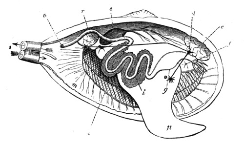
Fig. 129.—Diagram of the Anatomy of a Lamellibranch
f, mouth, with labial palps; g, stomach; i, intestine, surrounded by the liver; a, anus; r, posterior adductor muscle; e, anterior adductor muscle; c, heart; d, nerve ganglion; m, mantle (the right lobe has been removed); s, siphons; h, gills; ft, foot
Now let us obtain a few species of live lamellibranchs, put them in a vessel of sea water, and observe them after they have been left undisturbed for a time. The shell will be seen to gape slightly, exposing the edges of the two lobes of the mantle which lie closely on the inner surface of the valves, thus completely enveloping the body of the animal; and at one end, usually the narrower end in the case of irregular shells, we shall observe two openings—the siphons, sometimes enclosed within a tube formed by a prolongation of the united mantle lobes, and protruding from between the valves, and sometimes formed by the mere contact of the mantle lobes at two adjacent points. If now we introduce a little carmine or other colouring matter by means of a glass tube, setting it free near the lower siphon—the one more remote from the umbo of the shell, we observe that it enters the body of the mollusc through this opening, and reappears shortly afterwards through the upper or dorsal siphon. Thus we see that water currents are incessantly circulating in the body of the animal, entering by the inhalent or ventral siphon, and leaving by the exhalent or dorsal siphon. These currents are maintained by the vibratile action of thousands of minute cilia belonging to cells that line the cavities of the body, and serve to supply the animal with both air and food; for lamellibranchs, being gill-breathers, derive the oxygen necessary for respiration from the air held in solution by the water, and their food consists entirely of the minute living creatures that always abound in natural waters.
Again, we shall find that some of our live bivalves have protruded a thick, conical, fleshy mass—the foot, from the opposite end of the body. This organ is the means of locomotion in the case of the burrowing and other free-moving bivalves, but is developed to a less extent in those species that lead a sedentary life. Thus, the common Edible Mussel secretes a tuft of strong silky fibres (byssus) by means of which it fixes itself to a rock or other body, and therefore does not need the assistance of a muscular foot; and an examination of its body will show that the foot is very small in proportion to the size of the animal, as compared with that of the wandering and burrowing species. The same is true of the oyster, which lies fixed on its side, the lower valve being attached to the surface on which it rests.
We have made use of the terms dorsal and ventral in speaking of the shell of a bivalve, and it is important that these and a few other similar terms be well understood by those who are about to read the descriptions of the animals, or who may desire to describe them themselves. To do this, take a bivalve in your hand, and hold it before you in such a position that the hinge is uppermost, and the siphons turned towards you. The foot of the animal is now pointing in the direction you are looking, and the mouth, situated at the base of the foot, is also directed the same way. You have now placed the shell, and, of course, also the animal, in such a position that its dorsal side is uppermost, the ventral side below, the anterior end turned from you, the posterior (often narrower) end towards you, the right valve on your right, and the left valve on your left. Knowing the exact uses of these few terms you are in a better position to understand the descriptions of bivalves, and to locate the exact situations of the various internal organs named in such descriptions.
A great deal of the internal anatomy of a bivalve mollusc may be made out by easy dissections, and although the structure of the different species varies in several details, the general characteristics of the group are practically the same in all and may be gathered by the examination of a few specimens.

Fig. 131.—A Bivalve Shell
(Tapes virgineana)
a, anterior; p, posterior; l, left valve; r, right valve; u, umbo, on dorsal side
For this purpose the shell should be prised open by means of some flattened but blunt implement, such as the handle of a scalpel, and then, after inserting a piece of cork to keep the valves apart, gently remove the mantle lobe from the valve which is held uppermost with the same implement, being careful to separate it from the shell without doing any damage to the soft structures. Separating the mantle from the shell in this way we meet with one or more hard masses of muscle that are joined very firmly to the latter. These are the adductor muscles that pass directly from valve to valve, and on cutting them through close to the uppermost valve, the latter can be raised so as to expose the body of the animal, mostly hidden by the overlying mantle lobe.
Before raising the upper mantle lobe we observe the heart, on the dorsal margin of the body, near the hinge of the shell, situated in a transparent cavity (the pericardium) containing a colourless fluid. It consists of at least two cavities—a thick-walled ventricle and a thin-walled auricle, and its slow pulsations may be watched with or without the use of a hand lens. On opening the pericardium the heart is still better seen, and if we carefully cut into the thick-walled ventricle we find a tube running completely through its cavity. This is the rectum—the last part of the digestive tube, that commences at the mouth, and terminates in a cavity at the posterior end communicating with the exhalent siphon.
After noting the nature and position of the one or two adductor muscles previously cut through, we turn the upper mantle lobe upwards, laying it back over the hinge of the shell, cutting it through at the bases of the siphons if we find it is united with the opposite lobe at those points; or, if not united, we observe two points at which the lobes touch each other in order to form the siphonal openings.
Several organs are now exposed to view. The lower mantle lobe is seen in close contact with the valve below it, and if we touch its edge we shall probably observe that it is retracted slightly by the contraction of its own muscular fibres. The tip of the foot is also seen projecting towards the anterior end, its base being hidden between the two sets of plate-like gills that extend along the length of the body. On touching the tip of the foot we find it retract by the contraction of the muscular fibres of which it is composed, aided, perhaps, by the action of one or more retractor pedis muscles with which it is supplied. On raising the upper gill-plates we may observe the dark colour of the digestive gland (liver) at the base of the foot, and also see two or more tentacles or labial palpi on the anterior side of the same.
Between the labial palpi is the mouth, which leads into the stomach by a short, wide tube, and then into a convoluted tube which finally passes through the heart, and terminates near the exhalent siphon as above described. The whole length of this tube may be followed by careful dissection, its direction being determined at short intervals by probing it with a bristle that has been tipped with a little melted sealing wax. It will be seen to wind through the base of the foot, surrounded through the greater part of its course by the digestive gland, from which a digestive fluid enters it through small ducts.
The diagram on p. 194 shows the general internal anatomy of a lamellibranch, parts of which have been removed to reveal the underlying structures. The animal lies in its left valve, the right valve, the right mantle lobe, and the right set of gill-plates having been completely dissected away. The whole course of the digestive tube has also been exposed, and the positions of the three nerve ganglia, with their connecting nerve cords, constituting the central portion of the nervous system, are also indicated.
It will be interesting, finally, to learn the direction taken by the water currents which supply the animal with air and food in their course through the system. Passing in through the inhalent siphon, the water immediately enters a large cavity between the mantle lobes. This cavity (the branchial cavity) contains gills, as we have already seen, and also extends to the mouth. The water, urged on by the motion of myriads of minute ciliated cells in the walls of the cavity, passes in part through the digestive tube, and in part around, between, and through the gill plates, which are perforated by numerous holes. After thus completely bathing the gills, and supplying the oxygen necessary for respiration, this latter current passes into a second cavity above the gills, and thence into the exhalent siphon, where it mingles with the fluid from the digestive tube as well as with other excretory matter.
Lamellibranchs are, as a rule, exceedingly prolific, a single individual of some species discharging more than a million ova in one season. The larvæ swim freely in the water, and are provided with eyes that enable them to search for their food, but the eyes always disappear when the young settle down to a more sedentary life. It is true that adult bivalves sometimes possess visual organs, often in the form of conspicuous coloured spots on the edge of the mantle, these, however, are not the same that existed during the larval stage, but are of a more recent development.
Lamellibranchs are classified in various ways by different authorities, the arrangement being based principally on the number and position of the adductor muscles, or on the nature of the gills. For our present purpose we shall look upon them as consisting of two main divisions—the Asiphonida and the Siphonida, the former including those species which do not possess true tubular siphons, the inhalent and exhalent openings being formed merely by the touching of the mantle lobes; and the latter those in which the mantle lobes are more or less united and tubular siphons formed. Each of these divisions contains a number of families, most of which have representatives that inhabit the sea; and we shall now note the principal characteristics by which the more important families are distinguished, and take a few examples of each, starting with the Siphonida.
Examining the rocks that are left exposed at low tide we frequently find them drilled with holes that run vertically from the surface, seldom communicating with each other within, and varying in diameter from less than a quarter of an inch to half an inch or more. Some of these holes are the empty burrows of a boring mollusc, while others still contain the living animal in situ.
The molluscs in question belong to the family Pholadidæ, which contains a number of species that exhibit very remarkable features both as regards structure and habit. The shell is very thin and fragile, but yet composed of hard material, and its surface is relieved by a series of prominent concentric ridges that bear a number of little rasp-like teeth. It gapes at both ends, has neither true hinge nor ligament, and is often strengthened externally by two or more extra or accessory valves. The hinge-plate is a very peculiar structure, for it is reflected over the exterior of the umbones, above which they are supported by about ten thin shelly plates, the whole thus forming a series of chambers. The accessory valves are supported by these bridged structures, and a long, straight, calcareous plate also fills the space along the dorsal side of the shell in some species. The muscular scars and the pallial line are distinctly seen on the inner surface, and a peculiar curved shelly plate projects from under the umbo of each valve.
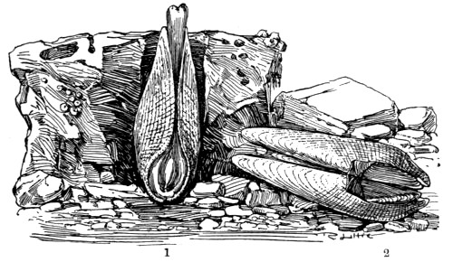
Fig. 132.—Pholas dactylus
1, ventral aspect, with animal; 2, dorsal side of shell showing accessory valves
The animal inhabiting the shell is somewhat wormlike in general form, and the mantle lobes are united in front—that is at the lower end of the shell as it lies in the burrow—except that an opening is left for the protrusion of the short foot. The siphons are united and much elongated, so that they protrude beyond the mouth of the burrow when the animal is active; the gills are narrow, and extend into the exhalent siphon; and the anterior adductor muscle, being very near the umbones, serves the double purpose of adductor and ligament.
Such are the general distinguishing features of this family, all the species of which burrow into stone or other material. Those more commonly met with on our coasts belong principally to the genus Pholas, and are popularly known as Piddocks.
It was long a puzzle as to how the fragile piddocks could excavate the tubular burrows in which they live, and, since their shells are so thin that it seemed almost impossible for hard stones to be ground away by them, it was suggested that the rocks were excavated by the action of an acid secretion. This, however, would not account for the formation of holes in sandstone and other materials which are insoluble in acids; and, as a matter of fact, no such acid secretion has ever been discovered. The boring is undoubtedly done by the mechanical action of the rasp-like shell, which is rotated backwards and forwards, somewhat after the manner of a brad-awl, though very slowly, by the muscular action of the foot of the animal.
Piddocks are found principally in chalk and limestones, though, as before hinted, they are to be seen in sandstones and other rocks, the material in any case being, of course, softer than the shell that bores it. The largest holes and the largest specimens are to be found in chalk and other soft rocks; while the piddocks that burrow into harder material are unable to excavate to the same extent and are, as a consequence, more stunted in their growth. The burrowing is continued as long as the animal grows, the hole being always kept at such a depth that the shell is completely enclosed; and not only this, for when the rock is soft, and the surface is worn down by the sea, the piddock has to keep pace with this action, as well as to allow for its increase in size.
As a result of the rasping action of the pholas shell on the surrounding rock the space hollowed out becomes more or less clogged with débris. This is ejected at intervals by the sudden contraction of the foot of the animal, which brings the shell quite to the bottom of the burrow, thus causing the water with its sediment to shoot upwards, It is not usually an easy matter to obtain perfect specimens of the pholas by simply pulling them from their burrows, the shells being so thin and fragile, and the mouth of the burrow being often narrower than the widest part of the shell. The best plan is to chip away the rock with the aid of a mallet and chisel, or to break it into pieces with a hammer, thus laying open the burrows so that the molluscs fall from their places.
The Common Piddock (Pholas dactylus) may be identified by the illustrations, and the other members of the family may be recognised at once by the similarity in structure and habit. The principal species are the Little Piddock (P. parva), the shell of which is wider in proportion to the length, with only one accessory valve; and the White Piddock (P. candida), also with a single accessory. In all the above the foot is remarkable for its ice-like transparency.
There is another genus—the Pholadidea—the species of which are very similar to pholas both in structure and habit. The shells are, however, more globular in form, and are marked by a transverse furrow. The gape at the anterior (lower) end is also very wide, and covered over with a hardened plate in the adult. Also, at the posterior (upper) end of the shell is a horny cup through which the siphons protrude, and the latter, which are combined throughout their length, terminate in a disc that is surrounded by a fringe of little radiating appendages.
In the same family are the molluscs popularly known as ship worms, which are so destructive to the woodwork of piers and jetties, or which burrow into masses of floating timber. Some of these, belonging to the genus Xylophaga—a word that signifies ‘wood eaters’—have globular shells with a wide gape in front, and burrow into floating wood, nearly always in a direction across the grain. The burrows are about an inch deep, and are lined with a calcareous deposit. The siphons, combined except at the ends, are slender and retractile; and the foot, which is thick, is capable of considerable extension.
Other ship worms belong to the genus Teredo, and are very similar in general characters. The shell is small and globular, with a wide gape at both ends, and consists of two three-lobed valves with concentric furrows. It is so small in proportion to the size of the animal that it encloses but a small portion of the body, and lies at the bottom of the burrow, which is of considerable length—often from one to two feet. The animal is very wormlike in form; and although the shell is so small, yet all the internal organs are enclosed by it. The mantle lobes are united in front, except where the sucker-like foot passes through them; the gills are long and narrow, and extend into the siphonal tube; and the two very long siphons are united almost throughout their length. It is also interesting to note that in these animals the rectum does not pass through the heart, as it does in nearly all molluscs, and that a pair of horny or calcareous ‘styles’ or ‘pallets’ project from the place where the two siphonal tubes begin to diverge.
Several species of Teredo are to be met with on our coasts, but they are so similar in general structure that the above brief description applies almost equally well to all.
Other boring molluscs frequent the British shores, but they belong to quite a distinct family called the Gastrochænidæ because their shells gape widely on the ventral side. Their valves are equal in size and very thin, the hinge has no teeth and the pallial line is sinuated. The margins of the mantle lobes are thickened and united except where a small aperture is left for the protrusion of the finger-like foot. The siphons are very long and retractile, and the gills extend into the inhalent tube. These animals burrow into mud, shells, or stone, often dwelling together in such numbers that their galleries cross one another and form a most intricate network, and the different species are to be found from low-water mark to a depth of a hundred fathoms or more.
The British species belong to two genera—the typical genus Gastrochæna, and the Saxicava or stone-borers.
The former contains the Common Flask shell (G. modiolina) which burrows into limestone and shells, in the latter case passing generally through the shells into the ground below, and completing its home by cementing together any fragments of hard material that come in its way into a flask-shaped cell. The opening of the burrow is shaped like an hour-glass, the two expansions serving for the protrusion of the siphonal tubes, and the neck of the flask-shaped abode is usually lined with a calcareous layer that projects slightly to afford further protection to the extended siphons. Although this species is very common on some parts of our coast, it is seldom obtained without the aid of a dredge, for it usually lives at a depth of from five to ten fathoms; and when found it is generally no easy matter to extricate them from their holes, to the sides of which they often cement their shells.
The genus Saxicava contains a few species that drill holes, often several inches deep, in shells and stone, and frequently do great damage to breakwaters and other artificial structures. The foot is usually provided with a byssus by which the animal fixes itself to a little projection on the side of its burrow. The species are to be found from low-water mark to a depth of one hundred fathoms or more.
The next family, named Anatinidæ, contains a number of molluscs that burrow in mud or sand or live in seclusion in the crevices of rocks. Their shells are thin, with a granulated outer surface, and the valves are united by a thin external ligament. The inner surface is pearly, the pallial line usually sinuated, and both valves are pitted for the reception of the somewhat stout internal cartilage. The mantle lobes are united, as are also the siphons to a greater or lesser extent; and there is only one gill on each side.
Some of the common species of this family are popularly known as Lantern shells, and perhaps the most familiar of these is Thracia phaseolina, the specific name of which is given on account of a fancied resemblance of the shell to a bean. The shell is very fragile, and although large numbers may often be seen stranded on sandy beaches, but few of them are perfect specimens.
The family Myacidæ may be recognised by the thick, strong, opaque shells, usually gaping at the posterior end; the wrinkled epidermis which covers the whole or part of the shell; and the united siphons, which are more or less retractile. The mantle cavity is also closed with the exception of a small hole left for the protrusion of the small foot. The pallial line of the shell is sinuated.
In the above illustration we represent the Common Gaper (Mya arenaria), which burrows to a considerable depth in the sand or mud, especially in the estuaries of rivers, from between the tide-marks to a depth of twenty fathoms or more. It may be readily distinguished, in common with the other species of the same genus, by the characteristic wrinkled, membranous tube that encloses its fringed siphons, the membrane being a continuation of the epidermis that extends over the shell. Another characteristic feature of the genus is the large, flat process inside the left valve for the attachment of the internal cartilage. An allied species, Mya truncata, is often found abundantly in company with the above, and may be known by the abruptly squared posterior end.
Other species of the Myacidæ inhabit our shores, including the little Basket shell (Corbula nucleus), the left valve of which is much smaller than the right, which overlaps it. The latter, also, is covered with epidermis, while the former, which is flat, is quite naked.

Fig. 139.—Solen siliqua
The valves have been separated and the mantle divided to expose the large foot
We now come to the interesting family of Razor shells (Solenidæ), specimens of which are washed up on almost every sandy beach, while the living molluscs may be dug out of their burrows at low-water mark. The shells are elongated, gaping at both ends with an external ligament; and the hinge has usually two teeth in one valve and three in the other. The foot of the animal is cylindrical, large and powerful; and the siphons are short and united in the long species, but longer and only partially united in the shorter ones. The gills are long and narrow, and are prolonged into the inhalent siphon.
These molluscs lie vertically in their deep burrows at low-water mark, the opening of the burrow having a form resembling that of a keyhole. While covered with water they occupy the upper portion of their abode, but sink to a depth of a foot or more when the tide goes out. As we walk along the water’s edge at extreme low tide we may observe jets of water that are shot into the air before us. These are produced by the sudden retreat of the ‘Razor-fish’ to the bottom of its burrow when alarmed by the approaching footsteps. Owing to this wariness on the part of the mollusc, and to the considerable depth of its burrow, specimens cannot be obtained by digging without much labour; but if a little salt or some other irritant be dropped into the hole, the animal will soon rise to eject it, and may then be shut out from the lower part of the burrow by sharply driving a spade below it. This is undoubtedly the best method of securing perfect specimens for study or preservation, but fishermen often obtain large numbers, either for food or for bait, by suddenly thrusting a long hook down into the gaping shells, and then pulling them out. This method always does injury to the soft body of the animal, and often damages the shell, but answers the fisherman’s purpose exactly.
We give illustrations of two shells belonging to the typical genus (Solen), including one on Plate V.; also a British representative of each of two other genera of the family—Cerati-solen and Solecurtus, the latter of which, as the name implies, contains shorter species.
The next family—the Tellinidæ—contains a number of well-known molluscs that burrow into sand or mud, and are enclosed in shells that are often very prettily marked; and although the family includes several genera, all may be recognised by the following general features. The shell is compressed, composed of two equal valves, with little or no gape, and the ligament situated on the shortest side. The central or cardinal teeth never exceed two in number in each valve, and the adductor impressions are round and polished. The mantle is quite open at the anterior end, and its margins are fringed; the foot is flattened and tongue-shaped; and the siphons, which are quite separate, are generally long and slender.
In the typical genus (Tellina), of which we represent two very common British species, the ligament is very prominent, and the slender siphons are often much longer than the shell. The members of this group move very freely, travelling about by means of a broad, flattened foot.

Fig. 141.—Tellinidæ
1. Psammobia ferroensis. 2. Donax anatinus. 3. Tellina crassa. 4. Tellina tenuis. 5. Donax politus
The shells of the genus Psammobia are popularly known as Sunset shells, being prettily marked with radiating bands of pink or other tint, reminding one of the beams of the sun when setting in a cloudy sky. In these, too, the ligament is very prominent, and the shell gapes slightly at both ends.
The same family contains the pretty little Wedge shells, which are so called on account of their triangular form, and constitute the genus Donax. These shells, which are seldom much over an inch long, are very common on some of our sandy beaches, being washed up in considerable numbers after the animals have died, but the specimens are seldom perfect. The molluscs themselves are burrowers, and live in the sand, at and just below low-water mark; and, as they usually burrow to a depth of only a few inches, are easily obtained alive.
The shells are rather thin, closed at both ends, blunt and rounded at the anterior end, but straight and more pointed at the shorter posterior end; and the margins of the valves are very finely grooved in such a manner as to resemble the milling of a coin. Each valve has two central hinge teeth, with one long lateral tooth on each side; and the ligament is external and prominent. The lobes of the mantle are fringed; the siphons are separate and diverging, but shorter and thicker than in most of the other Tellinidæ, and the foot is comparatively large, flattened, and pointed.
The genus contains many species, the commonest being, perhaps, D. anatinus, the colour of which is yellowish, banded with brown, and marked by a number of radiating white lines. This colour, however, is due entirely to the thin, shining epidermis that completely covers the valves; and if this is rubbed off the shell itself will exhibit a pale pinkish tint. Another common species (D. politus) may be recognised by the broad patch of white running from the hinge to the margin, on the posterior side of the middle of each valve.
The family Mactridæ contains some British shells popularly known as Trough shells, and the family name itself is derived from the word mactra, which signifies a kneading trough. In this group the shells are all more or less triangular in form, with the valves equal, and are either closed or very slightly gaping. The ligament, perhaps more correctly designated the cartilage, is generally internal, and contained in a deep triangular hollow; and the shell is covered with epidermis. The mantle of the animal is open in front, and the siphonal tubes are united and fringed. The foot is usually large and flattened.
The typical genus, Mactra, contains some common molluscs that bury themselves just beneath the surface of sandy beaches; and these are so abundant in some parts of Great Britain that they are used largely for feeding pigs. Some of the mactras are remarkable for the great power and extensibility of the foot, which, in some cases, is used so vigorously that the animal turns itself quickly over, or even leaps on the ground.
Our example of this genus is M. stultorum, which is a very common object of the shore. Its colour is very variable, usually some shade of grey or brown, and marked by radiating white lines.
The Otter shells (Lutraria), of which we figure one species, are much like the Mactræ in structure, and are usually included in the same family, but in some respects they resemble the Myacidæ or Gapers. The shell is oblong rather than triangular, and gapes at both ends; and the animal buries itself deep in sand or mud, principally in the estuaries of rivers, from low-water mark to a depth of about ten fathoms. The shells are not very common objects of the shore, for they are found only in muddy places, and those of the commonest species (L. elliptica) are too large and heavy to be washed ashore in the sheltered estuaries where they abound.

Fig. 142.—1. Lutraria elliptica. 2. Part of the Hinge of Lutraria, showing the Cartilage Pit. 3. Macra stultorum. 4. Interior of same showing Pallial Line
We now leave the burrowers, to consider a family of molluscs that move about somewhat freely by means of a flattened tongue-shaped foot, and which only rarely fix themselves in any way. The shells of the group are popularly known as Venus shells, probably on account of the beauty of some of the species, and the family in question as the Veneridæ.
The shells of the various species are usually of a graceful oval or oblong form, frequently marked by chevron-shaped lines in pretty colours, and distinctly grooved along the lines of growth. The ligament is external, the hinge has usually three diverging teeth in each valve, and the pallial line is sinuated.
The principal genus is Venus, in which the shells are ovate in form, thick, and smooth, and the margins of the valves are minutely crenulated. The genus is a very large one, and contains several British species, two of which we represent in the accompanying illustrations.
Allied to these is the larger but pretty shell Cytherea chione, which inhabits deep water off the southern coasts, to about one hundred and fifty fathoms. It is much like the Venus shells in form, but the margins are not crenulated.
The same family (Veneridæ) contains the large genus Tapes, so called because many of its shells are marked in such a manner as to recall the patterns of tapestry. The general form of these shells is oblong, and the margins are quite smooth. They are frequently washed up on the beach, especially during storms, but the animals may be found alive at low water, buried in sand, or hiding in the crevices of rocks or among the roots of the larger sea weeds. The mantle is open at the anterior end, and the siphons are either quite distinct or only partly united.
Some of the shells are very prettily coloured. One (T. aurea) receives its name from the yellow ground, which is variously marked by deeper tints; another (T. decussata) is so called on account of the cross grooves with which the shell is sculptured; and a third (T. virgineana), which inhabits the muddy bottoms of deep water, is prettily marked by radiating bands that run from the umbones to the ventral margins.
We now come to the family Cyprinidæ, in which the shell is regular in form, oval or elongated; and the valves, which are equal in size, are thick and solid, and fit closely. The teeth are beautifully formed, the central ones numbering from one to three in each valve, and the pallial line is not sinuated. The mantle lobes are united on the posterior side by means of a kind of curtain that is pierced by two siphonal openings. There are two gills on each side, united posteriorly, and the foot is tongue-shaped and thick.
The typical genus—Cyprina—contains a large mollusc (C. islandica), which is moderately common round our shores, especially in the north, but is not often seen above low-water mark, except when washed up by storms. The shell is oval and thick, with the umbones prominent and turned towards the posterior side, and the ligament is strong and prominent. It is entirely covered with a thick epidermis, of a rich brown colour, often exhibiting a fine silky gloss, especially near the margins. The interior of the shell is white, and the adductor impressions oval and polished.
The same family includes some smaller shells that inhabit deep water, and are therefore not commonly seen on the beach. Among these are two species of the genus Astarte, one of which is deeply furrowed in a direction parallel with the margins; also Circe minima, which seldom exceeds half an inch in length. Although so small compared with Cyprina, these shells may be identified by their clothing of epidermis, together with the family characteristics given above.
The Cyprinidæ also contains the interesting Heart Cockle (Isocardia cor), the form of which is so characteristic that identification is easy. The heart-shaped shell is thick and strong, and is swollen out in such a manner that the umbones are wide apart. These latter are also curved into a spiral form, and the ligament between them is prominent. The colour of the shell is variable, the epidermis being of any shade from a yellow to a dark brown. The foot is small and pointed, and the siphons fringed.
The Heart Cockle burrows in sand by means of its foot, going down just far enough to bury the whole of its shell, and always leaving its siphons exposed at the surface. It inhabits deep water, and is not likely to be obtained without the use of the dredge or trawl.

Fig. 144.—Cyprinidæ
1. Cyprina islandica. 2. Teeth of Cyprina. 3. Astarte compressa. 4. Circe minima. 5. Isocardia cor
The molluscs of the family Lucinidæ are found principally in tropical and sub-tropical seas, ranging from the shore to a very great depth, but a few are moderately common in our own waters. They are closely allied to the Cyprinidæ, but the shell is round rather than oval, and is obliquely grooved inside. The mantle lobes of the animal are not united on the ventral side, but at the posterior end they are continuous, except where they form one or two siphonal openings. The foot is long and of almost the same thickness throughout when extended; and the gills, numbering either one or two on each side, are large and thick. In all the members of this family, as in the last, the pallial line of the shell is simple. None of the shells are really common objects of our shores, since the animals inhabit deep water, some of them moving about freely on the bottom, while others moor themselves by means of a byssus.
We shall take only one example of the family—Galeomma Turtoni—the generic name of which means ‘weasel eye.’ This pretty little mollusc may be found on our southern coasts, where it often moors itself to the rocks or weeds by means of its silken byssus; or, having broken itself away from its temporary place of rest, creeps freely on the bottom by a long, flattened foot, applied closely to the surface over which it travels, and used much in the same way as the broad foot of a snail or whelk, its valves being all the time spread out nearly in the same plane.
The shell itself is oval, with central umbones, and is covered with a thick epidermis. The mantle lobes are united behind, where they form a single siphonal opening; and the margins are double, with a row of eye-like spots on the inner edge of each.
The true Cockles, some few species of which are known to almost every one, constitute the family Cardiadæ, so called on account of the cordate or heart-shaped form of the shell as viewed from the anterior or posterior side. The shell is regular, or nearly so, and the valves, which are equal, are ornamented with prominent rays that run from the umbones to the margin. The ligament is short, strong and prominent, and the valves fit closely by the interlocking of their crenulated margins, or gape slightly on the posterior side. There are two central teeth in each valve, and a long lateral tooth both on the anterior and posterior sides. The mantle lobes are open in front, with the margins plaited, and the siphons, which are usually short, are provided with a number of little tentacles. The foot is large and powerful, and is usually curved into the form of a sickle.
Although the general nature of the common edible cockle (Cardium edule) is so well known even to the inhabitants of inland towns that a description may seem out of place here, yet it is possible that but few of our readers have ever taken the trouble to place the animal in a vessel of sea water, either obtained direct from the sea or artificially prepared, for the purpose of studying its movements or other habits; and it will be well to remember that this and several other species of edible molluscs which reach our towns alive may be very conveniently studied at home, and often at times and seasons when work at the sea-side is undesirable or impossible.
The edible species referred to lives in banks of sand or mud, buried just below the surface, and frequently in spots that are exposed for several hours between the tides. They are usually obtained by means of a rake similar to that used in our gardens.
On the coasts of Devon and Cornwall we find a much larger species, also valued as an article of diet, and known locally as the Prickly Cockle (C. aculeatum). Its shell is beautifully formed, the rays being very prominent, each bearing a number of calcareous spines arranged in a single row. We give an illustration of this species, together with two sketches to show the nature of the teeth of the shell.
In addition to the two species named, we have the red-footed, C. rusticum, which can suddenly turn itself over by the action of its powerful pedal organ; the Banded Cockle (C. fasciatum), a very small species distinguished by the brown bands of the shell; and a still smaller one (C. pygmæum), with a triangular shell, occurring on the Dorset and Devon coasts (fig. 146).
Passing now to the Asiphonida, we deal first with the family Arcadæ. These include a number of shells which, though very variable in general form and appearance, may all be recognised by the long row of similar comb-like teeth that form the hinge. The shells of this group are regular in form, with equal valves, and are covered with epidermis. The mantle of the animal is open, the gills are united by a membrane behind, and the foot is large, curved, and grooved.
One of the prettiest shells in the family is Pectunculus glycimeris, which reaches a length of about two inches. The shell is grooved in the direction of the lines of growth, and there are also very delicate striations running radially from umbones to margin; and the ground colour of white or pale yellowish is beautifully mottled with reddish brown. We give a figure of this species, together with a drawing of the peculiar and characteristic teeth, but a more typical shell of this family may be seen in the Noah’s Ark (Arca tetragona). This shell is almost quadrate in form, swollen, and strongly ribbed. The hinge is straight, with many comb-like teeth—increasing in number with the age of the shell; and the umbones are separated by a diamond-shaped ligament. The foot of the animal is heeled—that is, it has a creeping surface that extends backwards as well as forwards; the mantle is furnished with minute eyes (ocelli), and the animal has two distinct hearts. We give a figure of this peculiar shell, and the other British members of the same genus, though varying more or less in form, may be recognised at once by the same general characteristics.
In the same family we have the small nutshells (genus Nucula), which are often dredged up from deep water in large numbers; and the elongated shells of the genus Leda, also inhabitants of deep water; and, as before stated, the affinities of all may be readily established by the characteristic nature of the teeth.
We now pass on to the family of Mussels (Mytilidæ), of which the common Edible Mussel (Mytilus edulis) is a typical species. In this interesting group the shell is oval or elongated, with equal valves, and is covered with a dark-coloured epidermis which is often distinctly fibrous in structure. The umbones are at the anterior end of the shell, which end is usually very narrow and pointed, while the posterior is broad and rounded. The hinge has small teeth or none, and the ligament, which is long, is internal. The shells of mussels consist of two distinct layers; on the inner, which is often of a most beautiful pearly lustre, may be traced the simple pallial line and the impressions of the small anterior and the large posterior muscles.
The mantle lobes of the animal are united only at a point between the two siphonal openings. There are two elongated gills on either side, and the foot is thick and more or less grooved.
Mussels inhabit salt, brackish, and fresh waters, generally attaching themselves by means of a silken byssus, but sometimes concealing themselves in ready-made holes, or in burrows of their own; and some even hide themselves in a nest which they prepare by binding together fragments of shells or sand.
The edible mussel, which forms such an important article of diet, especially among the poorer classes in our large towns, may be easily distinguished from similar species of another genus by the very pointed umbones, and the coarse and strong fibrous byssus by which it clings to any solid object. It is found most abundantly on muddy coasts, and on mud banks in the estuaries of rivers, generally in such situations as are uncovered at low tide. The fry abound just below low-water level, and grow so rapidly that they reach their full size in a single year.
It is well known that a diet of mussels occasionally produces very unpleasant and even dangerous symptoms in the consumer, and this result has been attributed to the action of a particular organ of the animal which has not been carefully removed before eating. This, however, is not the case, as proved by the fact that the eating of these edibles is usually perfectly safe when no such precautions have been taken. It is highly probable that the deleterious character referred to is due to a disease which sometimes attacks the mussels themselves, but the exact nature of this has not been thoroughly made out.
There is another genus (Modiola) containing several species commonly known as Horse Mussels, and these may be distinguished from Mytilus by their habit of burrowing, or of constructing a nest by spinning together various fragments. The shell, also, is more oblong in form, and much swollen near the anterior end; and the umbones are not so pointed. The epidermis covering the shell is of fibrous structure, and often extends beyond the edges of the valves in the form of a fringe.
Several species of Horse Mussels inhabit our shores, from low-water mark to a depth of fifty fathoms, but none of them is used for food. The commonest species is Modiola modiolus, which has a particularly strong byssus, and its fibres generally bind together such a number of stones &c. that the shell is completely hidden in the entangled mass. Other British species include M. barbata, so called on account of the peculiar fringed threads of the epidermis; M. phaseolina, in which the epidermis threads are not fringed; and M. tulipa, named from the streaks of crimson or purple that radiate from the umbones of the shell and remind us of the colouring of the tulip flower.
An allied sub-genus (Crenella) includes a few small British molluscs the shells of which are crenulated on the dorsal margin behind the ligament. The shells are short and swollen, and lined by a brilliant pearly layer. One species (C. discors) is pale green, with radiating lines from umbo to margin. It is common on many of our shores, but is not easily found, as it hides at or below low water mark, in a nest formed by binding together small stones. Other species, one of which is black, are less abundant, and are not readily obtained except by the use of the dredge.
Before leaving this family we must refer to the remarkable Dreissena polymorpha, sometimes called the Chambered Mussel, on account of the chamber which is formed in the beak of the shell by means of a pearly plate that stretches across it. This animal is not indigenous to Britain, but was introduced from the East by trading vessels, either attached by its silken byssus to timber that had been left floating in water previous to being shipped, or to the bottoms of the ships. It seems to thrive almost equally well in salt, brackish, and fresh waters, and has spread very rapidly since its introduction. It is more commonly found, however, in docks, canals, and rivers, and is on that account usually described with the fresh-water species.
The form of the shell is very similar to that of Mytilus, but has no internal pearly layer, and the valves are bluntly keeled. The mantle is closed, the siphons short, and the foot small.
Our next family—the Aviculidæ—contains those shells that are distinguished by peculiar flat processes on each side of the umbones, one of which, the posterior, is generally wing-like in form. They are popularly known as Wing Shells, and the family includes the so-called Pearl Oysters. Most of the species are natives of tropical seas, but several are common on our own shores.
One species of the typical genus is sometimes found off the coasts of Cornwall and Devon. The shell is very oblique, and the valves are unequal, the right one, on which the animal rests, being somewhat smaller than the left; and the epidermis is very scanty. The hinge is long and straight, without teeth, and the cartilage is contained in grooves. The interior of the shell is pearly. The posterior adductor impression is large, and not far from the middle of the shell, while the anterior, which is small, is close to the umbones. The mantle of the animal is open, and the margins of the lobes fringed; and the small foot spins a powerful byssus.
Most of the British species of the family belong to the genus Pinna, so called on account of the fins or wings on the dorsal side of the shell. In this group the shell is more or less wedge-shaped, with equal valves, and the umbones are quite at the anterior end, while it is blunted and gaping at the other end. The hinge has no teeth. The margins of the mantle are doubly fringed, and the byssus is extremely powerful.
The Common Pinna (P. pectinata) is a very large mollusc, sometimes measuring a foot in length, and is very abundant off the south-west coast, where it moors itself vertically at the bottom of the water with the pointed end buried, and the broad end gaping widely so as to expose its body. It has been stated that fishes are frequently tempted to intrude into the open shell for the purpose of devouring the animal within, and that they are immediately crushed by the sudden closing of the valves, which are pulled together by two large and powerful adductors.
We have already referred to the little Pea Crab that inherits the shell of the Pinna, living permanently in the mantle cavity of the animal.
The last family of the Lamellibranchs is the Ostreidæ or Oysters, of which the edible oyster may be taken as a type. In this group the shells are frequently unequal, and they lie on one side either free or adherent to the surface below them; the hinge is usually without teeth. The mantle is quite open, the gills number two on each side, and the foot is either small or absent.
The Edible Oyster is a type of the typical genus Ostrea, its scientific name being Ostrea edulis; and as this mollusc may be readily obtained at any time, it is a convenient species for the study of the general characteristics of its family. Its shell is irregular in form, and the animal always rests on its left valve, which is convex, while the upper or right valve is either flat or concave. The lower valve is also thicker and laminated in structure, and is attached to the surface on which it rests. On examining the interior we find that the shell is somewhat pearly in appearance, and that the edges of the mantle lobes are finely fringed. The gills, too, are united with each other and with the mantle on the posterior side, thus forming a distinct branchial chamber.
Oysters are found on banks at the depth of several fathoms, where they spawn in early summer, and the fry or spats are collected in large numbers and transferred to artificial beds or tanks, where they are kept in very shallow water so as to be easily obtainable when required for food. It is interesting to note, however, that their growth is slow on these artificial grounds, the full size being attained in about seven years, while, in the natural beds, they are full grown in a little more than half that time.
Native oysters—those that are reared on artificial beds—are of course removed as soon as they are ready for the market, but those that live on natural banks are often left undisturbed till their shells are thick with age. The latter, too, are often destroyed in large numbers by the boring sponge (p. 124), which so completely undermines the substance of the shell that it finally breaks to pieces.
In the genus Anomia the lower valve is concave, and perforated with a large oval hole very near the hinge, while the upper one is very convex, but the shell is very variable in shape, since the animal sometimes clings permanently to an object, and the shell, during its growth, accommodates itself to the surface of that object. The use of the hole is to allow of the protrusion of a set of muscles which proceed from the upper valve, and give attachment to a plug or button, more or less calcified, by which the animal clings.
One species (A. ephippium), known as the Saddle Oyster, is common on some parts of our coast. It is seldom found on the beach at low water, but the empty shells are often washed up by the waves.
The same family includes the Scallops, which constitute the genus Pecten. In these the shell is nearly round, with ears on each side of the umbones, those on the anterior side being generally much more prominent than the others, and both valves are ornamented by prominent radiating ribs. The shell is often very prettily coloured, and the animal rests on the right valve, which may be distinguished from the left by its greater convexity, and by the presence of a notch under the anterior ear. The hinge is straight, with a very narrow ligament, and the internal cartilage is situated in a central pit.
Plate V.
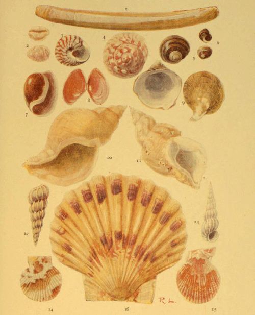
MOLLUSCS
| 1. Solen ensis | 8. Tellina |
| 2. Trivia Europæa | 9. Capulus hungaricus |
| 3. Trochus umbilicatus | 10. Chrysodomus antiquus |
| 4. Trochus magnus | 11. Buccinum undatum |
| 5. Littorina littorea | 12 & 13 Scalaria communis |
| 6. Littorina rudis | 14. Pecten opercularis |
| 7. Haminea (Bulla) hydatis | 15. Pecten varius |
| 16. Pecten maximus | |
The mantle of the animal is free, with double margins, the inner of which forms a finely fringed curtain all round, and on this curtain are a number of black eyes surrounded by very fine tentacles. The gills are in the form of very thin crescents, and the foot is shaped like a finger.
Although the majority of scallops are inhabitants of tropical seas, several species are to be found off our coasts, where they range from depths of about four to forty fathoms, and the empty shells, often in the most perfect condition, are frequently found on the beach.
The Common Scallop (P. maximus) is largely used as food, and is therefore a common object in the fishmonger’s shop. Its colour is very variable, and the shell has equal ears and about twenty radiating ribs. The Quin (P. opercularis) is also an important article of diet in some parts.
Perhaps the prettiest of the British species is the Variable Scallop (P. varius), so called on account of the very variable colour of the shell, the ground tint of which may be almost anything between a very pale yellow and a dark reddish brown, and this is irregularly patched with some lighter colour. The chief distinguishing features of the species are the spiny projections of the numerous ribs, most prominent near the margin of the valves, and the presence of a permanent byssus, which, in other species, occurs only in the young. Three of the species named above are shown on Plate V.
We may also mention the Tiger Scallop (P. tigrinus), the radiating ribs of which are sometimes slightly formed, and which has only one ear in each valve; and P. pusio, in which the adult shell is often greatly altered in form.
It may be noted, in conclusion, that all the species of this genus have the power of swimming rapidly by flapping their valves—a mode of locomotion very common among the bivalves especially during an early stage of their existence.
Before passing on to the univalve molluscs, we must refer briefly to a group of animals that are enclosed in bivalve shells, and which were once included with the Mollusca, but are now made to form quite a distinct group by themselves. We refer to the Brachiopods, at one time very abundant, as proved by the immense number of fossil shells embedded in various stratified rocks, but now represented by only a few living species.
The shells of these animals are commonly known as Lamp Shells, on account of their resemblance to an antique lamp; and although at first sight they bear a general likeness to certain bivalve shells of lamellibranchs, a close examination will show that not only the shell, but also the animal residing within it, are both of a nature very different from that of the molluscs with which they were at one time supposed to be closely related.
The valves of the shell are unequal, and are not placed respectively on the right and left sides of the body of the animal, but rather on the dorsal and ventral or upper and lower sides. The ventral shell is the larger, and is produced into a beak which sometimes has a round hole corresponding in position with the hole for the wick of an antique lamp, and the dorsal or smaller valve is always imperforate. The hinge is a perfect one, the junction of the two valves being so well secured by it that it is impossible to separate them without injury. It is formed by two curved teeth on the margin of the ventral valve that fit into corresponding sockets on the dorsal. A few brachiopods, however, have no hinge, the valves being secured by means of numerous muscles. The hole in the shell serves for the protrusion of a pedicel or foot by means of which the animal is enabled to attach itself.
Two long arms, covered with vibratile cilia, and capable of being folded or coiled, are attached at the sides of the mouth. They are practically processes of the lips, mounted on muscular stalks, and attached to a delicate calcareous loop on the dorsal valve; and serve not only to produce water currents for the conveyance of food to the mouth, but also answer the purpose of gills.
The digestive system of a brachiopod includes an œsophagus that leads into a simply formed stomach round which is a large digestive gland. The heart has only one cavity, but the animal is provided with two smaller and separate organs that assist in the propulsion of the blood, which circulates through numerous blood spaces in the bristly mantle.
About two thousand fossil species of brachiopods are known, extending over a vast range of time; and the living species, numbering less than a hundred, are found from shallow water to the greatest habitable depths.
Since the reader is hardly likely to form any extensive acquaintance with the Brachiopods, we shall illustrate our remarks by the introduction of only one species—the Serpent’s Head Terebratula (Terebratulina caput-serpentis), which is found in deep water in the North Sea. The interior of the dorsal valve, showing the calcareous loop above referred to, is represented in fig. 154, as is also the exterior of the shell, which is finely striated. The latter represents the dorsal aspect of the shell in order to show the hole in the upturned beak of the ventral valve.

Fig. 155.—Under side of the Shell of Natica catena, showing the Umbilicus; and outline of the Shell, showing the Right handed Spiral
We have now to consider the large group of head-bearing molluscs (Cephalophora), the study of which forms a very important part of the work of the sea-side naturalist; and while we deal with the general characteristics of this group, the reader will do well to have before him a few living typical species in order that he may be able to verify as many as possible of the descriptions here given by actual observation. These types may include such creatures as the whelk, periwinkle, and limpet; or if marine species are not at hand at the time, the garden snail, fresh-water snail, and slug will serve the purpose fairly well.
By far the large majority of Cephalopods are enclosed in a single shell, though a few have a rudimentary shell or none at all.
As is the case with the lamellibranchs, the shell is composed of both animal and mineral substance, the latter being a calcareous deposit secreted by the mantle of the animal. The shell is usually spiral in form, as in the whelk, but sometimes conical (limpet) or tubular.
Spiral shells are nearly always dextral or right-handed; that is, if we trace the direction of the spiral from the apex to the mouth, we find that its turns or whorls run in the same direction as the hands of a watch. A few, however, are sinistral, or left-handed, and occasionally we meet with left-handed varieties of those species that are normally of the right-handed type. The cavity of the shell is a single spiral chamber which winds round a central pillar, and each whorl of the shell generally overlaps the preceding one, the two being separated externally by a spiral depression called the suture.
Sometimes the coils of a shell are not close together internally, so that the central column of the spiral is hollow, and opens to the exterior at the base of the shell. In this case the shell is said to be umbilicated, and the opening referred to is the umbilicus. In others the spiral winds round a solid central pillar which is spoken of as the columella.
The apex of the shell, sometimes called the nucleus, is the oldest part, and represents what was once the whole. It is generally directed backwards as the animal crawls, and in adult shells is often more or less worn away by constant friction. We speak of the whorls as first, second, third, &c., taking them in the order of their growth, and it will generally be found that the last whorl is much larger than the others, so much so that it contains the greater part of the body of the animal; hence this one is commonly spoken of as the body-whorl, and the others make up the spire of the shell.
The mouth of the shell is of different forms in different species, but in the herbivorous kinds it is usually simple, while in the carnivorous species it is notched or produced. The edge of the mouth (peristome) is formed by an outer lip which is usually sharp in young shells and either thickened, reflected (turned outward), or inflected (turned inward) in adults; also it may be considerably expanded, or ornamented by a fringed margin. The inner lip is that side of the peristome adjacent to the central pillar of the shell.
If we examine the external surface of several different shells, we find that they are usually more or less distinctly furrowed or sculptured, and that they are often marked by lines or bands of a colour different from that of the ground tint. These furrows, lines, or bands sometimes pass directly from the apex, across the various whorls, to the base of the shell, in which case they are said to be longitudinal. If they follow the course of the whorls, they are described as spiral; and if parallel with the peristome, so that they mark the former positions of the mouth of the shell, thus denoting the lines of growth, they are said to be transverse.
Most univalve shells are covered with epidermis, but in some instances the animal, when extended, surrounds the exterior of the shell with its mantle, as do the cowries, and then the outside of the shell is always glazed. Other species keep their shells covered with the mantle, and in these the shell is always colourless.
The body of the head-bearing mollusc is attached to the shell internally by one or more muscles, and if we examine the interior surface we are generally able to distinguish the impressions or scars denoting the points of attachment.
The reader will have observed that the periwinkle, whelk, and other univalves close their shells by a kind of lid when they retract their bodies. This lid is called the operculum, and is constructed of a horny material, often more or less calcified on the exterior, and is attached to the hinder part of the foot. It sometimes fits accurately into the mouth of the shell, but in some species it only partially closes the aperture. The operculum, like the shell itself, often exhibits distinct lines of growth which display the manner in which it was built up. If these lines are concentric we know that the operculum grew by additions on all sides; but if its nucleus is at one edge, and the lines of growth widest apart at the opposite side, the growth must have taken place on one side only. Some, even, are of a spiral form, denoting that the additions were made continuously at one edge, and such opercula may be right-handed or left-handed spirals.
It will be noticed that in the above general description of univalve shells we have introduced a number of technical terms which are printed in italics, and this we have done advisedly, for the employment of these terms is a very great convenience when giving descriptions of individual shells, and we shall use them somewhat liberally in noting the distinguishing characteristics of the families and genera; but before entering into this portion of our work we must briefly note the general features of the bodies of the Cephalophora.

Fig. 157.—Diagram of the Anatomy of the Whelk, the Shell being removed
c, stomach; e, end of intestine; g, gills; h, ventricle of the heart; a, auricle; f, nerve ganglia; b, digestive gland; ft, foot; o, operculum; d, liver
Sometimes these bodies are bilaterally symmetrical, as we have observed is the case with the worms, but more commonly the organs on one side are aborted, while the growth proceeds apace on the opposite side. Thus the animal assumes a spiral form, being coiled towards the aborted side, with the gills and other organs developed on that side only. As a rule this curvature is such that the body takes the form of a right-handed or dextral spiral, as we have already observed in the shells which cover them, the mouth being thus thrown to the right, but sometimes it takes the opposite direction.
When one of these animals is extended and creeping, we observe that it has a distinct head, furnished with a mouth below, and tentacles and eyes above; also, if an aquatic species, the gills are more or less prominent. Further, the exposed portion of the body is covered with a leathery mantle, and the animal creeps on a broad, flattened surface which is called the foot.
The tentacles or feelers are usually retractile, and, when retracted, are turned outside-in. Each one is provided with a muscle that runs from the body internally to the tip; and, by the contraction of this muscle the tentacle is involuted just in the same way as the finger of a glove could be by pulling a string attached to the tip inside. In addition to these tentacles, and the eyes and mouth previously mentioned, the head is furnished with ear-sacs, which are little cavities, filled with fluid containing solid particles, with nerve filaments distributed in the walls.
On the floor of the mouth there is a ribbon, supported on a base of gristle, and covered with numerous minute teeth arranged regularly in rows. The gristle is moved backwards and forwards by means of muscles in such a manner that this ‘lingual ribbon’ acts like a rasp, and is employed in scraping or tearing away portions of the substance on which the animal is feeding. By this action the teeth are gradually worn away in front, but this is of no consequence, for the lingual ribbon is always growing forwards, the worn material being replaced by new growth behind.

Fig. 158.—A portion of the Lingual Ribbon of the Whelk, magnified; and a single row of Teeth on a much larger Scale
b, medial teeth; a and c, lateral teeth
The arrangement and form of the teeth are characteristic and important; and since they afford one of the means by which we may trace the natural affinities of similar species, they will be frequently referred to when dealing with the principles of classification. For this reason the student should be prepared to examine the lingual ribbons of molluscs with the aid of a compound microscope as occasion requires. As a rule the ribbon is easily stripped away from the floor of the mouth; and, if placed in a drop of water and covered with a cover-glass, the teeth are readily observed. Until a little experience has been gained the observations may be confined to some of the larger species, in which the ribbon is both large and easily obtained. In the common whelk, for example, it often measures more than an inch in length.
It is difficult to understand how the univalve mollusc manages to glide along so rapidly and gracefully on its expanded foot when we observe it from above, but the difficulty is cleared away when we see it creeping on the side of a glass aquarium, or when we place it on a sheet of glass and observe its movements from the other side. We then see that the foot is in complete contact with the glass, and that a steady but rapid undulatory movement is produced by the successive expansions and contractions of the disc, brought about, of course, by the action of muscular fibres.
A few of the univalves are viviparous—that is, they produce their young alive; but the majority lay eggs. The eggs are often enclosed in horny cases, some of which may be commonly seen washed up on the beach, or attached to rocks and weeds between the tide-marks. The larvæ are always enclosed in a shell, though they are sometimes wholly or partially concealed by the mantle. The shell is usually closed by an operculum; but as the animal advances in age the shell sometimes disappears altogether, or is reduced to a mere shelly plate, as is the case with the land and marine slugs and sea lemons. The young of the water-breathers always swim about freely by means of a pair of ciliated lobes or fins, but these remain only for a brief period, after which the animal settles to the bottom for a more or less sedentary existence.
The Cephalophora fall naturally into two fairly well-defined groups, which we may describe as the air-breathers and the water-breathers. The former breathe air direct from the atmosphere through an aperture on the right side of the body, the air passing into a pulmonary organ or lung, in the walls of which the bloodvessels ramify, and they include all the land snails and slugs. The latter breathe by gills which are more or less prominent on the sides of the body, and include all the fresh-water snails, as well as the marine species which fall within our special province.
We shall first consider the class Pteropoda or Wing-footed Molluscs, so called from the wing-like appendages that are attached to the side of the mouth, or to the upper side of the foot, which is either very small or altogether wanting.
These Pteropods are in many respects lowly organised as compared with the higher molluscs; and as they spend the whole of their existence in the open sea, they can hardly be considered as falling within the scope of the sea-side naturalist’s work. Yet since their shells are occasionally drifted on to the shore, and because a knowledge of them is essential to the student of the mollusca, we shall briefly note their principal characteristics.
The pteropods are extremely abundant in some seas, occurring in such vast numbers that they discolour the water for miles. They swim about by flapping the pair of wings already referred to. They are known to form an important article of the diet of the whale, and are also devoured in enormous numbers by various sea birds; and they are themselves carnivorous, feeding on various smaller creatures that inhabit the open waters.
In appearance they much resemble the young of higher species of molluscs. The nervous system consists of a single ganglion situated below the gullet, and the eyes and tentacles are either rudimentary or absent. The digestive system includes a muscular gizzard provided with teeth for the mastication of food, and a digestive gland or liver for the preparation of a digestive fluid. The heart has two cavities, and respiration is effected by a surface covered with minute cilia. This surface is either quite external or is enclosed in a chamber through which water freely circulates.
The shell is very different from that of a typical head-bearing mollusc, for it generally consists of two glassy, semitransparent plates, situated dorsally and ventrally respectively on the body of the animal, with an opening for the protrusion of the body, and others at the sides for processes of the mantle; and it terminates behind in one or three pointed processes. Sometimes, however, its form is conical or spiral, with or without an operculum. We append illustrations of a few pteropods, selecting for our purpose species that have been found in the Atlantic.
It will have been noticed from the above short description that the pteropod is very unlike the typical Cephalophore as outlined in our general remarks on the group, especially in the symmetrical form of both body and shell and in the total or almost total absence of the foot; and this distinction is so marked that the pteropods are often separated from all the other Cephalophora into a class by themselves, while all the remainder are placed in a separate extensive class called the Gasteropoda, because they creep on the ventral surface of the body, the term signifying stomach-footed.
These gasteropods are divided into four orders: the Nucleobranchiata, in which the respiratory and digestive organs form a nucleus on the posterior part of the back; the Opisthobranchiata, with gills more or less exposed towards the rear of the body; the Pulmonifera, or lung-breathing order; and the Prosobranchiata, in which the gills are situated in advance of the heart. The third order includes all the land snails and slugs, and does not therefore fall within the scope of our work; but the remaining three consist either exclusively or principally of marine species, and will be dealt with in the order in which they are named.
The Nucleobranchs are not really gasteropods in the strictest sense of the term, for they do not creep along by means of their foot, but all swim freely in the open ocean, always at the surface, and sometimes adhere to floating weed by means of a sucker. In fact, the foot of these creatures is greatly modified in accordance with their habits, one part being often expanded into a ventral swimming fin, and provided with a sucking-disc for adhesion, and another produced into a posterior fin for locomotion.
Like the pteropods, the nucleobranchs are purely pelagic, so that we can hardly expect to meet with a specimen on or near the shore; and thus we shall content ourselves with a brief notice of their general characters.
The shell is very variable in size and form, and sometimes even entirely absent. Large-bodied species often possess but a very small shell, while some are able to entirely retract themselves and close the mouth of the shell by an operculum. These animals are generally provided with a large cylindrical proboscis, and the tongue has recurved teeth. The body is usually very transparent, often so much so that the blood may be seen circulating within it, and the nervous system is much more perfectly developed than in the pteropods. The eyes, too, are perfectly formed.
The presence of special breathing organs may seem to be superfluous in such delicate and soft-bodied creatures as these, for it may be supposed that all the oxygen required could be absorbed directly from the water through their soft structures, as is really the case with many aquatic creatures; and as a matter of fact some of the nucleobranchs possess no gills, but others have these organs fully formed.
Passing now to the true gasteropods, we shall first consider the Opisthobranchs, which are commonly known as Sea Slugs and Sea Lemons. Some of these have no shell at all, and even where one exists it is very rudimentary, usually very small and thin, and concealed within the mantle. The gills are either branched and tree-like, or are composed of tufts or bundles of filaments; and, as the name of the order implies, are situated towards the posterior part of the body. They are also retractile, and when the animal is alarmed it will conceal its gills, thus reducing its body to a shapeless, slimy mass, inviting neither to sight nor to touch.
The sea slugs are principally animal feeders, subsisting on small crustaceans, other molluscs, &c.; the food being first reduced by the rasping action of the teeth, and then masticated in a gizzard which is provided internally with horny spines or hard, shelly plates.
It will not be necessary to enumerate all the different families of this order, especially as the species are mostly to be found beyond the tide-marks, and are therefore obtained only with the aid of the dredge; but we shall describe a few of the British species with a view of showing the general characteristics of the animals.
They are usually divided into two sections, those with exposed or naked gills (Nudibranchiata) forming the first, and those in which the gills are covered either by the shell or the mantle (Tectibranchiata) comprising the second.
In the Nudibranchs the shell exists only during the embryonic stage, and the external gills are arranged on the back or along the sides. The tentacles are not employed as organs of touch, but are probably connected only with the sensation of smell, being provided with filaments of the olfactory nerve; the eyes are small dark-coloured spots embedded in the skin behind the tentacles. Various species are to be found on all rocky coasts, where they range from low-water mark to a depth of fifty or sixty fathoms, but a few are pelagic, living on the surface of floating sea weeds.
It is almost impossible to identify the species of nudibranchs from dead specimens, for the classification of the section is based largely on the arrangement of the gills, which are almost always retracted in the dead animals. This is also the case even with living specimens when disturbed or removed from the water; hence they should always be examined alive in sea water, while the animals are extended and moving.

Fig. 161.—Nudibranchs
1. Doto coronata. 2. Elysia viridis. 3. Proctonotus mucroniferus. 4. Embletonia pulchra
It will be understood from the above statements that special methods will be necessary when it is required to preserve specimens for future study, the gills being always retracted when the animal is killed for this purpose by any rapid process. We have found two methods, however, that are fairly satisfactory in the majority of instances.—Place the living animals in a suitable vessel of sea water, and leave them quite undisturbed till they are fully extended, and then either gradually raise the temperature till they are dead, or introduce into the water, cautiously, a solution of corrosive sublimate. In the latter case a much larger proportion of the sublimate will be required than when used for a similar purpose with freshwater molluscs. When the animals are dead it will be found that their gills are more or less extended, sometimes fully so, and they may then be transferred to diluted spirit or a two per cent. solution of formaldehyde.

Fig. 162.—Nudibranchs
1. Dendronotus arborescens. 2. Tritonia plebeia. 3. Triopa claviger. 4. Ægirus punctilucens
In fig. 162 we represent four species. Two of these—Triopa claviger and Ægirus punctilucens—belong to the family Doridæ, the members of which are popularly known as Sea Lemons, and are distinguished by the presence of plume-like gills situated on the middle of the back. Another family (Tritoniadæ), characterised by the arrangement of the gills along the sides of the back, and by tentacles that can be retracted into sheaths, is represented by Tritonia plebeia and Dendronotus arborescens in the same figure, and by Doto coronata in fig. 161. The family Æolidæ also have their gills arranged along the sides of the back, but they differ from the last in that their tentacles are not retractile. They include the two species numbered 3 and 4 on fig. 161. The remaining one on fig. 161—Elysia viridis—is a member of the family Phillirhoidæ, characterised by a pair of tentacles on the dorsal side of the head and by the foot being either very narrow or absent, the latter feature denoting that the animals are not adapted for creeping on the bottom. In fact, several of the species of this family swim freely by means of flattened tails.
The Tectibranchs are similar in general structure, but are very different in appearance, inasmuch as the gills, so prominent in the last division, are here covered by the mantle, or by the shell, which is often well developed. The latter is very variable in form, being of a globular, twisted, spiral, or other shape, but is sometimes absent in the adult. In fig. 163 we give a few examples of the shells of British species; and one (Bulla hydatis) is shown on Plate V.
We now pass on to the largest and last order of gasteropods—the Prosobranchiata—so called because the gills are situated in front of the heart. This group is an important one to the sea-side naturalist, since it contains nearly all the univalve molluscs that are common between the tide-marks of our shores, as well as some abundant species that are protected by a shell of several distinct parts. In nearly all of them the abdomen is well developed, and the shell is sufficiently large to cover the whole animal when the latter is retracted; and the gills, which are either pectinated (comb-shaped) or plumed, are lodged in the chamber formed over the head of the animal by the mantle.
The order is often divided into two sections—the Holostomata or Sea Snails, in which the margin of the aperture of the shell is entire, and the Siphonostomata, in which the margin of the mantle is prolonged into a siphon by which water passes into the gill chamber. This division does not seem to be very satisfactory, as the sections are not separated by very prominent natural characteristics, but it becomes convenient on account of the great extent of the order.
In the Holostomata the shell is either spiral, conical, tubular, or composed of several valves, and the spiral forms are usually closed by a horny or shelly operculum of the spiral kind. The head is provided with a proboscis that is generally non-retractile, and the gills usually extend obliquely across the back, or are attached to the right side behind the head.
We shall first consider the lower forms, starting with the family Chitonidæ, the animals of which, as the name implies, are covered with a shell that resembles a coat of mail.
Some of these creatures are very common on our rocky coasts, and yet their nature is such that they are liable to be overlooked by those who are not acquainted with their appearance and habits. The shell is oval or oblong, often so coloured as to closely resemble the rocks and stones over which they crawl; and the animal is so inactive when left exposed by the receding tide, and its flat under surface so closely applied to that on which it rests, that it looks merely like a little convexity of the rock. But after a few have been discovered the eye becomes accustomed to their appearance, and large numbers may be obtained in a short space of time.
The shell will be seen to consist of eight transverse, curved plates, overlapping each other at their edges, and all enclosed in a leathery mantle, which also forms a projecting margin all round. The middle six plates are different from the first and last in that they are grooved in such a manner that each one displays a dorsal and two lateral areas.
The animal holds on tightly to the rocks by its large creeping disc-like foot, but may be removed without injury by forcing a knife-blade under the margin of its shell. When examined it will be found that it has not a well-formed head like the majority of the gasteropods, and both eyes and tentacles are wanting. The gills form a series of lamellæ round the posterior end of the body, between the edge of the foot and the mantle; and it is interesting to note that the Chitons further justify the low position assigned to them among the gasteropods by their possession of a simple, central, tubular heart, similar to that of worms.
Perhaps the commonest of the British species is Chiton cinereus. Its colour is a dull grey, but the ground is variously mottled, often in such a manner as to give it a protective resemblance to its surroundings. C. ruber is the largest of our species: its shell is variously mottled with shades of yellow and brown; C. fascicularis is bristled. Another rather common species (C. lævis) is distinguished by the glossy appearance of the dorsal portion of the shell.
It will have been observed that the chitons differ from the majority of gasteropods in that their shells and bodies are both bilaterally symmetrical, and the same is true of the next family—Dentaliadæ, which derive their name from the tooth-like form of their conical shells. They are popularly known as the Tooth Shells, and although they generally live beyond low-water level, they may sometimes be seen alive on the beach, and the empty shells are often washed up by the waves.
The shells (fig. 165) are curved, and open at both ends, the narrower extremity being the posterior. The mouth is circular, and the outer surface is quite smooth or grooved.
In these animals, too, the head is imperfectly formed, without eyes or tentacles. The foot is conical and pointed, with two symmetrical side lobes; and the gills, also two in number, are symmetrically disposed. The margin of the mouth is fringed, and the animal is attached to the shell near the posterior end.
The Dentaliadæ are carnivorous, subsisting on minute molluscs, foraminifera, &c., and generally live on sandy or muddy bottoms, in which they sometimes bury themselves.
Our next family includes the familiar Limpets, and is designated Patellidæ on account of the resemblance of the conical shell to a little dish. In these the apex of the cone is not central, but situated more or less towards the anterior; and the muscular impression within is shaped like a horseshoe, with its open end turned to the front.
Unlike the members of the preceding families, the limpets have a well-formed head furnished with both eyes and tentacles, the former situated at the bases of the latter. They have a horny upper jaw, and the tongue, which is very long, is supplied with numerous hooked teeth. The foot is a very large disc, as large as the shell, and the gills consist either of one or two branched plumes, or of a series of lamellæ almost or entirely surrounding the animal between the shell and the margin of the mantle.
The reader has probably experienced the difficulty of detaching a limpet from its hold on the rocks. The tenacity of the grip is not due to the mere adhesive power of the foot itself, but to atmospheric pressure, the effect of which is complete on account of the total exclusion of air from under the disc of the foot; and when we remember that this pressure amounts to fifteen pounds on every square inch of surface, we can readily understand the force required to raise a large limpet from its position.
The Common Limpet (Patella vulgata) is found on all our rocky coasts between the tide-marks, often at such a level that it is left exposed to the air for eight or nine hours at a time. The apex of the shell of this species is nearly central, and the exterior is sometimes nearly smooth, but more commonly relieved by radiating ribs.
Although the shell itself is not a particularly pretty object, it is often rendered very beautiful and interesting by the various animal and vegetable organisms that settle on it. Those shells that are left dry for hours together are commonly adorned with clusters of small acorn barnacles, while the limpets that have found a home in a rock pool and are perpetually covered with water, often resemble little moving gardens in which grow beautiful tufts of corallines or other weeds, as well as polyzoa and other animal forms.
It appears that limpets are not great travellers, the appearance of the rock from which they have been removed being such as to point to a very long period of rest. Those on hard rocks are generally situated on a smooth surface just the size of the shell and generally worn slightly below the surrounding level by the constant friction of the shell; while others that have settled on very rugged spots have their cones adapted to the irregular surface. It has been suggested that the animals make occasional short excursions from their chosen spot, but return again to it; and whether or not this is the case, it is evident that they frequently keep to one small spot for a considerable length of time.
Limpets on chalk and other soft rocks are sometimes in circular pits so deep that even the apex of the shell is below the general level around; and though it is possible that the abrasion is produced entirely by the friction of the shell as the animal turns, yet, in the case of chalk, the action may be partly due to the carbonic acid gas given off by the animal as a product of respiration, for it is a well-known chemical fact that this gas, in solution, has the power of dissolving calcareous material.
The other British Limpets include P. pellucida, which lives on the fronds and stalks of the tangle, the form of the shell varying according to that of the surface on which it rests; also the Horse Limpet (P. athletica), the bold radiating ribs of which are irregularly notched; and Acmæa testudinalis—the Tortoiseshell Limpet, with reddish-brown mottlings on the exterior, and a dark-brown patch at the apex within. The last-named species lives principally on sea weeds, and has a single pectinated gill in the cavity between foot and mantle, which is protruded on the right side when the animal is extended. This latter feature is interesting since it shows a tendency to that one-sided development already referred to as characteristic of the typical gasteropod, resulting in the spiral form of the adult.
In the limpets the lingual ribbon is proportionately long, and is easily removed for examination. In P. vulgata it may exceed an inch in length, and the teeth are arranged in rows each of which contains four central, with laterals on either side, while in Acmæa there are only three laterals on each side of the central line.
Other so-called limpets belong to separate families. Thus we have the Cup-and-Saucer Limpet and the Bonnet Limpet in the Calyptræidæ. Both these differ from Patella in that the apices of their shells show a tendency to assume a spiral form, thus denoting a somewhat closer relationship to the more advanced univalves. They have distinct heads, with prolonged muzzles, and well-formed antennæ and eyes. The teeth of the lingual ribbon are single, with dentated laterals on either side.
The Cup-and-saucer Limpet (Calyptræa sinensis) is so called on account of a curved plate that projects from the interior of the shell, at the apex; and though this plate takes the form of a half-cup rather than of a cup, the whole shell has suggested the popular name, while the generic name is derived from calyptra, which signifies a cap. This mollusc is occasionally found among stones at low tide, but usually lives beyond this line, thus necessitating the use of a dredge. The Bonnet Limpet (Pileopsis hungaricus) is of similar structure and habit, but the nucleus of the shell is a more decided spiral (see Plate V.). Both these animals adhere to stones and rocks, and, like the common limpet, seldom or never move from their selected sites; hence their shells are variable in form, being adapted to the rock below, and the movements of the shell often cause a little hollow to be scooped out of the softer materials.
Yet other limpets belong to the next family Fissurellidæ, which is characterised by a perforation or a notch in the shell. In these, too, the shell is conical, with a tendency to assume the spiral form, but the curve of the nucleus, which is always apparent in the young shell, frequently disappears as the growth proceeds.
In the Keyhole Limpet (Fissurella reticulata) which is found chiefly on our southern shores, the perforation is at the summit of the shell; but as the animal grows the hole increases in size, encroaching on the curved nucleus until the latter quite disappears. In the genus Puncturella the perforation is just in front of the recurved apex, and is surrounded by a rim internally; while in the Notched Limpets (genus Emarginula) it is represented by a fissure on the anterior margin of the cone. In all, however, the hole or notch serves the same purpose, for it is the means by which water enters the siphon.
It is doubtful whether we ought to claim the beautiful Ear shell (Haliotis tuberculata) as one of our own, but it is generally included among the British molluscs on the ground that it is abundant on the coast of the Channel Islands, where it is called the Omar; and it is certainly too beautiful an object to be excluded from the British species without ample cause.
It belongs to the family Haliotidæ, and our illustration will show that the shell is less elevated than that of limpets, and that the spire, though not prominent, is a fairly well-formed spiral. All along the outer lip of the very large aperture is a series of perforations, occupying the summit of a prominent, spiral ridge, and becoming gradually smaller and smaller towards the spire. The whole shell is pearly in structure, and displays a great variety of rich colouring. It is used largely for inlaying and other ornamental purposes, and for making the so-called pearl buttons. The animal is used largely as an article of food in the Channel Islands, but it is of so tough a nature that it requires a vigorous beating previously to being cooked.
The same family contains the beautiful violet Ianthina, which also is not a British species, but a free-swimming oceanic snail. It is, however, occasionally drifted to our shores, though generally in an imperfect condition. In the Atlantic and the Mediterranean it sometimes abounds in such multitudes as to distinctly colour the surface of the sea.
It will be seen that the shell is round, with a well-formed spiral. The spire is white, but the base is of a deep violet colour. The animal is very remarkable in some respects. In the first place, though it has pedicels similar to those on which the eyes of the higher univalves are placed, yet it has no eyes. Then the foot, which is in itself small, secretes a float or raft so large that it cannot be retracted into the shell, with numerous air vesicles to render it light, and the egg-capsules of the animal are attached to the underside of this. The animal has no power of sinking, but lives exclusively at the surface; and, when disturbed, it exudes a violet fluid that colours the surrounding water. It is apparently the only gasteropod that lives in the open sea and has a large and well-formed spiral shell.
Passing now to the family Turbinidæ we meet with turbinated or pyramidal shells that are of a brilliant pearly lustre within, and frequently without also when the epidermis is removed. The animals inhabiting them have well-formed heads with a short muzzle, long and slender tentacles, and eyes mounted on peduncles. The sides are ornamented with fringed lobes and several tentacle-like filaments, and the aperture of the shell is closed, when the animal is retracted, by a spiral operculum. They are all vegetable feeders; and, as is usual with the plant-eating molluscs, the teeth on the lateral portions of the lingual ribbon are very numerous.
We have a few common species belonging to this group, mostly members of the typical genus Trochus and commonly known as Top Shells. In these the shell is a pyramid formed of numerous flat whorls, with an oblique and rhomboidal aperture. Of the three species figured (including two on Plate V.) T. umbilicatus and the Large Top (T. magnus) are umbilicated, the umbilicus being very large in the latter; and the former is characterised by the zigzag greyish or reddish markings that run radially across the whorls. The other (T. zizyphinus) is usually of a yellowish or pink colour and has no umbilicus.
The same family contains the pretty little Pheasant Shell (Phasianella pullas), which is richly coloured with red, brown, and yellow on a light ground; and Adeorbis subcarinatus, shown in the same group.
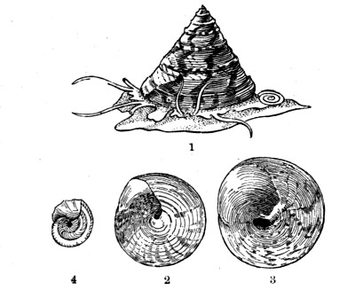
Fig. 171.—1. Trochus zizyphinus. 2. Under side of Shell. 3. Trochus magnus. 4. Adeorbis subcarinatus
The well-known Periwinkle (Littorina littorea) and the species to the right of it on Plate V., belong to the family Littorinidæ, the members of which are similar in structure and habit to Trochus, but the shell is usually more depressed, and is never pearly. The shell of the Periwinkle is thick, having but few whorls, and is not umbilicated; and the lingual ribbon, which is coiled up on the gullet, contains no less than about five hundred rows of teeth; but only a little more than twenty of these rows are in action at any one time, the remainder being a reserve stock to come into active service as the ribbon grows forward. In the genus Lacuna there is a narrow umbilicus, and the aperture of the shell is semilunar in form; and the species of Rissoa are very small, with white or horny shells, much more pointed and having more whorls than those of the Littorina.
Our next illustration shows three shells of the family Turritellidæ, so named from the resemblance of the shells to a tower or spire. The form indeed is so characteristic that they can hardly be mistaken. It will be seen that Turritella communis is striated spirally, while the surface of Scalaria communis (Plate V.) is relieved by strongly marked transverse ribs. Both these species are very common, and the latter is peculiar for its power of ejecting a dark purple fluid when molested. The other representative of the family—Cæcum trachea—has a shell something like that of Dentalium (p. 238), being cylindrical and tubular, but it differs in being closed at one end.
In the succeeding shells, of the family Cerithiadæ, the spire is also considerably produced, so much so that some of the species closely resemble the Turret shells, but they are distinguished by usually having an expanded lip, at least in the adult form; and the mouth is channelled in front, and sometimes also behind. The animals of the group have short muzzles that are not retractile, the tentacles are wide apart, and the eyes are mounted on short pedicels. The median teeth are arranged in a single row, with three laterals on either side of each.
Cerithium reticulatum receives its generic name from its appearance to a small horn, and the specific name refers to the netted appearance of its surface due to the presence of numerous little tubercles arranged in rows—a feature that serves to distinguish it from the small Turret shells. It is a common shell, as is also the other representative of the family illustrated, but the latter is rendered conspicuous by the enormously expanded lip that has earned for it the popular name of Spout Shell. Its scientific name is Aporrhais pes-pelicani, and the application of the specific term will be understood when the shell is viewed from above, for the expanded lip is drawn out into long finger-like lobes that suggest the foot of a bird. This is a very solid shell, sometimes reaching a length of two inches; and the animal inhabiting it is carnivorous.
We have yet some turreted shells to deal with, belonging to the family Pyramidellidæ, but they need not be confused with the preceding groups if carefully examined. In the first place, the aperture of the shell is very small; and the operculum, instead of being spiral, as in the turreted shells before mentioned, is imbricated or made up of parallel layers denoting that the growth took place on one side only. Another distinguishing feature is seen in the nucleus—that small portion of the spire that was developed within the egg—which is sinistral or left-handed. In addition to this, the animal has broad, ear-like tentacles, a retractile proboscis, and a lingual ribbon without teeth.
The British species of this family belong principally to the genera Odostomia, characterised by a tooth-like fold of the columella; Eulima, containing small, white, polished shells with numerous level whorls; and Aclis, with little polished shells not unlike Turritella.
The last family of the Holostomata is the Naticidæ, the shells of which are almost globular, with only a few whorls, and a small, blunt spire. The mouth is semilunar in form, and the lip sharp. The proboscis of the animal is long and retractile, and the foot large; but perhaps the most characteristic feature is the presence of large mantle lobes which hide some of the shell when the animal is crawling. In Natica (fig. 155), the typical genus, the shells are somewhat thick and smooth, with a large umbilicus. As the animal crawls a large fold of the mantle is reflected back over the head, completely covering it, and apparently obstructing its view; but this is not the case, for the creature has no eyes. Natica is very abundant on some sandy beaches, where it devours small bivalves and other animals; and it is frequently washed up alive by the waves. Its shell is also a favourite one with hermit crabs. Its eggs, all connected together in a spiral band, may often be seen stranded on sandy coasts. Several species of Natica are found on our shores. An allied mollusc—Velutina lævigata, so called on account of the velvety epidermis that clothes the shell, completely surrounds the shell by its mantle folds when creeping.
The Siphonostomata form a much smaller section than the last, and its members are distinguished mainly by the presence of a true siphon, formed by the prolongation of the mantle margin, and serving to convey water into the gill chamber. In all these the shell is spiral, usually without an umbilical opening, and the margin of the mouth is prolonged into a canal or distinctly notched. The operculum is horny, and lamellar or imbricated. The animal has a retractile proboscis, and the eyes or eye-pedicels are joined to the tentacles. All the species of this division are marine.
We will first take the family Cypræidæ, which contains the familiar Cowries, these forming the lowest group of the division. An examination of the shells may at first seem rather puzzling, for the spire is concealed, and the whole is convoluted in such a manner as to make the mouth long and narrow, with a channel at either end. The outer lip is also thickened and bent inward, and there is no operculum.
The animal itself is particularly interesting, for, as it creeps along on its broad foot, abruptly shortened in the front, the mantle lobes bend over the top, meeting along the middle line, where they are usually fringed with little tentacle-like processes; and, as a result, the whole shell is beautifully enamelled on the outer surface. In all the Cowries the central teeth are single, and the laterals are arranged either in twos or threes.
Perhaps the commonest representative of this family is the pretty little Cypræa (Trivia) europæa (Plate V.), the shells of which are sometimes washed up in large numbers on sandy beaches. The animal lives mainly below low-water level, but it may often be found in the larger rock pools, creeping rapidly over the tangles, and may be easily secured with the aid of a net.
In the same family we have the little Erato (Marginella) lævis, the white shell of which is minutely furrowed along the lips; and also Ovulum patulum (Calpurna patula), so called on account of its fancied resemblance to a poached egg.
We have also several species of Cone shells (family Conidæ) on our coasts, readily recognised by their form, which is a cone, with a long, narrow aperture, partially closed by a minute operculum. As in the last family, the foot is abruptly shortened in front. The head is very prominent, with eyes situated on the tentacles. There are two gills, and the teeth are arranged in pairs.
The Conidæ are principally inhabitants of tropical seas, where some very large species exist. Two of the British representatives, both common shells, are shown in fig. 180.
Our next family (Buccinidæ) is so well distributed on our coasts, that it would be difficult, we imagine, to find a spot quite free from its familiar forms. It contains all those creatures commonly known as Whelks, Dog Whelks, and Dog Winkles, ranging from deep water almost to high-water mark.
In all these the shell is notched in front, or the canal is turned abruptly upward. The foot of the animal is broad, the eyes are situated either on the tentacles or at their bases, and there are two gill plumes.
All the species are carnivorous, and some are said to be very destructive to mussels and young oysters.
The Common Whelk (Buccinum undatum, Plate V.) lives in deep water, whence it is dredged up largely for the market. Its clusters of egg cases are washed up in large numbers on the beach, where they form one of the commonest materials among the refuse at high-water mark. It is not uncommon, also, especially after storms, to find the unhatched eggs stranded by the waves, and these are so transparent that the embryos, several in each capsule, may be seen within. The hole through which the young escape may also be seen on the inner side.
The Dog Periwinkle (Purpura lapillus) abounds on all our coasts and is remarkable for the production of a dull crimson or purple fluid that may be obtained from it by pressing on the operculum. This fluid turns to a brighter colour on exposure to air, and is said to have been used largely in former times as a dye. It will be seen from our figure that the spire of this shell is shorter in proportion than that of Buccinum; but both are alike in that the operculum is made up of layers with a nucleus on the external edge.
The other species figured is Nassa reticulata, popularly known as the Dog Whelk, and characterised by a tooth-like projection of the inner lip close to the anterior canal. It is very common near low-water mark, where it may be seen crawling over the rocks on its broad foot, from which project two hornlike appendages in front and two narrow tails behind.
From the last family of the gasteropods (the Muricidæ) we select two common species—Murex erinaceus and Fusus antiquus (Plate V.). In both these the anterior canal of the shell is straight and the posterior wanting. The eyes are on the tentacles, and there are two plumed gills. Both are carnivorous species, feeding on other molluscs; and the former is said to bore through the shells of its prey with the prominent beak of its shell.
Murex may be readily distinguished by the prominent longitudinal ridges of the thick shell, its rounded aperture, and by the partly closed canal running through the beak. It is known to fishermen as the Sting Winkle; the other species is called the Red Whelk in some parts, and in Scotland is known as the Buckie. Like the common whelk, it is dredged largely for the market, and is said to be far more esteemed than the former, from which it may be distinguished by the fusiform shape of the shell and the long straight canal.
We now pass to the last and highest class of the mollusca, called the Cephalopoda because they have a number of arms attached to the head, round the mouth. Unlike the majority of molluscs they are bilaterally symmetrical: and are much more highly organised, in some respects even making an approach to the vertebrates. Thus they generally have an internal hard structure, either horny or calcareous in structure, representing the vertebral column, and the circulatory system consists of arteries and veins, connected by minute capillaries. The corpuscles of the blood are also similar in form to those of the vertebrates. Externally they are all naked, with the exception of the nautilus and argonaut of the warmer seas.
The arms, so characteristic of the class, are eight or ten in number, long and muscular, and provided with numerous suckers by which the animal can cling with remarkable tenacity. These suckers are situated on the inner surface of the arms, and the disc of each one displays a series of muscular fibres, all converging from the circumference towards the centre, which is occupied by a softer structure that works inwards and outwards like the piston of a pump. Thus the suckers form a system of exhausting air-pumps by which a vacuum can be produced, and the tenacity of the grip, maintained by atmospheric pressure, is so great that the arms, strong as they are, may be torn asunder by attempting to pull them from their hold; and yet the animal can release its grip with the greatest of ease by simply releasing the pistons of its pumps.
The cephalopods are further distinguished by their very large, glaring eyes, situated on the sides of the well-formed head, and by powerful jaws that work in a vertical plane, like those of the vertebrates, but somewhat resembling the beaks of certain birds. The tongue is also very large and fleshy, and in part armed with numerous hooked spines or teeth.
The class is usually divided into two orders, one characterised by the possession of two gills, and the other of four; but the British species belong to the former, known technically as the Dibranchiata. This order is subdivided into two sections according to the number of arms; and the divisions are called the Octopoda and Decapoda respectively.
The former section includes the Octopods, of which some species inhabit our seas. They all have eight arms, of unequal size, with the suckers arranged in two rows, and their round or oval bodies seldom have any fins, locomotion being effected by means of the arms, and by the sudden expulsion of water from the siphon. The shell is rudimentary, being represented merely by two short ‘styles’ within the mantle. The species vary considerably in size, some being only about an inch long when fully grown, while others measure two feet or more, and are looked upon as formidable creatures by man. Sometimes they are washed up on our beaches, but the best way to make their acquaintance is to examine the contents of the fishermen’s drag nets as they are hauled on the beach.
In the same manner we may secure various species of the Decapods or Ten-footed Cephalopods, which comprise the Calamaries, Squids, and Cuttlefishes. These, too, properly speaking, have but eight arms, the other two appendages being really tentacles, which are usually longer than the arms, and more or less retractile; they are also expanded at the ends. The decapods are also to be distinguished from the octopods by their elongated bodies, and a flattened, fin-like appendage on either side. Their eyes, also, are capable of being rotated within the orbits, while those of the octopods are fixed; and the shell consists of one or more horny ‘pens,’ or of a calcareous ‘bone,’ contained in a cavity so loosely that it drops out of its place when the cavity is opened.
The Common Calamary (Loligo vulgaris) may be recognised by the accompanying illustration, from which it will be observed that the body tapers behind, bearing two rhomboidal fins in the rear. The suckers are arranged in two rows on the arms, but in fours on the expanded tips of the tentacles. The animal is a good swimmer, and sometimes crawls, head downwards, on the disc surrounding the mouth, pulling itself along by means of its arms. Its shell is a horny pen, lanceolate in form, but it divides as the age of the animal advances, so that two or more may be found in the same specimen.
Belonging to the same family we have the Common Squid (Sepiola atlantica), also a very abundant species. Here the body is shorter and purse-like, and the fins are dorsal and rounded. It seldom exceeds four or five inches in length, and, like the Calamary, is used largely as a bait by fishermen.
Another family—the Sepiadæ—contains the Cuttlefish (Sepia officinalis), the ‘bone’ of which is such a common object on the beach. This latter is a broad, curved plate of carbonate of lime, made up of a number of regular layers, and having a cavity hollowed out at the posterior end. It is exceedingly light and porous in structure, and at one time was used largely as an antacid as well as a dentifrice. It is also proportionately large, being both as long and as broad as the body of the animal.
Cuttlefishes live principally in the shallow water close to shore, where they swim backwards by the sudden propulsion of water from their siphons; and their eggs, which look like clusters of black grapes, are frequently thrown up on the beach, generally attached to the stems and fronds of sea weeds.
As a rule the cephalopods swim slowly by the aid of their fins or by a rhythmic contraction by which water is expelled from their siphons, but when in danger the muscular contraction is so violent that they dart through the water with great speed, and even leap into the air to avoid their enemies. But they have another and much more remarkable way of escaping from their foes:—They possess a gland, the duct of which opens into the base of the funnel or siphon, that prepares an inky fluid; and when the animal is disturbed it suddenly ejects this fluid, rendering the surrounding water so cloudy that it is often enabled to retreat unobserved. The ‘ink’ of the Sepia was used for writing in former times, and is still employed in the preparation of the artist’s pigment that bears the same name. Fishermen are well acquainted with this peculiar characteristic of the animal, for they are frequently bespattered with the contents of the ink bag of the Sepia when the creature is included in the contents of their draw-nets, and have learnt to handle it cautiously until the objectionable fluid has been all discharged.
We will conclude this chapter by giving a tabular summary of the classification of the molluscs which will probably be useful to the collector of marine objects.
Class LAMELLIBRANCHIATA—Plate-gilled. Headless, usually enclosed in bivalve shell.
Section SIPHONIDA—Mantle lobes more or less united to form tubular siphons.
Families—Pholadidæ, Gastrochænidæ, Anatinidæ, Myacidæ, Solenidæ, Tellinidæ, Mactridæ, Veneridæ, Cyprinidæ, Lucinidæ, Cardiadæ, &c.
Section ASIPHONIDA—Mantle lobes free or nearly so. No true siphons.
Families—Arcadæ, Mytilidæ, Aviculidæ, Ostreidæ, &c.
Class CEPHALOPHORA—Head-bearing. Usually enclosed in a univalve shell.
Section PTEROPODA—Wing-footed molluscs.
Section GASTEROPODA—Stomach-footed molluscs.
Order Nucleobranchiata—Viscera form a nucleus on the back.
Order Opisthobranchiata—Shell generally absent. Gills more or less exposed.
Section Nudibranchiata—Naked gills.
Section Tectibranchiata—Gills covered by shell or mantle.
Order Pulmonifera—Lung-breathers. Terrestrial.
Order Prosobranchiata.
Section Holostomata—Aperture of shell entire (sea snails).
Families—Chitonidæ, Dentaliadæ, Patellidæ, Calyptræidæ, Fissurellidæ, Haliotidæ, Turbinidæ, Littorinidæ, Turritellidæ, Cerithiadæ, Pyramidellidæ, Naticidæ, &c.
Section Siphonostomata—Possess a true siphon. Carnivorous.
Families—Cypræidæ, Conidæ, Buccinidæ, Muricidæ, &c.
Class CEPHALOPODA—Sucker-bearing arms round the mouth.
Order Dibranchiata—Two gills.
Section Octopoda—Eight arms.
Families—Argonautidæ, Octopodidæ.
Section Decapoda.
Families—Teuthidæ (Calamaries, Squids), Sepiadæ, &c.
Order Tetrabranchiata—Four gills (containing Nautilidæ).
The sub-kingdom Arthropoda contains a vast assemblage of animals, all of which, as the name implies, possess jointed appendages. Their bodies are covered with a skin that is hardened by a horny substance (chitin), and frequently, also, by the deposit of carbonate of lime.
The body of Arthropods is made up of a chain of segments, all of which are built up on one common pattern, and each one is surrounded by a ring of the hardened skin or exo-skeleton that gives attachment to a pair of appendages. Commonly, however, two or more of the segments become fused together, being covered by a continuous plate or shield, in which the boundaries of the rings are almost or completely obliterated; but in such cases the appendages they bear always remain distinct, so that the true number of segments is always apparent. The skin between those segments that are not so fused together remains soft and flexible, thus allowing the body to be freely bent.
The appendages exhibit a great variety of structure, and are as varied in their functions. Some are used as feelers, and others as jaws for seizing or masticating food. Some are developed into powerful seizing organs for purposes of defence or attack, some into paddles for swimming, while others are legs adapted for walking.
All these appendages are made up of segments, each of which, like those of the body itself, is surrounded by a ring of hardened skin, and connected with its neighbours by a flexible integument that allows perfect freedom of movement; while within are the muscles, often very powerful, by which the appendage is moved.
In the arthropods we have a sub-kingdom of highly organised animals, with distinct, and often very complicated, systems of organs for digestion, circulation, and respiration; and the nervous system consists of a well-developed chain of ganglia, connected by nerve cords, and from which nerve fibres are distributed to the various parts of the body. It should be noted, however, that some members of the group have degenerated into parasites, and in these, as with all such degraded creatures, many of the organs have retrogressed to such an extent that they are quite functionless, or have even disappeared entirely. These parasitic forms, when very young, are really highly organised creatures, not unlike the young of their industrious and more noble relatives; but, as the natural result of their degraded mode of living, in which they find no use for their organs of locomotion, digestion, circulation and respiration, these eventually disappear, with the result that the organs of reproduction predominate to such an extent that they often fill the greater part of the cavity of the body.
It should be noted, too, that the sense organs of arthropods are well developed, most of them being supplied with complex eyes, hearing organs, and highly sensitive feelers.
This sub-kingdom consists of four classes—the Crustacea, including lobsters, crabs, shrimps, prawns, &c.; Arachnoidea, containing spiders, mites, and scorpions; Myriopoda—centipedes and millepedes; and Insecta.
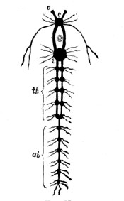
Fig. 188.—The Nerve-chain of an Arthropod (Lobster)
o, optic nerve; c, cerebral ganglion; i, large ganglion behind the œsophagus; th, ganglia of the thorax; ab, ganglia of the abdomen
The first of these classes consists mainly of marine animals, and will therefore occupy much of our attention, but the members of the other three are mostly terrestrial and aërial creatures that do not fall within the scope of this work, except in the case of a few species that are more or less decidedly marine in their tendencies. The aquatic members are generally provided with well-formed gills by means of which they are enabled to extract the dissolved oxygen from the water in which they live, while those of terrestrial and aërial habits breathe by means of a system of tracheæ or air-tubes that are open to the air and supply branches to all parts of the body.
The Crustaceans are mostly gill-breathers, though some of the aquatic species have no special organs for respiration, but obtain the oxygen necessary for respiration by absorption through their thin, soft skin, while the terrestrial species breathe by means of tracheæ, as we have just observed.
Most of them are covered with a calcified skin, as in the case of crabs and lobsters; but many are protected with a chitinous or horny covering such as we observe in shrimps and prawns. In either instance the hardened integument constitutes what is known as the exo-skeleton. None of the crustaceans have an internal skeleton of any kind, though some of the inner parts are supported by extensions of the hard skin that penetrate into the body.
It will be readily understood from the nature of the exo-skeleton of the crustacean, and especially of the more or less rigid calcareous covering of the crab and the lobster, that a uniform growth of the body is absolutely impossible, and, in fact, that an increase in size cannot take place without an occasional casting of the hard coat of mail. Hence we find most crustaceans throwing off their coverings at intervals, and growing by fits and starts during the periods between the ‘moultings’ and the hardening of the newly exposed skin.
When a crab or a lobster is about to undergo the process of moulting, it retires to a secluded niche in the rock, where it is not so easily found by its numerous enemies—a necessary precaution, since the creature in its soft or unarmoured condition is eagerly devoured by fishes and other marine animals—and there awaits the first stage of the ordeal. Presently the skin splits; and, after a time, the crustacean succeeds in extricating itself from its shell, which is cast off in a perfect condition, every joint being entire, even to the coverings of the antennæ, the stalked eyes, and other delicate appendages. And not only this, for the portions of the shell that penetrate inward into the body are also discarded, as well as the linings of the stomach and the gills; and these cast-off coats of crabs and lobsters—especially the former—may often be found in the most perfect condition on the sea shore, being washed up without injury on the sandy beach, or found in the very niche in which the creature changed its attire.
If one examines the powerful pincers of a crab or lobster, a thin plate of considerable size will be seen to extend within from the movable ‘jaw’ to give attachment to the muscles by which it is moved, and it seems impossible that this can be removed with the cast skin without considerable injury to the new claw that is already formed, though as yet in a soft condition, within the old and hard one. But it has been observed that this plate actually cuts through the new claw, and that the claw thus divided almost immediately closes up and unites again.
The moulting process being over, the crustacean’s body extends itself within the new, yielding skin; and, the latter becoming gradually hard by the deposition of carbonate of lime, the creature is able, after a period of rest, to roam at large again, without much fear of injury, until the time for the next moulting has arrived.
Those who have made but a slight acquaintance with the common crustaceans of our shores must have noted the frequency with which imperfect specimens occur—specimens with missing appendages, or with a well-formed limb on one side of the body opposed to a puny and almost useless fellow on the opposite side. As to the loss of appendages, this matter will be readily understood by those who have watched crustaceans, and especially crabs and lobsters, in their native element, so often do these pugnacious creatures become engaged in furious broils with their neighbours. And, when we are at work at the collection of various species on the sea shore, how often do we find that a creature escapes from our grip by leaving us in possession of a severed limb, while the owner retreats rapidly among the stones and weeds apparently none the worse for its trifling loss! This is, in fact, a very common method of securing its escape from an enemy; and it appears that many crustaceans have the power of thus rendering a seized limb so brittle that it may be snapped off with the greatest of ease.
We have spoken of the loss thus sustained as a trifling one; and so it is, for crustaceans have the faculty of reproducing lost appendages; and though the loss may be one of considerable inconvenience at first, a new limb eventually appears in the place of each one so willingly discarded.
When such mutilations occur, it will be observed that the severed limb invariably breaks away at the end of the first or basal joint—a point where the bloodvessels are so narrow and contractile that but little loss of blood takes place when the rupture is made—and it has been said that the animal would soon bleed to death if the fracture were to take place at any other point. As it is, the wound soon heals, but no trace of a new limb is to be seen, at least without dissection, until the time of the next moult. The part is developing, however, beneath the cover of the basal joint; and when the moulting period arrives, the new limb, still very small, is exposed to view. It then rapidly enlarges, though not to anything like its proper size, and its surrounding skin becomes hardened by the deposit of the calcareous secretion simultaneously with that of the rest of the body. Further enlargements of the new appendage take place at subsequent moults, with the final result that it is but slightly inferior to its fellow either in size or in power.
The eye of a crustacean is a very complicated structure, commonly described as a compound eye. It consists of a large number of conical, radiating, crystalline rods, collected together into a mass that presents a convex outer surface. This surface is covered with a transparent layer of chitin which naturally presents a more or less distinct netted appearance, the bases of the rods being in contact with its inner surface, and visible through it. Each rod is surrounded by a layer of pigment that prevents light from passing from one to another, and the optic nerve passing into the base of the compound structure sends a sensitive filament into each one.
In many crustaceans this compound eye is situated on the end of a movable stalk that generally allows it to be protruded or drawn under cover as occasion requires, but in others the organ does not project beyond the general surface of the body. Thus we hear of the animals of this class being divided into the stalk-eyed and the sessile-eyed groups; the former being represented by crabs, lobsters, shrimps, &c.; and the latter by sandhoppers and sandborers.
Crustaceans undergo metamorphoses while very young, the body being altered considerably in form at several successive moults. Some, in their earliest stage, consist of a little oval body that shows no signs of a division into segments. It swims about by means of three pairs of appendages, and has only one eye. Others start life with four pairs of limbs, attached to the front portion of the body, a segmented abdomen, as yet perfectly limbless, and a pair of compound eyes. Then as the successive moultings take place, new segments and new appendages are developed, until, at last, the form of the adult is assumed. The accompanying illustration shows four stages in the development of the Common Shore Crab.
The lowest division of the crustaceans contains the Cirripedia or Curl-footed crustaceans, which includes the Barnacles that are so frequently seen attached to the bottom of ships and of floating timber, and the Acorn Barnacles, the conical shells of which often completely cover large masses of rock on our shores.
For some time naturalists could not agree as to the proper place of these animals in the scale of life, but the matter was finally settled when some minute creatures only about a twelfth of an inch in length, and closely resembling the early stages of certain crustaceans, were seen to undergo metamorphoses, and finally develop into acorn barnacles. Their position in the animal kingdom was thus determined by their early stages; but these, instead of changing into a segmented and highly organised creature like the typical crustacean, lose some of their appendages, cease to be free-moving animals, and attach themselves to floating bodies by which they are carried about. Thus they are enabled to find the food they can no longer seek without such aid. In their young state they possess not only the means of freely moving in search of their food, but have organs of vision to aid them in the capture of their prey. As they grow, however, the foremost appendages are transformed into a sucking-disc, and the eyes, no longer necessary, disappear. It will thus be seen that the degenerated adult—the product of a retrograde development—is attached by what was originally the front of its body, while the abdomen is undeveloped, and the thorax, with its appendages, forms the summit of the free extremity.

Fig. 192.—Four Stages in the development of the Acorn Barnacle
a, newly hatched larva; b, larva after second moult; c, side view of same; d, stage immediately preceding loss of activity; a, stomach; b, base of future attachment. All magnified
Some of the Cirripedes attach themselves to the bodies of whales and other marine animals. The majority of these are pseudo-parasites—creatures that live on the bodies of other animals, but do not derive their food at the expense of their hosts; others, however, are true parasites, subsisting on the nourishing juices they extract from the animals to which they are attached.
The Acorn Barnacles, so numerous on our shores, are good types of the Cirripedia, and they are so easily kept alive in the indoor aquarium that their interesting movements may be well observed. A cluster of these animals may be obtained by chipping off a piece of the rock on which they grow; or, instead of this, a few minutes’ searching on a rocky coast at low tide will certainly provide us with a stone of suitable size, or the shell of a mollusc, on which the creatures have found a home.
Place them in the indoor aquarium, or in any shallow vessel containing just sufficient sea-water to cover them, and carry out your observations with the aid of a hand lens. They will soon open the inner cone of their many-valved shell, and slowly protrude six pairs of gracefully curved and delicately-feathered appendages which, as previously stated, are attached to the thoracic portion of the body. Then, with a much more rapid movement, the appendages will be withdrawn, and the shell closed. These alternate movements are continued incessantly, and are the means by which the animals provide themselves with both food and air. The reader should also obtain some specimens of the larger species for the examination of the shell, the structure of which is interesting and, of course, peculiar to this order.
In general structure and habits Barnacles are very similar to the acorn barnacles, except that the body is supported on a tough stalk, which, as we have already stated, is the modified anterior portion of the animal. These animals also may be easily kept alive and examined in the indoor aquarium. They are not creatures of the sea shore, but may often be obtained on masses of timber that have been washed ashore, or from the bottoms of ships that have been placed in the dry dock for repairs.
Another order of the crustaceans—the Copepoda, or oar-footed group—is so called on account of the bristled feet that are employed after the manner of oars when the creatures are swimming.
These Copepods are small animals, so small indeed that the compound microscope is generally necessary merely for the examination of their external characters. Many species inhabit fresh water, and the study of the group is more commonly pursued by the investigator of fresh-water pond life than by the sea-side naturalist. However, marine species are abundant, and may be captured in the open water or in rock pools by means of a muslin net. As with the last order, some degenerate from the comparatively complicated free-swimming and eyed larval state to blind and limbless parasites that feed on the bodies of fishes and are known as fish lice.
The body of the typical copepod is distinctly segmented, and the head and thorax are both enclosed in a hardened buckler. It has two pairs of antennæ, two pairs of foot jaws by which it captures its prey, and four or five pairs of bristled feet for swimming. The jointed abdomen has also a tuft of bristles at its extremity. The annexed illustration represents some marine species, and will serve to show the general features of the order.
The sea-side naturalist, intent on the collection of small life, may possibly meet with representatives of two other orders of crustaceans—the Ostracoda or shelled crustaceans, the bodies of which are enclosed in a bivalve, hinged shell; and the Branchiopoda, so called because the branchiæ or gills are attached to the feet.
The Ostracodes have two or three pairs of feet which subserve locomotion, but are not adapted for swimming; and two pairs of antennæ, one of which assists in locomotion. The mouth is provided with organs of mastication, the branchiæ are attached to the hind jaws, and the animals have but one eye. Some of these crustaceans inhabit deep water only, while others live in sand between the tide-marks; but several species, belonging chiefly to the genus Cythere, abound in rock pools, where they may be readily obtained by scraping the confervæ and corallines with a small muslin net.
The branchiopods are free swimmers, and are protected by a buckler-like envelope. Most of them are inhabitants of fresh water, and are popularly known as water fleas. We have figured one marine species, belonging to the genus Evadne, which has a colourless body, and a single conspicuous black eye, and is interesting as being the food of the herring.
The four orders of crustaceans that have been briefly described belong to the division Entomostraca, which signifies ‘shelled insects.’ This term is not a happy one when judged from the standpoint of our present knowledge of animal life, but it must be remembered that, at the time it was applied (1785), spiders and crustaceans were all included in the same class as the insects; and this is hardly surprising when we observe the close relationship of these animals, as shown in their segmented bodies and jointed appendages; for, as we have already shown, the lowly organised parasitic crustaceans which, in the adult state, lose most of their appendages and cease to be distinctly segmented, are more or less insect-like in their larval and free-swimming stage.
All the other crustaceans are included under the term Malacostraca, or soft shelled, since, although many of them are protected by an exo-skeleton that is hardened by the deposit of carbonate of lime, yet, generally speaking, their coverings are softer than those of the molluscs; and therefore the term Malacostraca was originally applied by Aristotle in order to distinguish them from the animals that are covered by harder and thicker shells.
This division of the crustaceans contains wood lice, sandhoppers, lobsters, shrimps, crabs, &c., and consists of two main groups—the Sessile-eyed (Edriophthalmata) and the Stalk-eyed (Podophthalmata) crustaceans.
We shall now consider the Sessile-eyed group, dealing first with the order Isopoda or equal legged, and then the Amphipoda, which have appendages adapted both for walking and swimming.
The general nature of an Isopod may be readily understood by the examination of the common woodlouse that abounds in gardens and damp places almost everywhere, and the reader will probably remember having seen similar creatures crawling over the rocks on the sea shore.
The body is generally depressed or flattened, but convex above, and is composed of seven segments, each segment bearing a pair of legs which terminate in a pointed claw, while the posterior appendages are modified into flat, leaf-like organs of respiration.

Fig. 199.—Marine Isopod
1. Sphæroma serratum. 2. Limnoria lignorum. 3. Ligia oceanica. 4. Nesæa bidentata. 5. Oniscoda maculosa
When engaged in ‘shrimping’ one frequently meets with shrimps or prawns that are disfigured by a tumourous swelling on the side of the body, and if the swelling be opened a little parasite will be dislodged. This parasite is an Isopod (Bopyrus), the appendages of which are imperfectly developed. The female is very much larger than the male, and, as is usual with parasitic creatures, the greater part of the body-cavity is occupied by the well-developed organs of reproduction.
There are several other parasitic isopods, some of which live on the bodies of fishes, and are popularly known as fish-lice, but these are not so likely to come in the way of the sea-side naturalist as the more typical forms that crawl about on the rocks and among the weeds of the coast. A few of the latter are shown in the accompanying illustration, including the Sea Pill-ball (Nesæa bidentata), common on the rocky coasts of the south-west, and distinguished by the two sharp projections on the last segment; the Serrated Pill-ball (Sphæroma serratum), very common on most rocky shores, and characterised by the fine sawlike teeth on the outer edge of the outer plates of the ‘tail’; the Great Sea-slater (Ligia oceanica), also an abundant species; the Spotted Hog Louse (Oniscoda maculosa) that lives among the tufted sea weeds; and the Boring Pill-ball (Limnoria lignorum) that bores into the woodwork of piers and jetties, often doing considerable damage.

Fig. 200.—Marine Amphipods
1. The spined sea screw (Dexamine spinosa). 2. Westwoodia cœcula. 3. Tetromatus typicus. 4. The sandhopper (Orchestia littorea). 5. Montagua monoculoides. 6. Iphimedia obesa. All enlarged
The above and other isopods feed on various animal and vegetable substances, some species being quite omnivorous in habit. Most of them are eagerly devoured by birds and fishes.
The Amphipods, six species of which are shown in the above illustration, include the Sandhoppers or Beach Fleas, so numerous on our coasts that it is almost impossible to go any distance without making their acquaintance. They are invaluable as scavengers, as they rapidly devour decaying sea weeds, and will speedily reduce the body of any animal washed on the beach to a clean skeleton. Although they are all small creatures, they make up in numbers for any deficiency in size; and though devoured in enormous quantities by the various shore birds, they multiply so prodigiously that they are never lacking wherever there is decomposing organic matter to be consumed.
The bodies of these animals are usually flattened from side to side, very distinctly segmented, and have a well-developed abdomen. The head is furnished with two pairs of antennæ and a pair of sessile eyes, though some species possess only one pair of antennæ, while others have four eyes. The limbs of the thorax are used either for walking or for swimming, and give attachment to the gills. The abdomen has generally six pairs of appendages, the foremost three pairs of which are usually small, and employed in swimming, while the others are stronger and directed backwards, and are often adapted for jumping.
It is very interesting to observe the habits of the Sandhoppers and other Amphipods both on the sandy beach and in the water, and the student will find that certain species burrow into the sand with considerable agility, and live principally at the extreme high-water mark, where they feed on the organic matter washed in by the breakers at each high tide, while others dwell almost exclusively in the water, among weeds and stones, and should be searched for at low water. The latter may be kept alive for some time in the aquarium providing they are the only occupants, but a little experience will show that these and all other Amphipods are readily devoured by many marine creatures, and consequently they are of real value to the aquarium keeper as food for other animals.
We now come to the Stalk-eyed Crustaceans (Podophthalmata), which contain those members of the class most generally known, such as crabs, lobsters, shrimps, and prawns. In these the eyes are mounted on movable pedicels, the head and thorax are generally covered by a large shield called the carapace, and the appendages are adapted partly for seizing and masticating, and partly for locomotion.
The group includes two orders—the Stomapoda or Mouth-footed crustaceans, so called because some of the limbs are crowded round the region of the mouth; and the Decapoda, or Ten-footed crustaceans.
The Stomapods, though very abundant in tropical seas, are not often met with on our own shores. However, since a few interesting species are inhabitants of our seas we will briefly describe the distinguishing characteristics of the group.
We have just mentioned the fact that the head and thorax of a decapod is usually covered by a large shield—the carapace. Now, the general character of this carapace may be seen at once in either the shrimp or the lobster. In these animals the segments that form the head and the thorax are all fused together, and are completely covered by the protective buckler of hardened skin; but in the Stomapoda the carapace is much smaller in proportion, and a few of the segments of the thorax, instead of being fused into the general mass of the cephalo-thorax, are quite distinct from it. The abdomen, also, is large and strongly formed in these animals. Five pairs of the thoracic limbs are directed forwards, and are adapted both for catching food and for climbing, while others are used in walking. The limbs of the abdomen generally number six pairs, of which the first five bear feathery gills.
Two species of Mantis Shrimps, one of which is represented in fig. 201, have been found off the south and south-west coasts, but these are not likely to be seen on the shore, since they inhabit deep water. Allied to these, and sometimes included with the Stomapods, are the Opossum shrimps, so called because the females of some species carry their eggs in a kind of pouch, thus reminding us of the marsupial quadrupeds of the same name. They are of very slender build compared with the mantis shrimps, and differ from them in that the carapace completely covers the thorax; but though this is the case, the fusion of the thoracic segments is not complete, since the posterior ones have still a certain amount of freedom of movement. Some species of opossum shrimps are abundant in the rock pools of our coasts, particularly in the south-west, but their bodies being often so transparent as to be almost invisible, they are consequently easily overlooked. Their general appearance may be gathered from our illustration of Mysis chamæleon, which is probably the most common species inhabiting our coast.
The highest crustaceans—the Decapods—are divided into two sub-orders—the Macrura, or Great-tailed, including lobsters, shrimps, &c.; and the Brachyura (Short-tailed), containing the crabs; but the number of British species is so large that it is impossible to give, in our limited space, a detailed description of all the commonest even. All we can do is to note a few of the more interesting features of certain species, to introduce such illustrations as will enable the young naturalist to identify a number of the commoner ones, and to give the general characteristics of the main divisions so that the student may be able to classify his specimens intelligently.
In the Macrura, as with other divisions of the crustaceans, we meet with very interesting modifications of the appendages, adapted to quite a variety of uses; and if the reader is unacquainted with these adaptations of structure to habit he cannot do better than secure a lobster or crayfish for study. It will be observed that the body may be divided into two main portions—the cephalothorax, consisting of head and thorax combined, and the abdomen. The former is composed of fourteen segments, so thoroughly fused together that they are denoted only by the fourteen pairs of appendages to which they give attachment, while the calcified skin forms one continuous shield surrounding the whole. The abdomen, on the other hand, consists of six distinct segments, each of which is surrounded by its own ring of the hardened integument, and is connected with its neighbours by means of a portion of uncalcified skin that renders the whole very flexible. A groove in the front portion of the great shield (carapace) marks the division between the head and the thorax, the former composed of six, and the latter of eight united segments.
The calcareous covering of each segment consists of an upper portion, called the tergum, and a lower, named the sternum, united at the sides; the sternal portion of the cephalothorax, which gives attachment to the walking limbs, is a most complicated and beautifully formed structure.
The six pairs of appendages belonging to the head are easily made out with a little care. The first are the jointed eye-stalks that bear the compound eyes previously described; and these are followed by two pairs of antennæ, or feelers, the first being shorter and double, while the second are very long. The former contain the organs of hearing. Then, in front of the mouth, and completely hiding it, are a pair of strong mandibles or jaws that move horizontally, and the two pairs of maxillæ that are also employed in reducing the food.

Fig. 204.—A Segment of the Abdomen of a Lobster
t, tergum; s, sternum, bearing a pair of swimmerets; h, bloodvessel; d, digestive tube; n, nerve chain
Following these, but belonging to the thorax, are three pairs of appendages that are known as foot-jaws; for, although they assist the preceding organs in breaking up the food, they bear a resemblance in some respects to the longer limbs behind them. Of the latter there are five pairs (hence the term decapoda), the first being a very powerful pair of seizers or pincers, and the remaining four, which are well adapted for walking, terminating in either double or single claws.
All the appendages above mentioned are not only attached to the body by movable joints, but are themselves made up of jointed parts, sometimes a considerable number, each of which, like the segments of the body itself, is surrounded by a ring of hardened skin, and connected with those above and below it by a portion of soft and flexible skin.
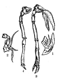
Fig. 205.—Appendages of a Lobster
1. Second maxilla. 2. Third foot-jaw. 3. Third walking leg. 4. Fifth walking leg
Lastly, beneath the abdomen, are paired limbs called swimmerets, which are used as paddles, and probably assist the animal more or less in its progress through the water; but the principal organ of locomotion in the macrura is undoubtedly the powerful muscular abdomen, aided by the broad and fanlike tail formed by the appendages of the last segment. To demonstrate this fact, put a live lobster, or even a shrimp, in a still rock pool, and threaten it from before, when it will rapidly retreat backwards by a series of powerful jerks, produced by suddenly doubling its abdomen forwards beneath its body.
In addition to the external characters above mentioned, there are many interesting features connected with the internal structure of the lobster that may be studied on making easy dissections. Thus, the gills, which are attached to the bases of the thoracic limbs, may be exposed by cutting away the side of the carapace, and at the same time we may discover the bailing organ by means of which a current of water is kept flowing forwards through the gill-cavity to keep up the necessary supply of oxygen for respiration. The removal of the upper portion of the carapace will expose the heart and some of the principal bloodvessels, and also the stomach with its powerful and complicated ‘gastric mill,’ formed by the hardening of portions of the wall of the latter organ for the purpose of crushing and masticating the food. Then, if these organs be carefully removed from above, together with the others we have not space to describe, and the powerful muscles that fill up the segments of the abdomen, the chain of ganglia and their connecting nerve cords that form the central part of the nervous system may be seen extending along the central portion of the body.

Fig. 206.—Longitudinal Section of the Lobster
a, antenna; r, rostrum or beak; o, eye; m, mouth; s, stomach; in, intestine; l, liver; gl, gills; h, heart; g, genital organ; ar, artery; n, nerve ganglia
Several species of lobsters inhabit our seas, but they are generally to be found beyond the tide-marks, and are, therefore, not often caught by sea-side collectors without the aid of some kind of trap or the assistance of fishermen. The common lobster (Homarus vulgaris), however, is often left behind by the receding tide on our rocky coasts, and may be seen and caught if one knows where to look and how to capture.
On cautiously approaching a deep rock pool one may often see a lobster rapidly retreat in its usual backward fashion, and snugly house itself in a narrow chink from which it is impossible to remove it. And, when once surprised, it is not likely to show itself again as long as the intruder is in view.
If one remains perfectly still for a time, a pair of waving antennæ may be seen gradually protruding from the safe retreat; but, as soon as the stalked eyes have advanced sufficiently to detect the figure of a stranger, the lobster silently withdraws itself till quite out of sight.
Lobsters, usually of rather small size, may often be seen quite out of the water at low tide, in the narrow chinks of the rock, or under large stones, but it is no easy matter, as a rule, to get them out. It is of little use poking a stick into the entrance of their hiding-places, though occasionally they will grasp the stick so tenaciously with their forceps that they may be pulled within reach. You may be able to haul them out by their long antennæ, but if you can find a second way into their home such that you can disturb them from behind you are pretty sure of your victim.
It will be unnecessary to describe other species of lobsters individually, but we have introduced figures of a few for identification. The Norway Lobster (Nephrops norvegicus) is often landed in large numbers by the fishermen of the east and south-east coasts and sold at a shilling or so a dozen under the name of Norway Prawns. They are pretty and interesting creatures, and may be easily kept alive in the indoor aquarium, where they may be fed on any kind of fish.
Fig. 209 represents the two allied creatures that may sometimes be dug out of the sandy beach, or from the mud in the estuary of a river. The one on the left is the mud-borer (Gebia stellata), which is of a dull yellowish colour, marked more or less distinctly by pinkish starlike spots—a feature that has suggested the specific name. The beak in front of the carapace is very prominent and spiny, and the long abdomen is narrower in front than in the middle. This creature hides in the holes that have been excavated by boring molluscs, and seems also to extend the cavities it inhabits by its own labours.
The other is very similar in general form, but has no spiny beak and the abdomen is much broader in the middle than at the base. It is also to be distinguished by the very unequal size of its front legs, one of which is much more developed than the other.
It is known as the mud-burrower (Callianassa subterranea), and is said to burrow very deeply into mud-banks, scooping out its retreat principally by means of the second and third pairs of legs. Although found at times between the tide-marks, its principal habitat is probably in the mud that is covered by deep water, for it is not uncommonly to be found in the stomachs of fishes that habitually feed in such localities.
Lobsters of all kinds, and, indeed, the marine crustaceans generally, are essentially the scavengers of the sea, for they are carrion-feeders, greedily devouring flesh in all stages of decomposition. Hence the value of their work on the sea shore is very considerable.
An examination of shrimps and prawns will at once show their close relationship with lobsters. The general build of their bodies is practically the same, and their appendages, though often different in form from the corresponding limbs of the lobster, will be seen to resemble them closely in arrangement and structure. The exo-skeletons of these creatures are, however, generally hardened by a horny substance (chitin) instead of a stony deposit of carbonate of lime.
The shrimps and prawns sold for food in our markets are very similar in appearance when alive, the leading distinguishing feature being, perhaps, the presence of a sharp, serrated beak projecting forward from the front portion of the carapace of the latter.
The reader is probably acquainted with the fact that the shrimps and prawns used as food have quite a different appearance when alive and in their native element to that displayed by the corresponding wares in the fishmonger’s shop—a fact that applies equally well to the edible crabs and lobsters. Most crustaceans change to a bright red colour when boiled, and, as stated in a previous chapter, the same result is produced by the action of strong spirit.
The Common Shrimp (Crangon vulgaris) is an exception, however, for it may be distinguished when boiled by its dull greyish brown colour. When alive this species is of a very pale greenish or greyish tint, lightly spotted with brown; and its habits are so interesting that it will well repay one to watch it either in the aquarium or in a rock pool. It frequents sandy coasts, and can hide itself very quickly by burying its body in the sand, using for this purpose both its legs and its antennæ.
The Prawn frequents rocky coasts, where it may often be obtained in large numbers by sweeping with a suitable net under the cover of weeds and stones. Its body is of an exceedingly pale greenish colour, and so transparent that it is quite inconspicuous when in the water. Prawns are turned to a rose-red colour by boiling, and they are captured in large numbers when young and sold as ‘red shrimps.’
In addition to the common species mentioned there are quite a number of shrimps and prawns to be found in our seas, but some of them inhabit deep water and are rarely to be found between the tide-marks. All, however, are eagerly devoured by fishes, and, on that account, are often to be obtained in good condition by examining the contents of the stomachs of freshly caught fishes. In fact, this mode of search for the smaller species of deep-sea life is not to be despised, for it is a means by which we can obtain specimens that are not often secured by the methods coming within the ordinary range of the amateur’s work.
It will be remembered that we spoke of the Decapods as consisting of two main groups—the Great-tailed (Macrura) and the Short-tailed (Brachyura). Frequently, however, we find the order divided into three sub-orders as follows:—
1. Macrura (Great-tailed), 2. Anomura (Peculiar-tailed),
3. Brachyura (Short-tailed);
the first containing lobsters, shrimps, &c.; the third the typical crabs, such as the shore crab and the edible crab; while in the second are placed those species of crabs which have been regarded as intermediate in character. Thus, in the Anomura we find decapods in which the abdomen, though not so well developed as in the Macrura, is either permanently extended or is capable of being extended and used for swimming as occasion requires. The hindmost legs, also, are not well developed and adapted for walking, but are employed only as organs of prehension; and, as is the case with the first sub-order, there are often two pairs of well-developed antennæ.
In this sub-order of ‘queer tails’ we find the Soldier or Hermit Crabs, and those flat-bodied crabs that live almost exclusively on the surface of stones, and are hence known as Stone Crabs; but as opinion now seems inclined against the formation of a special suborder for these creatures, we shall briefly deal with them as a first section of the Brachyura.
The Stone Crabs are extremely interesting creatures, and the observation of their habits, both in and out of the water, is particularly entertaining and instructive. One species—the Broad-Clawed Porcelain Crab (Porcellana platycheles), shown on Plate VI.—is very abundant on all our rocky coasts, and may be found in immense numbers near low-water mark.
Turn over some of the large encrusted stones that strew the beach among the rocks, and you are almost sure to find numbers of these little crabs clinging to the freshly exposed surface. A few of them may remain perfectly still, and exhibit no sign of surprise on their untimely exposure to the light; and these, on account of their small size, the closeness with which they apply their flattened bodies to the encrusted stone, and more than all to the protective colouring of their dingy bodies, which so closely resembles that of the surface to which they cling, may well be overlooked by the inexperienced collector. But the majority of them will immediately scamper away in their own peculiar fashion towards the edge of the stone, and rapidly make their way to what is now the under side. As they progress with a hasty, sliding movement they never for one moment loosen their firm hold on the rough surface of the stone, but keep both body and limbs in close contact with it, clinging hard by means of their pointed claws as well as by the numerous hairs and bristles with which their appendages are liberally fringed.
Attempt to pull one from its hold, or even take other than the gentlest means to arrest its progress, and you will probably find that it suddenly parts company with one of its broad claws in its endeavour to escape; and, unless some special precautions be taken to remove these crabs, it is possible that quite half the specimens taken will have been damaged in this way during their struggles to escape. If, however, you gently thrust the point of a penknife beneath the body, and then apply the thumb above, you may lift them from a stone without injury. Another plan is to press a frond of smooth sea weed as closely as possible to the surface of the stone in the front of the crabs, and then allow them to crawl on to it, or cause them to do so if necessary. The piece of weed, with crab or crabs attached, may then be bagged for future examination.
On turning over the Broad-clawed Crab its under surface will be seen to be perfectly smooth, with an appearance closely resembling that of white porcelain. Its foot-jaws, also, are proportionately large, and closely fringed with hairs; and the last pair of legs, which are very slender in build, are folded closely beneath the body. Further, the abdomen is wide, composed of six distinct movable segments, and terminating in a tail-fin composed of five fringed plates.
Drop the crab into water, and it will immediately extend its abdomen, which it will flap sharply under its body somewhat after the manner of lobsters and shrimps, and thus swim backward by a series of jerks as it sinks to the bottom. On reaching the bottom it instantly grasps the solid material, applies itself closely to the surface, and glides away into the nearest chink it can find.
As one observes the nature and movements of these interesting little crabs one cannot fail to see how beautifully their form and structure are adapted to their habits. They are peculiarly constructed for abode in narrow chinks and crannies, and for feeding on the small forms of life that inhabit such sheltered places. Their legs move in the plane of their flattened bodies, and as they glide among the confervæ and other low forms of life that encrust the stones of the beach they feel their way by, and are possibly also guided by the sense of smell located in, their long outer antennæ, while the close fringes of their claws and foot-jaws form admirable sweep-nets by means of which the little animals that form their food are swept towards the mouth.
We have other species of stone crabs, one or two of which resemble the last species, and belong to the same genus, but the others are very different in general appearance. The Northern Stone Crab (Lithodes), found principally on and off the coasts of Scotland and Ireland, has a spiny covering with a long beak. Another species—Dromia vulgaris—is somewhat similar in habit, though it can hardly be termed a stone crab, since it inhabits deep water, and apparently lives among the sponges, sea firs, and weeds that cover the bottom.
The remainder of the Peculiar-tailed Decapods belong to the Soldier or Hermit Crabs, and constitute the genus Pagurus.
Every one who has searched a few rock pools will have seen the familiar Hermits, and will probably have been interested in their varied antics. First you observe the shell of a mollusc—a Trochus, Periwinkle, or a Whelk—travelling at an abnormal rate for a member of its class. You approach closely to make an inquiry into the matter, when the motion suddenly ceases, and the shell instantly drops into position with its mouth close to the surface below. If left undisturbed for only a short time, the rapid and somewhat jerky motion is resumed, only to cease as suddenly as before as soon as the inhabitant is again threatened.
On examining the shell we find that it is the home of a species of crab, and that the animal within it is completely hidden with the exception of its head, stalked eyes and long, slender antennæ, one very large claw, and a few walking legs.
To remove the creature from its home is no easy matter as a rule. To pull it out by means of its legs or its antennæ would probably be to sever some portion of its body; but if you thrust the creature, shell and all, among the spreading tentacles of a large anemone, it will at once grasp the peril of the situation; and, if the shell has already been secured by the clinging petals of this dangerous marine flower, the hermit will speedily quit its home and endeavour to rush from the many snares in order to secure its freedom. Or, it not infrequently happens that the occupied shell is one that has withstood many a storm, but not without the loss of the apex of its cone. In this case the insertion of a very flexible fibre into the opening thus made will cause the hermit to leave its home in the possession of the enemy.
Having, by some means or other, managed to drive the crab from its shell, we place it in a shallow rock pool, or in a vessel of sea water, and observe the chief features of its structure.
The first thing that strikes one is the absence of a calcified skin on the extended abdomen, which is so soft that, remembering with what eagerness fishes will attack and devour crabs of all kinds, we can at once understand the necessity of such a home as the creature selects. Again, we observe the presence of appendages at the tip of the abdomen by means of which the crab is enabled to hold itself securely in the shell. Also, when we note the general form of the armoured portion of the body, and the position of the soft-skinned abdomen, we can see how well adapted the whole is to fit snugly into the spiral shell of a whelk or winkle.
We also observe that one of the pincers is much larger than the other, and the value of such an arrangement may be estimated when we see the animal at home. The smaller claw, together with the other appendages used for walking or prehension, can be retracted within the shell, but the large claw, which constitutes a formidable weapon of attack and defence, is not only in such a position as to be ready for immediate use; but, lying as it does in front of the body, with other portions hidden more or less behind it, it serves the purpose of a shield when the animal retires.
If we place a homeless hermit crab in a rock pool, the behaviour of the creature immediately suggests a feeling of uneasiness—a sense of danger—for it moves about in a very erratic fashion that is quite different from the straightforward and deliberate action of the same animal when properly protected; and very amusing results may be obtained by making it the subject of a few harmless experiments. For instance, drop down before it an empty whelk-shell that is much too large to properly accommodate its body. It will immediately approach the untenanted house, search and probe it well with its antennæ and other appendages, and then, finding it uninhabited, and having no apartment of more suitable size at hand, will abruptly gives its body a turn and hastily thrust itself backwards into it.
If at the time of this experiment the advancing tide disturbs the water of the pool, the result is somewhat ludicrous, for the shell, too cumbersome to be controlled by the creature within, is, regardless of its attempts to maintain a normal position, turned over and over as each wave advances and retreats.
Again, supposing the shell supplied to be too small for the intended occupant, it will, after the usual examination of the interior, thrust its soft abdomen as far in as possible, and make the best of the unsatisfactory circumstances until a more suitable home can be found. And if, at this distressing period, we drop before it a shell of just the right size—the one from which the creature was originally expelled for instance, it is astonishing how quickly the change of houses will be accomplished. After a brief examination of the shell with the object of determining whether all is right within, during which the crab continues to avail itself of the imperfect accommodation afforded by the previous shell, it rapidly extracts its body from the one and thrusts itself backwards into the other. Its normal habits are at once resumed, all its movements being now suggestive of confidence and contentment.
We have already referred (p. 153) to the fact that a large anemone (Sagartia parasitica) is commonly found attached to a whelk shell, which at the same time forms the home of the hermit crab, and (p. 44) that a marine worm (Nereis) is also a common associate of the hermit, taking up its abode in the interior of the same shell; and we also briefly discussed the mutual advantage of such an arrangement to the parties concerned. These triple combinations are not so frequently met with on the shore between the tide-marks, but are dredged in considerable numbers by the trawler; and the reader will find it repay him to secure one in order that he may be able to watch the interesting habits of the associates. The movements of the hermit crab are always pleasing, particularly the manner in which it seizes and manipulates its food; and still more so is the occasional appearance of the head of the worm, always in exactly the same place, for the purpose of deliberately stealing the food from the very jaws of the crab.
Hermit crabs are easily kept in captivity, and may be fed on any kind of animal food, but care should be taken not to allow an excess of food to remain in the water and render it putrid by decomposition. As long as the crabs are active and remain within their shells you may assume that the conditions are favourable; but when they become sluggish in their movements, and leave their homes, the sanitary condition of the aquarium should be regarded with suspicion; for hermit crabs, like many of the marine tube worms, generally quit their homes when the conditions are unfavourable, as if they preferred to die outside.
The Common Hermit Crab (Pagurus Bernhardus), also known as the Soldier Crab, on account of its very pugnacious habits, is common almost everywhere on our coasts, and may be distinguished by the numerous little tubercles on the claws and on the upper edge of the front legs; and there are several other species, belonging to the same genus, distributed more or less locally on the various shores. All are similar in general structure and habits, the various species being identified principally by means of their colour, the variations in the form of the appendages, and the general character—smooth, tubercular, spiny, &c.—of the exo-skeleton. One species, found in the sandy bays of Cornwall, burrows rapidly in the sand.
Coming now to the true crabs—the Brachyura, or Short-tailed crustaceans, as sometimes distinguished from the Anomura—we find quite a variety of interesting creatures, many species of which are always within the reach of the collector at work between the tide-marks. In all these the abdomen is only slightly developed, and is never used in swimming, being permanently folded beneath the thorax. This portion of the body, however, is usually very distinctly segmented, and if it be lifted from its position it will be found that some of the segments bear appendages corresponding with the swimmerets of the lobster. It is also wider in the female than in the male, and crabs of the former sex may often be found during the summer with the abdomen more or less depressed, and the space beneath it quite filled with eggs.
The upper surface of the carapace of crabs is often very distinctly grooved, and it is interesting to note that these features of the exo-skeleton are not merely of external significance, for they usually correspond in position with various internal structures, some of them denoting the areas of the insertions of important muscles, and others enclosing the regions of certain of the internal organs.
It will be noticed, too, that the carapace, which in lobsters is often less than half the length of the body, covers the entire body of the crab, except, perhaps, a very small linear portion between the bases of the last pair of legs, where the first part of the segmented abdomen is visible from above.
The true crabs of our seas may be divided into four groups, as follow:
1. Oxystomata, or Pointed-mouthed Crabs;
2. Oxyrhyncha, or Pointed-beaked Crabs;
3. Catometopa, with forehead turned downwards; and
4. Cyclometopa, or Round-headed Crabs;
and we shall briefly observe some of the more conspicuous and interesting species in the order of the tribes as just given.
The first division is not well represented in our seas, the principal species being the Nut Crabs and the Long-armed Crab, all of which may be distinguished by the peculiar arrangement of the foot-jaws, which, when closed, form a triangle with an acute angle turned towards the front. The Nut Crabs are mostly small; and, since they generally inhabit deep water, are not commonly seen on the shore; but perfect specimens may sometimes be found among the contents of fishes’ stomachs. They derive their name from the nature of the carapace, which is of a rounded form and very hard and strong.
Pennant’s Long-armed Crab (Corystes Cassivelaunus) may commonly be seen entangled among fishermen’s nets, but is not often seen on the shore at low tide. Its carapace is very convex above, with three sharp spines on each side, and the grooves are so arranged as to suggest the appearance of a face. Our illustration represents the female, but the ‘arms’ of the male are very much longer than those of this sex.
The Sharp-beaked Crabs (Oxyrhyncha) include all those long-legged creatures that are known collectively as the Spider Crabs; and here, again, we have to do with species that almost exclusively inhabit deep water. Although this is the case, but little difficulty is experienced, as a rule, in obtaining specimens. If you are unable to take a trip in a trawler for the purpose of examining the ‘rubbish’ that is dredged from deep water, simply obtain permission to search the nets and the boats as they arrive in port. In the latter case you are almost certain to find the crabs you require, though it is probable that some of the species will have been damaged by the hauling and shaking of the nets.
These interesting crabs have been spoken of as the monkeys of the sea, and the comparison will certainly be tolerated by anyone who has watched the creatures as they climb among the corallines and sea firs in an aquarium. Among such growths they are quite at home; and although their movements do not often suggest the extreme agility of the monkey tribe, yet the ease with which they seize the branches of the submarine forest with their long ‘arms’ and pull their bodies from one tree-like structure to another is decidedly monkey-like. Their comparison with the long-legged spiders is also a happy one as far as their general form and movements are concerned, but it must be remembered that they have not the same reputation for cruel, predaceous habits, for they are more truly the scavengers of the deep, subsisting mainly on the decomposing bodies of their dead associates. The movements of most spider crabs are so slow and deliberate that one can hardly imagine them capable of anything of the nature of violent action; yet, when occasion requires it, they will sometimes strike at the object of their wrath with a most vigorous snap of their claws.
In these crabs, too, we find most interesting instances of protective resemblance to their surroundings. Some of the small, slender-legged species are not to be recognised without a careful search when they are at rest among clusters of sea firs, their thin appendages and small bodies being hardly discernible in the midst of the slender, encrusted branches, and their peculiar forms are still more concealed by their colouring, which generally closely resembles that of the growths among which they live. Further, the carapace of spider crabs is in itself a garden on which thrive low forms of both animal and vegetable life. Minute Algæ, and occasionally some of moderate size, are rooted to the shell, often securely held by the aid of the rough hairs and tubercles that are so characteristic of the exo-skeletons of these creatures; and patches and tufts of animal colonies that have found a convenient settlement on the moving bed still further serve to obscure the nature of the living mass below—a mass that is always in danger of becoming the prey of the fishes which inhabit deep water. It is probable, therefore, that this association is one that is beneficial to both sides as far as the animal life is concerned, the lower species serving to disguise the true nature of the crab, thus protecting it from its numerous enemies, while they in return are conveyed, carriage paid, to the feeding-grounds, where they can freely partake of the fragments that become diffused in the surrounding water.
Our illustration on p. 288 shows three species of spider crabs, all of which are common on parts of our shores. The Scorpion Spider Crab (Inachus dorsetensis) derives its specific name from the fact that it was first found off the coast of Dorset; but it is abundant off many of our shores, both in the south and north, and may frequently be seen entangled among the fishermen’s nets. It may be distinguished from other and similar species by the four spines arranged in a line across the front portion of the carapace, and the five large, pointed tubercles behind them. This species is undoubtedly a favourite food of the cod, for several specimens may often be taken from the stomach of a single fish.
The next species—The Slender-beaked Spider Crab (Stenorhynchus tenuirostris)—is seldom missing from the dredgings hauled in off the south-west coast, and is fairly common in other parts. Its legs are extremely slender, and bear spines on the inner side, and its body, where free from the incrustations so often covering the carapace of spider crabs, is of a fresh pink colour.
The other one shown in the same illustration is Arctopsis lanata, sometimes known as Gibb’s Crab, the carapace of which is pointed behind, bears a large pointed tubercle on each side, and is completely covered with a thick clothing of stiff hairs. It is also common on many parts of our coasts, more especially the coasts of Devon and Cornwall.
Closely allied to the last-named, and belonging to the same family, is the well-known Thornback Crab (Maia Squinado), also a very common crab, of which we give a separate illustration.
The tribe Catametopa does not contain many British species, the principal being the Pea Crabs; the Floating Crab, which is occasionally washed on the south-west coast; and the beautiful Angular Crab. In these the front of the carapace is turned downwards—a feature that has suggested the name of the tribe.
The pea crabs are all small, and they are parasites, living within the shells of bivalve molluscs. One species—the Common Pea Crab (Pinnotheres pisum) is frequently found in the Edible Mussel; the female, which is much larger than the male, being much more commonly found. Another species—the Pinna Pea Crab (P. veterum), infests the Pinna and Modiolus.
On Plate VI. is a drawing of the Angular Crab (Gonoplax angulata) mentioned above, the striking form and delicate colouring of which can never be mistaken. We would, however, call particular attention to the broad and square front of the cephalothorax, with its two sharp spines, and to the length of the eye-stalks. Unfortunately for the amateur, this pretty crab is only to be found in deep water, off the coasts of Devon and Cornwall, so that here, again, the aid of the fisherman is valuable; but, as observed in the case of other deep-sea dwellers, may also be looked for in the stomachs of cod and other bottom fishes. The sex figured is the male, in which, when fully grown, the front legs are much longer than in the female.
Plate VI.

CRUSTACEA
| 1. Gonoplax angulata | 3. Portunus puber |
| 2. Xantho florida | 4. Polybius Henslowii |
| 5. Porcellana platycheles | |
The remaining division of the crabs—the Cyclometopa or Round-fronted Crabs, contains the larger number of species that may truly be described as common objects of the shore, for while some of them are well adapted for swimming, and live in the open water, the majority inhabit the shore, either between or just beyond the tide-marks, roaming about more or less freely when in the water, but usually hiding under stones or weeds, or burrowing into the sand, when left behind by the receding tide.
The members of this tribe may be known at sight by the form of the carapace, which is wide and rounded in front, and narrowed behind.
The accompanying illustration represents the commonest of the group—the Common Shore Crab (Carcinus mænas), which is found plentifully on all our coasts, and even in brackish water far up the estuaries of rivers. It is a very voracious and pugnacious creature, probably the most active of all our crabs, and its movements, whether connected with its feeding, its sports, or its warfare, are always very interesting when observed through clear water. This crab varies considerably in colour, but is usually of the greenish tinge shown in the frontispiece.
Another abundant and well-known species is the Edible Crab (Cancer pagurus), which is as familiar an object in town as on the sea coast. Unlike the common lobster, its natural colour is not considerably changed by boiling, being only turned from a dull to a brighter red.
The finest specimens of this crab are to be caught beyond low-water mark, the usual snare being the basket or pot, baited with fish refuse, but large numbers live among the stones and rocks left exposed at low tide, and sometimes include specimens of considerable size. They should be looked for under large stones that are loosely piled together, or in the narrow chinks of rocks.
It is very interesting to compare the habits of the two common crabs just mentioned. The former, when molested, will run off in great haste, but always retreat with its front to the enemy, and its sharp and powerful pincers far apart and wide open, ready for immediate use in its own defence if necessary. The latter species, on the other hand, though strongly built and provided with formidable claws, seldom runs far, and hardly ever attacks one in the act of pulling it out of its hiding-place; but, on the contrary, doubles all its ten legs under its body as if endeavouring to approach, as nearly as possible, the form of a ball, and will allow itself to be rolled about without showing any signs of life.
The genus Xantho contains two or three species that are common on the Cornish and Devon coasts, and which may be known by their depressed and deeply-grooved carapace and the presence of three or four prominent tubercles on the latero-anterior margins. The abdomen of the female has seven joints, while that of the male has only five. One of these (Xantho florida), shown on Plate VI., is a powerfully built crab, as may be seen when, after being disturbed, it pushes its way among the loose stones of the beach, often lifting masses many times its own weight.
On the same plate is also a figure of the pretty Velvet Crab (Portunus puber), also known as the Lady Crab and the Violet Fiddler. The first of these popular names has its origin in the dense covering of close hairs that clothe the carapace, and the last refers to the beautiful violet colouring of parts of the front legs, and, to a lesser extent, of the remaining legs. This is, perhaps, the most ferocious of all our shore crabs, and its attacks, when disturbed, are of such a determined nature that the catching of the larger specimens is quite a lively sport. Though it can hardly be described as an abundant species, yet it sometimes occurs locally in such numbers that it may be found under nearly every stone of any size. In fact, we have searched two or three localities on the south-west coast where this crab is not only extremely numerous, but is at the same time almost the only species to be found; and it seems not unlikely that the pugnacious Lady has been the means of driving the less formidable species from its favourite haunts.
When you disturb a Velvet Crab it will immediately raise itself in a menacing attitude, stretching its brightly coloured pincers as wide apart as possible, and then it will either retreat backwards, or even make a firm stand, ready to strike as soon as it is threatened with an attempted touch. Try to grasp it, and its two powerful weapons of defence are brought together with lightning-like rapidity giving one a decidedly smart blow, possibly followed by a grip of great tenacity for a creature of its size; but, should it miss its aim, its pincers strike together with a sharp click, only, however, to extend at once in preparation for the next attempt.
It will be observed that the walking legs of this crab are all flattened, and that while the first three pairs terminate in sharp, lance-like claws, the last pair are broad and fringed with hairs, thus showing their close relationship to the swimming crabs. In fact, the same genus contains British species which are popularly known as Swimming Crabs.
One of the swimmers is represented in fig. 4 of Plate VI. It is generally known as Henslow’s Swimming Crab or the Nipper, the scientific name being Polybius Henslowii. The carapace of this species is quite smooth, thus enabling the crab to move through the water with less resistance, and the walking legs, particularly the last pair, are flattened and fringed for use as paddles. It is said that this crab can raise itself from the bottom to the surface of moderately deep water by means of the swimming feet, and that it preys on fishes which it pursues with some vigour.
Other crabs than those briefly described will reveal themselves to the sea-side collector, but we have not the space to introduce them here. Sufficient information has been given, however, to enable the reader to broadly classify his specimens—a matter of more importance to the young naturalist than the mere naming of species.
Leaving the crustaceans now, and passing for a moment to the Arachnoidea—the second great division of the arthropods—we shall briefly describe the Shore Spider (Pycnogonum littorale), which is the only representative of the class likely to be met with by the sea-shore collector.
It will be seen by our illustration that this creature by no means resembles a typical spider. The powerful jaws, really modified antennæ, that are such formidable weapons in the latter, together with other appendages of the head, are undeveloped in the shore spider, and the head is prolonged forward to form a rigid beak with the mouth at the summit, and the head and thorax together form a cephalothorax of four distinct segments, each of which bears a pair of legs. Further, the cephalothorax forms almost the whole of the body, for the abdomen, usually so large in spiders, is here represented by a mere tubercle. The shore spider is unable to swim, but crawls about among the weeds and stones of the bottom, clinging firmly by means of the curved claws of its eight thick legs, and is protected by its dull grey colour which closely resembles that of the encrusted stones among which it spends the greater portion of its existence. It may sometimes be found hiding under stones near low-water mark, but is far more commonly seen among the ‘rubbish’ hauled in by the trawl.
We shall conclude our brief survey of the marine arthropods by a short account of the insect life of the sea shore, referring to a few of the more prominent forms and observing some of their habits; but since it is probable that some of our readers are not well acquainted with the general characters of this interesting class of animal life, it will be advisable to precede our remarks by a short summary of their principal distinguishing features, more particularly those in which they differ from the other arthropods.
Insects, then, may be defined as those arthropods in which the body is divided into three distinct parts—the head, composed of from four to six fused segments, and bearing as many pairs of appendages; the thorax, formed of three segments, each of which gives attachment to a pair of legs; and the abdomen, composed of eight segments that bear no appendages.
The head of an insect is furnished with a pair of compound eyes, very similar in structure to those of a crustacean, and often, in addition, a cluster of simple eyes; also a pair of antennæ, usually composed of many joints. These antennæ are important organs of touch, and are employed, at least by many forms, as a means of communication between one insect and another. In them are also located the organs of hearing, and, possibly, those of other senses.
The mouth varies very considerably in different insects, but is often supplied with a pair of mandibles or biting jaws, and, below them, a pair of maxillæ or chewing jaws, both pairs being jointed to the head in such a manner as to be capable only of horizontal movements. Above and below these jaws are, respectively, the upper lip or labrum, and the lower lip or labium, the latter having appended to it a pair of jointed feelers called the labial palpi, and an additional pair of palpi are also frequently attached to the maxillæ, and therefore called the maxillary palpi.
These organs of the mouth of an insect are modified in various ways according to the functions they are called upon to perform. Thus, in bees, the upper lip, as well as the mandibles, are adapted for chewing, while the maxillæ and the labium are grooved in such a manner that when brought together they form a tube through which fluids may be sucked into the mouth. Also, in the butterfly and the moth, the maxillæ are not constructed for chewing, but consist of two channelled rods which, when approximated, form a long tube or proboscis employed for suction; and in these insects the labial palps are large for the protection of the proboscis, which is retracted and closely coiled between them when not in use. Further, in the bugs, the labium is long and tubular, while the mandibles and maxillæ are often modified into sharp, stiff bristles that work within the tube, the whole thus forming a combined piercing and sucking arrangement.
The leg of an insect is built up much in the same manner as that of the typical crustacean. It consists of a basal hip joint or coxa, a ring segment or trochanter, a thigh (femur), a shin (tibia), and the tarsus or foot of several joints which terminates in a claw or claws, and is often provided with sucking-pads. The wings, when present, are attached to the second and third segments of the thorax, if two pairs, but if, as in the case of the house fly, the insect has only one pair of wings, these are always appended to the second segment.
Insects are developed from eggs, but in their young state they are segmented larvæ, with strong jaws, antennæ, simple eyes, and usually three pairs of legs attached to the first three segments next to the head.
As regards internal structure, we need only mention here that the body is traversed by numerous branching tubes (tracheæ) that open at the exterior and constitute the respiratory apparatus; that the insect is provided with a contractile, tubular heart by means of which the blood is propelled through a system of blood-vessels; that the nervous system consists of a chain of ganglia, connected by a nerve cord, sending nerve filaments to all parts of the body; and that the digestive tube is often a complicated structure, especially in the case of those insects that feed on herbivorous matter.
The above outline will be sufficient to show that insects are not very unlike the crustaceans in their general characteristics; and, indeed, when we examine certain forms, noting the distinct segmentation of the body, the hardened exo-skeleton of chitinous material, and the unhardened skin between the segments to admit of freedom of movement, we see a striking resemblance in external appearance to some of the typical crustaceans.
Insects are divided into several orders, and some of these are fairly well represented on the sea coasts, though it must be understood that but few species are strictly aquatic and marine in their habits. Fresh-water pools and streams teem with insect life, and quite a large number of the insects that live in these situations are peculiarly adapted for a life of submersion, their general form being often such as to allow of rapid progress through the water, their appendages modified into admirable swimming organs, and, in many cases, their breathing apparatus adapted for the direct absorption of oxygen dissolved in the water.
However, one would hardly expect to find similar forms of life abundant in the water that washes our shores, the disturbing action of the waves, even in calm weather, being more than such fragile creatures could withstand. And this is really the case, for there are but few insects that may be described as marine in the strictest sense of the word; and of these the species that have been observed are mostly inhabitants of warmer seas.
It is noteworthy that all the insects which exhibit marine tendencies are small, and they seldom, if ever, live permanently below the surface. But few of them can swim. A few run on the surface of the water, supporting themselves on the surface film after the manner of water-gnats, whirligig beetles, &c., without ever being wetted; and these are said to feed on different kinds of floating matter, and occasionally to dive below the surface.
A rambler on the sea shore in the summer time will always meet with plenty of insect life, but the number of species observed may not be large: and omitting all those which show no decided preference for the coast, but are found in inland districts as well, we find that by far the larger proportion live at or near the high-water mark, where they feed on the refuse washed up by the waves. Some species, however, live among the stones, or burrow into the sand, between the tide-marks; and these, as a rule, are not driven inland by each advancing tide, but allow the sea to wash over them, having at first protected themselves from disturbance by burrowing or seeking other suitable shelter.
These latter, like many of the insects that inhabit fresh water, are well adapted to withstand prolonged immersion. Their bodies are not capable of being wetted, a covering of short hairs effectually preventing the water from coming into actual contact with the body. The openings of the breathing tubes (spiracles) are also guarded by closely set hairs which prevent the water from entering; and, in some cases, the creatures are provided with special air-sacs in which a supply of air is stored for use while the insect is shut off from the external atmosphere.
The lowest order of insects includes the so-called Bugs (Rhynchota), which are parasitic on plants or animals. Quite a number of these are to be found inhabiting fresh water, but only one is truly marine in its tendencies. This one is a small insect, only about an eighth of an inch in length, and named Æpophilus (fig. 222). It has never been seen except between the tide-marks, and occurs so near low-water level that it is submerged during the greater part of its existence. But little is known of this peculiar creature. Even its food has not been ascertained. As with the other Rhynchota, but little change of form takes place during growth, the young being very much like the adult in appearance. It has been observed that the larvæ live crowded together under the protection of stones.
The reader is probably acquainted with those fresh-water bugs that are popularly known as ‘boatmen’ on account of the oar-like action of their long, fringed hind legs; and although none of these may be described as marine, yet certain species may often be seen in salt and brackish water, living in company with creatures that are decidedly inhabitants of the sea.
We frequently meet with a pretty, slender-bodied insect, measuring about half an inch in length without appendages, creeping over the rocks in the sunshine, generally very near the crevices in which they hide, and leaping from place to place when disturbed. These are the Bristle-tails (Machilis), belonging to the order Thysanura, the members of which, like the bugs, scarcely undergo any metamorphoses. This insect (fig. 222) has long antennæ, and also a long, stiff, and elastic bristle extending backwards from the tip of the abdomen; and this bristle is the means by which the creature leaps. Occasionally the machilis may be found resting on the surface of the still water of a rock pool, in which case its body is not wetted, its weight not being sufficient to break the surface film of the water; and, in fact, the film is even sufficiently firm to enable the insect to leap on the surface just as it would on a solid body.
Allied to the bristle-tails, and usually grouped with them in the same order, are the little Spring-tails, some species of which may often be seen huddled together on the surface of the water of a rock pool. They are so small that, unless closely examined, they may be mistaken for particles of floating inorganic matter which have been blown into a sheltered corner of the pool, and this idea may be strengthened by the fact that these minute creatures are driven by the wind into such sheltered spots. But when we disturb them their true nature immediately becomes apparent, for they may then be seen to move about on the surface of the water, sometimes creeping on the surface film, and clambering on the adjacent rock or weed, or leaping more or less vigorously, in which latter case their bodies do not become wetted, the surface film remaining unbroken by their exertions. And even when the rising tide drives the spring-tails into crevices where they remain submerged, perhaps for hours together, their bodies still remain dry, the water being kept off by numerous short bristles and prominences with which they are furnished.
When we examine a spring-tail by means of a lens we observe that it has no traces of wings, but that each of the three segments representing the thorax bears a pair of short legs, and that the abdomen consists of only five or six segments. The head is furnished with a pair of jaws, and the antennæ, which are short and thick, are composed of but few joints—never more than six in number.
Some spring-tails live among the refuse washed up on the beach, where they may be seen jumping about in company with the sandhoppers when the material is disturbed. Such is the case with Isotoma maritima, the illustration of which shows the forked tail that enables the little animal to jump about so vigorously. But some of the marine spring-tails are not so true to their name, since they are not provided with this characteristic jumping organ, and have to content themselves by creeping about slowly with the aid of their short legs. One of these springless spring-tails (Anurida maritima) is one of the commonest of the group, and is distributed over almost every part of our coast.
Passing over several orders of insects which do not seem to have any marine representatives, we come to the Diptera or two-winged insects, of which the familiar house-fly is a type, and here we have to deal with those troublesome creatures that literally swarm in the neighbourhood of the matter washed up to the highest level of the tide during the whole of the summer months. But although these insects are so very numerous, we do not find among them a particularly large number of species, their abundance being due more to the extreme prolificacy of those that occur.
In this order, which includes all gnat-like creatures, as well as those insects that are generally known as flies, the first pair of wings are well developed, while the second pair are rudimentary, and represented merely by a pair of scales, or by two little pin-like bodies called the balancers or halteres. Some are provided with piercing organs by means of which they can inflict a small wound and then extract the juices of their victim, as does the female gnat, but the majority have a proboscis adapted for suction only. The larvæ of the Diptera are generally limbless maggots, gifted with a pair of jaws, and they are usually very voracious feeders, devouring decomposing animal or vegetable matter in enormous quantities.
If we turn over a fermenting mass of the miscellaneous matter thrown up on the beach quite beyond the reach of the tides, we may observe a multitude of little maggots which feed on the moist, odorous portion that was protected from the direct rays of the sun, together with a number of dark-coloured pupæ that lie at the very bottom of the heap or buried in the sand below. These are two stages of the black fly (Cœlopa frigida) that is so attentive to us when we rest on the dry sand above high-water mark. This fly is very like the common house-fly in general appearance, though its body is rather smaller. Other species of the same genus often accompany them, all being very similar in general appearance and habits, and none of the larvæ seem adapted to a life in the water. They are always found beyond the reach of the tide, and are drowned if submerged for any length of time.
Another species belonging to the genus Actora will often be seen in the same company, and this is readily distinguished by their lighter greyish colour and its superior size. Also, along the water-line, we often meet with species of the family Dolichopodidæ, so called on account of the length of their legs, and noted for the beautiful metallic colours which adorn their bodies. These flies are carnivorous in habit, deriving their food from living as well as from freshly killed animals, and their short, fleshy proboscis contains a piercing bristle by which they can puncture the skins of the animals that provide them with food. Most of the flies of this group live on trees, walls, fences, &c., where they pursue and attack their prey, but certain species follow the line of breakers on the sea shore, as before indicated, and obtain their food from the various marine animals that are stranded on the beach. A peculiar feature of the family is the nature of the abdomen of the males, which is bent under the body and furnished with a number of appendages.
Another marine dipterous insect is a gnat-like fly closely allied to Chironomus, which we have described in a former work[*] of this series dealing with fresh-water life; and it will be sufficient to mention here that Chironomus is commonly known as the window-gnat on account of the frequency with which it may be seen flying on the windows of our dwellings; also that the larva, known popularly as the bloodworm, is truly aquatic in habit, being able to swim by rapidly looping its body in opposite directions, and being provided with a breathing apparatus adapted for the absorption of the oxygen gas contained in solution in water. The larva of the marine species referred to above may sometimes be seen in rock pools, where it shelters itself among the sediment at the bottom. It is much like the bloodworm in appearance and structure, but its body is greenish instead of red.
The last order of insects calling for notice here is the Coleoptera or sheath-winged insects, popularly known as beetles, and characterised by the hard and horny nature of the front pair of wings (elytra), which are modified into sheaths and serve to protect the second pair; the latter are thin and membranous, usually adapted for flight, and lie folded beneath the former when not in use.
One large section of beetles is known as the Geodephaga or Ground Beetles—a group of very predaceous insects that burrow into the soil and attack almost every living thing that comes in their way, and well represented by numerous species that may be found in our gardens, and, in fact, almost everywhere.
A considerable number of these insects show a decided preference for salt marshes and the sea shore, where they hide under stones, or burrow into the sand or mud in search of their prey. They are not marine in the strictest sense of the word, for they are not adapted for a life of submersion in water, either in the larval or in the perfect condition; yet they are often found below high-water level, and some species burrow into the sand of the beach as the tide advances, allowing the water to cover them for hours together.
One interesting family of the ground beetles (the Bembidiidæ) includes several small species, all of which frequent salt and wet places, such as salt marshes, the mouths of rivers, and the sea shore. We give enlarged illustrations of a few of these, the actual size being denoted at the side of each.

Fig. 223.—Marine Beetles of the genus Bembidium
1. B. biguttatum. 2. B. pallidipenne. 3. B. fumigatum. 4. B. quadriguttatum
Bembidium biguttatum may be identified by its brilliant bronze-green colour, and the two distinct impressions on the elytra which have suggested the specific name. B. pallidipenne is, as its name implies, a pale-winged species, the elytra being of a light yellowish colour. B. fumigatum is so called on account of the smoky tint of the elytra; and the last species of the same genus figured (B. quadriguttatum) may be known by the four conspicuous spots on the deep violet-coloured outer wings.
The same family contains an interesting little beetle—Cillenium laterale—only about one-sixth of an inch in length, that lives among the refuse washed on the beach, where it feeds on the sandhoppers; and although the latter are so much superior in size, the beetle has no difficulty in holding and killing its prey, always seizing it on the ventral side of the body, which is less protected by the hardened skin. This species, which is of a copper colour, does not confine its ravages to that portion of the beach which is above high-water mark, but often allows itself to be covered by the advancing tide, remaining submerged for a considerable time. Another species—Aëpus (Æpys) marinus—is even more aquatic in its habits, for it searches out its prey among stones, chiefly at the mouths of rivers, below high-water level, and is often submerged for hours together. It is even provided with air-sacs to enable it to withstand such prolonged submersions.
There is another section of beetles which has elytra so short that they cover only a small portion of the abdomen; but although so short, these elytra completely cover the long membranous wings, which are folded up beneath them in a wonderfully compact manner. The section referred to is termed Brachelytra, from the feature just mentioned, and includes a few species that are more or less marine in their habits. One of them—Micralymma brevipenne—lives under stones below high-water level, and apparently passes through all its stages within reach of the waves. Another of the Brachelytra (Bledius) burrows into the sand or mud near high-water mark, throwing up the débris as it proceeds. Both these beetles are carnivorous, and the latter is in turn preyed upon by a ground beetle of the genus Dyschirius, which hunts and devours it within its own home.
The reader will have observed that the sub-kingdom Arthropoda is not only a very extensive one in the sense that it contains a vast number of animal forms, but also that its members exhibit a very great variety of form and structure; and the beginner will probably find no little difficulty in locating his specimens in their correct position in the scale of life. The following table, however, will serve to show the general classification of the group at a glance, and thus form a basis for a more detailed study at any future time:—
CLASSIFICATION
Class CRUSTACEA.
Sub-class ENTOMOSTRACA.
Order Astracoda—Free. Body enclosed in a bivalve shell.
Order Copepoda—Free. Five pairs of feet adapted for swimming.
Order Cirripedia—Sessile. Enclosed in a shell of many valves.
Order Branchiopoda—Free. Gills attached to feet.
Sub-class MALACOSTRACA.
Division EDRIOPHTHALMATA, or Sessile-eyed Crustaceans.
Order Isopoda—Body flattened. Seven pairs of legs—equal.
Order Amphipoda—Body flattened laterally. Legs adapted for both walking and swimming.
Division PODOPHTHALMATA, or Stalk-eyed Crustaceans.
Order Stomapoda—Anterior appendages directed towards the mouth.
Order Schizopoda—Cleft-footed Crustaceans.
Order Decapoda—Ten-footed Crustaceans.
Sub-order Macrura—Great-tailed. Lobsters, &c.
Sub-order Brachyura—Short-tailed. Crabs.
Order Scorpionidæ—Scorpions.
Order Araneidæ—Spiders.
Order Acarina—Mites.
Order Rhynchota—Imperfect metamorphoses, suctorial mouth. Bugs.
Order Thysanura—Imperfect metamorphoses. No wings. Divided tail. Spring-tails.
Order Euplexoptera—Abdomen with terminal forceps. Earwigs.
Order Thysanoptera—Four equal membranous wings. Thrips.
Order Orthoptera—Anterior wings usually shorter and firmer. Grasshoppers, &c.
Order Neuroptera—Two pairs of glassy wings—equal.
Order Trichoptera—Wings unequal, clad with hairs or scales. Caddis flies.
Order Aphaniptera—No wings, no compound eyes. Fleas.
Order Diptera—Two membranous wings. Flies.
Order Lepidoptera—Wings clad with scales. Butterflies and Moths.
Order Coleoptera—Fore wings hard and horny. Beetles.
Order Hymenoptera—Four membranous wings. Larvæ, footless grubs. Ants, Bees, &c.
The vertebrates form the highest sub-kingdom of animal life—the sub-kingdom to which we ourselves belong, the chief distinguishing characteristic of the group being the presence of an internal skeleton, the principal part of which consists of a rod or column of cartilaginous or bony material running along the dorsal side of the body, known as the vertebral column.

Fig. 225.—Transverse section through the Bony Framework of a Typical Vertebrate Animal
1. Spinous process of the vertebra. 2. Neural arch. 3. Transverse process. 5. Body of the vertebra. 6. Breast-bone. 7. Rib. The space between 2 and 5 is the neural cavity; and that between 5 and 6 is the visceral cavity
This column is usually composed of a number of elements called vertebræ, each of which gives off two processes that unite and form an arch on its dorsal side, while all the arches form a tube through which passes the central portion of the nervous system.
Below, or on the ventral side of the column, is the body-cavity containing the organs of digestion and circulation; so that if we make a transverse section of a vertebrate animal we find that there are two distinct tubes or cavities—a neural or cerebro-spinal cavity on the dorsal side of the vertebral column, formed by extensions from the substance of the latter, and enclosing the chief portion of the nervous system; and a body-cavity on the ventral side containing the viscera or internal organs.
The above features are highly important, and will always prove quite sufficient to enable us to decide whether any particular animal is a vertebrate or an invertebrate, for it will be remembered that the body of the latter has only one cavity, containing the nervous system as well as the viscera, and that the nervous system is commonly placed along the ventral side, but never along the dorsal. In addition to this the vertebrates never have more than two pairs of limbs, and these are always directed from the nervous system; and the jaws, which are appendages that move in the horizontal plane in invertebrates, are, in the higher animals, portions of the framework of the head and move vertically. In vertebrates, too, there is always a complete blood system, consisting of a heart with two, three, or four cavities, a system of arteries to convey the blood to the different parts of the body, veins to return the blood to the heart, and networks of fine capillaries connecting the former with the latter.
All vertebrates, at an early stage of their existence, have a cartilaginous rod running through the dorsal portion of the body, called the notocord. In some of the lowest animals of the division this rod persists without any important alterations in structure, while in the higher forms it gives place to the series of cartilaginous or bony elements above referred to as the vertebræ; and the arrangement of the vertebrates into their relative positions in the scale of life is based largely on the degree of development of the vertebral column from the notocord. Another interesting feature in the development of a vertebrate is the formation of five or more transverse, archlike thickenings on each side of the digestive tube, just behind the head; and, in the spaces between them, of a series of slits forming a communication between the pharynx and the exterior. These arches and clefts have but a brief existence in many vertebrates, while in others they persist throughout life; and, like other points referred to, they assist us in recognising the relations of the vertebrates to one another.
The vertebrates are divided into the following classes:—
1. Cyclostomata—Lampreys.
2. Pisces—Fishes.
3. Amphibia—Frogs, Toads, Newts, &c.
4. Reptilia—Snakes, Lizards, Tortoises, &c.
5. Aves—Birds.
6. Mammalia—Mammals.
The first of these includes only a few species, one of which is found in our seas, and will receive a short notice here. The fishes will, of course, demand a fair share of our attention. Amphibians and reptiles have no British marine representatives, and are therefore quite excluded from this work. As to the birds, although there are so many that live entirely on the sea and in its immediate neighbourhood, these have been so ably dealt with by Mr. Hudson in one of the books of this series that it would be superfluous to mention them. The mammals include a considerable number of marine species, but as only one of these—the Porpoise—is really commonly observed round our coasts, it alone will be selected for description.
Lampreys and their few allies were formerly classified with fishes, but are now made to form a small class by themselves; and there is abundant reason for the separation. It will be remembered that vertebrates, in the early stages of their development, are characterised by a cartilaginous rod running through the dorsal region of the body, below the central cord of the nervous system, and that they possess a series of slits opening into the sides of the pharynx. Now, while these characteristics are usually only transitory in the vertebrates, the Lampreys and their relatives are the only animals in which they persist throughout life, and it is for this reason that they are exalted to the dignity of a class under the title Cyclostomata.
This name signifies ‘round-mouthed,’ while the Lampreys themselves form the still smaller division Marsipobranchii, which means ‘pouch-gilled,’ these two being among the most evident characters of the creatures concerned. They have no true jaws, the circular mouth being supported by a ring of cartilage, and provided with a rasp-like tongue that enables them to divide their food. They have no true bone in their bodies, the simple skeleton, without limbs and ribs, being entirely cartilaginous, and the rudimentary skull is not movable on the dorsal cartilage. Their bodies are elongated and eel-like, with a single medial fin, supported by fine cartilaginous rays, and with seven little slits on each side of the neck, communicating with as many gills in the form of little pouches. The mouth is suctorial, presenting, when open, a circular adhesive disc, by which the animals can attach themselves to any solid object, but assumes the form of a mere slit when closed. The young differ from the adult in a few points of structure. Thus they have no eyes, and the long fin, divided in the adult, is continuous. With the above characteristics in mind, there will be no danger of confusing the lampreys with the eels and other similar fishes.
There are three or four British lampreys, two or three of which inhabit fresh water. Their habits do not seem to be well understood, but it appears certain that the Sea Lamprey (Petromyzum marinus), which reaches a length of from one to two feet, ascends rivers to spawn, while the smaller River Lamprey (P. fluviatilis) has been caught in the sea; and it is probable that the migrations of both, together with the sojourn of the young of the former for a longer or shorter period in fresh waters, have been the cause of the widespread confusion between species.
Lampreys are carnivorous creatures, and attach themselves to fishes by their suctorial mouths, and rasp away the flesh. They have also been known to attack bathers.
Passing now to the true fishes, we must first study the general features of the group by which they are to be distinguished from other animals. Since there are so many creatures outside this class that are more or less fishlike in some respects, it becomes no easy matter to give a concise definition of a fish, and the shortest satisfactory description must necessarily include several points of structure. Thus, we may define a fish as a cold-blooded vertebrate that does not undergo metamorphoses, with limbs modified into fins, possessing also median fins on the dorsal and ventral surfaces, having distinct jaws, a heart with two chambers, and breathing by gills. To this we may add that the young are generally produced from eggs, and that the skin is covered with scales or bony plates, or is naked.
But let us now look more closely into the structure of fishes, so that we may be enabled to see how marvellously they are adapted to their aquatic life, and in order that we may become acquainted with the few technical terms which will, as a matter of convenience, be used in the descriptions of species.
Taking first the external features, we note that the body is generally covered with scales, sometimes very large and distinct, but often so small and closely set that they are not visible without careful examination; indeed they are often so small, and so thoroughly embedded in the slimy skin as not to be discovered without the aid of a microscope. When the scales have unbroken edges and overlap one another they are said to be cycloid, but when the projecting edges are toothed or serrated, giving a roughness to the touch, they are described as ctenoid. Sometimes the scales are modified into bony plates or little isolated bony granules, and in either case they are practically identical in structure with teeth, consisting as they do of dentine, capped with a little harder substance resembling enamel.

Fig. 227.—The Pilchard
1. Dorsal fin. 2. Pectoral fin. 3. Pelvic fin. 4. Ventral or anal fin. 5. Caudal fin.
We often observe a row of scales, of a different nature from those covering the body generally, running along each side of a fish from near the eye to the end of the tail; and these constitute what is called the lateral line. If we examine these scales closely, we observe that each one is pierced by a hole that communicates with a little sac beneath containing a gelatinous material, and in which a nerve tendril terminates. The presence of the nerve filament evidently denotes that the scales in question, with the little sacs beneath them, are organs connected with sensation, and it is also believed that they have something to do with the secretion of the slimy mucus that covers the scales of the body.
The mouth of a fish is generally situated on the extreme front of the head, but occasionally, as in the sharks and rays, quite on the under side. If it contains a tongue at all, this organ is of small size and simple structure; thus it is highly probable that the sense of taste is very feeble in these animals, and this is just what one might expect when one remembers that fishes never retain their food in the mouth for any length of time, but simply bolt it without any attempt at mastication.
The arrangement and nature of the teeth are very variable. Often they are developed on the membrane of the mouth only, in which case they are generally renewed as fast as they are worn down, but sometimes they are persistent structures more or less embedded in the bone of the jaws. In some cases teeth are altogether wanting, but in others they are situated not only on the jaws, but also on the tongue, the roof of the mouth, and even on the bony arches that support the gills.
A glance at the fins of a typical fish will suffice to show that they may be divided into two groups—the paired fins, representing the two pairs of limbs in most of the higher animals, and the median fins occupying the middle line of the body. The former comprise the two pectoral fins that correspond with our arms, and are attached to the bones of the pectoral or shoulder girdle; and the pelvic fins, corresponding with the lower extremities. The pectorals, too, are present in nearly all fishes, while the pelvic pair are rather more frequently absent than the pectorals.
The medial fins comprise the dorsal, the ventral, and the caudal or tail-fin, and are not to be regarded as limbs, but rather mere outgrowths of the skin. They are not directly connected with any part of the main bony framework of the body, though they are generally jointed with a series of bones (interspinal bones) that run between processes of the vertebral column. The dorsal and ventral fins are often divided into two or more parts, and the tail fin is commonly distinctly forked.
Although the seven fins above mentioned differ considerably in general form, some being fanlike, while others form fringe-like expansions, yet they generally agree in that they consist of bony or cartilaginous rays, between which is a soft membrane. The rays, however, vary much in character, being sometimes developed into very hard and sharp spines, and sometimes quite soft and flexible. The fins also differ in function, as will be seen when we observe the movements of a fish as it swims. It will then be noticed that the caudal fin, which is spread in the vertical plane and moved sharply from side to side by the powerful muscles of the tail, is the chief propelling organ, while the others are concerned principally in maintaining the balance of the body. This latter point becomes much more evident when we observe the movements of a fish in which one or more of the fins have been injured or lost, as we shall see presently.
It is true that the pectoral fins are sometimes used to propel, but forward movement is brought about almost entirely by the caudal fin, which acts much in the same way as the blade of the propelling ‘screw’ of a steam-vessel, the pectorals being used at the same time for steering. Occasionally, too, the latter are both spread out at right angles to the body when the fish desires to stop suddenly, and are even employed at times in swimming backwards.
When a fish wants to turn to one side, it will be seen to give the tail a sharp motion to the opposite side. The pectoral of the latter side is also brought into play, while the other is kept close against the body.
If the pectoral or pelvic fin of one side is injured, the body of the fish will incline to the opposite side; and if all the paired fins are functionless the fish swims with its head inclined downwards. Observations of fishes in which the dorsal or ventral fins are injured will also show that these organs are necessary to maintain a steady motion in the water.
In addition to the above facts, it may be mentioned here that the paired fins are often modified into long finger-like processes that serve as organs of touch, and even as means by which the fish can creep along the bottom. This is notably the case with gurnards and a few of the other fishes that spend their time almost exclusively on the bed of the sea.
Fishes are essentially gill-breathers, the gills being generally fringe-like organs, supported on bony arches (the gill arches), numbering four on each side, the cavity containing them being covered by a gill-cover (operculum) that opens behind. Water is taken in at the mouth, whence it passes into the gill-chamber; and after passing between and around the gills, it escapes under the opercula. The gills themselves are richly supplied with bloodvessels that are distributed close to the surface, and an exchange of gases takes place through their exceedingly thin walls, carbonic acid gas passing from the blood to the surrounding water, and oxygen, held in solution in the water, passing from the water to the blood.
When fishes are in foul water, containing but little oxygen in solution, they rise to the surface in order to make up the deficiency by taking oxygen direct from the air. This, however, is an unnatural proceeding with the majority of fishes; but there are some that are provided with accessory breathing organs specially adapted to the extraction of oxygen direct from the air, and these are so dependent on the supply from this source that they are suffocated if prevented from reaching the surface.
In other fishes, such as the sharks and rays, the gills are of an entirely different character from those described above, for they are pouch-like and five in number on each side, each pouch communicating with the pharynx as well as with the exterior by a slit-like opening.
Before leaving the external characters of fishes we must say a word or two about their forms and colours. As regards the former, it is well known that fishes are well adapted for rapid progression through water, but there are many exceptions to this rule. These exceptions, however, apply principally to those species that have no need to swim rapidly, and a study of their habits will show that their form is just as perfectly adapted to their mode of life. They are often species that live on the bottom, or hide in the crevices and holes of rocks, and examples will be given in our future descriptions.
Variations in colour are even more interesting, especially as they are so commonly connected with the nature of the surroundings and the protection of the animals. In nearly all cases the colour is darker on the upper surface than on the lower, thus making it appear that the influence of light has something to do with the formation of the pigments of the skin, and experiment proves that this is, at least to a certain extent, the case; for when fishes have been kept for some time in an aquarium into which light is admitted through the bottom only, pigment spots have formed in the skin on the lower surface.
Fishes that swim at the surface are generally tinted on the dorsal side with some shade that closely resembles the colour of the water as viewed from above, and are white and silvery below. Such colouring is of course highly protective, for they are not readily distinguished by the sea birds and other enemies that would pounce on them from above, and are almost invisible against the sky to eyes below. This form of protective resemblance is beautifully illustrated in the mackerel, which is barred on the back with black and green, closely imitating the ripples on the surface of the deep green sea, while the under side is of a silvery whiteness that is hardly visible from below with the bright sky as a background.
The flat fish afford other interesting examples, for these live on the bottom, and are coloured above so as to resemble the bed on which they live; the tints being those of mud, sand, or gravel.
But what are we to say of the gaudy colours of the gurnards, rock fishes, &c.? These are certainly not protective in all cases, for we sometimes find brightly coloured species conspicuous among duller surroundings. Such instances, however, are comparatively rare, the gaudy species living principally among the variously coloured rocks, weeds, and corals; and when they do occur it is probable that they serve principally as a means by which the brightly coloured sex—usually the male—attracts its mate. We say ‘usually the male,’ but why so? Because the female requires the protection of a more sombre colour in order that she may with safety deposit her spawn for the perpetuation of her species. Again, the male referred to needs the assistance of his gaudy coat only during the breeding season, hence we find that he assumes the bright colours as a wedding garment, to be cast off when the breeding season is over.
This leads us to the subject of changeability of colours in the same individual. That such changes do occur is well known, and it is still more remarkable that they are produced in rapid succession, apparently at the will of the fish concerned; for its tints will vary as it moves from place to place so as to always harmonise with the surroundings, and also in response to other conditions. The mechanism by which such variations are produced has also been studied and explained:—The colouring matter is held in little vesicles beneath the skin, and these vesicles are capable of being compressed by muscles quite under the control of the fish. When they are globular in form the contained pigment appears dark, but when they are flattened by muscular compression, the pigment is spread over a much larger area, and thus greatly reduced in depth of tint.
As with all vertebrates, the central axis of the internal skeleton of a fish consists of the backbone and the skull. The structure of the latter is so complicated, and its description so full of technicalities, that we deem it advisable to pass it over in a work like this where the scope is so large in proportion to the space available; and this we do with reluctance, because the detailed study of the skull is of real importance to those who would thoroughly understand the principles of classification.
The backbone consists of a variable number of cylindrical vertebræ, united end to end to form a continuous column, both the anterior and posterior faces of each being concave. On the dorsal surface of each vertebra there is a V-shaped arch, surmounted by a spine, the former serving to protect the spinal cord, and the latter giving attachment to the muscles of the back. Some of the vertebræ are also provided with processes for the attachment of the ribs, and those of the tail possess an arch and a spine on the ventral as well as on the dorsal side.
It has already been shown that the pectoral fins are jointed to a girdle. This girdle corresponds with the shoulder-blade of higher animals, and gives direct attachment to the rays of the fin, which may be regarded as the equivalent of the fingers, and thus there is no part of the limb corresponding with the arm. The pelvic fins also are frequently jointed to a pelvic girdle or hip, but this is a very rudimental structure, or is even entirely absent in some species.
The rays of the caudal fin articulate with the extremity of the backbone, but this portion of the fish’s anatomy undergoes such remarkable changes that we must devote a few words to it. It is probably well known to our readers that the tails of fishes exhibit three distinct forms. The first of these is a simple fringe formed by the union of unaltered dorsal and ventral fins; the second is the unsymmetrical or unequally lobed tail so characteristic of sharks, dogfishes, and rays; and the third is the broad symmetrical tail fin, often distinctly forked or bi-lobed, such as we meet with in the majority of our bony fishes. These three kinds are known respectively as the diphycercal, heterocercal, and the homocercal tails.

Fig. 228.—The Skeleton of a Fish (Perch)
d, dorsal fin; p, pectoral fin; v, pelvic fin; t, tail fin; a, anal fin
Now, it is an interesting fact that the most ancient fishes of our globe possessed tails of the first type; and that these gradually gave place to the heterocercal form; while the higher fishes of the present day nearly all possess the homocercal tail. Thus as time advanced the heterocercal tail was gradually evolved from the diphycercal, and the homocercal from the heterocercal.
Further, if we watch the development of one of the highest fishes of the present day from its embryo, we find that similar changes take place in the individual. At first its tail is a simple fringe round the extremity of the backbone, the latter being straight, or nearly so, to the end, so that the embryo fish, as yet still in the egg, reflects a characteristic of its very early ancestors. Then the end of the vertebral column turns upward, and strong fin-rays are developed on its ventral side, so that the tail becomes a heterocercal one like that of the less remote ancestors of a later geological period. Next, the upward-bending portion of the vertebral column is slowly absorbed, till nothing of it remains except a small upturned bony spine, while, at the same time, the ventral lobe expands on the upper side until the tail fin is once more of a symmetrical form.

Fig. 229.—The Internal Organs of the Herring
a, œsophagus; bc, stomach; e, intestine; l, duct of swimming bladder; k, air-bladder; h, ovary
Following these interesting changes, it becomes evident that the symmetry of the tail fin of the bony fishes is really a false one, the whole of it having been formed from the ventral lobe of a heterocercal tail; and although the backbone seems to terminate abruptly exactly opposite the middle of the fin, it still contains the remnant of the raised extremity of the backbone that ran to the tip of the dorsal lobe when the tail was of the heterocercal type.
The flesh or muscle of fishes is usually white, but it often assumes a pink colour in the case of those fishes that feed largely on crustaceans. This is due to the presence of a substance in the horny or calcareous skins of the crustaceans that is turned red by the action of the digestive fluids—the same substance that is turned red when the crustaceans are boiled. This is notably the case with the salmon; but the red pigment thus derived originally from the crustaceans frequently shows itself more in the skin of the fish than in the flesh, as observed in the common red gurnard.
Most fishes possess a membranous bag containing air, situated just below the backbone, and known as the air-bladder; but this organ does not exist in sharks and rays and in some of the heavier bony fishes that live on the bottom. The air-bladder is capable of being compressed by the action of certain muscles, and its principal use seems to be the adjustment of the specific gravity of the fish to that of the surrounding water; but it is interesting to note that the development of this air-bladder is precisely the same as that of the lungs of air-breathing animals, and that in some fishes which live in foul muddy waters it is really a functional lung by means of which the fishes can breathe direct from the atmosphere.
We can find space to refer only to one other internal structure of the fish, namely, the roe of the female. This usually consists of a very large number of eggs of small size, sometimes numbering many thousands, and even millions, in a single individual. So numerous, indeed, are the eggs, that were it not for the multitudes of carnivorous animals that devour both eggs and fry, the sea and fresh-water lakes and rivers would soon become so thickly populated that the fish would die in millions for lack of food and air.
In some cases, however, the eggs are much larger and fewer in number, but these are generally protected from the ravages of predaceous species by a hard covering, as we shall observe in the sharks and rays.
Finally, a word or two must be said about the distribution of fishes. We have already referred briefly to species that live principally at the surface, and others that make the bottom their home: but some of the former go to the bottom for food or to deposit their spawn, while some of the latter occasionally rise to the surface and swim in shoals. We have noticed, too, that the paired fins of bottom fishes are sometimes modified into feelers, or into fingerlike processes adapted for creeping. Similar organs, employed undoubtedly as organs of touch, and called barbels or barbules, are often developed on the chins or jaws of these fishes.
Although we have to deal principally with the species that belong more or less to the shore—the littoral fishes—we should like to refer briefly to one or two interesting features of those that live at great depths. It will be readily understood that much light is lost as the rays penetrate into deep water, so that the bottoms of deep seas must be more or less darkened. To allow for this loss, we find that the species living at moderate depths are provided with larger eyes to enable them to see their prey and their mates; but at still greater depths, where the sun’s light cannot penetrate, the fishes are either blind, or are possessed of luminous organs which enable them to see their way. Again, as the sea is so thinly populated at such great depths, the carnivorous species do not find abundant food always at hand, hence they are often provided with such mouths and stomachs as will allow them to make the best of favourable opportunities, some being capable of swallowing a fish quite as large as themselves.
We often find fishes roughly classified into fresh-water and salt-water species, and although such a division is at times convenient, it must be remembered that some of the former migrate into brackish and even into salt water, while some of the latter ascend estuaries and rivers either for the purpose of obtaining suitable food, or for the deposition of their eggs.
The fishes that frequent our coasts may be classified into two main groups, those with cartilaginous skeletons (Elasmobranchii), and the bony fishes (Teleostomi). Both these are divided into family groups, and we shall deal more or less briefly with all the important families that include common British marine fishes, but giving more attention to those species that are truly littoral in habit—species that may be found in the rock pools or under stones at low tide, and which may be obtained by the amateur angler working from rocks, piers, &c.
The cartilaginous fishes include the Sharks, Dogfishes, and Rays. They have pouchlike gills, five or more on each side, each one opening to the exterior by a separate slit. The skin generally contains bony elements that are toothlike in structure and often in form; the mouth is usually on the under side of the head, and the tail is nearly always of the heterocercal kind. They are all carnivorous creatures, and often exceedingly voracious; and are represented in our seas by the Rays and Dogfishes.
Rays or Skates (family Raiidæ), of which there are six or seven British species, are readily known by their broad flattened rhomboidal bodies, with the mouth on the under side of the head, a longitudinal fold on each side of the tail, and pectoral fins extending quite or nearly to the front point of the head.
Two of these fishes are very common in our markets, one being the Thornback Skate (Raia clavata), distinguished by the clawlike spines down the middle of the back as well as on other parts of the body; and the Common Skate (R. vulgaris), a very voracious species, from two to four feet long, with a very sharp muzzle.
All the members of this family are bottom fish, without air-bladders; and their eggs, which are large and detached, are enclosed in horn capsules which are so commonly washed up on the beach that they are well known to frequenters of the sea-side, who call them Skates’ Barrows or Shepherds’ Purses. These cases are oblong in form, with a process at each corner, and the material of which they are composed looks very much like that of some of the coarser sea weeds after they have been dried in the sun. As a rule only the empty cases are cast ashore by the waves, open at the end where the little skate made its escape; but occasionally we meet with the complete egg, and the case, while still wet, is sometimes sufficiently transparent to show the form of the embryo within.
Dogfishes are also fairly well known to sea-side ramblers, for not only are some species used as food in many places, but they are also frequently to be seen cast aside with the refuse from the fishermen’s nets. The common Spiny Dogfish (Acanthias vulgaris), belonging to the family Spinacidæ, frequents all parts of our coasts. It reaches a length of three or four feet, and is of a slate-blue colour above and very pale yellow below. The pectoral fins are very large, the ventral fin absent, and there is a very sharp spine in front of each dorsal. The creature is ovo-viviparous; that is, the eggs are hatched while still within the body of the parent.
Another family (Scylliidæ) contains two British species without spines, and is also characterised by having the first dorsal fin far behind. They are the Larger Spotted Dogfish (Scyllium canicula) also known as the Nurse Dog and the Bull Huss; and the Lesser Spotted Dogfish (S. catulus), called also the Huss and the Rough Hound. The egg capsules of both these are occasionally washed on the beach, and those of the latter species may be known by the yellowish colour and the long tendrils by which they are anchored to sea weeds.
In addition to these we may briefly refer to two of the Blue Sharks (family Carchariidæ) that frequent our shores, distinguished by their long and prominent muzzle, and the crescent-shaped mouth. They may be regarded as higher in the scale of fish life, as compared with the sharks and rays previously named, because the vertebræ are more or less hardened by the deposit of calcareous matter, and, therefore, make a nearer approach to the character of true bone. The species referred to are the Common Blue Shark (Carcharius glaucus), and the Smooth Hound (Mustelus lævis). The former often exceeds twelve feet in length, and is commonly seen off our south and west coasts during the summer months. It is a nocturnal marauder, and is said to sleep at the surface by day with its tail exposed above the water. The Smooth Hound is a bottom feeder, subsisting on molluscs and crustaceans, the shells of which are easily crushed by its flat and blunt teeth. It is a small shark, measuring only three or four feet in length, and brings forth its young alive.
The next division (Teleostomi) contains all the bony fishes, which may be distinguished generally from the cartilaginous group by the following features:—The skeleton is more or less hardened by the deposit of calcareous matter, and the tail is generally not of the heterocercal type. The paired fins are fan-like, and the pectoral girdle is attached to the hinder part of the skull. These fishes generally have an air-bladder, and the gills lie close together in a cavity covered by an operculum. The eggs, too, are generally very small and numerous, and massed together.
Of these we will take first the family Salmonidæ, of which the Salmon (Salmo salar), and the Smelt (Osmerus eperlanus) are well-known examples. Several species of the family are remarkable for their periodical migrations from fresh to salt water or vice versa, and we cannot do better than briefly relate the interesting life-history of the salmon as a striking instance of these peculiar wanderings. This fish quits the sea at the close of the summer, and ascends the rivers for the purpose of depositing its spawn, the colder water of the rivers being necessary for the development of the young. Its upward journey is beset with many difficulties, for it has to shoot the various rapids and leap the cascades, the latter often demanding the most prodigious efforts on the part of the fish, which frequently leaps several feet out of the water, and even then has sometimes to renew its attempts over and over again before it finally succeeds. Indeed, the difficulties to be overcome are so numerous that the fish often reaches the goal in such an exhausted condition that it would hardly be recognised as the salmon by those who have only seen it in the prime condition in which it is captured during its return to the sea in the following spring or summer. The male, at this period called the kipper, is of a dull red colour, irregularly blotched with yellow and light brown, and its skin is covered with a slimy secretion. Its body is lean, and the head, now large and out of all proportion, is rendered still more unsightly by the protrusion of the lower jaw, which at this season, when the males are particularly pugnacious, becomes a formidable weapon of offence. The condition of the female, now called the baggit, is equally poor, and the skin has changed its bright silvery colour for dark and dingy shades.
The female digs a nest in the form of a deep trench by wriggling her body in the gravel of the bed of the stream, and there deposits her eggs, many thousands in number, small quantities at a time. As each batch is deposited the eggs are fecundated by the kipper, and then covered over lightly with gravel by the baggit; and this work having been accomplished, both male and female rest and feed, with the result that their condition is rapidly improved.
After about eighteen weeks the eggs begin to hatch, and the fry wriggle out of the nest and seek shelter under stones in the immediate neighbourhood. They are now peculiar little creatures, as much like tadpoles as fishes, with big heads and narrow bodies, and a bag of albuminous yolk-matter attached to the ventral side. The young subsist on this store of food for from twelve to twenty days, during the whole of which time they remain under shelter, having, of course, no need to expose themselves to the numerous enemies with which they are surrounded, and they then leave their hiding-place in search of food, being now about an inch in length. They feed on aquatic and other insects, which are now becoming plentiful on the approach of the warm weather; and, growing rapidly, reach a length of four inches in a month or two. They are now called parr, and are distinguished by the dark bars that cross their bodies transversely—a feature that persists for a year or more from this time.
Towards the end of May the parr migrate seawards, accompanied by the adult salmon, but as their enemies include the voracious fishes, wading birds, and even the adults of their own species, it is probable that only a small proportion of the original number ever enter salt water.
In the sea they feed on crustaceans, molluscs, and small fishes, the young still growing rapidly, and attaining a weight of about five pounds in the following autumn, when both young (now called grilse) and old again ascend the rivers to spend the colder half of the year; the former will have reached a weight of ten pounds or more on their return to the sea in the following year.
The Smelt may be seen in thousands in our estuaries during the spring, for at that time they come up to spawn in the brackish water. In the summer they swim about in shoals along the coast, and are caught largely in nets for the market. In some parts they are taken in large shallow circular nets suspended on a line. This is lowered into the water, and hauled up when the fish are seen swimming above it. Many amateurs secure numbers of smelt by means of rod and line, fishing from piers, jetties, &c. They bite freely at almost any kind of bait, and will snap at an almost bare hook, with the tiniest fragment of the bait at its point.
The Herring family (Clupeidæ) contains some well-known food-fishes to which we need only casually refer. They are mostly littoral species, none inhabiting deep water, and none straying into the open ocean. Their bodies are covered with silvery scales, and are laterally compressed, so much so on the ventral side that there is a moderately sharp ridge along the middle line. The principal fishes of the family are the Herring (Clupea harengus), the Sprat (C. sprattus), and the Pilchard (C. pilchardus).
These fishes are particularly interesting on account of their gregarious habits and the enormous size of the shoals they form, a single shoal often containing millions of individuals; and they are often captured in such quantities that large numbers are sold to farmers as manure to enrich the soil. The shoals are followed closely by many larger carnivorous species that devour them in great numbers, as well as by flocks of sea birds that prey on them, and yet their numbers are not appreciably reduced by such ravages. They spawn in shallow waters near the coast, and feed principally on the crustaceans and worms of the littoral zone.
Sprats were once considered to be the young of the Herring, but it is now universally acknowledged that they are a distinct species, and quite a number of characteristics have been given as a means of distinguishing between the two. The young of the herring are, however, used largely as food, for that miscellaneous mixture of fry and small species known as Whitebait consists largely of these and the young of the sprat.
Herrings are captured principally off the north and east coasts, but the pilchards, which are often confused with them, and even at times sold under the same name, are caught chiefly off the coast of Cornwall.
Although the Eels (Anguillidæ) are so readily distinguished by their general form and appearance, yet it may be advisable to call attention to one or two of the leading characters that would possibly be overlooked by an ordinary observer, and in doing this we ask the reader to note that our remarks apply to the true eels only, and not to the sand eels and other fish that may be confused with them.
The elongated bodies of the Anguillidæ are covered with a slimy skin that is apparently scaleless, but an examination with the microscope will show that there are small scales embedded in it. The dorsal and ventral fins extend to the tail, and the pelvics are absent; the gill-slits, which are very narrow, are at the base of the pectorals.
It might well be expected that eels would be possessed of some form of accessory breathing apparatus, seeing that they can live so long out of water, but this is not the case. They have, however, a pouch-like gill-cavity which can be inflated and filled with water by the fish, thus keeping the gills moist and functional. In most other fishes the gill-chamber is not capable of holding water, and thus the gills soon become dry and sticky, so that they adhere together and fail to absorb the necessary oxygen when the fish is out of water.
Thus the Eel (Anguilla vulgaris), in the remarkable migrations for which it is noted, is capable of travelling over dry land for considerable distances in search of suitable homes.
If an eel be taken out of the water, these gill-pouches will be seen to swell out almost immediately, and remain filled with water as long as the fish is kept on land; but when it is returned to its natural element, it will at once discharge the water that kept its gills moist, and which has become foul with the products of respiration, and, with a few vigorous gulps, renew the supply.
Eels spend their breeding season, which extends from July to September, in salt or brackish waters; and early in the following summer, the young, which are now called elvers, and measure from three to five inches in length, ascend the rivers, travelling enormous distances and overcoming obstacles that we might well expect to be insurmountable. Thus they perform two migrations annually, though it is thought by some observers that the adult never returns to the sea, but dies soon after it has deposited its spawn.
The family of Flat-fishes (Pleuronectidæ) present many interesting points of structure and habit in which they stand alone, the variations in structure as compared with other fishes being due, of course, to the habits which they have acquired.
One of the first features that strike the observer on looking at a flat-fish is the unsymmetrical form of the body. It is very much compressed, and the fish having acquired the habit of lying on the bed of the sea, sometimes on the left and sometimes on the right side, the lower surface has become flattened more, and is of an almost pure white colour, while the upper convex side is more or less coloured with pigment produced by exposure to light. The dorsal and ventral fins are both very long; and, as is usual with bottom fishes, the swimming or air bladder is absent.
Young flat-fish are at first perfectly symmetrical in form, with one eye on each side of the head, and they swim freely in the water with their bodies in a vertical plane; but they very soon acquire the habit of swimming on one side, and the eye of that side slowly passes round to the other side of the skull, rotating in its orbit as it moves, till at last both are on the uppermost surface. This, of course, is accompanied by a considerable distortion of the bones of the skull, which is very evident in the skeleton of the adult. The young fish then takes to the bottom, with the result that its under-surface is flattened, while the upper becomes strongly pigmented.
These fish spend almost the whole of their time on the bottom, only occasionally rising for short intervals, when they swim by undulatory movements of their bodies and fins; their food consists of crustaceans, worms, and other small marine animals.
They furnish very interesting illustrations of protective colouring, the upper surface always closely resembling the ground on which they rest and feed; and thus they are not only protected from their own enemies, but are enabled to lie unseen by the animals that form their prey. Those which live on sandy shores are finely spotted with colours that closely imitate the sand, while those that lie on mud are of dark and dingy hues. Others, again, are irregularly marked with spots of various sizes and colours that resemble a gravelly bottom; and most species are still further protected by their habit of throwing sand or mud on the top of their bodies by means of their dorsal and ventral fins.
Small flat-fishes, especially young Plaice and Flounders, live so close to the shore that they are often left behind in rock pools and sandy hollows by the receding tide, and it is very interesting to observe the habits of these in their natural conditions. It will generally be noticed that it is most difficult to detect them while they are at rest; and when disturbed, they usually swim but a short distance, settling down very abruptly, and immediately throwing a little sand over their bodies by a few vibrations of their fins.
Another peculiarity of some of the flat-fishes is their indifference to the nature of the water in which they live. Flounders may not only be caught in the estuaries of our rivers, but they even ascend to, and apparently live perpetually in, perfectly fresh water. In many instances they may be seen miles from the sea, and even flourishing in little fresh-water streams only a few feet in width. Thus they may be found in numbers in the upper waters of the small rivers of the Isle of Wight and of many streams of the mainland.
The principal British flat-fishes are the Plaice (Pleuronectes platessa) and Flounder (P. flexus) above mentioned, and also the Sole (Solea vulgaris), the Lemon Sole (S. aurantiaca), the Turbot (Rhombus maximus), and the Halibut (Hippoglossus vulgaris); and as all these are well-known food-fishes it is hardly necessary to describe them.
Sand Eels (family Ophidiidæ) resemble the true eels in the general form of their elongated bodies, but may be readily distinguished by their bright silvery colour, the large gill-openings, and the more strongly developed dorsal and ventral fins, the former of which extends almost along the whole length of the back. The lower jaw is also longer than the upper.
Two species are to be found on our shores—the Lesser Sand Eel (Ammodytes tobianus), and the Greater Sand Eel (A. lanceolatus), the former attaining a length of six or seven inches, and the latter nearly three times this size. They may be seen off the south coast, swimming in shoals over sandy bottoms, and when disturbed they descend and burrow into the sand with remarkable agility. They approach the shore so closely that they are often washed up by the waves, but immediately disappear into the sand; and large numbers commonly remain behind as the tide recedes, burying themselves to the depth of a few inches, and are dug out by fishermen for bait.
The smaller species is by far the more common, and is taken in large numbers by means of the draw net to be sold as food. It is particularly abundant at Teignmouth, where it is known as the Sand Sprat, and forms an important article of diet.
Quite a number of our important food-fishes belong to the Cod family (Gadiadæ), and although some of these are caught almost entirely in deep water some distance from shore, others give employment to the angler fishing from rocks, piers, and jetties.
In all, the gill-openings are very wide, and the body is covered with small overlapping scales. The caudal fin is quite free, the dorsal is generally divided into three distinct parts which extend over the greater part of the back, and the ventral fin is also frequently divided.
The typical species—the Cod (Gadus morrhua)—is too well known to need a description, and although it is a large fish, often measuring four feet and more, it approaches so close to the shore that it may be caught with a hand line thrown out from rocks or piers. The barbel projecting from the chin denotes that it is a bottom feeder.
On the rocky coast of the south the Pollack or Pollock (G. pollachius) is very abundant, and may be taken with rod and line from the shore. It also enters estuaries in large numbers, and may be caught close to quays and jetties. This species is a very free biter, and will take almost any of the baits used for sea fishing. It has no barbel.
The same genus includes the Whiting (G. merlangus), distinguished by a black spot at the base of the pectoral fin and the absence of barbels; the Whiting Pout (G. luscus), with a similar black spot at the base of the pectorals, also dark, transverse bands, and a barbel; and the Haddock (G. æglefinus), with a black patch on either side above the pectorals, and a dark lateral line. The family also includes the Ling (Molva vulgaris) and the Hake (Merluccius vulgaris), both of which are caught in deep water; and the Rocklings (genus Motella), three species of which frequent our rocky shores.
The last mentioned are interesting little fishes that may be found on stony beaches at low tide, for they often remain under cover between the tide-marks, and may be seen on turning over stones and weeds. Perhaps the commonest of them is the Five-bearded Rockling (M. mustela), which has four barbels on the upper lip and one on the lower. It is of a dark-brown colour above, and light below, and makes nests of corallines in rock cavities. The Three-bearded Rockling (M. tricirrhata), known also as the Sea Loach and the Whistle-fish, is a larger species, sometimes reaching a length of a foot or more. Its colour is light brown, marked with darker spots, and, like the other species, it lives in the shallow water of rocky and weedy places. Another species—the Four-bearded Rockling (M. cimbria), known by the three barbels on the upper lip and one on the lower, is about eight inches long when full grown, and is found principally on the northern shores.
Our next family (Syngnathidæ) contains some peculiar creatures called Pipe-fishes because their jaws are united into a tube. They have long and slender bodies that are covered with bony plates which form a kind of coat of mail and give them an angular form. They have very small gill-openings, a single dorsal fin, and no pelvics.
Pipe-fishes are very sluggish in habit, swimming but little, and living in the shelter of weeds and stones on rocky coasts. In fact, they are not adapted for swimming, and their attempts at this mode of locomotion are awkward in the extreme, for their bodies are rigid and the tail very small. When removed from their hiding-places they move but little, and look as much like pieces of brown or greenish wood as fishes; and their rigid bodies are so completely encased in the bony plates that they alter but little in appearance when dried, and consequently the dried specimens are often seen in museum collections.
All the British species, four in number, are small fishes, inhabiting the shallow water of rocky shores, and are often found hiding under stones near low-water mark. The largest is the great Pipe-fish or Needle-fish (Syngnathus acus), which grows to a length of about fifteen inches; and the smallest is the Worm Pipe-fish (S. lumbriciformis), which is of an olive-green colour, and has a short, upturned snout. The Lesser Pipe-fish (S. typhle), also known as the Deep-nosed Pipe-fish, is very abundant on nearly all rocky coasts, and may be distinguished from the others by having the ridge on the tail continuous with the lateral line and not with the dorsal angle. The other species is the Slender-nosed Pipe-fish or Snake Pipe-fish (Nerophis ophidium), the body of which is extremely slender, and the tail long and narrow. The male is provided with a series of small, cup-like cells, in each of which he carries an egg.
In all the bony fishes previously mentioned the fin rays are soft and flexible, and in this respect they differ from those that are to follow, for the remaining families are all characterised by the presence of one or more sharp rigid spines on the dorsal fin, and often by similar spines on other fins. They constitute the group of Spiny-finned fishes.
Of these we shall first take the prettily coloured Wrasses (family Labridæ), which live in the holes of rocks and under the cover of weeds on rugged coasts. These fishes are very voracious in habit, and the sea angler will find that they are ready to seize almost any bait that may be offered them, and even to attack almost everything that moves within sight; but they are likely to give much trouble since they will rush into the crevices of rocks or among large weeds when hooked, and thus frequently lead to the breaking of the line.
Wrasses feed principally on molluscs and crustaceans, and are provided with extensile telescopic lips that enable them to pull the former from the rocks on which they creep, and the latter from their hiding-places among the rocks. They have also strong teeth in the gullet, by which they can crush the shells of their prey.
There are several British species of Wrasses, one of which is shown in the accompanying illustration. The commoner ones are known to fishermen and juvenile anglers by quite a variety of local names.
The family Gobioesocidæ contains some small and very prettily coloured fishes of very peculiar habits, known popularly as Sucker-fishes. They have one or two adhesive suckers between the pelvic fins by which they attach themselves to rocks, stones, and shells. Some are littoral species, and may be searched for at low tide; but others inhabit deeper water, and are seldom obtained without a dredge.
One of the former is the Cornish Sucker (Lepadogaster cornubiensis), which may sometimes be taken in a hand net by scraping the rocks and weeds at low tide on the south-west coast. It has two suckers, each circular in form, surrounded by a firm margin, within which is a soft retractile centre. This central portion is attached to muscles by which it can be withdrawn; and a vacuum is thus produced, so that the sucker adheres by atmospheric pressure. The structure of the sucking organs can be seen to perfection when the fish attaches itself to the side of a glass aquarium, and if it be taken in the hand it will cling quite firmly to the skin.
This peculiar little fish is only about three inches long, and its broad head is marked with two conspicuous purple spots, with a blue dot in the centre, and surrounded by a yellowish ring.
The allied species include the very small Two-spotted Sucker (L. bimaculatus), which is of a bright red colour, and adheres to stones and shells in deep water; the Sea Snail (Cyclopterus liparis), about four or five inches long, with a soft and slimy semi-transparent body; and Montagu’s sucker (C. Montagui), which is usually under three inches in length, and may be distinguished by its peculiar habit of curling the body laterally when at rest.
Equally interesting are the little Sticklebacks (family Gastrosteidæ), the fresh-water representatives of which are known to almost everyone. Their pugnacious habits, the bright colours assumed during the breeding season, and the wonderful nests which they build for the protection of their eggs and young, have all served to make them popular with those who take interest in the forms and ways of animals. They are, moreover, such hardy creatures that they may be kept alive for a considerable time in any well-managed aquarium.
In this family the hindmost portion of the dorsal fin is soft-rayed, but the front portion is represented by a row of strong, sharp, erectile spines, which constitute a formidable weapon of offence and defence. Most of the species live in fresh water, but all the members of the family seem to be able to live almost equally well in both salt and fresh water.
We have one marine species—the Sea Stickleback or Fifteen-spined Stickleback (Gastrosteus spinachia), which may be caught on rocky and weedy coasts. It derives one of its popular names from the presence of fifteen spines along the middle of the back. Its tail is long and narrow, and its snout elongated, with the under jaw projecting beyond the upper.
The nest of this species is a pear-shaped mass of soft sea weeds and corallines, all bound together by a silky secretion, and suspended to the rock in a sheltered spot. Within this the female deposits her eggs in little clusters, all of which are bound together and to the nest itself by the silk. If the nest is damaged while occupied, it is immediately repaired, the male, it is said, taking upon himself the responsibility of this task.
Sand Smelts (family Atherinidæ) resemble the true smelts previously described, but may be readily distinguished by the anterior dorsal fin, which is small and spinous. We have two species of this family, of which Atherina presbyter is by far the more common. It is a very pretty fish, about five inches long, with a broad silvery stripe along each side. It is very common on the sandy coasts of the south, where it also enters the brackish waters of estuaries. Young anglers catch them in considerable numbers by means of rod and line; but the professional fisherman, taking advantage of the fact that sand smelts swim in shoals, captures them in large, round, shallow nets. The net is baited with bread, crushed mussels, or offal of almost any kind, and is then lowered several feet below the surface by means of a long pole, to the end of which it is suspended. It is raised to the surface at short intervals, and will often enclose dozens of fish in a single haul.
The shallow waters of our southern coasts, including the estuaries and harbours, are also frequented by the Grey Mullet (Mugil capito), of the family Mugilidæ. This fish may be distinguished from other similar species by the four stiff spines of the front dorsal fin, and by the absence of a lateral line. The mouth is small, and without teeth, and the mode of feeding is somewhat peculiar. The food consists of worms, molluscs, and various organic matter contained in the sand or mud of the bottom. It is sucked into the mouth, together with more or less of the mud and sand, and the former is strained through a special straining apparatus situated in the pharynx.
The Grey Mullet may be taken with rod and line, and bites freely when the rag-worm is employed as bait. It is often taken in the fisherman’s drag net; but, being a splendid jumper, it frequently makes its escape as the net is drawn on the beach.
Few of our littoral fishes are so well known as the Little Blennies (family Blenniidæ), which are to be found hiding amongst the weeds in almost every rock pool, and under stones as they await the return of the tide. Their bodies are generally cylindrical, and are either naked or covered with very minute scales. The dorsal fin runs along the whole length of the back, and each pelvic has one spine and two soft rays. When taken out of the water the gill-cavities widen considerably, and the eyeballs will be seen to move independently of each other, like those of the chamæleon.
Most of the blennies are very active and voracious fishes, often giving considerable trouble to the angler when fishing with a rod among the rocks. They will bite at almost anything that moves, and, completely swallowing the angler’s hook, will immediately rush into a crevice from which it is often difficult to remove them.
Most of them have tentacles on the head by which they assist their movements among the rocks and stones; and some actually creep up the rugged surfaces of rocks by means of their ventral fins. They can all live for a long time out of the water, being able to retain a supply of water in their expanded gill-chambers to keep the gills moist.
The Smooth Blenny or Shanny (Blennius pholis) is one of the commonest species. It reaches a length of four or five inches, and has no tentacles on the head. The Eyed Blenny or Butterfly Blenny (B. ocellatus) may be distinguished by the conspicuous spot on the spinous portion of the dorsal fin. The Large Blenny (B. gattorugine) inhabits deeper water, chiefly off the south-west coast, and reaches a length of a foot or more. The Crested Blenny (B. cristatus) is named from the small crest on the head which can be raised and depressed; and the Viviparous Blenny (Zoarces viviparus), as its name implies, brings forth its young alive. The last species often exceeds a foot in length, and is found principally on the north and east coasts. The newly-born young are so transparent that the circulation of the blood within the body may be seen under the microscope quite as easily as in the web of the frog’s foot and in the tail of the tadpole.
One very common species of the Blenniidæ differs considerably in general form from the others, its body being elongated and eel-like, but much compressed laterally. We refer to the Butterfish or Butter Gunnel (Blennius gunellus), which is often mistaken for a small eel by young sea-side naturalists. It is exceedingly common under stones at low tide, and may be recognised at once by the light rectangular spots along the flattened sides of the body. It is quite as slippery and as difficult to hold as the eel itself.
It will be interesting to note that the ugly Sea Cat or Wolf-fish (Anarrhichas lupus), which is sometimes sold for food in our large towns, is also a member of the blenny family. It is a powerful, rapacious fish—a veritable wolf of the sea, always ready to attack anything. It feeds on molluscs and crustaceans, the shells of which are easily reduced between the powerful crushing teeth that line the jaws behind the formidable canines.
The Gobies (Gobiidæ) form another interesting family of small littoral fishes, easily distinguished by the fact that the ventral fins are united in such a manner that they enclose a conical cavity. The first portion of the dorsal fin has also six flexible spines. The Spotted Goby (Gobius minutus) is commonly to be found on sand-banks, where it is well protected by the colouring of its upper surface, which closely resembles that of the sand on which it rests. It is said to make a nest by cementing fragments together round some little natural hollow, or to utilise an empty shell for a similar purpose, fixing the shell to the surrounding bed, and constructing a tunnel by which it can enter or leave. The eggs are deposited in this nest, and the male keeps guard over the home. The Black or Rock Goby (G. niger) inhabits rocky coasts, clinging to the rocks by means of a sucker formed of the modified pelvic fins.
A brightly coloured fish known as the Dragonet (Callionymus lyra) is sometimes classed with the Gobies, though its pelvic fins are not united. It is not a well-known species, and is seldom obtained except with the dredge, as it inhabits deep water.
A peculiar little fish called the Pogge or Hook-nose (Agonus cataphractus), also known as the Armed Bull-head, is commonly taken in shrimpers’ nets on the south and east coasts. Its head and body are very angular, and are covered with an armour of keeled scales. It seldom exceeds six inches in length, and is classed with the Flying Gurnards in the family Dactylopteridæ.
The true Gurnards and the Sea Bullheads form the family Cottidæ. Several species of the former are included among our food-fishes, and are therefore more or less familiar to our readers. They are characterised by their large, square, bony heads, and by the finger-like rays of the pectoral fins which are used as organs of touch and for creeping along the bottom of the sea. The Bullheads are represented by the peculiar Father Lasher or Sting Fish (Cottus bubalis), which is very common on our rocky coasts and is frequently captured in shrimp nets. Its head and cheeks are armed with sharp spines which constitute formidable weapons of offence. When taken out of the water it distends its gills enormously; and, unless very cautiously handled, its sharp spines may be thrust deeply into the flesh. Young specimens, with imperfectly developed spines, may be seen in almost every rock pool, and the full-grown fish is easily taken with rod and line by fishing in the deep gulleys between the rocks.
The remarkable Angler Fish (Lophius piscatorius), known also as the Fishing Frog and the Sea Devil (family Lophiidæ) is sometimes taken off the coasts of Devon and Cornwall; and although it cannot be truly described as a littoral species, its structure and habits are so peculiar that it deserves a passing notice. It is an ugly fish, with an enormous head, a short naked body, and a comparatively slender tail. The mouth is very capacious, sometimes measuring over a foot from angle to angle, and is directed upwards. The scaleless body is furnished with numerous slender filaments that resemble certain filamentous sea weeds, and these together with the dull colouring of the body generally enable the fish to rest unobserved on the bottom. The front portion of the dorsal fin is on the head and fore part of the body, and consists of a series of six tentacles, three long ones on the top of the head and three shorter just behind them; and the foremost of these, which is the longest, terminates in a little expansion which is kept in constant movement by the fish. The mouth is armed with rasplike teeth which can be raised or depressed at will, and when raised they are always directed backward; the eyes are directed upward, and the gill-openings are very small.
This strange creature habitually rests on the bottom of the sea, disguised by its filamentous appendages and adaptive colouring, dangling the expanded extremity of its first dorsal filament just over its upturned cavernous mouth. It does not swim much, indeed it is at the best but a bad swimmer; and when it moves it simply shuffles its heavy body along the bottom, gliding between the stones and rocks, where it may remain unobserved, its movements being produced by the action of the tail, and of the paired fins, which are better adapted for walking than for swimming. Unwary fishes, attracted by the dangling of the angler’s bait, approach the watchful monster, and while speculating on the nature of the bait, are suddenly engulfed in the capacious mouth, from which there is no escape on account of the backward direction of the teeth.
The family Trachinidæ contains the fishes known popularly as the Stargazers and the Weavers. These are small, carnivorous species, with rather elongated bodies, terminating in tail fins that are not forked. The first dorsal fin is distinct and spinous, and the spines, as well as others that are developed on the gill-covers, are grooved for the passage of a poisonous fluid that is secreted at their bases.
Our littoral species include two well-known fishes (the Greater and Lesser Weavers) that are dreaded by fishermen on account of the very painful wounds they are capable of inflicting, and the smaller of the two is also a considerable annoyance to bathers on certain sandy coasts.
The Greater Weaver (Trachinus draco) lives at the bottom of deep water, and is often dredged up in the trawl. Some fishermen call it the Sting Bull, and always take the precaution of cutting off the poisonous spines before disposing of the fish. It lives on the bottom with its mouth and eyes directed upward, always in readiness to seize its unwary prey, and the sharp spines of the dorsal fins are kept erect for the purpose of promptly attacking approaching foes. Its mouth and palate are armed with sharp teeth which render the escape of its prey almost impossible. The smaller species (T. vipera) seldom exceeds six inches in length. It lives in shallow water on sandy coasts, with dorsal spines erect; and the wounds it produces on the unprotected feet of bathers are often exceedingly painful on account of the injected poison, which also causes the part to swell and turn to a dark purple colour.
The remaining important families, although they contain well-known British food-fishes, do not include littoral species, and for this reason we shall pass them over with but brief notice.
The Mackerel (Scomber vernalis) belongs to the family Scomberidæ, and is so well known that no description need be given for the purposes of identification. We have already referred to it as a beautiful illustration of protective colouring, its upper surface resembling the ripples of a deep green sea and the lower the brightness of the sky. Mackerel swim in shoals in the open sea, pursuing and devouring the fry of herrings and other fish; and in order that they may be enabled to cover enormous distances their muscles are richly supplied with blood. This not only gives a pinkish colour to the flesh, but results in a greater amount of oxidation and the maintenance thereby of a body temperature several degrees higher than that of the surrounding water. We would also call attention to the five or six small fins behind the dorsal and anal fins as characteristic of the Scomberidæ.
Our next family (the Cyttidæ) contains the John Dory (Zeus-faber), concerning which some superstitions are still prevalent in parts. It is brightly coloured, but not graceful in form, and is often caught in large numbers off the coasts of Devon and Cornwall. Some fishermen call it the Cock, on account of the crest on the back; while others know it as St. Peter’s Fish, and will point out the impression of the Apostle’s finger on each side—a black spot surrounded by a light ring.
The Horse Mackerel (Caranx trachurus) is found principally in the same parts, where it devours the fry of other fishes. It is not a very close relative of the common mackerel, but belongs to a distinct family (Carangidæ), of which it is the only British representative. It is a carnivorous fish, easily distinguished from Scomber by its conical teeth, as well as by the bony plates of the lateral line, the posterior of which are keeled or spined.
While the last-mentioned families contain only fishes of truly pelagic habits, the next (Sparidæ), formed by the Sea Breams, generally keep near the coast, and often enter fresh waters. In these the body is much compressed laterally, and is covered with large scales; the first half of the dorsal fin is also spinous. The Common Sea Bream (Sparus auratus), characterised by its red colour with brilliant golden reflections, and by a dark spot on the shoulder, may often be angled from rocks and piers. The young, in which the dark spots have not yet appeared, are known as Chads, and are often regarded as a distinct species. The Black Bream (Cantharus lineatus) is an omnivorous feeder, and will take both animal and vegetable baits.
The Red Mullets (family Mullidæ) may be distinguished from the grey mullets previously described by the two long erectile barbules on the lower jaw. The scales are large and thin, with serrated edges, and the front portion of the dorsal fin has weak spines. The common British species (Mullus barbatus) frequents our south and east coasts, being specially abundant round Devon and Cornwall, where they often occur in vast shoals, and the young are often to be caught in estuaries and harbours.
Our last example is the Common Bass or Sea Perch (Morone abrax), of the family Serranidæ. It is also known locally as the White Salmon and the Salmon Dace. This fish may be taken with rod and line on rocky coasts and at the mouths of rivers. The sand-eel, or an artificial imitation of it, is commonly used as bait, but the Cornish fishermen more frequently employ a piece of herring or pilchard for the purpose. The first dorsal fin of this fish has very strong spines which may inflict severe wounds when the live creature is carelessly handled.
Omitting all mention of sea birds, for the reason previously given, we now pass to the highest division of vertebrates—the Mammals—of which we shall describe but one species—the Common Porpoise, this being the only marine mammal that can be regarded as a frequent visitor to the British coasts in general.
It may be well at the outset to understand exactly why the porpoise is classed with the mammals and not with the fishes—to see how its structure and functions correspond with those of our own bodies rather than with those of the animals dealt with in the preceding portion of the present chapter.
First, then, while the young of fishes are almost invariably produced from eggs and are not nourished by the parents, the young of the porpoise are produced alive, and are nourished with milk secreted by the mammary glands of the mother. This is an all-important feature, and is the one implied in the term mammal. The porpoise also differs from nearly all fishes in that it breathes by lungs instead of gills, obtaining its air direct from the atmosphere, and not from the water. Hence we find it coming to the surface at frequent intervals to discharge its vitiated air and to inhale a fresh supply. The body-cavity of a mammal is divided into two parts by a muscular diaphragm, the foremost division, called the thorax, containing the heart and lungs, and the other (the abdomen) the remainder of the internal organs, while the diaphragm itself plays an important part in the respiratory movement by which air is drawn into the lungs. The body of the porpoise is so divided, but no such division ever occurs in any of the fishes. Lastly, the heart of the porpoise, in common with the rest of the mammals, is divided into four cavities, and the blood is warm, while the heart of a fish has generally only two divisions, and the blood propelled by it is of about the same temperature as that of the surrounding medium. Several other important differences between the porpoise and the fish might be given, but the above will be quite sufficient to show why they are placed in different classes.
Mammals are divided into several classes, and one of these (Cetacea) includes the fish-like Whales, Porpoises, and Dolphins, all of which are peculiarly adapted to a purely aquatic life. Like most of the fishes, their upper surfaces are of a dark colour, and the lower very light. Their fore limbs are constructed on the same plan as those of the higher mammals, the bones of the arm being attached to a large shoulder-blade, and the hand formed of four or five well-developed fingers which are enclosed in skin, so that they constitute a paddle or flipper well adapted for propulsion through water. There is no collar-bone, however, and the fingers have no nails or claws. There are no hind limbs visible externally, but a rudimentary pelvic girdle forms a part of the internal skeleton. A dorsal fin exists, but this is merely an extension of the skin of the back, and is not supported by either bones or rays. The skin itself has no scales, like that of most fishes, but is smooth and naked; and below it lies a large amount of fat, which, being a very bad conductor of heat, serves to prevent the escape of heat from the body.
The tails of cetaceans are also mere folds of the skin, supported in the centre by the extremity of the vertebral column; but unlike the tail fins of fishes, they are expanded horizontally instead of in the vertical plane. This latter is an important adaptive feature of the cetaceans, since the vertical movement of a tail so disposed is exactly what is required to assist the animals as they alternately rise to the surface for air and again descend into the sea in search of their food.
Among the other external characters of the cetacean we may note the nostrils, which are always situated on the highest point of the head, and are thus the first part exposed when the creature rises to renew its supply of air; also the ears, which are two small apertures behind the eye, without any form of external appendages.
The skeleton of the cetacean is formed of light spongy bones, saturated with oily matter; and although the animal has no true neck, visible as such externally, it is interesting to note that, in common with all other mammals, even with the long-necked giraffe, it possesses its seven cervical or neck vertebræ.
Porpoises and Dolphins together form the family Delphinidæ, characterised by having the blow-hole in the form of a crescent with its convexity turned towards the front, and of these the Porpoises constitute the genus Phocæna.
The Common Porpoise (P. communis) is the species that is so often seen close to our shores and in the harbours and estuaries, swimming in shoals with a graceful undulatory movement. Porpoises move forward entirely by the vertical action of their powerful horizontal tails, and extend their flippers only to change their course or to arrest their progress. At short intervals they rise to the surface, exposing their slate-coloured backs and dorsal fins for a moment, and then immediately dive downwards in such a manner as to appear to turn a series of somersaults. Occasionally they will leap quite out of the water, exhibiting their white under surfaces, which shine with a sudden flash when illuminated by the rays of a bright sun. The blow-hole is the first part exposed, and if one is sufficiently near the shoal a fountain of spray may be seen to shoot into the air, and the outrush of the expired air may be heard as each one makes its appearance.
The true nature of the spouting of a cetacean seems to be very generally misunderstood, the fountain of spray produced at each exhalation giving the idea that the animal is expelling a quantity of water from its nostrils. This, of course, is not the case; for the cetacean, being an air-breather, has no need to take in a supply of water, as the gill-breathing fishes have. Air only is expelled through the nostrils; but as the expiration sometimes commences before these apertures are brought quite to the surface, a certain amount of water is shot upwards with the expired air; and even if the expiration commences after the nostrils are exposed, the small quantity of water they contain is blown into a jet of spray; and in a cool atmosphere, the density of this is increased by the condensation of vapour contained in the warm and saturated air from the lungs of the animal. It will be noticed, too, that the creature does not check its course in the least for the purpose of respiration, the foul air being expelled and a fresh supply taken in exchange during the short time that the blow-hole remains above the surface of the water.
The Common Porpoise measures five or six feet in length, and subsists on pilchards, herrings, mackerel, and other fish, the shoals or ‘schools’ of which it pursues so closely that it is often taken in the fishermen’s nets. Its flesh was formerly eaten in our own country, but it is now seldom hunted except for its oil and its hide. About three or four gallons of the former may be obtained from each animal; and the latter is highly valued on account of its durability, though it should be known that much of the so-called porpoise-hide manufactured is really the product of the White Whale.
We now pass from the animal to the vegetable kingdom, our object being to give a general outline of the nature and distribution of the principal marine algæ or sea weeds that grow on our shores; and to supply a brief account of those flowering plants that either exhibit a partiality for the neighbourhood of the sea, or that grow exclusively on the rocks and cliffs of the coast. The present chapter will be devoted to the sea weeds themselves, but we consider it advisable to precede our account of these beautiful and interesting plants by a brief outline of the general classification of plant-life, in order that the reader may be able to understand the true position of both these and the flowering plants in the scale of vegetable life.
Plants are divided into two great groups, the Cryptogams or Flowerless Plants and the Phanerogams or Flowering Plants. In the former the reproductive organs are not true seeds containing an embryo of the future plant, but mere cells or spores, which give rise directly to a thread or mass of threads, to a cellular membrane, or to a cellular body of more or less complexity of form from which the flowerless plant is afterwards developed; while in the latter the reproductive organs are flowers that give rise to true seeds, each of which contains the embryo plant.
The Cryptogams are subdivided into four groups—the Thallophytes, the Charales, the Muscineæ, and the Vascular Cryptogams.
The first of these includes all the very low forms of vegetable life, the simplest of which (Protophyta) are minute plants, each consisting of a single microscopic cell that multiplies by a process of budding, no sexual organs of any kind being produced. Some of these minute unicellular organisms contain chlorophyll—the green colouring matter of plants, by the action of which, under the influence of light, the plant is enabled to decompose the carbonic acid gas of the atmosphere, using the carbon for the purpose of building up its own substance, and setting free the oxygen into the air again. Others contain no chlorophyll; and these, having no power of feeding on carbonic acid gas, are more or less dependent on organic matter for their supplies of carbon.
Only very slightly removed from these minute plants are the Algæ of fresh and salt water, varying in size from microscopic dimensions to enormous plants, the lengths of which may reach many yards and the weight several stone. They contain chlorophyll, and can therefore avail themselves of inorganic food material; and although some multiply only by repeated subdivision of their cells, others develop sexual organs by the union of which fertilised spores are formed. The nature of these Algæ will be more fully described presently; and we will go no further now than to justify the location of such large and conspicuous plants (as many are) so low in the scale of vegetable life by stating that they are entirely cellular in structure, never producing true vessels such as we see in higher plants; and that though some of them develop parts which more or less resemble the leaves and roots of higher forms, the former are far more simple in structure and function than true leaves and the latter are never engaged in the absorption of food from the soil to which they are fixed.
Another important group of the Thallophytes is formed by the Fungi, which include the familiar mushrooms, toadstools, and the sap-balls so commonly seen on decaying trees; also the smaller forms known as moulds, mildew, and smut. These, also, are entirely cellular in structure; and, since they develop no chlorophyll, are compelled to live as parasites on living beings or to derive their food from decaying organic matter. Thus they are the creatures of corruption, their presence always denoting the breaking down of living matter or of matter that has previously lived.
Now leaving the Thallophytes, and passing over the small group of aquatic plants known as the Charales, we come to the Muscineæ, which contains the Liverworts (Hepaticæ) and the Mosses (Musci).
The plants of both these groups require much moisture, and are found principally in damp, shady situations. Like the preceding groups they are cellular in structure, never producing true vascular bundles such as the higher plants possess; and their life histories are rendered interesting by the ‘alternation of generations’ which they exhibit. The first generation is a sexual one produced from the spores, and consists either of a mass of delicate threads from which a plant with a leafy axis is developed by a process of budding, or of a little green frond (the thallus). These bear the male and female elements, called respectively the antheridia and the archegonia; and when the central cells of the latter are fertilised by the former, they give rise to a case, with or without a stalk, containing a number of spores. When the case is ripe, it opens horizontally by means of a lid, thus liberating the spores.
Following these in the ascending scale are the Vascular Cryptogams, in which some of the cells become modified into true vessels. Here, too, the plants exhibit a distinct alternation of generations, the spore first giving rise to a small, leafless body, the prothallium, which bears the sexual organs; and then the female elements, after fertilisation, produce the spore-bearing plant.
This group contains quite a variety of beautiful and interesting plants, including the Ferns (Filicales), Horsetails (Equisetales), Club-mosses (Lycopodiales), Water Ferns (Rhizocarpeæ), and Selaginellales.
Ferns usually produce their little green prothallia above ground, and the perfect plant generally has a creeping rhizome or underground stem. Some, however, have strong, erect, woody stems, such as we see in the tree ferns of tropical and sub-tropical countries. The horsetails and the club-mosses are also produced from prothallia that are formed above ground. The perfect plants of the former have branching underground stems which give off numerous roots, and send up annually green, jointed, aërial stems that bear whorls of fine leaves, each whorl forming a toothed, ring-like sheath. The fertile shoots terminate in cones, on the modified leaves of which the sporangia are produced. The stems of the club-mosses are clothed with small overlapping leaves, in the axes of which the sporangia are produced; and the spores, which are formed in abundance, constitute the lycopodium powder with which druggists often coat their pills.
Water ferns either float on the surface of water or creep along the bottom, and produce their fruit either at the bases of the leaves or between the fibres of submerged leaves. The Selaginellas are characterised by a procumbent stem that branches in one plane only, producing small, sessile leaves, with a single central vein. A number of roots grow downward from the under side of the stem, and the fruit is developed in the axils of the leaves that form the terminal cones of the fertile branches.
The above are all the principal divisions of the flowerless plants, and we have now to note the general characteristics of the Phanerogams. The chief of these is, of course, the possession of flowers as reproductive organs; and although it is not convenient to give a full description of the flower at the present time, it will be necessary to say a little concerning it in order that we may be able to grasp the broad principles of classification.
A flower, in its most complex form, consists of parts arranged in four whorls arranged concentrically. The first and second whorls, commencing from the outside, usually consist of leaf-like bodies, united or distinct, and are called respectively the calyx and the corolla. The third whorl consists of stamens, which are the male reproductive organs of the plant, and each stamen consists essentially of a case—the anther—in which are formed a number of little pollen cells. When the anther is ripe it opens, thus liberating the pollen, so that it may be dispersed by insects, by the wind, or by other mechanical means. The remaining whorl constitutes the pistil, which is generally made up of parts (carpels) arranged round a common centre, and each surmounted by a stigma adapted for the reception of the pollen cells. This portion of the flower contains the ovules, enclosed in a case called the ovary, and is, therefore, the female organ of the plant. When the ovules have been fertilised by the pollen, they develop into seeds, each one of which contains an embryo plant; and the ovary itself, ripening at the same time, develops into the fruit.
Such is the general description of a flower in its most complex form, but it must be remembered that one or more of the whorls named above may often be absent. Thus, calyx or corolla, or both, may not exist; and the male and female organs may be developed on separate flowers of the same plant, or even, as is frequently the case, on different plants of the same species. In the latter instance the flowers are spoken of as unisexual, those bearing the stamens being the staminate or male flowers, and those bearing the pistil the pistillate or female flowers.
The Phanerogams are divided into two main groups, the Gymnosperms and the Angiosperms. In the former the ovules are naked, no ovary or seed-case being developed. The pollen, carried by the wind, falls directly on the ovule, and then develops a tube which penetrates to the nucleus of the ovule, thus fertilising it. In the Angiosperms the ovules are always enclosed in an ovary, and the pollen grains, alighting on the stigma, are held by a gummy secretion. The tubes they produce then penetrate through the underlying tissues, and thus come into contact with the ovules.
The Gymnosperms include a group of small palm-like trees and shrubs (the Cycadeæ), of which the so-called Sago Palm is a representative; and the Coniferæ or cone-bearing shrubs and trees, which may be spoken of collectively as the Pines. In the latter the leaves are either stiff, linear, and needle-like, or short and scale-like, or are divided into narrow lobes; and the plants are noted for their resinous secretions. The flowers are always unisexual, and are generally arranged in cylindrical or short catkins, where they are protected by closely packed scales; but the female flowers may be solitary. There is no calyx or corolla, but the naked ovules and seeds are sometimes more or less enclosed in the scales (bracts) or in a fleshy disc.
The Angiosperms form the highest division of the flowering plants; and are subdivided into two extensive groups—the Monocotyledons and the Dicotyledons. The chief distinguishing feature of these is that implied in the above names, the embryo of the former containing but one rudimentary leaf (cotyledon), while that of the latter contains two. The Monocotyledons are also characterised by having the bundles of vessels (vascular bundles) of the stems dispersed; the veins of the leaves are also usually parallel, and the parts of the flower are arranged in whorls of three or six. In the Dicotyledons the vascular bundles of the stem are united into a ring which surrounds a central pith; the veins of the leaves form a network, and the parts of the flower are arranged in whorls of four or five.
We are now enabled to understand the relative positions of the principal groups of plants in the scale of vegetable life, and to locate approximately the forms with which we have to deal; and to aid the reader in this portion of his work we present a brief summary of the classification of plants in the form of a table for reference:—
I. CRYPTOGAMIA—Flowerless plants.
(a) THALLOPHYTES—Leafless, cellular plants.
1. Protophyta—Unicellular plants.
2. Algæ—Sea weeds, &c.
3. Fungi—Mushrooms, &c.
(c) MUSCINEÆ.
1. Hepaticæ—Liverworts.
2. Musci—Mosses.
(d) VASCULAR CRYPTOGAMS.
1. Filicales—Ferns.
2. Equisetales—Horsetails.
3. Lycopodiales—Club-mosses.
4. Rhizocarpeæ—Water ferns.
5. Selaginellales.
II. PHANEROGAMIA.
(a) GYMNOSPERMIA.
1. Cycadeæ—Cycads.
2. Coniferæ—Cone-bearing trees.
b) ANGIOSPERMS.
1. Monocotyledons.
2. Dicotyledons.
We have now to deal more particularly with those marine Algæ that are commonly known as Sea Weeds, and which add so much to the beauty of our rocky coasts. These exhibit such a variety of graceful forms, and such charming colours, that they are admired and treasured by thousands of sea-side ramblers, who are attracted by them merely on account of their pleasing general appearance; but the naturalist has all this and a great deal more to interest and instruct him, for the sea weeds possess quite a number of peculiar and characteristic features that render them well worthy of a detailed study, especially when they are compared and contrasted with the better-known flowering plants of our fields, woods, and hedgerows.
It has already been observed that sea weeds differ from the majority of flowering plants in that they have no true roots or leaves, though they are often attached to rocks and other substances by a root-like disc, and sometimes have leaf-like expansions that are supported by stem-like rods. The root-like structures, however, serve simply for the attachment of the plant, and are never concerned in the absorption of nourishment like the true roots of higher plants; and the leaf-like expansions, though they are sometimes symmetrical in form, never exhibit the spiral arrangement that obtains in the leaves of higher plants, from which they also differ in function.
The plant-body of a sea weed is called a thallus, and differs considerably in the various species. Sometimes it has no expanded portion whatever, but is more or less cylindrical in all parts, and may be either branched or simple; and in some species it forms a simple crust or a soft jelly-like covering on a rock.
All portions of a sea weed are made up of cells, and these are never modified into vessels such as we see in the stems, leaves, and roots of higher forms of vegetable life; and one who is commencing the study of the algæ will find much interesting work in the examination of their microscopic structure. Thin sections of various parts of the larger weeds, cut with a sharp knife or a razor, and examined in a drop of water under a cover-glass, will show the cellular structure perfectly; while minute fragments of the small and slender species are sufficiently thin and transparent to display the form and arrangement of their cells without any previous preparation.
One of the principal charms of the marine algæ lies in the great variety of colour that they display. They all contain chlorophyll—that remarkable green colouring matter which enables a plant, under the influence of light, to feed on the carbonic acid gas existing in the atmosphere, or held in solution in water; and with its aid the sea weeds can utilise this product of decay and animal respiration that would otherwise accumulate in the water of the sea. But, in addition to this bright green chlorophyll, many of the sea weeds contain a second colouring substance, and in these the great variety of tint is dependent on the nature of the latter and on the proportion in which it is present as compared with the chlorophyll itself.
The different means by which the algæ reproduce their kind forms a most engrossing subject, and to the botanist a most important one, for it has much to do with the classification of the species. The affinities of plants may be better determined by the nature of their reproductive processes than by any other features, but unfortunately this is not so well understood with regard to the algæ generally as compared with many other divisions of the vegetable kingdom; and, as a consequence, there is still a considerable difference of opinion, not only as to the extent of the whole group, but also as to its divisions and subdivisions. The reason for this is clear; for while it is quite an easy matter to trace a flowering plant through its complete cycle from seed to seed, it requires a much more careful examination, combined with much microscopic work, to trace a lowly organised plant from spore to spore.
Some of the algæ may be reproduced without the agency of any sexual elements; that is, without the aid of parts that correspond with the ovules and the fertilising pollen of a flowering plant. Some of these are reproduced by a repeated subdivision, or by the separation of a portion of the plant that is capable of independent growth; while others produce spores that do not result from the fusion of two different cells. In most, however, sexual differences are to be observed, some cells being modified into female organs, containing one or two more minute bodies that are capable of developing into new plants after they have been fertilised, and other cells produce the male elements by means of which the fertilisation is accomplished. The fertilised cells are spores, but are named differently according to the nature of their development. They all differ from true seeds in that they never contain an embryo plant, but germinate by the elongation of some particular part, which subsequently grows by a continuous process of cell-division; or the cell-division may originate directly in the spore without any previous elongation or expansion.
The sea weeds are usually classified according to the colour of their spores; but, since this colour generally corresponds with that of the plant itself, we may almost say that they are grouped according to their general tints. There are three main divisions:—
The Chlorospermeæ, or Green-spored;
The Rhodospermeæ, or Red-spored; and
The Melanospermeæ, or Brown-spored.
The Chlorospermeæ contain no colouring matter other than the chlorophyll. They are mostly small weeds, of a delicate green colour; and, although they are not particularly conspicuous on our shores, they contribute very considerably to the beauty of the rock pools, where their delicate green fronds contrast richly with the olive Melanosperms and the pink and white corallines. The thallus or plant-body is very varied in form, sometimes consisting of a broad membrane, but more commonly of tufts of slender green filaments or of narrow, flattened fronds.
These weeds are most beautiful objects for the microscope, and they are generally so thin and transparent that no section-cutting is necessary, nothing being required except to mount very small portions in a drop of water. In this simple manner we may study the beautiful arrangement and the various forms of the cells of which they are composed. The more delicate species will be found to consist of a single layer of cells only, while in the larger forms—the Ulvaceæ, for example—the thallus may be formed of two or three distinct layers, and some of the cells may be elongated into tubes.
A remarkable feature of the green-spored weeds is the large size of the chlorophyll granules as compared with those of the other groups, and also the great variety of forms which these granules assume. They may be easily seen under a low power, and the examination of the weeds will show that the thalli are not uniformly green, but that the colour of the plants is due entirely to the chlorophyll granules, the remainder of the plant substance being quite colourless.
If a green sea weed be placed in alcohol for a short time, it will be found that the liquid assumes a green colour, while the plant itself becomes colourless. The explanation is, of course, that chlorophyll is soluble in alcohol. The presence of starch also in the weed may be proved in a very simple manner, as follows:—Mount a small piece in water, and then put a drop of iodine solution by the edge of the cover-glass. The solution will gradually diffuse itself around the object, turning the starch-grains to a deep blue colour, and so rendering them very conspicuous under a moderately high power.
The manner in which the green weeds are reproduced is very interesting also. In some cases the fragments of a thallus that have been detached by storms or other mechanical means possess the power of independent growth, and develop into plants; and this mode of reproduction may often be watched in the indoor aquarium. Another method is by the agency of little spores (zoospores) that are produced at the edges or extremities of the thallus. Certain of the cells become modified into what are called zoosporangia, and the minute zoospores are formed within them. The walls of the cells either gradually degenerate, or are fractured, and the zoospores are thus set free. The latter are provided with little vibratile cilia, by which they move about freely in the water. Some eventually settle down and germinate without any further aid, but others are unable to develop until they have been fertilised by fusion with another cell. The former is therefore an asexual development, while the latter is sexual.
Some of the delicate, filamentous green algæ are reproduced by another process termed conjugation. In this case two adjacent threads that lie close together become lightly united by a covering of gelatinous substance, and a cell of each throws out a process. The processes are directed towards each other, and unite to form a tube in which the contents of the two cells become fused together, with the result that zoospores are produced.
Among the lowest of the green sea weeds are the plants known collectively as the Confervaceæ, which consist of delicate green filaments, usually attached to rocks in dense masses, but often found floating freely in the rock pools. The filaments are composed of cells joined together at their ends, and are always unbranched.
Confervæ are found principally in the tide pools, especially near high-water mark, and often abound in hollows in the rock even above high-water mark, where the spray of the waves is mingled with rain-water or the drainage from the land. They exist in both fresh and salt water, and some species seem capable of thriving in brackish water of any degree of salinity.
Allied to the confervæ is a group of marine algæ called Cladophora, very similar to the former in general appearance, and found in similar situations, but readily distinguished by the branching of their jointed filaments. The various species of this group are very beautiful weeds, their delicate filaments looking very pretty as they float and sway in the water of the pools. They are also exquisite objects for the microscope; but, unfortunately, often lose their natural colour when preserved dry. They vary in colour, some few being of a dull green tint, while others are bright green, sometimes with a beautiful silky gloss.
One species (C. pellucida) is more rigid than most of the others; its fronds stand out erect and firm, and are repeatedly forked near the tips. It is a moderately common weed, found in the lower rock pools, and may be readily recognised by the long one-celled joints, from the tops of which the branches proceed. Another species (C. diffusa) is also very firm in structure, so much so that its bristly tufts retain their form when removed from the water, instead of becoming matted together in a shapeless mass. Its branches are rather long, and bear a few simple branchlets towards their extremities. It is found in rock pools between the tide-marks. C. lanosa is a very pretty little weed, growing in dense globular woolly tufts, an inch or more in diameter, on the olive tangles between the tide-marks. It is of a pale yellowish-green colour, which becomes much paler, or is even altogether lost, when the plant is preserved in a dry state, and, at the same time its fine glossy appearance is lost. Its fronds have straight branches, all making very acute angles, and they have also small root-like filaments. It much resembles another species (C. arcta), which grows in dense tufts on rocks, but the latter is larger, not so slender, and more freely branched. The cells, too, of C. arcta are longer, being about ten times the length of the diameter, and the fronds are silvery at the tips.
Nearly thirty species of Cladophora have been described, but it is impossible to give here a detailed description of all. We add, however, a brief summary of the distinguishing features of a few other species that are common on our coasts.
C. rupestris is common everywhere, and easily recognised by its rigid, branching, tufted fronds, of a dark greyish-green colour; its branches, which are opposite, bear awl-shaped branchlets. It is found in rock pools from half-tide downwards, and in deep water beyond the tide-marks, the plants growing in the latter situations being generally of a fine dark-green colour.
C. lætevirens is also very common on rocks between the tide-marks. Its fronds are tufted and freely branched, of a pale-green colour and soft flexible texture, and about six inches long. The branchlets are usually slightly curved.
C. gracilis is a beautiful plant that grows on large weeds, especially the Sea Grass (Zostera) in deep water; and although not very common, it is sometimes found on the beach after storms. It is characterised by its slender silky fronds, from a few inches to a foot in length, of a yellowish-green colour. It may always be known by the comb-like branchlets growing only on one side of each branch.
C. refracta grows in dense tufts, two or three inches long, in rock pools near low-water mark. Its fronds consist of rigid stems in rope-like bundles that are very freely branched, the whole tuft being of a yellow-green colour and silky texture. C. albida closely resembles it in structure and habit, but may be distinguished by its paler colour, which disappears when the weed is dried, and by its longer and more delicate branches.
In another order of the green-spored algæ (the Siphoneæ or Siphonaceæ) the frond is formed of single branching cells, and many of these are often interwoven into a spongy mass, and sometimes coated with a deposit of calcareous matter.
In the genus Codium the fronds are of a sponge-like texture, composed of interwoven branching fibres, and are of a globular, cylindrical, or flattened form. The commonest species is C. tomentosum (Plate VII.), which consists of sponge-like, dark-green cylindrical fronds, which are forked and covered with short hairs that give it a woolly appearance when in the water. Each frond is composed of slender interwoven fibres with club-shaped filaments passing vertically to the surface. It grows on rocks in the pools between the tide-marks, and is abundant on nearly all our coasts.
The Purse Codium (C. bursa) has spongy hollow fronds of a globular form, varying from a quarter of an inch to five or six inches in diameter. It is a rare species, being found only at a few places on the south coast. Another species (C. adhærens) adheres to rocks, over which the fronds spread in irregular soft patches, the club-shaped vertical filaments of its interwoven fibres giving it the appearance of rich green velvet.
An allied weed (Bryopsis), named from its moss-like appearance, grows in erect tufts, each frond consisting of a branched one-celled filament. There are two species of the genus, one (B. plumosa) characterised by the light feathery nature of its fronds, the stems of which are branched only near the top. It is found in rock pools on most of our coasts. The other (B. hypnoides) is more freely branched, and the branches are long, and issue from all sides of the stem. Like the last species, it has branches on the outer part of the stem only, but it is of a softer texture.
The best known of the green-spored weeds are certainly those belonging to the Ulvaceæ, characterised by their flat or tubular fronds, sometimes of a purplish colour, the cells of which multiply both horizontally and vertically as the plants grow. In the typical genus, Ulva, the frond is sometimes in two distinct layers, and becomes more or less inflated by the accumulation of either water or oxygen between them. The commonest species are U. lactuca and U. latissima, both of which are eaten by the dwellers on some of our coasts. The former, commonly known as the Lettuce Ulva, has a frond of a single layer of cells, and grows on rocks and weeds between the tide-marks. It is common on many oyster beds, and is employed by the fishermen to cover the oysters when sent to market; they call it ‘oyster green.’ This species is shown on Plate VIII. U. latissima or the Broad Ulva sometimes reaches a length of two feet, and a breadth of nearly a foot. The fronds are composed of two layers of cells, are of an irregular shape, with a very wavy, broken margin, and of a bluish-green colour, It is known as the Green Laver, and is used as food in districts where the true laver (Porphyra) is not to be obtained. A third species—the Narrow Ulva (U. Linza)—has smaller and narrower fronds, of a more regular shape and of a bright-green colour. The fronds are composed of two layers of cells.
Plate VII.

SEA-WEEDS
| 1. Fucus nodosus | 3. Codium tomentosum |
| 2. Nitophyllum laceratum | 4. Padina pavonia |
| 5. Porphyra laciniata | |
The Ulvæ retain their colour perfectly when dried, and, with the exception of U. latissima, are of a mucilaginous nature, and adhere well to paper, but, unfortunately, the graceful wavy outline of the fronds is lost in pressed specimens.
The ‘true laver’ mentioned above, which is also popularly known as Sloke, is closely allied to Ulva, but may be distinguished from it by the colour of its membranous fronds, which vary from a light rose to a deep purple or violet, occasionally inclining to olive, but never green. Its scientific name is Porphyra laciniata (Plate VII.), and it differs from the majority of the chlorospermeæ in having dark-purple spores, which are arranged in groups of four in all parts of the frond. The fronds are very variable in form and size, being sometimes ribbon-like, and sometimes spreading into an irregular sheet of deeply-divided segments; and the remarkable variety of form and colour has led to a division into several species. These, however, merge into one another so gradually that the separation seems to be hardly necessary.
The same remark concerning the multiplicity of species applies to another allied genus called Enteromorpha, in which the fronds are green and tubular, and often more or less branched. In these the colour varies from a pale to a dark green, and the cells are arranged in such a manner as to give a reticulated appearance. The commonest and best-defined species are E. intestinalis, the tubular fronds of which are constricted at intervals in such a manner as to resemble the intestines of an animal, and E. compressa, with branched fronds of variable form and size. The former is common on all our coasts, and may even be found in rivers and ditches some distance from the sea. It thrives equally well in fresh and salt water, and appears to grow most luxuriantly in the brackish waters of tidal rivers. The latter species also thrives best in similar situations.
Coming now to the red-spored sea weeds (Rhodospermeæ), we have to deal with some of the most charming of the marine algæ that invariably attract the sea-side rambler, and provide many of the most delightful objects in the album of the young collector. Their brilliant colours, varying from a light red to dark purple and violet, are sufficient in themselves to render them popular with the collector, but in addition to this striking feature they are characterised by extreme elegance of form and delicacy of texture. They are to be found in most rock pools, from near high-water mark downwards, the smaller and more delicate forms adding much to the beauty of these miniature seas; but the largest and many of the prettiest species exist only at or beyond the lowest ebb of the tide, and hence the algologist, in quest of these beautiful plants, will find it necessary to work at the very lowest spring tides, with the occasional aid of a small boat drifted into the narrow channels among outlying rocks, and a long hook with which to haul up submerged specimens; and it will also be advisable to search the line of débris at high-water mark after stormy weather for rare weeds that may have been detached and washed ashore by the angry waves.
While engaged in the former of these employments—the searching of outlying rocks with the boat—and also when examining the outer rock pools which are disturbed by the waves that wash over their banks, the simple instrument known as the water-telescope will prove invaluable. Everyone must have noticed how difficult it is to observe objects in water, the surface of which is disturbed by the wind or some other cause; but the simple appliance named, consisting only of a long tube of metal, a few inches in diameter, and painted a dead black inside, will enable the observer to see all submerged objects with the greatest of ease when the water is itself clear. The principle of the water-telescope is as simple as its construction; for the tube, protecting the surface of the water within it from the disturbances outside, prevents the light from being refracted successively in different directions, while the dead-black surface of the interior prevents those internal reflections that would otherwise cause the vision to be indistinct.
A few hours spent with the rhodosperms at the sea-side will be sufficient to show not only the great variety of their form and colouring, but also that the same species may vary according to the position in which it grows. Most of the smaller forms are delicate and filamentous, but others have expanded fronds which are very leaf-like. The brightest colours are usually to be found at or beyond low-water mark, where the weeds are covered with a considerable height of water for hours together, and also in shady situations at higher levels, while some of the species that grow in the upper rock pools are often of such a deep colour, with so much admixture of brown, that they may be easily mistaken for the olive melanospores to be presently described.
Most of the rhodosperms are attached directly to the rocks, and the larger species have often a root-like disc by which they are very firmly held; but some of the smaller species grow attached to larger weeds, into the substance of which they frequently penetrate; and it is possible that these derive some amount of nourishment from the sap of their supporters. Some are of a recumbent nature, being attached to the rock throughout their whole length, while others are so incrusted with carbonate of lime which has been extracted from the water that they resemble corals rather than forms of vegetable life. Nearly all of them contain a bright-red colouring matter in addition to the chlorophyll by which they are enabled to feed on carbonic acid gas.
None of the rhodosperms are of really microscopic dimensions, and they all grow by the repeated division of the cells of the apex, while the branches are derived by the similar division of new cells at the sides.
All plants are particularly interesting during the period of fruiting, and this is remarkably the case with many of our red-spored sea weeds, which are brighter and prettier while laden with their spore-producing cells; and the collector of marine algæ should always endeavour to obtain as many species as possible in fruit, not only on account of the brighter appearance that may characterise them at this time, but mainly because the opportunity of studying the mode of reproduction should not be missed.
In the rhodosperms the reproduction may be either asexual or sexual. In the former case fertile spores are produced without the necessity for any outside fertilising element, and four are usually produced in each one of the sporangia, hence they are generally known as tetraspores. Where the reproduction is of the sexual type, the male cells are produced singly in the terminal cells of the fronds, and since they are usually crowded together in considerable numbers, and contain none of the red colouring matter that exists in the other parts of the plant, their presence is easily observed.
The female cells (carpogonia) are also produced on the tips of the branches, and when the male elements escape from their cells, they are conveyed passively by the movements of the water, for they have no vibratile cilia by which they are propelled, and on coming into contact with the female cell they adhere closely. An opening is then formed in the latter, and the male element enters the carpogonium, which germinates, deriving its nourishment from the parent plant, and the spores are thus formed. Lastly, it is interesting to note that the asexual spores, the male cells, and the female cells are generally produced on different plants of the same species.
We will now proceed to examine some of the best known and most interesting of the rhodosperms, beginning with the order Ceramiaceæ, which contains a number of red or reddish-brown weeds with jointed, thread-like fronds that enclose a single tube, and which are generally surrounded by a cuticle of polygonal cells. The spores are contained in transparent berry-like sacs which are naked; and the four-parted spores (tetraspores) are formed in the cells of the cuticle or at the tips of the fronds.
Over twenty British species belong to the genus Callithamnion, and nearly all of them are pretty red or rose-coloured, feathery plants that are conspicuous for their beauty. Nearly all are of small size, the largest measuring only seven or eight inches, while some are so small that they would scarcely be noticed except by those who search diligently for them. The principal features of the genus are, in addition to those mentioned above as common to the order, that the spores are angular, and clustered within a transparent sac, and the tetraspores are naked and distributed on the branches.
In some species the fronds have no stem, and these are very small, generally only about a quarter of an inch in height or less, and they grow on rocks or weeds, sometimes clothing the surfaces with a velvet-like covering. C. floridulum forms a kind of reddish down on the rocks, sometimes in little rounded patches, but sometimes completely covering the surface. It occurs on several parts of the English coast, but is so abundant on the west coast of Ireland that the beach is strewn with it after stormy weather. Other allied species grow in minute tufts on rocks, or are parasitic on other weeds, and are so inconspicuous that they are but little known.
Another section of the genus is characterised by pinnate fronds with opposite segments, and the species are very pretty plants with fronds generally a few inches in length. One of the commonest of these is the Feathered Callithamnion (C. plumula), a great favourite with collectors of sea weeds, and a most interesting object for the microscope. Its soft and flexible fronds grow in tufts from two to five inches long. The branches are regularly arranged, and the comb-like branchlets bear the tetraspores on the tips of the plumules. This beautiful weed grows near low-water mark, and in deep water, and is often very abundant on the beach after storms. C. Turneri is another common species, easily known by its creeping fibres, attached by little discs to some larger weed, and from which the tufts of branched fronds stand out erect. On the west and south-west coasts of Britain we may often meet with the allied Crossed Callithamnion (C. cruciatum), which grows on rocks, close to low-water mark, that are covered with a muddy deposit. It grows in tufts, somewhat resembling those of C. plumula, but its plumules are arranged two, three, or four at a level, and are very crowded at the tips of the branches.
Still another section of this large genus contains weeds of a more shrubby growth, with veined stem and branches jointed obscurely. Of these the Rosy Callithamnion (C. roseum) is not uncommonly found on muddy shores, and especially in and near the estuaries of rivers. It grows in dense dark-coloured tufts, two or three inches long, with alternate branches much divided. The tetraspores occur singly, one at the top of each of the lower joints of the pinnules of the plumes. C. byssoideum grows on larger weeds in the rock pools, and especially on Codium tomentosum (p. 353), in dense tufts of exceedingly fine filaments, jointed, and branched irregularly. The upper branches are plumed, and their tips bear very fine colourless filaments. The spore-clusters are arranged in pairs, and the tetraspores are thinly scattered on the pinnules of the plumes. This species is so very delicate in structure that a lens is absolutely necessary to make out its structure. It is, in fact, impossible to distinguish between the various species of Callithamnion without such aid; and many of them, particularly the species last described, require the low power of a compound microscope.
Among the other common species, belonging to the same section, we may mention C. corymbosum, distinguished by its very slender, rosy, jointed fronds, with the ultimate divisions of the branches disposed in a level-topped (corymbose) manner, growing on rocks and weeds near low-water mark; C. polyspermum, growing in globular tufts on Fucus serratus and F. vesiculosus, with short awl-shaped pinnules, and closely-packed clusters of spores; C. Hookeri, with opaque stem and branches, and spreading branchlets that are themselves branched, and bear spreading plumules at their tips; and C. arbuscula, found on the west coasts, with a stout stem, naked below, and having a very bushy habit.
It is often by no means an easy matter to distinguish between the different species in such a large genus as Callithamnion, and we strongly recommend the beginner to first study the characteristics on which the classification of the Algæ is based, and to arrange his specimens according to the orders and genera to which they belong; and then, after mastering the principles of classification, he should refer to one of those larger works in which all known British species are described, and make himself acquainted with the features of each individual species in his collection.
Before leaving the present genus we ought also to mention the fact that many of the species lose their natural colour rapidly when placed in fresh water; hence when they are being cleansed for mounting salt water should be employed. Further, even after they have been satisfactorily mounted, they are liable to be spoiled if left exposed to moist air. The salt water used need not be the natural sea water; a solution of common table salt, made up to approximately the same strength as sea water, will answer the purpose just as well.
The genus Griffithsia includes some very beautiful weeds of delicate threadlike structure and of a fine rose colour. The frond contains a single tube, and is jointed and forked, the joints being usually transparent. The spore clusters are enclosed in a gelatinous sac surrounded by a whorl of little branchlets, the spores themselves being minute and angular. The tetraspores are attached to the inner side of whorled branchlets.
The commonest species is G. setacea, which is of a bright-red colour and slightly branched. It is also of a somewhat firm structure, but soon loses both firmness and colour when removed from salt water; and, like Callithamnion, rapidly fades if put into fresh water, which is readily absorbed through its membranes, causing them to burst and discharge their colouring matter. It receives its specific name from its bristle-like forked fronds. G. secundiflora is somewhat like Setacea, but is larger, and the tips of its branches are obtuse. Its fronds grow in fan-shaped tufts five or six inches long. It is not a common weed, but may often be met with on the coast of Devon and Cornwall.
G. barbata, or the Bearded Griffithsia, receives its name from its very delicate fibres, which bear spherical, pink tetraspores. It seems to occur only on the south and south-west coasts, where it grows on stones or attached to other weeds. Our last example of the genus is G. corallina, which is of a deep-crimson colour, and is so jointed as to have the appearance of a coralline. Its fronds are from three to eight inches long, regularly forked, and of a gelatinous nature. The joints are somewhat pear-shaped, and the spore clusters are attached to their upper ends. It soon fades, and even if its colour is satisfactorily preserved, the pressure of the drying press destroys the beautiful rounded form of its bead-like joints. It forms a lovely permanent specimen, however, when preserved in a bottle of salt water, with the addition of a single grain of corrosive sublimate.
Our next genus (Halurus) contains a common weed of the south coast which was once included in Griffithsia. It is the Equisetum-leaved Halurus (H. equisetifolius), so called because its branches are regularly whorled round the nodes of the jointed branches, thus resembling the semi-aquatic Mare’s Tail. Its frond is tubular, and the spore-clusters are situated on the tips of the branches, surrounded by a whorl of small branchlets.
The genus Pilota has a slightly flattened cartilaginous frond, divided pinnately, and the axis surrounded by a cuticle of two layers of cells. The spore-clusters, at the tips of the branches, are surrounded by a whorl of branchlets. It contains only two British species, one of which (P. plumosa) is a very feathery species, with comb-like branchlets, growing on the stems and fronds of other weeds found on our northern shores. The other (P. elegans), with narrower fronds, in long flaccid tufts, is found all round our coasts.
Our last genus of the Ceramiaceæ is the large and typical one Ceramium, which contains about a dozen British species in which the frond is threadlike, jointed, branched or forked repeatedly, with the tips of the branchlets usually curled. The spore-clusters are enclosed in transparent sessile sacs, surrounded by a whorl of very short branchlets; and the tetraspores are embedded in the cortex, but distinctly visible. As a rule the fronds are very symmetrical, and the branches radiate in a regular fan-like manner.
In one species of the genus the frond is completely covered with cortex cells, and at each node of the frond there is a single spine which, although so small as to be invisible without a lens, so effectually locks the threads together that they form an entangled mass that is not easily arranged to the satisfaction of the collector. The species referred to is C. flabelligerum—the Fan-bearing Ceramium—and is very rare except in the Channel Islands.
Other species are armed with one or more spines at the nodes, but the nodes only are covered with cortex cells, which render them opaque, while the internodes or joints are transparent. In this group we have C. ciliatum—the Hairy Ceramium, with reddish-purple segments, and a regular whorl of hairs, directed upwards, round each node; each hair or spine consists of three segments. This plant is common during the summer and autumn, and may be found in the tide pools at all levels, either attached to the rocks or parasitic on other weeds. The same section contains C. echinotum, with rigid, forked fronds, and irregularly-scattered one-jointed spines; it is common on the south coast, where it may be found on the rocks and weeds of the upper tide pools; and C. acanthonotum, also common in the rock pools, with a single strong three-jointed spine on the outer side of each filament. The last-named weed is found principally on the northern shores, especially on rocks covered with the fry of the common mussel.
Other species are characterised by transparent internodes as above described, but have no spines at the joints, and may thus be easily floated on to a sheet of paper without the troublesome matting of their fronds. These include the Straight Ceramium (C. strictum), with erect and straight branches growing in dense tufts, and conspicuous tetraspores arranged round the nodes of the upper branchlets, C. gracillimum, of the lower rock pools, with very slender gelatinous fronds, swollen nodes and small fan-shaped branchlets; C. tenuissimum, closely resembling C. strictum in general appearance, but distinguished by having its tetraspores only on the outer side of the nodes; and the Transparent Ceramium (C. diaphanum), which may be found throughout the year on rocks and weeds in the rock pools. The last species is the largest and most beautiful of the genus, and may be readily recognised by its light-coloured, transparent stem with swollen purple nodes, and its conspicuous spore-clusters near the tips of the filaments.
Our last example of the genus is the Common Red Ceramium (C. rubrum), which may be found in the rock pools at all levels. It is very variable in form, but may be known by its contracted nodes, in which the red tetraspores are lodged, and its spore-clusters surrounded by three or four short branchlets. It differs from most of the other species in having both nodes and internodes covered with cortex-cells, and hence the latter are not transparent.
The order Spyridiaceæ has a single British representative which may be found in various localities on the south coast. It is Spyridia filamentosa, a dull-red weed with thread-like, tubular, jointed fronds, from four inches to a foot in length. The main stem is forked, and densely clothed with short and slender branchlets. The frond is covered with a cortex of small cells. The spore-clusters are grouped together, several being enclosed in a membranous cell in conceptacles, or external sacs, at the ends of the branchlets; and the tetraspores are arranged singly along the jointed branchlets.
The next family (Cryptonemiaceæ) is an extensive one, containing nearly twenty British genera of red or purple weeds, with unjointed, cartilaginous, gelatinous, and sometimes membranous fronds. The spores are irregularly distributed, and are contained either in sunken cells or in conceptacles. The tetraspores are either in cells at the edges of the frond or collected together in compact groups.
Of the genus Dumontia we have only one species (D. filiformis), the frond of which is a simple or branched tube, from an inch to more than a foot in length, containing a loose network of filaments when young, and only a gelatinous fluid when the plant is mature. The spores exist in rounded clusters among the cells of the tube, and the tetraspores are similarly situated. A variety with wide wavy fronds is sometimes found in the brackish water near the mouths of rivers.
Gloiosiphonia capillaris is a very delicate and beautiful weed found in the lowest tide pools of the south coast. Its frond is a very slender branched tube, filled with a gelatinous fluid, and composed of delicate filaments embedded in transparent gelatine. It is a beautiful object for the microscope.
Schizymenia (Iridæa) edulis has flat, oval, dark-red fronds that grow in clusters; and, being eaten by various marine animals, is often found imperfect and full of holes. The fronds are sometimes a foot or more in length, and five or six inches wide. They are thick and leathery, and each is supported on a short, cylindrical stem.
In the lower tide pools we commonly meet with Furcellaria fastigiata, with brownish-red, cylindrical fronds, solid, forked, and densely tufted. The branches are all of the same height, with sharp tips; and the spore-clusters are contained in terminal lanceolate pods. This weed is very much like Polyides, of another order, but may be distinguished by its fibrous, creeping root, while that of Polyides is a disc.
The genus Chylocladia is characterised by a tubular rounded frond composed of two layers, the inner consisting of branching filaments, and the outer cellular. The spores are contained in external cones with a pore at the apex, and the tetraspores are among the superficial cells of the branches. There are two common British species of the genus, one of them—C. articulata—with long, tubular fronds, constricted at intervals, the lower branches forked and the upper whorled and tufted; and C. clavellosa, with freely branched fronds bearing short spindle-shaped branchlets.
One of the best-known algæ of the present family is the Irish Moss or Carrageen (Chondrus crispus), which will be at once recognised by its representation on Plate VIII. Its fronds are cartilaginous, forked and fan-shaped; and, when growing in deep, sheltered pools, its branches are often broad and much curled. This weed is an important article of commerce, being still used as a food for invalids. When boiled it yields a colourless gelatine.
In the genus Gigartina the frond is cartilaginous, flat, or threadlike, irregularly branched, and of a purplish-red colour. The spores are contained in external tubercles, and the tetraspores are arranged in masses beneath the surface. The only common species is G. mamillosa, which has a linear, furrowed stem, with fan-shaped, deeply-cleft fronds. The spores are contained in mamilliform tubercles scattered over the surface of the frond.
Callophyllis (Rhodymenia) laciniata is found on most rocky coasts. It has bright-red, fleshy fronds that are deeply cleft into wedge-shaped segments, the fertile specimens with waved edges and small marginal leaflets. It is found on rocks and Laminaria stems beyond the tide-marks, but is commonly washed up on the beach during storms. It is a beautiful weed, and retains its colour well when dried.
Cystoclonium (Hypnæa) purpurascens is a very common weed, growing on other algæ between the tide-marks, and sometimes reaching a length of two feet. Its cartilaginous, purple fronds are much branched, and become almost black when dried. The spores are embedded in the smallest branches, and the tetraspores are arranged among the superficial cells.
The genus Phyllophora contains a few British weeds with a stiff, membranous frond, bearing leaf-like appendages, and supported on a stalk. The tetraspores are contained in external wart-like swellings. The commonest species is P. membranifolia, the fronds of which are divided into wedge-shaped segments, and grow in tufts from an expanding root. The spores are contained in stalked sporangia, and the tetraspores are near the centres of the segments. Another species—P. rubens—has a shorter stem, and grows in deep and shady rock pools. Its fronds are densely tufted; and, as the plant grows, new series of segments are formed at the tips of the older ones. A third species (P. palmettoides) has a very bright-red frond and an expanded root.
The order Rhodymeniaceæ includes a number of red or purple sea weeds with flat or thread-like unjointed, cellular fronds, the surface cells forming a continuous coating. The spores are lodged in external conceptacles, and are at first arranged in beaded threads. The tetraspores are either distributed among the surface cells, collected in clusters, or situated in special leaflets.
The typical genus (Rhodymenia) contains two red, membranous weeds, the commoner of which is R. palmata (Plate VIII.), so common on the Scottish and Irish coasts, where it forms an important article of diet, and is known as the Dulse or Dillisk. It is also widely distributed over the English coasts. Its broad, fleshy fronds are divided into finger-like lobes, and are either sessile or supported on a stalk that proceeds from a small discoid root. The frond is very variable in form, being sometimes divided into very narrow segments, and sometimes quite undivided. One variety has a number of small stalked leaflets on its margin (see Plate VIII.); and another is very narrow, with wedge-shaped irregular lobes. R. palmetta is a smaller and less common species that grows on rocks and large weeds in deep water. The tetraspores form crimson patches on the tips of the lobes.
Maugeria (Delesseria) sanguinea (Plate VIII.) is a large and beautiful weed, of a blood-red colour, that grows in the lower rock pools or beyond low-water mark, under the shade of high rocks or hidden by the olive tangles. Its frond is thin and membranous, with a well-defined midrib. The spores are contained in globular stalked conceptacles, usually on one side of the midrib; and the tetraspores may be seen in pod-like leaflets attached to the bare midrib during the winter.
Passing over some of the rarer membranaceous Rhodymeniaceæ, we come to the beautiful Plocamium, distinguished by its linear compressed crimson fronds, which are pinnate, with comb-like teeth, the branchlets being alternately arranged on either side in threes and fours. The spores are on radiating threads, in globular conceptacles; and the tetraspores are in the outer divisions of the frond. We have only one species of this beautiful genus, and that is P. coccineum, which is of such a brilliant colour that it is always a favourite with collectors.
Our last example of the order is Cordylecladia (Gracilaria) erecta, with threadlike, cartilaginous frond, irregularly branched and cellular in structure. The fronds arise from a disc-like root; and bear spores in thickly-clustered spherical conceptacles, and tetraspores in lanceolate pods at the tips of the branches, both in the winter. It is a small weed, and grows principally on sand-covered rocks near low-water mark.
The order Sphærococcoideæ contains red or purple sea weeds with unjointed cartilaginous or membranaceous fronds, composed of many-sided, elongated cells, with spores in necklace-like strings, lodged in external conceptacles. The typical genus (Sphærococcus) contains the Buck’s-horn sea weed which grows at and beyond low-water mark on the south and west coasts, where it is sometimes washed up on the beach during storms. Its fronds are flattened and two-edged, freely branched, and the upper branches are repeatedly forked, and terminate in fan-shaped, cleft branchlets. Both branches and branchlets are fringed with slender cilia, in which the spores are embedded. It is a handsome weed, of a bright-red colour and a somewhat coral-like form.
Allied to this is Gelidium corneum, with flattened, horny fronds, repeatedly pinnate, with the smallest branchlets obtuse and narrower at the base. The spores are contained in conceptacles near the extremities of the branchlets, and the tetraspores are imbedded in club-shaped branchlets. There are a large number of varieties of this species, differing in form, size, and the mode of branching of the fronds. The size varies from one to five or six inches, and the colour is red or reddish green.
In the genus Gracilaria the frond is thick and horny, and the surface cells are very small, while the central ones are large. The spores, formed on necklace-like threads, are enclosed in sessile conceptacles along the branches, and the tetraspores are imbedded among the surface cells of the fronds. The only common species is G. confervoides, with cylindrical cartilaginous fronds bearing long thread-like branches, sometimes reaching a length of two feet. The spore conceptacles are situated on the slender branches, giving them a knotted or beaded appearance. The colour is a dark purple, which rapidly fades when the weed is placed in fresh water or left exposed to the air. Two other species—G. multipartita and G. compressa—are rare.
Calliblepharis ciliata, perhaps more commonly known as Rhodymenia ciliata, has a branching root, short round stem, and a broad, crisp frond that is generally ciliated. Sometimes the frond is simple and lanceolate, with small leaf-like appendages on its edge; and sometimes it is deeply cleft. The spores are arranged in beaded threads in sessile conceptacles on the marginal leaflets. Another species of the same genus (C. jubata) is very similar in structure, but is of a duller-red colour, gradually changing to olive green at the tips; and it has its tetraspores in the cilia only, while in C. ciliata they are collected in patches in all parts of the frond. Both species grow in deep water, and are frequently washed up during storms.
The large genus Nitophyllum contains some beautiful rose-red sea weeds, with irregularly cleft membranaceous fronds, either veinless, or with a few indistinctly visible veins only at the base. The spores are in rounded sessile conceptacles scattered on the surface of the frond; and the tetraspores occur in clusters similarly scattered.
One of the species—N. laceratum—so called from the torn and jagged appearance of the frond, is represented on Plate VII. The fronds are attached to a disc-like root, and are very variable in form, being sometimes so narrow as to appear almost threadlike. The plant grows on rocks and large weeds in the lower rock pools and in deep water. In the same genus we have N. punctatum, with broad pink fronds, dotted all over with spore-conceptacles and dark-red clusters of tetraspores; also a few other less common species that are seldom seen except after storms, as they grow almost exclusively in deep water.
The genus Delesseria contains some beautiful rose-coloured and reddish-brown weeds with delicate, leaf-like, symmetrical fronds, each of which has a darker midrib from which issue transverse veins. The spores are arranged like minute necklaces, and are contained in sessile conceptacles either on the midrib of the frond or on leaflets that grow from the midrib. The tetraspores are in clusters which are scattered over the frond or on its leaflets. The algæ of this genus are seldom found growing between the tide-marks, as they generally thrive in deep water, but splendid specimens are often washed up on the beach during storms, especially on the south and south-west coasts.
Among these we may specially mention D. alata, known popularly as the Winged Delesseria, with a dark-red, forked frond, consisting of a strong midrib, bordered by a wing-like lamina of very variable width, supported by opposite veins. In this species the clusters of tetraspores are arranged on each side of the midrib or special leaflets near the tips of the frond. D. sinuosa is a less common weed, with a disc-like root and an oblong, cleft and toothed frond, and tetraspores in leaflets growing from its margin. Another species—D. hypoglossum—is characterised by the leaflets of the midrib bearing still smaller leaflets in the same manner.
We have already referred (p. 366) to a sea weed commonly known as the Dock-leaved Delesseria, the scientific name of which is Maugeria (Delesseria) sanguinea. This plant was once included in the present order, but has been removed on account of the different structure of its fruit.
Our next order is the interesting one containing the coral-like weeds, some of which are so common and so conspicuous in the rock pools. The order is known as the Corallinaceæ, and all its species secrete carbonate of lime, which hides their vegetable structure and gives them more the appearance of stony corals.
The typical genus (Corallina) includes two weeds with jointed pinnate fronds, and spore-conceptacles at the tips of the branches with a terminal pore.
These and the allied sea weeds are very unlike plants in their general nature, their stony covering of carbonate of lime hiding all traces of the delicate cellular structure so characteristic of the various forms of vegetable life, and especially those of aquatic or marine habit. If, however, the weed is put into dilute hydrochloric (muriatic) acid the calcareous matter will be completely dissolved in a minute or two, with evolution of bubbles of carbonic acid gas; and if a portion of the frond be then examined in a drop of water under the microscope, the cellular structure referred to will be seen as well as in any other weed. Another characteristic of the plant, or rather of the carbonate of lime which it secretes, is its property of becoming intensely luminous when held in a very hot flame. Thus if a tuft of coralline be held in the flame of a Bunsen burner, it will glow so brilliantly as to remind us of the lime light. Further, if we examine the plant in its natural state, we find that the carbonate of lime is not secreted uniformly in all parts, but that the nodes of the jointed frond are free from the stony deposit, and are therefore flexible.
Our commonest species—C. officinalis—may be found in almost every rock pool between the tide-marks, growing on rocks, shells, and other weeds. The joints of the stem and branches are cylindrical or somewhat wedge-shaped, while those of the branchlets are linear; and the colour varies from a dark purple to white, the former prevailing in the deep and shady pools and the depth of tint decreasing according to the amount of exposure to the bleaching action of the sun.
A second species (C. squamata) is very similar in growth and habit, but is much less common, and is confined to the neighbourhood of low-water mark. It may be distinguished from the last by the form of the segments, which are short and globose in the lower portions of the stem, and become broader and more flattened towards the tips of the branches.
Another genus—Jania—contains a few coralline weeds that are somewhat like Corallina, but are of a more slender habit and smaller, and have a moss-like appearance. They may be distinguished by the forked branching of the slender frond, and by the position of the conceptacles in the axils of the branches, and not at the tips. J. rubens is a very common red species that grows in tufts on other weeds. It has cylindrical segments, longer towards the tips of the branches; while another and less common one (J. corniculata), found principally on the south coasts, has flattened segments except in the branchlets.
A third genus of the order—Melobesia—contains a very peculiar group of algæ that would certainly never be regarded as plants by those who did not know them. They are apparently mere solid incrustations of calcareous matter, without any jointed structure, and often of very irregular form, covering the surfaces of rocks, shells, or weeds. They are of varying colours, some prevailing tints being dark purple, lilac, rose, and yellow; and they are equally variable in form, some being decidedly lichen-like, some resembling fungoid masses, and others consisting of superimposed leaf-like layers. They are not weeds to be pressed for the collector’s album, but require storing in boxes or trays like sea shells. As in the case of the branched corallines, the hidden vegetable structure may be revealed by dissolving away the carbonate of lime; and the spore-conceptacles, with terminal pores, may be seen scattered irregularly over the surface.
The order Laurenciaceæ contains some beautiful pink, red, and purple weeds with round or flattened branching fronds. They may be known by the disposition of the tetraspores, which are irregularly scattered over the branches; and by the pear-shaped spores in rounded capsules. The typical genus (Laurencia) includes an abundant weed (L. pinnatifida) which was formerly eaten in parts of Scotland, where it is known as the Pepper Dulse on account of its peppery taste. It is found in the tide pools on many parts of the coast, and varies much in size, form, and colour according to the situation in which it grows. The plants which are exposed to the air at low tide are usually small, and of a pale brown colour, while those found in the permanent rock pools at or near low-water mark are larger and dark brown or purple. The fronds are flat and cartilaginous, with stout branches bearing alternate divided branchlets, which are blunt at the tips. The stem itself is unbranched. The spores are pear-shaped, in oval cells; and the tetraspores are irregularly distributed near the tips of the branches.
Another common species, known as the Tufted Laurencia (L. cæspitosa), is very similar to the last mentioned, and is not easily distinguished from it. It is, however, of a bushy habit, while L. pinnatifida is flat, and its fronds are less firm. This species grows on rocks and stones between the tide-marks, and is variegated in colour from a pale green to a purple.
A third species—the Obtuse Laurencia (L. obtusa)—is widely distributed on our coasts, and may be known by its thread-like bipinnate fronds with short blunt branchlets, cup-shaped at the tips. It is parasitic on various other weeds.
The genus Lomentaria includes a few weeds with tubular fronds that are constricted at intervals, and divided internally by transverse membranous septa. The spores are pear-shaped and lodged in spherical cells; and the tetraspores are scattered on the surface of the branches. One species called the salt-wort (L. kaliformis) is widely distributed. Its colour is pink, sometimes yellowish, and it grows on rocks or stones, and sometimes on other weeds. It may always be known by its spherical fruit, without any visible opening, containing crimson pear-shaped spores. Another species (L. ovalis), found on the coasts of Devon and Cornwall, may be recognised by its solid branched frond and little oval leaf-like branchlets, which are hollow, jointed, and divided by partitions internally.
The one remaining order of the red-spored sea weeds is the Rhodomelaceæ, which has either a jointed or a many-tubed axis, and the surface divided up into little definite areas. The fronds are either leafy or thread-like, and the prevailing colours are red, reddish brown, and purple. The spores are pear-shaped, and occupy the terminal cells of tufted threads in external, globular, or rounded conceptacles; and the tetraspores are lodged in special receptacles, or in special modified branchlets. The order contains some of our most beautiful weeds, while some of its members are of a very dark colour and unattractive form.
The typical genus—Rhodomela—contains two British species with dark-red, cartilaginous fronds, cylindrical, unjointed, and irregularly branched; and the tetraspores imbedded in the tips of the slender branchlets. The name of the genus signifies ‘red-black,’ and is applied on account of the tendency of the dark-red fronds to turn black when dried.
R. subfusca is very common on all our coasts. It has rigid fronds, irregularly branched; and is in its best condition during the summer. The other species—R. lycopodioides—has long undivided branches with thickly-set and freely-divided branchlets.
When turning over the fronds of different species of the larger olive weeds we commonly find them more or less clothed with tufts of filamentous plants, sometimes small and delicate, and sometimes larger and of more robust growth, varying in colour from a purplish brown to a dark violet, and the articulated filaments more or less distinctly striated with parallel lines. These weeds belong to the genus Polysiphonia, and derive their generic name from the fact that the threadlike fronds are composed of several parallel tubes. The surface cells are also arranged in regular transverse rows, and it is this which gives rise to the striated appearance above referred to.
Over twenty species of Polysiphonia are to be found on our shores, where they exist at all levels between the tide-marks. They are distinguished from one another partly by their general form and mode of growth, and also by the number of tubes in their threadlike fronds.
Although they would not always be considered as lovely weeds and are often anything but beautiful when dried and mounted, yet in their fresh condition they are generally pretty objects, and their microscopic structure is particularly interesting on account of the beautiful and symmetrical arrangement of their siphons and tubes.
If the reader is the fortunate possessor of a compound microscope, it will amply repay him to make transverse sections of the fronds for examination. A short length of the frond should be inserted into a slit cut in a piece of carrot or elder pith; and, while thus supported, very thin transverse sections may be easily cut with a sharp razor, care being taken to keep both razor and object very wet during the process. Allow the sections to fall into a vessel of water as they are cut, and then select the thinnest for examination, mounting them in a drop of water in the usual way.
Specimens in fruit should always be obtained when possible, so that the nature of the fructification may be observed. Two kinds of spores may be seen in each species, but, as is usually the case with the red sea weeds, on different plants. Some are small pear-shaped bodies, enclosed in oval cells at the tips of the fronds; and the others are arranged in clusters of four in swollen parts of the threads.
The commonest species is P. fastigiata, which may be found in abundance as bushy brownish tufts on the fronds of Fucus nodosus (p. 386). A transverse section of this weed is a very beautiful microscopic object. It resembles a wheel, with a dark centre to the nave, and several spokes enclosing about sixteen regularly arranged tubes. The swollen tips of fronds should also be examined for the urn-shaped cells containing the spores; and if a gentle pressure be applied to the cover-glass with a needle, the little pear-shaped spores may be expelled. The other kind of spores may be found near the bases of the branches on different plants.
Among other species we may briefly mention—P. parasitica, sometimes found near low-water mark, growing in little feathery tufts of a bright-red colour, on the lichen-like Melobesia or on corallines. It has seven or eight parallel siphons in its fronds, all regularly arranged round a small central space.
P. Brodiæi is moderately common on our coasts. This is a large brown species, with seven siphons surrounded by a thick cellular layer which conceals the articulations and is too opaque to allow the siphons to be seen without dissection. Its branches, which are alternate, bear short tufts of delicate branchlets.
P. byssoides, so called on account of the pink filaments that fringe the fronds, has also seven siphons. It is a large and beautiful weed, moderately common on our coasts, of a bright-red colour, with conspicuous fructification. The branches are alternate, and the branchlets are clothed with the byssoid filaments above referred to.
P. violacea is of a reddish-brown colour, with long silky alternate branches, and four siphons. It receives its specific name from the fact that it turns to a violet colour when dried.
P. nigrescens has, as the specific name implies, blackish fronds, and these are freely branched. The tubes, about twenty in number, are flat, and are arranged round a large central space.
Our last example—P. atro-rubescens—is of a dark reddish-brown colour, with rigid and densely-tufted fronds. It has twelve tubes, arranged spirally round a central cavity. It is common in the lower rock pools of some coasts.
In the same order we have the genus Chondria, so called on account of the cartilaginous nature of its thread-like fronds. These are pinnately branched, and the club-shaped branchlets taper below. The main stem is jointed and contains many siphons. The genus includes a common species (C. dasyphylla), with thick fronds, that is found in shallow sandy pools, where it grows on pebbles, shells, or on other weeds, the colour varying from pink to a dark purple. C. tenuissima is a very similar weed, but may be distinguished by its more slender growth, and by its long, rod-like simple branches, clothed with slender, bristle-like branchlets that taper from the middle towards both ends.
On the northern coasts of Britain we may meet with Odonthalia dentata, the blood-red fronds of which are tufted, and arise from a hard, disc-like root. Each frond projects from the axil of a tooth-like projection of the main stem, and is deeply pinnatifid, with a distinct midrib in the lower part, and thin and membranaceous towards the tip. The pinna are dentate, and the spores are in stalked, oval conceptacles in the axils of the pinnæ. The tetraspores are similarly situated in stalked, lanceolate leaflets.
The weeds of the genus Rytiphlæa are very similar to some of the Polysiphonia, the axis of the frond being jointed and transversely striped, but the nodes are less distinct and are not constricted. They are shrub-like weeds, with tufted spores in oval, sessile conceptacles; and tetraspores in spindle-shaped branchlets or in little pod-like leaflets. The principal British species are:—
R. pinastroides, a much-branched and shrub-like weed, of a dull-red colour, which turns black when the plant is dried. The branches have rigid, hooked branchlets arranged in such a manner as to give a combed appearance. This species occurs on the south coast, and is in its prime in very early spring. It is often rendered peculiarly interesting by the colonies of zoophytes and the patches of Melobesia with which it is more or less covered.
R. fruticulosa is another shrubby species, with irregularly branched, interlacing stems. It is to be found in the rock pools of the south and west coasts, and is of a deep-purple colour in the deeper shady pools, but varying to a yellowish tint where exposed to the full light of the sun. The whole of the frond is covered with hooked branchlets, and the weed is peculiar for the fact that, when removed from the rock pool, little glistening beads of water remain attached to the tips of the terminal branches. The tetraspores are situated in distorted branchlets.
R. thuyoides has creeping, fibrous roots, from which arise the erect stems of purple-brown, branched fronds with short spine-like branchlets. It occurs in the shallower rock pools, where it grows attached to rocks or to other weeds. It is in its best condition during the summer, when we may see its oval spore-conceptacles and the tetraspores in distorted branchlets.
The last genus of the Rhodomelaceæ is Dasya, which contains some very graceful and brightly-coloured weeds that are found principally on our south and west coasts. In these the fronds are thread-like or flattened, branched, and without visible joints. The main stem contains many tubes, but the tubular structure is hidden by the outer layer of cells; and the branchlets, which are slender, one-tubed, and jointed, bear little lanceolate pods that contain the tetraspores.
D. ocellata has small tufted fronds, about two or three inches long, attached to a small discoid root. The main stems are densely covered with slender, forked branchlets, those at the tips being clustered in such a manner as to recall the eye-like marks of the peacock’s tail. It grows principally on the mud-covered rocks beyond low-water mark, and is not by any means a common weed. Another species—D. arbuscula—is somewhat plentiful on parts of the Scottish and Irish coasts, but comparatively rare in South Britain. It has a small disc-like root, and stems thickly clothed with short branchlets. The spore-conceptacles are tapering, on short stalks, and the tetraspores are contained in pointed pods on the branchlets. The scarlet Dasya (D. coccinea) may be commonly seen at and beyond low-water mark during late summer, at which time splendid specimens may also be found on the beach after storms. Its stem is thick, proceeding from a discoid root, and is clothed with hair-like filaments; and the branches bear short, slender branchlets that give them a feathery appearance. The tetraspores are contained in elongated, pointed, and stalked pods. There are three other species on the British list, but they are not common weeds.
The last of the three great groups into which the sea weeds are divided is the Melanospermeæ, or olive-spored algæ, the different species of which are generally very readily distinguished by their olive-green or olive-brown colour, for the whole plant, as well as the spores, contains a dark olive colouring matter, in addition to the chlorophyll which is always present.
These weeds are often very large, frequently attaining a length of twenty feet or more in our seas, and from eighty to a hundred feet in warmer parts; and, being also extremely abundant almost everywhere, they form a most conspicuous feature of the shore. They usually grow on rocks and stones, from high-water mark to moderately deep water, but some of the smaller species are pseudo-parasitic on other algæ.
Their form is most varied. Some are minute filamentous plants, consisting only of slender jointed threads, and others are mere shapeless masses; but many of the larger species exhibit a great differentiation of form, having root-like and stem-like structures, and expansions that resemble leaves. The latter, too, often have large vesicles that contain air, sometimes arranged singly along the median line of the frond, or in lateral pairs, or a single vesicle at the base of each segment of the thallus.
The air vesicles, of course, serve to buoy up the plant when it is submerged, thus enabling the light to penetrate between its fronds to lower portions; and when the plants have been wrenched from their moorings by the force of the waves, they immediately rise to the surface and are drifted on to the shore or accumulate in the eddies of the surface currents. In this way immense masses of floating weeds are formed, the most remarkable being that of the Sargasso Sea in the North Atlantic.
Like other algæ, the melanospores grow by a continued process of cell-division, and when portions of the thallus are worn away during stormy weather, they are renewed by the same process.
The cell-walls of many species are very mucilaginous, the gelatinous covering being either the result of the degeneration of the cell-walls themselves, or the secretion of special glands.
As with the last division, the reproduction of the melanospores may be asexual or sexual. The asexual spores, which are not motile, are formed in some of the surface cells of the thallus. The male and female sexual organs, called respectively the antheridia and the oogonia, are produced in cavities on special portions of the thallus, both kinds being often formed in the same cavity or depression. The latter contains from one to eight little bodies called oospheres. These escape and float passively away when the wall of the oogonia ruptures. The antheridia are also discharged whole, but the minute fertilising elements (antherozoids), which are eventually set free from them, swarm round the oospheres, being attracted by the latter. Soon one of the antherozoids enters the oosphere, and from that moment all attraction ceases, the remainder of the antherozoids floating passively away; and the oosphere, previously naked and barren, now develops a cell-wall, and becomes the fertile progenitor of a new plant.
Starting with the lowest of the melanospores, we first deal with the order Ectocarpaceæ, which is characterised by olive, thread-like, jointed fronds, with spores on the branchlets or embedded in their substance; two kinds of spores often existing in the same plant.
The typical genus (Ectocarpus) contains many British species, though several of them are rare. They are soft and flexible weeds, generally of a dull olive colour, with slimy, tubular fronds, and grow in tufts on other weeds or on mud-covered rocks. Spores of various shapes are scattered over the fronds, and are also contained in pod-like bodies formed of the branchlets. This latter feature is, perhaps, the best distinguishing characteristic of the genus, but it is not an easy matter to identify the several species it contains.
E. tomentosus is very commonly found on Fucus and other weeds, where it forms matted tufts of slender threads of a yellowish-brown colour. The threads are clothed with transparent cilia, and together form a dense, spongy mass. The spores are contained in narrow pods supported on short stalks. E. littoralis is another common species, of a very unattractive appearance. It grows in matted tufts on other weeds, on rocks, mud, or any submerged object, and its spores are contained in linear swellings of the branches. This species thrives well in brackish water, and may be seen far up certain tidal rivers.
Among the other species we may briefly mention E. granulosus, an abundant and beautiful weed that grows in feathery tufts on rocks and weeds, with elliptical, stalkless pods, quite visible to the naked eye, freely distributed over the opposite branchlets; E. siliculosus, a pale olive, parasitic species with lanceolate stalked pods, pointed and striped; E. sphærophorus, a small, soft, brownish-yellow species, with dense matted branches and spherical pods arranged either opposite to one another or to a branchlet; and E. Mertensii, a pretty species that grows on muddy rocks, freely branched but not matted, and having pods enclosed by the branchlets. The last species is rare, but may be found in Cawsand Bay and a few other localities about Plymouth Sound. The genus includes several other species, but all these are more or less rare.
In the genus Myriotrichia we have two parasitic species with fragile, hair-like, jointed fronds bearing simple straight branches that are covered with transparent fibres. In these the spore-cases are rounded and transparent, and arranged along the main threads; and the dark olive spores are readily visible within. In M. filiformis the branchlets are short, and clustered at intervals, thus giving a somewhat knotted appearance to the threads, and both branches and branchlets are covered with long fibres. The other species—M. clavæformis—is very similar, but may be distinguished by the arrangement of the branchlets, which are not clustered at intervals, but are distributed regularly, and are longer towards the tip of the frond, giving the appearance of minute fox-brushes.
The genus Sphacelaria contains several British weeds with rigid branched and jointed fronds, most easily distinguished by the tips of the branches, which are flattened, contain a granular mass, and have a withered appearance. S. cirrhosa forms hair-like tufts of slender fibres with closely-set branches on small weeds, the tufts varying from a quarter of an inch to over an inch in length. The fronds are naked at the base, and the spore-cases, which are globular, are arranged on the branches. S. filicina is, as its name implies, of a fern-like appearance, but is very variable in form. Its fronds vary from one to three inches in length, and the spores are arranged singly in the axils of the branchlets. Excluding some rarer species we mention one other example—the broom-like S. scoparia, the frond of which is coarse and very rigid, of a dark-brown colour, two or three inches long, with the lower portion clothed by woolly fibres. Its spores are arranged in clusters in the axils of the branchlets.
The last genus of the Ectocarpaceæ is Cladostephus, which grows in dark-green tufts, usually five or six inches long, in the deeper tide pools. The fronds are cylindrical, branched, inarticulate, and rigid; and the branchlets, which are short and jointed, are arranged in whorls. The spores are situated in short accessory branchlets, or in masses at the tips of the ordinary branchlets. C. verticillatus is a very common species, the whorled branchlets of which are deciduous in winter, when the accessory branchlets that bear spores begin to develop. C. spongiosus is densely clothed with branchlets, and is of a bushy habit, with a very spongy feeling. It is by some regarded as a variety of C. verticillatus.
The order Chordariaceæ is characterised by a compound gelatinous or cartilaginous frond, consisting of interlacing horizontal and vertical threads. The spores are not external as in the Ectocarpaceæ, but contained in cells in the substance of the frond. In the typical genus the frond has a cylindrical, branched, cartilaginous axis, surrounded by whorls of club-shaped threads and slender gelatinous fibres. We have only one common species—Chordaria flagelliformis, the fronds of which are from four to twenty inches long, of uniform thickness throughout, with long, glistening, soft and slimy branches among which the spores are disposed. It may be found in rock pools at almost all levels.
In the genus Elachista there are some very small and peculiar weeds that are almost sure to be overlooked by inexperienced collectors. They are parasitic, and are composed of two kinds of jointed threads, the inner of which are forked and combined into a tubercle, while the outer are simple and radiate from the tubercle. The spores are attached to the inner threads. The largest species (E. fucicola) is parasitic on Fucus, growing in brush-like tufts about an inch long. Some of the smaller ones are mere star-like tufts of no attractive appearance, and would be disregarded as troublesome parasites by most young collectors, but all of them are very interesting objects for the microscope.
The members of the genus Myrionema are similarly liable to be neglected, for they are minute parasites appearing only as decaying spots on larger weeds, but nevertheless form interesting studies for the microscope. Like the last group, they have two sets of jointed fibres, the inner being branched, and spread over the surface of the plant on which it grows, while the outer are simple and stand out at right angles, but all are united into a rounded mass by a gelatinous substance. Perhaps the best known is M. strangulans, which infests Ulva and Enteromorpha, producing the appearance of small decaying spots.
In the genus Leathesia we have other unattractive weeds, the jointed and forked threads of which are all united together into tuber-like fronds that are common on rocks and weeds between the tide-marks. There are three or four species, all similar in general appearance, with the spores distributed among the outer threads. These weeds cannot be satisfactorily pressed and dried in the usual way, and should be preserved in formaldehyde or dilute spirit, when they will always be available for microscopic examination.
The last genus of the Chordariaceæ is Mesogloia, so called because the central axis of loosely-packed, interlacing threads is covered with gelatinous substance. Around this axis there are radiating, forked threads which are tipped with clubbed and beaded fibres among which the spores are distributed. One species (M. vermicularis), common in most rock pools, is of a wormlike form, of a dirty olive or yellow colour, with soft, elastic fronds growing in tufts from one to two feet long. M. virescens, also a common species, is of a pale greenish or olive colour, and very soft and slimy. Its stem is round and slender, freely branched, with short, simple branchlets.
The order Dictyotaceæ contains the olive weeds with inarticulate fronds, and superficial spores disposed in definite lines or spots. In the typical genus (Dictyota) the frond is flat and forked, somewhat ulva-like and ribless, and the spores are produced in little superficial discs just beneath the cuticle. There is only one British species—D. dichotoma—but that is a very common one, and it assumes a great variety of forms as regards the shape and division of its fronds according to the situation in which it grows, the fronds being broadest and strongest in the deepest water. The root is covered with woolly fibres, and the frond is regularly forked.
One of the most interesting algæ of this order is the Turkey-feather Laver (Padina pavonia), which is the only British representative of its genus (see Plate VII.). Its very pretty fan-shaped fronds are of a leathery nature, curved, fringed along the upper margin, and marked with concentric lines. They often bear small leaflets, and are partially covered with a powdery substance which renders them beautifully iridescent when in the water. The root has woolly fibres, and the spores are arranged in lines along the upper margin. This weed seems to be confined to the south coast, where it may often be seen in the deeper tide pools; though in some of the sandy bays of the Isle of Wight it may be seen in shallow pools, and even in places left exposed to the air at low tide.
The genus Zonaria contains a British species (Z. parvula) that covers the rocks in round patches; and though moderately common is not very frequently seen by collectors on account of the fact that it grows in the deep crevices of rocks at or near low-water mark. Its frond is flat and membranaceous, more or less divided into lobes, without veins, and rather obscurely divided into concentric zones. It is attached to the rock by fibres that proceed from the under surface of the frond, and the spores are arranged in clusters beneath the superficial cells.
Cutleria multifida, though not very abundant, is to be found on most of our coasts; but since it grows almost exclusively beyond low-water mark, it should be looked for on the beach after storms, or in the fishermen’s nets. The frond is olive-green, fan-shaped, rather thick, and irregularly divided into forked branches; and it has a beautifully netted surface. The spore-cases may be seen scattered over the surface of the frond as so many black dots, and the antheridia are elongated, cylindrical bodies attached to tufted filaments on all parts of the frond.
In the genus Stilophora the root is discoid; the frond cylindrical, hollow, and branched; and the spores arranged in clusters over the surface. One species (S. rhizodes) is occasionally to be seen on the south coast. It is of a yellowish colour, from six to twenty inches long, and may be known by its long thread-like branches, with scattered, forked branchlets, and by the wart-like projections of the stem which contain the spores. This weed is often the source of some disappointment to the collector, for it soon turns to a jelly-like mass when removed from the water, and should therefore be mounted as soon as possible after it has been collected.
The fennel-like Dictyosiphon fœniculaceus is abundant in tide pools, where it may be seen in its best condition during spring and early summer. Its root is a small disc, the frond is tubular, thread-like and branched, and the branches bear hooked branchlets. The spores are naked, and distributed either singly or in clusters over the surface of the frond.
Our next genus—Punctaria—contains a few British species with a shield-shaped root, and a flat, membranous, undivided frond, without a midrib, and having the spores disposed as minute dots over the surface. A plantain-like species (P. plantaginea) has broad, leathery, lanceolate fronds, of a dark olive-brown colour, usually from six inches to a foot in length. Two other weeds—the broad-leaved P. latifolia of the tide pools, and the slender, tufted P. tenuissima, which is parasitic on Zostera and soe algæ, are sometimes regarded as mere varieties of P. plantaginea.
In the genus Asperococcus the root is shield-shaped, and the frond is a membranous tubular sac of two distinct layers. The colour is pale green, and the general appearance very similar to that of Ulva. The spores are arranged in small oblong clusters which appear as dark dots on the surface of the frond. A. compressus has slightly swollen flat fronds of a linear lanceolate form, tapering below. It grows in deep water, but is often washed up during storms. A second species—A. Turneri—has large, puffy, green fronds, contracted at intervals, and grows in tufts on rocks between the tide-marks, being specially partial to muddy shores. The genus also includes the prickly A. echinatus, the long, thin fronds of which grow in dense tufts in deep water.
The last genus of the order is Litosiphon, a parasitic group characterised by a cylindrical, cartilaginous, unbranched frond, with scattered, naked spores. A very small species (L. pusillus) with tufted green fronds grows parasitic on the fronds of Chorda and the stems of Laminaria. It is only two or three inches long, has a reticulated surface, and is covered with minute jointed fibres. A still smaller species (L. laminariæ), seldom exceeding half an inch in length, forms brown tufts on Alaria, and the rounded apex of its frond is covered with minute fibres.
The order Laminariaceæ contains olive, inarticulate algæ, mostly of large size, and generally growing in deep water beyond the tide-marks. Their spores are superficial, either covering the whole surface of the frond or collected into indefinite cloudy patches.
The typical genus (Laminaria) is characterised by flat leathery, ribless fronds, either simple or cleft, and supported on a stem which is often very thick and strong. The old laminæ fall off every year, and are replaced by new fronds. The well-known Tangle or Sea Girdle (L. digitata), is a very common species on the rocks just beyond low-water mark. It has a very thick, solid, cylindrical stem, and an oblong leathery frond which is entire when young but deeply cleft later. Small specimens may be found just above low-water mark, but fine large ones are commonly washed up on the beach. Although this weed may not be regarded as an acquisition from the collector’s point of view, it will generally repay a careful examination, as it frequently bears rare parasitic species. The other common species are the Furbelows (L. bulbosa), known by its flat stem with waved margin, oblong frond cleft into narrow strips, and the hollow bulb or tuber just above the root; and the Sugared Laminaria (L. saccharina) characterised by a round solid stem, and a lanceolate, entire, membranous frond. The last species is the one most commonly used by the sea-side cottager as a weather indicator.
Plate VIII.
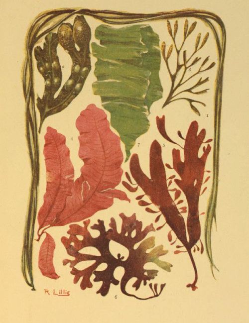
SEA-WEEDS
| 1. Chorda filum | 4. Delesseria (Maugeria) sanguinea |
| 2. Fucus vesiculosus | 5. Rhodymenia palmata |
| 3. Fucus canaliculatus | 6. Chondrus crispus |
| 7. Ulva lactuca | |
Alaria esculenta is an edible species known as the Badderlocks in Scotland, and also locally as the Henware, Honeyware, and the Murlins. It has a fibrous root, and a stalked, lanceolate, entire frond with a distinct midrib throughout. The stem is winged with finger-like leaflets, in which the spores are arranged in oblong clusters.
In the genus Chorda the frond is a simple, cylindrical tube, divided internally by numerous transverse membranes, and the spores are distributed over the surface. The commonest species is C. filum (see Plate VIII.), the frond of which is very slimy, and often from ten to twenty feet in length. In its young state it is covered with gelatinous hairs, but these are worn off as the plant develops. A smaller species (C. lomentaria) is sometimes found on our shores. Its fronds are constricted at intervals, taper at the tip, and grow in tufts. It is seldom more than a foot long, and is not of a slimy nature.
The Sporochnaceæ have inarticulate, thread-like fronds, and the spores are contained in oblong, stalked receptacles, each of which is crowned with a tuft of slender jointed filaments. The typical genus contains only one British species—Sporochnus pedunculatus—and even that is by no means common. It is, however, a very pretty weed of a delicate texture and pale olive-green colour. Its stem is long and slender, pinnately branched, and the branches bear numerous small thread-like tufts.
The same order contains the genus Desmarestia, in which the frond is long and narrow, thread-like or flattened, with a tubular jointed thread running through it. Young specimens have marginal tufts of branching filaments. The species decay very rapidly after removal from the water, and should therefore be dried and mounted as quickly as possible. D. ligulata, so named from the flat, strap-like frond, is common on all our coasts. It is pinnately branched, and all the branches and branchlets taper towards both ends. D. viridis has a cylindrical, thread-like and freely-divided frond, with opposite branches and branchlets. It occurs more commonly on the northern shores.
The last order of olive-spored weeds is the Fucaceæ, some species of which are so abundant between the tide-marks, from high-water to low-water levels, that they form a very important characteristic of our shores. They are mostly large, tough, and leathery weeds, without joints, and the spores are contained in spherical receptacles embedded in the substance of the frond.
In the typical genus—Fucus—the root is a conical disc, and the frond flat or compressed and forked. Most of the species are furnished with one-celled air-vessels in the substance of the frond, and these serve to buoy up the plants and keep them more or less erect when submerged. The spore-receptacles are usually embedded near the tips of the branches, but are sometimes borne on special branches or shoots. They are filled with a slimy mucus and contain a network of jointed filaments. The weeds are very hardy, capable of withstanding long exposures to air and sun, and are sometimes to be found above high-water mark, where they are watered only by the spray of the waves for a brief period at intervals of about twelve hours. Although they are not usually looked upon as ornaments in the collector’s herbarium, they will repay examination for the tufts of smaller and more beautiful weeds to which they often give attachment and shelter.
Four species are common on our coasts, and these may be readily distinguished by the most cursory examination. The Serrated Wrack (F. serratus) has a flat, forked frond with toothed edges and a strong midrib, ranging from one to four feet long, and no air-vessels. The Knotted Wrack (F. nodosus—Plate VII.) may be known by its flattened, thick and narrow frond, without a distinct rib, from one to five feet long. The branches are narrow at the base, pointed at the tip, and are jointed to short projections on the main stem; and both these and the main stem have very large oval air-vessels. The spore-receptacles are mounted on slender stalks which arise from projections on the branches, and are of a bright yellow colour when mature. This species does not grow so near to high-water mark as do the others. Another species, the Twin-Bladder Wrack (F. vesiculosus—Plate VIII.)—is abundant everywhere along the coast, and is largely used by agriculturists both as manure and as fodder for cattle. The frond is flat, with a distinct midrib, and a non-serrated edge. Air-vessels are not always present, but when they are they usually occur in pairs, one on each side of the midrib, and are globular in form. The spore-receptacles are situated at the tips of the branches, are full of mucus, and are frequently forked. The last of the common species is the Channelled Wrack (F. canaliculatus—Plate VIII.), distinguished by a narrow frond, rounded on one side and channelled on the other. It has no midrib or air-vessels, and the fruit is contained in forked receptacles at the tips of the branches. This is the smallest of the genus, and may be found at all levels between the tide-marks. Stunted specimens may also be seen in situations where they are never submerged, but watered only by the spray of the highest tides.
The genus Himanthalia provides us with a single species (H. lorea) which is very peculiar on account of the small size of the frond as compared with the enormous dimensions of the spore-receptacles. The young frond is a pear-shaped sac which soon becomes flattened into a hollow disc. This disc then becomes solid, and concave above, and from its centre there arises a bi-forked, strap-like receptacle that often reaches a length of three or four feet, and may be mistaken for the frond of the weed by those who do not take the trouble to examine it. This weed is commonly known as the Sea Thong.
Belonging to the genus Cystoseira we have a few well-known weeds with conical disc-roots, and shrubby fronds with woody stem and alternate branches. The air-cells are in the substance of the frond, and the spore-receptacles at the tips of the branches. One of the species (C. ericoides) is of a heath-like habit, with a short, woody stem, and slender branches bearing hooked, leaf-like branchlets. Its air-cells are small, and are arranged singly near the tips of the branches; and the spore-receptacles are cylindrical, with hooked points. This weed is common on the south and west coasts, and may be readily distinguished by the beautiful iridescence it displays when in the water. C. fibrosa is very similar in general form, but is larger, and the air-vesicles are more conspicuous. It is not iridescent when in the water. A third species is named C. granulata from the rough and knobby appearance of the stem, due to numerous oval projections, from some of which spring the slender, much-divided branches. The air-vesicles are arranged in groups of two or three, and the spore-receptacles are at the ends of the branchlets. Our last example is C. fœniculacea, found on the south coast only, and readily distinguished by the numerous blunt spines that cover its long branches. The air-vesicles are narrow and pointed, and situated just below the forkings of the branchlets.
We conclude our résumé of the British sea weeds with a short description of the Podded Sea Oak (Halidrys siliquosa), which grows in the tide pools from high-water to low-water mark, the specimens inhabiting the shallow pools being only a few inches long, while those that grow in deep water often reach a length of three or four feet. It is an olive, shrub-like weed, with a conical, disc-like root that adheres very firmly to the rock, and a pinnately-branched frond with leaf-like branchlets. The air-vesicles are cylindrical and pod-like, divided internally into about ten cells, and the spores are contained in globular receptacles at the tips of the branchlets.
The young algologist will probably meet with many difficulties in his attempts to classify his sea weeds and name the various species in his collection. In dealing with an unknown weed we strongly recommend him to first determine the order to which it belongs. The genus should next be settled; and then, if possible, the species. It must be remembered, however, that he who has made himself acquainted with the principles of classification has done good work, and that it is far better to be able to arrange the weeds into properly-classified groups than to merely learn the names of the different species without regard to the relations which they bear to one another. The following table will probably assist the reader in the determination of the orders, but it must be remembered that a microscope will often be necessary for the examination of the spores and the minute structure of fronds.
A. Chlorospermeæ—Green-spored weeds. Fronds usually grass-green, and filamentous or membranous.
1. Confervaceæ—Frond thread-like, composed of cylindrical cells placed end to end. Spores very minute, formed within the cells.
2. Ulvaceæ—Frond grass-green or purple, flat or tubular. Spores minute, ciliated, formed in the cells of the frond.
3. Siphonaceæ—Frond a single, thread-like, branching cell, or a spongy mass of many such cells.
B. Rhodospermeæ—Red-spored weeds. Spores in globular conceptacles. Tetraspores (four-clustered spores) in globular or cylindrical cells. Frond red, reddish brown, or purple.
4. Ceramiaceæ—Frond thread-like, jointed, one-siphoned, and more or less covered with a layer of cortical cells. Spores grouped in transparent, membranous sacs, sometimes surrounded by a whorl of short branchlets.
5. Spyridiaceæ—Frond thread-like, jointed, one-siphoned, more or less covered with small cells. Spores formed in the upper cells of branched, jointed, radiating threads, enclosed in a cellular membrane in external conceptacles.
6. Cryptonemiaceæ—Frond more or less cartilaginous, composed of numerous jointed threads compacted by gelatine. Spores grouped without order in internal cells or in external conceptacles.
7. Rhodymeniaceæ—Frond inarticulate, membranaceous, composed of polygonal cells, the surface cells forming a continuous layer. Spores in beaded threads in external conceptacles.
8. Wrangeliaceæ—Frond inarticulate, thread-like, traversed by a jointed tubular axis. Spores formed in the terminal cells of clustered, branching, naked threads.
9. Helminthocladiæ—Frond cylindrical, gelatinous, composed of filaments imbedded in gelatine. Spores formed on branching, radiating threads that are enclosed in the frond without conceptacles.
10. Squamariæ—Frond lichen-like, rooted by under surface, composed of vertical filaments imbedded in firm gelatine. Spores in beaded threads in wart-like projections.
11. Spongiocarpeæ—Frond cylindrical, branching, cartilaginous, composed of netted filaments imbedded in firm gelatine. Spores large, in radiating clusters in wart-like excrescences.
12. Gelidiaceæ—Frond cartilaginous, inarticulate, composed of hair-like filaments. Spores attached to slender threads in internal conceptacles.
13. Sphærococcoideæ—Frond leaf-like or thread-like, inarticulate, cellular. Spores formed in beaded threads in external conceptacles.
14. Hapalidiaceæ—Frond minute, calcareous, composed of a single layer of cells.
15. Corallinaceæ—Frond calcareous. Spores in tufted threads at the bases of the conceptacles.
16. Laurenciaceæ—Frond rounded or flattened, branching, inarticulate, cellular. Spores in external oval or globular conceptacles. Tetraspores irregularly scattered over the branches.
17. Rhodomelaceæ—Frond leafy, thread-like, or jointed, composed of polygonal cells. Spores in external conceptacles. Tetraspores in distorted branchlets or in receptacles.
C. Melanospermeæ—Olive-spored weeds. Frond tough, leathery. Spores in globular cavities in substance of frond.
18. Ectocarpaceæ—Frond jointed, thread-like. Spores attached to or imbedded in branchlets.
19. Chordariaceæ—Frond gelatinous or cartilaginous, composed of interlacing vertical and horizontal filaments. Spores internal, attached to the filaments.
20. Dictyotaceæ—Frond inarticulate. Spores superficial, arranged in definite spots or lines.
21. Laminariaceæ—Frond inarticulate. Spores covering the whole frond or in cloud-like patches.
22. Sporochnaceæ—Frond inarticulate. Spores attached to jointed filaments which are either free or compacted.
23. Fucaceæ—Frond inarticulate, large and tough. Spores in globular cavities.
A considerable number of our flowering plants exhibit a decided partiality for the neighbourhood of the sea, and many are to be found only on the sea cliffs or in salt marshes not far from the shore. The principal of these will be now briefly described, dealing first with the monocotyledons, and then with the more highly organised dicotyledons.
The chief distinguishing features of these two groups have already been referred to, but it will be advisable here to give them in somewhat fuller detail.
The monocotyledonous plants, then, are those in which the stem is more or less woody and cylindrical, without either true bark or pith; and the woody tissue is not arranged in concentric rings, but in isolated bundles, which first bend inwards, as they rise, towards the centre of the stem, and then curve outwards towards the surface, which is hardened by the formation of a layer of hard woody matter. As a rule the stem is unbranched, and its growth takes place by a single bud at the summit. In nearly all of them the leaves are long and narrow, with veins running parallel throughout their length; and the parts of the flower are arranged in whorls of three or six. The outer whorl of the flower is often a conspicuous white or coloured perianth (that portion of the flower which lies outside the anthers), but in some the perianth is absent, the flower being protected by scaly bracts. The seeds are produced in a case called the ovary, and are fertilised by pollen grains which are developed in the anthers. When the pollen grains are set free they alight on the adhesive stigma, and grow, sending their tubes down into the ovary. The term monocotyledon is applied to these plants because the embryo has only one cotyledon or seed-leaf.
The principal divisions of this group are the Glumaceous Monocotyledons, in which the flower has no perianth, but is enclosed in scaly bracts or husks called glumes; and the Petaloid Monocotyledons, distinguished generally by the presence of a more or less conspicuous white or coloured perianth. The first of these includes the rushes, sedges, and grasses; and the other contains the lilies and orchids, with their allies, together with certain aquatic and semi-aquatic plants.
Among the Grasses there are several species that show a preference for the immediate neighbourhood of the sea, some growing luxuriantly at the bases of the cliffs where the beach is sandy, and others thriving best in salt marshes; but before dealing with these individually we shall note the general characteristics of the order (Gramineæ) to which they belong.
Grasses are distinguished by their jointed stems, which are usually hollow, with a split sheath, and bearing alternately arranged narrow leaves. The flowers, which are disposed either in spikes (sessile flowers arranged along a common axis) or in panicles (flowers stalked and arranged as in fig. 281), consist of scale-like bracts enclosing the stamens and the pistil. The bracts are in two series, the outer usually consisting of two glumes, and the inner of two pales; the upper pale, however, has two ribs running through it, and is therefore usually looked upon as a combination of two. In some species both glumes and pales are absent; but the former, when present, enclose one or more flowers, among which may be some that are abortive. The stamens are generally three in number, attached to the base of the flower; and the ovary is superior or free, that is, it grows above the other parts of the flower, and contains but one seed.
It will be convenient at this stage to refer briefly to the two principal methods by which the pollen of flowers is transferred to the stigmas for the purposes of fertilisation, and to see how various species are structurally adapted to the means by which the transfer is brought about.
Speaking generally, we may classify flowers into those which are fertilised by the wind (anemophilous flowers) and those in which the pollen is transferred by insects (entomophilous flowers). The former offer no attractions to allure the various forms of insect life. They are, generally speaking, very inconspicuous, being of small size and having no bright corollas. None of them are scented, nor do they produce the sweet nectar that forms the principal food of so many insects. Their anthers are borne on long filaments, so that they are exposed freely to the wind; and they produce abundance of pollen to compensate for the very wasteful method of wind-dispersion. The pollen, too, is not very adherent, so that it may be readily carried away by the breeze; and the plants concerned often produce their flowers early in the spring, before the leaves have appeared, thus giving the wind very free play.
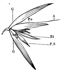
Fig. 277.—Expanded Spikelet of the Oat
G. glumes; P.e, outer pale; P.i, inner pale; A, awn; F.S, a sterile flower. The stamens and the feathery stigmas of the fertile flower are also shown
Insect-fertilised flowers, on the other hand, are usually of attractive appearance; and, though often small and inconspicuous individually, they are in such cases grouped together in more or less showy clusters. They are also usually scented, and supply nectar and pollen to the insects which they allure. Some are fertilised by insects that fly by day, and these often close their petals on the approach of night, thus protecting their pollen during the period in which their fertilisers sleep. Others, fertilised by nocturnal insects, always spread their petals during the night, and generally protect their pollen from waste by sleeping throughout the day. As a rule, too, these night-bloomers have large and pale-coloured petals that are more easily seen by night; they also evolve a powerful scent to aid the insects in searching them out.
It will be seen that the economic relationship existing between flowers and insects is a mutual one, the latter visiting the former in order to obtain food, while the former derive in return the advantage of a direct transfer of pollen from flower to flower.
It is a well-known fact that the self-fertilisation of a flower often results in the development of very weak seedlings as compared with those that are produced by crossing; and it often happens that the pollen of a flower is incapable of producing the least effect when deposited on the stigma of the same bloom. In some cases the contact of the pollen of a flower with its own stigma will even act as a poison, causing the whole to shrivel and die; and truly wonderful are the varied means by which flowers contrive to secure a cross-fertilisation. It is here that the work of the wind and insects proves so valuable to flowers; but, in addition to this, a very large number of flowers are absolutely incapable of self-fertilisation, for the anthers and the stigma are not mature at the same time, or they exist in separate flowers, either on the same plant or on distinct plants of the same species. It is most interesting and instructive to study the many contrivances by which flowers compel certain insects to convey the pollen exactly in the way that best serves their purpose, sometimes even entrapping them after they have been allured, and not allowing them to escape until they are thoroughly dusted with the pollen which they are required to convey; but it is hardly our province to enter more fully into this matter in these pages.
An examination of the grasses will show at once that they are adapted for fertilisation by the wind. The flowers produce no nectar; and, consistently, develop no bright petals and evolve no odours to attract insects. On the other hand, their anthers produce abundance of lightly-adhering pollen, and are mounted on long filaments which hold them well exposed to the wind; and the stigmas are well adapted for catching the scattered grains, being long and protruding, and often covered with sticky hairy or feathery appendages.
Although the flowers of grasses are generally wanting in attractive colours, the clusters of blossoms are often very graceful and pretty, especially when the large anthers, covered with bright-yellow pollen, dangle in the breeze.
We will now briefly describe the principal British grasses that grow chiefly or exclusively in the immediate neighbourhood of the sea.
The Sea Hard Grass (Lepturus filiformis) is a perennial species, usually about six inches in height, very common on some sandy coasts, and found in flower during the hottest months of the summer. The flowers are arranged in simple spikes, on slender erect stems; and the glumes, which are united at their bases, enclose a single bloom.
In similar situations we may find the Sea Lyme Grass (Elymus arenarius), a tall species, often reaching a height of four feet, with glaucous rigid leaves. The flowers are arranged in a simple spike, but the spikelets are clustered two or three together. This species flowers in August.
Of the well-known Barley Grasses there is one species (Hordeum maritimum) that has its habitat along the coast. Like the others of its genus, the spikelets are arranged in threes, each bearing a single flower, and the pales have long slender processes (awns) which constitute the so-called beard. It also resembles the common Meadow Barley Grass in having the middle flower of each three perfect, while the two laterals are abortive, but may be distinguished by its rough and bristly glumes, and the semi-oval form of the pales of the lateral flowers. It is a somewhat stunted species, sometimes only five or six inches in height, and may be found in flower about Midsummer.
The Brome Grasses have also a representative of a sea-loving nature, which is to be found in fields near the cliffs. It is the Field Brome Grass (Bromus arvensis), an annual grass that grows to a height of two or three feet. Brome grasses generally are known by their loose panicles of flowers, lanceolate and compressed spikelets, and awned florets enclosed in unequal glumes; and B. arvensis may be distinguished by its hairy leaves and stem-sheath, and the drooping panicle with the lower peduncle branched.
Among the Meadow Grasses we have three or four coast species. In these the florets are in panicles and are not awned. The outer glumes are keeled and traversed by several veins; and the lower pales are also keeled, with five or more nerves. The Sea Meadow Grass (Poa maritima) grows in salt marshes near the sea, its erect rigid panicles reaching a height of about eight or ten inches. It has a creeping root, and its leaves are curved inward at the margins. The Procumbent Meadow Grass (P. procumbens) and a variety of the Reflexed Meadow Grass (P. distans) are also plentiful in salt marshes. The former may be known by the short rigid branches of its panicle and the five ribs of the lower pales; and the latter is much like P. maritima, but grows taller, and its spikelets are crowded. The Wheat Meadow Grass (P. loliacea) grows on sandy shores. Its spikelets are arranged singly and alternately along the central axis, and the upper glume reaches to the base of the fourth floret. This species flowers in June, but the other three of the same genus bloom from July to September.
The reader is probably acquainted with the Fescue Grass, with its awned flowers arranged in one-sided panicles. There are no less than seven species, one of which—the Single-husked Fescue (Festuca uniglumis)—grows on sandy shores, flowering in June and July, and reaching a height of from nine to twelve inches. The panicles are upright and unbranched, and the species may be readily known by the flowers, which are compressed, with long awns, and with the lower glumes wanting.
Knappia agrostidea is a dwarf species, rarely exceeding four inches in height, that is found on certain sandy shores, but is very local. Its flowers are arranged in a simple spike, the spikelets being solitary and unilateral, with only a single flower, and the pales are shaggy. The plant has several stems which bear short, rough leaves.
The Mat Grass or Sea Reed (Ammophila arundinacea) is common on many sandy coasts, where it grows to a height of three or four feet, and flowers in July. The white flowers are clustered in dense cylindrical, pointed spikes; and the leaves are of a glaucous green colour, rigid, and curved inward at the edges.
Dog’s-tooth Grass (Cynodon dactylon). This species has a creeping root, and the leaves are downy on the under side. The flowers are arranged in a compound spreading spike, of three to five parts, and the spikelets are of a purplish colour, ovate in form, and arranged in pairs. The glumes are equal in size. It is found on sandy shores, grows to a height of about six inches, and flowers in July.
A species of Canary Grass (Phalaris arundinacea) is also to be seen on sandy coasts. Unlike the other species of the same genus, its flowers form an erect spreading panicle, and the glumes are not keeled. It is also taller than the common canary grass of waste places, often reaching a height of three feet, and is commonly known as the Reed Canary Grass.
The Sea Cat’s-tail Grass (Phleum arenarium) is common on many coasts. It is much smaller than the common species of Cat’s-tail, being generally less than a foot high. The spike is of an elongated oval form, blunt at the tip and narrow at the base; and the glumes are narrow, pointed at both ends, and fringed. Each spikelet has but one flower.
In salt marshes we occasionally meet with the Perennial Beard Grass (Polypogon littoralis), but it is somewhat rare. It has a creeping root, and the flowers form a somewhat dense spike-like panicle. The glumes have a slender awn. It grows to a height of one to two feet, and flowers in July.
The Tuberous Fox-tail Grass (Alopecurus bulbosus) is another rare grass of the salt marshes, where it grows to the height of twelve to sixteen inches, flowering in May and June. The genus to which it belongs is very closely allied to Phleum, but may be distinguished by having only one pale to each flower, and this species has a long awn attached to the back portion. The panicle, too, is cylindrical and slender, the glumes quite free and abruptly pointed, and the awns longer than the pales.
The last of the sea-side grasses are two rare species of Cord Grass (Spartina), both of which are found in salt marshes. In these the inflorescence is a compound spike, with one-sided spikelets inserted in a double row. The glumes are keeled and pointed; the pales cleft, pointed and without awns; and the styles two in number, very long. The only British species of the genus are the two (S. stricta and S. alternifolia) referred to above. They both grow to a height of about eighteen inches, and flower in late summer. In the former the spikes number two or three, and are longer than the leaves; and the outer glume is hairy, with a single nerve. The latter, which is the rarer of the two, bears several spikes, shorter than the leaves; and the outer glume has five nerves.
Certain of the sedges (order Cyperaceæ) are also more or less familiar to the sea-side naturalist, and must therefore receive a small share of our attention. In general terms these are grasslike, monocotyledonous plants, the stems of which are solid, jointed, and frequently angular. The leaves are very similar to those of grasses, except that the sheaths, which surround the stem, are not split. The flowers are generally arranged in a spike, overlapping each other, and each one supported on a scale-like bract. In some sedges the flowers are perfect, each one possessing both stamens and pistil; but in some species the flowers are unisexual, some bearing stamens and no pistil, and others pistil only. The stamens are generally three in number, the ovary is superior, and the stigmas either two or three.
Sedges abound in moist places, some being peculiar to salt marshes, while others grows on sandy shores; and a few of the British species of the latter habitat are often so abundant that their creeping roots bind the sand together, effectually holding it in place while the surrounding portions of the beach are mercilessly driven by the wind.
A few of the sea-side sedges belong to the genus Carex, in which the flowers are imperfect, and the fruit is enclosed in the outer parts of the flower. C. extensa thrives in salt marshes, growing to a height of a foot or more, and flowering about midsummer. Its fertile flowers form oblong erect spikelets, while the barren spikelets are solitary. The bracts are long and leafy, with short sheaths surrounding the stem. The leaves are curved in at the edges, and the fruit is oval and ribbed, with a short straight beak.
On sandy shores the Sea Sedge (C. arenaria) is often common, and its underground stems are used for sarsaparilla. It is a perennial species, growing to a height of about nine inches, and flowering in June and July. The flowers grow in an oblong interrupted spike, the upper spikelets being barren, and the intermediate ones barren at the tip. The fruit is oval, veined, and winged.
Another species of this genus—the Curved Sedge (C. incurva) —is sometimes to be seen on sandy shores, but it is rare, and is also a very small sedge, growing only to a height of about three inches. It derives its specific name from its curved stem, and may be further distinguished by its channelled leaves and the globular mass of spikelets which are barren on the top.
Some of the so-called rushes belong to the same order as the sedges, and a few of these are more or less restricted to the neighbourhood of the sea. The Salt-marsh Club Rush (Scirpus maritimus), as its name implies, is to be found in marshes near the sea. It is very variable in height, ranging from one to three feet, and displays its dense terminal cluster of spikelets in July and August. In this genus all the flowers are perfect, the glumes imbricated and bristled; and the present species may be distinguished by the glumes being divided into two sharply pointed lobes. A variety of S. lacustris may also be found on the sea shore, but it is somewhat rare. It has a leafless glaucous stem, and flowers arranged in compound spikes. The glumes are rough, and contain a compressed fruit.
A very small species of the Spike Rush (Eleocharis parvula), growing only one or two inches high, is sometimes found on the muddy shores of Ireland. It has perfect flowers, in a single terminal spikelet. The leaves are very narrow, growing from the base of the plant; and the round stem is enclosed in a single leafless sheath.
The true rushes belong to the order Juncaceæ. These have fibrous roots and narrow leaves, and bear clusters of brown flowers. The perianth consists of six parts, and the stamens are usually six in number. The ovary is generally three-celled, developing into a three-valved capsule. The Lesser Sea Rush (Juncus maritimus) is common in salt marshes, growing to a height of two or three feet, and flowering in July. It has a rigid leafless stem, bearing lateral clusters of flowers. The segments of the perianth are very narrow and sharp, and the seeds are enclosed in a loose testa. Closely allied to this species is the Great Sea Rush (J. acutus), which grows three or four feet high on sandy shores. In general characteristics it resembles J. maritimus, but the segments of the perianth are oval and have thin transparent margins; and it is a much rarer species.
We now pass to the peculiar Sea Grasses or Grass Wracks (Zostera) which grow in salt water. They belong to the order Naiadaceæ, and are characterised by cellular leaves with parallel veins, and inconspicuous unisexual or bisexual flowers. The perianth, when present at all, consists of two or four scale-like parts, and the stamens correspond in number with these. The ovary is free, and the carpels, one or more in number, contain each a single ovule. In Zostera the flowers are imperfect, and seem to grow in the slit of the leaf. There are two species, both of which grow in shallow water close to the shore, often in such dense masses that they impede the progress of boats. They have long creeping stems that lie buried in the sand, giving off numerous root-fibres, and send up to the surface slender branches that bear grass-like leaves. The flowers are unisexual, and are arranged in two rows on the same side of a flattened stalk that is enclosed in a sheath formed by short leaves. They have no perianth, the male flowers being composed of a single anther, and the female of a one-celled ovary containing a single ovule, and surmounted by a style with two long stigmas.
There are two species—the Broad-leaved Grass Wrack (Z. marina) with leaves one to three feet long and traversed by three or more parallel veins, and the Dwarf Grass Wrack (Z. nana), the leaves of which are less than a foot long, with veins numbering one to three. There is a variety of the former, however, named Angustifolia, in which the leaves are much narrower than usual, and the veins fewer in number.
The order Alismaceæ, which contains the water plantains, arrow-heads, and other semi-aquatic plants, has a representative of marine tendencies in the Sea-side Arrow Grass (Triglochin maritimum). The flowers of this order are bisexual, with six stamens and a six-parted perianth. The fruit consists of many carpels; and, although the plants are monocotyledons, their leaves have netted veins; and altogether they somewhat resemble the ranunculaceous exogens. The Sea-side Arrow Grass is abundant in some salt marshes, growing to a height of about a foot, and produces loose simple spikes of green flowers all through the summer. The leaves are radical, narrow and fleshy; and the ovary consists of six carpels.
Of the interesting order Liliaceæ we have only one plant of the coast, and even that—the Asparagus—is not by any means generally common. It is the same plant that is so largely cultivated as an article of diet, and which is so highly valued on account of its diuretic properties. It is moderately common on parts of the south coast, particularly in the Isle of Portland and in West Cornwall, and its general appearance is so graceful that it is largely employed as an ornamental garden plant. The stem is erect and freely branched, bearing feathery bunches of bristled leaves and pale-yellow axillary flowers. As is the case with the Liliaceæ generally, the flowers are bisexual, with a six-parted perianth, six stamens, and a three-celled superior ovary; and the last named, in the Asparagus, forms a bright-red berry in the autumn.
We have now to leave the monocotyledonous plants and pass on to the dicotyledons, which form the most highly developed of the primary divisions of the vegetable kingdom. A few of the general characteristics of this group have already been given, but we must now look rather more closely into the nature of the plants included.
The class receives its name from the presence of two cotyledons or seed-leaves in the embryo plant, and is also known as the Exogenæ because the stems increase in thickness by the addition of zones of woody tissue at the exterior. When the young dicotyledonous plant first appears above the ground, the two cotyledons, which formerly served to shelter the immature bud, usually appear as tiny fleshy leaves; but these soon wither away, while the bud produces the more permanent leaves that are of a very different structure. A section of the stem will reveal distinct pith, wood, and bark, the wood being more or less distinctly divided into wedge-shaped masses by rays from the pith; and, in the case of perennial stems, the wood is arranged in concentric rings, the number of which correspond approximately with the years of growth. The leaves of exogens have their veins in the form of a network, and the parts of the flower are generally arranged in whorls of two or five or of some multiple of these numbers.
The flowers always have stamens and pistil, but in some these organs exist in separate flowers, either on the same plant, or on different plants of the same species, and the ovules are nearly always contained in a case called the ovary.
Dicotyledons are divided into three main groups, the division being based on the structure of the flowers. They are the Apetalæ in which the petals are absent, but the perianth is frequently petaloid, though it is occasionally also absent; the Gamopetalæ, in which the petals are united; and the Polypetalæ, in which the petals are always distinct.
Dealing with these divisions in the above order we come first to the Spurges, three species of which occur on sandy shores. They belong to the order Euphorbiaceæ, which includes, in addition to the spurges, a number of herbs, trees, and shrubs with entire leaves often a milky juice, and small flowers, sometimes enclosed in calyx-like bracts. The flowers may have one or several stamens, and the perianth, if present, consists of three or four parts; but perhaps the best distinguishing feature of the order is the nature of the fruit, which separates elastically into three carpels.
The Sea Spurge (Euphorbia Paralias) is commonly seen on sandy shores, where its yellow flowers bloom in late summer and in autumn. It may be distinguished among the numerous species of the genus by its narrow oblong imbricated leaves, of a tough leathery nature, the broad heart-shaped bracts, and the wrinkled capsules containing smooth seeds. The Portland Spurge (E. portlandica) is a similar plant, found in similar situations, and flowering from May to September. Its leaves are oval and narrow, obtuse, and of a glaucous colour, and the bracts are more triangular than those of the last species. The capsules are slightly rough, as are also the seeds. There is yet another sea-side spurge—the Purple Spurge (E. peplis)—a somewhat rare plant, found on some of the sandy shores of the south of England. It grows to about eight or nine inches in length, and blooms in late summer, the flowers, like those of most of the spurges, being yellow. The stem is of a glaucous colour, and trails along the ground; the leaves are opposite and somewhat heart-shaped, and the flowers solitary. This species may be distinguished from other spurges by its stipuled leaves.
On sandy cliffs we sometimes meet with the Sea Buckthorn (Hippophaë rhamnoides)—a spiny shrub, ranging from about two to seven feet in height, the bark of which is covered with a silvery scaly scurf that forms a beautiful object for the microscope. It is the British representative of the Oleasters (order Eleagnaceæ). The leaves are alternate, lanceolate, with a silvery surface; and the flowers are small, green and unisexual. The male flowers grow in catkins, each arising from a scaly bract, and have a green perianth. The female flowers have a tubular perianth, and a free one-celled ovary. The latter forms a hard nut-like fruit, which is surrounded by a succulent mass formed by the former. This shrub flowers in the spring, while the leaves are still very small.
Of the order Polygonaceæ, which includes the docks, knot grasses, buckwheats, and sorrels, we have two sea-side representatives, both belonging to the typical genus Polygonum. These are the sea-side Knot Grass (P. maritimum) and Ray’s Knot Grass (P. Raii). The plants of this order are herbs, characterised by their alternate leaves with sheathing stipules; and small flowers, usually bisexual, often with a coloured perianth. Most of the species are remarkable for their astringent and acid properties. In the genus Polygonum the flowers are usually in spikes or racemes; the perianth funnel-shaped, regular, and five-cleft. The stamens vary from five to eight in number, and the styles number two or three. The fruit is a small angular nut, usually enclosed in the perianth.
The sea-side Knot Grass is very common on some parts of the shore, where it grows from one to three feet long, and flowers in August. The stem is recumbent, tough and woody, bearing fleshy glaucous leaves with curled edges. It may be further distinguished from the other knot grasses by its long stipules, with freely-branching veins, and by the length of the fruit exceeding that of the perianth. As in the other knot grasses, the flowers arise from the axils of the leaves.
Ray’s Knot Grass is very much like the common knot grass so abundant in all waste places, the leaves being flat; and the stipules, shorter than in the last species, having but few veins; but while in the latter the fruit is shorter than the calyx, in P. Raii it is longer. This species is found on many sandy shores, and flowers in July and August.
The order Chenopodiaceæ is particularly rich in sea-side plants, more than a dozen of the British species growing almost exclusively near the shore. They are mostly inconspicuous plants, with small flowers which are sometimes unisexual. The perianth is deeply divided, and the stamens are inserted in its base, opposite the divisions. The ovary is free, containing a single ovule.
The typical genus (Chenopodium) contains the weeds designated by the name of Goosefoot, all characterised by their straggling stems and small flat leaves. One species (C. botryoides) is common on some sandy shores. It is a small weed, its prostrate stem measuring only a few inches in length. The leaves are triangular and fleshy, and the flowers are arranged in dense leafy clusters. A variety of the Red Goosefoot (C. rubrum) is also found on the coast. It is of a reddish colour, with rhomboid leaves and short crowded spikes of flowers.
On muddy shores we meet with the Common Beet (Beta maritima), the leaves of which are often cooked and eaten where the plant is abundant; and it is this species from which the different varieties of garden beet and mangold wurzel have been produced by cultivation. There are two distinct varieties of the wild plant. In one the root and leaves are of a purple colour, while in the other they are of a yellowish green. The former has been cultivated for its root, while the latter is sometimes grown for the leaves. In the wild state it has many stems, the lower parts being more or less procumbent, and the leaves are fleshy, gradually narrowing down into the stalk. The flowers, which are arranged in long, simple, leafy spikes, are bisexual, with a five-parted perianth, five stamens inserted opposite each segment, in a fleshy ring and a flattened one-celled ovary which develops into a one-seeded utricle.
In similar situations we meet with two species of Sea Purslane (Obione), in which the flowers are unisexual, both male and female flowers being on the same plant. They are also distinguished from most other Chenopods by the perianth adhering to the wall of the ovary. The Shrubby Sea Purslane (O. portulacoides) is, as its name implies, a shrubby plant. It grows to a height of eighteen inches or two feet, bearing silvery oval lanceolate leaves and sessile fruit. The other species referred to—the Stalked Sea Purslane (O. pedunculata)—is herbaceous, with oval, mealy leaves, and stalked fruit.
The Oraches (genus Atriplex) resemble the Purslanes in the granular mealiness of the foliage, and the two are so closely allied that they are often placed in the same genus. Oraches are most readily distinguished among the Chenopods by the two bracts which enclose the fruit and enlarge after flowering; and, like the Purslanes, they have unisexual flowers, both male and female being on the same plant. Three of our five British species are sea-side plants. The Frosted Sea Orache (A. arenaria) grows on sandy shores, about six or eight inches in height, and flowers during late summer and autumn. It may be known by its buff-coloured stem, with triangular or rhomboidal, jagged, silvery leaves, and clusters of sessile flowers in the axils of the leaves. Another species (A. Babingtonii) may be seen on both rocky and sandy shores, usually from one to two feet in height, and flowering from July to September. Its stem is procumbent, green with reddish stripes; leaves oval-triangular, lanceolate towards the top, three-lobed at the base of the stem, light green, with a mealy surface; flowers in terminal clusters as well as in the axils of the leaves. A third species—the Grass-leaved Orache (A. littoralis) grows in salt marshes. All its leaves are grass-like and entire, and the stem is generally marked with reddish stripes as in A. Babingtonii. The flowers, too, are in sessile axillary clusters only. This plant reaches a height of from one to two feet, and flowers in the late summer.
The Prickly Salt Wort (Salsola kali) is a very common sea-side plant on some of our coasts, and may be recognised at a glance by its general form and habit. The stem is very much branched and prostrate, forming a very bushy plant about a foot in height. It is also very brittle and succulent, furrowed and bristly, and of a bluish-green colour. The leaves are fleshy, awl-shaped, nearly cylindrical, with a spiny point, and little prickles at the base. The flowers are axillary and solitary. This plant and its exotic allies are very rich in alkaline salts, particularly carbonate of soda, and were formerly the principal source from which this compound was obtained.
Our last example of the sea-side chenopods is the Glass Wort (Salicornia), which thrives in salt marshes. In this genus the stem is jointed and the flowers bisexual. The Jointed Glass Wort (S. herbacea) is common in most salt marshes, where its erect, herbaceous, leafless stem may be seen growing to a height of a foot or more. The joints are thickened upwards, and shrink to such an extent when dry that the upper part of each segment of the stem forms a membranous socket into which fits the base of the next segment above. The flowers are arranged in dense tapering spikes, also jointed, with a cluster of three flowers on the two opposite sides of the base of each segment. Each flower is composed of a perianth, closed with the exception of a small aperture through which the stigma and, later, the stamens protrude. The Creeping Glass Wort (S. radicans) has a woody procumbent stem, with the joints only slightly thickened, and the spikes do not taper so much as in S. herbacea. Both these plants yield considerable quantities of soda, and they are named ‘Glass Wort’ because they formerly constituted one of the sources from which soda was obtained for the manufacture of glass.
We now come to those flowers in which both calyx and corolla exist, and shall deal first with the division Gamopetalæ or Monopetalæ, in which the petals are united.
Our first example of this division is the Seaside Plantain (Plantago maritima), of the order Plantaginaceæ. This is a stem-less herbaceous plant, with ribbed leaves and small green flowers, common on many parts of the coast, and also found on the mountains of Scotland, flowering throughout the summer. It may be distinguished from the other plantains by its narrow fleshy leaves. As in the other species, the flowers form a cylindrical spike.
The order Plumbaginaceæ contains several sea-side plants, including the Sea Pink or Thrift (Armeria maritima) and the various species of Sea Lavender (genus Statice). They are characterised by a tubular membranous calyx, persistent and often coloured, a regular corolla of five petals united at their bases, five stamens opposite the petals and attached at the base of the ovary, and a free one-celled and one-seeded ovary. The well-known Sea Pink, with its compact head of rose-coloured flowers, in bloom throughout the spring and summer, and linear one-veined leaves, may be seen on most of our coasts, as well as on high ground in inland districts. The Sea Lavender, of which there are four British species, have their flowers arranged in spikes. The commonest species (Statice limonium) may be found principally on muddy shores. Its leaves are narrow and one-ribbed, and the bluish-purple flowers arranged in short dense spikes, the flower stalk being branched only above the middle. One variety of it has its flowers in a loose pyramidal cluster, while another bears its spikes in a compact level-topped corymb with short firm branches. Another species (S. bahusiensis) is characterised by long spikes of distant flowers, the stalk being branched from near the base. The Upright Sea Lavender (S. binervosa) of rocky shores has the stalk branched from the middle, with, usually, nearly all the branches flowering, though there are varieties in which the flowers are differently arranged. The Matted Sea Lavender (S. caspia) grows in salt marshes on the east coast of England. Its flower stalk is branched from the base, but the lower branches are barren and tangled, while the upper bear small crowded lilac flowers. The leaves of the last two species are spatulate in form.
The Bittersweet or Woody Nightshade (Solanum Dulcamara) of the order Solanaceæ is common in hedgerows and waste places almost everywhere, but a variety of it (marinum) has its habitat along the coast. It may be distinguished from the normal form by its prostrate branched and non-climbing stem, and by its fleshy leaves. The latter are all cordate, while in the normal the upper leaves are auricular. The order to which Solanum belongs is characterised by a regular five-cleft calyx and corolla, four or five stamens attached to the corolla, and a superior two-celled ovary. The flowers are in axillary cymes, and the fruit is a berry.
Convolvulaceæ is represented on sandy shores by the Sea-side Bindweed (Convolvulus Soldanella), a small species, with pinkish purple flowers, the prostrate stem of which rarely measures more than a foot in length. The plants of this order are generally climbing plants with alternate leaves and regular showy flowers. The calyx is composed of five sepals, the corolla of four or five lobes, and the stamens are attached to the corolla. The ovary is superior, two- or four-celled, and the fruit a capsule. The above species may be recognised by its reniform leaves (sagittate in the others), which are also fleshy.
To the order Gentianaceæ belong the Centaury (Erythræa), three out of the four British species of which grow on sandy shores. In the flowers of this order the calyx has from four to ten lobes; the stamens also number four to ten, and are alternate with the lobes of the corolla. The ovary is one- or two-celled, and the fruit is a berry with many seeds. The leaves are usually opposite and entire, and the flowers are generally showy, regular, and solitary. Erythræa has a funnel-shaped corolla, five stamens, and two stigmas, on a deciduous style; and in all our species the flower is rose-coloured. The Dwarf Centaury (E. pulchella), which is common on some sandy shores, is much smaller than the species that thrives in pastures, being only two or three inches in height. Its stem is also more freely branched, and its flowers are axillary and terminal. The Tufted Centaury (E. littoralis) and the Broad-leaved Centaury (E. latifolia) occur in similar situations, but are comparatively rare. They are both small species, the former with an unbranched stem, narrow leaves, and corymbose inflorescence; and the latter with branched stem, broad elliptical leaves, and flowers in dense forked tufts.
The extensive order Compositæ contains comparatively few sea-side plants, and, in dealing with these, we pass to another division of the monopetalous flowers, in which the ovary is inferior and the stamens are on the corolla. The order includes those herbaceous plants in which sessile flowers are collected together into compound heads (capitula) surrounded by a whorl of bracts. The corolla is either tubular or strap-shaped (ligulate), the stamens four or five in number, and the fruit one-seeded, usually crowned with the limb of the calyx in the form of a scaly feathery or hairy pappus.
The Little Lettuce (Lactuca saligna) is found in chalky pastures near the east and south-east coasts, growing to a height of about a foot, and bearing heads of yellow flowers in July and August. All the flowers are ligulate and perfect, the pappus is composed of silvery hairs, and the fruit is compressed and beaked, the beak being twice as long as the fruit. The leaves are smooth, linear, and sagittate, terminating in a sharp point. The Sea-side Cotton Weed (Diotis maritima) is occasionally met with on sandy shores, and may be recognised by its dense coating of downy hair, its sessile obtuse leaves, and heads of yellow flowers forming a corymb. The heads are discoid, and the fruit has no pappus. The Sea Wormwood (Artemisia maritima) is a common sea-shore composite, bearing drooping heads of reddish-white flowers in August. This is another of the downy species, its pinnatifid leaves having quite a woolly appearance. The capitulum contains but few flowers, all of which are perfect; and the fruit has no pappus. A variety of this plant is sometimes seen, with dense erect capitula. The Sea Aster or Michaelmas Daisy (Aster tripolium) of salt marshes may be known by the yellow discs and purple rays of its flower heads, which are arranged in a corymb. The florets of the ray form a single row, and the fruit has a hairy pappus. The leaves of this plant are spatulate and fleshy. A variety occurs in which the purple florets of the ray are absent. The Golden Samphire (Inula crithmoides) is a very local sea-side plant, being found principally on the south-west coast. Its leafy stems grow to a height of a foot or more, and bear yellow heads of flowers that radiate in all directions. The leaves are linear, acute, and fleshy, and the bracts are linear and imbricated. Our last example of the sea-side composites is the Sea-side Corn Feverfew or Scentless Mayweed, which is a variety of Matricaria inodora of waste places. The leaves are sessile and pinnatifid, with very narrow segments, and the white flowers grow in solitary heads. The maritime variety differs from the normal form in having fleshy leaves.
We next deal with another very extensive order (the Umbelliferæ), which, however, has only three or four representatives on the shore, and these introduce us to the last great division of the flowering plants, namely, the Polypetalous Dicotyledons, in which the petals are not united. Of these we shall first deal with that subdivision in which the stamens are attached at the side of or upon the ovary.
The most obvious characteristic of the Umbelliferæ is that implied in the name—the arrangement of the flowers in that form of inflorescence, called the umbel, in which the pedicels all branch from one point in the main stalk, and are such that the flowers are all approximately on a level. The flowers are mostly small and white, with five sepals (when present), five petals, and five stamens. The inferior ovary is two-celled, bearing two styles; and the fruit separates into two dry one-seeded carpels that are ribbed longitudinally.
Our first example of this group is the Sea Carrot, a variety of the Wild Carrot (Daucus carota). In the ordinary form, which is so common in fields, the leaflets are pinnatifid, with acute segments; and the central flowers of each umbel are purple, while the outer ones are white. The umbel, when in fruit, is concave above. The maritime variety differs from this in having fleshy leaves, and the umbel convex above when in fruit. The Sea Samphire (Crithmum maritimum) grows on the rocks close to the sea, and thrives well where there is hardly a vestige of soil. It usually grows to a height of seven or eight inches, bearing greenish-white flowers surrounded by a whorl of very narrow leaves. The other leaves are glaucous and bi-ternate, the leaflets being narrow, fleshy, and tapering towards both ends. On cliffs near the sea, especially in chalky districts, we meet with the Fennel, with its finely-divided leaves, split up into numerous capillary leaflets, and its small yellow flowers without bracts. It may be distinguished from other closely-allied plants by the form of the fruit, which is flattened at the sides. It is grown in some parts for use as a potherb, and an aromatic oil is also obtained from the seeds. The plant grows to a height of four or five feet, but there is a smaller variety known as the Sweet Fennel, and distinguished by the stem being compressed at the base. Our next example of the Umbelliferæ is the Sea Holly (Eryngium maritimum), easily distinguished from the other umbellifers by its spiny glaucous leaves, and the thistle-like heads of blue flowers surrounded by a whorl of spiny bracts. Its fleshy creeping roots were formerly gathered largely for the purpose of converting them into the once-prized ‘candied eryngo root,’ which is still prepared in a few of the fishing villages of our coast. The lower leaves of this plant are spinous and very glaucous, and the upper ones palmate. The venation is particularly strong and durable, so that the leaves and flowers are used largely by the sea-side cottagers in the construction of skeleton bouquets and wreaths. Another plant of the same genus—The Field Eryngo (E. campestre)—is occasionally seen on sandy shores. It differs from the last in having ternate radical leaves with pinnatifid lobes, and the upper leaves, bi-pinnatifid. Our last example of the sea-side umbellifers is the Wild Celery (Apium graveolens) of salt marshes and ditches. This is the plant from which our highly-valued garden celery has been produced, and it is remarkable that this sweet crisp and wholesome vegetable has been derived from a wild plant of coarse taste and odour, the acrid sap of which is highly irritating if not dangerous. The plant may be known by its furrowed stem, and ternate leaves, the leaflets of the lower leaves being round and lobed, while those of the upper ones are notched. The umbels are sessile or nearly so, the flowers have no calyx, and the fruit has five prominent ridges.
On the sandy shores of the south-western counties we may meet with the very local Four-leaved Allseed (Polycarpon tetraphyllum) of the order Illecebraceæ. It is a small plant, only four or five inches in height, with the lower leaves in whorls of four and the upper ones in opposite pairs. The flowers are minute, and are disposed in small dense clusters.
Another rare species is the shrub known as the English Tamarisk (Tamarix anglica), which is our only representative of the order Tamariscaceæ. There is some doubt, however, whether even this is indigenous to Britain, though it occurs in a wild state on the coast. It is a very twiggy shrub growing from six to ten feet in height, with minute scale-like, acute leaves, and slender spikes of small pinkish-white flowers.
We now pass to the large order of Leguminous plants, characterised by their stipuled leaves, and irregular papilionaceous flowers. The latter usually have five united sepals, five petals forming an irregular, butterfly-like corolla, ten stamens, and a superior ovary that develops into a pod.
Of these the Starry-headed Trefoil (Trifolium stellatum) is very partial to the sea shore, though it is sometimes found some distance inland. The genus to which it belongs is so called on account of its trifoliate leaves which are characteristic of the clovers, trefoils, and vetches, and which have stipules adhering to the petioles. The species under notice receives its name from the star-like arrangement of the long teeth of the hairy calyx. The stem of the plant is procumbent, usually about six or eight inches long, with cylindrical and terminal heads of yellowish-grey flowers.
The Rough-podded Yellow Vetch (Vicia lutea) is somewhat rare, and occurs principally on very rocky coasts. In common with the other vetches it has pinnate, tendrilled leaves, without a terminal leaflet, one stamen free and the rest united into a bundle, and a long, slender, hairy style. Its stem is tufted and prostrate, averaging about a foot in length, the leaflets long and narrow, and the yellow flowers sessile and solitary. The teeth of the calyx are unequal, and the pods hairy and curved.
The Sea-Side Everlasting Pea (Lathyrus maritimus) is a much commoner plant of the coast, and may be readily recognised by its general resemblance to the garden sweet-pea. The genus to which it belongs is closely allied to the vetches, but may be distinguished by the style, which is flattened below the stigma, hairy on the inner or upper side, but quite smooth on the outer side. The sea-side species has an angled (but not winged) stem, from one to three feet long, compound tendrilled leaves with many oval leaflets, and large oval or cordate stipules. Its purple flowers are in bloom during July and August. A variety of this plant (acutifolius), with a slender straggling stem and narrow acute leaflets, occurs on some parts of the Scottish coast.
The Geraniaceæ is represented at the sea-side by the Sea Stork’s-bill (Erodium maritimum), which, however, is by no means a very common flower. Its relationship to the other stork’s-bills and the crane’s-bills may be readily proved by the five persistent sepals, five distinct clawed petals, the five to ten stamens attached under the ovary (for we have now reached that division of the polypetalous exogens distinguished by this mode of insertion of the stamens), and the five carpels surrounding a long beak resembling that of the stork and the crane. The plant may sometimes be seen on sandy shores, averaging a foot in height, though very variable in this respect, and displaying its pretty pink flowers during the whole of the summer. The principal features by which it is to be distinguished from the two other British plants of the same species are its ovate or cordate leaves with very short petioles, and the presence of only one or two flowers on each peduncle.
Passing now to the Sea Mallow (Lavatera arborea), we are dealing with another rather rare plant, of the order Malvaceæ, sometimes met with on rocky coasts, chiefly, it appears, on the north coast of Cornwall and Devon. This is a very shrubby plant, as its specific name implies, and it is sometimes popularly known as the Tree Mallow on that account. It has a very woody stem, growing to a height of four or five feet, and bearing seven-pointed, downy leaves, and solitary, axillary, purple flowers. As in the other mallows, the flowers have five petals, which are curiously twisted when in the bud, five sepals, a large number of stamens united into a tube, and an ovary of many cells, but it may be distinguished from the other species of the order by its three-lobed bracts. The plant is found principally in wild, uncultivated spots, but is commonly grown as a garden plant by the cottagers of villages in the south-west, and under cultivation it frequently grows to a height of nine or ten feet, with a tree-like stem three or four inches in thickness; and it produces such a quantity of fibre that its cultivation for manufacturing purposes has been suggested.
We now come to another of the very extensive orders, at least as far as British plants are concerned, although it contains only a few sea-side species. We refer to the Caryophyllaceæ, containing the pinks, campions, catchflies, chickweeds, &c. The chief features of the order are jointed, herbaceous stems, opposite leaves, and regular white or red flowers with four or five sepals and petals, eight or ten stamens, and a capsular fruit opening at the top with teeth.
One of the commonest species we have to consider is the Sea Campion (Silene maritima), common on nearly all coasts, and often growing in small crevices of the bare rocks quite within the reach of the spray of storm-waves. In common with the other members of its genus it is characterised by a tubular calyx of united sepals, ten stamens, and a three-celled capsule opening at the top with six teeth; but it may be known at once by its small size, being only a few inches in height, and its solitary flowers with calyx much inflated and the corolla only shortly cleft.
The Sea Sand Wort (Spergularia marina) is another common plant of the coast, recognised by its slender, creeping stems; linear, stipuled, fleshy leaves, convex below and blunt at the apex; and its pinkish-white flowers. The Sea Purslane (Honckenya peploides), belonging to the same order, is also a creeping plant, with ovate, acute fleshy leaves, flowering from May to August. It is the only British plant of its genus, and may be distinguished from others by the absence of stipules, distinct sepals, petals entire, ten stamens, and from three to five styles. The flowers are white, solitary, and sessile. The one remaining species of the sea-side Caryophyllaceæ is the Sea Pearl Wort (Sagina maritima). This plant is closely allied to the last, being a creeper with exstipulate leaves and distinct sepals, but its flowers are reddish white, on erect peduncles, with very small petals. The leaves, too, are linear, fleshy, and obtuse. There are three distinct varieties of this plant, two of which have erect stems with short internodes, while the third is procumbent with long internodes; and in all three the capsules are shorter than the sepals.
A variety of the Common Milk Wort (Polygala vulgaris)—order Polygalaceæ—is moderately common on sandy shores. The ordinary form of the species, which is so common on heaths, is a small plant with a woody stem, small ovate leaves crowded below, and opposite lanceolate leaves above. The flowers are irregular with five persistent sepals, two larger than the others; three to five petals, the lowest keeled, and all united to the tube formed by the eight stamens, which are divided above into two bundles; and the fruit is a flat capsule with two one-seeded cells. The flowers are very variable in colour, being white, pink, lilac, or blue; and the seeds are downy. The sea-side variety (oxyptera) has smaller flowers than the normal form, and the wings of the calyx are narrower.
One species of Pansy (Viola Curtisii) is occasionally to be met with on sandy shores, and may be at once recognised as one of the Violaceæ by its irregular spurred corolla, its five persistent sepals, and the three-parted, one-celled ovary. The flowers are variable in colour and size, the prevailing tints being blue and yellow, and the diameter of the corolla occasionally reaching to one inch. It has a creeping woody rootstock, and a rough angular stem; and the petals are generally but little longer than the sepals.
The Shrubby Mignonette (Reseda suffruticulosa), of the order Resedaceæ, is a common sea-side plant that grows to a height of one or two feet on sandy shores, bearing spikes of white flowers in July and August. The order is characterised by alternate exstipulate leaves, persistent calyx with four or five sepals, corolla of from four to seven petals, many stamens, and a three-lobed, one-celled ovary. The sea-side species is very much like the wild mignonette so common in chalky districts, but differs in having all its leaves pinnate, waved, and glaucous, with linear segments; and in having five equal sepals and petals. In a variety of the species, however, the sepals and petals are six in number.
The Crucifers are fairly well represented by coast plants, there being several maritime species of the order. The Cruciferæ are named from the nature of the corolla, the limbs of the four petals of which are arranged so as to resemble the Maltese cross. The flowers have also four sepals, six stamens, two of which are shorter than the other four, and the fruit takes the form of a two-celled pod or pouch which opens by the separation of its two valves from the central partition.
Our first example is the Wild Cabbage (Brassica oleracea), which, although so unlike the cabbage of our gardens, is really the parent of all the cultivated varieties, including the cauliflower, broccoli, Brussels sprouts, &c. It is a biennial plant, with fleshy lobed wavy leaves that are covered with bluish bloom, and a fleshy cylindrical root. It grows erect to a height of one or two feet, bearing yellow flowers during the summer months. An allied species (B. monensis), with a prostrate stem and deeply-divided leaves, occurs locally on the sandy shores of the Isle of Man.
Two species of Stock (Matthiola) are to be found on the coast, both being characterised by purple flowers. The Great Sea Stock (M. sinuata) is a rare plant growing on the shores of Wales and Cornwall, and may be known by its herbaceous stem and narrow downy leaves; and the other species—the Hoary Shrubby Stock (M. incana)—is also a rare plant, found principally on the cliffs of the Isle of Wight, and is the parent of the Brompton Stocks of our gardens. The latter has a branched woody stem and narrow leaves. Both species grow to a height of about eighteen inches, and the latter flowers in May and June, while the former is in bloom during the hottest summer months.
The Hare’s-ear Treacle Mustard (Erysimum orientale) is a rare crucifer, frequenting the cliffs of the southern and eastern counties. It grows to a height of one to two feet, and bears its white flowers about midsummer. It has glaucous leaves, and the fruit-pods are quadrangular in form.
The Common Scurvy Grass (Cochlearia officinalis) is abundant on many shores, and its fleshy leaves, once highly valued as an antiscorbutic, are still used for salad by the cottagers near the sea. It generally grows to a height of six or seven inches, and displays its white flowers during late spring and early summer. The root-leaves are cordate in form, and the upper ones are sessile and angled, half embracing the stem. The fruit is a rounded pouch. A variety (danica) with stalked, deltoid leaves and an oval veiny pod, is plentiful in some places.
On some coasts we find the Sweet Alyssum (Koniga maritima)—a naturalised plant with procumbent stem, narrow lanceolate, acute leaves, and white flowers. It may be recognised by its compressed, pointed pouch with one-seeded cells. This species flowers towards the end of the summer.
The Sea Radish (Raphanus maritimus) is a much larger plant, growing three or four feet in height. In common with the Wild Radish of our corn-fields, it has a tapering pod divided into one-seeded joints, but it may be distinguished from the latter by its superior height and the deeply-divided radical leaves. Its flowers are always yellow, while in the field species they may be either yellow or white; and the style is also shorter, being about the same length as the last joint of the pod.
On sandy shores the Sea Rocket (Cakile maritima) is commonly seen, and is readily distinguished by its zigzag branches, deeply-lobed, smooth, fleshy leaves of a glaucous colour, and its succulent pod, which is divided into two one-seeded cells by a horizontal partition. It grows from one to two feet high, and bears pretty lilac flowers about midsummer.
Our last example of the crucifers is the Sea Kale (Crambe maritima), a hardy perennial, commonly seen growing among the sand and shingle of the shore, which is the parent of the sea kale now so commonly cultivated in our market gardens. It may be readily recognised by the fine glaucous bloom of its stem, and its broad wavy toothed leaves of a glaucous grey colour. It grows to a height of about eighteen inches, and bears white flowers in June. The fruit is a two-jointed pouch, the upper being rounded and one-seeded, while the lower is stalk-like and barren. This plant is particularly common in the south-west of England, where the leaves are sometimes blanched for food by burying them in the sand.
One of the most striking plants of the coast is the Yellow Horned Poppy (Glaucium luteum) of the order Papaveraceæ, which contains the well-known poppies of corn-fields. The general characteristics of the order are two deciduous sepals, four petals, many stamens inserted below the ovary, and the ovary one-celled with membranous divisions. The plants of this species usually contain a milky juice, have alternate leaves without stipules, and the flowers, which are regular, generally nod when in bud. The Horned Poppy is a very conspicuous plant, usually growing quite alone on some inaccessible portion of the cliff, or among the pebbles or shingle not far from high-water mark. Its stem is glaucous and branched, and the large waved and deeply-cut leaves, which clasp the stem, are also of a glaucous hue. The flowers are rendered conspicuous by their large yellow petals, which, however, last only for a day, and are succeeded by the hornlike seed-pods that sometimes reach a foot in length.
We will conclude our list of sea-side flowers by a brief mention of the Lesser Meadow Rue (Thalictrum minus), a variety of which (maritimum) grows on sandy shores. The Meadow Rue belongs to the Ranunculaceæ, as may be seen from the fruit of several distinct carpels, each containing a single seed, the corolla of distinct petals, and the numerous stamens inserted below the carpels. The normal form of the Lesser Meadow Rue, which grows freely in some chalky pastures and thickets, has leaves three or four times pinnate, and lax panicles of drooping flowers without any petals. The sea-side variety differs from this in having the stem leafless at the base, and the panicles leafless and broad. The flowers are greenish white, and bloom in July and August.
To assist the reader in the identification of sea-side flowers we append a list of the orders to which they belong, together with the principal distinguishing characteristics of each.
I. MONOCOTYLEDONS
A. GLUMIFERÆ
Flowers without a Perianth, enclosed in Glumes
1. Gramineæ—Grassy plants with hollow stems enclosed in split sheaths. Flowers generally bisexual with (usually) three stamens.
2. Cyperaceæ—Grassy plants with solid stems and entire sheaths. Flowers arranged in spikelets, unisexual or bisexual, with from one to three stamens.
B. PETALOIDÆ
Perianth Petaloid
3. Juncaceæ—Rushes, with narrow leaves and small brown flowers. Perianth 6-partite, with scarious segments. Stamens usually 6; ovary superior; fruit a 3-valved capsule.
4. Naiadaceæ—Aquatic herbs with inconspicuous, unisexual or bisexual flowers. Perianth absent or scale-like. Stamens as many as the segments of the perianth. Fruit of from one to four carpels—superior.
5. Alismaceæ—Aquatic plants with radical net-veined leaves, and (generally) conspicuous, white, bisexual flowers. Perianth 6-partite. Stamens 6. Fruit of many carpels—superior.
6. Liliaceæ—Herbs with narrow leaves and showy, bisexual flowers. Perianth 6-partite. Stamens 6. Ovary superior, 3-celled. Fruit a berry or capsule.
II. DICOTYLEDONS
A. CALYX, OR COROLLA, OR BOTH ABSENT
7. Euphorbiaceæ—Herbs with entire leaves and (generally) a milky juice. Flowers small, unisexual, diœcious (male and female flowers on separate plants), sometimes enclosed in calyx-like bracts. Perianth 3- or 4-partite or absent. Stamens one or more. Ovary inferior. Fruit separating into carpels elastically.
8. Eleagnaceæ—Shrub with silvery scales, alternate, entire leaves, and small, unisexual flowers—the staminate flowers in catkins. Sepals of male flowers 3 or 4. Stamens 4 to 8. Ovary superior. Fruit indehiscent (not splitting).
9. Polygonaceæ—Herbs with sheathing stipules, alternate leaves, and small (generally) bisexual flowers. Stamens 5 to 8. Ovary superior. Fruit indehiscent.
10. Chenopodiaceæ—Herbs with jointed stems and small unisexual or bisexual flowers. Stamens usually 5, sometimes 1 or 2, opposite the sepals. Ovary superior. Fruit indehiscent.
B. PLANTS WITH BOTH CALYX AND COROLLA
a. Corolla Monopetalous
1. Ovary Superior and Stamens generally on the Corolla
11. Plantaginaceæ—Herbs with radical entire leaves, and spikes of small, green flowers. Calyx 4-cleft. Corolla 4-lobed, scarious. Stamens 4. Ovary 2- to 4-celled. Fruit many-seeded.
12. Plumbaginaceæ—Herbs with radical or alternate leaves, and (generally) regular, blue flowers. Calyx tubular, scarious. Corolla of 5 petals, united below. Stamens 5, opposite the petals, attached below the ovary. Ovary 1-celled and 1-seeded.
13. Primulaceæ—Herbs with (generally) radical leaves and conspicuous, regular flowers. Calyx 4- to 7-cleft. Corolla 4- to 7-cleft. Stamens 4 to 7, generally opposite the petals. Ovary 1-celled. Fruit a capsule with many seeds.
14. Solanaceæ—Herbs with alternate leaves and axillary clusters of regular flowers. Calyx 5-cleft. Corolla 5-cleft. Stamens 4 or 5. Ovary 2-celled. Fruit a berry.
15. Convolvulaceæ—Climbing herbs with alternate leaves and showy, regular flowers. Sepals 5. Corolla 4- or 5-lobed. Stamens 4 or 5. Ovary 2- to 4-celled. Fruit a capsule.
16. Gentianaceæ—Herbs with opposite entire leaves and solitary regular flowers. Calyx 4- to 10-lobed. Corolla 4- to 10-lobed. Stamens 4 to 10, alternate with the lobes of the corolla. Ovary 1- or 2-celled. Fruit a capsule.
2. Ovary Inferior and Stamens on the Corolla
17. Compositæ—Herbs with flowers (generally yellow or white) collected into compact heads. Calyx absent or represented by a pappus. Corolla tubular or ligulate. Stamens 4 or 5.
b. Corolla Polypetalous
1. Stamens Perigynous (around the Ovary), or Epigynous (upon the Ovary)
18. Umbelliferæ—Herbs with (generally) compound leaves, and small, white, umbelled flowers. Sepals (if present) 5. Petals 5. Stamens 5. Ovary inferior. Fruit of two adhering carpels.
19. Illecebraceæ—Small herbs with sessile, entire leaves, and small flowers. Sepals 4 or 5. Petals 4 or 5 or absent. Stamens 1 to 5. Ovary superior.
20. Tamariscaceæ—Shrub with small, scale-like leaves, and lateral spikes of small regular flowers. Sepals 4 or 5. Petals 4 or 5. Stamens 4 or more.
21. Leguminosæ—Herbs or shrubs with alternate, stipuled, pinnate or ternate leaves, sometimes tendrilled, and irregular flowers. Sepals 4 or 5. Corolla of 5 petals, papilionaceous (butterfly-like). Stamens usually 10. Ovary superior. Fruit a pod.
2. Stamens Hypogynous (attached below the Ovary)
22. Geraniaceæ—Herbs with stipuled, lobed leaves, and showy regular flowers. Sepals 5. Petals 5. Stamens 5 or 10. Fruit of 5 carpels surrounding a long beak.
23. Malvaceæ—Herbs with alternate, stipuled leaves, and axillary, red, or purple flowers. Sepals 5. Petals 5, twisted in the bud. Stamens numerous, united into a tube. Ovary of many cells.
24. Caryophyllaceæ—Herbs with (generally) jointed stems, opposite leaves, and regular white or red flowers. Sepals 4 or 5. Petals 4 or 5. Stamens 8 or 10. Fruit a 1-celled capsule opening at the top with teeth.
25. Polygalaceæ—Herbs with alternate, simple leaves (without stipules), and irregular flowers. Sepals 5, the inner petal-like. Petals 3 to 5, unequal. Stamens 8, in two clusters. Fruit a 2-celled capsule.
26. Violaceæ—Herbs with alternate, stipuled leaves and irregular flowers. Sepals 5. Petals 5, unequal, the lower one spurred. Stamens 5. Ovary 3-partite, but 1-celled.
27. Resedaceæ—Herbs or shrubs with alternate, exstipulate leaves, and spikes of irregular, green flowers. Sepals 4 or 5. Petals 4 to 7, unequal. Stamens more than 10. Ovary 3-lobed, and 1-celled.
28. Cruciferæ—Herbs with alternate, exstipulate leaves, and regular flowers. Sepals 4. Petals 4, cruciate. Stamens 6—4 longer and 2 shorter. Ovary 1- or 2-celled. Fruit a siliqua or a silicula.
29. Papaveraceæ—Herbs with alternate, exstipulate leaves, a milky juice, and regular, showy flowers. Sepals 2, deciduous. Petals 4. Stamens numerous. Ovary 1-celled with membranous partitions.
30. Ranunculaceæ—Herbs with (generally) alternate leaves and regular flowers. Sepals generally 5, distinct. Petals 5 or more. Stamens numerous. Fruit of many, distinct carpels.
Minor inconsistencies in punctuation of tables or captions are silently corrected.
Hyphenation is variable. Those compound words which are hyphenated only on line breaks are rendered using modern usage.
The word ‘movable’ appears only once as ‘moveable’ (165), which is retained.
The index entry for 'Œpophilus' is considered to be an error. All instances of the word appear in the text as 'Æpophilus'. This has been corrected and moved to the appropriate alphabetic position.
The following corrections were made to obvious printer’s errors: devel[e/o]ped (336); co[n/m]posed (364).
The following list contains punctuation corrections made:
| p. 65 | one of them[.] | Added. |
| p. 255 | [Class] LAMELLIBRANCHIATA | Added to match other entries. |
| p. 257 | their tendencies[,/.] | Corrected. |
| p. 292 | low-water[-]mark | Unhyphenated elsewhere. |
| p. 340 | [(]Cetacea) | Added. |
| p. 390 | in firm gelatine[,/.] | Corrected. |
| p. 403 | by its stipuled leaves[.] | Added. |
| p. 434 | Rhodospermeæ, 350, 355, 38[9] | Added. |