The Project Gutenberg eBook of A Monograph on the Sub-class Cirripedia (Volume 1 of 2), by Charles Darwin
This eBook is for the use of anyone anywhere in the United States and
most other parts of the world at no cost and with almost no restrictions
whatsoever. You may copy it, give it away or re-use it under the terms
of the Project Gutenberg License included with this eBook or online
at
www.gutenberg.org. If you
are not located in the United States, you will have to check the laws of the
country where you are located before using this eBook.
Title: A Monograph on the Sub-class Cirripedia (Volume 1 of 2)
The Lepadidae; or, Pedunculated Cirripedes
Author: Charles Darwin
Release Date: March 8, 2010 [eBook #31558]
[Most recently updated: December 23, 2021]
Language: English
Character set encoding: UTF-8
Produced by: Bryan Ness, Leonard Johnson and the Online Distributed Proofreading Team
*** START OF THE PROJECT GUTENBERG EBOOK MONOGRAPH ON CIRRIPEDIA (VOL 1 OF 2) ***
Transcriber Added
| List of Species |
|---|
| Lepas | 67 |
| 1. Lepas anatifera | 73 |
| 2. Lepas Hillii | 77 |
| 3. Lepas anserifera | 81 |
| 4. Lepas pectinata | 86 |
| 5. Lepas australis | 89 |
| 6. Lepas fascicularis | 92 |
| Pæcilasma | 99 |
| 1. Pæcilasma Kæmpferi | 102 |
| 2. Pæcilasma aurantia | 105 |
| 3. Pæcilasma crassa | 107 |
| 4. Pæcilasma fissa | 109 |
| 5. Pæcilasma eburnea | 112 |
| Dichelaspis | 115 |
| 1. Dichelaspis Warwickii | 120 |
| 2. Dichelaspis Grayii | 123 |
| 3. Dichelaspis pellucida | 125 |
| 4. Dichelaspis Lowei | 128 |
| 5. Dichelaspis orthogonia | 130 |
| Oxynaspis | 133 |
| 1. Oxynaspis celata | 134 |
| Conchoderma | 136 |
| 1. Conchoderma aurita | 141 |
| 2. Conchoderma virgata | 146 |
| C. virgata, var. chelonophilus | 151 |
| C. virgata, var. Olfersii | 152 |
| 3. Conchoderma Hunteri | 153 |
| Alepas | 156 |
| 1. Alepas minuta | 160 |
| 2. Alepas parasita | 163 |
| 3. Alepas cornuta | 165 |
| 4. Alepas tubulosa | 169 |
| Anelasma | 169 |
| 1. Anelasma squalicola | 170 |
| Ibla | 180 |
| 1. Ibla Cumingii | 183 |
| 2. Ibla quadrivalvis | 203 |
| Scalpellum | 215 |
| Sub-Carinâ Nullâ | 222 |
| 1. Scalpellum vulgare | 222 |
| 2. Scalpellum ornatum | 244 |
| 3. Scalpellum rutilum | 253 |
| Sub-Carinâ Presente | 259 |
| 4. Scalpellum rostratum | 259 |
| 5. Scalpellum Peronii | 264 |
| 6. Scalpellum villosum | 274 |
| Pollicipes | 293 |
| 1. Pollicipes cornucopia | 298 |
| 2. Pollicipes elegans | 304 |
| 3. Pollicipes polymerus | 307 |
| 4. Pollicipes mitella | 316 |
| 5. Pollicipes spinosus | 324 |
| 6. Pollicipes sertus | 327 |
| Lithotrya | 331 |
| 1. Lithotrya dorsalis | 351 |
| 2. Lithotrya cauta | 356 |
| 3. Lithotrya nicobarica | 359 |
| 4. Lithotrya rhodiopus | 363 |
| 5. Lithotrya truncata | 366 |
| 6. Lithotrya Valentiana | 371 |
THE
RAY SOCIETY.
INSTITUTED MDCCCXLIV.
LONDON.
MDCCCLI.
A MONOGRAPH
ON THE SUB-CLASS
CIRRIPEDIA,
WITH
FIGURES OF ALL THE SPECIES.
BY
CHARLES DARWIN, F.R.S., F.G.S.
THE LEPADIDÆ;
OR,
PEDUNCULATED CIRRIPEDES.
LONDON:
PRINTED FOR THE RAY SOCIETY.
MDCCCLI.
C. AND J. ADLARD, PRINTERS, BARTHOLOMEW[Pg v]
PREFACE.
My duty, in acknowledging the great obligations
under which I lie to many naturalists, affords me most
sincere pleasure. I had originally intended to have
described only a single abnormal Cirripede, from the
shores of South America, and was led, for the sake of
comparison, to examine the internal parts of as many
genera as I could procure. Under these circumstances,
Mr. J. E. Gray, in the most disinterested manner, suggested
to me making a Monograph on the entire class,
although he himself had already collected materials for
this same object. Furthermore, Mr. Gray most kindly
gave me his strong support, when I applied to the
Trustees of the British Museum for the use of the public
collection; and I here most respectfully beg to offer my
grateful acknowledgments to the Trustees, for their most
liberal and unfettered permission of examining, and when
necessary, disarticulating the specimens in the magnificent
collection of Cirripedes, commenced by Dr. Leach, and
steadily added to, during many years, by Mr. Gray.
Considering the difficulty in determining the species in
this class, had it not been for this most liberal permission
by the Trustees, the public collection would have been of[Pg vi]
no use to me, or to any other naturalist, in systematically
classifying the Cirripedes.
Previously to Mr. Gray suggesting to me the present
Monograph, Mr. Stutchbury, of Bristol, had offered to
intrust to me his truly beautiful collection, the fruit of
many years’ labour. At that time I refused this most
generous offer, intending to confine myself to anatomical
observations; but I have since accepted it, and still
have the entire splendid collection for my free use.
Mr. Stutchbury, with unwearied kindness, further supplied
me with fresh specimens for dissection, and with
much valuable information. At about the same period,
Mr. Cuming strongly urged me to take up the subject,
and his advice had more weight with me than that of
almost any other person. He placed his whole magnificent
collection at my disposal, and urged me to treat
it as if it were my own: whenever I told him that I
thought it necessary, he permitted me to open unique
specimens of great value, and dissect the included animal.
I shall always feel deeply honoured by the confidence
reposed in me by Mr. Cuming and Mr. Stutchbury.
I lie under obligations to so many naturalists, that I
am, in truth, at a loss how to express my gratitude.
Mr. Peach, over and over again, sent me fresh specimens
of several species, and more especially of Scalpellum
vulgare, which were of invaluable assistance to me in
making out the singular sexual relations in that species.
Mr. Peach, furthermore, made for me observations on
several living individuals. Mr. W. Thompson, the distinguished
Natural Historian of Ireland, has sent me the[Pg vii]
finest collection of British species, and their varieties,
which I have seen, together with many very valuable
MS. observations, and the results of experiments. Prof.
Owen procured for me the loan of some very interesting
specimens in the College of Surgeons, and has always
given me his invaluable advice and opinion, when consulted
by me. Professor E. Forbes has been, as usual,
most kind in obtaining for me specimens and information
of all kinds. To the Rev. R. T. Lowe I am indebted for
his particularly interesting collection of Cirripedes from
the Island of Madeira—a collection offering a singular
proof what treasures skill and industry can discover in
the most confined locality. The well-known conchologist,
Mr. J. G. Jeffreys, has sent for my examination a very
fine collection of British specimens, together with a
copious MS. list of synonyms, with the authorities
quoted. To the kindness of Messrs. M^c Andrew, Lovell
Reeve, G. Busk, G. B. Sowerby, Sen., D. Sharpe,
Bowerbank, Hancock, Adam White, Dr. Baird, Sir John
Richardson, and several other gentlemen, I am greatly
indebted for specimens and information: to Mr. Hancock
I am further indebted for several long and interesting
letters on the burrowing of Cirripedes.
Nor are my obligations confined to British naturalists.
Dr. Aug. Gould, of Boston, has most kindly transmitted to
me some very interesting specimens; as has Prof. Agassiz
other specimens collected by himself in the Southern
States. To Mr. J. D. Dana, I am much indebted for
several long letters, containing original and valuable information
on points connected with the anatomy of the[Pg viii]
Cirripedia. To Mr. Conrad I am likewise indebted for
information and assistance. Both the celebrated Professors,
Milne Edwards and Müller, have lent me, from the
great public collections under their charge, specimens which
I should not otherwise have seen. To Professor W. Dunker,
of Cassel, I am indebted for the examination of his whole
collection. I have, in a former publication, expressed my
thanks to Professor Steenstrup, but I must be permitted
here to repeat them, for a truly valuable present of a
specimen of the Anelasma squalicola of this work. I will
conclude my thanks to all the above British and foreign
naturalists, by stating my firm conviction, that if a person
wants to ascertain how much true kindness exists amongst
the disciples of Natural History, he should undertake, as I
have done, a Monograph on some tribe of animals, and
let his wish for assistance be generally known.
Had it not been for the Ray Society, I know not how
the present volume could have been published; and
therefore I beg to return my most sincere thanks to the
Council of this distinguished Institution. To Mr. G. B.
Sowerby, Junr., I am under obligations for the great
care he has taken in making preparatory drawings, and
in subsequently engraving them. I believe naturalists
will find that the ten plates here given are faithful delineations
of nature.
In Monographs, it is the usual and excellent custom to
give a history of the subject, but this has been so fully
done by Burmeister, in his ‘Beiträge zur Naturgeschichte
der Rankenfüsser,’ and by M. G. Martin St. Ange, in
his ‘Mémoire sur l’Organisation des Cirripèdes,’ that it[Pg ix]
would be superfluous here to repeat the same list of
authors. I will only add, that since the date, 1834, of
the above works, the only important papers with which
I am acquainted, are, 1st. Dr. Coldstream ‘On the
Structure of the Shell in Sessile Cirripedes,’ in the
‘Encyclopædia of Anatomy and Physiology;’ 2d. Dr.
Lovén ‘On the Alepas squalicola,’ (‘Ofversigt of Kongl.
Vetens.,’ &c. Stockholm, 1844, p. 192,) giving a short
but excellent account of this abnormal Cirripede; 3d.
Professor Leidy’s very interesting discovery, (‘Proceedings
of the Academy of Natural Sciences,’ Philadelphia,
vol. iv, No. I, Jan. 1848,) of eyes in a mature Balanus;
4th. Mr. A. Hancock’s Memoir, (‘Annals of Natural
History, 2d series, Nov. 1849,) on his Alcippe lampas,
the type of a new order of Cirripedes; 5th. Mr. Goodsir’s
Paper, (‘Edinburgh New Philosoph. Journal,’ July 1843,)
on the Larvæ in the First Stage of Development in
Balanus; 6th. Mr. C. Spence Bate’s valuable Paper on
the same subject, lately published, (Oct. 1851,) in the
‘Annals of Natural History;’ and lastly, M. Reinhardt
has described, in the ‘Copenhagen Journal of Natural
History, Jan. 1851,’ the Lithotrya nicobarica, and has
discussed its powers of burrowing into rocks.
I have given the specific or diagnostic characters, deduced
from the external parts alone, in both Latin and
English. As I found, during the progress of this work,
that a similarly abbreviated character of the softer internal
parts, was very useful in discriminating the species, I
have inserted it after the ordinary specific character.
In those cases in which a genus includes only a single[Pg x]
species, I have followed the practice of some botanists,
and given only the generic character, believing it to be
impossible, before a second species is discovered, to know
which characters will prove of specific, in contradistinction
to generic, value.
In accordance with the Rules of the British Association,
I have faithfully endeavoured to give to each species
the first name attached to it, subsequently to the introduction
of the binomial system, in 1758, in the tenth
edition[1] of the ‘Systema Naturæ.’ In accordance with
the Rules, I have rejected all names before this date,
and all MS. names. In one single instance, for reasons
fully assigned in the proper place, I have broken through
the great law of priority. I have given much fewer
synonyms than is usual in conchological works; this
partly arises from my conviction that giving references
to works, in which there is not any original matter, or
in which the Plates are not of a high order of excellence,
is absolutely injurious to the progress of natural history,
and partly, from the impossibility of feeling certain to
which species the short descriptions given in most works
are applicable;—thus, to take the commonest species, the
Lepas anatifera, I have not found a single description
(with the exception of the anatomical description by
M. Martin St. Ange) by which this species can be
certainly discriminated from the almost equally common
Lepas Hillii. I have, however, been fortunate in having[Pg xi]
been permitted to examine a considerable number of authentically
named specimens, (to which I have attached the
sign (!) used by botanists,) so that several of my synonyms
are certainly correct.
The Lepadidæ, or pedunculated Cirripedes, have been
neglected under a systematic point of view, to a degree
which I cannot quite understand: no doubt they are
subject to considerable variation, and as long as the
internal surfaces of the valves and all the organs of the
animal’s body, are passed over as unimportant, there
will occasionally be some difficulty in the identification
of the several forms, and still more in settling the
limits of the variability of the species. But I suspect
the pedunculated Cirripedes have, in fact, been neglected
owing to their close affinity, and the consequent necessity
of their being included in the same Work with the
Sessile Cirripedes; for these latter will ever present,
I am fully convinced, insuperable difficulties in their
identification by external characters alone.
I will here only further remark, that in the Introduction
I have given my reasons for assigning distinct names to
the several Valves, and to some parts of the included
animal’s body; and that in the Introductory Remarks,
under the general description of the Lepadidæ, I have
given an abstract of my Anatomical Observations.[Pg xii]
CORRIGENDA AND ADDENDA.
Page
12, twenty lines from bottom, for “hinder pair of true thoracic
limbs,” read “pair of true thoracic limbs.”
42, 43. I should have added, that the number of the segments in the
cirri increases with the age of the specimen; but that the
relative numbers in the different cirri keep, as far as I have
seen, nearly constant; hence the numbers are often given in
the descriptions.
99 et passim, for Pæcilasma, read Pœcilasma.
156. In a foot-note, I have alluded to a new genus of sessile
Cirripedes, under the name of Siphonicella, I now find that
this species has been called, by Professor Steenstrup,
Xenobalanus globicipitis.
[Pg 1]
MONOGRAPH
ON
THE CIRRIPEDIA.
INTRODUCTION.
I should have been enabled to have made this Volume
more complete, had I deferred its publication until I had
finished my examination of all the other known Cirripedes;
but my work would thus have been rendered
inconveniently large. Until this examination is completed,
it will be more prudent not to discuss, in detail,
the position of the Lepadidæ amongst the Cirripedia, or
of these latter in the great class of Crustacea, to which
they now, by almost universal consent, have been
assigned. I may, however, remark that I believe the
Cirripedia do not approach, by a single character, any
animal beyond the confines of the Crustacea: where such
an approach has been imagined, it has been founded on
erroneous observations; for instance, the closed tube
within the stomach, described by M. Martin St. Ange
(to whose excellent paper I am greatly indebted), as
indicating an affinity to the Annelides, is, I am convinced,
nothing but a strong epithelial lining, which I have
often seen ejected with the excrement. Again, a most
distinguished author has stated that the Cirripedia differ
from the Crustacea:—1st. In having “a calcareous shell
and true mantle;” but there is no essential difference, as[Pg 2]
shown by Burmeister, in the shells in these two classes;
and Cirripedes certainly have no more claim to a mantle
than have the bivalve entomostraca. 2d. “In the sexes
joined in one individual;” but this, as we shall see, is not
constant, nor of very much weight, even if constant. 3d.
“In the body not being ringed;” but if the outer integument
of the thorax of any Cirripede be well cleaned, it will
be seen, (as was long ago shown by Martin St. Ange), to
be most distinctly articulated. 4th. “In having salivary
glands;” but these glands are, in truth, the ovaria. 5th.
“In the liver being formed on the molluscous type;” I do
not think this is the case, but I do not quite understand
the point in question. 6th. “In not having a head or
organs of sense;” this is singularly erroneous: Professor
Leidy has shown the existence of eyes in the mature
Cirripede; the antennæ, though preserved, certainly
become functionless soon after the last metamorphosis;
but there exist other organs of sense, which I believe
serve for smelling and hearing: and lastly, so far from
there being no head, the whole of the Cirripede externally
visible, consists exclusively of the three anterior segments
of the head.
The sub-class, Cirripedia, can be divided into three
Orders; the first of which, mainly characterised by having
six pair of thoracic cirri, includes all common Cirripedes:
these latter may be divided into three families,—the
Lepadidæ, or pedunculated Cirripedes, the subject of the
present memoir; the Verrucidæ containing the single
genus Verruca or Clisia; and, lastly, the Balanidæ, which
consist of two very distinct sub-families, the Balaninæ and
Chthamalinæ. Of the other two Orders above alluded
to, one will, I believe, contain the remarkable burrowing
genus Alcippe, lately described by Mr. Hancock, and a
second burrowing genus, or rather family, obtained by
me on the coast of South America. The third Order
is highly singular, and differs as much from all other
Cirripedes as does a Lernæa from other crustaceans; it
has a suctorial mouth, but is destitute of an anus; it has[Pg 3]
not any limbs, and is as plainly articulated as the larva
of a fly; it is entirely naked, without valves, carapace, or
capitulum, and is attached to the Cirripede, in the sack
of which it is parasitic, by two distinct threads, terminating
in the usual larval, prehensile antennæ. I intend to call
this Cirripede, Proteolepas. I mention it here for the sake
of calling attention to any parasite at all answering to
this description.
NOMENCLATURE OF THE VALVES.
 Figure II.
SCUTUM of LEPAS.
 Figure III.
TERGUM of LEPAS.
Although the present volume is strictly systematic, I
will, under the general description of the Lepadidæ, give
a very brief abstract of some of the most interesting
points in their internal anatomy, and in the metamorphoses
of the whole class, which I hope hereafter to treat,
with the necessary illustrations, in detail. I enter on the
subject of the metamorphoses the more readily, as by this
means alone can the homologies of the different parts be
clearly understood.
On the Names given to the different parts of Cirripedes.
I have unwillingly found it indispensable to give
names to several valves, and to some few of the softer
parts of Cirripedes. The accompanying figure of an
imaginary Scalpellum includes every valve; the two most
important valves of Lepas are also given, in which the
direction of the lines of growth and general shape differ
from those of Scalpellum as much as they do in any genus.
The names which I have imposed will, I hope, be thus
acquired without much difficulty.
Whoever will refer to the published descriptions of
recent and fossil Cirripedia, will find the utmost confusion
in the existing nomenclature: thus, the valve named in the
woodcut the Scutum, has been designated by various well-known
naturalists as the “ventral,” the “anterior,” the
“inferior,” the “ante-lateral,” and the “latero-inferior”
valve; the first two of these titles have, moreover, been
applied to the rostrum or rostral valve of sessile Cirripedes.[Pg 4]
The Tergum has been called the “dorsal,” the “posterior,”
the “superior,” the “central,” the “terminal,”
the “postero-lateral,” and the “latero-superior” valve.
The Carina has received the first two of these identical
epithets, viz. the “dorsal” and the “posterior;” and
likewise has been called the “keel-valve.” The confusion,
however, becomes far worse, when any individual
valve is described, for the very same margin which is
anterior or inferior in the eyes of one author, is the
posterior or superior in those of another; it has often
happened to me that I have been quite unable even to
conjecture to which margin or part of a valve an author
was referring. Moreover, the length of these double
titles is inconvenient. Hence, as I have to describe all
the recent and fossil species, I trust I may be thought
justified in giving short names to each of the more important
valves, these being common to the pedunculated
and sessile Cirripedes.
The part supported by the peduncle, and which is
generally, though not always, protected by valves, I have
designated the Capitulum.
The title of Peduncle, which is either naked or squamiferous,
requires no explanation; the scales on it, and
the lower valves of the capitulum, are arranged in whorls,
which, in the Latin specific descriptions, I have called by
the botanical term of verticillus.
I have applied the term Scutum to the most important
and persistent of the valves, and which can generally be
recognised by the hollow giving attachment to the
adductor scutorum muscle, from the resemblance which
the two valves taken together bear to a shield, and from
their office of protecting the front side of the body.
From the protection afforded by the two Terga to the
dorso-lateral surface of the animal, these valves have
been thus called. The term Carina[2] is a mere translation[Pg 5]
of the name already used by some authors, of Keel-Valve.
The Rostrum has been so called from its relative
position to the carina or keel. There is often a Sub-carina
and a Sub-rostrum.
The remaining valves, when present, have been called
Latera; there is always one large upper one inserted
between the lower halves of the scuta and terga, and this
I have named the Upper Latus or Latera; the other
latera in Pollicipes are numerous, and require no special
names; in Scalpellum, where there are at most only three
pair beneath the Upper Latera, it is convenient to speak
of them (vide Woodcut, I,) as the Carinal, Infra-median,
and Rostral Latera.
As each valve often requires (especially amongst the
fossil species) a distinct description, I have found it indispensable
to give names to each margin. These have
mostly been taken from the name of the adjoining valve,
(see fig. I.) In Lepas, Pollicipes, &c., the margin of the
scutum adjoining the tergum and upper latus, is not divided
(fig. II) into two distinct lines, as it is in Scalpellum,
and is therefore called the Tergo-lateral margin. In
Scalpellum (fig. I) these two margins are separately
named Tergal and Lateral. The angle formed by the
meeting of the basal and lateral or tergo-lateral margins,
I call the Baso-lateral angle; that formed by the basal
and occludent margins, I call, from its closeness to the
Rostrum, the Rostral angle. In Pollicipes the carinal
margin of the tergum can be divided into an upper and
lower carinal margin; of this there is only a trace (fig. I)
in Scalpellum.
That margin in the scuta and terga which opens and
shuts for the exsertion and retraction of the cirri, I have
called the Occludent margin. In the terga of Lepas
(fig. III) and some other genera, the occludent margin
is highly protuberant and arched, or even formed of two
distinct sides.
Occasionally, I have referred to what I have called the[Pg 6]
primordial valves: these are not calcified; they are formed
at the first exuviation, when the larval integuments are
shed: in mature Cirripedes they are always seated, when
not worn away, on the umbones of the valves.
The membrane connecting the valves, and forming the
peduncle, and sometimes in a harder condition replacing
the valves, I have often found it convenient to designate
by its proper chemical name of Chitine, instead of by
horny, or other such equivalents. When this membrane
at any articulation sends in rigid projections or crests, for
the attachment of muscles or any other purpose, I call
them, after Audouin, apodemes. For the underlying true
skin, I use the term corium.
The animal’s body is included within the capitulum,
within what I call the sack (see Pl. IV, figs. 2 and 8´ a, and
Pl. IX, fig. 4). The body consists of the thorax supporting
the cirri, and of an especial enlargement, or downward
prolongation of the thorax, which includes the stomach,
and which I have called the prosoma. (Pl. IX, fig. 4 n).
The cirri are composed of two arms or rami, supported
on a common segment or support, which I call the pedicel.
The caudal appendages are two little projections, either
uni-or multi-articulate (Pl. IV, fig. 8´ a), on each side of
the anus, and just above the long proboscis-like penis.
On the thorax and prosoma, or on the pedicels of the
cirri, there are in several genera, long, thin, tapering
filaments, which have generally been supposed to serve
as branchiæ; these I call simply filaments, or filamentary
appendages (Pl. IX, fig. 4 g-l). The mouth (fig. 4 b) is
prominent, and consists of palpi soldered to the labrum;
mandibles, maxillæ, and outer maxillæ, these latter serve
as an under lip; to these several organs I sometimes
apply the title used by Entomologists, of “trophi.”
Beneath the outer maxillæ, there are either two simple
orifices or tubular projections; these, I believe, serve as
organs of smell, and have hence called them the olfactory
orifices. Within the sack, there are often two sheets of
ova (Pl. IV, fig. 2 b), these I call (after Steenstrup, and[Pg 7]
other authors) the ovigerous Lamellæ; they are united to
two little folds of skin (Pl. IV, fig. 2 f), which I call the
ovigerous Fræna.
From the peculiar curved position which the animal’s
body occupies within the capitulum, I have found it far
more convenient (not to mention the confusion of nomenclature
already existing) to apply the term Rostral instead
of ventral, and Carinal instead of dorsal, to almost
all the external and internal parts of the animal. Cirripedes
have generally been figured with their surfaces of
attachment downwards, hence I speak of the lower or
Basal margins and angles, and of those pointing in an
opposite direction as the Upper; strictly speaking, as we
shall presently see, the exact centre of the usually broad
and flat surface of attachment is the anterior end of the
animal, and the upper tips of the Terga, the posterior
end of that part of the animal which is externally visible;
but in some cases, for instance in Coronula, where the
base is deeply concave, and where the width of the shell
far exceeds the depth, it seemed almost ridiculous to call
this, the anterior extremity; as likewise does it in
Balanus to call the united tips of the Terga, lying deeply
within the shell, the most posterior point of the animal,
as seen externally.
I have followed the example of Botanists, and added
the interjection [!] to synonyms, when I have seen an
authentic specimen bearing the name in question.
Every locality, under each species, is given from specimens
ticketed in a manner and under circumstances
appearing to me worthy of full confidence,—the specific
determination being in each case made by myself.[Pg 8]
Class—CRUSTACEA. Sub-Class—CIRRIPEDIA.
Family—LEPADIDÆ.
Cirripedia pedunculo flexili, musculis instructo: scutis[3]
musculo adductore solummodô instructis: valvis cæteris,
siquæ adsunt, in annulum immobilem haud conjunctis.
Cirripedia having a peduncle, flexible, and provided
with muscles. Scuta[3] furnished only with an adductor
muscle: other valves, when present, not united into an
immovable ring.
Metamorphoses; larva, first stage,
pp. 9-12; larva, second stage,
p. 13; larva, last stage, p. 14;
its carapace, ib.; acoustic organs,
p. 15; antennæ, ib.; eyes, p. 16;
mouth, p. 17; thorax and limbs,
p. 18; abdomen, p. 19; viscera,
ib.; immature cirripede, p. 20;
homologies of parts, p. 25.
Description of mature Lepadidæ,
p. 28; capitulum, ib.; peduncle,
p. 31; attachment, p. 33; filamentary
appendages, p. 38; shape
of body, and muscular system, p.
39; mouth, ib.; cirri, p. 42; caudal
appendages, p. 43; alimentary
canal, 44; circulatory system,
p. 46; nervous system, ib.;
eyes, p. 49; olfactory organs, p.
52; acoustic(?) organs, p. 53; male
sexual organs, p. 55; female organs,
p. 56; ovigerous lamellæ, p. 58;
ovigerous fræna, ib.; exuviation,
p. 61; rate of growth, ib.; size,
ib.; affinities of family, p. 64;
range and habitats, p. 65; geological
history, p. 66.
Metamorphoses.—I will here briefly describe the Metamorphoses,
as far as known, common to all Cirripedia,
but more especially in relation to the present family.
I may premise, that since Vaughan Thompson’s capital
discovery of the larvæ in the last stage of development in
Balanus, much has been done on this subject: this same
author subsequently published[4] in the ‘Philosophical
Transactions,’ an account of the larvæ of Lepas and
Conchoderma (Cineras) in the first stage; and seeing how
totally distinct they were from the larva of the latter stage
in Balanus, he erroneously attributed the difference to[Pg 9]
the difference in the two families, instead of to the stage
of development. Burmeister[5] first showed, and the discovery
is an important one, that in Lepas the larvæ pass
through two totally different stages. This has subsequently
been proved by implication to be the case in
Balanus, by Goodsir,[6] who has given excellent drawings
of the larva in the first stage; and quite lately,
Mr. C. Spence Bate, of Swansea, has made other detailed
observations and drawings of the larvæ of five species
in this same early stage, and has most kindly permitted
me to quote from his unpublished paper[7]. I am enabled
to confirm and generalise these observations, in all the
Cirripedes in the Order containing the Balanidæ and
Lepadidæ.
The ova, and consequently the larvæ of the Lepadidæ,
in the First Stage, whilst within the sack of the parent,
vary in length from .007 to .009 in Lepas, to .023 of an
inch in Scalpellum: my chief examination of these larvæ
has been confined to those of Scalpellum vulgare; but I
saw them in all the other genera. The larva is somewhat
depressed, but nearly globular; the carapace anteriorly is
truncated, with lateral horns; the sternal surface is flat
and broad, and formed of thinner membrane than the
dorsal. The horns just alluded to are long in Lepas
and short in Scalpellum; their ends are either rounded
and excessively transparent, or, as in Ibla, furnished with
an abrupt, minute, sharp point: within these horns, I
distinctly saw a long filiformed organ, bearing excessively
fine hairs in lines, so exactly like the long plumose spines
on the prehensile antennæ of the larvæ in the last stage;
that I have not the least doubt, that these horns are the
cases in which antennæ are in process of formation. Posteriorly[Pg 10]
to them, on the sternal surface, near each other,
there are two other minute, doubly curved, pointed
horns, about .004 in length, directed posteriorly; and
within these I again saw a most delicate articulated filiformed
organ on a thicker pedicel: in an excellent drawing,
by Mr. C. S. Bate, of the larva of a Chthamalus
(Balanus punctatus of British authors), after having
kept alive and moulted once, these organs are distinctly
shown as articulated antennæ (without a case),
directed forwards: hence, before the first moult in Scalpellum,
we have two pair of antennæ in process of formation.
Anteriorly to the bases of these smaller antennæ
is seated the heart-shaped eye, (as I believe it to be,)
.001 in diameter, with apparently a single lens, surrounded,
except at the apex, by dark-reddish pigment-cells.
In some cases, as in some species of Lepas, the
larvæ, when first excluded from the egg, have not an eye,
or a very imperfect one.
There are three pairs of limbs, seated close together
in a longitudinal line, but some way apart in a transverse
direction: the first pair always consists of a single
spinose ramus, it is not articulated in Scalpellum, but is
multi-articulate in some genera; it is directed forwards.
The other two pair have each two rami, supported on
a common haunch or pedicel: in both pair, the longer
ramus is multi-articulate, and the shorter ramus is without
articulations, or with only traces of them: the longer
spines borne on these limbs (at least, in Scalpellum and
Chthamalus,) are finely plumose. The abdomen terminates,
a little beyond the posterior end of the carapace, in a
slightly upturned horny point; a short distance anteriorly
to this point, a strong, spinose, forked projection depends
from the abdominal surface.
Messrs. V. Thompson, Goodsir, and Bate, have kept
alive for several days the larvæ of Lepas, Conchoderma,
Balanus, Verruca, and Chthamalus, and have described
the changes which supervene between the first and third
exuviations. The most conspicuous new character is the[Pg 11]
great elongation of the posterior point of the carapace
into an almost filiform, spinose point in Lepas, Conchoderma,
Chthamalus, and Balanus, but not according to
Goodsir, in one of the species of the latter genus. The
posterior point, also, of the abdomen becomes developed
in Balanus (Goodsir) into two very long, spear-like processes,
serrated on their outer sides; in Lepas and Conchoderma,
according to Thompson, into a single, tapering
spinose projection; and in Chthamalus, as figured by Mr.
Bate, the posterior bifid point, as well as the depending
ventral fork, increase much in size. Another important
change, which has been particularly attended to by Mr.
Bate, is the appearance of spinose projections and spines
(some of which are thick, curved, and strongly plumose,
or, almost pectinated along their inner sides) on the
pedicels and lower segments of the shorter rami of the
two posterior pairs of limbs.
The mouth in its earliest condition alone remains to
be described; in S. vulgare, it is seated on a very slight
prominence, in a most remarkable situation, namely, in a
central point between the bases of the three pairs of legs.
I traced by dissection the œsophagus for some little way,
until lost in the cellular and oily matter filling the whole
animal, and it was directed anteriorly, which is the
direction that might have been expected, from the course
followed by the œsophagus in the larva in the last stage,
and in mature Cirripedes. Mr. A. Hancock has called
my attention to a probosciformed projection on the under
side of the larva of Lepas fascicularis, when just escaped
from the egg. Mr. Bate has described this same proboscis
in Balanus and Chthamalus, and states the important
fact, that it is capable of being moved by the
animal; and, lastly, I have seen it in an Australian Chthamalus,
and in Ibla, of remarkable size. This proboscis,
which is always directed posteriorly, (like the mouth in
the mature animal,) certainly answers to the mouth as
made out by dissection in Scalpellum; and I believe I
saw, as has Mr. Bate, a terminal orifice: it certainly does[Pg 12]
not possess any trophi. In Ibla (in which the larva is
large enough for dissection), the base of the proboscis
arises posteriorly to the first pair of legs, and the orifice
at the other end reaches beyond or posteriorly to the
point, where the mouth in Scalpellum opens, namely between
the middle pair of legs. The mouth being either
so largely probosciformed or seated only on a slight
eminence, in two genera so closely allied as Ibla and
Scalpellum, and (judging from Mr. Thompson’s figures,
and from what I have seen myself,) in the species of the
same genus Lepas, is a singular difference: in the cases in
which, at first, the proboscis is absent, it would probably
soon be developed. I cannot but suppose that the inwardly
directed spines on the bases of the two posterior
legs, which are so rapidly developed, serve some important
end, namely, as organs of prehension for the larvæ, like the
mandibles and maxillæ of mature Cirripedes, for seizing
their prey, and conveying it to their moveable mouths,
conveniently seated for this purpose.
The first pair of legs answers, as I believe from reasons
hereafter to be assigned, to the outer pair of maxillipods
in the higher crustacea; and the other four legs to the
first two pair of thoracic limbs in these same crustacea;
this being the case, the highly remarkable position of the
mouth in the larva, either between the bases of the two
posterior pair of legs, or at least posteriorly to the first
pair, together with the probable functions of the spiny
points springing from the basal segments of the two
hinder pair of true thoracic limbs, forcibly bring to mind
the anomalous structure of the mouth being situated in
the middle of the under side of the thorax, in Limulus,—that
most ancient of crustaceans, and therefore one
likely to exhibit a structure now embryonic in other orders.
I will only further remark, that I suspect that the truncation
of the anterior end of the carapace, has been
effected by the segments having been driven inwards,
and consequently, that the larger antennæ within the
lateral horns, though standing more in front than the[Pg 13]
little approximate pair, are normally the posterior of the
two pair. According to Milne Edwards, the posterior
pair are normally seated outside the anterior pair, and this
is the case with those within the lateral horns.
Larva in the Second Stage.—Notwithstanding the
considerable changes, already briefly given, which the
larva undergoes during the first two or three exuviations
after leaving the egg, all these forms may be conveniently
classed under the first stage. The larva in the
Second stage is known only from a single specimen
described, figured, and found by Burmeister,[8] adhering to
sea-weed in the midst of other larvæ of Lepas in the
last stage. In its general shape and compressed form,
it seems to come nearer to the last than to the first stage.
It has only three pair of legs, situated much more posteriorly
on the body than in the first stage, and all directed
posteriorly; they are much shorter than heretofore, and
resemble rather closely those of the last stage, with the
important exception that the first pair has only one ramus.
It is this circumstance which leaves no doubt on my mind,
that we here have the three pair of limbs, of the first
stage, metamorphosed. The body is prolonged some way
behind these limbs, and ends in a blunt, rounded point, in
which, probably, are developed the three posterior pair
of legs and the abdomen of the larva in the last stage.
The mouth is now seated some way anteriorly to the
limbs, is large and probosciformed, and is, I presume, still
destitute of trophi. There are now two closely approximate
eyes, but as yet both are simple. The smaller pair of
antennæ has disappeared. The whole animal was attached
to the sea-weed by a (I presume, pair of,) “fleischigen
Fortsatz,” which Burmeister considers as the prehensile
antennæ, to be presently described, in an early state of
development. I have little doubt that this is correct, for
in an abnormal Cirripede of another order, in which
the larva appears in the first stage with prehensile antennæ,
the eggs have two great projecting horns including[Pg 14]
these organs, and attached by their tips, through some
unknown means, to the sack of the parent, apparently in
the same manner as Burmeister’s larva was attached to
the sea-weed. I will only further remark on the larva
of this Second stage, that its chief development since the
first stage, has been towards its anterior end. The next
great development, to be immediately described, is towards
the posterior end of the animal.
Larva, Last Stage.—My chief examination has been
directed, at this stage of development, to the larvæ of
Lepas australis, which are of unusual size, namely, from
.065 to even almost .1 of an inch in length; I examined,
however, the larvæ of several other species of Lepas, of
Ibla and of Balanus, with less care, but sufficiently to
show that in all essential points of organisation they were
identical; this, indeed, might have been inferred from
the similarity of the larval prehensile antennæ, preserved
in the bases of all mature Cirripedes, and which I have
carefully inspected in almost every genus. The larvæ in
this final stage, in most of the genera, have increased many
times in size since their exclusion from the egg; for
instance, in Lepas australis, from .007 to .065, or even to
.1 of an inch. They are now much compressed, nearly
of the shape of a cypris or mussel-shell, with the anterior
end the thickest, the sternal surface nearly or quite
straight, and the dorsal arched. Almost the whole of
what is externally visible consists of the carapace; for
the thorax and limbs are hidden and enclosed by its
backward prolongation; and even at the anterior end of
the animal, the narrow sternal surface can be drawn up,
so as to be likewise enclosed. As in several Stomapod
crustaceans, the part of the head bearing the antennæ
and organs of sense, in front of the mouth, equals, or
even exceeds in length, and more than exceeds in bulk,
the posterior part of the body, consisting of the enclosed
thorax and abdomen. I will now briefly describe, in the
following order, the carapace, the organs of sense, mouth,
thorax and limbs, abdomen, and internal viscera.[Pg 15]
The form of the Carapace has been sufficiently described;
it consists of thick chitine membrane, marked with
lines, and sometimes with stars and other patterns; it is
obscurely divided into two halves by a line or suture along
part of the dorsal margin; these halves or two valves are
drawn together by an adductor muscle, in the same relative
position as in the mature Cirripede. The part overhanging
and enclosing the thorax is lined by an excessively
delicate membrane, obviously homologous with the lining
of the sack in the mature animal, and is nothing but a
duplicature of the carapace, rendered very thin from being
on the under or protected side: a layer of true skin or
corium, probably double, separates these two folds.
Acoustic Organs.—On the borders of the carapace, at
the anterior end, on the sternal surface, there are two
minute orifices, in L. australis .002 in diameter, sometimes
having a distinct border round them; the membrane
of the carapace on the inside is prolonged upwards
and inwards in two short funnel-shaped tubes, lodged
in closed sacks of the corium: within these sacks on each
side a delicate bag is suspended, and hangs in the mouth
of the above funnel; at the upper end a large nerve could
be distinctly seen to enter the bag: I cannot doubt that
this is a sense-organ; from its position and from the animal
not feeding (as we shall presently see), I conclude that
it is an acoustic organ.
Antennæ.—These are large and conspicuous; they are
attached very obliquely on the sternal surface, a little way
from the anterior end of the carapace, beyond which,
when exserted, they extend;[9] they can (at least in Ibla)[Pg 16]
be retracted within the carapace. They consist of three
segments: the first or basal one is much larger than the
others, and apparently always has a single spine on the
outer distal margin. The second segment consists either
of a large, thin, circular, sucking disc, or is hoof-like
(Tab. V, figs. 5, 10, 11, 12); in all cases it is furnished
with one or more spines, (seven very long ones in Lepas,) on
the exterior-hinder margin. The third and ultimate segment
is small; it is articulated on the upper surface of the
disc, and is directed rectangularly outwards; it is sometimes
notched, and even shows traces of being bifid; it
bears about seven spines at the end; some of these spines
are hooked, others simple, and in Lepas and Conchoderma,
two or three are very long, highly flexible, and plumose,
a double row of excessively fine hairs being articulated on
them. I can hardly doubt that these latter spines, (within
which the purple corium could be seen to enter a little
way,) floating laterally outwards, serve as feelers. The
antennæ, at first, are well furnished with muscles. They
serve, in Lepas, according to Mr. King, and in Balanus,
according to Mr. Bate, and as I saw myself in another
unnamed order, for the purpose of walking, one limb being
stretched out before the other; but their main function
is to attach the larva for its final metamorphosis into a
Cirripede. The disc can adhere even to so smooth a
surface as a glass tumbler.[10] The attachment is at first
manifestly voluntary, but soon becomes involuntary and
permanent, being effected by special and most remarkable
means, which will be most conveniently described in a
later part of this Introduction. I will here only state
that I traced with ease the two cement-ducts running
from two large glandular bodies, to within the antennæ
up to the discs.
Eyes.—Close behind the basal articulations of the
antennæ, the sternal surface consists of two approximate,
elongated, narrow, flat pieces, or segments. These[Pg 17]
Burmeister considers as the basal segments of the antennæ:
as they are not cylindrical, I do not see the
grounds for this conclusion: their posterior ends are
rounded, and the membrane forming them is reflected
inwards, in the form of two, forked, horny apodemes,
together resembling two letters, UU, close together; these
project up, inside the animal, for at least one third of its
thickness from the sternal to the dorsal surface. The
two great, almost spherical eyes in L. australis, each 1/150th
of an inch in diameter, are attached to the outer arms,
thus, °UU°, in the position of the two full stops. Hence
the eyes are included within the carapace. Each eye consists
of eight or ten lenses, varying in diameter in the
same individual from 1/2000 to 3/2000th of an inch, enclosed in
a common membranous bag or cornea, and thus attached
to the outer apodemes. The lenses are surrounded half
way up by a layer of dark pigment-cells. The nerve does
not enter the bluntly-pointed basal end of the common
eye, but on one side of the apodeme. The structure here
described is exactly that found, according to Milne
Edwards, in certain crustacea. In specimens just
attached, in which no absorption has taken place,
two long muscles with transverse striæ may be found
attached to the knobbed tips of the two middle arms
of the two °UU°, and running up to the antero-dorsal
surface of the carapace, where they are attached; other
muscles (without transverse striæ) are attached round
the bases, on both sides of both forks. The action of
these muscles would inevitably move the eyes, but I
suspect that their function may be to draw up the narrow,
deeply folded, sternal surface, and thus cause the retraction
of the great prehensile antennæ within the carapace.
Mouth.—This is seated in exactly the same position
as in the mature Cirripede, on a slight prominence,
fronting the thoracic limbs, and so far within the carapace,
that it was obviously quite unfitted for the seizure
of prey; and it was equally obvious, that the limbs were
natatory, and incapable of carrying food to the mouth.[Pg 18]
This enigma was at once explained by an examination of
the mouth, which was found to be in a rudimentary condition
and absolutely closed, so that there would be
no use in prey being seized. Underneath this slightly
prominent and closed mouth, I found all the masticatory
organs of a Cirripede, in an immature condition. The
state of the mouth will be at once understood, if we
suppose very fluid matter to be poured over the protuberant
mouth of a Cirripede, so as to run a little way
down, in the shape of internal crests, between the different
parts, and in the shape of a short, shrivelled,
certainly closed tube, a little way (.008 of an inch in
L. australis) down the œsophagus. Hence, the larva in
this, its last stage, cannot eat; it may be called a
locomotive Pupa;[11] its whole organisation is apparently
adapted for the one great end of finding a proper site for
its attachment and final metamorphosis.
Thorax and Limbs.—The thorax is much compressed,
and consists of six segments, corresponding with the six
pair of natatory legs; the anterior segments are much
plainer (even the first being distinctly separated by a fold
from the mouth), than the posterior segments, which is
exactly the reverse of what takes place in the mature
Cirripede; in the latter, the first segment is confounded
with the part bearing the mouth. The epimeral elements
of the thorax are distinguishable; the sternal surface is
very narrow, and is covered with complicated folds and
ridges. The six pair of legs are all close, one behind the
other, and all are alike in having a haunch or pedicel of
two segments, directed forwards, bearing two arms or
rami, each composed of two segments, the outer ramus[Pg 19]
being a little longer than the inner one. On the lower
segments in both rami of all the limbs, there is a single
spine. In all the limbs, the obliquely truncated summit
of the terminal segment of the inner ramus bears three
very long, beautifully plumose spines: in the first pair,
the summit of the outer ramus bears four, and in the
five succeeding pair, six similar spines. This difference,
small as it is, is interesting, as recalling the much greater
difference between the first and succeeding pairs, in the
first and second stage of development. The terminal segments
of all the rami, bearing the long plumose spines,
are directed backwards. The limbs and thorax are well
furnished with striated muscles. The animal, according
to Mr. King, swims with great rapidity, back downwards.
The limbs can be withdrawn within the carapace.
Abdomen and Caudal Appendages.—The abdomen is
small, and its structure might easily be overlooked without
careful dissection of the different parts: it consists of
three segments; the first can be seen to be distinct from
the last thoracic segment, bearing the sixth pair of limbs,
only from the fold of the epimeral element, and from its
difference in shape; the second segment is very short,
but quite distinct; the third is four or five times as long
as the second, and bears at the end two little appendages,
each consisting of two segments, the lower one with a
single spine, and the upper one with three, very long,
plumose spines, like those on the rami of the thoracic
limbs. The abdomen contains only the rectum and two
delicate muscles running into the two appendages, between
the bases of which the anus is seated.
Internal Viscera.—Within the body, in front of the
mouth, it was easy to find the stomach (with two pear-shaped
cæca at the upper end), running first anteriorly,
and then curving back and reaching the anus by a long
rectum, difficult to be followed: it appeared, however,
to me, that this stomach had more relation to the young
Cirripede, of which every part could now generally be
traced, than to the larva, with its closed and rudimentary[Pg 20]
mouth: the fact, however, of its being prolonged to the
anus, which is in a different position in the larva and
mature state, shows that the stomach serves, at least, as
an excretory channel. Besides the stomach, the several
muscles already alluded to, and much pulpy and oily
matter, the only other internal organs consist of two long,
rather thick, gut-formed masses, into the anterior ends
of which the cement-ducts running from the prehensile
antennæ could be traced. These masses are formed
of irregular orange balls, about .001 of an inch in
diameter, made up of rather large cells, so to have a
grape-like appearance, held together by a transparent
pale yellowish substance, but apparently not enclosed in
a membrane: these masses lie rather obliquely, and approach
each other at their anterior ends; they extend
from above the compound eyes, to the cæca of the stomach
to which they cohere, but in young specimens, they extend
some way beyond the cæca, between the folds of the
carapace. The two cement-ducts, at the points where
they enter these bodies, expand and are lost; at this point,
also, the little orange-coloured masses of cells have the
appearance of being broken down into a finer substance.
Within the cement-ducts I saw a distinct chord of rather
opaque cellular matter. We shall presently see, that these
gut-formed masses are the incipient ovaria.
The Young Cirripede within the Larva.—Several times
I succeeded in dissecting off the integuments of the
lately-attached larva, and in displaying the young Lepas
australis entire. The following description applies to the
Cirripede in this state; but for convenience sake, I shall
occasionally refer to its condition when a little more
advanced. I may premise, and the fact in itself is curious,
that the bivalve-like shell of the larva, together with the
compound eyes, is first moulted, and some time afterwards,
the inner lining of the sack, together with the integuments
of the thorax and of the natatory legs: hence, I
often found specimens, which externally seemed to have
perfected their metamorphoses, but which, within their[Pg 21]
sacks, retained all the characters of the natatory larva.
According to Mr. King, the larva of Lepas throws off its
external shell five days after becoming attached. Whilst
the young Lepas is closely packed within the larva, the
capitulum, as known by the five valves, about equals in
length the peduncle. The peduncle occupies the anterior
half of the larva; when fully stretched, it becomes narrower
and slightly longer than the capitulum; the separation
between the capitulum and peduncle is almost
arbitrary in the mature animal, and corresponds with no
particular line in the larva. Even at this early period,
the muscles of the peduncle are quite distinct. No
vestige is preserved in the outer integument, of the sternal
and dorsal sutures of the larval carapace; but in the
corium of the peduncle, three coloured marks which
occur near the eyes, and two little curled marks which
occur near the acoustic orifices of the larva, are all preserved
for some time after maturity. The compound
eyes, as we have seen, are attached to apodemes, springing
from the sternal surface of the larval carapace, and
are consequently cast off with it: whilst the young Cirripede
is packed within the larva, the outer integument of
its peduncle necessarily forms a deep transverse fold passing
over the eyes and apodemes, and this, as we shall
presently see, plays an important part in the future
position of the animal. The antennæ are not moulted
with the carapace, but left cemented to the surface of attachment;
their muscles are converted into sinewy fibres,
the corium after a short period is absorbed, and they are
then preserved in a functionless condition. No trace of
the two acoustic sacks can be perceived in the corium
of the young Cirripede, excepting the coloured marks
above alluded to.
In the young capitulum, the five valves stand some
way apart from each other; they are elegant objects
under the microscope; they are not calcified, but consist
exclusively of chitine; they are rather thick, composed
of an outer membrane lined by hexagonal prisms,[Pg 22]
quite unlike any other membrane in the animal. These
valves, which I have called primordial valves, resemble
pretty closely in shape the valves of the mature animal;
the fork of the carina, however, is indicated only by a
slight constriction above the lower end. After the
exuviation of the larval integuments, and when calcification
commences, the first layer of shell is deposited
under, and then round these primordial valves. The latter,
in well preserved old specimens, may often be detected
on the umbones of the scuta, terga, and carina, but not
on the umbones of any other valves.
The mouth seems one of the earliest parts developed:
in the youngest larva dissected, I could make out at least
points corresponding with each organ; and, at the period
when the young Cirripede could be dissected out of its
larval envelopes, their general details were quite plain.
The labrum, however, had not become bullate. The
mouth, as we have seen, is formed under the rudimentary
mouth of the larva, and at the same relative
spot occupied by the probosciformed mouth of the larva
in the second stage. Thus far, in the young Cirripede
and larva, there has been no great change in the relative
positions of the parts: the rudimentary eyes, however, of
the former are developed posteriorly to (or above, as applied
to a Cirripede,) the cast-off compound eyes of the
larva; but the position of the mouth, of the antennæ, and
of the several coloured marks in the corium, prove to
demonstration, the correspondence in both of part to part.
The case is rather different with what follows.
The Cirri are developed at first of considerable length,
so that the young animal may soon provide itself with
food; in Lepas australis they are of great length, the
sixth pair consisting of seventeen or eighteen obscure
segments. The extreme tips of the twenty-four rami
of the six pair of cirri, are formed within the twenty-four,
corresponding, little, bi-segmental rami of the six
pair of natatory legs; but as the cirri are many times
longer than these legs, they occupy in a bundle the[Pg 23]
whole thorax of the larva; no part whatever of the
thorax of the Cirripede is formed within the thorax
of the larva, but (together with the pedicels of the anterior
cirri) within the cephalic cavity. As a consequence
of this, the longitudinal axis of the thorax of the young
Cirripede lies almost transversely to the longitudinal axis
of the larva; and the Cirripede, from this transverse
position of its thorax, comes to be, as it were, internally,
almost cut in twain, and the sack thus produced. As
soon as the young Cirripede is free and can move itself,
the cirri are curled up, and the thorax is advanced towards
the orifice of the capitulum, its longitudinal axis
resuming the position of approximate parallelism to the
longitudinal axis of the whole body, which it had in the
larval condition. The reader will, perhaps, understand
what I mean, if he will look at the mature Cirripede,
figured in Pl. IX, fig. 4. In this, he will see that the
body or thorax is united to the peduncle only by a small
part below the mouth; on the other hand, if he imagines
the whole bottom of the body (as high up as the letter h)
united and blended into the peduncle, he will see the
state in which these parts exist in the larva. Now, let him
greatly shorten the cirri, so as to resemble the natatory
legs of the larva, and then imagine a young Cirripede,
with cirri of full length, formed within the old one, he
will see that the new thorax supporting the cirri will
have to be developed in an almost transverse position,—the
animal consequently being internally almost separated
into twain.
Of the internal organs, whilst the Cirripede is still
within the larva, I have already mentioned the stomach
with its pair of cæca: from the retracted position of the
thorax and rudimentary abdomen, and consequently of
the anus, compared with these parts in the larva, the
alimentary canal is not above half its former length.
There is, as yet, no trace of the filaments supposed by
some to act as branchiæ, at the base of the first pair
of cirri. Nor could I perceive a trace of the testes or[Pg 24]
vesiculæ seminales: the penis is represented by a minute,
apparently imperforate projection. I have already briefly
described the pair of large, gut-formed bodies in the
larva, into the anterior ends of which the cement-ducts
ran, and evidently derived their slightly opaque, cellular
contents. At a very early age, before the young Cirripede
can be distinctly made out, the posterior ends of these
gut-formed bodies are absorbed, so as not to pass beyond
the cæca of the stomach. When the young Cirripede is
plainly developed within the larva, these bodies in a relatively
reduced condition are still distinct near the cæca,
and at the opposite or anterior end (i. e. lower, in the
position in which Cirripedes are usually figured), they have
branched out into a sheet of delicate inosculating tubes;
these could be traced by every stage, until, in the young
perfected Cirripede, they filled the peduncle as ordinary
ovarian tubes. In the larva, the two gut-formed bodies
or incipient ovaria keep of equal thickness from one to
the other end, but in the mature Cirripede, the ovarian
tubes in the peduncle and the small, glandular, grape-like
masses, near the stomach-cæca, are connected only
by a delicate tube; this I failed in tracing in specimens
in the very immature condition of those now under
description.
The larva fixes itself with its sternal surface parallel
and close to the surface of attachment, and the antennæ
become cemented to it: if the Cirripede, after its
metamorphosis had remained in this position, the cirri
could not have been exserted, or only against the surface
of attachment; but there is a special provision, that
the young Cirripede shall immediately assume its proper
position at right angles to the position which it held
whilst within the larva, namely with its posterior end
upwards. This is effected in a singular manner by the
exuviation of the great compound eyes, which we have
seen are fastened to the outer arms of the double °UU°-like,
sternal apodemes: these together with the eyes
stretch transversely across, and internally far up into,[Pg 25]
the body of the larva; and, as the whole has to be rejected
or moulted, the membrane of the peduncle of the young
Cirripede has necessarily to be formed with a wide and
deep inward fold, extending transversely across it; this
when stretched open, after the exuviation of the larval
carapace and apodemes, necessarily causes the sternal
side of the peduncle to be longer than the dorsal, and,
as a consequence, gives to the young Cirripede its normal
position, at right angles to that of the larva when first
attached.
I may here state, that I have examined the larvæ in
this the final or perfect stage in four species of Lepas, in
Conchodermavirgata, Ibla quadrivalvis, and, though rather
less minutely, in Balanus balanoides, and I find all
essential points of organisation similar. With the exception
of diversities in the proportional sizes of the different
parts, and in the patterns on the carapace, the differences,
even in the arrangement of the spines on the limbs and
antennæ, are less than I should have anticipated.
I have in this abstract treated the metamorphoses at
greater length than I should otherwise have done, on
account of the great importance of arriving at a correct
homological interpretation of the different parts of the
mature animal. In Crustacea, according to the ordinary
view, there are twenty-one segments; of these I can recognise
in the Cirripede, on evidence as good as can
generally be obtained, all with the exception of the four
terminal abdominal segments; these do not occur in any
species known to me, in any stage of its development.
If that part of the larva in front of the mouth, bearing the
eyes, the prehensile antennæ, and in an earlier stage two
pair of antennæ, be formed, as is admitted in all other
Crustacea, of three segments, then beyond a doubt, from
the absolute correspondence of every part, and even every
coloured mark, the peduncle of the Lepadidæ is likewise
thus formed. The peduncle being filled by the branching
ovarian tubes is no objection to this view, for I am[Pg 26]
informed on the high authority of Mr. J. D. Dana,[12]
that this is the case with the cephalo-thorax in some true
Crustaceans, for instance, in Sapphirina. To proceed,
the mouth, formed of mandibles, maxillæ, and outer
maxillæ, correspond with the fourth, fifth, and sixth
segments of the archetype Crustacean. Posteriorly
to the mouth, we come, in the larva, to a rather wide
interspace without any apparent articulation or organ,
and then to the thorax, formed of six segments, bearing
the six pair of limbs, of which the first pair differs slightly
from the others. The thorax is succeeded by three small
segments, differently shaped, with the posterior one alone
bearing appendages; these segments, I cannot doubt,
from their appearance alone, and from their apparent
function of steering the body, are abdominal segments.
If this latter view be correct, the thoracic segments are
the six posterior ones of the normal seven segments, and
there must be two segments missing between the outer
maxillæ and first thoracic pair of legs, which latter on this
view springs from the ninth segment. Now, in a very singular
Cirripede, already alluded to under the name of
Proteolepas, the two missing segments are present, the
mouth being actually succeeded by eight segments, and
these by the three usual abdominal segments,—every
segment in the body being as distinct as in an Annelid:
hence in Proteolepas, adding the three segments for the
mouth and three for the carapace, we have altogether[Pg 27]
seventeen segments, which, as I stated, is the full number
ever observed in any Cirripede, the four missing ones
being abdominal, and, I presume, the four terminal segments.
That the cavity in which the thorax is lodged,
in the larva and therefore in the mature Cirripede, is
simply formed by the backward production of the carapace,
does not require any discussion. The valves have
no homological signification.
As we have just seen that the first pair of natatory legs
is borne on the ninth segment of the body, so it must be
with the first pair of cirri, which consequently correspond
to the outer maxillipods (the two inner pair of maxillipods
or pied-machoires being here aborted) of the higher Crustacea,
and hence their difference from the five posterior
pair, which correspond with the five, ordinary pair of ambulatory
legs in these same Crustacea. The part of the
body, which I have called the prosoma, that is the protuberant,
non-articulated, lower part of the thorax (Pl. IX,
fig. 4 n), is a special development, either of the ninth
segment, bearing the first pair of cirri, or of the segments
corresponding with the organs of the mouth. The three
abdominal segments of the larva are represented in the
mature Cirripede, in the Order containing the Lepadidæ,
only by a minute, triangular gusset, let in between the
V-shaped tergal arches of the last thoracic segment: in
this gusset, small as it is, is seated the anus, and on each
side the caudal appendages, often rudimentary and sometimes
absent. In another order, I may remark, (including,
probably, the Alcippe of Mr. Hancock,) the cirri,
of which there are only three pair, are abdominal.
I feel much confidence, that the homologies here given
are correct. The cause of their having been generally
overlooked arises, I believe, from the peculiar manner,
already described, in which the animal, during its last
metamorphosis, is internally almost intersected: even for
some little time after discovering that the larval antennæ
were always embedded in the centre of the surface of
attachment, I did not perceive, that this was the anterior[Pg 28]
end of the whole animal. The accompanying woodcut
gives at a glance, a view of the homologies of the external
parts: the upper figure (from Milne Edwards) is a
Stomapod Crustacean, Leucifer of Vaughan Thompson,
and the abdomen, which we know becomes in Cirripedes,
after the metamorphosis, rudimentary, and therefore does
not fairly enter into the comparison, is given only in
faint lines: the lower figure is a mature Lepas, with
the antennæ and eyes, which are actually present in the
larva, retained and supposed to have gone on growing.
All that we externally see of a Cirripede, whether pedunculated
or sessile, is the three anterior segments of the
head of a Crustacean, with its anterior end permanently
cemented to a surface of attachment, and with its posterior
end projecting vertically from it.
CAPITULUM.
I will now proceed to a general description of the
different parts and organs in the Lepadidæ. The Capitulum
is usually much flattened, but sometimes broadly
oval in section. It is generally formed of five or more
valves, connected together by very narrow or broad strips
of membrane; sometimes the valves are rudimental or
absent, when the whole consists of membrane. When
the valves are numerous, and they occasionally exceed[Pg 29]
a hundred in number, they are arranged in whorls,
with each valve generally so placed as to cover the
interval between the two valves above. Of all the valves,
the scuta are the most persistent; then come the terga,
and then the carina; the rostrum and latera occur only in
Scalpellum and Pollicipes, and in a rudimentary condition
in Lithotrya, and, perhaps, in the fossil genus Loricula.
The valves are formed sometimes of chitine (as in Ibla and
Alepas), but usually of shell, which varies from transparency
to entire opacity. The shell is generally white,
occasionally reddish or purple; exteriorly, the valves are
covered by more or less persistent, generally yellow, strong
membrane. The scuta and terga are always considerably
larger than the other valves: in the different genera
the valves differ so much in shape that little can be predicated
of them in common; even the direction of their
lines of growth differs,—thus, in Lepas and some allied
genera, the chief growth of the scuta and of the carina is
upwards, whereas in Pollicipes and Lithotrya, it is entirely
downwards; in Oxynaspis, and some species of Scalpellum,
it is both upwards and downwards. Even in the
same species, there is often very considerable variation in
the exact shape of the valves, more especially of the
terga. The adductor muscle is always attached to a
point not far from the middle of the scuta, and it generally
has a pit for its attachment. In several genera,
namely, Pæcilasma, Dichelaspis, Conchoderma, and
Alepas, the scuta show a tendency to be bilobed or
trilobed. The valves are placed either at some distance
from each other, or close together; but their growing
margins very rarely overlap each other, though this is
sometimes the case with their upper, free, tile-like apices;
in a few species the scuta and terga are articulated together,
or united by a fold. The membrane connecting
the valves, where they do not touch each other, is like
that forming the peduncle, and is sometimes brilliantly
coloured crimson-red; generally, it appears blueish-gray,
from the corium being seen through. Small pointed[Pg 30]
spines, connected with the underlying corium by tubuli,
are not unfrequently articulated on this membrane: the
tubuli, however, are often present where there are no
spines. To allow of the growth of the capitulum, the
membrane between the valves splits at each period of
exuviation, when a new strip of membrane is formed
beneath, connected on each side with a fresh layer of shell,—the
old and outer slips of membrane disintegrating
and disappearing: when there are many valves, the line of
splitting is singularly complicated. This membrane
consists of chitine,[13] and is composed of numerous fine
laminæ. After the valves have been placed in acid, a
residue, very different in bulk in different genera, is left,
also composed of successive laminæ of chitine. It appears
to me that each single lamina of calcified chitine, composing
the shell, must once have been continuous with a
non-calcified lamina in the membrane connecting the
several valves: at the line where this change in calcification
supervenes, the chitine generally assumes some[Pg 31]
colour, and becomes much harder and more persistent;
and as the whole valve is formed of component laminæ
thus edged (the once continuous laminæ of non-calcified
chitine connecting the valves, having disintegrated and
disappeared) the surfaces of the valves are generally left
covered by a persistent membrane, constituted of these
edgings: this membrane has been called the epidermis.
In some genera, as in Lepas, this so-called epidermis is
seldom preserved, excepting on the last zone of growth:
in Scalpellum and Pollicipes it usually covers the whole
valves. It appears to me that the laminæ of chitine,
and of calcified chitine composing the valves, are both
formed not by secretion, but by the metamorphosis of an
outer layer of corium into these substances.
Within the capitulum is the sack, which, together with
the upper internal part of the peduncle, encloses the
animal’s body. The sack is lined by a most delicate membrane
of chitine, under which there is a double layer of
corium; this double layer is united together by short,
strong, transverse bundles of fibres, branched at both
ends:[14] in some genera, the ovarian tubes extend between
these two layers. We have seen, under the head of the
Metamorphoses, that the delicate tunic lining the sack is
simply a duplicature of the thick membrane and valves
forming the capitulum, the whole being the posterior
portion of the carapace of the larva slightly modified.
Peduncle.—Its length varies greatly in different species,
and even in the same species, according to the situation
occupied by the individual; its lower end is sometimes
pointed, but generally only a little narrower than
the upper end. In outline, the peduncle is usually
flattened, but sometimes quite cylindrical. It is composed
of very strong, generally thick, transparent membrane,
rarely coloured reddish, and often penetrated by
numerous tubuli. The underlying corium is sometimes[Pg 32]
coloured in longitudinal bands. At each period of
growth a new and larger integument is formed under the
old one, which gradually disintegrates and disappears;
the extreme lower point is often deserted by the corium,
and ceases to grow, whilst the whole upper part still
continues increasing in diameter: in length the chief
addition is made (as is clearly seen in those genera having
calcified scales), round the upper margin, at the base of
the capitulum. The surface of the membrane is either
naked or superficially clothed with minute, pointed,
articulated spines, or it is penetrated by calcified scales
or styles, (in Ibla alone formed of chitine,) which pass
through it to the corium, and are added to at their bases,
like the valves, at each period of growth. In Lithotrya
alone the scales of the peduncle are moulted together
with the connecting membrane. These scales on the
peduncle are generally placed symmetrically in whorls,
with each scale corresponding with the junctions of two
scales, both above and below. Except in Scalpellum
ornatum and the fossil Loricula pulchella, they are very
small compared with the valves of the capitulum. When
the scales are symmetrical, new ones are first formed
only round the summit of the peduncle, and only those
in the few uppermost whorls continue to grow or to be
added to at their bases; afterwards membrane is deposited
under them. The shelly matter of the scales
resembles that of the valves, and the manner of growth
is the same; tubuli generally run to and through them
from the corium. From the continued enlargement of
the membrane of the peduncle, the scales come to stand,
in the lower portion, some way apart. In Ibla, new
horny styles are formed indifferently in all parts of the
peduncle. In some species of Pollicipes, the calcareous
styles are not symmetrical or symmetrically arranged;
and besides those first formed round the top of the peduncle,
there are other and larger ones formed near its
base. Lastly, in Lithotrya we have a row of calcareous
discs or an irregular, basal cup, formed in the same[Pg 33]
manner as the valves of the capitulum: in this genus
alone (as already stated,) the calcified scales are moulted,
and here alone their edges are serrated.
The peduncle is lined within by three layers of muscles,
longitudinal, transverse, and oblique, all destitute of the
transverse striæ, characteristic of voluntary muscles; they
run from the bottom of the peduncle to the base of the
capitulum, as in Lepas, or half way up it, as in Conchoderma;
in Alepas alone they surround the whole
capitulum up to its summit. In Lithotrya there are two
little, fan-like, transverse muscles (involuntary), extending
from the basal points of the terga to a central line on the
under side of the carina. The gentle swaying to and
fro movements, and the great power of longitudinal contraction,—movements
apparently common, as I infer
from facts communicated to me by Mr. Peach, to all the
Pedunculata,—are produced by these muscles. The
interior of the peduncle is filled up with a great mass of
branching ovarian tubes; but in Ibla and Lithotrya, the
upper part of the peduncle is occupied by the animal’s
body.
Means of Attachment.—If the peduncle be very carefully
removed (Tab. IX, fig. 7 and Tab. I, fig. 6 b), from
the surface of attachment, quite close to the end, but not
at the actual apex, the larval prehensile antennæ can
always be found: these have been sufficiently described
for our present purpose under the head of the Metamorphoses;
but I may add, that the diagnostic differences
between them in the several genera are briefly given, for
a special purpose, in a discussion on the sexes of Scalpellum
at the end of that genus. We have seen in the
larva, that the cement-ducts, with their opaque cellular
contents, can be traced from within the discs of the
antennæ to the anterior or lower ends of the two gut-formed
bodies, which it can be demonstrated are the
incipient ovaria.
In mature Cirripedes these ducts can be followed, in a
slightly sinuous course, along the muscles on each side[Pg 34]
within the peduncle, till they expand into two small
organs, which I have called cement-glands. These glands
are found with great difficulty, except in Conchoderma
aurita, where they are placed on each side under the inner
layer of corium, at the bottom of the sack, so as to be just
above the top of the peduncle; they resemble in shape a
retort, (Pl. IX, fig. 3.). In Pollicipes mitella and polymerus
they lie half way down the peduncle, close together,
and apparently enclosed within a common membrane; in
these two species the broad end of the gland is bent
towards the neck of the retort. In Scalpellum the position
is the same, but the shape is more globular. In Ibla the
structure is more simple, namely, a tube slightly enlarged,
running downwards, bent a little upwards, and then
resuming its former downward course, the lower portion
forming the duct. The gland contains a strongly coherent,
pulpy, opaque, cellular mass, like that in the
cement-ducts; but in some instances, presently to be
mentioned, this cellular mass becomes converted within
either the ducts or gland, or within both, into transparent,
yellow, tough cement. Generally in Conchoderma, Pollicipes,
and Scalpellum, two ovarian tubes, but in one
specimen of Conchoderma aurita, three tubes, and in Ibla
one tube could be seen running into or forming the
gland; of the nature of the tubes there could not be the
least doubt, for at a little distance from the glands they
gave out branches (Pl. IX, fig. 3), containing ova in every
state of development. In some specimens as in that
figured of Conchoderma aurita, the ovarian tube on one side
of the gland is larger than on the other, and has rather
the appearance of being deeply embedded in the gland
than of forming it; but, in other specimens, the two
ovarian tubes first formed a little pouch, into which their
cellular contents could be clearly seen to enter; and then
this pouch expanded into the gland; thus quite removing
a doubt which I had sometimes felt, whether the ovarian
tube was not simply attached to or embedded in the
gland, without any further connection. By dissection[Pg 35]
the multiple external coats of the gland and ovarian tubes
could be seen to be continuous. The cellular contents of
the tubes passed into the more opaque cellular contents of
the gland, by a layer of transparent, pulpy, pale, yellowish
substance. There appeared in several instances to be a
relation, between the state of fulness and condition of the
contents of the gland, and of the immediately adjoining
portions of the ovarian tubes. In one specimen of
Pollicipes mitella it was clear that the altered, tough,
yellow, transparent, non-cellular contents of the two
glands and ducts, had actually invaded for some little
distance, the two ovarian tubes which ran into them, thus
showing the continuity of the whole. From these facts I
conclude, without hesitation, that the gland itself is a part
of an ovarian tube specially modified; and further, that
the cellular matter, which in the ovarian tubes serves for
the development of the ova, is, by the special action of the
walls of the gland, changed into the opaquer cellular matter
in the ducts, and this again subsequently into that tissue
or substance, which cements the Cirripede to its surface
of attachment.
As the individuals grow and increase in size, so do the
glands and cement-ducts; but it seems often to happen,
that when a specimen is immovably attached, the
cementing apparatus ceases to act, and the cellular contents
of the duct become converted into a thread of
transparent tough cement; the investing membrane, also,
of the ducts, in Conchoderma sometimes becomes hard
and mamillated. I have already alluded to the case of
a Pollicipes, in which both glands and ducts, and even
a small portion of the two adjoining ovarian tubes, had
become thus filled up. As in sessile Cirripedes, at every
fresh period of growth a new cement gland is formed, it
has occurred to me, that possibly in Pollicipes something
similar may take place. In sessile Cirripedes, the old
cement-glands are all preserved in a functionless condition,
adhering to the membranous or calcareous basis,
each new larger one attached to that last formed, and[Pg 36]
each giving out cement-ducts, which, bifurcating in the
most complicated manner, pass outside the shell and thus
attach it to some foreign body.
The cement, removed from the outside of a Cirripede,
consists of a thin layer of very tough, bright-brown,
transparent, laminated substance, exhibiting no structure
under the highest powers, or at most a very fine dotted
appearance, like a mezzotinto drawing. It is of the
nature of chitine; but boiling caustic potash has rather
more effect on it than on true chitine; and I think
boiling nitric acid rather less effect. In one single
instance, namely, in Coronula, the cement comes out of
the four orifices of the two bifurcating ducts, in the
shape of distinct cells, which, between the whale’s skin
and the basal membrane, arrange themselves so as to
make a circular, continuous slip of cement; then the
cells blend together, and are converted into transparent,
structureless cement. Cementing tissue or membrane
would, perhaps, have been a more correct title than
cement; but, in ordinary cases, its appearance is so little
like that of an organised tissue, that I have for this
reason, and for brevity-sake, preferred the simple term of
Cement.
In the larva the cement always escapes through the prehensile
antennæ; and it thus continues to do throughout
life in most or all of the species of Lepas, Conchoderma,
Dichelaspis and Ibla. In the first two of these genera,
the cement escapes from the borders of the lower side of
the disc or penultimate segment of the antennæ, and can
be there seen radiating out like spokes, which at their
ends divide into finer and finer branches, till a uniform
sheet of cement is formed, fastening the antennæ and
the adjoining part of the peduncle down to the surface of
attachment. In Dichelaspis Warwickii and Scalpellum Peronii, the cement, or part at least, comes out of the
ultimate segment of the antennæ, in the shape of one tube,
within another tube of considerable diameter and length.
In Scalpellum vulgare, and probably in some of the other[Pg 37]
species, which live attached to corallines, the cement
soon ceases to debouch from the antennæ, but instead,
bursts through a row of orifices on the rostral margin of
the peduncle (Pl. IX, fig. 7), by which means this margin
is symmetrically fastened down to the delicate, horny
branches of the zoophyte. In Pollicipes, the two cement-ducts,
either together or separately (Pl. IX, fig. 2, 2 a´),
wind about the bottom of the peduncle in the most
tortuous course, at each bend pouring out cement through
a hole in the membrane of the peduncle. In Ibla the
lower part of the peduncle is internally filled by cement,
and thus rendered rigid. In Lepas fascicularis a vesicular
ball of cement surrounding the peduncle is thus formed
(Pl. I, fig. 6), and serves as a float! All these curious,
special adaptations are described under the respective
genera. How the cement forces its way through the
antennæ, and often through apertures in the thick membrane
of the peduncle, I do not understand. I do not
believe, though some appearances favoured the notion,
that the duct itself debouches and divides, at least this
is not the case in Coronula, but only that the internal
chord of cellular matter thus acts and spreads itself out;
nor do I understand how, when the antennæ and immediately
adjoining parts are once cemented down, any more
cement can escape; yet this must take place, as may be
inferred from the breadth of the cemented, terminal portion
of the peduncle in Lepas and Conchoderma; and
from the often active condition in old individuals of the
cementing organs.
I have entered on this subject at some length, (and I
wish I had space for more illustrations,) from its offering,
perhaps, the most curious point in the natural history of
the Cirripedia. It is the one chief character of the Sub-class.
I am well aware how extremely improbable it
must appear, that part of an ovarian tube should be converted
into a gland, in which cellular matter is modified, so
that instead of aiding in the development of new beings,
it forms itself into a tissue or substance, which leaves the[Pg 38]
body[15] in order to fasten it to a foreign support. But on
no other view can the structure, clearly seen by me both
in the mature Cirripede and in the larva, be explained,
and I feel no hesitation in advancing it. I may here
venture to quote the substance of a remark made by Professor
Owen, when I communicated to him the foregoing
facts, namely, that there was a new problem to solve,—new
work to perform,—to attach permanently a crustacean
to a foreign body; and that hence no one could,
a priori, tell by what singular and novel means this would
be effected.
Filamentary Appendages.—These have generally been
considered to act as branchiæ; they occur at the bases of
the first pair of cirri in Lepas, Alepas, Conchoderma,
and in three species of Pollicipes: in Conchoderma there
are similar appendages attached to the pedicels of the
cirri (Pl. IX, fig. 4, g-k); and in the above three species
of Pollicipes there is a double row of them on the prosoma:
their numbers differ in different species (in some there
being none) of the same genus, and even in different individuals
of the same species; they are entirely absent in
the majority of the genera. These facts would indicate
that they are not of high functional importance; and they
seem so generally occupied by testes (Pl. iv, fig. 5), that
I suspect their function is quite as much to give room
for the development of these glands, as to serve for
respiratory purposes. With the exception of the four
above-named genera, the mere surface of the body and
of the sack must be sufficient for respiration: in Conchoderma
aurita the two great expansions of surface, afforded
by the folded, tubular, ear-like projections, aid, as I
believe, towards this end.[Pg 39]
The shape of the body varies, owing to the greater or less
development of the lower part of the prosoma, the greater
or less distance of the first from the second pair of cirri,
and of the mouth from the adductor scutorum muscle,
(Pl. IX, fig. 4, and Pl. IV, 8 a´). In all the genera, the body
is much flattened. I may here mention a few particulars
about the muscular system. One of the largest muscular
masses is formed by the adductor scutorum, and by the
muscles which surround in a double layer (the fasciæ
being oblique to each other) the whole of the upper part
of the prosoma. From under the adductor, a pair of
delicate muscles runs to the basal edge of the labrum, so
as to retract the whole mouth, and two other pair to the
integument between the mouth and the adductor, so as
to fold it: again, there are other delicate muscles in some
(for instance in Lepas Hillii) if not in all the Lepadidæ,
crossing each other in the most singular loops, and serving
apparently to fold the membrane between the occludent
edges of the scuta. Within the prosoma there is a strong
adductor muscle, running straight from side to side, for
the purpose, as it appears, of flattening the body. The
thorax, on the dorsal and ventral surfaces, is well furnished
with straight and oblique muscles (without striæ),
which straighten and curl up this part of the body. The
muscles running into the pedicels of the cirri, cross each
other on the ventral surface of the thorax; the muscles
within the rami are attached to the upper segments of
the pedicels. Finally, I may remark that the whole of the
body and the cirri are capable of many diversified movements.
Mouth.—This is prominent, and almost probosciformed
(Pl. IX, fig. 4 b), and in the abnormal Anelasma
(Pl. IV, fig. 2 d), quite probosciformed,—such, also, was
its character in the larval condition. In outline, it is either
sub-triangular, or oval with the longer axis transverse; the
whole is capable, as well as the separate organs, of considerable
movement, as I have seen in living sessile
Cirripedes. It is composed (Tab. V, fig. 2) of a labrum,[Pg 40]
swollen or bullate, often to such an extent as to equal
in its longitudinal axis the rest of the mouth; of palpi
soldered to the labrum; of mandibles, maxillæ, and outer
maxillæ, the latter serving as a lower lip. These organs
have only their upper segments free, but there are traces,
clearly seen in the mandibles (Pl. X, fig. 1, a, b), of their
being formed of three segments. The two lower segments
are laterally united, and open into each other, the
prominence of the mouth being thus caused: this condition
appears to me curious, and is, to a certain limited
extent, intermediate between those articulated animals
which have their trophi soldered into a proboscis, and
those furnished with entirely free masticatory or prehensile
organs. The palpi adhere to the corners of the labrum;
and I call them palpi only from seeing that they spring
laterally from above the upper articulation of the mandibles.
The prominence of the mouth, measured from
the basal fold by which the whole is separated from the
body, is much greater on the half formed by the labrum
and mandibles, than on the other half facing the cirri.
The trophi surround a cavity—the supra-œsophageal
cavity—in the middle of which, between the mandibles
is seated the orifice of the œsophagus. The œsophagus
is surrounded by long, fine, muscular fasciæ, radiating in
all directions, opposing the constrictor muscles, and is
capable of violent swallowing movements,—constriction
after constriction being seen to run down its whole
course: there are also some fine muscles attached to the
membrane forming the supra-œsophageal cavity. The
trophi serve merely for the prehension of prey, and not
for mastication.
The Labrum, as stated, is always bullate or swollen;
and sometimes the upper exterior part forms, as in
Ibla (Pl. IV, fig. 8 a, c), and Dichelaspis, an overhanging
blunt point. The object, I suspect, of this bullate form
is to give, in the upper part, attachment to longer muscles
running to the lateral surfaces of the mandibles, and
lower down to the œsophagus. The crest close over the[Pg 41]
supra-œsophageal cavity, is generally furnished with small,
often bead-like teeth. The Palpi are small, their apices
never actually touching each other; they are more or
less blunt, not differing much in shape in the different
genera (Pl. X, figs. 6 to 8), and clothed with spines. They
are not capable of movement; their function seems to be
to prevent prey, brought by the cirri, escaping over the
labrum; I infer this from finding in Anelasma and in
the male of Ibla, which have the cirri functionless, that
the palpi are rudimentary.
The Mandibles (Pl. X, figs. 1-5) have from two to
ten strong teeth in a single row; where the number
exceeds five, several of the teeth are small; the inferior
angle is generally pectinated with fine spines;
in Lithotrya (fig. 2), the interspaces between the teeth are
also pectinated. In the same individual there is not unfrequently
one tooth, more or less, on opposite sides of
the mouth. Internally, the mandibles are furnished on
their outer and inner sides with several ligamentous
apodemes, in Lithotrya roughened with points (Pl. X,
fig. 2), for the attachment of the muscles; of these (fig. 1),
there is a chief depressor and elevator, attached at their
lower ends to near the basal fold of the mouth, and a
lateral muscle, attached to the broad basal end of the palpi,
and serving, apparently, to oppose the edge of mandible to
mandible. The Maxillæ in the different genera (Pl. X,
figs. 9 to 15) differ considerably in outline; they are generally
about half the size of the mandibles; at the upper
corner, there are always two or three spines larger than
the others, and often separated from them by a notch;
the rest of the spinose edge is straight, or irregular, or
step-formed, or with the lowest part projecting, or with
one or two narrow prominences bearing fine spines. All
these spines, quite differently from the teeth of the mandibles,
are articulated on the edge of the organ, and stand
in a double row. At a point corresponding with the
upper articulation of the mandibles, a long, thin, narrow,
rigid apodeme, projects inwards (fig. 10), and running[Pg 42]
down nearly parallel to the thin, outer, flexible membrane
of the mouth, is attached to the corium, and thus serves as
a support to the whole organ. This apodeme is embedded
in muscles (Pl. X, fig. 10); there are other large muscles
attached to the inner side of the organ, and again others
running laterally towards the mandibles. The apodeme,
of course, is moulted with the integuments of the mouth.
The Outer Maxillæ (Pl. X, figs. 16, 17) serve as a lower
lip; they are thicker than the other trophi; they have
their inner surfaces clothed with spines, sometimes divided
into an upper and lower group, and occasionally separated
by a deep notch: there are often long bristles outside.
They are furnished with at least two muscles; in sessile
Cirripedes I have seen that they are capable of a rapid
to and fro movement, and I have no doubt that their
function is to brush any small creature, caught by the
cirri, towards the maxillæ, which are well adapted to aid
in securing the prey, and to hand it over to the mandibles,
by them to be forced down the œsophagus. On the exterior
face of the outer maxillæ, above a trace of an upper
articulation, either two small orifices or two large tubular
projections can always be discovered; and these, as will
presently be mentioned, I believe to be olfactory organs.
Cirri.—The five posterior pair are seated close to each
other and equidistant; the first pair is generally seated at a
little distance, and sometimes at a considerable distance
from the second pair. The first pair is the shortest; the
others, proceeding backwards, increase gradually in length.
The rami of each pair are either equal in length or slightly
unequal: those of the first pair are oftenest unequal.
The number of segments in the posterior cirri is sometimes
very great; in one species of Alepas, there were
above sixty segments in one ramus, the other ramus being
in this unique case (Pl. X, fig. 28) small and rudimentary.
The pedicels consist of two segments, a lower, longer, and
upper short one (fig. 18, c, d.) In the usual arrangement
of the spines on the segments of the three posterior pair
of cirri, there are (figs. 26, 27) from three to six pair of[Pg 43]
long spines on the anterior face, with generally some
minute spines (occasionally forming a tuft) intermediate
between them: on the dorsal surface, in the uppermost
part of each segment, there is a tuft of short spines
generally mingled with some longer, finer ones: on the
inner side of each segment, on the upper rim, there are
generally a few extremely minute and short spines. From
the increase of these latter and of the intermediate spines,
the antero-lateral faces of the segments of the first cirrus,
and of the lower segments of the anterior ramus of the
second cirrus (Pl. X, fig. 25), are almost always thickly
paved with brush-like masses of spines. The lower segments
of the anterior ramus of the third cirrus is generally,
though not always, thus paved: these paved segments
are much broader than the others. The posterior rami
of the second and third cirri are often in some slight
degree paved, though in other cases they resemble the
three posterior pair of cirri. The two segments of the
pedicels have bristles on their anterior faces, essentially
arranged on the same plan as on the segments of the
rami: the bristles are generally not so symmetrically
arranged on the pedicels of the second and third cirri, as
on the three posterior pair. There are some exceptions
to the foregoing general rules: in the posterior cirri of
Alepas cornuta, there is only one pair of long spines to
each segment (fig. 28); in Dichelaspis Lowei, there are
eight pair; in Lepas fascicularis, in old specimens, the
segments are paved with a triangular brush of spines;
the upper segments in Pæcilasma eburnea support small
oblong brushes; and, lastly, in Pæcilasma fissa (fig. 29),
and crassa, the spines form a single circle round each
segment, interrupted on the two sides. These spines are
often doubly serrated or plumose: many of them on the
protuberant segments of the first three pair of cirri, are
sometimes coarsely and doubly pectinated.
Caudal Appendages.—These are present (Pl. X,
figs. 18 to 24) seated on each side of the anus, in all the
genera, except in Conchoderma, Anelasma, and Scalpellum villosum;[Pg 44]
they consist of a very small single segment,
destitute of spines in Lepas, and spinose in Pæcilasma,
Dichelaspis, Oxynaspis, Scalpellum, and some species of
Pollicipes; they consist of several segments in Alepas,
Ibla, Lithotrya, and in some species of Pollicipes. In the
latter genus, some species have their caudal appendages
multiarticulate, though so obscurely articulated, that the
passage (fig. 22) from several to one segment is seen to
be easily effected. When the appendage consists of many
articulations, it is generally about as long as the pedicel
of the sixth cirrus; but in Ibla quadrivalvis, it is four
times as long. The segments are narrow, slightly flattened,
much tapering; each (fig. 24) is surmounted by
a ring of short spines, which are generally longest on
the apex of the terminal segment. I could never trace
muscles into these appendages.
Alimentary Canal.—The œsophagus is of considerable
length: it is formed of strong, transparent, much folded
membrane, continuous with the outer integuments, and
moulted with them: it is surrounded by corium, and as
already stated, by numerous muscles: at its lower end it
expands into a bell, with the edges reflexed, and sometimes
sinuous: this bell lies within the stomach, and
keeps the upper broad end expanded. According to the
less or greater distance of the mouth from the adductor
muscle, the œsophagus runs in a more or less parallel
course to the abdominal surface between the first and succeeding
pairs of cirri, and enters the stomach more or less
obliquely. In Ibla alone, it passes exteriorly to, and
over the adductor scutorum muscle. The stomach lies
in a much curved, almost doubled course; it is often a
little constricted where most bent; it is broadest at the
upper end, and here, in Lepas and Conchoderma, there
are some deep branching cæca; in the latter of these
two genera, the whole surface is, in addition, pitted in
transverse lines. The stomach is coated by small,
opaque, pulpy, slightly arborescent glands, believed to be
hepatic; these are arranged in longitudinal lines, in all[Pg 45]
the genera, except in Alepas, in which they are transverse
and reticulated: the whole stomach is thus coated. There
is, also, a coating of excessively delicate, longitudinal and
transverse muscles without striæ. The rectum varies in
length, extending inwards from the anus to between the
bases of the second and fifth pair of cirri: it is narrow,
and formed of much folded transparent membrane, resembling
the œsophagus, continuous with the outer
integuments, with which it is periodically moulted. The
anus is a small longitudinal slit, in the triangular piece of
membrane representing the abdomen, let in between the
last thoracic tergal arches, as already mentioned under the
head of the Metamorphoses; it lies almost between the
caudal appendages, and opens on the dorsal surface.
Within the stomach, there can generally be plainly seen,
in accordance with the period of digestion when the specimen
was taken, a thin, yet strong, perfectly transparent
epithelial membrane, not exhibiting under the highest
power of the microscope any structure: it enters the
branching cæca, and extends from the edge of the bell
of the œsophagus to the commencement of the closed
rectum, and consequently terminates in a point: it consists
of chitine, like the outer integuments of the animal,
and by placing the whole body in caustic potash, I have
dissolved the outer coats of the stomach, and seen the
bag open at its upper end, perfectly preserved, floating
in the middle of the body, and full of the debris of the
food. In most of the specimens which I have examined,
preserved in spirits of wine, this epithelial lining was some
little way distant and separate from the coats of the
stomach; and hence was thought by M. Martin St. Ange
to be a distinct organ, like the closed tube in certain
Annelids. Occasionally, I have seen one imperfect epithelial
bag or tube within another and later-formed one.
Digestion seems to go on at the same rate throughout the
whole length of the stomach; if there be any difference,
the least digested portions lie in the lower and narrower
part. The prey, consisting generally of crustacea, infusoria,[Pg 46]
minute spiral univalves, and often of the larvæ of
Cirripedes, is not triturated: when the nutritious juices
have been absorbed, the rejectamenta are cast out through
the anus, all kept together in the epithelial bag, which is
excluded like a model of the whole stomach, with the
exception of that part coated by the bell of the œsophagus.
I have sometimes thought that the bag was formed so
strong, for the sake of thus carrying out the excrement
entire, so as not to befoul the sack. I believe Lepas can
throw up food by its œsophagus; at least, I found in one
case, many half-digested small Crustaceans in the sack,
and others of the same kind in the stomach.
Circulatory System.—I can add hardly anything to
what little has been given by M. Martin St. Ange: like
others, I have failed, as yet, in discovering a heart. The
whole body is permeated by channels, which have not
any proper coat: there is one main channel along the
ventral surface of the thorax, dividing and surrounding
the mouth, and giving out branches which enter the inner
of the two channels in each cirrus: as Burmeister has
shown, there are also two channels in the penis. There
are two dorso-lateral channels in the prosoma, which are
in direct connection with the great main channel, running
down the rostral (i. e., ventral) side of the peduncle. This
latter main channel branches out in the lower part, and
transmits the fluid through the ovarian tubes, whence, I
believe, it flows upwards and round the sack, re-entering
the body near the sides of the adductor scutorum muscle.
The main rostral channel (or artery?) in the uppermost
part of the peduncle, has a depending curtain, which, I
think, must act as a valve, so as to prevent the circulating
fluid regurgitating into the animal’s body during the contractions
of the peduncle.
Nervous System and Organs of Sense.—In most of the
genera, there are six main ganglia, namely, the supra-œsophageal,
and five thoracic ganglia; but in Pollicipes
mitella there are only four thoracic ganglia. Of these,
the first thoracic or infra-œsophageal ganglion is considerably[Pg 47]
the largest and most massive; it is squarish,
or oval, or heart-shaped; it presents no trace of being
formed by the union of two lateral ganglia. Two great
nerves spring from its under side (A), represented in the
woodcut on page 49, by dotted lines, and run straight
down amongst the viscera in the prosoma: these nerves
are about as large as those forming the collar and those
running to the second ganglion; hence, six great nerves
meet here, two in front, two behind, and two on the
under side. At the anterior end, over the junction with
the collar chord, three equal-sized nerves rise on each side,
with a fourth, smaller one, outside; these go to the
trophi and to the two olfactory sacks. At the posterior
end, on each side, a pair of nerves branch out rectangularly,
one of which (a,) goes to the first cirrus, and there
divides into two branches; of these, the upper runs up
the cirrus, and the lower one downwards. The other
nerve (b), proceeding on each side from this first thoracic
ganglion, runs to the muscles beneath the basal articulation
of the first cirrus. The collar surrounding the œsophagus
is generally very long, sometimes equalling the
whole thoracic chord; at a middle point, a small branch
is sent off, and at the anterior end (e, e), close to the
supra-œsophageal ganglia, double or treble fine branches
run to the true ovaria, lying close to the upper end of the
stomach. The four (or only three) other thoracic ganglia,
when viewed as transparent bodies, are seen to be solid;
but in some of the genera, as in Conchoderma, the outline
plainly shows, that each consists of a lateral pair fused
together. The second thoracic ganglion (B) is rather
small; it is either close to the first, as in Pollicipes
mitella and Lepas fascicularis, or far distant, as in Ibla.
The third (C) and fourth are of about the same size with
the second: these three ganglia send large branches to
the second, third, and fourth pair of cirri: other minute
branches spring from their under sides, and from the
intermediate double chords. The fifth ganglion is larger
and longer than the three preceding ones, and gives off[Pg 48]
nerves to the fifth and sixth pair of cirri; it is clearly
formed by the union of the fifth, with what ought to have
formed a sixth ganglion. The two nerves going to the
sixth cirrus give off on their inner sides, each a great
branch to the penis. In Pollicipes mitella, in which
there are only four instead of five thoracic ganglia, it is
evident from the outline and position of the nerves going
to the fourth pair of cirri, that the fourth ganglion is
fused into the fifth, itself, as we have just seen, normally
composed of two consecutive ganglia. In this
Pollicipes there is other evidence of concentration in
the nervous system, for none of the ganglia show signs
of being formed of lateral pairs; the second is close to
the first; and the abdominal double chord is in part
separated by a mere cleft; lastly, as we shall immediately
see, the same remark is applicable to the supra-œsophageal
ganglia.
The latter (D) alone remain to be described; they
present far more diversity in shape than do the thoracic
ganglia; they are almost always seen in outline to be
laterally distinct, and usually resemble two pears with
their tapering ends cut off and united; in a transverse
line they are as long as the infra-œsophageal ganglion,
but are much less massive. In Lepas fascicularis (D),
they are pear-shaped; in Pollicipes mitella they are
globular, and separated by a third globular ganglion,
which I believe is the ophthalmic ganglion, presently
to be described; in Pollicipes spinosus, however, the
ophthalmic ganglion is, as usual, placed in advance of
the supra-œsophageal ganglion, which latter, in this one
species, shows no sign of being formed of a lateral pair
fused together. In Alepas cornuta the supra-œsophageal
ganglion consists of two quite distinct ganglia, elongated
in the longitudinal axis of the body, and separated from
each other by the whole width of the mouth; the chord
which unites them is of the same thickness as the rest of
the collar. In all the genera, from the front of each
of the two supra-œsophageal ganglia, a pair of nerves,[Pg 49]
(f, f,) united and together as large as the collar nerve,
rises, and can be traced running unbranched, in a nearly
straight line, for a length equalling the whole rest of the
nervous chord, so as to supply the peduncle and the inside
of the capitulum or sack. At the inner ends of these two
same ganglia, from a central point where they are united,
a little central branch runs in front to the adductor
scutorum and other adjoining muscles; and still smaller
fibrils run behind to the œsophageal muscles.

Diagram of the anterior portion of the nervous system in Lepas fascicularis.
A. First thoracic or infra-œsophageal ganglion. B. Second thoracic.
C. Third thoracic ganglion. D. Supra-œsophageal ganglion. E. The two
ophthalmic ganglia. F. Double eye. a. Nerve going to first cirrus; b, to
the muscles below the first cirrus; c, to the second cirrus; d, to the third;
e, nerves running to the ovaria; f, double nerves supplying the sack and
peduncle.
Ophthalmic Ganglia and Eyes.—Owing to Professor
Leidy’s[16] discovery of eyes in a Balanus, I was led to
look for them in the Lepadidæ. Extending from the front
of the two supra-œsophageal ganglia, two chords may be
seen in Lepas fascicularis (of which a rude diagram is
here given), to run into two small, perfectly distinct oval[Pg 50]
ganglia (E), which are not united by any transverse commissure.
From the opposite ends of these two ganglia
smaller nerves run, and, bending inwards at right angles,
enter, beyond the middle, an elongated (F), almost black,
eye, composed of two eyes united together. Although in
outline the eye appears single, two lenses can be distinctly
seen at the end, directed upwards and towards the
ganglia; two pigment-capsules can also be distinguished;
these are deep and cup-formed, and of a dark reddish-purple.
The following measurements will show the proportions
of the parts in a specimen of the Lepas fascicularis
having a capitulum 4/10ths of an inch in length.
| Double eye | length | 26/6000 |
| | width | 13/6000 |
| Diameter of single lens | | 6/6000 |
| Ophthalmic ganglion | length | 16/6000 |
| | breadth | 11/6000 |
| Supra-œsophageal ganglion, transverse or longest axis of both together | 126/6000 |
| Supra-œsophageal ganglion, longitudinal axis of | 45/6000 |
| Infra-œsophageal ganglion, transverse axis of | 120/6000 |
| Infra-œsophageal ganglion, longitudinal axis of | 114/6000 |
In Conchoderma aurita the ophthalmic ganglia are
much smaller, and nearer to the supra-œsophageal ganglion,
than in L. fascicularis. In Alepas cornuta the
ophthalmic chords run towards each other from the two
distant and separate supra-œsophageal ganglia; and the
ophthalmic ganglia, (instead of being quite separate, as in
L. fascicularis,) are united by their front ends, and the
two eyes instead of standing some way in front, with
nerves running to them, are embedded on the double ophthalmic
ganglion; the pigment-capsules here, also, have
the shape of mere saucers, and are joined back to back,
with the two lenses projecting far out of them. In neither
sex of Ibla could I perceive that the eye was double. In
Pollicipes spinosus the ophthalmic ganglion stands in
front of the single supra-œsophageal ganglion, and shows
no signs of being formed of a lateral pair; the eyes themselves,
however, differently from, in all the foregoing cases,
are, though approximate, quite distinct. In Pollicipes[Pg 51]
mitella I did not see the eyes; but the ophthalmic ganglion
consists, as I believe, of a single globular one, placed
exactly between the two globular, supra-œsophageal ganglia,
all three being of nearly equal size. Professor Leidy
does not mention the ophthalmic ganglia; hence I infer
that in Balanus, which is a more highly organised Cirripede,
they are fused into the supra-œsophageal ganglion.
In all the genera, the double eye is seated deep within
the body; it is attached by fibrous tissue to the radiating
muscles of the lowest part of the œsophagus, and lies
actually on the upper part of the stomach; consequently,
a ray of light, to reach the eye, has to pass through the
exterior membrane and underlying corium connecting the
two scuta, and to penetrate deeply into the body. In
living sessile Cirripedes, vision seems confined to the
perception of the shadow of an object passing between
them and the light; they instantly perceived a hand
passed quickly at the distance of several feet between a
candle and the basin in which they were placed.
As the infra-œsophageal ganglion sends nerves to the
trophi and to the first pair of cirri, it must correspond
to the segments, from the fourth to the ninth inclusive,
of the archetype crustacean. The state of the supra-œsophageal
and ophthalmic ganglia appears to me very
interesting: I do not believe that in any mature ordinary
crustacean, the first or ophthalmic ganglion can be shown
to be distinct from the two succeeding ganglia, or to be
itself composed of a pair laterally distinct. The ganglia,
corresponding with the second and third segments of the
body, which should normally support two pair of antennæ,
are in the Lepadidæ united together; but laterally they
are generally distinct in outline, and are actually separate
in Alepas: the supra-œsophageal ganglion shows also its
double nature, by giving rise to a pair of large double
nerves, evidently corresponding with the two pair of antennular
nerves in ordinary crustaceans. The embryonic
condition of the whole supra-œsophageal portion of the
nervous system in the Lepadidæ, corresponds with the[Pg 52]
rudimentary state of the only organ of sense supplied by
it, namely, the eye, which in size and general appearance
has retrograded to the state in which it was in,
during the first stage of development of the larva;—I
have used the term embryonic, because, in the embryos of
ordinary crustacea, all the ganglia are at first longitudinally
distinct, and laterally quite separate. The conclusion
at which we before arrived from studying the metamorphoses,
namely, that the whole peduncle and capitulum
consisted of the first three segments of the head, is beautifully
supported by the structure of the nervous system,
in which these parts are seen to be supplied with nerves
exclusively from the supra-œsophageal ganglion: now in
ordinary crustacea the supra-œsophageal ganglion sends
nerves to the eyes and the two pair of antennæ corresponding,
as is known by embryological dissections, to the first
three segments of the body. Moreover, it is asserted that
the carapace which covers the thorax in crustacea, is not
formed by the development of the first segment; and this,
likewise, may be inferred to be the case with the peduncle
and capitulum in the Lepadidæ, as the nerves
of the ophthalmic ganglia go exclusively to the eyes.
Finally, I may remark that in Pollicipes, looking to the
whole nervous system, the state of concentration nearly
equals that in certain macrourous decapod crustaceans,
for instance the Astacus marinus, of which a figure is given
by Milne Edwards.
Olfactory Organs.—In the outer maxillæ, at their bases
where united together, but above the basal fold separating
the mouth from the body, there are, in all the genera,
a pair of orifices (Pl. X, fig. 16); these are sometimes
seated on a slight prominence, as in Lithotrya, or on the
summit of flattened tubes (Pl. X, fig. 17), projecting upwards
and towards each other, as in Ibla, Scalpellum,
and Pollicipes. In Ibla these tubular projections rise from
almost between the outer and inner maxillæ. It is impossible
to behold these organs, and doubt that they are of
high functional importance to the animal. The orifice leads[Pg 53]
into a deep sack lined by pulpy corium, and closed at the
bottom. The outer integument is inflected inwards, (hence
periodically moulted,) and becoming of excessive tenuity,
runs to near the bottom of the sack, where it ends in an
open tube: so excessively thin is this inflected membrane,
that, until examining Anelasma, I was not quite certain
that I was right in believing that the outer integument
did not extend over the whole bottom. I several times
saw a nerve of considerable size entering and blending
into a pulpy layer at the bottom of the sack of corium;
but I failed in tracing to which of the three pair of
nerves, springing from the front end of the infra-œsophageal
ganglion, it joined. I can hardly avoid concluding,
that this closed sack, with its naked bottom, is an organ
of sense; and, considering that the outer maxillæ serve
to carry the prey entangled by the cirri towards the
maxillæ and mandibles, the position seems so admirably
adapted for an olfactory organ, whereby the animal could
at once perceive the nature of any floating object thus
caught, that I have ventured provisionally to designate
the two orifices and sacks as olfactory.
Acoustic (?) Organs.—A little way beneath the basal
articulation of the first cirrus (Pl. IX, fig. 4 d, and Pl. IV,
fig. 2 e), on each side, there may be seen a slight swelling,
and on the under side of this, a transverse slit-like orifice,
1/20th of an inch in length in Conchoderma, but often only
half that size. In Ibla this orifice is seated lower down
(Pl. IV, fig. 8 a´, e), between the bases of the first and
second cirri, which are here far apart: in Alepas cornuta
it is placed rather nearer to the adductor scutorum
muscle, namely, beneath the mandibles. The orifice leads
into a rather deep and wide meatus; the external integument
is turned in for a short distance, widening a little,
and then ends abruptly. The meatus, enlarging upwards,
is lined by thick pulpy corium, and is closed at the upper
end; from its summit is suspended a flattened sack of
singular and different shapes in the different genera.
This, the so-called acoustic sack of Conchoderma virgata,[Pg 54]
is figured Pl. IX, fig. 6. The deep and wide notch faces
towards the posterior end of the animal; the inferior lobe,
thus almost cut off, is flattened in a different plane from
the upper part; the lobe is lodged in a little pouch of corresponding
form, leading from the open meatus in which
the upper part is included. In Conchoderma aurita, the
top of the acoustic sack is narrower and more constricted,
the whole more rounded, and the lobe more turned down.
In Lepas fascicularis the notch is not so deep or wide,
and the lobe larger. In Ibla Cumingii the sack is of the
shape of a vase, with one corner folded over. In Scalpellum
vulgare it is small, oval, with the lower end much
pushed in, and furnished with a little crest. Lastly, in
Pollicipes mitella it is simply oval. In all cases the sack
is empty, or contains only a little pulpy matter: it
consists of brownish, thick, and remarkably elastic tissue,
formed, apparently, of transverse little pillars, becoming
fibrous on the outside, and with their inner ends appearing
like hyaline points. The mouth of the acoustic sack
(removed in the drawing) is closed by a tender diaphragm,
through which I saw what I believe was a moderately-sized
nerve enter; I have not yet succeeded in tracing
this nerve. The first pair of cirri seem, to a certain extent,
to serve as antennæ, and therefore the position of an
acoustic organ at their bases, is analogous to what takes
place in crustacea; but there are not here any otolites,
or the siliceous particles and hairs, as described by Dr.
Farre, in that class. Nevertheless, the sack is so highly
elastic, and its suspension in a meatus freely open to the
water, seems so well adapted for an acoustic organ, that
I have provisionally thus called it. In the larva, as I
have shown, a pouch, certainly serving for some sense,
I believe for hearing, is seated in quite a different
position at the anterior end of the carapace. I may
mention that I found sessile Cirripedes very sensitive of
vibrations in objects adjoining them, though not, apparently,
of noises in the air or water. In a group of
specimens, I could not touch one even most delicately[Pg 55]
with a needle, without all the adjoining ones instantly
withdrawing their cirri; it made no difference
if the one touched had its operculum already closed and
motionless.
Reproductive System,—Male Organs.—All the Cirripedia
which I have hitherto examined, with the exception
of certain species of Ibla and Scalpellum, are hermaphrodite
or bisexual.[17] I shall so fully describe the
sexual relations of the several species of these two genera,
under their respective headings, and at the end of the
genus of Scalpellum, that I will not here give even an
abstract of the grounds on which my firm belief is based,
that the masculine power of certain hermaphrodite species
of Ibla and Scalpellum, is rendered more efficient by
certain parasitic males, which, from their not pairing,
as in all hitherto known cases, with females, but with
hermaphrodites, I have designated Complemental Males.
The male organs have been well described by
M. Martin St. Ange, whose observations have since been
confirmed by R. Wagner.[18] The testes are small,
often leaden-coloured, either pear or finger-shaped, or
branched like club-moss,—these several forms sometimes
occurring in the same individual; they coat the stomach,
enter the pedicels, and even the basal segments of the
rami of the cirri, and in some genera occupy certain[Pg 56]
swellings on the thorax and prosoma, and in others the filamentary
appendages: the testes seen in the apex in one of
these appendages in Conchoderma, is represented in Pl. IX,
fig. 5. The two vesiculæ seminales are very large; they
lie along the abdominal surface of the thorax, and generally
(but not in some species of Scalpellum) enter the
prosoma, where their broad ends are often reflexed; here
the branched vessels leading from the testes enter. The
membrane of the vesiculæ seminales is formed of circular
fibres; and is, I presume, contractile, for I have seen the
spermatozoa expelled with force from the cut end of a
living specimen. The two canals leading from the vesiculæ
generally unite in a single duct at the base of the
penis; but in Conchoderma aurita, half-way up it. The probosciformed
penis, except in certain species of Scalpellum,
is very long; it is capable of the most varied movements;
it is generally hairy, especially at the end; it is supported
on a straight unarticulated basis, which in Ibla quadrivalvis
alone (Pl. IV, fig. 9 a), is of considerable length;
in this species, the upper part is seen to be as plainly
articulated as one of the cirri; in Alepas, the articulations
are somewhat less plain, and in the other genera,
the organ can be said only to be finely ringed, but these
rings no doubt are in fact obscure articulations. In the
females of Ibla Cumingii and Scalpellum ornatum, there
is, of course, no penis.
Female Organs.—M. Martin St. Ange has described
how the peduncle[19] is gorged with an inextricable mass
of branching ovarian tubes, filled with granular matter
and immature ova. In Conchoderma and Alepas, the
ovarian tubes run up in a single plane (Pl. IX, fig. 3,)
between the two folds of corium round the sack. Here
the development of the ova can be well followed: a
minute point first branches out from one of the tubes; its[Pg 57]
head then enlarges, like the bud of a tulip on a footstalk;
becomes globular; shows traces of dividing, and at last
splits into three, four, or five egg-shaped balls, which
finally separate as perfect ova. Within the peduncle,
the ovarian tubes branch out in all directions, and within
the footstalks of the branches (differently from what
takes place round the sack), ova are developed, as well
as at their ends. Close together, along the rostral
(i. e., ventral) edge of the peduncle, two nearly straight,
main ovarian tubes or ducts may be detected, which do
not give out any branches till about half way down the
peduncle, where they subdivide into branches, which inosculate
together, and give rise to the mass filling the
peduncle, and sometimes, as we have just seen, sending up
branches round the sack. These two main unbranched
ovarian ducts, followed up the peduncle, are seen to
enter the body of the Cirripede (close along side the
great double peduncular nerves), and then separating,
they sweep in a large curve along each flank of the prosoma,
under the superficial muscles, towards the bases of
the first pair of cirri; and then rising up, they run into
two glandular masses. These latter rest on the upper
edge of the stomach, and touch the cæca where such exist;
they were thought by Cuvier to be salivary glands. They
are of an orange colour, and form two, parallel, gut-formed
masses, having, in Conchoderma, a great flexure,
and generally dividing at the end near the mouth into
a few blunt branches. I was not able to ascertain
whether the two main ducts, coming from the peduncle,
expanded to envelope them, or what the precise connection
was. The state of these two masses varied
much; sometimes they were hollow, with only their
walls spotted with a few cellular little masses; at other
times they contained or rather were formed of, more or
less globular or finger-shaped aggregations of pulpy matter;
and lastly, the whole consisted of separate pointed
little balls, each with a large inner cell, and this again
with two or three included granules. These so closely[Pg 58]
resembled, in general appearance and size, the ovigerms
with their germinal vesicles and spots, which I have often
seen at the first commencement of the formation of the
ova in the ovarian tubes in the peduncle, that I cannot
doubt that such is their nature. Hence I conclude,
that these two gut-formed masses are the true ovaria.
I may add, that several times I have seen in the two
long, unbranched ducts, connecting the true ovaria and the
ovarian tubes in the peduncle, pellets of orange-coloured
cellular matter (i. e., ovigerms) forming at short intervals
little enlargements in the ducts, and apparently travelling
into the peduncle.
The structure here described is quite conformable with
that which we have seen in the larva; in the latter, two
gut-formed masses of equal thickness extended from the
cæca of the stomach to within the future peduncle, where
the cement-ducts entered them, and where, after a short
period, they were seen to expand into a mass of ovarian
tubes. In the mature Cirripede, the cement-ducts can
still be found united to the ovarian tubes in the middle of
peduncle; and the cause of the wide separation of the true
ovaria and ovarian tubes, can be simply accounted for by
the internal, almost complete intersection of the animal,
which takes place during the last metamorphosis.
The ova, when excluded, remain in the sack of the
animal until the larvæ are hatched; they are very numerous,
and generally form two concave, nearly circular,
leaves, which I have called after Steenstrup and other
authors, the ovigerous lamellæ (Pl. IV, fig. 2 b). These
lamellæ lie low down on each side of the sack: in Conchoderma
virgata, however, there is often only a single lamella,
forming a deeply concave cup: in C. aurita there are generally
on each side four lamellæ, one under the other. The
ova lie in a layer from two to four deep; and all are held
together by a most delicate transparent membrane, which
separately enfolds each ovum: this membrane is often
thicker and stronger round the margins of the lamellæ,
where they are united, in a peculiar manner, presently to be[Pg 59]
described, to a fold of skin, on each side of the sack: these
two folds, I have called the ovigerous fræna (Pl. IV, fig. 2 f).
M. Martin St. Ange, describes an orifice under the
carina, by which he supposes the ova to enter the sack;
this, after repeated and most careful examinations, I venture
to affirm does not exist; on the contrary, I have
every reason to believe that the ova enter the sack in the
following curious manner. Immediately before one of
the periods of exuviation, the ova burst forth from the
the ovarian tubes in the peduncle and round the sack,
and, carried along the open circulatory channels, are collected
(by means unknown to me) beneath the chitine-tunic
of the sack, in the corium, which is at this period
remarkably spongy and full of cavities. The corium
then forms or rather (as I believe) resolves itself into the
very delicate membrane separately enveloping each ovum,
and uniting them together into two lamellæ; the corium
having thus far retreated, then forms under the lamellæ
the chitine-tunic of the sack, which will of course be of
larger size than the last-formed one, now immediately to
be moulted with the other integuments of the body. As
soon as this exuviation is effected, the tender ova, united
into two lamellæ, and adhering, as yet, to the bottom
of the sack, are exposed: as the membranes harden, the
lamellæ become detached from the bottom of the sack, and
are attached to the ovigerous fræna. To demonstrate this
view, an individual should have been found, with both the
old and new chitine tunic of the sack, and with the lamellæ
lying between them; this, I believe, I have seen, but it
was before I understood the full importance of the fact:
a great number of specimens would have to be examined
in order to succeed again, for the changes connected with
exuviation supervene very quickly. I have, however,
several times found the ova so loose under the sack, as
to be detached with a touch from the ovarian tubes;
and I have twice carefully examined specimens, which
had just moulted, as shown by even the mandibles being
flexible, in which the lamellæ had not become united to[Pg 60]
the fræna, but still adhered to the newly-formed chitine
tunic of the sack; in these, the ova were so tender, that
they broke into pieces rather than be separated from the
membrane of the lamella, itself hardly perfectly developed,
for pulpy cellular matter adhered outside some of
the ova. These and other facts are quite inexplicable on
any other view than that advanced.
As the lamellæ are formed without organic union
with the parent, they would be liable to be washed out
of the widely open sack of the Lepadidæ, if they had
not been specially attached to the fræna. These fræna
consist of a pair of more or less semicircular folds of
skin, depending inside the sack, on each side of the point
of attachment of the body. The fræna are often of considerable
size, but in Ibla, they are very minute; they are
formed of chitine tunic with underlying corium, like the
rest of the sack; on their crests, there is a row, or a set
of circular groups, or a broad surface, covered, either
with minute, pointed, bead-like bodies mounted on long
hair-like footstalks, or with staff-formed bodies on very
short footstalks. I measured some of the bead-like bodies,
in Lepas anserifera, and they were 1/2000th of an inch in
diameter, and the footstalks three or four times as long
as the elongated heads. These heads, of whatever shape
they may be, have an opaque, and, I believe, glandular
centre; I could not make out with certainty an aperture
at their ends, but, I believe, such exists, and they seem to
secrete a substance, which hardens into a strong membrane,
serving to unite the crest of the frænum to the edges of
the lamellæ. In one case, this bit of membrane seemed
formed of a woven mass of threads. These little glandular
bodies, with the membrane formed by them, are
cast off at each exuviation, and new glands formed on the
crest of the frænum underneath. In some species of
Pollicipes, (viz., P. cornucopia and elegans,) the fræna,
though present and large, are functionless and destitute
of the glands: I believe, they exist in this same functionless
condition, and in rather a different position in the[Pg 61]
sessile Cirripedes, and that in this family they serve as
Branchiæ.
The above-described method by which Cirripedia lay
their eggs, namely, united together in a common membrane,
placed between their old outer and new inner
integuments, and the manner in which the lamellæ,
when thus formed, are retained for a time fastened to
the fræna, and are then cast off, appears to me very
curious. In some of the lower Crustacea, it is known,
that the ova escape by rupturing the ovisacs formed
by the protruded ovarian tubes, and this is the nearest
analogy with which I am acquainted. The ova are
impregnated (as I infer from the state of the vesiculæ
seminales), when first brought into the sack, and whilst
the membrane of the lamellæ is very tender: the long
probosciformed penis seems well adapted for this end.
In the male of Ibla Cumingii, which has not a probosciformed
penis, the whole flexible body, probably, performs
the function of the penis: in Scalpellum ornatum, however,
the spermatozoa must be brought in by the action of the
cirri, or of the currents produced by them. That cross impregnation
may and sometimes does take place, I infer
from the singular case of an individual, in a group of
Balani, in which the penis had been cut off, and had
healed without any perforation; notwithstanding which
fact, larvæ were included in the ova.
Exuviation; Rate of Growth; Size.—I have had
occasion repeatedly to allude to the exuviation of the
Lepadidæ: with the exception of the genus Lithotrya,[20] in
which the calcareous scales on the peduncle, together
with the membrane connecting them, is cast off, neither
the valves nor the membrane uniting them, nor that
forming the peduncle with its scales and styles, are[Pg 62]
moulted; but the surface gradually disintegrates and is
removed, perhaps sometimes in flakes, whilst new and
larger layers are formed beneath. In Scalpellum, I ascertained
that the new membrane, connecting together the
newly-formed calcified rims under the valves of the capitulum,
was formed as a fold, with the articulated spines
which it bears, all adpressed in certain definite directions.
This fold of new membrane, when the old membrane
splits and yields, of course expands, and thus the size of
the capitulum is increased. In the peduncle, lines of
splitting can seldom be perceived, except, indeed, in the
sub-globular, embedded, downward-growing peduncle of
Anelasma, as described under that genus. I do not
understand what determines the complicated lines of
splitting of the old membrane between the several valves
of the capitulum,—without it be simply, that along these
lines alone, the old membrane is not strengthened by the
new membrane being closely applied under it, the new
being formed, as we have just said, in a fold, in order to
allow of increase in size. Although, as I believe, there is
strictly no exuviation in the outer membranes of mature
Lepadidæ, it seems that narrow strips of membrane are
cast off from between the valves, for the few first moults,
after the final metamorphosis of the larva. I may here
remark that, in most sessile Cirripedes, the outside membrane
connecting the operculum and shell, is regularly
moulted.
The delicate tunic lining the sack, (a mere duplicature
of that thick one, forming the outside of the capitulum,
and generally transformed into valves,) and the integuments
of the whole body, are regularly moulted. With these
integuments, the membrane lining the œsophagus, the
rectum, and the deep olfactory pouches, and the horny
apodemes of the maxillæ, are all cast together. I have
seen a specimen of Lepas, in which, from some morbid
adhesion, the old membrane lining one of the olfactory
pouches had not been moulted, but remained projecting
from the orifice as a brown shrivelled scroll. The new[Pg 63]
spines on the cirri (and on the maxillæ) are formed within
the old ones; but as they have to be a little longer than
the latter, and as they cannot enter these up to their
very points, their basal portions are not thus included,
but are formed, running obliquely across the segments
of the cirri; and what is curious, these same basal portions
are turned inside out, like the fingers of a glove
when hastily drawn off. After the exuviation of the old
spines, the new spines have their inverted basal portions
drawn out from within the segments, and turned outside
in, so as to assume their proper positions.
All Cirripedia grow rapidly: the yawl of H. M. S.
Beagle was lowered into the water, at the Galapagos
Archipelago, on the 15th of September, and, after an
interval of exactly thirty-three days, was hauled in: I
found on her bottom, a specimen of Conchoderma virgata
with the capitulum and peduncle, each half an inch in
length, and the former 7/20ths in width: this is half the size
of the largest specimen I have seen of this species: several
other individuals, not half the size of the above, contained
numerous ova in their lamellæ, ready to burst
forth. Supposing the larva of the largest specimen became
attached the first day the boat was put into the water,
we have the metamorphosis, an increase of length from
about .05, the size of the larva, to an whole inch, and the
laying of probably several sets of eggs, all effected in thirty-three
days. From this rapid growth, repeated exuviations
must be requisite. Mr. W. Thompson, of Belfast, kept
twenty specimens of Balanus balanoides, a form of much
slower growth, alive, and on the twelfth day he found
the twenty-first integument, showing that all had moulted
once, and one individual twice within this period. I
may here add, that the pedunculated Cirripedes never
attain so large a bulk as the sessile; Lepas anatifera is
sometimes sixteen inches in length, but of this, the far
greater portion consists of the peduncle. Pollicipes
mitella is the most massive kind; I have seen a specimen
with a capitulum 2.3 of an inch in width.[Pg 64]
Affinities.—Considering the close affinity between the
several genera, there are, I conceive, no grounds for
dividing the Lepadidæ into sub-families, as has been
proposed by some authors, who have trusted exclusively
to external characters. In establishing the eleven genera
in the Lepadidæ, no one part or set of organs affords
sufficient diagnostic characters: the number of the valves
is the most obvious, and one of the most useful characters,
but it fails when the valves are nearly rudimentary,
and when they are numerous: the direction of their
lines of growth is more important, and fails to be characteristic
only in Scalpellum: with the same exception, the
presence or abscence of calcified or horny scales on the
peduncle is a good generic character. For this same
end, the shape of the scuta and carina, but not of the
other valves, comes into play. In three genera, the presence
of filamentary appendages on the animal’s body is
generic; in Pollicipes, however, they are found only on
three out of the six species. The number of teeth in the
mandibles, and the shape of the maxillæ, often prove
serviceable for this end; as does more generally the
presence of caudal appendages, and whether they be
naked or spinose, uniarticulate or multiarticulate; in
Pollicipes alone this part is variable, being uni-and multi-articulate;
and in one species of Scalpellum they are
absent, though present in all the others. The shape of
the body, the absence or presence of teeth on the labrum,
the inner edge of the outer maxillæ being notched or
straight, the prominence of the olfactory orifices, the
arrangement of the spines on the cirri, and the number
and form of their segments, are only of specific value.
Comparing the pedunculated and sessile Cirripedes, it is,
I think, impossible to assign them a higher rank than that
of Families. The chief difference between them consists,
in the Lepadidæ, in the presence of three layers of striæ-less
muscles, longitudinal, transverse and oblique, continuously
surrounding the peduncle, but not specially attached to
the scuta and terga; and on the other hand, in the Balanidæ,[Pg 65]
of five longitudinal bundles of voluntary muscles,
with transverse striæ, fixed to the scuta and terga, and
giving them powers of independent movement. In the
Lepadidæ, the lower valves, or when such are absent, the
membranous walls of the capitulum, move with the scuta
and terga when opened or shut; and the lower part of the
capitulum is separated by a moveable peduncle from the
surface of attachment; in the sessile Cirripedes, the
lower valves are firmly united together into an immovable
ring, fixed immovably on the surface of attachment. I
will not compare the softer parts, such as the cirri and
trophi, of the Lepadidæ with those of the Balanidæ, as
my examination of this latter family is not fully completed:
I will only remark, that there is a very close
general resemblance, more especially with the sub-family
Chthamalinæ.
Geographical Range; Habitats.—The Pedunculated
Cirripedes extend over the whole world; and most of the
individual species have large ranges, more especially, as
might have been expected, those attached to floating
objects; excepting these latter, the greater number inhabit
the warmer temperate, and tropical seas. Of
those attached to fixed objects, or to littoral animals,
it is rare to find more than three or four species in
the same locality. On the shores of Europe I know of
only three, viz., a Scalpellum, Pollicipes, and Alepas.
At Madeira (owing to the admirable researches of the
Rev. R. T. Lowe), two Pæcilasmas, a Dichelaspis, and an
Oxynaspis are known. In New Zealand, there are two
Pollicipes and an Alepas, and, perhaps, a fourth form.
From the Philippine Archipelago, in the great collection
made by Mr. Cuming, there are a Pæcilasma, an Ibla,
a Scalpellum, Pollicipes, and Lithotrya. Of all the
Lepadidæ, nearly half are attached to floating objects, or
to animals which are able to change their positions; the
other half are generally attached to fixed organic or inorganic
bodies, and more frequently to the former than
to the latter. Most of the species of Scalpellum are inhabitants[Pg 66]
of deep water; on the other hand, most of
Pollicipes,[21] of Ibla, and Lithotrya are littoral forms. The
species of Lithotrya have the power of excavating burrows
in calcareous rocks, shells, and corals; and the singular
manner in which this is effected, is described under that
genus. Anelasma has its sub-globular peduncle deeply
embedded in the flesh of Northern Sharks; and I have
seen instances of the basal end of the peduncle of Conchoderma
aurita, being sunk into the skin of Cetacea; in
the same way the point of the peduncle in the male of
Ibla, is generally deeply embedded in the sack of the
female. I believe in all these cases, the cementing substance
affects and injures the corium or true skin of the
animal on which the creature is parasitic, whilst the surrounding
parts, being not injured, continue to grow
upwards, thus causing the partial embedment of the
Cirripede. In the case of Anelasma, we have growth at
the end of the peduncle, and consequently downward
pressure, and this may possibly cause absorption to take
place in the skin of the shark at the spot pressed on.
Geological History.—Having treated this subject at
length, in the volume of the Palæontographical Society
for 1851, I will not here enter on it: I will only remark,
that the Lepadidæ or Pedunculated Cirripedes are much
more ancient, according to our present state of knowledge,
than the Balanidæ. The former seem to have
been at their culminant point during the Cretaceous
Period, when many species of Scalpellum and Pollicipes,
and a singular new genus, Loricula, existed;
Pollicipes is the oldest genus, having been found in the
Lower Oolite, and, perhaps, even in the Lias. The fossil
species do not appear to have differed widely from
existing forms.[Pg 67]
Genus—Lepas. Plate I.
Lepas. Linnæus.[22] Systema Naturæ, 1767.
Anatifa. Brugière.[23] Encyclop. Method. (des Vers), 1789.
Anatifera. (Lister) et plerumque Auctorum Anglicorum.
Pentalasmis. (Hill.) Leach. Journal de Physique, July, 1817.
Pentalepas. De Blainville. Dict. des Sci. Nat., 1824.
Dosima. J. E. Gray. Annals of Philosophy, vol. x, 1825.
Valvæ 5, approximatæ: carina sursùm inter terga
extensa, deorsùm aut furcâ infossâ aut disco externo
terminata: scuta subtriangula, umbonibus ad angulum
rostralem positis.
Valves 5, approximate: carina extending up between
the terga, terminating downwards in an embedded fork,
or in an external disc: scuta sub-triangular, with their
umbones at the rostral angle.
Filaments seated beneath the basal articulation of the
first cirri; mandibles with five teeth; maxillæ step-formed;
caudal appendages uniarticulate, smooth.
Distribution.—Mundane; attached to floating objects.
Description.—Capitulum flattened, sub-triangular,
composed of five approximate valves. The valves are[Pg 68]
either moderately thick and translucent, or very thin and
transparent; and hence, though themselves colourless, they
are often coloured by the underlying corium. Their surfaces
are either smooth and polished, or striated, or furrowed,
and sometimes pectinated. They are not subject
to disintegration; they are generally naked, except on the
borders, where they are coated, and held together by
membrane; in L. fascicularis, however, the valves are
covered with thin membrane, bearing very minute spines.
The manner of growth of the valves will be best described
under each. All the valves, even in the same
species, are subject to considerable variation in shape,
more especially the terga.
Scuta.—These valves are sub-triangular in outline,
with the basal margin straight and rather short; and
with occludent and tergo-carinal margins more or less
protuberant; in L. fascicularis, however, the basal (Pl. I,
fig. 6), and occludent margins are slightly reflexed and
prominent. A ridge, generally runs from the umbo to the
upper point. Internally, there is no conspicuous pit for the
adductor muscle; under the umbones, there is generally
either on both valves, or only on the right-hand side
(Pl. I, fig. 1 c), a small calcareous projection or tooth, of
variable size and shape, even in the same species; it is
generally largest on the right-hand valve; these teeth at
first sight appear to form a hinge, uniting the opposite
scuta at their umbones, but this is not really the case,
and their use appears to be only to give attachment to
the membrane uniting the valves together, and to the
peduncle. The basal margin is internally strengthened
by a calcified rim, more or less developed. The umbones
(and primordial valves when distinguishable,) are seated
at the rostral angles; during growth the basal margin
is not added to, and the occludent margin only to small
extent; hence the main growth of the valve is at the
upper end, and along the carina-tergal margin. In
L. fascicularis, however, the basal reflexed margin is
slightly added to beneath the umbo.[Pg 69]
Terga,—flat, small compared with the scuta, usually of
an irregular quadrilateral figure, with the two upper or
occludent margins very short, in proportion to the two
(carinal and scutal) lower margins; all the margins are
nearly straight. The two occludent margins, generally
meet each other at about right angles, forming a small
triangular projection; in L. fascicularis, however, the
occludent margin is formed by a single, slightly curved line.
The umbones (and primordial valves when distinguishable)
are not seated at the uppermost point, but at the angle
where the carinal margin unites to the upper of the two
occludent margins: during growth the terga are added
to, both on the occludent and on the scutal margins, and
slightly along the carinal margin; hence their growth is
unequally quaqua-versal, except at one angle of the irregular
quadrilateral figure.
Carina.—This is always very narrow and curved, concave
within, often carinated and barbed exteriorly; it extends
upwards between the terga for one half or two
thirds of their length: at the lower extremity it ends
(with the exception of L. fascicularis), in a small fork
(Pl. I, fig. 1, a, b) rectangularly inflected and embedded
in the membrane, beneath the basal margin of the scuta.
From comparing this lower part of the carina in L. australis
(fig. 5 a), with the same part in some of the species
of the allied genus Pæcilasma, it would appear that
the fork is formed by an oblong disc, more and more
notched at the end, and with the rim between the two
points more or less folded backwards: conformably with
this view, in very young specimens of L. australis, instead
of a large and sharp fork, there is a small disc. The only
use of the fork appears to be to give firm attachment
to the membrane uniting the valves and peduncle. In
L. fascicularis, instead of a fork, there is a broad, oblong
disc (figs. 6, 6 a), rectangularly inflected; it is much longer
than the fork, in proportion to the upper part of the
carina; the disc is not more deeply embedded than the
upper part. The umbo (and primordial valve when[Pg 70]
distinguishable,) of the carina is seated just above the
embedded fork (or disc in L. fascicularis), at the point
where the inflection takes place; hence the main growth
of the carina is upwards,—the fork, however, being of
course, likewise added to at its point: in L. fascicularis,
the growth is both upwards and downwards.
Peduncle and Attachment.—The peduncle is generally
quite smooth: though with a high power its surface may
be seen to be studded with minute beads, or larger discs,
of yellowish and hard chitine; in the young of L. australis,
and I suspect of some other species, it is covered
with very minute spines. The peduncle in this genus
attains its greatest development. The cement-tissue debouches,
I believe, only through the functionless larval
antennæ, except in one species, L. fascicularis, in which a
ball of this substance is formed in a most peculiar manner
round the peduncle (Pl. I, fig. 6), apparently for the purpose
of serving as a float, as will be presently described.
Size and Colour.—The species of this genus are the
largest of the Pedunculata, with the exception of some
Pollicipes: even in the smallest species (L. pectinata),
the capitulum sometimes attains a length of about half
an inch. The peduncle varies much in length in the
same species: in L. anatifera, it is occasionally above a
foot long. The colours of L. anatifera, L. Hillii, and
L. anserifera, are very bright and striking; the membrane
bordering the valves and that round the top of peduncle
in two of the species, is of the brightest scarlet-orange;
the valves, owing to the underlying corium, are pale
blueish-grey, and the interspaces between them dark
leaden-purple. The cirri and trophi are generally dark
purple or lead-colour.
Filamentary Appendages.—These are attached to beneath
the basal articulation of first pair of cirri; they vary
in the several species, from one to five or six on each
side, the lowest being always the longest. Several of
them are occupied by testes. In L. pectinata, generally,
not even one is developed. They are subject to great[Pg 71]
variation in their proportional lengths, and in number, in
the same species. These organs have generally been considered
to serve as branchiæ; I see no reason to believe
that they are more especially designed for this end, than
is the general surface of the body.
Mouth.—The labrum is moderately bullate, the longitudinal
diameter of this part equalling about one third,
or half of that of the rest of the mouth. The palpi are
moderately developed. The mandibles (Pl. X, fig. 5)
have five teeth with the inferior point either broad, or
very narrow and tooth-like. The maxillæ are step-formed
(Pl. X, fig. 9); the first step is sometimes indistinct and
curved; and in L. pectinata, all the steps vary much, and
are more or less blended together. The outer maxillæ
(like those at Pl. X, fig. 16), are internally clothed continuously
with spines. The olfactory orifices are not at all
prominent.
Cirri.—The first pair is placed near the second pair,
and is of considerable length; the second has the anterior
ramus thicker than the posterior ramus, and the segments
brush-like; the segments (Pl. X, fig. 26) of the
four posterior cirri bear from four to six pair of long
spines, with a row of small intermediate spines: in the
posterior cirri of L. australis the lateral rim spines are
much developed; and in those of L. fascicularis, the
usual pairs of large spines are lost in a broad triangular
brush, formed by the increase of the lateral marginal, and
intermediate spines.
Caudal Appendages (Pl. X, fig. 18 b), very small, either
blunt or pointed, and quite destitute of spines.
The prosoma is well developed. The stomach is surrounded
in the upper part by a circle of large branching
cæca. The generative system is highly developed; the
testes coating the whole of the stomach, entering the filamentary
appendages and the pedicels of the cirri; the two
ovigerous lamellæ contain a vast number of ova; they are
united to rather large fræna, of which the sinuous margin
supports either a continuous row or separate tufts of glands.[Pg 72]
Distribution.—The species abound over the arctic,
temperate and tropical parts of the Atlantic, Indian and
Pacific Oceans, and are always, or nearly always, attached
to floating objects, dead or alive. The same species have
enormous ranges; in proof of which I may mention that
of the six known species, five are found nearly all over the
world, including the British coast; and the one not found
on our shores, the L. australis, apparently inhabits the
whole circumference of the southern ocean.
General Remarks and Affinities.—The first five species
form a most natural genus; they are often sufficiently difficult
to be distinguished, owing to their great variability.
The sixth species (L. fascicularis) differs to a slight
extent in many respects from the other species, and
has considerable claims to be generically separated, as
has been proposed by Mr. Gray, under the name of
Dosima; but as it is identical in structure in all the
more essential parts, I have not thought fit to separate it.
As far as external characters go, some of the species of
Pæcilasma have not stronger claims, than has L. fascicularis,
to be generically separated; and I at first retained
them altogether, but in drawing up this generic description,
I found scarcely a single observation applicable to
both halves of the genus; hence I was led to separate
Lepas and Pæcilasma. If I had retained these two genera
together, I should have had, also, to include the species
of Dichelaspis and Oxynaspis; and even Scalpellum
would have been separable only by the number of its
valves; this would obviously have been highly inconvenient.
Although some of the species of Pæcilasma so
closely resemble externally the species of Lepas, yet if we
consider their entire structure, we shall find that they are
sufficiently distinct; as indirect evidence of this, I may
remark that Conchoderma (as defined in this volume),
includes two genera of most authors, and yet certainly
comes, if judged by its whole organisation, nearer to
Lepas than does Pæcilasma.[Pg 73]
1. Lepas anatifera. Tab. I. fig. 1. (var.)
L. anatifera. Linnæus. Systema Naturæ, 1767.
Anatifa vel anatifera vel pentalasmis lævis[24], plerumque auctorum.
———— engonata (!).[25] Conrad. Journal Acad. Nat. Sc. Philadelphia,
vol. vii, 1837, p. 262, Pl. xx, fig. 15.
———— dentata (var.) Brugière. Encyclop. Meth. (des Vers), 1789.
Pentalasmis dentatus (var.) Brown. Illust. Conch., Pl. lii, fig.
5.
Anatifa . . . . . . . . . . . . Martin St. Ange. Mem. sur l’organisation des
Cirripedes, 1835.
L. valvis aut lævibus aut delicate striatis: è duobus
scutis, dextro solùm dente interno umbonali instructo;
pedunculi parte superiore fuscâ.
Valves smooth, or delicately striated. Right-hand
scutum alone furnished with an internal umbonal tooth:
uppermost part of peduncle dark-coloured.
Filaments, two on each side.
Var. (a). Fig. 1. Scuta and terga with one or more
diagonal lines of dark greenish-brown, square, slightly
depressed marks.
Var. (b). (Fig. 1 b.) Carina strongly barbed.
Extremely common; attached to floating timber, vessels, sea-weed, bottles,
&c., and to each other, in the Atlantic Ocean, Mediterranean, West Indies,
Indian Ocean, Philippine Archipelago, Sandwich Islands, Bass’s Straits,
Van Diemen’s Land.
General Appearance.—Valves white, more or less
translucent and thick, with a tinge of blueish-grey, from
the underlying corium; sometimes brownish cream-coloured,
rarely with a tint of purple. Surfaces smooth,[Pg 74]
with traces of very fine lines radiating from the umbones,
sometimes rather plain on the basal part of the scuta.
Length in proportion to the breadth of the capitulum
variable, owing to the varying degree to which the scuta
and terga have their apices produced. Scuta with the
occludent margin either considerably curved or nearly
straight. The internal tooth of the right-hand scutum,
close to the umbo, varies in size and form, being either
pointed, square, or obliquely truncated on either side, or
it has a notch on the summit; internal basal rim of the
scuta either plainly developed or nearly absent. In many
specimens (Pl. I, fig. 1), on the scuta, or on the scuta
and terga, (and sometimes more on one side of the individual
than on the other,) a nearly straight line, running
diagonally across the capitulum, of slight, quadrilateral
depressions, of a dirty greenish colour, with the edges
blending away, is either conspicuously developed, or can
only just be discerned. These marks increase in size from
the umbones to the margins of the valves. There are
sometimes two or even three rows on the scuta. They
are formed by the retention of a portion of the chitine
membrane, which is cast off the rest of the surface; the
margins of the valves are occasionally notched slightly on
the line of marks; there is no difference along this line in
the underlying corium. Specimens both with and without
a barbed carina are thus characterised. Carina; the
interspace between the carina and the scuta and terga is
not wide. The carina exteriorly, is either convex and
smooth, or furnished with knobs or with extremely sharp,
long teeth (Pl. I, fig. 1 b); small specimens, with the
capitulum under half an inch in length, are generally most
strongly barbed.[26] Apex more or less acuminated; width
and thickness variable; sides strongly furrowed. Fork
(fig. 1 a) generally less wide than the widest upper part
of the valve, with the two prongs diverging from each[Pg 75]
other at less than a right angle; their sharpness and precise
form variable; rim between them reflexed (figs. 1 a and b),
making a slight notch behind. Peduncle smooth, wrinkled,
length in proportion to that of the capitulum varying, from
barely equalling it, to six or seven times as long. I have
noticed a specimen including mature ova, with a capitulum
under half an inch long.
Filamentary Appendages;—never more than two on
each side, with sometimes only one developed; of variable
length; one seated on the flank of the prosoma, under
the first cirrus; the second close under the basal articulation
of this cirrus, on the posterior face of a slight swelling:
these appendages correspond with g and h in Fig. 4,
Pl. IX.
Mouth.—Mandibles (Pl. IX, fig. 5), with, as usual, five
teeth, all pointing downwards. Maxillæ (Pl. IX, fig. 9),
with the lower step of variable width compared to the two
upper steps. Cirri; posterior cirri with segments (fig. 26)
bearing six pair of spines; intermediate fine spines rather
long; first cirrus, anterior ramus longer by only about
two segments than the posterior ramus; second cirrus
with anterior ramus, with very broad transverse rows
of bristles; spine-bearing surfaces considerably protuberant;
caudal prominences smooth, rounded.
Size.—The largest specimen which I have seen had a
capitulum two inches in length; the longest, including
the peduncle, was sixteen inches.
Colours.—Calcareous valves already described. Edges
of the orifice bright scarlet orange; basal edges of the
scuta, and sometimes of all the valves, with a torn border
of orange membrane. Interspaces between the valves dull
orange-brown. Peduncle darkish purplish-brown, with
the lower part sometimes pale; chitine membrane itself
tinted orange; in young specimens, peduncle pale, the
colour first appearing in the uppermost part, close under
the capitulum; this upper part is often darker than the
other parts, and never orange-coloured, as in L. Hillii
and L. anserifera. Sack internally dark purplish lead-colour,[Pg 76]
sometimes with a tinge of orange, darkest under
the growing edges of the valves; body of animal pale
purplish lead-colour. The four posterior cirri blackish
purple; the second, and often the third cirrus, appear
as if the colour had been laterally abraded off; these
latter cirri have sometimes a tinge of orange. In very
young specimens, the cirri are only barred with purple.
The ova and the contents of the ovarian tubes are of a
beautiful azure blue, becoming yellow in spirits.
In museums a vast amount of difference is seen in the
colours of this species, caused by the method of preparation:
if dried without having been in spirits, and
subsequently kept dry, the orange tint round the orifice
is preserved; if kept long in spirits, this is quite lost;
but sometimes in specimens in spirits the colour of the
membrane of peduncle is preserved and rendered pinker.
The colours of the sack and animal are either quite discharged
or rendered extremely dark. The valves themselves
also often become more opaque. In some specimens
well preserved in spirits, the sack and cirri were purplish-brown
or lead-colour, tinted with dirty green, or orange,
or bright yellow, or brick-red.
General Remarks.—From the foregoing description it
will be seen how extremely variable almost every part
of this species is. I find, in the British Museum, ten
distinct specific names given by Dr. Leach to different
varieties, or rather to different specimens, for some of them
are undistinguishable. A specimen from the Sandwich
Islands, sent by Mr. Conrad to Mr. Cuming, is marked
A. engonata.
In looking over a large collection of specimens in a
museum, the most distinctive characters appear at first
to be the colours, the dentation or barbed condition of
the carina, the row of square marks on the scuta and
terga, and the more or less produced form of the whole
capitulum: all these characters are absolutely worthless
as distinctive characters, and blend into each other. In
a fresh condition, the colours of this species, and of[Pg 77]
L. anserifera and L. Hillii are surprisingly alike, though in
L. anatifera alone, the uppermost part of the peduncle is
dark. As far as I have seen, the smoothness of the
valves, together with the presence of a tooth beneath the
umbo, on the right-hand scutum, and its entire absence
on the left side, (in other species it is smaller on this,
than on the right-hand side,) is an unfailing diagnostic
mark. I believe this species is always attached to floating
objects, though there are some very young specimens
in the British Museum, collected by Sir G. Grey, adhering
to sandstone, but this may have been buoyed up by
some large sea-weed. Mr. Peach has given me the
particulars of two instances, in which, after gales of wind,
this species, of nearly full size, adhering to apparently
freshly broken-off Laminariæ, has been cast upon the coast
of England and Scotland.
2. Lepas Hillii. (Pl. I, fig. 2).
Anatifa vel pentalasmis lævis (!) plerumque auctorum.
Pentalasmis Hillii (!). Leach. Tuckey’s Congo Expedit. p. 413,
1818.
—————— cheloniæ (!) Ib. Ib.
Anatifa tricolor (?). Quoy et Gaimard. Ann. des Sc. Nat., 1st
series, tom. x, 1827, Pl. vii, fig. 7, et Voyage de
l’Astrolabe, Pl. xciii, fig. 4.
———— substriata (!). Conrad. Journal Acad. Nat. Sc., Philadelphia,
vol. vii, 1837, p. 262, Pl. xx, fig. 14.
L. valvis lævibus; scutorum dentibus internis umbonalibus
nullis; carinâ à cæteris valvis, furcâ etiam a scutorum
basali margine, paululum distante; pedunculi parte
superiore aut pallidâ aut aurantiacâ.
Valves smooth; scuta destitute of internal umbonal
teeth; carina standing a little separate from the other
valves, with the fork not close to the basal margin of the
scuta; uppermost part of peduncle either pale or orange-coloured.
Filaments three on each side.[Pg 78]
Extremely common; attached to ships’ bottoms, from all parts of the
world; on floating timber; associated with L. anatifera and L. anserifera.
Mediterranean. Attached to turtles, in the Atlantic, lat. 30° north. West
Indies. Falkland Islands. “South Seas,” collected by A. Menzies. Port
Stephen, Australia.
General Appearance.—Capitulum laterally flat; length
varies in proportion to the breadth; valves white, somewhat
translucent, moderately thick, very smooth, but with faint
traces of radiating lines; in some varieties, surface rather
irregular along the zones of growth. Scuta without any
internal teeth, and with scarcely any trace of the internal
basal rim; upper angle little acuminated; the occludent
margins of the two scuta stand rather separate from each
other, showing a wide space of corium between them: these
margins are arched and protuberant, but with the lower
part a little hollowed out; basal margin a little curved.
In one specimen alone, I saw a trace of a diagonal line of
square coloured marks, like those common in L. anatifera.
Terga rather broad, with the basal angle not much
acuminated. The degree of prominence and outline of
the double occludent margin varies very much. Carina,
separated by a rather wide space from the scuta and
terga; of very varying shape, the upper part not much
acuminated, generally very flat, sometimes exteriorly
marked by a central depressed line; never barbed;
occasionally, (in a specimen from Australia,) middle part
so wide as almost to become spoon-shaped; on the other
hand occasionally of nearly the same width throughout;
somewhat constricted above the fork. Fork deeply
embedded as usual; situated, in fresh specimens, a little
way beneath the basal margins of the scuta, instead
of touching them, as in the other species; forks of varying
width, not so abruptly inflected as in many species;
sometimes much narrower than the upper widest part of
the valve, sometimes nearly twice as wide; prongs of fork
not very sharp, diverging at about a right angle, with the
rim between them reflexed. The apex of the carina
extends up between the terga for barely half their length,[Pg 79]
instead of up fully three fourths of their length, as in
L. anatifera.
The chitine membrane at the base of the capitulum,
especially at the anterior and posterior ends, is covered
with beautiful, little, embedded, yellowish beads, about
3/2000th of an inch in diameter; above this, on each side of
the carina, there is a space with similar but smaller little
spheres, and still higher up still minuter ones; others
occur on different parts of the capitulum; these spaces
are seen to be distinctly separated from each other, and
present a beautiful appearance under a high power.
Peduncle, as long as, or rather longer than, the capitulum:
in one set of specimens, however, it was thrice or
four times as long as the capitulum. The peduncle, in
some specimens, was conspicuously covered with transverse
plates of yellowish hard chitine.
Filamentary Appendages.—Three on each side; one on
the flank of the prosoma, with a pair beneath the basal
articulation of the first cirrus; relative lengths various,
but the posterior filament of the pair under the cirrus, is
the shortest. Mouth; palpi not much acuminated;
maxillæ step-formed, but with the upper or first step in
some specimens indistinct, or forming a curve. Cirri;
the segments of the first cirrus and of the posterior arm
of the second cirrus are highly protuberant, the protuberances
sometimes equalling half the thickness of the segments
themselves. Caudal appendages smooth, rounded.
Size.—The largest specimen which I have seen, in
the collection of Mr. Cuming, had a capitulum 1-1/10th of
an inch long, and 1-1/4 wide; therefore not quite equalling
in size the largest specimens of L. anatifera.
Colours.—When fresh, valves blueish-grey from the
underlying corium, edges of all the valves and round the
orifice, and round the top of the peduncle, bright orange-yellow,
passing into the finest scarlet, and varying
slightly in tint in different specimens. Space between the
carina and the other valves, and between the occludent
margins of the scuta, rich purplish-brown; peduncle[Pg 80]
either pale or purplish-brown, or only clouded on the
sides with the same. In young specimens, peduncle
nearly colourless; and in those under a quarter of an inch
long in the capitulum, the top of the peduncle has not
acquired its orange tint. Sack pale, leaden-purple, body
the same, but paler and more reddish; cirri (but only
the tips of first pair) tinted with fine golden orange.
Immature ova in peduncle beautiful blue. After being
long kept in spirits, the colours are changed, weakened,
or discharged, as in L. anatifera and L. anserifera,
and the valves become opaque. In some long-kept specimens
the corium everywhere had become pale brown;
more usually it assumes a dirty purplish lead-colour.
Monstrous Variety.—Amongst a set of ordinary specimens
from a ship from Genoa, sent me by Mr. Stutchbury,
there were three, one full-grown and two very young,
with the whole capitulum, (and likewise with the scuta
and terga taken separately,) not above half the usual
length in proportion to the breadth. Neither the colours
nor animal in this variety presented any difference.
General Remarks.—This species is almost universally
confounded with L. anatifera. Quoy and Gaimard, however,
appear to have distinguished it, under the name of
A. tricolor, from its colours. Leach named it accidentally,
for he specifies not one distinctive character,
and besides his two published names, he has appended
two other names to specimens in the British Museum.
A specimen, from the Sandwich Islands, sent by Mr.
Conrad to Mr. Cuming, is marked A. substriata. In a
dry state, from the shrinking of the membranes, and
consequent approach of the carina to the other valves,
and of the fork to the basal margin of the scuta, it is
most difficult to distinguish this species, though so
decidedly distinct, from L. anatifera; the absence, however,
of a tooth on the under side of the right-hand
scutum is at once characteristic. Even in specimens
kept in spirits, in which there has been no shrinking,
but in which the colours have changed, and taking into[Pg 81]
account the variation in the carina and upper part of the
terga, this species is not always readily distinguished
from L. anatifera, without opening the valves and looking
for the right-hand tooth of the latter. In fresh specimens,
the orange ring at the top of the peduncle, and the broad
purplish interspace between the carina and other valves,
are characteristic. In all states, the filamentary appendages
offer a good character.
3. Lepas anserifera. Pl. I, fig. 4.
L. anserifera. Linnæus. Syst. Naturæ, 1767.
Anatifa striata. Brug. Encyclop. Meth. (des vers), Pl. clxvi,
fig. 3.
Pentalasmis dilatata! (young). Leach. Tuckey’s Congo
Expedit., p. 413, 1818.
Anatifa sessilis (?). Quoy et Gaimard. Voyage de l’Astrolabe,
Pl. xciii, fig. 11.
Lepas nauta.[27] Macgillivray. Edin. New Phil. Journ.,
vol. xxxviii, p. 300.
Pentalasmis anseriferus. Brown. Illust. Conch., 1844, Pl. li,
fig. 1.
L. valvis approximatis leviter sulcatis (tergis præcipuè);
scuto dextro dente forti interno umbonali, lævo aut dente
exiguo, aut merâ cristâ instructo; margine occludente
arcuato, prominente: pedunculi parte superiore aurantiacâ.
Valves approximate, slightly furrowed, especially the
terga; right-hand scutum with a strong internal umbonal
tooth; left-hand with a small tooth, or mere ridge;
occludent margin arched, protuberant: uppermost part of
peduncle orange-coloured.[Pg 82]
Filaments five or six on each side.
Var. (dilatata, young); valves rather thin, finely furrowed,
often strongly pectinated; scuta broad, with the
occludent margins much arched, making the space wide
between this margin and the ridge connecting the umbo
and the apex: carina often barbed.
Common on ships’ bottoms from the Mediterranean, West Indies, South
America, Mauritius, Coast of Africa and the East-Indian Archipelago. Central
Pacific Ocean. China Sea. Chusan. Sydney. Attached to pumice,
various species of fuci, Janthinæ, Spirulæ; often associated with L. anatifera
and L. Hillii, and, in a young state, with L. fascicularis.
General Appearance.—Capitulum more or less elongated
relatively to its breadth; in two specimens, with
scuta of equal width, one was longer than the other by
the whole of the occludent margin of the terga. Valves
white, thick, (in young specimens sometimes diaphanous
and thin,) closely approximate to each other; surfaces
furrowed to a very variable amount. Terga generally
more plainly furrowed than the scuta, of which the basal
portion is generally less furrowed than the upper part;
ridges, often rough, generally much narrower than the
furrows: in half-grown specimens (var., dilatata of
Leach,) the ridges are frequently denticulated, and there
is even sometimes a row of bead-like teeth along the
basal margins of the scuta. The ridges vary much,
sometimes alternately wide and narrow; in two specimens
of equal size, there were, in one, thirty-two ridges,
and in the other only eighteen, on the scutum.
Scuta, with the occludent margin rounded and protuberant
to a variable degree, but always leaving a
rather wide space between the margin, and the ridge
which runs from the umbo to the apex; apex pointed.
Right-hand internal tooth considerably larger than that
on the left, which is often reduced to a mere ridge;
internal basal rim thick, sometimes furrowed along its
upper edge, but of variable thickness, sometimes not
extending as far as the baso-carinal angle. Terga, sometimes[Pg 83]
equalling, sometimes only two-thirds of, the length
of the scuta; in young specimens, the two occludent
margins form a right-angle with each other; in older
specimens they form less than a right-angle, and hence
the portion of valve thus bounded is unusually protuberant.
Carina, within deeply concave; exterior sides finely
furrowed longitudinally, generally denticulated; valve only
slightly narrowed in above the fork, of which the prongs
diverge at an angle of 90°, or rather more, and are wider
than the widest upper part of the valve; rim between the
prongs reflexed; the heel or external angle, just above
the fork, sometimes considerably prominent. I have seen
only a single large specimen with its carina barbed.
In half-grown specimens, (var. dilatata, Leach,) the carina
is often strongly barbed, with the upper point much acuminated,
the fork about twice as wide as the widest upper part,
and the prongs diverging at rather more than a right-angle.
In some specimens, especially very young ones, there are
at the base of the carina, above the fork, some strong,
downward-pointed, inwardly-hooked, calcareous teeth;
such occur also in some specimens along the basal margins
of the scuta, two of these hooked teeth under the umbones
of the scuta being larger than the rest: specimens
conspicuously thus characterised came from the Navigator
Islands; in these, I may add, the acutely triangular primordial
valves were quite plain.
Peduncle, generally about as long as the capitulum; in
young specimens generally short.
Filamentary Appendages, generally five, sometimes six,
on each side; one is seated on the side of the prosoma,
and the four others placed in pairs beneath the basal
articulation of the first cirrus; the lowest posterior
filament of the four generally is the largest. In young
specimens, having a capitulum only half an inch long,
the upper pair of the four often is not developed, or
is represented by mere knobs. The mouth presents
no distinctive characters. Cirri, with the longer ramus
of the first pair almost equal to the shorter arms of the[Pg 84]
second pair; spine-bearing surfaces only slightly protuberant.
Caudal appendages smooth, curved, pointed.
Size.—The largest specimen which I have seen, had a
capitulum one inch and a half in length.
Colours.—The white valves are edged with bright
orange membrane; and are so close to each other that
no interspaces, coloured from the underlying corium, are
left. Peduncle, dark orange-brown, with the uppermost
part under the capitulum bright orange all round; the
chitine membrane itself being thus coloured. Sack, internally,
dark purplish lead-colour. Body and cirri,
either nearly white or pale purplish-lead colour, with the
arms of the second, third, and fourth cirri, and pedicels of
the fifth and sixth, more or less tinted with orange. A
specimen preserved during fourteen months in good spirits
had only a tinge of orange left round the orifice and round
the upper part of peduncle, and on the cirri. In some
other specimens, badly preserved, the chitine membrane
was quite colourless, and sack and cirri dirty lead-colour.
Fresh ova, peach-blossom-red; immature ova, in ovarian
tubes, pale pink.
Monstrous Variety.—In Mr. Stutchbury’s collection,
there was a specimen, with the scuta, broad, smooth, thin,
and fragile, without any ridge running from the umbo
to the apex, and with the occludent margin reflexed.
This seemed caused by the shell having been attacked by
some boring animal, and from having supported Balani.
In the same specimen the first cirrus on one side was
monstrously thick and curled; the second cirrus had its
posterior ramus in a rudimentary condition. In Mr.
Cuming’s Collection, there are small specimens with
the zones of growth overlapping each other, with thick
irregular margins, and with the carina distorted.
This species has cost me much trouble: I have
examined vast numbers of specimens, from a tenth to
half an inch in length, attached to light floating objects,
such as Janthinæ and Spirulæ from the tropical oceans,
which all resembled each other, and slightly differed[Pg 85]
from the common appearance of L. anserifera: this
variety is the Pentalasmis dilatata of Leach; and for
a long time I considered it as a distinct species. It
differs from L. anserifera, in the less thickness of the
valves, in their being more finely and yet plainly furrowed;
in the greater width of the scuta; and more
especially, of that part of the valve lying between the
occludent margin, and the ridge running from the
umbo to the apex; in the less elongation of the area in
the terga, bounded by the two occludent margins; and,
lastly, in the less size of the whole individual. The
trophi and cirri are absolutely identical. Lately, however,
in carefully going over a great suite of specimens,
all the above few distinctive characters broke down and
insensibly graduated away; and I am convinced that this
form is only a variety of L. anserifera; its different aspect
being caused partly by youth, but chiefly, I suspect, from
being attached to light objects floating close to the surface
of the sea.
The Lepas anserifera can be distinguished by the
slight furrows on its valves from all the other species,
excepting L. pectinata: this latter species can be
readily known, by the close proximity in the scuta
of the occludent margin, and the ridge extending from
the umbo to the apex; by its carina being very narrow
above the fork; by the prongs of the fork diverging
at an angle of from 135° to 180°; by the thinness of
its valves; by the coarseness of the furrows on them;
and lastly, by there being at most in L. pectinata only
one filamentary appendage beneath the first cirrus.
4. Lepas pectinata. Pl. I, fig. 3.
Lepas pectinata. Spengler. Skrifter Naturhist.
Selbskabet, 2, B. 2, H., 1793, Tab. X,
fig. 2.
——— muricata (var.) Poli. Test. Utriusque Scicil., vol.
i, Pl. vi, figs. 23, 29, 1795.[Pg 86]
Lepas anserifera. Poli. Test. Utriusque Scicil., vol. i,
Pl. vi, figs. 25-27.
——— sulcata. Montagu. Test. Brit., Pl. i, fig. 6, 1803.
Pentalasmis sulcata. Leach. Encyclop. Brit. Suppl., tom.
iii, Pl. lvii, 1824.
——————— spirulæ (!) (var.) Leach. Tuckey’s Congo Expedit.
Appendix, 1818.
——————— radula (var.) et sulcatus. Brown. Illust. of
Conchology, Pl. li, figs. 3-6, 1844.
——————— inversus. Chenu. Illust. Conchy., Pl. i, fig. 14.
Anatifa sulcata. Quoy et Gaimard. Voyage de l’Astrolabe,
Pl. xciii, figs. 18, 20.[28]
L. valvis tenuibus, crassè sulcatis, sæpe pectinatis; scutorum
cristâ prominente ab umbone ad apicem juxta marginem
occludentem pertinente: furcæ carinalis cruribus
inter angulos 135° et 180° divergentibus.
Valves thin, coarsely furrowed, often pectinated. Scuta
with a prominent ridge extending, from the umbo to the
apex, close to the occludent margin; fork of the carina
with the prongs diverging at an angle of from 135° to
180°.
Filaments absent, or only one on each side.
Var. (Pl. I, fig. 3 a), upper part of the terga (bounded
by the two occludent margins) produced and sharp;
surface of all the valves often coarsely pectinated, and
with the carina barbed.
Atlantic Ocean, from the North of Ireland to off Cape Horn; common,
under the tropics; Mediterranean: attached to wood, cork, charcoal, sea-weed,
a reed-like leaf, spirulæ, cuttle-fish bones, to a bottle together with L.
anatifera; to a ship’s bottom, Belfast, (W. Thompson.) Often associated
with L. fascicularis. Montagu states (‘Test. Brit.,’ p. 18) that this species
is sometimes attached to the fixed Gorgonia flabellum.
General Appearance.—The capitulum varies considerably
in length compared to its breadth, caused chiefly by
the greater or less production of the occludent portion of
the terga; valves thin, brittle; the furrowed surface varies[Pg 87]
much in character, narrow and broad ridges often alternating;
frequently each ridge (but more especially the
ridge running from the umbo to the apex of each scutum,
and sometimes that alone,) is covered with prominent,
curled, flat, calcareous spines, giving the shell an appearance
like that of many mollusca. Other specimens show
no trace of these calcified projections. From the thinness
of the valves and the depth of the furrows, the margins
of the valves are sinuous. Scuta: the ridge running
from the umbo to the apex is unusually prominent and
curved; it runs very close to the occludent margin, so
that, differently from in all the other species, only a very
narrow space is left between this margin and the ridge.
Internal teeth, under the umbones, either sharp and prominent,
or mere knobs; sometimes that on the right side
is much larger than that on the left; sometimes they are
nearly equal; sometimes that on the left is scarcely distinguishable.
Internal basal rim absent, or barely developed.
Terga: these valves have a conspicuous notch to receive
the apex of the scuta; the two occludent margins either
meet each other at a rectangle, or at a much smaller angle,
causing the portion thus bounded to vary much in outline,
area, and degree of prominence. This at first led
me to think that the P. spirulæ of Leach, in which the
point is very sharp and prominent, was a distinct species;
but there are so many intermediate forms, that the idea
must be given up. I may remark, that in all the species
of Lepas, the upper part of the tergum seems particularly
variable. The degree of acumination of the basal portion
of the tergum also varies; the internal surface sometimes
has small crests radiating from the umbo.
Carina, broad, within deeply concave; edges sinuous,
externally sometimes strongly barbed; narrow above the
fork, which latter is wider than the widest upper part of
the valve; prongs sharp, thin, diverging at an angle of
from 135° to 180°; the rim connecting the prongs not,
or only slightly, reflexed.
Peduncle, narrow, shorter than the capitulum.[Pg 88]
Filamentary Appendages, none, or only one, short,
obtuse projection on each side, on the posterior face of
the swelling under the first cirrus.
Mouth.—Mandibles, with the inferior point produced
into a single pectinated tooth, rarely into two pectinated
teeth; on one side of one specimen, there were only four
instead of five teeth. Palpi very narrow. Maxillæ highly
variable; they may be described as formed of five steps,
of which the two lower ones are generally united into a
single one, divided by a mere trace of a notch; or with the
three lower steps blended into an irregular, projecting
surface, and with even the fourth step indistinct. I have
seen these two extreme forms on opposite sides of the
mouth of the same individual,—on one side the maxillæ
being regularly step-form, on the other the whole inferior
part forming an almost straight edge, standing high up
above the first notch or step which bears the two upper
great spines.
Cirri.—First pair rather far removed from the second
pair, with the longer ramus about three-fourths of the
length of shorter ramus of second cirrus; spine-bearing
surfaces, hardly at all protuberant; lateral marginal spines
on the posterior cirri rather long; caudal appendages
smooth, rounded, extremely minute: penis very spinose.
Size.—Capitulum in the largest specimen, six-tenths of
an inch long; only a few arrive at this size.
Colours, after having been kept in spirits,—sack and
cirri, especially first cirrus, clouded with pale purple;
peduncle brownish; valves appear blueish in specimens
not long preserved, but in specimens kept longer they
become perfectly and delicately white.
General Remarks.—Under the head of L. anserifera, I
have made some remarks on the diagnostic characters of
this species. In the thinness of the valves,—form of the
carina, with the rim connecting the prongs being not, or
scarcely, reflexed,—and in the shortness and narrowness
of the peduncle, there is some approach to L. australis,
and thence to L. fascicularis. In the form of the maxillæ,—in[Pg 89]
one specimen having the mandible on one side
bearing only four teeth,—and in the frequent absence of
filamentary appendages, there is some approach to the
genus Pæcilasma; but there is no such approach in the
characters derived from the capitulum. We have seen
that, as in so many other species of this genus, most
of the parts are variable, and this is the case to a most
unusual extent in the form of the maxillæ. Dr. Leach
has attached eight specific names to the specimens
preserved in the British Museum.
5. Lepas australis. Pl. I, fig. 5.
L. valvis glabris, tenuibus, fragilibus; scutorum dentibus
umbonalibus utrinque internis; carinæ parte superiore
latâ, planâ, suprâ furcam valdè constrictâ; furcæ cruribus
latis, planis, tenuibus, acuminatis, intermedio margine
non relexo.
Valves smooth, thin, brittle; scuta with internal umbonal
teeth on both sides. Carina with the upper part
broad, flat; much constricted above the fork, which has
wide, flat, thin, pointed prongs, with the intermediate
rim not reflexed.
Filaments, two on each side.
Common on Laminariæ in the whole Antarctic Ocean: Bass’s Straits,
Van Diemen’s Land: Bay of Islands, New Zealand, lat. 35° S.: lat.
50° S., 172° W.: coast of Patagonia, lat. 45° S.: attached to bottom of
H. M. S. Beagle, lat. 50° S., Patagonia: attached to a Nullipora, (I presume
a drift piece,) British Museum.
General Appearance.—Capitulum rather obtuse and
thick; valves thin, brittle, approximate, either white
and transparent, or dirty-brown and opaque; or sometimes
tinted internally with purple (perhaps the effects of
being preserved in spirits); surface plainly marked by
lines of growth, rarely marked with traces of lines radiating
from the umbones. Scuta with teeth on both sides,[Pg 90]
nearly equal; internal basal rim rather wide, sometimes
furrowed; basal margin considerably curved inwards.
Terga rather wide; basal angle blunt; angle formed by
the two occludent margins blunt and rounded. Carina
(fig. 5 a) with the apex blunt, flat; the middle part generally
very broad; much constricted above the fork, where
it is internally deeply concave, and externally carinated;
fork twice as broad as the broadest upper part of the
valve; with the prongs flat, broad, thin, pointed, diverging
at about an angle of 75°, with the intermediate rim not
at all reflexed; the fork generally not deeply imbedded in
the chitine membrane of the peduncle, so as to be quite
easily visible externally; sometimes there is an internal,
transverse, depressed line on the fork. In young specimens,
with the capitulum about a quarter of an inch long, the
fork of the carina is not developed, the lower slightly inflected
portion consisting simply of an oval plate, twice
as wide as the upper part. Until I had carefully examined
a perfect series, showing the gradual changes in
this part, I did not doubt that the young specimens formed
a distinct species, and named it accordingly: the shortness
of the penis first made me perceive that the specimens
were immature. At this early age, I may add, the filamentary
appendages were not developed. Peduncle either
quite short, or as long as the capitulum, close under which
it is considerably constricted all round.
Filamentary Appendages.—Two on each side; one long,
tapering, placed on the prosoma (in one specimen represented
by a mere knob), and the second shorter, situated
on the posterior margin of the swelling beneath the first
cirrus.
Mouth.—Maxillæ, with three large spines at the
upper angle, and with the first step distinct, but narrow;
mandibles with five teeth; in young specimens the inferior
point ends in a single spine; sides of the supra-oral
cavity very hairy; the membrane, forming the inner fold
of the labrum, yellow and thickened in the form of a spoon.
Cirri.—In the posterior cirri there are, at the upper[Pg 91]
lateral edges of the segments on both sides, small spines;
the segments in the first cirrus, and in the broad anterior
ramus of the second cirrus, are hemispherically and considerably
protuberant. Caudal appendages smooth.
Size.—The largest specimen had a capitulum one inch
long.
The Colours (after having been long in spirit) of the
valves have already been given; sack and peduncle dirty
yellowish-brown, with the parts corresponding to the margins
of the valves much darker brown, or almost black; segments
of the cirri clouded with dark brown; body and pedicels
of the cirri dirty yellowish. I have reason to believe
that the colours are totally different in living specimens.
Monstrous Varieties.—Most of the specimens from lat.
50° S., on the coast of Patagonia, were more or less
deformed, with the successive zones of growth overlapping
each other, and forming coarse concentric ridges. The
carina in several specimens was laterally distorted.
I have already remarked that this species has some
affinity to L. pectinata; but it is much more closely related
to L. fascicularis, the affinity being clearly shown
by the thinness and translucency of the valves, their convexity,
by the width and little acumination of the upper
part of the carina, by the width of the fork, and by its not
being deeply imbedded. In young specimens, moreover,
before the fork is fully developed, there is a remarkable
similarity between the two species, in the form of this
lower part of the carina. Again, the narrowness and
inflection of the peduncle under the capitulum in L. australis,
and lastly, the lateral marginal spines on both sides
of the segments of the posterior cirri, all clearly indicate
this same affinity to L. fascicularis.
I believe this species is confined to the southern ocean;
and perhaps there represents L. fascicularis of the northern
and tropical seas. It must, judging from the number
of specimens brought home by Captain Sir J. Ross, and
from those previously in the British Museum, and from
those collected by myself, be a very common species.[Pg 92]
6. Lepas fascicularis. Pl. I, fig. 6.
Lepas fascicularis. Ellis and Solander. Zoophytes,
1786, Tab. xv, fig. 5.
——— ———— Montagu. Test. Brit. Suppl., 1808, pp. 5,
164.
——— cygnea. Spengler. Skrifter Naturhist. Selbskabet,
Bd. i, 1790, Tab. vi, fig. 8.
——— dilata. Donovan. British Shells, 1804.
Pentalasmis fascicularis. Brown. Illust. Conch., 1844,
Pl. li, fig. 2.
————— spirulicola (!) et Donovani (!) Leach. Tuckey’s
Congo Expedit., p. 413, 1818.
Anatifa vitrea. Lamarck. Animaux sans Vertebres.
Dosima fascicularis. (!) J. E. Gray. Annals of
Philosophy, vol. x, 1825.
Pentalepas vitrea. Lesson. Voyage de la Coquille.
Mollusca, Pl. xvi, fig. 7, 1830.
Anatifa oceanica (!) Quoy et Gaimard. Voyage de
l’Astrolabe, Pl. xciii.
L. valvis glabris, tenuibus, pellucidis; carinâ rectangulè
flexâ, parte inferiore in discum planum oblongum expansâ.
Valves smooth, thin, transparent; carina rectangularly
bent, with the lower part expanded into a flat oblong
disc.
Filaments, five on each side; segments of the three
posterior cirri with triangular brushes of spines.
Var. (Donovani, of Leach.) Carina with the upper
part flat, spear-shaped, externally with a narrow central
ridge.
Var. (Villosa. Pl. I, figs. 6 b, c.) Valves placed rather
distant from each other; carina extremely narrow, with
the upper part of nearly the same width throughout;
terga with the lower part much acuminated; body of
animal finely villose.
Coasts of Great Britain and France; Baltic Sea, according to Montagu
Southern United States (from Agassiz); tropical Atlantic Ocean; East-Indian[Pg 93]
Archipelago, off Borneo and Celebes; Pacific Ocean, between the
Sandwich and Mariana Archipelagos; New Zealand: attached to fuci, Spirulæ
Janthinæ, Velellas, often to feathers and cork; often associated with the
young of L. anserifera, (var. dilatata,) and L. pectinata.
General Appearance.—Capitulum highly variable in
all its characters; thick and broad in proportion to its
length, but the breadth is variable,—in some specimens,
the capitulum being longer by one-fifth of its total length
than broad; in others, one-fifth broader than long. Valves
generally approximate; in some varieties, however, from
the narrowness of the carina and terga, the valves stand
far apart, there being an interval between the carina
and scuta of nearly half the breadth of the latter. Valves
excessively thin, brittle, transparent, colourless, smooth,
but generally sinuous along the zones of growth, which are
conspicuous: valves generally covered throughout by thin
chitine membrane, which is thickly clothed, especially in
the interspaces between the valves, with minute spines,
barely visible to the naked eye. Scuta with the lower
part of the tergo-carinal margin extremely protuberant;
occludent margin, more or less, but slightly reflexed,
with a depressed line running from the umbo to the
apex; basal margin much reflexed, but to a variable
extent and at a varying angle, even up to a right angle,—an
external rim or collar being thus formed. There
are no distinct internal teeth, but the basal margin
under the umbones, is more or less distinctly produced
into a rounded disc or projection, which is generally
not so much outwardly reflexed as the rest of the basal
margin: there is no distinct internal basal rim. The
primordial valves are generally visible, but they do not
lie, as in all other species, close to the basal margin, but
a little above it,—the lower reflexed portion having been
subsequently developed. Terga flat, with the occludent
margin slightly arched, and not, as in the foregoing species,
formed of two sides; apex bent towards the carina;
width of the lower half highly variable, owing to the
varying extent to which the scutal margin is hollowed[Pg 94]
out; in some specimens, the whole lower half beneath the
apex of the scuta is of nearly the same width throughout;
in other specimens this lower part is spear-shaped. The
widest part of the tergum either equals in width, or is only
two-thirds of the width of the widest part of the carina
beneath its umbo. Carina (Pl. I, fig. 6 a) highly variable
in shape, with the part above the umbo either spear-shaped
and slightly concave within, or nearly flat and
furnished with a central external ridge; or the upper
part (fig. 6 c) is of equal and extreme narrowness throughout,
and deeply concave within, appearing as if only the
central ridge had been developed. The part below the
umbo, (answering to the fork in the foregoing species,)
is about one-third of the length of the whole valve, and
generally twice as wide as the upper part, but in the
variety with the upper part of the carina equally narrow
throughout, the lower part is thrice as wide as the upper;
the disc, or lower part, is generally slightly concave
within, exteriorly either with or without a central ridge;
basal margin rounded; lateral margin more or less
curved, according to the form of the upper part. The
disc is not more deeply imbedded in membrane than is
the upper part of the valve. The heel or umbo is either
angular and prominent, or rounded. In very young
specimens the carina is simply bowed, instead of being
rectangularly bent.
Peduncle,—short, narrow, being abruptly inflected all
round under the basal edges of the capitulum; lower part
of very variable shape, being often suddenly contracted
into a mere thread (fig. 6 b), which sometimes widens
again at the extreme end. The external membrane is
very thin, and is penetrated by the usual fine tubuli
leading to the corium; its surface is wrinkled and destitute
of spines, or with extremely few. The peduncle is
often completely surrounded by a yellowish ball, (of
which I have seen specimens from the coast of England,
and from off Borneo,) sometimes half as wide as the
capitulum, composed of very tender, vesicular, structureless[Pg 95]
membrane, and of a pulpy substance: perhaps the
yellow colour may be owing to long immersion in spirits.
Some authors have supposed that the ball was the ovisac
of the animal; and for the first few minutes, deceived by
the numerous included spores of, as I believe, Bacillariæ,
I thought that this was the case; others have supposed
that it consisted of some encrusting algæ or other foreign
organism; but it is, in reality, a most singular development
of the cement-tissue, which ordinarily serves
to attach Cirripedes by their bases to some extraneous
object, but here surrounding that object and the peduncle,
gives buoyancy, by its vesicular structure, to the
whole. The membrane of the ball falls to pieces in
caustic potash, differently from the chitine membrane of
the enclosed peduncle, and this shows that there is some
difference in composition from ordinary cement. The
ball, when cut in two, exhibits an obscure concentric
structure. The whole is excreted by the two cement-ducts,
through two rows of orifices, one on each side of
the surrounded portion of the peduncle; and I actually
traced, in one case, the yellow pulpy substance coming
out of the cement-ducts. The upper apertures are in
gradation larger than those below them, and they stand
a little further apart from each other; these are figured
as seen from the outside, much magnified, at Pl. I,
fig. 6 d. I did not succeed in finding the cement-glands,
but I followed the ducts, of rather large size, running
for a considerable distance as usual along and within the
longitudinal muscles of the peduncle. Nearly opposite
the uppermost aperture, on each side, the duct passes
out through the corium, and becomes laterally attached
to the outer membrane of the peduncle, at which point
an aperture is formed (as in other cases, by some unknown
process), thus giving exit to the contents of the
duct. Beneath this upper aperture the duct runs down
the peduncle, between the corium and the outer membrane,
till it comes to the next aperture, to which it is
also attached, and so on to all the lower ones; but I[Pg 96]
believe no cement tissue continues to pass out through these
lower apertures. Beneath the lowest aperture the two
ducts run into the two prehensile antennæ of the larva,
which, as usual, terminate the peduncle. The antennæ are
attached to some small foreign body in the centre of the
vesicular ball, by the usual tough, light brown, transparent
cement. The two upper apertures are nearly on a
level with the outside surface of the ball; and it was
evident that as the animal grows, new apertures are
formed higher and higher up on the sides of the peduncle,
and that out of these, fresh vesicular membrane proceeds,
and grows over the old ball in a continuous layer.
It appears that the growth of the vesicular ball is not
regular,—that it is not always formed,—and that when
formed the whole, or the lower part, sometimes disintegrates
and is washed away. As that portion of the
peduncle which is enclosed ceases to grow, and has its
muscles absorbed, retaining only the underlying corium,
whereas the upper unenclosed portion, and likewise, (as it
appears) lower portions once enclosed but since denuded,
continue to increase in diameter, the peduncle, when the
vesicular ball is removed, often has the most irregular
outline, contracting suddenly into a mere thread, and
then occasionally expanding again at the basal point.
Frequently two or three specimens have their peduncles
imbedded in one common ball, of which there is a fine
specimen in the College of Surgeons (Pl. I, fig. 6), the
ball being about one inch and a quarter in diameter,
with a slice cut off. In this specimen, it is seen that the
vesicular membrane proceeding from several individuals,
unites to form one more or less symmetrical whole, and
that the original common object of attachment is entirely
hidden. Dr. Coates[29] gives a curious account of the infinite
number of specimens, through which he sailed during
several days, in the Southern Atlantic Ocean: the balls
appeared like bird’s eggs, and were mistaken for some[Pg 97]
fucus, which was supposed to have encrusted the scales of
the Velellæ, to which the Cirripede had originally become
attached. Several individuals had their peduncles imbedded
in the same ball, “which floated like a cork on
the water.” As this species grows into an unusually
bulky animal, we here see a beautiful and unique contrivance,
in the cement forming a vesicular membranous
mass, serving as a buoy to float the individuals, which,
when young and light, were supported on the small
objects to which they originally had been cemented in
the usual manner.
Filamentary Appendages.—Five on each side, of which
four lie in pairs at the base of the first cirrus (of these,
only three are sometimes developed), and one on the
flank of the prosoma.
Mouth.—Palpi much acuminated. Mandibles with five
teeth; the first not far remote from the second; inferior
point rather broad and finely pectinated. Maxillæ with
two large, unequal, upper spines, and four regular steps.
Cirri.—Posterior cirri, with the upper parts of the
segments slightly protuberant; in young specimens, the
spines can be seen to consist of five pairs, placed in two
converging lines in the upper half of each segment, with
numerous minute, latero-marginal, and intermediate little
bristles: in large specimens, all these latter have so increased
in number, that the normal five pair cannot be
distinguished, and the front of each segment is covered
by a triangular thick brush of bristles, all pointing in the
same direction, thus giving a very unusual character to
the posterior cirri: the dorsal tuft on each segment consists
of six or seven large spines, with from one to three
dozen fine ones. First cirrus and anterior ramus of second
cirrus with broad brushes of bristles. The pedicels of
all the cirri are thickly covered with bristles. Caudal
appendages smooth, with rounded summits.
Penis very hairy: vesiculæ seminales purple, much
convoluted, lying within the prosoma; testes dendritic,
scarcely enlarged at their terminal points, purplish;[Pg 98]
ovigerous fræna large with sinuous margins, the glandular
beads being arranged in groups.
Size.—The largest specimen (from the coast of Devonshire)
had a capitulum 1.6 of an inch long, and 1.2
broad, and of unusual thickness.
Colours, after having been in spirits: front surfaces of
the segments of the cirri and of the pedicels purple.
In some specimens from off Borneo, parts of the sack
and the interspaces between the two scuta, were of a fine
purple. Montagu states, that the whole shell and body
of animal, when fresh, are pale blue, with the cirri spotted
with brown.
General Remarks.—The extreme variability of this
species is remarkable. In the College of Surgeons, there
is a group of specimens collected by Mr. Bennett, I
believe, in the Atlantic, in which the extreme narrowness
of the carina and of the terga (Pl. I, fig. 6, b, c)
(with consequent wide spaces of membrane left between
these valves), led me, at first, to entertain no doubt, that
it was quite a distinct species, which was strengthened
by finding that the whole surface of the cirri were villose,
with very minute spines; hence I called this variety,
villosa. On the closest examination, however, I could
detect no other differences, and the narrowness of the
carina and terga varied very considerably: moreover, in
one of the specimens, which was about intermediate in
the form of its valves between this variety and the common
form, the surfaces of the cirri were not in the least
degree villose. Again, in some other specimens, the
terga were as narrow as in Mr. Bennett’s, whilst the
carina had its usual outline.
In a var. (called by Leach, P. Donovani,) from the
Atlantic, under the Equator, the carina is remarkable from
the extreme flatness of the upper part, and from the presence
of an exterior, narrow, central ridge. In one
specimen from Jersey, in the British Museum, the carina
made an extremely near approach to this same form.
Affinities.—This species is certainly much the most[Pg 99]
distinct of any in the genus, and Mr. Gray has proposed
to separate it under the name of Dosima; but considering
the close similarity of the whole organisation of the
internal parts, together with the transitional characters
afforded by L. australis, I think the grounds for this
separation are not quite sufficient. I have remarked,
under L. australis, on the affinity between that and
the present species. In the carina terminating in a disc
(though here not imbedded), there is some slight affinity
to Pæcilasma eburnea and crassa, and markedly so in the
arrangement of the bristles on the posterior cirri. In
the valves being covered with villose membrane, and to a
certain extent in the form of the carina and of the occludent
margin of the terga, and especially in the two rows
of cement-orifices in the peduncle, there is some affinity
to Scalpellum.
Pæcilasma. Nov. Genus.[30] Plate II.
Anatifa. J. E. Gray. Proc. Zoolog. Soc., 1848, p. 44.
Trilasmis. Hinds. Voyage of the Sulphur. Mollusca, 1844.
Valvæ, 3, 5, aut 7, approximatæ: carina solùm ad
basales apices tergorum extensa, termino basali aut
truncato aut in discum profunde infossum producto:
scuta pænè ovalia, umbonibus ad angulum rostralem
positis.
Valves, 3, 5, or 7, approximate: carina extending only
to the basal points of the terga; with its lower end
either truncated or produced into a deeply imbedded
disc. Scuta nearly oval, with their umbones at the
rostral angle.
Mandibles with four teeth; maxillæ notched, with the
lower part of edge prominent; anterior ramus of the[Pg 100]
second cirrus not thicker than the posterior ramus;
caudal appendages uniarticulate, spinose.
Generally attached to Crustacea.
I have already given my reasons for instituting and
separating this genus from Lepas; as far as the capitulum
is concerned, the differences between these genera
certainly appear trivial; they consist in the carina not
extending up between the terga, and in the lower end
being either truncated, or produced into an imbedded
disc: the terga have a single occludent margin. The
included animal’s body differs in more important respects;
for both mandibles and maxillæ are very distinct;
the cirri of some of the species also differ; and
the caudal appendages are here always spinose: there
are no filamentary appendages: and lastly, the habits are
different.
The genus may be divided into two sections, firstly,
P. Kæmpferi and P. aurantia, which have their carinæ
basally truncated, the basal angles of their terga cut
off, and the anterior rami of their second cirri shorter
than the posterior rami; and, secondly, P. crassa, P. fissa,
and P. eburnea, which in these several respects are otherwise
characterised. The P. eburnea, however, differs
rather more from P. crassa and P. fissa, than these two
do from each other; but certainly not enough to allow of
the retention of Mr. Hinds’ genus of Trilasmis. P. crassa,
in an especial degree, connects together all the forms.
General Appearance.—Capitulum oval, more or less
produced, flat or gibbous; formed of three, five, or seven
approximate valves; the lesser number arising from the
abortion of the terga, and the greater number from the
scuta being divided into two segments. Valves moderately
thick, either white or reddish, smooth or striated,
and sometimes partly covered by membrane, bearing
minute spines. Scuta oval, of varying proportions; the
basal margin is generally narrow, and blends into the[Pg 101]
carina-tergal margin; the internal basal rim generally is
well developed, sometimes with, and sometimes without
internal teeth beneath the umbones. In P. eburnea, and
sometimes in P. crassa, there is a line of apparent fissure,
and in P. fissa of actual disseverment, running from the
umbo to the apex of each scutum, nearly in the line in
which a ridge extends in Lepas: the primordial valves
of the scuta in these three species, are seated at the
basal angles of the lateral and larger segments. The
positions of the primordial valves, and the direction of
growth in the calcified valves, are, in all the species, the
same as in Lepas. In several of the species attached to
Crustacea, the two scuta are unequally convex, which is
caused, as was pointed out to me by Mr. Gray, by that
valve which lies close and nearly parallel to the body of
the crab, being least developed. The Terga are either
quite absent, or rudimentary as in P. crassa, or pretty
well developed as in the other species: the occludent
margin is single, and not double as generally in Lepas;
the basal angle is either pointed or truncated. The Carina
varies considerably in shape, but never extends up between
the terga, nor ends downwards in a fork; in the first
two species it is truncated; in the others, it terminates
in a deeply-imbedded oblong disc, which in P. eburnea
seems almost entirely (but of course not quite) to separate
the inside of the capitulum from the peduncle;
a similar separation is effected in P. fissa, where the
imbedded disc is small, by two large teeth on the
internal basal rims of the two scuta. The carina is
always narrow, and either solid internally or very slightly
concave.
Peduncle, is very short and narrow; the membrane is
generally ringed with thicker, yellower portions, and often
bears very minute spines.
Size.—All the species are small, with a capitulum not
exceeding half an inch in length.
Filamentary Appendages.—None.
Mouth.—Labrum generally considerably bullate in[Pg 102]
the upper part, with a row of teeth on the crest. The
mandibles have four teeth, with the inferior point narrow
and spine-like, or rudimentary and absent. The maxillæ
have, under the two or three upper great spines, a deep
notch itself bearing spines; beneath this, the lower part
is straight and considerably prominent, Pl. X, fig. 15.
Outer maxillæ are covered on their inner sides continuously
with spines.
Cirri.—The first pair is sometimes seated very distant
from the second. The arrangement of the spines on the
posterior cirri varies, to an unusual degree within the
limits of the same genus. We have either the ordinary
structure of anterior pairs, with single fine intermediate
spines (as in P. Kæmpferi and aurantia), or we have the
pairs increased by one or two additional longitudinal
lateral rows, as in P. eburnea; or we have the front
spines forming a single transverse row, as in P. crassa
and P. fissa, Pl. X, fig. 29, a. The segments in none
of the species are protuberant; the anterior ramus of
the second cirrus does not seem to be thicker than the
posterior ramus, as is usually the case. The rami of the
second, and of most of the other cirri, are unequal in
length,—the anterior ramus, contrary to the ordinary
rule, being longer in P. eburnea, P. fissa, and P. crassa,
than the posterior ramus by several segments; I have
hitherto observed this inequality only in the sessile genus
Chthamalus.
The Caudal Appendages are small, uniarticulate, and
always furnished with bristles.
Distribution.—Four out of the five species live attached to Crustacea in
the European and Eastern warmer temperate and tropical oceans; the fifth
species was found attached to the dead spines of an Echinus, off New
Guinea. It is probable that several more species will be hereafter discovered.
1. Pæcilasma Kæmpferi. Pl. II, Fig. 1.
P. valvis 5; carinæ basi truncatâ et cristatâ: scutorum
dentibus internis umbonalibus fortibus: tergorum[Pg 103]
acumine basali truncato, margini occludenti pæne
parallelo.
Valves 5; carina with a truncated and crested base;
scuta with strong internal umbonal teeth; terga with
the basal point truncated, almost parallel to the occludent
margin.
Maxillæ with short thick spines in the notch under
the two upper great spines; caudal appendages with
scattered bristles on their summits, and along their whole
outer margins.
Japan; attached, in great numbers, to the upper and under sides of the
Inachus Kæmpferi of De Haan, a slow-moving brachyourous crab, probably
from deep water. British Museum.
General Appearance.—Capitulum rather compressed,
narrow, and produced. Valves white, tinged with orange,
smooth, moderately thin, occasionally with faint traces
of striæ radiating from the umbones. Scuta, apex pointed,
with a very slight ridge running to the umbo; basal
margin equalling two thirds of the length of the terga,
with an internal basal rim; on the under side of each
valve, beneath the umbo, there is a strong tooth. Out
of the numerous specimens, all excepting one had their
scuta unequally convex, with their occludent margins
unequally curved, that of the more convex valve at
the umbo, curling beyond the medial line. The basal
end of the carina is, likewise, slightly curved laterally,
and always turns towards the more convex valve. This
inequality, as Mr. Gray pointed out to me, depends on
the position of the specimens; the flatter side lying close
to the carapace of the crab. Terga, flat, oblong, nearly
rectangular; occludent margin straight; basal angle,
truncated, almost parallel to the occludent margin; in
width, three or four times as wide as the carina. Carina,
(fig. 1, a) short, narrow, slightly curved, upper part
broadest, with the apex rounded, only just passing up
between the basal broad ends of the terga; externally
carinated, internally very slightly concave; basal end[Pg 104]
abruptly truncated, crested, not deeply imbedded in the
membrane of the peduncle.
Peduncle, barely as long as the capitulum, apparently
(for specimens dry and much shrunk) narrow, surrounded
by rings or folds of thicker yellowish membrane, of which
the upper ones retain moderately long spines; low down
these rings become confluent; whole surface finely dotted,
dots largest on the rings.
Mouth.—Labrum highly bullate in the upper part, with
a row of teeth on the crest; mandibles with four teeth,
the fourth close to the inferior apex, which is very little
developed, sometimes making the fourth tooth appear
simply bifid. Maxillæ with two large spines on the
upper angle, beneath which there is a large depression,
bearing one rather long and thick, and four short and
thick, spines; inferior upraised part with a double row of
longer and thinner spines.
Cirri.—Posterior cirri with segments bearing five pairs
of spines, of which the lowest pair is very minute; intermediate
spines minute; spines of the dorsal tuft thin,
of nearly equal size; segments not at all protuberant,
elongated. First cirrus, standing far separated from the
second (as in Scalpellum), with its nearly equal rami
rather above half as long as those of the second cirrus.
Second cirrus with anterior ramus not thicker, and
scarcely more thickly clothed with spines, than the posterior
ramus, but shorter than it by three or four segments;
the spines not forming a very thick brush on the
anterior ramus. Both rami of third cirrus with a longitudinal
row of minute spines, parallel to the main pairs.
Between the bases of the pedicels of the first pair of cirri,
there are two closely approximate, conical flattened protuberances,
like the single one to be described in Ibla.
Caudal Appendages, about one third of the length of
the pedicel of the sixth cirrus, with some moderately long
and strong spines at the end, and down the whole outer
sides.
Ova, much pointed. Penis, hairy.[Pg 105]
Size.—Capitulum in largest specimens half an inch
long.
2. Pæcilasma aurantia. Pl. II, Fig. 2.
P. valvis 5; carinæ basi truncatâ: scutis ovatis,
margine basali perbrevi, dentibus parvis, internis, umbonalibus
instructo: tergorum acumine basali perobliquè
truncato.
Valves 5; carina with a truncated base; scuta oval,
with the basal margin very short, furnished with small
internal umbonal teeth; terga, with the basal point very
obliquely truncated.
Maxillæ with fine spines in the notch under the three
great upper spines; caudal appendages with scattered
bristles on their summits, and along only the upper part
of their outer margins.
Madeira; found by the Rev. R. T. Lowe, attached to the rare Homola
Cuvierii, probably a deep-water crab. British Museum.
General Appearance.—This species so closely resembles
P. Kæmpferi, that it is superfluous to describe it in
detail; and I will indicate only the points of difference.
When the valves have been well preserved, they are of
fine pale orange colour, and hence the name above given,
which was proposed by the Rev. R. T. Lowe.
Scuta, with the internal umbonal teeth small; basal
internal marginal rim very prominent, furrowed within;
basal margin short, (only equalling half the length of
terga), owing to the great curvature of the lower part of
the carino-tergal margin; hence, the outline of the scuta
is almost pointed oval. I saw no appearance of inequality
in the two sides.
Terga, rather smaller in proportion to the scuta, than in
P. Kæmpferi, with the basal end very obliquely truncated,
so as to appear at first simply pointed, not parallel to the
occludent margin; apex considerably more pointed and
produced than in the foregoing species.[Pg 106]
Carina, almost of equal narrowness throughout, barely
concave within; lower end triangular, abruptly truncated,
and not crested.
Primordial valves very plain, with the usual hexagonal
structure: those of the terga, rounded at both ends, instead
of being square, as in the mature calcified valves.
Peduncle short, narrow, not half as long as the capitulum;
paved with minute equal beads, as in the genus
Dichelaspis.
Mouth.—Mandibles with the fourth tooth very small;
inferior angle rudimentary. Maxillæ, with three great
upper spines, beneath which there is a deep notch bearing
some delicate spines; inferior upraised part, as in
P. Kæmpferi.
Cirri.—Rami of first cirrus hardly more than one
third as long as the rami of the second cirrus, which
latter rami are unequal in length by only two segments;
the posterior ramus being the longer one.
Caudal Appendages, with only two or three lateral
bristles, besides those on the summit.
Size.—Capitulum, three to four tenths of an inch long.
General Remarks.—This species has the closest general
resemblance to P. Kæmpferi, and is evidently a representative
of it. On close examination, however, almost
every part differs slightly; the chief points being the
narrowness of the basal margin of the scuta; the obliqueness
of the truncated basal end of the terga and the
sharpness of the upper end; the rudimentary state of
the inferior angle of the mandibles; the character of
the spines on the maxillæ; the proportional lengths of
the cirri, and the fewness of the spines on the outer
sides of the caudal appendages. The fact of Madeira
having this Pæcilasma, a representative both in structure
and habits of a Japan species, is interesting, inasmuch,
as I am informed by Mr. Lowe, that some of the Madeira
fishes are analogues of those of Japan.[Pg 107]
3. Pæcilasma crassa. Pl. II, Fig. 3.
Anatifa crassa. J. E. Gray. Proc. Zoolog. Soc., 1848, p. 44,
Annulosa, Tab. iii, figs. 5, 6.
P. valvis 5; carinæ termino basali in discum parvum
infossum producto: scutis convexis, dentibus internis
umbonalibus nullis: tergis pæne rudimentalibus, vix carinâ
latioribus.
Valves 5; carina with the basal end produced into a
small imbedded disc; scuta convex, without internal
umbonal teeth; terga almost rudimentary, scarcely broader
than the carina.
Spines on the segments of the posterior cirri arranged
in single transverse rows.
Madeira; attached to the Homola Cuvierii, Rev. R. T. Lowe. British
Museum.[31]
General Appearance.—Capitulum highly bullate, or
thick. Valves rather thick, opaque, either pale or dark
flesh-red, smooth, yet rather plainly striated from the
umbones. There are a few very minute spines on the
membranous borders of the valves.
Scuta highly convex, broadly oval, apex broad rounded;
basal margin narrow, much curved; no internal, umbonal
teeth; basal internal rim strong, running up part
of the occludent margin. A slightly prominent ridge,
either rounded or angular, but in one specimen a
narrow depressed fissure-like line, runs parallel to the
occludent margin and ends near the apex in a slight
notch; this fact is of interest in relation to the structure
of the scuta in P. eburnea and P. fissa. The scuta are
either equally or very unequally convex; in the latter case,
the occludent margin of one valve is curled, so that its
umbo is not quite medial.
Terga, minute, almost rudimentary, scarcely broader
than the carina, and half as long as the chord of its arc;
carinal margin slightly curved; scutal margin straight,
with a slight prominence fitting into a notch in the scuta;
basal end bluntly pointed.
Carina, (fig. 3, a) rather shorter than the scuta,
extending up only to the basal ends of the terga;
moderately curved; apex moderately sharp; middle part
broadest, externally carinated; internally not concave,
with the inner lamina of shell, at the basal end, produced
into a very small oblong disc or tooth, which is
only as wide as the narrowest upper part of the valve.
The exterior keel does not extend on to this disc, which
is slightly constricted at its origin.
Peduncle very short, narrow, ringed, and apparently
without spines.
Size.—Capitulum four tenths of an inch long.
The following parts of the animal are described from
some small and not well preserved specimens from
Madeira, which I owe to the kindness of Mr. Lowe.
Mouth.—Labrum highly bullate in the upper part,
with large, inwardly pointed, unequal teeth. Mandibles,
with four large, pointed, equal-sized teeth, with the inferior
angle very narrow, acuminated like a single spine.
Maxillæ, with three (?) large upper spines, of which the
middle one is extremely strong and long, beneath which,
there is a deep notch with a single strong spine, and
with the whole inferior part square and much upraised,
so as to stand on a level almost with the tips of the great
upper spines.
Cirri in a miserable state of preservation; first cirrus
short, second cirrus with rami unequal, and I suspect the
anterior one the longest; some of the other cirri also
have unequal rami. The segments of the posterior
cirri are not protuberant, they have on their anterior
faces a single transverse row of bristles: in the upper
segments, some of the spines in each dorsal tuft (which
is much spread out), are much thicker, though rather[Pg 109]
shorter than those on the anterior face. This peculiar
structure is common to all five posterior cirri.
Caudal Appendages.—I can only say that they are
spinose on their summits.
Affinities.—This species is allied to P. eburnea in the
rudimentary condition of its terga; in the disc-shaped
basal end of its carina; and in the presence in some
specimens, of a fissure-like line on the scuta parallel to
their occludent margins. Its affinity, however, is closer
to P. fissa, as is more especially shown by the remarkable
arrangement of the spines on the five posterior cirri.
4. Pæcilasma fissa. Pl. II, Fig. 4.
P. valvis 7; scuto utroque è duobus juxtapositis segmentis
formato; segmento altero intus dentato: tergis brevibus,
ter aut quater carinâ latioribus: carinæ termino basali in
discum parvum angustum infossum producto.
Valves 7; each scutum being formed of two closely
approximate segments; of which one is internally toothed:
terga short, three or four times as wide as the carina:
carina with the basal end produced into a small, narrow,
imbedded disc.
Spines on the segments of the posterior cirri arranged
in single transverse rows.
Philippine Archipelago; Island of Bohol; parasitic on a spinose crab,
found under a stone at low water; single specimen, in Mus., Cuming.
General Appearance.—Capitulum gibbous, broadly
oval, nearly a quarter of an inch long. Valves white,
smooth, moderately thick, marked by the lines of growth.
The occludent segments of the scuta, and nearly the
whole of the terga, and the whole of the carina, enveloped
in lemon-yellow membrane, tinged with orange, but the
specimen had long been kept dry.
Scuta formed of two, apparently always separate,
segments, closely united, so that externally their separation[Pg 110]
is hardly visible, and does not allow of movement;
the fissure thus formed runs almost in the line connecting
the umbo and apex, (where in most species a ridge
extends,) but a little on the carinal side of it. The
occludent segment is narrowly bow-shaped, pointed at
both ends, with the upper end projecting slightly beyond
the apex of the lateral segment, and with the occludent
margin regularly curved from end to end. The lateral
segment is large, of an oval shape, with a narrow strip
cut off on one side. Primordial valves very plain at the
umbones of the lateral segments, but none are visible on
the occludent segments; and this makes me believe that
these two pieces are normally parts of a single valve;
having only one specimen of P. fissa, I was not able to
make out quite certainly whether the two segments are
continuously united at their umbones by a non-calcified
portion of valve, as is certainly the case with Dichelaspis.
The basal margin of the lateral segment is narrow,
inflected, and blends with the carino-tergal margin; it
has an internal, prominent, basal rim, and towards the
occludent margin a large, prominent, internal tooth. This
internal basal rim is not parallel to the outer basal
margin, but rises to a point a little way up the occludent
margin, in the same manner as in P. eburnea, but in
a lesser degree; in this latter species the peduncle is
internally almost cut off by the large disc of its carina;
here, on the other hand, it is internally almost cut off by
these rims and the two large teeth of the lateral segments
of the scuta.
Terga sub-triangular, short, nearly half as broad as
long; three or four times as wide as the carina, and rather
wider than the occludent segment of the scuta; occludent
margin single, arched; carinal margin slightly arched;
basal angle bluntly pointed.
Carina very narrow, much arched, running up just
between the basal ends of the terga; exterior ridge enveloped
in membrane; heel blunt, prominent; internally,
not concave, even slightly convex, produced at the lower[Pg 111]
end into a very narrow, short, imbedded disc, (or rather
tooth,) which is itself a little curved downwards and
blunt at the end.
Peduncle very narrow, about half as long as the
capitulum; yellow, finely beaded, plainly ringed, without
spines.
Mouth.—Labrum, with a row of minute teeth; palpi
narrow. Mandibles with all the lower part narrow; of
the four teeth, the second and third are narrow, the
fourth is pectinated and placed very close to the inferior
angle, which is produced into a long thin tooth. Maxillæ
unknown.
Cirri.—First pair lost. The arrangement of the spines
on all is most abnormal, Pl. X, fig. 29: dorsal tuft long,
arranged in a transverse line and seated in a deep notch;
in the sixth cirrus, the spines on the lower segments are
fine, those on the upper segments are thick and claw-like,
mingled with some fine spines; in the four anterior
cirri the spines of the dorsal tufts are even thicker
and more claw-like. On the anterior faces, also, of all
the segments the spines form a single row; they are
shorter than those composing the dorsal tuft; hence the
spines on each segment are arranged in a circle, interrupted
widely on the two sides: this arrangement is
common to all five posterior cirri. Second cirrus, with
the anterior ramus one third longer and thinner than
the posterior ramus (this is the reverse of the usual
arrangement); this longer ramus equals in length the
sixth cirrus. Third cirrus, with the anterior ramus considerably
longer than the posterior ramus; in the three
posterior pair of cirri, also, the anterior rami are a little
longer than the posterior: except in length, there is
little difference of any kind between the five posterior
pair of cirri. Pedicels of the cirri long; rami rather
short; segments elongated, not protuberant.
Caudal Appendages nearly as long as the pedicels of the
sixth cirrus, thickly clothed with very fine bristles, like a
camel’s-hair pencil brush.[Pg 112]
Affinities.—In the structure of the carina, and more
especially of the scuta, there is a strong affinity between
the present and following species; for we shall immediately
see that in P. eburnea there is evidence of the
scuta being composed of two segments fused together;
and the larger segment is furnished with an internal
oblique, strong, basal rim. To this same species there is
an evident affinity in the form of the mandibles and of the
caudal appendages, and in the anterior rami of the cirri
being longer than the posterior rami. Notwithstanding
these points of affinity, I consider that P. fissa is more
closely related in its whole organisation, as more particularly
shown in the arrangement of the spines on the
cirri and in the presence of terga, to P. crassa than to
P. eburnea. Although in Dichelaspis, the scuta are
invariably composed of two almost separate segments,
yet P. fissa shows no special affinity to this genus.
5. Pæcilasma eburnea. Pl. II, Fig. 5.
Trilasmis eburnea. Hinds. Voyage of Sulphur, 1844, vol. i,
Mollusca, Pl. xxi, fig. 5.
P. valvis 3; scutis acuminatis, ovatis; ad pedunculum
pæne transversè spectantibus; dentibus internis umbonalibus
fortibus: tergis nullis: carinæ termino basali in
discum amplum oblongum infossum producto.
Valves 3; scuta pointed, oval, placed almost transversely
to the peduncle; internal umbonal teeth strong:
terga absent: carina with the basal end produced into a
large, oblong, imbedded disc.
Spines on the upper segments of the posterior cirri,
arranged in three or four approximate longitudinal rows,
making small brushes.
Habitat.—New Guinea, attached to the spines of a dead Echinus. Brit.
Mus., and Cuming.
General Appearance.—Capitulum flat, pear-shaped,[Pg 113]
placed almost transversely to the peduncle. Valves
white, smooth, moderately thick.
Scuta: the basal margin, as seen externally, is narrow,
and can hardly be separated from the carinal margin;
but an internal basal rim, (fig. 5, b) (along which the
imbedded disc of the carina runs,) shows where, in
the other species, the basal and carinal margins are
separated. This basal internal rim is not parallel to the
external basal margin, but runs upwards to the occludent
margin, leaving beneath it a large triangular space, to
which the membrane of the peduncle is attached; and
this makes it appear as if the rostral umbones of these
valves had grown downwards; but, judging from the
allied species, P. fissa, I have no doubt that the primordial
valves really lie on the umbones, and that the
growth has been in the usual direction, that is, exclusively
upwards. The occludent margin is curved, and
blends by a regular sweep into the carinal margin, so
that there is no acute upper angle. A distinct line can
be seen, as if two calcareous valves had been united,
running from the umbo to the upper end of the valve,
thus in appearance separating a slip of the occludent
margin; internally this appearance is more conspicuous;
this structure is important in relation to that of P. fissa.
The pointed umbones are divergent, and internally under
each, there is a large tooth. The two valves are equally
convex.
Terga, entirely absent.
The Carina (Tab. II, fig. 5, a, c), including the disc,
is three fourths as long as the scuta; it is placed almost
transversely to the longitudinal axis of the peduncle; it is
narrow and internally convex; the imbedded disc is very
large, forming a continuous curve with the upper part of
the carina; this disc runs along the internal basal rim of
the scuta, and hence almost separates, internally, the
peduncle from the capitulum; it equals one fourth of the
total length of the valve, and is thrice as wide as the
upper part; it is oval, externally marked by a central[Pg 114]
line, and with a slight notch at the end, giving a divided
appearance to the whole, and indicating how easily a fork
might be formed from it. The carina is thick, measured
from the inner convex to the exterior surface, which is
carinated; heel prominent.
Peduncle, narrow, very short, not nearly so long as the
capitulum.
Mouth.—Labrum considerably bullate, with the lower
part much produced towards the adductor muscle; crest
with small bead-like teeth; palpi small, pointed; mandibles,
with the first tooth standing rather distant from
the second; inferior angle spine-like and bifid; maxillæ
(Pl. X, fig. 15), with two considerable spines (only one is
shown in the Plate) beneath the upper large pair; the
inferior upraised part bears seven or eight pair of spines,
and its edge is not quite straight; close to the main
notch, lying under the four upper spines, there are two
minute notches, with the interspace bearing a tuft of fine
spines and a pair of larger ones.
Cirri.—The rami in all are rather unequal in length,
the anterior rami being rather the longest; the anterior
rami of the second and third cirri are not thicker than
the posterior rami. The segments in the three posterior
cirri are not protuberant; the upper segments bear three
or four pair of spines, with some minute intermediate
ones, and with the lateral marginal spines unusually large
and long, so as to form, with the ordinary pairs, a third
or fourth longitudinal row; hence a small brush is formed
on each segment. The dorsal tuft is large and wide, so as
to contain even fourteen spines, of which some are as long
as those in front. In the lower segments of these same
posterior cirri, the lateral marginal spines are not so much
developed (nor is the dorsal tuft), and hence the segments
can hardly be said to be brush-like. The first cirrus is
placed rather distant from the second pair. The second
and third cirri differ from the three posterior pair, only
in the bristles being slightly more numerous, and in the
dorsal tufts being more spread out.[Pg 115]
Caudal Appendages about half the length of the lower
segments of the pedicels of the sixth cirrus; truncated and
rounded at their ends; thickly clothed with long excessively
fine bristles, so as to resemble camel-hair pencils.
The Stomach, I believe, is destitute of cæca; in it was
a small crustacean.
General Remarks.—I was at first unwilling to sacrifice
Mr. Hind’s genus, Trilasmis, which is so neatly characterised
by its three valves; moreover, the present
species does differ, in some slight respects, from the
other species of Pæcilasma; but under the head of
P. fissa I have shown how that species, P. crassa and
P. eburnea are tied together. The absence of terga,
which are rudimentary in P. crassa, (and we shall hereafter
see, in Conchoderma, how worthless a character their
entire absence is,) and the arrangement of the spines
in the upper segments of the posterior cirri, are the only
characters which could be used for a generic separation.
Genus—Dichelaspis. Plate II.
Octolasmis.[32] J. E. Gray. Annals of Philosophy, vol. x, new series,
p. 100, August 1825.
Heptalasmis. Agassiz. Nomenclator Zoologicus.
Valvæ 5, quæ ferè pro septem haberi possent, scuto in
segmenta planè duo, ad angulum autem rostralem conjuncta,
diviso: carina plerumque sursum inter terga extensa,
deorsum aut disco infosso aut furcâ aut calyce terminata.
Valves 5, generally appearing like 7, from each scutum
being divided into two distinct segments, united at the
rostral angle; carina generally extending up between the
terga terminating downwards in an imbedded disc, or
fork, or cup.
Mandibles, with three or four teeth; maxillæ notched,
with the lower part of edge generally not prominent;
anterior ramus of the second cirrus not thicker than the
posterior ramus, not very thickly clothed with spines;
caudal appendages uniarticulate, spinose.
Distribution.—Eastern and Western warmer oceans in the Northern hemisphere,
attached to crustacea, sea-snakes, &c.
Description.—The capitulum appears to contain seven
valves; but, on examination, it is found that two of the
valves on each side, are merely segments of the scutum;
these are united at the umbo, in three of the species, by a
narrow, non-calcified portion of valve, where the primordial
valve is situated; in D. orthogonia, however, the junction
of the two segments is perfectly calcified, and of the
same width as the whole of the basal segment. The
capitulum is much compressed, broad at the base, and
extends a little beneath the basal segments of the scuta.
The valves are very thin, often imperfectly calcified, and
generally covered with membrane. They are not placed
very close together, and in all the species a considerable
interspace is left between the carina and the two other
valves: in the D. Grayii the valves are so narrow that
they form merely a calcified border round the capitulum.
The membrane between the valves and over them, is
very thin, and is thickly studded, in some of the species,
with minute blunt conical points, apparently representing
spines. The valves in the same species present considerable
variations in shape; in their manner or direction of growth,
and in the position of their primordial valves, they agree
with Lepas and Pæcilasma.
Scuta.—In three of the species the two segments,[Pg 117]
named the occludent and basal, appear like separate
valves, but these, by dissection, can be most distinctly
seen to be united at the rostral angle. The primordial
valve, formed of the usual hexagonal tissue, is elliptic,
elongated, and placed in the direction of the occludent
segment; calcification commences at its upper point, so
as to form the occludent segment, and afterwards at its
lower point, but rectangularly outwards, to form the basal
segment; in the minute space between these two points
of the primordial valve, there is, in four of the species,
no calcification; so that the two segments are united by
what may be called a flexible hinge; in D. orthogonia the
two calcareous segments are absolutely continuous. The
occludent segment is longer than the basal segment; it
either runs close along the orifice, or in the upper part
bends inwards; both segments are narrow, except in
D. Warwickii, in which the basal segment is moderately
broad; the two segments are placed at an angle, varying
from 45° to 90°, to each other. The capitulum generally
extends for a little space beneath the basal segments of
the scuta, where it contracts to form the peduncle.
The Terga present singular differences in shape, and
are described under the head of each species; scarcely
any point can be predicated of them in common, except
that they are flat and thin.
The Carina is much bowed, narrow, and internally
either slightly concave or convex and solid; the upper
end extends far up between the terga; the lower end is
formed by a rectangularly inflected, imbedded, triangular
or oblong disc, deeply notched at the end, or as in
H. Lowei, of a fork, the base, however, of which is wider
than the rest of the carina, so as to present some traces of
the disc-like structure of the other two species; or lastly,
as in D. orthogonia, it terminates in a crescent-formed
cup.
Peduncle.—This is narrow, compressed, and about as
long, or twice as long, as the capitulum; in D. Warwickii
it is studded with minute beads of yellowish chitine.[Pg 118]
Size.—Small, with a capitulum scarcely exceeding a
quarter of a inch in length.
Filamentary Appendages.—None. There are two small
ovigerous fræna, which, in D. Warwickii, had the glands
collected in seven or eight little groups on their margins.
Mouth.—Labrum highly bullate, with small teeth on
the crest; palpi small, not thickly covered with spines.
Mandibles narrow, with three or four teeth. Maxillæ
small, with a notch beneath the two or three great upper
spines; lower part bearing only a few pair of spines,
generally not projecting, but in D. orthogonia largely
projecting. Outer maxillæ, with their inner edges continuously
covered with bristles.
Cirri.—First pair short, situated rather far from the
second pair; second pair with the anterior ramus not
thicker than the posterior ramus, and hardly more thickly
clothed with spines than it, excepting sometimes the few
basal segments. All the five posterior pair of cirri resemble
each other more closely than is usual. In
D. Lowei, the segments of the posterior cirri bear the
unusual number of eight pair of main spines.
Caudal Appendages.—Uni-articulate, spinose; in D.
pellucida they are twice as long as the pedicels of the
sixth cirrus, but I could not perceive in them any distinct
articulations.
Distribution.—Attached to crabs at Madeira, and off Borneo; to sea-snakes
in the Indian Ocean. The individuals of all the species appear to be
rare.
General Remarks.—Four of the five species, forming
this genus, though certainly distinct, are closely allied. I
have already shown, that although the characters separating
Lepas, Pæcilasma, and Dichelaspis are not very important,
yet if they be neglected these three natural little
groups must be confounded together. Dichelaspis is
much more closely united to Pæcilasma than to Lepas,
and, as far as the more important characters of the
animal’s body are concerned, there is no important difference[Pg 119]
between them. Consequently, I at first united
Pæcilasma and Dichelaspis, but the latter forms so natural
a genus, and is so easily distinguished externally, that I
have thought it a pity to sacrifice it. The carina, (which
seems to afford better characters than the other valves in
Dichelaspis,) from generally running up between the terga
and in ending downwards, in three of the species, in a
deeply notched disc or fork, more resembles that in
Lepas than in Pæcilasma; in the manner, however, in
which the imbedded disc, in D. Warwickii and D. Grayii,
nearly cuts off the inside of the capitulum from the
peduncle, there is a resemblance to Pæcilasma eburnea.
In the extent to which the valves are separated from
each other, in the bilobed form of the scuta, (the two
segments in Dichelaspis, perhaps, answering to the upper
and lateral projections in the scuta of Conchoderma virgata,)
and in the basal half of the scuta not descending
to the base of the capitulum, there is a considerable resemblance
to Conchoderma; in both genera the adductor
muscle is attached under the umbones of the scuta; but
the structure of the mouth and cirri and caudal appendages
shows that the affinity is not stronger to Conchoderma than
to Lepas. It appears at first probable, that Dichelaspis
would present a much closer affinity to Pæcilasma fissa, in
which, owing to the scuta being formed of two segments,
there are seven valves, than to any other species of
that genus; but in P. fissa the primordial valve is triangular
and is situated on the basal segment, whereas, in
Dichelaspis, it is elliptic and is seated between the two
segments, and is more in connection with the occludent
than with the basal segment; and this I cannot but think
is an important difference: in other respects, P. fissa
shows no more affinity to Dichelaspis than do the other
species of the genus. Finally, I may add that Dichelaspis
bears nearly the same relation to Pæcilasma, as Conchoderma
does to Lepas.[Pg 120]
1. Dichelaspis Warwickii. Pl. II, figs. 6, 6 a, b.
Octolasmis Warwickii. J. E. Gray. Annals of Philosophy,
vol. x, p. 100, 1825; Spicilegia Zoologica. t. vi, fig. 16, 1830.
D. scutorum segmento basali duplo latiore quam segmentum
occludens: tergorum parte inferiore paulò latiore
quam occludens scutorum segmentum.
Scuta, with the basal segment twice as wide as the
occludent segment; terga, with the lower part slightly
wider than the occludent segment of the scuta.
Mandibles, generally with four teeth.
Off Borneo, attached to a crab (Belcher): China Sea. British Museum.
General Appearance.—Capitulum much compressed,
elongated, with the valves not very close together, the
carina being separated by a rather wide space from the
scuta and terga. Valves variable in shape, very thin and
translucent, covered by thin membrane, which, over the
whole capitulum, is studded with minute blunt points.
Scuta.—Segments without internal teeth or an internal
basal rim; the occludent segment long, narrow, pointed,
not quite flat, sometimes slightly wider in the upper
part; about one third of its own length longer than
the basal segment; occludent margin slightly arched;
basal segment about twice as wide as the occludent segment,
triangular, slightly convex; in young specimens
(Pl. II, fig. 6 b), the carinal margin of the basal segment
is protuberant, and the occludent margin hollowed out; in
old specimens the occludent margin of the basal segment
is straight, and the carinal margin much hollowed out.
In very young specimens the basal segment is very small
compared to the occludent.
Terga, variable in shape; flat, lower part wider than
the occludent segment of the scuta; occludent margin[Pg 121]
double, forming a considerable rectangular projection,
as in the terga of Lepas; scutal margin deeply excised at
a point corresponding with the apex of the scuta, a flat
tooth or projection being thus formed; there is sometimes
a second tooth (fig. 6 b) a little above the basal
point. The terga, in the first variety, somewhat resemble
in shape the scuta of Conchoderma aurita.
Carina, much bowed, narrow, slightly concave within,
(in the Borneo specimen, rather wider and more concave,)
extending up between the terga for half their
length, terminating downwards in a rectangularly inflected,
deeply imbedded, oblong, rather wide, flat disc,
at its extremity more or less deeply notched. This disc
is externally smooth; internally it sometimes has two
divergent ridges on it; it extends across about two-thirds
of the base of the capitulum (fig. 6 a, as seen
from beneath, when the peduncle is cut off), to under
the middle of the basal segments of the scuta.
Peduncle, narrow, flattened; united to the capitulum
some little way below the scuta; about as long as the
capitulum; the membrane of which it is composed is
thin, externally studded with bluntly conical beads of
yellowish chitine, of which the largest were 1/2000 of an
inch in diameter; on their internal surfaces these are
furnished with a small central, circular depression, apparently
for a tubulus; the arrangement of the beads varied
in concentric zones. Similar conical points on the capitulum
have an internal concave surface about 1/3000 in
diameter, with a central circle 1/12000 in diameter, for the
insertion, as I believe, of a tubulus.
Size.—The largest specimen had a capitulum a quarter
of an inch long.
Mouth.—Labrum highly bullate; crest with not very
minute, blunt teeth, which towards the middle lie closer
and closer to each other, so as to touch. Palpi rather
small, with a few very long bristles at the apex.
Mandibles, narrow, produced, with four teeth, and the
inferior angle tooth-like and acuminated; in one specimen,[Pg 122]
on one side of the mouth, the mandible had only
three teeth.
Maxillæ, small; at the upper angle there are two large
spines and a single small one, beneath which there is a
deep notch, and beneath this a straight but projecting
edge, bearing a few moderately large and some smaller
spines. Outer maxillæ sparingly covered with bristles
along the inner margin.
Cirri.—First pair far removed from the second pair,
and not above half their length; segments rather broad,
with transverse rows of bristles not very thickly crowded
together; terminal segments very obtuse, and furnished
with thick spines. The segments of the three posterior
pair have each three or four pair of spines, with a few
minute spines scattered in an exterior, parallel, longitudinal
row; dorsal tufts, with four or five long spines.
The second cirrus has its anterior ramus not thicker,
but rather shorter than the posterior ramus; the former
is only a little more thickly clothed with spines, owing
to those in the longitudinal lateral row being longer and
more numerous, than is the sixth pair of cirri. Bristles
not serrated.
Caudal Appendages, narrow, thin, slightly curved, about
half as long as the pedicels of the sixth cirrus; in young
specimens, the appendage bore seven or eight pair of long
bristles rectangularly projecting; in some older specimens,
there was a tuft of bristles on the summit, and two
other tufts on the sides.
I at first thought that the Borneo specimen was a
distinct species, but after careful comparison of the external
and internal parts, the only difference which I can
detect is, that the terga are slightly larger, and that the
carina, to a more evident degree, is wider, more especially
in the middle and lower portions.[Pg 123]
2. Dichelaspis Grayii. Pl. II, fig. 9.
D. scutorum segmento basali angustiore quam segmentum
occludens; longitudine pæne dimidiâ: tergis bipenniformibus,
margine crenato, spinâ posticâ, manubrio
angustiore quam occludens scutorum segmentum.
Scuta, with the basal segment narrower than the occludent
segment, and about half as long as it. Terga like
a battle-axe, with the edge crenated and a spike behind;
the handle narrower than the occludent segment of the
scuta.
Mandibles with three teeth; cirri unknown.
Attached to the skin of a sea-snake, believed to have been the Hydeus or
Pelamis bicolor, and therefore from the Tropical, Indian or Pacific Oceans;
associated with the Conchoderma Hunteri; single specimen, in a very bad
condition, in the Royal College of Surgeons.
General Appearance.—Capitulum much compressed,
elongated, formed of very thin membrane, with the valves
forming round it a mere border. Valves thin, imperfectly
calcified, covered with membrane.
Scuta formed of two narrow plates at very nearly
right-angles to each other, one extending along the
occludent, and the other along the basal margin; both
become very narrow at the point of junction, and are
there not calcified, but are evidently continuous and form
part of the same valve; the basal segment is about half
as long and narrower than the occludent segment, flat
and bluntly pointed at the end; occludent segment
slightly curled, and therefore the whole does not lie
quite in the same plane; narrow close to the umbo,
with a very minute tooth on the under side; apex
rounded. In the upper part, the occludent segments
leave the membranous margin of the orifice, and run in
near to the terga, bending towards them at an angle of
45° with their lower part. I was unable to distinguish
the primordial valves.[Pg 124]
Terga.—These valves are of the most singular shape,
resembling a battle-axe, with a flat and rather broad
handle; the upper part consists of an axe, with a broad
cutting crenated edge, behind which is a short blunt
spike. The spike and cutting edge together answer to
the double occludent margin of the tergum in Lepas.
The whole valve is flat, thin, and lies in the same plane;
the carinal margin is nearly straight; the scutal margin
bulges out a little, and at a short distance above the blunt
basal point is suddenly narrowed in, making the lowermost
portion very narrow; the widest part of the handle
of the battle-axe, is narrower than the occludent segment
of the scuta. The two spikes behind the cutting and
crenated edges of the two terga, are blunt and almost
touch each other; above their point of juncture, the membrane
of the orifice forms a slight central protuberance.
Carina, very narrow throughout, concave within, much
bowed; upper point broken and lost, but it must have
run up between the terga for more than half their
length; basal portion inflected at nearly right angles,
and running in between, and close below, the linear
basal segments of the scuta, so as almost entirely to
cut off internally the peduncle and capitulum. This
lower inflected and imbedded portion, or disc, gradually
widens towards its further end, which is, at least, four times
as wide as the upper part of the carina, and is deeply
excised, but to what exact extent I cannot state, as the specimen
was much broken. On each side of this elongated
triangular disc, there is a slight shoulder corresponding
to the ends of the basal segments of the scuta; and on
the upper surface of each shoulder, there is a small tooth
or projection. The middle part of the disc is barely
calcified, and is transparent.
Peduncle, rather longer than, and not above half as
wide as, the capitulum; the latter being nearly 2/10ths
of an inch in length: the membrane of the peduncle is
thin, naked and structureless.
Mouth.—Labrum highly protuberant in the upper part,[Pg 125]
with a row of beads on the crest. Palpi small, with few
bristles. Mandibles, with the whole inferior part, very
narrow; three teeth very sharp, with a slight projection,
perhaps, marking the place of a fourth tooth; inferior
angle ending in the minutest point; first tooth as far
from the second, as the latter from the inferior angle.
Maxillæ with a broad shallow notch; inferior angle much
rounded, bearing only four or five pair of spines.
Cirri.—First pair apparently remote from the second
pair; all five posterior pair lost; first pair short, with
the rami unequal by about two segments; segments
clothed with several transverse rows of bristles; terminal
segments blunt.
3. Dichelaspis pellucida. Pl. II, fig. 7.
D. valvarum singularum acuminibus superioribus et
inferioribus vix intersecantibus: scutorum segmento basali
multo angustiore quam segmentum occludens; longitudine
ferè dimidiâ: tergis bipenniformibus, margine integro,
manubrii acumine ad carinam flexo.
Valves with the upper and lower points of the several
valves only just crossing each other. Scuta with the basal
segment much narrower than the occludent segment, and
about half as long as it. Terga like a battle-axe, with
the edge smooth, and the point of the handle bent towards
the carina.
Mandibles with four teeth; caudal appendages twice
as long as the pedicels of the sixth cirrus.
Indian Ocean; attached to a sea-snake.
This species comes very close to the D. Grayii, which
likewise was attached to a snake; but I cannot persuade
myself, without seeing a graduated series, that the differences
immediately to be pointed out can be due to
ordinary variation. I am much indebted for specimens
to the kindness of Mr. Busk.[Pg 126]
General Appearance.—The membrane of the capitulum
and peduncle is surprisingly thin and pellucid, so that
the ovarian tubes within the peduncle can be traced with
the greatest ease. The valves are small, the apices only
just crossing each other, and are composed of yellow
chitine, with mere traces of calcification. The capitulum
is pointed, oval, .15 of an inch long; the peduncle is
narrow, and fully twice as long as the capitulum.
Scuta.—The two segments stand at right-angles to
each other; the basal segment is linear and pointed,
fully half as long, but only one third as wide, as the
occludent segment. The point of junction of the two
segments is wider than the rest of the basal segment.
This latter segment lies some little way above the top of
the peduncle. The occludent segment is bluntly pointed;
it is directed a little inwards from the edge of the orifice
towards the terga; the apex reaches up just above the
slightly reflexed lower point of the terga. The adductor
muscle is fixed under the point of junction of the two
segments.
The Terga are battle axe-shaped, with the blade part
very prominent, smooth-edged; behind the blade there is
a short upwardly-turned prominence. The lower point
of the handle of the axe, is bent towards the carina. The
tergum, measured in a straight line, equals in length
two thirds of the occludent segment of the scutum, the
handle being rather narrower than this same segment.
The Carina is extremely narrow and much bowed; the
apex reaches up only to just above the lower bent points
of the terga. The basal end is rectangularly inflected, and
stretches internally nearly across the peduncle; it consists
(fig. 7 a) of a triangular disc of yellow thin membrane,
four or five times as wide as the upper part of the valve;
the end of this disc is hollowed out; its edges are thickened
and calcified, and hence, at first, instead of a disc,
this lower part of the carina appears like a wide fork;
the tips of the prongs stretch just under the tips of the
basal segments of the scuta.[Pg 127]
Peduncle.—Its narrowness and transparency are its
only two remarkable characters.
Mouth.—All the parts closely resemble those of
D. Grayii, but being in a better state of preservation I
will describe them. The labrum is highly bullate, with
a row of minute teeth on the crest, placed very close
together in the middle. Palpi small, thinly clothed with
spines; mandibles extremely narrow, hairy, with four
teeth, but the lower tooth is so close to the inferior
angle, as only to make the latter look double. Maxillæ,
with a very deep broad notch, dividing the whole into
two almost equal halves; in the upper part there are
three main spines.
Cirri.—The first pair are placed at a considerable distance
from the second pair; they are short with equal
rami, and rather broad segments furnished with a few
transverse rows of bristles. The five posterior cirri have
singularly few, but much elongated segments, bearing
four pair of spines: the two rami of the second pair are
alike, and differ only from the posterior cirri in a few
of the basal segments having a few more spines.
The Caudal Appendages are twice as long as the pedicels,
and nearly half as long as the whole of the sixth
cirrus; they have a small tuft of long thin spines at their
ends, and a few in pairs, or single, along their whole
length; at first I thought that they were multi-articulate,
but after careful examination I can perceive no distinct
articulations; I have seen no other instance of so long
an appendage without articulations.
Diagnosis.—This species differs from D. Grayii in all
the valves being shorter, so that their points only just
cross each other; but this, I conceive, is an unimportant
character. In the scuta, the basal segment is here narrower,
but the point of junction of the two segments
wider than in that species; in the terga, the edge of the
axe is smooth instead of being crenated, and the handle
and the point behind are of a rather different shape; in
the carina the imbedded basal disc has not shoulders and[Pg 128]
small teeth, as in D. Grayii. Notwithstanding these
differences, I should not be much surprised if the present
form were to turn out to be a mere variety.
4. Dichelaspis Lowei. Pl. II, fig. 8.
D. scutorum segmento basali angustiore quam occludens
segmentum, longitudine ferè 4/5: tergorum parte inferiori
duplo latiore quam occludens scutorum segmentum.
Scuta with the basal segment narrower than the occludent
segment, and about four-fifths as long as it. Terga
with the lower part twice as wide as the occludent segment
of the scuta.
Mandibles with four teeth; segments of the three posterior
cirri with eight pair of main spines.
Hab.—Madeira; attached to a rare Brachyourous Crab, discovered by
the Rev R. T. Lowe. Very rare.
General Appearance.—Capitulum much compressed,
sub-triangular, formed of very thin membrane; valves imperfectly
calcified, and thin.
Scuta formed of two narrow plates placed at about an
angle of 50° to each other, and united at the umbo by a
non-calcified flexible portion. The primordial valve is
situated at this point, but chiefly on the occludent segment.
The occludent segment is about twice as wide
and about one fifth longer than the basal segment, which
latter is rather sharply pointed at its end. The occludent
segment is slightly arched, a little narrowed in on the
occludent margin close to the umbo; its upper end is
broad and blunt; it runs throughout close to the edge of
the orifice of the sack, and its longer axis is in the same
line with that of the terga. Close to the umbones, on
the under side of the basal segment, there is, on each
valve, a longitudinal calcified fold, serving as a tooth.[Pg 129]
Terga broad, with a deep notch corresponding to the
apex of the occludent segment of the scuta; the part
beneath the notch is of nearly the same width throughout,
and is twice as broad as the occludent segment of the
scuta; it has its basal angle very broad and blunt. The
entire length of the terga equals two thirds of that of the
occludent segment of the scuta; occludent margin simply
and slightly curved.
The Carina is of nearly the same width throughout,
with the upper part rather the widest, and the apex
blunt; within convex; it extends up between three fourths
of the length of the terga, terminating downwards in a
fork with very sharp prongs, standing at right-angles to
each other (fig. 8 a.) The fork, measured from point to
point, is thrice as wide as, and measured across at the
bottom of the prongs it is wider than, the widest upper
part of the valve,—a resemblance being thus shown with
the triangular notched disc in D. Grayii. The points
of the prong extend under about one fourth of the length
of the basal segments of the scuta.
Peduncle rather longer than the capitulum, which, in
the largest specimen, was 2/10ths of an inch in length;
peduncle narrow, close under the capitulum; membrane
thin and structureless. The larger specimen had almost
mature ova in the lamellæ.
Mouth.—Labrum with a few bead-like teeth on the
crest, distant from each other even in the central part;
palpi rather small, moderately clothed with bristles.
Mandibles, with four teeth; the inferior angle blunt
and broad, showing, apparently, a rudiment of a fifth
tooth; the first tooth is as far from the second, as is this
from the inferior angle; second, third, and fourth teeth
very blunt, whole inferior part of mandible not much narrowed.
Maxillæ small, with a small notch under the
three upper spines, which are followed by five or six pair,
nearly as large as the upper spines.
Cirri.—First pair remote from the second; their rami
nearly equal, and about one third of the length of the[Pg 130]
rami of the second cirrus; thickly clothed with bristles:
rami of the second cirrus of equal thickness, but little
shorter than those of the sixth cirrus; the three or four
basal segments of the anterior ramus are thickly clothed
with spines; the other segments, and all the segments
on the third pair, resemble the segments of the three
posterior pair. These latter are elongated, not protuberant,
and support eight pairs of spines with very
minute intermediate spines; those in the dorsal tufts are
numerous and long.
Caudal Appendages nearly as long as the pedicels of the
sixth cirrus; oval, moderately pointed, with their sides,
for one fourth of their length, thickly clothed with long
very thin spines.
Affinities.—In the form of the scuta and of the carina
this species is most nearly allied to D. Grayii or D. pellucida,
in the form of the terga to D. Warwickii.
5. Dichelaspis orthogonia. Pl. II, fig. 10.
D. scutorum basali segmento angustiore quam occludens
segmentum; longitudine ferè dimidiâ; duorum segmentorum
junctione calcareâ: tergorum prominentiis marginalibus
inæqualibus quinque: carinâ deorsum in parvo calyce
lunato terminatâ.
Scuta with the basal segment narrower than the
occludent segment, and about half as long as it; junction
of the two segments calcified. Terga with five unequal
marginal projections. Carina terminating downwards in
a small crescent-formed cup.
Maxillæ with the inferior part of edge much upraised.
Hab. unknown; associated with Scalpellum rutilum, apparently attached
to a horny coralline. British Museum.
The specimens are in a bad condition, not one with
all the valves in their proper positions, and most of them
broken; animal’s body much decayed and fragile.[Pg 131]
General Appearance.—Capitulum apparently much
flattened; valves naked, coloured reddish, separated from
each other by thin structureless membrane.
The Scuta consist of two bars placed at right-angles to
each other, with the point of junction fully as wide as
any part of the basal segment, and perfectly calcified;
the primordial valve lies at the bottom of the occludent
segment. The basal segment is equally narrow throughout,
and very slightly concave within; the occludent
segment widens a little above the junction or umbo, and
then keeps of the same width to the apex, which is
obliquely truncated; internally this segment is concave;
externally it has a central ridge running along it; the
occludent segment is twice as long and twice as broad as
the basal segment. Both segments are a little bowed
from their junction to their apices.
Terga.—These are of a singular shape; they are about
three-fourths as long as the occludent segment of the
scuta, and in their widest part, of greater width than it.
They consist of four prominent ridges proceeding from
the umbo, and united together for part only of their
length, and, therefore, ending in four prominences; one
of these, the longest, has the same width throughout,
and forms the basal point; a second, very small one, is
seated high up on the carinal margin just above the apex
of the carina; the third and fourth, are nearly equal in
length, and project one above the other on the scutal
margin. There are two occludent margins, meeting each
other at right angles, and forming a prominence, as in
Lepas; and this gives to the margin of the valve the five
prominences. The whole valve internally is flat; externally,
it is ridged as described.
Carina (fig. 10, a, b), much bowed, narrow, long;
externally, the central ridge is quite flattened; internally,
slightly concave, but scarcely so towards the lower part,
which is narrow; the upper part widens gradually, and the
apex is rounded. The basal embedded portion is as
wide as the uppermost part, and forms a cup, unlike anything[Pg 132]
else known: the outline of this cup is semi-oval and
crescent-formed; it is moderately deep; it is formed by
the external lamina of the carina bending rectangularly
downwards and a little outwards, whereas the inner
lamina of the lower part (which is slightly concave), is
continued with the same curve as just above, and forms
the concave chord to the semi-oval rim of the cup. This
cup, I believe, lies under the points of the basal segments
of the scuta.
Peduncle unknown, probably short.
Length of capitulum, above 2/10ths of an inch.
Mouth.—Labrum with the upper part highly bullate,
and produced into a large overhanging projection; crest
with a row of rather large bead-like teeth; palpi small,
their two sides parallel, very sparingly covered with long
bristles.
Mandibles, narrow, produced, with four teeth, and
the inferior angle produced into a single strong spine:
the distance between the tips of the first and second teeth
almost equals that between the tip of the second tooth
and of the inferior angle.
Maxillæ with three large upper unequal spines, beneath
which, there is a deep and wide notch (bearing one spine),
and the inferior part projects highly, bearing three or
four pairs of spines, and is, itself, obscurely divided into
two steps.
Outer Maxillæ, very sparingly covered with bristles;
outline, hemispherical.
Cirri.—The rami of the five posterior pair are extremely
long, as are the pedicels; the segments are much elongated,
with their anterior faces not at all protuberant;
each bears five pair of very long and thin spines, with an
excessively minute one between each pair; the dorsal tuft
consists of very fine and thin spines. The second cirrus
has its anterior ramus not at all thicker than the posterior
ramus; but has an exterior third longitudinal row of
small bristles. First cirrus, separated by a wide interval
from the second pair; very short with the two rami[Pg 133]
slightly unequal in length; the segments are broad, and
are paved moderately thickly with spines; the terminal
spines not particularly thick.
Caudal Appendages consist of very small and narrow
plates, about half the length of the pedicels of the sixth
cirrus, with a few long spines at their ends.
This well-marked species, I think, has not more affinity
to one than to another of the previous species: it differs
from all, in the junction between the two segments of the
scuta being perfectly calcified; in the peculiar cup, forming
the base of the carina; and lastly, in the inferior part of
the maxillæ projecting.
Oxynaspis.[33] Gen. Nov. Pl. III.
Valvæ 5, approximatæ: scutorum umbones in medio
marginis occludentis positi: carina rectangulè flexa, sursùm
inter terga extensa, termino basali simpliciter concavo.
Valves 5, approximate; scuta with their umbones in
the middle of the occludent margin; carina rectangularly
bent, extending up between the terga, with the basal end
simply concave.
Mandibles with four teeth; maxillæ notched, with the
lower part of edge nearly straight, prominent; anterior
ramus of the second cirrus thicker than the posterior
ramus; caudal appendages, uniarticulate, spinose.
Attached to horny corallines.
I have most unwillingly instituted this genus; but it
will be seen by the following description, that the one
known species could not have been introduced into Lepas
or Pæcilasma, without destroying these genera, although
it has a close general resemblance with both. As far as the
valves are concerned, it is more nearly related to Lepas
than to Pæcilasma; but taking the entire animal, its[Pg 134]
relation is much closer to the latter genus than to Lepas:
it differs from both these genera in the manner of growth
of the scuta, which is both upwards and downwards, the
primordial valve being situated in nearly the middle of
the occludent margin. In this respect, and in the shape
of the carina and terga, there is an almost absolute identity
with Scalpellum; I may, however, remark that in Scalpellum,
the scuta first grow downwards, and afterwards
in most of the species upwards, whereas here from the
beginning, the growth is both upwards and downwards.
In the mouth and cirri, there is rather more resemblance
to Scalpellum than to Pæcilasma and Lepas: in habits,
also, this genus agrees with Scalpellum, and if it had
possessed a lower whorl of valves, it would have quite
naturally entered that genus. It is unfortunate, that so
insignificant and poorly characterised a form should require
a generic appellation. In natural position, it appears to
lead from Scalpellum through Pæcilasma to Lepas.
1. Oxynaspis celata. Pl. III, fig. 1.
Madeira; attached in numbers to an Antipathes; Rev. R. T. Lowe.
Mus., Hancock.
General Appearance.—The capitulum is rather thin,
and broad in proportion to its length; it seems always
entirely covered by the horny muricated bark of the
Antipathes, and hence externally is coloured rich brown
and covered with little horny spines. The membrane
over the valves is very thin, and is with difficulty separated
from the Antipathes; it has, I believe, no spines of
its own. The corium lining the peduncle is a fine
purple. All the individuals are attached to the coralline,
with their capitulums upwards in the direction of the
branches, and in this respect fig. 1. is erroneous.
The valves, when cleared of the bark, are white, or are
strongly tinged with pinkish-orange. The upper parts
of the scuta and terga are plainly furrowed in lines[Pg 135]
radiating from their umbones; hence their margins are
serrated with blunt teeth; their surfaces, moreover, are
sparingly studded with small calcareous points.
Scuta (fig. 1, a), sub-triangular, with the lower part
rounded and protuberant, the upper produced and pointed.
The umbo is situated in the middle of the occludent
margin, instead of at the rostral angle, as in the foregoing
genera. The occludent margin is straight, and is bordered
by a narrow step or ledge, formed of transverse growth-ridges,
and therefore has its edge serrated: the rostral
angle is often slightly produced into a small projection.
The basal margin is short, and forms an angle above a
rectangle with the occludent margin: the tergal margin
is straight; the carinal margin is rounded, protuberant,
and of unusual length compared to the basal margin. The
surface of the valve is convex near the umbo; and beneath
there is a large deep hollow for the adductor muscle.
Terga (fig. 1, b) large, flat, triangular, as long as the
scuta or the carina, all three valves being nearly equal in
length; occludent margin straight, or slightly arched,
basal angle broad, not very sharp.
Carina short (fig. 1, c, drawn rather too long), deeply
concave, rectangularly bent, with the lower part not quite
as long as the upper, and a little wider: the basal margin
is truncated, rounded, and slightly sinuous. The umbo
is situated at the angle, and therefore nearly central.
The umbo of the terga, I may add, is in the same place,
as in Lepas.
The peduncle is very short and narrow, and is, I believe,
without spines; it is enveloped by the bark of the Antipathes.
The capitulum in the largest specimens was
.2 of an inch in length.
Filamentary Appendages, apparently none.
Mouth, with the orifice rather inclined abdominally.
Labrum, with the upper part extremely protuberant,
forming a projecting horn; no teeth on the crest. Palpi
rather small, with only a few bristles at the end.
Mandibles, with four teeth and the inferior angle[Pg 136]
pointed: first tooth as far from the second, as is the latter
from the inferior angle; in one specimen, on one side,
there were five teeth.
Maxillæ with three great spines at the upper angle,
beneath which a deep notch, and with the inferior part
much upraised; this lower part rather rounded at both
corners, with the upper spines longer than the lower.
Outer Maxillæ, with the bristles continuous in front;
externally, slightly protuberant, with a tuft of bristles
longer than those on the front side. Olfactory orifices
apparently not protuberant; but all the specimens were
in a bad state.
Cirri.—Prosoma very little developed. First cirrus
very far removed from the second. The three posterior
cirri are straight and long; the segments are elongated
and bear four or five pairs of very long spines, with a
single minute intermediate spine between each pair;
dorsal tufts, with long spines. First cirrus, rami unequal
by two or three segments, and thickly covered with spines;
the first cirrus is short compared to the second, owing to
the length of the pedicel of the latter, though the longer
ramus of the first, nearly equals the shorter ramus of the
second pair. Second cirrus, with its anterior ramus
shorter by two or three segments than the posterior
ramus, and thicker than it, with the segments covered like
brushes with bristles; posterior ramus, and both rami of
the third cirrus, a little more thickly clothed with bristles
than are the three posterior cirri.
Caudal Appendages, minute, broadly oval, with six or
seven long bristles on their summits.
Genus—Conchoderma. Plate III.
Conchoderma. Olfers. Magaz. der Gesellsch. Natuforsch.
Freunde zu Berlin, Drittes Quartel, 1814.[Pg 137][34]
Lepas. Linnæus. Systema Naturæ, 1767.
Branta. Oken. Lehrbuch der Naturgeschichte, Th. 2, p. 362,
1815.
Malacotta et Senoclita. Schumacher. Essai d’un Nouveau Syst.
des Habitations des Vers., 1817.
Otion et Cineras. Leach. Journal de Phys., vol. lxxxv, p. 67,
July, 1817.
Gymnolepas. De Blainville. Dict. des Sci. Nat., Art. Mollusca,
1824.
Pamina. J. E. Gray. Annals of Philosophy, vol. x, (Second
Series,) August, 1825.[35]
Valvæ 2 ad 5, minutæ, inter se remotæ: scuta bi-aut
tri-lobata, umbonibus in medio marginis occludentis
positis: carina arcuata, terminis utrinque pæne similibus.
Valves 2 to 5, minute, remote from each other: scuta
with two or three lobes, with their umbones in the
middle of the occludent margin: carina arched, upper
and lower ends nearly alike.
Filaments seated beneath the basal articulations of the
first pair of cirri, and on the pedicels of four or five anterior
pairs; mandibles, with five teeth, finely pectinated;
maxillæ step-formed; caudal appendages, none.
Distribution.—Mundane, throughout the equatorial, temperate, and cold
seas; attached to floating objects, living or inorganic.
The Capitulum is formed of smooth membrane, including
five small valves, of which the terga and carina
are often quite rudimentary or absent. Valves minute,
thin, generally more or less linear, placed far distant from
each other; sometimes imperfectly calcified and covered
by chitine membrane, or imbedded in it. The umbones[Pg 138]
of the valves (together with the primordial valves) are nearly
central, so that they are added to at their upper and lower
ends; hence their manner of growth is considerably different
from that of the valves in Lepas. The adductor
muscle is attached to a slight concavity on the under side
of each scutum, at the point whence the lobes diverge.
The Terga are placed almost transversely to the scuta;
at their lower ends, there is either a very slight prominence
in the capitulum, or there is a large tubular, folded appendage,
opening into the sack, and apparently serving
for respiratory purposes.
Peduncle, smooth, moderately long; attachment effected
by the cement-stuff being poured out exclusively, as it
appears, from the larval antennæ. These antennæ in
C. aurita and C. virgata, resemble, in the form of the
disc and in the long feathered spines on the ultimate segment,
those in Lepas.
The Filamentary Appendages are highly developed;
there are six or seven on each side; two are attached
beneath the basal articulation of the first cirrus (as is usual
in Lepas), and near them there are one or two small
pap-formed projections of apparently similar nature; the
rest of the filaments are attached to the posterior edges
low down, on the lower segments of the pedicels of the
cirri. I believe, in all cases, these appendages are occupied
by testes.
Prosoma, moderately developed.
Mouth, situated not far from the adductor muscle;
labrum considerably bullate, with the crest hairy and
pectinated with inwardly pointing, approximate, flattened
teeth: inner fold of the supra-œsophageal cavity slightly
thickened and yellowish, villose on the sides.
Palpi of the usual shape, not meeting, moderately
broad.
Mandibles, with five teeth, graduated in size, nearly
equidistant, finely pectinated either on one or both sides
towards their bases; inferior angle narrow, either produced
into a fine tooth, or almost rudimentary.[Pg 139]
Maxillæ, about 3/4ths of the size of the mandibles, step-formed,
with five steps generally distinct; at the upper
angle there are two large unequal spines, of which the
lower one is the largest, with a third long thin one on
the first step; lower spines doubly serrated. Apodeme
directed inwards and backwards.
Outer Maxillæ (Pl. X, fig. 16) simply arched; the
membrane of the supra-œsophageal cavity under these
maxillæ is highly bullate and villose. Olfactory orifices
not prominent.
Cirri.—First pair not seated far distant from the
second pair. The three posterior pair have the anterior
faces of their segments considerably protuberant, supporting
four or five pairs of long bristles; between which,
there is a row of minute, fine, upwardly pointing bristles:
on the lateral upper margins of each segment, there are
a few very minute spines; dorsal tuft short, with thick
and thin spines intermingled. In the first cirrus (of
which the rami are nearly equal in length), and in the
anterior ramus of the second cirrus, the faces of the segments
are highly protuberant, and clothed with thick
transverse rows of finely and doubly serrated spines: the
anterior ramus of the second cirrus is considerably thicker
than the posterior ramus, which latter, together with
both rami of the third cirrus, differ from the three posterior
cirri only in the intermediate and in the lateral
marginal spines being slightly more developed.
Caudal Appendages, absent.
Alimentary Canal.—The upper part of the stomach has
four large cæca, of which the posterior one is the largest;
the whole surface, also, is covered with minute pits,
arranged in transverse rows.
Generative System, developed to an extraordinary degree.
The testes run into all the filamentary appendages,
as well as more or less, into the pedicels of the
cirri: the two vesiculæ seminales unite within the penis,
either just beyond its basal constriction, or up one third
of its length. Penis short, hairy. The ovarian tubes not[Pg 140]
only fill the peduncle, but extend in a thin sheet between
the two folds of corium all round the sack, close up to
the terga. The two ovigerous fræna are present in the
usual position; the ovigerous lamellæ either form several
layers, in pairs, one under the other, or are united in a
single large cup-formed sheet enclosing the whole animal.
Colours.—The prevailing tint is a dark purplish-brown,
which forms, or tends to form, broad longitudinal
bands on the peduncle and capitulum.
General Remarks.—This genus is intimately related,
as has been remarked by Professor Macgillivray,[36] to
Lepas: if we look to the body of the animal, which from
being less exposed to external influences must, in the
Cirripedia, offer the most trustworthy characters, we find
that in Conchoderma there are additional filamentary appendages
attached to the cirri, that there are no caudal
appendages, that the teeth of the mandibles are finely
pectinated, and that the ovarian tubes run higher up round
the sack; in every other respect, there is the closest similarity,
even to the arrangement of the bristles on the
cirri. In the capitulum, the difference consists chiefly,
though not exclusively, in the less development of the
valves, and their consequent wide separation: the scuta,
however, in Conchoderma, are added to beneath their
umbones, or original centres of growth, which is never the
case, or only to a very slight degree, in Lepas. Conchoderma
has no very close affinity to any other genus. As the
majority of authors have ranked the two common species
under two distinct genera (Otion and Cineras), I may
observe, that there is no good ground for this separation;
in the above few specified points in which Conchoderma
differs from the genus most closely allied to it, the two
species essentially agree together. If we take the nearest
varieties of C. virgata and C. aurita, there is but a very
slight difference even in the form of their valves, and
these hold the same relative positions to each other; the[Pg 141]
carina, however, is always less developed in C. aurita;
even the colouring in both tends to follow the same
arrangement. The only obvious distinction between the
two species, are the ear-like appendages of C. aurita,
which, however, are not developed in its early age, are
subject to considerable variation, are of no high functional
signification, and are indicated in C. virgata by two
prominences on the same exact spots. On these grounds
I conclude, that the generic separation of the two species
is quite inadmissible.
1. Conchoderma aurita. Pl. III, fig. 4.
Lepas aurita. Linn.[37] Systema Naturæ, 1767.
Otion Cuvieranus (!) Blainvillianus (!) Bellianus (!) Dumerillianus (!)
Rissoanus. Leach. Encyclop. Brit.,
vol. iii, Supp., 1824, and Zoological Journal, vol. ii,
p. 208, July 1825.
Otion depressa et SACCUTIFERA. Coates. Journal Acad. Nat.
Sci. of Philadelphia, vol. vi, p. 132, 1829.
Otion auritus. Macgillivray. Edinburgh New Phil. Journal,
vol. xxxviii, 1845.
Lepas leporina. Poli. Test. utriusq. Sicil., pl. vi, fig. 21,
1795.
Lepas Cornuta. Montagu. Linn. Trans., vol. xi, p. 179, 1815.
Conchoderma auritum et LEPORINUM. Olfers. Magaz. der
Gesell. Freunde zu Berlin, 3d Quartel., p. 177, 1814.
Branta aurita. Oken. Lehrbuch der Naturgesch., Th. 11,
p. 362, 1815.
Malacotta bivalvis. Schumacher. Essai d’un Nouveau Syst.,
&c., 1817.
Gymnolepas Cuvierii. De Blainville. Dict. des Sc. Nat., Art.
Mollusc., Plate, fig. 1, 1824.
C. capitulo duobus tubularibus quasi-auribus instructo,
pone terga rudimentalia (sæpe nulla) positis: scutis bilobatis:
carinâ nullâ, aut omnino rudimentali: pedunculo
longo, a capitulo distincte separato.
Capitulum with two tubular ear-like appendages, seated
behind the rudimentary and often absent terga; scuta
bilobed; carina absent, or quite rudimentary; peduncle
long, distinctly separated from the capitulum.
Filaments attached to the pedicels of the second cirrus;
two upper spines of the maxillæ pectinated.
Hab.—Mundane; extremely common. On ships’ bottoms from all parts
of the world. Arctic Sea. Greenland. Pacific Ocean. Often attached
to Coronulæ on Whales. On slow-moving fish, according to Dr. A. Gould.
Often associated with C. virgata, and Lepas anatifera, L. Hillii, and
L. anserifera.
General Appearance.—The capitulum (seen from above
in Pl. III, fig. 4 a) is slightly compressed, almost globular,
composed of thick membrane, with two large, ear-like,
flexible, tubular, folded appendages, at the upper end,
opening into the sack. These appendages are seated
behind the rudimentary terga when such are present, or
behind the spots which they would have held if not
aborted. In a young condition they are tubular, but not
folded; and often, according to Prof. Macgillivray, either
one or both are at first imperforate. They are formed externally
of the outer membrane of the capitulum (rendered
thin where folded), and internally of a prolongation of
the inner tunic of the sack; between the two, there is,
as around the whole sack, a double layer of corium. A
section across both appendages, near their bases, is given
in Pl. III, fig. 4 b, showing how they are folded,—the
chief fold being directed from below upwards, with a
smaller fold, not always present, from between the two,
outwards. The folds sometimes do not exactly correspond
on opposite sides of the same individual; they are
almost confined to the lower part, the orifice itself being
often simply tubular. These appendages are sometimes
very nearly as long as the whole capitulum: a section[Pg 143]
near their bases is sub-triangular. I shall presently make
some remarks on their functions and manner of formation.
The Scuta, as well as the other valves, are imperfectly
calcified: shape, variable. They usually consist of two
lobes or plates, placed at above a right angle to each
other, and rarely (fig. 4 c) almost in a straight line; the
lower lobe is more pointed and narrower than the upper;
the two correspond to the lower and middle lobes in the
scuta of C. virgata, the upper one being here absent.
The Terga are developed in an extremely variable
degree; they are often entirely cast off and absent. In
very young specimens, they are of the same length with
the carina, but after the carina has ceased to grow, the
terga always increase a little, and sometimes to such a
degree as to be even thirty or forty times as long as
carina. When most developed (fig. 4 a) they are not
above one third as long as the scuta, to which they lie
at nearly right angles; they consist of imperfectly calcified
plates, square at both ends, slightly broader and thinner
at the end towards the carina, where they are a little
curled inwards, than at the opposite end; they are not
quite flat in any one plane; internally they are slightly
concave; finally, I may add, they nearly resemble in
miniature the terga of C. virgata. In full grown specimens,
the terga almost invariably drop out and are lost; but
even in this case, a long brownish cleft in the membrane
of the capitulum, marks their former position. The
orifice of the capitulum is usually notched between the
terga, or between the clefts left by them; on each side
of the notch there is a slight prominence. In some few
cases, however, there is no trace of this notch. Behind
the terga or the clefts, the great ear-like appendages, as
we have seen, are situated.
Carina, rudimentary (fig. 4.) and often absent; it is
pointed-elliptical, and is rarely above the 1/40th of an inch
long. After arriving at this full size, calcareous matter
is added to the under surface over a less and less area, so
that it becomes internally pointed, and finally, in place of[Pg 144]
calcareous matter, continuous sheets of chitine are spread
out beneath it; hence, during the disintegration of the
outer surface, the carina comes to project more and more,
and at last drops out; subsequently, even the little hole
in which it was imbedded, disintegrates and disappears.
Peduncle, cylindrical, distinctly separated from the
capitulum, and generally twice or thrice as long as it:
the thickness of the outer membrane generally great, but
variable: surface of attachment variable, either pointed,
or widely expanded, or formed into divergent projections.
Filamentary Appendages, seven on each side, highly
developed, long and tapering; there are two beneath the
basal articulation of the first cirrus, and one on the posterior
margin of the pedicel of each cirrus, excepting the
sixth pair; the filaments on the pedicels are nearly twice
as long as the cirri themselves.
Mouth,—mandibles, with the five teeth nearly equidistant,
and towards their bases finely pectinated on both
sides; inferior angle rudimentary, often represented by a
single minute spine: in one specimen, there were only
four teeth on one side. Maxillæ, with five steps, not very
distinct from each other, with the first step much curved.
The larger of the two upper great unequal spines is
pectinated, like the teeth of the mandibles; there is a
third long finer spine beneath the upper large pair.
Cirri rather short, broad, with the anterior faces of
the segments protuberant, especially those of the first
cirrus and of the anterior ramus of the second pair: spines
on the anterior cirri doubly serrated. Posterior cirri, with
the intermediate spines between the pairs, long; dorsal
tufts, minute. On the lower segment of the pedicels of the
four posterior cirri, there are two separate tufts of bristles.
Colours extremely variable; sometimes five longitudinal
bands of dark purple can be distinctly seen (as in C. virgata)
on the peduncle, these bands becoming more or less
confluent on the capitulum; at other times, the capitulum
is more or less spotted, or often nearly uniformly purple:
the sack, cirri and trophi are, also, purple.[Pg 145]
Size.—The largest specimen which I have seen was,
including the peduncle and ears, five inches in length,
the capitulum itself being rather above one inch in length,
and 7/10ths of an inch in breadth.
General Remarks.—I have come to the same conclusion
with Prof. Macgillivray, concerning the variability of this
form, and I believe there is only one true species. With
respect to Dr. Coates’s species, viz., Otion depressa and
O. saccutifera, though I have not seen specimens, I can
hardly doubt, from the insufficient characters given, that
they are mere varieties.
With respect to the ear-like appendages, we shall presently
see in C. virgata, that at corresponding points on
the capitulum (Tab. III, fig. 2 b), there are two slight,
closed prominences. According to Professor Macgillivray,
in C. aurita, every gradation can be followed by which
the appendages, at first closed, become tubular and open.
The opening would ensue, if the corium became absorbed
at the bottom of the appendages whilst still imperforate,
for then the inner tunic would be cast off at the next
moult and would not be re-formed, whilst the outer
membrane would gradually disintegrate together with the
other external parts of the capitulum, and not being
re-formed at this point, an aperture would at last be left.
These appendages have no relation to the generative
system: the ovarian tubes, which surround the sack do
not extend into them; nor do the ovigerous lamellæ. I
believe, that their function is respiratory: the corium
lining them is traversed by river-like circulatory channels,
and their much-folded, tubular and open structure must
freely expose a large surface to the circumambient water.
Why this species should require larger respiratory organs
than any other, I know not. In this species, moreover,
the filamentary appendages are developed to a greater
extent than in any other cirripede; in most genera, the
surface of the body and of the sack suffices for respiration.[Pg 146]
2. Conchoderma virgata. Pl. III, fig. 2. Pl. IX, fig. 4.
Lepas Virgata. Spengler. Skrifter Naturhist. Selbskabet., B. i,
1790, Tab. vi, fig. 9.
—— coriacea. Poli. Test. utriusque Sicil., Pl. vi, fig. 20, 1795.
—— membranacea. Montagu. Test. Brit. Supp., p. 164, 1808,
et Linn. Trans., vol. xi, Tab. xii, fig. 2.
Conchoderma Virgatum. Olfers. Magaz. Gesells. Naturfor.
Freunde, Berlin, 1814, p. 177, (3d Quartel).[38]
Branta Virgata. Oken. Lehrbuch der Gesell., Th. ii, p. 362, 1815.
Senoclita Fasciata. Schumacher. Essai d’un Nouveau Syst.,
1817.
Cineras vittata. Leach. Encyclop. Brit. Supp., Tom. iii, Plate.
1824.
——— cranchii (!) chelonophilus (!) Olfersii (!). Leach.
Tuckey’s Congo Expedition, p. 412, 1818.
——— megalepis (!) Montagui (!) Rissoanus. _Leach._ Zool. Journal, vol. ii, p. 208, 1825.
——— membranacea. Macgillivray. Edin. New Phil. Journal,
vol. xxxix, p. 171, 1845.
——— bicolor. Risso. Hist. Nat. des Productions, &c., 1826,
Tom. iv, p. 383.
——— vittatus. Brown. Illust. of Conch., 1844, Pl. li, figs.
16-18.
Gymnolepas Cranchii. De Blainville. Dict. des Sci. Nat. Hist.,
1824.
Pamina trilineata (!) (Var. Monstr.). J. E. Gray. Annals of
Phil., vol. x, 1825.
C. Scutis trilobatis: tergis intùs concavis, apicibus introrsùm
leviter curvatis: carinâ modicâ, leviter curvatâ:
pedunculo in capitulum coalescente.
Scuta three-lobed: terga concave internally, with their
apices slightly curved inwards: carina moderately developed,
slightly curved: peduncle blending into the
capitulum.
No filament attached to the pedicel of the second cirrus.
Var. chelonophilus (Pl. III, fig. 2 c). Terga, minute,
nearly straight, solid, acuminated at both ends, placed
far distant from the other valves: carina, either minute[Pg 147]
and acuminated at both ends, or moderately developed
and slightly arched and blunt at both ends: lateral lobes
of the scuta broad: valves imperfectly calcified.
Hab.—Mundane: extremely common on ships’ bottoms from all parts of
the world. Falkland Islands. Galapagos Islands, Pacific Ocean. Attached
to sea-weed, turtle and other objects. Often associated with Conchoderma
aurita, Lepas anatifera, L. Hillii, and L. anserifera.
General Appearance. Capitulum, flattened, gradually
blending into the peduncle; summit square, rarely obtusely
pointed. Membrane, thin. Valves, thin, small,
sometimes imperfectly calcified, very variable in shape and
in proportional length, and therefore, situated at variable
distances from each other, but always remote and imbedded
in membrane.
Scuta, trilobed, consisting of an upper and lower lobe
(the latter generally the broadest), united into a straight
flat disc, with a third lobe standing out from the middle
of the exterior margin, generally at an angle of from
50° to 70° (rarely at right angles) to the upper part, and
generally (but not always) bending a little inwards. The
shape of the lateral lobe varies from rounded oblong to an
equilateral triangle; as it approaches this latter form, it
becomes much wider than the upper or lower lobes. In
one specimen, and only on one side, the scutum (fig. 2 d)
presented five points or projections. In some specimens,
the scuta are very imperfectly calcified, and consist of
several quite separate beads of calcareous matter of irregular
shape, held together by tough brown membrane.
Terga, extremely variable in shape, placed at nearly
right angles to the scuta: beyond their carinal ends
(fig. 2 b), the capitulum presents two small prominences,
which are important as indicating the position of the
homologous, ear-like appendages in C. aurita.[39] The
upper ends of the terga are imbedded in membrane, and
project freely like little horns for about one third of their
length: this free portion exactly answers to the projecting[Pg 148]
portion, bounded by the two occludent margins,
in the terga of Lepas. The freely projecting portion is
generally curled inwards, and the carinal portion more
or less outwards,—the form of the letter S being thus approached;
but the curvatures are not exactly in the same
plane. The whole valve is generally of nearly equal width
throughout, the carinal part being a very little (but in some
specimens considerably) wider; internally, it is deeply
concave; both points generally are blunt and rounded. In
some rare varieties (Cineras chelonophilus of Leach, fig. 2 c),
the terga are much smaller and flat, with both points sharp,
the whole upper portion being much and abruptly attenuated,
and internally, without a trace of a concavity.
Generally, the terga are about two thirds of the length
of the scuta, rarely only half their length; generally,
they are separated from the apices of the scuta by about
their own length, rarely by twice their own length.
Generally, the terga are shorter than the carina, but
sometimes a very little longer than it: generally they are
distant by one third or one fourth of their own length
from the apex of the carina, rarely by their entire length.
Carina (fig. 2 a), lying nearly parallel to the scuta, concave
within, very slightly bowed, of nearly the same width
throughout, but with the lower third beneath the umbo,
generally a trace wider than the upper part. Length,
variable, generally rather longer (sometimes by even one
third of its own length) than the scuta, but sometimes
equalling only three fourths of the length of the scuta;
generally longer than the terga. Upper and lower points
rounded; in rare varieties, both ends are sharply acuminated.
The carina and terga are generally most acuminated
where they are smallest and least perfectly calcified; and
consequently, in this same state, the valves stand furthest
apart.
Peduncle, flattened, gradually widening as it joins the
capitulum, to which it is generally about equal in length,
or a little longer.
Filamentary Appendages.—Six on each side (Pl. IX, fig. 4),[Pg 149]
of which one (h) is seated on the posterior margin of a
swelling, beneath the basal articulation of the first cirrus,
and this is the longest; the second (g) is short and thick,
and is seated a little lower on the side of the prosoma,
(near to this, there are also two little pap-like eminences;)
the third (i) is seated on the posterior margin of the pedicel
of the first cirrus, above the basal articulation; the fourth,
fifth, and sixth (j, k, l) in similar positions on the pedicels
of the third, fourth, and fifth cirri. These three latter
filaments are shorter and smaller than the first three.
At the base of the second cirrus, which has no proper
filament, there is a swelling as if one had been united to it.
Mouth.—Mandibles, with the basal edges of the five
teeth pectinated by minute, short, strong spines on
one side; inferior angle extremely short. In one
specimen, there was a minute pectinated tooth between
the first and second; in another, the second tooth was bifid
on its summit; in another, the fourth was rudimentary.
Maxillæ, with five steps: sometimes each step commences
with a spine rather larger than the others; at the
upper angle, there are two large unequal spines (neither
pectinated,) with a third longer and thinner, seated a
little below. Outer maxillæ (Pl. X, fig. 16), simple.
Cirri, with twice as many segments in the sixth cirrus as
in first; spines on the first and second cirri doubly serrated.
Colours (when alive).—Capitulum and peduncle grey,
with a tinge of blue, with six black bands, tinged with
purplish brown. The two bands near the carina become
confluent on the peduncle, and sometimes disappear;
the carina is edged, and the interspace between the
two scuta, coloured with the same dark tint. The whole
body and the pedicels of the cirri are dark lead-colour,
with the segments of the cirri almost black: in some
specimens, the colour seems laterally abraded from the
cirri. Ova white, becoming in spirits pinkish, and then
yellow. The dark bands on the capitulum and peduncle
become in spirits purple; but are sometimes discharged;
the general grey tint disappears. Professor Macgillivray[Pg 150]
states that many individuals are light-brown or yellowish-grey,
with irregular brown streaks, or crowded dots:
he states that in very young specimens the colours are
paler, and the valves spicular.
Size.—The largest specimen which I have seen, had a
capitulum rather above one inch long and three fourths
of an inch wide: growth very rapid.
Monstrous Variety.—In the British Museum, there is
a dried and somewhat injured specimen of a monstrous
variety, the Pamina trilineata of J. E. Gray: it differs
from the common form only in having a tubular projection,
just behind the notch separating the upper points of
the terga; this tube springs from over the terga, and is,
therefore, in a different position from the ear-like appendages
in Conchoderma aurita. It does not open into the
sack: the membrane composing it appears to have been
double in the upper part, and to have been lined with
corium: in short, this tube seems to have been an excrescence
or tumour, of a cup or tubular form.
General Remarks.—It will have been seen how much
subject to variation the valves of this species are. When
I first examined the Cineras chelonophilus of Leach, from
36° N. lat., Atlantic Ocean, and found in many specimens,
both old and young, that the terga were very small, flat,
acuminated at both ends, with a projecting shoulder on
the carinal margin, and situated at about their own
length from the apex of the carina, and at twice their own
length from the scuta; and when I found the carina
acuminated at both ends, and the scuta very imperfectly
calcified, with the lateral lobe broad, flat, and standing
out at right angles; and lastly, when I found the whole
capitulum bluntly pointed, instead of being square on
the summit, I had not the least doubt, that it was a
quite distinct species. Afterwards, I found in the Cineras
Olfersii of Leach, from the South Atlantic, the same form
of terga; but within slightly more concave or furrowed,
and not nearly so small, and therefore not placed at above
half so great a distance from the other valves; and here,[Pg 151]
the carina had its usual outline, as had nearly the scutum
on one side, whereas, on the other side, it presented a
new and peculiar form, having five ridges or points, and
was imperfectly calcified; seeing this, it was impossible
to place much weight in the precise form or size (and
therefore, relative separation,) of the calcified valves; and
on close examination, I found every part of the mouth
and cirri identical in Leach’s Cineras chelonophilus and
C. Olfersii, and in the common form. Therefore, I conclude,
that C. chelonophilus, and still more C. Olfersii,
are only varieties; the terga presenting the greatest, yet
variable, amount of difference, namely, in their acumination
and flatness. We know, also, that in the species
of the closely allied genus of Lepas, the terga are very
variable in shape, and this is the case, even in a still
more marked degree, in Conchoderma aurita. Professor
Macgillivray, I may add, has come to a similar conclusion
regarding the extreme variability of the valves of this
species.
As the varieties here mentioned are very remarkable,
and may perhaps turn out to be true species, I think they
are worth describing in some detail: I will only further
add, that we must either make several new species, or
consider, as I have done, several forms as mere varieties.
C. virgata, var. chelonophilus of Leach. Pl. III, fig. 2 c.
Atlantic Ocean, 35° 15´ N., 16° 32´ W. On the Testudo caretta.
Capitulum not above half an inch long, composed of very
thin membrane, with six bands (as stated by Leach) of
faint colour; summit bluntly pointed; valves very small,
far distant from each other; the scuta are imperfectly calcified,
the central part of the umbo consisting of thick,
brown chitine, with imbedded shelly beads; terga and
carina perfectly calcified.
Scuta trilobed, flat, within slightly concave, upper lobe[Pg 152]
rather more acuminated than the lower; lateral lobe
triangular in outline, twice as wide as either the upper
or lower lobes; lying in the same plane with them and
standing out at almost exactly right angle.
Terga, flat; placed obliquely to the scuta, and barely
half as long; separated from them by nearly twice their
own length; upper and lower points acuminated; the
umbo on the carinal margin forms a projecting shoulder;
the scutal margin is straight, they are separated by nearly
their own length from the apex of the carina.
Carina narrow, very slightly arched, within slightly
concave, both points acuminated; lower third rather wider
than the upper part; in length equalling three fourths of
the scuta, and longer by one third than the terga; about
as wide as the latter.
Filaments, Cirri, and Mouth exactly as before.
In some specimens sent to me by the Rev. R. T. Lowe
from off the Testudo caretta, taken near Madeira, the
scuta have their lateral lobes broad and nearly rectangular:
the carina extends nearly to between the terga:
the terga are nearly straight, somewhat pointed at both
ends, distant from the scuta, almost solid within, with
their upper points bowed outwards: the whole capitulum
is bluntly pointed, as in the var. chenophilus, to which
form this makes a rather near approach.
C. virgata, var. Olfersii.
Cineras Olfersii. Leach. Tuckey’s Congo Expedition.
Hab. South Atlantic Ocean.
Scuta, unlike on the opposite sides of the same individual,
on one side with a single lateral lobe as usual,
but this very narrow, on the other (fig. 2 d), with five
lobes or projections.
Terga slightly concave within, separated by a little more
than their own length from the tips of the scuta, and by
one third of their own length from the tip of the carina.[Pg 153]
Carina longer than the scuta by about one fifth or one
sixth of its own length, blunt at both ends, considerably
bowed.
Again, I possess a group of remarkably fine specimens
given me by Mr. L. Reeve, from the southern ocean, (as
I infer from a young Lepas australis adhering to them,) in
which all the individuals, young and old, are characterised
as follows:—Scuta, with the lateral lobe generally broad,
but to a very varying extent, with the upper and lower
lobes extremely sharp. Terga separated from the scuta,
by one and a fourth of their own length, and by their
own length from the carina; somewhat acuminated at
both ends, nearly straight, with a very slight shoulder
near the umbo. Carina equalling the terga in length, and
about three fourths of the length of the scuta; neither the
upper nor lower point much acuminated. All the valves
most imperfectly calcified: in one specimen, the scutum
on one side was simply horny, without a particle of calcareous
matter. The summit of the capitulum nearly
intermediate in outline between the common square, and
bluntly-pointed form of var. chelonophilus. I compared
the cirri and trophi with those of a common variety, and
could detect not the smallest difference. This variety
differs from var. Olfersii, in the less development of its
carina, and from chelonophilus, in the greater development
of its carina, and especially of its terga. It would appear
as if the great variability of the valves was connected with
the absence of calcareous matter.
3. Conchoderma Hunteri. Pl. III, fig. 3.
Cineras Hunteri. R. Owen. Cat. Mus. Coll. of Surgeons,
(1830), Invert. Part I., p. 71.
C. valvis angustis: scutis trilobatis, prominentiâ laterali
non latiore quam inferior: tergorum parte superiore pæne
rectangulè secundùm aperturæ marginem flexâ: carinâ
valde arcuatâ: pedunculo brevi, in capitulum coalescente.[Pg 154]
Valves, narrow: scuta, trilobed, with the lateral lobe
not wider than the lower one: terga, with the upper part
bent almost rectangularly along the margin of the orifice:
carina considerably arched: peduncle short, blending into
the capitulum.
No filament attached to the pedicel of the second
cirrus.
Var.—Carina absent; scuta, with the upper lobe
absent; terga, with the rectangular projection little developed.
Attached to the skin of a snake, probably the Hydeus or Pelamis bicolor,
and therefore from the tropical Indian or Pacific Oceans. Mus. Coll. of
Surgeons.[40]
Capitulum, with the membrane very thin; summit obtusely
pointed. Valves linear and thin.
Scuta, elongated, flat, with the upper projecting lobe
rather more acuminated than the lower, and equalling it
in length; lateral lobe not wider than the lower, and
about as long as it, forming an angle of about 55° with
the upper one.
Terga, of somewhat variable length, generally about
half as long as the carina, narrow, and of nearly equal
width throughout; lower point sharp; externally convex;
internally solid, with a trace of a central depressed line;
the upper fourth part generally a little bowed out of the
plane of the lower part, and abruptly bent at rather above
a right angle along the occludent margin of the orifice.
These valves are situated at about half their own length
from the upper points of the scuta.
Carina considerably arched, extending to the lower
points of the terga, or running up between them for even
half their length; equally narrow throughout; scarcely
broader than the terga; both points rounded; internally
concave; the lower point does not extend as far down as
that of the lower lobe of the scuta.[Pg 155]
Peduncle, narrow, shorter than the capitulum, which,
in the largest specimen was 4/10ths of an inch long. Longitudinal
purple bands appear to have originally existed
on the peduncle.
Filamentary Appendages, trophi and cirri all similar
to the same parts in C. virgata; but perhaps the anterior
faces of the segments in the posterior cirri are rather less
protuberant; perhaps also the first cirrus is rather shorter
in proportion to the sixth cirrus.
Variety (monstrous).—Amongst the specimens, I found
one very young one, in which the scuta had not upper
lobes, so that in outline they exactly resembled the scuta
in the quite distinct C. aurita: there was not even a rudiment
of a carina: the tergum, on one side, was externally
bordered by a projecting, semicircular, calcified disc; and
the upper points of both terga showed only traces of the
rectangular projection, which is the chief characteristic of
C. Hunteri. From these traces alone, and from the
specimen being mingled with the others, do I here
include this variety.
General Remarks.—I have very great doubts whether
I have acted rightly in considering this as a species; but
as there were many specimens, old and young, all differing
remarkably from the common species, this form anyhow
deserves description. The points by which it can be distinguished
from C. virgata, are—the almost rectangular
manner in which the upper portion of the tergum is bent
outwards and along the orifice of the sack—the narrowness
of all the valves, and especially of the lateral lobes
of the scuta,—and lastly, the greater curvature of the
carina, which in some specimens runs up far between the
terga; had this last character been constant, it would
have been an important one, but such is far from being
the case. Great as are these differences in the valves,
and though common to many specimens, they are not
sufficient to convince me that it is a true species, and I
should not be at all surprised at varieties, intermediate
between it and the common form, being hereafter found;—had[Pg 156]
a name not been already attached to it, I should not
have given one. In the monstrous variety described, we
see to what an extent the valves may vary. The C. Hunteri
approaches nearest to the var. of C. virgata, called by
Leach Cineras chelonophilus, for in both, the top of the capitulum
is bluntly pointed and the terga are solid within;
in the Var. chelonophilus, the terga and carina are minute,
whereas here, though very narrow, they are much elongated.
Certainly C. chelonophilus has almost as strong
a claim to rank as a species as C. Hunteri; but, in the
former, by the aid of other varieties, the differences were
almost reduced to the peculiarities in the terga—the
valves, the most subject to variation. In C. Hunteri we
have other differences, and the form of the terga is even still
more peculiar. I have, therefore, provisionally attached
to it the specific name by which it is designated in the
Museum of the College of Surgeons. From having been
long kept in spirits, all aid from colour is lost.
Genus—Alepas. Pl. III.
Alepas. Sander Rang. Manuel des Mollusques, 1829.
Anatifa. Quoy et Gaimard. Voyage de l’Astrolabe, 1834.
Triton. Lesson. Voyage de la Coquille, 1830.
Cineras. Lesson. Secundum Sander Rang.
Capitulum aut sine valvis, aut scutis corneis, pæne
abditis.
Capitulum without valves,[41] or with horny, almost
hidden, scuta.
Filaments seated beneath the basal articulations of the
first pair of cirri; mandibles, with two or three teeth;[Pg 157]
maxillæ notched, with the lower part irregular, projecting;
caudal appendages multi-articulate.
Attached to various living objects, fixed or floating.
Capitulum either entirely destitute of valves, or with
transparent horny scuta, not containing any calcareous
matter, and almost hidden in membrane. These scuta
are formed of a lower and a lateral lobe, placed at above
right angles to each other; they are added to by successive
layers, and closely resemble in shape the scuta of the
Conchoderma aurita. The orifice in A. tubulosa projects
so much as to be almost tubular. In A. parasita and
A. minuta it does not project, and is either moderately
large, or very small in proportion to the length of the
capitulum; from contraction it is much wrinkled. The
membrane forming the capitulum is smooth and very
transparent; it contains very few tubuli, except under
certain irregular projections in A. cornuta.
The Peduncle is rather short and narrow; it blends
into the capitulum, and is not, in some of the species,
separated from it by any distinct line; the surface of
attachment is rather wide. Within the peduncle we have
the three usual layers of striæ-less muscles; namely, the
innermost and longitudinal, which run lower down than
the others; the middle and transverse; and, lastly, the exterior,
oblique muscles, which cross each other (becoming
transparent) on the rostral central line. These several
muscles run up from the peduncle and surround the
capitulum; from the transparency of the membranes
they can be seen from the outside: they are particularly
conspicuous round the orifice, which they probably
serve to close. There is, in all cases, the usual adductor
scutorum muscle (with transverse striæ), which is
attached under the horny scuta, where such exist. The
fact of the striæ-less muscles of the peduncle surrounding
the whole capitulum, has been observed only in one other
genus, namely Anelasma. In consequence of this structure,[Pg 158]
the capitulum must possess considerable powers of
contraction.
The antennæ of the larva in the Alepas cornuta and
A. minuta have the sucking disc nearly circular, with the
spines unusually plain on the distal as well as proximal
margin. Basal segment broad, much constricted where
united to the disc. The ultimate segment has on the
middle of the outer margin, in A. cornuta, two minute
spines, which I have not observed in any other cirripede:
on the summit there are the usual spines.
Size.—Three of the species are small.
Filamentary Appendages.—These are rather small;
there is only one on each side, situated on the posterior
margin of a slight swelling, beneath the basal articulation
of the first cirrus; and therefore in the position in which
the filaments are most constant in Lepas, and where they
likewise occur in Conchoderma.
Body.—The prosoma is either pretty well developed
or is small, according as the first cirrus is placed near to,
or far from the second cirrus.
Mouth.—Labrum moderately bullate, with the lower
part more or less produced; crest with blunt, bead-like
teeth, and short hairs.
Palpi (Pl. X, fig. 8), acuminated and narrow to an
unusual degree.
Mandibles, with two or three teeth, and the inferior
angle acuminated; the lateral bristles unusually strong,
so as to give the main teeth the appearance of being pectinated.
Maxillæ, widely notched, with three great upper spines;
the part beneath the notch projecting, and either straight
or irregular.
Outer Maxillæ, with the inner bristles either continuous
or divided into two groups: exteriorly there is a smaller
or larger prominence, with long bristles. The olfactory
orifices are either slightly, or not at all protuberant.
Cirri.—In the three posterior pair, the segments have
their bristles arranged in a transverse row, either in the[Pg 159]
form of a narrow brush, or consisting only of a single
pair with two or three minute, intermediate, and lateral
marginal spines. The anterior ramus of the second cirrus
is thicker, and more thickly clothed with spines than is
the posterior ramus: this latter ramus, however, and both
rami of the third cirrus, are rather more thickly clothed
with spines than are the three posterior pair. The unique
case in A. cornuta of the inner rami of the fifth and sixth
cirri being rudimentary (Pl. X, fig. 28) will be minutely
described under that species.
Caudal Appendages, thin, tapering, multi-articulate,
about as long as the pedicels of the sixth cirrus.
Stomach.—The œsophagus runs in a somewhat sinuous
course, and enters the top of the stomach obliquely. There
are no cæca. The biliary envelope presents a reticulated
structure, instead of the usual longitudinal folds.
Generative System.—The penis is hairy, not very long,
and ringed or articulated in an unusually plain manner;
the space between each ring being about one fourth of the
diameter of the penis: the unarticulated basal portion or
support is here remarkably long. The vesiculæ seminales
are long, tortuous, and enter the prosoma. The ovarian
tubes are of wide diameter: in A. cornuta they surround
the whole capitulum. The ovigerous fræna are small, constricted
at the base, and square on the free margin, which
is studded with minute glandular beads, borne on the finest
footstalks.
Range.—Southern shores of England, Mediterranean, Atlantic, West
Indies, New Zealand, attached to various objects. A. parasita has been
always taken on Medusæ.[42]
Affinities.—This genus differs from all, except Anelasma,
in the manner in which the striæ-less muscles of the peduncle
run up and surround the capitulum, and likewise[Pg 160]
in the reticulated character of the biliary envelope of the
stomach. To Conchoderma, especially to C. aurita, there
is manifest affinity in the form of the horny scuta: there
is also some affinity to this same genus in the presence of
filamentary appendages though here little developed, and
in the circular form of the disc of the larval antennæ, and,
lastly, in the ovarian tubes in A. cornuta surrounding the
capitulum. There is quite as close, if not closer affinity
to Ibla, in the following peculiarities,—in the curved œsophagus,—in
the general character of the cirri and trophi,
with the olfactory orifices in one species in some degree
prominent,—in the multi-articulated caudal appendages,—and
in the plainly-articulated penis, with its elongated
unarticulated support, though both these characters are
exaggerated in Ibla. Lastly, the scuta in Ibla, though
not at all resembling in shape those of A. cornuta, are
formed without calcareous matter; and again, in Ibla,
the muscles of the peduncle run up to the bases of the
valves, and so almost surround the space in which the
animal’s body is lodged.
The four species of Alepas appear to form two little
groups; viz. A. parasita and A. minuta on the one hand,
and A. cornuta and A. tubulosa on the other.
1. Alepas minuta. Tab. III, fig. 5.
Alepas minuta. Philippi. Enumeratio Mollusc. Siciliæ, 1836,
Tab. xii, fig. 23.
——— ——— A. Costa. Esercitazione Accadem., vol. ii,
part I, Naples, 1840, Pl. iii, fig. 5 (secundum
Guerin in Revue Zoolog., 1841, p. 250.)
——— ——— Chenu. Illust. Conch., Pl. iii, figs. 8-10.
A. aperturâ non prominente, capituli longitudinis vix
tertiam partem æquante: scutis corneis, pæne absconditis:
longitudine totâ ad quartam unciæ partem.
Orifice not protuberant, one third of the length of the[Pg 161]
capitulum: scuta horny, almost hidden. Total length
quarter of an inch.
Outer maxillæ, with the spines in front continuous;
posterior cirri, with several long spines arranged in a
transverse row on each segment; caudal appendages
longer than the pedicels of the sixth cirrus.
Sicily; attached to a Cidaris:[43] island of Capri (A. Costa).
Capitulum oval, blending insensibly into the peduncle;
moderately flattened; composed of thin structureless
membrane, with the exception of two horny, almost quite
hidden scuta. Orifice situated near the summit, and in
a line, which is oblique to the longitudinal axis of the
peduncle; much wrinkled; barely one third of the length
of the whole capitulum.
The Scuta, consist of yellowish, transparent, horny,
laminated chitine, without any calcareous matter; externally
covered by the common integument of the capitulum;
these valves are placed very near to each other,
close under the orifice, and therefore high up on the
capitulum; the membrane between them is smooth and
unwrinkled; they are formed of two rather acuminated
lobes, joining each other at above a right angle; one lobe
(the longer one) stretching nearly transversely across the
capitulum, the other running down parallel to its rostral
margin: in shape and position they resemble the scuta of
Conchoderma aurita; and if another lobe had been developed
it would have run along the orifice, and then
these valves would have resembled the scuta of Conchoderma
virgata. In a specimen with a capitulum 2/10ths of
an inch long, the scuta from point to point were 1/20th of
an inch in length.
Peduncle, much wrinkled, about one third in diameter
of the capitulum, and shorter than it; at the base it is
generally expanded into two or three finger-like projections.[Pg 162]
Length of the largest specimen, about one fourth
of an inch. Colour, according to A. Costa in the work
above cited, “rufo-flava vittatâ;” but after spirits the
whole becomes uniformly yellowish.
Filamentary Appendages, situated beneath the basal
articulation of the first cirrus, on the posterior edge of
the usual enlargement; acuminated, about two thirds of
the length of the shorter ramus of the first cirrus.
Prosoma well developed.
Mouth.—On each side there are two slight prominences;
one under the mandibles, the other transverse
nearer to the adductor muscle.
Labrum, placed near the adductor muscle, with the
upper part not more bullate than the lower part; crest
with a row of blunt teeth, and many fine bristles growing
chiefly outside the teeth; there are many fine bristles
on the inner or supra-œsophageal fold of the labrum.
Palpi not nearly touching each other, pointing towards
the adductor: much hollowed out on their inner sides, hence
narrow and acuminated, with doubly serrated bristles.
Mandibles, with three teeth and the inferior angle
ending in a single sharp spine; whole inferior portion
narrow; first tooth as far from the second, as the latter
from the inferior angle; owing to the presence of short
thick spines projecting from the sides of the jaw, the lower
edges of the second and third teeth appear pectinated.
Maxillæ, nearly two thirds of the width of the mandibles;
beneath the three larger upper spines there is a
considerable notch, and the whole lower part is very
slightly upraised; edge irregular, with obscure traces of
either two projections, or perhaps of four steps.
Outer Maxillæ, with bristles in front continuous;
exteriorly there is a slight prominence near each olfactory
orifice, with a tuft of long bristles.
Cirri not much elongated; first pair placed not quite
close to the second; five posterior cirri nearly equal in
length; pedicels long, with irregularly scattered spines,—those
on the pedicel of the first cirrus beautifully and[Pg 163]
conspicuously feathered. The segments of the three
posterior pair are not very short or broad; very slightly
protuberant, each with a long transverse, crescentic,
narrow brush of bristles, which stand two or three
deep in the middle, but on the sides are single: dorsal
tufts long, and in the upper segments the spines are thick
and claw-like. This structure is common to all the cirri.
First cirrus with the rami unequal in length by two
segments; from the shortness of the pedicel, this cirrus is
much shorter than the second, but its rami are about two
thirds of the length of those of the second cirrus. Second
cirrus (and in a less degree the third cirrus), with the
anterior ramus a shade broader than the posterior ramus,
and rather more thickly covered with spines than are the
three posterior cirri. Fifteen segments in the sixth cirrus;
nine in the longer ramus of the first cirrus.
Caudal Appendages, rather longer than the pedicels of the
sixth cirrus, composed of seven cylindrical, tapering segments,
each with a circle of very fine bristles on its summit.
The acoustic (?) sacks are situated some way below the
basal articulations of the first cirrus.
2. Alepas parasita.
Alepas parasita. Sander Rang. Man. des Mollusq., p. 364,
Pl. viii, fig. 5, 1829.[44]
Anatifa univalvis. Quoy et Gaimard. Annales des Sciences,
Nat., tom. x, p. 234, 1827, Pl. vii, fig. 8.
——— parasita. Quoy et Gaimard. Voyage de l’Astrolabe,
Pl. xciii, 1834.
Triton (Alepas) fasciculatus. Lesson. Voyage de la Coquille.
Mollusc. Pl. xvi, fig. 6, tom. ii, part I,
1830, p. 442.
A. aperturâ non prominente, capituli longitudinis 2/3
æquante: scutis corneis: longitudine totâ ad 2 uncias.
Orifice not protuberant, equalling two thirds of the
length of the capitulum: scuta horny. Total length two
inches.
Animal unknown.
Parasitic on Medusæ, Mediterranean and Atlantic Oceans: south shore of
England(?)[45]
I have not seen this species, and have drawn up the
above specific character from the Plates and brief
descriptions in the Voyages of the Coquille and Astrolabe.
M. Lesson thinks that his species differs from that of
MM. Quoy and Gaimard; but as the peculiar yellow colour
of the capitulum, general shape, short cirri, habits and
range, are all common to both, I believe that they are
identical. There is, however, one singular difference,
namely, that the cirri are coloured bright blue in the
Plate in the Voyage of the Astrolabe, and yellowish
in that in the Voyage of the Coquille: this possibly may
have resulted from the drawing in the latter case having
been made from a specimen long kept in spirits.
M. Lesson says that there are seven pair of cirri, from
which I infer that this species has a pair of long, articulated,
caudal appendages: he asserts that each cirrus has
ten segments; the cirri are short and little curled.
M. Lesson remarks, that “deux languettes bifurques
occupent le bas de l’ouverture ovale:” I can hardly
doubt but that these are horny scuta of nearly the same
shape as in A. minuta. The whole animal seems to be
extremely transparent, and of a “jaune-citron clair.”
MM. Quoy and Gaimard, however, remark, that different
specimens vary from white to yellow. Entire length two
inches, of which the capitulum is fourteen French lines.
The peduncle is narrow and short.
3. Alepas cornuta. Pl. III, fig. 6.
A. aperturâ parvâ, leviter prominente: scutis nullis:
capitulo plerumque tribus, parvis, compressis eminentiis
secundum carinalem marginem instructo.
Orifice small, slightly protuberant; capitulum without
horny scuta; generally with three small flattened projections
along the carinal margin.
Outer maxillæ with the inner bristles divided into two
groups; segments of the posterior cirri extremely numerous,
each with one pair of main spines; inner rami
of the fifth and sixth cirri rudimentary.
St. Vincent’s, West Indies, attached to an Antipathes, collected by the
Rev. L. Guilding.
Capitulum globular, slightly flattened, smooth, translucent,
entirely destitute of valves; orifice slightly projecting
or tubular, parallel to the longitudinal axis of the
peduncle, with the edges sinuous; it appears more tubular
than it really is, from the convexity of the part of the
capitulum immediately beneath the orifice. Three small,
flexible, horny, irregular prominences project from the
carinal margin; one at the bottom of the capitulum; a
second about half-way up it; and a third generally close
to the orifice; but their positions vary a little, and the
prominences vary still more in shape and size, being
either rounded and very small, or much flattened and
considerably prominent; they are imperforate; in the
membrane under them a few tubuli may be seen, which
are not elsewhere visible; their summits are roughened
with very minute points and beads of chitine; others,
still minuter, are scattered over the whole capitulum.
Peduncle short, narrower than the capitulum, into
which it insensibly blends; strongly wrinkled; surface of
attachment wide; position with respect to the branches
of the coralline, various.[Pg 166]
Size and Colour.—The largest specimen, including the
peduncle, was half an inch in length, and 3/10ths of an inch
across the capitulum; colour, after having been long
in spirits, brownish-yellow.
Filamentary Appendages, one on each side, short,
tapering and pointed; seated on the posterior margin
of a slight swelling beneath the basal articulation of
the first cirrus; they are about equal in length to the
pedicels of this cirrus.
The Mouth is directed abdominally; labrum much
produced downwards, so as to be far separated from the
adductor muscle; moderately bullate, forming about one
third of the longitudinal axis of the entire mouth; upper
part forming a slightly overhanging prominence; crest
with a row of blunt, bead-like teeth, and externally to
them there are numerous curved short bristles.
Palpi (Pl. X, fig. 8,) unusually narrow, a little hollowed
out along their inner margins; pointing towards the
adductor muscle; thickly covered with doubly serrated
bristles.
Mandibles, with either two or three teeth; inferior angle
narrow and tooth-like; both sides covered with strong
bristles or spines, projecting beyond the toothed edge.
Maxillæ, with two large upper spines, and a third
rather distant from them; beneath these, there is a wide
notch or hollow; inferior part square, projecting, bearing
six pair of moderately long spines, (of which the central
one is the longest,) mingled with finer ones.
Outer Maxillæ, with a semicircular outline; the serrated
bristles in front are divided into two groups; externally
there is a rounded and very considerable projection
covered with long bristles. Olfactory orifices slightly
prominent, approximate, seated within and just beneath
the rounded projections at the base of the maxillæ.
Body.—Prosoma little developed; thorax small.
Cirri, extremely long, but slightly curled, capable of
being protruded so as almost to touch the base of the
peduncle or the surface of attachment; segments short,[Pg 167]
extraordinarily numerous. In the three posterior cirri
(excepting the rudimentary rami), each segment supports
two long, slightly serrated spines, with two or three
minute intermediate ones, and with one or two very
short, thick spines on the inner and upper lateral margins:
dorsal tufts with only two or three long, fine, unequal
spines. All the segments are extremely flat, broad, short,
with their anterior faces not protuberant; the greater
number of the segments, especially the lower ones, have
very obscure articulations, to be seen only with a high
power, and these can be capable of little or no movement.
First Cirrus placed far from the second, with the top
of its pedicel on a level with the top of the lower segment
of the pedicel of the second cirrus; rami short,
barely half the length of those of the second cirrus;
unequal, the anterior ramus being only two thirds of the
length of the posterior one; the shorter ramus contains
thirteen inverted-conical segments, with one side rather
protuberant; the longer ramus contains twenty-three
thinner segments; the segments on both rami are clothed
with bristles, arranged in two or three rows, forming
narrow transverse brushes.
Second Cirrus, with its pedicel long, and its rami nearly
equalling in length those of the sixth pair; the two rami
of nearly equal length; the anterior one rather thicker
than the posterior one; this posterior ramus has fifty-five
segments! The bristles on the second and third cirri
are arranged on the same principle as on the three
posterior pair; but from an increase in size and number
of the little intermediate bristles between the main pairs,
and of those on the lateral rims, the segments, especially
the basal ones, of the anterior ramus of the second cirrus,
are clothed with thin brushes of bristles; these same
bristles, on the posterior ramus of the second, and on both
rami of the third cirrus, can hardly be said to form
brushes, though longer and more numerous than those
on the three posterior pair of cirri.
Fifth and Sixth Cirri.—These resemble each other,[Pg 168]
and have their inner or posterior rami in an almost rudimentary
condition. In the sixth cirrus (Pl. X, fig. 28)
the outer ramus (a) has actually sixty-three segments,
whereas the rudimentary ramus (k) has only eleven, nearly
cylindrical segments. These are furnished with extremely
minute spines, of which those on the dorsal face are
longer than those on the anterior face; the spines on
the summit of the terminal segment are the longest;
the segments are not half as thick as the normal ones in
the outer ramus. The rudimentary ramus is only one
seventh part longer than the pedicel which supports
both it and the normal ramus. In the fifth cirrus,
the rudimentary ramus is rather longer, and has thirteen
segments, resembling those in the rudimentary ramus of
the sixth. In the fourth cirrus there is no trace of this
peculiar structure, the rami being equal in length and
strength. The two rudimentary rami on each side are
nearly straight, and seem incapable of movement; they
project out behind the normal rami, and closely resemble
in general appearance, the two caudal appendages; hence
this cirripede, at first sight, appears to be six-tailed.
Pedicels of Cirri.—The pedicel of the first pair is very
short; that of the second is the longest; those of the posterior
cirri decreasing in length. Upper segments short;
lower segments in the second, third and fourth cirri, irregularly
and rather thickly clothed with bristles, but in the
fifth and sixth cirri, there is a regular double row of main
spines, with some minute intermediate ones: hence there
is a difference, both in the rami and in the pedicels, between
the fourth cirrus and the fifth and sixth, and this is a
unique case. On the dorsal surface of the pedicel of the
second cirrus, there is a tuft of much feathered fine spines.
Caudal Appendages.—Each consists of eight much tapering,
very thin segments, furnished with a few short
simple spines round their upper margins, and with a
longer tuft on the terminal short segment; basal segments
twice as thick as the middle ones. In length,
these caudal appendages equal the pedicels of the sixth[Pg 169]
pair of cirri, and are a very little shorter than the rudimentary
rami of these same cirri.
General Remarks.—Having examined this species first
in the genus, I fully anticipated that the very remarkable
character of the inner rami of the fifth and sixth cirri
being rudimentary, and serving the same function (if any)
with the caudal appendages, would have been generic; but
this is not the case, for Alepas cornuta cannot be separated
from A. minuta without violating a clear natural affinity.
4. Alepas Tubulosa.
Quoy et Gaimard. Voyage de l’Astrolabe, Pl. xciii, fig. 5, 1834.
A. aperturâ parvâ prominente et tubulosâ: scutis et prominentiis
secundùm marginem carinalem, nullis.
Orifice small, tubular, protuberant; capitulum without
horny scuta or projections along the carinal margin.
Animal unknown.
New Zealand, Tolaga Bay. Attached to a living Palinurus.
I have given the above brief character from the plate,
and imperfect description in the voyage of the Astrolabe.
The small and distinctly tubular orifice, and the smooth
carinated edge of the globose capitulum, appear sufficiently
to distinguish this species from A. cornuta. The
colour is stated to have been white with violet tints.
Length, two (French) lines.
Anelasma. Gen. Nov. Pl. IV.
Alepas. Lovén. Ofversigt of Kongl. Vetenskaps-Akad. Fördhandlinger:
Forsta Argangen. Stockholm, 1844,
p. 192, Tab. 3.
Capitulum sine valvis: aperturâ amplâ: pedunculus
fimbriatus, sub-globosus, infossus.[Pg 170]
Capitulum without valves; aperture large; peduncle
fimbriated, sub-globular, imbedded.
Cirri without spines; outer maxillæ and palpi rudimentary,
spineless; mandibles minute, with several small
teeth irregularly placed; maxillæ minute, with very minute
irregularly scattered spines. No caudal appendages.
I owe to the great kindness of Professor Steenstrup, an
examination of this very curious cirripede, well described
and figured by Lovén, who considered it an Alepas. It
lives parasitic, with its peduncle imbedded in the skin of
sharks, in the North Sea. According to the principles of
classification which I have followed, this cirripede cannot
possibly remain in Alepas, and must form a new genus;
for some time, indeed, I thought that a new family or
sub-family ought to have been instituted for its reception;
but when I considered that its highly peculiar characters
are all negative, as the non-articular, non-spinose structure
of the cirri, and that no new or greatly modified functional
organ is present, I concluded that it might properly
remain amongst the Lepadidæ. We shall, moreover,
hereafter see that the male of Ibla, which, of course, must
remain in the same family with the female, is, in some
analogous respects, even more abnormal than Anelasma.
1. Anelasma squalicola. Pl. IV, figs. 1-7.
Alepas squalicola. Lovén, ut supra.
North Sea. Parasitic on Squalus.
Capitulum, destitute of valves; oval, much flattened;
the double membrane composing it, thin, highly flexible,
coloured externally and internally, by the underlying
corium, of a blackish purple; aperture, extremely large,
extending from the upper end of the capitulum, to close
above the peduncle, gaping, and not protecting (in the
dead condition) the cirri and mouth.[Pg 171]
The Peduncle is about half as long as the capitulum,
but, according to Lovén, this part varies in length; it is a
little narrower than the capitulum; colourless, from being
imbedded in the shark’s skin; sub-globular; basal end
almost hemispherical. Total length of animal 1.3; diameter
of peduncle .4 of an inch.
The external membrane of the capitulum is not nearly
so thick as is usual in other Cirripedes, and is, therefore,
unusually flexible. The internal membrane, on the other
hand, is very much thicker than is usual, being only a
little thinner than the outside coat; this circumstance, as
well as the similarity in colour on both sides, is evidently
due to the remarkable openness of the sack, and consequent
exposure of its inside. The inner membrane,
when viewed under a high power, is seen to be covered
with the minutest spines; the external membrane is
structureless, except that there are a few rows of very
minute beads of hard chitine, like those which occur on
the capitulum of Conchoderma aurita. Lovén, however,
states that there are imbedded in the outer membrane,
scattered, minute, dendritic, calcareous particles. Of
these, I could see no trace. There is a very thin muscular
layer between the two coats, all round the capitulum, and
this layer becomes rather thicker round the base, near the
peduncle. The adductor muscle, occupying its usual
place close below the mouth, is thinner than in any other
Cirripede of the same size seen by me; nor does it end so
abruptly at each extremity, as is usual: where attached
to the outer coat, no impression is left. It is a singular
fact, that in this Cirripede alone, the fibres of the adductor,
and of the muscles of the cirri, and of the trophi of the
mouth, are destitute of transverse striæ; but it is not
singular, that the muscles surrounding the capitulum
should, also, be destitute of striæ, for this is the case
with the muscles which, running up from the peduncle,
surround the capitulum in Alepas, and partly surround
it in Conchoderma. It must not be inferred from the
absence of transverse striæ in the muscular fibres of the[Pg 172]
adductor and of the cirri and trophi, that they are involuntary,
but only that they are in an embryonic condition,
for I find in the natatory larva, that all the muscles, with
the exception of some connected with the eyes, are similarly
destitute, and yet perform voluntary movements.[46]
Although in the dead state, the aperture of the capitulum
seems to be always gaping, yet I have little doubt,
that the living animal can fold the flexible membrane,
like a mantle, round its thorax and cirri, and thus protect,
though feebly compared with most Cirripedes, these
organs. I suspect that the mouth is always exposed.
Peduncle.—The membrane of the peduncle is thin; the
whole surface is sparingly and quite irregularly studded
with minute, much-branched filaments (Pl. IV, fig. 3,
highly magnified); these are occasionally as much as l/5th of
an inch in length; the degree of branching varies much,
but is generally highly complex; the ordinary diameter
of the branches is about 1/200th of an inch; their tips are
rounded, and even a little enlarged, and frequently torn
off, as if they had been attached to or buried in the flesh
of the shark, in which the whole peduncle is imbedded.
These filaments are formed of, and are continuous with
the external transparent membrane of the peduncle, and
they contain, up to the tips of every sub-branch, a hollow
thread of corium, prolonged from the layer internally
coating the whole peduncle. In all other Lepadidæ, the
peduncle increases in length, chiefly at the summit where
joined to the capitulum, and in diameter, throughout
nearly its whole length, except close to the base; but,
owing to the constant disintegration of the outer surface,
the old outside coat does not split in defined lines, like
the membrane of the capitulum. In Anelasma, however,
owing to the imbedded position of the peduncle, the old
outer coats are preserved, the lines in which they have[Pg 173]
split during continued growth being thus exhibited: those
in the uppermost part almost symmetrically surround the
peduncle, showing that here, as in other Lepadidæ, has
been one regular line of growth; but in the lower part
the lines are extremely irregular; and what is almost
unique, it appears that the blunt basal end is constantly
increasing in length and breadth, and, apparently, at a
greater rate than any other part. I judge of this latter
fact, from the whole bottom of the peduncle being covered
with numerous curved, or nearly circular, lines of natural
splitting, the nature of which can be best understood
by examining the much-enlarged drawing (Pl. IV, fig. 3)
of a small portion (taken by chance) of the membrane
of the base, seen from the outside, and bearing some of
the simplest branched filaments: other branches, as may
be seen, have been cut off. This manner of growth explains
the broad, blunt basal termination of the peduncle,
so unlike that in other Lepadidæ. New membrane is
formed, not continuously as in other cases, under the
whole surface of the old membrane, but in irregular
patches; thus the portion marked (a) runs under (b), but
not under the little circles (c, c), for these are the last-formed
portions and underlie the membrane (a) and (b). I
do not understand how the splitting of the old membrane
is effected; but no doubt it is by the same process by which
the membrane of the capitulum in other genera, as in
Scalpellum, splits symmetrically between the several
valves. In the branched filaments it is particularly difficult
to understand their growth, for it is not possible,
after examining them, to doubt that they continue to
increase, and send off sub-branches, which it would
appear probable, penetrate the shark’s flesh like roots.
I may remark that one, or more commonly two or three
branched filaments stand nearly in the centre of each
circular line of exuviation or splitting. The branched
filaments first commence as mere little pustules, and
these appear to be most numerous at the bottom of the
peduncle.[Pg 174]
The final cause of the downward growth of the bottom
of the peduncle, is obviously to allow of the animal burying
itself in the shark’s body, in the same way as Coronula
and Tubicinella become imbedded by the downward
growth of their parietes in the skin of Cetacea. The
only other genus of Lepadidæ, in which the growth of the
peduncle is at all analogous, is Lithotrya, in this genus,
however, the animal burrows mechanically into soft rock
or shells.
I looked in vain for cement, or for the cement-glands,
(but the specimen was in an extremely unfavorable state
for finding the latter) or for the prehensile antennæ of
the larva. No doubt this Cirripede at first becomes
attached in the same way as others, but after early life, I
suspect it is retained in its place, by being so deeply imbedded
in the shark’s body, and perhaps by the root-like
branched filaments. The irregular growth and splitting
of the membrane at the base of the peduncle, where the
prehensile antennæ of the larva must originally have been
situated, would account for not finding them.
The inside of the peduncle (fig. 2 g) was gorged, in the
specimen examined by me, with immature ova. The innermost
muscular layer consists of longitudinal bundles of
unusual size, but placed rather far apart from each other;
these do not extend to the very base of the peduncle, and
at the upper end they curve inwards, almost to the middle
of the under side of the diaphragm, separating the
peduncle and capitulum. Outside these longitudinal
muscles, there are delicate transverse ones, but apparently
there are no oblique muscles in the upper part of the peduncle,
as in other Lepadidæ; near the bottom, the
transverse muscles form a thicker layer with many of the
bundles running in oblique lines.
Mouth.—uLovén has not described this part quite accurately,
owing to his not having used high enough magnifying
powers. He states that the trophi are soft and
functionless, which is far from the case. The whole
mouth (fig. 2 d), is unusually small; it is, to a certain[Pg 175]
extent, probosciformed, and being curved a little downwards,
projects slightly over the adductor muscle, to
which it is closely placed. The labrum does not project
more beyond the general surface of the body, than in
many other Cirripedes, but the probosciformed structure
is caused by the elongation of the surface fronting the
thorax. The summit of the mouth stands above the level
of the top of the pedicels of the first pair of cirri. The
labrum is slightly hollowed out in the middle of its upper
margin; it can scarcely be called bullate, in which it
differs from all other Lepadidæ; on the other hand, the
outer and inner folds of the labrum are not so close
together as in Balanus. On each upper corner, there is,
as usual, a small rounded prominence, close to which
there is a second slight, rounded, spineless swelling;
these latter represent the quite rudimentary Palpi.
The Mandibles (figs. 4, 5) are more highly developed
than the other trophi; they are, however, very minute, the
toothed edge being only about 16/1000th of an inch in length,
measured in its longest direction; the edge is unusually
thick, with the teeth placed rather on one side; this organ,
when viewed on the labrum side (fig. 5), shows two large
teeth placed low down, with the inferior angle pectinated
and broadly truncated; but when viewed on the other or
maxillæ side (fig. 4), several large and small teeth, placed
alternately and irregularly in pairs, are seen extending
along the whole edge. The mandibles are furnished, as
usual, with three principal sets of muscles attached to the
basal fold of the mouth.
The Maxillæ (fig. 7) are still smaller than the mandibles;
the spinose edge being only the 1/100th of an inch in
length; the edge, instead of being square, and furnished
with a double row of long spines, as in all other Cirripedes,
is rounded, thick, club-shaped, and with the side facing the
mandibles, thinly and irregularly strewed with short, thick,
very minute spines; there is a large broad apodeme (a),
in the usual place, but it is much more transparent and
flexible than common: there are also the usual muscles.[Pg 176]
In other cirripedes, the mandibles alone seem to force the
prey down the œsophagus; but here, the mandibles and
maxillæ equally stand over the orifice, and their adjoining
spinose faces and edges, seem excellently adapted to force,
by their united action, any minute living creature down
the passage.
The Outer Maxillæ are almost in as rudimentary a
condition as the palpi; they are quite spineless; viewed
externally, they appear like two smooth, blunt, very
minute projecting points; but viewed internally, the
membrane forming the supra-œsophageal hollow seems to
be united actually to their tips, so that they do not project
at all. I was surprised to find that the longitudinal
muscles going to these organs were developed, in proportion
to the other muscles, quite as fully as in ordinary
cirripedes: hence, these two little outer maxillæ, no doubt,
serve as an under lip, and possess the usual backward
and forward movement.
The surface of the probosciformed mouth facing the
first pair of cirri, has a deep central longitudinal fold, and
rather more than half-way down, a transverse fold; just
above this latter fold, and therefore quite below the outer
maxillæ themselves, the two olfactory orifices are seated;
these are unusually large, and the sack into which they
lead, is most unusually large and deep. In this Cirripede,
I was first enabled to observe that the membrane lining
the sack is tubular, and open at the bottom.
Cirri.—There are, as usual, six pair, and not of very
small size; they have a shapeless and rudimentary appearance;
they are coloured, like the rest of the body,
blackish purple: they are quite spineless, and not articulated,
but their anterior faces are either obscurely or very
plainly lobed, so that in some (for instance in the third
pair, Pl. IV, fig. 6), nine or ten prominent steps could be
counted, manifestly representing so many segments. The
rami are equal in length in the first pair, and slightly
unequal in the second and third pair; these two latter
are longer than either the first or three posterior pair.[Pg 177]
There is a small interspace as usual between the first and
second pair of cirri. Internally, the cirri are occupied,
even up to their tips, by delicate striæ-less muscles.
The external membrane of the thorax and limbs, when
examined under a very high power, is seen to be covered
with minute toothed scales, as in most Cirripedes.
The thorax is articulated as usual: the posterior part,
however, is smaller, and tapers more suddenly than in
other species, and this corresponds with the smaller
size and more rudimentary condition, of the three posterior
pair of cirri, compared with the anterior pair.
The prosoma is hardly at all developed. The orifice
(Pl. IV, fig. 2 e) of the acoustic (?) sack, beneath the first
cirrus, is unusually large.
There are no filamentary appendages.
Alimentary Canal.—The membrane lining the œsophagus
is unusually thin: it is furnished with the ordinary
constrictor muscles, and others radiating from them like
spokes of a wheel. The stomach is lined by unusually
prominent biliary folds, which in the duodenum are transverse,
sending forth, however, short folds at right angles;
and these latter, in the proper stomach, become so much
developed that the folds appear longitudinal. The rectum
extends inwards, about as far as the base of the fourth
pair of cirri, but is very short, owing to the little development
of the three posterior segments of the thorax.
The anus is seated in its usual place, at the dorsal
basis of the penis, and is hidden by loose folds of skin;
but there are no distinct caudal appendages. The stomach,
in the specimen examined, was quite empty.
Reproductive Organ.—The penis (fig. 2, c) is thick,
short (about twice as long as the sixth cirrus), constricted
at the base, ringed, spineless, with the terminal aperture
large; internally it is well furnished with muscles. The
two vesiculæ seminales, appeared to be unusually small;
and one was much smaller than the other; they do not
(I believe) become united into a common tube, till near
the apex of the penis. They were empty; and, I presume,[Pg 178]
from the state of the ova, that their contents had lately
been discharged. The whole thorax was filled with a
white, fibrous and cellular mass, consisting perhaps of the
testes in their undeveloped state. The individual dissected
by me, appeared to have been defective in its last
act of reproduction, for there were only two or three ova
attached to the frænum on one side, and not very many on
the other. The ova are much less elongated than is usual;
they are of a remarkable size, namely 22/1000ths of an inch
in their longer diameter; the membrane by which they
are united into a pair of lamellæ is remarkably strong;
the frænum (Pl. IV, fig. 2 f) on each side is large, strong,
with rounded edges, pale coloured and hence conspicuous;
on the side nearest the body, the whole surface is covered
with club-shaped glands, having very short footstalks,
and being in total length 5/6000ths of an inch; these
glands secrete a reticulated layer of gut-formed fibres,
attached to the ovigerous lamellæ. In the specimen described
by Lovén, the lamellæ (fig. 1, and fig. 2, b, b) appear
to have been very large: and in that examined by myself,
the peduncle was gorged with immature ova, showing
that the female reproductive powers were ample, though
at the foregoing period, only a few eggs had been formed.
Habits.—According to Lovén, this species lives imbedded
in the skin of Squalus maximus and spinax, in
the North Sea: I suspect that it is not closely compressed
in its cavity, otherwise, I do not see the use of
the two layers of muscles round the whole peduncle; it
probably adheres to the sides of the cavity by the tips of
the branched, root-like filaments; owing to the flexible
nature of the capitulum, this Cirripede can offer little resistance
to the water, and, therefore, is little likely to be
torn out of its cavity. I have no doubt that it can fold
the membrane of the capitulum, like a cloak, round its
thorax and cirri; but it certainly can offer far less resistance,
than other Cirripedes, to any enemy. This creature
must obtain its food, and considering its productiveness
much food must be required, in a manner quite different[Pg 179]
from nearly every other member of its Order. As the
whole of the peduncle is imbedded, and as the mouth is
probosciformed, with the labrum a little curled over the
adductor muscle, I conclude that this Cirripede can reach
minute animals crawling by on the surface of the shark’s
body.
It must be borne in mind that the mouth, as in all
Cirripedes, has the power of independent movement, and
that the mandibles and maxillæ are here beautifully adapted
to catch and force down any small living creature into
the muscular œsophagus; the rudimentary outer maxillæ,
moreover, no doubt have the power of scraping, like a lip,
anything towards these prehensile organs. It will hereafter
be seen, that the male of Ibla Cumingii, in which
the cirri are quite rudimentary, obtains its food in a
somewhat analogous manner, though in this case the
whole peduncle moves, and not merely a probosciformed
mouth: it deserves attention, that in the male Ibla and
in Anelasma, in neither of which the cirri are prehensile,
the palpi are rudimentary and useless. I am tempted
to believe, that the largely developed olfactory sacks,
and perhaps, likewise, acoustic (?) sacks, in Anelasma,
replace, by giving notice of the proximity of prey, the loss
of tactile cirri. It should be remembered that all Cirripedes
subsist on animals which happen to swim or float within
reach of the cirri; but here it is only those which happen
to crawl within reach of the probosciformed mouth. It
would, however, be rash to assert that the cirri in Anelasma,
considering their muscular though feeble structure, may
not be of some slight use, when thrown over the prey,
in preventing its escape.
Professor Steenstrup informs me that, from late observations,
it appears that this animal always adheres to the
shark’s body in pairs. I regret extremely that I have not
been able to examine a pair: that the individual examined
by me was bisexual, I can hardly doubt, though the male
organs certainly were feebly developed; it appears probable,
that the individual described by Lovén was likewise[Pg 180]
bisexual: but after the facts presently to be revealed
regarding the sexes in Ibla and Scalpellum, it is quite
possible that the male and female organs may be developed
in inverse degrees in different and adjoining individuals.
The genus Anelasma is, I think, properly placed between
Alepas and Ibla. In several of its characters, such
as the absence of calcareous valves, the broad blunt end
of the peduncle, the spineless cirri, the small size of the
trophi, and more especially the absence of transverse striæ
in those muscles, which in mature cirripedes are thus
furnished, we see that this genus is in some degree in an
embryonic condition.
Genus—Ibla. Pls. IV, V.
Ibla. Leach. Zoolog. Journal. vol. ii, July, 1825.
Anatifa. Cuvier. Mem, pour servir, ... Mollusques, Art.
Anatifa, 1817.
Tetralasmis. Cuvier. Regne Animal, 1830.
(Fœm. et Herm.) Valvæ 4, corneæ: pedunculus spinis
corneis, persistentibus vestitus.
(Fem. and Herm.) Valves four, horny: peduncle clothed
with persistent, horny spines.
Body partly lodged within the peduncle; mandibles
with three teeth; maxillæ with two obscure notches; outer
maxillæ pointed; olfactory orifices prominent; caudal
appendages multiarticulate.
Male and Complemented Male, parasitic within the sack
of the female or hermaphrodite; mouth and thorax seated
on a long tapering peduncle, but not enclosed within a
capitulum; mouth with normal trophi, but palpi small and
almost rudimental; cirri rudimental, reduced to two pairs;
penis reduced to a pore; caudal appendages rudimentary.
Attached to fixed littoral objects: Eastern Hemisphere.
General Remarks.—As there are only two species as[Pg 181]
yet known, and as these resemble each other in every
respect most closely, a generic description would be a
useless repetition of the full details given under Ibla
Cumingii. I have taken this latter species as the type,
from having, owing to the kindness of Mr. Cuming,
better and more numerous specimens. Ibla and Lithotrya
are the only two recent genera in which the body
of the animal is lodged within the peduncle; but there
is no distinction of any importance, though useful for
classification, between the capitulum and peduncle; and
these two parts, as we have seen, tend to blend together
in some species of Conchoderma and Alepas. The entire
absence of calcareous matter in the valves and spines of
the peduncle, at first appears very remarkable; but we have
seen a similar fact in Alepas, and there is an approach to
it in some varieties of Conchoderma aurita and C. virgata.
In all four valves of Ibla, the umbones, or centres of
growth, are at their upper points. The horny spines on
the peduncle, are the analogues of the calcareous scales in
Scalpellum and Pollicipes; and in this latter genus, two
of the species have their scales, almost cylindrical, placed
irregularly, with new ones forming over all parts of the
surface, and not exclusively at the summit,—in which
several respects there is an agreement with Ibla. The
shape of the body (i. e. thorax and prosoma, Pl. IV, fig. 8 a´)
is peculiar; but it is only a slight exaggeration of what we
have seen in several genera, and shall meet again in some
species of Scalpellum. The presence of hairs on the outer
membrane of the prosoma is a peculiarity confined to this
genus amongst the Lepadidæ, though observed in the
sessile genus, Chthamalus. The caudal appendages in the
I. quadrivalvis attain a greater length than in any other
species of the family, being four times the length of the
pedicels of the sixth cirrus. A far more important peculiarity
is the fact of the œsophagus, in both species, running
over or exteriorly to the adductor scutorum muscle, instead
of, as in every other species, close under this muscle. I
took great pains in ascertaining the truth of this singular[Pg 182]
anomaly: the course of the œsophagus is approximately
represented in Pl. IV, fig. 8 a´ by faint dotted lines.
The stomach has no cæca; the biliary folds are longitudinal;
there is a marked constriction at the line corresponding
with the junction of the thorax and prosoma.
There are no filamentary appendages.
The generative system gives the chief interest to this
genus. We here first meet with Males and Females
distinct; and, within the limits of this same restricted
genus, the far more wonderful fact of hermaphrodites,
whose masculine efficiency is aided by one or two Complemental
Males. The complemental and simple males
closely resemble each other, as do the female and hermaphrodite
forms; but under the two following species I enter
into such full and minute details on these remarkable
facts, that I will not here dilate on them. I may add that,
at the end of the genus Scalpellum, I give a summary of
the facts, and discuss the whole question. The penis
(Pl. IV, fig. 9 a) in the hermaphrodite, I. quadrivalvis,
is singular, from the length of its unarticulated support,
and from the distinctness of the segments in the articulated
portion.
As ovigerous fræna occur in the usual place in I. quadrivalvis,
though much smaller than in any other species, I
have no doubt that they occur in I. Cumingii, although
I failed in observing them. The glands on the margin,
in I. quadrivalvis, are singular, from not being borne on
a long, hair-like footstalk.
Affinities.—Ibla, though externally very different in
appearance from Scalpellum, is more nearly related to
that genus than to any other; in both genera some
species have the sexes separate, the imperfect males being
parasitic on the female, and other species are bisexual or
hermaphrodite, but aided by parasitic complemental
males. In Scalpellum, again, the œsophagus pursues a
sinuous course, resembling that in Ibla, though it does
not pass exteriorly to the adductor scutorum muscle.
The disc of the prehensile antennæ of the larva, in[Pg 183]
both genera, has an unusual oblong form, like a mule’s
hoof; there is also an affinity between the two genera in
the size and form of the ova, in the prominent orifices of
the olfactory cavities, and in the peduncle not being
naked; though, in these two latter respects, in the
structure of the cirri, and in the multiarticulate caudal
appendages, there is an equal affinity to Pollicipes and
Lithotrya. I have already shown that Alepas is likewise
related to Ibla.
1. Ibla Cumingii. Pl. IV, fig. 8.
I. (fœm.) valvarum marginibus lateralibus, et superficie
interiore, cæruleis: pedunculi spinis plerumque annulis
cæruleo-fuscis.
Fem.—Valves coloured, along the lateral margins and on
the upper interior surface, blue: spines on the peduncle,
generally ringed with blueish-brown.
Caudal appendages barely exceeding in length the
pedicels of the sixth cirrus: rami of the first cirrus
unequal in length by about two segments.
Male,—with scarcely a vestige of a capitulum: maxillæ
with fewer spines than in the female.
Hab.—Philippine Archipelago, Island of Guimavas; invariably attached
to the peduncle of Pollicipes mitella, in groups of two or three together;
Mus. Cuming. Tavoy, British Burmah Empire; Mus. A. Gould of Boston.
FEMALE.
The capitulum is formed of four valves, but is hardly
distinct from the peduncle. The latter includes, in
its wide upper part, the animal’s body. The valves,
namely, a pair of scuta and terga, are composed of an
extremely hard, horny substance, or properly chitine, and
do not contain any calcareous matter; they are extremely
flat or thin, and both pairs project freely, like curved
horns, to a considerable height above the sack enclosing[Pg 184]
the body: the terga project about twice as much as the
scuta, and their flat apices generally diverge a little. The
tips of the valves are frequently broken off; their surfaces
are plainly marked or ribbed by the layers of growth,
which are wide apart. The bases of the valves externally
are hidden by the long spines of the peduncle.
Scuta.—These are shorter and broader than the terga;
their internal (Pl. IV, fig. 8 b´) growing or corium-covered
surfaces are slightly concave, triangular, with the basal
margin longer than the other margins and slightly excised
in the middle: there is no depression for the strong
adductor muscle: the internal surface of the free horn-like
portion, has a small central fold (formed by an oblique
crest) running from the summit of the triangular growing
surface to the tip of the valve: in perfect specimens, the
growing and the free horn-like portions (the latter represented
much too long in fig. 8 a´ and b´) are about equal
in length: the basal portion of one side of the scutum
overlaps the tergum.
Terga.—The internal glowing surface (fig. 8 b´) is
almost diamond-shaped, and less in area than the sputa:
external surface rounded; internal surface of the free
horn-like portion, slightly concave.
Colour and Structure of Valves.—The external surfaces
of the scuta and terga are yellow along the middle, plainly
marked by zones of growth, and finely ribbed longitudinally:
the internal surfaces and sides of the horns of the two
valves, are coloured fine blue or purple; in the terga, however,
the internal surface is mottled with yellow. In some
specimens, especially in one from Tavoy, each zone of
growth was only very narrowly edged with blue. When
a thin layer is removed from one of the valves, the dark
blue or rather purple appears by transmitted light a
beautiful pale blue; and it is a very singular fact, that
this blue portion is permanently turned by very gentle
into a fiery red; the same singular effect is
produced by muriatic and acetic acids. This blue part is
much harder than the yellow; the latter exhibits, under[Pg 185]
a high power, a folded structure, and is penetrated by a few
tubuli, whereas the harder blue portion has a cellular or
scaled appearance. The spines of the peduncle exhibit,
in a smaller degree, similar phenomena.
Peduncle.—This, as already remarked, cannot be
distinctly separated from the capitulum; it is much
compressed; it is composed of unusually thin and delicate
membrane, transversely wrinkled and thickly clothed
with long cylindrical horns or spines of chitine. These
horns (fig. 8 c´) are not the analogues of the spines which
are articulated on the external membranes of many
Pedunculated and Sessile Cirripedes, but of the calcified
scales on the peduncle of Scalpellum and Pollicipes; for
they pass through the membrane (the underlying corium
being marked by their bases) and are persistent, being
added to, like the valves, during each successive period
of growth. Their bases are concave, so that a section of
the layers of growth exhibits a series of pointed cones,
one within another. Each spine is nearly cylindrical,
irregularly curled, and nodose or slightly enlarged at
intervals: the apex smooth and pointed; the exterior
surface longitudinally and finely ribbed, like the valves.
The spines increase irregularly in size from the bottom
to the top of the peduncle, those at the carinal and rostral
ends being generally the longest; they point upwards and
hide the bases of the valves. They are not arranged symmetrically,
and new ones are formed over all parts of the
peduncle. They are formed of the same substance as
the valves, and do not contain any calcareous matter.
These horns are yellowish, generally ringed with pale and
dark blueish brown, which on pressure becomes slightly
opalescent with pale blue and fiery red: sometimes only
the upper horns are thus ringed, and in rare instances all
are simply yellowish. The muscles of the peduncle run
up to the bases of the four valves.
Surface of Attachment.—The cement appears to proceed
from only two points. In some specimens, a considerable
length of one side of the peduncle was fastened[Pg 186]
to the surface of attachment, the horns or spines being
enveloped in the cement. The prehensile antennæ of the
larva will presently be described under the male.
The length of an average specimen, including the peduncle
and valves, is about half an inch, and the width
across the widest part one fifth of an inch. Mr. Cuming
has one specimen an inch in length, but this is owing
to the peduncle being unusually tapering. In a specimen
kept some years in spirits, the cirri, trophi, caudal appendages,
and corium under the membrane between the
scuta, were all dark purple; the sack and corium of
peduncle clouded with purple, and the prosoma pale-coloured.
The Body (Pl. IV, fig. 8 a´) is small compared with the
capitulum and peduncle; it is much flattened; the prosoma
is of a very peculiar shape, being square, the sides
of equal length, and, in an average-sized specimen, 75/1000th
of an inch long. The peculiar shape arises from the
great distance between the first and second cirrus—from
the mouth being far removed from the adductor scutorum
muscle—and lastly, from the lower part of the prosoma
being not at all protuberant. The thorax which supports
the cirri is also unusually small, plainly articulated, and
separated from the prosoma by a deep fold. The thin
membrane of the prosoma is studded with some fine,
pointed hairs, about 3/400ths in length, and articulated on
little circular discs.
Mouth, placed at a considerable distance from the
adductor, and directed in an unusual manner towards the
ventral surface of the thorax: the trophi are arranged,
in a curved line, facing the thorax (see Pl. V, fig. 2,
for this part in the male), and therefore less laterally
than is usual.
Labrum (Pl. IV, fig. 8 a´ opposite c) highly bullate;
the upper part produced into a blunt point: on its crest
there are no teeth.
Palpi (fig. 8 a´ opposite d) small, blunt and rounded
at their ends; inner margins slightly concave.[Pg 187]
Mandibles (Pl. X, fig. 4), with three teeth, of which the
first is much larger than the second and third, and distant
from them: inferior angle produced and pectinated; upper
edges of the second and third teeth finely pectinated.
Maxillæ (Pl. X, fig. 11) small, slightly but distinctly
indented by two notches, supporting, besides the three
upper great spines, three pairs of moderately long spines
and some finer ones: apodeme short, thick.
Outer Maxillæ, unusually pointed, with the inner
bristles not very numerous, continuously arranged; externally,
the bristles are longer. Olfactory orifices, tubular,
projecting, flattened, square on the summit, smooth: they
point upwards and obliquely towards each other: they
arise more laterally than in the other genera, namely outside
the bases of the outer maxillæ, and between them
and the inner maxillæ.
Between the bases of the first pair of cirri, there is a
conical prominence, clothed with bristles and coloured
purple: it projects nearly as high as the top of the lower
segment of the pedicel of the first cirrus: it lies over the
infra-œsophageal ganglion, and serves, I suspect, to fill up
a little interval between the outer maxillæ.
Cirri long, little curved: the first pair (Pl. IV, fig. 8 a´)
is situated at an extraordinary distance from the second;
hence its basal articulation is on a level with the upper
articulation of the pedicel of the second cirrus. In the
three posterior cirri, the segments are laterally very flat,
with their anterior surfaces not protuberant; each supports
three pairs of thin, non-serrated bristles, of which
the second pair is much shorter than the upper, and the
lowest pair minute; between each pair there is a minute,
rectangulary projecting bristle; dorsal tufts consist of two
or three spines, of which one is longer than the others.
The two bristles forming each pair, are not of equal
length; for in the rami of each cirrus, the inner row of
bristles is much shorter than the outer; and this seems
to be connected with the flatness of the whole animal,
and the consequent little power of divergence in the rami[Pg 188]
of the cirri. The first cirrus is rather short, with the rami
unequal in length by about two segments: the anterior
ramus is shorter and thicker than the other: segments
numerous, each clothed with several rows of bristles.
The second cirrus has the anterior ramus thicker and
more thickly clothed with spines than the posterior ramus;
this latter is rather more thickly clothed with spines
than are the three posterior cirri; the third cirrus is in
all these respects characterised like the second cirrus,
but in a lesser degree. The pedicels of the second and
third cirri are thickly and irregularly clothed with spines;
in the three posterior pairs, the spines are placed in two
regular rows, with some minute intermediate spines.
Caudal Appendages (Pl. IV, fig. 8 a´, f), multiarticulate,
thin, tapering, in one specimen equalling, in another just
exceeding, in length the pedicels of the sixth cirrus. In the
latter specimen there were thirteen segments, of which the
basal segments were broader and shorter than the upper;
these latter are slightly constricted round the middle, so
that they resemble, in a small degree, an hour-glass.
Their upper margins are surrounded by rings of bristles;
the terminal segment being surmounted by one or two
very fine bristles much longer than the others. The two
appendages are closely approximate; each arises from a
narrow elongated slip, attached to the side of the pedicel
of the sixth cirrus.
Nervous system.—I examined the upper part of the
nervous chord, in order to ascertain whether the infra-œsophagean
ganglion, which is of a globulo-oblong shape,
was far separated from the second ganglion; and this I
found to be the case, in accordance with the distance of
the first cirrus from the second. I may here remark, that
in S. quadrivalvis I discovered the eye, which, though in
all probability really double, appeared to be single; it was
situated near to the supra-œsophageal ganglion; and this
ganglion was situated near to the adductor scutorum
muscle, and at a considerable distance from the labrum.
The aperture leading into the acoustic (?) sack, is situated[Pg 189]
much lower down than is usual (Pl. IV, fig. 8 a´), namely,
at the length of the pedicel of the first cirrus beneath its
basal articulation.
Generative system.—The specimens here described, of
which I examined six, are exclusively female; they have
no trace of the external, probosciformed penis, or of the
two great vesiculæ seminales, or of the testes: on the other
hand, the ovarian tubes within the peduncle are developed
in the usual manner, and owing to the large size of the ova,
are of large diameter, and hence very distinct: I detected,
also, the true ovaria at the upper edge of the stomach.
MALE. Plate V, figs. 1-8.
Of the above-described Ibla Cumingii I dissected six
specimens, four from the Philippine Archipelago,[47] and
two from the Burmah Empire, and none of them, as we
have just seen, possessed the probosciformed penis, the
vesiculæ seminales, or the testes, so conspicuous in other
Cirripedes; on the other hand, all were furnished with
the usual branching ovarian tubes and sometimes with
ova, and consequently were unquestionably of the female
sex. Within each of these specimens there was attached
within the sack, in a nearly central line, at the rostral
end, (Pl. IV, fig. 8 a´, h, magnified five times,) a flattened,
purplish, worm-like little body, projecting about the 1/20th
of an inch: in one of the six individuals, there was a
second similar little creature attached at the carinal end
of the sack. Before giving the reasons which I think conclusively
prove that these little animals are the Males of
the ordinary form of the Ibla Cumingii, it will be convenient
to describe their structure in detail.
The whole consists of a long, much flattened peduncle,
separated from the mouth and thorax by an oblique fold,
(Pl. V, fig. 1 h, b), which is conspicuous on the dorsal[Pg 190]
margin under the cirri, and can be traced with difficulty
to the ventral margin. The thorax, itself rudimentary,
and supporting rudimentary cirri, is in some individuals,
as in the one represented (fig. 1, magnified 32 times),
covered by, or received in the oblique fold h, just mentioned:
in other individuals the thorax is drawn out, and
then the fold shows merely as a notch on the dorsal margin,
and the basal articulations of the cirri stand some little way
above it. The basal edge of the large, well-developed
month can be traced all round, and on the ventral margin
(b), is generally marked by a slight notch. The dimensions
and proportions vary much: the longest specimen,
including the imbedded portion, was 8/100th, and the shortest
barely 5/100ths of an inch in length; the width of the widest
portion varied from 1 to 2/100ths of an inch: the specimen
figured (Pl. IV, fig. 8 a´, and Pl. V, fig. 1,) is a broad,
short individual. Generally, the middle of the peduncle
is rather wider than the upper part.
Peduncle.—The main part of the animal, as may be
seen in the drawing, consists of the peduncle, of which
the imbedded portion tapers more or less suddenly in a
very variable manner, and is of variable length,—in one
specimen being one fourth of the entire length, and in
another consisting of a mere minute blunt point. The
free upper part of the animal is bent in various directions,
in relation to the imbedded portion. The latter passes
obliquely through the chitine membrane and corium,
lining the sack of the female, and running along amidst
the underlying muscles and inosculating fibrous tissue,
is attached to them by cement at the extremity. The
peduncle is often, but not in the individual represented,
much constricted at the point where it passes through
the skin of the female, and generally at several other
points, especially towards the extremity (see fig. 1); the
stages of its deeper and deeper imbedment being thus
marked. The constrictions are, I believe, simply due to the
continued growth of the male, whilst the hole through
the membrane of the female does not yield. The imbedment,[Pg 191]
which is considerable only when the lower part
of the peduncle is almost parallel to the coats of the sack,
seems caused by the growth and repeated exuviations
of the female; I believe, that the larva attaches itself
to the chitine tunic of the sack, and that the cement, by
some unknown means, affects the underlying corium, so
that this particular portion of the tunic is not moulted
with the adjoining integuments, and that the growth of
the surrounding parts subsequently causes this portion
to be buried deeper and deeper: it is, I believe, in the
same way as the end of the peduncle in Conchoderma
aurita, sometimes becomes imbedded in the skin of the
whale to which it is attached.
The outer tunic of the peduncle is thin and structureless:
in the fold (fig. 1 h) under the cirri, there is a central
triangular gusset of still thinner membrane, corresponding
in position to the membrane connecting the two terga in
the female, and there subjected to much movement. I
may here remark, that this fold, in its office of slightly protecting
the thorax and in its position, evidently represents
the capitulum with its valves, enclosing the whole body
of the female. The outer tunic is lined by corium, mottled
with purple, and within this there are two layers of
striæ-less muscles, transverse and longitudinal, as in all
pedunculated Cirripedes. The corium extends some way
into the imbedded portion of the peduncle, and consequently,
the outer tunic there continues to be added to
layer under layer, and as it cannot be periodically moulted,
it becomes much thicker than in the upper free part of
the animal: the corium, however, does not extend to the
extreme point, so that in it growth of all kind ceases.
Antennæ.—The peduncle terminates (Pl. V, fig. 1 e)
in the two usual, larval, prehensile antennæ, which it is
very difficult to see distinctly; they are tolerably well
represented in fig. 5, greatly magnified. Their extreme
length, measured from the basal articulation to the tip
of the hoof-like disc, is 22/6000ths of an inch, the disc itself
being 7/6000ths of an inch. The disc is slightly narrower[Pg 192]
than the long basal segment, from which it is divided
by a broad conspicuous articulation; its lower surface
is flat and its upper convex, altogether resembling in shape
a mule’s hoof; its apex is fuzzy with the finest down;
it bears a narrow ultimate segment, thrown, as usual,
on one side; this segment supports on its rounded
irregular summit, at least five, I believe, judging from the
structure of the same part in the male larva of Ibla quadrivalvis,
six or seven spines, longer than the segment itself:
one long spine arises from the under side of the disc,
near the base of the ultimate segment, and points backward:
there is also a single curved spine on the outside,
near the distal end of the basal segment. These organs
were imbedded in a heart-shaped ball or cylinder of
brown, transparent, finely laminated cement, and thus
attached to the fibrous tissue of the female. The two
cement-ducts (fig. 1 f) were very plain, each about 1/6000th of
an inch in diameter, containing the usual inner chord
of opaque cellular matter. I traced them at the one end
into the prehensile antennæ as far as the disc; and at the
other, up the peduncle for about one fourth of its length,
where I lost them, and could not discover with certainty
any cement glands. I may, however, here mention, that
I found in the lower half of the peduncle, numerous,
yellowish, transparent, excessively minute, pyramidal
bodies, with step-formed sides; of these two or three
often cohered by their bases like crystals; I have never
seen anything like these in other Cirripedes, but it has
occurred to me that they may possibly be connected with
the formation of the cement: for in the last larval condition
of Lepas, the cement-ducts run up to the gut-formed
ovaria, filled at this period with yellowish, grape-like,
cellular masses, without the intervention of cement
glands, and I can imagine that similar masses, not being
developed into functional ovaria, might give rise to the
yellow pyramidal bodies.
Mouth.—The mouth is well developed; it is represented
as seen vertically from above, in Pl. V, fig. 2, magnified[Pg 193]
about 60 times; the positions of the cirri and the
outline of the thorax are accurately shown by dotted
lines; a lateral view is given in fig. 1. In the specimen
figured, the longitudinal diameter of the mouth, including
the labrum, was 5/400th of an inch. The muscles of the
several trophi have transverse striæ, and are the strongest
and most conspicuous of any in the body. The labrum
is largely bullate, with its summit slightly concave; the
trophi are arranged in a remarkable manner, in a semicircular
line, so as to be opposed to the labrum rather than
to each other: there are no teeth or spines on the crest
of the labrum, which overhangs the œsophageal cavity.
The Palpi (fig. 2 b and fig. 3) are very small, dark
purple, bluntly pointed, with a few small bristles at the
point; they do not extend beyond the knob at each
corner of the labrum, which is here present, as in all
other Lepadidæ; they are much smaller than in the female,
though of a similar shape, and consequently, their points
are much further apart: within their bases, the lateral
muscles of the mandibles are, as usual, attached; they are
represented in fig. 3, as seen from the inside, with the
eye on a level with the concave summit of the labrum.
The rudimentary condition of the palpi is connected, as
remarked under the Anelasma squalicola, with the absence
of efficient cirri.
The Mandibles (fig. 7) are well developed; they so
closely resemble those of the female that it is superfluous
to describe them: they are, however, smoother, without
any trace of the teeth being pectinated, and with the
inferior point smaller: measured in their longer direction,
they are 7/2000th of an inch in length, and, therefore, a little
less than one third of the size of those of the female.
These organs have the usual muscles well developed, and
the usual articulations.
The Maxillæ (fig. 8) have a rather rudimentary appearance;
yet they have the same size relatively to the
mandibles, as in the female, the spinose edge being 3/2000ths
of an inch in length. These organs resemble, to a certain[Pg 194]
extent, those of the female, differing from them in
being less prominent,—in the outline being more rounded,
with the notches even less distinct,—and in the spines
being fewer. The apodeme is short and broad.
The Outer Maxillæ (fig. 6) are pointed, with a small
tuft of bristles at the apex; they are much less hairy
than in the female, but have nearly the same unusual
shape. Outside their bases, and between them and
the inner maxillæ, the two well-developed, tubular, flattened,
square-topped, olfactory orifices, project in exactly
the same remarkable position as in the female; these are
not represented in fig. 2, though sometimes they can be
very distinctly seen, when the mouth is viewed from
vertically above.
Thorax and Cirri.—The thorax is in a rudimentary
condition: I did not observe the usual articulations. The
whole, as seen from vertically above, is of small size,
compared with the mouth; the outline is accurately shown
by dotted lines in Tab. 5, fig. 2, together with the positions
of the two pair of cirri, the caudal appendages, and
anus. The posterior end of the thorax does not rise to
the level of the summit of the mouth; and the thorax
seems of no service, excepting perhaps as a sort of outer
lip to protect the mouth. The cirri are in an extreme
state of abortion, and evidently functionless; they are
lined with purplish corium, without the vestige of a
muscle; they are usually distorted and bent in different
directions; they vary in size, and even those on opposite
sides of the same individual, sometimes do not correspond,
and do not arise from exactly corresponding points
of the thorax. There are always two pair of cirri, which,
as I conclude from the position of the excretory orifices,
answer to the fifth and sixth pair in other Cirripedes.
Each cirrus (fig. 4) usually carries only one ramus, placed
on a large basal segment, evidently corresponding to the
pedicel of a normal cirrus. The posterior are larger
than the anterior cirri, which latter spring from points a
little lower down on the thorax. In the posterior cirrus[Pg 195]
figured, the great basal articulation or pedicel, almost
equals in length, and much exceeds in thickness, the four
segments of the ramus; these segments are furnished on
their upper dorsal edges with little brushes of spines, but
have not even a trace of the normally larger and far more
important anterior spines. In one specimen, the anterior
cirrus had a large pedicel, carrying three segments, like
those of the posterior pair; but in another specimen, one
of the three segments showed traces of being divided into
two, thus making four imperfect segments; whilst on the
corresponding side of this same individual there were only
two ill-formed segments, with their few spines differently
arranged. Again, in a third specimen, the great basal
segment of the anterior cirrus on one side, bore, exteriorly
to the usual ramus, a single segment furnished with
bristles, and evidently representing a second ramus; thus
showing that the great basal segment certainly answers
to a pedicel. I may here add, that on the integuments of
these cirri, I observed with a high power, the serrated scale-like
appearance common in other Cirripedes. Directly
between the bases of the sixth cirrus, there is a very
minute papillus, which, under the highest power, can be
seen to consist of two closely approximate, flattened
points; these, I have no doubt, are the caudal appendages
in an extremely rudimentary condition, for I traced
the vesiculæ seminales to this exact spot: close outside
these rudimentary points, on a slight swelling, is the
anus. It will presently be seen that in the male of the
closely allied Ibla quadrivalvis, the nature of these caudal
appendages admits of no doubt, for in this species they
consist of more than one segment, are spinose, and close
under them towards the mouth, there is a perfectly distinct
papillus, representing the usual probosciformed penis.
Alimentary Canal.—The œsophagus is very narrow,
and of remarkable length; from the orifice under the
mandibles, it first runs back (in this respect not well
represented in Pl. V, fig. 1,) under the bullate labrum,
and then straight down the peduncle, where it terminates[Pg 196]
in the usual bell-shaped expansion, entering one side of
the small globular stomach; the latter, at its lower end,
is slightly constricted, and then is rather abruptly upturned.
The rectum is of unparalleled length, and extremely
narrow; it can be best detected after the dissolution
by caustic potash of the softer parts, when its
inner coat of chitine can be seen to be continuous, in the
ordinary manner, with the outer integuments of the thorax.
The anus, as already stated, is seated on a slight swelling,
and consists of a small longitudinal slit (f, fig. 2), placed
close outside the two very minute caudal appendages.
Organ of Sight.—In all the specimens, a little below
the fold separating the mouth from the peduncle, and near
the abdominal (or rostral) edge, a black ball (c, fig. 1),
about 1/1000th of an inch in diameter, is conspicuous. When
dissected out, it is somewhat conical in form, and appears
to consist of an outer coat, with a layer of pigment-cells
of a dark purple colour, surrounding a transparent, rather
hard lens, apparently leaving a circular orifice at the
summit, and forming a short tube at the base, surrounding
what I believe to be a nerve. I was not able
to perceive that this eye consisted of two eyes united,
which the analogy of other Cirripedes makes me suppose
probable, although in the ordinary and hermaphrodite
Ibla quadrivalvis, the eye also appeared single. It is
seated under the two transparent muscular layers, close
upon the upper end of the stomach, and this is the exact
position, as stated in the introductory discussion (p. 49),
in which the eyes of pedunculated Cirripedes are commonly
situated.
Generative System.—Within the muscular layer all
round the upper part of the peduncle, and surrounding
the stomach, there are numerous, little, rather irregular
globular balls, with brown granular centres, so closely
resembling the testes in other Cirripedes, though of
smaller size, that I cannot doubt that this is their nature:
they were much plainer, larger, and more numerous in
some specimens than in others. The vesiculæ seminales[Pg 197]
can seldom be made distinctly out; but having cut one
specimen transversely across the thorax, they were as
plain as could be desired, lying parallel and close to each
other above the rectum, (the animal being in the position
as drawn,) and therefore in their normal situation. Each
had a diameter four times as great as that of the rectum.
In this individual the contents seemed (whether from
decomposition or state of development, or from my not
having used high enough power, I know not,) merely
pulpy; but I have since found, in another specimen,
masses of the most distinct spermatozoa, with the usual
little knots on them, associated with numerous cells, about
as large as and resembling those which I have examined in
living Cirripedes, and from which I have every reason
to believe the spermatozoa are developed. The vesiculæ
seminales unite and terminate under the two extremely
minute caudal appendages, and here I think I saw an orifice;
but there is certainly no projecting, probosciformed penis.
Having dissected the six specimens with the utmost
care, and having scrupulously examined the ovaria in
other Cirripedes during their early stages of development,
even before the exuviation of the larval locomotive organs,
and in specimens of smaller size than the male Ibla, I
am prepared to assert that there are no ovaria, and that
these little creatures are exclusively males. It should be
borne in mind, that in some of the specimens there were
perfect spermatozoa in the vesiculæ seminales (as likewise
in some of the males of I. quadrivalvis), and, therefore,
if these individuals had been hermaphrodites, their ova
would have been, at this period, well developed, and ready
for impregnation: in this state it is almost impossible that
they could have been overlooked. Moreover, it is probable
that such ova would not have been very small, for the
larvæ whence the parasitic males are derived, attain (as
might have been inferred from the known dimensions of
their prehensile antennæ, and as we shall show actually is
the case in I. quadrivalvis,) the size common amongst
ordinary Cirripedia.[Pg 198]
Concluding Remarks.—That these animals are true
Cirripedes, though having so different an external appearance
from others of the class, admits of not the least
doubt. The prehensile antennæ, enveloped in cement and
including the two cement-ducts, would have been amply
sufficient, without other parts—for instance, the mouth, by
itself perfectly characteristic with each organ, together with
the whole alimentary canal, constructed on the normal plan,—to
have proved that they were Cirripedia. Under the
head of the closely-allied Ibla quadrivalvis, we shall, moreover,
see that the males are developed from larvæ, having
every point of structure—the peculiar quasi-bivalve shell,
the two compound eyes, the six natatory legs, &c.,—characteristic
of the Order. But in some respects, the
males are in an embryonic condition, though unquestionably
mature, as shown by the spermatozoa;—thus, in the
thorax and mouth opening throughout their whole width
into the cavity of the peduncle, that is, homologically into
the anterior part of the head, and in the viscera being there
lodged instead of in the thorax and prosoma, there is a
manifest resemblance to the larva in its last stage of development:
the absence of a probosciformed penis, the spineless
peduncle, the food being obtained without the aid of
cirri, and the length of the rectum, are likewise embryonic
characters. Not only are these males, as just remarked,
Cirripedia; but they manifestly belong to the Pedunculated
Family. If a specimen had been brought to me to class,
without relation to its sexual characters, I should have
placed it, without any hesitation, next to the genus Ibla;
if the mouth alone had been brought, I should assuredly
have placed it actually in the genus Ibla: for let it be
observed how nearly all the parts resemble those of Ibla
Cumingii, excepting only in size and in being less hairy.
The trophi are arranged in the same peculiar position
as in the female; the labrum is largely bullate, without
teeth on the crest; the palpi, though relatively smaller,
are of the same shape; so are the mandibles; the maxillæ
are more rounded and less prominent, but have the same[Pg 199]
exact size relatively to the mandibles; the outer maxillæ
have the same, quite peculiar pointed outline, and the
olfactory orifices are tubular, and hold the same unusual
position. It is most rare to find so close a resemblance
in the parts of the mouth, except in very closely allied
genera, and often species of the same natural genus
differ more. Again, in the long œsophagus and constricted
stomach there is a resemblance to Ibla. In the male
of Ibla quadrivalvis, the caudal appendages are multi-articulate;
now, this is a character confined to four genera,
namely, Ibla, Alepas, Pollicipes, and Lithotrya. I may
add, that large tubular olfactory orifices are confined to
the same genera, together with Scalpellum. Lastly, it
particularly deserves notice, that the prehensile antennæ,
in having a hoof-like and pointed disc, with a single spine
on the heel, much more closely resemble these organs in
Scalpellum, certainly the nearest ally of Ibla, than in any
other genus; they differ from the antennæ in Scalpellum,
only in the ultimate segment not having a notch on one
side. These organs, unfortunately for the sake of comparison,
were not found in the female and ordinary form
of Ibla. The full importance of the above generic resemblance
in the antennæ, will hereafter be more clearly seen,
when their classificatory value is shown in the final discussion
on the sexual relations of Ibla and Scalpellum.
Here, then, we have a pedunculated Cirripede very
much nearer in all its essential characters to Ibla than to
any other genus, and exclusively of the male sex; and this
Cirripede in six specimens, from two distant localities,
adhered to an Ibla exclusively of the female sex. May
we not, then, safely conclude that these parasites are the
males of the Ibla Cumingii? Considering that, in the same
class with the Cirripedia, there is a whole family of crustaceans,
the Lerneidæ, in which the males, compared with
the females to which they cling, differ as much in appearance
as in Ibla, and are even relatively smaller, I should
not have added another remark, had there not been under
the head of the following species, and of the next genus[Pg 200]
Scalpellum, a class of allied facts to be advanced, which
in some respects support the view here taken, but in
others are so remarkable and so hard to be believed, that
I will call attention to the alternative, if the above view
be rejected. The ordinary Ibla Cumingii must have a
male, for that it is not an hermaphrodite can hardly be
questioned, seeing how easy it always is to detect the
male organs of generation; and we must consequently
believe in the visits of a locomotive male, though the
existence of a locomotive Cirripede is improbable in the
highest degree. Again, as the little animal, considered by
me to be the male of I. Cumingii, is exclusively a male,
(for there were no traces of ova or ovaria, though the
spermatozoa were perfect,) we must believe in a locomotive
Cirripede of the opposite sex, though the existence
in any class of a female visiting a fixed male is unknown:[48]
in short, we should have hypothetically to make two locomotive
Cirripedes, which, in all probability, would differ
as much from their fixed opposite sexes, as does the Cirripede,
considered by me to be the male of I. Cumingii,
from the ordinary form. This being the case, I conclude
that the evidence is amply sufficient to prove that
the little parasitic Cirripede here described, is the male
of Ibla Cumingii.
If we look for analogies to the facts here given, we
shall find them in the Lerneidæ already alluded to, but
in these the males are not permanently attached to the
females, only cling, I believe, to them voluntarily. The
extraordinary case of the Hectocotyle, originally described
as a worm parasitic on certain Cephalopoda, but now
shown by Kölliker to be the male of the species to which
it is attached, is perhaps more strictly parallel. So again
in the entozoic worm, the Heteroura androphora the[Pg 201]
sexes cohere, but are essentially distinct: “this singular
species, however,” according to Professor Owen,[49] “offers
the transitional grade to that still more extraordinary
Entozoon, the Syngamus trachealis, in which the male is
organically blended by its caudal extremity with the
female, immediately anterior to the slit-shaped aperture
of the vulva. By this union a kind of hermaphroditism
is produced; but the male apparatus is furnished with its
own peculiar nutrient system; and an individual animal
is constituted distinct in every respect, save in its terminal
confluence with the body of the female. This condition
of animal life, which was conceived by Hunter as within
the circle of physiological possibilities, has hitherto been
exemplified only in the single species of Entozoon, the
discovery of the true nature of which, is due to the sagacity
and patient research of Dr. C. Th. Von Siebold.” In
Ibla, the males and females are not organically united,
but only permanently and immovably attached to each
other. We have in this genus the additional singularity
of occasionally two males parasitic on one female.
I have used the term parasitic, which perhaps ought
strictly to be confined to cases where one creature derives
its nutriment from another, inasmuch as the male is invariably
and permanently attached to and imbedded in
the female,—from its being protected by her capitulum,
so that its own capitulum is not developed—and from
its feeding on minute animals infesting her sack. The
male Ibla must seize its prey, guided probably by its
well-developed olfactory organs, through the movement
of its long, flexible body, furnished with muscles, and
with the mouth seated on the summit. We have already
seen one instance of a Cirripede, the Anelasma, obtaining
its food without the aid of cirri, by means of its probosciformed,
flexible mouth. The eye can serve only to
announce to the male when the female opens her valves,
allowing occasionally some minute prey to enter. In[Pg 202]
ordinary Cirripedes the penis is long, articulated, and
capable of varied movements, I presume for the purpose
of impregnating each separate ovum: the male Ibla has
no such organ; and no doubt the whole body, furnished
like the penis with longitudinal and transverse muscles,
serves the same purpose! I may remark, that it seems
surprising that so small a male should secrete sufficient
semen to impregnate the ova of the female, but the ova
are not nearly so numerous in Ibla as in most genera of
Cirripedes; and the smallness of the males in some parasitic
Crustacea has already been alluded to. The male
must always be younger than the female, for the latter
must first grow large enough for the larva of the male to
crawl into her sack. Whether the male lives as long as
the female I know not, but he certainly lives for a considerable
period and increases in size, as shown by the
depth to which the end of the peduncle is imbedded.
Moreover we shall see, under the next species, that the
male is metamorphosed from a larva, not one sixth of its
own size.
In the male Ibla, abortion has been carried to an extraordinary
and, I should think, almost unparalleled
extent. Of the twenty-one segments believed to be normally
present in every Crustacean, or of the seventeen
known to be present in Cirripedes, the three anterior
segments are here well developed, forming the peduncle:
the mouth consists as usual of three small segments:
the succeeding eight segments are represented by the
rudimentary and functionless thorax, supporting only
two pair of distorted, rudimentary and functionless cirri:
the seven segments of the abdomen have disappeared,
with the exception of the excessively minute caudal
appendages; so that, of the twenty-one normal segments,
fifteen are more or less aborted. The state of the cirri
is curious, and may be compared to that of the anthers
in a semi-double flower; for they are not simply rudimentary
in size and function, but they are monstrous, and
generally do not even correspond on opposite sides of the[Pg 203]
same individual. As males in other classes of the animal
kingdom often retain some female characters, so here
(though the case is not strictly analogous[50]) the male possesses
the cementing apparatus, which homologically is
part of an ovarian tube modified.
The individuals in every other genus (with the exception
of Scalpellum), in the several families, in the three Orders
of Cirripedia, are hermaphrodite or bisexual. Why, then,
is Ibla unisexual; yet, becoming, in the most paradoxical
manner, from its earliest youth, essentially bisexual?
Would food have been deficient, and was the seizure of
infusoria by another and differently constructed individual,
necessary for the support of the male and female organs?
The orifice of the sack of the female is unusually narrow;
would the presence of testes and vesiculæ seminales have
rendered her thorax and prosoma inconveniently thick?
Seeing the analogous facts in the six, differently-constructed
species of the allied genus Scalpellum, I infer
there must be some profounder and more mysterious
final cause.
2. Ibla quadrivalvis. Pl. IV, fig. 9.
Anatifa quadrivalvis. Cuvier. Mém. pour servir ... Mollusq.
1817, Art. Anatifa, Plate, figs. 15, 16.
Ibla cuvieriana. J. E. Gray. Annals of Philosophy, vol. x,
New Series, Aug. 1825.
—— ————— J. E. Gray. Spicilegia. Zoolog. Tab. iii, fig. 10.
Tetralasmis hirsutus. Cuvier. Regne Animal, vol. iii, 1830.
Anatifa hirsuta. Quoy et Gaimard. Voyage de l’Astrolabe,
Pl. xciii, figæ. 7-10, 1834.
[Pg 204]
I. (Herm.), valvis et pedunculi spinis sub-flavis: basali
tergorum angulo, introrsùm spectanti, hebete, quia margo
carinalis inferior longiùs quam margo scutalis prominet.
Hermaph.—Valves and spines on the peduncle yellowish:
basal angle of the terga, viewed internally, blunt,
owing to the lower carinal margin being more protuberant
than the scutal margin.
Caudal appendages four times as long as the pedicels
of the sixth cirrus: rami of the first cirrus unequal in
length by about six segments.
Complemental Male, with a notched crest on the
dorsal surface, forming a rudiment of a capitulum:
maxillæ well furnished with spines.
Kangaroo Island, South Australia (Mus. Brit., given by Cuvier to Leach);
Adelaide, South Australia (Mus. Stutchbury); King George’s Sound, Voyage
of Astrolabe; New South Wales, attached to a mass of the Galeolaria
decumbens, (Mus. Hancock).
HERMAPHRODITE.
All the external parts so closely resemble those of
I. Cumingii, that it would be superfluous to describe
more than the few points of difference. The horny
substance of both scuta and terga is uniformly yellow;
though in dryed specimens, from the underlying corium
being seen through the valves, these generally have a
tinge of blue.
The Scuta, viewed internally, are less elongated transversely;
they have their basal margins slightly more
hollowed out, and the fold on the upper free and horn-like
portion rather deeper.
The Terga, viewed internally, have the apex of the
growing or corium-covered surface higher relatively to
the scuta than in I. Cumingii; and the basal angle is
much broader, owing to the lower carinal margin being
much more protuberant than the scutal margin. The
spines on the peduncle are all yellowish-brown, and are
rather longer than in I. Cumingii. I observed in three
or four specimens, that the lowest part of the peduncle[Pg 205]
had become internally filled up with the usual, brown,
transparent, laminated cement, cone within cone, so that
this lower part was rendered rigid and stick-like; this
latter effect, I apprehend, is the object gained by the formation
of cement within the peduncle, of which I have not
observed any other instance. The entire length of the
largest specimen was one inch; some other specimens were
only half this size.
The thorax and prosoma are of the same shape as in
I. Cumingii, and in the largest specimen, about one tenth
of an inch square; the prosoma, as in that species, is hairy.
In the Mouth, all the parts are closely similar to those of
I. Cumingii, but one third larger; the crest of the labrum
is a little roughened with minute points: the palpi are
squarer and blunter at their extremities: the mandibles
have their second and third teeth nearly equal in size to
the first, and they do not appear pectinated: the maxillæ
have their spinose edge very nearly straight: the outer
maxillæ are pointed. The olfactory orifices are similarly
situated, and of similar shape; they are dark coloured.
Cirri.—These also are similar to those of I. Cumingii;
the segments, however, of the three posterior cirri have
each four pair of spines, placed very close together in a
transverse direction. First cirrus has its two rami unequal
in length by about six segments. The anterior
rami of the second and third cirri are thicker, and more
thickly clothed with spines, than the posterior rami, to
perhaps a greater degree than in I. Cumingii. In the
posterior cirri, the upper segments of the pedicels are
nearly as long as the lower segments.
Caudal Appendages, four times as long as the pedicel
of the sixth cirrus, and three fourths of the length of the
rami of this same cirrus: segments thirty-two in number,
and therefore as many as those forming the sixth cirrus:
the upper segments are much thinner and longer than
the basal segments; each furnished with a circle of short
bristles; whole appendage excessively thin and tapering:
the two closely approximate.[Pg 206]
Colour.—From some well-preserved dryed specimens
in Mr. Stutchbury’s possession, it appears that the sack,
cirri and trophi, were dark blue, as in I. Cumingii; after
being long kept in spirits, these parts become brown.
Generative System.—The penis (Pl. IV, fig. 9 a) is very
singular in structure; it is of the ordinary length, but
of small diameter; it tapers but little; it consists of a
moveable articulated, and a fixed unarticulated portion;
this latter is smooth, much flattened, not divided into
segments, and projects straight out under the caudal
appendages; it is about one third of the length of the
entire penis; it corresponds with a part present in all
Cirripedes, but here surprisingly elongated. The articulated
portion consists of separate segments, twenty in
number, quite as distinct as those of the cirri; each one
is oblong, being longer by about a third part than broad;
each has a few short bristles round its upper margin;
the terminal segment has a circular brush of bristles.
The vesiculæ seminales are easily seen, though they are
narrow; they are slightly tortuous; they enter the prosoma,
and lie on each side of the stomach; their outer case
has a ringed structure, but is not fibrous; the contents
in the best specimen consisted of a mass of spermatozoa,
which I saw with perfect distinctness. The testes are
unusually large and egg-shaped.
Ova, spherical, 5/400ths of an inch in diameter, united as
usual into two ovigerous lamellæ. The ovigerous fræna
are extraordinarily small, and might be very easily overlooked;
their length, in a full-sized specimen, was only
7/400ths of an inch, and they projected only 2/400ths from the
inner surface of the sack. The glands on their margin,
to which the lamellæ adhere, are pointed oval, with
an extremely short footstalk, and that rather thick; the
entire length of gland and footstalk, being only 2/3000ths of
an inch. The larvæ, in their first stage of development,
offer the usual characters, and closely resemble those of
Scalpellum; the probosciformed mouth, however, is remarkably
prominent, and the limbs unusually thick.[Pg 207]
Affinities.—This species most closely resembles I.
Cumingii, and cannot be distinguished externally, except
by the absence of the blue colour on the marginal and
interior portions of the valves; and this can hardly be
ascertained without separating and cleaning them, owing
to the blueness of the underlying corium. Internally
some slight differences may be perceived in the form of
the valves. Considering these so slight differences, it is
highly remarkable that this species should be hermaphrodite,
whilst I. Cumingii is unisexual. There is a greater,
though still slight, difference in the included animal’s
body; the palpi in I. quadrivalvis are blunter, the mandibles
smoother, the olfactory orifices darker-coloured;
the rami of the first cirrus more unequal, the spines
more numerous on the segments of the posterior cirri, and
lastly and most conspicuously, the caudal appendages
are very much longer relatively to the length of the sixth
cirrus, than in Ibla Cumingii.
COMPLEMENTAL MALE.
I have examined one specimen of the hermaphrodite
I. quadrivalvis, preserved in spirits from Kangaroo Island,
and one dry from Adelaide, both places in South Australia,
and four from an unknown locality, purchased from
Mr. Sowerby; and within five out of these six specimens,
males were attached. In one of them, two males of different
ages were included, one adhering to the peduncle
of the other: in I. Cumingii, also, it may be remembered,
there was a case of two males parasitic on one
female. I may add that I opened another quite young
specimen, from Adelaide, not counted with the above, and
it was without a male. The males in the five specimens
were attached low down, at the rostral end, almost in a
horizontal position, stretching across the bottom of the
sack; one of them, however, was placed considerably on
one side. One individual which I measured, was 16/100ths of
an inch in length, and 5/100ths in width in the widest part,[Pg 208]
namely, about half down the peduncle. I may state, for
the sake of comparison, that the hermaphrodite to which
this individual was attached, was, including the peduncle
and capitulum, one inch in length, that is, six times as
long as the male, and one fifth of an inch in width, that
is, four times as wide. The above measurements show
that the male of this species is rather more than twice
as large as that of I. Cumingii. In consequence of this
greater size, I dissected, with the utmost care, the one
specimen which was excellently preserved in spirits, and
found every part, with a few exceptions, so exactly the
same as in the male of I. Cumingii, only larger and more
conspicuous, that it will be sufficient to indicate the few
points of difference.
The most conspicuous difference is, that the oblique
fold separating the thorax and peduncle is more plainly
developed, projecting at the point corresponding to h in
fig. 1, Pl. V, 8/1000ths of an inch; in the middle the fold is
notched; it can be traced more easily than in I. Cumingii,
running beneath and parallel to the basal edge of the
mouth, to the ventral margin of the body. In the mouth
there is hardly any difference; the maxillæ, however,
have two notches even plainer than in the hermaphrodite
I. quadrivalvis, or than in the male I. Cumingii, but the
depth of such notches is always a variable character;
there are also more spines on the edge in the male of the
present species, than in I. Cumingii. Both mandibles
and maxillæ in the male I. quadrivalvis, are larger than in
the male I. Cumingii, to a greater degree than the larger
proportional size of the body in the former will account
for; and this, likewise, is the case with these same organs
in the hermaphrodite I. quadrivalvis compared with the
female I. Cumingii. The tubular olfactory orifices are
situated in the same peculiar position as in the hermaphrodite,
and as in both sexes of I. Cumingii: they are
1/500th of an inch in diameter, and about as thick as one of
the lower segments in the rami of the sixth cirrus.
The thorax, as in the male of I. Cumingii, is quite[Pg 209]
rudimentary, and serves as a mere flap to protect the
mouth. In the three specimens carefully examined, the
posterior cirri had each only one ramus, whilst the anterior
cirri generally had two: in one specimen, one of the rami
in the anterior cirrus was formed of five segments, and
the other ramus of three segments, both rami being supported
on a uni-articulated pedicel; but on the opposite
side of the same individual, the anterior cirrus was represented
by a mere knob. The longer ramus of the anterior
cirrus, in the best-developed individual, barely exceeded
in length the mandibles measured along the line of the
teeth! In one specimen between the bases of the posterior
cirri, there were two perfectly distinct caudal appendages;
these, like the cirri, are in a quite rudimentary condition;
one was 5/1000ths of an inch in length, and consisted
of three segments, the upper edges of which had short
spines; the other was shorter, uni-articulated, but spinose.
In a second specimen, these appendages were quite
aborted. Close under them, on the inside or towards the
mouth, (that is, in the normal position,) there was a
rudimentary but quite distinct penis, with the apex projecting
freely, and with the sides distinguishable from the
ventral surface of the thorax, for the length of 1/1000th of an
inch: the corium lining this little penis made the terminal
orifice plainly visible. The vesiculæ seminales lie in the
usual position, and are conspicuous; they are slightly
tortuous, with their ends blunt: in the specimen so well
preserved in spirits, they were filled with a mass of spermatozoa,
perfectly distinct; and the whole cavity of the
body was lined with globular and pear-shaped testes.
Assuredly there was no vestige of ovarian tubes. From
the greater size and excellent preservation of this specimen,
which rendered the examination of the generative system
so easy, I was able to examine the contents of the stomach,
in which I found the delicate epithelial coat, separated as
usual, and containing cellular matter, on which the animal
had preyed, but the nature of which I was unable to[Pg 210]
make out. The anus was much plainer than in the
male of I. Cumingii. I saw the eye distinctly. I could
not distinguish the orifices of the acoustic (?) sacks; and
I think I should have seen them, if they had existed.
Prehensile Antennæ.—I examined these in the larvæ
presently to be mentioned, and therefore they were in
better condition than in the mature animal when
cemented. Their total length, measured along the outside,
from the basal articulation to the end of the disc, is
32/6000ths or 33/6000ths of an inch—that is, one third longer than
in I. Cumingii; whilst the hoof-like disc itself is 8/6000ths, or
only 1/6000th of an inch longer than this same part in
I. Cumingii: the apex of the disc is downy, or bears
some excessively minute spines. The ultimate segment
has its end irregularly rounded, with the spines obscurely
divided into two groups, the outer group consisting of two
or three longer and thinner spines, and the inner group of,
as I believe, five rather shorter spines: the longer spines
equal in length the whole ultimate segment. I could not
perceive that they were plumose, as in many other genera.
A single, rather thicker and long spine, pointing backwards,
is attached to the under side of the disc, nearly
opposite to the point where the ultimate segment is articulated
on the upper convex surface. Another single,
curved spine is attached on the outer side of the basal
segment, near its distal end.
Development of the Male.—In the specimen before alluded
to, which included two males, one of these was only
the 30/1000ths of an inch in length, and therefore between one
fifth and one sixth of the size of the mature male. It had,
probably, undergone only one exuviation since its metamorphosis,
for the larva is nearly as long, namely, 25/1000ths
of an inch. In this young male, the mouth formed one
third of the entire length: it was attached, not as in every
other case to the sack of the hermaphrodite, but low
down to the peduncle of the other male.
In the sack with these two males, there were certainly[Pg 211]
four, I believe five, larvæ, which in every main point of
structure resembled the larvæ of other pedunculated
Cirripedes. From the peculiar form of their prehensile
antennæ, differing in no respect, except in the proportional
lengths of the segments, from the same organ in
the male I. Cumingii, I can feel no doubt that these were
the larvæ of the male I. quadrivalvis;—for a moment’s
reflection will show how excessively improbable it is, that
several larvæ of some other Cirripede, and that a Cirripede
intimately allied to the parasitic male Ibla, should
have forced themselves, without any apparent object, into
the sack of the hermaphrodite Ibla. The larvæ, though
not yet attached, were on the point of attachment,
so that the single eye of the mature animal could be
distinctly seen, lying near to the two great compound eyes
of the larva. We have also just seen, that one male quite
recently here had undergone its metamorphosis. The
larvæ are 25/1000ths of an inch in length, and rather more than
10/1000ths in width in the widest part: they are boat-shaped,
the dorsal edge forming the keel of the boat; the anterior
end is only a little blunter than the posterior end;
the quasi-bivalve carapace is smooth. All the essential
points of structure in the larvæ of other Cirripedes at
this stage, could be distinctly here seen,—such as the
two compound eyes, with the apodemes to which they
are attached, and the two oblong sternal plates whence
the apodemes spring,—the adductor muscle,—the six
natatory legs, with long plumose spines,—the abdomen,
with its three small segments and the caudal appendages,—the
prehensile antennæ already described,—and, lastly,
the two little (auditory?) sacks at the antero-sternal edges
of the carapace, but not so near the anterior extremity as
in Lepas. The four or five larvæ, after having undergone
in the open sea the several preparatory metamorphoses
common to the class, must have voluntarily entered the
sack of the hermaphrodite: ultimately would they, on
finding two males already attached there, have retired,
and sought another individual less well provided; or[Pg 212]
would they all have remained, and so formed a polyandrous
establishment, such as we shall presently see occurs sometimes
in Scalpellum? This must remain quite uncertain.
In this same hermaphrodite specimen of I. quadrivalvis,
the two ovigerous lamellæ contained some hundreds
of larvæ in the first stage of development, which were
liberated from their enveloping membranes by a touch of
a needle: they were about the 16/1000ths of an inch in length,
and presented all the usual characters of larvæ at this
period. What a truly wonderful assemblage of beings
of the same species, but how marvellously unlike in
appearance, did this individual hermaphrodite present!
We have the numerous, almost globular larvæ, with
lateral horns to their carapaces, with their three pair of
legs, single eye, probosciformed mouth and long tail:—we
have the somewhat larger larvæ in the last stage of
development, much compressed, boat-formed, with their
two great compound eyes, curious prehensile antennæ,
closed rudimentary mouth and six natatory legs so different
from those in the first stage:—we have the two
attached males, with their bodies reduced almost to a
mouth placed on the summit of a peduncle, with a minute,
apparently single eye shining through the integuments,
without any carapace or capitulum, and with the thorax
as well as the legs or cirri rudimentary and functionless:—lastly,
we have the hermaphrodite, with all its complicated
organisation, its thorax supporting six pairs of
multi-articulated two-armed cirri, and its well-developed
capitulum furnished with horny valves, surrounding this
wonderful assemblage of beings. Unquestionably, without
a rigid examination, these four forms would have been
ranked in different families, if not orders, of the articulated
kingdom.
Concluding Remarks.—If the creature which I have considered
as the male of Ibla Cumingii be really so, and the
evidence formerly given seems to me amply conclusive,
then the animal just described, from its close affinity in
every point of structure with the former, assuredly is the[Pg 213]
male of Ibla quadrivalvis. But feeling strongly how improbable
it is, that an additional or complemental male
should be associated with an hermaphrodite, I will make
a few remarks on the only possible hypothesis, if my view be
rejected,—namely, that the two parasites considered by me
to be exclusively males, are not so, but are independent hermaphrodite
Cirripedes, the female organs and ova (which,
if present, would have been nearly mature, judging from
the presence of spermatozoa in both species) having been
overlooked by me in every specimen: and again, that in
the animal described as the female I. Cumingii, I have,
though minutely dissecting several specimens, and finding
far smaller parts, such as the organs of sense and
nervous system, entirely overlooked all the conspicuous
male organs, though when I came to I. quadrivalvis, and
naturally expected to find it likewise exclusively female, a
single glance showed me the great probosciformed penis,
and by the simplest dissection the vesiculæ seminales and
testes were exhibited. Such an oversight is scarcely credible;
but even if assumed, we have to believe in the
extraordinary circumstance of the two parasites being
species of an independent genus, not only the very next
in alliance to the animals to which they are attached, but
in certain most important points, namely, the organs
of the mouth, actually deserving a place in the very
same genus. Moreover, the two parasites differ from
each other, not only in about the same slight degree, but
in a corresponding manner, as do the two Iblas to which
they are attached; thus the mouths of Ibla quadrivalvis
and I. Cumingii are closely similar, (the difference being
barely of specific value,) so are the mouths of the two
parasites; but the parts are larger in the hermaphrodite
I. quadrivalvis, than in I. Cumingii, so are they in the
parasites. Again, the most conspicuous character in
I. quadrivalvis, is the number of segments in the caudal
appendages, far exceeding those in the other species of
Ibla, as well as of every other pedunculated Cirripede,
and the parasite of this species has articulated spinose[Pg 214]
appendages, far larger than the barely visible, non-articulated
pair in I. Cumingii.
Considering the whole case, there seems no room to
doubt the justness of the conclusion arrived at, under the
former as well as under the present species, namely, that
these little parasites are the males of the two species of
Ibla to which they are attached;—wonderful though the
fact be, that in one case, the male should pair with an
hermaphrodite already provided with efficient male organs.
It is to bring this fact prominently forward, that I have
called such males, Complemental Males; as they seem to
form the complement to the male organs in the hermaphrodite.
We look in vain for any, as yet known, analogous
facts in the animal kingdom. In the genus Scalpellum,
however, next in alliance to Ibla, in which, consequently,
if anywhere, we might expect to find such facts, they
occur; and until these are fully considered, I hope the
conclusions here arrived at, will not be summarily rejected.
Although the existence of Hermaphrodites and Males
within the limits of the same species, is a new fact
amongst animals, it is far from rare in the Vegetable
Kingdom: the male flowers, moreover, are sometimes in
a rudimentary condition compared to the hermaphrodite
flowers, exactly in the same manner as are the male Iblas.
If the final cause of the existence of these Complemental
Males be asked, no certain answer can be given; the
vesiculæ seminales in the hermaphrodite of Ibla quadrivalvis,
appeared to be of small diameter; but on the
other hand, the ova to be impregnated are fewer than in
most Cirripedes. No explanation, as we have seen, can
be given of the much simpler case of the mere separation
of the sexes in Ibla Cumingii: nor can any explanation,
I believe, be given of the much more varied
arrangement of the parts of fructification in plants of
the Linnean class, Polygamia.[Pg 215]
Genus—Scalpellum. Pls. V, VI.
Scalpellum. Leach. Journ. de Physique, t. lxxxv, July, 1817.
Lepas. Linn. Systema Naturæ, 1767.
Pollicipes. Lamarck. Animaux sans Vertebres, 1818.
Polylepas. De Blainville. Dict. des Sc. Nat., 1824.
Smilium (pars generis). Leach. Zoolog. Journal, vol. 2, July,
1825.
Calantica (pars generis). J. E. Gray. Annals of Philosophy,
vol. x, (new series,) Aug. 1825.
Thaliella (pars generis). J. E. Gray. Proc. Zoolog. Soc., 1848.
Anatifa. Quoy et Gaimard. Voyage de l’Astrolabe, 1826-34.
Xiphidium (pars generis). Dixon. Geology of Suffolk, 1850.
(Herm. et Fœm.) Valvis 12 ad 15: lateribus verticilli
inferioris quatuor vel sex, lineis incrementi plerumque
convergentibus: sub-rostrum rarissime adest: pedunculo
squamifero, rarissime nudo.
(Herm. and Fem.) Valves 12 to 15 in number: latera
of the lower whorl, four or six, with their lines of growth
generally directed towards each other: sub-rostrum very
rarely present: peduncle squamiferous, most rarely naked.
Filamentary appendages, none: labrum, with the upper
part highly bullate: trophi, various: olfactory orifices,
more or less prominent: caudal appendages, uniarticulate
and spinose, or none.
Males, parasitic at or near the orifice of the sack of the
female or of the hermaphrodite: thorax enclosed within
a capitulum, furnished with three or four rudimentary
valves, or with six perfect valves: peduncle either short
and distinct, or confounded with the capitulum: sometimes
mouth and stomach absent, and cirri non-prehensile;
sometimes mouth and cirri normal.
Generally attached to horny corallines, in the warmer temperate seas over
the whole world.
I have felt much doubt in limiting this genus: the six
recent species which it contains, differ more from each
other than do the species in the previous genera. Mr.[Pg 216]
Gray has proposed or adopted generic names for four of
the species, and a fifth certainly has equal claims to this
same rank. These genera have been founded almost
exclusively on the number of the valves; and oddly
enough, the numbers have generally been given wrongly,
namely, in Scalpellum, Calantica, Thaliella, and Xiphidium.
Scalpellum blends through S. villosum into
Pollicipes; and this latter genus has an equal right with
Scalpellum, to be divided into sub-genera, three in number.
Hence, no less than eight genera might be made out of the
twelve recent species of Scalpellum and Pollicipes, and
their formation, in some degree, be justified; but, in my
opinion, this inordinate multiplication of genera destroys
the main advantages of classification. At one time, I
even thought that it would be best to follow Lamarck,
and keep the twelve recent species in one genus; but
considering the number of fossil species, I believe the
more prudent course has been followed, in retaining the
two genera Scalpellum and Pollicipes; more especially
as I can hardly doubt, that several other species will be
hereafter discovered.
Having so lately described in the Memoirs of the
Palæontographical Society, the fossil species, I will not
here further allude to them, than to state, that out of the
fifteen species therein described, S. magnum comes very
close to the recent S. vulgare, and that several Eocene
and Cretaceous species, such as S. quadratum, S. fossula,
and S. maximum, are allied to S. rutilum and S. ornatum.
Scalpellum villosum, a recent species, has stronger claims
than any other species to be generically separated; and
its habits, in not being attached to horny corallines, are
also different, but the identity of its Complemental Male
with that of S. Peronii, and its numerous points of resemblance
in structure with the other species, have determined
me not to separate it. Scalpellum Peronii, villosum,
and rostratum, in having a sub-carina,—in the
rostrum being pretty well developed,—and in the Complemental
Male being pedunculated, and furnished with[Pg 217]
a functional mouth and prehensile cirri, may be separated
from S. vulgare, ornatum and rutilum; but even between
these two little groups, S. rostratum is in some respects
intermediate, namely, in having three pairs of latera, and
more especially in the rudimentary condition of the
valves of its Complemental Male, and in the position in
which the male is attached to the hermaphrodite. The
three species in the second little group, namely, S. vulgare,
S. ornatum, and S. rutilum, are more nearly allied to
each other in all their characters, especially in the characters
drawn from their Males, than are the other three
species. S. ornatum and S. rutilum are considerably
nearer to each other than any other two of the species.
Upon the whole I conclude that the six species must be
thrown either into five or into four genera (the first three
species making one genus), or all into one genus, and this
latter has appeared to me the preferable course. The
separation even of Scalpellum and Pollicipes, as already
stated, is hardly natural. The fact of these genera having
existed from a remote epoch, and having given rise during
successive periods to many species now extinct, is probably
the cause that the few remaining species are so much
more distinct from each other, than is common in the
other genera of Lepadidæ. Whenever the structure of
the whole capitulum in the fossil species is well known,
and as soon as more species, recent and fossil, shall have
been discovered, then probably the genus Scalpellum will
have to be divided into several smaller genera.
Description.—The Capitulum is much compressed,
and generally produced upwards; it is formed of from
twelve to fifteen valves, which are rather thin, and
with the exception of S. ornatum, almost entirely covered
by membrane, bearing spines: the valves are seldom
locked very closely together. A sub-rostrum exists only
in S. villosum, which species leads on to Pollicipes; in
S. vulgare the rostrum is rudimentary and hidden. The
scuta, terga and carina, are much larger than the other
valves: these five valves seem to differ essentially from[Pg 218]
the others in being at first developed under the form of
the so-called primordial valves: the other valves commence
by a small indistinct brown spot, very different
from the hexagonal tissue of the primordial valves: I
saw this very clearly in young specimens of S. vulgare.
At first, the scuta, terga and carina, grow exclusively
downwards (and permanently so in most fossil species),
and therefore the growth of the scuta and carina is in an
absolutely opposite direction to what it is in Lepas,
Pæcilasma and Dichelaspis. After a short period the
scuta are added to at their upper ends; the portion thus
added, stands at a rather lower level, and projects in a
rather different direction from the first-formed part of
the valve, giving to it, in some respects, the appearance
of having been broken and mended. This structure is
common to S. vulgare, S. rostratum and S. Peronii. The
upper Latera (except in S. villosum) grow in the same
manner, namely, at first exclusively downwards, and then
both upwards and downwards. The rostral and carinal
latera (with the same exception of S. villosum) have their
umbones seated laterally, at opposite ends of the capitulum,—the
umbones of the rostral latera being close to
the rostrum, and those of the carinal pair close to the
carina, and consequently their chief growth is directed
towards each other. The carina in all the species, except
S. villosum, is either bowed or angularly bent; in the
latter case the lower half is parallel to the peduncle, and
the upper half, extending far up between the terga, is
parallel to their longer axes. In some of the species the
carina is added to almost equally at both ends; in
S. ornatum it grows but little at the upper end, and to a
varying degree in different individuals according to their
age; in S. rutilum the umbo is at the apex, and there is
consequently no upward growth; lastly, in S. villosum
the carina widening much from the apex to the basal
margin, grows exclusively downwards, and a portion of
the apex projects freely,—characters all common to the
carina in the genus Pollicipes. The upper latera occur[Pg 219]
in all the species; in the lower whorl there are either
two or three pair of latera, in the former case the infra-median
pair being absent. The latera differ considerably
in shape in the different species.
The Peduncle is generally rather short, and, with the
exception of S. Peronii, is covered with calcified scales.
These scales are generally small, and placed symmetrically
in close whorls, in an imbricated order, with each
scale corresponding to the interspace between two scales
in the whorls above and below. In S. ornatum, the
scales are so wide, transversely, that there are only four
in each whorl. In S. villosum, the scales are spindle-shaped
and arranged somewhat irregularly in transverse
rows, not very near to each other. New calcareous scales
originate only round the top of the peduncle, and they
continue to grow only in the few upper whorls; and as
the peduncle itself continues to increase in diameter by
the formation of new inner membranous layers and the
disintegration of the old outer layers, the calcareous scales
come in the lower part of the peduncle to stand further
and further apart. In the earliest stage of growth there
are no calcareous scales on the peduncle in S. vulgare;
they first appear under the carina. Spines are articulated
in great numbers on the surface of the peduncle in
S. vulgare, S. Peronii, and S. villosum, and very short ones
on that of S. rostratum.
Attachment.—All the species, except S. villosum, are
attached to horny corallines: the singular means of
attachment in S. vulgare will be described under that
species, and is probably common to several of the other
species. The larva in most, or in all cases, when it proceeds
to attach itself, clings head downwards to the
branch, and hence the capitulum comes to be placed
upwards, with its orifice fronting the branch and the
carina outwards. The sucking disc of the prehensile
antennæ of the larva, in the five species examined, was a
little pointed, and in shape resembled the hinder hoof
of a mule: this may perhaps be accounted for by the[Pg 220]
narrowness of the branches of the corallines, to which it
has to adhere: a large circular disc, as in Lepas, would
have been worse than useless: the ultimate segment in
most or all the species, has on its inner side (the segment
being supposed to be extended straight forward) a notch
or step, bearing, I believe, two spines.
Size and Colour.—Some of the species attain a medium
size, others are small. The valves are generally clouded
red or pink, but sometimes white.
Mouth.—The various parts vary far more than in any
genus hitherto described. The labrum is highly bullate,
with the upper part forming a rounded overhanging
projection, and with the lower part much produced, so
that the mouth is placed far from the adductor scutorum
muscle, and consequently the orifice is directed more
towards the ventral surface of the thorax than in most
other Cirripedes: on the crest of the labrum there are
some very small teeth in several of the species, but not
in all. The mandibles have either three or four main
teeth, generally with either one or two small teeth intermediate
between the first and second large teeth, and
in the case of S. Peronii, with small teeth between all
the larger ones. The maxillæ have their edges furnished
with many spines, and are either straight or have the
inferior part prominent and step-formed. The outer
maxillæ have the spines on their inner edges either continuous
or divided into two groups, of which latter structure
we have not hitherto had any very well characterised
example. The olfactory orifices are either highly or
moderately protuberant.
In most of the species the prosoma is little developed,
and the first cirrus is placed far from the second. The
Cirri are generally but little curled, and have elongated
segments, with long, generally serrated spines: the first
cirrus varies in proportional length; the second and
third cirri have both their rami more thickly clothed with
spines than are the three posterior cirri, the spines being
generally arranged in three or four longitudinal rows:[Pg 221]
the cirri, however, of S. villosum in all respects resemble
closely the cirri of Pollicipes sertus and P. spinosus.
The Caudal Appendages are uniarticulate, small, and
clothed with spines: in S. villosum, however, differently
from in all other allied forms, there are no appendages.
The Stomach, in those species which I opened, is destitute
of cæca. There are no filamentary appendages.
Generative System. The ova are nearly spherical, and
remarkably large, as was stated to be the case in the introductory
discussion, in which the larva of S. vulgare,
in the first stage of development, was described: the
ovigerous fræna are small. The testes are large, but the
vesiculæ seminales in some of the species extraordinarily
small. Scalpellum ornatum, and perhaps S. rutilum,
are unisexual; the other species are hermaphrodite, but
most or at least some of the individuals, are furnished
with Complemental Males. These latter are fully described
under each species, so I will here only remark,
that S. ornatum, which alone (excepting perhaps S. rutilum)
is unisexual, has less claim than the other species to be
generically separated: we have seen also, in Ibla, that
similar sexual differences occur in two most closely allied
species. It is very singular how much more some of
the Males and Complemental Males in Scalpellum differ
from each other, than do the female and hermaphrodite
forms; this seems due to the different stages of embryonic
development at which the males have been arrested.
In the males, however, of S. rostratum, S. Peronii, and
S. villosum, compared one with another, but not with the
males of the other species, the parts of the mouth and
apparently the cirri, resemble each other more closely,
than do the same organs in the hermaphrodites. At the
end of this genus I shall give a summary on the highly
remarkable sexual relations both in Scalpellum and Ibla.
Distribution.—The species seem distributed over the whole world, but as
far as we can trust our present scanty materials, are most common in the
warmer temperate regions. The S. vulgare ranges from the Norwegian
seas to Naples. Most of the species are inhabitants of deep water.
[Pg 222]
Affinities.—In the preliminary remarks, we have seen
how this genus blends into Pollicipes; and under the
head of Oxynaspis, I have shown its close affinity to
that genus. If, indeed, we take Pollicipes spinosus, and
destroy all but six of the already minute and almost
rudimentary latera, we shall, as far as the capitulum
is concerned, convert it into a Scalpellum, closely
similar to S. villosum. If we take any species of Scalpellum,
(excepting S. villosum and S. rutilum,) and destroy
all the valves, but the scuta, terga and carina, we shall
convert it into an Oxynaspis. Lastly, I have shown
under Ibla, that in several most remarkable peculiarities
of structure, there is a manifest affinity between Scalpellum
and that genus.
Geological History.—Full details on this subject have
been given in the Memoirs of the Palæontographical
Society. I will here only state, that the oldest known
form of Scalpellum occurs in the Lower Green Sand.
[† SUB-CARINÂ NULLÂ.]
1. Scalpellum Vulgare. Pl. V, fig. 15.
Scalpellum vulgare. Leach. Encyclop. Brit. Suppl., vol. iii,
1824.
Lepas Scalpellum. Linn. Systema Naturæ, 1767.
——— ————— Poli. Test. utriusque Siciliæ, Pl. vi., fig. 16.
1795.
Pollicipes Scalpellum. Lamarck. An. sans Vertebres, 1818.
Polylepas vulgare. De Blainville. Dict. Sc. Nat., Plate,
fig. 4. 1824.
Scalpellum læve, var. Leach. Zoolog. Journal, vol. ii, p. 215,
1825.
————— Siciliæ, var. Chenu. Illust. Conch. Pl. iv, fig. 9.
Scalpellum vulgare, (et var.) Brown. Illust. of Conch., 1844,
Pl. li., figs. 7 to 20.
S. (Herm.) valvis 14, si rostrum pæne rudimentale
includatur: lateribus superioribus inæqualiter ovatis.[Pg 223]
(Herm.) Capitulum with 14 valves, including the
rudimentary rostrum: upper latera irregularly oval.
Mandibles, with four or five teeth: maxillæ, with the
edge straight, bearing numerous spines.
Complemental Male flask-formed, with four rudimentary
valves; no mouth; cirri not prehensile; attached
to the occludent margin of the scutum, near the umbo.
Great Britain, Ireland, France, Norway, Naples. Attached to horny
corallines, at from twenty to thirty, sometimes even to fifty fathoms in depth,
according to Forbes and MacAndrew.
HERMAPHRODITE.
Description.—Capitulum much flattened with the apex
produced, of a pale brown colour, sometimes faintly
tinted purple, composed of 14 valves, of which the rostrum
is rudimentary and barely visible externally; valves
thin, white, translucent, smooth, slightly marked by the
lines of growth, separated from each other by rather
wide interspaces of colourless membrane, which is thickly
clothed by small, articulated spines of unequal length.
The valves, excepting sometimes their umbones, are also
covered with membrane, bearing spines, placed in rows
parallel to the lines of growth; the spines are particularly
numerous round the orifice of the sack.
Scuta slightly convex, thrice as long as broad; upper
part much acuminated; occludent margin almost straight;
basal margin nearly at right angles to the occludent margin;
the tergal margin is separated from the lateral
margin by an angle more or less prominent; a slight
curved ridge runs from the umbo to this angle, and this
deserves especial notice, inasmuch as it indicates the outline
which the valve assumed in its earliest growth, and
which is permanently retained in most of the older fossil
species. Along the occludent margin, there is a trace of
a ledge, developed in a variable degree, and which is
noticed only on account of the plainly visible ledge along
this same margin, in the allied genus Oxynaspis. The[Pg 224]
umbo, or centre of calcification, is seated close to the
occludent margin, and at about one fourth of the length
of the valve from the apex. Internally, (fig. 15, a´,
Pl. V,) the part above the umbo is flat; and beneath this
upper part, there is a large rounded hollow (d) for the
adductor muscle: a fold or indentation (a) running downwards
from the umbo, extends in a very oblique line
across the occludent margin. This fold is of high interest
as giving lodgment to the Complemental Males, and
will hereafter often be referred to.
Terga, triangular, flat; occludent margin, very slightly
arched.
Carina much bent, with the umbo placed at barely
one third of the entire length of the valve from the apex.
Two very slight ridges can be perceived, one on each side,
running from the umbo to the basal margin, and separating
the roof from the parietes of the valve; these
ridges are of great use in distinguishing the fossil carinæ
of Scalpellum, from the carinæ of Pollicipes. The part
above the umbo is formed by the upward production of
a marginal slip along each side of the valve, which slips
in the fossil species (C in the woodcut, fig. 1, given in
the Introduction,) I have designated as the intra-parietes.
The lower part of the valve gradually widens from the
umbo downwards; internally, the whole is deeply concave,
and continuously curved. The angle varies at
which the upper and lower portions externally meet each
other; but is never less than 135°. The upper part of
the carina runs up between the terga for three-quarters
of their length; the basal margin does not extend down
low enough to pass between the carinal latera.
Rostrum, (fig. 15 b´, seen externally, and highly magnified,)
minute, almost hidden by the enveloping membrane
and by the small prominent umbones of the rostral latera;
in area equalling about one fourth of the rostral latera;
externally pyramidal, with the upper side rather longer
than the lower; internally slightly concave, square,
with the upper margin and sometimes with the lower[Pg 225]
margin, slightly hollowed out. Umbo of growth nearly
central.
Upper Latera, flat, irregularly oval, with an almost
rectangular shoulder under the basal angle of the terga;
in area, about one third larger than the largest valve
of the lower whorl; the exact degree of elongation of the
oval figure varies a little. Umbo seated a little above
the central point.
Lower Whorl,—Rostral Latera, nearly twice as long as
broad, lying under the basal margins of the scuta:
umbo seated over the rostrum; opposite end, towards
which the valve widens either sensibly or but little, is
either square or rounded; in area, less than any of the
other valves, excepting the rostrum; in breadth, equalling
either half or one third of the height of the infra-median
latera; growth, directed chiefly towards the infra-median
latera. The freely-projecting umbo is about one sixth
part of the entire length of the valve.
Infra-median Latera, rather larger than the carinal
latera; their shape varies from elongated pentagonal
with the angles rounded, to oval, with the longer axis
directed upwards. The umbo is seated a little above the
middle of the basal margin, so that there is some little
growth downwards, but the main growth is upwards.
The upper point generally stands a little above that of
the carinal latera.
Carinal Latera, flat, less in area than the infra-median
latera; basal margin nearly straight; carinal margin
slightly hollowed out, terminal margin arched and protuberant.
The umbones of the two valves almost touch each
other under the middle of the carina; main growth
towards the infra-median latera and upwards; umbones
projecting not above one fifth of the entire length of the
valve.
Peduncle, much flattened, rarely as long as the capitulum,
with the upper end nearly as wide as it; the lower
end is either blunt, or tapers to a very fine point. The
calcareous scales are transversely elongated, and are about[Pg 226]
four times as wide as high; their internal surfaces are
slightly concave, and their external, convex; the two ends
are pointed. Viewed internally, the scales approach in
shape to rhomboids. There are, in a medium-sized specimen,
about twenty scales in each whorl, their tips overlapping
each other: the whorls are placed not very near
each other and at rather unequal distances, except round
the uppermost part, where, being in process of formation,
they are packed closely together. The membrane uniting
the scales, supports numerous transverse rows of articulated
spines, varying from 1/100th to 1/500th of an inch in length,
and each furnished with a long sinuous tubulus, 1/10,000th
of an inch in diameter, running through the membrane
to the underlying corium.
Attachment.—Specimens are attached to various horny
corallines, and occasionally to the peduncles of each
other.[51] In both cases, supposing the coralline to be
erect, the capitulum is placed upwards, with its orifice
towards the branch to which it is attached, and consequently
with its carina outwards. Where several are
crowded in a group, their peduncles often become twisted
and their positions irregular, with their orifices facing
in any direction. This uniform position is simply the
consequence of the larva attaching itself head-downwards,
and from the position of the prehensile antennæ, necessarily
with its sternal surface parallel and close to the
branch of the coralline; hence the dorsal surface, which
afterwards is converted into the carina, faces outwards.
The peduncle, as already stated, often tapers, at its basal
extremity, to a sharp point. In very young specimens,
for instance in one with a capitulum only 1/20th of an inch
in length, the method of attachment is the same as in
Lepas and many other genera, namely, by cement proceeding
exclusively from the antennæ of the larva; but
in older and full-grown specimens, instead of the whole[Pg 227]
bottom of the peduncle becoming flattened and broadly
attached, which would be here impossible, the cement is
poured out through a straight row of orifices along the
rostral edge, thus causing, by an excellent adaptation, a
narrow margin to adhere firmly to the thin and cylindrical
branches of the coralline. These orifices are represented,
magnified seven times, in Pl. IX, fig. 7, in which
the lower attached portion of the peduncle is split
open and exhibited; they are circular, and stand at
regular intervals, in a straight line; the higher orifices
are larger, but further apart from each other than the
lower ones; in one full-grown specimen, I counted ten of
these orifices in a length of exactly a quarter of an inch.
At each period of growth, the corium recedes a little
from the attached portion of the peduncle; of which
portion, the greater part is thus left empty and as incapable
of further growth, as are the larval antennæ at the
extreme point: in the specimen figured, the corium
extended a little below the upper orifice. The prehensile
antennæ, however, I must remark, do not strictly rise
from the extreme point of the peduncle, but at a little
distance from it, on the rostral surface; this simply
ensues from the antennæ in the larva, being situated on
the sternal surface, close to, but not actually on the front
of the head. The two cement glands are seated high up
on the sides of the peduncle, and remote from each other;
they are small, unusually globular and transparent. The
two cement-ducts (fig. 7 a a) proceeding from them, are
3/2000ths of an inch in diameter, and run in a zig-zag line;
at the point where they pass through the corium to enter
the lower attached portion of the peduncle, they become
closely approximated, and partially imbedded in the
membrane of the peduncle. Together they run along
the rostral edge, giving out through each orifice a little
disc of brownish cement, and finally they enter the larval
antennæ. The peduncle, just above the attached portion,
where still lined by corium, no doubt increases in
diameter at each period of growth, and must, I presume,[Pg 228]
become pressed against the almost parallel branch of
the coralline. The corium, at this same period, shrinks,
or is absorbed, and the two cement-ducts come in contact
with, and adhere to, the inner surface of the outer
membrane of the peduncle; and then, by a process which
I do not understand in this or any other Cirripede,
apertures are formed both in the ducts and through the
membrane, so that the cement passes through, firmly
fastening the outer surface of the peduncle with its calcareous
scales and spines, to the coralline.
The structure of the larval prehensile antennæ will be
most conveniently described when we come to the Complemental
male; and figures (10-12, Pl. V) will be given.
Size and Colours.—Montagu states (‘Test. Brit.,’ p. 18)
that British specimens rarely have a capitulum .62 of an
inch in length; I have, however, seen an Irish specimen,
.7 long; and several specimens, from the Bay of Naples,
.8 long, and including the peduncle, 1.3 in length. The
valves in all the specimens are white, and the membrane
connecting them either nearly white, or dirty pale
yellowish, or purplish-brown. Within the sack the
corium under the valves is tinted pale purple, and two
very faint bands of the same colour can generally be distinguished
running down the two sides of the peduncle.
Body, coloured yellowish-white, with the upper segments
of the pedicels of the cirri, tinted in front with purple.
Body, much flattened, the prosoma is very little developed;
the mouth placed far from the adductor muscle,
and is directed in a remarkable manner towards the
ventral surface of the thorax: the first pair of cirri stands
far separated from the second pair.
Mouth.—Labrum with the upper part highly bullate,
forming an overhanging projection equalling the longitudinal
axis of the mouth; basal margin much produced;
crest with a row of bead-like teeth.
Palpi rather small, with their external margin straight,
and internal margin oblique: the bristles on the two
palpi just meet each other.[Pg 229]
Mandibles, with five or six teeth, with the second, (or
second and third, when there are six teeth,) smaller than
the others; in two specimens, there were five teeth on one
side and six on the other; inferior angle rather broad
and strongly pectinated.
Maxillæ with the edge nearly straight, without any
notch, but with the inferior portion very slightly projecting;
there are twelve or thirteen pairs of unequal
spines, of which some of the middle ones are rather
longer than the others, and almost as long as the two
upper great spines.
Outer Maxillæ.—On the inner margin the bristles are
divided into two separate tufts; exteriorly, near the base,
there is a distinct rounded swelling with bristles. The
olfactory orifices are highly protuberant, approximate,
flattened, scarcely tapering towards their upper ends.
Cirri.—The five posterior pair are elongated, very
little curled, with short pedicels; their segments are
long, not at all protuberant in front, bearing five or six
pairs of long, slightly serrated spines, with a very minute
tuft of bristles between each pair, and with some short
lateral spines on the inner side of each segment; on the
fourth pair of cirri, these lateral spines are considerably
developed; dorsal tufts consist of fine spines, with one
much longer than the others. First pair short, separated
by a wide interval from the second; rami unequal in
length, by between two and four segments; longer
ramus having nine segments, scarcely half as long as the
rami of the second cirrus; shorter ramus with seven
segments; in the same individual there were twenty
segments in the sixth cirrus. The segments in the
shorter ramus of the first cirrus are oblong in a transverse
direction, and may be compared to a set of shields
placed transversely and strung together; in the longer
ramus the segments are longitudinally oblong; in both
they are thickly covered with spines. Second cirrus; the
anterior ramus is a little broader than the posterior ramus,
with the segments bearing about five rows of bristles;[Pg 230]
fifteen segments in the shorter ramus. Third pair, with
the two rami equal in thickness, and with the segments
differing very little from those of the posterior cirri,
excepting that the serrated spines in the external lateral
rows are rather larger. The fourth pair is remarkable by
having, on the inner side of the upper edge of each segment,
a little tuft of minute smooth spines, flattened, and
a little enlarged near their ends, so as to be spear-shaped;
I could not see these singular spines on the other cirri.
The lower segments of the pedicels of all the cirri, excepting
the sixth pair, are remarkable from having their inner
edges, in the middle, produced into a considerable, abrupt,
rounded projection, irregularly covered with spines.
Caudal Appendages, (Pl. X, fig. 21,) very small,
flattened, of nearly the same width throughout; in a
medium-sized specimen, only 1/100th of an inch in length;
each bears from ten to twenty small bristles placed distantly
from each other, of which those on the rounded
apex are the longest.
Generative System.—The penis is remarkably acuminated;
the vesiculæ seminales are unusually small, and
enter only for a short distance into the prosoma; the
testes are large. The ovarian tubes are of large diameter;
the ova are nearly spherical and large, namely, 9/400ths of
an inch in diameter; they are not numerous, and lie in
single layers in the two lamellæ. The ovigerous fræna
are well developed, and lie under the scuta; one I
measured was 5/100ths of an inch in length and 2/100ths in
width; the margin is obliquely truncated and slightly
sinuous. This species breeds late in the autumn, and
even in mid-winter; I have examined a specimen from
Cornwall with ova containing larvæ, taken on the 26th
of October; again, in another specimen from Belfast,
sent to me by Mr. Thompson, taken in January, there
were ova in the lamellæ, and therefore no doubt impregnated;
and on February the 12th I received from Mr. Peach,
from Cornwall, specimens so very young that they must
have become attached during the first days of the month.[Pg 231]
Varieties.—The specimens from near Naples, (which I
owe to the kindness of the Rev. F. W. Hope,) are somewhat
larger, and differ slightly from those of Britain:
they form, I imagine, the S. Siciliæ of Chenu. After
carefully examining them internally and externally, I
think it is quite impossible to consider them specifically
distinct, for although in several specimens, the valves
were placed a little further apart from each other,—the
upper latera a little more elongated,—the carinal latera
rather narrower in their upper half,—the infra-median
latera rather more rounded,—and, lastly, in the scuta,
the tergal margin extended almost in the same line with
the lateral margin; nevertheless in other specimens, I
could perceive no difference whatever. It is, however,
remarkable that in several full-grown Neapolitan specimens
there were no Complemental males, whereas I have
never seen a single full-grown British specimen without
such being present. In some specimens in the British
Museum, without any given locality, I have observed
considerable variation in the breadth of the carinal and
rostral latera.
COMPLEMENTAL MALE. Pl. V, figs. 9-14.
When first dissecting Scalpellum vulgare, I was surprised
at the almost constant presence of one or more
very minute parasites, on the margins of both scuta, close
to the umbones: these are represented, but rendered
darker and therefore more conspicuous than in nature,
in the drawing, Pl. V, fig. 15, which is three times the
natural size. I carelessly dissected one or two specimens,
and concluded that they belonged to some new class or
order amongst the Articulata; but did not at that time
even conjecture, that they were Cirripedes. Many months
afterwards, when I had seen in Ibla, that an hermaphrodite
could have a complemental male, I remembered that I
had been surprised at the small size of the vesiculæ
seminales in the hermaphrodite S. vulgare, so that I[Pg 232]
resolved to look with care at these parasites; on doing
so, I soon discovered that they were Cirripedes, for I
found that they adhered by cement, and were furnished
with prehensile antennæ, which latter, I observed with
astonishment, agreed in every minute character, and in
size, with those of S. vulgare: the importance of this
agreement will not at present be fully appreciated.
I also found, that these parasites were destitute of a
mouth and stomach; that consequently they were short-lived,
but that they reached maturity; and that all were
males. Subsequently the five other species of the genus
Scalpellum were found to present more or less closely
analogous phenomena. These facts, together with those
given under Ibla (and had it not been for this latter
genus, I never probably should have even struck on
the right track in my investigation,) appear sufficient to
justify me, in provisionally considering the truly wonderful
parasites of the several species of Scalpellum, as Males and
Complemental Males. When these parasites are fully
described, will be the proper time to discuss and weigh
the evidence on their sexual relations and nature. I will
now describe the parasite of S. vulgare.
General Appearance.—Shape, flask-like, compressed
(Pl. V, fig. 9, magnified 36 times), with a short neck:
the outline is usually symmetrical, but sometimes is a little
distorted on the under side. The creature is imbedded
more than half its length or depth in the transparent,
spine-bearing chitine border of the scutum of the hermaphrodite.
Its length, or longer axis, varies from 10 to 11/400ths;
its breadth, or transverse axis, is 6 to 7/400ths; and its thickness,
for it is much flattened, is only 4/400ths of an inch. On the
summit, there is a fimbriated orifice (a), the size of which
can rarely be made out quite distinctly, owing to the
extreme thinness of the membranous edges. A little
way beneath the orifice, there are four little blunt, bristly
points (b), generally rather more than the 1/1000th of an inch
in length; they are rather variable in size, and seem to
be of no functional importance; directly beneath them,[Pg 233]
there are four little calcareous beads (as may be known
by their dissolving with effervescence in any acid, and
breaking easily under the needle); these are the 3/2000ths of
an inch in their larger external diameter; they are rather
deeply imbedded in the outer integument, and taper a
little downwards ending in a concave terminal point, into
which a minute tubulus enters, like those passing into
and through the valves of ordinary Cirripedia: along the
axis of imbedment, they are often 4/2000ths of an inch in
length. These calcareous beads or rudimental valves are
seated in pairs, at the two ends of the flattened animal,
so that when the animal is laid on one side, the upper
bead in each pair exactly covers and hides the lower one.
The outer integument is composed of chitine, as may be
inferred from boiling caustic potash having no effect on
it; the upper part is thicker than the imbedded portion
and is wrinkled transversely; it is covered with minute
spines 4/10,000ths of an inch in length, either single or in
groups of two and three, (Pl. V, fig. 14.) This outer
tunic is lined by corium, sometimes slightly mottled
with dull purple; and this by delicate, longitudinal,
striæ-less muscles, running from the base up to the under
edge of the orifice; these longitudinal muscles are crossed,
at least, in the upper part, by still finer transverse muscles.
Thorax and Abdomen.—When the external integument
is cut open, the thorax (Pl. V, fig. 13) is found lodged
within an inner sack or rather tube, extending from near
the bottom of the animal, up to the external orifice. The
whole thorax is sometimes forced through the orifice, owing
perhaps to the action of the spirits of wine and consequent
endosmose, and is thus well displayed without dissection.
The thorax tapers a little, is much flattened and
straight; its length, together with the terminal abdominal
lobe, is about 6/400ths of an inch; it is formed of very
thin, most finely hirsute membrane, transversely wrinkled
and so extensible, that when everted by the internal
muscles being seized, it stretches to twice its former
length; in this condition, five transverse articulations are[Pg 234]
displayed. The abdominal lobe is smooth, and cannot
be stretched, or turned inside out by pulling the above
muscles. On the thorax, corresponding with the interspaces
between the five transverse articulations, there are
four pair of short limbs, but their bases, I believe, are
prolonged across the inner or ventral surface of the thorax,
so as almost to touch each other. These limbs, I believe,
have no articulations, except, perhaps, where united to the
thorax. The anterior or lowest limb, on each side, supports
two or sometimes only a single spine; this pair
is rather smaller than the second, and is placed a little
more distant from it, than are the upper pairs from each
other. The second pair differs from the upper two, only
in having its three spines a very little shorter. The two
upper or posterior pair exactly resemble each other; each
has two spines on the summit, and a third seated lower
down, on a little notch on the outer side, but with its
point on a level with the others. The points of the spines
of the two upper limbs, stand on a level with the external
spines at the end of the abdomen. All the spines
are of excessive tenuity and sharpness; they are straight,
long, and not plumose.
The abdominal lobe is square, and from not being
wrinkled, has a different appearance from the thorax:
on each of the posterior angles, there are three moderately
long, very sharp spines, with the tips of the outer
pair bent a little inwards; in the middle between them,
there are two little spines, and a little below and outside
these latter, on the ventral surface, there are two other
longer spines with their tips bent inwards; and again,
lower down, two other pair, one beneath the other, of
short spines. Perhaps, the three pair of spines on the
ventral surface, mark the three segments, which are
distinct on the abdomen of the larva in the last stage
of its development, in Lepas and other genera. In the
same way, it is probable that the lateral spine on the
notch in each limb, marks the point where, in the larva,
there is an articulation. Altogether, there are seven[Pg 235]
pairs of spines on the abdomen, and eleven pairs on the
thoracic limbs.
A little way beneath the lower or anterior pair of limbs,
the thorax is abruptly bent, and becomes confluent with
the lower internal parts of the whole animal. Here, the
very delicate membrane of chitine which lines the sack or
tube, extending from the external orifice, can be seen to
be continuous, as in all Cirripedes, with the outer tunic
of the thorax. Within the thorax, there are some longitudinal
muscles, without transverse striæ, which, I believe,
enter the short limbs, but not the abdomen, as I infer from
the latter not being everted when they are pulled. At
their lower ends these muscles terminate abruptly, and
from being contracted are often a little enlarged. They
extend a short way beneath the lower pair of limbs, and
are, I suspect, attached to the outer integument of the
animal, near the base.
After the most careful dissection of very many specimens,
and their examination in many different methods
(as by caustic potash, &c.), I can venture positively to
assert that there is no vestige of a mouth, or masticatory
organs, or stomach: I did not see any anus, but I will
not affirm that such does not exist.
In the upper part of the animal, lying under the
superficial muscles, and close beneath the upper line
of their attachment, I found in all the specimens, an
eye, of a pointed oval form, rather less than 11/12,000ths
of an inch in diameter, formed of an outer capsule, lined
with purple pigment-cells, and surrounding, as it appeared,
a lens. The eye is not introduced in fig. 9, for I could not
see it, except by dissection, and therefore do not know
its exact relative position.
Generative System.—The contents of the animal, between
the sack containing the thorax and the outer integuments,
and directly under the thorax, varied much in
condition: in young and lately attached specimens the
whole consisted of a pulpy mass with numerous oil-globules;
in other specimens, apparently more mature,[Pg 236]
there were vast numbers of cells, sometimes cohering in
sheets, about 3/10,000ths of an inch in diameter, and having
darkish granular centres; these I believe to be the testes,
for in a specimen presently to be mentioned, in which
the vesicula seminalis was gorged with spermatozoa, I
found adhering to its outside, a mass of cells of exactly
the same diameter, but now empty and transparent instead
of having brownish centres. Lastly, in several other
specimens, at the very bottom of the sack-formed animal,
there was a brownish, pear-shaped bag, of different sizes
in different individuals, and occasionally broader even than
the thorax. This bag contained either pulpy matter, or
a great mass of spermatozoa. Before being disturbed,
these spermatozoa lay parallel to each other in flocks, and
they yielded to the needle in a peculiar manner, so that I
found (having had experience with these bodies in living
Cirripedia) I could almost tell before examination under
the compound microscope, whether or not I should see
spermatozoa. Many had distinct heads,[52] which were two
or three times as broad as the filamentary bodies; the
latter when placed between glass were the 1/20,000th of an
inch in diameter. I compared these spermatozoa with
others taken out of the vesiculæ seminales of the individual
hermaphrodite S. vulgare, to which the parasite was
attached, and could not perceive the slightest difference
in them. The brownish pear-shaped bag, or vesicula
seminalis, the coat of which seems fibrous, could sometimes
be distinctly traced, sending a chord or prolongation
far up the thorax: at the end of the abdominal lobe,
no doubt there is an orifice; and this, I believe, I once
distinguished. Owing to this chord, the bag often[Pg 237]
adheres to the thorax, when the latter is dissected out of
the general integuments; in this condition, I twice clearly
made out that it was single: in one other specimen, however,
there appeared to be two small vesiculæ seminales.
By using a condenser and very brilliant light, the outline
of the vesicula seminalis could sometimes be distinguished
before dissection, at the bottom of the sack-formed
animal; and such was the case in the specimen drawn
in fig. 9.
Although I have dissected, at least, thirty specimens,
taken at different times of the year, and from different
localities, and when many of the specimens were mature
and ready for the impregnation of ova, as clearly shown
by the presence of innumerable spermatozoa, I have never
seen even a trace of an ovum or ovaria.
Antennæ and Attachment.—The prehensile antennæ
(Pl. V, fig. 10), are seated a little above the very base
of the sack-like animal; and this might have been expected
from the antennæ in the larva, being seated on the ventral
surface, not at the very extremity of the head. By a
very strong light, they can sometimes just be seen whilst
the parasite is attached to the hermaphrodite (the scutum
of the latter having been cleaned on the under side), and
are thus represented in fig. 9. They are formed of thicker
membrane than the general integument of the body: the
second segment, or disc, is pointed and hoof-like; when
seen in profile (fig. 11), the upper convex surface has a
uniform slope with the upper surface of the basal segment;
it is furnished with a single backward pointing
spine, attached, I believe, on the under side, nearly opposite
the articulation of the ultimate segment: at the apex,
there are some excessively minute hairs or down. The
ultimate segment projects rectangularly outwards as usual,
and has on its inner side, rather beneath the middle,
a conspicuous notch (fig. 12), which bears two or three
long, non-plumose spines; on the summit there are three
or four rather shorter spines. On the outside of the great
basal segment there is a single spine curving backwards.[Pg 238]
The importance of the following measurements (in fractions
of an inch) will hereafter be seen.
| Length of whole organ, from end of disc to the further margin of the oblique basal articulation | 38-39/6000 |
| Length of whole organ, to the inner margin of the oblique basal articulation | 1/6000 |
| Breadth of basal segment, measured half-way between the basal and second articulations,—the limb being viewed from vertically above | 8/6000 |
| Length of hoof-like disc, measured from the apex to the middle of the articulation with the basal segment | 9-10/6000 |
| Breadth of ditto | 5/6000 |
| Length of ultimate segment | 6/6000 |
| Breadth of ———— ———— beneath the notch | 7/20000 |
| Breadth of ———— ———— above the notch | 5/20000 |
I did not see the cement-ducts, which, perhaps, was
owing to the corium extending from the inside of the
whole animal some way into the antennæ, thus rendering
them rather less transparent than in common Cirripedes.
That the ducts and cement-glands exist, is certain, for the
antennæ in every case were enveloped in a little irregular
mass or capsule of the usual, brown, transparent, laminated
cement. When several of these parasites were
attached close together, the cement ran up between them.
I may here state, that I found on one Scalpellum, three
males very lately attached, and not as yet imbedded in
the chitine border; they were white, opaque, pulpy, and
full of oily globules; the lower part was considerably
more pointed, and extended further beyond the prehensile
antennæ, than in the older and imbedded specimens.
There were distinct remnants of two great reddish-brown
eyes, showing that in this respect the larvæ of the male in
their last stage of development, are characterised like the
larvæ of other Lepadidæ. The male larva would, probably,
be a little larger than the male itself; but yet
compared with the larva in the earliest stage, there can
have been unusually little increase of size during the
several intermediate metamorphoses; I judge of this from
the dimensions of the larva of the hermaphrodite in the
first stage, namely, 9/400ths of an inch, exactly the size of
some of the smaller males. In the allied genus Ibla,[Pg 239]
the increase is also less than is usual, namely, from 15/1000ths
of an inch, the diameter of the ovum, to only 25/1000ths of
an inch, the length of the boat-shaped larva, just before
its final metamorphosis.
Habits and Concluding Remarks.—The males are imbedded
in the spinose chitine border of the occludent
margin of the scuta, exactly over an oblique fold or
notch (fig. 15 á a), close by the umbo. This fold has no
direct relation to the males, but being present is taken
advantage of by them; for it occurs in the young hermaphrodite,
before the attachment of the males, and in
species of the genus in which the males are attached
to other parts. It occurs, also, in fossil species of
Pollicipes, and in these it seems caused by the upper
inner part of the valve being rendered more and more
prominent during growth: in the present species, I suspect,
its origin is connected with the formation of a
ridge bounding the outer side of the pit for the adductor
scutorum muscle: we shall see in the next species, that
this fold is of the highest importance in relation to the
position of the Males. The transparent chitine border
of the scuta is broad, and fills up the fold in the shell, so
that the outline of the occludent margin is not affected
by it: in the drawing (fig. 9) some of the inner layers of
chitine (e e), which dipped into and filled up the fold,
have been removed, that the lower part of the animal
might be more plainly exhibited. The chitine bears
numerous spines of various lengths, which must afford
some protection to the males, rudely arranged in lines,
parallel to the edge of the valve, indicating the successively-formed
layers of chitine; each spine has a fine, tortuous
tubulus connecting its base with the underlying corium.
The extreme outer edge of the border is thin, forming a
kind of lip, close beneath which the delicate tunic lining
the sack is attached. During continued growth, the
valve is added to in thickness, and so is the chitine
border, and likewise in breadth. It appears that the
larva of the male must attach itself on the under side[Pg 240]
of this border, on the edge of the tunic of the sack, and
that by the action of the cement, the corium beneath
is killed (as I believe always is the case with other parasitic
Cirripedia), whereas on both sides, the chitine continues
to be added to, so that the male, excepting the
upper and always projecting portion, becomes imbedded
at first laterally, and ultimately all round: I have seen
specimens in several different stages of imbedment.
Hence, in old specimens, with a thick and broad chitine
border, it might and does come to pass that one male is
imbedded (the valve being laid flat) directly beneath
another.
I have examined a great number of specimens from
various localities, taken at different times of the year,—some
dozen specimens from Cornwall,[53] and several from
unknown localities in various collections; some from
Ireland, from the Shetland Islands, from Norway, and
from near Naples. Every one of these specimens, with
the exception of some of the Neapolitan ones, had parasitic
males attached to them: I must also except very
young specimens, on which they never occur. On a
Cornish specimen, with a capitulum a little more than
one fifth of an inch in length, it may be mentioned as
unusual that there were three males. In young specimens
there is generally one male on each scutum, but
sometimes there are two, and sometimes none on one
side. In large old Cornish specimens I have counted
on the two sides together, six, seven, and eight males,
and in one Irish specimen no less than ten, seven all
close together on one valve and three on the other, but
I do not suppose that all these were alive at the same
time. In the Neapolitan specimens, however, which are
the largest that I have seen, there was in no case more[Pg 241]
than two; and out of seven or eight specimens, four had
not any male; so that it would appear there is something
in this locality hostile to the development of the parasitic
males. I have noticed only one instance (that given in
fig. 9) in which the males were imbedded a little way
apart; generally they touch each other, and are cemented
together: where there are several males, they occur at different
levels, as measured from the under or upper surface
of the chitine border: in one instance of four males adhering
to one valve, I distinctly perceived that the lowest
one was white, pulpy, and recently attached; the two
above, which were placed close together and between the
same laminæ of chitine, were mature; and the third still
higher up, was dead, empty, transparent, and half decayed:
in some other instances, I have found the uppermost
parasites dead, and, together with the surrounding
chitine, partially worn away.
The larva of the male must have a different instinct
from the larva of the hermaphrodite; for the latter
attaches itself head downwards to a coralline, whilst the
male larva crawling on the scuta of the hermaphrodite,
discovers, I presume by eye-sight, the fold in the shell
beneath the translucent border of chitine, and there invariably
attaches itself. Its object in choosing this particular
spot, I believe, simply is that the depth or thickness
of the chitine is there greater, and sufficient for its
imbedment, which would hardly be the case elsewhere.
This parasite has, as we have seen, no mouth or stomach,
and indeed, considering its fixed position and the non-prehensile
condition of its limbs or cirri, a mouth would
have been of no service to it, without it had been extraordinarily
elongated. The male must live on the
nourishment acquired during its locomotive larval condition;
and its life no doubt is short, but yet not very
short, as I infer from the depth to which mature specimens
are buried in the chitine border. The full development
of the spermatozoa consumes, I suppose, some considerable
lapse of time. The thorax and limbs, though[Pg 242]
furnished with muscles, are obviously, as already remarked,
of no use for prehension; these parts serve,
probably, to defend the little creature, when its eye
announces the passing shadow of some enemy, and for
this purpose they are well adapted from the extreme
sharpness of the spines. The thorax, into which I traced
the vesicula seminalis, no doubt also serves for the emission
and first direction of the spermatozoa; and hence,
perhaps, its singularly extensible structure. I have already
remarked, that in specimens preserved in spirits, the
thorax is often largely protruded, and bent down at
right angles to the orifice. I presume this is caused by
endosmose; nevertheless it deserves notice, that it was
in these protruded specimens that the vesicula seminalis
was most conspicuously gorged with spermatozoa. I
suspect the longitudinal and transverse muscles lining the
upper part of the outer integuments of the whole animal,
can be of little use to the creature, without it be to aid
in the protrusion of the thorax, and perhaps in the violent
expulsion of the spermatozoa, thus causing them to reach
the ovigerous lamellæ within the sack of the hermaphrodite.
It is also probable, that the action of the cirri of
the hermaphrodite, would tend to draw inwards the spermatozoa
in the right direction. In one specimen, the
spermatozoa in the hermaphrodite and in the male were
mature at the same time; in another this was not the
case; and as the males, apparently, become attached at
all periods of the year, this want of coincidence in maturity
must often occur. Can the males retain their spermatozoa,
till told by some instinct, that the ova in the sack
of the often fecundated hermaphrodite are ready for impregnation;
or are the spermatozoa sometimes wasted,
as must annually happen with such incalculable quantities
of the pollen of many diœcious plants?
This little Cirripede is, in many respects, in a partially
embryonic condition. There is no separation between
the capitulum and peduncle; there is no mouth; and the
thorax, throughout its whole width, opens into the anterior[Pg 243]
part of the animal: the limbs differ greatly from those
both of the mature Cirripede and of the larva, but come
closest to the latter: the preservation of the abdomen is
a well-marked embryonic character. On the other hand,
the four rudimentary calcareous valves, the narrow orifice,
the hirsute outer integument, the two muscular layers, the
single eye, and male internal organs, are all characteristic
of the fully-developed condition. The four little valves,
as I believe, represent the scuta and terga, though they are
placed considerably below the orifice: the little bristly
points have no homological signification, and are absent
in the male of the following closely allied species. The
four pairs of limbs answer to the four posterior cirri,
as may be inferred from their proximity to the abdominal
lobe, and from the three posterior pairs closely resembling
each other, and differing a little from the first pair; this
latter pair corresponds with the third pair in the hermaphrodite
form of Scalpellum. If I am right in believing
that only a single vesicula seminalis is ordinarily developed
in the male, this is a special and singular character.
As stated in the beginning of this description, from
the one great fact of the absolute correspondence of the
prehensile antennæ of the parasite, with those of the hermaphrodite
Scalpellum vulgare, together with its fixed
condition, its short existence, and exclusively male sex, I
have thought myself justified in provisionally considering
it as the Complemental Male of the Cirripede to which it
is attached; but I hope final judgment will not be passed
on this view, until the whole case is summed up at the
end of the genus.[54]
2. Scalpellum ornatum. Pl. VI, fig 1.
Thaliella ornata. J. E. Gray. Proc. Zoolog. Soc., 1848, p. 44,
Annulosa, Plate.
S. (Fœm.) valvis 14, sub-rufis: lateribus superioribus
quadranti-formibus, arcu crenâ profundâ notato.
(Fem.) Capitulum with 14 reddish valves: upper latera
quadrant-shaped, with the arched side deeply notched.
Mandibles with three teeth; maxillæ narrow, bearing
only four or five pair of spines.
Males, two, lodged in cavities on the under sides of
the scuta; pouch-formed, with four unequal, rudimentary
valves: no mouth: cirri not prehensile.
Algoa Bay, South Africa. Attached to Sertularia and Plumularia. British
Museum.[55]
FEMALE.
Capitulum oblong, with the upper portion much produced;
valves, 14, thick, naked, closely locked together,
irregularly clouded with pale crimson; the membrane
connecting the valves is not furnished with spines. On
most of the valves there are furrows and ridges diverging
from the umbones, and the lines of growth are plainly
marked: in the valves of the lower whorl, the umbones
are slightly protuberant.
Scuta, convex, unusually thick, oblong, quadrilateral,
with the occludent margin the longest; lateral margin
slightly hollowed out. The umbo (and primordial valve)
is situated at the uppermost point of the valve, and consequently
the growth is exclusively downwards. On the
under side (Pl. VI, figs. 1 b´ and 1 c´), in about the middle
of the valve, there is a pit (a) for the adductor scutorum
muscle, the depth and distinctness of which varies a little;[Pg 245]
above the pit, and between it and the apex, there is a
transverse, oblong, deeper depression (b), within which,
the male is lodged. A small portion of the apex of the
valve projects over the terga.
Terga, large, nearly equalling the scuta in area, flat and
sub-triangular; the scutal margin is not quite straight.
The apex of the valve is thick and solid, and must have
projected freely for a length equalling one third of the
occludent margin.
Carina, laterally broad, angularly bent; slightly widening
from the apex to the base; internally, deeply concave.
The position of the umbo varies, in young specimens it is
seated at the uppermost point, and consequently in such
there is no upward growth; in older specimens, from the
junction and upward production of that part on each side
of the valve, which I have called in fossil specimens the
intra-parietes, the valve is added to above the umbo,
but to a lesser degree than in S. vulgare. Slight ridges
separate the roof from the parietes, and the parietes from
the intra-parietes.
Rostrum, minute, narrow, widening a little from the
apex downwards, inserted like a wedge between the
umbones of the rostral latera, and hardly projecting above
their upper margins, so as to be easily overlooked:
internally concave.
Upper Latera (fig. 1 á), quadrant-shaped, with a deep
square notch cut out of the arched margin, which notch
receives the upper point of the carinal latera; the surface of
the valve between the notch and the umbo is depressed.[56]
Rostral Latera, small, gradually widening from the
umbo to the opposite end, which is obliquely rounded.
Infra-median Latera, approaching to diamond-shaped,
placed obliquely to the longer axis of the capitulum; or
the upper part may be described as spear-shaped.
Carinal Latera: these appear as if formed of two
valves united together; the upper portion, widening as it
ascends in a curved line, terminates in a rounded margin,
which enters the deep notch in the upper latera; the
other and lower portion is shorter, and terminates in a
square margin abutting against the infra-median latera;
the umbones of the carinal latera project beyond the line
of the carina.
Direction of the Lines of Growth in the Valves.—This
should always be carefully observed, on account of the
great diversity there is in this respect between the different
species, especially when the recent are compared with the
older fossil species; moreover one of the chief characters
between the genus Scalpellum and Pollicipes, depends on
the direction of the lines of growth. In the scuta, terga,
rostrum, and upper latera of the present species, the chief
growth is downwards; in the carina, in mature specimens,
it is both upwards and downwards; in the carinal latera,
both upwards and towards the infra-median latera; in the
infra-median latera chiefly upwards; and, lastly, in the
rostral latera, towards the infra-median latera.
Peduncle, short, not half as long as the capitulum;
calcareous scales imbricated as usual, tinged red, almost
crescent-shaped, acuminated at both ends, of remarkable
length, so that in each whorl there are only four scales:
a full-sized scale equals in length one of the rostral latera.
The tips of two scales, in one whorl, lie under the middle
points of the carina and rostrum; and in the whorl, both
above and below, a single much curved scale occupies
this same medial position. The peduncle does not seem
to have been attached in any definite position to the horny
coralline, as is the case with S. vulgare.
Length of capitulum in the largest specimen .2 of an
inch.
The Mouth is directed towards the ventral surface of the
thorax. The Labrum is far removed from the adductor
muscle, with the upper part forming an overhanging projection;
I believe there are some very minute bead-like[Pg 247]
teeth on the crest. Palpi, small, narrow, thinly clothed
with bristles.
Mandibles, with three teeth, of which the first is distant
from the second; inferior angle not much acuminated,
pectinated on both edges.
Maxillæ, small, narrow, produced, without any notch,
with two large upper spines, of which one is much
thicker than the other; on the convex upper margin
there are some minute tufts of very small hairs.
Outer Maxillæ, with few bristles, arranged in a continuous
line on the anterior surface; on the external
surface there is a tuft of long bristles. Olfactory orifices
situated laterally, forming two flattened, tubular projections.
Cirri.—First pair placed not far from the second; the
three posterior pair not very long, with their segments
elongated, not protuberant, bearing four pair of non-serrated
spines, with a single short bristle between each
pair; dorsal tufts small, with one spine longer than the
others. First cirrus rather short, segments not very
broad; second cirrus with the rami nearly equal in length,
anterior ramus rather thicker than the posterior ramus,
with three longitudinal rows of spines.
Caudal Appendages.—These are minute, rather broad,
not half as long as the lower segments of the pedicels of
the sixth cirrus, with four very long spines at the tip.
Penis.—There is no trace of a probosciformed penis in
the four specimens examined; and as this organ is present
in every ordinary cirripede, with the exception of Ibla
Cumingii which we know to be exclusively female, so we
may infer with some confidence that the form here described
is female, although it is impossible in specimens
once dried to demonstrate the absence of the vesiculæ
seminales and testes.
Affinities.—This is a very distinct species; it is, however,
much more nearly related to S. rutilum, than to any
other species; and next to this, to S. vulgare; from this
latter species it chiefly differs in the large scales of the[Pg 248]
peduncle, in the scuta not being added to at their upper
ends, and in the membrane covering and connecting the
valves being spineless; but there is a greater difference in
the trophi and in the cirri. The peduncle of S. ornatum
presents some resemblance to that of the singular cretaceous
genus, Loricula.
MALE.
All the specimens, as already stated, were dry, but in
an excellent state of preservation, so that after having
been soaked in spirits, they could be minutely examined.
In the four which I opened, I found, in a transverse pouch
on the under side of each scutum, a male lodged; in a
fifth dead and bleached specimen, the cavities in the shell
for the reception of the males, were present; and in a
sixth young specimen, also dead, cavities were in process
of formation. As compared with plants, the relation of
the sexes in this species may be briefly given, by saying
that it belongs to the class Diandria monogynia. I will
first describe the males themselves, and then the cavities
in the shell of the female. The males differ in every point
of detail, from the complemental males of S. vulgare, but
yet present so close a general resemblance, that a comparative
description will be most convenient.
The general shape of the whole animal is rather more
elongated, and I suspect flatter, but this latter point
could not be positively ascertained in dry specimens.
The entire length is greater, being in the largest specimen
13/400 (instead of at most 11/400), and the width, 7/400 of an inch.
The orifice is not fimbriated; the four bristly points over
the calcareous beads are absent. The whole outer integument
is much thinner, owing evidently to its protected
position, and is not covered by little bristles, but
with an extremely high power, minute points arranged in
transverse lines can be distinguished. The calcareous
beads, or rudimentary valves, are thin and regularly oval.
It is remarkable that in all the specimens, two on one[Pg 249]
side were smaller than the two on the other side,—the
smaller beads being 16/6000, and the larger, 22/6000 of an inch
in diameter; therefore more than twice the size of one of
the beads in S. vulgare, which are only 9/6000 externally in
diameter. From the position of the eye, close to one
margin, near the upper end of the flattened animal, and
from the manner in which the little limbs and spines
lay between two of the beads at the opposite end, it was
manifest that these latter, one large and one small, corresponded
with the terga of the other cirripedes, and that
the other two, near the eye, answered to the scuta. The
valves being of unequal sizes on the right and left-hand
sides of the animal, is probably connected with one side
being pressed against the hard, shelly valve of the female;
in the same way as the valves in certain Pæcilasmas;
are smaller and flatter on the side nearest to the crustacean
to which they are attached. The eye, in being
slightly notched on the upper and lower edge, shows
signs of really consisting of two eyes, which I believe is
always normally the case; it is rather larger, in the proportion
of 13 to 11, being 13/12,000 of an inch in diameter,
than in S. vulgare; and from the almost perfect transparency
of the integuments, is far more conspicuous than
in that species. Hence when the valves of the female are
opened, the black little eye is the first part of the male
which catches the attention. No vestige of a mouth could
be discovered.
Thorax and Abdomen.—The thorax, as in S. vulgare,
is highly extensible, and when stretched exhibits the
same five transverse folds or articulations; when contracted,
it is broader, so that even the truncated end of the
abdomen is wider than the lower (properly anterior) end
of the thorax in S. vulgare. Its thin outer integument
is studded with excessively minute points in transverse
rows. The four pair of limbs are longer than in S. vulgare,
but the spines on them much shorter and thicker; each
limb (including the first) supports three spines, of which
one is seated on a notch low down on the outside, and[Pg 250]
is longer than the other two; of these two, the one on
the same side with the notch, is a little longer than the
other. The spines on the first and second pair of limbs
are considerably shorter than those on the third pair, and
these latter, are a little shorter than those on the fourth
or posterior pair. Hence, the spines on the thoracic
limbs, compared with those of S. vulgare, present considerable
differences, both in their relative and absolute
dimensions. The abdominal lobe is in proportion rather
shorter; its end is less abruptly truncated, and supports
a row of, I believe, six moderately long, and basally
thick spines; these spines are not so long as those surmounting
the fourth pair of limbs. On both lateral
margins of the abdomen, rather on the ventral face, there
is a row of, I believe, seven long spines, but it is very difficult
to count the spines in specimens which have been
once dried. I was able to distinguish that the two lower
pair of spines on the ventral surface, are seated a little
way one below and within the other, as in S. vulgare.
The abdominal spines altogether form quite a brush, and
there are certainly several more than in S. vulgare, and
those on the two sides are much longer.
Antennæ.—The disc is hoof-like, with the upper surface
forming a straight line with the upper edge of the
basal segment; the apex is pointed and clothed with some
fine down; there is a single spine pointing backwards,
which rises from the lower flat surface. The ultimate
segment was hidden in laminæ of cement; and I was
not able to make out its structure. There is a single
spine on the outer edge of the basal segment, in the
usual position. The entire length of the limb, measured
from the end of the disc to the further margin of the basal
articulation, is 36/6000ths of an inch; measured to the inner
margin, it is; 21/6000ths of an inch; the disc itself is 12/6000ths of
an inch long; these measurements differ a little both absolutely
ad proportionally, compared with those of the
antennæ of S. vulgare.
Cavities in the Scuta of the Female for the reception[Pg 251]
of the Males.—These extend nearly parallel to the tergal
margin, transversely across the valves, for three fourths
of their width; they are seated above the depression for
the adductor muscle, and are more conspicuous than it;
they are deep and well defined, and each exactly contains
one male. The males are placed with their orifices in a
little notch in the occludent margin, and their prehensile
antennæ at the further end. The distance to which the
cavities extend across the valve, and their distance from
the upper or tergal margin, varies a little, but chiefly
in accordance with the age of the specimens; for the valve
continues to increase in width, whilst the size of the
cavity remains the same. The occludent margin of the
scutum in the largest female, was .1 of an inch in length;
of another, in which there was a fully developed cavity,
.084; of a third, in which there was no cavity, only a
slight concavity, with a preparatory impression, the length
of the occludent margin was .062. The larger and
smaller of these three valves, are drawn of their proper
proportional sizes, in Pl. VI, figs. 1 b´, 1 c´. The preparatory
impression (fig. 1 c´, b), consists of a narrow, not
quite straight, extremely slight furrow, of slightly irregular
width, bordered on each side by a very minute ridge,
which is distinctly continuous with the inner edge of the
occludent margin, both above and below the cavity. The
furrow appears to have been formed by calcareous matter
not having been deposited along this line, during the
thickening or growth of the internal surface of the valve:
I suspect, that it originates at a single period of growth,
for I could see no signs of successively-formed transverse
lines. I believe that it is strictly homologous with the
fold, over which the complemental male is attached in
S. vulgare, but carried, for a special purpose, much further
across the valve and rectangularly inwards, for in structure
and position both are identical. In comparing the internal
views of the scuta in S. vulgare and S. ornatum (Pl. V,
fig. 15 a´, and Pl. VI, fig. 1 c´), it must be borne in mind,
that the latter should be compared, as clearly shown by[Pg 252]
the lines of growth, with that portion alone of the scutum
in S. vulgare, which lies under the curved ridge connecting
the umbo and tergo-lateral angle. The deep cavity in
which the male is lodged, is formed subsequently to the
preparatory furrow, simply by the gradual thickening of
the surrounding surface of the valve, more especially of a
ridge just above the pit for the adductor muscle, and of
another broad ridge just beneath the tergal margin. The
deepest part of the cavity lies parallel to the tergal margin
along the upper side, and here, in the older valves, the
preparatory furrow can by care be distinctly traced. In
conformity with the shape of the cavity, the orifice or notch
in the occludent margin of the scutum, is situated at the
point where the preparatory furrow sweeps round and
enters. I believe that the cavity is lined by membrane,
and that between the cavity and the body of the female,
there is a complex membranous layer,—a pouch or bag
being thus formed. An imaginary section of this pouch
(with the thickness of all the parts extremely exaggerated
and in a reversed position) is given in Pl. VI, fig. 1 d´: a
is the shell; x the cavity, converted, as I believe, into a
pouch by, firstly, the delicate tunic (c) lining the sack of
the female; secondly, a double layer (d) of corium; and,
thirdly, by a special, rather thick membranous layer (b),
which thinning out round the cavity coats only part of
the under surface of the scutum. This latter membrane
I have not seen in any other Cirripede, and I believe it
is nothing but the tissue, here not calcified, which, in a
calcified condition, ordinarily forms the valves. On this
view, the males may be said to be lodged in pouches,
formed in the thickness of the valves.
Concluding Remarks.—The males from the absence of
a mouth (and no doubt of a stomach), must necessarily be
short-lived, and, I suppose, are periodically replaced by
fresh males.[57] In one instance, the remnants of the two[Pg 253]
great compound eyes of the larva, could be seen at the end
of the pouch, opposite the orifice. The larvæ, I conclude,
crawl in at the orifice, one side of which is formed, as we
have seen, of yielding membrane, and scratch out the dead
exuviæ of the former occupant: certainly, the males are
less firmly attached to their pouches, though some small
quantity of cement is excreted, than are other Cirripedes
to the objects to which they are attached. The small
size of the female, and her valves not being thickly edged
with chitine, accounts for the males having pouches specially
formed for them, instead of being, as in S. vulgare,
laterally imbedded in the chitine-border of the scuta.
In hereafter weighing the evidence on the nature of the
parasites in Ibla and in Scalpellum, the fact of the valves
of the supposed female being here modified for the special
purpose of lodging the males, will be seen to be important.
If we imagine the male parasites to be extraneous animals,
and that by adhering to the sack of the Scalpellum, they
injure the corium and thus prevent the growth of the shell
over an area exactly corresponding to their own size,
and so form for themselves cavities; yet what can be said
regarding the preparatory furrows? surely these narrow
lines cannot have been produced by the pressure of the
much broader parasites. Must we not see in the furrows,
the first marking out, if such an expression may be used,
of the habitation for the male, which has to be specially
formed by the independent laws of growth of the female?
3. Scalpellum rutilum. Pl. VI, fig. 2.
S. (Fœm. an Herm.) valvis 14 sub-rufis: carinæ tecto
plano, utrinque cristâ rotundatâ instructo; margine basali
truncato: lateribus superioribus latitudine duplo longioribus.
(Fem. or Herm.) Capitulum with 14 reddish valves:
carina with the roof flat, bordered on each side by a
rounded ridge; basal margin truncated: upper latera
twice as long as broad.[Pg 254]
Mandibles with three teeth: maxillæ narrow, bearing
only four or five pair of spines: segments of the second
and third pair of cirri with one side wholly covered with
spines.
Males, two, lodged in hollows, on the under sides of
the scuta; pouch-formed, with four (?) rudimentary valves;
no mouth; cirri not prehensile.
FEMALE OR HERMAPHRODITE.
There is only a single specimen in the British
Museum, and this had nearly all its valves separated,
and many of them in fragments: from its state of decay,
I think the specimen must have been dead, when originally
collected.
Description.—The capitulum consists of fourteen valves,
including from analogy a rostrum.[58] Valves, apparently
covered with membrane, bearing some thin spines on the
margins; clouded with a fine, though pale, orange tint;
surfaces plainly marked with lines of growth.
Scuta, elongated, nearly three times as long as broad;
apex, pointed; basal margin extremely oblique, forming
an acute angle with the occludent margin; the lateral
margin is slightly hollowed out, and is separated from the
tergal margin by a large rectangular projection or shoulder.
The occludent margin is nearly straight; externally, there
is a slight ridge running down the middle of the valve,
from the apex to the baso-lateral angle; and a second[Pg 255]
ridge running from the apex to the tergo-lateral angle.
The lines of growth do not end abruptly at the tergo-lateral
angle, as is the case with S. ornatum and several fossil
species, but run up a little way along the tergal margin.
The umbo is seated at the uppermost point, and, therefore,
the main growth is downwards. There is a large
rounded depression for the adductor muscle (a, fig. 2 a´),
and higher up, opposite the tergo-lateral angle, there is
another hollow (b), for the lodgment of the males; this
latter is of nearly the same shape as the hollow for the
adductor muscle, but rather more conspicuous than it.
From the appearance of the under surface of the scuta, it
might readily have been thought, that there had been two
adductor muscles.
Terga, of large size, longer than the scuta, flat, triangular,
with the whole inferior part much produced and
spear-like. A portion of the apex, must have projected
freely above the sack.
Carina (Pl. VI, fig. 2 b´), simply bowed (i. e., not rectangularly
bent), with the umbo (and primordial valve)
seated at the upper point; rather massive, narrow, only
slightly increasing in width from the upper to the lower
end; the two sides are flat, and at right angles to the
roof, which is bordered on each side by a rather broad,
square-topped ridge (see section fig. 2 c´), or the roof may
be said to have a square-edged furrow running from the
apex to the basal margin, and widening downwards;
these two ridges have their lines of growth oblique, and
hence have a twisted appearance; the central depressed
portion of the basal margin, which is square or truncated,
descends lower down than the two ridges. The sides of
the valve close to the apex are broad, and consist, as I
believe, of intra-parietes, as well as of parietes, but these
parts are not separated from each other by ridges, as is
commonly the case, more especially with the fossil species.
I have described the carina in some detail, on account
of its resemblance to that of the cretaceous S. fossula,
S. trilineatum, and S. quadricarinatum.[Pg 256]
Rostrum, unknown; but one probably existed.
Upper Latera, of large size, elongated, quadrilateral,
approaching to diamond-shaped, with the angles rounded,
nearly twice as long as broad; almost flat; upper half
acuminated, lying between the scuta and terga; the
lower half broad, forming a rectangular projection lying
between two latera of the lower whorl. The umbo is
near the apex, the greater part of the growth being downwards,
but the valve is added to a little, round the two
sides of the apex; these additions do not take place in the
early stages of growth, (as explained under S. vulgare,)
and, therefore, they form a depressed rim.
Rostral Latera, almost exactly triangular, curved;
basal margin furnished with a just perceptible rim.
Infra-median Latera, quadrilateral, sides unequal in
length; the carino-basal margin being the longest; in
area not quite twice double the rostral latera; directed
obliquely upwards.
Carinal Latera, sub-triangular, produced upwards,
with the apex rounded, and the two lateral margins hollowed
out; the basal margin exceeds a little in length
the basal margin of the rostral latera. The umbones of
these two latera are seated at their basal outer angles, so
that the growth of the valves is towards each other and
upwards. The umbo of the infra-median latus is seated
at the baso-rostral angle, and hence the growth is obliquely
upwards. The umbones of the rostral latera must have
been close together, over the unknown rostrum.
Length of capitulum about 4/10th of an inch.
Peduncle, only small fragments are preserved; the
calcified scales are small, closely imbricated, several of them
together only equalling in length the basal margin of
the rostral latera. Each scale is thin, transversely elongated;
basal imbedded portion straight; upper margin
rounded.
Mouth.—Labrum with the upper part highly bullate,
forming an overhanging projection; palpi apparently small
and narrow.[Pg 257]
Mandibles, narrow, produced, with three teeth; inferior
angle pectinated, as is sometimes the third tooth; the
distance between the tips of the first and second teeth
equals that between the second tooth and the inferior
angle.
Maxillæ, extremely narrow, produced, without any
notch; spinose edge exactly one third of the length of
the mandibles: beneath the two upper great spines there
are only three or four pair of spines; on the convex
upper margin there are some minute tufts of the smallest
hairs.
Outer Maxillæ, rounded with the inner margins very
sparingly but continuously covered with bristles. I could
not ascertain whether the olfactory orifices were tubular.
Cirri.—These consisted, in the one specimen, of merely
small fragments. The segments of the posterior cirri are
elongated, not protuberant, and support, I believe, five
pair of non-serrated spines, and an exterior row of very
minute spines: dorsal spines fine and long. Either the
second or third cirri, or probably both, are remarkable for
having the whole of one side of each segment covered
with irregular rows of long spines. Moreover, in the upper
segments of these same cirri, between each separate dorsal
tuft, there is placed one or two long bristles. The first
cirrus appears to have had very broad segments, and these
are singular from the spines in the dorsal rows, being extremely
long. In some of the cirri, several of the basal
segments are soldered together.
Caudal Appendages, lost.
From the state of the specimen, it was quite impossible
to ascertain whether the individual here described was an
hermaphrodite or female; from the analogy of its nearest
congener, S. ornatum, the latter is the most probable; but
the genus Ibla shows how the sexes may differ in the
most closely-allied forms.
Affinities.—From the hollows on the under sides of the
scuta, for the lodgment of the males; from the umbones
of the scuta and of the carina being situated on the apices[Pg 258]
of these valves; and from all the characters of the mouth,
S. rutilum is much more closely allied to S. ornatum than
to any other species.
MALE, OR COMPLEMENTAL MALE.
In the concavity or hollow above the depression for the
adductor muscle (Pl. VI, fig. 2 a´), I found males, but in
so extremely decayed a condition, that they could hardly
be examined. On one side, however, I distinctly saw the
larval prehensile antennæ, with pointed, hoof-like discs;
and part of the thorax, with its small limbs and long
spines, as in S. vulgare or S. ornatum. I also saw clearly
the eye. The four calcified beads or rudimentary valves,
I believe, were present; but in removing the specimen,
the whole fell to pieces and was lost. The outer integument
was covered with rather thick, very minute bristles,
each about, 2/10,000th of an inch in length, and therefore only
half the length of those on the complemental males of
S. vulgare. The cavities for the males are not formed,
as in S. ornatum, by the thickening of the internal surface
of the valve round a defined space, but by the scutum
being externally convex and internally concave down the
middle, hollows being thus produced both for the lodgment
of the males and for the attachment of the adductor
muscle. These hollows are separated from each other by
a slight transverse ridge. I do not know at which point
of the margin of the valve, the orifice of the male is situated,
but I presume close under the apex. In this species, as
in S. ornatum, there can be no question that the scuta of
the female are specially modified by their own growth for
the reception of the males. It must be added that, as it
was not possible to ascertain whether the ordinary form
of S. rutilum was hermaphrodite or female, so it must
remain doubtful whether the parasites are males or
complemental males; but the former, I think, is most
probable.[Pg 259]
[† † SUB-CARINÂ PRESENTE.]
4. Scalpellum rostratum. Pl. VI, fig. 7.
S. (Herm.) valvis 15: rostro permagno: laterum paribus
quatuor: pari superiore pentagono.
(Herm.) Capitulum with 15 valves: rostrum very large:
four pair of latera; upper latera pentagonal.
Mandibles with four teeth; maxillæ with the inferior
angle prominent.
Complemental Male, attached between the mouth
and adductor scutorum muscle; pedunculated; capitulum
bearing a pair of elongated scuta and a rudimentary
carina; mouth and cirri prehensile.
Philippine Archipelago; Island of Bantayan. Attached to a horny
coralline: 20 fathoms. Mus. Cuming.
HERMAPHRODITE.
Capitulum, with the upper part narrow and produced.
Valves, 15 in number, placed close together, clouded
pale red, covered with membrane, which is thickly clothed
with minute points.
Scuta rather small, oval, with the upper end pointed;
rather convex; basal and lateral margins blending into
each other; the upper produced portion above the umbo
is small; there is a deep pit for the adductor muscle, and
there is a fold on the occludent margin in the usual
position; occludent margin not straight.
Terga large, one third of their own length longer than
the scuta; fat, sub-triangular; the three margins are not
quite straight; the carinal margin projects a little above
the apex of the carina, and the scutal margin is excised
to fit the upper part of the scuta.
Carina bowed, internally deeply concave; upper portion
above the umbo, about one fourth of the total length,
extending between the terga for two thirds of their length,[Pg 260]
up to the slight prominences on their carinal margins: a
ridge separates, on each side, the parietes from the tectum.
Rostrum (fig. 7 a) unusually large, about two thirds of
the length of the scuta, and twice as long as the rostral
pair of latera; internally concave, externally carinated;
outline of the upper portion acutely triangular, of the
lower portion rounded; umbo seated at the upper end.
Upper Latera pentagonal, with the apex rounded.
Rostral Latera flat, four-sided, with the basal margin
the longest, and the baso-carinal angle produced.
Infra-median Latera nearly equalling in area the upper
latera; not descending so low down as the rostral and
carinal latera; outline of lower half semi-oval, of upper
half rectangular.
Carinal Latera flat, four-sided, with the basal margin
the longest, and slightly protuberant; baso-rostral angle
produced; whole valve larger than the rostral latus, but
closely resembling it in form.
Sub-carina minute, not above one third of the size of
the rostral latera, which are the smallest of the other valves;
internally deeply concave; externally solid, pyramidal,
standing out beyond the surface of the carina, with the
umbo at the apex.
The umbones of the four pair of latera are seated a little
above the centre in each valve, on the summit of a raised
triangular portion; this arises from the valve at first
growing only downwards, and when added to at the
upper end, the new part forms a ledge at a lower level
round the old part, which had already acquired some
thickness.
Peduncle, short, about half the length of the capitulum;
narrow; thickly clothed with minute, longitudinally elongated,
spindle-shaped, calcareous scales or beads, which
project but little.
Length of the capitulum, rather under 3/10ths of an inch.
In a Young Specimen, with its capitulum, together with
the peduncle, only 1/10th of an inch long, the scuta, terga,
and carina are very large in proportion to the valves of the[Pg 261]
lower whorl. The latter project more, and are externally
more pointed, as in the genus Pollicipes. The rostrum
is well developed; the infra-median latera, in proportion,
are the least of all the valves. The carina is straight and
pointed, and not, relatively to the scuta, quite so long.
The scuta are rather broader in proportion to their length,
which would naturally follow from less having been added
to their apices,—these valves at first growing only downwards.
The membrane covering and connecting the
valves is furnished with long thin spines.
Mouth.—Labrum placed far from the adductor scutorum
muscle, with the upper part exceedingly prominent;
apparently there are no teeth on the crest. Palpi blunt.
Mandibles, narrow, with four teeth, of which the second
is not smaller than the others; inferior angle sharp and
produced, barely pectinated.
Maxillæ.—Under the two or three great upper spines,
there is a tuft of fine bristles; the inferior part of the
edge is step-like, and much upraised.
Outer Maxillæ, with the inner edge deeply notched,
and the bristles arranged in two quite distinct tufts; the
bristles on the outer surface are long. Olfactory orifices,
thin, tubular, and projecting.
Cirri.—The first pair is placed far from the second;
the three posterior pair are long and straight, with their
segments much elongated, not protuberant, bearing four
or five pair of long spines, with little intermediate tufts
of minute spines, and with the minutest spines on
the lateral upper edges. Dorsal tufts with one spine extremely
long, equalling a segment and a half in length;
the others very short. Spines all serrated. First cirrus
not very short; rami nearly equal, with the four terminal
segments of both tapering; all the basal segments much
thicker, and thickly covered with bristles. Second cirrus
(as well as the third in a less degree), with the anterior
ramus thicker than the posterior ramus, and with all the
lower segments in both rami thickly clothed with three or
four longitudinal rows of spines.[Pg 262]
Caudal Appendages, spinose, uni-articulate; but the
specimen was injured, and I could not exactly make out
their shape: I believe it was oval, and thickly fringed with
fine spines.
Penis, very small, almost rudimentary, narrow, and
hairy, scarcely exceeding in length the pedicel of the sixth
cirrus.
COMPLEMENTAL MALE. Pl. VI, fig. 5.
Before describing the parasite of the present species,
which departs entirely from the character of the males of
the three preceding species, it is proper to state that I
consider it to be a Complemental Male simply from
analogy, as will hereafter be more fully shown at the end
of the genus. Had a specimen of the parasite been
brought to me without any information, I should have
concluded that it was an immature individual of a new
genus of pedunculated Cirripedes, remarkable from the
rudimentary condition of the valves, and exhibiting, in
one important character, namely, in the form of the
larval prehensile antennæ, an alliance to Scalpellum.
Had I been then told that three individuals in a group,
had been found attached to S. rostratum, not outside
the valves, but to the integument, in a central line,
between the labrum and the adductor scutorum muscle,
in such a position that when the Scalpellum closed its
valves, these parasites were enclosed within the capitulum,
my surprise would have been great; for it is very improbable
that this singular and unparalleled position
was accidental in this one group of specimens, inasmuch
as there seems to be a relation between the naked
condition of the capitulum of the parasite, and the protection
afforded to it by the capitulum of the Scalpellum.
It further becomes apparent on reflection, that these
minute parasites, though having the appearance of immaturity,
can not increase in size, or but little, for if they
did grow, and acquired an ordinary size, they would[Pg 263]
either be killed by the pressure of the scuta of the
Scalpellum, or they would destroy the latter, and in doing
so soon lose their own support, and thus necessarily perish!
The one full-grown specimen of S. rostratum, in Mr.
Cuming’s collection, was in a good state of preservation,
but dry. The three parasites were attached, as stated,
close under the labrum, between it and the adductor
muscle. They are constructed like ordinary Cirripedia,
and have a mouth, thorax and cirri, enclosed in a capitulum,
supported on a peduncle of moderate length and
narrow. The entire length of the capitulum and peduncle,
as far as could be ascertained in the shrivelled condition
of the specimens, was 35/1000ths, and the greatest width of the
capitulum 11/1000ths of an inch. Both capitulum and peduncle
are hirsute with spines, nearly 1/1000th of an inch in
length, mingled with shorter hairs in little rows of three
and four together. The figure (5) in Pl. VI is merely
a restoration, as accurate as could be made from the
much shrivelled specimens. There are only three valves,—namely,
an oval carina (a), seated rather high up on the
capitulum, in a rudimentary condition and only 1/1000th of
an inch in length, and a pair of scuta; these latter consist
of a narrow, slightly curved plate, 8/1000ths in length,
broadest at the lower end, where the breadth is 2/1000ths
of an inch. The prehensile antennæ, at the end of the
peduncle, have pointed hoof-like discs: I was not able to
make out the other parts. It deserves notice, that in
the young specimen of the ordinary form of S. rostratum,
1/10th of an inch in length, and therefore only thrice as
long as the parasites, all the valves were perfect, and
seemed to have followed the ordinary law of development.
Mouth.—The largely bullate labrum is placed far from
the adductor, in the same manner as in the hermaphrodite.
The mandibles have three large sharp teeth, with the
inferior point very sharp and small, so that there is one
less tooth than in the hermaphrodite. The maxillæ have
two or three large upper spines, the others being very
thin; I believe the lower part is upraised and step-like,[Pg 264]
as in the hermaphrodite. The outer maxillæ are bilobed
in front, with a few short bristles on the outer side near
the bottom. I was not able, from the dried state of the
specimens, to discover whether the olfactory orifices were
tubular. Altogether it was apparent, from this imperfect
examination, that there was a close similarity between
the mouth of the parasite and of the hermaphrodite.
The Thorax is unusually elongated.
Cirri.—The first pair is very short, and is distant
from the second. All have the appearance of immaturity,
with their pedicels very long in proportion to their rami;
the latter are slightly unequal in length, even in the
sixth pair. There appeared to be six segments in the rami
of the sixth pair, each segment bearing two or three pair
of long spines.
Caudal Appendages, with two or three little spines on
their summits.
Penis, short, blunt, thick at the apex, with one or two
spines on it. I did not see any ovaria, but this could
hardly have been expected in specimens in a dried condition,
without they had happened to have been in a
gorged condition. Certainly there were no ova.
In the general summary at the end of the genus, I
shall give my reasons for believing this parasite to be the
Complemental Male of the Scalpellum rostratum.
5. Scalpellum Peronii. Pl. VI, fig. 6.
Smilium Peronii. J. E. Gray. Annals of Philosoph., new series,
tom. x, 1825.
———— ———— . . . . . . . . Spicilegia Zoologica, tab. iii, fig. 10,
1830.
Anatifa obliqua. Quoy et Gaimard. Voyage de l’Astrolabe,
Pl. xciii, fig. 16, 1823-1834.
Pollicipes obliqua. Lamarck. An. sans Vertebres (2d edition).
S. (Herm.) valvis 13: laterum paribus tribus; pari superiore
multùm elongato: pedunculi squamis calcareis nullis.[Pg 265]
(Herm.) Capitulum with 13 valves: three pair of latera;
upper latera much elongated: peduncle without calcareous
scales.
Mandibles with 10 or 11 unequal teeth: maxillæ with
the edge nearly straight, bearing numerous spines.
Complemental Male, attached externally, between the
scuta and below the adductor muscle; pedunculated; capitulum
formed of six valves, with the carina descending
far beneath the basal angle of the terga; mouth and cirri
prehensile.
Swan River, Australia, attached to a coralline; Mus. Cuming. Port
Western, Bass’s Straits, as stated in the Voyage of the Astrolabe. Mus. Brit.
HERMAPHRODITE.
Capitulum formed of 13 valves; namely, two scuta,
two terga, a carina and sub-carina, a rostrum, a pair of
upper latera, and two pair of lower latera; these latter
valves, with the sub-carina and the rostrum, make a
whorl of six pieces. The upper part of the capitulum is,
as usual, produced. The upper valves are separated (in
specimens which have not been dried) by rather wide
interspaces of membrane; they are covered (excepting,
generally, their umbones,) by membrane, which in the
interspaces is clothed with fine spines. The spines, or
the marks where they were once articulated, are visible
over nearly the entire surface of the membrane covering
the valves. The spines are particularly numerous round
the orifice of the sack. The whole capitulum, (in a dried
condition), is coloured dull purplish-red, which is only in
part due to the underlying corium, for the valves themselves
are pale red. After having been long kept in spirits,
the whole capitulum becomes colourless. The valves are
smooth, faintly marked by lines of growth. The umbones
of the lower valves project outwards, giving a denticulated
appearance to the base of the capitulum.
Scuta, slightly convex, oblong, breadth about two
thirds of the length, almost quadrilateral, with the[Pg 266]
upper portion produced into a flat projection; this projection
is almost spear-shaped, being constricted a little
on each side below the apex. There is a deep pit
for the adductor muscle. The umbo is near the apex,
the part above not being above one fifth of the whole
length of the valve. As in S. vulgare, the growth is at
first downwards, and subsequently a little upwards and
downwards, thus producing the upper, small, spear-like
projection, which lies at a lower level than the umbo.
There is a fold on the occludent margin.
Terga, large, flat, triangular; carinal margin slightly
hollowed out; occludent margin slightly arched, with a
small portion protuberant to a variable amount. The
apex is slightly curved towards the carina.
Carina, long, internally deeply concave, angularly bent,
the lower portion slightly longer and wider than the upper
part; the two halves meet each other at about an angle
of 135°; the upper half is parallel to the longer axis of
the terga, between which it extends for three fourths of
their length. The external surface is rounded, except near
the umbo, where the edge is carinated; growth almost
equally upwards and downwards; the parietes and tectum
are not separated by ridges.
The Sub-carina lies close under the carina, and is placed
almost transversely to the longer axis of the capitulum;
external surface arched and smooth, the whole having the
shape of half of a cone, with the apex a little curved
outwards; seen internally, it may be said to be formed
of two triangular wings placed at right angles to each
other; basal margin straight; in size equalling the
carinal latera.
Rostrum, lying almost transversely to the longer axis
of the capitulum, under the basal margins of the scuta;
in shape (fig. 6 a) closely resembling the sub-carina, but
about one third larger than it; larger also than either the
rostral or carinal latera; seen externally, appears like a half
cone; seen internally, is formed of two triangular wings
(with curved edges), placed at right-angles to each other.[Pg 267]
Upper Latera, internally flat, oblong, twice as long as
broad; upper end square, truncated; upper half rather
wider than the lower half; fully twice as large as either
of the lower latera. The basal points extend below the
basal margins of the scuta. The umbo is placed a little
above the centre.
Rostral Latera, minute, scarcely exceeding one third
of the size of the carinal latera, and very much less than
the rostrum; they are placed transversely under the basal
point of the upper latus, or rather between it and the
baso-lateral angle of the scutum; basal margin, as seen
internally, straight; upper margin arched; rostral angle
produced; internally flat; the whole valve is very thick
and solid, so that the umbo which lies at the rostral end,
projects rectangularly outwards.
Carinal Latera, oblong, nearly quadrilateral, with the
upper angle produced; placed obliquely, parallel to the
lower half of the upper latera; umbo slightly prominent,
seated near the apex, with three rounded ridges proceeding
from it; internal surface very slightly concave.
Peduncle and Attachment.—The peduncle is short, not
equalling the capitulum in length. The whole surface is
most thickly clothed with minute spines, which are
not visible when the specimen is dry; I think it probable
that they may sometimes all drop off before a new
period of exuviation. The peduncle does not (at least
in the specimens which I have examined, which were
grouped in a bunch) taper at the lower end to a point;
and after careful examination, I feel sure that the cement
does not debouch from several successively formed orifices,
as in S. vulgare and as in some Pollicipes, but only
from the two original orifices in the prehensile antennæ
of the larva. In these latter organs, the sucking disc is
hoof-like and pointed, and is narrower than the basal
segment. The ultimate segment has on its inner side
(supposing this segment stretched straight forwards,) a
notch or step bearing at least three spines. The proportions
of the different parts differ slightly from those[Pg 268]
in S. vulgare; but, as I shall hereafter have to give all
the measurements, I do not think them worth repeating
here. In the one large group of specimens examined by
me, in Mr. Cuming’s possession, all were attached symmetrically
to the coralline, as in the case of S. vulgare,
capitulum upwards, and their carinas outwards.
Length of capitulum about three quarters of an inch;
width about half an inch; entire length, with peduncle, a
little more than one inch.
The Mouth is placed far from the adductor muscle.
Labrum, with its basal margin much produced; upper
part highly bullate, forming a rounded projection equalling
the longitudinal axis of the rest of the mouth; crest
without any teeth.
Palpi, triangular, with the two margins, thickly clothed
with bristles; on each side of the mouth, near where
the palpi are united to the mandibles, there is a slight,
orbicular, shield-like swelling.
The Mandibles (Pl. X, fig. 3) have nine or ten very
unequal teeth, with the inferior angle rather broad and
pectinated; of these, there are four main teeth, of which
the second is always the smallest, and between the four,
one or two small teeth are interpolated; so that the total
number is either nine or ten, and often varies on the two
sides of the same individual, as likewise does the shape
of the inferior angle.
Maxillæ, with the edge nearly half as long as that of
the mandibles, supporting from seventeen to twenty pairs
of spines; the upper pair is only slightly larger than
the others; a part near the inferior angle projects slightly
beyond the rest of the nearly straight edge. The
apodeme, at its base or point of origin, is unusually
broad and flat.
Outer Maxillæ, large and triangular. The inner margin
is slightly concave, and continuously covered with short
spines. The outer margin is bilobed, as in S. vulgare,
with the basal part supporting a great tuft of long bristles,
of which the greater number turn outwards, and almost[Pg 269]
cover the olfactory orifices. The latter are slightly prominent,
placed some way apart from each other, with
the above-mentioned tufts of bristles between them. All
the spines of the trophi are in some degree doubly
serrated.
Cirri.—The first pair is seated rather far from the
second pair, and the prosoma being little developed, the
shape of the body nearly resembles that of S. vulgare.
The posterior cirri are elongated, very little curled, with
the segments much flattened, not at all protuberant,
bearing from five to seven pair of long serrated spines,
with a few small spines in an exterior row; between each
pair there is a very minute tuft of small bristles; the
upper lateral rim of each segment is toothed with small
spines; spines of the dorsal tufts, long, serrated. First
pair, elongated, having numerous segments, namely,
seventeen, whilst the sixth pair in the same individual
had only twenty-one segments; rami nearly equal; segments
short, nearly cylindrical, thickly clothed with long
serrated spines. The second and third pair are nearly
equal in length; they have their anterior rami slightly
thicker than their posterior rami, both being much more
thickly clothed with spines, than are the three posterior
pair of cirri. Pedicels, rather short, with their inner
edges not forming a projection, as in S. vulgare.
Caudal Appendages (Pl. X, fig. 20), uni-articulate, flat,
rounded at their ends and moderately long; clothed most
thickly, like brushes, with very fine bristles, which latter
are serrated, and are longer than the appendages themselves.
Penis, of small size, narrow, pointed, and thickly
clothed with delicate hairs; in length equalling only one
fourth of the sixth cirrus.
Ovigerous Fræna, small, semicircular; entire edge
thickly covered with glands. Ovarian tubes, within the
peduncle, fully developed as usual.
Affinities.—This species differs from all the others in
the absence of calcareous scales on the peduncle; but it[Pg 270]
has no other character which at all justifies its generic
separation. In the shape of the scuta and carina it
comes nearest to S. vulgare. Taking all the characters
together, it is scarcely possible to say to which of the
other species it is most closely allied, having close affinities
with all. In the entire structure, however, of the
Complemental Male, immediately to be described, this
species certainly comes nearer to S. villosum than to any
other species. I may add, that in S. villosum the latera
are almost rudimentary, and therefore tend to disappear,
whereas in S. Peronii it is the calcareous scales on the
peduncle which have actually disappeared.
COMPLEMENTAL MALE. Pl. VI, fig. 3.
I examined, owing to the great kindness of Mr. Cunning,
six dry specimens of the hermaphrodite S. Peronii, from
Swan River, and one in spirits from another locality, in
the British Museum. Out of these seven specimens, only
three appeared to have had parasites attached to them, and
these I infer, from reasons to be more fully given at the
end of the genus, are Complemental Males. One of the
three specimens, however, had two males close together.
These parasites were firmly cemented to the integument of
the hermaphrodite, in a fold, in a central line between the
scuta, a little below (the animal being in the position in
which it is figured) the adductor scutorum muscle, and
therefore some way below the umbones of these valves.
When the scuta are closed, the parasites, from their small
size, are enclosed and protected. In every detail of structure,
they are obviously pedunculated Cirripedia.
The Capitulum (Pl. VI, fig. 3) has six valves; namely, a
pair of scuta and of terga, a carina, and a rostrum, all united
by finely-villose membrane, furnished near the orifice with
some much longer and thicker spines. The capitulum is
truncated in a remarkable manner, the orifice not being,
as in the hermaphrodite, in the same line with the
peduncle, but almost transverse to it, and therefore almost[Pg 271]
parallel to the surface of attachment. The largest specimen
measured transversely, through the scuta and terga,
was 30/1000ths of an inch in breadth; another was only 26/1000ths
to 27/1000ths: this latter specimen, measured longitudinally,
from the base of the carina to the tips of the terga, was
15/1000ths of an inch. A scutum of the largest specimen
was 17/1000ths in length. The scuta and terga are broadly
oval, with the primordial valves very plain at their upper
ends. I may here mention, that in a central line between
the scuta, I observed the apparently single, minute, black
eye, as in ordinary Cirripedia.
The Carina is straight, triangular, and internally slightly
concave; its basal margin descends far below the basal
points of the terga.
The Rostrum is shorter, and internally more concave
than the carina: I believe it projects more abruptly outwards
than is represented in the figure.
The Peduncle commences some little way below the scuta:
it is narrow and very short: it is finely villose: it is
lined by delicate transverse striæ-less muscles, within which
there are the usual stronger, longitudinal muscles. The
base is flat and truncated. I examined, and carefully
compared, the prehensile antennæ with those of the
hermaphrodite, and found every part and every measurement
the same. The full importance of this identity will
hereafter be more fully insisted on. The antennæ are
represented of their proper proportional size in fig. 3.
Mouth.—The labrum, as in the hermaphrodite, is highly
bullate, and far removed from the adductor scutorum
muscle. The Palpi are small and triangular, with their
blunt apices clothed with a very few scattered bristles.
Mandibles, with only three teeth, and the lower angle
minute, slightly pectinated; the first tooth is distant
from the second, and larger than it. Width of the whole
organ, .0021 of an inch.
Maxillæ, bearing only a few spines, furnished with a long
apodeme; beneath the upper large pair there is a notch,
under which there are two spines of considerable size and[Pg 272]
a small tuft of fine bristles; width .001 of an inch, and
therefore only 1/16th of the size of the same organ in the
hermaphrodite: the relative sizes of the maxillæ and
mandibles are the same in the male and hermaphrodite.
Outer Maxillæ blunt, triangular, with a few thinly-scattered
bristles on the inner face; those on the outside
being longer.
Cirri.—The First pair is far removed from the second;
the rami are very short, barely exceeding the pedicel
in length; they are formed of only four segments, each
bearing a pair of spines; but on the end of the terminal
segment, there are three spines, of which the central
one is very long. Second pair also short. In the sixth
pair there are five or six elongated segments, each bearing
three pair of long spines; dorsal tufts large. The cirri
are furnished with transversely-striated muscles.
The Caudal Appendages exist as two very minute plates,
with a few bristles at their apices.
The Penis is not acuminated, with four bristles at the
end; it is short, equalling only the lower segment of the
pedicel of the sixth cirrus. In the one specimen preserved
in spirits, I unfortunately omitted to search for the
vesiculæ seminales; I cannot doubt that such existed, but
it would have been important to have ascertained whether
they contained spermatozoa. I made out, most distinctly,
that there was no trace of ovarian tubes within the peduncle;
and my assertion may be believed when I state, that I
traced the two much finer and more transparent cement-ducts,
from the prehensile antennæ up to the body of the
animal: in Lepas I have repeatedly detected, with ease,
the ovarian tubes within the peduncle, before the calcification
of the valves had even commenced, and therefore
at a much earlier period of growth than in these parasites.
Consequently I am prepared to affirm, that these parasites
are not females, but that, as far as can be judged, from
external organs, they are exclusively males.
Concluding Remarks.—In comparing the capitulum of
the hermaphrodite with that of the complemental male[Pg 273]
(Pl. VI, figs. 6 and 3), we must be struck with the differences
in their shape, in the number, relative sizes, and
forms of the several valves. It should, however, be borne
in mind, that the scuta and carina in the hermaphrodite
at first grow exclusively downwards; so that if we remove
the upper portions subsequently added, the difference in
shape in these valves is not so great as it at first appears.
The rostrum in the male is of much larger relative size;
whilst of the upper latera there is not a trace, although
in the hermaphrodite these valves are larger than the
rostrum. The terga, compared with those of the hermaphrodite,
differ more essentially than do the other valves;
and the manner in which the primordial valves project,
shows that from the first commencement of calcification,
the lines of growth have followed an unusual course. The
great breadth and shortness of the terga is evidently related
to the shortening of the whole capitulum, and the
transverse position of the orifice; and this shortening of
the capitulum, no doubt, is rendered necessary for its
reception and protection within the shallow furrow between
the scuta of the hermaphrodite. Finally, if we
compare the internal parts of the hermaphrodite and
male, the differences are considerable, though partly
to be accounted for by the youth of the latter: the form
and position of the labrum, and the distance between the
first and second pair of cirri, is the same in both; but
the mandibles and maxillæ differ considerably.
To put the case as I have before done, if a specimen
of one of these parasites had been brought to me to class
without any information of its habits,—the downward
direction of growth in all the valves, the presence of a
rostrum, the villose outer integument, all the details of
the prehensile antennæ, the form of the animal’s body,
and the position of the labrum, would have convinced me
that, though a quite new genus, it ought to have stood
close to Scalpellum, and nearer to it than to Ibla.[Pg 274]
6. Scalpellum villosum. Pl. VI, fig. 8.
Pollicipes villosus on Plate (TOMENTOSUS in text). Leach.
Encyclop. Brit., Suppl., vol. iii, 1824,
Pl. lvii.
————— villosus.[59] G. B. Sowerby. Genera of Shells, Pollicipes,
fig. 3, 1826.
Calantica Homii. J. E. Gray. Annals of Phil., vol. x, p. 100,
1825.
S. (Herm.) valvis 14: sub-rostro præsente: carinâ pæne
rectâ: laterum paribus tribus; pari superiore triangulo.
(Herm.) Capitulum with 14 valves: sub-rostrum
present: carina nearly straight: three pair of latera;
upper latera triangular.
Mandibles with four teeth, of which the second is the
smallest: maxillæ with a projection near the inferior
angle: no caudal appendage.
Complemental male, attached externally between the
scuta, below the adductor muscle; pedunculated; capitulum
formed of six valves, with the carina not descending
much below the basal angles of the terga: mouth and cirri
prehensile.
Eastern Seas[60] (?) attached to shells and rocks. Mus. Brit.; College of
Surgeons; Cuming.
HERMAPHRODITE.
Capitulum with fourteen valves, consisting of a pair of
scuta and of terga, a carina, (which five valves are much[Pg 275]
larger than the others,) a rostrum, sub-rostrum, sub-carina,
and three pair of small latera. All the valves are
covered by membrane, as are the calcareous scales on the
peduncle; and this membrane everywhere is densely
clothed with spines. The upper valves are not very
thick; they stand rather close together. The eight valves
of the lower whorl are more solid, and are placed far
apart; they are small, tending to become rudimentary.
None of the valves are added to at their upper ends, in
which respect this species differs remarkably from the
others of the genus, and approaches in character to
Pollicipes.
Scuta, with a deep hollow for the adductor muscle,
triangular, with the basal margin elongated, and protuberant.
Terga, large, flat, triangular, basal point blunt, with
the carinal margin slightly hollowed out, and the scutal
margin protuberant. Apex solid.
Carina, rather longer than the terga, straight, gradually
widening from the upper to the basal end, deeply concave.
In young specimens the upper part is slightly bowed
inwards. Apex solid.
Sub-carina, with the inner surface crescent-shaped;
the umbo points transversely outwards; in width it
exceeds the largest of the latera.
Rostrum, triangular, internally (fig. 8 a) concave; basal
margin slightly hollowed out, and deeply notched; rather
less in width than the carina; short, with the umbo
pointing upwards and outwards. In young specimens
the apex curves a little inwards.
Sub-rostrum, with the inner surface transversely elongated
(fig. 8 b), slightly crescent-shaped, about two thirds
as wide as the rostrum. The apex points transversely
outwards.
Latera, three pair; the middle pair apparently corresponds
with the upper latera of the other species of the
genus. The two other pair of latera, together with the
rostrum and sub-carina, form a whorl. The sub-rostrum[Pg 276]
lies by itself, a little beneath this whorl. The latera are
smaller than the rostrum or the sub-carina. They are
placed far distant from each other; their inner surfaces
are triangular; their umbones point upwards; the rostral
pair is smaller than the other two pair, which are of
equal size. The exact position of the rostral latus differed
on the two sides of the specimen examined; apparently
its normal position is at the baso-lateral angle of the scuta.
Peduncle, wide at the summit, longer than the capitulum;
calcified scales small, not arranged very regularly;
flattened, spindle-shaped, rather far separated from each
other; imbedded in membrane, so that even their summits
are rarely uncovered. The surface of the membrane is
thickly clothed with spines, which are strong, thick,
yellow, pointed, and furnished with large tubuli running
to the underlying corium. These spines are arranged in
groups of from three or four, to five or six. Besides these
larger spines, the whole surface is villose with very
minute colourless spines, not above 1/20th of the length of
the larger ones. The surface of attachment is broad.
This species, not being symmetrically attached to a coralline,
the peduncle does not curve, as in most of the other
species, towards the rostrum.
The capitulum is above half an inch in length.
Mouth.—The labrum is much produced downwards,
but yet the mouth is not very far distant from the adductor
muscle: the upper part is bullate, forming a small
overhanging point, and in longitudinal diameter equals
the rest of the mouth. Palpi blunt.
Mandibles with four teeth, strong, short, thick, the
second tooth much smaller than the others; inferior angle
broad, pectinated.
Maxillæ with a long, rather sinuous edge, which, near
the inferior angle, has a narrow projecting point, bearing
rather finer spines; there is, also, apparently, a very
minute tuft of small spines close under the two large
upper spines: there are, altogether, about twenty pair of
spines, without counting the smaller ones.[Pg 277]
Outer Maxillæ, with the inner edge slightly concave,
continuously covered with bristles; exteriorly, with a
prominence covered with longer bristles. Olfactory orifices
prominent, protected by a slight punctured swelling between
the bases of the first pair of cirri.
Cirri.—Prosoma moderately developed; first pair of
cirri rather far removed from the second pair. The segments
of the three posterior pair are not elongated, short,
slightly protuberant in front, bearing four or five pairs of
strong spines; a little below each pair, there is an intermediate
tuft of very fine straight bristles, of which the
upper tuft is the largest; on the lateral upper rims there
are some short, strong spines; dorsal tufts rather small
and thick; spines all more or less serrated, especially on
the broad basal segments of the three anterior cirri.
Pedicels of the cirri not particularly protuberant in front.
First cirrus with rami, slightly unequal in length; not
short; basal segments much thicker and more protuberant
than the upper segments. Second cirrus; anterior ramus
with six or seven basal segments highly protuberant, and
crowded with spines; posterior ramus with about six segments,
similarly characterised. Third cirrus with the
anterior ramus having six, and the posterior ramus five
segments, also similarly characterised.
Caudal Appendages absent, there being only a slight
swelling on each side of the anus.
The œsophagus runs parallel to the labrum, and enters
obliquely the summit of the stomach, which is destitute
of cæca: the biliary envelope is longitudinally plicated.
There are no Filamentary Appendages.
Testes large, branched like a stag’s horns, attached in
a sheet to the ventral surface of the stomach: the vesiculæ
seminales enter the prosoma, and have their reflexed ends
not very blunt. The Penis is rather narrow, with the
terminal half plainly ringed, and bearing tufts of fine
bristles arranged in circles, one tuft below the other; on
the basal half there are only a few scattered minute
bristles.[Pg 278]
Affinities.—In the downward growth of all the valves,
in the presence of a sub-rostrum, in the shape of the
scuta, carina, and more especially of the triangular latera,
in the form of the peduncle, with its irregularly-scattered
calcified scales, in the shape of the animal’s body, in the
structure both of the mandibles and maxillæ, in the
arrangement of the spines, both on the anterior and posterior
cirri, Scalpellum villosum most closely resembles, or
rather is identical with, Pollicipes. Had it not been for
the formation of the valves forming the capitulum, and from
the presence of Complemental Males, I should have placed
this species alongside of Pollicipes spinosus and sertus.
In not having caudal appendages, S. villosum differs from
all the species of Scalpellum and Pollicipes; but this
organ is variable to an unusual degree in Pollicipes.
COMPLEMENTAL MALE. Pl. VI, fig. 4.
From the kindness of Professor Owen, Mr. Gray, and
Mr. Cuming, I have been enabled to examine six specimens
of this species; and on two of them I found Complemental
males. They were attached in the same position
as in S. Peronii; namely, beneath the adductor muscle,
in the fold between the scuta, so as to be protected by
the latter when closed. This parasite is six-valved, and has
a close general resemblance with that of S. Peronii, but
differs in very many points of detail. It is represented
of the natural size at á fig. 4. The capitulum is 43/1000ths
of an inch, measured across the scuta and terga; and
the same measured from the base of the carina to the top
of the capitulum; hence it is broader, by a quarter of the
above measurement, and considerably higher than the
male of S. Peronii. From the capitulum being higher,
that is, not so much truncated, the orifice is placed more
obliquely. The membrane connecting the valves is
finely villose, and is besides furnished with spines, conspicuously
thicker and longer than those on the male
S. Peronii. The scuta and terga are much more elongated,[Pg 279]
a scutum being here 35/1000ths of an inch in length. The
carina descends only just below the basal points of the
terga, instead of far below them. The rostrum is a little
broader and more arched than the carina; it is 2/1000ths in
length, and therefore more than two thirds of the length
of the carina, the latter being 28/1000ths of an inch from the
apex to the basal margin. The primordial valves, with
the usual hexagonal tissue, are seated on the tips of the
scuta, terga, and carina, but not on the rostrum; so that
these valves follow the same law of development, as in the
ordinary and hermaphrodite form of Scalpellum. The
scuta (a, fig. 4, greatly enlarged), the terga (b), and carina
(c) of the male, resemble the same valves in the hermaphrodite,
much more closely than do these valves in the
male and hermaphrodite S. Peronii. The rostrum has not
its basal margin hollowed out, and is very much larger
relatively to the carina, than in the hermaphrodite. The
large relative size of the rostrum in the complemental male
both of this species and of S. Peronii, is a remarkable
character, which I can in no way account for.
The peduncle is narrow and short, but in a different
degree in the two specimens examined. It is naked. The
prehensile antennæ were not in a good state of preservation:
the disc is narrower than the basal segment, and
only slightly pointed, in which important respect it differs
from the same part in the foregoing species; at its distal
end, rather on the inner side, there are two or three
spines, apparently in place of the excessively minute hairs,
which are found at the same spot in some or in all the
other species of Scalpellum, and in Ibla: similar strong
spines occur in Pollicipes. Unfortunately, for the sake of
comparison, I was not able to find the prehensile antennæ
in the hermaphrodite S. villosum.
Mouth.—Labrum bullate, with teeth on the crest.
Palpi blunt, spinose.
Mandibles, with three teeth; inferior point rather
strongly pectinated.
Maxillæ, with a considerable notch under the upper pair
of large spines; inferior part of the edge not prominent.[Pg 280]
Outer Maxillæ, with the spines on the inner edge
arranged into two groups. Olfactory orifices tubular and
prominent, with some long bristles near their bases. In
the mandibles having only three teeth, in the maxillæ
being notched and in the lower part not being prominent,
and, lastly, in the bristles on the inner face of the outer
maxillæ being arranged in two groups, these several
organs differ from those in the hermaphrodite.
Cirri.—First pair short, with only three or four segments
in each ramus: second cirrus, with the basal
segments not very thickly clothed with spines: sixth
cirrus with seven segments, not protuberant in front,
each bearing four pairs of spines, without intermediate
tufts.
Caudal appendages, none. This is an interesting fact,
considering that these organs are likewise absent in the
hermaphrodite S. villosum,—an absence highly remarkable,
and confined to the genus Conchoderma and the one
species of Anelasma.
Penis thick, not tapering, rather exceeding in length
the pedicel of the sixth cirrus, square at the end, and
furnished with some spines. In one specimen, I believe
I distinguished the vesiculæ seminales: if so, they contained
only pulpy matter, and not spermatozoa. There
were no ovarian tubes within the peduncle, which was
lined by the usual muscles; I traced the two delicate
cement-ducts, running from within the antennæ close up
to the animal’s body. Hence in this case, as in that
of S. Peronii, I dare positively affirm that ovarian tubes
do not occur; for it is out of the question that I could have
traced the cement-ducts, and, at the same time, overlooked
the far larger and more conspicuous ovarian tubes, into
which, moreover, the ducts, had they existed, would have
run. Consequently, these parasites are not females;
but judging from the probosciformed penis, and from
the presence, as I believe, of vesiculæ seminales, they are
males.
The complemental males of the present species, and of
S. Peronii, so closely resemble each other, that what I[Pg 281]
have stated regarding the affinities of the latter, are here
quite applicable. It is singular how much more alike
the parts of the mouth and the cirri of these two complemental
males are, than the corresponding parts in the
two hermaphrodites: this no doubt is due to the two males
having been arrested in their development, at a corresponding
early period of growth. Several of the characters,
by which the hermaphrodite S. villosum so closely approaches,
and almost blends into the genus Pollicipes,—such
as the thicker cirri, with the intermediate tufts of
bristles, the small second tooth of the mandibles, and
the little brush-like prominence on the maxillæ,—are not
in the least apparent in the complemental male.
SUMMARY ON THE NATURE AND RELATIONS OF THE MALES
AND COMPLEMENTAL MALES, IN IBLA AND SCALPELLUM.
Had the question been, whether the parasites which I
have now described, were simply the males of the Cirripedes
to which they are attached, the present summary and discussion
would perhaps have been superfluous; but it is
so novel a fact, that there should exist in the animal kingdom
hermaphrodites, aided in their sexual functions by
independent and, as I have called them, Complemental
males, that a brief consideration of the evidence already
advanced, and of some fresh points, will not be useless.
These parasites are confined to the allied genera Ibla and
Scalpellum; but they do not occur in Pollicipes,—a genus
still more closely allied to Scalpellum; and it deserves
notice, that their presence is only occasional in those
species of Scalpellum which come nearest to Pollicipes. In
the genera Ibla and Scalpellum, the facts present a singular
parallelism; in both we have the simpler case of a female,
with one or more males of an abnormal structure attached
to her; and in both the far more extraordinary case of
an hermaphrodite, with similarly attached Complemental
males. In the two species of Ibla, the complemental and
ordinary males resemble each other, as closely as do the
corresponding hermaphrodite and female forms; so it is[Pg 282]
with two sets of the species of Scalpellum. But the males
of Ibla and the males of Scalpellum certainly present no
special relations to each other, as might have been expected,
had they been distinct parasites independent of the animals
to which they are attached, and considering that they
are all Cirripedes having the same most unusual habits.
On the contrary, it is certain that the animals which I
consider to be the males and complemental males of the
two species of Ibla, if classed by their own characters,
would, from the reasons formerly assigned, form a new
genus, nearer to Ibla than to the parasites of Scalpellum:
so, again, the assumed males of the three latter
species of Scalpellum would form two new genera, both
of which would be more closely allied to Scalpellum,
than to the parasites of Ibla. With respect to the
parasites of the first three species of Scalpellum, they are
in such an extraordinarily modified and embryonic condition,
that they can hardly be compared with other Cirripedes;
but certainly they do not approach the parasites
of Ibla, more closely than the parasites of Scalpellum; and
in the one important character of the antennæ, they are
identical both with the parasitic and ordinary forms of Scalpellum.
That two sets of parasites having closely similar
habits, and belonging to the same sub-class, should be
more closely related in their whole organisation to the
animals to which they are respectively attached, than to
each other, would, if the parasites were really distinct and
independent creatures, be a most singular phenomenon;
but on the view that they differ only sexually from the
Cirripedes on which they are parasitic, this relationship is
obviously what might have been expected.
The two species of Ibla differ extremely little from each
other, and so, as above remarked, do the two males. In
Scalpellum the species differ more from each other, and so
do the males. In this latter genus the species may be
divided into two groups, the first containing S. vulgare,
S. ornatum and S. rutilum, characterised by not having a
sub-carina, by the rostrum being small, by the constant
presence of four pair of latera, and by the peculiar shape[Pg 283]
of the carinal latera; the second group is characterised by
having a sub-carina and a large rostrum, and may be subdivided
into two little groups; viz., S. rostratum having
four pairs of latera, and S. Peronii and villosum having
only three pairs of latera: now the males, if classed by
themselves, would inevitably be divided in exactly the
same manner, namely, into two main groups,—the one including
the closely similar, sack-formed males of S. vulgare,
ornatum, and rutilum, the other the pedunculated males of
S. rostratum, Peronii, and villosum; but this latter group
would have to be subdivided into two little sub-groups,
the one containing the three-valved male of S. rostratum,
and the other the six-valved males of S. Peronii and S. villosum.
It should not, however, be overlooked, that the
two main groups of parasites differ from each other, far
more than do the two corresponding groups of species to
which they are attached; and, on the other hand, that
the parasitic males of S. Peronii and S. villosum resemble
each other more closely, than do the two hermaphrodite
forms;—but it is very difficult to weigh the value of the
differences in the different parts of species.
Besides these general, there are some closer relations
between the parasites and the animals to which they are
attached; thus the most conspicuous internal character by
which Ibla quadrivalvis is distinguished from I. Cumingii,
is the length of the caudal appendages and the greater
size of the parts of the mouth; in the parasites, we have
exactly corresponding differences. Out of the six species
of Scalpellum in their ordinary state, S. ornatum is alone
quite destitute of spines on the membrane connecting the
valves; and had it not been for this circumstance, I should
even have used the presence of spines as a generic
character; on the other hand, S. villosum, in accordance
with its specific name, has larger and more conspicuous
spines than any other species. In the parasites we have
an exactly parallel case; the parasite of S. ornatum being
the only one without spines, and the spines on the parasite
of S. villosum being much the largest! This latter species
is highly singular in having no caudal appendages, and[Pg 284]
the parasite is destitute of these same organs, though
present inn the parasites of S. rostratum and S. Peronii.
Again, S. villosum approaches, in all its characters, very
closely to the genus Pollicipes, and the parasite in having
prehensile antennæ, with the disc but little pointed, and
with spines at the further end, departs from Scalpellum
and approaches Pollicipes! Will any one believe that
these several parallel differences, between the Cirripedial
parasites and the Cirripedes to which they are attached, are
accidental, and without signification? yet, this must be
admitted, if my view of their male sex and mature be
rejected.
One more, and the most important special relation
between the parasites and the cirripedes to which they are
attached, remains to be noticed, namely that of their
prehensile larval antennæ. I observed the antennæ more
or less perfectly in the males of all, and except in S. villosum,
in all the species, though so utterly different in
general appearance and structure, I found the peculiar,
pointed, hoof-like discs, which are confined, I believe, to
the genera Ibla and Scalpellum. In the hermaphrodite
forms of Scalpellum, I was enabled to examine the antennæ
only in two species, S. vulgare and S. Peronii, (belonging,
fortunately, to the two most distinct sections of the genus,)
and after the most careful measurements of every part, I
can affirm that, in S. vulgare, the antennæ of the male and
of the hermaphrodite are identical; but that they differ
slightly in the proportional lengths of their segments, and
in no other respect, from these same organs in S. Peronii,—in
which again the antennæ of the male and of the hermaphrodite
are identical. The importance of this agreement
will be more fully appreciated, if the reader will consider
the following table, in which the generic and specific
differences of the antennæ in the Lepadidæ, as far as
known to me, are given. These organs are of high functional
importance; they serve the larva for crawling, and
being furnished with long, sometimes plumose spines, they
serve apparently as organs of touch; and lastly, they are
indispensable as a means of permanent attachment, being[Pg 285]
adapted to the different objects, to which the larva adheres.
Hence the antennæ might, à priori, have been deemed of
high importance for classification. They are, moreover, embryonic
in their nature; and embryonic parts, as is well
known, possess the highest classificatory value. From
these considerations, and looking to the actual facts as
exhibited in the following table, the improbability that
the parasites of S. vulgare and S. Peronii, so utterly different
in external structure and habits one from the other,
and from the Cirripedes to which they are attached, should
yet have absolutely similar prehensile antennæ with these
Cirripedes, appears to me, on the supposition of the parasites
being really independent creatures, and not, as I fully
believe, merely in a different state of sexual development,
insurmountably great.
The parasites of S. vulgare take advantage of a pre-existing
fold on the edge of the scutum, where the chitine
border is thicker; and in this respect there is nothing
different from what would naturally happen with an independent
parasite; but in S. ornatum the case is very different,
for here the two scuta are specially modified, before
the attachment of the parasites, in a manner which it is
impossible to believe can be of any service to the species
itself, irrespectively of the lodgment thus afforded for the
males. So again in S. rutilum, the shape of the scutum
seems adapted for the reception of the male, in a manner
which must be attributed to its own growth, and not to
the pressure or attachment of a foreign body. Now there
is a strong and manifest improbability in an animal being
specially modified to favour the parasitism of another,
though there are innumerable instances in which parasites
take advantage of pre-existing structures in the animals
to which they are attached. On the other hand, there
is no greater improbability in the female being modified
for the attachment of the male, in a class in which all the
individuals are attached to some object, than in the mutual
organs of copulation being adapted to each other throughout
the animal kingdom.[Pg 286]
| Generic Characters of the larval prehensile Antennæ, in the Lepadidæ, as far as known from their imperfect state of preservation, and the number of species examined. | Name of Species. | Length of, from end of disc to the further margin of the oblique basal articulation: Scale, fractions of the 1/6000ths of an inch. | Length of, from end of disc to the inner margin of the basal articulation. Scale same. | Width of basal segment, in widest part. Scale same. | Disc, length of. Scale same. | Disc, width of. Scale same. | Ultimate segment, length of. Scale same. | Ultimate segment, width of. Scale, fractions of the 1/20,000ths of an inch. |
| Lepas: disc large, thin, almost circular, slightly elongated, with several long spines on the hinder margin; end segment with three very long, plumose spines on the upper exterior angle.[61] | L. anatifera (?) | 62 | — | 20 | 23 | 22 | — | — |
| L. australis, | 111 | — | 40 | 42 | 39 | 18 | 30 |
| L. pectinata, | 51 | — | 23 | 16 | 14 | 9 | 16 |
| L. fascicularis, | 60 | 40 | 22 | 16 | 15 | — | — |
| Dichelaspis: disc small, thin, circular, with several spines on the hinder margin; end segment, with two long spines on the upper exterior angle. | D. Warwickii, | 54 | — | 11 | 7-8 | 7-8 | 6 | 13-14 |
| Conchoderma: disc large, thin, transversely elongated, with several long spines on the hinder margin; end segment, with two excessively long, plumose spines on the upper exterior corner. | C. virgata, | 82 | 40 | 28 | 25 | 35 | 12 | 26 |
| C. aurita. | — | — | — | 28 | 40 | 11 | 26 |
| Alepas: disc small, slightly elongated, with two or more spines on the hinder margin; end segment, with two long spines on the upper inner corner, and four shorter ones on the exterior corner. | A. cornuta, | 60 | — | 24 | 14 | 12 | 8 | 20 |
| Ibla (parasitic males of): disc, hoof-like, pointed, elongated, with a single spine on the hinder margin; end segment, with four short spines on the upper exterior corner. | I. Cumingii, | 22 | — | 7-8 | 7 | — | 3-4 | 7-8 |
| I. quadrivalvis, | 32-33 | — | 10 | 8 | 5 | 4 | 8 |
| Scalpellum: disc hoof-like, generally pointed and elongated, with a single spine on the hinder margin; end segment, with a notch on the inner[61] side, bearing two spines, longer than on the exterior corner. | S. vulgare, | 39 | 19 | 10 | 10-11 | 5-6 | 6 | 7 |
| S. ornatum, | 36 | 21 | 10 | 12 | — | — | — |
| S. Peronii, | 30 | 19 | — | 9 | 6 | 5 | 10 |
| Pollicipes: disc small, hoof-like, not pointed, with a single spine on the hinder margin; end segment, as in Scalpellum. | P. cornucopia, | 20 | — | 6 | 6 | 6 | 6 | 8 |
It should be observed that the evidence in this summary
is of a cumulative nature. If we think it highly, or in
some degree probable,—from the ordinary form of Ibla Cumingii having been shown on good evidence to be exclusively
female,—from the absence of ova and ovaria
in the assumed males of both species of Ibla, at the period
when their vesiculæ seminales were gorged with spermatozoa,—from
the close general resemblance between the
parts of the mouth in the parasites and in the Iblas to which
they are attached,—from the differences between the two
parasites being strictly analogous to the differences between
the two species of Ibla,—from the generic character
of their prehensile antennæ,—and from other such points,—if
from these several considerations, we admit that these
parasites really are the males of the two species to which
they adhere, then in some degree the occurrence of parasitic
males in the allied genus Scalpellum is rendered
more probable. So the absolute similarity in the antennæ
of the males and hermaphrodites both in S. vulgare and
S. Peronii; and such relations as that of the relative
villosity of the several species in this same genus, all
in return strengthen the case in Ibla. Again, the six-valved
parasites of S. Peronii and S. villosum are so closely
similar, that their nature, whatever it may be, must be the
same; hence we may add up the evidence derived from
the identity of the antennæ in the parasite and hermaphrodite
S. Peronii, with that from the antennæ in
the male S. villosum, approaching in character to Pollicipes,
to which genus the hermaphrodite is so closely
allied; and to this evidence, again, may be added the
singular coincident absence of caudal appendages in the
male and hermaphrodite S. villosum. If these two six-valved
parasites be received as the complemental males
of their respective species, no one, probably, will doubt
regarding the nature of the parasite of S. rostratum, in
which the direct evidence is the weakest; but even in this
case, the particular point of attachment, and the state of
development of the valves, form a link connecting in some[Pg 289]
degree, the parasites of the first three species with the last
two species of Scalpellum, in accordance with the affinities
of the hermaphrodites.
When first examining the parasites of S. rostratum,
S. Peronii, and S. villosum, before the weight of the
cumulative evidence had struck me, and noting their
apparent state of immaturity, it occurred to me that possibly
they were the young of their respective species, in
their normal state of development, attached to old individuals,
as may often be seen in Lepas; this, however,
would be a surprising fact, considering that S. rostratum
and S. Peronii are ordinarily attached, in a certain definite
position, to horny corallines, and considering that the
exact points of attachment in these three parasites, (of
which I have seen no other instance amongst common
Cirripedes,) namely, between the scuta, would inevitably
cause their early destruction, either directly or indirectly,
by their living supports being destroyed. Nevertheless,
I carefully examined a young specimen of S. rostratum
only thrice as large as the parasite; and not having very
young specimens of S. Peronii and villosum, I procured
the young of closely-allied forms, namely, of S. vulgare,
(with a capitulum only 4/100th of an inch in length,) and of
Pollicipes polymerus, (with a capitulum of less size than
that of one of the parasites,) and there was not the least
sign of anything abnormal in the development of the
valves. In S. vulgare, at a period when the calcified scuta
could have been only 1/100th of an inch in length, (and
therefore considerably less than the scuta in the parasites,)
the upper latera must have been as much as 4/1000ths of
an inch in length, and the valves of the lower whorl certainly
distinguishable.
To sum up the evidence on the sex of the parasites, I
was not able to discover a vestige of ova or ovaria in the
two male Iblas; and I can venture to affirm positively,
that the parasites of S. Peronii and S. villosum are not
female. On the other hand, in the two male Iblas, I was
enabled to demonstrate all the male organs, and I most[Pg 290]
distinctly saw spermatozoa. In the parasitic complemental
male of S. vulgare, I also most plainly saw spermatozoa.
In the parasites of S. rostratum, S. Peronii, and
S. villosum, the external male organs were present. I
may here just allude to the facts given in detail under
Ibla, showing that it was hardly possible that I could be
mistaken regarding the exclusively female sex of the
ordinary form of I. Cumingii, seeing how immediately
I perceived all the male organs in the hermaphrodite
I. quadrivalvis; and as the parasite contained spermatozoa
and no ova, the only possible way to escape from the
conclusion that it was the male and I. Cumingii the female
of the same species, was to invent two hypothetical creatures,
of opposite sexes to the Ibla and its parasite, and
which, though Cirripedes, would have to be locomotive!
I insisted upon this alternative, because if the parasite of
I. Cumingii be the male of that species, then unquestionably
we have in I. quadrivalvis a male, complemental to
an hermaphrodite,—a conclusion, as we have seen, hardly
to be avoided in the genus Scalpellum, even if we trust
exclusively to the facts therein exhibited.
With respect to the positions of the parasitic males, in
relation to the impregnation of the ova in the females and
hermaphrodites, it may be observed that in the two male
Iblas, the elongated moveable body seems perfectly adapted
for this end; in the males of the first three species of
Scalpellum, the spermatozoa, owing to the manner in
which the thorax is bent when protruded, would be easily
discharged into the sack of the female or hermaphrodite;
this would likewise probably happen with the complemental
male of S. rostratum, considering its position within
the orifice of the capitulum, between the mouth and the
adductor scutorum muscle. The males of S. Peronii and
villosum being fixed a little way beneath the orifice of the
sack, below the adductor muscle, are less favorably situated,
but the spermatozoa would probably be drawn into the
sack by the ordinary action of the cirri of the hermaphrodite,
and therefore would at least have as good a chance of[Pg 291]
fertilising some of the ova, as the pollen of many diœcious
plants, trusted to the wind, has of reaching the stigmas of
the female plants. Regarding the final cause, both of the
simpler case of the separation of the sexes, notwithstanding
that the two individuals, after the metamorphosis of the
male, become indissolubly united together, and of the much
more singular fact of the existence of Complemental males,
I can throw no light; I will only repeat the observation
made more than once, that in some of the hermaphrodites,
the vesiculæ seminales were small, and that in others the
probosciformed penis was unusually short and thin.
Viewing the parasitic males, in relation to the structure
and appearance of the species to which they belong, they
present a singular series. In S. Peronii and S. villosum,
the internal organs have the appearance of immaturity;
the shape of the capitulum is specially modified for its
reception between the scuta of the hermaphrodite, and
several of the valves have not been developed. This
atrophy of the valves, is carried much further in S. rostratum.
In Ibla, many of the parts are embryonic in
character, but others mature and perfect; some parts,
as the capitulum, thorax, and cirri, are in a quite extraordinary
state of atrophy; in fact, the parasitic males
of Ibla consist almost exclusively of a mouth, mounted on
the summit of the three anterior segments of the 21 normal
segments of the archetype crustacean. In the males of
the first three species of Scalpellum, some of the characters
are embryonic,—as the absence of a mouth, the
presence of the abdominal lobe, and the position of the
few existing internal organs; other characters, such as
the general external form, the four bead-like valves, the
narrow orifice, the peculiar thorax and limbs, are special
developments. These three latter parasites, certainly,
are wonderfully unlike the hermaphrodites or females to
which they belong; if classed as independent animals, they
would assuredly be placed not in another family, but in
another Order. When mature they may be said essentially
to be mere bags of spermatozoa.[Pg 292]
In looking for analogies to the facts here described, I
have already referred to the minute male Lerneidæ which
cling to their females,—to the worm-like males of certain
Cephalopoda, parasitic on the females,—and to certain
Entozoons, in which the sexes cohere, or even are
organically blended by one extremity of their bodies. The
females in certain insects depart in structure, nearly or
quite as widely from the Order to which they belong,
as do these male parasitic Cirripedes; some of these
females, like the males of the first three species of Scalpellum,
do not feed, and some, I believe, have their
mouths in a rudimentary condition; but in this latter
respect, we have, amongst the Rotifera, a closely analogous
case in the male of the Asplanchna of Gosse, which was
discovered by Mr. Brightwell[62] to be entirely destitute of
mouth and stomach, exactly as I find to be the case with
the parasitic male of S. vulgare, and doubtless with its
two close allies. For any analogy to the existence of males,
complemental to hermaphrodites, we must look to the
vegetable kingdom.
Finally, the simple fact of the diversity in the sexual
relations, displayed within the limits of the general Ibla
and Scalpellum, appears to me eminently curious; we
have (1st) a female, with a male (or rarely two) permanently
attached to her, protected by her, and nourished by
any minute animals which may enter her sack; (2d) a
female, with successive pairs of short-lived males, destitute
of mouth and stomach, inhabiting two pouches formed
on the under sides of her valves; (3d) an hermaphrodite,
with from one or two, up to five or six similar short-lived
males without mouth or stomach, attached to one particular
spot on each side of the orifice of the capitulum;
and (4th) hermaphrodites, with occasionally one, two, or
three males, capable of seizing and devouring their prey[Pg 293]
in the ordinary Cirripedial method, attached to two different
parts of the capitulum, in both cases being protected
by the closing of the scuta. As I am summing up
the singularity of the phenomena here presented, I will
allude to the marvellous assemblage of beings seen by
me within the sack of an Ibla quadrivalvis,—namely, an
old and young male, both minute, worm-like, destitute of
a capitulum, with a great mouth, and rudimentary thorax
and limbs, attached to each other and to the hermaphrodite,
which latter is utterly different in appearance and
structure; secondly, the four or five, free, boat-shaped
larvæ, with their curious prehensile antennæ, two great
compound eyes, no mouth, and six natatory legs; and
lastly, several hundreds of the larvæ in their first stage of
development, globular, with horn-shaped projections on
their carapaces, minute single eyes, filiformed antennæ,
probosciformed mouths, and only three pair of natatory
legs; what diverse beings, with scarcely anything in
common, and yet all belonging to the same species!
Genus—Pollicipes. Pl. VII.
Pollicies. Leach. Journal de Physique, tom. lxxxv, Julius,
1817.[63]
Lepas. Linn. Systema Naturæ, 1767.
Anatifa. Brugière. Encyclop. Méthod. (des Vers), 1789.
Mitella. Oken. Lehrbuch der Naturgeschichte, 1815.
Ramphidiona. Schumacher. Essai d’un Nouveau Syst. &c., 1817
(ante Julium).
Polylepas. De Blainville. Dict. des Sc. Nat., 1824.
Capitulum (secundum Klein). J. E. Gray. Annals of Philos.,
tom. x, new series, Aug. 1825.
Valvæ ab 18 usque ad 100 et amplius: lateribus verticilli
inferioris multis; lineis incrementi deorsùm ordinatis: sub-rostrum
semper adest: pedunculus squamiferus.
Valves from 18 to above 100 in number: latera of the
lower whorl numerous, with their lines of growth directed
downwards: sub-rostrum always present: peduncle squamiferous.
Hermaphrodite; filamentary appendages either none,
or numerous and seated on the prosoma and at the bases
of the first pair of cirri; labrum bullate; trophi various;
olfactory orifices generally highly prominent; caudal appendages
uni-articulate and spinose, or multi-articulate.
Attached to fixed, or less commonly to floating objects, in the warmer
temperate, and tropical seas.
It has been remarked, under Scalpellum, how imperfectly
that genus is separated from Pollicipes; and we
have seen under Scalpellum villosum that the addition of
a few small valves to the lower whorl, would convert it
into a Pollicipes, most closely allied to P. sertus and
spinosus. It has also been shown, that the six recent
species of Pollicipes might be divided into three genera,
of which P. cornucopia, P. elegans, and P. polymerus,
would form one thoroughly natural genus, as natural as
Lepas and the earlier genera; P. mitella would form a
second; and P. sertus and P. spinosus a third; but I have
acted to the best of my judgment in at present retaining
the six species together. As far as the valves of the capitulum
are concerned, it would be very difficult to separate
P. mitella from P. sertus and spinosus.[Pg 295]
Description. The number of valves in the capitulum
has in this genus acquired its maximum. The number
varies considerably in the same species, and even on
opposite sides of the same individual, and generally increases
with age. It is more important, that the number
of the whorls in P. cornucopia, and in the two following
closely-allied forms, also increases with age. In P. sertus
and P. spinosus, even the number of the whorls varies in
different individuals, independently of age. The valves are
arranged alternately with those above and below; they
are generally thick and strong, making the capitulum
somewhat massive; in some species they are subject to
much disintegration; but in others, the apices of the
several valves, especially of the carina and rostrum, are
well preserved, and project freely: they are covered with
membrane, which, differently from in most species of Scalpellum,
either does not bear any spines, or only exceedingly
minute points. In all the species there is a sub-rostrum
and sub-carina, and often beneath these a second
sub-rostrum and sub-carina. In medium-sized specimens
there are at least 20 valves in the lowermost whorl.
The carina is either straight or curved, but never rectangularly
bent, and is always of considerable breadth.
None of the valves are added to at their upper ends.
The scuta have a deep pit for the adductor muscle. The
valves lie either some little way apart, or more commonly
close together. In P. mitella the scuta and terga are
locked together by a fold, and the valves of the lower
whorl overlap each other in a peculiar manner, resembling
that in which the compartments in the shells of Sessile
Cirripedes fold over each other.
The Peduncle is of considerable length in some of
the species, and rather short in others; it is, in every
case, clothed with calcified scales. The scales in the first
four species are placed alternately and symmetrically;
they are formed and added to in the same manner as
in Scalpellum; they differ in size according to the size
of the individual, and consequently the lower scales[Pg 296]
on the peduncle, formed when the specimen was young,
are smaller than the upper scales; the lower scales
are separated from each other by wide interspaces of
membrane, owing to the continued growth of the peduncle
by the formation of new layers of membrane,
and the disintegration of the old outer layers. Each
scale is invested by tough membrane (or has been,
for it is often abraded off), in the same manner as the
valves; each is furnished with one or more tubuli,
in connection with the underlying corium. In P. sertus
and P. spinosus, the scales are small, spindle-shaped,
and not of equal sizes, and the rows are distant from
each other, so that their alternate arrangement is not
distinguishable; in these two species, new scales are formed
round the summit of the peduncle, and the growth of
each is completed whilst remaining in the uppermost
row; but, besides these normal scales, such as exist in the
other species of Pollicipes and in Scalpellum, new scales
are formed in the lower part of the peduncle, which
are generally of very irregular shapes, are often larger
than the upper ones, are crowded together, and sometimes
do not reach the outer surface of the membrane.
This formation of scales in the lower part of the peduncle,
independently of the regular rows round the uppermost
part, is perhaps a feeble representation of the calcareous
cup at the bottom of the peduncle in the genus Lithotrya.
The prehensile antennæ will be described under P. cornucopia.
Size.—Most of the species are large: and P. mitella
is the most massive of the Pedunculated Cirripedes.
The Mouth is not placed far from the adductor muscle.
The labrum is highly bullate. The mandibles have either
three or four main teeth (Pl. X, fig. 1), with often either
one or two smaller teeth inserted between the first and
second. The maxillæ (Pl. X, figs. 13, 14), have their edges
either straight and square, or notched, or more commonly
with two or three prominences bearing tufts of finer spines.
The outer maxillæ (fig. 17) generally have a deep notch[Pg 297]
on their inner edges, but this is not invariable. The olfactory
orifices in most of the species are highly prominent.
Cirri.—The first pair is never placed far distant from
the second. The posterior cirri have strong, somewhat
protuberant segments; and between each of the four or
five pair of main spines (Pl. X, fig. 27), there is a rather
large tuft of straight, fine, short bristles. The second and
third pair have the basal segments, either of the anterior
rami, or of both rami, so thickly clothed with spines
(fig. 25), as to be brush-like: in P. mitella, however, the
third pair is like the three posterior pair in the arrangement
of its spines, in this respect resembling the sessile
Chthamalinæ. The caudal appendages are either uni-articulate
and spinose, or multi-articulate: it is remarkable
that there should be this difference in such closely
allied species as P. cornucopia and P. polymerus: the
short, obtuse, obscurely-articulated caudal appendage of
the former species (fig. 22) makes an excellent passage
from the uni-articulate (fig. 19) to the multi-articulate
form, as in P. mitella.
The stomach, in those species which I opened, is destitute
of cæca; the hepatic glands are arranged in straight
lines; the rectum is unusually short. The prosoma is well
developed.
In P. cornucopia, P. elegans, and P. polymerus, there
are numerous filamentary appendages both on the prosoma,
and at the bases of the first pair of cirri: these
appendages are occupied by testes, and I suspect stand
in relation to the length of the peduncle and consequent
great development of the ovaria. In order to give space
for the filamentary appendages, the sack (generally roughened
by small inwardly-pointing papillæ) penetrates more
deeply than usual into the upper part of the peduncle.
There are small ovigerous fræna in P. sertus, P. spinosus,
and P. mitella: in the three other species, the frænum or
fold occupies the usual position on each side, and is large;
but in one specimen carefully examined by me, I was
unable to see any glands; and in another specimen, the[Pg 298]
ovigerous lamellæ were not attached to the fræna; hence
I conclude that the fræna are functionless in these three
species.
Affinities.—I have already remarked on the close relationship
between this genus and Scalpellum; there is
also some affinity with Lithotrya.
Distribution.—All over the world. The P. cornucopia ranges from Scotland
to Teneriffe: the P. polymerus is found in opposite hemispheres in the
Pacific Ocean, extending from California to at least as far as 32° south of the
Equator.
Geological History.—Having so lately given, in the
‘Memoirs of the Palæontographical Society,’ a full account
of all the fossil species known, I will not repeat
here the conclusions there arrived at. I will only state,
that species of Pollicipes are found in all the formations,
extending from the Lower Oolite to the Upper Tertiary
beds.
1. Pollicipes cornucopia. Pl. VII, fig. 1.
Pollicipes cornucopia. Leach. Encyclop. Brit. Supp., vol. iii,
1824.
————— Smythii, var. Leach. Ibid.
Lepas Pollicipes. Gmelin. Systema Naturæ, 1789.
————— gallorum. Spengler. Skrivter Naturhist. Selskabet,
Bd. i, Tab. vi, fig. 9, 1790.
P. capitulo, valvarum duobus aut pluribus sub-rostro
verticillis instructo: valvis albis, aut glaucis: pedunculo,
squamarum densis verticillis symmetricè dispositis.
Capitulum with two or more whorls of valves under
the rostrum; valves white or gray; scales on the peduncle
symmetrically arranged in close whorls.
Maxillæ with three tufts of fine bristles, separated by
larger spines: segments in the first cirrus less than half
the number of those in the sixth cirrus: caudal appendages
multi-articulate: filamentary appendages attached to the
prosoma.[Pg 299]
Coast of Portugal; mouth of the Tagus. England,[64] Ireland, and the
Frith of Forth in Scotland. Mediterranean (according to Brugière):
Teneriffe: Mogador, Africa.
Capitulum, obtusely triangular, massive: valves close
together, rather thick, with their exterior surfaces convex,
naked, except in the lower parts, where united together
by tough, greenish-brown membrane, destitute of spines.
The edges of the orifice are widely bordered by membrane,
coloured fine crimson red. The valves, in a specimen
with a capitulum above three quarters of an inch
long, were 52 in number; in a specimen one fifth of an
inch long, only between 20 and 30. Two whorls of
valves are distinct beneath the carina and rostrum. In
one specimen in Mr. Cuming’s collection, with a capitulum
1.4 of an inch long, there were three whorls beneath
the rostrum, and four beneath the carina. The scuta,
terga, and carina are much larger than the other valves.
Scuta, oval, the basal and tergo-lateral margins sweeping
into each other, and the apex pointed; internally
(Pl. VII, fig. 1 a) the pit for the adductor muscle is deep.
Terga, larger than the scuta, internally (fig. 1 a) slightly
concave; carinal margin much curved and protuberant;
basal angle blunt; scutal margin either curved with the
upper part straight, or formed of two almost distinct
lines, corresponding with the tergal margin of the scutum,
and with one of the sides of the upper latus.
Carina, much curved, extending far up between the
terga, internally deeply concave, widening much from the
top to the bottom; basal margin highly protuberant, with
a central portion either truncated and very slightly hollowed
out, or bluntly and rectangularly pointed, with the
apex itself rounded.
Rostrum, not one third of the length of the carina,
concave, triangular, with the basal margin slightly protuberant.[Pg 300]
Of the other valves, including the sub-carina
and sub-rostrum, the shape of their inner surfaces is sub-triangular,
with the basal margin convex; externally the
umbones are pointed, and slightly curled inwards, so as
to overlap each other like tiles: the smaller valves, however,
of the lower whorls (fig. 1 a) are more or less transversely
elongated, so as to become almost elliptic instead
of triangular. Of the latera, the upper pair, which corresponds
to the interspace between the scuta and terga, is
the largest, but barely exceeds in size the pair answering
to the carinal latera in Scalpellum, which lie between
the terga and carina: the next largest pair is the rostral,
or that between the scuta and rostrum. Some, however,
of the lower latera are of nearly equal size.
Peduncle, narrower, but generally longer than the
capitulum; upper part encased with small calcareous scales,
with their apices curved inwards, and overlapping each
other. The inner surface of each scale is triangular, with
the basal margin protuberant. The scales continue to
grow or be added to, only in about the ten upper whorls,
which form but a small part of the whole peduncle; in
the lower part, the scales become further and further
separated from each other. The surface of attachment,
in full-grown specimens, is broad; but in two very young
specimens, which I removed with great care after the
action of potash, I found the peduncle ending in a filiform
prolongation, such as often occurs in Scalpellum vulgare
and in Lepas fascicularis. At the extremity of the pointed
peduncle, there were seated the larval prehensile antennæ,
of which the following measurements are given to show
how minute they are.
| | Inch. |
| Length, from apex of disc, to the further edge of the basal articulation | 20/6000 |
| Breadth of basal segment, in broadest part | 6/6000 |
| Hoof-like disc, length of | 6/6000 |
| Ultimate segment, entire length of | 6/6000 |
| Ultimate segment, breadth, in broadest part | 6/20000 |
The disc resembles a broad, rounded hoof, very little
longer than broad, and narrowed in at the heel; the[Pg 301]
apex is not at all pointed, and bears some minute and
thin spines. There is one large spine on the under side
of the disc; and another on the basal segment, on the
outside, in the usual position. The ultimate segment is
long and thin; it has a notch on the inner side (the
segment supposed to be stretched forward), bearing two
or three long flexuous spines; and there are three or four
other spines on the summit: altogether there is a close
resemblance with the antennæ in Scalpellum, excepting
that the hoof-like disc is not here pointed.
Colours.—Valves internally tinted, in parts, grey;
peduncle, brown; corium of sack, purplish-brown, of peduncle,
rich coppery brown; cirri, banded dorsally, and
with the front surfaces of the segments, purplish-brown.
Edge of the orifice of sack, fine crimson red. The specimen
here described had been dried for a few weeks, and
was then moistened.
Dimensions.—The largest specimen which I have seen,
in Mr. Cuming’s collection, had a capitulum 1 and 4/10ths
of an inch long; a fine specimen, from Teneriffe, was 9/10ths
in length. In a specimen with a capitulum 1/20th of an inch
long, and about the same in breadth, there were eighteen
valves; so that, besides the principal valves, five pair of
latera, the sub-carina, and sub-rostrum, were already developed,
and on the upper part of the peduncle, there
were many calcareous scales.
Filamentary Appendages.—The prosoma is well-developed,
with thirteen or fourteen pair of short, blunt
filaments, placed close together in two longitudinal rows;
those nearest the thorax are the longest; outside this
double row, on each side, there is a row of papillæ, indicating
a tendency to the formation of two other rows of
filaments. There is a pair of longer filaments, one on each
side of the mouth, pointing upwards, and thinly clothed
with long spines; at the bases of the first pair of cirri there
is a second pair of filaments, shorter and bearing a few
minute spines. The bottom of the sack is studded with
small rounded papillæ, with roughened summits.[Pg 302]
Mouth, not placed very far from the adductor muscle.
Labrum, highly bullate, equalling, in its longitudinal
diameter, the rest of the mouth; upper part square, not
overhanging the lower part; there are some small teeth
on the crest.
Palpi, oval, outer and inner margins nearly alike,
thickly clothed with spines.
Mandibles, with three very strong, yellow teeth; inferior
point broad, coarsely pectinated. In one specimen, on
one side, the third tooth was represented by two smaller
teeth.
The Maxillæ bear three conspicuous tufts of fine
bristles, separated by larger spines; the first tuft is placed
close to the two, upper, large, but unequally-sized spines;
the second tuft is placed in the middle, and the third at
the inferior angle. The two latter tufts stand on prominences;
between the two upper tufts there are three
pair, and between the two lower tufts four or more pair
of rather strong spines: (see the figure, 13, Pl. X, in the
allied P. polymerus.)
Outer Maxillæ, with the inner edge divided in the
middle by a conspicuous notch, and with the bristles
above and below short, making two equal combs. On
the exterior surface, the bristles are longer and more
spread out. Olfactory orifices prominent, protected by
a punctured swelling between the bases of the first pair
of cirri.
Cirri, short and rather thick; the first pair is not far
removed from the second. The segments of the three
posterior pair are somewhat protuberant, bearing six pair
of short, strong spines, graduated in length, between
which there is a very thick, longitudinal brush of short,
fine, straight bristles, of which the lower ones are the
longest; some thick, minute spines arise from the upper
lateral edges of the segments. The spines in the dorsal
tufts are short, much crowded, and of nearly equal
length; see figure, 27, Pl. X, in the allied P. polymerus.
In a specimen in which the sixth cirrus had seventeen[Pg 303]
segments, the first cirrus had, in the shorter ramus, eight
segments, of which the lower four were thick and protuberant,
with the spines doubly serrated. In this same
specimen, the anterior ramus of the second cirrus had
twelve segments, of which the five basal ones were highly
protuberant, and thickly clothed with non-serrated spines.
In the third cirrus the basal segments of the anterior
ramus are highly protuberant. The basal segments in
the posterior rami of both these cirri, are slightly protuberant,
but otherwise resemble the segments in the
three posterior pair.
The Caudal Appendages (Pl. X, fig. 22), in full-grown
specimens, just exceed in length the lower segments of
the pedicels of the sixth cirrus; they are nearly cylindrical,
bluntly pointed, with five oblique imperfect articulations;
the lower or basal articulations cannot be traced all round,
being distinct only on the ventral surface. There is a
row of short spines round the upper edge of each segment,
with a little, short tuft on the point of the terminal
segment. In a rather young specimen, however, with
its capitulum one fifth of an inch long, each appendage
certainly consisted of a single segment, with spines only
on the summit.
Penis purple, with excessively short and fine spines in
tufts, chiefly near the extremity. In a specimen with a
capitulum only one fifth of an inch long, the penis consisted
of a mere pointed papilla, not so long as the caudal
appendage, and therefore equalling in length only the
lower segment of the pedicel of the sixth cirrus.
Ovigerous fræna.—I could see none, though there were
two large lamellæ in the sack. The ova were flesh-coloured,
but they had been dried and then placed in
spirits. The ova were wonderfully numerous, oval, much
elongated, and 1/100th of an inch in length.[Pg 304]
2. Pollicipes elegans.
Pollicipes elegans. Lesson. Voyage de la Coquille, tom. ii,
p. 441, 1830, et Illust. Zool., Pl. xxxix,
1831.
————— ruber. G. B. Sowerby. Zoolog. Proc., 1833, p. 74.
P. capitulo, valvarum duobus aut pluribus sub-rostro
verticillis instructo: valvis et pedunculi squamis rufo-aurantiacis:
squamarum verticillis densis symmetricè dispositis.
Capitulum with two or more whorls of valves under the
rostrum: valves and scales of peduncle reddish-orange;
the latter symmetrically arranged in close whorls.
Maxillæ with three tufts of fine bristles, separated by
larger spines; segments is in the first cirrus more than half
the number of those in the sixth cirrus; caudal appendages
multi-articulate; filamentary appendages attached
to the prosoma.
Coast of Peru, Payta, attached to wooden posts, according to Lesson:
Lobos Island, Peru, Mus. Cuming: West Coast of Mexico, Tehuantepec, on
an exposed rock, according to Hinds.
The resemblance of this species is so close to P. cornucopia,
that it is quite useless to do more than point out
the few points of difference. Valves of the capitulum
and scales of the peduncle, coloured (after having been
in spirits,) reddish-orange. In a specimen in which the
capitulum was 1.3 of an inch in length, there were three
whorls of valves below the carina; in this large specimen
altogether there were about eighty valves; in medium-sized
specimens, the number is about the same as in
P. cornucopia. The upper latus, (viewed internally,) has
an area about twice as large as that latus, which corresponds
to the interspace between the carina and terga;
whereas in P. cornucopia the upper latus is only slightly
larger than this same valve. The apex of the basal internal[Pg 305]
margin of the carina is here rounded, instead of
being square, as is generally the case with P. cornucopia.
The strong membranous margin of the orifice of
the sack, in its upper part, is almost one third as wide as
the widest part of the terga, whereas in P. cornucopia it
is only one fourth of this same width. The peduncle
apparently is rather longer, compared with P. cornucopia,
and the calcareous scales on it perhaps a little larger
in proportion.
In a very young specimen, with the capitulum barely
exceeding 1/20th of an inch in length, I could distinguish
the sub-rostrum, sub-carina, the upper, and some of the
lower latera.
Filamentary Appendages.—These, in a medium-sized
specimen, are arranged on the prosoma in four longitudinal
approximate rows, there being twelve in each
row; those in the two outer rows are only half the length
of those in the two inner rows; those nearest the thorax
are the longest; there are some papillæ outside the outer
rows. In a very large specimen with its capitulum 1.3 in
length, these filaments were very much more numerous,
and some were placed on the first segment of the thorax,
and at the bases of several of the posterior cirri. Some
of the filaments are bifid, trifid, and even branched. In all
the specimens, at the bases of the first pair of cirri, there
are, on each side, a pair of filaments, (one below the
other,) pointing upwards, less than half as long as those
on the prosoma: also on each side of the mouth, there is a
longer and thicker filament, pointing upwards, with a
few very minute scattered spines on it; the apices of these
three pair of filaments, as well as of some of the others,
are roughened with very minute pectinated scales. All
these filaments were gorged with the branching testes.
Mouth.—The parts are closely similar to those in P. cornucopia;
in the mandibles, the interspace between the
third tooth and the inferior angle, is slightly pectinated:
in the maxillæ, there are six or eight pairs of spines
between the two upper tufts of fine spines.[Pg 306]
Cirri.—These are in most respects similar, to those of
P. cornucopia. In a specimen in which the sixth cirrus
had eighteen segments, the shorter ramus of the first
pair had ten segments, of which the five lower segments
were thick and clothed with doubly serrated spines.
In the second cirrus the anterior ramus had fifteen segments,
of which the four basal ones were highly protuberant,
and thickly clothed with spines. These spines,
and some on the third cirrus, and a few on the first cirrus,
have peculiar bent teeth, presently to be described under
P. polymerus. These singularly toothed spines are absent
in P. cornucopia. From the above numbers, we see that
the first and second pairs of cirri have more segments in
proportion to the sixth pair, than in P. cornucopia; and in
the second pair, a fewer proportional number of the basal
segments are protuberant and thickly clothed with spines.
Caudal Appendages, shorter than the lower segments of
the pedicels of the sixth cirrus, with only four articulations;
rather constricted near the base.
The Ovigerous Fræna consist of very long and prominent
folds, thinning out to nothing towards the bases of
the scuta, but not furnished, as far as I could see, with
glands, and therefore not normally functional.
Diagnosis with P. cornucopia.—The reddish-orange
colour of the valves alone suffices. There is a very slight
difference, in the larger proportional size of the upper
latera, and in the outline of the basal margin of the
carina. In the maxillæ there is, in P. elegans, a greater
width between the two upper tufts of fine spines. In the
cirri, the segments in the first pair, are more than half as
many as those in the sixth pair; in the anterior ramus of
the second pair, only 4/15ths of the segments are protuberant
and brush-like, whereas in P. cornucopia 5/12ths are in this
condition.[Pg 307]
3. Pollicipes polymerus. Pl. VII, fig. 2.
Pollicipes polymerus.(!) G.B. Sowerby. Proc. Zool. Soc., 1833,
p. 74.
————— Mortoni. (!) Conrad. Journal Acad. Nat. Sci. Philadelphia,
vol. vii, p. 261, Pl. xx,
fig. 12, 1837.
P. capitulo, valvarum duobus, tribus, aut pluribus sub-rostro
verticillis instructo: valvis sub-fuscis: lateribus à
supremo ad infimum gradatim quoad magnitudinem positis:
carinæ margine basali (introrsùm spectanti) ad medium
excavato: pedunculi squamarum verticillis densis, symmetricè
dispositis.
Capitulum with two, three, or more whorls of valves
under the rostrum: valves brownish: latera regularly
graduated in size from the uppermost to the lowest:
carina with the basal margin, (viewed internally,) hollowed
out in the middle: scales of the peduncle symmetrically
arranged in close whorls.
Maxillæ with three tufts of fine bristles, separated by
larger spines; caudal appendages uniarticulate; filamentary
appendages attached to the prosoma.
Upper California, St. Diego and Barbara, 32° to 35° N., according to
Conrad; Mus. Cuming: Low Archipelago, Pacific Ocean; Mus. Coll. of
Surgeons: Southern Pacific Ocean, collected during the Antarctic Expedition,
Mus. Brit.
Capitulum, but little compressed, broad, with the scuta
and terga placed in a more oblique direction, with respect
to the peduncle, than is usual, so that the line of orifice
forms an unusually small angle with the basal margin
of the capitulum. The capitulum is composed of several
whorls of valves, which gradually decrease in size from
above downwards. In a medium-sized specimen there
were four whorls under the rostrum; in the lowest of these
whorls, there were between eighty and ninety valves, and
in the whole capitulum from one hundred and seventy, to
one hundred and eighty. The valves in the lower whorls[Pg 308]
are not of equal sizes. Viewed externally, the valves
seem to touch and overlap each other; viewed internally
(Pl. VII, fig. 2 a) they are found to be just separated from
each other by transparent membrane; none of the valves
are articulated together. The outer surfaces of nearly
all the valves, except in the two last formed whorls, are
much disintegrated, and seem to be composed of alternate
white and brown layers of shell. The membrane connecting
the valves, as well as that of the peduncle, (in
specimens long kept in spirits,) is brown; but in some
dried specimens, there are indications of its having been
coloured crimson (as in P. cornucopia), round the orifice
and between the valves.
Scuta, irregularly oval, convex, narrow at the upper
end; basal margin may be almost said to be formed of
three short, unequal margins, corresponding with the
rostrum, the rostral and the adjoining latus. The edge
corresponding with the latter, is the best marked, and is
generally slightly hollowed out, as if a piece had been
broken off. The tergo-lateral margin is curved and protuberant.
The umbo projects a little over the scutal
margin of the terga.
Terga, projecting beyond the other valves to an unusually
small degree, broadly oval; basal angle bluntly
pointed, apex rounded, blunt; scutal margin, hollowed
out to receive the upper part of the tergal margin of the
scuta; carinal margin curved and protuberant; occludent
margin consists of two short sides at right angles to each
other. The whole valve in length and area is about equal
to the scuta; internally, somewhat concave.
Carina, triangular, rather narrow, internally deeply
concave, very slightly curled inwards; basal margin protuberant,
with a large central portion considerably
hollowed out.
Rostrum, triangular, of nearly the same shape as the
carina, but only one third of its length, internally very
slightly concave, and with the basal margin various, being
either truncated or angularly prominent in the middle.[Pg 309]
Latera.—The upper pair (corresponding to the interval
between the scuta and terga) is only a trifle larger than the
latera immediately beneath; and these only a little larger
than those lower down. In the lowest whorl, the valves
are very minute, though still about twice as large as the
scales on the peduncle, and of a different shape from them.
The upper latera (viewed internally) are almost diamond-shaped,
owing to the prominence of the basal margin, but
this varies considerably in degree. The latera in the
next whorl are triangular, with the basal margins protuberant
and arched, in a less and less degree in the lower
whorls, until in the lowest, the valves are elongated transversely.
Microscopical Structure.—A valve placed in acid leaves
a thick opaque mass, formed of three different kinds of
tissue, one having a finely shaded appearance; a second
with a largely hexagonal reticulated structure, and the
third thin, transparent, and marked with arborescent
lines, which I imagine to be tubes, as will be hereafter
seen in Lithotrya. Near the exterior surface, there are
many tubuli. It appears to me probable that the strong
tendency which the valves in this species have to disintegrate,
is connected with the unusual quantity of animalized
tissue contained by them. Externally the valves
are covered by a strong membrane, either white or yellow,
or white streaked with yellow, and marked by lines of
growth, and by longitudinal, sinuous, little ridges.
Peduncle, in the upper part, of rather less diameter
than the capitulum; twice or thrice as long as it; tapering
a little downwards; surface of attachment wide and flat.
Calcareous scales, minute, symmetrically and closely
packed together: each scale is much flattened, and its
shape, including the imbedded portion, is that of a spear
with its point broken off. The basal end of each scale
is conically hollow, and from the layers of growth conforming
to this hollow, there is a false appearance of an
open tube running through the scale.
Attachment.—The surface of attachment is wide: the[Pg 310]
two cement-ducts, after running down the sides of the
peduncle in a sinuous course, within the longitudinal
muscles and close outside the ovarian tubes, pass through
the corium, and then separately form the most abrupt
loops or folds. These are represented in Pl. IX, fig. 2,
in which a space about 1/10th of an inch square is given,
as seen from the outside. At each of the bends, an
aperture has been formed through the membrane of the
peduncle, and cement poured forth. The manner in
which the discs of cement (b) come out of the two ducts
(a a), and reach the external surface, is shown in the
section, figure 2 a´. The two tubes are firmly attached to
the older layers of membrane, and are covered by the last-formed
layers. In a young specimen, the cement-ducts
were a little above 2/2000ths of an inch in diameter, which
had increased, in a medium-sized specimen, to 5/2000. The
cement-glands are retort-shaped, seated near each other,
high up in the peduncle.
Size.—The largest specimen which I have seen, was
three inches in length including the peduncle; the capitulum
was 9/10ths of an inch long, and one in width.
Young Specimen.—I examined one with a capitulum
18/1000ths of an inch long, measured from the lowest whorl
to the tips of the terga; the width was only 13/1000ths of
an inch; in old specimens the width of the capitulum is
greater than the length. The length of one of the scuta
was 14/1000ths of an inch, therefore, greater than the width
of the entire capitulum, which is not the case with mature
specimens. Besides the scuta and terga, the carina and
rostrum, and three pair of large latera, there was a lower
whorl formed of ten or twelve valves, giving altogether to
the capitulum of this very small specimen, either twenty-two
or twenty-four valves.
Shape of Body, Sack, Colours, &c.—From the position
of the orifice of the capitulum, the animal’s body is
suspended to the scuta in a more transverse direction
than is usual. The prosoma is well-developed, and is
distinctly separated from the three posterior thoracic segments,[Pg 311]
by a band of thin membrane. The tunic of the
basal part of the sack, where it enters the peduncle in a
blunt point, is thickened and covered with roughened
rounded papillæ. The corium of the sack under the
valves, is coloured (after spirits) so dark a brown as to
be nearly black; the cirri and trophi are similar, but
with a tinge of greenish-purple.
Filamentary Appendages.—Of these there were, on the
prosoma of one specimen, twelve pairs, and in another
specimen fourteen pairs, seated in two approximate rows;
the middle filaments are the longest, equalling about half
the diameter of the thorax: each is flattened, and tapers
but little towards its summit, which is roughened with
microscopical crests serrated on both sides; on the summit,
also, there are a few bristles and some very short, thick,
minute spines. These appendages are directed rather
towards each other, and towards the thorax. I do not
doubt that their numbers vary according to the size of
the specimen. I believe that they are occupied by testes.
Outside these filaments, on each side of the prosoma, there
are two very irregular rows of papillæ, intermediate in
length between the filaments and the rounded swellings
at the bottom of the sack. Beneath the basal articulation
of the first cirrus, there is on each side, a short appendage,
with a few bristles on its summit. Lastly, on each side of
the middle of the mouth, on the prosoma, there is a longer
appendage, dark-coloured, furnished with a few scattered
bristles on its sides and apex, and directed upwards and
a little towards the adductor scutorum muscle.
Mouth.—Labrum highly bullate, but with the uppermost
part not more bullate than the lower part, and therefore
not overhanging it; basal margin much produced;
crest with some small blunt teeth and some bristles.
The inner fold of the labrum is much thickened, yellow,
punctured, and with a tuft of fine bristles on each side.
Palpi, approaching each other but not touching, club-shaped,
or with broad and square extremities, thickly
fringed with serrated bristles.[Pg 312]
Mandibles with three unusually strong teeth, slightly
graduated in size, with the inferior angle very coarsely
pectinated; the lower edges of the main teeth are roughened.
Maxillæ, (Pl. X. fig. 13). Spinose edge about half the
length of the mandibles; the two upper spines are unusually
strong; close under, and almost hidden by them,
there is a tuft of fine spines; in the middle there is a
second similar tuft mounted on a prominence; and at
the inferior angle there is a third tuft, also mounted on a
rather wider prominence, not quite accurately figured.
In the interspaces between these tufts there are three or
four pairs of spines of the usual appearance and projecting
just beyond the fine tufts; the upper of the two interspaces
is rather narrower, but rather deeper, than the lower interspace.
Apodeme very long, irregularly shaped, like
an S, with a remarkable elbow near its attachment; apex
slightly enlarged, thin and rounded.
Outer Maxillæ.—On the inner margin there is a deep
and conspicuous notch, above and beneath which, there is
a compact row of serrated bristles; exteriorly the bristles
are rather longer.
Olfactory Orifices very prominent, pointing obliquely
towards each other.
Cirri.—Posterior cirri moderately long, much curled,
with the segments (Pl. X, fig. 27) flattened and wide; the
anterior surface hemispherically protuberant, supporting
six pairs of spines, of which the lower ones approach each
other; between these spines there is a large tuft of very
fine spines, of which the central ones are the longest; there
is an upper lateral group of very short strong spines; dorsal
tufts composed of short, fine numerous spines. First
pair seated close to the second pair, short, having in both
rami eight segments, whereas in the same individual the
second pair, which is nearly twice as long, had thirteen,
and the sixth pair eighteen segments. Rami of the first
pair nearly equal in length, with their segments, excepting
the two upper ones, thickly paved with bristles, in the[Pg 313]
midst of which a tuft of fine spines, as in the posterior
cirri, may be distinguished; the dorsal tufts encircle the
whole of each segment; the spine-bearing anterior surfaces
are protuberant chiefly in the upper part, so that they are
oblique. The posterior (?) ramus has its segments much
wider than those on the other ramus; and amongst the
common spines, in the third and fourth segments, (counting
from the bottom,) there are some very strong spines
with their upper ends coarsely and doubly pectinated,
each tooth being upwardly bent into a rectangular elbow.
In the fifth segment, some of the spines are doubly
pectinated with simple teeth; and most of the spines are
doubly serrated. The Second (Pl. X, fig. 25) and Third
cirri have the five basal segments (5/13ths of the whole
number in the second cirrus, and 5/14ths in the third cirrus)
of their anterior rami, extremely broad, protuberant, and
paved with serrated bristles, amongst which, (except on
the actual lowest segment,) there are some simply pectinated
spines, and others with their teeth elbowed, exactly
as in the first cirrus. The basal segments of the posterior
rami of the second and third cirri, differ from the three
posterior cirri only in the spines being slightly more
numerous; but none of them are pectinated.
Pedicels, rather short; the upper segment resembles, in
the arrangement of its spines, the segments of the posterior
cirri; the lower segment is longer than the upper,
and has two tufts of fine spines, between the two rows
of long spines. In the second and third cirri, these two
intermediate tufts on the lower segment of the pedicel,
are not so distinctly separated from each other.
Caudal Appendages, very small, uniarticulate, blunt
and rounded; tips bearing a few, very short, thick spines.
Alimentary Canal.—Œsophagus, somewhat curved at
the lower end, where it enters the stomach, which has no
cæca; rectum, unusually short, extending from the anus
only to the base of the fifth pair of cirri. Within the
stomach, from top to bottom, there were thousands of
a bivalve entomostracous crustacean.[Pg 314]
Generative System.—Both ovaria and testes are largely
developed; the former fill the long peduncle; the testes
enter both the pedicels of the cirri, and the filamentary
appendages on the prosoma; vesiculæ seminales very large,
reflected at their ends, extending across each side of the
stomach. Penis rather small, coloured purplish, with numerous
little tufts of bristles.
Variation.—In some specimens in the British Museum,
collected by Sir J. Ross, in the Southern ocean, and in
another older set from an unknown source, several parts
of the outer tunic of the animal’s body presented the
remarkable fact of being calcified, but to a variable
degree; whereas in several specimens from California,
there was no vestige of this encasement. Considering
it most improbable that the calcification of the integuments
should be a variable character, I most carefully
compared the above-mentioned sets of specimens, valve
by valve, trophi by trophi, and cirri by cirri, and found no
other difference of any kind; therefore I cannot hesitate to
consider both to be the same species. The first Southern
specimen which I examined presented the following characters:
on the prosoma there was a central longitudinal
band, formed of a thin, brittle, brown-coloured calcified
layer, which became irregularly rather narrow towards the
thorax; on each side it sent out six or seven irregular
rectangular plates, which surrounded and supported the
bases of the two rows of filamentary appendages; and outside
these, some of the papilliform projections also had their
bases surrounded by small, calcified, separate rings. The
thoracic segments corresponding with the second, fourth,
fifth, and sixth cirri had, on each side, an elongated calcified
plate; on the ventral surface of the thorax, between
the first and second cirri, there were two minute plates.
In all the cirri, excepting the first pair, the segments of
the rami, and in the three posterior pairs, the segments
of the pedicels, had their dorsal surfaces strengthened
by oblong, quadrilateral, calcified shields, the upper margins
of which are notched for the dorsal tufts of spine,[Pg 315]
and the two lateral margins are also slightly hollowed out;
these are represented in figure 27. The lower segments
of the pedicels of some of the cirri, had an additional
calcified plate on the antero-lateral face.
These plates are of a faint-brown or yellowish colour,
and are conspicuous: the degree of calcification differs
considerably; some are quite brittle and very thin, others
half horny, and effervesce only slightly in acids. After
having been placed in acid, there is no apparent difference
between the parts before occupied by the calcified plates
and the surrounding membrane; these plates, however,
are not superficial, but consist of several of the laminæ,
which together compose the ordinary integument, in a
calcified condition. Like the integuments of the body,
and unlike the valves of the capitulum, these calcified
plates are thrown off at each exuviation. Neither the
exact shape nor number of the plates corresponded in
different individuals, nor even on opposite sides of the
same individual. The margins of the plates often have
a sinuous corroded appearance; they are, moreover, often
penetrated by minute rounded holes, that is, by minute,
rounded, non-calcified portions. In one specimen from
the Antarctic expedition, there were only here and there
a single shield on the segments of the posterior rami, and
no plate on the prosoma. Of two specimens in another
and older set in the British Museum, from an unknown
locality, both had shields on the segments of the cirri,
but only one had the large plate on the prosoma. I may
here mention that in one specimen, in which the calcified
plates were most developed, and which was nearly ready
to moult, there were, within the filamentary appendages
on the prosoma, small irregular balls of calcareous matter,
appearing to me as if calcareous matter had been morbidly
excreted, and not like a provision for the future.
Range.—This species, in the present state of our
knowledge, seems to range further than any other of the
genus, extending from Upper California, (lat. 32° to
35° N.,) across the Pacific, to at least 32° S., perhaps[Pg 316]
much farther south, for it was collected during the
Antarctic expedition, and 32° was the highest latitude
traversed by that expedition.
Affinities.—This species is closely related to P. cornucopia
and P. elegans, but differs rather more from them,
than these two do from each other. In the capitulum the
chief distinctive characters are—the more perfect graduation
in size, and the greater number, (taking equal-sized
specimens,) of the whorls of latera—the darker colours—the
central part of the basal margin of the carina in
this species, being considerably excised—the peculiar form
of the basal margin of the scuta—and lastly, the scutal
margin of the terga being more hollowed out. In the
animal’s body, the most obvious distinctive character is
the uniarticulate caudal appendage. This species agrees
with P. elegans, in the presence of the singular elbowed
teeth, on some of the spines in the first three pairs of cirri.
4. Pollicipes mitella. Pl. VII, fig. 3.
Lepas mitella. Linn. Systema Naturæ, 1767.
Pollicipes mitella. G. B. Sowerby. Genera of Shells, fig. 2.
Polylepas mitella. De Blainville. Dict. Sc. Nat. (1824) Plate,
fig. 5.
Capitulum Mitella. J. E. Gray. Annals of Philosoph., new
series, vol. x, 1825.
P. capitulo valvarum unico sub-rostro verticillo instructo:
laterum pari superiore (introrsum spectanti) inferiorum
magnitudinem ter aut quater superante: lateribus inferioribus
utrinque obtegentibus: pedunculi squarmarum verticillis
densis, symmetricè dispositis.
Capitulum with only one whorl of valves under the
rostrum: the upper pair of latera, viewed internally, are
three or four times as large as the lower latera, which
overlap each other laterally: scales of the peduncle symmetrically
arranged in close whorls.[Pg 317]
Maxillæ, deeply notched: caudal appendages, multi-articulated:
filamentary appendages, none.
Philippine Archipelago, Mus. Cuming: China Sea, Mus. Brit.: Amboyna
and East Indian Archipelago, according to Rumphius and other authors:
Madagascar, according to J. E. Gray.
Capitulum, compressed, consisting of the scuta, terga,
carina, rostrum, and a large pair of upper latera, with a
single lower whorl of smaller valves; these latter vary
from 22 in very small specimens, to 26 in large specimens.
The capitulum, therefore, is formed of at most
34 valves; but in the largest specimen seen by me, the
capitulum being 2.3 of an inch in width, there were only
32 valves. In the smallest, namely, with a capitulum
.15 of an inch in width, there were 30 valves. The valves
are remarkably strong, and formed of white shelly matter;
they are closely approximate, and overlap each other: the
scuta and terga are articulated together by a fold; the
apices of the valves are either worn and disintegrated, or
they project freely like horns beyond the sack, to a much
greater extent than in any other recent species of the
genus: even a considerable portion of the scuta projects
obliquely upwards. The exterior surfaces of the valves
(when not worn) are covered by a strong yellow membrane,
and the upper free parts are generally attached
together for some little length by this same membrane.
The valves are plainly marked by the zones of successive
growth; and most of them are ribbed and furrowed
slightly, from their umbones to their basal margins. The
yellow external membrane, examined microscopically, is
marked by, or rather formed of, numerous growth-lines,
crossed by longitudinal beaded ridges. The tubuli are not
numerous, and of small diameter.
Scuta (Pl. VII, fig. 3 a´, a) triangular, with the apex
more or less produced, according to the state of its preservation,
and a little curved towards the terga; basal
margin, and in some degree the tergo-lateral margin,
arched, and slightly protuberant; occludent margin thickened,[Pg 318]
slightly prominent, with the inner edge covered
by the yellow membrane, like the exterior surface of the
valve. The upper part of the tergo-lateral margin overlaps
a little the edge of the tergum, and receives it in a
furrow,—the two valves being thus locked together.
This furrow lies in the freely-projecting, membrane-covered
portion, and extends up to the apex; it is of
variable depth. Internally the scuta are concave, and in
some old specimens to a high degree. In these latter, the
basal margin, towards the tergo-lateral side, is strongly
sinuous; the prominences are formed by the terminations
of the external longitudinal ridges, and correspond to
the interspaces between the valves of the lower whorl.
These ridges, which are interesting, from throwing light
on similar ridges in some fossil species, are present, both
on old and young specimens, and run from the apex of
the valve, in a slightly curved line, to the tergo-lateral
half of the basal margin, where, as we have just seen, they
sometimes form prominences. They consist of three or
even four obscure, almost confluent, ridges, of which the
middle one is generally (but not always) the smallest:
together they cover the whole of that part of the scutum,
which is not overlapped along the basal margin by the
rostrum and large upper latus; and they seem evidently
due to the growth of the shell in this interspace having
been freer. So, again, the three or four small, confluent,
component ridges have the same relation to the interspaces
between the small latera of the lower whorl.
Terga large, four-sided, with the internal growing
surface (fig. 3 a´ b), almost diamond-shaped; basal angle
blunt, rounded; exteriorly, from the apex to the basal
angle there is a rather broad, very slight prominence,
which bears the same relation to the carina and upper
latus, as do the compound ridges on the scuta to the
rostrum and upper latus. The upper part of the scutal
margin forms a slightly-projecting, rounded shoulder,
though variable in its degree of prominence, in relation
to the variable depth of the recipient furrow in the scuta.[Pg 319]
Externally, parallel to the occludent margin, and close
below the prominent shoulder, just mentioned, there is
a slight and variable depression, extending up to the apex
of the valve. This depression is due to the prominence,
variable in degree, of the tergal edge of the recipient
furrow in the scuta.
Carina, triangular, strong, inwardly bowed, generally
with a large upper portion freely projecting; exteriorly
with a narrow, sharp, central ridge or keel, which is solid,
the interior concavity not reaching so deep; inner growing
surface (fig. 3 b´, b) deeply concave, triangular. Basal
margin square—that is, transverse to the longer axis of the
carina, or it even rises (as is best seen in the growth-ridges)
a little towards the exterior keel. On each side
of the central exterior keel, there is a narrow longitudinal
ridge, corresponding with the interspace between the
sub-carina and the next-but-one latus of the lower whorl;
the latus next to the sub-carina is very small, and overlies
the ridge itself. In a very large specimen, these
lateral longitudinal ridges formed (as they likewise did on
the rostrum) slight prominences on the basal margin. In
one specimen the carina was straight.
Rostrum closely similar, in almost every respect, to the
carina, even to the exterior, lateral, longitudinal ridges,
and in their relation to the interspaces in the lower whorl.
The valve is generally not so long, but rather wider, more
inwardly bowed, and with the exterior solid keel less
prominent than in the carina. The inner growing surface
(fig. 3 b´ d) is less acuminated at its upper end.
Upper pair of Latera.—These are much larger than
the remaining valves of the lower whorl; they are straight,
triangular, and much acuminated, with their apices, when
well preserved, extending far up, for fully three fourths of
the height of the scuta. They nearly equal in length the
carina. The growing surface (fig. 3 b´, a) is flat, triangular,
in well-preserved specimens forming only a third or a
quarter of the entire length of the valve. In the middle
of the basal margin there is a very slight prominence,[Pg 320]
corresponding with a slight external central ridge, formed
as heretofore by the overlapping of two of the valves of
the lower whorl. Basal margin nearly on a level with that
of the scuta and with the basal points of the terga.
The foregoing eight larger valves form the main cavity,
in which the body of the animal is lodged.
Valves of the Lower Whorl.—These, seen externally,
seem to belong to more than one whorl, but internally
their basal margins stand on a level. They vary in
number, as already stated, from 22 to 26. I have seen
an individual with a valve more on one side than on the
other. They are of unequal sizes, but they are rather
variable in this respect: the largest are not above half the
size of the upper latera: three or four pairs, together with
the sub-rostrum (e) and sub-carina (c), are always larger
than the others: these two latter valves differ from the
others only in being more concave. Seen externally, all
these valves project considerably, and curl a little inwards,
with their apices generally worn and truncated. Viewed
internally (fig. 3 b´), whilst the valves are in their proper
places, the inner and growing surfaces of the smallest are
seen to be triangular,—of the larger, some are rhomboidal,
and others quadrilateral with the upper side much longer
than the lower. These latter valves overlap the upper
parts of the little valves on both sides of them; the rhomboidal
valves overlap a valve on one side, and are overlapped
on the other; the triangular valves are overlapped
on both sides.
The corium lining the capitulum is produced into
narrow purple crests, which enter the interstices between
the valves, more especially along the line separating the
upper and lower whorls. There is, also, a distinct flattened,
tapering, free projection of corium, which enters between
the carina and sub-carina; and another between the rostrum
and sub-rostrum.
Peduncle, much compressed, short, rarely as long as
the capitulum; in one very large specimen it was extremely
short, barely one fifth of the length of the capitulum.[Pg 321]
The attached portion, which is moderately pointed
in young specimens, becomes extremely broad in old specimens.
The calcified scales sometimes differ a little in
size, in specimens of the same age: they are always compactly
and symmetrically arranged: in old specimens they
are much larger than in young ones: each scale has,
at first, a transversely elliptic growing base, which ultimately
becomes nearly circular. Exteriorly the tips of
the scales are always disintegrated; they are sometimes
club-shaped, owing to the scales having been re-added to
after a period of reduced growth. The scales are fringed
with brown disintegrating membrane.
Attachment.—At the base of the peduncle, the two
cement-ducts running together, twist about in a singular
manner, and at their bends pour forth cement. According
to the age of the specimen, the ducts vary in diameter from
1/2000th to 5/2000ths of an inch. The two cement glands are
small and difficult to find; they are retort-shaped, with two
ovarian tubes entering each. They lie close together, in
nearly the centre of the peduncle, and less than half-way
down it. This proximity of the two cement-glands, and
their position low down the peduncle, are of interest in
relation to the position of these same glands in the sessile
Cirripedes.
Size and Colours.—This is the largest and most massive
species in the family. I have seen one specimen in the
British Museum, from the Coast of China, 2.3 inches
across the capitulum, and 1.5 in length, with the valves
surprisingly thick. The relative width and length of the
capitulum varies. The sack (in specimens long kept in
spirits) is dirty purple, and exteriorly between the scuta,
dark purple. The cirri, trophi, penis, caudal appendages,
three posterior segments of the thorax, and the abdominal
surface are dark-brownish purple.
Body.—Thorax remarkably compressed and carinated;
prosoma pretty well developed. Extending from the base
of the second cirrus, to nearly a central line on the thorax,
there is on each side a rounded ridge: there is a second[Pg 322]
transverse ridge, running from the base of the first cirrus
to near the adductor scutorum muscle: these ridges seem
formed merely to allow of the larger development of the
testes.
Mouth.—Labrum highly bullate; crest without any
teeth, but with a few minute hairs. The inner fold of the
labrum forming the supra-œsophageal cavity, is thickened,
and shows a trace of a central line of junction, as in Sessile
Cirripedes.
Palpi (Pl. X, fig. 7), small; of a singular club-like
shape, owing to the convexity of the outer margin; exterior
spines long, all doubly serrated.
Mandibles (Pl. X. fig. 1), with five teeth, of which the
second is very small; inferior angle coarsely pectinated.
Maxillæ (fig. 14), with a deep narrow notch (bearing
some fine spines) beneath the two upper great spines,
which stand on a prominence; edge straight, bearing
fourteen or fifteen pairs of spines: on the inferior angle
there is an obscure tuft of shorter and finer spines: apodeme
long, sinuous, and slender.
Outer Maxillæ (fig. 17), with the inner margin divided
by a deep notch into two lobes, of which the upper one
is rather short; both are clothed with a compact row of
short bristles; exterior margin with longer bristles.
Olfactory Orifices, large and prominent to an unusual
degree.
Cirri, moderately long and curled; the four posterior
pair are alike; each segment has its anterior face somewhat
protuberant, and bears six pairs of long spines, with
a rather large, narrow tuft of intermediate spines, some
of which are finely and doubly serrated. The dorsal tufts
consist of short, thick spines, with some fine longer ones.
The first cirrus is seated near the second; its rami are
slightly unequal in length; lower segments paved with
bristles; one ramus is thicker than the other, and some
of its segments have coarsely pectinated spines. Second
cirrus has the five basal segments of its anterior ramus
highly protuberant, and paved with bristles, of which[Pg 323]
some are coarsely pectinated; the basal segments of the
posterior ramus are rather more thickly clothed with
bristles than are the posterior cirri, but otherwise resemble
them. The third cirrus, as already stated, is exactly like
the three posterior pairs; and this is a very unusual circumstance.
On the dorsal surfaces and sides of the
pedicels of the posterior cirri, there are some scattered,
short, thick, minute spines.
Caudal Appendages, multi-articulate: in a medium-sized
specimen, each contained eight segments, which
reached half-way up the upper segment of the pedicel of
the sixth cirrus. Lower segments flattened; the upper,
tapering, and cylindrical; all have their upper margins
furnished with stiff, little spines. In a young specimen
(only .3 of an inch in length, including the peduncle),
the caudal appendage contained only four segments, and
the tip did not reach to the upper edge of the lower
segment of the pedicel of the sixth cirrus.
Stomach, without cæca.
Generative System.—Vesiculæ seminales not reflexed
at their broad ends; white, spotted with black. Testes,
pear-shaped, borne on long footstalks: penis covered with
minute bristles, in little tufts arranged in straight lines.
The ovarian tubes fill up the peduncle to its base, but do
not surround the sack; they are of small diameter, and
simply branched. There is a very narrow ovigerous frænum,
with a straight edge, lying on each side under the
line of junction between the scutum and upper latus.
Affinities.—This species differs from all the others of
the genus, in the third cirrus resembling exactly the three
posterior pairs. In most of its characters—namely, in the
symmetrical arrangement of the scales on the peduncle,
in the considerable size of the valves of the lower whorl,
in the general approximation of the valves, in the multi-articulated
caudal appendages, in the form of the outer
maxillæ, in the prominent olfactory orifices, in the basal
segments of the anterior ramus alone of the second cirrus
being paved with bristles, there is more affinity to P. cornucopia,[Pg 324]
P. elegans, and P. polymerus than to P. sertus
and P. spinosus.
In the scuta and terga being articulated together, in
the union of all the valves by stiff membrane, in the
peculiar manner in which the valves of the lower whorl
overlap each other, in the corium entering between some
of the valves in filiformed appendages, in the near equality
of size of the rostrum and carina, in the shortness of the
peduncle in old specimens, in the position of the cement-glands,
and lastly in the characters of the third pair of
cirri, this species presents a closer affinity to the sessile
Cirripedes, more especially to the Chthamalinæ, than does
any other species of any other genus amongst the Lepadidæ.
The movements, however, of the four opercular
valves are not at all more independent of the other valves,
than in the other Pedunculated Cirripedes; and the peduncle
is furnished with all its characteristic muscles.
5. Pollicipes spinosus. Pl. VII, fig. 4.
Anatifa spinosa. Quoy et Gaimard. Voyage de l’Astrolabe. Pl.
xciii, fig. 17.
P. capitulo valvarum uno aut pluribus sub-rostro verticillis
instructo: laterum pari superiore vix inferioribus
longiore: membranâ valvas tegente (post desiccationem)
subfuscâ flavescente: pedunculi squamis inæqualibus, non
symmetricis: verticillis longiusculè distantibus.
Capitulum with one or more whorls of valves under
the rostrum: upper pair of latera only slightly larger than
the lower latera: membrane covering the valves (when
dried) light yellowish-brown: scales of the peduncle of
unequal sizes, unsymmetrical, arranged in rather distant
whorls.
Maxillæ, with the edge square and straight: caudal
appendages uniarticulate: filamentary appendages, none.
New Zealand. Mus. Jardin des Plantes, Paris: Mus. Cuming.
[Pg 325]
Capitulum, flattened, triangular, broad, with the valves
varying in number, in full-grown specimens of the same
size, from 30 to above 60; the scuta, terga, and carina
are very much larger than the other valves; the rostrum,
however, is nearly half the size of the carina; the remaining
valves are exceedingly small. In some specimens there is
only one whorl under the carina; in other specimens there
are distinctly two whorls. The scuta, terga, and carina
stand pretty close together; they are moderately thick,
and are covered, in chief part, by yellowish-brown membrane,
which is destitute of spines.
Scuta, triangular, broad, basal margin slightly protuberant.
Terga, as large as the scuta, flat, regularly oval, basal
point blunt and rounded.
Carina very slightly curved, triangular, internally
rather deeply concave, basal margin straight. The inner
and growing surface is four fifths of the entire length of
the valve. In half-grown specimens the apex projects a
little outwards.
Rostrum, small, much curled inwards; the basal margin
is much hollowed out; the inner surface is broadly triangular,
more than twice as wide as high, and about
one fourth of the entire length of the valve. The remaining
valves, about 26 in number, do not correspond
on the opposite sides of the same individual, they are
exceedingly small, with the sub-carina, sub-rostrum, and
three pairs of latera a trifle larger than the lower latera,
which are generally arranged in two whorls. In shape
all the latera are nearly alike; they consist of flattened
styles, with their inner surfaces transversely oval, and more
or less elongated, the larger ones being most elongated.
Peduncle, broad, barely as long as the capitulum. The
calcareous scales are irregularly shaped, minute, elongated
and pointed, placed in separate transverse rows, and
crowded together in each row. Only the scales in the
uppermost row grow regularly; but some of the lower
scales continue to be added to irregularly, and hence are[Pg 326]
the largest. On the other hand, the lower part of the
peduncle, from the first formed scales having been worn
away, is often quite naked. From this cause, and from
the continued and irregular growth of some of the lower
scales, the rows in this part of the peduncle, generally
become irregular. The surface of attachment is broad.
In a half-grown specimen, with a capitulum only 3/10ths
of an inch long, all the lower valves were considerably
larger in proportion to the scuta, terga, and carina, than
in full-grown individuals.
Size and Colours.—Length of capitulum in the largest
specimen, 7/10ths of an inch; breadth, slightly exceeding
the length. Colours after having been long in spirits—upper
part of sack, thorax, pedicels of cirri, and penis,
clouded with fine purple; cirri banded with the same;
exterior convex surface of the outer and inner maxillæ
and palpi dark purple; prosoma yellow. The membrane
of the peduncle and of the capitulum is dirty yellow, with
bands of purple between some of the valves.
Filamentary Appendages, none. Ovigerous fræna placed
near the middle of the basal margin of the scuta; small,
semi-oval, with an elliptical ring of bead-like glands;
glands seated on long footstalks.
Mouth.—Labrum far produced towards the adductor
muscle; upper part highly bullate, nearly equalling the
longitudinal diameter of the rest of the mouth, and very
slightly overhanging the lower part; crest with very
minute bead-like teeth.
Palpi, with their inner margins considerably excised,
most thickly clothed with spines.
Mandibles, with three strong teeth, two unequal-sized
small teeth being placed between the first and second,
thus making five altogether; inferior angle broad, pectinated.
Maxillæ, with its edge broad, straight, bearing about
twenty pairs of spines, shorter than the large upper spines.
Outer Maxillæ, with the bristles in front, continuous,
and without any notch; exterior surface with a prominence[Pg 327]
clothed with long spines. Olfactory orifices slightly
prominent.
Cirri.—First cirrus placed near to the second; posterior
cirri not much elongated, with their segments slightly
protuberant, bearing four pairs of spines, of which the
lower pair is small; spines slightly serrated. In the lower
segments, these spines are exceedingly unequal in length,
the inner spines on both rami, not being above one fourth
of the length of the outer corresponding spine in each
pair. The tufts intermediate between these pairs, are not
very large: on the lateral upper rims there are some strong,
short spines: dorsal tufts with short, thick spines. First
cirrus about three fourths as long as the second cirrus, with
numerous tapering segments, three or four of the lower
ones being thick and protuberant: in the first cirrus there
are eleven segments, and in the sixth cirrus, seventeen.
Second cirrus, with the anterior ramus slightly thicker
than the posterior ramus: a few of the basal segments of
both rami are protuberant, and thickly clothed with spines.
In the third cirrus, the two rami are nearly equally thick,
with some of the basal segments in both clothed, like a
brush, with spines. In these brushes on the first, second,
and third cirri, most of the spines are doubly toothed,
each tooth being simply conical.
Caudal Appendages, small, much flattened, straight on
the exterior side, and curved on the inner side, with a
row of short, rather thick spines on the crest, and a few
on the exterior margin.
The Affinities of this species will be given under the
head of the following, P. sertus.
6. Pollicipes sertus. Pl. VII, fig. 5.
P. capitulo valvarum uno aut pluribus sub-rostro verticillis
instructo: laterum pari superiore vix inferioribus
longiore: membranâ valvas tegente (post desiccationem) fusco
rufescente obscuro: rostro dimidiam carinæ longitudinem[Pg 328]
æquante, superficiei internæ altitudine latitudinem plus
duplo superante: pedunculi squamis inæqualibus, non symmetricis:
verticillis longiusculè distantibus.
Capitulum with one or more whorls of valves under the
rostrum: upper pair of latera only slightly larger than the
lower latera: membrane covering the valves (when dried)
dark reddish-brown: rostrum half as long as the carina,
with its inner surface more than twice as high as broad:
scales of peduncle of unequal sizes, unsymmetrically
arranged in rather distant whorls.
Maxillæ with two tufts of fine bristles, separated by
larger spines: caudal appendages uniarticulate: filamentary
appendages none.
New Zealand; Mus. Cuming.
Capitulum, much flattened, broad, sub-triangular.
Valves exceedingly various in number; in the largest
specimen with a capitulum 8/10ths of an inch high, and
9/10ths of an inch wide, there were only thirty-one valves,
and these formed only a single whorl under the carina
and rostrum; whereas, in another specimen, which was
barely 6/10ths of an inch in length, there were fifty-two
valves, and these formed two or three distinct whorls under
the carina. Scuta, terga, carina, and rostrum, much
larger than the other valves. All are moderately thick,
placed rather distant from each other, covered with thick
membrane which abounds with tubuli, arranged in rows;
surface apparently smooth, but with a very high power,
extremely minute spines can be seen at the extremities of
almost all the tubuli. Little bunches of reddish fibrous
matter are imbedded in the membrane, like tufts of sea-weed
floating in water.
Scuta, triangular, basal margin curved, protuberant;
the upper part of the tergo-lateral margin is, also, slightly
protuberant.
Terga, large, oval, basal angle broad, square; lower
part of carinal margin straight, upper part narrowed in;
the apex is covered with membrane and projects freely.[Pg 329]
Carina, triangular, internally deeply concave, either
straight, and with the apex free, or inwardly and considerably
curved; basal margin nearly straight.
Rostrum, about half the length of the carina; either
straight or inwardly curved; it projects freely for full half
its length; inner growing surface triangular, more than
twice as high as wide; basal margin very slightly hollowed
out. The sub-carina and sub-rostrum are larger than
the largest of the latera; their inner surfaces are transversely
elongated, rounded at both ends, and slightly
concave; externally they are pointed, and project outwards;
sometimes the sub-carina, and sometimes the
sub-rostrum is the largest.
Latera, small, with their inner surfaces transversely
elongated, the larger being the most elongated. Externally
they are acuminated, and directed upwards; they project
but very little beyond the thick membrane in which they
are imbedded. Neither the number, size, nor shape of
the latera agree on opposite sides of the same individual;
and it would appear that, occasionally, some of them cease
to grow, and disappear. In the large specimen with only
thirty-one valves, the three pairs of latera, corresponding to
the upper, rostral, and carinal latera in Scalpellum, were
larger in a marked manner than the others; but in the
specimen with fifty-four valves, this could hardly be said
to be the case. In this latter specimen, some of the valves
in the lowermost whorl were exceedingly minute.
Peduncle, broad, about as long as the capitulum;
surface of attachment wide; calcareous scales minute,
placed in transverse rows, which become less and less
regular in the lower part. The scales do not stand very
close together; they are of unequal sizes and irregular
outline; generally spindle-shaped; calcareous matter is
added regularly only to the scales in the uppermost row,
and irregularly to some of the lower scales. The latter, consequently,
are the largest, and often much elongated; they
are sometimes of singular and irregular shapes.
Colour.—The membrane covering the valves and[Pg 330]
forming the peduncle, (after having been long kept dry,
and not having been in spirits,) is dark reddish chocolate-brown;
corium of sack dark purple; cirri banded with
dark purplish-brown, with the lower parts of the trophi
similarly coloured.
Filamentary Appendages, none, but on the prosoma
there are scattered some small papillæ, which are roughened
by finely spinose scales, like combs; these papillæ
certainly seem to represent the filaments in Pollicipes
cornucopia and its two allies.
Ovigerous Fræna, seated in the same position as in
P. spinosus, but rather longer, with an elliptical tuft of
glands on the crest.
Mouth, not placed far from the adductor muscle.
Labrum, moderately bullate, with the upper part not overhanging;
no teeth on the crest. Palpi, short, broad, blunt.
Mandibles, with three main teeth, with either one or
two smaller teeth inserted between the first and second,
making four or five altogether; inferior angle rather
narrow, pectinated with long and fine spines.
Maxillæ, rather broad, with two long upper spines;
beneath which there is a very small prominence bearing a
minute tuft of fine bristles; beneath this, there are eleven
pairs of rather long and strong spines; and the inferior
angle is formed by a rather broad, upraised, and obliquely
rounded prominence, bearing a broad tuft of fine spines.
Outer Maxillæ, with the inner surface continuously
clothed with short spines; exteriorly there is a slight
prominence with long hirsute spines.
Olfactory Orifices barely prominent.
Cirri.—First pair placed near the second; the segments
of the three posterior pairs are slightly protuberant,
and bear three or four pairs of finely serrated spines;
intermediate tufts long, the middle spines being the
longest; spines on the upper lateral edges long and strong;
dorsal tufts rather short. First cirrus, long, multiarticulate,
having fourteen or fifteen segments, whilst the sixth
cirrus had nineteen segments; rami unequal in length by[Pg 331]
about two segments; basal segments protuberant brush-like.
Second and third cirri with five basal segments of
both rami protuberant and brush-like; but the anterior
rami in both cirri are broader than the posterior rami.
Spines on the protuberant segments of both rami of both
cirri, coarsely and doubly pectinated.
Caudal Appendages (Pl. X, fig. 19), minute, uniarticulate,
club-shaped, with the enlarged ends directed inwards,
or towards each other; summits sparingly clothed with
very short spines.
Penis, small.
Affinities.—This species makes a very close approach
in the general form and relative sizes of all the valves,
and in the variability of the number of the whorls, to
P. spinosus; there is a still closer and more important
resemblance, in the inequality and manner of growth of
the calcareous scales on the peduncle. These species
differ, in the colour of the membrane covering the valves,
and in the greater development of both rostrum and sub-rostrum
in P. sertus. The rostrum of the latter is longer
than half the length of the carina, and its inner surface
is more than twice as high as wide; and the sub-rostrum
is twice as large as any of the latera,—all points of difference
from P. spinosus.
In the characters of the mandibles, and more especially
of the outer maxillæ; in the length of the first pair of
cirri; in both rami of the second and third cirri having
their basal segments brush-like, with pectinated spines;
and in the shape of the caudal appendages, there is a
close relationship to P. spinosus, and through this species
to Scalpellum villosum. In the little prominence of the
olfactory orifices, P. sertus differs from most of the allied
forms, excepting P. spinosus. In the maxillæ having two
prominences bearing fine tufts of bristles, in the roughened
knobs on the prosoma, and in the presence, in some
individuals, of two or three whorls of valves under the
carina and rostrum, there is a marked tendency in P. sertus
to approach P. cornucopia, P. elegans, and P. polymerus.[Pg 332]
Genus—Lithotrya. Pl. VIII, IX.
Lithotrya. G. B. Sowerby. Genera of Shells, April 1822.
Litholepas. De Blainville. Dict. des Scienc. Nat., 1824.
Absia.[65] Leach. Zoological Journal, vol. ii, July 1825.
Brisnæus et CONCHOTRYA. J. E. Gray. Annals of Philosophy,
vol. x, (new series,) August 1825.
Lepas. Gmelin. Systema Naturæ, 1789.
Anatipa. Quoy et Gaimard. Voyage de l’Astrolabe, 1832.
Valvæ 8, si inter eas parvum (sæpe rudimentale) rostrum
et duo parva latera numerentur; incrementi lineis concinnè
crenatis: pedunculus squamis calcareis parvis vestitus, in
verticillis superioribus crenatis; aut calyci basali calcareo
aut discorum ordini affixus.
Valves 8, including a small, often rudimentary rostrum
and a pair of small latera: lines of growth finely crenated.
Peduncle covered with small calcareous scales, those of
the upper whorls crenated; attached either to a basal
calcareous cup, or to a row of discs.
Body lodged within the peduncle: mandibles with
three teeth, the interspaces being pectinated; maxillæ
various: olfactory orifices slightly prominent: caudal
appendages multiarticulate.
Lodged in cavities, bored in calcareous rocks, or shells, or corals; generally
within the Tropics.
Description.—The capitulum is not much compressed,
a horizontal section giving an oval figure; it is placed
obliquely on the peduncle, the scuta descending lower
than the terga and carina. There are eight valves, of
which the scuta, terga, and carina are large; the rostrum
and a pair of latera are very small and often rudimentary.
These three latter valves are essentially distinguished from
the scales of the peduncle, the upper ones of which they[Pg 333]
sometimes hardly exceed in size, by not being moulted at
each period of exuviation. The latera overlie the carinal
half of the terga; I presume that they are homologous
with the carinal latera in Scalpellum. Each successive
layer of shell forming the valves is thick, and extends
over nearly the whole inner surface; hence the carina and
terga, and to a certain extent the scuta, either actually do
project freely much beyond the sack, or would have done
so, had not their upper ends been removed; for the upper
and old layers of shell, in most of the species, either scale
off or disintegrate and wear away. A rectangularly projecting
rim, serrated by small teeth, is formed at the
bottom of each fresh layer of growth, along the external
surfaces of each valve (see upper part of fig. 1 b´ Pl. VIII.)
This structure, as well as that of the crenated scales on
the peduncle, is important, for by this means the animal,
as we shall presently see, forms and enlarges the cavity
in the rock or shell in which it is imbedded.
The scutum overlaps either about one third or even one
half of the entire width of the tergum, and abuts against a
prominent longitudinal ridge on its exterior surface. In
L. truncata and L. Valentiana, this ridge on the tergum
being folded over towards the scutum, forms a conspicuous
furrow, receiving the tergal margin of the latter. In
L. Valentiana, there is a second furrow on the carinal
side of the tergum, receiving the upper end of the corium-covered
or growing surface of the carina. Besides these
provisions for holding together the valves, there are, apparently,
others for a similar purpose; thus in each scutum,
under the rostral angle, there is a roughened knob-like
tooth, which touches the under side of the little rostrum,
and no doubt serves to give attachment to the membrane
uniting the three valves together. In some species,
the adjoining basal margins of the scuta and terga, where
touching each other, are inflected and roughened; again
in L. Rhodiopus, the carinal angles of the terga are produced
into points, and in L. truncata and L. Valentiana
into prominent roughened knobs, which touch two corresponding[Pg 334]
small knobs, on the upper part of the growing
surface of the carina. Moreover, considerable portions
of the inner surfaces of the scuta and terga, are roughened
with minute sharp, imbricated points, apparently for
the firmer attachment of the corium. The roughened
knobs at the rostral angles of the scuta, no doubt are
homologous with the teeth in a similar position on one or
both scuta in Lepas, and in some fossil species of Pollicipes,
as in P. validus. The other projections and roughened
surfaces are peculiar to Lithotrya. The growth of all the
valves is, as in Pollicipes, simply downwards.
The Scuta are triangular, with their umbones or centres
of growth at the apex; the tergal margin, as seen from
within, is either nearly straight or much hollowed out,
accordingly as the scuta simply overlap the terga, or are
received in a furrow. In some of the species there is a
distinct pit for the adductor muscle, and in others this
cannot be distinguished.
Terga.—These present great differences in shape; but
all appear to be modifications, (as seen internally,) of a
rhomboidal figure, which seems to be the normal form of
the terga in the Lepadidæ. Of the lower part of the
valve, the whole exterior surface, with the exception of
a narrow ridge running from the apex down to the basal
angle, is hidden by the overlapping of the scuta, latera,
and carina.
The Carina, in outline is triangular, with the basal
margin in some species extremely protuberant. In the
first four species, the internal surface is concave, in
L. truncata and L. Valentiana it is convex, with a central
raised ridge, and consequently the upper freely-projecting
portion of the valve, has a prominent central crest or ridge;
in L. Nicobarica and L. Rhodiopus there is only a trace
of this ridge. The rostrum, as before stated, is always
very small; it, as well as the latera, are most developed
in L. Nicobarica, and least in L. truncata and L. Valentiana;
generally only a few zones of growth are preserved,
and from their being enlarged at their basal serrated rims,[Pg 335]
the rostrum sometimes appears like a few beads of a necklace
strung together.
The Latera are remarkable from being placed over the
carinal half of the terga, in an oblique position, parallel
to the lower carinal margin of the terga. A section,
parallel to the growth layers, varies in the different
species from elliptic to broadly oval, and in L. Nicobarica
it is triangular. Only a few layers of growth are ever
preserved. In L. truncata, where the latera are represented
by mere stiles, (like strings of beads), and are even
less in width than the rostrum, they are imperfectly calcified.
Microscopical Structure of the Valves.—The shelly
layers are white, and generally separate easily, so that in
L. dorsalis it is rare to find a specimen with the upper part
of the valves perfect. The valves are so translucent, that
in the thin margins, even the tubuli could be sometimes
distinguished. The valves are coated by strong yellow
membrane, which, after the shelly matter in L. dorsalis
had been dissolved in acid, separated into broad slips,
answering to each zone of growth. On the lower margin
of each slip, there is a row of closely approximate spines,
generally slightly hooked, pointed, 1/650th of an inch in
length, and 1/10000th of an inch in diameter; they arise out of
a little fold; all are furnished with tubuli of the same
diameter with themselves, running through the whole
thickness of the shelly layers, and attached, apparently, by
their apices, to the underlying corium. As the spines are
very numerous, so are the parallel rows of tubuli. After the
shelly layers had been dissolved, there was left in L. dorsalis
(well seen in the latera), an extraordinary, conferva-like
mass of branching, jointed, excessively thin tubes,
sometimes slightly enlarged at the articulations, and appearing
to contain brown granular matter: other portions
of the valves, instead of this appearance, exhibited membranes
or films with similar, branching, articulated tubes
or vessels attached to them: I have not seen this appearance
in any other cirripede. The yellow exterior enveloping[Pg 336]
membrane, with its spines, is present in all the
species of the genus; in L. Rhodiopus these spines are
much larger than in L. dorsalis, and on the inner sides
of the carina they are trifid and quadrifid, and large
enough to be conspicuous with a lens of weak power.
Peduncle.—The most remarkable fact concerning this
part, is that the outer tunic, together with the calcareous
scales with which it is covered, is moulted at each
successive period of exuviation and growth. I demonstrated
this fact in L. dorsalis and L. truncata, by removing
the old tunic and finding a new membrane with perfect
calcified scales beneath; and as these two species, (I obtained,
also, pretty good evidence in L. Nicobarica,) are
at the opposite extremes of the genus, no doubt this fact
is common to the whole genus. I know of no other
instance, amongst Cirripedia, in which calcified valves or
scales are moulted. I am not certain that the whole skin
of the peduncle is thrown off in a single piece; though
this almost certainly is the case with the uppermost and
lowest portions. The animal’s body is partly lodged
within the peduncle, which is generally from one to three
times as long as the capitulum, and in the upper part is
fully as broad as it. The scales with which it is clothed,
extend up in the triangular interspaces between the basal
margins of the valves. The scales of the upper whorl, or
of the two or three upper whorls (Pl. VIII, figs. 1 b´
and 3 d) are larger than those below; and these latter
rapidly decrease in size, so as to become low down on the
peduncle, almost or quite invisible to the naked eye. The
scales in each whorl, are placed alternately with those in
the whorls, above and below. All the upper scales are
packed rather closely together; those in the uppermost
row are generally nearly quadrilateral; those in the few
next succeeding whorls, are triangular, with their basal
margins protuberant and arched; the scales, low down
on the peduncle, stand some way apart from each other,
and generally consist of simple rounded calcareous beads,
of which some of the smallest in L. dorsalis were only[Pg 337]
1/400th of an inch in diameter. In the lowest part of the
peduncle these scales, after each fresh exuviation, are apparently
soon worn entirely away by the friction against
the sides of the cavity; hence in most specimens this part
of the peduncle is quite naked. This same part, however,
is furnished with nail- or rather star-headed little
projections of hard, yellow, horny chitine (fig. 3 e). The
star on the summit seems generally to have about five
irregular points; one star which I measured was 7/6000th of
an inch in total width, the footstalk being only 2/6000th of
an inch in diameter; the whole projected 10/6000ths of an inch
above the surface of the peduncle; from the footstalk a
fine tubulus runs through the membrane to the underlying
corium. These star-headed little points are often
much worn down; in one specimen which was on the
point of exuviation, there remained, in the lower part,
close above the basal calcareous cup, only some hard,
smooth, yellow, little discs, on a level with the general
surface of the membrane,—these being the intersected or
worn down footstalks, with every trace of the calcareous
beads gone. But in this same specimen, under the old
peduncular membrane, there was a new one, studded with
the usual circular calcareous beads, slightly unequal in
size, generally about 1/400th of an inch in diameter, and
each furnished with a tubulus; but as yet none of the
star-headed points of chitine had been formed. I believe
that these latter are developed from the tubuli leading to
the calcified beads, and, therefore, are formed directly
under them. In L. cauta the lowest scales on the peduncle
are a little larger than in L. dorsalis, giving a frosted
appearance to it, and all of them are serrated (fig. 3 d)
round their entire margins. Generally only the scales in
the uppermost, or in the three or four upper rows are
serrated, and this only on their arched and protuberant
lower margins. The state of the serrated edge varies
extremely in the same species, from elongated conical
teeth to mere notches, according to the amount of wear
and tear the individual has suffered since the last period[Pg 338]
of exuviation; so also do the teeth or serrated margins
on the valves of the capitulum. Each scale has a fine
tubulus passing from the corium through the membrane
of the peduncle to its bluntly-pointed imbedded fang or
base. The membrane is transparent, thin, and tender,
to a degree I have not seen equalled in the other Lepadidæ,
except, perhaps, in Ibla. It is much wrinkled
transversely.
Muscles of the Peduncle.—These consist of the usual
interior and longitudinal,—exterior and transverse—and
oblique fasciæ; the former are unusually strong;
downwards they are attached to the basal calcareous cup
or disc, and upwards they extend all round to the lower
curved margins of the valves. They are, as usual, without
transverse striæ. Besides these, there are, (at least in
L. dorsalis and L. Nicobarica,) two little fans of striæ-less
muscles, which occur in no other pedunculated cirripede;
they are attached on each side of the central line of the
carina, near its base; they extend transversely and a
little upwards, and each fan converges to a point where
the lower margins of the carina and terga touch; of these
muscles, the upper fasciæ are the longest. Their action,
I conceive, must be either to draw slightly together the
basal points of the terga, and so serve to open their
occludent margins, or to draw inwards the base of the
carina: these muscles apparently first shadow forth the
posterior or carinal, transversely-striated, opercular muscles
of sessile cirripedes.
Basal Calcareous Cup or Discs.—I have seen this part
in all the species, except L. Valentiana, and in this it
probably occurs, considering its very close alliance with
L. truncata. The size, form, and conditions of the cup
or disc varies infinitely according to the age, size, and
position of the individual specimen. We will commence
with a full-sized animal, which has ceased to burrow
downwards into the rock, in which case the discs usually
grow into a cup, and become largely developed. In
L. dorsalis alone, I have seen many specimens, so that[Pg 339]
the following description and remarks, though applicable
I believe to all the species, are drawn up from that alone.
The cup (Pl. VIII, fig. 1 a´, 1 c´) is hardly ever regular
in outline, and is either slightly or very deeply concave;
I have seen one, half an inch in diameter; it is formed of
several thick layers of dirty white, translucent, calcareous
matter, with sinuous margins; externally the surface is
very irregular, and is coated by yellow membrane presently
to be described. The innermost and last-formed layer
sometimes covers the whole inside of the cup, and extends
a little beyond its margin all round; but more
generally it projects beyond only one side, leaving the
other sides deserted. I have seen a single new layer
extending beyond the underlying old layers, as much as
one sixth of an inch; and again I have seen a part of the
cup, as much as a quarter of an inch in width, deserted
and covered with serpulæ. So irregular, however, is the
growth, that after a period an old deserted portion will
occasionally be again covered by a new layer, though of
course without organic adhesion. Again it sometimes
happens that the last-formed layer, remaining central, is
very much less than the older layers; in one such instance
the innermost and last-formed layer (fig. 1 a´) had a diameter
of only a quarter of that of the whole cup, in the
middle of which it was placed; the cup thus tends to
become filled up in the middle. The cup, in its fully developed
condition, is seated at the very bottom of the
cavity in the rock. From the aggregate thickness of the
several component layers forming the cup, the old and
mature animal rises a little in its burrow; for instance,
the bottom of the cup in one specimen which I measured,
was 4/10ths of an inch in thickness.
In a younger condition, before the animal has bored
down to the full depth, and whilst the cavity is only of
moderate diameter, the lower part of the peduncle, instead
of being attached to the inside of a cup, adheres to small,
irregular, nearly flat, calcareous discs, overlapping each
other like tiles (figs. 1, 2 a´). They are placed one[Pg 340]
below the other, generally in a straight line, and are
attached firmly to one side of the burrow. The discs are
oval, or rounded, or irregular, and are commonly from
1/20th to 1/10th of an inch across: they usually form a quite
straight ribbon, widening a little downwards: each little
disc overlaps and extends beyond the one last formed,
fully half its own diameter. I have seen one row of discs
an inch in length, but the upper discs are always worn
away by the friction of the calcified serrated scales on the
peduncle. It is very important to observe that the lowest
disc is not fixed, (as was the case with the cup,) at the very
bottom of the burrow, but on one side, just above the
bottom, which latter part is occupied by the blunt basal
end of the peduncle.
In a valuable paper on L. Nicobarica, by Reinhardt,
presently to be referred to, the disc is said to be attached
on the carinal side (see fig. 2) of the peduncle; and this,
I believe, is general. I have seen one instance in which,
during the excavation of a new burrow, an old burrow
was met with, and the row of discs turned down it,
making, with their previous course, nearly a right-angle.
In another similar instance, the discs, instead of turning
down, became very large and broad, and so fairly formed
a bridge across the old burrow (fig. 1),—becoming narrow
again as soon as the animal recommenced burrowing into
the solid rock. Sometimes, as it appears, the animal,
whilst still small, from some unknown cause, stops burrowing
downwards, and then a cup is formed at the bottom
of the hole. As soon as the animal has got to its full
depth, the burrow increases only in diameter, and during
this process the linear row of discs is ground away and
lost; a cup is then formed. The little discs can be deposited
or formed only at each fresh exuviation; and as
some of the burrows are above two inches in depth, and
as on an average each disc does not extend beyond the
underlying disc more than 1/15th of an inch, an animal
which has bored two inches in depth, must have moulted
at least thirty times. I may here remark that I have[Pg 341]
reason to believe, from some interesting observations made
by Mr. W. Thompson, of Belfast, that some sessile cirripedes
moult about every fortnight.
Internal Structure of the Cup.—When the cup is dissolved
in acid, each shelly layer is represented by a rather
tough, pale-brown membrane, itself composed of numerous
fine laminæ, which, under a one-eighth of an inch
object glass, exhibit generally only the appearance of a
mezzotinto drawing; but there often were layers of branching
vessels, (like moss-agate,) less than the 1/10,000th of an
inch in diameter, and of a darkish colour; these vessels
are not articulated, but otherwise resemble the same
peculiar structure in the valves of the capitulum. The
exterior yellow membrane is marked, or rather composed
of successive narrow rims, which, in fact, are the lines of
termination of the laminæ of membrane, which in a calcified
state form the cup itself. In most parts, both on
the borders and under the centre of the cup, but not
everywhere, there are imbedded in the yellow membrane,
elongated, irregular, top-shaped masses of bright yellow
chitine, each furnished with a tubulus, which penetrating
the calcareous laminæ leads to the corium; the little apertures
thus formed, are clearly visible in the layers of membrane,
left after exposure to acid. In L. Nicobarica, the
innermost shelly layer of the cup was punctured, like the
surface of the shell in Chthamalus and many other sessile
Cirripedes, by the internal orifices of these tubuli. The
top-shaped masses often have star-shaped summits; and
they differ in no essential respects from those on the lower
part of the peduncle, excepting that they are quite imbedded
in the membrane covering the under surface of the
cup, whereas those on the peduncle project freely. I
found these top-shaped bodies in the outer membrane of
the cups in L. dorsalis, L. cauta, and L. Rhodiopus, which
alone I was enabled to dissolve in acid; and I mention
this fact, as indicating the probable presence of the more
important star-headed projections on the lower parts of
the peduncle in these same species. The basal calcareous[Pg 342]
cup resembles, in essential structure, the valves of the
capitulum; the chief difference being that in the former
there is a larger proportion of animal matter or membranous
layers.
After the dissolution of the cups, in L. dorsalis and L.
Rhodiopus, I most distinctly traced the two cement-ducts;
they included the usual darker chord of cellular matter;
they were of rather small diameter, namely, 2/3000th of an
inch. The two (in L. dorsalis) ran in a very irregular
course, not parallel to each other, making the most abrupt
bends. They passed through the membranous layers, (as
seen after dissolution,) and running for short spaces parallel
to the component laminæ, were attached to them.
In their irregular course, these cement-ducts resemble
those of Pollicipes mitella, but I could not perceive that
any cement had been poured out at the abrupt bends.
In one specimen of a basal cup, which I was enabled
to examine whilst still attached to the rock, I found
under the very centre, (and of course outside the yellow
membrane,) a very small area of dark brown cement of
the usual appearance. In several specimens of full-sized
cups, I was not able to perceive any cement on the external
surfaces of the upper and later-formed layers; hence I
believe that the cup is cemented to the bottom of the hole
only during the early stages of its formation; and this,
considering its protected situation, would no doubt be
sufficient to affix the animal. This probably accounts for
the small size of the cement-ducts, and for the facility with
which, as it appears, the cups can be removed in an unbroken
condition from the rock. In the case, however,
of the small, flat, calcareous discs, which are formed whilst
the animal is burrowing into the rock, these are attached
firmly to the sides of the holes, in the usual manner, by
cement. In this cirripede it would be useless to look for
the prehensile antennæ of the larva under the cup, for the
animal, during the formation of the successive discs, must
have travelled some distance from the spot on which the
larva first attached itself.[Pg 343]
The membrane of the peduncle is continuous with the
yellow membrane coating the external surface of the cup;
and this latter membrane is continuous with those delicate
laminæ which, in a calcified condition, form the layers of
the cup itself. In an exactly similar manner, in this and
other cirripedes, the membrane of the peduncle, at the
top, is continuous with that coating the valves, and is
attached to the lower exterior edge of the last-formed
layer of shell. When a new shelly layer is formed, both
under the valves of the capitulum and inside the basal
calcareous cup, it projects beyond the old layer, and is
included within the old, as yet not moulted, membrane of
the peduncle. Within the cup of L. Nicobarica I found
a lately-formed layer of shell, projecting 1/10th of an inch on
one side of the cup, and by its protuberance distinguishable
even through the old coat of the peduncle, which was
nearly ready to be moulted. In an analogous manner,
in the capitulum of L. dorsalis and L. truncata, I have
found a new peduncular membrane bearing the usual,
but then sharp, calcified scales, attached to the lower projecting
edge of the last-formed shelly layer, lying under
the old peduncular membrane, which was attached to the
penultimate layer of shell, and with its worn scales was
just ready to be moulted.
The final cause of the moulting of the calcified scales,
together with the membrane of the peduncle to which they
are attached,—a case confined to Lithotrya,—I have
scarcely any doubt is the reproduction of a succession of
scales, sharply serrated for the purpose of enlarging the
cavity in which the animal is lodged. The extreme thinness
of the membrane of the peduncle has been noticed; this
may be partly related to its protected condition, but partly,
I think, to the necessity of its being formed in a very
extensible condition; for the new coat, owing to the projection
of the new shelly layers under the valves, and
within the basal cup, is by so much shorter than the old
peduncle, yet after exuviation it has to stretch to a greater
length than the old membrane, to allow of the growth of[Pg 344]
the Cirripede. Owing to the thinness and fragility of
this membrane, the basal attachment of the Cirripede is,
no doubt, chiefly effected by the unusually strong longitudinal
muscles; and the necessity of a surface of attachment
for these muscles, stronger than the external membrane
of the peduncle, probably is one of the final causes
of the basal calcareous disc and cup, and likewise for the
unusual manner in which the valves of the capitulum are
locked together by folds and small roughened projections.
The basal discs and cup, however, apparently serve for
several other purposes, namely, for raising the animal a
little in its burrow, (which is narrow and pointed at the
bottom,) at that period of growth when it has ceased to
burrow downwards, but still increases in diameter; also
for carrying the animal, as over a bridge, across any pre-existing
cavity in the rock; and lastly, perhaps, for removing
lower down, in the intervals of exuviation, the point of
attachment for the longitudinal peduncular muscles.
Position of the animal in the rock, and its power of excavation.—A
specimen of rock, two or three inches square,
in Mr. Cuming’s possession, is full of Lithotryas; the
cavities extend in every possible direction, and several
were parallel, but with the animals in reversed positions;
the same thing is apparent in some specimens of Mr.
Stutchbury’s, and it was evident that the positions
occupied by the animals were entirely due to chance.
In Mr. Cuming’s specimen of rock, a considerable portion
of the external surface is preserved, and here it can be
seen that many of the specimens have their capitulums
directed from the external surface directly inwards. These
individuals, which were of full size, must have preyed on
infusoria inhabiting the cavities of the porous, calcareous
rock. On the other hand, I have seen some young
specimens of L. dorsalis with their valves not at all rubbed,
and others of full size with uninjured Balani and corallines
on the tips of the valves, and again a specimen of L. truncata
with minute pale-green sea-weed on the summit of
the capitulum,—all which appearances induce me to[Pg 345]
believe that in these cases, the valves had projected freely
beyond the cavity in which their peduncles were lodged.
I may here also mention that in Mr. Cuming’s specimen,
above alluded to, the basal cups of five specimens touched
and adhered to each other; I was not able to make out
whether there had originally existed separate burrows, as
I think is most probable, and that the walls had been
wholly worn away, or whether the five specimens had fixed
themselves on one side of a large pre-existing, common
cavity. Young specimens seem to burrow to the full
depth, before nearly acquiring the diameter which they
ultimately attain. I measured one burrow, 1.2 of an
inch in depth, which, at its mouth or widest part, was
only .17 in diameter.
The several species occur imbedded in soft calcareous
rocks, in massive corals, and in the shells of mollusca and
of cirripedes. It has been doubted by several naturalists,
whether the basal calcareous cup at all belongs to the
Lithotrya, but after the foregoing microscopical observations
on its structure, it is useless to discuss this point.
So again it has been doubted whether the cavity is formed
by the cirripede itself; but there is so obvious a relation
between the diameters of specimens of various sizes, and
the holes occupied by them, that I can entertain no doubt
on this head. The holes, moreover, are not quite cylindrical,
but broadly oval, like the section of the animal.
The simple fact, that in this genus alone each fresh shelly
layer round the bases of the valves, and therefore at the
widest part of the capitulum, are sharply toothed; and
secondly, that in this genus alone a succession of sharply
serrated scales, on the upper and widest part of the peduncle,
are periodically formed at each exuviation; and
that consequently the teeth on the valves and scales are
sharp, and fit for wearing soft stone, at that very period
when the animal has to increase in size, would alone
render the view probable that the Lithotrya makes or at
least enlarges the cavities in which it is imbedded.
Although it may be admitted that Lithotrya has the[Pg 346]
power of enlarging its cavity, how does it first bore down
into the rock? It is quite certain that the basal cup is
absolutely fixed, and that neither in form nor state of
surface it is at all fitted for boring.[66] I was quite unable
to answer the foregoing question, until seeing the admirable
figures by Reinhardt[67], (Pl. VIII, figs. 2 and 2 a´)
of L. Nicobarica, still attached in its cavity. Subsequently
I obtained from Mr. Stutchbury several pieces of rock
completely drilled with holes, many of small diameter, by
L. dorsalis, and in these I found numerous instances of
the linear rows of little discs, like those of L. Nicobarica,
showing in the plainest manner, that each time a new disc
is formed, that is, at each exuviation, the animal moves a
short step downwards; and as the lowest of these little discs
in none of the burrows was placed at the very bottom, we
see that the lowest point of the peduncle must be the[Pg 347]
wearing agent. In the peduncle of an individual of
L. dorsalis, nearly ready to moult, I found, it may be remembered,
beneath and round the basal disc, under the
old membrane of the peduncle, a new membrane studded
with calcified beads, but with the horny star-headed spines
not yet developed, whilst on the old outer coat these latter
had been worn down quite smooth, and the calcified beads
worn entirely away. Here, then, we have an excellent
rasping surface. With respect to the power of movement
necessary for the boring action, the peduncle is amply
furnished with transverse, oblique, and longitudinal striæ-less
muscles,—the latter attached to the basal disc. In
all the pedunculata, I have reason to believe that these
muscles are in constant slight involuntary action. This
being the case, I conceive that the small, blunt, spur-like
portion of the peduncle, descending beneath the basal rim
of the lowest disc, would inevitably partake slightly of the
movements of the whole distended animal. As soon as
the Lithotrya has reached that depth, which its instinct
points out as most suitable to its habits, the discs are
converted into an irregularly growing cup, and the animal
then only increases in diameter, enlarging its cavity by
the action of the serrated scales on the peduncle, and of
the serrated lower edges of the valves of the capitulum.
With respect to those reversed individuals attached with
their capitulums downwards, I suppose that the larvæ
had crept into some deep cavity, perhaps made originally
by a Lithotrya, of which the rock in the specimen in question
was quite full, and had there attached themselves.
Finally, it appears that in Lithotrya the burrowing is
simply a mechanical action; it is effected by each layer
of shell in the basal attached discs overlapping, in a
straight line, the last-formed layer,—by the membrane
of the peduncle and the valves of the capitulum having
excellent and often renewed rasping surfaces,—and,
lastly, by the end of the peduncle (that is homologically
the front of the head) thus roughened, extending beyond[Pg 348]
the surface of attachment, and possessing the power of
slight movement.
We will now proceed with our generic description.—
Animal’s Body.—This, as already stated, is partially
lodged within the peduncle. The prosoma is rather largely
developed.
The Mouth is placed at a moderate distance from the
adductor muscle.
The Labrum is moderately bullate, with a row of blunt
bead-like teeth, mingled with fine bristles, on the crest,
which in the middle part is generally somewhat flattened.
The Palpi are blunt, and even squarely truncated at
their ends; they are of large size, so that, if they had been
half as large again, or even less, their tips would have met.
Mandibles (Pl. X, fig. 2), with three nearly equal large
teeth, and the inferior angle produced, broad, and strongly
pectinated: in the interspaces between these teeth there
are, in all the species, some very fine teeth or pectinations,
which are seated a little on one side of the medial line.
The mandibles are somewhat singular from the size of
the transparent flexible apodemes (a a) to which the
muscles are attached; these are oval and constricted at
their origins: in L. dorsalis they are roughened with little
points; in L. cauta and L. truncata they are large, of the
same shape, but smooth.
Maxillæ.—These are larger, compared to the mandibles,
than is usual with pedunculated Cirripedes; they differ in
shape in the different species, being either nearly straight
on their edge, and notched or not (fig. 10), or notched with
the inferior part forming a double prominence (fig. 12);
the spines on the inferior angle, which is sometimes
slightly produced, are always crowded together into a
brush, and are finer than those on the upper parts. The
apodemes are less straight than is usual, and at their
origin take, in all the species, a rather abrupt bend;
their extremity is enlarged into a little disc, which in[Pg 349]
L. dorsalis is covered with strong points, but in the
other species is, as usual, smooth.
Outer Maxillæ.—The inner margin is slightly concave,
and in L. truncata alone, the bristles are hardly continuous,
being interrupted in the middle part. The olfactory
orifices are only very slightly prominent. The spines on
all the trophi are more or less doubly serrated.
Cirri.—The three posterior pair are elongated, with
their anterior surfaces not at all protuberant. The segments
bear from three to five pair of spines, with a row
of three or four small intermediate spines; there are,
as usual, some little lateral upper rim spines; the dorsal
tufts contain some thick and thin spines mingled. First
cirrus is short, and placed not quite close to the second
pair; the basal segments are broad and thickly paved
with bristles. The second pair is rather short compared
with the third pair; a varying number of the basal segments
in both rami of both these cirri are protuberant,
and are thickly paved with bristles; such segments are
more numerous and are broader on the anterior rami than
on the posterior rami. In L. cauta alone, none of the
basal segments in the posterior rami of the second and
third cirri are thickly paved with bristles. The pedicels
of the first three pair are irregularly covered with spines;
those of the three posterior pair have the spines arranged
in a regular double line. Most of the spines are doubly
serrated.
Caudal Appendages (Pl. X, fig. 23 and 24), multiarticulate,
with thin elongated segments fringed with short
spines; in length generally exceeding the pedicel of the
sixth cirrus, and in L. Nicobarica equalling half the entire
length of this cirrus.
Stomach, destitute of cæca; œsophagus somewhat curled.
Filamentary Appendages, none.
Ovaria filling up the peduncle and surrounding the
sack, but not extending up to the bases of the scuta and
terga; I saw the ova only in L. truncata; they were here
oval and large, being nearly 9/400ths of an inch in length.[Pg 350]
Penis, elongated; vesiculæ seminales extending into the
prosoma. I noticed the ovigerous fræna only in L. truncata;
here they were large, with an almost bilobed outline;
the margin and whole lateral surface being covered
with elongated cylinders, finely pointed, but not enlarged
at their extremities, as are the glands observed in most
of the other genera.
Colours.—The posterior thoracic segments, the pedicels,
the anterior and dorsal surfaces of the segments of the
cirri, the caudal appendages, and the outer sides of the
trophi are, in most of the species, more or less mottled
with dark purple; parts of the interior surfaces of the
valves in some of the species are coloured fine purple.
Geographical Distribution.—The species are found all
round the world in the tropical seas; this fact may have
some connection with the presence of soft coral-reef limestone
and of massive corals in these seas. The presence,
however, of L. cauta on the shores of New South Wales,
shows that the genus is not strictly tropical.
Affinities.—Lithotrya is a well-pronounced distinct
genus; although there is a considerable difference in the
shape of the valves between L. dorsalis and L. Valentiana,
at the opposite extremes of the genus, the strict uniformity
of the internal characters shows that there are no grounds
whatever for any generic separation; moreover, L. Rhodiopus
neatly blends together these extreme forms. Indeed
it is not easy to imagine a better marked series of transitional
forms, than those presented by the terga, in passing
from L. dorsalis through L. Nicobarica, L. Rhodiopus,
and L. truncata, to L. Valentiana. Lithotrya has most
affinity to Scalpellum villosum or to Pollicipes spinosus and
P. sertus; though the affinity is far from close. In these
two species of Pollicipes, we have seen that large irregular
calcified spines are formed at the base of the peduncle,
whereas in the other Pedunculata the scales or spines
are formed exclusively round the upper margin of the
peduncle. Lithotrya, as has been remarked by Sowerby
and other authors, exhibits some affinity to the sessile[Pg 351]
Cirripedes, as shown by the calcareous basis,—by the
manner in which the scuta and terga are locked together,—by
the two little fans of muscle attached to near the
basal points of the terga,—and perhaps by some of the
characters of the trophi; nevertheless, this affinity is far
from being well-marked, and I think is hardly so plain as
in Pollicipes mitella.
1. Lithotrya dorsalis. Pl. VIII, fig. 1 a´.
Lithotrya dorsalis. G.B. Sowerby. Genera of Shells, April, 1822.
Lepas dorsalis. Ellis. Nat. Hist. Zoophytes, Tab. xv, fig. 5,
1786.
Litholepas de Mont Serrat. De Blainville. Dict. des Sc. Nat.,
Plate, fig. 5, 1824.
L. scutis terga angustè obtegentibus: carinâ intùs concavâ:
rostro, duorum aut trium squamarum subjacentium
latitudinem æquante: lateribus, squamarum quinque subjacentium
longitudinem æquantibus, superficie internâ angustè
ellipticâ: pedunculi squamis superioribus verticillum
secundum minus duplo superantibus.
Scuta, narrowly overlapping the terga: carina internally
concave: rostrum as wide as two or three of the
subjacent scales: latera with their internal surfaces narrowly
elliptical, as long as five of the subjacent scales:
upper scales of the peduncle less than twice as large as
those in the second whorl.
Mandibles, with twice as many pectinations between
the first and second main teeth, as between the second
and third teeth. Maxillæ without a notch, edge nearly
straight, and spines very numerous: caudal appendages
exceeding, by half, the length of the pedicel of the sixth
cirrus.
Barbadoes, West Indies; Venezuela; Honduras; imbedded in limestone;
Mus. Brit. Cuming and Stutchbury.
The state of preservation of the valves in different
specimens varies greatly; generally only two or three, or[Pg 352]
even only the last-formed shelly layer, is preserved, the
upper ones having scaled off; in a few young specimens,
however, all the layers were perfect. The carina is
generally better preserved than the other valves, and
hence the upper part usually projects freely; in one specimen
no less than ten zones of growth were preserved in
the carina, whilst the other valves consisted of only three:
the terga generally project rather more than the scuta.
As each growth-layer is thick, if the scaling process had
not taken place, all the valves would have projected greatly.
The little teeth lie close together on the prominent serrated
rims, on each zone of growth. The internal surfaces
of the valves are roughened with small imbricated points.
Exteriorly the valves are covered with yellow membrane,
with rows, corresponding with each zone of growth, of very
minute, yellow, horny spines, generally having their tips
bent over, and so made hook-shaped. These spines are
less than 1/600th of an inch in length.
Scuta, triangular; internally concave, with a large depression
for the adductor muscle; there is the usual small
roughened internal knob, or tooth, at the rostral angle of
both the right and left hand valves. Tergal margin
straight, overlapping about one third of the entire width
of the terga.
Terga, irregularly oval, with the scutal margin straight;
basal point blunt, with the two sides placed at about an
angle of 45° to each other; the lower part of the carinal
margin, immediately over the latera, (as seen internally,)
is slightly hollowed out. Exteriorly, towards the bottom
of the valve, from the overlapping of the scuta, of the
latera, and of the carina, only a narrow rounded ridge is
exposed, which runs down to the basal angle at about
one third of the entire width of the valve, from the scutal
margin. Internally the valve is slightly concave.
The Carina slightly overlaps the terga; internally concave;
generally with a large upper portion freely projecting;
inwardly curved, without any central crest or
ridge; valve nearly as wide as the middle part of the[Pg 353]
terga; inner growing or corium-covered surface, with its
basal margin, protuberant and arched.
Rostrum (Pl. VIII, fig. 1 a´, a, and greatly magnified
1 b´) very narrow; rarely more than two or three layers
of growth are preserved; the sides are deeply sinuous,
owing to each zone widening downwards; basal margin
rounded; in width equalling about two and a half of the
uppermost scales of the peduncle, and about half as wide
as the latera.
Latera, small, placed obliquely, and parallel to the lower
carinal margin of the terga; longer axis equal to five of
the uppermost scales of the peduncle, and to nearly half
the width of the base of the carina; growing surface (or
a section made parallel to the growth-layers,) is narrow,
elliptic, pointed at both ends, but the carinal half rather
thicker than the scutal half.
The Peduncle varies in length, generally about twice as
long as the capitulum, in one specimen above thrice as
long. The upper part as wide as the capitulum, the lower
part sometimes much attenuated. The calcified scales in
the uppermost whorl (Pl. VIII, fig. 1 b´) are only slightly
larger than those in the second whorl; the scales in the
succeeding three or four whorls, are considerably larger
than those below, which latter very gradually decrease in
size, till, low down on the peduncle, they are barely visible
to the naked eye. In this lower part, they may be called
calcareous beads; they stand some way apart from each
other; they are nearly hemispherical, smooth, translucent,
and furnished with a conical fang; some of the smallest
were 1/325th and 1/400th of an inch in diameter. The upper
scales vary somewhat in the outline, the most usual shape
being sub-triangular, with the lower margin arched and
protuberant; and this margin, in the two or three upper
whorls, is crenated with teeth, which are conical and
sharp, after exuviation, but soon become reduced to mere
notches. The scales in the uppermost whorl are usually
nearly quadrilateral; the imbedded portion, or fang of
each scale, is, in all, produced into a blunt rounded point.[Pg 354]
The basal calcareous cup (fig. 1 a´ and 1 c´) is well developed,
and is sometimes even half an inch in diameter.
Before the cup is formed, there is a row of small, flat
discs (fig. 1, and like those in fig. 2 a´) attached to the
sides of the burrow: but a full account of these parts of
the peduncle, and of the burrowing habits of this species,
has been given under the generic description.
Size and Colour.—Full average-sized specimens have a
capitulum half an inch in width and height; the entire
length, with the contracted peduncle, being about an
inch and a half. Valves coloured dirty white, with the
enveloping membrane, when preserved, yellow. The
outer maxillæ, palpi, pedicels of the cirri, anterior faces
of the segments, dorsal tufts, caudal appendages, and
penis, dark purple. Thoracic segments brown. There
is a purple spot between the bases of the first pair of
cirri.
Mouth.—Labrum considerably bullate, equalling about
half the longitudinal diameter of the mouth; inferior part
produced so as to separate the mouth some way from the
adductor muscle; crest with a row of blunt teeth and
hairs; central part depressed and flattened.
Palpi, rather large, separated from each other by only
half their own length; bluntly pointed, thickly clothed
with spines.
Mandibles (Pl. X, fig. 2), with twice as many pectinations,
namely 15, between the first and second main teeth,
as between the second and third teeth, namely about 7;
inferior angle strongly and coarsely pectinated; distance
between the tips of the first and second main teeth, considerably
less than between the tips of the second tooth
and of the inferior angle; sides hirsute.
Maxillæ (fig. 10), with the edge not quite straight, with
the whole inferior part slightly projecting; spines very
numerous, thirty or forty pairs; those close beneath the
two upper great unequal spines, form a tuft and are rather
thinner than the others, as are also those near the inferior
angle; sides hirsute.[Pg 355]
Outer Maxillæ, rather pointed, with the inner edge
slightly concave, continuously and thickly clothed with
short spines; spines on the outer edge long; there are
also some minute, short, thinly scattered spines or points
on the sides. Bristles on all the trophi doubly serrated.
Cirri.—The first pair is placed at a small distance
from the second. The segments in the three posterior
pairs, support five pairs of very long spines, with a row of
(I believe) four small intermediate spines; on the lateral
upper edges, there are some short blunt spines; anterior
faces of the segments not protuberant; the dorsal tufts
consist of thick serrated, and of thin spines. The whole
integument is hirsute with minute pectinated scales. Two
or three of the basal segments in the sixth cirrus are
confluent. First cirrus, anterior ramus rather shorter and
thicker than the posterior ramus; basal segments thickly
paved with serrated spines; in the posterior ramus, the
six terminal segments are not paved with bristles. Second
cirrus has the seven basal segments of the anterior ramus
very broad, and paved with bristles; the eight terminal
segments having the usual structure; in the posterior
ramus the three or four basal segments are similarly
paved, but to a very much less degree, and the remaining
thirteen have the usual structure. Third cirrus has the
six basal segments of the anterior ramus very broad and
paved, and the fourteen terminal ones of the usual structure;
in the posterior ramus, the three or four basal segments
are similarly paved, but to a very much less degree,
and the seventeen terminal ones have the usual structure.
The pedicel of the first cirrus has very few spines; those
of the second and third cirrus are thickly and irregularly
clothed with spines; and those of the three posterior pair
have a double row with intermediate small spines. On the
antero-lateral faces of the pedicels of the second, third,
and fourth pairs of cirri, there is an elongated white
swelling or shield. Moreover, on the posterior thoracic
segments, there are similar white-coloured swellings, with
the membrane more plainly marked with scales than in[Pg 356]
other parts. The spines on the first three pairs of cirri
are coarsely serrated.
Caudal Appendages (Pl. X, fig. 23), with numerous
tapering segments, almost equalling one and a half times
the length of the pedicel of the sixth cirrus. Each segment
is elongated and somewhat constricted in the middle,
with its upper edge (fig. 24) crowned with short spines;
in a full-sized specimen there were seventeen segments.
2. Lithotrya cauta. Pl. VIII, fig. 3.
L. scutis terga amplè obtegentibus: carinâ intus concavâ:
rostro squamarum subjacentium latitudinem vix æquante:
lateribus, squamas subjacentes sesquitertio superantibus;
superficie internâ latè ellipticâ: pedunculi squamis superioribus
verticillum secundum pæne quadruplo superantibus.
Scuta largely overlapping the terga: carina internally
concave: rostrum hardly as wide as one of the subjacent
scales: latera with their internal surfaces broadly elliptical,
as long as two and a half of the subjacent scales:
upper scales of the peduncle nearly four times as large
as those in the second whorl.
Mandibles with an equal number of pectinations
between the first, second, and third main teeth: maxillæ
notched, edge nearly straight: posterior rami of the
second and third cirri, with their basal segments not
paved with bristles: caudal appendages slightly exceeding
in length the pedicels of the sixth cirrus.
New South Wales, Australia, imbedded in a Conia, (unique specimen,)
Mus. Stutchbury.
Valves thin, white, translucent; upper layers of growth
well preserved, excepting on the terga. A large portion
of the carina projected freely. The teeth on the projecting
margins of the growth-layers are broad, blunt, and often
stand rather distant from each other.
Scuta (Pl. VIII, fig. 3 a), triangular, internally concave[Pg 357]
with no distinct pit for the adductor muscle. The scuta
largely overlap the terga.
Terga (fig. 3 b) approaching to rhomboidal; basal angle
rectangular, almost central, and consequently the exterior
longitudinal ridge, which is rounded, is likewise nearly
central.
Carina, internally concave, with no trace of a central
internal ridge in the upper free portion; the growing
or corium-covered surface is transversely oval, and is as
wide as the widest part of the terga.
Rostrum, exceedingly minute, enlarged at each zone of
growth, not so wide as the immediately subjacent scale
on the peduncle.
Latera (fig. 3 c), in width equalling two and a half of
the upper peduncular scales, or about one fourth or one
fifth of the width of the carina; growing surface, (or a
section parallel to the layers of growth,) broadly elliptic,
pointed at both ends.
Peduncle, about twice as long as the capitulum; the scales
of the uppermost whorl are quadrilateral (fig. 3 d), and
nearly four times as large as those in the second whorl;
these latter are about twice as large as those in the third
whorl, which are very little larger than the small, almost
equal-sized, equally distant, round beads scattered over
the rest of the peduncle, down to the basal cup. All
these scales are dentated, the upper rows most plainly
and only on their basal margins; the lower little beads
are very slightly crenated round their entire margins;
they are mingled with star-headed spines (fig. 3 e) of yellow
chitine. Basal calcareous discs thin, plainly marked exteriorly
by concentric lines of growth, and covered by the
usual yellow membrane, including the horny, spindle-shaped
bodies.
Size and Colours.—The whole specimen, including the
peduncle, was only one fifth of an inch in length; the
capitulum being 3/40ths of an inch in width. I do not know
whether the specimen had attained its full size, but think
this is probable, as a large-sized species would not have[Pg 358]
made its habitation in one of the valves of so small a shell
as a Conia. Shell white, exterior membrane, where preserved,
yellow, and bearing small spines. Thoracic segments,
the lower segments of the second, third, and fourth
cirri, all the segments of the first cirrus and the trophi,
slightly mottled with darkish purple.
Mouth.—The teeth or beads on the crest of the labrum
are blunt, few, not very small, and equidistant.
Palpi, bluntly pointed.
Mandibles, with the three main teeth nearly equal in
size; the pectinations are equal in number, namely, only
three between the first and second, and the second and
third main teeth; the inferior angle is coarsely pectinated,
with one central spine much longer than the others; the
distance between the tips of the first and second main
teeth, equals that between the second tooth and the inferior
angle.
Maxillæ, with the two upper spines very large; beneath
them there are two small spines, and a considerable notch;
the inferior part of the edge is nearly straight, bearing
about thirteen pairs of spines, obscurely divided into two
groups, the lower spines being smaller than the upper
ones. The upper convex margin is hirsute with long hairs.
Outer Maxillæ, blunt, with the inner margin slightly
concave; continuously, but thinly clothed with spines.
Cirri.—The segments of the three posterior pairs bear
four pairs of spines, with the usual intermediate fine spines;
dorsal spines thin and thick mingled together. First
cirrus, short, with the anterior ramus rather the thickest
and shortest; all the segments thickly paved with bristles,
except the two terminal segments, of which the ultimate
one bears some serrated spines of most unusual length,
namely, equalling within one segment the entire length
of the ramus. I presume that these spines serve as feelers.
Second cirrus; anterior ramus much thicker and considerably
shorter than the posterior ramus; six basal
segments paved with bristles, the two terminal segments
having the usual structure; posterior ramus with all its nine[Pg 359]
segments on the usual structure. Third cirrus, longer, to
a remarkable degree, than the second cirrus, with its anterior
ramus having the four basal segments paved, and
the seven terminal ones on the usual structure; posterior
ramus with twelve segments, of which none are paved.
The pedicels of the second and third cirri thickly and
irregularly clothed with spines. The upper segments of
the pedicels of all the cirri are unusually long.
Caudal Appendages, longer than the pedicels of the sixth
cirrus, by barely one third of their own length. Segments
much elongated, seven in number; I may add for comparison
that each ramus of the sixth cirrus contained, in
this specimen, sixteen or seventeen segments.
General Remarks.—It is difficult to give obvious characters,
(excepting the smallness of the rostrum compared
with the scales on the peduncle,) by which this species can
be externally discriminated from L. dorsalis, L. Nicobarica,
and L. Rhodiopus; yet almost all the valves differ slightly
in shape. In this species alone, (the peduncle of L. Rhodiopus
is not known,) the lower, microscopically minute,
bead-like scales of the peduncle are crenated, though obscurely,
all round. In the animal’s body, the diagnostic
characters are strongly marked;—the long spines on the
terminal segment of the first cirrus,—none of the segments
in the posterior rami of the second and third cirri
being thickened and paved with bristles,—the pectinations
being equal in number between the main teeth of
the mandibles,—are all characters exclusively confined to
this species.
3. Lithotrya nicobarica. Pl. VIII, fig. 2.
L. nicobarica. Reinhardt, Naturhist; Selskabet,
Copenhagen. No. I. 1850.
Tab. I, fig. 1-3.[68]
L. scutis terga angustè obtegentibus: carinæ cristâ internâ
tenui in parte superiore positâ: rostro conspicuo,
squamarum sex subjacentium latitudinem æquante: lateribus,
superficie internâ triangulâ, squamarum septem subjacentium
latitudinem æquantibus.
Scuta narrowly overlapping the terga: carina with a
slight central internal ridge in the upper part: rostrum
conspicuous, as wide as six of the subjacent scales: latera,
with their internal surfaces triangular, as wide as seven
of the subjacent scales.
Palpi square at their ends: mandibles with twice as
many pectinations between the first and second main
teeth, as between the second and third: maxillæ slightly
notched, with the inferior angle slightly prominent:
caudal appendages more than twice as long as the
pedicels of the sixth cirrus.
Timor; Brit. Mus., (given by Cuvier to Leach); Nicobar Islands, according
to Reinhardt.
Capitulum as in L. dorsalis. The teeth on the prominent
rims of the valves are small and approximate; but
the specimen was much worn.
Scuta, triangular, slightly overlapping the terga; the
line of junction between these valves slightly sinuous, the
upper part of the tergal margin of the scuta being slightly
hollowed out, and the corresponding upper portion of the
margin of the terga being slightly protuberant. Internally,
there is a considerable depression for the adductor
muscle; and besides the usual knob at the rostral angle,
there is a trace of a knob at the baso-tergal angle.
Terga, as seen internally, irregularly rhomboidal, ending
downwards in a blunt point, of which the two sides,
(neither being sensibly hollowed out,) stand at about an
angle of 45° to each other. Scutal margin, with the
upper part, (as above remarked,) slightly protuberant:[Pg 361]
near the bottom of this margin, there is a very slight projection,
answering to the small knob at the baso-tergal
angle of the scutum. Externally, towards the basal angle,
the narrow strip not concealed by the overlapping of
the latera and carina is square-edged, with the zones of
growth on it straight.
Carina, internally concave in the upper free part, with
a slight, central, internal crest, caused by the projection
of each successive zone of growth. The inner growing
surface is almost pentagonal in outline; with the basal
margin square and truncated in the middle.
Rostrum (fig. 2 a), rather conspicuous, many zones of
growth being preserved. It equals in width six of the
subjacent scales of the peduncle, but as these are rather
smaller than elsewhere, the width equals about five of the
ordinary uppermost scales; compared with the latera, it
is nearly 5/7ths of their width.
Latera, unusually large; as seen on their interior surfaces,
(or in a section parallel to the zones of growth,)
they are triangular, elongated transversely, with the
carinal angle a rectangle. In width they equal the seven
subjacent scales of the peduncle, and are more than half
as long as the basal margin of the carina.
Peduncle, with the upper scales varying from circular
to quadrilateral, thrice as large as those in the second
whorl; beneath which, in the next three or four whorls,
the scales rapidly decrease in size; and beneath these the
whole peduncle is studded with equal-sized, rounded, calcareous
beads, so minute as to be quite invisible to the
naked eye. This specimen was nearly ready to moult,
and perhaps in consequence of this, even the upper scales
were most obscurely serrated on their lower margins, and
all the others quite smooth: there were some much worn
horny spines close to the bottom of the peduncle. Basal
calcareous cup slightly concave, of moderate size; its diameter,
in the one specimen examined, was 9/10ths of an
inch; it was composed of several layers. In the specimen
figured (2 a´) by Reinhardt, instead of a cup, there is a[Pg 362]
straight row of small discs, which are attached to the walls
of the cavity, as explained in the generic description.
Mouth.—Palpi with their ends square and truncated;
thickly clothed with long spines.
Mandibles, with fully twice as many pectinations, (viz.
from 16 to 20,) between the first and second main teeth,
as between (viz. 8 to 10) the second and third main teeth.
Inferior angle, coarsely pectinated. The distance between
the tips of the first and second teeth, is considerably less
than between the tip of the second tooth and the inferior
angle.
Maxillæ, with the edge very slightly irregular; beneath
the two great upper spines there is a slight notch, with
some small spines: inferior angle slightly prominent, with
a brush of moderately fine spines; besides these, there
are about seventeen pairs of large spines; sides very hairy.
Outer Maxillæ, with the inner margin slightly concave,
and with the spines continuous.
Cirri.—The segments in the three posterior pairs support
three or four pairs of long spines, with a single row
of moderately long intermediate spines; the dorsal tufts
consist of a few rather thick, and some long and thin
spines. The front of the segments is not protuberant;
the whole surface is hirsute with minute comb-like scales.
Second cirrus, with the anterior ramus having its eight
basal segments highly protuberant and thickly clothed
with spines, the upper nine having the usual structure;
the posterior ramus has four or five basal segments
thickly clothed with spines, and the twelve upper ones
with the usual structure. Third cirrus, with the anterior
ramus having six segments highly protuberant and thickly
clothed with bristles, and the fifteen upper ones on the
usual structure; in the posterior ramus, only three or four
of the basal segments are paved with bristles. The spines
on the first three pairs of cirri, are coarsely and doubly
serrated.
The Caudal Appendages are more than twice as long
as the pedicels of the sixth cirrus, and equal half the[Pg 363]
length of the whole cirrus. In a specimen in which the
sixth cirrus contained twenty-two segments, the caudal
appendages actually contained twenty. The segments
are thin, with their upper edges clothed with serrated
spines. The slip of membrane on each side, whence this
organ springs is united, for a little space, to the lower
segment of the pedicel of the sixth cirrus.
Size and Colour.—Width of the capitulum rather above
4/10ths of an inch; length, including the peduncle, (contracted
by spirits,) nearly one inch. Valves, as usual,
dirty white, partly invested by yellow membrane, furnished
with a few minute yellow horny spines. Pedicels
of the first four cirri, caudal appendages, penis, the two
posterior thoracic segments, the segments of the cirri, and
the trophi, clouded, banded, or spotted, with blackish
purple.
Affinities.—This species, in the characters derived from
the valves, comes perhaps nearest to L. Rhodiopus; in the
characters derived from the animal’s body, it is nearest to
L. dorsalis.
4. Lithotrya rhodiopus. Pl. VIII, fig. 4.
Brisnæus rhodiopus. J. E. Gray. Annals of Philosoph., vol. x,
(new series,) 1825.
————— ————— J. E. Gray. Spicilegia Zoolog., Tab. xvi,
fig. 17, 1830.
L. scutis terga ample obtegentibus: carinæ cristâ internâ
tenui, in parte superiore positâ: lateribus, superficie
internâ symmetricè et latè ovatâ, carinæ latitudinis plus
quam tertiam partem æquantibus: tergorum basali apice
tenui, et angulo carinali producto: rostro et pedunculo
ignotis.
Scuta largely overlapping the terga. Carina with a
slight central internal ridge in the upper part. Latera
with their internal surfaces symmetrically and broadly
oval, more than one third of the width of the carina.[Pg 364]
Terga with the basal points narrow, and the carinal angle
produced. Rostrum and peduncle unknown.
Mandibles, with four times as many pectinations
between the first and second main teeth, as between the
second and third; distance greater between the tips of
the first and second teeth, than between the tip of the
second tooth and the inferior angle. Maxillæ widely
notched, with the inferior part forming two obscure
prominences.
Hab. unknown. Imbedded in a massive coral. Brit. Mus.
The specimens are in a rather bad condition, and have
been disarticulated. They are of rather small size; the
rostrum and peduncle are lost, and animal’s body much
injured.
Valves white, thin, translucent; teeth on the projecting
rims small, narrow, standing further apart than their own
width. The upper layers have undergone but little disintegration
or scaling off, and consequently the carina and
terga project freely. The valves, where not rubbed, are
covered by bright yellow membrane, which is thickly
clothed with rows of spines; these are small on the
exterior surfaces, but are very large and hooked in certain
parts, as near the tergal margins of the scuta, and on the
carinal margins of the terga, and especially on the inner
face of the upper free part of the carina. Here the
hooked spines (fig. 4 d) are trifid or quadrifid, and are
very conspicuous.
Scuta, as seen externally, triangular; they overlap half
the width of the terga; on their internal faces (fig. 4 a),
in the upper projecting part, there is a strong ridge,
against which the scutal margin of the terga abuts. There
is a deep and conspicuous pit for the adductor muscle.
Terga, as seen externally, nearly triangular. The ridge
which leads from the apex to the basal angle, is rounded,
central, and extremely prominent; but does not form a
furrow, or include the overlapping margin of the scuta.
The basal angle is narrow, spur-like, and slightly hollowed[Pg 365]
out on both margins. The growing corium-covered surface
(fig. 4 b) is transversely elongated, with the occludent
margin rounded, and the carinal angle much produced,
but not forming a roughened knob.
Carina (fig. 4 d), concave within, with a slight central
ridge in the upper free portion. The inner growing surface
is concave, almost pentagonal, with a just perceptibly
raised central rim in the upper part, and with two minute
prominences on each side, against which the produced
carinal angles of the terga abut.
Rostrum, lost.
Latera (fig. 4 c), growing surface (or a section parallel
to the growth-layers,) symmetrically oval, more than one
third as wide as the basal margin of the carina. Several
zones of growth preserved.
Peduncle, lost, but a few scales accidentally adhering
to one of the valves, show that they are crenated in the
three or four upper whorls. No basal calcareous cup was
preserved, but by clearing out the base of one of the holes
in the coral, in which a specimen had been imbedded, I
found a little flat disc about the size of a pin’s head; it
was composed of two or three layers, and was externally
coated by yellow membrane, including the usual spindle-shaped
bodies and tubuli. The cement-ducts were also
discovered after dissolution in acid. So that there could
be no doubt regarding the nature of the little disc.
Mouth.—Labrum with a row of little blunt teeth.
Palpi, blunt, rather expanded at their ends, with the
extreme margin much arched and furnished with two
rows of long spines; there is a fringe of short spines on
the straight inner side.
Mandibles.—There are nine pectinations between the
first and second main teeth, and only two between the
second and third teeth; the inferior angle is coarsely
pectinated, with one central spine twice as long as the
others. The distance between the tips of the first and
second main teeth, is greater than between the tip of the
second tooth and the inferior angle.[Pg 366]
Maxillæ (Pl. X, fig. 12).—These may be described as
having their edge formed into three prominences; or, as
having a very wide notch under the two upper great spines,
and with the whole inferior part forming two prominences.
There are, altogether, about twelve pairs of spines, of
which two stand singly on the inferior side of the wide
notch under the two upper great spines. The spines on
the inferior angle are rather smaller than those above;
sides hirsute.
Outer Maxillæ, with the inner margin slightly concave,
and sparingly covered with bristles.
Cirri, imperfectly preserved; the three posterior pairs
have segments of the usual character, bearing five pairs of
very long spines, with the usual little intermediate, the
minute lateral, and the dorsal spines. First cirrus lost;
second and third with only their few basal segments preserved,
sufficient, however, to show that at least two or
three segments, in both the anterior and posterior rami
of both cirri, were paved with bristles.
Pedicels, as in the other species.
Caudal Appendages, lost.
This species comes very close, as far as the characters
derived from the trophi serve, to the L. truncata, though
readily distinguished from that species by the shape of the
valves. On the other hand, the capitulum of this species
is distinguished with difficulty from that of L. Nicobarica
and L. cauta; no doubt this difficulty is much enhanced
by the rostrum and peduncle having been lost.
5. Lithotrya truncata. Pl. IX, fig. 1.
Anatifa truncata. Quoy et Gaimard. Voyage de l’Astrolabe,
Pl. xciii, figs. 12 to 15, 1834.
L. scutis in profundam tergorum plicam insertis: carinæ
cristâ centrali prominente et rotundatâ in parte superiore:
rostro et lateribus rudimentalibus, carinæ latitudinis
quindecimam fere partem æquantibus.[Pg 367]
Scuta locked into a deep fold in the terga: carina
with a prominent central rounded ridge in the upper part:
rostrum and latera rudimentary, about 1/15th of the width of
the carina.
Mandibles, with nearly three times as many pectinations
between the first and second teeth, as between the
second and third teeth; distance between the tips of the
first and second teeth equal to that between the tip of
the second tooth and inferior angle. Maxillæ widely
notched, with the inferior part forming two prominences.
Caudal appendages shorter than, or barely exceeding in
length, the pedicels of the sixth cirrus.
Friendly Archipelago, Mus. Paris; Philippine Archipelago, Mus. Cuming;
imbedded in coral rock.
Capitulum rather thick, with the five main valves
having their free apices, diverging and truncated. The
upper and old layers of shell do not here scale off so
readily as in many of the foregoing species; and hence an
unusually large proportional length of each valve projects
freely above the sack; and the valves are of unusual
thickness. The capitulum is very nearly as wide at its
summit as at its base, owing to the divergence of the
apices of the valves. The scuta and terga are articulated
together by a conspicuous fold, which, when seen from
vertically above, (Pl. IX, fig. 1 a´,) appears like a deep
wedge-formed notch in the terga. On the exterior surfaces
of the valves, the teeth on the successive rims are
approximate; on the inner surfaces, the rims are covered
by strong yellow membrane, which is generally fringed
with small horny spines.
Scuta, exterior surface convex, sub-triangular, with the
apex truncated: seen vertically from above, there is a
small rectangular indentation or fold which receives the
projecting scutal margin of the terga. The inner growing
or corium-covered surface (fig. 1 b, b´) is triangular, with its
tergal margin largely hollowed out. Along the occludent
margin there is a slight ridge, which terminates at the[Pg 368]
rostral angle, in both the right and left-hand valves, in a
rounded, knob-like, roughened tooth. The lower part
of the tergal margin is slightly inflected and roughened,
where it meets the corresponding lower part of the scutal
margin of the terga. There is a deep pit for the adductor
muscle. The interior surface of the valve above this pit
is faintly-coloured purple. The inner surfaces of both
scuta and terga, are roughened with little points.
Terga, seen externally, are almost quadrilateral (owing
to the apex being truncated), with the free margin facing
the scutum, arched. Seen vertically from above, each
shows a deep fold, which receives the lower part of the
tergal margin of the scutum. In the foregoing species, a
prominent ridge runs down the exterior surface of the
terga from the apex to the basal angle, against which
ridge, the margin of the overlapping scuta abuts: here
this ridge, instead of projecting straight out, is oblique
or folded over, and thus forms a furrow, receiving the
margin of the scuta. The interior growing surface of the
tergum (fig. 1 b´, c), presents so irregular a figure, that
it can hardly be described; in area it quite equals the
scuta; it is slightly concave; at the upper point of the
carinal margin, there is a large, rounded, protuberant,
roughened knob, which corresponds with a small knob on
each side of the inner face of the carina; these knobs seem
firmly united together by membrane. The scutal margin
of the terga, in the upper part, forms a shoulder, largely
projecting over the scuta; on its lower part, there is a
small roughened projection. The occludent margin is
arched and protuberant, with a slight fold above the knob
on the carinal margin, just mentioned: this fold is caused
by the protuberance of the central internal ridge of the
carina, but is so small, that when the capitulum is seen
from vertically above, it can hardly be distinguished.
Finally, the basal half of the carinal margin, runs in the
same line with the basal margin of the scuta.
Carina, moderately large; seen externally, the surface
presents an elongated triangle, with the apex truncated;[Pg 369]
on the internal face (fig. 1 b´, d) of the free part, there is
(instead of being concave as is usual) a great central
ridge, which projects between the diverging apices of the
terga, as may be seen from vertically above; hence the
thickness of the upper part of the carina, in a longitudinal
plane, almost equals its breadth. The edge of this ridge
is rounded. The inner or growing surface of the carina
is tinted purple, and lies in a plane, oblique to the longer
axis of the valve; it is triangular, with the apex cut off,
and the basal margin rounded and protuberant; it is not
concave. There is a central raised line or slight ridge on
this inner surface, and on each side in the upper part
there is a small, white, roughened knob, corresponding
with the similar knobs on the carinal margins of the
terga.
Rostrum (fig. 1 b´, a), rudimentary; in one specimen it
was about 1/50th of an inch in width; it is either as wide,
or only half as wide, as the subjacent scale on the
peduncle.
Latera, rudimentary, placed between the edges of the
carina and the terga; rather smaller than the rostrum;
almost cylindrical, slightly flattened, enlarged at each
zone of growth, with one or two sharp teeth or spines on
both faces; imperfectly calcified; in width barely 1/15th
part of the carina.
Peduncle, short; the scales alone in the uppermost
whorl are plainly toothed; they are transversely elongated,
and almost quadrangular, and are nearly twice as
large as those in the second whorl. Beneath this second
whorl, there are two or three whorls, with scales, graduated
in size; and the rest of the peduncle is covered by
rather distantly scattered, minute, rounded or acutely
pointed scales: the pointed scales are directed upwards,
and are best developed under the carina. The basal calcareous
cup, judging from two specimens, is thin, and
not much developed.
Size and Colour.—The largest specimen was nearly
6/10ths of an inch across its capitulum. The calcareous[Pg 370]
valves are dirty white. The sack is (after having been
long kept in spirits) pale coloured, excepting a small
purple space, between the scuta and another over the
carina. The three posterior segments of the thorax and
portions under the second and third cirri, the trophi, the
pedicels and the anterior faces of the segments (especially
of the basal segments in the second and third cirri), and a
spot on their dorsal surfaces, and the penis are all coloured
dark purplish-black. The prosoma is pale coloured.
Mouth.—Crest of labrum with a row of bead-like teeth
and hairs. Palpi bluntly pointed, with neither margin
hollowed out.
Mandibles, with eight pectinations between the first
and second main teeth, and three between the second
and third teeth; inferior angle coarsely pectinated, with
a central spine much longer than the others; the distance
between the tips of the first and second main teeth, is
about equal to that between the tip of the second tooth
and of the inferior angle.
Maxillæ.—Under the two upper long spines (associated
with some smaller ones), there is a slight and wide hollow;
and the whole inferior edge obscurely forms two blunt
points, with the spines on the lower projection smaller
than the upper spines.
Outer Maxillæ, considerably concave in front, with the
spines almost discontinuous in the middle part.
Cirri.—First pair rather far separated from the second
pair. The segments of the three posterior cirri bear three
or four pairs of main spines, and are otherwise characterised
like the foregoing species. First cirrus, with its
anterior ramus much thicker than the posterior ramus,
and of nearly equal length; all the segments, except the
two terminal ones, thickly clothed with serrated spines.
Second cirrus considerably shorter than the third cirrus:
anterior ramus with the seven basal segments very protuberant,
and paved with bristles, and the four terminal
ones on the usual structure; posterior ramus, with the five
basal segments paved (but much less thickly than in the[Pg 371]
anterior ramus), and the nine terminal ones on the usual
structure. Third cirrus, the anterior ramus, with the
five basal segments, thick and paved, and eleven terminal
segments on the usual structure: posterior ramus, with
one basal segment paved, and sixteen other segments on
the usual structure. In the posterior rami, however, of
both the second and third cirri, it is difficult to draw any
distinct line between the paved segments and the others.
Caudal Appendages, short, either just exceeding in
length the pedicels of the sixth cirrus, or equalling only
the lower segment: segments flattened, cylindrical, six in
number, there being, in the same individual, twenty-one
segments in both rami of the sixth cirrus.
6. Lithotrya Valentiana. Pl. VIII, fig. 5.
Conchotrya Valentiana. J. E. Gray. Annals of Philosoph.,
vol. x (new series), 1825.
L. scutis in profundam tergorum plicam invertis: tergorum
opposito superiore margine, plicâ alterâ æquè profundâ
instructo: carinæ cristâ prominente centrali, marginibus
quadratis, in parte superiore: rostro rudimentali: lateribus
et pedunculo ignotis.
Scuta locked into a deep fold in the terga; the latter
having a second equally deep fold on the opposite upper
margin. Carina with a prominent, central, square-edged
ridge in the upper part: rostrum rudimentary. Latera
and peduncle unknown.
Animal unknown.
Red Sea, imbedded in an oyster-shell. British Museum.
General Remarks.—The two specimens in the British
Museum are small, and in an imperfect condition, without
the peduncle or the latera, and without the body of the
animal. The capitulum so closely resembles that of
L. truncata, that it is quite superfluous to do more than[Pg 372]
point out the few differences. It is just possible, though
not probable, that this form may prove to be merely a
variety or younger state of L. truncata, in which case this
latter name would have to be sunk. The difference,
though one only of degree, in the form of the terga of the
two species is conspicuous, and there is a slight difference
in the carina, and again some dissimilarity in habits.
Description.—The valves, as just stated, generally
resemble those of L. truncata; scarcely any appreciable
difference can be detected in the scuta; the apex, however,
of the inner surface seems coloured a darker purple.
The terga, as seen from vertically above (Pl. VIII, fig. 5 b),
have a fold or indentation on the upper or occludent
margin, as large and as conspicuous as that receiving the
margin of the scuta: this fold, as seen on the inner
corium-covered surface (fig. 5 a), descends below the
roughened knob at the upper angle of the carinal margin,
which is not the case with the slight fold in the same
place in L. truncata; its presence seems caused by the
edge of the central internal crest, in the upper part of the
carina, being square (instead of round, as in L. truncata),
and thus more deeply affecting the outline of the terga,
between which it is inserted. The upper part of the scutal
margin of the terga, as seen internally (fig. 5 a), overlaps
the scuta in a large rectangular projection. From the
depth of the two opposite folds, namely, that caused by
the tergal edge of the scuta and that by the crest of the
carina, the inner face of the tergum is divided into two
almost equal areas. The carina has its central crest square
(fig. 5 c, d,) instead of being rounded as in L. truncata.
The inner growing or corium-covered face is nearly at right
angles to the longitudinal axis of the whole valve, instead
of being oblique to it; it is convex or protuberant, with
a central raised line, and two little knobs on each side
of the upper part; the two lateral margins are slightly
hollowed out, and the basal margin is not highly protuberant.
The rostrum is excessively minute, barely above
1/200th of an inch in width; it is a little enlarged at each[Pg 373]
zone of growth. Latera lost; no doubt they were rudimentary.
A fragment of a posterior cirrus, which adhered to one
of the valves, shows that each segment supported four
pairs of spines.
Width of the capitulum before disarticulation, probably
was about 1/10th of an inch.
Species mihi non satis notæ, aut dubiæ.
Anatifa Villosa. Brugière. Encyclop. Meth. Des. Vers., tom. i,
1789, p. 62, Pl. clxvi.
Anatifa hirsuta[69] Conrad. Journal of the Acad. of Nat. Sc.,
Philadelphia, vol. vii, 1837, p. 262.
The specimens, to which these names have been given
by the above two authors, are described as small, and the
A. villosa was suspected by Brugière to be young. The
A. hirsuta is said by Conrad to have the valves minutely
striated, granulated, and covered by a strong hirsute
epidermis; the scuta, compared with the other valves,
are very large; the entire length of this specimen was a
quarter of an inch. The A. villosa is described as having
smooth valves, and apparently the peduncle alone is
hirsute. Now, in young individuals of Lepas australis,
the peduncle is hairy, whilst in full-grown specimens it
is quite smooth. Again, in some varieties of L. fascicularis,
the thorax, prosoma, and cirri are hirsute, whereas
they are generally quite smooth; hence I am inclined to
suspect that A. villosa is the young, in a state of variation,[Pg 374]
of L. anatifera; and that A. hirsuta bears a similar relation
to L. anserifera. In Lamarck’s ‘Animaux sans
Vertèbres,’ Pollicipes villosus of Sowerby is quite incorrectly
given as a synonym to the above A. villosa.
Anatifa elongata. Quoy et Gaimard. Voyage de l’Astrolabe,
Pl. xciii, fig. 6.
This, I think, is certainly a distinct and new species,
but I am unable to decide whether to place it in Lepas or
Pæcilasma. It is briefly described and pretty well figured
in the above work. It was procured at New Zealand,
but it is not stated to what object it was attached. The
capitulum is much elongated, and one inch in length;
the peduncle is from six to eight lines long. The carina
is said to be very narrow; it is not stated whether it
terminates downwards in a fork or disc; judging from
the figure, it extends some way up between the terga,
the basal ends of which are bluntly pointed. The scuta
are almost quadrilateral. The peduncle is short, yellow,
and tuberculated. The general appearance of the drawing
makes me suspect that it is a Pæcilasma.
Clyptra. Leach. Zoological Journal, vol. ii, p. 208, July, 1825.
Leach has most briefly characterised a specimen in
Savigny’s Museum, from the Red Sea, under the above
name of Clyptra. It has only four valves, and its peduncle
is smooth; by the latter character it is distinguished from
Ibla. Apparently this is a distinct and new genus.
Mr. J. E. Gray, in ‘Proc. Zoolog. Soc.,’ 1848, p. 44,
quotes a description by Stroem (‘Nym. Saml. Danske,’
1788, 295, n. iii, f. 20), namely, “Lepas testâ compressâ
7-valvis, stipite lamellosâ.” It is found attached to[Pg 375]
Gorgonia placomus, in the North Sea. I suspect that
this is the common Scalpellum vulgare, and that Stroem
counted the valves only on one side, overlooking the
rudimentary and concealed rostrum; and this would
give seven for the number of the valves. Had it not
been for the expression “stipite lamellosâ,” I should
have thought this might have been an unknown species
of Dichelaspis.
Scalpellum lævis. Risso. Hist. Nat. des Product. de l’Europe
Mérid., 1826, Tom. iv, p. 385.
The chief characteristic of this species appears to be
indicated by its specific name. It is found in the Mediterranean,
attached to Cidarites. I am inclined to
believe that it is distinct from S. vulgare.
Scalpellum papillosum. King. Zoolog. Journal, vol. v, p. 334.
Captain King has described this species, taken from
the depth of 48 fathoms, on the coast of Patagonia, in
Lat. 44° 30´ S. It is probably distinct, but is so imperfectly
described, that not even the number of the
valves is given.
Polylepas (Pollicipes), Sinensis. Chenu. Illust. Conchyliolog.,
Pl. II, fig. 7.
This species is said to come from China; it is nearest
to P. spinosus, but is, I think, distinct.[Pg 376]
[Pg 377]
EXPLANATION OF THE PLATES.
TAB. I.
Fig.
1. Lepas anatifera, (nat. size.) Var., with a row of square,
dark-coloured marks on the scuta and terga.
1a. Lepas anatifera, external view of carina, magnified thrice.
1b. Lepas anatifera, lateral view of carina, magnified thrice; var.
dentata.
1c. Lepas anatifera, internal view of right-hand scutum, to show the
tooth at the umbo.
2. Lepas Hillii, (nat. size.)
3. Lepas pectinata, (magnified thrice.)
3a. Lepas pectinata, var. (spirulæ), tergum, magnified thrice.
4. Lepas anserifera, (nat. size.)
5. Lepas australis, (nat. size.)
5a. Lepas australis, carina, external view of, magnified twice.
6. Lepas fascicularis, (nat. size,) with its peduncle, together with
those of three other specimens, imbedded in a
vesicular ball of their own formation, of which a
slice has been cut off to show the internal
structure. The specimen is in the College of
Surgeons.[Pg 378]
6a. Lepas fascicularis, carina of, nat. size.
6b. Lepas fascicularis, var. villosa.
6c. Lepas fascicularis, var. villosa, carina of.
6d. Part of the membrane from one side of the peduncle
of Lepas fascicularis, with the ball removed,
showing one of the cement-ducts, and the orifices
through which the vesicular membrane forming the
ball has been secreted; greatly magnified; viewed
from the outside.
TAB. II.
Fig.
1. Pœcilisma Kæmpferi, (magnified two and a half times.)
1a. Pœcilisma Kæmpferi, carina of.
2. Pœcilisma aurantia, (magnified two and a half times.)
3. Pœcilisma crassa, (magnified twice.)
3a. Pœcilisma crassa, carina of.
4. Pœcilasma fissa, (magnified five times.)
5. Pœcilasma eburnea, (magnified five times.)
5a. Pœcilasma eburnea, carina of, external view of.
5c. Pœcilasma eburnea, carina of, lateral view of.
5b. Pœcilasma eburnea, scutum, internal view of.
6. Dichelaspis Warwickii, (magnified five times.)
6a. Dichelaspis Warwickii, transverse section of the top
of the peduncle, showing
the deeply-notched end of
the inwardly bent carina;
magnified five times.
6b. Dichelaspis Warwickii, var., scutum and tergum.
7. Dichelaspis pellucida, (magnified five times.)
7a. Dichelaspis pellucida, basal end of carina of, much
magnified.
8. Dichelaspis Lowei, (magnified nearly ten times.)
8a. Dichelaspis Lowei, fork of carina of, viewed internally.[Pg 379]
9. Dichelaspis Grayii, (magnified eight or nine times.)
10. Dichelaspis orthogonia, (magnified six times.)
10a. Dichelaspis orthogonia, carina, lateral view of.
10b. Dichelaspis orthogonia, basal end of carina, viewed
internally, much magnified.
TAB. III.
Fig.
1. Oxynaspis celata, (magnified three times.)
1a´. Oxynaspis celata, with the skin of the encrusting
horny zoophyte removed. (a),
scutum; (b), tergum; and (c),
carina.
2. Conchoderma virgata (magnified twice.)
2a. Conchoderma virgata carina, viewed externally.
2b. Conchoderma virgata summit of capitulum, showing
the terga from vertically
above.
2c. Conchoderma virgata var. chelonophila, (magnified
four times).
2d. Conchoderma virgata var. Olfersii, (scutum.)
3. Conchoderma Hunteri, (magnified five times.)
4. Conchoderma aurita, (nat. size,) with the rudimentary
carina exhibited on the right
hand.
4a. Conchoderma aurita, summit of capitulum, viewed
from vertically above, showing
the ear-like appendages
and the rudimentary terga.
4b. Conchoderma aurita, section near the bases of the
ear-like appendages, showing
their folds.
4c. Conchoderma aurita, (var.), scutum.
5. Alepas minuta, (magnified five times.)
6. Alepas cornuta, (magnified five times.)
[Pg 380]
TAB. IV.
Fig.
1. Anelasma squalicola, (copied from Lovèn.) The
ovigerous lamellæ are seen
within the edges of the aperture
of the capitulum. Enlarged
about one and a half times.
2. Anelasma squalicola, (from Lovèn), with the membranes
removed from one side
of the capitulum and of the
peduncle, exhibiting the body.
(a.) External membrane of
the capitulum.
(a, a.) Inner membrane of
ditto, lining the sack, and
separated from the external
membrane by a double
fold of corium.
(b.) The ovigerous lamellæ,
the edge projecting beyond
the orifice of the capitulum.
(c.) Penis, succeeded by six
pairs of rudimentary cirri.
(d.) Probosciformed mouth.
(e.) Orifice of the acoustic (?)
sack.
(f.) Ovigerous frænum.
(g.) Ovarian branching tubes
filling up the peduncle.
(h.) Outer integument of peduncle,
lined by corium
and muscles, continuous
with the outer membrane
(a) of the capitulum.
3. Anelasma squalicola, Small portion of the outer integument
of the peduncle, greatly
magnified, exhibiting the natural
lines of splitting, and showing[Pg 381]
that it is composed of several
distinct portions or layers, which
are displayed by the corners
having been turned over. Three
of the branching filaments, filled
with pulpy corium, are given;
the others have been cut off.
The membrane (a) extends under
(b), but not under the circular
patches of membrane, (c, c.)
4. Anelasma squalicola. Mandibles, seen from the side
towards the maxillæ.
5. Anelasma squalicola. Mandibles, seen from the side towards
the labrum.
6. Anelasma squalicola. The right-hand, rudimentary cirrus,
the third from the mouth.
7. Anelasma squalicola. Maxillæ. The thin horny apodeme,
(a).
8. Ibla Cumingii, female, (magnified four times.)
8a´. Ibla Cumingii, female, (magnified about five times), with
the right hand valves and right
side of the peduncle removed. The
Male (h) is seen attached in the
sack. The peculiar form of the
body, caused by the small development
of the prosoma, by the
distance of the first and second
pairs of cirri, and by the distance
of the mouth from the adductor
muscle, (a dark dotted circle opposite
i,) and lastly, the remarkable
course of the œsophagus over
the adductor muscle, together with
the outline of the stomach, are
here all exhibited.
(a.) Scutum; the end of the large
rounded adductor muscle, which[Pg 382]
was attached to the valve now removed,
near its apex, is plainly seen.
(b.) Tergum.
(c.) On a line with this letter, is seen
the largely bullate labrum, forming
a blunt overhanging projection.
(d.) Palpus, close to the upper segment
of the pedicel of first cirrus.
(e.) Orifice of the acoustic (?) sack,
between the bases of the first and
second cirrus.
(f.) Caudal appendages.
(g.) Branching ovarian tubes within
the peduncle.
(h.) Male, on the same scale, lying in
its natural position within the sack,
with the lower part of its peduncle
bent upwards, and imbedded in the
corium and muscles of the female.
(i.) Adductor scutorum muscle.
8b´. Ibla Cumingii, Internal view of the scutum and tergum,
and of the upper part of the
outer integument of the peduncle,
with its horny spines magnified
about three times.
8c´. Ibla Cumingii, A small portion of the outer integument
of the peduncle, greatly
magnified, showing the horny persistent
spines; two of the spines
have been torn out.
9. Ibla quadrivalvis; internal view of scutum and tergum,
and of the upper part of
the outer integument of the
peduncle; magnified four times.
9a´. Ibla quadrivalvis, Penis supported on a long unarticulated
projection; greatly
magnified.
[Pg 383]
TAB. V.
Fig.
(a.) Mouth.
(b.) A slight double fold, formed by the basal edge
of the labrum, and by a lower fold, which at (h)
becomes well developed; the latter is a rudimentary
representation of the double membrane
and valves forming the capitulum.
(c.) Eye.
(d, d.) Torn membrane from the sack of the
female, constricted round the body of the male.
(e.) Terminal or basal point, with the prehensile
larval antennæ, represented on rather too large
a scale.
(f.) The imbedded portion of the male.
(g.) Two pairs of cirri.
(h.) The fold above alluded to, concealing a small
portion of the slightly retracted thorax.
2. The male of Ibla Cumingii, viewed from vertically
above; magnified about sixty times. The
dotted lower portion, represents the outline of
the thorax and the positions of the cirri, which,
from standing below the mouth, could not be
well seen, when the summit of the mouth was
in the proper focus.
(a.) Labrum, largely bullate.
(b.) Palpi.
(c.) Mandibles.
(d.) Maxillæ.
(e.) Outer maxillæ; between which and the crest
of the labrum, the orifice of the œsophagus can
be obscurely seen.
(f.) Anus.
(g.) Rudimentary caudal appendages, under which
is the pore leading from the vesiculæ seminales.
(h.) Posterior cirrus. (i.) Anterior cirrus.
[Pg 384]
3. Male of Ibla Cumingii; labrum and palpi, as seen
with the eye on a level with
the summit of the mouth.
4. Male of Ibla Cumingii, Posterior cirrus (h in fig. 2)
much magnified.
5. Male of Ibla Cumingii, Larval antennæ; from the
terminal point of the body
(e in fig. 1), as seen with
a 1/8th of an inch object glass.
6. Male of Ibla Cumingii, Outer maxillæ.
7. Male of Ibla Cumingii, Mandibles, with the underlying
articulated membrane,
forming the side of the
mouth.
8. Male of Ibla Cumingii, Maxillæ, with the apodeme.
9. Complemental Male of Scalpellum vulgare, attached
over the fold in the occludent margin of the
scutum of the hermaphrodite.
(a.) Orifice of the sack of the male.
(b.) Spinose projections above the rudimental
valves; at the bottom of the figure are represented,
as seen through the whole thickness of
the animal, the prehensile larval antennæ.
(d.) The depression for the attachment of the adductor
scutorum muscle of the hermaphrodite;
see fig. 15 a´.
(e, e.) A transparent layer of chitine, which forms
a border to the occludent margin of the scutum
of the hermaphrodite. This border supports
long spines, which are connected with the underlying
corium by sinuous tubuli.
10. The basal (normally anterior) portion of the above
complemental Male, greatly magnified, viewed
dorsally from above, exhibiting the larval prehensile
antennæ, attached to the antero-sternal
surface of the animal.[Pg 385]
11. One of the antennæ of ditto, viewed laterally and on
the outside.
12. Ditto, ultimate segment of.
13. Body of the above complemental male, consisting of
the thorax supporting the four pairs of limbs,
and of the terminal abdominal lobe.
14. Small portion of the outer integument of the complemental
male, as seen with a 1/8th of an inch
object glass.
15. Scalpellum vulgare (hermaphrodite), magnified three
times.
(a, a.) Complemental males.
(b.) Rostrum, of which a separate enlarged figure
(b´) is given.
(a.) Fold on the occludent margin.
(d.) Pit for the adductor muscle.
TAB. VI.
Fig.
(a.) Depression for the adductor
muscle.
(b.) Depression for the reception
of the male.
1c´. Scalpellum ornatum, cutum of half-grown specimen,
viewed internally, much magnified,
on same scale with fig. 1 b´.
The depression (b) for the reception
of the male is here seen,
in almost the first stage of
formation.[Pg 386]
1d´. Scalpellum ornatum. An imaginary section through the
cavity (x) in which the male is
lodged.
(a.) Section of the shell of the
scutum of the female.
(b.) A layer of chitine homologous
with the shell, and partially
lining the scutum.
(c.) The inner lining (of chitine)
of the sack of the female.
(d.) A double fold of corium.
(a.) Depression for the adductor
muscle.
(b.) Cavity for the reception of the
male.
(a.) Scutum.
(b.) Tergum.
(c.) Carina.
(a.) Rostrum a little more enlarged, front view of.
(a.) Rostrum, front view of.
[Pg 387]
(a.) Internal view of rostrum.
(b.) Internal view of sub-rostrum.
TAB. VII.
Fig.
(a.) Scutum, and of
(b.) Tergum, showing articular fold.
3b´. Pollicipes mitella, Internal view of other valves, in a
small specimen, showing the manner in which
the valves of the lower whorl overlap each other.
(a.) Upper latera.
(b.) Carina,
(c.) Sub-carina, both viewed a little obliquely.
(d.) Rostrum,
(e.) Sub-rostrum, both viewed a little obliquely.
TAB. VIII.
Fig.
1. A piece of rock bored in two directions by Lithotrya
dorsalis, with the calcareous basal discs in
the upper cavity, serving as a bridge for crossing
an old cavity. About twice natural size.[Pg 388]
1a´. Lithotrya dorsalis, (nearly twice nat. size), with the
basal calcareous cup adherent; (a), rostrum on same scale,
seen externally.
1b´. Lithotrya dorsalis, rostrum and the rostral corners
of the two scuta, together
with a small portion of the
subjacent membrane of the
peduncle, with its calcareous
scales; viewed externally,
greatly magnified, showing
the inferior crenated edges
of the scales.
1c´. Lithotrya dorsalis, basal calcareous cup, one and a
half the natural size; this is
the largest specimen which I
have seen.
2. Lithotrya nicobarica, (magnified nearly twice;) attached
to the rock, copied
from Reinhardt; (a), rostrum
on the same scale, with the
other valves, seen externally;
(b), section of the row of discs;
(c), extreme point of the peduncle,
extending beneath
the row of discs.
2a´. Rock bored by Lithotrya nicobarica, showing the
row of calcareous discs, copied from Reinhardt.
3. Lithotrya cauta, magnified between seven and eight
times; (a), scutum; (b), tergum.
3c. Lithotrya cauta, latus, greatly magnified.
3d. Lithotrya cauta, uppermost scales of the peduncle,
greatly magnified.
3e. Lithotrya cauta, star-shaped discs of hard chitine,
supported on a peduncle of the
same substance, taken from the
lower exterior surface of the peduncle,
very greatly magnified.[Pg 389]
4. Lithotrya rhodiopus, (magnified five times,) internal
views of; (a), scutum; (b),
tergum; (c), latus; (d),
carina.
5. Lithotrya Valentiana, (magnified between three and
four times;) (a), internal
view of scutum and tergum,
locked together; (b), capitulum
seen from vertically
above; (c), internal view of
carina; (d), section across
the middle of the carina.
TAB. IX.
Fig.
1. Lithotrya truncata, (magnified four times.)
1a´. Lithotrya truncata, capitulum seen from vertically
above, not so distinctly represented
as in fig. 5 b, Pl. VIII.
1b´. Lithotrya truncata, internal views of valves; (a),
rostrum, with a few subjacent
scales of the peduncle; (b),
scutum; (c), tergum; (d),
carina.
2. A portion (about 1/10th of an inch square) of the surface
of attachment of the peduncle of Pollicipes
polymerus, seen from the outside, greatly magnified,
showing the small circular (bb) patches of
cement, poured out from the cement-ducts (aa)
which lie within the peduncle.
2a´. A portion of a section, still more magnified,
through the basal membrane
of the peduncle, through one
of the loops of the cement-ducts
(aa), and through one
of the circular patches (b) of
cement.[Pg 390]
3. Cement gland, duct, and ovarian tubes of Conchoderma
aurita; (aa), ovarian tubes, with ova in
process of formation; (b), cement-gland; (c),
cement-duct.
4. Conchoderma virgata, enlarged, with one side of the capitulum
and of the peduncle
removed, to show the form
and position of the body.
(a.) tergum, edge of.
(b.) mouth, with one of the palpi
seen on the inner, upper
corner.
(c.) adductor scutorum muscle.
(d.) orifice of acoustic (?) sack.
(e.) scutum, occludent margin of.
(f.) branching ovarian tubes
within the peduncle.
(g.) filamentary appendage on
the prosoma.
(h.) ditto, close to basal articulation
of the first cirrus.
(i.) ditto, on the pedicel of the
first cirrus.
(j.) ditto, on the pedicel of the
third cirrus.
(k.) ditto, on the pedicel of the
fourth cirrus.
(l.) ditto, on the pedicel of the
fifth cirrus.
(m.) edge of the carina.
(n.) prosoma.
5. Apex of one of the filamentary appendages of Conchoderma
aurita, greatly magnified, exhibiting the
included branching testes.
6. Acoustic (?) sack of Conchoderma virgata, taken out
of the acoustic meatus, with the diaphragm from
the summit removed; greatly magnified.[Pg 391]
7. Terminal part (magnified seven times), of the peduncle
of an elongated specimen of Scalpellum vulgare,
slit open, with the corium removed, showing
the two cement-ducts (aa), and a row of circular
patches (bb) of cement, by which the peduncle,
along its rostral edge, is attached to the thin horny
branches of the coralline. The larval antennæ
are seen at the terminal point, and the two
cement-ducts can be traced into them.
TAB. X.
Figures all greatly magnified.
Fig.
1. Mandibles of Pollicipes mitella: exhibiting the upper
(a) and lower (b) articulations, and
the three principal muscles; the
short upper cut off muscle runs to
its attachment at the base of the
palpus.
2. Mandibles of Lithotrya dorsalis, exhibiting four (aa)
roughened, thin, ligamentous apodemes
for the attachment of the
muscles.
3. Mandibles of Scalpellum Peronii.
4. Mandibles of Ibla Cumingii.
5. Mandibles of Lepas anatifera.
6. Palpus of Lepas anatifera.
7. Palpus of Pollicipes mitella.
8. Palpus of Alepas cornuta.
9. Maxilla of Lepas anatifera.
10. Maxilla of Lithotrya dorsalis, exhibiting the horny,
rigid apodeme (a) buried in muscles,
together with the two other principal
bundles of muscles.
11. Maxilla of Ibla Cumingii.[Pg 392]
12. Maxilla of Lithotrya rhodiopus.
13. Maxilla of Pollicipes polymerus.
14. Maxilla of Pollicipes mitella.
15. Maxilla of Pœcilasma eburnea.
16. Outer maxilla of Conchoderma virgata; (a), orifice
of the olfactory cavity, the inner
delicate chitine membrane of
which is seen within, the specimen
having been treated with
caustic potash.
17. Outer maxilla of Pollicipes mitella, showing the
two principal muscles, and the
prominent, tubular, (b) olfactory
orifices.
18. Caudal appendages, and basal segments of the sixth
pair of cirri, of Lepas anatifera; (a), anus; (b),
caudal appendages; (c), lower segment of pedicel
of sixth cirrus; (d), upper segment of ditto; (e),
basal segments of the two rami.
19. Caudal appendage (right-hand side) of Pollicipes sertus.
20. Caudal appendage (right-hand side) of Scalpellum Peronii.
21. Caudal appendage (right-hand side) of Scalpellum vulgare.
22. Caudal appendage (right-hand side) of Pollicipes cornucopia.
23. Caudal appendage (left-hand) Lithotrya dorsalis; (a),
caudal appendage; (c), lower
segment of pedicel of sixth
cirrus; (d), upper segment of
ditto; (e), segments of one of
the rami.
24. Portion of caudal appendage of Lithotrya dorsalis,
highly magnified.
25. Pollicipes polymerus; anterior ramus of the second
cirrus.
26. Lepas anatifera; a segment of the sixth cirrus, showing
the arrangement of the spines; (a), main anterior[Pg 393]
spines, of which there is a corresponding row on
the opposite side; (c), dorsal tuft.
27. Pollicipes polymerus; a segment of the sixth cirrus,
showing the arrangement of the spines; (a), main
anterior spines, of which there is a corresponding
row on the opposite side; (b b), calcareous shields
on the dorsal surfaces, with tufts of fine spines
near their upper edges.
28. Alepas cornuta; sixth cirrus of; (a) basal portion of
one ramus, consisting of numerous segments;
(k), the other and almost rudimentary ramus.
29. Pœcilasma fissa; segments of the sixth cirrus, showing
the arrangement of the spines; (a), anterior spines;
(c), dorsal tufts.
[Pg 394]
[Pg 395]
INDEX.
Synonyms and doubtful species are printed in italics.
- Abortion, extreme, in the male of Ibla, 202.
- Absia, 332.
- Acari, development of, 18.
- Acoustic (?) organs, general description of, 53.
- Adductor scutorum muscle, 39.
- Affinities of the Lepadidæ, 64.
- Alepas, Genus, 156.
- Allman, Professor, on Cyclops, 38.
- Anatifa vel Anatifera, Genus, 67, 99, 215.
- crassa, 107.
- dentata, 73.
- elongata, 374.
- engonata, 73.
- hirsuta, 203.
- lævis, 73, 77.
- oceanica, 92.
- obliqua, 264.
- parasita, 163.
- quadrivalvis, 203.
- sessilis, 81.
- spinosa, 324.
- striata, 81, 86.
- substriata, 77.
- sulcata, 86.
- tricolor, 77.
- truncata, 361.
- univalvis, 163.
- villosa, 367.
- vitrea, 92.
- Anelasma, Genus, 169.
- Antennæ, larval, 33.
- in the Lepadidæ, table of measurements, 286.
- of Ibla Cumingii, 191.
- of Lepas australis, 15.
- of Scalpellum vulgare, 237.
- Appendages, caudal, 43.
- in larva, 19.
- filamentary, 38.
- Asplanchna, male of, 292.
- Attachment of Cirripedes, 33.
- of Scalpellum vulgare, 226.
- of Pollicipes polymerus, 310.
- Balanidæ, affinities of, 64.
- Bate, Mr. C. S., on the metamorphoses of Cirripedes, 9-16.
- Bopyrus, parasite allied to, 55.
- Branta, 137.
- Brightwell, Mr., on the Asplanchna, 292.
- Brisnæus, 332.
- Brugière, date of work of, 67.
- Buoyancy, means of, in Lepas fascicularis, 95.
- Burmeister, Professor, on the metamorphoses of Cirripedes, 9, 13.[Pg 396]
- Burrowing powers of, in Lithotrya, 337.
- Calentica, 215.
- Capitulum, general description of, 28.
- Capitulum, Genus, 293.
- Carapace of the larva, 15.
- Caudal appendages, 43.
- Cement-discs,
- in a straight row, in Scalpellum vulgare, 226.
- in Pollicipes polymerus, 310.
- Cement-ducts, 34.
- Cement-glands, incipient in larva, 24, 34.
- Cement, nature of, 36.
- Cement-tissue, modified as a float in Lepas fascicularis, 95.
- Chitine, chemical nature of, 30.
- Chthamalinæ, 2, 65.
- Cineras, Genus, 137, 156.
- bicolor, 146.
- Cranchii, 146.
- chelonophilus, 146, 151.
- megalepas, 146.
- membranacea, 146.
- Montagui, 146.
- Olfersii, 146, 152.
- Rissoanus, 146.
- vittatus, 146.
- Circulation, 46.
- Cirri, general description of, 42.
- Cirripede, immature whilst within the larva, 20.
- Cirripedes, sessile, affinities of, 64.
- sub-families of, 2.
- useful as food, 66.
- Clyptra, 374.
- Coates, Dr., on Lepas fascicularis, 96.
- Conchoderma, Genus, 136.
- Conchotrya, 332.
- Cuming, Mr., obligations to, 181, 189.
- on the Cirripedes of the Philippine Archipelago, 65.
- on Balanus psittacus, 66.
- Cup, basal calcareous, in Lithotrya, 338.
- Dana, Mr. J. D., on the ovaria in certain Crustacea, 26.
- on the antennæ of larval Cirripedes, 15, 26.
- Dichelaspis, Genus, 115.
- Distribution, geographical, 65.
- Dosima, 67.
- Dujardin, on the larvæ of Acari, 18.
- Encyclopédie Method., date of, 67
- Entozoons, sexes of, 201.
- Epidermis of valves, 31.
- Exuviation, 61, 63.
- of the larval eyes, 24.
- of the larval integuments, 20.
- of the membrane of peduncle in Lithotrya, 336.
- Eyes, in the Lepadidæ, 49.
- of the larva, first stage, 10.
- last stage, 16, 24.
- Families of Cirripedes, 2.
- Farre, Dr., on the acoustic organs in Crustacea, 54.
- Female organs of generation in the Lepadidæ, 56.
- Filaments, 38.
- Forbes, Prof. E., on the homology of the peduncle, 26.
- Fræna, ovigerous, 59.[Pg 397]
- Ganglia, ophthalmic, 49.
- Generation, organs of, in the Lepadidæ, 55.
- Glands, supposed salivary, 57.
- on the ovigerous lamellæ, 60.
- Goodsir, Mr., on the metamorphosis of Cirripedes, 9, 16.
- on the supposed male of Balanus, 55.
- Gray, Mr. J. E., on the genus Dosima, 99.
- on the metamorphosis of Cirripedes, 9.
- on the inequality of the valves in Pæcilasma, 101, 103.
- on an unknown 7-valved Lepas, 374.
- on the genus Scalpellum, 216.
- Growth, rate of, 63.
- Gymnolepas, 137.
- Habitats, 65.
- Hancock, Mr., on the burrowing of Cirripedes, 346.
- on the larva of Lepas, 11.
- Hectocotyle, 200.
- Heptalasmis, 115.
- Hermaphroditism, peculiar kind of, 201.
- Heteroura androphora, 201.
- Homologies of the Cirripedia, 25-28.
- Ibla, Genus, 180.
- Cumingii (female), 183.
- Cuvieriana, 203.
- quadrivalvis (hermaphrodite), 203.
- (complemental male), 207.
- general summary on its sexual relations, 281.
- Impregnation of the females and hermaphrodites in Ibla and Scalpellum, 290.
- King, Captain, on a new Scalpellum, 375.
- Kölliker, on the males of Cephalopoda, 200.
- Labrum, general description of, 40.
- Lamellæ, ovigerous, 58.
- Larvæ, general description of, 8.
- Larva of Ibla quadrivalvis, 210.
- Leidy, Professor, on the eyes of Cirripedes, 2, 49.
- Lepas, Genus, 67.
- anatifera, 73.
- anserifera, 81, 86.
- australis, 89.
- australis, metamorphosis of, 14.
- coriacea, 146.
- cornuta, 141.
- cygnea, 92.
- dilata, 92.
- dorsalis, 351.
- fascicularis, 92.
- fascicularis, peduncle, remarkable structure of, 95.
- Gallorum, 298.
- Hillii, 77.
- leporina, 141.
- membranacea, 146.
- mitella, 316.
- muricata, 85.
- nauta, 81.
- pectinata, 85.
- pollicipes, 298.
- scalpellum, 222.
- sulcata, 86.
- virgata, 146.
- Lerneidæ, males of, 200.
- Leucifer, 28.
- Litholepas, 332.
- Lithotrya, Genus, 332.
- cauta, 356.
- dorsalis, 351.
- Nicobarica, 354.
- Rhodiopus, 363.
- truncata, 366.
- Valentiana, 371.
- powers of burrowing, 337.
- Lovèn, Dr., on the habits of the Alepas squalicola, 178.
- on the homologies of Cirripedes, 26.
- Lowe, Rev. R. T., on the fishes of Madeira and Japan, 106.[Pg 398]
- on the Cirripedes of Madeira, 65.
- Macgillivray, Prof., on Conchoderma, 140.
- Malacotta, 137.
- Male Cirripedes, discussion on, 281.
- of Ibla Cumingii, 189.
- of Scalpellum ornatum, 248.
- organs of generation in the Lepadidæ, 55.
- Mandibles, general description of, 41.
- Martin St. Ange, on the affinities of Cirripedes, 1.
- on a closed tube within the stomach, 45.
- on the generative organs, 55.
- Maxillæ, general description of, 41.
- Membrane covering valves, 30.
- Metamorphoses, first stage, 9.
- second stage, 13.
- last stage, 14.
- Mitella, Genus, 293.
- Mouth, general description of, 39.
- of young Cirripede, 22.
- of the larva, first stage, 11.
- Muscles, 39.
- without striæ in Anelasma, and in embryonic Cirripedes, 172.
- Nerves, general system of, 46.
- Nomenclature of the parts of Cirripedes, 3.
- Octolasmis, 115.
- Œsophagus, general description of, 44.
- Orders of Cirripedes, 2.
- Organs acoustic (?) general description of, 53.
- of the larva of Lepas, 15.
- female, of generation, in the Lepadidæ, 56.
- male, of generation, in the Lepadidæ, 55.
- olfactory, general description of, 52.
- Otion, 137.
- auritus, 141.
- Bellianus, 141.
- Blainvillianus, 141.
- Cuvieranus, 141.
- depressa, 141.
- Dumerillianus, 141.
- Rissoanus, 141.
- saccutifera, 141.
- Ova, 58.
- Ovaria, incipient in the larva, 20, 24.
- Oviducts (supposed), 59.
- Owen, Professor, on certain Entozoic Worms, 201.
- on the Conchoderma Hunteri, 154.
- Oxynaspis, Genus, 133.
- Pamina, 137.
- Peach, Mr., obligations to, 240.
- on the movements of pedunculated Cirripedes, 33.
- Peduncle, general description of, 31.
- origin and homologies of, 21.
- Penis, general description of, 56.
- of Ibla quadrivalvis, 206.
- Pentalasmis, vel Pentalepas, 67.
- anseriferus, 81.
- dentatus, 73.
- dilatata, 81.
- Donovani, 92.
- fascicularis, 92.
- Hillii, 77.
- inversus, 86.
- lævis, 73, 77.
- radula, 86.
- spirulæ, 86.
- spirulicola, 92.[Pg 399]
- sulcata, 86.
- Pentalepas vitrea, 92.
- Pœcilasma, Genus, 99.
- Pollicipes, 293.
- cornucopia, 298.
- elegans, 304.
- mitella, 316.
- Mortoni, 307.
- obliqua, 264.
- polymerus, 307.
- ruber, 304.
- scalpellum, 222.
- sertus, 327.
- sinensis, 375.
- Smythii, 298.
- spinosus, 324.
- tomentosus, 274.
- villosus, 274.
- Polylepas, 215, 293.
- Primordial valves, 22.
- Prosoma, shape of, 39.
- Proteolepas, 3, 26.
- Pupa, locomotive or last larval state, in Cirripedes, 18.
- Ramphidiona, 293.
- Range, geographical, 65.
- Rate of growth, 63.
- Reinhardt on the burrowing of Lithotrya, 346.
- Reproduction, organs of, in the Lepadidæ, 55.
- Rotifera, sexes of, 292.
- Rules of nomenclature, 293.
- Sack, description of, 31.
- Scalpellum, genus, 215.
- lævis, 376.
- læve, 222.
- Sicilice, 222.
- ornatum, (female,) 244.
- papillosum, 375.
- Peronii, 264.
- rostratum, 259.
- rutilum, 253.
- villosum, 274.
- vulgare, 222.
- larva of, 9.
- (complemental male,) 231.
- general summary on sexual relations, 281.
- Schmidt, Dr., on chitine, 30.
- on the muscles in young crustacea, 172.
- Senoclita, 137.
- Sexes, discussion on, in Ibla and Scalpellum, 281.
- Siebold, Dr. C. Von, 201.
- Smilium, 215.
- Spermatozoa in Scalpellum vulgare, 236.
- Sprengel, Ch. K., on compositous flowers, 203.
- Steenstrup, Prof., on the homology of the peduncle, 26.
- on the non-hermaphroditism of Cirripedes, 55.
- Stomach of larva, 19.
- general description of, 44.
- Stroem on a seven-valved Lepas, 374.
- Syngamus trachealis, 201.
- Testes in the Lepadidæ, 55.
- Tetralasmis, 180.
- Thaliella, 215.
- Thompson, Mr. W., on Lepas anatifera, (var.) 74.
- on the exuviations of sessile Cirripedes, 63.
- obligations to, 240.
- Mr. Vaughan, on the metamorphoses of Cirripedes, 9, 10.[Pg 400]
- Trilasmis, genus, 99.
- Triton, genus, 156.
- Upopi, or young acari, 18.
- Vesiculæ seminales, 56.
- Valves, general description of, 28.
- Valves, chemical nature of, 30.
- horny, colour changed by pressure, 184.
- primordial, 22.
- Wagner, R., on the male organs of generation, 55.
[Pg 402]
[Pg 403]
 Pl. II.
PŒCILASMA: DICHELASPIS.
George Sowerby
[Pg 404]
[Pg 405]
 Pl. III.
OXYNASPIS: CONCHODERMA: ALEFAS
George Sowerby
[Pg 406]
[Pg 407]
 Pl. IV.
ANELASMA: IBLA.
George Sowerby
[Pg 408]
[Pg 409]
 Pl. V.
IBLA: SCALPELLUM.
George Sowerby
[Pg 410]
[Pg 411]
[Pg 412]
[Pg 413]
[Pg 414]
[Pg 415]
[Pg 416]
[Pg 417]
 Pl. IX.
LITHOTRYA. &c.
George Sowerby
[Pg 418]
[Pg 419]
 Pl. X.
MANDIBLES, PALPI, MAXILLÆ, OUTER MAXILLÆ, CIRRI, & CAUDAL APPENDAGES.
George Sowerby
Transcriber’s Notes:
Throughout
Ditto is often represented by ————, or . . . . , or ”.
Page ix
Enclycopædia of Anatomy and Physiology
‘Enclycopædia’ changed to ‘Encyclopædia’.
Page xii
Pæcilasma is used throughout (et passim) most of the text.
It should read Pœcilasma.
No change from the original.
Page xii
CORRIGENDA AND ADDENDA.
These changes were not added, due to the general nature of most of the
comments.
Page 27 Footnote 12
pedunculum mutatæ et invoucrum
‘invoucrum’ changed to ‘involucrum’.
Page 57
touch the cæca were such exist
‘were’ changed to ‘where’.
Page 75
References to Mouth parts read Pl. IX, where I believe that Pl. X was
meant. No change except for inserted links that refer to plate X.
Page 136
Magaz. der Gesellsch. Natuforsch.
‘Natuforsch’ may be ‘Naturforsch’.
Page 238
Length of whole organ, to the inner margin of the oblique
basal articulation } 1/6000
The number here was not legible, as printed. I think it may
be 19/6000 from the table of comparative measurements later in this book.
Page 299
Frith of Forth
‘Frith’ may be ‘Firth’.
Page 397
Lepas
membrancea, 146
‘membrancea’ changed to ‘membranacea’.
Inconsistent accents were verified and follow the original.
Hyphen variability
| multiarticulate | multi-articulate |
| uniarticulate | uni-articulate |
*** END OF THE PROJECT GUTENBERG EBOOK MONOGRAPH ON CIRRIPEDIA (VOL 1 OF 2) ***
Updated editions will replace the previous one—the old editions will
be renamed.
Creating the works from print editions not protected by U.S. copyright
law means that no one owns a United States copyright in these works,
so the Foundation (and you!) can copy and distribute it in the United
States without permission and without paying copyright
royalties. Special rules, set forth in the General Terms of Use part
of this license, apply to copying and distributing Project
Gutenberg™ electronic works to protect the PROJECT GUTENBERG™
concept and trademark. Project Gutenberg is a registered trademark,
and may not be used if you charge for an eBook, except by following
the terms of the trademark license, including paying royalties for use
of the Project Gutenberg trademark. If you do not charge anything for
copies of this eBook, complying with the trademark license is very
easy. You may use this eBook for nearly any purpose such as creation
of derivative works, reports, performances and research. Project
Gutenberg eBooks may be modified and printed and given away--you may
do practically ANYTHING in the United States with eBooks not protected
by U.S. copyright law. Redistribution is subject to the trademark
license, especially commercial redistribution.
START: FULL LICENSE
THE FULL PROJECT GUTENBERG LICENSE
PLEASE READ THIS BEFORE YOU DISTRIBUTE OR USE THIS WORK
To protect the Project Gutenberg™ mission of promoting the free
distribution of electronic works, by using or distributing this work
(or any other work associated in any way with the phrase “Project
Gutenberg”), you agree to comply with all the terms of the Full
Project Gutenberg™ License available with this file or online at
www.gutenberg.org/license.
Section 1. General Terms of Use and Redistributing Project Gutenberg™ electronic works
1.A. By reading or using any part of this Project Gutenberg™
electronic work, you indicate that you have read, understand, agree to
and accept all the terms of this license and intellectual property
(trademark/copyright) agreement. If you do not agree to abide by all
the terms of this agreement, you must cease using and return or
destroy all copies of Project Gutenberg™ electronic works in your
possession. If you paid a fee for obtaining a copy of or access to a
Project Gutenberg™ electronic work and you do not agree to be bound
by the terms of this agreement, you may obtain a refund from the person
or entity to whom you paid the fee as set forth in paragraph 1.E.8.
1.B. “Project Gutenberg” is a registered trademark. It may only be
used on or associated in any way with an electronic work by people who
agree to be bound by the terms of this agreement. There are a few
things that you can do with most Project Gutenberg™ electronic works
even without complying with the full terms of this agreement. See
paragraph 1.C below. There are a lot of things you can do with Project
Gutenberg™ electronic works if you follow the terms of this
agreement and help preserve free future access to Project Gutenberg™
electronic works. See paragraph 1.E below.
1.C. The Project Gutenberg Literary Archive Foundation (“the
Foundation” or PGLAF), owns a compilation copyright in the collection
of Project Gutenberg™ electronic works. Nearly all the individual
works in the collection are in the public domain in the United
States. If an individual work is unprotected by copyright law in the
United States and you are located in the United States, we do not
claim a right to prevent you from copying, distributing, performing,
displaying or creating derivative works based on the work as long as
all references to Project Gutenberg are removed. Of course, we hope
that you will support the Project Gutenberg™ mission of promoting
free access to electronic works by freely sharing Project Gutenberg™
works in compliance with the terms of this agreement for keeping the
Project Gutenberg™ name associated with the work. You can easily
comply with the terms of this agreement by keeping this work in the
same format with its attached full Project Gutenberg™ License when
you share it without charge with others.
1.D. The copyright laws of the place where you are located also govern
what you can do with this work. Copyright laws in most countries are
in a constant state of change. If you are outside the United States,
check the laws of your country in addition to the terms of this
agreement before downloading, copying, displaying, performing,
distributing or creating derivative works based on this work or any
other Project Gutenberg™ work. The Foundation makes no
representations concerning the copyright status of any work in any
country other than the United States.
1.E. Unless you have removed all references to Project Gutenberg:
1.E.1. The following sentence, with active links to, or other
immediate access to, the full Project Gutenberg™ License must appear
prominently whenever any copy of a Project Gutenberg™ work (any work
on which the phrase “Project Gutenberg” appears, or with which the
phrase “Project Gutenberg” is associated) is accessed, displayed,
performed, viewed, copied or distributed:
This eBook is for the use of anyone anywhere in the United States and most
other parts of the world at no cost and with almost no restrictions
whatsoever. You may copy it, give it away or re-use it under the terms
of the Project Gutenberg License included with this eBook or online
at
www.gutenberg.org. If you
are not located in the United States, you will have to check the laws
of the country where you are located before using this eBook.
1.E.2. If an individual Project Gutenberg™ electronic work is
derived from texts not protected by U.S. copyright law (does not
contain a notice indicating that it is posted with permission of the
copyright holder), the work can be copied and distributed to anyone in
the United States without paying any fees or charges. If you are
redistributing or providing access to a work with the phrase “Project
Gutenberg” associated with or appearing on the work, you must comply
either with the requirements of paragraphs 1.E.1 through 1.E.7 or
obtain permission for the use of the work and the Project Gutenberg™
trademark as set forth in paragraphs 1.E.8 or 1.E.9.
1.E.3. If an individual Project Gutenberg™ electronic work is posted
with the permission of the copyright holder, your use and distribution
must comply with both paragraphs 1.E.1 through 1.E.7 and any
additional terms imposed by the copyright holder. Additional terms
will be linked to the Project Gutenberg™ License for all works
posted with the permission of the copyright holder found at the
beginning of this work.
1.E.4. Do not unlink or detach or remove the full Project Gutenberg™
License terms from this work, or any files containing a part of this
work or any other work associated with Project Gutenberg™.
1.E.5. Do not copy, display, perform, distribute or redistribute this
electronic work, or any part of this electronic work, without
prominently displaying the sentence set forth in paragraph 1.E.1 with
active links or immediate access to the full terms of the Project
Gutenberg™ License.
1.E.6. You may convert to and distribute this work in any binary,
compressed, marked up, nonproprietary or proprietary form, including
any word processing or hypertext form. However, if you provide access
to or distribute copies of a Project Gutenberg™ work in a format
other than “Plain Vanilla ASCII” or other format used in the official
version posted on the official Project Gutenberg™ website
(www.gutenberg.org), you must, at no additional cost, fee or expense
to the user, provide a copy, a means of exporting a copy, or a means
of obtaining a copy upon request, of the work in its original “Plain
Vanilla ASCII” or other form. Any alternate format must include the
full Project Gutenberg™ License as specified in paragraph 1.E.1.
1.E.7. Do not charge a fee for access to, viewing, displaying,
performing, copying or distributing any Project Gutenberg™ works
unless you comply with paragraph 1.E.8 or 1.E.9.
1.E.8. You may charge a reasonable fee for copies of or providing
access to or distributing Project Gutenberg™ electronic works
provided that:
• You pay a royalty fee of 20% of the gross profits you derive from
the use of Project Gutenberg™ works calculated using the method
you already use to calculate your applicable taxes. The fee is owed
to the owner of the Project Gutenberg™ trademark, but he has
agreed to donate royalties under this paragraph to the Project
Gutenberg Literary Archive Foundation. Royalty payments must be paid
within 60 days following each date on which you prepare (or are
legally required to prepare) your periodic tax returns. Royalty
payments should be clearly marked as such and sent to the Project
Gutenberg Literary Archive Foundation at the address specified in
Section 4, “Information about donations to the Project Gutenberg
Literary Archive Foundation.”
• You provide a full refund of any money paid by a user who notifies
you in writing (or by e-mail) within 30 days of receipt that s/he
does not agree to the terms of the full Project Gutenberg™
License. You must require such a user to return or destroy all
copies of the works possessed in a physical medium and discontinue
all use of and all access to other copies of Project Gutenberg™
works.
• You provide, in accordance with paragraph 1.F.3, a full refund of
any money paid for a work or a replacement copy, if a defect in the
electronic work is discovered and reported to you within 90 days of
receipt of the work.
• You comply with all other terms of this agreement for free
distribution of Project Gutenberg™ works.
1.E.9. If you wish to charge a fee or distribute a Project
Gutenberg™ electronic work or group of works on different terms than
are set forth in this agreement, you must obtain permission in writing
from the Project Gutenberg Literary Archive Foundation, the manager of
the Project Gutenberg™ trademark. Contact the Foundation as set
forth in Section 3 below.
1.F.
1.F.1. Project Gutenberg volunteers and employees expend considerable
effort to identify, do copyright research on, transcribe and proofread
works not protected by U.S. copyright law in creating the Project
Gutenberg™ collection. Despite these efforts, Project Gutenberg™
electronic works, and the medium on which they may be stored, may
contain “Defects,” such as, but not limited to, incomplete, inaccurate
or corrupt data, transcription errors, a copyright or other
intellectual property infringement, a defective or damaged disk or
other medium, a computer virus, or computer codes that damage or
cannot be read by your equipment.
1.F.2. LIMITED WARRANTY, DISCLAIMER OF DAMAGES - Except for the “Right
of Replacement or Refund” described in paragraph 1.F.3, the Project
Gutenberg Literary Archive Foundation, the owner of the Project
Gutenberg™ trademark, and any other party distributing a Project
Gutenberg™ electronic work under this agreement, disclaim all
liability to you for damages, costs and expenses, including legal
fees. YOU AGREE THAT YOU HAVE NO REMEDIES FOR NEGLIGENCE, STRICT
LIABILITY, BREACH OF WARRANTY OR BREACH OF CONTRACT EXCEPT THOSE
PROVIDED IN PARAGRAPH 1.F.3. YOU AGREE THAT THE FOUNDATION, THE
TRADEMARK OWNER, AND ANY DISTRIBUTOR UNDER THIS AGREEMENT WILL NOT BE
LIABLE TO YOU FOR ACTUAL, DIRECT, INDIRECT, CONSEQUENTIAL, PUNITIVE OR
INCIDENTAL DAMAGES EVEN IF YOU GIVE NOTICE OF THE POSSIBILITY OF SUCH
DAMAGE.
1.F.3. LIMITED RIGHT OF REPLACEMENT OR REFUND - If you discover a
defect in this electronic work within 90 days of receiving it, you can
receive a refund of the money (if any) you paid for it by sending a
written explanation to the person you received the work from. If you
received the work on a physical medium, you must return the medium
with your written explanation. The person or entity that provided you
with the defective work may elect to provide a replacement copy in
lieu of a refund. If you received the work electronically, the person
or entity providing it to you may choose to give you a second
opportunity to receive the work electronically in lieu of a refund. If
the second copy is also defective, you may demand a refund in writing
without further opportunities to fix the problem.
1.F.4. Except for the limited right of replacement or refund set forth
in paragraph 1.F.3, this work is provided to you ‘AS-IS’, WITH NO
OTHER WARRANTIES OF ANY KIND, EXPRESS OR IMPLIED, INCLUDING BUT NOT
LIMITED TO WARRANTIES OF MERCHANTABILITY OR FITNESS FOR ANY PURPOSE.
1.F.5. Some states do not allow disclaimers of certain implied
warranties or the exclusion or limitation of certain types of
damages. If any disclaimer or limitation set forth in this agreement
violates the law of the state applicable to this agreement, the
agreement shall be interpreted to make the maximum disclaimer or
limitation permitted by the applicable state law. The invalidity or
unenforceability of any provision of this agreement shall not void the
remaining provisions.
1.F.6. INDEMNITY - You agree to indemnify and hold the Foundation, the
trademark owner, any agent or employee of the Foundation, anyone
providing copies of Project Gutenberg™ electronic works in
accordance with this agreement, and any volunteers associated with the
production, promotion and distribution of Project Gutenberg™
electronic works, harmless from all liability, costs and expenses,
including legal fees, that arise directly or indirectly from any of
the following which you do or cause to occur: (a) distribution of this
or any Project Gutenberg™ work, (b) alteration, modification, or
additions or deletions to any Project Gutenberg™ work, and (c) any
Defect you cause.
Section 2. Information about the Mission of Project Gutenberg™
Project Gutenberg™ is synonymous with the free distribution of
electronic works in formats readable by the widest variety of
computers including obsolete, old, middle-aged and new computers. It
exists because of the efforts of hundreds of volunteers and donations
from people in all walks of life.
Volunteers and financial support to provide volunteers with the
assistance they need are critical to reaching Project Gutenberg™’s
goals and ensuring that the Project Gutenberg™ collection will
remain freely available for generations to come. In 2001, the Project
Gutenberg Literary Archive Foundation was created to provide a secure
and permanent future for Project Gutenberg™ and future
generations. To learn more about the Project Gutenberg Literary
Archive Foundation and how your efforts and donations can help, see
Sections 3 and 4 and the Foundation information page at www.gutenberg.org.
Section 3. Information about the Project Gutenberg Literary Archive Foundation
The Project Gutenberg Literary Archive Foundation is a non-profit
501(c)(3) educational corporation organized under the laws of the
state of Mississippi and granted tax exempt status by the Internal
Revenue Service. The Foundation’s EIN or federal tax identification
number is 64-6221541. Contributions to the Project Gutenberg Literary
Archive Foundation are tax deductible to the full extent permitted by
U.S. federal laws and your state’s laws.
The Foundation’s business office is located at 809 North 1500 West,
Salt Lake City, UT 84116, (801) 596-1887. Email contact links and up
to date contact information can be found at the Foundation’s website
and official page at www.gutenberg.org/contact
Section 4. Information about Donations to the Project Gutenberg Literary Archive Foundation
Project Gutenberg™ depends upon and cannot survive without widespread
public support and donations to carry out its mission of
increasing the number of public domain and licensed works that can be
freely distributed in machine-readable form accessible by the widest
array of equipment including outdated equipment. Many small donations
($1 to $5,000) are particularly important to maintaining tax exempt
status with the IRS.
The Foundation is committed to complying with the laws regulating
charities and charitable donations in all 50 states of the United
States. Compliance requirements are not uniform and it takes a
considerable effort, much paperwork and many fees to meet and keep up
with these requirements. We do not solicit donations in locations
where we have not received written confirmation of compliance. To SEND
DONATIONS or determine the status of compliance for any particular state
visit
www.gutenberg.org/donate.
While we cannot and do not solicit contributions from states where we
have not met the solicitation requirements, we know of no prohibition
against accepting unsolicited donations from donors in such states who
approach us with offers to donate.
International donations are gratefully accepted, but we cannot make
any statements concerning tax treatment of donations received from
outside the United States. U.S. laws alone swamp our small staff.
Please check the Project Gutenberg web pages for current donation
methods and addresses. Donations are accepted in a number of other
ways including checks, online payments and credit card donations. To
donate, please visit: www.gutenberg.org/donate
Section 5. General Information About Project Gutenberg™ electronic works
Professor Michael S. Hart was the originator of the Project
Gutenberg™ concept of a library of electronic works that could be
freely shared with anyone. For forty years, he produced and
distributed Project Gutenberg™ eBooks with only a loose network of
volunteer support.
Project Gutenberg™ eBooks are often created from several printed
editions, all of which are confirmed as not protected by copyright in
the U.S. unless a copyright notice is included. Thus, we do not
necessarily keep eBooks in compliance with any particular paper
edition.
Most people start at our website which has the main PG search
facility:
www.gutenberg.org.
This website includes information about Project Gutenberg™,
including how to make donations to the Project Gutenberg Literary
Archive Foundation, how to help produce our new eBooks, and how to
subscribe to our email newsletter to hear about new eBooks.


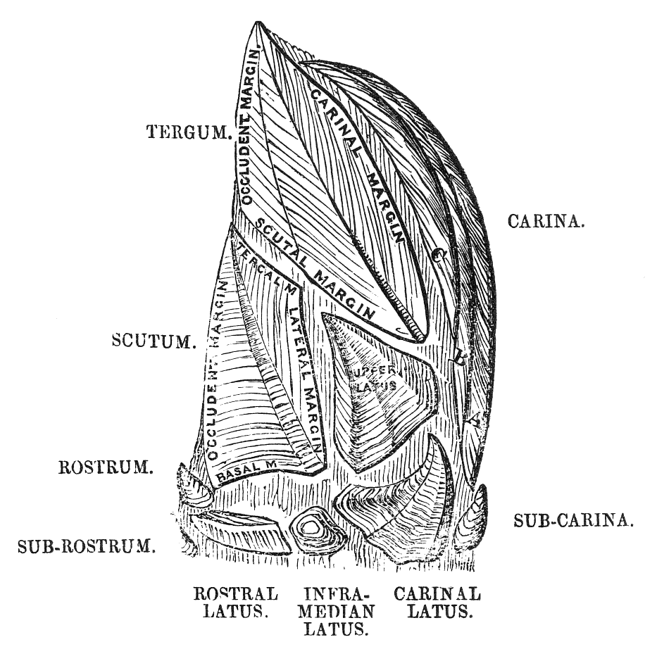
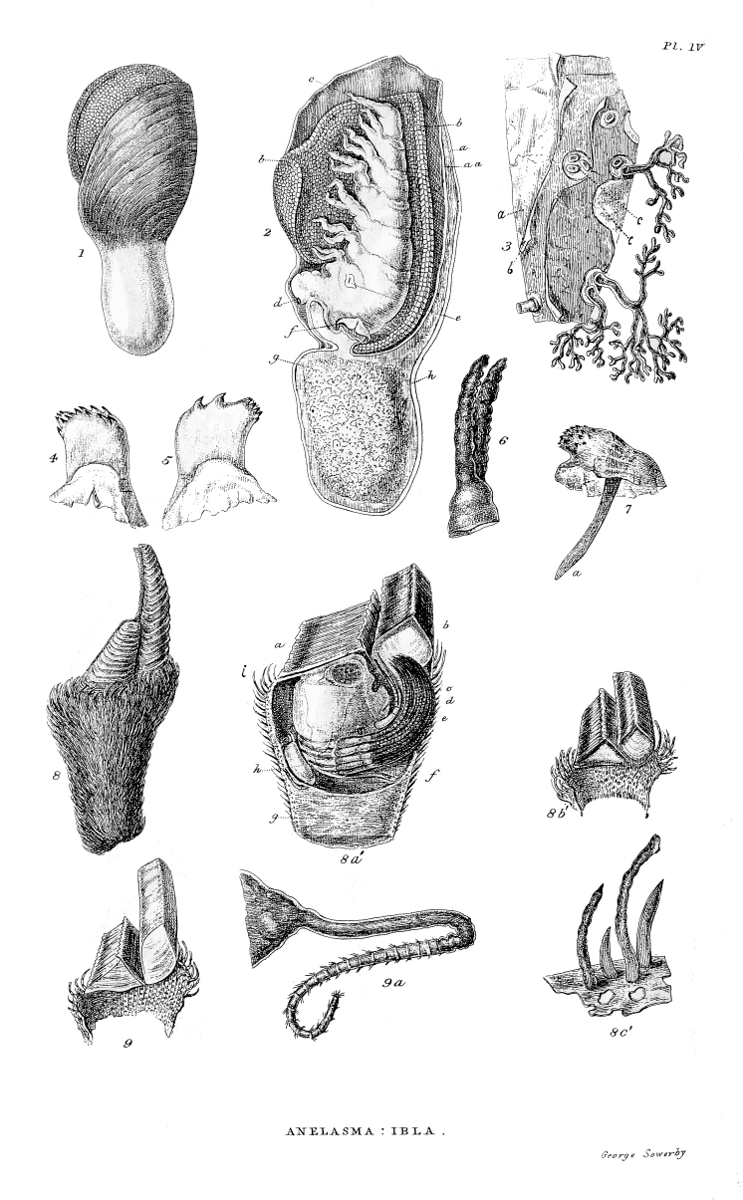
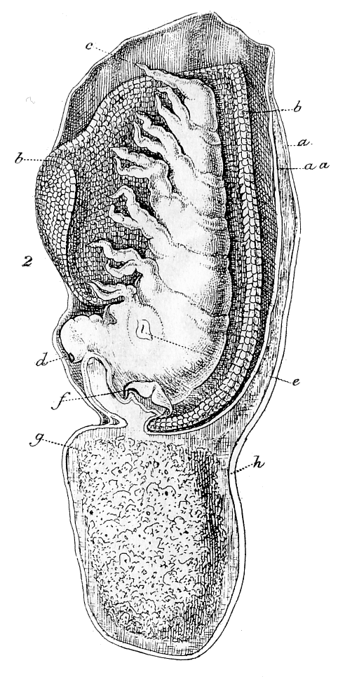
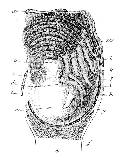
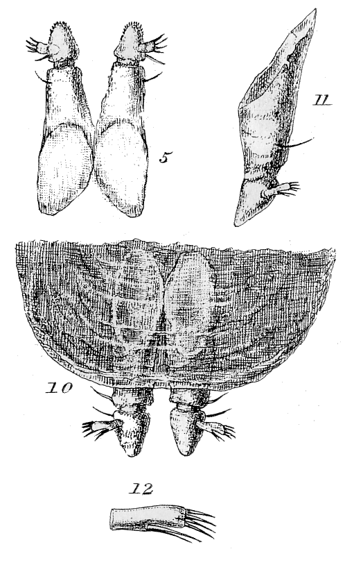
![[m.—Mouth.]](images/f05.png)
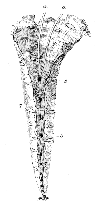
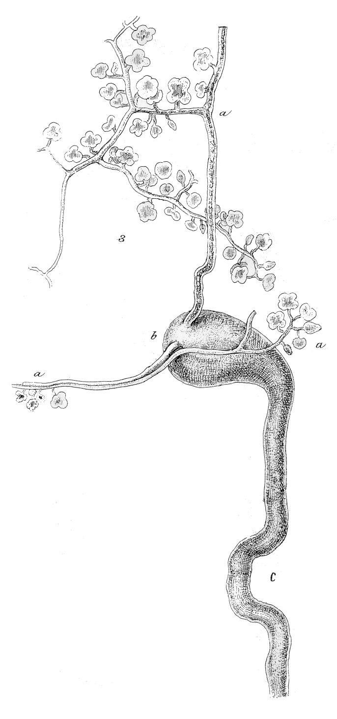
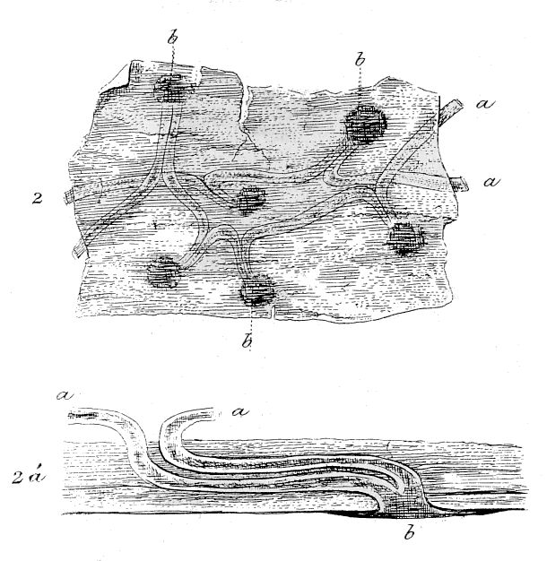
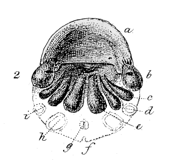
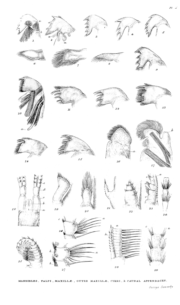
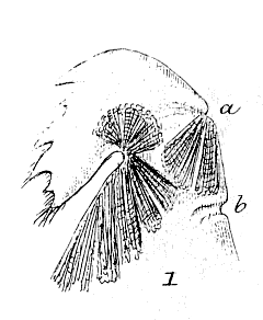
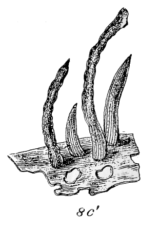
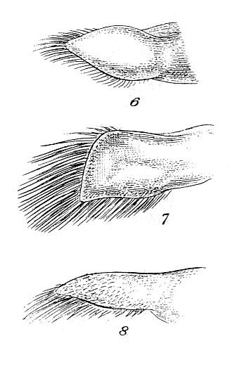
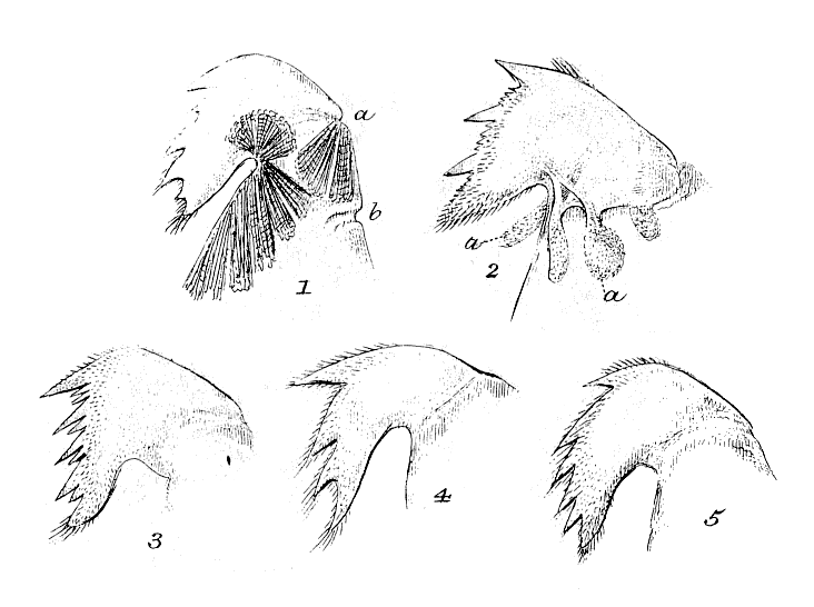
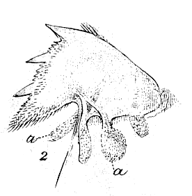
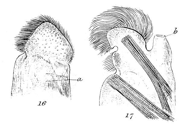
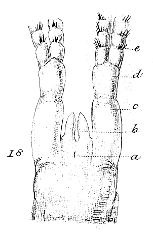
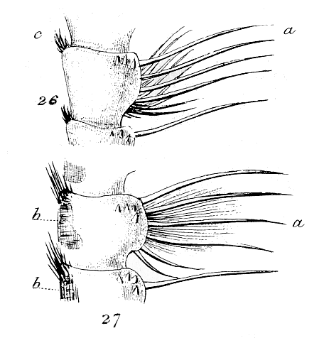
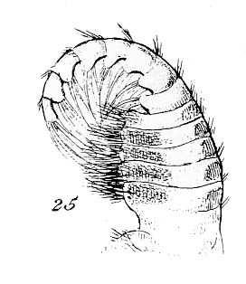
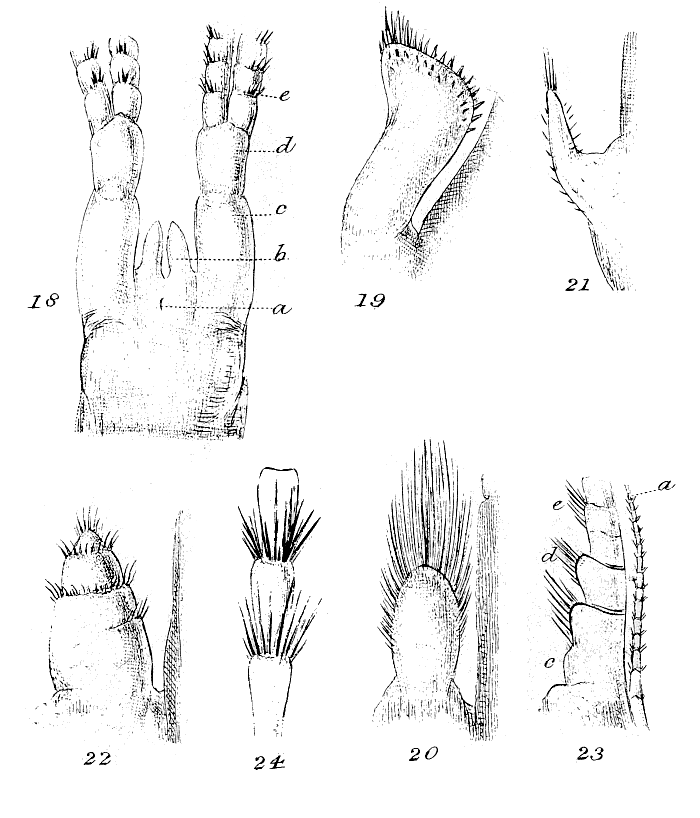
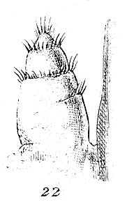
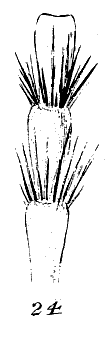
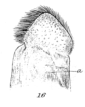
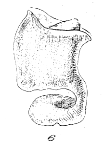
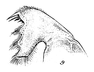
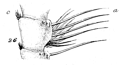
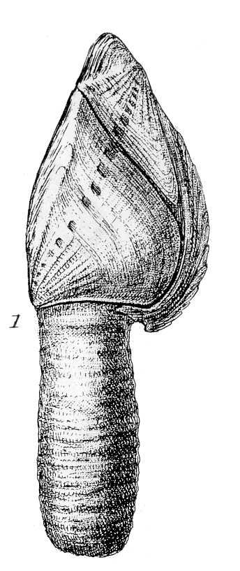
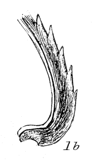
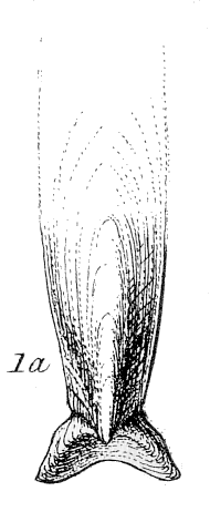
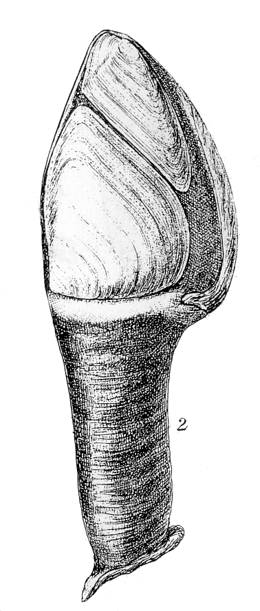
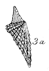
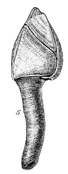
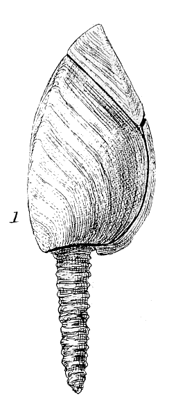
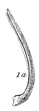
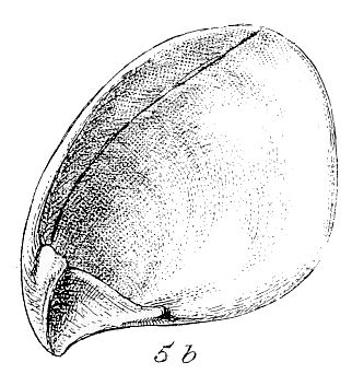
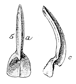
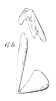
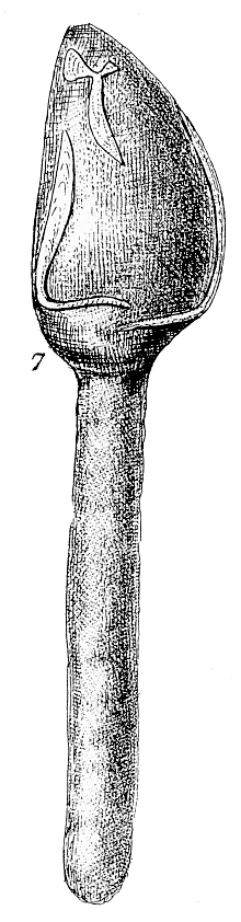
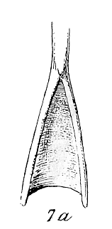
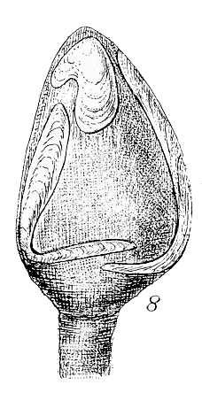
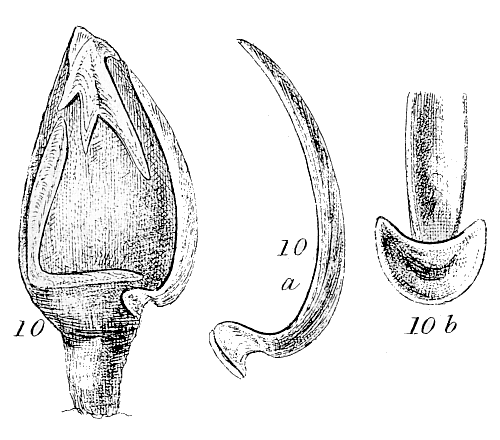
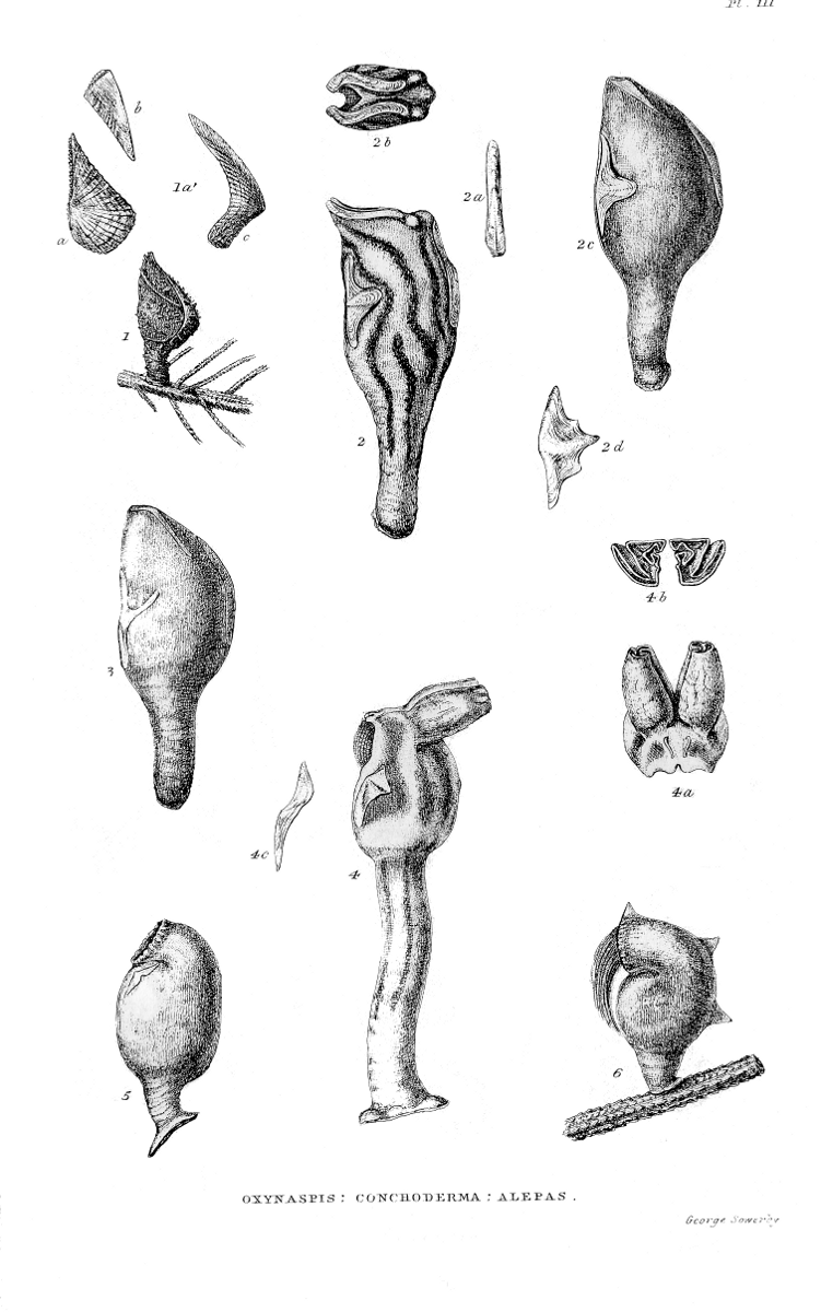
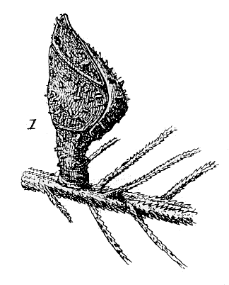
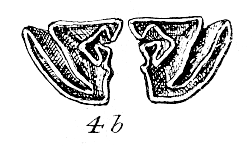
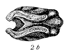
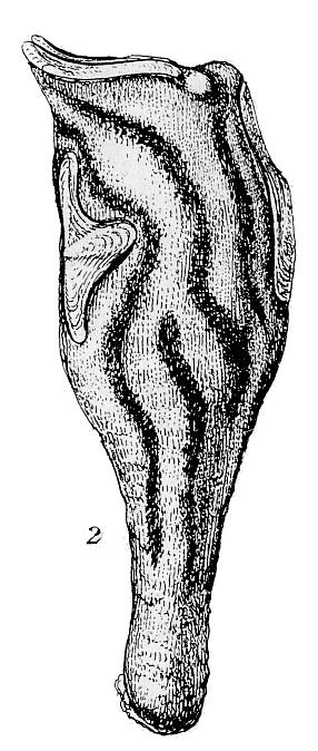
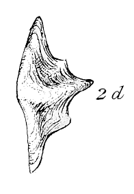
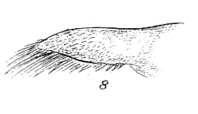
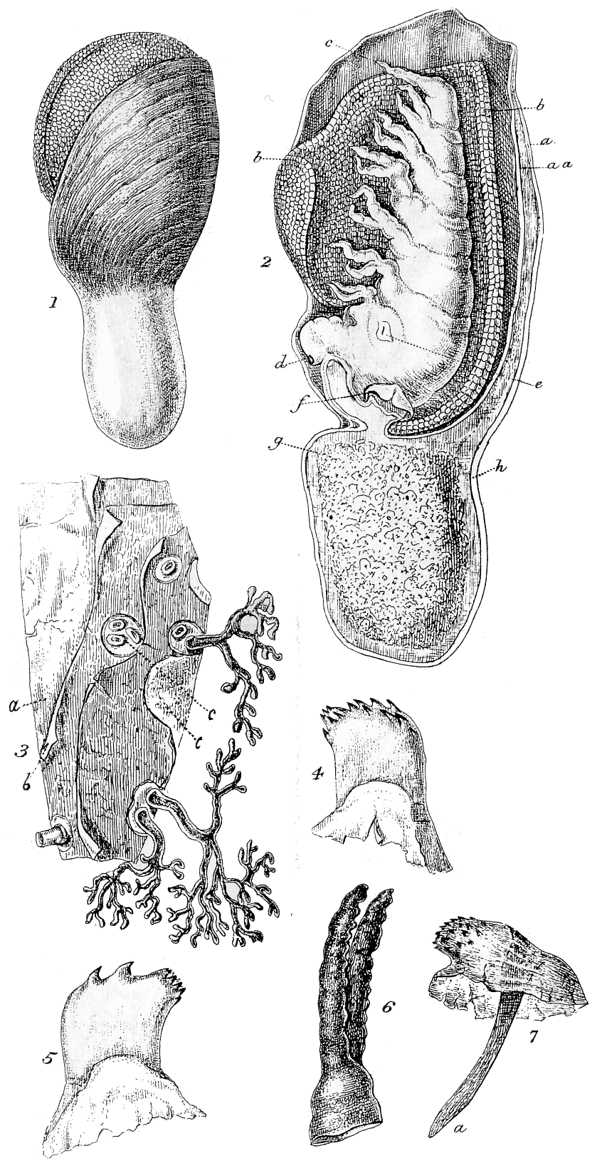
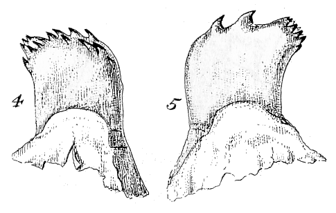
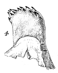
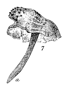
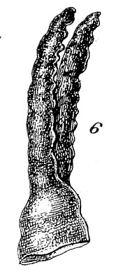
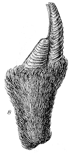
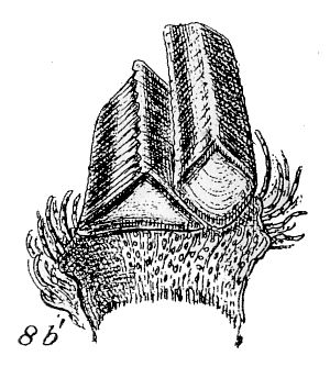
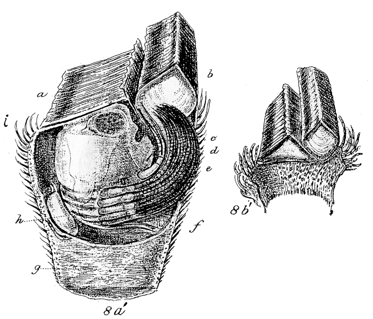
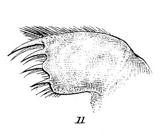
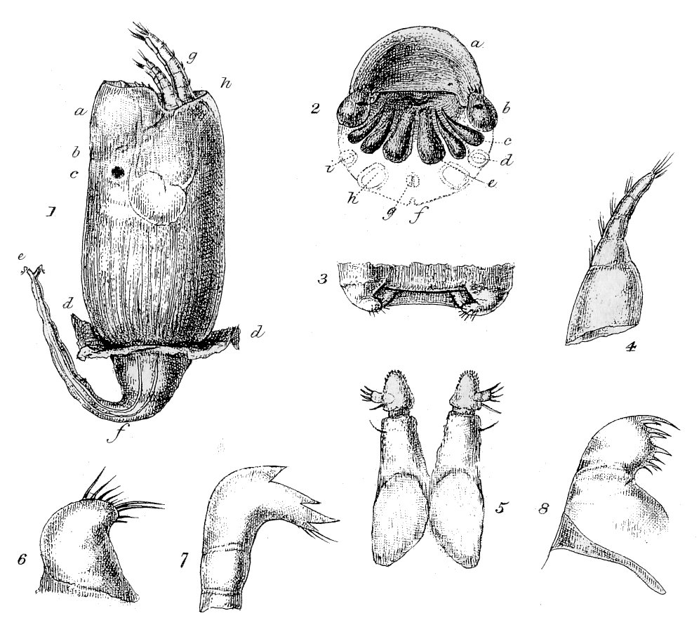
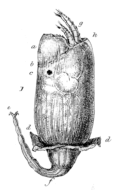
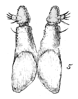
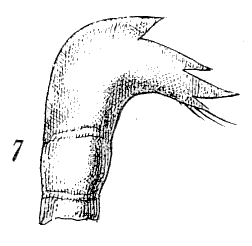
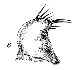
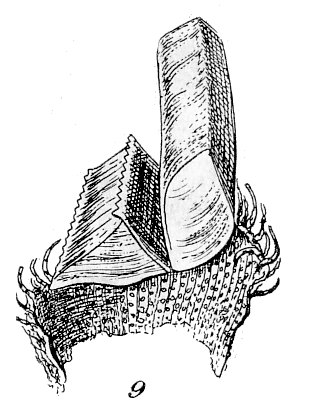
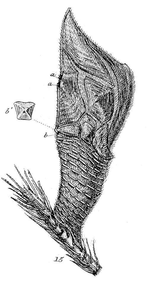
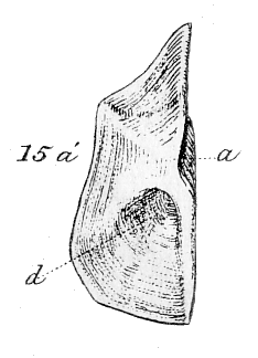
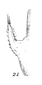
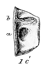
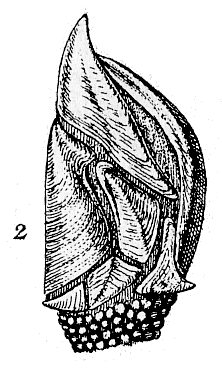
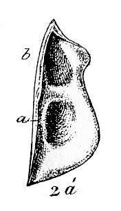
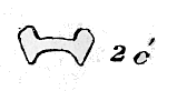
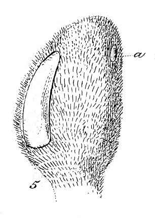
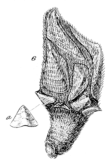
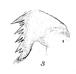
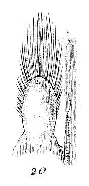
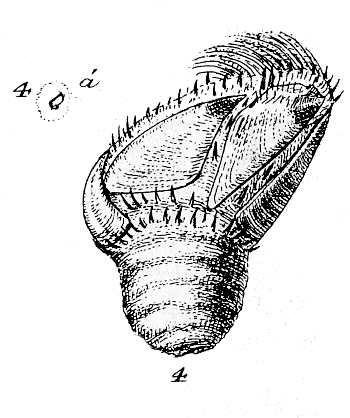
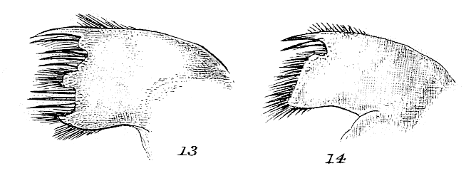
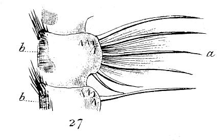
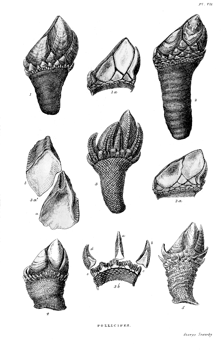
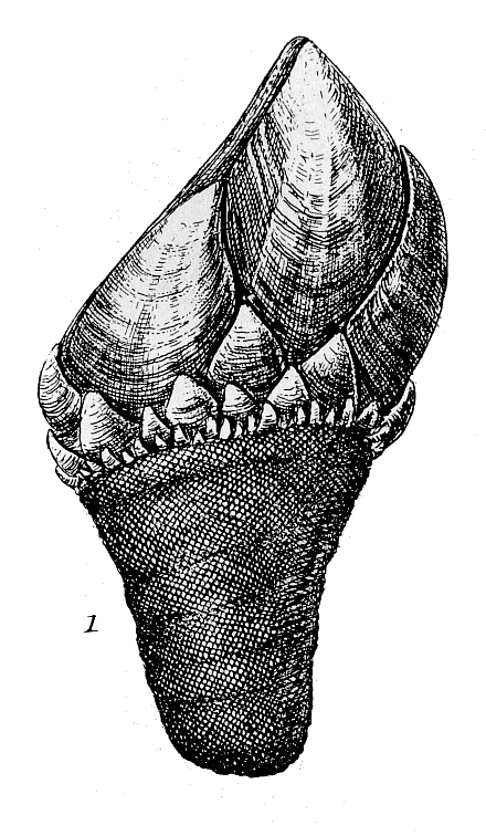
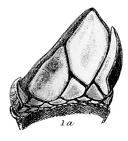
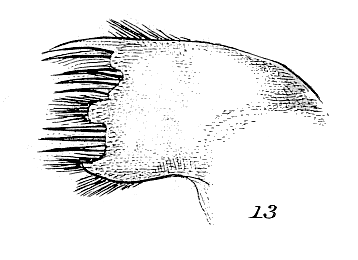
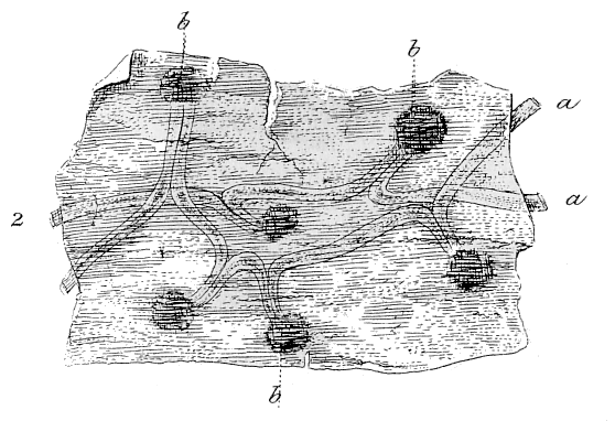
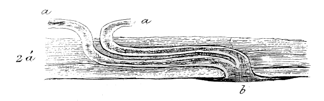
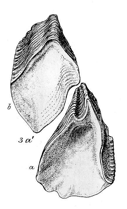
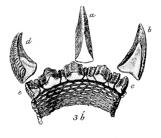
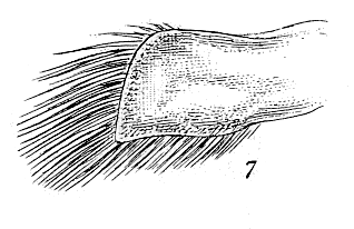
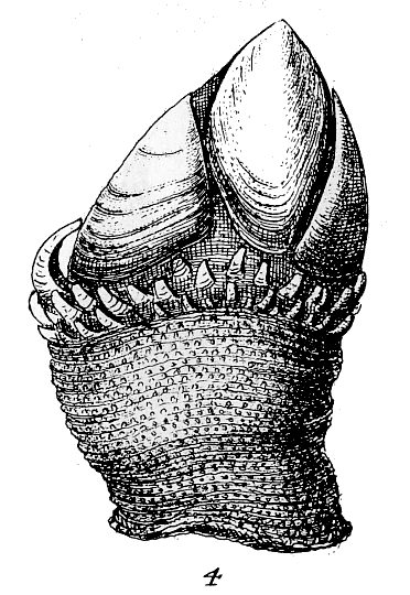
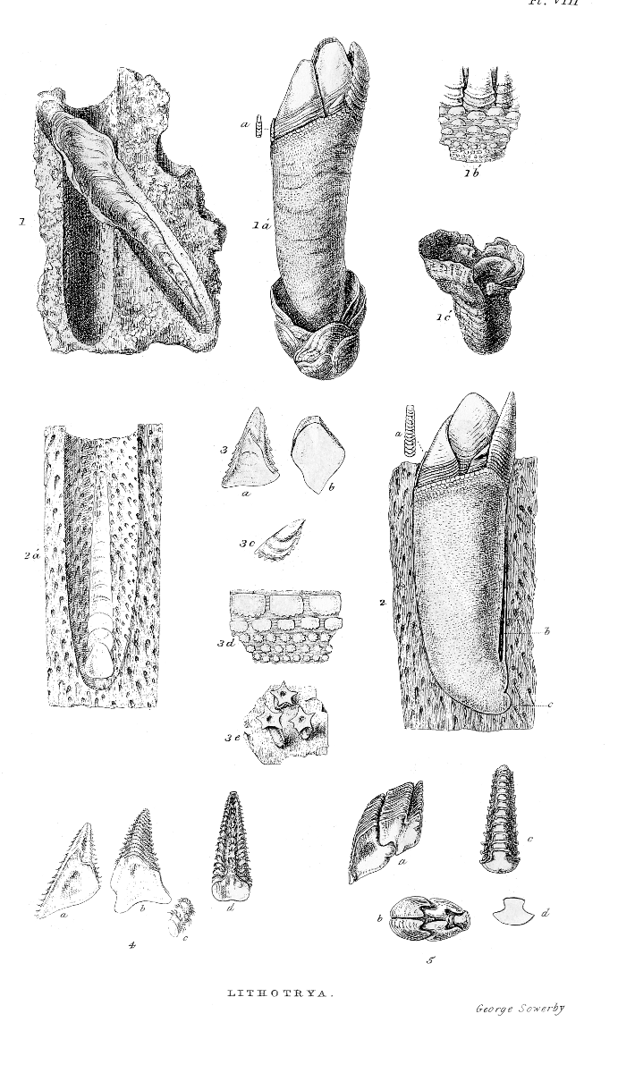
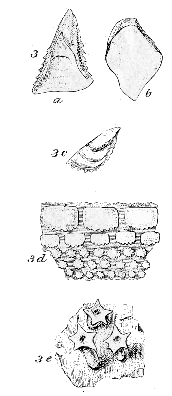
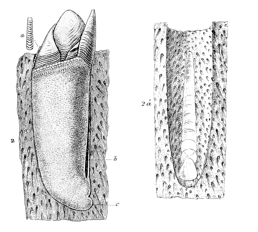
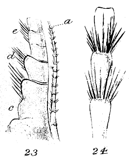
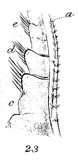
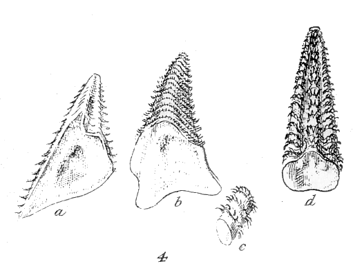
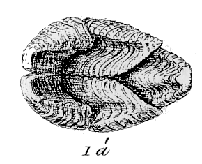
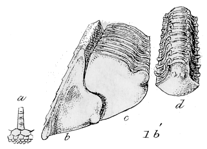
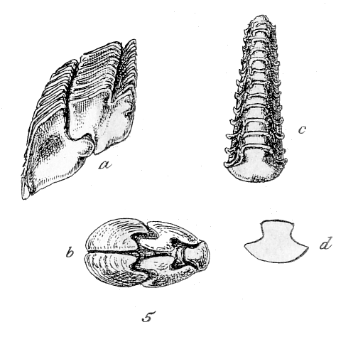
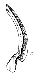
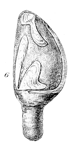
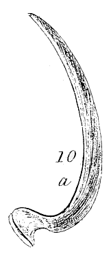
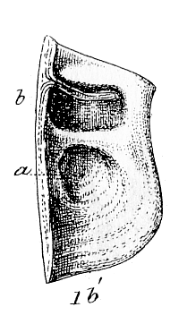
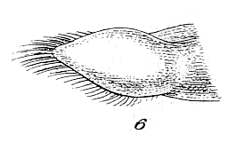