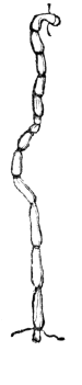
Fig. 2.

Fig. 2. Represents a portion of broken muscular fibre of animal life, (magnified about seven hundred diameters.)
The Project Gutenberg EBook of A Treatise on Anatomy, Physiology, and
Hygiene (Revised Edition), by Calvin Cutter

This eBook is for the use of anyone anywhere at no cost and with
almost no restrictions whatsoever.  You may copy it, give it away or
re-use it under the terms of the Project Gutenberg License included
with this eBook or online at www.gutenberg.org


Title: A Treatise on Anatomy, Physiology, and Hygiene (Revised Edition)

Author: Calvin Cutter

Release Date: November 24, 2009 [EBook #30541]

Language: English

Character set encoding: ISO-8859-1

*** START OF THIS PROJECT GUTENBERG EBOOK TREATISE ON ANATOMY (REVISED) ***


Produced by Bryan Ness, Dan Horwood and the Online
Distributed Proofreading Team at https://www.pgdp.net (This
file was produced from images generously made available
by The Internet Archive/American Libraries.)
DESIGNED FOR
COLLEGES, ACADEMIES, AND FAMILIES.
BY CALVIN CUTTER, M.D.
WITH ONE HUNDRED AND FIFTY ENGRAVINGS.
REVISED STEREOTYPE EDITION.
NEW YORK:
CLARK, AUSTIN AND SMITH.
CINCINNATI:—W. B. SMITH & CO.
ST. LOUIS, MO.:—KEITH & WOODS.
1858.
Entered according to Act of Congress, in the year 1852, by
CALVIN CUTTER, M. D.,
In the Clerk’s Office of the District Court of the District of Massachusetts.
| C. A. ALVORD, Printer, No. 15 Vandewater Street, N. Y. |
Agesilaus, king of Sparta, when asked what things boys should learn, replied, “Those which they will practise when they become men.” As health requires the observance of the laws inherent to the different organs of the human system, so not only boys, but girls, should acquire a knowledge of the laws of their organization. If sound morality depends upon the inculcation of correct principles in youth, equally so does a sound physical system depend on a correct physical education during the same period of life. If the teacher and parents who are deficient in moral feelings and sentiments, are unfit to communicate to children and youth those high moral principles demanded by the nature of man, so are they equally incompetent directors of the physical training of the youthful system, if ignorant of the organic laws and the physiological conditions upon which health and disease depend.
For these reasons, the study of the structure of the human system, and the laws of the different organs, are subjects of interest to all,—the young and the old, the learned and the unlearned, the rich and the poor. Every scholar, and particularly every young miss, after acquiring a knowledge of the primary branches,—as spelling, reading, writing, and arithmetic,—should learn the structure of the human system, and 6 the conditions upon which health and disease depend, as this knowledge will be required in practice in after life.
“It is somewhat unaccountable,” says Dr. Dick, “and not a little inconsistent, that while we direct the young to look abroad over the surface of the earth, and survey its mountains, rivers, seas, and continents, and guide their views to the regions of the firmament, where they may contemplate the moons of Jupiter, the rings of Saturn, and thousands of luminaries placed at immeasurable distances, ... that we should never teach them to look into themselves; to consider their own corporeal structures, the numerous parts of which they are composed, the admirable functions they perform, the wisdom and goodness displayed in their mechanism, and the lessons of practical instruction which may be derived from such contemplations.”
Again he says, “One great practical end which should always be kept in view in the study of physiology, is the invigoration and improvement of the corporeal powers and functions, the preservation of health, and the prevention of disease.”
The design of the following pages is, to diffuse in the community, especially among the youth, a knowledge of Human Anatomy, Physiology, and Hygiene. To make the work clear and practical, the following method has been adopted:—
1st. The structure of the different organs of the system has been described in a clear and concise manner. To render this description more intelligible, one hundred and fifty engravings have been introduced, to show the situation of the various organs. Hence the work may be regarded as an elementary treatise on anatomy.
2d. The functions, or uses of the several parts have been briefly and plainly detailed; making a primary treatise on human physiology.
3d. To make a knowledge of the structure and functions of the different organs practical, the laws of the several parts, and the conditions on which health depends, have been clearly and succinctly explained. Hence it may be called a treatise on the principles of hygiene, or health.
To render this department more complete, there has been added the appropriate treatment for burns, wounds, hemorrhage from divided arteries, the management of persons asphyxiated from drowning, carbonic acid, or strangling, directions for nurses, watchers, and the removal of disease, together with an Appendix, containing antidotes for poisons, so that persons may know what should be done, and what should not be done, until a surgeon or physician can be called.
In attempting to effect this in a brief elementary treatise designed for schools and families, it has not been deemed necessary to use vulgar phrases for the purpose of being understood. The appropriate scientific term should be applied to each organ. No more effort is required to learn the meaning of a proper, than an improper term. For example: a child will pronounce the word as readily, and obtain as correct an idea, if you say lungs, as if you used the word lights. A little effort on the part of teachers and parents, would diminish the number of vulgar terms and phrases, and, consequently, improve the language of our country. To obviate all objections to the use of proper scientific terms, a Glossary has been appended to the work.
The author makes no pretensions to new discoveries in physiological science. In preparing the anatomical department, the able treatises of Wilson, Cruveilhier, and others have been freely consulted. In the physiological part, the splendid works of Carpenter, Dunglison, Liebig, and others have been perused. In the department of hygiene many valuable hints have been obtained from the meritorious works of Combe, Rivers, and others.
We are under obligations to R. D. Mussey, M. D., formerly Professor of Anatomy and Surgery, Dartmouth College, N. H., now Professor of Surgery in the Ohio Medical College; to J. E. M’Girr, A. M., M. D., Professor of Anatomy, Physiology, and Chemistry, St. Mary’s University, Ill.; to E. Hitchcock, Jr., A. M., M. D., Teacher of Chemistry and Natural History, Williston Seminary, Mass.; to Rev. E. Hitchcock, D. D., President of Amherst College, Mass., who examined the revised edition of this work, and whose valuable suggestions rendered important aid in preparing the manuscript for the present stereotype edition.
We return our acknowledgments for the aid afforded by the Principals of the several Academies and Normal Schools who formed classes in their institutions, and examined the revised edition as their pupils progressed, thus giving the work the best possible test trial, namely, the recitation-room.
To the examination of an intelligent public, the work is respectfully submitted by
CALVIN CUTTER.
Warren, Mass., Sept. 1, 1852.
As the work is divided into chapters, the subjects of which are complete in themselves, the pupil may commence the study of the structure, use, and laws of the several parts of which the human system is composed, by selecting such chapters as fancy or utility may dictate, without reference to their present arrangement,—as well commence with the chapter on the digestive organs as on the bones.
The acquisition of a correct pronunciation of the technical words is of great importance, both in recitation and in conversation. In this work, the technical words interspersed with the text, have been divided into syllables, and the accented syllables designated. An ample Glossary of technical terms has also been appended to the work, to which reference should be made.
It is recommended that the subject be examined in the form of topics. The questions in Italics are designed for this method of recitation. The teacher may call on a pupil of the class to describe the anatomy of an organ from an anatomical outline plate; afterwards call upon another to give the physiology of the part, while a third may state the hygiene, after 10 which, the questions at the bottom of the page may be asked promiscuously, and thus the detailed knowledge of the subject possessed by the pupils will be tested.
At the close of the chapters upon the Hygiene of the several portions of the system, it is advised that the instructor give a lecture reviewing the anatomy, physiology, and hygiene, of the topic last considered. This may be followed by a general examination of the class upon the same subject. By this course a clear and definite knowledge of the mutual relation of the Anatomy, Physiology, and Hygiene, of different parts of the human body, will be presented.
We also suggest the utility of the pupils’ giving analogous illustrations, examples, and observations, where these are interspersed in the different chapters, not only to induce inventive thought, but to discipline the mind.
To parents and others we beg leave to say, that about two thirds of the present work is devoted to a concise and practical description of the uses of the important organs of the human body, and to show how such information may be usefully applied, both in the preservation of health, and the improvement of physical education. To this have been added directions for the treatment of those accidents which are daily occurring in the community, making it a treatise proper and profitable for the FAMILY LIBRARY, as well as the school-room.
Chapter. |
Page. |
|
| 1. | General Remarks, | 13 |
| 2. | Structure of Man, | 17 |
| 3. | Chemistry of the Human Body, | 25 |
| 4. | Anatomy of the Bones, | 29 |
| 5. | Anatomy of the Bones, continued, | 39 |
| 6. | Physiology of the Bones, | 48 |
| 7. | Hygiene of the Bones, | 53 |
| 8. | Anatomy of the Muscles, | 64 |
| 9. | Physiology of the muscles, | 76 |
| 10. | Hygiene of the Muscles, | 85 |
| 11. | Hygiene of the Muscles, continued, | 96 |
| 12. | Anatomy of the Teeth, | 105 |
| 12. | Physiology of the Teeth, | 109 |
| 12. | Hygiene of the Teeth, | 110 |
| 13. | Anatomy of the Digestive Organs, | 113 |
| 14. | Physiology of the Digestive Organs, | 124 |
| 15. | Hygiene of the Digestive Organs, | 129 |
| 16. | Hygiene of the Digestive Organs, continued, | 142 |
| 17. | Anatomy of the Circulatory Organs, | 154 |
| 18. | Physiology of the Circulatory Organs, | 164 |
| 19. | Hygiene of the Circulatory Organs, | 172 |
| 20. | Anatomy of the Lymphatic Vessels, | 181 |
| 20. | Physiology of the Lymphatic Vessels, | 183 |
| 20. | Hygiene of the Lymphatic Vessels, | 188 |
| 21. | Anatomy of the Secretory Organs. | 192 |
| 21. | Physiology of the Secretory Organs, | 193 |
| 21. | Hygiene of the Secretory Organs, | 197 |
| 22. | Nutrition, | 200 |
| 22. | Hygiene of Nutrition, | 205 |
| 23. | Anatomy of the Respiratory Organs, | 209 |
| 24. | Physiology of the Respiratory Organs, | 217 |
| 25. | Hygiene of the Respiratory Organs, | 228 |
| 26. | Hygiene of the Respiratory Organs, continued, | 239 |
| 27. | Animal Heat, | 252 |
| 28. | Hygiene of Animal Heat, | 261 |
| 29. | Anatomy of the Vocal Organs, | 268 |
| 29. | Physiology of the Vocal Organs, | 272 |
| 30. | Hygiene of the Vocal Organs, | 274 |
| 31. | Anatomy of the Skin, | 282 |
| 32. | Physiology of the Skin, | 293 |
| 33. | Hygiene of the Skin, | 301 |
| 34. | Hygiene of the Skin, continued, | 311 |
| 35. | Appendages of the Skin, | 322 |
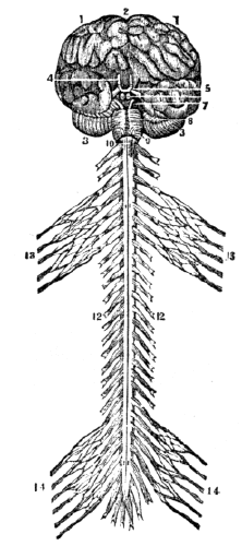
| 36. | Anatomy of the Nervous System, | 327 |
| 37. | Anatomy of the Nervous System, continued, | 340 |
| 38. | Physiology of the Nervous System, | 346 |
| 39. | Hygiene of the Nervous System, | 358 |
| 40. | Hygiene of the Nervous System, continued, | 368 |
| 41. | The Sense of Touch, | 378 |
| 42. | Anatomy of the Organs of Taste, | 384 |
| 42. | Physiology of the Organs of Taste, | 386 |
| 43. | Anatomy of the Organs of Smell, | 389 |
| 43. | Physiology of the Organs of Smell, | 391 |
| 44. | Anatomy of the Organs of Vision, | 394 |
| 45. | Physiology of the Organs of Vision, | 404 |
| 45. | Hygiene of the Organs of Vision, | 410 |
| 46. | Anatomy of the Organs of Hearing, | 414 |
| 47. | Physiology of the Organs of Hearing, | 420 |
| 47. | Hygiene of the Organs of Hearing, | 422 |
| 48. | Means of preserving the Health, | 425 |
| 49. | Directions for Nurses, | 432 |
| APPENDIX, | 439 | |
| GLOSSARY, | 451 | |
| INDEX, | 463 | |
1. Anatomy is the science which treats of the structure and relations of the different parts of animals and plants.
2. It is divided into Vegetable and Animal anatomy. The latter of these divisions is subdivided into Human anatomy, which considers, exclusively, human beings; and Comparative anatomy, which treats of the mechanism of the lower orders of animals.
3. Physiology treats of the functions, or uses of the organs of animals and plants. Another definition is, “the science of life.”
4. This is also divided into Vegetable and Animal physiology, as it treats of the vegetable or animal kingdom; and into Human and Comparative physiology, as it describes the vital functions of man or the inferior animals.
5. Hygiene is the art or science of maintaining health, or a knowledge of those laws by which health may be preserved.
6. The kingdom of nature is divided into organic and inorganic bodies. Organic bodies possess organs, on whose 14 action depend their growth and perfection. This division includes animals and plants. Inorganic bodies are devoid of organs, or instruments of life. In this division are classed the earths, metals, and other minerals.
1. What is anatomy? 2. How is it divided? How is the latter division subdivided? 3. What is physiology? Give another definition. 4. How is physiology divided? Give a subdivision. 5. What is hygiene? 6. Define organic bodies.
7. In general, organic matter differs so materially from inorganic, that the one can readily be distinguished from the other. In the organic world, every individual of necessity springs from some parent, or immediate producing agent; for while inorganic substances are formed by chemical laws alone, we see no case of an animal or plant coming into existence by accident or chance, or chemical operations.
8. Animals and plants are supported by means of nourishment, and die without it. They also increase in size by the addition of new particles of matter to all parts of their substances; while rocks and minerals grow only by additions to their surfaces.
9. “Organized bodies always present a combination of both solids and fluids;—of solids, differing in character and properties, arranged into organs, and these endowed with functional powers, and so associated as to form of the whole a single system;—and of fluids, contained in these organs, and holding such relation to the solids that the existence, nature, and properties of both mutually and necessarily depend on each other.”
10. Another characteristic is, that organic substances have a certain order of parts. For example, plants possess organs to gain nourishment from the soil and atmosphere, and the power to give strength and increase to all their parts. And animals need not only a digesting and circulating apparatus, but organs for breathing, a nervous system, &c.
6. Define inorganic bodies. 7. What is said of the difference, in general, between organic and inorganic bodies? 8. What of the growth of organic and inorganic bodies? 9. What do organized bodies always present? 10. Give another characteristic of organized substances.
11. Individuality is an important characteristic. For instance, a large rock may be broken into a number of smaller pieces, and yet every fragment will be rock; but if an organic substance be separated into two or more divisions, neither of them can be considered an individual. Closely associated with this is the power of life, or vitality, which is the most distinguishing characteristic of organic structure; since we find nothing similar to this in the inorganic creation.
12. The distinction between plants and animals is also of much importance. Animals grow proportionally in all directions, while plants grow upwards and downwards from a collet only. The food of animals is organic, while that of plants is inorganic; the latter feeding entirely upon the elements of the soil and atmosphere, while the former subsist upon the products of the animal and vegetable kingdoms. The size of the vegetable is in most cases limited only by the duration of existence, as a tree continues to put forth new branches during each period of its life, while the animal, at a certain time of life, attains the average size of its species.
13. One of the most important distinctions between animals and plants, is the different effects of respiration. Animals consume the oxygen of the atmosphere, and give off carbonic acid; while plants take up the carbonic acid, and restore to animals the oxygen, thus affording an admirable example of the principle of compensation in nature.
14. But the decisive distinctions between animals and plants are sensation and voluntary motion, the power of acquiring a knowledge of external objects through the senses, and the ability to move from place to place at will. These 16 are the characteristics which, in their fullest development in man, show intellect and reasoning powers, and thereby in a greater degree exhibit to us the wisdom and goodness of the Creator.
11. What is said of the individuality of organized and inorganized bodies? What is closely associated with this? 12. Give a distinction between animals and plants as regards growth. The food of animals and plants. What is said in respect to size? 13. What important distinction in the effects of respiration of animals and plants? 14. What are the decisive distinctions between animals and plants?
15. Disease, which consists in an unnatural condition of the bodily organs, is in most cases under the control of fixed laws, which we are capable of understanding and obeying. Nor do diseases come by chance; they are penalties for violating physical laws. If we carelessly cut or bruise our flesh, pain and soreness follow, to induce us to be more careful in the future; or, if we take improper food into the stomach, we are warned, perhaps immediately by a friendly pain, that we have violated an organic law.
16. Sometimes, however, the penalty does not directly follow the sin, and it requires great physiological knowledge to be able to trace the effect to its true cause. If we possess good constitutions, we are responsible for most of our sickness; and bad constitutions, or hereditary diseases, are but the results of the same great law,—the iniquities of the parents being visited on the children. In this view of the subject, how important is the study of physiology and hygiene! For how can we expect to obey laws which we do not understand?
15. What is said of disease? 16. Why is the study of physiology and hygiene important?
17. In the structure of the human body, there is a union of fluids and solids. These are essentially the same, for the one is readily changed into the other. There is no fluid that does not contain solid matter in solution, and no solid matter that is destitute of fluid.
18. In different individuals, and at different periods of life the proportion of fluids and solids varies. In youth, the fluids are more abundant than in advanced life. For this reason, the limbs in childhood are soft and round, while in old age they assume a hard and wrinkled appearance.
19. The fluids not only contain the materials from which every part of the body is formed, but they are the medium for conveying the waste, decayed particles of matter from the system. They have various names, according to their nature and function; as, the blood, and the bile.
20. The solids are formed from the fluids, and consequently they are reduced, by chemical analysis, to the same ultimate elements. The particles of matter in solids are arranged variously; sometimes in fi´bres, (threads,) sometimes in lam´i-næ, (plates,) sometimes homogeneously, as in basement membranes. (Appendix A.)
21. The parts of the body are arranged into Fi´bres, Fas-cic´u-li, Tis´sues, Or´gans, Ap-pa-ra´tus-es, and Sys´tems.
17. What substances enter into the structure of the human body? Are they essentially the same? 18. What is said of these substances at different periods of life? 19. What offices do the fluids of the system perform? 20. What is said of the solids? How are the particles of matter arranged in solids? 21. Give an arrangement of the parts of the body.
22. A FIBRE is a thread of exceeding fineness. It is either cylindriform or flattened.
23. A FASCICULUS is the term applied to several fibres united. Its general characteristics are the same as fibres.
24. A TISSUE is a term applied to several different solids of the body.
25. An ORGAN is composed of tissues so arranged as to form an instrument designed for action. The action of an organ is called its function, or use.
Example. The liver is an organ, and the secretion of the bile from the blood is one of its functions.[1]
26. An APPARATUS is an assemblage of organs designed to produce certain results.
Example. The digestive apparatus consists of the teeth, stomach, liver, &c., all of which aid in the digestion of food.
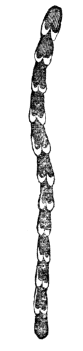
Fig. 2.
Fig. 2. Represents a portion of broken muscular fibre of animal life, (magnified about seven hundred diameters.)
27. The term SYSTEM is applied to an assemblage of organs arranged according to some plan, or method; as the nervous system, the respiratory system.
22. Define a fibre. 23. Define a fasciculus. 24. Define a tissue. 25. Define an organ. What is the action of an organ called? Give examples. Mention other examples. 26. What is an apparatus? Give an example 27. How is the term system applied?
28. A TISSUE is a simple form of organized animal substance. It is flexible, and formed of fibres interwoven in various ways; as, the cellular tissue.
29. However various all organs may appear in their structure and composition, it is now supposed that they can be reduced to a few tissues; as, the Cel´lu-lar, Os´se-ous, Mus´cu-lar, Mu´cous, Ner´vous, &c. (Appendix B.)
30. The CELLULAR TISSUE,[2] now called the areolar tissue, consists of small fibres, or bands, interlaced in every direction, so as to form a net-work, with numerous interstices that communicate freely with each other. These interstices are filled, during life, with a fluid resembling the serum of blood. The use of the areolar tissue is to connect together organs and parts of organs, and to envelop, fix, and protect the vessels and nerves of organs.
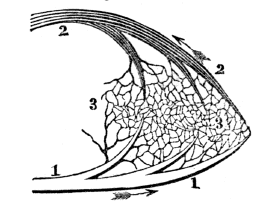
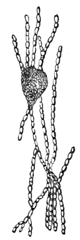
Fig. 3.

Fig. 3. Arrangement of fibres of the cellular tissue magnified one hundred and thirty diameters.
28. What is a tissue? 29. What is said respecting the structure and composition of the various organs? Name the primary membranes. 30. Describe the cellular tissue. How are the cells imbedded in certain tissues? Give observation 1st, relative to the cellular tissue.
Observations. 1st. When this fluid becomes too great in quantity, in consequence of disease, the patient labors under general dropsy. The swelling of the feet when standing, and their return to a proper shape during the night, so often noticed in feeble persons, furnish a striking proof both of the 20 existence and peculiarity of this tissue, which allows the fluid to flow from cell to cell, until it settles in the lower extremities.
2d. The free communication between the cells is still more remarkable in regard to air. Sometimes, when an accidental opening has been made from the air-cells of the lungs into the contiguous cellular tissue, the air in respiration has penetrated every part until the whole body is so inflated as to occasion suffocation. Butchers often avail themselves of the knowledge of this fact, and inflate their meat to give it a fat appearance.
31. “Although this tissue enters into the composition of all organs, it never loses its own structure, nor participates in the functions of the organ of which it forms a part. Though present in the nerves, it does not share in their sensibility; and though it accompanies every muscle and every muscular fibre, it does not partake of the irritability which belongs to these organs.”
32. Several varieties of tissue are formed from the cellular; as, the Se´rous, Der´moid, Fi´brous, and several others.
33. The SEROUS TISSUE lines all the closed, or sac-like cavities of the body; as, the chest, joints, and abdomen. It not only lines these cavities, but is reflected, and invests the organs contained in them. The liver and the lungs are thus invested. This membrane is of a whitish color, and smooth on its free surfaces. These surfaces are kept moist, and prevented from adhering by a se´rous fluid, which is separated from the blood. The use of this membrane is to separate organs and also to facilitate the movement of one part upon another, by means of its moist, polished surfaces.
Give observation 2d. 31. What is said of the identity of this tissue? 32. Name the varieties of tissue formed from the cellular. 33. Where is the serous tissue found? What two offices does it perform? Give its structure. What is the use of this membrane?
34. The DERMOID TISSUE covers the outside of the body. 21 It is called the cu´tis, (skin.) This membrane is continuous with the mucous at the various orifices of the body, and in these situations, from the similarity of their structure, it is difficult to distinguish between them.
Observations. 1st. In consequence of the continuity and similarity of structure, there is close sympathy between the mucous and dermoid membranes. If the functions of the skin are disturbed, as by a chill, it will frequently cause a catarrh, (cold,) or diarrhœa. Again, in consequence of this intimate sympathy, these complaints can be relieved by exciting a free action in the vessels of the skin.
2d. It is no uncommon occurrence that diseased or irritated conditions of the mucous membrane of the stomach or intestines produce diseases or irritations of the skin, as is seen in the rashes attendant on dyspepsia, and eating certain species of fish. These eruptions of the skin can be relieved by removing the diseased condition of the stomach.
35. The FIBROUS TISSUE consists of longitudinal, parallel fibres, which are closely united. These fibres, in some situations, form a thin, dense, strong membrane, like that which lines the internal surface of the skull, or invests the external surface of the bones. In other instances, they form strong, inelastic bands, called lig´a-ments, which bind one bone to another. This tissue also forms ten´dons, (white cords,) by which the muscles are attached to the bones.
Observation. In the disease called rheumatism, the fibrous tissue is the part principally affected; hence the joints, where this tissue is most abundant, suffer most from this affection.
34. Describe the dermoid tissue. What is said of the sympathy between the functions of the skin and mucous membrane? Give another instance of the sympathy between these membranes. 35. Of what does the fibrous tissue consist? How do these appear in some situations? How in others? What tissue is generally affected in rheumatism?
36. The ADIPOSE TISSUE is so arranged as to form distinct bags, or cells. These contain a substance called fat. This 22 tissue is principally found beneath the skin, abdominal muscles, and around the heart and kidneys; while none is found in the brain, eye, ear, nose, and several other organs.
Observation. In those individuals who are corpulent, there is in many instances, a great deposit of this substance. This tissue accumulates more readily than others when a person becomes gross, and is earliest removed when the system emaciates, in acute or chronic diseases. Some of the masses become, in some instances, enlarged. These enlargements are called adipose, or fatty tumors.
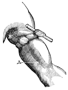
Fig. 4.

Fig. 4. 1, A portion of the adipose tissue. 2, 2, 2, Minute bags containing fat. 3, A cluster of these bags, separated and suspended.
37. The CARTILAGINOUS TISSUE is firm, smooth, and highly elastic. Except bone, it is the hardest part of the animal frame. It tips the ends of the bones that concur in forming a joint. Its use is to facilitate the motion of the joints by its smooth surface, while its elastic character diminishes the shock that would otherwise be experienced if this tissue were inelastic.
36. Describe the adipose tissue. Where does this tissue principally exist? Give observation in regard to the adipose tissue. 37. Describe the cartilaginous tissue. What is its use?
38 The OSSEOUS TISSUE, in composition and arrangement of matter, varies at different periods of life, and in different bones. In some instances, the bony matter is disposed in plates, while in other instances, the arrangement is cylindrical. Sometimes, the bony matter is dense and compact; again, it is spongy, or porous. In the centre of the long bones, a space is left which is filled with a fatty substance, called mar´row.
Observation. Various opinions exist among physiologists in regard to the use of marrow. Some suppose it serves as a reservoir of nourishment, while others, that it keeps the bones from becoming dry and brittle. The latter opinion, however, has been called in question, as the bones of the aged man contain more marrow than those of the child, and they are likewise more brittle.
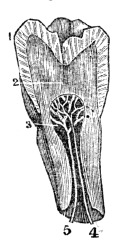
Fig. 5.

Fig. 5. A section of the femur, (thigh-bone.) 1, 1, The extremities, showing a thin plate of compact texture, which covers small cells, that diminish in size, but increase in number, as they approach the articulation. 2, 2, The walls of the shaft, which are very firm and solid. 3, The cavity that contains the marrow.
39. The MUSCULAR TISSUE is composed of many fibres, that unite to form fasciculi, each of which is enclosed in a delicate layer of cellular tissue. Bundles of these fasciculi constitute a muscle.
Observation. A piece of boiled beef will clearly illustrate the arrangement of muscular fibre.
38. What is said of the osseous tissue? How is the bony matter arranged in different parts of the animal frame? What is said of the use of marrow? 39. Of what is the muscular tissue composed? How may the arrangement of muscular fibre be illustrated?
40. The MUCOUS TISSUE differs from the serous by its lining all the cavities which communicate with the air. The nostrils, the mouth, and the stomach afford examples. The external surface of this membrane, or that which is exposed to the air, is soft, and bears some resemblance to the downy rind of a peach. It is covered by a viscid fluid called mu´cus. This is secreted by small gland-cells, called ep-i-the´li-a, or secretory cells of the mucous membrane. The use of this membrane and its secreted mucus is to protect the inner surface of the cavities which it lines.
Observation. A remarkable sympathy exists between the remote parts of the mucous membrane. Thus the condition of the stomach may be ascertained by an examination of the tongue.
41. The NERVOUS TISSUE consists of soft, pulpy matter, enclosed in a sheath, called neu-ri-lem´a. This tissue consists of two substances. The one, of a pulpy character and gray color, is called cin-e-ri´tious, (ash-colored.) The other, of a fibrous character and white, is named med´ul-la-ry, (marrow-like.) In every part of the nervous system both substances are united, with the exception of the nervous fibres and filaments, which are solely composed of the medullary matter enclosed in a delicate sheath.
40. How does the mucous differ from the serous tissue? What is the appearance of the external surface of this membrane? Where is the mucus secreted? What is the use of this membrane? 41. Of what does the nervous tissue consist? Describe the two substances that enter into the composition of the nervous tissue.
42. An ULTIMATE ELEMENT is the simplest form of matter with which we are acquainted; as gold, iron, &c.
43. These elements are divided into metallic and non-metallic substances. The metallic substances are Po-tas´si-um, So´di-um, Cal´ci-um, Mag-ne´si-um, A-lu´min-um, I´ron, Man´ga-nese, and Cop´per. The non-metallic substances are Ox´y-gen, Hy´dro-gen, Car´bon, Ni´tro-gen, Si-li´-ci-um, Phos´phor-us, Sul´phur, Chlo´rine, and a few others.
44. Potash (potassium united with oxygen) is found in the blood, bile, perspiration, milk, &c.
45. Soda (sodium combined with oxygen) exists in the muscles, and in the same fluids in which potash is found.
46. Lime (calcium combined with oxygen) forms the principal ingredient of the bones. The lime in them is combined with phosphoric and carbonic acid.
47. Magnesia (magnesium combined with oxygen) exists in the bones, brain, and in some of the animal fluids; as milk.
48. Silex (silicium combined with oxygen) is contained in the hair and in some of the secretions.
49. Iron forms the coloring principle of the red globules of the blood, and is found in every part of the system.
Observation. As metallic or mineral substances enter into the ultimate elements of the body, the assertion that all minerals are poisonous, however small the quantity, is untrue.
42. What is an ultimate element? Give examples. 43. How are they divided? Name the metallic substances. Name the non-metallic substances. 44. What is said of potash? 45. Of soda? 46. Of lime? 47. Of magnesia? 48. Of silex? 49. What forms the coloring principle of the blood? What is said of mineral substances?
50. Oxygen is contained in all the fluids and solids of the body. It is almost entirely derived from the inspired air and water. It is expelled in the form of carbonic acid and water from the lungs and skin. It is likewise removed in the other secretions.
51. Hydrogen is found in all the fluids and in all the solids of the body. It is derived from the food, as well as from water and other drinks. It exists in the greatest abundance in the impure, dark-colored blood of the system. It is removed by the agency of the kidneys, skin, lungs, and other excretory organs.
52. Carbon is an element in the oil, fat, albumen, fibrin, gelatin, bile, and mucus. This element likewise exists in the impure blood in the form of carbonic acid gas. Carbon is obtained from the food, and discharged from the system by the secretions and respiration.
53. Nitrogen is contained in most animal matter, but is most abundant in fibrin. It is not contained in fat and a few other substances.
Observation. The peculiar smell of animal matter when burning is owing to nitrogen. This element combined with hydrogen forms am-mo´ni-a, (hartshorn,) when animal matter is in a state of putrefaction.
54. Phosphorus is contained in many parts of the system, but more particularly in the bones. It is generally found in combination with oxygen, forming phosphoric acid. The phosphoric acid is usually combined with alkaline bases; as lime in the bones, forming phosphate of lime.
55. Sulphur exists in the bones, muscles, hair, and nails. It is expelled from the system by the skin and intestines.
56. Chlorine is found in the blood, gastric juice, milk, perspiration, and saliva.
50. What is said of oxygen? 51. Of hydrogen? 52. What is said of carbon? 53. Of nitrogen? How is ammonia formed? 54. What is said of phosphorus? 55. What is said of sulphur? 56. Of chlorine?
57. Proximate elements are forms of matter that exist in organized bodies in abundance, and are composed chiefly of oxygen, hydrogen, carbon, and nitrogen, arranged in different proportions. They exist already formed, and may be separated in many instances, by heat or mechanical means. The most important compounds are Al-bu´men, Fi´brin, Gel´a-tin, Mu´cus, Fat, Ca´se-ine, Chon´drine, Lac´tic acid, and Os´ma-zome.
58. Albumen is found in the body, both in a fluid and solid form. It is an element of the skin, glands, hair, and nails, and forms the principal ingredient of the brain. Albumen is without color, taste, or smell, and it coagulates by heat, acids, and alcohol.
Observation. The white of an egg is composed of albumen, which can be coagulated or hardened by alcohol. As albumen enters so largely into the composition of the brain, is not the impaired intellect and moral degradation of the inebriate attributable to the effect of alcohol in hardening the albumen of this organ?
59. Fibrin exists abundantly in the blood, chyle, and lymph. It constitutes the basis of the muscles. Fibrin is of a whitish color, inodorous, and insoluble in cold water. It differs from albumen by possessing the property of coagulating at all temperatures.
Observation. Fibrin may be obtained by washing the thick part of blood with cold water; by this process, the red globules, or coloring matter, are separated from this element.
60. Gelatin is found in nearly all the solids, but it is not known to exist in any of the fluids. It forms the basis of the cellular tissue, and exists largely in the skin, bones, ligaments, and cartilages.
57. What are proximate elements? Do they exist already formed in organized bodies? Name the most important compounds. 58. What is said of albumen? Give observation relative to this element. 59. Of fibrin? How does albumen differ from fibrin? How can fibrin be obtained? 60. What is said of gelatin?
Observation. Gelatin is known from other organic principles by its dissolving in warm water, and forming “jelly.” When dry, it forms the hard, brittle substance, called glue. Isinglass, which is used in the various mechanical arts, is obtained from the sounds of the sturgeon.
61. Mucus is a viscid fluid secreted by the gland-cells, or epithelia. Various substances are included under the name of mucus. It is generally alkaline, but its true chemical character is imperfectly understood. It serves to moisten and defend the mucous membrane. It is found in the cuticle, brain, and nails; and is scarcely soluble in water, especially when dry. (Appendix C.)
62. Osmazome is a substance of an aromatic flavor. It is of a yellowish-brown color, and is soluble both in water and alcohol, but does not form a jelly by concentration. It is found in all the fluids, and in some of the solids; as the brain.
Observation. The characteristic odor and taste of soup are owing to osmazome.
63. There are several acids found in the human system; as the A-ce´tic, Ben-zo´ic, Ox-al´ic, U´ric, and some other substances, but not of sufficient importance to require a particular description.
How is it known from other organic principles? 61. What is said of mucus? 62. Of osmazome? To what are the taste and odor of soup owing? 63. What acids are found in the system?
64. The bones are firm and hard, and of a dull white color. In all the higher orders of animals, among which is man, they are in the interior of the body, while in lobsters, crabs, &c., they are on the outside, forming a case which protects the more delicate parts from injury.
65. In the mechanism of man, the variety of movements he is called to perform requires a correspondent variety of component parts, and the different bones of the system are so admirably adapted to each other, that they admit of numerous and varied motions.
66. When the bones composing the skeleton are united by natural ligaments, they form what is called a natural skeleton, when united by wires, what is termed an artificial skeleton.
67. The elevations, or protuberances, of the bones are called proc´es-ses, and are, generally, the points of attachment for the muscles and ligaments.
68. The BONES are composed of both animal and earthy matter. The earthy portion of the bones gives them solidity and strength, while the animal part endows them with vitality.
64. What is said of the bones? 65. Is there an adaptation of the bones of the system to the offices they are required to perform? 66. What is a natural skeleton? What an artificial? 67. What part of the bones are called processes? 68–73. Give the structure of the bones. 68. Of what are the bones composed? What are the different uses of the component parts of the bones?
Experiments. 1st. To show the earthy without the animal matter, burn a bone in a clear fire for about fifteen minutes, and it becomes white and brittle, because the gelatin, or animal matter of the bone, has been destroyed.
2d. To show the animal without the earthy matter of the bones, immerse a slender bone for a few days in a weak acid, (one part muriatic acid and six parts water,) and it can then be bent in any direction. In this experiment, the acid has removed the earthy matter, (carbonate and phosphate of lime,) yet the form of the bone is unchanged.
69. The bones are formed from the blood, and are subjected to several changes before they are perfected. At their early formative stage, they are cartilaginous. The vessels of the cartilage, at this period, convey only the lymph, or white portion of the blood; subsequently, they convey red blood. At this time, true ossification (the deposition of phosphate and carbonate of lime) commences at certain points, which are called the points of ossification.
70. Most of the bones are formed of several pieces, or centres of ossification. This is seen in the long bones which have their extremities separated from the body by a thin partition of cartilage. It is some time before these separate pieces are united to form one bone.
71. When the process of ossification is completed, there is still a constant change in the bones. They increase in bulk, and become less vascular, until middle age. In advanced life, the elevations upon their surface and near the extremities become more prominent, particularly in individuals accustomed to labor. As a person advances in years, the vitality diminishes, and in extreme old age, the earthy 31 substance predominates; consequently, the bones are extremely brittle.
How can the earthy matter of the bones be shown? The animal? 69. What is the appearance of the bones in their early formative stage? When does true ossification commence? 70. How are most of the bones formed? 71. What is said of the various changes of the bones after ossification?
72. The fibrous membrane that invests the bones is called per-i-os´te-um; that which covers the cartilages is called per-i-chon´dri-um. When this membrane invests the skull, it is called per-i-cra´ni-um.
Fig. 6.

Fig. 6. A section of the knee-joint. The lower part of the femur, (thigh-bone,) and upper part of the tibia, (leg-bone,) are seen ossified at 1, 1. The cartilaginous extremities of the two bones are seen at d, d. The points of ossification of the extremities, are seen at 2, 2. The patella, or knee-pan, is seen at c. 3, A point, or centre of ossification.
73. The PERIOSTEUM is a firm membrane immediately investing the bones, except where they are tipped with cartilage, and the crowns of the teeth, which are protected by enamel. This membrane has minute nerves, and when healthy, possesses 32 but little sensibility. It is the nutrient membrane of the bone, endowing its exterior with vitality; it also gives insertion to the tendons and connecting ligaments of the joints.
72. What is the membrane called that invests the bones? That covers the cartilage? That invests the skull? Explain fig. 6. 73. Describe the periosteum.
74. There are two hundred and eight[3] bones in the human body, beside the teeth. These, for convenience, are divided into four parts: 1st. The bones of the Head. 2d. The bones of the Trunk. 3d. The bones of the Upper Extremities. 4th. The bones of the Lower Extremities.
75. The bones of the HEAD are divided into those of the Skull, Ear, and Face.
76. The SKULL is composed of eight bones. They are formed of two plates, or tablets of bony matter, united by a porous portion of bone. The external tablet is fibrous and tough; the internal plate is dense and hard, and is called the vit´re-ous, or glassy table. These tough, hard plates are adapted to resist the penetration of sharp instruments, while the different degrees of density possessed by the two tablets, and the intervening spongy bone, serve to diminish the vibrations that would occur in falls or blows.
77. The skull is convex externally, and at the base much thicker than at the top or sides. The most important part of the brain is placed here, completely out of the way of injury, unless of a very serious nature. The base of the cranium, or skull, has many projections, depressions, and apertures; the latter affording passages for the nerves and blood-vessels.
74. How many bones in the human body? How are they divided? 75–81. Give the anatomy of the bones of the head. 75. How are the bones of the head divided? 76. Describe the bones of the skull. 77. What is the form of the skull? What does the base of the skull present?
78. The bones of the cranium are united by ragged edges, called sut´ures. The edges of each bone interlock with each other, producing a union, styled, in carpentry, dovetailing. They interrupt, in a measure, the vibrations produced by external blows, and also prevent fractures from extending as far as they otherwise would, in one continued bone. From infancy to the twelfth year, the sutures are imperfect; but, from that time to thirty-five or forty, they are distinctly marked; in old age, they are nearly obliterated.
Fig. 7.
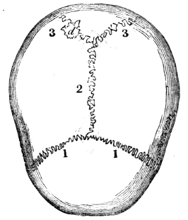
Fig. 7. 1, 1, The coronal suture at the front and upper part of the skull, or cranium. 2, The sagittal suture on the top of the skull. 3, 3, The lambdoidal suture at the back part of the cranium.
79. We find as great a diversity in the form and texture of the skull-bone, as in the expression of the face. The head of the New Hollander is small; that of the African is compressed; while the Caucasian is distinguished for the beautiful oval form of the head. The Greek skulls, in texture, are close and fine, while the Swiss are softer and more open.
78. How are the bones of the skull united? What are the uses of the sutures? Mention the appearance of the sutures at different ages. What does fig. 7 represent? 79. What is said respecting the form and texture of the skull in different nations?
80. In each EAR are four very small bones. They aid in hearing.
81. In the FACE are fourteen bones, some of which serve for the attachment of powerful muscles, which are more or less called into action in masticating food; others retain in place the soft parts of the face.
Fig. 8.

Fig. 8. 1, The frontal, or bone of the forehead. 2. The parietal bone. 3, The temporal bone. 4, The zygomatic process of the temporal bone. 5, The malar (cheek) bone. 6, The superior maxillary bone, (upper jaw.) 7, The vomer, that separates the cavities of the nose. 8, The inferior maxillary bone, (lower jaw.) 9. The cavity for the eye.
82. The TRUNK has fifty-four bones—twenty-four Ribs; twenty-four bones in the Spi´nal Col´umn, (back-bone;) four in the Pel´vis; the Ster´num, (breast-bone;) and the Os hy-oid´es, (the bone at the base of the tongue.) They are so arranged as to form, with the soft parts attached to them, two cavities, called the Tho´rax (chest) and Ab-do´men.
80. How many bones in the ear? 81. How many bones in the face? What is their use? Explain fig. 8. 82–94. Give the anatomy of the bones of the trunk. 82. How many bones in the trunk? Name them. What do they form by their arrangement?
83. The THORAX is formed by the sternum in front; the ribs, at the sides; and the twelve dorsal bones of the spinal column, posteriorly. The natural form of the chest is a cone, with its apex above; but fashion, in many instances, has nearly inverted this order. This cavity contains the lungs, heart, and large blood-vessels.
Fig. 9.
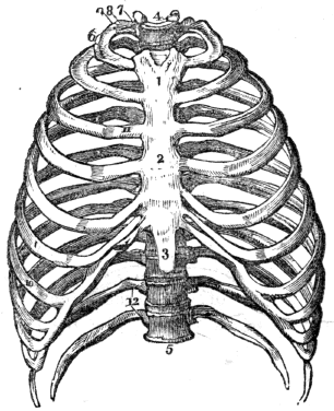
Fig. 9. 1, The first bone of the sternum, (breast-bone.) 2. The second bone of the sternum. 3, The cartilage of the sternum. 4, The first dorsal vertebra, (a bone of the spinal column.) 5, The last dorsal vertebra. 6, The first rib. 7, Its head. 8, Its neck. 9, Its tubercle. 10, The seventh, or last true rib. 11, The cartilage of the third rib. 12, The floating ribs.
84. The STERNUM is composed of eight pieces in the child. These unite and form but three parts in the adult. In youth, the two upper portions are converted into bone, while the lower portion remains cartilaginous and flexible until extreme old age, when it is often converted into bone.
85. The RIBS are connected with the spinal column, and increase in length as far as the seventh. From this they successively 36 become shorter. The direction of the ribs from above, downward, is oblique, and their curve diminishes from the first to the twelfth. The external surface of each rib is convex; the internal, concave. The inferior, or lower ribs, are, however, very flat.
83. Describe the thorax. Explain fig. 9. 84. Describe the sternum. 85. Describe the ribs.
86. The seven upper ribs are united to the sternum, through the medium of cartilages, and are called the true ribs. The cartilages of the next three are united with each other, and are not attached to the sternum; these are called false ribs. The lowest two are called floating ribs, as they are not connected either with the sternum or the other ribs.
87. The SPINAL COLUMN is composed of twenty-four pieces of bone. Each piece is called a vert´e-bra. On examining one of the bones, we find seven projections, called processes; four of these, that are employed in binding the bones together, are called articulating processes; two of the remaining are called the transverse; and the other, the spinous. The last three give attachment to the muscles of the back.
88. The large part of the vertebra, called the body, is round and spongy in its texture, like the extremity of the round bones. The processes are of a more dense character. The projections are so arranged that a tube, or canal, is formed immediately behind the bodies of the vertebræ, in which is placed the me-dul´la spi-na´lis, (spinal cord,) sometimes called the pith of the back-bone.
89. Between these joints, or vertebræ, is a peculiar and highly elastic substance, which much facilitates the bending movements of the back. This compressible cushion of cartilage also serves the important purpose of diffusing and diminishing the shock in walking, running, or leaping, and tends to protect the delicate texture of the brain.
86. How are the ribs united to the sternum? 87. Describe the spinal column. 88. Give the structure of the vertebra. Where is the spinal cord placed? 89. What is placed between each vertebra? What is its use?
90. Another provision for the protection of the brain, which bears convincing proof of the wisdom and beneficence of the Creator, is the antero-posterior, or forward and backward curve of the spinal column. Were it a straight column, standing perpendicularly, the slightest jar, in walking, would cause it to recoil with a sudden jerk; because, the weight bearing equally, the spine would neither yield to the one side nor the other. But, shaped as it is, we find it yielding in the direction of the curves, and thus the force of the shock is diffused.
Fig. 10.

Fig. 10. A vertebra of the neck. 1, The body of the vertebra. 2, The spinal canal. 4, The spinous process, cleft at its extremity. 5, The transverse process. 7, The inferior articulating process. 8, The superior articulating process.
Fig. 11.

Fig. 11. 1, The cartilaginous substance that connects the bodies of the vertebræ. 2, The body of the vertebra. 3, The spinous process. 4, 4, The transverse processes. 5, 5, The articulating processes. 6, 6, A portion of the bony bridge that assists in forming the spinal canal, (7.)
Observation. A good idea of the structure of the vertebræ may be obtained by examining the spinal column of a domestic animal, as the dog, cat, or pig.
91. The PELVIS is composed of four bones; the two in-nom-i-na´ta, (nameless bones,) the sa´crum, and the coc´cyx.
92. The INNOMINATUM, in the child, consists of three pieces. 38 These, in the adult, become united, and constitute but one bone. In the sides of these bones is a deep socket, or depression, like a cup, called the ac-e-tab´u-lum, in which the round head of the thigh-bone is placed.
90. What is said of the curves of the spinal column? What is represented by fig. 10? By fig. 11? How can the structure of the vertebræ be seen? 91. Of how many bones is the pelvis composed? 92. What is said of the innominatum in the child?
93. The SACRUM, so called because the ancients offered it in sacrifices, is a wedge-shaped bone, that is placed between the innominata, and to which it is bound by ligaments. Upon its upper surface it connects with the lower vertebra. At its inferior, or lower angle, it is united to the coccyx. It is concave upon its anterior, and convex upon its posterior surface.
Fig. 12.

Fig. 12. 1, 1, The innominata, (nameless bones.) 2, The sacrum. 3, The coccyx. 4, 4, The acetabulum. a, a, The pubic portion of the innominata. d, The arch of the pubes; e, The junction of the sacrum and lower lumbar vertebra.
94. The COCCYX, in infants, consists of several pieces, which, in youth, become united and form one bone. This is the terminal extremity of the spinal column.
In the adult? Describe the acetabulum. 93. Describe the sacrum. Explain fig. 12. 94. Describe the coccyx.
95. The bones of the upper and lower limbs are enlarged at each extremity, and have projections, or processes. To these, the tendons of muscles and ligaments are attached, which connect one bone with another. The shaft of these bones is cylindrical and hollow, and in structure, their exterior surface is hard and compact, while the interior portion is of a reticulated character. The enlarged extremities of the round bones are more porous than the main shaft.
96. The UPPER EXTREMITIES contain sixty-four bones—the Scap´u-la, (shoulder-blade;) the Clav´i-cle, (collar-bone;) the Hu´mer-us, (first bone of the arm;) the Ul´na and Ra´di-us, (bones of the fore-arm;) the Car´pus, (wrist;) the Met-a-car´pus, (palm of the hand;) and the Pha-lan´ges, (fingers and thumb.)
97. The CLAVICLE is attached, at one extremity, to the sternum; at the other, it is united to the scapula. It is shaped like the Italic ∫. Its use is to keep the arms from sliding toward the breast.
98. The SCAPULA is situated upon the upper and back part of the chest. It is flat, thin, and of a triangular form. This bone lies upon and is retained in its position by muscles. By their contractions it may be moved in different directions.
99. The HUMERUS is cylindrical, and is joined at the elbow with the ulna of the fore-arm; at the scapular extremity, it is 40 lodged in the glenoid cavity, where it is surrounded by a membranous bag, called the capsular ligament.
95–104. Give the anatomy of the bones of the upper extremities. 95. Give the structure of the bones of the extremities. 96. How many bones in the upper extremities? Name them. 97. Give the attachments of the clavicle. What is its use? 98. Describe the scapula. How is it retained in its position? 99. Describe the humerus.
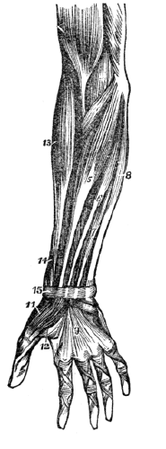
Fig. 13.

Fig. 13. 1, The shaft of the humerus. 2, The large, round head that is placed in the glenoid cavity. 3, 4, Processes, to which muscles are attached. 5, A process, called the external elbow. 6, A process, called the internal elbow. 7, The articulating surface upon which the ulna rolls.
Fig. 14.

Fig. 14. 1, The body of the ulna. 2, The shaft of the radius. 3, The upper articulation of the radius and ulna. 4, Articulating cavity, in which the lower extremity of the humerus is placed. 5, Upper extremity of the ulna, called the olecranon process, which forms the point of the elbow. 6, Space between the radius and ulna, filled by the intervening ligament. 7, Styloid process of the ulna. 8, Surface of the radius and the ulna, where they articulate with the bones of the wrist. 9, Styloid process of the radius.
100. The ULNA articulates with the humerus at the elbow, and forms a perfect hinge-joint. This bone is situated on the inner side of the fore-arm.
What is represented by fig. 13? By fig. 14? 100. Describe the ulna.
101. The RADIUS articulates with the bones of the carpus and forms the wrist-joint. This bone is situated on the outside of the fore-arm, (the side on which the thumb is placed.) The ulna and radius, at their extremities, articulate with each other, by which union the hand is made to rotate, permitting its complicated and varied movements.
102. The CARPUS is composed of eight bones, ranged in two rows, and so firmly bound together, as to permit only a small amount of movement.
Fig. 15.

Fig. 15. U, The ulna. R, The radius. S, The scaphoid bone. L, The semilunar bone. C, The cuneiform bone. P, The pisiform bone. These four form the first row of carpal bones. T, T, The trapezium and trapezoid bones. M, The os magnum. U, The unciform bone. These four form the second row of carpal bones. 1, 1, 1, 1, 1, The metacarpal bones of the thumb and fingers.
Fig. 16.
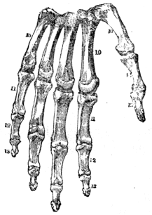
Fig. 16. 10, 10, 10, The metacarpal bones of the hand. 11, 11, First range of finger-bones. 12, 12, Second range of finger-bones. 13, 13, Third range of finger-bones. 14, 15, Bones of the thumb.
103. The METACARPUS is composed of five bones, upon four of which the first range of the finger-bones is placed; and 42 upon the other, the first bone of the thumb. The five metacarpal bones articulate with the second range of carpal bones.
101. The radius. 102. How many bones in the carpus? How are they ranged? 103. Describe the metacarpus.
104. The PHALANGES of the fingers have three ranges of bones, while the thumb has but two.
Observation. The wonderful adaptation of the hand to all the mechanical offices of life, is one cause of man’s superiority over the rest of creation. This arises from the size and strength of the thumbs, and the different lengths of the fingers.
105. The LOWER EXTREMITIES contain sixty bones—the Fe´mur, (thigh-bone;) the Pa-tel´la, (knee-pan;) the Tib´i-a, (shin-bone;) the Fib´u-la, (small bone of the leg;) the Tar´sus, (instep;) the Met-a-tar´sus, (middle of the foot;) and the Pha-lan´ges, (toes.)
106. The FEMUR is the longest bone in the system. It supports the weight of the head, trunk, and upper extremities. The large, round head of this bone is placed in the acetabulum. This articulation is a perfect specimen of the ball and socket joint.
107. The PATELLA is a small bone connected with the tibia by a strong ligament. The tendon of the ex-tens´or muscles of the leg is attached to its upper edge. This bone is placed on the anterior part of the lower extremity of the femur, and acts like a pulley, in the extension of the limb.
108. The TIBIA is the largest bone of the leg. It is of a triangular shape, and enlarged at each extremity.
109. The FIBULA is a smaller bone than the tibia, but of similar shape. It is firmly bound to the tibia, at each extremity.
110. The TARSUS is formed of seven irregular bones, which are so firmly bound together as to permit but little movement.
104. How many ranges of bones have the phalanges? 105–112. Give the anatomy of the bones of the lower extremities. 105. How many bones in the lower extremities? Name them. 106. Describe the femur. 107. Describe the patella. What is its function? 108. What is the largest bone of the leg called? What is its form? 109. What is said of the fibula? 110. Describe the tarsus.
Fig. 17.

Fig. 17. 1, The shaft of the femur, (thigh-bone.) 2, A projection, called the trochantar minor, to which are attached some strong muscles. 4, The trochantar major, to which the large muscles of the hip are attached. 3, The head of the femur. 5, The external projection of the femur, called the external condyle. 6, The internal projection, called the internal condyle. 7, The surface of the lower extremity of the femur, that articulates with the tibia, and upon which the patella slides.
Fig. 18.

Fig. 18. 1, The tibia. 5, The fibula. 8, The space between the two, filled with the inter-osseous ligament. 6, The junction of the tibia and fibula at their upper extremity. 2, The external malleolar process, called the external ankle. 3, The internal malleolar process, called the internal ankle. 4, The surface of the lower extremity of the tibia, that unites with one of the tarsal bones to form the ankle-joint. 7, The upper extremity of the tibia, upon which the lower extremity of the femur rests.
Explain fig. 17. Explain fig. 18.
111. The METATARSAL bones are five in number. They articulate at one extremity with one range of tarsal bones; at the other extremity, with the first range of the toe-bones.
Fig. 19.
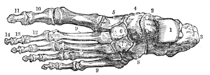
Fig. 19. A representation of the upper surface of the bones of the foot. 1, The surface of the astragulus, where it unites with the tibia. 2, The body of the astragulus. 3, The calcis, (heel-bone.) 4, The scaphoid bone. 5, 6, 7, The cuneiform bones. 8, The cuboid. 9, 9, 9, The metatarsal bones. 10, The first bone of the great toe. 11, The second bone. 12, 13, 14, Three ranges of bones, forming the small toes
Fig. 20.
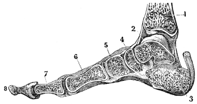
Fig. 20. A side view of the bones of the foot, showing its arched form. The arch rests upon the heel behind, and the ball of the toes in front. 1, The lower part of the tibia. 2, 3, 4, 5, Bones of the tarsus. 6, The metatarsal bone. 7, 8, The bones of the great toe. These bones are so united as to secure a great degree of elasticity, or spring.
Observation. The tarsal and metatarsal bones are united so as to give the foot an arched form, convex above, and concave 45 below. This structure conduces to the elasticity of the step, and the weight of the body is transmitted to the ground by the spring of the arch, in a manner which prevents injury to the numerous organs.
111. Describe the metatarsal bones. Explain fig. 19. What is represented by fig. 20? What is said of the arrangement of the bones of the foot?
112. The PHALANGES (fig. 19) are composed of fourteen bones; each of the small toes has three ranges of bones, while the great toe has but two.
113. The JOINTS form an interesting part of the body. In their construction, every thing shows the regard that has been paid to the security and the facility of motion of the parts thus connected together. They are composed of the extremities of two or more bones, Car´ti-lages, (gristles,) Syn-o´vi-al membrane, and Lig´a-ments.
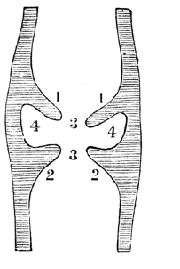
Fig. 21.
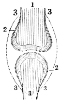
Fig. 21 The relative position of the bones, cartilages, and synovial membrane. 1, 1, The extremities of two bones that concur to form a joint. 2, 2, The cartilages that cover the end of the bones. 3, 3, 3, 3, The synovial membrane which covers the cartilage of both bones, and is then doubled back from one to the other; it is represented by the dotted lines.
Fig. 22.
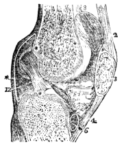
Fig. 22. A vertical section of the knee-joint. 1, The femur. 3, The patella. 5, The tibia. 2, 4, The ligaments of the patella. 6, The cartilage of the tibia 12, The cartilage of the femur. * * * *, The synovial membrane.
114. Cartilage is a smooth, solid, elastic substance, of a pearly whiteness, softer than bone. It forms upon the articular 46 surfaces of the bones a thin incrustation, not more than the sixteenth of an inch in thickness. Upon convex surfaces it is the thickest in the centre, and thin toward the circumference; while upon concave surfaces, an opposite arrangement is presented.
112. Describe the phalanges. 113–118. Give the anatomy of the joints. 113. What is said of the joints? Of what are the joints composed? What is illustrated by fig. 21? By fig. 22? 114. Define cartilage.
115. The SYNOVIAL MEMBRANE is a thin, membranous layer, which covers the cartilages, and is thence bent back, or reflected upon the inner surfaces of the ligaments which surround and enter into the composition of the joints. This membrane forms a closed sac, like the membrane that lines an egg-shell.
Fig. 23.
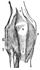
Fig. 23. The anterior ligaments of the knee-joint. 1, The tendon of the muscle that extends the leg. 2, The patella. 3, The anterior ligament of the patella, near its insertion. 4, 4, The synovial membrane. 5, The internal lateral ligament. 6, The long external lateral ligament. 7, The anterior and superior ligament that unites the fibula to the tibia.
Fig. 24.

Fig. 24. 2, 3, The ligaments that extend from the clavicle (1) to the scapula (4.) The ligaments 5, 6, extend from the scapula to the first bone of the arm.
116. Beside the synovial membrane, there are numerous smaller sacs, called bur´sæ mu-co´sæ. These are often associated with the articulation. In structure, they are analogous to synovial membranes, and secrete a similar fluid.
115. Describe the synovial membrane. 116. Describe the bursæ mucosæ. What is represented by fig. 23? By fig. 24?
117. The LIGAMENTS are composed of numerous straight fibres, collected together, and arranged into short bands of various breadths, or so interwoven as to form a broad layer, which completely surrounds the articular extremities of the bones, and constitutes a capsular ligament. These connecting bands are white, glistening, and inelastic. Most of the ligaments are found exterior to the synovial membrane.
118. The bones, cartilages, ligaments, and synovial membrane are insensible when in health; yet they are supplied with organic nerves, as well as with arteries, veins, and lymphatics.
Observation. The joints of the domestic animals are similar in their construction to those of man. To illustrate this part of the body, a fresh joint of the calf or sheep may be used. After divesting the joints of the skin, the satin-like bands, or ligaments, will be seen passing from one bone to the other, under which may be observed the membranous bag, called the capsular ligament. This is very smooth, as it is lined with the soft synovial membrane, beneath which will be seen the cartilage, that may be cut with a knife, and under this the rough extremity of the ends of the bones.
117. Of what are ligaments composed? What is the appearance of these bands? Where are they found? 118. With what vessels are the cartilages and ligaments supplied? How can the structure of the joints be explained?
119. The bones are the framework of the system. By their solidity and form, they not only retain every part of the fabric in its proper shape, but afford a firm surface for the attachment of the muscles and ligaments. By means of the bones, the human frame presents to the eye a wonderful piece of mechanism, uniting the most finished symmetry of form with freedom of motion, and also giving security to many important organs.
120. To give a clear idea of the relative uses of the bones and muscles, we will quote the comparison of another, though, as in other comparisons, there are points of difference. The “bones are to the body what the masts and spars are to the ship,—they give support and the power of resistance. The muscles are to the bones what the ropes are to the masts and spars. The bones are the levers of the system; by the action of the muscles their relative positions are changed. As the masts and spars of a vessel must be sufficiently firm to sustain the action of the ropes, so the bones must possess the same quality to sustain the action of the muscles in the human body.”
121. Some of the bones are designed exclusively for the protection of the organs which they enclose. Of this number are those that form the skull, the sockets of the eye, and the cavity of the nose. Others, in addition to the protection they give to important organs, are useful in movements of certain 49 kinds. Of this class are the bones of the spinal column, and ribs. Others are subservient to motion. Of this class are the upper and lower extremities.
119–128. Give the physiology of the bones. 119. How may the bones be considered? 120. To what may the bones be compared? 121. Give the different offices of the bones.
122. The bones are subject to growth and decay; to removal of old, useless matter, and the deposit of new particles, as in other tissues. This has been tested by the following experiment. Some of the inferior animals were fed with food that contained madder. In a few days, some of the animals were killed, and their bones exhibited an unusually reddish appearance. The remainder of the animals were, for a few weeks, fed on food that contained no coloring principle. When they were killed, their bones exhibited the usual color of such animals. The coloring matter, which had been deposited, had been removed by the action of the lymphatics.
123. The extremities of the bones that concur in forming a joint, correspond by having their respective configurations reciprocal. They are, in general, the one convex, and the other concave. In texture they are porous, and consequently more elastic than if more compact. These are covered with a cushion of cartilage. The elastic character of these parts acts as so many springs, in diminishing the jar which important organs of the system would otherwise receive.
124. The synovial membrane secretes a viscous fluid, which is called syn-o´vi-a. This lubricating fluid of the joints enables the surfaces of the bones and tendons to move smoothly upon each other, thus diminishing the friction consequent on their action.
Observations. 1st. In this secretion is manifested the skill and omnipotence of the Great Architect; for no machine of human invention supplies to itself, by its own operations, the necessary lubricating fluid. But, in the animal frame, it is 50 supplied in proper quantities, and applied in the proper place, and at the proper time.
122. What is said of the change in bones? How was it proved that there was a constant change in the osseous fabric? 123. What is said of the extremities of the bones that form a joint? 124. What is synovia? Its use? What is said of this lubricating fluid?
2d. In some cases of injury and disease, the synovial fluid is secreted in large quantities, and distends the sac of the joint. This affection is called dropsy of the joint, and occurs most frequently in that of the knee.
125. The function of the ligaments is to connect and bind together the bones of the system. By them the small bones of the wrist and foot, as well as the large bones, are as securely fastened as if retained by clasps of steel. Some of them are situated within the joints, like a central cord, or pivot, (3, fig. 26.) Some surround it like a hood, and contain the lubricating synovial fluid, (8, 9, fig. 25,) and some in the form of bands at the side, (5, 6, fig. 23.)
Fig. 25.

Fig. 25. 8, 9, The ligaments that extend from the hip-bone (6) to the femur, (5.)
Fig. 26.

Fig. 26. 2, The socket of the hip-joint. 5, The head of the femur, which is lodged in the socket. 3, The ligament within the socket.
126. By the ligaments the lower jaw is bound to the temporal bones, and the head to the neck. They extend the whole 51 length of the spinal column, in powerful bands, on the outer surface, between the spinal bones, and from one spinous process to another. They bind the ribs to the vertebræ, to the transverse process behind, and to the sternum in front; and this to the clavicle; and this to the first rib and scapula; and this last to the humerus.
What is the effect when the synovial fluid is secreted in large quantities? 125. What is the function of the ligaments? 126. Mention how the bones of the system are connected.
127. They also bind the two bones of the fore-arm at the elbow-joint; and these to the wrist; and these to each other and to those of the hand; and these last to each other and to those of the fingers and thumb. In the same manner, they bind the bones of the pelvis together; and these to the femur; and this to the two bones of the leg and patella; and so on, to the ankle, foot, and toes, as in the upper extremities.
Fig. 27.

Fig. 27. 1, A front view of the lateral ligaments of the finger-joints. 2, A view of the anterior ligaments (a, b,) of the finger-joints. 3, A side view of the lateral ligaments of the finger joints.
128. The different joints vary in range of movement, and in complexity of structure. Some permit motions in all directions, as the shoulder; some move in two directions, permitting only flexion and extension of the part, as the elbow; while others have no movement, as the bones of the head in the adult.
Explain fig. 27. 128. Describe the variety of movements in the different joints.
Fig. 28

Fig. 28. 1, 1, The spinal column. 2, The skull. 3, The lower jaw. 4, The sternum. 5, The ribs. 6, 6, The cartilages of the ribs. 7, The clavicle. 8, The humerus. 9, The shoulder-joint. 10, The radius. 11, The ulna. 12, The elbow joint. 13, The wrist. 14, The hand. 15, The haunch-bone. 16, The sacrum. 17, The hip-joint. 18, The thigh-bone. 19, The patella. 20, The knee-joint. 21, The fibula. 22, The tibia. 23, The ankle-joint. 24, The foot. 25, 26, The ligaments of the clavicle, sternum, and ribs. 27, 28, 29, The ligaments of the shoulder, elbow, and wrist. 30, The large artery of the arm. 31, The ligaments of the hip-joint. 32, The large blood-vessels of the thigh. 33, The artery of the leg. 34, 35, 36, The ligaments of the patella, knee, and ankle.
Note. Let the pupil, in form of topics, review the anatomy and physiology of the bones from fig. 28, or from anatomical outline plates No. 1 and 2.
129. The bones increase in size and strength by use, while they are weakened by inaction. Exercise favors the deposition of both animal and earthy matter, by increasing the circulation and nutrition in this texture. For this reason, the bones of the laborer are dense and strong, while those who neglect exercise, or are unaccustomed to manual employment, are deficient in size, and have not a due proportion of earthy matter to give them the solidity and strength of the laboring man.
Observation. The tendons of the muscles are attached near the extremities of the bones. Exercise of the muscles increases the action of the vessels of that part to which the tendons are attached, and thus increases the nutrition and size of this portion of the bone. Hence the joints of an industrious mechanic or farmer are larger than those of an individual who has not pursued manual vocations.
130. The gelatinous bones of the child are not so well adapted for labor and severe exercise as those of an adult. 1st. They are liable to become distorted. 2d. They are consolidated by the deposition of earthy material before they are fully and properly developed. If a young animal, as the colt, be put to severe, continued labor, the deposition of earthy 54 matter is hastened, and the bones are consolidated before they attain full growth. Such colts make small and inferior animals. Similar results follow, if a youth is compelled to toil unduly before maturity of growth is attained. On the other hand, moderate and regular labor favors a healthy development and consolidation of the bones.
129–148. Give the hygiene of the bones. 129. What effect has exercise upon the bones? What effect has inaction? Why are the joints of the industrious farmer and mechanic larger than those of a person unaccustomed to manual employment? 130. Give the first reason why the bones of the child are not adapted to severe exercise. The second reason.
131. The kind and amount of labor should be adapted to the age, health, and development of the bones. Neither the flexible bones of the child nor the brittle bones of the aged man are adapted, by their organization, to long-continued, and hard labor. Those of the one bend too easily, while those of the other fracture too readily. In middle age, the proportions of animal and earthy matter are, usually, such as to give the proper degree of flexibility, firmness, and strength for labor, with little liability to injury.
132. The imperfectly developed bones of the young child will not bear long-continued exertions or positions without injury. Hence the requisitions of the rigid disciplinarian of schools, are unwise when he compels his pupils to remain in one position for a long time. He may have a “quiet school;” but, not unfrequently, by such discipline, the constitution is impaired, and permanent injury is done to the pupils.
133. The lower extremities, in early life, contain but a small proportion of earthy matter; they bend when the weight of the body is thrown upon them for a long time. Hence, the assiduous attempts to induce children to stand or walk, either naturally or artificially, when very young, are ill advised, and often productive of serious and permanent evil. The “bandy” or bow legs are thus produced.
What effect has moderate, regular labor upon the growing youth? 131. What remark respecting the kind and amount of labor? At what age are the bones best fitted for labor? 132. What effect has long-continued exertions or positions on the bones of a child? What is said of the requisitions of some teachers, who have the famed “quiet schools”? 133. Why should not the child be induced to stand or walk, either naturally or artificially, at too early an age?
134. The benches or chairs for children in a school-room should be of such a height as to permit the feet to rest on the floor. If the bench is so high as not to permit the feet to rest upon the floor, the weight of the limbs below the knee may cause the flexible bone of the thigh to become curved. The child thus seated, is inclined to lean forward, contracting an injurious and ungraceful habit. Again, when the feet are not supported, the child soon becomes exhausted, restless, and unfit for study. In the construction of a school-room, the benches should be of different heights, so as to be adapted to the different pupils, and they should also have appropriate backs.
Fig. 29.
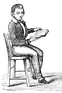
Fig. 30.
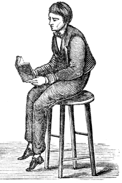
Fig. 29. The position assumed when the seat is of proper height, and the feet supported.
Fig. 30. The position a child naturally assumes when the seat is so high that the feet are not supported.
134. What is said of the benches or chairs in a school-room? What is represented by fig. 29? By fig. 30? What is the effect when the lower limbs are not supported?
135. Compression of the chest should be avoided. In children, and also in adults, the ribs are very flexible, and a small amount of pressure will increase their curvature, particularly at the lower part of the chest, and thus lessen the size of this cavity. The lower ribs are united to the breast-bone, by long, yielding cartilages, and compression may not only contract the chest, but an unseemly and painful ridge may be produced, by the bending of the cartilages, on one or both sides of the sternum.
Fig. 31.

Fig. 32.
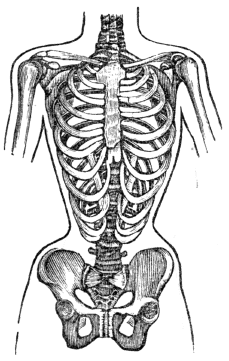
Fig. 31. A natural and well-proportioned chest.
Fig. 32. A chest fashionably deformed.
135. Why should compression of the chest be avoided? What is represented by fig. 31? By fig. 32?
136. Again, the cartilages on one side may be bent outward, while those on the opposite side are bent inward, thus forming a depression parallel with the sternum. In some instances, the anterior extremity of the lower ribs on each side 57 are brought nearly or quite together. In these instances, the movable extremities of the ribs are drawn down toward the haunch-bones, while the space between the ribs is lessened. All this may be effected by tight or “snug” clothing. Therefore the apparel of a child should be loose, and supported over the shoulders, to avoid the before-mentioned evils. The same may be said of the clothing for adults.
137. The erect position in sitting and standing should be assiduously observed. The spinal column, in its natural position, curves from front to back, but not from side to side The admirable arrangement of the bones, alternating with cartilages, permits a great variety of motions and positions; and when the spine is inclined to either side, the elasticity of its cartilages tends to restore it to its natural position. For this reason we may incline the spinal column in any direction for a short time, without danger of permanent curvature, if, afterward, the erect position is assumed.[4]
138. But if a stooping position, or a lateral curved posture, is continued for a long time, the spinal column does not easily recover its proper position, for the compressed edges of the cartilages lose their power of reaction, and finally one side of the cartilage becomes thinned, while the other is thickened; and these wedge-shaped cartilages produce a permanent curvature of the spinal column. In a similar way, the student, seamstress, artisan, and mechanic acquire a stooping position, and become round shouldered, by inclining forward to bring their books or work nearer the eyes.
136. May simply “snug” clothing compress the cartilages? How should the apparel of a child be worn? 137. In what direction does the spinal column, in its natural position, curve? What restores it to its natural position when curved laterally? 138. What is the effect if a lateral curved position of the spinal column is continued for a long time? 139. When one shoulder is elevated for a long time, what is the effect upon the spinal column?
139. Pupils, while writing, drawing, and sometimes while 58 studying, frequently incline the spinal column to one side, in order to accommodate themselves to the desks at which they are seated. Often, these are higher than the elbow as it hangs from the shoulder while at rest. This attitude elevates one shoulder while it depresses the other; consequently, the upper part of the spinal column is inclined toward the elevated shoulder, and the lower part is curved in the opposite direction, giving the form of the letter S to the supporting column of the body.
Fig. 33.
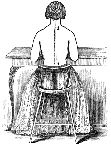
Fig. 33. The table is of proper height, the position is correct, and the spinal column, 1, 1, is straight, while the shoulders are of equal height.
What does fig. 33 represent?
Experiment. Let a pupil be placed at a desk or table with one elbow raised, as is frequently seen while writing, or at study, and observe the condition of the shoulder and spinal column in this position. Place another pupil at a table no higher than the elbow when it hangs by the side while sitting, and observe the appearance of the shoulders and spinal column. By a comparison of the two attitudes, the preceding remarks will be comprehended and appreciated.
Fig. 34.

Fig. 34. The table is too high, and the position is oblique and improper. The right shoulder is seen higher than the left, while the spinal column, 1, 1, exhibits three curves.
140. One shoulder may be elevated, and no injurious results 60 follow, provided care is taken not to keep it in the raised position too long, or if the opposite shoulder is elevated for the same period of time. The right shoulder projects more frequently than the left. This arises from the greater use of the right hand with the shoulder elevated, and not unfrequently the oblique positions assumed in performing the daily vocations of life. With proper care, and by calling into action the left shoulder, this deformity can be prevented.
What experiment is mentioned? What does fig. 34 represent? 140. How can one shoulder be elevated and no injurious results follow?
Fig. 35.

Fig. 35. A representation of a deformed trunk.
141. The loss of symmetry and diminution of height from deformed spines are minor considerations, compared with the distortions that the chest experiences, thereby impairing respiration and inducing diseases of the heart and lungs. The 61 invasion of the functions of these two important organs lessens the vitality of the whole system, and causes general ill health. Again, the curvature of the spinal column is frequently attended by irritation and disease of the spinal cord.
Why does the right shoulder project more frequently than the left? How can this deformity be prevented? 141. What is said of deformed spinal columns?
142. Eminent physicians, both in this country and France state that not more than one female in ten, who has been fashionably educated, is free from deformities of the shoulder or spinal column. Teachers, as well as mothers, should notice the positions of the child in performing the tasks allotted to it, whether studying or pursuing any employment. The feebler the organization of the child, the more frequently should there be a change of position.
143. When a slight projection of the shoulder, with a curvature of the spine, exists, it can be improved by walking with a book, or something heavier, upon the head; to balance which, the spinal column must be nearly erect. Those people that carry burdens upon their heads seldom have crooked spines.
Observation. Persons from the North, in travelling through the Southern States, are surprised to see the heavy burdens that the porters carry on their heads. It is not unusual to see them walking at a rapid pace, with one or two trunks, weighing fifty or eighty pounds each, upon their heads. Occasionally, we meet an itinerant toy-man, with his tray of fragile merchandise upon his head, walking with as much apparent security, as though his toys, or images, were in his hands. This is the easiest method of carrying burdens, because the position of the head and spinal column is erect.
144. If the animal and earthy matter of the bones is not deposited in proper proportions, they are deficient in strength. If the gelatin predominates, the bones are weak, and become 62 distorted. When nutrition is defective in the cylindrical bones, the heads are generally enlarged, and the shafts crooked; if in the spinal column, it may be curved; or in the cranium, it may be enlarged. This disease is familiarly known by the name of rickets. It is most common among these who have poor and insufficient food, live in dark, damp rooms, and breathe a vitiated air. The prevention and remedies for this disease are cleanliness, regular exercise, pure air, and nutritious food.
142. What statement by eminent physicians respecting deformities of the spine? What caution to teachers and mothers? 143. Why should we stand and sit erect? How may slight deformities of the spine be prevented? What is frequently noticed in travelling South? 144. What is the effect upon the bones when the gelatin preponderates?
145. When a bone is broken, some days elapse before the substance that reunites it is thrown out from the blood. In young persons, it may be secreted during the second or third week, and in individuals advanced in life, usually during the third and fourth week. When the bone is uniting, during the second, third, or fourth week, the attention of a surgeon is more needed than during the first week. At this time, the ends of the bone should be placed together with accuracy, which requires the careful application of proper dressing. After the bones have united, it will take some weeks to consolidate the uniting material and render the “callus,” or union, firm. During this time, the limb should be used with care.
Observation. When a bone is fractured, a surgeon is immediately called, and the bone is “set.” While the limb remains swelled and painful, the surgeon is required to attend and keep the dressings (bandages and splints) on. When the swelling has abated, and the pain subsided, frequently the patient intimates to the surgeon that his services can be dispensed with, as the “limb is doing well.” This is the most important period, as the bone is uniting, and, unless the ends are nicely adjusted, the dressing properly applied, the person 63 will find, on recovery, a shortened and crooked limb. The surgeon is then censured, when he is not blamable.
What is one cause of rickets? What are the prevention and remedies for this disease? 145. Does the time vary when the reuniting substance of the bone is secreted from the blood? When is the surgeon’s care most needed? Why?
146. It is seldom that a bone is displaced without injury to the connecting ligaments and membranes. When these connecting bands are lacerated, pain, swelling, and other symptoms indicating inflammation succeed, which should be removed by proper treatment, directed by a surgical adviser.
147. In sprains, but few, if any, of the fibres of the connecting ligaments are lacerated; but they are unduly strained and twisted, which occasions acute pain at the time of the injury. This is followed by inflammation and weakness of the joints. The treatment of these injuries is similar to that of a dislocated bone after its reduction. The most important item in the treatment during the few first days, is rest.
148. In persons of scrofulous constitutions, and those in whom the system is enfeebled by disease, white swellings and other chronic diseases of the joints frequently succeed sprains. Such persons cannot be too assiduous in adopting a proper and early treatment of injured joints.
146. What parts are injured in the displacement of a bone? 147. What causes the acute pain in sprains? What is a good remedy for this kind of injury? 148. What caution to persons of scrofulous constitutions?
149. All the great motions of the body are caused by the movement of some of the bones which form the framework of the system; but these, independently of themselves, have not the power of motion, and only change their position through the action of other organs attached to them, which, by contracting, draw the bones after them. In some of the slight movements, as the winking of the eye, no bones are displaced. These moving, contracting organs are the Mus´cles, (lean meat.)
150. The MUSCLES, by their size and number, constitute the great bulk of the body, upon which they bestow form and symmetry. In the limbs, they are situated around the bones, which they invest and defend, while they form, to some of the joints, their principal protection. In the trunk, they are spread out to enclose cavities, and constitute a defensive wall, capable of yielding to internal pressure, and reassuming its original state.
151. In structure, a muscle is composed of fas-cic´u-li (bundles of fibres) of variable size. These are enclosed in a cellular membranous investment, or sheath. Every bundle composed of a number of small fibres, and each fibre consists of a number of filaments, each of which is enclosed in 65 a delicate sheath. Toward the extremity of the organ the muscular fibre ceases, and the cellular structure becomes aggregated, and so modified as to constitute ten´dons, (cords,) by which the muscle is tied to the surface of the bone. The union is so firm, that, under extreme violence, the bone will sooner break than permit the tendon to separate from its attachment. In some situations, there is an expansion of the tendon, in the manner of a membrane, called Ap-o-neu-ro´sis, or Fas´ci-a.
149. How are all the motions of the body produced? What are these motor organs called? 150–160. Give the anatomy of the muscles. 150. What is said of the muscles? 151. Give their structure.
Observation. The pupil can examine a piece of boiled beef, or the leg of a fowl, and see the structure of the fibres and tendons of a muscle.
Fig. 36.

Fig. 36. 1, A representation of the direction and arrangement of the fibres in a fusiform, or spindle-shaped muscle. 2, In a radiated muscle. 3, In a penniform muscle. 4, In a bipenniform muscle. t, t, The tendons of a muscle.
152. Muscles present various modifications in the arrangement of their fibres, as relates to their tendinous structure. Sometimes they are completely longitudinal, and terminate, at each extremity, in a tendon, the entire muscle being spindle-shaped. In other situations, they are disposed like the rays of 66 a fan, converging to a tendinous point, and constituting a ra´di-ate muscle. Again they are pen´ni-form, converging, like the plumes of a pen, to one side of a tendon, which runs the whole length of the muscle; or they are bi-pen´ni-form, converging to both sides of the tendon.
How are tendons or cords formed? What is the expansion of a tendon called? How can the structure of muscles and their fibres be shown? What does fig. 36 represent? 152. Give the different arrangements of muscular fibres.
153. In the description of a muscle, its attachments are expressed by the terms “origin” and “insertion.” The term origin is generally applied to the more fixed or central attachment, or to the point toward which motion is directed; while insertion is assigned to the more movable point, or to that most distant from the centre. The middle, fleshy portion is called the “belly,” or “swell.” The color of a muscle is red in warm-blooded fish and animals; and each fibre is supplied with arteries, veins, lymphatics, and both sensitive and motor nervous filaments.
154. The FASCIA is of various extent and thickness, distributed through the different regions of the body, for the purpose of investing and protecting the softer and more delicate organs. An instance is seen in the membrane which envelopes a leg of beef, and which is observed on the edges of the slices when it is cut for broiling. When freshly exposed, it is brilliant in appearance, tough, and inelastic. In the limbs it forms distinct sheaths to all the muscles.
155. This tendinous membrane assists the muscles in their action, by keeping up a tonic pressure on their surface. It aids materially in the circulation of the fluids, in opposition to the laws of gravity. In the palm of the hand and sole of the foot, it is a powerful protection to the structures that enter into the formation of these parts. In all parts of the system, the separate muscles are not only invested by fascia, but they 67 are arranged in layers, one over another. The sheath of each muscle is loosely connected with another, by the cellular membrane.
153. What is meant by the origin of a muscle? The insertion? The swell? What is the color of muscles? With what is each muscular fibre supplied? 154. What is said of fascia? What is its appearance when freshly exposed? 155. What effect has it on the muscles? Give other uses of the fascia.
156. The interstices between the different muscles are filled with adipose matter, or fat. This is sometimes called the packing of the system. To the presence of this tissue, youth are indebted for the roundness and beauty of their limbs.
Fig. 37.

Fig. 37. A transverse section of the neck. The separate muscles, as they are arranged in layers, with their investing fasciæ, are beautifully represented. As the system is symmetrical, figures are placed only on one side. In the trunk the muscles are arranged in layers, surrounded by fasciæ, as in the neck. The same is true of the muscles of the upper and lower limbs.
12, The trachea, (windpipe.) 13, The œsophagus, (gullet.) 14, The carotid artery and jugular vein. 28, One of the bones of the spinal column. The figures that are placed in the white spaces represent some of the fasciæ; the other figures indicate muscles.
157. The muscles may be arranged, in conformity with the general division of the body, into four parts: 1st. Those of the Head and Neck. 2d. Those of the Trunk. 3d. Those of the Upper Extremities. 4th. Those of the Lower Extremities.
156. Give a reason why the limbs of youth are rounder than those of the aged. Describe fig. 37.
Fig. 38.

Fig. 38. The superficial layer of muscles on the face and neck. 1, 1, The occipito-frontalis muscle. 2, The orbicularis palpebrarum. 6, The levator labii superioris 7, The levator anguli oris. 8, The zygomaticus minor. 9, The zygomaticus major 10, The masseter. 11, The depressor labii superioris. 13, The orbicularis oris. 15, The depressor anguli oris. 16, The depressor labii inferioris. 18, The sterno-hyoideus. 19, The platysma-myodes. 20, The superior belly of the omo-hyoideus. 21, The sterno-cleido mastoideus. 20, The scalenus medius. 23, The inferior belly of the omo-hyoideus. 24, The trapezius.[5]
Practical Explanation. The muscle 1, 1, elevates the eyebrows. The muscle 2 closes the eye. The muscle 6 elevates the upper lip. The muscles 7, 8, 9, elevate the angle of the mouth. The muscle 10 brings the teeth together when eating. The muscle 11 depresses the upper lip. The muscle 13 closes the mouth. The muscle 15 depresses the angle of the mouth. The muscle 16 draws down the lower lip. The muscles 18, 19, 20, 23, depress the lower jaw and larynx and elevate the sternum. The muscle 21, when both sides contract, draws the head forward, or elevates the sternum; when only one contracts, the face is turned one side toward the opposite shoulder. The muscles 18, 19, 20, 21, 22, 23, 24, aid in respiration.
Observation. When we are sick, and cannot take food, the body is sustained by absorption of the fat. The removal of it into the blood causes the sunken cheek, hollow eye, and prominent appearance of the bones after a severe illness.
158. The number of muscles in the human body is more than five hundred; in general, they form about the skeleton two layers, and are distinguished into superficial and deep-seated muscles. Some of the muscles are voluntary in their motions, or act under the government of the will, as those which move the fingers, limbs, and trunk; while others are involuntary, or act under the impression of their proper stimulants, without the control of the individual, as the heart.
Observations. 1st. The abdominal muscles are expiratory, and the chief agents for expelling the residuum from the rectum, the bile from the gall bladder, the contents of the stomach and bowels when vomiting, and the mucus and irritating substances from the bronchial tubes, trachea, and nasal passages by coughing and sneezing. To produce these effects they all act together. Their violent and continued action sometimes produces hernia, and, when spasmodic, may occasion ruptures of the different organs.
2d. The contraction and relaxation of the abdominal muscles and diaphragm stimulate the stomach, liver, and intestines to a healthy action, and are subservient to the digestive powers. If the contractility of their muscular fibres is destroyed or impaired, the tone of the digestive apparatus will be diminished, as in indigestion and costiveness. This is frequently attended by a displacement of those organs, as they generally gravitate towards the lower portion of the abdominal cavity, when the sustaining muscles lose their tone and become relaxed.
What causes the hollow eye and sunken cheek after a severe sickness? 158. How many muscles in the human system? Into how many layers are they arranged? What is a voluntary muscle? Give examples. What is an involuntary muscle? Mention examples. Give observation 1st, respecting the use of the abdominal muscles? Observation 2d.
Fig. 39.

Fig. 39. A front view of the muscles of the trunk. On the left side the superficial layer is seen; on the right, the deep layer. 1, The pectoralis major muscle. 2, The deltoid muscle. 6, The pectoralis minor muscle. 9, The coracoid process of the scapula. 11, The external intercostal muscle. 12, The external oblique muscle 13, Its aponeurosis. 16, The rectus muscle of the right side. 18, The internal oblique muscle.
Practical Explanation. The muscle 1 draws the arm by the side, and across the chest, and likewise draws the scapula forward. The muscle 2 elevates the arm. The muscle 6 elevates the ribs when the scapula is fixed, or draws the scapula forward and downward when the ribs are fixed. The muscles 12, 16, 18, bend the body forward or elevate the hips when the muscles of both sides act. They likewise depress the rib in expiration. When the muscles on only one side act, the body is twisted to the same side.
Explain fig. 39. Give the function of some of the most prominent muscles, from this figure.
Fig. 40.

Fig. 40. A lateral view of the muscles of the trunk. 3, The upper part of the external oblique muscle. 4, Two of the external intercostal muscles. 5, Two of the internal intercostals. 6, The transversalis muscle. 7, Its posterior aponeurosis. 8, Its anterior aponeurosis. 11, The right rectus muscle. 13, The crest of the ilium, or haunch-bone.
Practical Explanation. The rectus muscle, 11, bends the thorax upon the abdomen when the lower extremity of the muscle is the fixed point; but when the upper extremity is the fixed point, the effect is to bring forward and raise the pelvis and lower extremities. They likewise depress the ribs in respiration. The transverse muscle, 6, 7, 8, lessens the cavity of the abdomen, and presses the intestines; stomach, and liver upward, against the diaphragm, in expiration.
3d. The region of the back, in consequence of its extent, is common to the neck, the upper extremities, and the abdomen. The muscles of which it is composed are numerous, and are arranged in six layers.
What is represented by fig. 40? Give the function of some of the muscles represented by this figure.
Fig. 41.

Fig. 41 The first, second, and part of the third layer of muscles of the back. The first layer is shown on the right, and the second on the left side. 1, The trapezius muscle. 2, The spinous processes of the vertebræ. 3, The acromion process and spine of the scapula. 4, The latissimus dorsi muscle. 5, The deltoid muscle. 7, The external oblique muscle. 8, The gluteus medius muscle. 9, The gluteus maximus muscle, 11, 12, The rhomboideus major and minor muscles. 15, The vertebral aponeurosis. 16, The serratus posticus inferior muscle. 22, The serratus magnus muscle. 23, The internal oblique muscle.
Practical Explanation. The muscles 1, 11, 12, draw the scapula back toward the spine. The muscles 11, 12, draw the scapula upward toward the head, and slightly backward. The muscle 4 draws the arm by the side, and backward, The muscle 5 elevates the arm. The muscles 8, 9, extend the thigh on the body. The muscle 1 draws the head back and elevates the chin. The muscle 16 depresses the ribs in expiration. The muscle 22 elevates the ribs in inspiration.
159. The diaphragm, or midriff, is the muscular division between the thorax and the abdomen. It is penetrated by the œsophagus on its way to the stomach, by the aorta conveying blood toward the lower extremity, and by the ascending vena cava, or vein, on its way to the heart.
Fig. 42.

Fig. 42. A representation of the under, or abdominal side of the diaphragm. 1, 2, 3, 4, The portion which is attached to the margin of the ribs. 8, 10, The two fleshy pillars of the diaphragm, which are attached to the third and fourth lumbar vertebræ. 9, The spinal column. 11, The opening for the passage of the aorta. 12, The opening for the œsophagus. 13, The opening for the ascending vena cava, or vein.
Observation. The diaphragm may be compared to an inverted basin, its bottom being turned upward into the thorax, while its edge corresponds with the outline of the edges of the lower ribs and sternum. Its concavity is directed toward the abdomen, and thus, this cavity is very much enlarged at the expense of that of the chest, which is diminished to an equal extent.
159. Describe the diaphragm. What vessels penetrate this muscular septum?
160. “The motions of the fingers do not merely result from the action of the large muscles which lie on the fore-arm, these being concerned more especially in the stronger actions of the hands. The finer and more delicate movements of the fingers are performed by small muscles situated in the palm and between the bones of the hand, and by which the fingers are expanded and moved in all directions with wonderful rapidity.”
Fig. 43. A front view of the superficial layer of muscles of the fore-arm. 5, The flexor carpi radialis muscle. 6, The palmaris longus muscle. 7, One of the fasciculi of the flexor sublimis digitorum muscle, (the rest of the muscle is seen beneath the tendons of the pintails longus.) 8, The flexor carpi ulnaris muscle. 9, The palmar fascia. 11, The abductor pollicis muscle. 12, One portion of the flexor orevis pollicis muscle. 13, The supinator longus muscle. 14, The extensor ossis metacarpi, and extensor primi internodii pollicis muscles, curving around the lower border of the fore-arm. 15, The anterior portion of the annular ligament, which binds the tendons in their places.
Practical Explanation. The muscles 5, 6, 8, bend the wrist on the bones of the fore-arm. The muscle 7 bends the second range of finger-bones on the first. The muscle 11 draws the thumb from the fingers. The muscle 12 flexes the thumb. The muscle 13 turns the palm of the hand upward. The muscles 8, 13, 14, move the hand laterally.
Fig. 44. A back view of the superficial layer of muscles of the fore-arm. 5, The extensor carpi radialis longior muscle. 6, The extensor carpi radialis brevior muscle. 7, The tendons of insertion of these two muscles. 8, The extensor communis digitorum muscle. 9, The extensor minimi dlgiti muscle. 10, The extensor carpi ulnaris muscle. 13, The extensor ossis metacarpi and extensor primi internodii muscles, lying together. 14, The extensor secundi internodii muscle; its tendon is seen crossing the two tendons of the extensor carpi radialis longior and brevior muscles. 15, The posterior annular ligament. The tendons of the common extensor muscle of the fingers are seen on the back of the hand, and their mode of distribution on the back of the fingers.
Practical Explanation. The muscles 5, 6, 10, extend the wrist on the fore-arm. The muscle 8 extends the fingers. The muscle 9 extends the little finger. The muscles 13 extend the metacarpal bone of the thumb, and its first phalanx. The muscle 14 extends the last bone of the thumb. The muscles 10, 13, 14, move the hand laterally.
160. Where are the muscles situated that effect the larger movements of the hand? That perform the delicate movements of the fingers? Give the use of some of the muscles represented by fig. 43. Those represented by fig. 44.
161. The muscles exercise great influence upon the system. It is by their contraction that we are enabled to pursue different employments. By their action the farmer cultivates his fields, the mechanic wields his tools, the sportsman pursues his game, the orator gives utterance to his thoughts, the lady sweeps the keys of the piano, and the young are whirled in the mazy dance. As the muscles bear so intimate a relation to the pleasures and employments of man, a knowledge of the laws by which their action is governed, and the conditions upon which their health depends, should be possessed by all.
162. The peculiar characteristic of muscular fibres is contractility, or the power of shortening their substance on the application of stimuli, and again relaxing when the stimulus is withdrawn. This is illustrated in the most common movements of life. Call into action the muscles that elevate the arm, by the influence of the will, or mind, (the common stimulus of the muscles,) and the hand and arm are raised; withdraw this influence by a simple effort of the will, and the muscles, before rigid and tense, become relaxed and yielding.
163. The contractile effect of the muscles, in producing the varied movements of the system, may be seen in the bending of the elbow. The tendon of one extremity of the muscle is attached to the shoulder-bone, which acts as a fixed point; the tendon of the other extremity is attached to one of the bones 77 of the fore-arm. When the swell of the muscle contracts, or shortens, its two extremities approach nearer each other, and by the approximation of the terminal extremities of the muscle, the joint at the elbow bends. On this principle, all the joints of the system are moved. This is illustrated by fig. 45.
161–172. Give the physiology of the muscles. 161. What are some of the influences exerted by the muscles on the system? 162. What is peculiar to muscular fibres? How is this illustrated? 163. Explain how the movements of the system are effected by the contraction of the muscles.
Fig. 45.
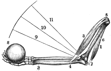
Fig. 45. A representation of the manner in which all of the joints of the body are moved. 1, The bone of the arm above the elbow. 2, One of the bones below the elbow. 3, The muscle that bends the elbow. This muscle is united, by a tendon, to the bone below the elbow, (4,) at the other extremity, to the bone above the elbow, (5,) 6, The muscle that extends the elbow. 7, Its attachment to the point of the elbow. 8, A weight in the hand to be raised. The central part of the muscle 3 contracts, and its two ends are brought nearer together. The bones below the elbow are brought to the lines shown by 9, 10, 11. The weight is raised in the direction of the curved line. When the muscle 6 contracts, the muscle 3 relaxes and the fore-arm is extended.
Experiments. 1st. Clasp the arm midway between the shoulder and elbow, with the thumb and fingers of the opposite hand. When the arm is bent, the inside muscle will become hard and prominent, and its tendon at the elbow rigid, while the muscle on the opposite side will become flaccid. Extend the arm at the elbow, and the outside muscle will swell and become firm, while the inside muscle and its tendon at the elbow will be relaxed.
Explain fig. 45. Give experiment 1st.
2d. Clasp the fore-arm about three inches below the elbow, then open and shut the fingers rapidly, and the swelling and relaxation of the muscles on the opposite sides of the arms, alternating with each other, will be felt, corresponding with the movement of the fingers. While the fingers are bending, the inside muscles swell, and the outside ones become flaccid; and, while the fingers are extending, the inside muscles relax, and the outside ones swell. The alternate swelling and relaxation of antagonist muscles may be felt in the different movements of the limbs.
164. Each fibre of the several muscles receives from the brain, through the nervous filament appropriated to it, a certain influence, called nervous fluid, or stimulus. It is this that induces contraction, while the suspension of this stimulus causes relaxation of the fibres. By this arrangement, the action of the muscular system, both as regards duration and power, is, to a limited extent, under the control of the mind. The more perfect the control, the better the education of the muscular system; as is seen in the graceful, effective, and well-educated movements of musicians, dancers, skaters, &c.
165. The length of time which a muscle may remain contracted, varies. The duration of the contraction of the voluntary muscles, in some measure, is in an inverse ratio to its force. If a muscle has contracted with violence, as when great effort is made to raise a heavy weight, relaxation will follow sooner than when the contraction has been less powerful, as in raising light bodies.
166. The velocity of the muscular contraction depends on the will. Many of the voluntary muscles in man contract with great rapidity, so that he is enabled to utter distinctly 79 fifteen hundred letters in a minute; the pronunciation of each letter requiring both relaxation and contraction of the same muscle, thus making three thousand actions in one minute. But the contraction of the muscles of some of the inferior animals surpasses in rapidity those of man. The race-horse, it is said, has run a mile in a minute; and many birds of prey will probably pass not less than a thousand miles daily.
Give experiment 2d. 164. With what is each muscular fibre supplied? What effect has this stimulus on the muscles? 165. how long does a voluntary muscle remain contracted? 166. On what is the velocity of muscular contraction dependent? How many letters may be pronounced in a minute?
167. The functions of the involuntary muscles are necessary the digestion of food, the absorption and circulation of the nutritive fluids. They could not be trusted with safety to the control of the will, lest the passions or the indiscretions of the person should continually avert those operations so necessary to health, and even to life. The Divine Builder of this complicated machine has wisely ordered that the muscles upon which these motions depend, shall act under the impression of their proper stimulants, without the control of the individual.
168. Again, there are certain operations which could not be safely intrusted to the absolute government of the voluntary muscles, or entirely removed from their control. Thus life can be supported only a few minutes without breathing; but it would be impossible to perform the daily vocations of life if we were compelled to breathe at all times, or at perfectly regular intervals.
169. It has been observed that, among men of the same size, a wide difference exists in their strength and activity—qualities which depend upon the size and number of the nerves, the size and activity of the brain, and the education, or training of the muscles. Men having large nerves leading 80 to the muscles, with the brain active, and muscles well trained will perform feats of strength and agility, that other men, of the same size, cannot effect. Rope-dancers, harlequins, and other performers of feats, are persons thus constituted.
How many contractions and relaxations of the same muscle? What is said of the rapidity of muscular contractions in other animals? 167. When are the involuntary muscles called into action? Why would it not have been safe to trust these important operations to the exclusive control of the will? 168. Give an instance where some of the muscles act under the government of the will, conjoined with those that are involuntary. 169. On what does the difference in muscular activity and strength depend?
170. Persons with small muscles, and largely developed nervous systems, will sometimes exhibit very great muscular power for a time; but it will not be of long continuance, unless the brain is functionally diseased, as in hysteria, delirium of fever, insanity, &c. Men of large muscles and small nerves can never perform feats of great strength; but they have the power of endurance, and are better capacitated for continued labor. Thus we cannot judge of the ability of persons to make exertions and continue them, by their stature alone. Strength, and the power of endurance, are the result of a combination of well-developed muscles, large nerves, and a full-sized, healthy, and active brain.
Observation. The muscles of fishes are large, and the nerves distributed to them, comparatively small. The muscles of birds are small, but their fibres are very compact. The nerves appropriated to the muscles that are called into action in flying, are large as well as numerous.
171. The contractile portion of a muscle is, in general, at a distance from the part to be moved. Thus the principal muscles that move the fingers are situated upon the forearm; and when the limb is nearly or quite extended, the angle formed by the part to be moved and the contractile muscles is small. Again, the attachment of the muscles to the part to be moved is near the joint that forms the fulcrum, (fig. 45.) By these arrangements there is a loss of power; but we are compensated for this disadvantage by increased celerity of movement, beauty of form, and adaptation of the limbs to the varied pursuits of man.
170. What is said of those persons who have small muscles and largely developed nervous systems? Of those who have large muscles and small nerves? Upon what do strength and the power of endurance depend? 171. Why is there a loss of power in the action of the muscles?
Illustration. The muscle that bends the elbow acts at disadvantage, and this is greatest when the arm is nearly or quite extended, as the angle of action is then least. This disadvantage is further increased by the attachment of the motive muscles near the joint.
172. The number of muscles which are called into action in the movements of the different joints, varies. The hinge-joints, as the elbow, have two sets of muscles—one to bend the joint, the other to extend it. The ball and socket joints, as the shoulder, are not limited to mere flexion and extension. No joint in the system has the range of movement that is possessed by that of the shoulder. By the action of the muscles attached to the arm, it is not only carried upward and forward, but forward and backward. Hence the arm may be moved at any angle, by a combined action of its muscles.
Observation. “Could we behold properly the muscular fibres in operation, nothing, as a mere mechanical exhibition, can be conceived more superb than the intricate and combined actions that must take place during our most common movements. Look at a person running or leaping, or watch the motions of the eye. How rapid, how delicate, how complicated, and yet how accurate, are the motions required! Think of the endurance of such a muscle as the heart, that can contract, with a force equal to sixty pounds, seventy-five times every minute, for eighty years together, without being weary.”
Note. It would be a profitable exercise for pupils to press their fingers upon prominent muscles, and, at the same time, vigorously contract them, not only to learn their situations, but their use; as the one that bends the arm, 14, fig. 46.
How is this illustrated? 172. Do all joints require the same number of muscles, when called into action? How many are called into action in the movement of the elbow? What is their office? What is said of the movement of the ball and socket joint?
Fig 46.

Fig. 46. An anterior view of the muscles of the body. 1. The frontal swell of the occipito-frontalis. 2, The orbicularis palpebrarum. 3, The levator labli superioris. 4, The zygomaticus major. 5, The zygomaticus minor. 6, The masseter. 7, The orbicularis oris. 8, The depressor labli inferioris. 9. The platysma myodes. 10, The deltoid. 11, The pectoralis major. 12, The latissimus dorsi. 14, The biceps flexor cubiti. 15, The triceps extensor cubiti. 16, The supinator radii longus. 18, The flexor carpi radialis longior. 19, The flexor communis digitorum. 20, The annular ligament. 21, The palmar fascia. 22, The obliquus externus abdominis. 26, The psoas magnus. 27, The adductor longus. 28, The sartorius. 29, The rectus femoris. 30, The vastus externus. 31, The vastus internus. 32, The tendon patellæ. 33, The gastrocnemius. 34, The tibialis anticus. 36, The tendons of the extensor digitorum communis.
Fig 47.

Fig. 47. A posterior view of the muscles of the body. 3, The complexus. 4, The splenius. 5, The masseter. 6, The sterno-cleido mastoideus. 7, The trapezius. 8, The deltoid. 10, The triceps extensor. 13, The tendinous portion of the triceps. 14, The anterior edge of the triceps. 15, The supinator radii longus. 17, The extensor communis digitorum. 18, The extensor ossis metacarpi pollicis. 19, The tendons of the extensor communis digitorum. 20, The olecranon process of the ulna and insertion of the triceps. 21, The extensor carpi ulnaris. 22, The extensor communis digitorum. 24, The latissimus dorsi. 25, Its tendinous origin. 26, The obliquus externus. 27, The gluteus medius. 28, The gluteus magnus. 29, The biceps flexor cruris. 30, The semi-tendinosus. 31, 32, The gastrocnemius. 33, The tendo Achillis.
Practical Explanation. The muscle 1, fig. 46, by its contraction, raises the eyebrows. The muscle 2, fig. 46, closes the eyelids. The muscle 3, fig. 46, elevates the upper lip. The muscles 4, 5, fig. 46, elevate the angles of the mouth. The muscles 6, fig. 46, and 5, fig. 47, bring the teeth together. The muscle 7, fig. 46, closes the mouth. The muscle 8, fig. 46, depresses the lower lip. The muscles 9, fig. 46, and 6, fig. 47, bend the neck forward. The muscles 3, 4, fig. 47, elevate the head and chin. The muscle 22, fig. 46, bends the body forward, and draws the ribs downward. The muscle 11, fig. 46, brings the shoulder forward. The muscle 7, fig. 47, draws the shoulder back. The muscles 10, fig. 46, and 8, fig. 47, elevate the arm. The muscles 11, fig. 46, and 24, fig. 47, bring the arm to the side. The muscle 14, fig. 46, bends the arm at the elbow. The muscle 10, fig. 47, extends the arm at the elbow. The muscles 16, 18, fig. 46, bend the wrist and fingers. The muscle 19 bends the fingers. The muscles 18, 21, 23, fig. 47, extend the wrist. The muscle 23, fig. 47, extends the fingers. The muscles 26, 27, 28, fig. 46, bend the lower limbs on the body, at the hip. The muscle 28, fig. 46, draws one leg over the other, (the position of a tailor when sewing.) The muscles 27, 28, fig. 47, extend the lower limbs on the body, at the hip. The muscles 29, 30, 31, fig. 46, extend the leg at the knee. The muscles 29, 30, fig. 47, bend the leg at the knee. The muscles 34, 36, fig. 46, bend the foot at the ankle, and extend the toes. The muscles 31, 32, 33, fig. 47, extend the foot at the ankle.
Note. Let the anatomy and physiology of the muscular system be reviewed, in form of topics, from figs 46, 47, or from the anatomical outline plates No. 3 and 4.
173. The muscles should be used, in order that the size and strength of these organs may be adequate to the demand made upon them. It is a law of the system that the action and power of an organ are commensurate, to a certain extent, with the demand made upon it; and it is a law of the muscular system that, whenever a muscle is called into frequent use, its fibres increase in thickness within certain limits, and become capable of acting with greater force; while, on the contrary, the muscle that is little used decreases in size and power.
Illustrations. 1st. The blacksmith uses and rests the muscles of his arm when striking upon the anvil. They not only increase in size, but become very firm and hard.
2d. The student uses the muscles of the arm but little, in holding his books and pen; they not only become small, but soft.
3d. Let the student leave his books, and wield an iron sledge, and the muscles of his arm will increase in size and firmness. On the other hand, let the blacksmith assume the student’s vocation, and the muscles of his arm will become soft and less firm.
174. When the muscles are called into action, the flow of blood in the arteries and veins is increased. The increased flow of blood in the arteries and veins, causes a more rapid deposition of the particles of matter of which the muscles are 86 composed. If the exercise is adequate to the power of the system, the deposit of new material will exceed in quantity the particles of matter removed, and both the size and energy of the muscles are increased. But there is a limit to the muscles becoming strong by labor. Sooner or later, man will attain his growth or power; yet by judicious exercise, care, and discreet management, the greatest power of the muscles may be preserved until advanced age.
173–211. Give the hygiene of the muscles. 173. What is necessary that muscles may attain size and strength? Give a law of the muscular system. Show this by practical illustrations. 174. Why do muscles increase in size when exercised?
175. The muscles are lessened in size and diminished in power when the exercise is continued so as to produce a feeling of exhaustion. The loss of material, in this instance, will exceed the deposition of the atoms of matter. This is seen in the attenuated frames of over-tasked domestic animals, as the horse. The same truth is illustrated by the laborious agriculturist, who, in consequence of too severe toil while gathering the products of the field, frequently diminishes his weight several pounds in a few weeks. Exercise, either for pleasure or profit, may fatigue, yet it should never be protracted to languor or exhaustion, if the individual desires “a green old age.”
176. The same amount of exercise will not conduce to the health of all individuals. If riding or walking one mile causes slight fatigue, this may be beneficial; while, by travelling two miles, the exhaustion may be highly injurious. Exercise and labor should be adapted to the strength of particular individuals. How little soever the strength, that must be the measure of exertion. Any other rule would be fatal to the hopes of invigorating the system, either by exercise or labor.
Is there a limit to the muscles becoming powerful by action? How may the strength of muscles be kept until advanced age? 175. What is the effect when exercise is continued until there is a feeling of exhaustion? Give a practical illustration. What rule is mentioned in regard to exercise? 176. Can all persons take the same amount of exercise? What rule is given as to the amount of exercise?
177. Relaxation must follow contraction, or, in other words, rest must follow exercise. The necessity of relaxation, when a muscle has been called into action, is seen in the example of a boy extending his arm with a book in his hand, as a penalty. The boy can keep the arm extended but a short time, make what effort he may. It is also seen in the restlessness and feverish excitement that are evinced by persons gazing on troops during days of review. The same is noted in shopping. Such employments call into action the muscles that support the spinal column in an erect position, and the languor or uneasiness is muscular pain. The long-continued tension of a muscle enfeebles its action, and eventually destroys its contractility.
178. In school, the small children, after sitting a short time, become restless. If their position be changed, their imperfectly developed muscles will acquire tone, and will again support the spinal column erect without pain. The necessity for frequent recesses in school, is founded on the organic law of muscular action alternating with rest. The younger and feebler pupils are, the greater the necessity for frequent recesses. We would not have the teacher think that one half of the time should be spent in recesses; or the mother, that her daughter is going to school to play. But we do maintain that recesses should be given, and that they should be short and frequent, especially for small and feeble scholars.
179. Exhaustion is the inevitable result of continued muscular contraction. For example, let a lady ply the needle quickly for some hours, and the muscles of the back and right arm will become exhausted, which will be indicated by a sense of weariness in these parts. A change of employment 88 and position calls into action a different set of muscles, and the exhausted organs are relieved.
177. What is said of the contraction and relaxation of the muscles? Give examples of the necessity of relaxing the muscles. 178. Why should not small children be confined in one position for a long time? What evils result from this practice? What class of pupils should have recesses most frequently? 179. What effect has continued muscular contraction?
180. Much more labor will be accomplished by taking time to relax the exhausted muscles, or by so changing the employment as to bring into action a new set of muscles; the woodman thus relieves himself, by sawing and splitting alternately. This principle applies to the labor of the horse and ox; and it is also applicable to all kinds of employment. With the invalid convalescing from fever, relapses result from inattention to these laws. When a patient is recovering from sickness, his physician should take care that his exercise be proper, neither too much, too little, nor too long continued.
181. The muscles of growing youths will not endure so much exercise or labor as those of mature men. In youth a portion of the vital, or nervous energy of the system, is expended upon the growth of the organs of the body, while in the individual who has attained his growth, this expenditure is not demanded; consequently severe labor or exercise should not be imposed on growing children.
Observation. In the campaigns of Napoleon Bonaparte, his army was frequently recruited by mere boys. He complained to the French government, because he was not supplied with men of mature years, as the youths could not endure the exertion of his forced marches.
182. The muscles should be gradually called into action. These organs in action require more blood and nervous fluid than when at rest. As the circulation of these fluids can only be increased in a gradual manner, it follows, that, when the muscular system has been in a state of rest, it should not suddenly be called into vigorous action. On arising from a bed, lounge, or chair, the first movements of the limbs should be slow, and then gradually increased.
Observation. if a man has a certain amount of work to 89 perform in nine hours, and his muscles have been in a state of rest, he will do it with less fatigue by performing half the amount of the labor in five hours, and the remainder in four hours. The same principle should be regarded in driving horses and other beasts of burden.
180. How can the greatest amount of labor be secured with the least exhaustion to the muscles? 181. Why should not severe labor be imposed on growing children? 182. How should the muscles be called into action?
183. The muscles should be rested gradually, when they have been vigorously used. If a person has been making great muscular exertion in cutting wood, or any other employment, instead of sitting down to rest, he should continue muscular action, for a short time, by some moderate labor or amusement.
184. If the system has been heated by muscular action, and the skin is covered with perspiration, avoid sitting down “to cool” in a current of air; rather, put on more clothing, and continue to exercise moderately. In instances when severe action of the muscles has been endured, bathing and rubbing the skin of the limbs and joints that have been used, are of much importance. The laboring agriculturist and industrious mechanic, by reducing to practice this suggestion, would thus prevent soreness of the muscles, and stiffness of the joints.
185. The muscles should be abundantly supplied with pure blood. This state of the circulating fluid requires a healthy condition of the digestive apparatus, and that the skin should be kept warm by proper clothing, clean by bathing, and be acted upon by pure air and good light; the movements of the ribs and diaphragm should be unrestricted, and the lungs should have ample volume and be supplied with pure air. In all instances, muscular power is greatest when the preceding conditions exist, as the muscles are then stimulated by pure blood; consequently, it is of practical importance to 90 the mechanic, the farmer, the man of leisure, and not less so to the ladies, to observe these conditions, whatever vocation of life they pursue.
183. How should the muscles be rested when they have been vigorously used? 184. What precaution is given when the skin is covered with perspiration? How may soreness of the muscles, consequent upon severe action, be prevented? 185. Should the muscles be supplied with pure blood? When is muscular power the greatest?
186. The muscles should be used in pure air. The purer the air we breathe, the more stimulating the blood supplied to the muscles, and the longer they can be used in labor, walking, or sitting, without fatigue and injury; hence the benefit derived in thoroughly ventilating all inhabited rooms. For the same reason, if the air of the sick-room is pure, the patient will sit up longer than when the air is impure.
Observation. It is a common remark that sick persons will sit up longer when riding in a carriage, than in an easy chair in the room where they have lain sick. In the one instance, they breathe pure air; in the other, usually, a confined, impure air.
187. The muscles should be exercised in the light. Light, particularly that of the sun, exercises more or less influence on man and the inferior animals as well as on plants. Both require the stimulus of this agent. Shops occupied by mechanics, kitchens, and sitting-rooms, should be well lighted, and situated on the sunny side of the house. Cellar kitchens and underground shops should be avoided. For similar reasons, students should take their exercise during the day, rather than in the evening, and, as much as possible, laborers should avoid night toil.
Illustrations. Plants that grow in the shade, as under trees, or in a dark cellar, are of lighter color and feebler than those that are exposed to the light of the sun. Persons that dwell in dark rooms are paler and less vigorous than those who inhabit apartments well lighted, and exposed to the rays of the sun.
186. Why should the muscles be used in pure air? Give a common observation. 187. What effect has light on the muscular system? What should the laborer avoid? Why should not students take their daily exercise in the evening? How is the influence of solar light illustrated?
188. Exercise should be regular and frequent. The system needs this means of invigoration as regularly as it does new supplies of food. It is no more correct that we devote several days to a proper action of the muscles, and then spend one day inactively, than it is to take a proper amount of food for several days, and then withdraw this supply for a day. The industrious mechanic and the studious minister suffer as surely from undue confinement as the improvident and indolent. The evil consequences of neglect of exercise are gradual, and steal slowly upon an individual. But sooner or later they are manifested in muscular weakness, dyspepsia, and nervous irritability.
Observation. The custom among farmers of enduring severe and undue toil for several successive days, and then spending one or two days in idleness to rest, is injudicious. It would be far better to do less in a day, and continue the labor through the period devoted to idleness, and then no rest will be demanded.
189. Every part of the muscular system should have its appropriate share of exercise. Some employments call into exercise the muscles of the upper limbs, as shoe-making; others, the muscles of the lower limbs; while some, the muscles of both upper and lower limbs, with those of the trunk, as farming. In some kinds of exercise, the lower limbs are mainly used, as in walking; in others, the upper limbs; and again, the muscles of the trunk, together with those of the upper and lower limbs, as in archery, quoits, playing ball. Those trades and kinds of exercise are most salutary, in which all the muscles have their due proportion of action, 92 as this tends to develop and strengthen them equally. Thus labor upon the farm and domestic employment are superior as vocations, and archery, quoits, and dancing, if the air is pure, among the pastimes. For sedentary persons, that kind of exercise is best which calls into action the greatest number of muscles.
188. How should exercise be taken? What is said respecting irregular exercise? Are the consequences of neglected exercise immediately apparent? What practical observation is given? 189. Should every muscle have its due amount of exercise? Mention some employments that only call into action the muscles of the upper limbs. Those of the lower limbs, those of the trunk and limbs. Mention, in the different pastimes, what muscles are called into action.
190. The proper time for labor or exercise should be observed. This is modified by many circumstances. As a general rule, the morning, when the air is pure and the ground dry, is better than the evening; for then, the powers of the body are greatest. Severe exercise and labor should be avoided immediately before or after eating a full meal, for the energies of the system are then required to perform the digestive function. For similar reasons, it is not an appropriate time for energetic muscular action immediately before or after severe mental toil, as the powers of the system are then concentrated upon the brain.[6]
What kinds of exercise are best? 190. What rule is given respecting the time for exercise? 191. Why do the muscles require sleep? What is the effect of an inversion of the law of rest?
191. The muscles require sleep to restore their expended energies. Among the arrangements of creative wisdom, no one harmonizes with the wants of the system more than the alternation of day and night. The natural inclination of man to sleep, is in the stilly hour of night, when all nature reposes, and to be in action during the light of day. An inversion of this law of rest causes greater exhaustion of the system than 93 the same amount of exertion during daylight. This is illustrated by the wearied and exhausted condition of watchers, night-police, and other individuals who spend a part of the night in some active business of life.
192. The muscles should not be compressed. Compression prevents the blood from passing to the muscles with freedom; consequently, they are not supplied with material to renovate and promote their growth. Again, pressure stimulates the lymphatics to action; and by the increased activity of these vessels the muscles are attenuated. In the case of a man with a fractured limb, the muscles are not only enfeebled by inaction, but diminished in size by compression from the dressing. Limbs enfeebled in this way will not recover their size, tone, and strength, until the bandages are removed, and a proper amount of exercise taken.
193. The pressure of tight dresses, under the name of a “snug fit,” enfeebles the muscles of the back, and is a common cause of projecting shoulders and curvature of the spinal column. Thus every appendage to the dress of ladies which prevents free motion of the muscles of the chest and spinal column, weakens the muscles thus restrained, and not only prevents the proper expansion of the lungs, but, by weakening the muscles which sustain the spine, induces curvature and disease. Whalebone, wood, steel, and every other unyielding substance, should be banished from the toilet, as enemies of the human race.
194. The mind exerts a great influence upon the tone and contractile energy of the muscular system. A person acting under a healthy mental stimulus will make exertion with less fatigue than he would without this incentive. For this reason, a sportsman will pursue his game miles without fatigue, while 94 his attendant, not having any mental stimulus, will become weary. Again, if the sportsman spends some hours in pursuit of his favorite game without success, a feeling of languor creeps over him; but while he is thus fatigued and dispirited, let him catch a glimpse of the game,—his wearied feelings are immediately dissipated, and he presses on with renewed energy and recruited strength.
192. Why should not the muscles be compressed? 193. What is the effect of tight clothing upon the muscles? 194. What is said of the influence of the mind upon muscular activity? Give an illustration of mental stimulus coöperating with muscular activity in the case of a sportsman.
195. This principle was well illustrated in the retreat from Russia of the defeated and dispirited French army. When no enemy was near, they had hardly strength sufficient to carry their arms; but no sooner did they hear the report of the Russian guns, than new life seemed to pervade them, and they wielded their weapons powerfully until the foe was repulsed, then there was a relapse to weakness, and prostration followed. It is thus with the invalid when riding for his health;—relate an anecdote, or excite this mental stimulus by agreeable conversation, and much benefit will accrue from the ride to the debilitated person. So it is in the daily vocations of life; if the mind have some incentive, the tiresomeness of labor will be greatly diminished. Let an air of cheerfulness ever pervade our every employment, and, like music, “it sweetens toil.”
196. Facts illustrative of the inutility of calling the muscles into action, without the coöperation of the mind, are seen in the spiritless aspect of many of our boarding school processions, when a walk is taken merely for exercise, without having in view any attainable object. But present to the mind a botanical or geological excursion, and the saunter will be exchanged for the elastic step, the inanimate appearance for the bright eye and glowing cheek. The difference 95 is, simply, that, in the former case, the muscles are obliged to work without that full nervous impulse so essential to their energetic action; and that, in the latter, the nervous influence is in full and harmonious operation.
195. Give an illustration of mental stimulus coöperating with muscular activity in the case of the dispirited French army in their retreat from Russia. How can a union of mental impulse and muscular action be beneficial to an invalid? Does this same principle apply to those who labor? 196. Give an instance of the different effects produced by the absence and presence of the mental stimulus.
197. It must not, however, be supposed that a walk simply for the sake of exercise can never be beneficial. Every one, unless prevented by disease, should consider it a duty to take exercise every day in the open air; if possible, let it be had in combination with harmonious mental exhilaration; if not, let a walk, in an erect position, be made so brisk as to produce rapid respiration and circulation of the blood, and in a dress that shall not interfere with free motions of the arms and free expansion of the chest.
Observation. The advantages of combining harmonious mental excitement, with muscular activity, is thus given by Dr. Armstrong:—
|
“In whate’er you sweat, |
197. May not a walk, simply as an exercise, be beneficial? What is preferred?
198. The erect attitude lessens the exhaustion of the muscles. A person whose position is erect will stand longer, walk further, and perform more labor, than an individual whose position is stooping, but equal in all other respects. The manly port in an erect attitude, depends chiefly upon the action of the muscles of the back; and it follows that the fewer the muscles in a state of tension, the less the draught upon the nervous system, and the less its exhaustion. Another advantage which attends the erect position is, the trunk and head are balanced upon the bones and cartilages of the spinal column. If the body slightly incline forward, the muscles attached to the posterior side of the spine, by a gentle contraction, will bring it to the perpendicular, and even incline it backward. This is immediately removed by a slight contraction of the muscles upon the anterior side of the spinal column.
199. In the erect position, there is a constant slight oscillation of the body backward and forward, like the movement of a pendulum; while, in the stooping posture, the muscles on the posterior side of the spinal column are kept in a state of continued tension and contraction, to prevent the body from falling forward. This enfeebles the muscles of the back, and exhausts the nervous energy, while the erect position favors their development and power, because there is an alternate contraction and relaxation of the muscles. Again, in the stooping position, the lower limbs are curved at the knee. In 97 this attitude, there is a constant tension of the muscles of the lower extremities, which produces muscular exhaustion.
198. Why will a person who stands erect walk further, and perform more labor, than if he assumed the stooping posture? 199. Why are the muscles of the back so soon exhausted in the stooping position?
Fig. 48. 1, A perpendicular line from the centre of the feet to the upper extremity of the spinal column, where the head rests. 2, 2, 2, The spinal column, with its three natural curves. Here the head and body are balanced upon the spinal column and joints of the lower extremities, so that the muscles are not kept in a state of tension. This erect position of the body and head is always accompanied with straight lower limbs.
Fig. 49. 1, A perpendicular line from the centre of the feet. 2, Represents the unnatural curved spinal column, and its relative position to the perpendicular, 1. The lower limbs are curved at the knee, and the body is stooping forward. While standing in this position, the muscles of the lower limbs and back are in continued tension, which exhausts and weakens them.
What is represented by figs. 48 and 49?
200. When it is necessary to call into action a part of the muscles of the system in the performance of any duty, as those of the lower limbs in walking, if the muscles of other parts are in a state of inaction, the influence of the nervous system can be determined in an undivided manner upon those parts of the lower limbs in action; hence they will not so soon become wearied or exhausted, as when this influence is divided between a greater number of muscles. In performing any labor, as in speaking, reading, singing, mowing, sewing, &c., there will be less exhaustion, and the effort can be longer maintained in the erect position of the body and head, than in a stooping attitude.
Experiment. Hold in each hand a pail of water or equal weights, in a stooping posture, as long as it can be done without much suffering and injury. Again, when the muscular pain has ceased, hold the same pails of water, for the same length of time, in an erect posture, and note the difference in the fatigue of the muscles.
201. If the stooping posture is acquired in youth, we are quite certain of seeing the deformed shoulders in old age. Hence the importance of duly exercising the muscles of the back, for when they are properly developed, the child can and will stand erect. In this attitude, the shoulders will be thrown back, and the chest will become broad and full.
202. Pupils, while standing during recitations, often inadvertently assume the attitude represented by fig. 49, and it is the duty of teachers to correct this position when assumed. When a child or adult has contracted a habit of stooping, and has become round-shouldered, it can be measurably, and generally, wholly, remedied by moderate and repeated efforts to bring the shoulders back, and the spinal column in an erect 99 position. This deformity can and should be remedied in our schools. It may take months to accomplish the desired end, yet it can be done as well under the direction of the kind instructor, as under the stern, military drill sergeant, who never fails to correct this deformity among his raw recruits.
200. What suggestion when it is necessary to call into action a part of the muscular system? Give the experiment that illustrates this principle. 201. Why should a child he taught to stand erect? 202. How can round shoulders acquired by habit be remedied?
Fig. 50.

Fig. 50. A proper position in sitting.
203. The child should be taught to sit erect when employed in study or work. This attitude favors a healthy action of the various organs of the system, and conduces to beauty and symmetry of form. Scholars are more or less inclined to lean forward and place the elbow on the table or desk, for support 100 and this is often done when their seats are provided with backs. Where there is a predisposition to curvature of the spine, no position is more unfavorable or more productive of deformities than this; for it is usually continued in one direction, and the apparent deformity it induces is a projection of the shoulders. If the girl is so feeble that she cannot sit erect, as represented by fig. 50, let her stand or recline on a couch; either is preferable to the position represented by fig. 51. In furnishing school-rooms, care should be taken that the desks are not so low as to compel the pupils to lean forward in examining their books.
203. Why should the erect attitude be assumed in sitting?
204. The muscles, when exhausted, cannot endure continued effort. When the energies of the muscular system have been expended by severe and long-continued exercise, or the brain and nervous system prostrated by protracted mental effort, the muscles are unfitted to maintain the body erect in standing or sitting for a long time, as the nervous system, in its exhausted state, cannot supply a sufficient amount of its peculiar influence to maintain the supporting muscles of the body and head in a state of contraction. Hence, a child or adult, when much fatigued, should not be compelled to stand or sit erect in one posture, but should be permitted to vary the position frequently, as this rests and recruits both the muscular and the nervous system.
205. A slight relaxation of the muscles tends to prevent their exhaustion. In walking, dancing, and most of the mechanical employments, there will be less fatigue, and the movements will be more graceful, when the muscles are slightly relaxed. When riding in cars or coaches, the system does not suffer so severely from the jar if there is a slight relaxation of the muscles, as when they are in a state of rigid contraction.
Experiments. Attempt to bow with the muscles of the limbs and trunk rigid, and there will be a stiff bending of the body only at the hip-joint. On the other hand, attempt to bow with the muscles moderately relaxed; the ankle, the knee, and the hip-joint will slightly bend, accompanied with an easy and graceful curve of the body.
206. The muscles when relaxed, together with the yielding character of the cartilage, and the porous structure of the ends of the bones that form a joint, diffuse or deaden the force of 102 jars, or shocks, in stepping suddenly down stairs, or in falling from moderate heights. Hence, in jumping or falling from a carriage, or any height, the shock to the organs of the system may be obviated in the three following ways: 1st. Let the muscles be relaxed, not rigid. 2d. Let the limbs be bent at the ankle, knee, and hips; the head should be thrown slightly forward, with the trunk a little stooping. 3d. Fall upon the toes, not the heel.
204. When are the muscles unfitted to maintain the system erect either in standing or sitting? What is necessary when this condition of the system exists? 205. Why should the muscular system be slightly relaxed in walking, &c.? Give illustrative experiments. 206. What is the reason that we do not feel the jar in falling from a moderate height?
Experiments. Stand with the trunk and lower limbs firm, and the muscles rigid; then jump a few inches perpendicularly to the floor, and fall upon the heels. Again, slightly bend the limbs, jump a few inches, and fall upon the toes, and the difference in the force of the shock, to the brain and other organs, will be readily noticed.
207. The muscles require to be educated, or trained. The power of giving different intonations in reading, speaking, singing, the varied and rapid executions in penmanship, and all mechanical or agricultural employments, depend, in a measure, upon the education of the muscles. In the first effort of muscular education, the contractions of the muscular fibres are irregular and feeble, as may be seen when the child begins to walk, or in the first efforts of penmanship.
208. Repetition of muscular action is necessary. To render the action of the muscles complete and effective, they must be called into action repeatedly and at proper intervals. This education must be continued until not only each muscle, but every fibre of the muscle, is fully under the control of the will. In this way persons become skilful in every employment. In training the muscles for effective action, it is very important that correct movements be adopted at the commencement. 103 If this is neglected, the motions will be constrained and improper, while power and skill will be lost.
How is this shown by experiment? 207. Upon what do the different intonations of sound or mechanical employments depend? Why are the first efforts in educating the muscles indifferent or irregular? 208. Why is repetition of muscular action necessary? Why is it important that correct movements be adopted in the first efforts of muscular education?
Illustration. If a boy, while learning to mow, is allowed to swing his scythe in a stooping position, twisting his body at every sweep of the scythe, he will never become an easy, efficient mower. Proper instruction is as necessary in many of the agricultural branches as in the varied mechanical employments.
Fig. 52.

Fig. 53.

Fig. 52. An improper, but not an unusual position, when writing.
Fig. 53. A proper position, when writing.
209. Good penmanship requires properly trained muscles. To a deficient analysis of the movements of the arm, hand, and fingers, on the part of teachers and pupils in penmanship, together with an improper position in sitting, is to be 104 ascribed the great want of success in acquiring this art. The pen should be held loosely, and when the proper position is attained, the scholar should make an effort to imitate some definite copy as nearly as possible. The movements of the fingers, hand, and arm, necessary to accomplish this, should be made with ease and rapidity, striving, at each effort, to imitate the copy more nearly.
How is this illustrated? 209. Why have so many pupils failed in acquiring good penmanship?
210. When the arm, hand, and fingers are rigid, the large muscles, that bend and extend these parts, are called into too intense action. This requires of the small muscles, that produce the lateral movements, which are essential to rapidity in writing, an effort which they cannot make, or can with difficulty accomplish.
Experiment. Vigorously extend the fingers by a violent and rigid contraction of the muscles upon the lower part of the arm, and the lateral movement which is seen in their separation cannot be made. But gently extend the fingers, and their oblique movements are made with freedom.
211. An individual who is acquainted with the laws of health, whose muscles are well trained, will perform a certain amount of labor with less fatigue and waste to the system, than one who is ignorant of the principles of hygiene, and whose muscles are imperfectly trained. Hence the laboring poor have a deep interest in acquiring a knowledge of practical physiology, as well as skill in their trade or vocation. It is emphatically true to those who earn their bread by the “sweat of their brow,” that “knowledge is power.”
210. What is said of the lateral and oblique movements of the arm, hand, and fingers in writing? How is this shown by experiment? 211. Why is the study of physiology and hygiene of utility to the laborer?
212. The teeth, in composition, nutrition, and growth, are different from other bones of the body. They vary in number at different periods of life, and, unlike other bones, they are exposed to the immediate action of atmospheric air and foreign substances. The bones of the system, generally, when fractured, unite; but there is never a permanent union of a tooth when broken.
213. The TEETH are attached to the upper and lower jaw-bone, by means of bony sockets, called al´ve-o-lar processes. These give great solidity to the attachment of the teeth, and frequently render their extraction difficult. The gums, by their fibrous, fleshy structure, serve to fix the teeth more firmly in the jaw.
Observation. When a permanent tooth is extracted, these bony processes are gradually absorbed, so that in advanced age there remains only the jaw-bone covered by the lining membrane of the gum. This accounts for the narrow jaw and falling in of the lips in old age. Frequently, a piece of the alveolar process comes out with the tooth when extracted, and the dentist has then the credit of “breaking the jaw.” 106 No great injury results from the removal of the process in this manner.
212. What is said of the teeth? In what respect do they differ from other bones of the body? 213–218. Give the anatomy of the teeth. 213. What confines the teeth in the jaw-bone? What becomes of the socket when a tooth is removed? What effect has this absorption upon the jaw and lips?
214. The teeth are formed in the interior of the jaws, and within dent´al cap´sules, (membranous pouches,) which are enclosed within the substance of the bone, and present in their interior a fleshy bud, or granule, from the surface of which exudes the ivory, or the bony part of the tooth. In proportion as the tooth is formed, it rises in the socket, which is developed simultaneously with the tooth, and passes through the gum, and shows itself without.
Fig. 54.

Fig. 54. 1, The body of the lower jaw. 2, Ramus, or branch of the jaw, to which the muscles that move it are attached. 3, 3, The processes which unite the lower jaw with the head. i, The middle and lateral incisor tooth of one side. b, The bicuspid teeth. c, The cuspids, or eye teeth. m, The three molar teeth. A, shows the relation of the permanent to the temporary teeth.
215. The first set, which appears in infancy, is called tem´po-ra-ry, or milk teeth. They are twenty in number; ten in each jaw. Between six and fourteen years of age, the temporary teeth are removed, and the second set appears, called per´ma-nent teeth. They number thirty-two, sixteen in each jaw.
214. Where and how are the teeth formed? Explain fig. 54. 215. What are the first set called? How many in each jaw? The second set? How many in number?
216. The four front teeth in each jaw are called in-ci´sors, (cutting teeth;) the next tooth in each side, the cus´pid, (eye tooth;) the next two, bi-cus´pids, (small grinders;) the next two, mo´lars, (grinders.) The last one on each side of the jaw is called a wisdom tooth, because it does not appear until a person is about twenty years old. The incisors, cuspids, and bicuspids, have each but one root. The molars of the upper jaw have three roots, while those of the lower jaw have but two.
Fig. 55.
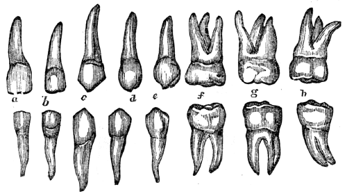
Fig. 55. The permanent teeth of the upper and lower jaw. a, b, The incisors. c, The cuspids. d, e, The bicuspids. f, g, The molars, (double teeth.) h, The wisdom teeth.
Observation. The shape of the teeth in different species of animals is adapted to the kind of food on which they subsist. Those animals that feed exclusively on flesh, as the lion, have the cuspids, or canine teeth, largely developed, and the molars have sharp cutting points. Those animals that feed on grass and grain, as the horse and the sheep, have their molar teeth more rounded and flat on the crown. The human teeth are 108 adapted to feed on fruits, grain, or flesh, as they are less pointed than those of the cat, and more pointed than those of the sheep.
216. Give the names of the permanent teeth. What teeth have but one root, or “fang”? How many roots have the molars of the upper jaw? Of the lower jaw? What is said of the shape of the teeth in different species of animals?
217. The teeth are composed principally of two substances—the i´vo-ry and the en-am´el. The internal part of the tooth or the ivory, is harder and more enduring than bone, and forms the body of the tooth. The enamel is remarkable for its hardness, and varies somewhat in color with the age, temperament, habits, and manner of living of different individuals. When any part of the enamel is destroyed, it is never regenerated.
Fig. 56. A side view of the body and enamel of a front tooth.
Fig. 57. A side view of a molar tooth. 1, The enamel. 2, The body of the tooth. 3, The cavity in the crown of the tooth that contains the pulp. 4, A nerve that spreads in the pulp of the tooth. 5, An artery that ramifies in the pulp of the tooth.
218. Each tooth is divided into two parts, namely, crown and root. The crown is that part which protrudes from the jaw-bone and gum, and is covered by the highly polished enamel. The root, or “fang,” is placed in the sockets of the jaw, and consists of bony matter. Through this bony substance several small vessels pass, to aid in the growth and 109 also in the removal of the tooth. There are, beside these vessels, small white cords passing to each tooth, called nerves. (See fig. 57.) When these nerves are diseased, we have the toothache.
217. Give the structure of the teeth. What is said of the enamel? 218. Into how many parts are the teeth divided? Describe the crown. The root. What vessels pass through the bony matter? What is their use?
219. The use of the teeth is twofold. 1st. By the action of the incisors the food is divided, while the molars grind or break down the more solid portions of it. By these processes, the food is prepared to pass more easily and rapidly into the stomach.
220. In the mastication of food there are two movements of the lower jaw—the action by which the teeth are brought together, and the lateral motion. In the former, the food is cut or divided, the jaws acting like shears. This movement is produced by the action of two large muscles situated on each side of the head and face.
Observation. The muscles attached to the lower jaw are of great strength; by their action alone, some persons are enabled to bite the hardest substances. By putting the fingers upon the side of the head above and in front of the ears, and upon the face above the angle of the jaw, while masticating food, the alternate swelling and relaxation of these muscles will be clearly felt.
221. The lateral, or grinding movement of the teeth, is produced by the action of a strong muscle that is attached to the lower jaw on the inside.
Observation. Those animals that live solely on flesh, have only the cutting, or shear-like movement of the jaws. Those 110 that use vegetables for food, have the grinding motion; while man has both the cutting and grinding movement.
219–222. Give the physiology of the teeth. 219. Give one of the functions of the teeth. 220. How many movements of the lower jaw in masticating food? What effect has the first movement upon the food? How produced? What is the character of the masticating muscles? 221. How is the grinding motion of the teeth produced? What is said of the movements of the teeth in different animals?
222. 2d. The teeth aid us in articulating with distinctness certain letters and words. An individual who has lost his front teeth cannot enunciate distinctly certain letters called dental. Again, as the alveolar processes are removed by absorption soon after the removal of the teeth, the lips and cheeks do not retain their former full position, thus marring, in no slight degree, the symmetry of the lower part of the face. Consequently, those simple observances that tend to the preservation of the teeth are of great practical interest to all persons.
223. To preserve the teeth, they must be kept clean. After eating food, they should be cleansed with a brush and water, or rubbed with a piece of soft flannel, to prevent the tartar collecting, and to remove the pieces of food that may have lodged between them. Toothpicks may be useful in removing any particles inaccessible to the brush. They may be made of bone, ivory, or the common goose-quill. Metallic toothpicks should not be used, as they injure the enamel.
224. The mouth should be cleansed with pure tepid water at night, as well as in the morning; after which the teeth should be brushed upward and downward, both on the posterior and anterior surfaces. It may be beneficial to use refined soap, once or twice every week, to remove any corroding substance that may exist around the teeth; care being taken to thoroughly rinse the mouth after its use.
225. Food or drink should not be taken into the mouth when very hot or very cold. Sudden changes of temperature will crack the enamel, and finally produce decayed teeth.
222. What is another use of the teeth? 223–232. Give the hygiene of the teeth. 223. How can the teeth be preserved? By what means? 224. How often should they be cleansed? 225. What is said of very hot or cold drinks?
Observation. On this account, smoking is pernicious, because the teeth are subjected to an alternate inhalation of both cold and warm air.
226. The temporary teeth should be removed as soon as they become loose. If a permanent tooth makes it appearance before the first is removed, or has become loose, the milk tooth, although not loose, should be removed without delay. This is necessary that the second set of teeth may present a regular and beautiful appearance.
227. In general, when the permanent teeth are irregular, one or more should be removed. If the teeth are crowded and irregular, in consequence of the jaw being narrow and short, or when they press so hard upon each other as to injure the enamel, remove one or more to prevent their looking unsightly, and in a few months the remaining teeth, with a little care, will fill the spaces.
Observation. When it is necessary to remove a tooth, apply to some skilful operator. It requires as much skill and knowledge to extract teeth well, as it does to amputate a limb; yet some persons, who possess strong arms, will obtain a pair of forceps, or a tooth-key, and hang out the sign of “surgeon-dentist,” although ignorant of the principles that should guide them.
228. It is not always necessary to have teeth extracted when they ache. The nerve, or the investing membrane of the root, may be diseased, and the tooth still be sound. In such instances, the tooth should not be extracted, but the diseased condition may be remedied by proper medication. There are many sound teeth, that become painful, as already mentioned, which are unnecessarily removed.
Why is smoking injurious to the teeth? 226. What remark respecting the temporary teeth? 227. What remarks respecting the permanent teeth? Do those persons that extract teeth require skill as well as knowledge? 228. Why should not teeth be extracted at all times when they are painful?
Illustration. Dr. H. M., of Belfast, Me., related to me that an individual in that vicinity had his teeth, (all of them sound,) on one side of the lower jaw, extracted by an ignoramus of a “tooth-puller,” and this without any relief from pain. The disease was tic douloureux, which was relieved by Dr. M.
229. The preservation of the teeth requires that they be frequently examined. When a part of the enamel is removed, and a small portion of the body of the tooth has become carious, in many instances such teeth may be preserved from further decay by having them filled or “plugged” with gold foil. All amalgams, pastes, and cheap patent articles for filling, should be avoided, if you would preserve both the teeth and the general health.
230. The practice of cracking nuts with the teeth, or of lifting heavy bodies, and the constant habit of biting thread, should be avoided, as they finally destroy the enamel.
231. All acidulated drinks and mineral waters, that “set the teeth on edge,” are injurious. All tooth-powders and washes that contain any article that is acid, corrosive, or grinding, should be banished from the toilet. Tobacco is not a preservative of the teeth. It contains “grit,” which wears away the enamel; beside, when chewed, it debilitates the vessels of the gums, turns the teeth yellow, and renders the breath and the appearance of the mouth disagreeable.
232. Healthy persons have generally sound teeth, while feeble persons have decayed teeth. For this reason, we should try to learn and practise the few simple rules that promote health.
Give an illustration of the removal of sound teeth. 229. How may decaying teeth be preserved? What should be avoided in the filling of teeth? 230. What practices should also be avoided? 231. What is said of acidulated drinks? What effect has the chewing of tobacco upon the teeth? 232. What is one reason for preserving health?
233. From the earliest existence of the human system to the last ray of life, change is impressed upon it by the Giver of this curious fabric. New atoms of matter are deposited, while the old and now useless particles are constantly removed. The material necessary to sustain the growth of the body in early life, and also to repair the waste that is unceasing to animal existence, is the food we eat.
234. Food, animal or vegetable, contains most of the elements of the different tissues of the system, yet it must undergo certain essential alterations before it can become a part of the body. The first change is effected by the action of the Digestive Organs.
235. The DIGESTIVE ORGANS are the Mouth, Teeth,[7] Sal´i-va-ry Glands, Phar´ynx, Œ-soph´a-gus, (gullet,) Stom´ach, In-tes´tines, (bowels,) Lac´te-als, (milk, or chyle vessels,) Tho-rac´ic Duct, Liv´er, and the Pan´cre-as, (sweetbread.)
233. What is impressed upon the human system from its earliest existence? What maintains this change? 234. Has animal or vegetable food any resemblance to the different tissues of which it finally forms a part? By what organs is the first change in the food effected? 235–258. Give the anatomy of the digestive organs. 235. Name them. 236. Describe the mouth.
MOUTH is an irregular cavity, which contains the instruments of mastication and the organs of taste. It is 114 bounded in front by the lips; on each side by the internal surface of the cheeks; above, by the hard palate (roof of the mouth) and teeth of the upper jaw; below, by the tongue and teeth of the lower jaw; behind, it is continuous with the pharynx, but is separated from it by a kind of movable curtain, called the soft palate. This may be elevated or depressed, so as to close the passage or leave it free.
237. The SALIVARY GLANDS are six in number; three on each side of the jaw. They are called the pa-rot´id, the sub-max´il-la-ry and the sub-lin´gual.
Fig. 58.
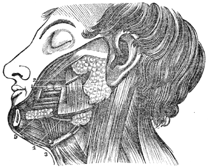
Fig. 58. A view of the salivary glands in their proper situations. 1, The parotid gland. 2, Its duct. 3, The submaxillary gland. 4, Its duct. 5, The sublingual gland, brought to view by the removal of a section of the lower jaw.
238. The PAROTID GLAND, the largest, is situated in front of the external ear, and behind the angle of the jaw. A duct (Steno’s) from this gland opens into the mouth, opposite the second molar tooth of the upper jaw.
237. How many glands about the mouth? Give their names. What does fig. 58 represent? 238. Describe the parotid gland.
239. The SUBMAXILLARY GLAND is situated within the lower jaw, anterior to its angle. Its excretory duct (Wharton’s) opens into the mouth by the side of the fræ´num lin´guæ, (bridle of the tongue.)
240. The SUBLINGUAL GLAND is elongated and flattened, and situated beneath the mucous membrane of the floor of the mouth, on each side of the frænum linguæ. It has seven or eight small ducts, which open into the mouth by the side of the bridle of the tongue.
Observation. In the “mumps,” the parotid gland is diseased. The swelling under the tongue called the “frog” is a disease of the sublingual gland.
Fig. 59.

Fig. 59. A side view of the face, œsophagus, and trachea. 1, The trachea (wind pipe.) 2, The larynx. 3, The œsophagus. 4, 4, 4, The muscles of the upper portion of the œsophagus forming the pharynx. 5, The muscle of the cheek. 6, The muscle that surrounds, the mouth. 7, The muscle that forms the floor of the mouth.
239. The submaxillary. 240. The sublingual. What observation respecting these glands? What does fig. 59 represent?
241. The PHARYNX is a membranous sac, situated upon the 116 upper portion of the spinal column. It extends from the base of the skull to the top of the tra´che-a, (windpipe,) and is continuous with the œsophagus. From the pharynx are four passages; one opens upward and forward to the nose, the second leads forward to the mouth, the third downward to the trachea and lungs, the fourth downward and backward to the stomach.
242. The ŒSOPHAGUS is a large membranous tube that extends behind the trachea, the heart, and lungs, pierces the diaphragm, and terminates in the stomach. It is composed of two membranes—an internal, or mucous, and a muscular coat. The latter is composed of two sets of fibres; one extends lengthwise, the other is arranged in circular bands.
243. The STOMACH is situated in the left side of the abdomen, immediately below and in contact with the diaphragm. It has two openings; one connected with the œsophagus, called the car´di-ac orifice; the other connected with the upper portion of the small intestine, called the py-lor´ic orifice. It is composed of three coats, or membranes. The exterior or serous coat is very tough and strong, and invests every part of this important organ. The middle, or muscular coat is composed of two layers of muscular fibres, one set of which is arranged longitudinally, the other circularly. The interior coat is called the mucous, and is arranged in ru´gæ, (folds.) The stomach is provided with a multitude of small glands, in which is secreted the gastric fluid.
Illustration. The three coats of the stomach anatomically resemble tripe, which is a preparation of the largest stomach of the cow or ox. The outer coat is smooth and highly polished. The middle coat is composed of minute threads, which are 117 arranged in two layers. The fibres of these layers cross each other. The inner coat is soft, and presents many folds, usually named “the honey-comb.”
241. Describe the pharynx and the passages leading from it. 242. Give the structure of the œsophagus. 243. Where is the stomach situated? How many coats has it? Describe them. What article prepared for food does the stomach resemble?
Fig. 60.

Fig. 60. The inner surface of the stomach and duodenum. 1, The lower portion of the œsophagus. 2, The opening through which the food is passed into the stomach. 8, The stomach. 9, The opening through which the food passes out of the stomach into the duodenum, or upper portion of the small intestine. 10, 11, 14, The duodenum 12, 13, Ducts through which the bile and pancreatic fluid pass into it. a, b, c, The three coats of the stomach.
244. The INTESTINES, or alimentary canal, are divided into two parts—the small and large. The small intestine is about twenty-five feet in length, and is divided into three portions, namely, the Du-o-de´num, the Je-ju´num, and the Il´e-um. The large intestine is about five feet in length, and is divided into three parts, namely, the Cæ´cum, the Co´lon, and the Rec´tum. (Appendix D.)
245. The DUODENUM is somewhat larger than the rest of the small intestine, and has received its name from being in 118 length about the breadth of twelve fingers. It commences at the pylorus, and ascends obliquely backward to the under surface of the liver. It then descends perpendicularly in front of the right kidney, and passes transversely across the lower portion of the spinal column, behind the colon, and terminates in the jejunum. The ducts from the liver and pancreas open into the perpendicular portion, about six inches from the stomach.
244. Explain fig. 60. What is the length of the small intestine, and how is it divided? What is the length of the large intestine? Give its divisions. 245. Describe the duodenum.
246. The JEJUNUM is continuous with the duodenum. It is thicker than the rest of the small intestine, and has a pinkish tinge.
247. The ILEUM is smaller, and thinner in texture, and somewhat paler, than the jejunum. There is no mark to distinguish the termination of the one or the commencement of the other. The ileum terminates near the right haunch-bone, by a valvular opening into the colon at an obtuse angle. This arrangement prevents the passing of substances from the colon into the ileum. The jejunum and ileum are surrounded above and at the sides by the colon.
248. The small intestine, like the stomach, has three coats. The inner, or mucous coat is thrown into folds, or valves. In consequence of this valvular arrangement, the mucous membrane is more extensive than the other tissues, and gives a greater extent of surface with which the aliment comes in contact. There are imbedded under this membrane an immense number of minute glands, and it has a great number of piles, like those upon velvet. For this reason, this membrane is sometimes called the vil´lous coat.
249. The CÆCUM is the blind pouch, or cul-de-sac, at the commencement of the large intestine. Attached to its extremity is the ap-pend´ix verm-i-form´is, (a long, worm-shaped 119 tube.) It is from one to six inches in length, and of the size of a goose-quill.
What important ducts open into it? 246. Describe the jejunum. 247. The ileum. 248. What is said of the coats of the intestines? Why is the mucous membrane sometimes called the villous coat? 249. Describe the cæcum.
250. The COLON is divided into three parts—the ascending, transverse, and descending. The ascending colon passes upward from the right haunch-bone to the under surface of the liver. It then bends inward, and crosses the upper part of the abdomen, below the liver and stomach, to the left side under the name of the transverse colon. At the left side, it turns, and descends to the left haunch-bone, and is called the descending colon. Here it makes a peculiar curve upon itself, which is called the sig´moid flex´ure.
Fig. 61.

Fig. 61. 1, 1, The duodenum. 2, 2, The small intestine. 3, The junction of the small intestine with the colon. 4, The appendix vermiformis. 5, The cæcum. 6, The ascending colon. 7, The transverse colon. 8, The descending colon. 9, The sigmoid flexure of the colon. 10, The rectum.
250. Describe the course of the divisions of the colon. Explain fig. 61.
251. The RECTUM is the termination of the large intestine. The large intestine has three coats, like the stomach and small intestine. The longitudinal fibres of the muscular coat are collected into three bands. These bands are nearly one half shorter than the intestine, and give it a sacculated appearance, which is characteristic of the cæcum and colon.
252. The LACTEALS are minute vessels, which commence in the villi, upon the mucous surface of the small intestine. From the intestine they pass between the membranes of the mes´en-ter-y to small glands, which they enter. The first range of glands collects many small vessels, and transmits a few larger branches to a second range of glands; and, finally, after passing through several successive ranges of these glandular bodies, the lacteals, diminished in number and increased in size, proceed to the enlarged portion of the thoracic duct, into which they open. They are most numerous in the upper portion of the small intestine.
253. The THORACIC DUCT commences in the abdomen, by a considerable dilatation, which is situated in front of the lower portion of the spinal column. From this point, it passes through the diaphragm, and ascends to the lower part of the neck. In its ascent, it lies anterior to the spine, and by the side of the aorta and œsophagus. At the lower part of the neck, it makes a sudden turn downward and forward, and terminates by opening into a large vein which passes to the heart. The thoracic duct is equal in diameter to a goose-quill, and, at its termination, is provided with a pair of semilunar valves, which prevent the admission of venous blood into its cylinder.
251. What is said of the arrangement of the fibres of the muscular coat of the large intestine? 252. What are the lacteals? Give their course from the mucous coat of the intestine to the thoracic duct. 253. Describe the course of the thoracic duct. How is the venous blood prevented from passing into this duct?
Fig. 62.
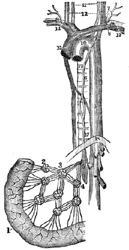
Fig. 62. A portion of the small intestine, lacteal vessels, mesenteric glands, and thoracic duct. 1, The intestine. 2, 3, 4, Mesenteric glands, through which the lacteals pass to the thoracic duct. 5, 6, The thoracic duct. 7, The point in the neck where it turns down to enter the vein at 8. 9, 10, The aorta. 11, 12, Vessels of the neck. 13, 14, 15, The large veins that convey the blood and chyle to the heart. 16, 17, The spinal column. 18, The diaphragm, (midriff.)
Explain fig. 62. What is said respecting the mesenteric glands?
Observation. The mesenteric glands, which are situated between two layers of serous membrane (mesentery) that connects the small intestine with the spinal column, occasionally become diseased in childhood, and prevent the chyle 122 from passing to the thoracic duct. Children thus affected have a voracious appetite, and at the same time are becoming more and more emaciated. The disease is called mesenteric consumption.
254. The LIVER, a gland appended to the alimentary canal, is the largest organ in the system, and weighs about four pounds. It is situated in the right side, below the diaphragm, and is composed of several lobes. Its upper surface is convex; its under, concave. This organ is retained in its place by several ligaments. It performs the double office of separating impurities from the venous blood, and of secreting a fluid (bile) necessary to chylification. On the under surface of the liver is a membranous sac, called the gall-cyst, which is generally considered as a reservoir for the bile.
Fig. 63.

Fig. 63. The under surface of the liver. 1, The right lobe. 2, The left. 3, 4, Smaller lobes. 10, The gall-bladder, or cyst, lodged in its depression. 17, The notch on the posterior border, for the spinal column.
Observation. A good idea of the liver and intestines can be obtained by examining these parts of a pig. In this animal, the sacs, or pouches, of the large intestine are well defined.
255. The PANCREAS is a long, flattened gland, analogous to 123 the salivary glands. It is about six inches in length, weighs three or four ounces, and is situated transversely across the posterior wall of the abdomen, behind the stomach. A duct from this organ opens into the duodenum.
254. Describe the liver. 255. What is said of the pancreas?
256. The SPLEEN, (milt,) so called because the ancients supposed it to be the seat of melancholy, is an oblong, flattened organ, situated in the left side, in contact with the diaphragm, stomach, and the pancreas. It is of a dark, bluish color, and is abundantly supplied with blood, but has no duct which serves as an outlet for any secretion. Its use is not well determined.
Fig. 64.

Fig. 64. The pancreas with its duct, through which the pancreatic secretion passes into the duodenum.
257. The OMENTUM (caul) consists of four layers of the serous membrane, which descends from the stomach and transverse colon. A quantity of adipose matter is deposited around its vessels, which ramify through its structure. Its function is twofold in the animal economy. 1st. It protects the intestines from cold. 2d. It facilitates the movements of the intestines upon each other during their vermicular, or worm-like action.
258. Every part of the digestive apparatus is supplied with arteries, veins, lymphatics, and nervous filaments, from the ganglionic system of nerves.
256. Why is the spleen so called? What is peculiar to this organ? 257. Of what is the omentum composed? What is its use? 258. With what is every part of the digestive apparatus supplied?
259. Substances received into the stomach as food, must necessarily undergo many changes before they are fitted to form part of the animal body. The solid portions are reduced to a fluid state, and those parts that will nourish the body are separated from the waste material.
260. The first preparation of food for admission into the system, consists in its proper mastication. The lips in front, the cheeks upon the side, the soft palate, by closing down upon the base of the tongue, retain the food in the mouth, while it is subjected to the; process of mas-ti-ca´tion, (chewing.) The tongue rolls the mass around, and keeps it between the teeth, while they divide the food to a fineness suitable for the stomach.
261. While the food is in process of mastication, there is incorporated with it a considerable amount of sa-li´va, (spittle.) This fluid is furnished by the salivary glands, situated in the vicinity of the mouth. The saliva moistens and softens the food, so that, when carried into the pharynx. it is passed, with ease, through the œsophagus into the stomach.
262. When the food has been properly masticated, (and in rapid eaters when it is not,) the soft palate is raised from the base of the tongue backward, so as to close the posterior opening through the nostrils. By a movement of the muscles of the tongue, cheeks, and floor of the mouth, simultaneous with 125 that of the soft palate, the food is pressed into the upper part of the pharynx.
259–272. Give the physiology of the digestive organs. 259. What is necessary before food can nourish the body? 260. Describe how mastication is performed. 261. Of what use is the saliva in the process of mastication? 262. How is the food pressed into the pharynx?
263. When in the pharynx, the food and drink are prevented from passing into the trachea by a simple valve-like arrangement, called the ep-i-glot´tis. The ordinary position of this little organ is perpendicular, so as not to obstruct the passage of air into the lungs; but in the act of swallowing, it is brought directly over the opening of the trachea, called the glot´tis. The food, being forced backward, passes rapidly over the epiglottis into the œsophagus, where the circular band of muscular fibres above, contracts and forces the food to the next lower band. Each band relaxes and contracts successively, and thus presses the alimentary ball downward and onward to the stomach.[8]
Observation. If air is inhaled when the food or drink is passing over the glottis, some portions of it may be carried into the larynx or trachea. This produces violent spasmodic coughing, and most generally occurs when an attempt is made to speak while masticating food; therefore, never talk when the mouth contains food.
263. When the food is in the pharynx, how is it prevented from passing into the trachea, or windpipe? Describe how it is passed into the stomach? Give the observation. 264. Describe how the food in the stomach is converted into chyme.
264. When the food reaches the stomach, the gastric glands are excited to action, and they secrete a powerful solvent, called gastric juice. The presence of food in the stomach also increases a contractile action of the muscular coat, by which the position of the food is changed from one part of this cavity to another. Thus the aliment is brought in contact with the mucous membrane, and each portion of it becomes saturated with gastric juice, by which it is softened, or dissolved into 126 a pulpy homogeneous mass, of a creamy consistence, called Chyme. The food is not all converted into chyme at the same time; but as fast as it is changed, it passes through the pyloric orifice into the duodenum.
Observation. The gastric juice has the property of coagulating liquid albuminous matter when mixed with it. It is this property of rennet, which is an infusion of the fourth stomach of the calf, by which milk is coagulated, or formed into “curd.”
265. The CHYME is conveyed through the pyloric orifice of the stomach into the duodenum. The chyme not only excites an action in the duodenum, but also in the liver and pancreas. Mucus is then secreted by the duodenum, bile by the liver, and pancreatic fluid by the pancreas. The bile and pancreatic fluid are conveyed into the duodenum, and mixed with the chyme. By the action of these different fluids, the chyme is converted into a fluid of a whitish color, called Chyle, and into residuum.
Observation. The bile has no agency in the change through which the food passes in the stomach. In a healthy condition of this organ, no bile is found in it. The common belief, that the stomach has a redundancy of this secretion, is erroneous. If bile is ejected in vomiting, it merely shows, not only that the action of the stomach is inverted, but also that of the duodenum. A powerful emetic will, in this way, generally bring this fluid from the most healthy stomach. A knowledge of this fact might save many a stomach from the evils of emetics, administered on false impressions of their necessity, and continued from the corroboration of these by the appearance of bile, till derangement, and perhaps permanent disease, are the consequences.
266. The CHYLE and residual matter are moved over the 127 mucous surface of the small intestine, by the action of its muscular coat. As the chyle is carried along the tract of the intestine, it comes in contact with the villi, where the lacteal vessels commence. These imbibe, or take up, the chyle, and transfer it through the mesenteric glands into the thoracic duct, through which it is conveyed into a large vein at the lower part of the neck. In this vein the chyle is mixed with the venous fluid. The residual matter is conveyed into the large intestine, through which it is carried and excreted from the system. (Appendix E.)
What peculiar property has gastric juice? 265. Where and how is chyme converted into chyle? What is said in regard to the bile? 266. What becomes of the chyle? Of the residuum?
267. In the process of digestion, the food is subjected to five different changes. 1st. The chewing and admixture of the saliva with the food; this process is called mastication.
268. 2d. The change through which the food passes in the stomach by its muscular contraction, and the secretion from the gastric glands; this is called chymification.
269. 3d. The conversion of the homogeneous chyme, by the agency of the bile and pancreatic secretions, into a fluid of milk-like appearance; this is chylification.
270. 4th. The absorption of the chyle by the lacteals, and its transfer through them and the thoracic duct, into the subclavian vein at the lower part of the neck.[9]
271. 5th. The separation and excretion of the residuum.
272. Perfection of the second process of digestion requires thorough and slow mastication. The formation of proper chyle demands appropriate mastication and chymification; while a healthy action of the lacteals requires that all the anterior stages of the digestive process be as perfect as possible. (Appendix F.)
267. Recapitulate the five changes in the digestive process.
Note. Let the pupil review the anatomy and physiology of the digestive organs from figs. 62 and 65, or from anatomical outline plate No. 5.
Fig. 65.
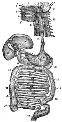
Fig. 65. An ideal view of the organs of digestion, opened nearly the whole length. 1, The upper jaw. 2, The lower jaw. 3, The tongue. 4, The roof of the mouth. 5, The œsophagus. 6, The trachea. 7, The parotid gland. 8, The sublingual gland. 9, The stomach. 10, 10, The liver. 11, The gall-cyst. 12, The duct that conveys the bile to the duodenum, (13, 13.) 14, The pancreas. 15, 15, 15, 15, The small intestine. 16, The opening of the small intestine into the large intestine. 17, 18, 19, 20, The large intestine. 21, The spleen. 22, The upper part of the spinal column.
273. It is a law of the system, that each organ is excited to healthy and efficient action, when influenced by its appropriate stimulus. Accordingly, nutrient food, that is adapted to the wants of the system, imparts a healthy stimulation to the salivary glands during the process of mastication. The food that is well masticated, and has blended with it a proper amount of saliva, will induce a healthy action in the stomach. Well-prepared chyme is the natural stimulus of the duodenum, liver, and pancreas; pure chyle is the appropriate excitant of the lacteal vessels.
274. The perfection of the digestive process, as well as the health of the general system, requires the observance of certain conditions. These will be considered under four heads:—1st. The Quantity of food that should be taken. 2d. Its Quality. 3d. The Manner in which it should be taken. 4th. The Condition of the system when food is taken.
275. The QUANTITY of food necessary for the system varies. Age, occupation, temperament, temperature, habits, amount of clothing, health and disease are among the circumstances which produce the variation.
273–330. Give the hygiene of the digestive organs. 273. Give a law of the system. What is the appropriate stimulus of the salivary glands during mastication? Of the stomach? Of the duodenum? Of the lacteal vessels? 274. What does the perfection of the digestive organs require? 275. What exert an influence on the quantity of food necessary for the system?
276. The child and youth require food to promote the 130 growth of the different parts of the body. The more rapid the growth of the child, the greater the demand for food. This accounts for the keen appetite and vigorous digestion in childhood. When the youth has attained his full growth, this necessity for nutriment ceases; after this period of life, if the same amount of food is taken, and there is no increase of labor or exertion, the digestive apparatus will become diseased, and the vigor of the whole system diminished.
Observation. When the body has become emaciated from want of nutriment, either from famine or disease, there is an increased demand for food. This may be gratified with impunity until the individual has regained the usual size, but repletion should be avoided.
277. Food is required to repair the waste, or loss of substance that attends action. In every department of nature, waste, or loss of substance, attends and follows action. When an individual increases his exercise,—changes from light to severe labor,—or the inactive and sedentary undertake journeys for pleasure, the fluids of the system circulate with increased energy. The old and exhausted particles of matter are more rapidly removed through the action of the vessels of the skin, lungs, kidneys, and other organs, and their places are filled with new atoms, deposited by the small blood-vessels.
278. As the chyle supplies the blood with the newly vitalized particles of matter, there is, consequently, an increased demand for food. This want of the system induces, in general, a sensation of hunger or appetite, which may be regarded as an indication of the general state of the body. The sympathy that exists throughout the system accords to 131 the stomach the power of making known this state to the nervous system, and, if the functions of this faithful monitor have not been impaired by disease, abuse, or habit, the call is imperious, and should be regarded.
276. At what age is the appetite keen and the digestion vigorous? Why? What is said in regard to the quantity of food when the youth has attained his growth? What exception, as given in the observation? 277. Give another demand for food. What effect has increased exercise upon the system? 278. How are the new particles of matter supplied? What does this induce?
279. When exercise or labor is lessened, the quantity of food should be diminished. When a person who has been accustomed to active exercise, or even hard manual labor, suddenly changes to an employment that demands less activity, the waste attendant on action will be diminished in a corresponding degree; hence the quantity of food should be lessened in nearly the same proportion as the amount of exercise is diminished. If this principle be disregarded, the tone of the digestive organs will be impaired, and the health of the system enfeebled.
280. This remark is applicable to those students who have left laborious employments to attend school. Although the health is firm, and the appetite keen from habit, yet every pupil should practise some self-denial, and not eat as much as the appetite craves, the first week of the session. After some days, the real wants of the system will generally be manifested by a corresponding sensation of hunger.
Observation. It is a common observation that in academies and colleges, the older students from the country, who have been accustomed to hard manual labor, suffer more frequently from defective digestion and impaired health than the younger and feebler students from the larger towns and cities.
281. Food is essential in maintaining a proper temperature of the system. The heat of the system, at least in part, is produced in the minute vessels of the several organs, by the union of oxygen with carbon and hydrogen, which the food and drink contain. The amount of heat generated, is greatest 132 when it is most rapidly removed from the system, which occurs in cold weather. This is the cause of the system requiring more food in winter than summer.
279. Why should the quantity of food be diminished when the exercise is lessened? What effect if this principle be disregarded? 280. To what class is this remark applicable? What is often observed among students in academies and colleges? 281. State another demand for food. What is one source of heat in the body?
Observation. Persons that do not have food sufficient for the natural wants of the system, require more clothing than those who are well fed.
282. The last-mentioned principle plainly indicates the propriety and necessity of lessening the quantity of food as the warm season approaches. Were this practised, the tone of the stomach and the vigor of the system would continue unimpaired, the “season complaints” would be avoided, and the “strengthening bitters” would not be sought to create an appetite.
Observation. Stable-keepers and herdsmen are aware of the fact, that as the warm season commences, then animals require less food. Instinct teaches these animals more truly, in this particular, than man allows reason to guide him.
283. The quantity of food should have reference to the present condition of the digestive organs. If they are weakened or diseased, so that but a small quantity of food can be properly digested or changed, that amount only should be taken. Food does not invigorate the system, except it is changed, as has been described in previous paragraphs.
Observation. When taking care of a sick child, the anxiety of the mother and the sufferings of the child may induce her to give food when it would be highly injurious. The attending physician is the only proper person to direct what quantity should be given.
Why do we eat more in the winter than in the summer? What practical observation is given? 282. Why should the quantity of food be lessened as warm weather commences? What would be avoided if this principle were obeyed? 283. Why should the present condition of the digestive organs be regarded in reference to the quantity of food? Mention an instance in which it would be injudicious to give food.
284. The quantity of food is modified, in some degree, by 133 habit. A healthy person, whose exercise is in pure air, may be accustomed to take more food than is necessary. The useless excess is removed from the system by the waste outlets, as the skin, lungs, liver, kidneys, &c. In such cases, if food is not taken in the usual quantity, there will be a feeling of emptiness, if not of hunger, from the want of the usual distention of the stomach. This condition of the digestive organs may be the result of disease, but it is more frequently produced by inordinate daily indulgence in eating, amounting almost to gluttony.
285. Large quantities of food oppress the stomach, and cause general languor of the whole body. This is produced by the extra demands made on the system for an increased supply of blood and nervous fluid to enable the stomach to free itself of its burden. Thus, when we intend to make any extraordinary effort, mental or physical, at least for one meal, we should eat less food than usual, rather than a greater quantity.
286. No more food should be eaten than is barely sufficient to satisfy the appetite. Nor should appetite be confounded with taste. The one is a natural desire for food to supply the wants of the system; the other is an artificial desire merely to gratify the palate.
287. Although many things may aid us in determining the quantity of food proper for an individual, yet there is no certain guide in all cases. It is maintained by some, that the sensation of hunger or appetite is always an indication of the want of food, while the absence of this peculiar sensation is regarded as conclusive evidence that aliment is not demanded. 134 This assertion is not correct, as an appetite may be created for food by condiments and gormandizing, which is as artificial and as morbid as that which craves tobacco or ardent spirits. On the other hand, a structural or functional disease of the brain may prevent that organ from taking cognizance of the sensations of the stomach, when the system actually requires nourishment. Observation shows, that disease, habit, the state of the mind, and other circumstances, exert an influence on the appetite.
284. Show the effect of habit upon the quantity of food that is eaten. What is said in regard to inordinate eating? 285. What is the effect of eating large quantities of food? What suggestion when an extraordinary effort, either mental or physical, is to be made? 286. How much food should generally be eaten? 287. What is the assertion of some persons relative to the quantity of food necessary for the system?
Observation. Dr. Beaumont noticed, in the experiments upon Alexis St. Martin, that after a certain amount of food was converted into chyme, the gastric juice ceased to ooze from the coats of the stomach. Consequently, it has been inferred by some writers on physiology, that the glands which supply the gastric fluid, by a species of instinctive intelligence, would only secrete enough fluid to convert into chyme the aliment needed to supply the real wants of the system. What are the reasons for this inference? There is no evidence that the gastric glands possess instinctive intelligence, and can there be a reason adduced, why they may not be stimulated to extra functional action as well as other organs, and why they may not also be influenced by habit?
288. While all agree that the remote or predisposing cause of hunger is, usually, a demand of the system for nutrient material, the proximate or immediate cause of the sensation of hunger is not clearly understood. Some physiologists suppose that it is produced by an engorged condition of the glands of the stomach which supply the gastric juice; while others maintain that it depends on a peculiar condition of the nervous system.
289. The QUALITY of the food best adapted to the wants of the system is modified by many circumstances. There are 135 many varieties of food, and these are much modified by the different methods of preparation. The same kind of food is not equally well adapted to different individuals, or to the same individual in all conditions; as vocation, health, exposure, habits of life, season, climate, &c., influence the condition of the system.
What does observation show? 288. What is said of the causes of hunger? 289. Why is not the same kind of food adapted to different individuals?
290. All articles of food may be considered in two relations: 1st, As nutritive. 2d, As digestible. Substances are nutritious in proportion to their capacity to yield the elements of chyle, of which carbon, oxygen, hydrogen, and nitrogen are the most essential; they are digestible in proportion to the facility with which they are acted upon by the gastric juice. These properties should not be confounded in the various articles used for food.
291. As a “living body has no power of forming elements, or of converting one elementary substance into another, it therefore follows that the elements of which the body of an animal is composed must be in the food.” (Chap. III.) Of the essential constituents of the human body, carbon, hydrogen, oxygen, and nitrogen are the most important, because they compose the principal part of the animal body; while the other elements are found in very small proportions, and many of them only in a few organs of the system. (Appendix G.)
Observation. Nitrogen renders food more stimulating, particularly if combined with a large quantity of carbon, as beef. Those articles that contain the greatest amount of the constituent elements of the system are most nutritious. As milk and eggs contain all the essential elements of the human system, so they are adapted to almost universal use, and are highly nutritious.
290. In what proportion are substances nutritious? Digestible? Why does beef stimulate the system? What is said of milk and eggs?
292. The following table, by Pereira, in his treatise on 136 Food and Diet may aid the student in approximating to correct conclusions of the quantity of nutriment in different kinds of food, and its adaptation to the wants of the system.
TABLE,
SHOWING THE AVERAGE QUANTITY OF DRY, OR SOLID MATTER, CARBON, NITROGEN, AND MOISTURE, IN DIFFERENT ARTICLES OF DIET.
| One hundred Parts. | Dry Matter. | Carbon. | Nitrogen. | Water |
|---|---|---|---|---|
| Arrowroot, | 81.8 | 36.4 | 18.2 | |
| Beans, | 85.89 | 38.24 | 14.11 | |
| Beef, fresh, | 25 | 12.957 | 3.752 | 75 |
| Bread, rye, | 67.79 | 30.674 | 32.21 | |
| Butter, | 100 | 65.6 | ||
| Cabbage, | 7.7 | 0.28 | 92.3 | |
| Carrot, | 12.4 | 0.30 | 87.6 | |
| Cherries, | 25.15 | 74.85 | ||
| Chickens, | 22.7 | 77.3 | ||
| Codfish, | 20 | 80 | ||
| Cucumbers, | 2.86 | 97.14 | ||
| Eggs, whites, | 20 | 80 | ||
| Eggs, yolk, | 46.23 | 53.77 | ||
| Lard, hog’s, | 100 | 79.098 | ||
| Milk, cow’s, | 12.98 | 87.02 | ||
| Oats, | 79.2 | 40.154 | 1.742 | 20.8 |
| Oatmeal, | 93.4 | 6.6 | ||
| Olive-oil, | 100 | 77.50 | ||
| Oysters, | 12.6 | 87.4 | ||
| Peaches, | 19.76 | 80.24 | ||
| Pears, | 16.12 | 83.88 | ||
| Peas, | 84 | 35.743 | 16 | |
| Plums, greengage, | 28.90 | 71.10 | ||
| Potatoes, | 24.1 | 10.604 | 0.3615 | 75.9 |
| Rye, | 83.4 | 38.530 | 1.417 | 16.6 |
| Suet, mutton, | 100 | 78.996 | ||
| Starch, potato, | 82 | 36.44 | 18 | |
| Starch, wheat, | 85.2 | 37.5 | 14.8 | |
| Sugar, maple, | 42.1 | |||
| Sugar, refined, | 42.5 | |||
| Sugar, brown, | 40.88 | |||
| Turnips, | 7.5 | 3.2175 | 0.1275 | 92.5 |
| Veal, roasted, | 52.52 | 14.70 | ||
| Wheat, | 85.5 | 39.415 | 1.966 | 14.5 |
Note. Let the pupil mention those articles of food that are most nutritious, from a review of this table, and the last four paragraphs.
293. Those articles that do not contain the essential elements of the system should not be used as exclusive articles of diet. This principle has been, and may be illustrated by experiment. Feed a dog with pure sugar, or olive-oil, (articles that contain no nitrogen,) for several weeks, and the evil effects of non-nitrogenous nutriment will be manifested. At first, the dog will take his food with avidity, and seem to thrive upon it; soon this desire for food will diminish, his body emaciate, his eye become ulcerated, and in a few weeks he will die; but mix bran or sawdust with the sugar or oil, and the health and vigor of the animal will be maintained for months. A similar phenomenon will be manifested, if grain only be given to a horse, without hay, straw, or material of like character. (Appendix H.)
294. Some articles of food contain the elements of chyle in great abundance, yet afford but little nutriment, because they are difficult of digestion; while other articles contain but a small quantity of these elements, and afford more nourishment, because they are more easily affected by the digestive process.
293. How has the effect of non-nitrogenous nutriment been illustrated? 294. Why do some articles of food that contain the elements of chyle afford but little nutriment? Why do articles that contain a small quantity of these elements afford more nourishment? 295. How was the time required for digesting different articles of food ascertained?
295. The following table exhibits the general results of experiments made on Alexis St. Martin, by Dr. Beaumont, when he endeavored to ascertain the time required for the digestion of different articles of food.[10] The stomach of St. Martin was ruptured by the bursting of a gun. When he recovered from the effects of the accident under the surgical care of Dr. Beaumont, the stomach became adherent to the 139 side, with an external aperture. Nature had formed a kind of valve, which closed the aperture from the interior, and thus prevented the contents of the stomach from escaping; but on pushing it aside, the process of digestion could be seen. Through this opening, the appearance of the coats of the stomach and food, at different stages of digestion, were examined.
TABLE,
SHOWING THE MEAN TIME OF DIGESTION OF THE DIFFERENT ARTICLES OF DIET.
| Articles. | Preparation. | Time h. m. |
|---|---|---|
| Apples, sour, hard, | Raw, | 2 50 |
| Apples, sour, mellow, | Raw, | 2 |
| Apples, sweet, do., | Raw, | 1 30 |
| Bass, striped, fresh, | Broiled, | 3 |
| Beans, pod, | Boiled, | 2 30 |
| Beef, fresh, lean, rare, | Roasted, | 3 |
| Beef, fresh, lean, dry, | Roasted, | 3 30 |
| Beef steak, | Broiled, | 3 |
| Beef, with salt only, | Boiled, | 3 36 |
| Beef, with mustard, | Boiled, | 3 10 |
| Beef, fresh, lean, | Fried, | 4 |
| Beef, old, hard, salted, | Boiled, | 4 15 |
| Beets, | Boiled, | 3 45 |
| Bread, wheat, fresh, | Baked, | 3 30 |
| Bread, corn, | Baked, | 3 15 |
| Butter, | Melted, | 3 30 |
| Cabbage head, | Raw, | 2 30 |
| Cabbage, with vinegar, | Raw, | 2 |
| Cabbage, | Boiled, | 4 30 |
| Cake, sponge, | Baked, | 2 30 |
| Carrot, orange, | Boiled, | 3 15 |
| Catfish, | Fried, | 3 30 |
| Cheese, old, strong, | Raw, | 3 30 |
| Chicken, full-grown, | Fricas’d, | 2 45 |
| Codfish, cured, dry, | Boiled, | 2 |
| Corn, green, & beans, | Boiled, | 3 45 |
| Corn bread, | Baked, | 3 15 |
| Corn cake, | Baked, | 3 |
| Custard, | Baked, | 2 45 |
| Dumpling, apple, | Boiled, | 3 |
| Ducks, domesticated, | Roasted, | 4 |
| Ducks, wild, | Roasted, | 4 30 |
| Eggs, fresh, | Boiled hard, | 3 30 |
| Eggs, fresh, | Boiled soft, | 3 |
| Eggs, fresh, | Fried, | 3 30 |
| Eggs, fresh, | Raw, | 2 |
| Flounder, fresh, | Fried, | 3 30 |
| Fowl, domestic, | Boiled, | 4 |
| Fowl, domestic, | Roasted, | 4 |
| Goose, | Roasted, | 2 30 |
| Lamb, fresh, | Broiled, | 2 30 |
| Liver, beef’s, fresh, | Broiled, | 2 |
| Meat hashed with vegetables, | Warm’d, | 2 30 |
| Milk, | Boiled, | 2 |
| Milk, | Raw, | 2 15 |
| Mutton, fresh, | Roasted, | 3 15 |
| Mutton, fresh, | Broiled, | 3 |
| Mutton, fresh, | Boiled, | 3 |
| Oysters, fresh, | Raw, | 2 55 |
| Oysters, fresh, | Roasted, | 3 15 |
| Oysters, fresh, | Stewed, | 3 30 |
| Parsnips, | Boiled, | 2 30 |
| Pig, sucking, | Roasted, | 2 30 |
| Pigs’ feet, soused, | Boiled, | 1 |
| Pork, fat and lean, | Roasted, | 5 15 |
| Pork, recently salted, | Boiled, | 4 30 |
| Pork, recently salted, | Fried, | 4 15 |
| Pork, recently salted, | Broiled, | 3 15 |
| Pork, recently salted, | Raw, | 3 |
| Pork, steak, | Broiled, | 3 15 |
| Potatoes, Irish, | Boiled, | 3 30 |
| Potatoes, Irish, | Baked, | 2 30 |
| Rice, | Boiled, | 1 |
| Sago, | Boiled, | 1 45 |
| Salmon, salted, | Boiled, | 4 |
| Sausage, fresh, | Broiled, | 3 20 |
| Soup, beef, vegetables, and bread, | Boiled, | 4 |
| Soup, chicken, | Boiled, | 3 |
| Soup, mutton, | Boiled, | 3 30 |
| Soup, oyster, | Boiled, | 3 30 |
| Suet, beef, fresh, | Boiled, | 5 30 |
| Suet, mutton, | Boiled, | 4 30 |
| Tapioca, | Boiled, | 2 |
| Tripe, soused, | Boiled, | 1 |
| Trout, salmon, fresh, | Boiled, | 1 30 |
| Trout, salmon, fresh, | Fried, | 1 30 |
| Turkey, domesticated, | Roasted, | 2 30 |
| Turkey, | Boiled, | 2 25 |
| Turkey, wild, | Roasted, | 2 18 |
| Turnips, flat, | Boiled, | 3 30 |
| Veal, fresh, | Broiled, | 4 |
| Veal, fresh, | Fried, | 4 30 |
| Venison steak, | Broiled, | 1 35 |
296. In view of this table, the question may be suggested, Is that article of food most appropriate to the system which is most easily and speedily digested? To this it may be replied, that the stomach is subject to the same law as the muscles and other organs; exercise, within certain limits, strengthens it. If, therefore, we always eat those articles most easily digested, the digestive powers will be weakened; if over-worked, they will be exhausted. Hence the kind and amount of food should be adapted to the maintenance of the digestive powers, and to their gradual invigoration when debilitated.
Observation. Food that is most easily digested is not always most appropriate to a person convalescing from disease. If the substance passes rapidly through the digestive process, it may induce a recurrence of the disease. Thus the simple preparations which are not stimulating, as water-gruel, are better for a sick person than the more digestible beef and fish.
297. The question is not well settled, whether animal or vegetable food is best adapted to nourish man. There are nations, particularly in the torrid zone, that subsist, exclusively, on vegetables; while those of the frigid zone feed on fish or animal food. In the temperate zone, among civilized nations, a mixed diet is almost universal. When we consider the organization of the human system, the form and arrangement of the teeth, the structure of the stomach and intestines, we are led to conclude, that both animal and vegetable food is requisite, and that a mixed diet is most conducive to strength, health, and long life.
296. How is the question answered, whether that article is most appropriate to the system which is most easily digested? Give observation. 297. What is said of the adaptation of animal and vegetable food to man?
298. The food should be adapted to the distensible character of the stomach and alimentary canal. The former will be full, if it contain only a gill; it may be so distended as to contain a quart. The same is true of the intestines. If the food is concentrated, or contains the quantity of nutriment which the system requires, in small bulk, the stomach and intestines will need the stimulation of distention and friction, which is consequent upon the introduction and transit of the innutritious material into and through the alimentary canal. If the food is deficient in innutritious matter, the tendency is, to produce an inactive and diseased condition of the digestive organs. For this reason, nutrient food should have blended with it innutritious material. Unbolted wheat bread is more healthy than hot flour cakes; ripe fruits and vegetables than rich pies, or jellies.
Observation. 1st. The observance of this rule is of more importance to students, sedentary mechanics, and those individuals whose digestive apparatus has been enfeebled, than to those of active habits and firm health.
2d. The circumstance that different articles of food contain different proportions of waste, or innutritious matter, may be made practically subservient in the following way: If, at any particular season of the year, there is a tendency to a diarrhœa, an article that contains a small proportion of waste should be selected for food; but, if there is a tendency to an inactive or costive condition of the intestinal canal, such kinds of food should be used as contain the greatest proportion of waste, as such articles are most stimulating to the digestive organs, and, consequently, most laxative.
299. In the selection of food, the influence of season and climate should be considered. Food of a highly stimulating character may be used almost with impunity during the cold 141 weather of a cold climate; but in the warm season, and in a warm climate, it would be very deleterious. Animal food, being more stimulating than vegetable, can be eaten in the winter but vegetable food should be used more freely in the spring and summer.
298. What is said of the distensible character of the stomach and alimentary canal? What is the effect of eating highly concentrated food? Why is the unbolted wheat bread more healthy than flour cakes? Give observation 1st. Observation 2d. 299. What kind of food is adapted to cold weather? To warm weather?
300. The influence of food on the system is modified by the age of the individual. The organs of a child are more sensitive and excitable than those of a person advanced in years. Therefore a vegetable diet would be most appropriate for a child, while stimulating animal food might be conducive to the health of a person advanced in life.
Observation. When the digestive organs are highly impressible or diseased, it is very important to adopt a nutritious, unstimulating, vegetable diet, as soon as the warm season commences.
301. Habit is another strong modifying influence. If a person has been accustomed to an animal or vegetable diet, and there is a sudden change from one to the other, a diseased condition of the system, particularly of the digestive apparatus, usually follows. When it is necessary to change our manner of living, it should be done gradually.[11]
302. Some temperaments require more stimulating food than others. As a general rule, those persons whose sensations are comparatively obtuse, and movements slow, will be benefited by animal food; while those individuals whose constitutions are highly impressible, and whose movements are quick and hurried, require a nutritious and unstimulating vegetable diet.
300. What kinds of food are appropriate to old age? Why? What kinds to childhood? Why? 301. What is the effect when there is a sudden change from a vegetable to an animal diet? How should all changes of the system be made? 302. Do different temperaments require different kinds of food? What general rule is given?
303. The MANNER in which food should be taken is of much practical importance; upon it the health of the digestive organs measurably depends. But few circumstances modify the proper manner of taking food, or should exercise any controlling influence.
304. Food should be taken at regular periods. The interval between meals should be regulated by the character of the food, the age, health, exercise, and habits of the individual. The digestive process is more energetic and rapid in the young, active, and vigorous, than in the aged, indolent, and feeble; consequently, food should be taken more frequently by the former than by the latter class.
305. In some young and vigorous persons, food may be digested in one hour; in other persons, it may require four hours or more. The average time, however, to digest an ordinary meal, will be from two to four hours. In all instances, the stomach will require from one to three hours to recruit its exhausted powers after the labor of digesting a meal before it will again enter upon the vigorous performance of its duties.
306. Food should not be taken too frequently. If food is taken before the stomach has regained its tone and energy by repose, the secretion of the gastric juice, and the contraction 143 of the muscular fibres, will be imperfect. Again, if food is taken before the digestion of the preceding meal has been completed, the effects will be still worse, because the food partially digested becomes mixed with that last taken. Therefore the interval between each meal should be long enough for the whole quantity to be digested, and the time of repose should be sufficient to recruit the exhausted organs. The feebler the person and the more debilitated the stomach, the more important to observe the above directions.
303. Why is it important that we regard the manner of taking our food? 304. How should the intervals between meals be regulated? 305. What is the average time required to digest an ordinary meal? 306. Why should not food be taken too frequently?
Observation. In the feeding of infants, as well as in supplying food to older children, the preceding suggestions should always be regarded. The person who has been confined by an exhausting sickness, should most scrupulously regard this rule, if he wishes to regain his strength and flesh with rapidity. As the rapidity of the digestive process is less in students and individuals who are engaged in sedentary employments, than in stirring agriculturists, the former class are more liable to take food too frequently than the latter, while its observance is of greater importance to the sedentary artisan than to the lively lad and active farmer.
307. Food should be well masticated. All solid aliments should be reduced to a state of comparative fineness, by the teeth, before it is swallowed; the gastric fluid of the stomach will then blend with it more readily, and act more vigorously in reducing it to chyme. The practice of swallowing solid food, slightly masticated, or “bolting” it down, tends to derange the digestive process and impair the nutrition of the system.
308. Mastication should be moderate, not rapid. In masticating food, the salivary glands are excited to action, and some time must elapse before they can, secrete saliva in sufficient 144 quantities to moisten it. If the aliment is not supplied with saliva, digestion is retarded; besides, in rapid eating, more food is generally consumed than the system demands, or can be easily digested. Laborers, as well as men of leisure, should have ample time for taking their meals. Imperfect mastication is a prevailing cause of indigestion.
What persons would be benefited by observing the preceding remarks? 307. Why should food be well masticated? What is the effect of “bolting down” food? 308. How should mastication be performed? Why?
309. Food should be masticated and swallowed without drink. As the salivary glands supply fluid to moisten the dry food, the use of tea, coffee, water, or any other fluid, is not demanded by nature’s laws while taking a meal. One objection to “washing down” the food with drink is, the aliment is moistened, not with the saliva, but with the drink. This tends to induce disease, not only in the salivary organs, by leaving them in a state of comparative inactivity, but in the stomach, by the deficiency of the salivary stimulus. Another is, large quantities of fluids, used as drinks, give undue distention to the stomach, and lessen the energy of the gastric juice by its dilution, thus retarding digestion. Again, drinks taken into the stomach must be removed by absorption before the digestion of other articles is commenced.
Observation. Were it customary not to place drinks on the table until the solid food is eaten, the evil arising from drinking too much at meals would be obviated. The horse is never known to leave his provender, nor the ox his blade of grass, to wash it down; but many persons, from habit rather than thirst, drink largely during meals.
310. The peculiar sensation in the mouth and fauces, called thirst, may not always arise from the demand for fluids to increase the serum (water) of the blood, as in the desire for drink attendant on free perspiration, for then, pure water or some diluent drink is absolutely necessary; but it may be the 145 result of fever, or local disease of the parts connected with the throat. In many instances, thirst may be allayed by chewing some hard substance, as a dry cracker. This excites a secretion from the salivary glands, which removes the disagreeable sensation. In thirst, attendant on a heated condition of the system, this practice affords relief, and is safe; while the practice of drinking large quantities of cold fluids, is unsafe, and should never be indulged.
Why should all persons have ample time for eating? 309. Why are drinks not necessary while masticating food? Give the objections to “washing down” food. What observation relative to drink? 310. Does the sensation of thirst always arise from a real want of the system?
311. Food or drink should not be taken when very hot. When food or drink is taken hot, the vessels of the mucous membrane of the gums, mouth, and stomach are unduly stimulated for a short time; and this is followed by reaction, attended by a loss of tone, and debility of these parts. This practice is a fruitful cause of spongy gums, decayed teeth, sore mouth, and indigestion.
312. Food or drink should not be taken very cold. If a considerable quantity of very cold food or liquid be taken immediately into the stomach, the health will be endangered, and the tone of the system will be impaired, from the sudden abstraction of heat from the coats of the stomach, and from surrounding organs, to impart warmth to the cold food or drink. This arrests the digestive process, and the food is retained in the stomach too long, and causes oppression and irritation. Consequently, food and drink that are moderately heated are best adapted to the natural condition of the digestive apparatus.
Observation. Food of an injurious quality, or taken in an improper manner, affects the inferior animals as well as man. The teeth of cows that are closely penned in cities, and are fed on distillery slops, or the unhealthy slops and remnants of kitchens, decay and fall out in about two years. Can the milk of such diseased animals be healthy—the proper nourishment for children?
Give instances when it does and when it does not. 311. Why should not food or drink be taken hot? 312. Why should they not be taken cold? Show some of the effects of improper food upon the inferior animals.
313. The CONDITION of the system should be regarded when food is taken. This is necessary, as the present and ulterior condition of the digestive apparatus is strongly influenced by the state of the other organs of the system.
314. Food should not be taken immediately after severe exertion, either of the body or mind. For all organs in action require and receive more blood and nervous fluid, than when at rest. This is true of the brain, muscles, and vocal organs, when they have been actively exercised. The increased amount of fluid, both sanguineous and nervous, supplied to any organ during extra functional action, is abstracted from other parts of the system. This enfeebles and prostrates the parts that supply the blood and nervous fluid to the active organ. Again, when any organ has been in vigorous action for a few hours, some time will elapse before the increased action of the arteries and nerves abates, and a due supply of fluids is transmitted to other organs, or an equilibrium of action in the system is reëstablished.
315. Thus food should not be taken immediately after severe mental labor, protracted speaking, continued singing, or laborious manual toil; as the digestive organs will be in a state of comparative debility, and consequently unfit to digest food. From thirty to sixty minutes should elapse, after the cessation of severe employment, before food is taken. This time may be spent in cheerful amusement or social conversation.
Observation. The practice of students and accountants going immediately from severe mental labor to their meals, is a pernicious one, and a fruitful cause of indigestion and mental debility. The custom of farmers and mechanics 147 hurrying from their toil to the dinner-table, does much to cause dyspepsia and debility among these classes in community.
313. Should the condition of the system be regarded in taking food? 314. When should food not be taken? Why? What is the result when an organ has been in vigorous action? 315. After the cessation of severe toil, how much time should expire before eating? What is one cause of indigestion among students and accountants?
316. Severe mental or manual toil should not be entered upon immediately after eating. As there is an increased amount of blood and nervous fluid supplied to the stomach and alimentary canal during the digestion of food, a deficiency exists in other organs. This is evinced by a slight paleness of the skin, and a disinclination to active thought and exercise. Under such circumstances, if either the mind, vocal organs, or muscles are called into energetic action, there will be an abstraction of the necessary amount of blood and nervous fluid from the stomach, and the process of digestion will be arrested. This will not only cause disease of the digestive organs, but chyle will not be formed, to nourish the system.
Illustration. An English gentleman fed two dogs upon similar articles of food. He permitted one to remain quiet in a dark room; the other he sent in pursuit of game. At the expiration of one hour, he had both killed. The stomach of the dog that had remained quiet was nearly empty. The food had been properly changed and carried forward into the alimentary canal. In the stomach of the dog that had used his muscles in chasing game, the aliment remained nearly unaltered.
317. The same principle may be applied to the action of the organs of man. If his mind or muscles act intensely soon after eating, the stomach will not be sufficiently stimulated by blood and nervous fluid to change the food in a suitable period. The Spanish practice of having a “siesta,” or sleep after dinner, is far better than the custom of the 148 Anglo-Saxon race, who hurry from their meals to the field, shop, or study, in order to save time, which, in too many instances, is lost by a sense of oppression and suffering which soon follows.
316. Why should not severe manual or mental exertion be made immediately after eating? State the illustration. 317. May this principle be applied to the action of the human stomach? What is said of the Spanish custom of resting after dinner?
318. In some instances of good health, the infringement of this organic law may seem to pass with impunity, but Nature, though lenient, sooner or later asserts her claims. The practice of the Spaniard may be improved by indulging, for an hour before resuming toil, in moderate exercise of the muscular system, conjoined with agreeable conversation and a hearty laugh, as this facilitates digestion, and tends to “shake the cobwebs from the brain.”
Observation. No judicious teamster drives his animals as soon as they have swallowed their food, but gives them a period for repose, so that their food may be digested, and their systems invigorated. In this way, he secures the greatest amount of labor from his team.
319. The mind exerts an influence upon the digestive process. This is clearly exhibited, when an individual receives intelligence of the loss of a friend or of property. He may at the time be sitting before a plentiful board, with a keen appetite; but the unexpected news destroys it, because the excited brain withholds its stimulus. This shows the propriety of avoiding absorbing topics of thought at meals, as labored discussions and matters of business; but substitute cheerful and light conversation, enlivening wit, humor, the social intercourse of family and friends; these keep the brain in action, but not in toil. Under such circumstances, the blood and nervous fluid flow freely, the work of digestion is readily commenced, and easily carried on.
320. Indigestion arising from a prostration of the nervous system, should be treated with great care. The food should 149 be simple, nutritious, moderate in quantity, and taken at regular periods. Large quantities of stimulating food, frequently taken, serve to increase the nervous prostration. Those afflicted should exercise in the open air, and engage in social conversation, that the brain may be excited to a natural or healthy action, in order that it may impart to the digestive organs the necessary stimulation.
Of the Anglo-Saxon race? 318. How can the Spanish custom be improved? 319. How is the influence of the mind on the digestive process exhibited? What does it show the necessity of avoiding? 320. How should indigestion arising from nervous prostration be treated?
321. Persons should abstain from eating, at least three hours before retiring for sleep. It is no unusual occurrence, for those persons who have eaten heartily immediately before retiring to sleep, to have unpleasant dreams, or to be aroused from their unquiet slumber by colic pains. In such instances, the brain becomes partially dormant, and does not impart to the digestive organs the requisite amount of nervous influence. The nervous stimulus being deficient, the unchanged food remains in the stomach, causing irritation of this organ.
Illustration. A healthy farmer, who was in the habit of eating one fourth of a mince pie immediately before going to bed, became annoyed with unpleasant dreams, and, among the varied images of his fancy, he saw that of his deceased father. Becoming alarmed, he consulted a physician, who, after a patient hearing of the case, gravely advised him to eat half of a mince pie, assuring him that he would then see his grandfather.
322. When the general system and digestive organs are enfeebled, mild, unstimulating food, in small quantities, should be given. In the instance of a shipwrecked and famished mariner, or a patient recovering from disease, but a small quantity of nourishment should be given at a time. The reason for this, is, that when the stomach is weakened from want of nourishment, it is as unfitted for a long period of action in 150 digesting food, as the muscles are, under like circumstances, for walking. Consequently, knowledge and prudence should direct the administration of food under these circumstances. The popular adage, that “food never does harm when there is a desire for it,” is untrue, and, if practically adopted, may be injurious and destructive to life.
321. What is the effect of eating immediately before retiring for sleep? How is this illustrated in the case of a healthy farmer? 322. How should the food be given when both the digestive organs and general system are debilitated? Give the reason.
Observation. Liquids are rapidly removed from the stomach by absorption. Hence, in cases of great prostration, when it is desirable to introduce nutriment into the system, without delay, the animal and vegetable broths are a desirable and convenient form of supplying aliment.
323. The condition of the skin exercises an important influence on the digestive apparatus. Let free perspiration be checked, either from uncleanliness or from chills, and it will diminish the functional action of the stomach and its associated organs. This is one of the fruitful causes of the “liver and stomach complaints” among the half-clothed and filthy population of the crowded cities and villages of our country. Attention to clothing and bathing would likewise prevent many of the diseases of the alimentary canal, called “season complaints,” particularly among children.
324. Restricting the movements of the ribs and diaphragm impairs digestion. At each full inspiration, the ribs are elevated, and the central portion of the diaphragm is depressed, from one to two inches. This depression is accompanied by a relaxation of the anterior abdominal walls. At each act of expiration, the relaxed abdominal muscles contract, the ribs are depressed, the diaphragm relaxes, and its central parts ascend. These movements of the midriff cause the elevation and depression of the stomach, liver, and other abdominal organs, which is a natural stimulus of these parts.
In cases of great prostration, what is recommended? 323. How is the influence that the skin exercises on the digestive organs illustrated? 324. What effect on the digestive process has the restriction of the ribs and diaphragm?
325. It is noted of individuals who restrain the free movements of the abdominal muscles by tight dresses, that the tone and vigor of the digestive organs are diminished. The restricted waist will not admit of a full and deep inspiration and so essential is this to health, that abuse in this respect soon enfeebles and destroys the functions of the system.
326. Pure air is necessary to give a keen appetite and vigorous digestion. The digestive organs not only need the stimulus of blood, but they absolutely need the influence of pure blood, which cannot exist in the system, except when we breathe a pure air. From this we learn why those persons who sleep in small, ill ventilated rooms, have little or no appetite in the morning, and why the mouth and throat are so dry and disagreeable. The effect of impure blood, in diminishing the desire for food, and enfeebling the digestive organs, is well illustrated by the following incidents.
Illustrations. 1st. Dr. Reid, in his work on “Ventilation of Rooms,” relates that an innkeeper in London, when he provided a public dinner, always spread his tables in an under-ground room, with low walls, where the air was confined and impure. He assigned as a reason for so doing, that his guests consumed only one third as much food and wine, as if the tables were laid in the open air.
2d. A manufacturer stated before a committee of the British Parliament, that he had removed an arrangement for ventilating his mill, because he noticed that his men ate much more after his mill was ventilated, than previous to admitting fresh air into the rooms, and that he could not afford to have them breathe pure air.
Observation. Many of the cases of indigestion among clergymen, seamstresses, school teachers, sedentary mechanics, 152 and factory operatives, are produced by breathing the impure air of the rooms they occupy. These cases can be prevented, as well as cured, by proper attention to ventilation.
325. What is observed of those individuals that restrict the movements of the abdominal muscles? 326. Why is pure air necessary to vigorous digestion? Give illustration 1st. Illustration 2d. What is one cause of indigestion among the sedentary class in community?
327. The position of a person, in standing or sitting, exerts an influence upon the digestive organs. If a person lean, or stoop forward, the distance between the pelvic bones and the diaphragm is diminished. This prevents the depression of the diaphragm, while the stomach, liver, pancreas, and other abdominal organs, suffer compression, which induces many severe diseases of these organs. As healthy and well-developed muscles keep the spinal column in an erect position, which conduces to the health of the organs of digestion, the child should be taught to avoid all positions but the erect, while studying or walking. This position, combined with unrestricted waists, will do much to remove the now prevalent disease, dyspepsia.
328. Whatever kind of aliment is taken, it is separated into nutriment and residuum; the former of which is conveyed, through the medium of the circulation, to all organs of the system, and the latter, if not expelled, accumulates, causing headache and dizziness, with a general uneasiness; and, if allowed to continue, it lays the foundation of a long period of suffering and disease. For the preservation of health, it is necessary that there should be a daily evacuation of the residual matter.
Observation. In chronic diseases of the digestive organs, very frequently, there is an inactive, or costive condition of the alimentary canal. This may be removed in many cases, and relieved in all instances, by friction over the abdominal organs, and by making an effort at some stated period each day, (evening is best,) to evacuate the residuum. In acute 153 diseases, as fever, regard should be given to regularity in relieving the intestines of residuum. Attention to this suggestion will in many instances obviate the necessity of cathartic medicine.
327. Why does the position of a person affect digestion? 328. Into what are different kinds of aliment separated?
329. We would add, for the benefit of those afflicted with hemorrhoids, or piles, that the best time for evacuating the intestinal canal would be immediately before retiring for the night. During the night, while recumbent, the protruding parts return to their proper place, and the surrounding organs acquire increased tone to retain them. The same observance will do much to prevent such prostrating diseases.[12]
330. To recapitulate: digestion is most perfect when the action of the cutaneous vessels is energetic; the brain and vocal organs moderately stimulated by animated conversation; the blood well purified; the muscular system duly exercised; the food of an appropriate quality, taken in proper quantities, at regular periods, and also properly masticated.
330. Give the summary when digestion is most perfect.
331. The ultimate object of the food and drink introduced into the body, is to furnish material to promote the growth and repair the waste of the organs of the system. The formation of chyle (the nutrient portion of the food) has been traced through the digestive process, and its transfer into the vein at the lower part of the neck, from which it is conveyed to the heart; and, finally, in the lungs it assimilates to the character of blood.
332. The BLOOD, after standing a short time, when drawn from its vessels, separates into se´rum, (a watery fluid,) and co-ag´u-lum, (clot.) This fluid is distributed to every part of the system. There is no part so minute that it does not receive blood. The organs by which this distribution is effected are so connected that there is properly neither beginning nor end; but as it respects their functions, they are connected in a complete circle. From this circumstance, they are called the Circulatory Organs.
333. The CIRCULATORY ORGANS are the Heart, Ar´te-ries, Veins, and Cap´il-la-ries.
334. The HEART is placed obliquely, in the left cavity of the chest, between the right and left lung. Its general form 155 is that of an inverted cone, the base of which is directed upward and backward, toward the right shoulder, while its apex points forward to the left side, about three inches from the sternum to the space between the fifth and sixth ribs. Its under side rests upon the tendinous portion of the diaphragm. The heart is surrounded by a sac, called the per-i-car´di-um, (heart-case.) The interior surface of this membrane secretes a watery fluid, that lubricates the exterior of the heart, and obviates friction between it and the pericardium.
331. what is the ultimate object of the food? 332. Of what is the blood composed? What is said of the distribution of the blood? 333. Name the circulatory organs. 334–351. Give the anatomy of the circulatory organs. 334. Describe the heart.
Fig. 66. A front view of the heart. 1, The right auricle of the heart. 2, The left auricle. 3, The right ventricle. 4, The left ventricle. 5, 6, 7, 8, 9, 10, The vessels[13] through which the blood passes to and from the heart.
Fig. 67. A back view of the heart. 1, The right auricle. 2, The left auricle. 3, The right ventricle. 4, The left ventricle. 5, 6, 7, The vessels that carry the blood to and from the heart. 9, 10, 11, The nutrient vessels of the heart.
With what is it surrounded? What is its use? How much fluid does this membrane contain when healthy?
Observation. In health, there is usually about a tea-spoonful of fluid in the pericardium. When these parts are diseased, it may be thrown out more abundantly, and sometimes 156 amounts to several ounces, producing a disease called dropsy of the heart. But all the unpleasant sensations in the region of the heart are not caused by an increased amount of fluid in the pericardium, as this disease is not of frequent occurrence.
335. The heart is composed of muscular fibres, that traverse it in different directions, some longitudinally, but most of them in a spiral direction. The human heart is a double organ, or it has two sides, called the right and the left. The compartments of the two sides are separated by a muscular sep´tum, or partition. Again, each side of the heart is divided into two parts, called the Au´ri-cle (deaf ear) and the Ven´tri-cle.
Fig. 68

Fig. 68. A section of the heart, showing its cavities and valves. 3, The right auricle. 4, The opening between the right auricle and right ventricle. 5, The right ventricle. 6, The tricuspid valves. 7, The pulmonary artery. 9, The semilunar valves of the pulmonary artery. 10, The septum between the right and left ventricle. 12, The left auricle. 13, The opening between the left auricle and left ventricle. 14, The left ventricle. 15, The mitral valves. 16, The aorta. 17, The semilunar valves of the aorta.
336. The AURICLES differ in muscularity from the ventricles. Their walls are thinner, and of a bluish color. These cavities are a kind of reservoir, designed to contain the blood arriving by the veins.
337. The VENTRICLES not only have their walls thicker than the auricles, but they differ in their internal structure. 157 From the interior of these cavities arise fleshy columns, called co-lum´næ car´ne-æ. The walls of the left ventricle are thicker and stronger than those of the right.
335. Of what is the heart composed? Give its divisions. 336. Describe the auricles. 337. Describe the ventricles.
338. The cavities in the right side of the heart are triangular in shape; those of the left, oval. Each cavity will contain about two ounces of blood. Between the auricle and ventricle in the right side of the heart, there are three folds, or doublings, of thin, triangular membrane, called the tri-cus´pid valves. Between the auricle and ventricle in the left side, there are two valves, called the mi´tral. There are seen passing from the floating edge of these valves to the columnæ carneæ, small white cords, called chor´dæ ten´di-næ, which prevent the floating edge of the valve from being carried into the auricle.
339. The right ventricle of the heart gives rise to the Pul´mo-na-ry artery; the left ventricle, to a large artery called the A-ort´a. At the commencement of each of these arteries there are three folds of membrane, and from their shape, they are called sem-i-lu´nar valves.
340. The heart is supplied with arteries and veins, which ramify between its muscular fibres, through which its nutrient blood passes. It has, likewise, a few lymphatics, and many small nervous filaments from the sympathetic system of nerves. This organ, in its natural state, exhibits but slight indications of sensibility, and although nearly destitute of the sensation of touch, it is yet, however, instantly affected by every painful bodily excitement, or strong mental emotions.
Observation. To obtain a clear idea of the heart and its valves, it is recommended to examine this part of an ox or 158 calf. In order that each ventricle be opened without mutilating the fleshy columns, tendinous cords, and valves, cut on each side of the septum parallel to it. This may be easily found between the ventricles, as they differ in thickness.
338. How do the cavities in the heart differ? What is found between the auricle and ventricle in the right side of the heart? How many valves in the left side, and their names? Where are the tendinous cords, and what is their use? 339. What vessels proceed from the ventricles? What is said of their valves? 340. With what is the heart supplied? What is said of its sensibility? How can an idea of the structure of the heart be obtained?
341. The ARTERIES are the cylindrical tubes that convey the blood from the heart to every part of the system. They are dense in structure, and preserve, for the most part, the cylindrical form, when emptied of their blood, which is their condition after death.
342. The arteries are composed of three coats. The external, or cellular coat, is firm and strong; the middle, or fibrous coat, is composed of yellowish fibres. This coat is elastic, fragile, and thicker than the external coat. Its elasticity enables the vessel to accommodate itself to the quantity of blood it may contain. The internal coat is a thin, serous membrane, which lines the interior of the artery, and gives it the smooth polish which that surface presents. It is continuous with the lining membrane of the heart.
343. Communications between arteries are free and numerous. They increase in frequency with diminution in the size of the branches, so that through the medium of the minute ramifications, the entire body may be considered as one circle of inosculation. The arteries, in their distribution through the body, are enclosed in a loose, cellular investment, called a sheath, which separates them from the surrounding tissues.
344. The PULMONARY ARTERY commences in front of the origin of the aorta. It ascends obliquely to the under surface of the arch of the aorta, where it divides into two branches, one of which passes to the right, the other to the left lung. These divide and subdivide in the structure of the lungs, and terminate in the capillary vessels, which form a net-work 159 around the air-cells, and become continuous with the minute branches of the pulmonary veins. This artery conveys the impure blood to the lungs, and, with its corresponding veins, establishes the lesser, or pulmonic circulation.
341. What are arteries? 342. Give their structure. 343. What is said of the communications between the arteries? In their distribution, how are they separated from the surrounding tissues? 344. Describe the pulmonary artery.
Fig. 69.

Fig. 69. t, The trachea. h, The heart. a, The aorta. p, The pulmonary artery 1, The branch of the pulmonary artery that divides in the left lung. 2, The branch that divides in the right lung.
The divisions of this artery continue to divide and subdivide, until they become no larger than hairs in size. These minute vessels pass over the air-cells, represented by small dark points around the margin of the lungs.
345. The AORTA proceeds from the left ventricle of the heart, and contains the pure, or nutrient blood. This trunk gives off branches, which divide and subdivide to their ultimate ramifications, constituting the great arterial tree which pervades, by its minute subdivisions, every part of the animal frame. This great artery and its divisions, with their returning veins, constitute the greater, or systemic circulation.
What does this artery and its corresponding veins establish? Explain fig. 69. 345. Describe the aorta. What do this artery and its corresponding veins constitute?
Fig. 70.
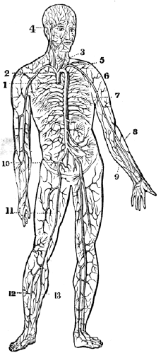
Fig. 70. The aorta and its branches. 1, The commencement of the aorta. 2, The arch of the aorta. 3, The carotid artery. 4, The temporal artery. 5, The subclavian artery. 6, The axillary artery. 7, The brachial artery. 8, The radial artery. 9, The ulnar artery. 10, The iliac artery. 11, The femoral artery. 12, The tibial artery, 13. The peroneal artery.
346. The VEINS are the vessels which return the blood to the auricles of the heart, after it has been circulated by the 161 arteries through the various tissues of the body. They are thinner and more delicate in structure than the arteries, so that when emptied of their blood, they become flattened and collapsed. The veins commence by minute radicles in the capillaries, which are every where distributed through the textures of the body, and coalesce to constitute larger and larger branches, till they terminate in the large trunks which convey the dark-colored blood directly to the heart. In diameter they are much larger than the arteries, and, like those vessels, their combined area would constitute an imaginary cone, the apex of which is placed at the heart, and the base at the surface of the body.
What does fig. 70 represent? 346. What are the veins?
347. The communications between the veins are more frequent than between the arteries, and take place between the larger as well as among the smaller vessels. The office of these inosculations is very apparent, as tending to obviate the obstructions to which the veins are peculiarly liable, from the thinness of their coats, and from inability to overcome great impediments by the force of their current. These tubes, as well as the arteries, are supplied with nutrient vessels, and it is to be presumed that nervous filaments from the sympathetic nerves are distributed to their coats.
348. The external, or cellular coat of the veins, is dense and firm, resembling the cellular tunic of the arteries. The middle coat is fibrous, like that of the arteries, but extremely thin. The internal coat is serous, and also similar to that of the arteries. It is continuous with the lining membrane of the heart at one extremity, and with the lining membrane of the capillaries at the other.
349. At certain intervals, the internal coat forms folds, or 162 duplicatures, which constitute valves. They are generally composed of two semilunar folds, one on each side of the vessel. The free extremity of the valvular folds is concave, and directed forward, so that while the current of blood sets toward the heart, they present no impediment to its free passage; but let the current become retrograde, and it is impeded by their distention. The valves are most numerous in the veins of the extremities, particularly the deeper veins situated between the muscles; but in some of the larger trunks, and also in some of the smaller veins, no valves exist.
Where do they commence? 347. What is said of their communications? What is the apparent design of the inosculations of the veins? What vessels are distributed to the coats of the veins? 348. Give the structure of the coats of the veins. 349. How are the valves in the veins formed?
Fig. 71.
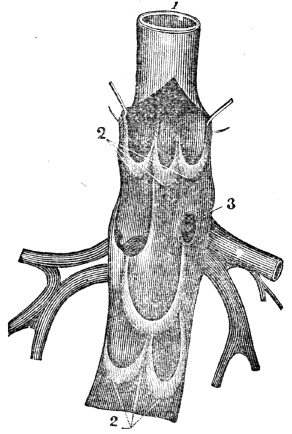
Fig. 71. A vein laid open to show the valves. 1, The trunk of the vein. 2, 2, Its valves. 3, An opening of a branch into the main trunk.
What is their use? Where are they the most numerous?
350. The CAPILLARIES constitute a microscopic net-work, and are so distributed through every part of the body as to render it impossible to introduce the smallest needle beneath the skin, without wounding several of these fine vessels. They are remarkable for the uniformity of diameter, and for the constant divisions and communications which take place between them.
351. The capillaries inosculate, on the one hand, with the terminal extremity of the arteries, and on the other, with the commencement of the veins. They establish the communication between the termination of the arteries and the beginning of the veins. The important operations of secretion and the conversion of the nutrient materials of the blood into bone, muscle, &c., are performed in these vessels.
Fig. 72. An ideal view of a portion of the pulmonic circulation. 1, 1, A branch of the artery that carries the impure blood to the lungs. 3, 3, Capillary vessels. 2, 2, A vein through which red blood is returned to the left side of the heart.
Fig. 73. An ideal view of a portion of the systemic circulation. 1, 1, A branch of the aorta. This terminates in the capillaries, (3, 3.) 2, 2, A vein through which the impure blood is carried to the right side of the heart.
350. What do the capillaries constitute? For what are they remarkable? 351. What relation do they bear to the arteries and veins? What important operations are performed in these vessels? What is represented by fig. 72? By fig. 73?
352. The walls of all the cavities of the heart are composed of muscular fibres, which are endowed with the property of contracting and relaxing, like the muscles of the extremities. The contraction and relaxation of the muscular tissue of the heart, produce a diminution and enlargement of both auricular and ventricular cavities. The auricles contract and dilate simultaneously, and so do the ventricles; yet the contraction and dilatation of the auricles do not alternate with the contraction and dilatation of the ventricles, as the dilatation of the one is not completed before the contraction of the other commences. The dilatation of the ventricles is termed the di-as´to-le of the heart; their contraction, its sys´to-le.
353. The ventricles contract quicker and more forcibly than the auricles, and they are three times longer in dilating than contracting. The walls of the right ventricle, being thinner than the left, are more distensible, and thus this cavity will contain a greater amount of blood. This arrangement adapts it to the venous system, which is more capacious than the arterial. The thicker and more powerful walls of the left ventricle adapt it to expel the blood to a greater distance.
354. The valves in the heart permit the blood to flow from the auricles to the ventricles, but prevent its reflowing. The valves at the commencement of the aorta and pulmonary 165 artery, permit the blood to flow from the ventricles into these vessels, but prevent its returning.
352–366. Give the physiology of the circulatory organs. 352. What do the contraction and relaxation of the muscular walls of the heart produce? How do the auricles and ventricles contract and dilate? 353. What is said of the contraction and dilatation of the ventricles in the heart? How is the right ventricle adapted to its function? How the left? 354. What is the use of the valves in the heart? Those of the aorta and pulmonary artery?
355. The function of the different parts of the heart will be given, by aid of fig. 74. The blood passes from the right auricle (3) into the right ventricle, (5,) and the tricuspid valves (6) prevent its reflux; from the right ventricle the blood is forced into the pulmonary artery, (7,) through which it passes to the lungs. The semilunar valves (9) prevent this circulating fluid returning to the ventricle. The blood, while passing over the air-cells in the lungs, in the minute divisions of the pulmonary artery, is changed from a bluish color to a bright red. It is then returned to the left auricle of the heart by the pulmonary veins, (11, 11.)
Fig. 74.

Fig. 74. 1, The descending vena cava, (vein.) 2, The ascending vena cava, (vein.) 3, The right auricle. 4, The opening between the right auricle and the right ventricle. 5, The right ventricle. 6, The tricuspid valves. 7, The pulmonary artery. 8, 8, The branches of the pulmonary artery that pass to the right and left lung. 9, The semilunar valves of the pulmonary artery. 10, The septum between the two ventricles of the heart. 11, 11, The pulmonary veins. 12, The left auricle. 13, The opening between the left auricle and ventricle. 14, The left ventricle. 15, The mitral valves. 16, 16, The aorta. 17, The semilunar valves of the aorta.
355. Describe the course of the blood from the right auricle in the heart to the lungs.
Observation. If the blood is not changed in the lungs, it will not flow to the pulmonary veins. This phenomenon is seen in instances of death from drowning, strangling, carbonic acid, &c. The same is true, but in a less degree, of individuals whose apparel is tight, as well as of those who breathe impure air, or have diseased lungs.
356. The left auricle, (12,) by its contraction, forces the blood into the left ventricle, (14.) The mitral valves (15) prevent its reflowing. From the left ventricle the blood is forced into the aorta, (16,) through which, and its subdivisions, it is distributed to every part of the system. The semilunar valves (17) prevent its returning.
Observation. The parts of the circulatory organs most liable to disease are the valves of the heart, particularly the mitral. When these membranous folds become ossified or ruptured, the blood regurgitates, and causes great distress in breathing. The operations of the system are thus disturbed as the movements of the steam engine would be if its valves were injured, or did not play freely.
357. The difference between the functions of the pulmonary artery and aorta is, the former communicates with the right ventricle of the heart, and distributes only impure blood to the lungs; the other connects with the left ventricle of the heart, and distributes pure blood to the whole body, the lungs not excepted. At the extremity of the divisions of the aorta, as well as the pulmonary artery, are found capillary vessels. This curious net-work of vessels connects with the minute veins of the body, which return the blood to the heart.
Observation. The function of the veins of the systemic 167 circulation is similar to the office of the arteries in the lungs, and that the veins of the pulmonic circulation transmit to the heart the pure, or nutrient blood, and thus supply the arteries of the general system with assimilating fluid.
What is the effect when the blood is not changed in the lungs? 356. Describe the circulation of the blood from the left auricle to the general system. What part of the circulatory organs is most liable to disease? What is the effect when the valves are diseased? 357. Give the difference in the functions of the pulmonary artery and aorta. Show the relation between the functions of the arteries and veins both of the pulmonic and systemic circulation.
358. The veins that receive the blood from all parts of the body, follow nearly the same course as the arteries. The myriads of these small vessels beneath the skin, and others that accompany the arteries, at last unite and form two large trunks, called ve´na ca´va as-cend´ens, and de-scend´ens.
Observation. A peculiarity is presented in the veins which come from the stomach, spleen, pancreas, and intestines. After forming a large trunk, they enter the liver, and ramify like the arteries, and in this organ they again unite into a trunk, and enter the ascending vein, or cava, near the heart. This is called the portal circulation.
359. The ventricles of the heart contract, or the “pulse” beats, about seventy-five times every minute; in adults; in infants, more than a hundred times every minute; in old persons, less than seventy-five times every minute. The energy of the contraction of this organ varies in different individuals of the same age. It is likewise modified by the health and tone of the system. It is difficult to estimate the muscular power of the heart; but, comparing it with other muscles, and judging from the force with which blood is ejected from a severed artery, it must be very great.
Observation. The phenomenon known under the name of pulse, is the motion caused by the pressure of the blood against the coats of the arteries at each contraction of the ventricles.
360. The following experiment will demonstrate that the blood flows from the heart. Apply the fingers upon the artery 168 at the wrist, at two different points, about two inches apart; if the pressure be moderately made, the “pulse” will be felt at both points. Let the point nearest the heart be pressed firmly, and there will be no pulsation at the lower point; but make strong pressure upon the lower point only, and the pulsation will continue at the upper point, proving that the blood flows from the heart, in the arteries, to different parts of the system.
358. What is the course of the veins? What peculiarity is observable in the veins of the liver? 359. How often does the heart contract, or the pulse beat, in adults? In infants? In old persons? What is said of the energy of its contraction in different persons? How is the pulse produced? 360. Demonstrate by experiment that the blood flows from the heart.
361. There are several influences, either separately or combined that propel the blood from the heart through the arteries, among which may be named,—1st. The contraction of the muscular walls of the heart. 2d. The contractile and elastic middle coat of the arteries aids the heart in impelling the blood to the minute vessels of the system. 3d. The peculiar action of the minute capillary vessels is considered, by some physiologists, as a motive power in the arterial circulation. 4th. The pressure of the muscles upon the arteries, when in a state of contraction, is a powerful agent, particularly when they are in active exercise.
362. The following experiments will demonstrate that the blood from every part of the system flows to the heart by the agency of the veins. 1st. Press firmly on one of the veins upon the back of the hand, carrying the pressure toward the fingers; for a moment, the vein will disappear. On removing the pressure of the finger, it will reappear, from the blood rushing in from below.
2d. If a tape be tied around the arm above the elbow, the veins below will become larger and more prominent, and also a greater number will be brought in view, while the veins above the tape are less distended. At this time, apply the finger at the wrist, and the pulsation of the arteries still continues, showing that the blood is constantly flowing from the heart 169 through the arteries, into the veins; and the increased size of the veins shows that the pressure of the tape prevents its flowing back to the heart.
361. State the influences that propel the blood from the heart. 362. Demonstrate by the first experiment that the blood flows to the heart. By the second experiment.
363. The influences that return the blood to the heart through the veins, are not so easily understood as those that act on the blood in the arteries. Some physiologists have imputed an active propulsive power to the capillary vessels in carrying the blood through the veins. This is not easily explained, and perhaps it is as difficult to understand. An influence upon which others have dwelt, is the suction power of the heart in active dilatation, acting as a vis a fronte (power in front) in drawing blood to it.
364. Another influence that aids the venous circulation is attributed to the propulsive power of the heart. It is not easy to comprehend how this power of the heart can be extended through the capillary vessels to the blood in the veins. Again, an important agency has been found, by some physiologists, in the inspiratory movements, which are supposed to draw the blood of the veins into the chest, in order to supply the vacuum which is created there by the elevation of the ribs and the descent of the diaphragm.
365. One of the most powerful causes which influence the venous circulation, is the frequently-recurring action of the muscles upon the venous trunks. When the muscles are contracted, they compress that portion of the veins which lie beneath the swell, and thus force the blood from one valve to the other, toward the heart. When they are relaxed, the veins refill, and are compressed by the recurring action of the muscles.
Observation. The physician, in opening a vein, relies on the energetic contractions and sudden relaxations of the muscles, when he directs the patient to clasp the head of a cane, or the arm of a chair; these alternate motions of the muscles cause an increased flow of blood to the veins of the ligated arm.
363. What is said of the influences that return the blood to the heart? What is said of the propulsive power of the capillaries? Of the suction power of the heart? 364. Give another influence. State another agency. 365. What is one of the most powerful causes which influence venous circulation? Give practical observation.
Fig. 75.

Fig. 75. An ideal view of the circulation in the lungs and system. From the right ventricle of the heart, (2,) the dark, impure blood is forced into the pulmonary artery, (3,) and its branches (4, 5) carry the blood to the left and right lung. In the capillary vessels (6, 6) of the lungs, the blood becomes pure, or of a red color, and is returned to the left auricle of the heart, (9,) by the veins, (7, 8.) From the left auricle the pure blood passes into the left ventricle, (10.) By a forcible contraction of the left ventricle of the heart, the blood is thrown into the aorta, (11.) Its branches (12, 13, 13) carry the pure blood to every organ or part of the body. The divisions and subdivisions of the aorta terminate in capillary vessels, represented by 14, 14. In these hair-like vessels the blood becomes dark colored, and is returned to the right auricle of the heart (1) by the vena cava descendens, (15,) and vena cava ascendens, (16.) The tricuspid valves (17) prevent the reflow of the blood from the right ventricle to the right auricle. The semilunar valves (18) prevent the blood passing from the pulmonary artery to the right ventricle. The mitral valves (19) prevent the reflow of blood from the left ventricle to the left auricle. The semilunar valves (20) prevent the reflow of blood from the aorta to the left ventricle.
366. The muscles exercise an agency in maintaining the venous circulation at a point above what the heart could perform. As the pulsations are diminished by rest, so they are accelerated by exercise, and very much quickened by violent effort. There can be little doubt that the increased rapidity of the return of blood through the veins, is, of itself, a sufficient cause for the accelerated movements of the heart during active exercise.
Observation. The quantity of blood in different individuals varies. From twenty-five to thirty-five pounds may be considered an average estimate in a healthy adult of medium size. The time in which the blood courses through the body and returns to the heart, is different in different individuals. Many writers on physiology unconditionally limit the period to three minutes. It is undeniable that the size and health of a person, the condition of the heart, lungs, and brain, the quantity of the circulating fluid, the amount and character of the inspired air, and the amount of muscular action, exert a modifying influence. The time probably varies from three to eight minutes.
366. What causes the accelerated movements of the heart during active exercise?
Note. Let the pupil review the anatomy and physiology of the circulatory organs from fig. 75, or from anatomical outline plates, No. 6 and 7.
367. If any part of the system is deprived of blood, its vitality will cease; but, if the blood is lessened in quantity to a limited extent, only the vigor and health of the part will be impaired. The following conditions, if observed, will favor the free and regular supply of blood to all portions of the system.
368. The clothing should be loosely worn. Compression of any kind impedes the passage of blood through the vessels of the compressed portion. Hence, no article of apparel should be worn so as to prevent a free flow of blood through every organ of the body.
369. The blood which passes to and from the brain, flows through the vessels of the neck. If the dressing of this part of the body is close, the circulation will be impeded, and the functions of the brain will be impaired. This remark is particularly important to scholars, public speakers, and individuals predisposed to apoplexy, and other diseases of the brain.
370. As many of the large veins lie immediately beneath the skin, through which the blood is returned from the lower extremities, if the ligatures used to retain the hose, or any other article of apparel, in proper position, be tight and inelastic, the passage of blood through these vessels will be 173 obstructed, producing, by their distention, the varicose, or enlarged veins. Hence elastic bands should always be used for these purposes.
367–386. Give the hygiene of the circulatory organs. 367. What effect will be produced on the body if it is deprived of blood? If the blood is only lessened in quantity? 368. Why should the clothing be worn loose? 369. What is said of dressing the neck? To what persons is this remark applicable? 370. How are enlarged veins frequently produced?
371. An equal temperature of all parts of the system promotes health. A chill on one portion of the body diminishes the size of its circulating vessels, and the blood which should distend and stimulate the chilled part, will accumulate in other organs. The deficiency of blood in the chilled portion induces weakness, while the superabundance of sanguineous fluid may cause disease in another part of the system.
372. The skin should be kept not only of an equal, but at its natural temperature. If the skin is not kept warm by adequate clothing, so that chills shall not produce a contraction of the blood-vessels and a consequent paleness, the blood will recede from the surface of the body, and accumulate in the internal organs. Cleanliness of the skin is likewise necessary, for the reason, that this condition favors the free action of the cutaneous vessels.
Observation. When intending to ride in a cold day, wash the face, hands, and feet, in cold water, and rub them smartly with a coarse towel. This is far better to keep the extremities warm, than to take spirits into the stomach.
373. Exercise promotes the circulation of the blood. As the action of the muscles is one of the important agents which propel the blood through the arteries and veins, daily and regular exercise of the muscular system is required to sustain a vigorous circulation in the extremities and skin, and also to maintain a healthy condition of the system. The best stimulants to improve the sluggish circulation of an indolent patient, whose skin is pale and whose extremities are cold, are the 174 union of vigorous muscular exercise with agreeable mental action, and the systematic application to the skin of cold water, attended with friction.
371. Why should the temperature of the body be equal? 372. Why should the skin be kept at its natural, as well as at an equal temperature? What practical observation when intending to ride in a cold day? 373. Why does exercise promote health? What are good stimulants for sluggish circulation in the indolent?
Illustration. The coach-driver and teamster throw their arms around their bodies to warm them when cold. The muscles that are called into action in swinging the arms, force a greater quantity of blood into the chilled parts, and consequently, more heat is produced.
374. When a number of muscles are called into energetic action, a greater quantity of blood will be propelled to the lungs and heart in a given time, than when the muscles are in a state of comparative inaction. It is no uncommon occurrence, that before there is a proper expansion of the respiratory organs to correspond with the frequency and energy of the movements of the muscles, there is an accumulation of blood in the lungs, attended by a painful sensation of fulness and oppression in the chest, with violent and irregular action of the heart. This condition of the organs of the chest, called congestion, may be followed by cough, inflammation of the lungs, asthma, and a structural disease of the heart.
375. To avoid these sensations and results, when we feel necessitated to walk or run a considerable distance in a short time, commence the movements in a moderate manner increasing the speed as the respiratory movements become more frequent and their expansion more extensive, so that a sufficient amount of air may be received into the lungs to purify the increased quantity of blood forced into them. The same principles should be observed when commencing labor, and in driving horses and other animals.
Observation. When a large number of muscles are called into action after repose, as when we rise from a recumbent or 175 sitting posture, the blood is impelled to the heart with a very strong impetus. If that organ should be diseased, it may arrive there in larger quantities than can be disposed of, and death may be the result. Hence the necessity of avoiding all sudden and violent movements, on the part of those who have either a functional or structural disease of the heart.
Mention the illustration. 374. What is the effect when a number of muscles are called into energetic action? What effect has this accumulation of blood in the lungs? 375. How can such disagreeable sensations be avoided? Mention a practical observation.
376. The mind exercises no inconsiderable influence upon the circulatory organs. When an individual is stimulated by hope, or excited by anger, the heart beats more forcibly, and the arteries act more energetically, than when a person is influenced by fear, despair, or sorrow. Consequently, the system is more fully nourished, and capable of greater exertion, when the former condition obtains, than when the latter exists.
377. The quality and quantity of the blood modify the action of the heart and blood-vessels. If this fluid is abundant and pure, the circulatory vessels act with more energy than when it is deficient in quantity or defective in quality.
Illustrations. 1st. In an athletic man, whose heart beats forcibly, and whose pulse is strong, if a considerable quantity of blood is drawn from a vein, as in bleeding, the heart will beat feebly, and the pulse will become weak.
2d. When the blood is made impure by inhaling vitiated air, the action of the heart and arteries is diminished, which produces an effect similar to that which takes place when blood is drawn from a vein.
378. Hemorrhage from divided arteries should be immediately arrested. When large blood-vessels are wounded or cut, the flow of blood must be immediately stopped, or the person soon faints, and the heart ceases its action. If it is a 176 large artery that is wounded, the blood will be thrown out in jets, or jerks, every time the pulse beats. The flow of blood can be stopped until a surgeon arrives, either by compressing the vessel between the wound and the heart, or by compressing the end of the divided artery in the wound.
376. State some of the effects that the mind has on circulation. 377. What effect have the quantity and quality of blood upon the circulatory organs? Give illustration 1st. Illustration 2d. 378. What is necessary when large blood-vessels are wounded or cut?
Fig. 76. The track of the large artery of the arm. 1, The collar-bone. 9, The axillary artery. 10, The brachial artery.
Fig. 77. B, The manner of compressing the artery near the collar-bone. A, The manner of compressing the large artery of the arm, with the fingers. C, The manner of compressing the divided extremity of an artery in the wound, with a finger.
379. After making compression with the fingers, as described and illustrated, take a piece of cloth or handkerchief, twist it cornerwise, and tie a hard knot midway between the 177 two ends. This knot should be placed over the artery, between the wound and the heart, and the ends carried around the limb and loosely tied. A stick, five or six inches long, should be placed under the handkerchief, which should be twisted until the knot has made sufficient compression on the artery to allow the removal of the fingers without a return of bleeding. Continue the compression until a surgeon can be called.
What is shown by fig. 76? By fig. 77? 379. What is to be done after compressing the wound, as before described?
Fig. 78. A, B, The track of the large artery of the arm. The figure exhibits the method of applying the knotted handkerchief to make compression on this artery.
Fig. 79. A, C, The track of the large artery of the thigh. B, The method of applying the knotted handkerchief to compress this artery. In practice, the twisting stick B should be placed opposite the knot over the artery A, C.
380. When an artery of the arm is cut, elevating the wounded limb above the head will tend to arrest the flow of blood. In a wound of a lower limb, raise the foot, so that it shall be higher than the hip, until the bleeding ceases.
Illustration. On one occasion, the distinguished Dr. Nathan Smith was called to a person who had divided one of 178 the large arteries below the knee. After trying in vain to find the bleeding vessel, so as to secure it, he caused the foot to be elevated higher than the hip. At the first instant the blood was forced from the wound about twelve inches; in a minute, it was diminished to three or four; and, in a short time, the bleeding ceased. This Dr. S. called his “great” operation; and it was truly great in simplicity and science.
What is shown by fig. 78 and 79? 380. What suggestion relative to the position of a limb when bleeding? Relate a simple operation by Dr. Nathan Smith.
381. The practical utility of every person knowing the proper means of arresting hemorrhage from severed arteries, is illustrated by the following incidents. In 1848, in the town of N., Mass., a mechanic divided the femoral artery; although several adult persons were present, he died in a few minutes from loss of blood, because those persons were ignorant of the method of compressing severed arteries until a surgeon could be obtained.
382. In 1846, a similar accident occurred in the suburbs of Philadelphia. While the blood was flowing copiously, a lad, who had received instruction on the treatment of such accidents at the Philadelphia High School, rushed through the crowd that surrounded the apparently dying man, placed his finger upon the divided vessel, and continued the compression until the bleeding artery was secured by a surgeon.
383. In “flesh wounds,” when no large blood-vessel is divided, wash the part with cold water, and, when bleeding has ceased, draw the incision together, and retain it with narrow strips of adhesive plaster. These should be put on smoothly, and a sufficient number applied to cover the wound. In most instances of domestic practice, the strips of adhesive plaster are too wide. They should not exceed in width one 179 fourth of an inch. Then apply a loose bandage, and avoid all “healing salves,” ointments, and washes. In removing the dressing from a wound, both ends of the strips of plaster should be raised and drawn toward the incision. The liability of the wound re-opening is thus diminished.
381. Relate the first incident showing the utility of every person knowing the proper method of arresting the flow of blood from divided arteries. 382. The second incident. 383. How should “flesh wounds” be dressed?
Observation. The union of the divided parts is effected by the action of the divided blood-vessels, and not by salves and ointments. The only object of the dressing is to keep the parts together, and protect the wound from air and impurities. Nature, in all cases of injuries, performs her own cure. Such simple wounds do not generally require a second dressing and should not be opened until the incisions are healed.
Fig. 80.

Fig. 80. The manner in which strips of adhesive plaster are applied to wounds.
384. In wounds made by pointed instruments, as a nail, or in lacerated wounds, as those made by forcing a blunt instrument, as a hook, into the soft parts, there will be no direct and immediate union. In these cases, apply a soothing poultice, as one made of linseed meal, and also keep the limb still. It is judicious to consult a physician immediately, in punctured or lacerated wounds, because they often induce the most dangerous diseases.
385. Wounds caused by the bite of rabid animals or venomous serpents, should be immediately cleansed with pure 180 water. In many instances, the application of suction, either with “cupping glasses,” or the mouth, will prevent the introduction of the poisonous matter into the system by absorption. When this is effected, cover the wound with a soothing poultice, as one made of slippery elm bark.
What should be avoided? How should the strips of plaster be removed from a wound? How is the union of the divided parts effected? 384. How should punctured and lacerated wounds be dressed? 385. What is the treatment of wounds caused by the bite of rabid animals?
Observation. Although animal poisons, when introduced into the circulating fluid through the broken surface of the skin, frequently cause death, yet they can be taken into the mouth and stomach with impunity, if the mucous membrane which lines these parts is not broken.
Fig. 81.
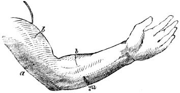
Fig. 81. a, a, Representation of wounds on the back part of the arm and fore-arm b, b, Wounds on the anterior part of the arm and fore-arm. By bending the elbow and wrist, the incisions at a, a, are opened, while those at b, b, are closed. Were the arm extended at the elbow and wrist, the wounds at a, a, would be closed, and those at b, b, would be opened.
386. The proper position of the limbs favors the union of wounds. If the incision be upon the anterior part of the leg, between the knee and ankle, extending the knee and bending the ankle will aid its closing. If it be upon the back part of the leg, by extending the foot and bending the knee, the gaping of the incision will be diminished. When wounds occur upon the trunk or upper extremities, let the position of the person be regarded.
386 Does the proper position of the limbs favor the union of wounds?
387. Absorption is the process by which the materials of nutrition are removed from the alimentary canal, to be conveyed into the circulatory vessels. It is likewise the process by which the particles of matter that have become injurious, or useless, are removed from the mass of fluids and solids of which the body is composed. These renovating and removing processes are performed by two sets of vessels
388. The vessels that act exclusively for the growth and renovation of the system, are found only in the alimentary canal. They are called lacteals. The vessels whose sole function is to remove particles of matter already deposited, are called Lym-phat´ics. The radicles, or commencement of the veins, in many, and it may be in all parts of the body, perform the office of absorption.
Observation. This fact accounts for the capacity of the venous system exceeding the arterial. Had the veins no other function to perform, beside returning the blood that had been distributed by the arteries, it would be reasonable to suppose that this system would be less than the arterial, but the reverse is known to be true.
389. The LYMPHATIC VESSELS, in structure, resemble the 182 lacteals. They exist in great numbers in the skin and mucous membranes, particularly those of the lungs. Though no lymphatics have been traced to the brain, it is presumed that they exist there, as this part of the body is not exempt from the composition and decomposition, which are perpetual in the body. These vessels are extremely minute at their origin, so that in many parts of the system they cannot be detected without the aid of a microscope.
387. Define absorption. 388–391. Give the anatomy of the lymphatic vessels. 388. What are those vessels called that act exclusively for the growth and renovation of the body? Those whose office is to remove the atoms already deposited? What other vessels perform the office of absorption? Give observation. 389. Describe the lymphatics.
Fig. 82. A single lymphatic vessel, much magnified.
Fig. 83. The valves of a lymphatic trunk.
Fig. 84. 1, A lymphatic gland with several vessels passing through it.
390. The lymphatic vessels, like the veins, diminish in number as they increase in size, while pursuing their course toward the large veins near the heart, into which they pour their contents. The walls of these vessels have two coats of which the external one is cellular, and is capable of considerable distention. The internal coat is folded so as to form 183 valves, like those in the veins. Their walls are so thin, that these folds give them the appearance of being knotted.
What is represented by fig. 82? By fig. 83? By fig. 84? 390. In what respect do these vessels resemble the veins of the system? Give the structure of their coats.
391. At certain points, the lymphatic vessels pass through distinct, soft bodies, peculiar to themselves, which are called lymphatic glands, which are to these vessels what the mesenteric glands are to the lacteals. The lymphatic glands vary in form and in size. They are extremely vascular, and appear to consist of a collection of minute vessels. These glands are found in different parts of the body, but are most numerous in the groins, axilla, or arm-pits, neck, and cavities of the chest and abdomen.
Observation. From exposure to cold, these glands are frequently enlarged and inflamed. They are known under the name of “kernels.” They are often diseased, particularly in scrofula, or “king’s evil.”
392. Though the lacteals and lymphatics resemble each other in their structure and termination, yet they differ as to the nature of the fluids which they convey, as well as the nature of their functions. The lacteals open into the small intestine, and possess the power of rejecting all substances in the passing aliment, but the chyle. The lymphatics, on the contrary, not only imbibe all the various constituents of the body, both fluid and solid, but they sometimes absorb foreign and extraneous substances, when presented to their mouths, as in vaccination.
393. The varieties of absorption are, the In-ter-sti´tial, Rec-re-men-ti´tial, Ex-cre-men-ti´tial, Cu-ta´ne-ous, Res-pi´ra-to-ry, Ve´nous, and the Lac´te-al.
391. Describe the lymphatic glands. What observation is given in regard to these glands? 392–403. Give the physiology of the lymphatic vessels. 392. Explain the difference between the lacteals and lymphatics 393. Name the varieties of absorption.
394. Interstitial absorption is that change which is constantly going on in the animal economy among the particles of matter of which every texture is composed. The ordinary functions of the body, in health, require incessant action of the lymphatics; the circulatory system, with its myriads of small vessels, is constantly depositing new atoms of matter, which become vitalized, and perform a course of actions, then die, or become useless. These old atoms are removed by the absorbent system. Thus, wherever there is a minute artery to deposit a living particle of matter, there is a lymphatic vessel, or venous radicle, to remove it as soon as it shall have finished its particular office.
395. The action of the lymphatic vessels counterbalances those of nutrition, and thus the form and size of every part of the body is preserved. When their action exceeds that of the nutrient vessels, the body emaciates; when it is deficient, plethora is the result. In youth, they are less active than the nutrient vessels, and the limbs are plump; but in later periods of life, we find these actions reversed, and the body diminishes in size. It is not unfrequent that wens, and other tumors of considerable size, disappear, and even the entire bone of a limb has been removed from the same general cause. The effused fluids of bruises are also removed by absorption.
Observations. 1st. When little or no food is taken into the stomach, life is supported by the lymphatic vessels and veins imbibing the fat and reconveying it into the blood vessels. It is the removal of this secretion which causes the emaciation of the face and extremities of a person recovering from a fever. In consumption, the extreme attenuation of the limbs is caused by the absorption, not only of the fat, but also of the muscles and more solid parts of the system.
394. What is interstitial absorption? Flow are the new atoms of matter deposited? How removed? 395. What vessels do the lymphatics counterbalance in action? What is the result when their action exceeds that of the nutrient vessels? When it is less? Mention some instances of active absorption. What causes the emaciated limbs of a person recovering from fever? The extreme attenuation in consumption?
2d. Animals which live in a half torpid state during the winter, derive their nourishment from the same source. In other words, we may say the starving animal lives for a time upon itself, eating up, by internal absorption, such parts of the body as can be spared under urgent necessity, to feed those organs and continue those functions that are absolutely essential to life.
396. Recrementitial absorption is the removal of those fluids from the system, which are secreted upon surfaces that have no external outlet. These fluids are various, as the fat, the marrow, the synovia of joints, serous fluids, and the humors of the eye. Were it not for this variety of absorption, dropsy would generally exist in the cavities of the brain, chest, and abdomen, from the continued action of the secretory vessels.
397. Excrementitial absorption relates to the fluids which have been excreted, such as the bile, pancreatic fluid, saliva, milk, and other secretions.
398. Cutaneous absorption relates to the skin. Here the lymphatic vessels extend only to the cuticle, which they do not permeate. There has been much diversity of opinion on the question of cutaneous absorption; some maintaining that this membrane absorbs, while others deny it. Many experiments have proved that the skin may absorb sufficient nutriment to support life for a time, by immersing the patient in a bath of milk or broth. It has been found that the hand, immersed to the wrist in warm water, will absorb from ninety to one hundred grains of fluid in the space of an hour.
399. Thirst may be quenched by applying moist clothes to the skin, or by bathing. It is no uncommon occurrence, during a passage from one continent to the other, for the saliva 186 to become bitter by the absorption of sea water. Medicinal substances, such as mercury, morphine, and Spanish flies, are frequently introduced into the system through the skin.
396. What is recrementitial absorption? 397. Define excrementitial absorption. 393. To what does cutaneous absorption relate? Is there a diversity of opinion respecting this variety of absorption? What do well attested experiments show? 399. What remark in reference to quenching thirst? What agency conveys medicinal substances and ointments into the system when tabbed on the skin?
400. Respiratory absorption has reference to the lungs. The mucous membrane of these organs is abundantly supplied with lymphatic vessels. By their action, substances finely pulverized, or in the form of gas, are readily imbibed when inhaled into the lungs, such as metallic vapors, odoriferous particles, tobacco smoke, and other effluvia. In this way, contagious diseases are frequently contracted.
Illustration. In inhaling sulphuric ether, or letheon, it is introduced into the vessels of the lungs in the form of vapor, and through them it is rapidly conveyed to the brain, and thus influences the nervous system.
401. Venous absorption is the function which the veins perform in absorbing from the alimentary canal liquids of various kinds that have been taken into the stomach and are not converted into chyle. In other parts of the body, they also perform the common office of lymphatics.
402. Lacteal, or digestive absorption has reference to the absorption of chyle only, which is destined for the nutrition of the body.
403. Absorption is not only very abundant, but generally very rapid, and all these varieties are maintained through life, except when suspended by disease.
400. What is said of respiratory absorption? How is letheon introduced into the system? 401. Define venous absorption. 402. What is lacteal absorption? 403. What is said of absorption?
Fig. 85.
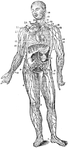
Fig. 85. A representation of the lymphatic vessels and glands. 1, 2, 3, 4, 5, 6, The lymphatic vessels and glands of the lower limbs. 7, Lymphatic glands. 8, The commencement of the thoracic duct. 9, The lymphatics of the kidney. 10, Of the stomach. 11, Of the liver. 12, 12, Of the lungs. 13, 14, 15, The lymphatics and glands of the arm. 16, 17, 18, Of the face and neck. 19, 20, Large veins. 21, The thoracic duct. 26, The lymphatics of the heart.
404. By the action of the lymphatics, substances of an injurious, as well as of a beneficial, character may be conveyed into the system. These vessels, under certain conditions, are more active in their office than at other periods; and it is of practical utility to know what influences their action.
405. The function of these vessels is increased by moisture, and lessened by an active state of the lacteals. Observation shows that the ill-fed, and those persons that live in marshy districts, contract contagious diseases more readily than those individuals who are well fed, and breathe a dry and pure air.
406. The air of the sick-room should be dry. If due attention is not given to ventilation, the clothing of the nurse and patient, together with the air of the room, will be moistened by the exhalations from the skin and lungs. This exhalation may contain a poison of greater or less power, according to its quantity and degree of concentration, and may be absorbed and reconveyed into the system, causing inflammatory diseases, and not unfrequently death.
Observations. 1st. When we are attending a sick person a current of air that has passed over the patient should be avoided. We may approach with safety very near a person who has an infectious disease, provided care is taken to keep on the side from which the currents of air are admitted into the room.
2d. When we have been visiting or attending on a sick person, it is judicious to change the apparel worn in the sick-room, and also give the skin a thorough bathing. The outside garments, also, should be aired, as poisonous matter may have penetrated the meshes of the clothing.
404–413. Give the hygiene of the lymphatic vessels. 404. What is said respecting the action of the lymphatic vessels? 405. What influences the function of these vessels? What does observation show? 406. Why should the air of the sick-room be dry? What suggestion when we have been visiting or attending on the sick?
407. The stomach should be supplied with food of a nutrient and digestible character, in proper quantities, and at stated periods. The chyle formed from the food stimulates the lacteals to activity, which activity is attended with an inactive state of the lymphatics of the skin and lungs. Thus due attention should be given to the food of the attendants on the sick, and the members of the family. Before visiting a sick person it is judicious to take a moderate amount of nutritious food.
Observation. Many individuals, to prevent contracting disease that may be communicated from one person to another, use tobacco, either chewed or smoked; and sometimes alcohol, with decoctions of bitter herbs. These substances do not diminish, but tend to increase, the activity of the lymphatics. Thus they make use of the means by which the poisonous matter formed in the system of the diseased person, may be more readily conveyed into their own.
408. The skin and clothing, as well as the bed-linen, should be frequently cleansed. This will remove the poisonous matter that may be deposited upon the skin and garments, which, if suffered to remain, might be conveyed into the system by the action of the lymphatics. This points also to a frequent change of the wearing apparel, as well as the coverings of the bed. In visiting the unhealthy districts of the South and West, the liability of contracting disease is much lessened by taking a supply of food at proper periods, keeping the skin and clothing in a clean state, the room well ventilated, and avoiding the damp chills of evening.
409. Absorption by the skin is most vigorous when the cuticle is removed by vesication, or blistering. Then external 190 applications are brought into immediate contact with the orifices of the lymphatics of the skin, and by them rapidly imbibed and circulated through the system. Thus arsenic applied to the cutaneous vessels, and strong solutions of opium to extensive burns, have been absorbed in quantities sufficient to poison the patient.
407. Why should the stomach be supplied with food of a nutrient and digestible character? What is said of the use of alcohol, or tobacco, in preventing the introduction of the poisonous matter of contagious diseases? 408. Why should the clothing and bed-linen be frequently washed? What suggestion to persons in visiting the unhealthy districts of the South and West? 409. When is cutaneous absorption most vigorous? Why?
410. When the cuticle is only punctured or abraded, poisonous matter may be introduced into the system. The highly respected Dr. W., of Boston, lost his life by poisonous matter from the body of a patient subjected to a post mortem examination. He had removed from his finger, previous to the examination, a “hang-nail,” and the poison from the dead body was brought in contact with the denuded part, and through the agency of the lymphatics it was conveyed into the system.
411. Puncture any part of the cuticle with the finest instrument that has upon its point the smallest conceivable quantity of the vaccine virus, or small-pox matter, and it will be brought into contact with the lymphatic vessels, and through their agency conveyed into the system. The result is, that persons thus operated upon have the small-pox, or the vaccine disease.
412. When we expose ourselves to any poisonous vapors, or handle diseased animals or sick persons, safety and health require that the cuticle be not broken or otherwise injured. In many instances, the poisonous animal matter upon hides has been introduced into the systems of tanners, through small ulcers upon their fingers or hands. From these sores there would be seen small red lines extending up the arm. These swelled tracts indicate an inflammation of the large lymphatic 191 trunks, that have been irritated and diseased by the passage of poisonous matter through them into the system.
410. Do the same results follow, if the cuticle is only punctured? Relate an instance of death by the absorption of poisonous matter. 411. By what means is the vaccine matter introduced into the system? 412. What caution is necessary when we expose ourselves to poisonous vapors?
Observation. A distressing illustration of the absorption of deleterious substances from the surface of a sore, is seen in the favorite experiments of that class of “quacks,” who style themselves “cancer doctors.” With them, every trifling and temporary enlargement, or tumor, is a cancer. Their general remedy is arsenic; and happy is the unfortunate sufferer who escapes destruction in their hands, for too frequently their speedy cure is death.
413. In case of an accidental wound, it is best immediately to bathe the part thoroughly in pure water, and to avoid all irritating applications. In some instances, it would be well to apply lunar caustic immediately. When handling or shrouding dead bodies, or removing the skin from animals that have died of disease, it would be well to lubricate the hands with olive-oil or lard. This affords protection to the minute portions of the skin, from which the cuticle may be removed. In all cases where there is an ulcer or sore, the part should be covered with something impervious to fluids, as court-plaster, before exposing the system to any animal, vegetable, or mineral poison.
413. What direction is given when the cuticle is broken? What suggestion is given when shrouding dead bodies?
414. In the human body are found many fluids and solids of dissimilar appearance and character. These are produced by the action of organs, some of which are of simple structure, while others are very complicated in their arrangement. These organs are called Se-cre´to-ry.
415. The SECRETORY ORGANS are the Ex-ha´lants, Fol´li-cles, and the Glands.
416. The EXHALANTS were supposed to be terminations of arteries or capillaries. The external exhalants terminate on the skin and mucous membranes; the internal in the cellular and medullary tissues. (Appendix I.)
Fig. 86.

Fig. 86. A secretory follicle. An artery is seen, which supplies the material for its secretion. Follicles are also supplied with veins and organic nerves.
417. The FOLLICLES are small bags, or sacs, situated in the true skin, and mucous membrane. The pores seen on the skin are the outlets of these bodies.
414. How are the fluids and solids of the body produced? 415–419. Give the anatomy of the secretory organs. 415. Name the secretory organs. 416. Describe the exhalants. What is represented by fig. 86? 417. Define follicles.
418. The GLANDS are soft, fleshy organs, and as various in their structure, as the secretions which it is their function to produce. Each gland is composed of many small lobules united in a compact mass, and each lobule communicates by a small duct with the principal outlet, or duct of the organ. Every gland is supplied with arteries, veins, lymphatics, and nerves. These, with the ducts, are arranged in a peculiar manner, and connected by cellular membrane.
419. There are two classes of glands, one for the modification of the fluids which pass through them, as the mesenteric and lymphatic glands; and the other for the secretion of fluids which are either useful in the animal economy, or require to be rejected from the body.
Fig. 87.
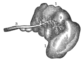
Fig. 87. 1, 1, A secretory gland. 2, 2, Minute ducts that are spread through the glands. These coalesce to form the main duct, 3.
420. Secretion is one of the most obscure and mysterious functions of the animal economy. “It is that process by which various substances are separated from the blood, either with or without experiencing any change during their 194 separation.” Not only is the process by which substances are separated from the blood, called secretion, but the same term is also applied to substances thus separated. Thus physiologists say, that by the process of secretion, bile is formed by the liver; and also, that bile is the secretion of this organ.
418. Give the structure of the glands. 419. How are the glands arranged? 420–431. Give the physiology of the secretory organs. 420. What is secretion?
421. The secreted fluids do not exist in that form in the blood, but most of the elements of which they are made do exist in this fluid, and the “vessels by which it is accomplished may well be called the architects and chemists of the system; for out of the same material—the blood—they construct a variety of wonderful fabrics and chemical compounds. We see the same wonderful power possessed, also, by vegetables; for out of the same materials the olive prepares its oil, the cocoa-nut its milk, the cane its sugar, the poppy its narcotic, the oak its green pulpy leaves, and its dense woody fibre. All are composed of the same few, simple elements, arranged in different order and proportions.”
422. “In like manner we find the vessels, in animated bodies, capable of forming all the various textures and substances which compose the frame; the cellular tissue, the membranes, the ligaments, the cartilages, the bones, the marrow, the muscles with their tendons, the lubricating fluid of the joints, the pulp of the brain, the transparent jelly of the eye; in short, all the textures of the various organs of which the body is composed, consist of similar ultimate elements, and are manufactured from the blood.”
423. Of the agents that produce or direct the different secretions, we have no very accurate knowledge. Some have supposed this function to be mechanical, others a chemical process, but experiments prove that it is dependent on nervous influence. If the nerves are divided which are distributed to 195 any organ, the process of secretion is suspended. It is no uncommon occurrence, that the nature of milk will be so changed from the influence of anger in the mother, as to cause vomiting, colic, and even convulsions, in the infant that swallows it. Unexpected intelligence either of a pleasant or unpleasant character, by its influence on the nervous system, will frequently destroy the appetite. Sometimes mental agitation, as fear, will cause a cold sweat to pervade the surface of the body.
421. What is said respecting secreted substances? Do vegetables possess the property of secretion? 422. From what are the various textures formed? 423. Have we accurate knowledge of the agents that produce secretion?
424. Secretions are constantly maintained, during life, from the serous membrane, by the action of the internal exhalants. The fluid which is exhaled bears some resemblance to the serum of the blood. Its use is to furnish the organs, which are surrounded by this membrane, with a proper degree of moisture, and thus enables them to move easily on each other, as those within the chest and abdomen.
425. The cellular tissue exhales a serous fluid, and when it becomes excessive in quantity, general dropsy is produced. Fat is another secretion, which is thrown out, in a fluid state, from the cellular membrane. It is deposited in little cells, and exists in the greatest abundance between the skin and the muscles. Its use seems to be, to form a cushion around the body for its protection; to furnish nutriment for the system when food cannot be taken; to supply the carbon and hydrogen necessary to sustain the generation of heat, when these articles of combustion are not otherwise furnished. The med´ul-la-ry substance, (marrow,) in the cavities of the long bones, is very much like fat.
Observation. During sickness, if there is not emaciation or absorption of this secretion, it is considered an unfavorable symptom, because it indicates a want of power in the absorbing system, which is among the last to be affected.
How is it proved that secretion depends on nervous influence? 424. What is said of the secretions from the serous membrane? 425. From what tissue is a serous fluid exhaled? What is the effect when this fluid becomes excessive in quantity? What is fat? Its use? What is marrow?
426. The mucous secretion is a transparent, viscid fluid which is secreted by those membranes that line the cavities of the body, which have an external communication, as the trachea and alimentary canal. This secretion serves to protect these parts from the influence of the air, and concurs, by means of its peculiar properties, in the performance of their functions. 427. There are two external secretions, namely, one from the skin, called perspiration, and the other from the lungs. The cutaneous exhalation, or transpiration[14] exists in two forms, called sensible perspiration (sweat) and insensible perspiration. The pulmonary exhalation is the most important and universal, and closely resembles that of the skin.
428. The follicles are found only in the skin and mucous membrane. They secrete an oily, unctuous substance, which mixes with the transpiration, and lubricates the skin. At the root of each hair there is a minute follicle, which secretes the fluid that oils the hair. The wax in the passage of the ear is secreted from these bodies.
429. All the blood distributed to the different glands is similar in composition and character; but the fluids secreted by them, vary in appearance in a remarkable degree. The office of the glands appears to be principally to form different secretions. Thus the salivary glands secrete the insipid saliva; the lachrymal glands, the saline tears; the liver, the yellow, ropy bile; and the kidneys, the acrid urine.
426. What is said relative to the mucous secretion? 427. Name the external secretions. 428. Give the office of the follicles. 429. What appears to be the principal office of the glands? 430. Mention a secretion produced in a particular emergency.
430. Some secretions are evidently produced only in particular 197 emergencies, as is seen in the increased secretion of bony matter when a limb is broken.
431. When any substance which is not demanded for nutrition, or does not give nourishment to the system, is imbibed by the lymphatic vessels, and conveyed into the blood, it is eliminated in the secretions.
Illustration. A few years since, a poor inebriate was carried to a London hospital in a state of intoxication. He lived but a few hours. On examining his brain, nearly half a gill of fluid, strongly impregnated with gin, was found in the cavities of this organ. This was secreted from the vessels of the brain.
432. Unless the secretions are regularly maintained, disease will be the ultimate result. Let the secretions from the skin be suppressed, and fever or some internal inflammation will follow. If the bile is impeded, digestion will be impaired. If any other secretion is suppressed, it will cause a derangement of the various internal organs.
Observation. Ardent spirits derange the secretions, and change the structure of the brain. This is one reason why inebriates do not generally live to advanced age.
433. The quantity of blood influences the character of the secretions. If it is lessened to any great extent, the secretions will be lessened as well as changed in character.
Illustration. When a person has lost a considerable quantity of blood, there is a sensation of thirst in the fauces, attended with a cold, pale, dry skin. When reaction comes 198 on, the perspiration is cold, attended with nausea, and sometimes vomiting.
431. What becomes of those substances imbibed by the lymphatics that do not give nourishment to the body. 432–437. Give the hygiene of the secretory organs. 432. What effect on the system when the secretions are not regularly maintained? 433. Does the quantity of blood influence the secretions? Give an illustration.
434. The secretory organs require the stimulus of pure blood. If this fluid is vitiated, the action of the secretory organs will be more or less modified. Either the quantity will be affected or the quality will be altered.
Observation. The impurity of the blood arising from the inhalation of the vitiated air of sleeping rooms, diminishes and changes the character of the secretions of the mouth and stomach. This accounts for the thirst, coated tongue, and disagreeable taste of the mouth when impure air is breathed during sleep. The disease it induces, is indigestion or dyspepsia.
435. The amount of action modifies the condition of the secretory organs. When a secretory organ is excessively stimulated, its vigor and energy are reduced. The subsequent debility may be so great as to suppress or destroy its functional power.
Illustrations. 1st. In those sections of the country where flax is spun on a “foot-wheel,” it is not unfrequent that the spinners moisten the thread with the secretions of the mouth. This seems to operate economically for a time, but debility of the salivary organs soon follows, which incapacitates them from supplying saliva sufficient to moisten the food, producing in a short time disease of the digestive organs.
2d. The habit of continual spitting, which attends the chewing of tobacco and gums, and other substances, between meals, induces debility, not only of the salivary glands, but of the system generally.
436. One secretory organ may do the office of another. This increased action of a secretory organ may be sustained 199 for a limited time without permanent injury, but, if long continued, a diseased action of the organ will follow. Of morbid secretions we have examples in the ossification of the valves of the heart, cancerous and other tumors.
434. What is the effect of impure blood on the secretory organs? 435. What results from stimulating excessively a secretory organ? How is this illustrated? 436. What is the effect when one secretory organ performs the office of another?
Observation. In the evenings of the warm season, a chill upon the impressible skin, that suppresses the perspiration, is frequently followed by a diarrhœa, dysentery, or cholera morbus. These can be prevented by avoiding the chill. An efficient means of relief, is immediately to restore the skin to its proper action.
437. The secretions are much influenced by the mind. How this is effected, it is difficult to explain; but many facts corroborate it. Every one has felt an increased action of the tear-glands from distressing feelings. Cheerfulness of disposition and serenity of the passions are peculiarly favorable to the proper performance of the secretory function. From this we may learn how important it is to avoid such things as distract, agitate, or harass us.
Observation. In fevers and other diseases, when the skin, mouth, and throat are dry from a suppression of the secretions, let the mind of the patient be changed from despondency to hope, and the skin and the membrane that lines the mouth and throat will exhibit a more moist condition, together with a general improvement of the vital organs of the system. Consequently, all just encouragement of the restoration to health should be given to a sick person.
Give examples of morbid secretions. What is one cause of dysentery and cholera morbus? How can these affections he relieved? 437. Show the influence of the mind on the secretions. Mention instances of its influence.
438. Nutrition is the vital act by which the different parts of the body renew the materials of which they are composed. Digestion, circulation, absorption, and respiration, are but separate links in the chain of nutrition, which would be destroyed by the absence of any one of them.
439. The nutritive process is also a kind of secretion, by which particles of matter are separated from the blood and conveyed with wonderful accuracy to the appropriate textures. The function of the nutrient vessels antagonizes those of absorption: while one system is constructing, with beautiful precision, the animal frame, the other is diligently employed in pulling down this complicated structure.
440. This ever-changing state of the body is shown by giving animals colored matter, mixed with their food, which in a short time tinges their bones with the same color as the matter introduced. Let it be withdrawn, and in a few days the bones will assume their former color—evidently from the effects of absorption. The changeful state of the body is further shown by the losses to which it is subjected; by the necessity of aliment; by the emaciation which follows abstinence from food.
438–454. What remarks respecting nutrition? 438. What is nutrition? 439. What is said of the nutritive process? The function of the nutrient vessels? 440. Give a proof of the ever-changing state of the body. Give other instances illustrative of the changeful state of the body.
441. Every part of the body is subject to this continual change of material, yet it is effected with such regularity, that the size, shape, and appearance, of every organ is preserved; and after an interval of a few years, there may not remain a particle of matter which existed in the system at a former period. Notwithstanding this entire change, the personal identity is never lost.
442. Many calculations have been made to determine in what length of time the whole body is renewed. Some have supposed that it is accomplished in four years; others have fixed the period at seven years; but the time of the change is not definite, as was supposed by a genuine son of the Emerald Isle, who had been in America seven years and three months, and consequently maintained that he was a native American.
Observation. India ink, when introduced into the skin, is not removed; hence some assert that this tissue is an exception to the alternate deposition and removal of its atoms. The ink remains because its particles are too large to be absorbed, and when in the skin it is insoluble.
443. “Those animals which are most complicated in their structure, and are distinguished by the greatest variety of vital manifestations, are subject to the most rapid changes of matter. Such animals require more frequent and more abundant supplies of food; and, in proportion as they are exposed to the greater number of external impressions, will be the rapidity of this change of matter.”
444. “Animals may be situated so that they lose nothing by secretion; consequently, they will require no nutriment. Frogs have been taken from fissures in solid lime rock, which 202 were imbedded many feet below the surface of the earth, and, on being exposed to the air, exhibited signs of life.”
441. Why is the personal identity never lost in the change of materials, which is unceasing in the system? 442. Give the opinion of physiologists respecting the time required for the renewal of the whole body. What exception to the changing state Of the different textures? 443. What animals are subject to the most rapid changes of material? 444. May animals be situated so that they require no nutriment? What is related of frogs?
445. The renovation of the bone, muscle, ligament, tendon, cartilage, fat, nerve, hair, &c., is not perfected merely by the general circulation of the fluid which is expelled from the left side of the heart, but through the agency of a system of minute vessels, which, under ordinary circumstances, cannot be seen by the eye, even when aided by the microscope; still, minute as they are, the function of these agents is necessary to the continuance of life. They are the smallest capillary vessels.
446. “As the blood goes the round of the circulation, the nutrient capillary vessels select and secrete those parts which are similar to the nature of the structure, and the other portions pass on; so that every tissue imbibes and converts to its own use the very principles which it requires for its growth; or, in other words, as the vital current approaches each organ, the particles appropriate to it feel its attractive force,—obey it,—quit the stream,—mingle with the substance of its tissue,—and are changed into its own true and proper nature.”
447. Thus, if a bone is broken, a muscle or a nerve wounded, and, if the system is in a proper state of health, the vital economy immediately sets about healing the rupture. The blood, which flows from the wounded vessels, coagulates in the incision, for the double purpose of stanching the wound, and of forming a matrix for the regeneration of the parts. Very soon, minute vessels shoot out from the living parts into the coagulum of the blood, and immediately commence their operations, and deposit bony matter, where it is required to unite fractured bones, and nervous substance to heal the wounded nerve, &c.
445. Show how the renovation of the bones, muscles, &c., is perfected. 446. What is said of the office of the nutrient capillary vessels? 447. When a bone is fractured, by what process is it healed?
448. But the vital economy seems not to possess the power of reproducing the muscles and true skin, and therefore, when these parts are wounded, the rupture is repaired by a gelatinous substance, which gradually becomes hard, and sometimes assumes something of a fibrous appearance. It so perfectly unites the divided muscle, however, as to restore its functional power. When the cuticle is removed, it is reproduced and no scar remains; but, when the true skin is destroyed, a scar is formed.
449. It is not uncommon that the nutrient arteries have their action so much increased in some parts, as to produce preternatural growth. Sometimes the vessels whose function it is to deposit fat, are increased in action, and wens of no inferior size are formed. Again, there may be a deposition of substances unlike any known to exist in the body. Occasionally, these nutrient arteries of a part take on a new action, and not only deposit their ordinary substance, but others, which they have not heretofore secreted, but which are formed by vessels of other parts of the body. It is in this way that we account for the bony matter deposited in the valves of the heart and brain, also the chalky deposits around the finger-joints.
450. In infancy and childhood, the function of nutrition is very active; a large amount of food is taken, to supply the place of what is lost by the action of the absorbents, and also to contribute to the growth of the body. In middle age, nutrition and absorption are more equal; but in old age, the absorbents are more active than the nutrient vessels. The size, consequently, diminishes, the parts become weaker, the bones more brittle, the body bends forward, and every function exhibits marks of decay and dissolution.
451. A striking instance of active absorption in middle 204 age was exhibited in the person of Calvin Edson, of Vermont, who was exhibited in the large towns of New England, as the “living skeleton.” In early manhood he was athletic, and weighed one hundred and sixty pounds; but the excessive action of the absorbents over the nutrient vessels, reduced his weight, in the interval of eighteen years, to sixty pounds.
448. What occurs when a muscle is divided? 449. State some of the results of an increased action of the nutrient arteries. 450. When is nutrition most active? How in middle age? How in old age? 451. Relate a striking instance of active absorption in middle age.
452. Instances, on the other hand, have occurred, of the action of the nutrient vessels exceeding, in an extreme degree, those of absorption; as in the person of a colored girl, thirteen years of age, who was exhibited in New York in the summer of 1840. She was of the height of misses at that age, but weighed five hundred pounds. Several cases are on record of persons weighing eight hundred pounds.
453. As already mentioned, the blood is the nutritive fluid of animals. When this fluid is coagulated, a thick, jelly-like mass floats in the serum, called coagulum. This coagulated mass is composed of fibrin, and red globulated matter. The color of the red globules is owing to the presence of iron, though some physiologists think it depends on an animal substance of a gelatinous character.
Observation. That portion of the serum which remains fluid after coagulation by heat has taken place, is called se-ros´i-ty. It is more abundant in the blood of old, than in that of young animals; and it forms the “red gravy” in roasted meats.
454. The blood is not necessarily red. It may be white, as in most fish. There is no animal in which the blood is equally red in all parts of the body. The ligaments, tendons, and other white tissues in man are supplied but sparingly with red blood. The fluid that supplies these tissues is whitish.
452. Of excessive nutrition in early life. 453. Describe the parts that enter into the composition of the blood. What part of the blood forms the red gravy in roasted meats? 454. Is the blood necessarily red? Of what color is the blood of the fish? What part of the human system has white blood?
455. Healthy nutrition requires pure blood. If the nutrient arteries of the bones are supplied with impure blood, they will become soft or brittle, their vitality will be impaired, and disease will be the ultimate result. The five hundred muscles receive another portion of the blood. These organs are attached to, and act upon the bones. Upon the health and contractile energy of the muscles depends the ability to labor. Give these organs of motion impure blood, which is an unhealthy stimulus, and they will become enfeebled, the step will lose its elasticity, the movement of the arm will be inefficient, and every muscle will be incapacitated to perform its usual amount of labor.
456. When the stomach, liver, and other organs subservient to the digestion of food, are supplied with impure blood, the digestive process is impaired, causing faintness and loss of appetite, also a deranged state of the intestines, and, in general, all the symptoms of dyspepsia.
457. The delicate structure of the lungs, in which the blood is or should be purified, needs the requisite amount of pure blood to give them vigor and health. When the blood is not of this character, the lungs themselves lose their tone, and, even if permitted to expand freely, have not power fully to change the impure quality of this circulating fluid.
458. The health and beauty of the skin require that the blood should be well purified; but, if the arteries of the skin receive vitiated blood, pimples and blotches appear, and the individual suffers from “humors.” Drinks, made of various 206 kinds of herbs, as well as pills and powders, are taken for this affection. These will never have the desired effect, while the causes of impure blood exist.
455–462. Give the hygiene of nutrition. 455. What is the effect of impure blood upon the bones? On the muscles? 456. On the digestive organs? 457. On the lungs? 458. What is the effect if the vessels of the skin are supplied with vitiated blood?
459. If the nutrient arteries convey impure material to the brain, the nervous and bilious headache, confusion of ideas, loss of memory, impaired intellect, dimness of vision, and dulness of hearing, will be experienced; and in process of time, the brain becomes disorganized, and the brittle thread of life is broken.
Observations. 1st. An exertion of any organ beyond its powers, induces weakness that will disturb the nutrition of the part that is called into action; and it recovers its energy more slowly in proportion to the excess of the exertion. The function of the organ may be totally and permanently destroyed, if the exertion is extremely violent. We sometimes see palsy produced in a muscle simply by the effort to raise too great a weight. The sight is impaired, and total blindness may be produced, by exposure to light too strong or too constant. The mind may be deranged, or idiocy may follow the excess of study or the over-tasking of the brain.
2d. When the function of an organ is permanently impaired or destroyed by over-exertion, the nutrition of the part is rendered insufficient, or is entirely arrested; and then the absorbents remove it wholly or partially, as they do every thing that is no longer useful. Thus, in palsied patients, a few years after the attack, we often find scarce any trace of the palsied muscles remaining; they are reduced almost to simple cellular tissue. The condition of the calf of the leg, in a person having a club-foot, is a familiar proof of this.
460. The blood may be made impure, by the chyle being deficient in quantity or defective in quality. This state of 207 the chyle may be produced by the food being improper in quantity or quality, or by its being taken in an improper manner, at an improper time, and when the system is not prepared for it. The remedy for impure blood produced in any of these ways is to correct the injudicious method of using food. (See Chapters XV. and XVI.)
459. How does impure blood affect the brain? What is the effect when any organ is exerted beyond its powers? What is the effect when an organ is permanently impaired? 460. How may the blood become impure?
461. The blood may also be rendered impure, by not supplying it with oxygen in the lungs, and by the carbon not being eliminated from the system through this channel. The remedy for “impurities of the blood,” produced in this manner, would be, to carefully reduce to practice the directions in the chapters on the hygiene of the respiratory organs, relative to the free movements of the ribs and diaphragm, and the proper ventilation of rooms.
462. A retention of the waste products of the skin produces impure blood. When the vessels of the skin, by which the waste, useless material is eliminated from the system, have become inactive by improper and inadequate clothing, or by a want of cleanliness, the dead, injurious atoms of matter are retained in the circulatory vessels. The only successful method of purifying the blood and restoring health when this condition exists, is to observe the directions given relative to clothing and bathing. (See Chapters XXXIII. and XXXIV.)
Observation. If the blood has become “impure,” or “loaded with humors,” (an idea generally prevalent,) it is not and cannot be “purified” by taking patent pills, powders, drops, &c. But, on the contrary, by observing the suggestions in the preceding paragraphs, the blood can be freed of its impurities, and, what is of greater importance, such “injurious humors” will be prevented.
461. Mention another means by which the blood may be made impure. How remedied? 462. What is the effect of want of cleanliness upon the blood? What is said respecting “humors” in the blood?
Fig. 88.
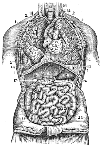
Fig. 88. A front view of the organs within the chest and abdomen. 1, 1, 1, 1, The muscles of the chest. 2, 2, 2, 2, The ribs. 3, 3, 3, The upper, middle, and lower lobes of the right lung. 4, 4, The lobes of the left lung. 5, The right ventricle of the heart. 6, The left ventricle. 7, The right auricle of the heart. 8, The left auricle. 9, The pulmonary artery. 10, The aorta. 11, The vena cava descendens. 12, The trachea. 13, The œsophagus. 14, 14, 14, 14, The pleura. 15, 15, 15, The diaphragm. 16, 16, The right and left lobe of the liver. 17, The gall-cyst. 18, The stomach. 26, The spleen. 19, 19, The duodenum. 20, The ascending colon. 21, The transverse colon. 25, The descending colon. 22, 22, 22, 22, The small intestine. 23, 23, The abdominal walls turned down. 24, The thoracic duct, opening into the left subclavian vein, (27.)
463. The nutrient portion of the food is poured into the left subclavian vein, (24, 27, fig. 88,) at the lower part of the neck, and is carried to the right cavities of the heart. The fluid in these cavities consists of the chyle incorporated with the impure blood. Neither of these two elements is fitted to promote the growth or repair the waste of the body. They must be subjected to a process, by which the first can be converted into blood, and the second freed of its carbonic acid gas and water. This is effected by the Respiratory Organs.
464. The RESPIRATORY ORGANS are the Lungs, (lights,) the Tra´che-a, (windpipe,) the Bronch´i-a, (subdivisions of the trachea,) and the Air-Ves´i-cles, (air-cells at the extremities of the bronchia.) The Di´a-phragm, (midriff,) Ribs, and several Muscles, also aid in the respiratory process.
465. The LUNGS are conical organs, one on each side of the chest, embracing the heart, (fig. 88,) and separated from each other by a membranous partition. The color of the lungs is a pinkish gray, mottled, and variously marked with black. Each lung is divided into lobes, by a long and deep 210 fissure, which extends from the posterior surface of the upper part of the organ, downward and forward, nearly to the anterior angle of the base. In the right lung, the upper lobe is subdivided by a second fissure. This lung is larger and shorter than the left. It has three lobes, while the left has only two.
463. What fluids are conveyed into the right cavities of the heart? What is necessary before they can be adapted to the wants of the body? By what organs are these changes effected? 464–474. Give the anatomy of the respiratory organs. 464. Name the respiratory organs. What organs also aid in the respiratory process? 465. Describe the lungs.
Fig. 89.

Fig. 89. A back view of the heart and lungs. The posterior walls of the chest are removed. 1, 2, 3, The upper, middle, and lower lobes of the right lung. 8, 9, 10, The two lobes of the left lung. 6, 13, The diaphragm. 7, 7, 14, 14, The pleura that lines the ribs. 4, 11, The pleura that lines the mediastine. 5, 12, 12, The portion of the pleura that covers the diaphragm. 15, The trachea, 16, The larynx. 19, 19, The right and left bronchia. 20, The heart. 29, The lower part of the spinal column.
Explain fig. 89.
466. Each lung is enclosed, and its structure maintained by a serous membrane, called the pleu´ra, which invests it as far as the root, and is thence reflected upon the walls of the chest. The lungs, however, are on the outside of the pleura, in the same way as the head is on the outside of a cap doubled upon itself. The reflected pleuræ in the middle of the thorax form a partition, which divides the chest into two cavities. This partition is called the me-di-as-ti´num.
Fig. 90.
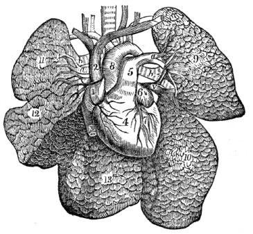
Fig. 90. The heart and lungs removed from the chest, and the lungs freed from all other attachments. 1, The right auricle of the heart. 2, The superior vena cava. 3, The inferior vena cava. 4, The right ventricle. 5, The pulmonary artery issuing from it. a, a, The pulmonary artery, (right and left,) entering the lungs. b, b, Bronchia, or air-tubes, entering the lungs. v, v, Pulmonary veins, issuing from the lungs. 6, The left auricle. 7, The left ventricle. 8, The aorta. 9, The upper lobe of the left lung. 10, Its lower lobe. 11, The upper lobe of the right lung. 12, The middle lobe. 13, The lower lobe.
Observation. When this membrane that covers the lungs, 212 and also lines the chest, is inflamed, the disease is called “pleurisy.”
466. By what are the lungs enclosed? What is the relative position of the lungs and pleura? What is said of the reflected pleuræ? Explain fig. 90. What part of the lungs is affected in pleurisy?
467. The lungs are composed of the ramifications of the bronchial tubes, which terminate in the bronchial cells, (air-cells,) lymphatics, and the divisions of the pulmonary artery and veins. All of these are connected by cellular tissue, which constitutes the pa-ren´chy-ma. Each lung is retained in its place by its root, which is formed by the pulmonary arteries, pulmonary veins, and bronchial tubes, together with the bronchial vessels and pulmonary nerves.
468. The TRACHEA extends from the larynx, of which it is a continuation, to the third dorsal vertebra, where it divides into two parts, called bronchia. It lies anterior to the spinal column, from which it is separated by the œsophagus.
469. The BRONCHIA proceed from the bifurcation, or division of the trachea, to their corresponding lungs. Upon entering the lungs, they divide into two branches, and each branch divides and subdivides, and ultimately terminates in small sacs, or cells, of various sizes, from the twentieth to the hundredth of an inch in diameter. So numerous are these bronchial or air-cells, that the aggregate extent of their lining membrane in man has been computed to exceed a surface of 20,000 square inches, and Munro states that it is thirty times the surface of the human body.
Illustration. The trachea may be compared to the trunk of a tree; the bronchia, to two large branches; the subdivisions of the bronchia, to the branchlets and twigs; the air-cells, to the buds seen on the twigs in the spring.
470. The AIR-VESICLES and small bronchial tubes compose 213 the largest portions of the lungs. These, when once inflated, contain air, under all circumstances, which renders their specific gravity much less than water; hence the vulgar term, lights, for these organs. The trachea and bronchial tubes are lined by mucous membrane. The structure of this membrane is such, that it will bear the presence of pure air without detriment, but not of other substances.
467. Of what are the lungs composed? How retained in place? 468. Where is the trachea situated? 469. Describe the bronchia. What is the aggregate extent of the lining membrane of the air-cells? To what may the trachea and its branches be compared? 470. What is said of the air-cells and bronchial tubes?
Fig. 91.
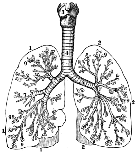
Fig. 91. A representation of the larynx, trachea, bronchia, and air-cells. 1, 1, 1, An outline of the right lung. 2, 2, 2, An outline of the left lung. 3, The larynx 4, The trachea. 5, The right bronchial tube. 6, The left bronchial tube. 7, 7, 7, 8, 8, 8, The subdivisions of the right and left bronchial tubes. 9, 9, 9, 9, 9, 9, Air-cells.
What membrane lines the trachea and its branches? What is peculiar in its structure? What does fig. 91 represent?
Observation. The structure of the trachea and lungs may be illustrated, by taking these parts of a calf or sheep and inflating the air-vesicles by forcing air into the windpipe with a pipe or quill. The internal structure may then be seen by opening the different parts.
471. The lungs, like other portions of the system, are supplied with nutrient arteries and nerves. The nervous filaments that are distributed to these organs are in part from the tenth pair, (par vagum,) that originates in the brain, and in part from the sympathetic nerve. The muscles that elevate the ribs and the diaphragm receive nervous fibres from a separate system, which is called the respiratory.
Fig. 92.

Fig. 92. 1, A bronchial tube. 2, 2, 2, Air-vesicles. Both the tube and vesicles are much magnified. 3, A bronchial tube and vesicles laid open.
Observation. When the mucous membrane of a few of the larger branches of the windpipe is slightly inflamed, it is called a “cold;” when the inflammation is greater, and extends to the lesser air-tubes, it is called bronch-i´tis. When the air-cells and parenchyma become inflamed, it is called inflammation of the lungs. Coughing is a violent expulsory effort by which air is suddenly forced through the bronchia and trachea to remove offending matter.
How may the structure of the trachea and its branches be illustrated? 471. Are the lungs supplied with nutrient arteries? Where are the respiratory nerves distributed? From what source do these organs derive their nervous filaments?
472. The RIBS are joined to the spinal column at their posterior extremity; and in front, they terminate in cartilages, which unite them to the sternum. They incline downward, from the spinal column to the breast-bone, and form resisting walls that assist in producing the partial vacuum necessary for inspiration.
Fig. 93. A section of the chest when the lungs are inflated. 1, The diaphragm. 2, The muscular walls of the abdomen.
Fig. 94. A section of the chest when the lungs are contracted. 1, The diaphragm in common expiration. 2, 2, The muscular walls of the abdomen. 3, The position of the diaphragm in forced expiration.
These engravings show the diaphragm to be more convex, and the walls of the abdomen more flattened, when the lungs are collapsed, than when they are inflated.
473. The DIAPHRAGM is a flexible circular partition, that separates the respiratory from the digestive organs, and the chest from the abdomen. Its margin is attached to the spinal column, the sternum, and cartilages of the lower ribs. The lungs rest upon its upper surface, while the liver and stomach 216 are placed below it, (fig. 88.) In a state of repose, its upper surface forms an arch, the convexity of which is toward the chest. In forced expiration, its upper point reaches as high as the fourth rib. In an ordinary inspiration, it is depressed as low as the seventh rib, which increases the capacity of the chest.
472. Describe the ribs. Explain figs. 93 and 94. 473. Describe the diaphragm.
474. The RESPIRATORY muscles are, in general, attached at one extremity to the parts about the shoulders, head, and upper portion of the spinal column. From these, they run downward and forward, and are attached, at the opposite extremity, to the sternum, clavicle, and upper rib. Other muscles are attached at one extremity to a rib above, and by the opposite extremity to a rib below. These fill the spaces between the ribs, and, from their situation, are called in-ter-cost´al muscles.
Observation. 1st. There are several actions of common occurrence, that are intimately connected with respiration; such as hiccough, sneezing, &c. Hiccough is an involuntary contraction of the muscles of respiration, particularly the diaphragm.
2d. Sneezing is a violent, involuntary contraction of the respiratory muscles, as in hiccough. When an acrid stimulant, as snuff, is applied to the mucous membrane of the nose, an irritation is produced which is accompanied by a violent expulsion of air from the lungs. This is owing to the connection between the nasal and respiratory nerves.
What is its form when not in action? 474. Where do the respiratory muscles make their attachment? What name is given to those muscles that fill the places between the ribs? What is hiccough? What is sneezing?
475. Respiration, or breathing, is that process by which air is taken into the lungs and expelled from them. The object of respiration is, 1st. To supply the system with oxygen, which is essential to the generation of animal heat; 2d. To convert the chyle into blood. This is done by the oxygen of the inspired air; 3d. To relieve the organs of the body of the principal elements (carbon and hydrogen) that compose the old and useless particles of matter. The organs of the system, as already mentioned, are principally composed of carbon, hydrogen, oxygen, and nitrogen.
476. By the action of the lymphatics and capillary veins, the old and worn-out particles are conveyed into the veins of the systemic circulation. The hydrogen, in form of watery vapor, is easily discharged in the perspiration and other secretions. The nitrogen and oxygen are, or may be, separated from the blood, through the agency of several different organs; but carbon does not escape so readily. It is probable that a part of the surplus carbon of the venous blood is secreted by the liver; but a far greater amount passes to the lungs, and these may be considered as special organs designed to separate this element from the venous blood.
477. An ordinary inspiration may be accomplished by the action of the diaphragm, and a slight elevation of the ribs. In 218 full inspiration, the diaphragm is not only more depressed but the ribs are evidently elevated. To produce this effect on the ribs, two sets of muscles are called into action. Those which are attached to the upper rib, sternum, and clavicle, contract and elevate the lower and free extremities of the ribs. This enlarges the cavity of the chest between the spinal column and the sternum. But the lateral diameter, in consequence, is only slightly increased, because the central portion of the ribs sinks lower than their posterior extremities, or their cartilaginous attachment to the sternum.
475–494. Give the physiology of the respiratory organs. 475. What is respiration? What is the principal object in breathing? 476. How are the useless atoms of matter conveyed into the veins of the systemic circulation? How may the principal elementary substances be separated from the blood? 477. How may an ordinary inspiration be accomplished?
Fig. 95.

Fig. 95. 6, Four of the vertebræ, to which are attached three ribs, (7, 7, 7,) with their intercostal muscles, (8, 8.) These ribs, in their natural position, have their anterior cartilaginous extremity at 4, while the posterior extremity is attached to the vertebræ, (6,) which are neither elevated nor depressed in respiration. 1, 1, and 2, 2, parallel lines, within which the ribs lie in their natural position. If the anterior extremity of the ribs is elevated from 4 to 5, they will not lie within the line 2, 2, but will reach the line 3, 3. If two hands extend from 1, 1, to 2, 2, they will effectually prevent the elevation of the ribs from 4 to 5, as the line 2, 2, cannot be moved to 3, 3.
What effect has a full inspiration on the ribs and diaphragm? How is the chest enlarged between the spinal column and sternum? What is said of the lateral diameter of the chest? Explain fig. 95.
478. The central portion of the ribs is raised by the action of intercostal muscles. The first, or upper rib, has but little movement; the second has more motion than the first, while 219 the third has still more than the second. The second rib is elevated by the contraction of the muscles between it and the first. The third rib is raised by the action of two sets of muscles; one lies between the first and second ribs, the other between the second and third. The motion of each succeeding rib is increased, because it is not only acted upon by the muscles that move the ribs above, but by an additional intercostal; so that the movement of the twelfth rib is very free, as it is elevated by the contraction of eleven muscles.
479. The tenth rib is raised eight times as much as the second rib, and the lateral diameter of the lower portion of the chest is increased in a corresponding degree. At the same time, the muscular margin of the diaphragm contracts, which depresses its central portion; and in this way, the chest is enlarged forward, laterally, and downward, simultaneously with the relaxation of the walls of the abdomen.
480. The lungs follow the variations of capacity in the chest, expanding their air-cells when the latter is enlarged, and contracting when the chest is diminished. Thus, when the chest is expanded, the lungs follow, and consequently a vacuum is produced in their air-cells. The air then rushes through the mouth and nose into the trachea and its branches, and fills the vacuum as fast as it is made. This mechanical process constitutes inspiration.
481. After the expansion of the chest, the muscles that elevated the ribs relax, together with the diaphragm. The elasticity of the cartilages of the ribs depresses them, and the cavity of the chest is diminished, attended by the expulsion of a portion of the air from the lungs. At the same time, the muscles that form the front walls of the 220 abdominal cavity, contract, and press the alimentary canal, stomach, and liver, upward against the diaphragm; this, being relaxed, yields to the pressure, rises upward, and presses upon the lungs, which retreat before it, and another portion of air is expelled from these organs. This process is called expiration.
478. Describe the action of the intercostal muscles upon the ribs. 479. How does the elevation of the tenth rib compare with the second? What effect has this elevation upon the lateral diameter of the chest? 480. Describe the process of inspiration. 481. Describe the process by which the air is forced out of the lungs.
Fig. 96.

Fig. 96. A front view of the chest and abdomen in respiration. 1, 1, The position of the walls of the chest in inspiration. 2, 2, 2, The position of the diaphragm in inspiration. 3, 3, The position of the walls of the chest in expiration. 4, 4, 4, The position of the diaphragm in expiration. 5, 5, The position of the walls of the abdomen in inspiration. 6, 6, The position of the abdominal walls in expiration.
482. Thus it is obvious that the enlargement of the chest, or inspiration, is produced in two ways: 1st. By the depression of the convex portion of the diaphragm; 2d. By the elevation of the ribs. On the contrary, the contraction of the 221 chest, or expiration, is produced by the depression of the ribs, and elevation of the central part of the diaphragm. These movements are successive during life, and constitute respiration.
Explain fig. 96. 482. In how many ways may the chest be enlarged, and how is it accomplished? How is the contraction of the chest effected?
Fig. 97.
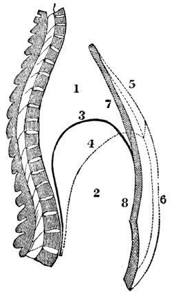
Fig. 97. A side view of the chest and abdomen in respiration. 1, The cavity of the chest. 2, The cavity of the abdomen. 3, The line of direction for the diaphragm when relaxed in expiration. 4, The line of direction for the diaphragm when contracted in inspiration. 5, 6, The position of the front walls of the chest and abdomen in inspiration. 7, 8, The position of the front walls of the abdomen and chest in expiration.
Experiment. Place the ear upon the chest of a person, and a murmuring sound will be heard, somewhat like the soft sighings of the wind through forest trees. This sound is 222 caused by the air rushing in and out of the lungs, and is peculiarly distinct in the child.
Explain fig. 97. How may the murmur of respiration be heard?
483. It is not easy to decide how much air is taken into the lungs at each inspiration. The quantity, however, must vary in different individuals, from the difference in the condition and expansion of the lungs, together with the size of the chest. From numerous experiments, the quantity, at an ordinary inspiration, of a common-sized man, is fixed at forty cubic inches. It has been estimated that one hundred and seventy cubic inches can be thrown out of the lungs by a forcible expiration, and that there remain in the lungs two hundred and twenty cubic inches; so that these organs, in their quiescent state, may be considered as containing about three hundred and ninety cubic inches of air, or more than a gallon.
484. Respiration is more frequent in females and children than in adult men. In diseases, particularly those of the lungs, it is more increased in frequency than the action of the heart. In health, the smallest number of inspirations in a minute by an adult, is not less than fourteen, and they rarely exceed twenty-five. Eighteen may be considered an average number. The quantity of oxygen taken into the lungs at each inspiration is about eight cubic inches, one half of which disappears in every act of respiration.
Observation. Under different circumstances, however, the consumption of oxygen varies. It is greater when the temperature is low, than when it is high; and during digestion the consumption has been found one half greater than when the stomach was empty.
483. Can it be ascertained with accuracy how much air is taken into the lungs at each inspiration? Why not? What is the probable quantity that an ordinary sized man inspires? How much can be thrown out of the lungs at a forcible expiration, and how much remains in the lungs? From these calculations, how much may they contain in their quiescent state? 484. In whom is respiration most frequent? How in disease? How in health? How many may be considered an average number? When is the consumption of oxygen the greatest?
485. Dr. Southwood Smith has lately performed a series of very interesting experiments, from which he deduces the following general results: “1st. The volume of air ordinarily present in the lungs is about twelve pints. 2d. The volume of air received by the lungs at an ordinary inspiration is one pint. 3d. The volume of air expelled from the lungs at an ordinary expiration, is a little less than one pint. 4th. Of the volume of air received by the lungs at one inspiration, only one fourth part is decomposed at one action of the heart. 5th. The quantity of blood that flows to the lungs, to be acted upon by the air at one action of the heart, is two ounces, and this is acted on in less than one second of time. 6th. The quantity of blood in the whole body of the human adult, is twenty-five pounds avoirdupois, or twenty pints. 7th. In the mutual action that takes place between the air and blood, every twenty-four hours, the air loses thirty-seven ounces of oxygen, and the blood fourteen ounces of carbon.”
486. Apparently, atmospheric air is a simple element. But chemical analysis shows its composition to be oxygen and nitrogen, in the proportion of twenty-one parts of the former, and about seventy-nine of the latter. In addition, there is a small amount of vapor of water and carbonic acid. The pressure of this invisible, elastic fluid upon the body of an ordinary sized adult, is estimated to equal thirty-five thousand pounds.
487. The principal substance of a vitiated character in the dark-colored blood is carbonic acid. And since there is no chemical affinity between the oxygen and nitrogen of the air, the former readily unites with some of the elements of the blood. Hence, whenever blood is presented to the 224 air in the lungs, the oxygen leaves the nitrogen, and becomes mixed with the circulating fluid. (Appendix J.)
485. State the 1st, 2d, 3d, and 4th deductions from the experiments of Dr. Southwood Smith. The 5th, 6th, and 7th. 486. Of what is atmospheric air composed? What is the weight of air upon a common sized man? 487. What is the principal substance of a vitiated character in the dark-colored blood? What is said of the chemical affinity between oxygen and nitrogen?
488. Again, carbonic acid and water have a stronger affinity for atmospheric air than for the other elements of the blood. Consequently, when they are brought into contact with the air in the lungs, the carbonic acid and water leave the other constituents of the blood, and unite with the air. In this way the bluish, or impure blood is relieved of its impurities, and becomes the red, or pure blood, which contains the principles so essential to life. (Appendix K.)
489. The formation of carbonic acid and water, eliminated from the system through the lungs and skin, is explained by the following theory: In the lungs and upon the skin the oxygen separates from the nitrogen and unites with the blood in the capillary vessels of these organs. The oxygen is conveyed with the blood to the capillary arteries and veins of the different tissues of the system. In these membranes there is a chemical union of the oxygen with the carbon and hydrogen contained in the blood and waste atoms of the system. This combustion, or union of oxygen with carbon and hydrogen, is attended with the disengagement of heat, and the formation of carbonic acid and water. (Appendix L.)
490. The following experiment will illustrate the passage of fluids through membranes, and the different affinity of gases for each other. Put a mixture of water and alcohol into a phial and leave it uncorked. Both the water and alcohol have a greater affinity for air than for each other. Alcohol has a greater affinity for the air, and will be diffused through it more readily than the water, when there is no intervening obstacle. But tie a piece of bladder over the mouth of the phial, and let it stand a few days,—the water 225 will leave the alcohol, and pass through the membrane. By the aid of this experiment, we shall endeavor to explain the interchange of fluids in the lungs.
488. What is formed when oxygen unites with carbon or hydrogen? 489. Give the theory for the formation of carbonic acid and watery vapor thrown out of the system. 490. Illustrate the passage of fluids through membranes, and the different affinities of gases.
491. The walls of the air-vesicles, and coats of the blood-vessels, are similar, in their mechanical arrangement, to the membranous bladder in the before described experiment. As the oxygen of the air has greater affinity for blood than for nitrogen, so it permeates the membranes that intervene between the air and blood more readily than the nitrogen. As the carbonic acid and water have a greater affinity for air than for the other elements of the blood, so they will also pass through the walls of the blood-vessels and air-cells more readily than the other elements of the dark-colored blood.
Fig. 98.
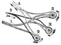
Fig. 98. 1, A bronchial tube divided into three branches. 2, 2, 2, Air-cells. 3, Branches of the pulmonary artery, that spread over the air-cells. Through the pulmonary artery the dark, impure blood is carried to the air-cells of the lungs. 4, Branches of the pulmonary vein, that commence at the minute terminations of the pulmonary artery. Through the pulmonary vein the red blood is returned to the heart.
492. As the impure blood is passing in the minute vessels over the air-cells, the oxygen passes through the thin coats of the air-cells and blood-vessels, and unites with the blood. At the same time, the carbonic acid and water leave the blood, and pass through the coats of the blood-vessels and air-cells, and mix with the air in the cells. These are thrown out of the system every time we breathe. This interchange of products produces the change in the color of the blood.
Explain fig. 98. 492. How and where is the blood changed?
Experiment. Fill a bladder with dark blood drawn from any animal. Tie the bladder closely, and suspend it in the air. In a few hours, the blood next to the membrane will have become of a bright red color. This is owing to the oxygen from the air passing through the bladder, and uniting with the blood, while the carbonic acid has escaped through the membrane.
Fig. 99.
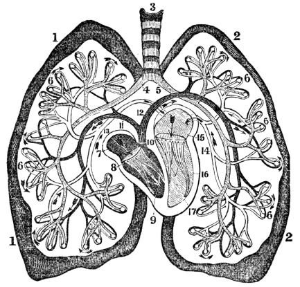
Fig. 99. An ideal view of the pulmonary circulation. 1, 1, The right lung. 2, 2, The left lung. 3, The trachea. 4, The right bronchial tube. 5, The left bronchial tube. 6, 6, 6, 6, Air-cells. 7, The right auricle. 8, The right ventricle. 9, The tricuspid valves. 10, The pulmonary artery. 11, The branch to the right lung. 12, The branch to the left lung. 13, The right pulmonary vein. 14, The left pulmonary vein. 15, The left auricle. 16, The left ventricle. 17, The mitral valves.
493. The presence of carbonic acid and watery vapor in the expired air, can be proved by the following experiments: 227 1st. Breathe into lime-water, and in a few minutes it will become of a milk-white color. This is owing to the carbonic acid of the breath uniting with the lime, forming the carbonate of lime. 2d. Breathe upon a cold, dry mirror for a few minutes, and it will be covered with moisture. This is condensed vapor from the lungs. In warm weather, this watery vapor is invisible in the expired air, but in a cold, dry morning in winter, the successive jets of vapor issuing from the mouth and nose are sufficiently obvious.
Give the experiment showing that oxygen changes the dark-colored blood to a bright red color. What is represented by fig. 99? 493. How can the presence of carbonic acid in the lungs be proved?
494. From the lungs are eliminated other impurities beside carbonic acid, the perceptible quality of which is various in different persons. The offensive breath of many persons may be caused by decayed teeth, or the particles of food that may be retained between them, but it often proceeds from the secretion, in the lungs, of certain substances which previously existed in the system.
Illustration. When spirituous liquors are taken into the stomach, they are absorbed by the veins and mixed with the dark-colored blood, in which they are carried to the lungs to be expelled from the body. This will explain the fact, which is familiar to most persons, that the odor of different substances is perceptible in the breath, or expired air, long after the mouth is free from these substances.
How the watery vapor? 494. Are there other excretions from the lungs? Give the illustration.
Note. Let the anatomy and physiology of the respiratory organs be reviewed from figs. 96, 97, and 99, or from anatomical outline plates Nos. 5 and 7.
495. For man to enjoy the highest degree of health, it is necessary that the impure “venous” blood be properly changed. As this is effected in the lungs by the action of the air, it follows that this element, when breathed, should be pure, or contain twenty-one per cent. of oxygen to about seventy-nine per cent. of nitrogen.
496. The volume of air expelled from the lungs is somewhat less than that which is inspired. The amount of loss varies under different circumstances. An eightieth part of the volume taken into the lungs, or half a cubic inch, may be considered an average estimate.
497. The quality and purity of the air is affected by every respiration. 1st. The quantity of oxygen is diminished. 2d. The amount of carbonic acid is increased. 3d. A certain proportion of watery vapor is ejected from the lungs in the expired air. Of the twenty-one parts of oxygen in the inspired air, only eighteen parts are expired, while the carbonic acid and watery vapor are increased about four per cent. The quantity of nitrogen is nearly the same in the expired as in the inspired air.
Observation. It is now fully ascertained that while the chemical composition of the blood is essentially changed, its 229 weight remains the same, as the carbon and hydrogen discharged are equal to the united weight of the oxygen and nitrogen absorbed.
495–546. Give the hygiene of the respiratory organs. 495. What is necessary that man enjoy the highest degree of health? 496. How does the volume Of expired air compare with that which was inspired? Does this loss vary, and what is an average estimate? 497. How is the purity of the air affected by respiration? How is the inhaled oxygen affected? What effect on the carbonic acid and watery vapor? On the nitrogen? What is said respecting the weight of the blood?
498. If one fourth part of the volume of air received by the lungs at one inspiration is decomposed at one “beat” of the heart, it might be supposed that if the expired air be again received into the lungs, one half of the oxygen would be consumed, and, in a similar ratio, if re-breathed four times, all the oxygen would be consumed. But it does not follow, if the air is thus re-breathed, that the same changes will be effected in the lungs. For air that has been inspired does not part with its remaining oxygen as freely as when it contains the proper amount of this life-giving element, and thus the changes in the impure blood are not so completely effected.
Illustration. In the process of dyeing, each successive article immersed in the dye weakens it; but it does not follow that the dye each time is affected in the same degree, or that the coloring matter by repeated immersions can be wholly extracted. The same principle applies to the exchange of oxygen and carbonic acid gas in the lungs.
499. If the inspired air is free from moisture and carbonic acid, these substances contained in the blood will be more readily imparted to it. When the air is loaded with vapor, they are removed more slowly; but if it is saturated with moisture, no vapor will escape from the blood through the agency of the lungs. This may be illustrated by the following experiment: Take two and a half pounds of water, add to it half a pound of common salt, (chloride of sodium,) and it will readily mix with the water; and to this solution add the same quantity of salt, and it will be dissolved more slowly. Again, add more salt, and it will remain undissolved, as the water has become saturated by the pound before dissolved.
498. Does air that is re-breathed freely impart its oxygen? Why? 499. What is the effect on the blood when the air is free from vapor and carbonic acid? When loaded with vapor? When saturated? How is this illustrated?
500. The principle in this experiment is analogous to that of the union between carbonic gas and atmospheric air. Allen and Pepy showed by experiment, that air which had been once breathed, contained eight and a half per cent. of carbonic acid. They likewise showed, that no continuance of the respiration of the same air could make it take up more than ten per cent. This is the point of saturation.
Experiment. Sink a glass jar that has a stop-cock, or one with a glass stopper, into a pail of water, until the air is expelled from the jar. Fill the lungs with air, and retain it in the chest a short time, and then breathe into the jar, and instantly close the stop-cock. Close the opening of the jar that is under the water with a piece of paper laid on a plate of sufficient size to cover the opening, invert the jar, and sink into it a lighted candle. The flame will be extinguished as quickly as if put in water.[15] Remove the carbonic acid by inverting the jar, and place a lighted candle in it, and the flame will be as clear as when out of the jar.
Observations. 1st. It is familiarly known that a taper will not burn where carbonic acid exists in any considerable quantity, or when there is a marked deficiency of oxygen. From this originated the judicious practice of sinking a lighted candle into a well or pit before descending into it. If the flame is extinguished, respiration cannot there be maintained, and life would be sacrificed should a person venture in, until the noxious air is removed.
500. What did the experiments of Allen and Pepy show? How can the presence of carbonic gas in the expired air be demonstrated? State observation 1st. Observation 2d.
2d. It is the action of carbonic acid upon the respiratory 231 organs, that gives rise to a phenomenon frequently seen in mines and caves. A man may enter these subterranean rooms, and feel no inconvenience in breathing; but the dog that follows him, falls apparently dead, and soon dies if not speedily removed to pure air. This arises from the fact that this gas is heavier than air, and sinks to the bottom of the room or cave.
3d. While it is true that carbonic acid possesses properties that render it unfit to be breathed, it is, notwithstanding, productive of very agreeable effects, when conveyed into the stomach. It forms the sparkling property of mineral waters, and fills the bubbles that rise when beer or cider is fermenting.
501. Pure atmospheric air is best adapted to a healthy action of the system. As the air cannot be maintained pure under all circumstances, the question may be asked, To what degree may the air be vitiated and still sustain life? and what is the smallest quantity of pure air a person needs each minute to maintain good health? Birnan says, that air which contains more than three and a half per cent. of carbonic acid is unfit for respiration, and, as air once respired contains eight and a half per cent. of carbonic acid, it clearly shows that it is not fitted to be breathed again.
502. No physiologist pretends that less than seven cubic feet of air are adequate for a man to breathe each minute, while Dr. Reid allows ten feet. The necessity of fifteen or twenty times the amount of air actually taken into the lungs, arises from the circumstance, that the expired air mixes with and vitiates the surrounding element that has not been inhaled.
503. The quantity of air which different persons actually 232 need, varies. The demand is modified by the size, age, habits, and condition of the body. A person of great size who has a large quantity of blood, requires more air than a small man with a less amount of circulating fluid. Individuals whose labor is active, require more air than sedentary or idle persons, because the waste of the system is greater. On the same principle, the gormandizer needs more of this element than the person of abstemious habits. So does the growing lad require more air than an adult of the same weight, for the reason that he consumes more food than a person of mature years. Habit also exerts a controlling influence. A man who works in the open air suffers more when placed in a small, unventilated room, than one who is accustomed to breathe the confined air of workshops.
Observation 3d. 501. What questions may be asked respecting the inspired air? Give the remark of Birnan. 502. How many cubic feet of air are adequate for a man to breathe each minute? How much does Dr. Reid allow? 503. Mention some reasons why different persons do not require the same amount of air.
504. Air, in which lamps will not burn with brilliancy, is unfitted for respiration. In crowded rooms, which are not ventilated, the air is vitiated, not only by the abstraction of oxygen and the deposition of carbonic acid, but by the excretions from the skin and lungs of the audience. The lamps, under such circumstances, emit but a feeble light. Let the oxygen gas be more and more expended, and the lamps will burn more and more feebly, until they are extinguished.
Illustrations. 1st. The effects of breathing the same air again and again, are well illustrated by an incident that occurred in one of our halls of learning. A large audience had assembled in an ill-ventilated room, to listen to a lecture; soon the lamps burned so dimly that the speaker and audience were nearly enveloped in darkness. The oppression, dizziness, and faintness experienced by many of the audience induced them to leave, and in a few minutes after, the lamps were observed to rekindle, owing to the exchange of pure air on opening the door.
How is it with the laborer? With the gormandizer? With the person that works in the open air? 504. What effect has impure air on a burning lamp? Give the illustration of the effects of impure air on lighted lamps.
2d. In the “Black Hole of Calcutta,” one hundred and forty-six Englishmen were shut up in a room eighteen feet square, with only two small windows on the same side to admit air. On opening this dungeon, ten hours after their imprisonment, only twenty-three were alive. The others had died from breathing impure air.
505. Air that has become impure from the abstraction of oxygen, an excess of carbonic acid, or the excretions from the lungs and skin, has a deleterious effect on the body. When this element is vitiated from the preceding causes, it prevents the proper arterialization, or change in the blood. For this reason, pure air should be admitted freely and constantly into work-shops and dwelling-houses, and the vitiated air permitted to escape. This is of greater importance than the warming of these apartments. We can compensate for the deficiency of a stove, by an extra garment or an increased quantity of food; but neither garment, exercise, nor food will compensate for pure air.
506. School-rooms should be ventilated. If they are not, the pupils will be restless, and complain of languor and headache. Those unpleasant sensations are caused by a want of pure air, to give an adequate supply of oxygen to the lungs. When pupils breathe for a series of years such vitiated air, their life is undoubtedly shortened, by giving rise to consumption and other fatal diseases.
Illustration. A school-room thirty feet square and eight feet high, contains 7200 cubic feet of air. This room will seat sixty pupils, and, allowing ten cubic feet of air to each pupil per minute, all the air in the room will be vitiated in twelve minutes.
Observation. In all school-rooms where there is not adequate 234 ventilation, it is advisable to have a recess of five or ten minutes each hour. During this time, let the pupils breathe fresh air, and open the doors and windows, so that the air of the room shall be completely changed.
Of the effects of breathing impure air. 505. In preserving health, what is of greater importance than warming the room? 506. Why should a school-room be ventilated? Give the illustration.
507. Churches, concert halls, and all rooms designed for a collection of individuals, should be amply ventilated. While the architect and workmen are assiduous in giving these public rooms architectural beauty and splendor, by adorning the ceiling with Gothic tracery, rearing richly carved columns, and providing carefully for the warming of the room, it too frequently happens that no direct provision is made for the change of that element which gives us beauty, strength, and life.
Illustration. A hall sixty feet by forty, and fifteen feet high, contains 36,000 cubic feet of air. A hall of this size will seat four hundred persons; by allowing ten cubic feet of air to each person per minute, the air of the room will be rendered unfit for respiration in nine minutes.
508. Railroad cars, cabins of steam and canal-boats, omnibuses, and stage-coaches, require ample ventilation. In the construction of these public conveyances, too frequently, the only apparent design is, to seat the greatest number of persons, regardless of the quantity and character of the air to maintain health and even life. The character of the air is only realized when, from the fresh, pure air, we enter a crowded cabin of a boat or a closed coach; then the vitiated air from animal excretions and noxious gases is offensive, and frequently produces sickness.
509. The influence of habit is strikingly expressed by Birnan, in the “Art of Warming and Ventilating Rooms:” “Not the least remarkable example of the power of habit is 235 its reconciling us to practices which, but for its influence, would be considered noxious and disgusting. We instinctively shun approach to the dirty, the squalid, and the diseased, and use no garment that may have been worn by another. We open sewers for matters that offend the sight or the smell, and contaminate the air. We carefully remove impurities from what we eat and drink, filter turbid water, and fastidiously avoid drinking from a cup that may have been pressed to the lips of a friend. On the other hand, we resort to places of assembly, and draw into our mouths air loaded with effluvia from the lungs, skin, and clothing of every individual in the promiscuous crowd—exhalations offensive, to a certain extent, from the most healthy individuals; but when arising from a living mass of skin and lungs, in all stages of evaporation, disease, and putridity,—prevented by the walls and ceiling from escaping—they are, when thus concentrated, in the highest degree deleterious and loathsome.”
What suggestion when a school-room is not ventilated? 507. What is said in regard to ventilating churches, concert halls, &c.? State the illustration. 508. What remarks relative to public conveyances? 509. State the influence of habit by Birnan.
510. The sleeping-room should be so ventilated that the air in the morning will be as pure as when retiring to rest in the evening. Ventilation of the room would prevent morning headaches, the want of appetite, and languor—so common among the feeble. The impure air of sleeping-rooms probably causes more deaths than intemperance. Look around the country, and those who are most exposed, who live in huts but little superior to the sheds that shelter the farmer’s flocks, are found to be the most healthy and robust. Headaches, liver complaints, coughs, and a multitude of nervous affections, are almost unknown to them; not so with those who spend their days and nights in rooms in which the sashes of the windows are calked, or perchance doubled, to prevent the keen but healthy air of winter from entering their apartments. Disease and suffering are their constant companions.
510. What is said of the ventilation of sleeping-rooms? What would adequate ventilation prevent? Give a common observation.
Illustration. By many, sleeping apartments twelve feet square and seven feet high, are considered spacious for two persons, and good accommodations for four to lodge in. An apartment of this size contains 1008 cubic feet of air. Allowing ten cubic feet to each person per minute, two occupants would vitiate the air of the room in fifty minutes, and four in twenty-five minutes. When lodging-rooms are not ventilated, we would strongly recommend early rising.
511. The sick-room, particularly, should be so arranged that the impure air may escape, and pure air be constantly admitted into the room. It is no unusual practice in some communities, when a child or an adult is sick of an acute disease, to prevent the ingress of pure air, simply from the apprehension of the attendants, that the patient will contract a cold. Again, the prevalent custom of several individuals sitting in the sick-room, particularly when they remain there for several hours, tends to vitiate the air, and, consequently, to increase the suffering and danger of the sick person. In fevers or inflammatory diseases of any kind, let the patient breathe pure air; for the purer the blood, the greater the power of the system to remove disease, and the less the liability to contract colds.
Observation. Among children, convulsions, or “fits,” usually occur when they are sleeping. In many instances, these are produced by the impure air which is breathed. To prevent these alarming and distressing convulsions, the sleeping-room should be ventilated, and there should be no curtains around the bed, or coverings over the face, as they produce an effect similar to that experienced when sleeping in a small, unventilated room. To relieve a child when convulsed, carry it into the open air.
What is said of the size of sleeping-rooms? 511. What is said of the sick-room? Mention some prevailing customs in reference to these rooms. What is said of convulsions among children?
512. While occupying a room, we are insensible of the 237 gradual vitiation of the air. This is the result of the diminished sensibility of the nervous system, and gradual adaptation of the organs to blood of a less stimulating character. This condition is well illustrated in the hibernating animals. We are insensible of the impure air of unventilated sleeping-rooms, until we leave them for a walk or ride. If they have been closed, we are made sensible of the character of the air as soon as we reënter them, for the system has regained its usual sensibility while inhaling a purer atmosphere.
513. In the construction of every inhabited room, there should be adequate means of ventilation, as well as warming. No room is well ventilated, unless as much pure air is brought into it as the occupants vitiate at every respiration. This can be effected by making an aperture in the ceiling of the room, or by constructing a ventilating flue in the chimney. This should be in contact with the flues for the escape of smoke, but separated from them by a thin brick partition. The hot air in the smoke flues will warm the separating brick partition, and consequently rarefy the air in the ventilating flue. Communication from every room in a house should be had to such flues. The draught of air can be regulated by well-adjusted registers, which in large rooms should be placed near the floor as well as near the ceiling.
514. While provision is made for the escape of rarefied impure air, we should also provide means by which pure air may be constantly admitted into the room, as the crevices of the doors and windows are not always sufficient; and, if they should be adequate, air can be introduced in a more convenient, economical, and appropriate manner. There should be an aperture opposite the ventilating flue, at or near the floor, to connect with the outer walls of the building or external air. 238 But if pure heated air is introduced into the room, it obviates the necessity of the introduction of the external air.[16]
512. Why are we insensible to the gradual vitiation of the air of an unventilated room? 513. What is very important in the building of every inhabited room? How can a room be well ventilated? 514. What is said relative to a communication with the external air?
515. In warming rooms, the hot air furnaces, or box and air-tight stoves converted into hot air furnaces, should be used in preference to the ordinary stoves. The air thus introduced into the room is pure as well as warm. In the adaptation of furnaces to dwelling-houses, &c., it is necessary that the air should pass over an ample surface of iron moderately heated; as a red heat abstracts the oxygen from the contiguous air, and thus renders it unfit to be respired.[17]
Observation. Domestic animals need a supply of pure air as well as man. The cows of cities, that breathe a vitiated air, have, very generally, tubercles. Sheep that are shut in a confined air, die of a disease called the “rot,” which is of a tuberculous character. Interest and humanity require that the buildings for animals be properly ventilated.
515. How should rooms be warmed? What is necessary in the adaptation of furnaces to dwelling-houses?
516. The change that is effected in the blood while passing through the lungs, not only depends upon the purity of the air, but the amount inspired. The quantity varies according to the size of the chest, and the movement of the ribs and diaphragm.
517. The size of the chest and lungs can be reduced by moderate and continued pressure. This is most easily done in infancy, when the cartilages and ribs are very pliant; yet it can be effected at more advanced periods of life, even after the chest is fully developed. For want of knowledge of the pliant character of the cartilages and ribs in infants, too many mothers, unintentionally, contract their chests, and thus sow the seeds of disease by the close dressing of their offspring.
518. If slight but steady pressure be continued from day to day and from week to week, the ribs will continue to yield more and more, and after the expiration of a few months, the chest will become diminished in size. This will be effected without any suffering of a marked character; but the general health and strength will be impaired. It is not the violent and ephemeral pressure, but the moderate and protracted, that produces the miscalled, “genteel,” contracted chests.
519. The style of dress which at the present day is almost universal, is a prolific cause of this deformity. These baneful 240 fashions are copied from the periodicals, so widely circulated, containing a “fashion plate of the latest fashions, from Paris.” In every instance; the contracted, deformed, and, as it is called, lady-like waist, is portrayed in all its fascinating loveliness. These periodicals are found on almost every centre-table, and exercise an influence almost omnipotent. If the plates which corrupt the morals are excluded by civil legislation, with the same propriety ought not those to be suppressed that have a tendency so adverse to health?
516. What varies the amount of air received into the lungs? 517. How can the size of the chest be diminished? When is this most easily effected? 518. How are the miscalled, “genteel,” contracted chests usually produced? 519. What is said of the style of the dress at the present day?
Fig. 100. A correct outline of the Venus de Medici, the beau ideal of female symmetry.
Fig. 101. An outline of a well-corseted modern beauty.
One has an artificial, insect waist; the other, a natural waist. One has sloping shoulders, while the shoulders of the other are comparatively elevated, square, and angular. The proportion of the corseted female below the waist, is also a departure from the symmetry of nature.
Observations. 1st. The Chinese, by compressing the feet of female children, prevent their growth; so that the foot of a 241 Chinese belle is not larger than the foot of an American girl of five years.
What does fig. 100 represent? Fig. 101? Give observation 1st.
2d. The American women compress their chests, to prevent their growth; so that the chest of an American belle is not larger than the chest of a Chinese girl of five years. Which country, in this respect, exhibits the greater intelligence?
3d. The chest can be deformed by making the linings of the waists of the dresses tight, as well as by corsets. Tight vests, upon the same principle, are also injurious.
520. In children, who have never worn close garments, the circumference of the chest is generally about equal to that of the body at the hips; and similar proportions would exist through life, if there were no improper pressure of the clothing. This is true of the laboring women of the Emerald Isle, and other countries of Europe, and in the Indian female, whose blanket allows the free expansion of the chest. The symmetrical statues of ancient sculptors bear little resemblance to the “beau ideal” of American notions of elegant form. This perverted taste is in opposition to the laws of nature. The design of the human chest is not simply to connect the upper and lower portions of the body, like some insects, but to form a case for the protection of the vital organs.
521. Individuals may have small chests from birth. This, to the particular individual, is natural; yet it is adverse to the great and general law of Nature relative to the size of the human chest. Like produces like, is a general law of the animal and vegetable kingdoms. No fact is better established, than that which proves the hereditary transmission 242 from parents to children of a constitutional liability to disease and the same may be said in regard to their conformations. If the mother has a small, taper waist, either hereditary or acquired, this form may be impressed on her offspring;—thus illustrating the truthfulness of scripture, “that the sins of the parents shall be visited upon the children unto the third and fourth generation.”
Observation 2d. Observation 3d. 520. What is the size of the chest of a child that has always worn loose clothing? What is said of the size of the laboring women of Ireland, and the Indian female? How is it in ancient statues? What is the design of the chest? 521. What is a general law of both the animal and vegetable kingdoms? What fact in this connection is well established?
522. The quantity of air inhaled is modified by the capacity of the respiratory organs. The necessity of voluminous lungs may he elucidated by the following experiment: Suppose a gill of alcohol, mixed with a gill of water, be put into a vessel having a square foot of surface, and over the vessel a membrane be tied, and that the water will evaporate in twenty-four hours. If the surface had been only six inches square, only one fourth of the water would have evaporated through the membrane in the given time. If the surface had been extended to two square feet, the water would have evaporated in twelve hours.
523. Apply this principle to the lungs: suppose there are two hundred feet of carbonic acid to be carried out of the system every twenty-four hours. This gas, in that time, will pass through a vesicular membrane of two thousand square feet. If the lungs were diminished in size, so that there would be only one thousand square feet of vesicular membrane, the amount of carbonic acid would not, and could not, be eliminated from the system. Under such circumstances, the blood would not be purified.
524. Again; suppose the two thousand square feet of membrane would transmit two hundred cubic feet of oxygen into the system every twenty-four hours. If it should be diminished one half, this amount of oxygen would not pass into the blood. From the above illustrations we may learn 243 the importance of well-developed chests and voluminous lungs; for, by increasing the size of the lungs, the oxygen is more abundantly supplied to the blood, and this fluid is more perfectly deprived of its carbon and hydrogen.
What does this hereditary transmission prove? 522. How is the necessity of voluminous lungs illustrated? 525. How is this principle applied to the interchange of products in the lungs?
525. The chest is not only most expanded at its lower part, but the portion of the lungs that occupies this space of the thoracic cavity contains the greater part of the air-cells; and, from the lower two thirds of the lungs the greatest amount of carbonic acid is abstracted from the blood, and the greatest amount of oxygen gas is conveyed into the circulating fluid. Hence, contracting the lower ribs is far more injurious to the health than diminishing the size of the upper part of the chest.
526. The question is often asked, Can the size of the chest and the volume of the lungs be increased, when they have been injudiciously compressed, or have inherited this unnatural form? The answer is in the affirmative. The means for attaining this end are, a judicious exercise of the lungs, by walking in the open air, reading aloud, singing, sitting erect, and fully inflating the lungs at each act of inspiration. If the exercise be properly managed and persevered in, it will expand the chest, and give tone and health to the important organs contained in it. But, if the exercise be ill-timed or carried to excess, the beneficial results sought will probably not be attained.
Observation. Scholars, and persons who sit much of the time, should frequently, during the day, breathe full and deep, so that the smallest air-cells may be fully filled with air. While exercising the lungs, the shoulders should be thrown back and the head held erect.
527. The movement of the ribs and diaphragm is modified by the dress. When the lungs are properly filled with air, 244 the chest is enlarged in every direction. If any article of apparel is worn so tight as to prevent the full expansion of the chest and abdomen, the lungs, in consequence, do not receive air sufficient to purify the blood. The effect of firm, unyielding clothing, when worn tight, in preventing a due supply of air to the lungs, may be shown by the following illustration.
525. Why is it more injurious to contract the lower part of the chest than the upper? 526. How can the size of the chest be increased when it is contracted? Give the observation. 527. How is the movement of the ribs and diaphragm modified?
Illustration. If the diameter of a circle is three feet, the circumference will be nine feet. If the diameter is extended to four feet, the circumference will be increased to twelve feet. Should a tight band be thrown around a circle of nine feet, its diameter cannot be increased, for the circumference cannot be enlarged.
528. Any inelastic band, drawn closely around the lower part of the chest, or the abdomen, below the ribs, operates like the band in the preceding illustration, in restricting the movement of the ribs. When any article of dress encircles either the chest or abdomen, so as to prevent an increase of its circumference, it has an injudicious tendency, as it prevents the introduction of air in sufficient quantities to purify the blood. The question is not, How much restriction of the respiratory movements can be endured, and life continue? but, Does any part of the apparel restrict the movements? If it does, it is a violation of the organic laws; and though Nature is profuse in her expenditures, yet sooner or later, she sums up her account.
529. In determining whether the apparel is worn too tight, inflate the lungs, and, if no pressure is felt, no injurious effects need be apprehended from this cause. In testing the tightness of the dress, some persons will contract to the utmost the abdominal muscles, and thus diminish the size of the 245 chest, by depressing the ribs; when this is done, the individual exclaims, “How loose my dress is!” This practice is both deceptive and ludicrous. A good test is, to put the hand on the chest below the arm; if there is no movement of the ribs during respiration, the apparel is too tight. The only reliable test, however, is a full inflation of the lungs.
How is the effect of unyielding clothing, when worn tight, illustrated? 528. What effect has an inelastic band upon the lower part of the chest? What question is asked? 529. How can we determine whether the apparel is worn too tight?
Observation. Many individuals do not realize the small amount of force that will prevent the enlargement of the chest. This can be demonstrated by drawing a piece of tape tightly around the lower part of the chest of a vigorous adult, and confining it with the thumb and finger. Then endeavor fully to inflate the lungs, and the movement of the ribs will be much restricted.
530. The position in standing and sitting influences the movement of the ribs and diaphragm. When the shoulders are thrown back, and when a person stands or sits erect, the diaphragm and ribs have more freedom of motion, and the abdominal muscles act more efficiently; thus the lungs have broader range of movement than when the shoulders incline forward, and the body is stooping.
531. Habit exercises an influence upon the range of the respiratory movements. A person who has been habituated to dress loosely, and whose inspirations are full and free, suffers more from the tightness of a vest or waistband, than one, the range of movements of whose chest has long been subjected to tight lacing.
532. The condition of the brain exercises a great influence upon respiration. If the brain is diseased, or the mind depressed by grief, tormented by anxiety, or absorbed by abstract thought, the contractile energy of the diaphragm and 246 muscles that elevate the ribs, is much diminished, and the lungs are not so fully inflated, as when the mind is influenced by joy or other exhilarating emotions. The depressing passions likewise lessen the frequency of respiration. By the influence of these causes, the blood is but partially purified, and the whole system becomes enfeebled. Here we may see the admirable harmony between the different parts of the body, and the adaptation of all the functions to each other.
Give another test. How can the amount of pressure necessary to prevent the enlargement of the chest be demonstrated? 530. Show the effect of position on the movements of the ribs and diaphragm. 531. Show the effect of habit on the respiratory movements. 532. State the influence of the mind upon respiration.
533. As the quantity of air inhaled at each unimpeded inspiration in lungs of ample size, is about forty cubic inches, it follows, if the movement of the ribs and diaphragm is restricted by an enfeebled action of the respiratory muscles, or by any other means, the blood will not be perfectly purified. In the experiment, (§ 522, 523,) suppose forty cubic inches of air must pass over the membrane twenty times every minute, and that this is the amount required to remove the vapor which arises from the membrane; if only half of this amount of air be supplied each minute, only one half as much water will be removed from the alcohol through the membrane in twenty-four hours; consequently, the alcohol would be impure from the water not being entirely removed.
534. Restrain the elevation of the ribs and depression of the diaphragm, so that the quantity of air conveyed into the lungs will be reduced to twenty cubic inches, when forty are needed, and the results will be as follows: Only one half of the carbonic acid will be eliminated from the system, and the blood will receive but one half as much oxygen as it requires. This fluid will then be imperfectly oxydated, and partially freed of its impurities. The impure blood will be returned to the left side of the heart, and the whole system will suffer from an infringement of organic laws.
533. Illustrate the effect upon the blood when the respiratory muscles are enfeebled in their action. 534. Show how the blood is imperfectly purified by restricting the movements of the ribs and diaphragm.
535. Scrofula, or consumption, frequently succeeds a depressed state of the nervous system. These diseases arise from the deposition of tuberculous matter in different parts of the body. Those individuals who have met with reverses of fortune, in which character and property were lost, afford painful examples. Hundreds yearly die from the effect of depressed spirits, caused by disappointed hopes, or disappointed ambition.
Illustration. A striking instance of the effects of mental depression is related by Lænnec. In a female religious establishment in France, great austerities were practised; the mind was absorbed in contemplating the terrible truths of religion, and in mortifying the flesh. The whole establishment, in the space of ten years, was several times depopulated—with the exception of the persons employed at the gate, in the kitchen, and garden—with that fatal disease, consumption. This institution did not long continue, but was suppressed by order of the French government.
536. The purity of the blood is influenced by the condition of the lungs. When the bronchial tubes and air-cells have become partially impervious to air, from pressure upon the lungs, from fluids in the chest, from tumors, or from the consolidation of the cells and tubes from disease,—as inflammation, or the deposition of yellow, cheesy matter, called tubercles,—the blood will not be purified, even if the air is pure, the lungs voluminous, and the respiratory movements unrestricted, as the air cannot permeate the air-cells.
Observations. 1st. The twenty-three who escaped immediate death in the Black Hole of Calcutta were soon attacked with inflammation of the lungs, by which these organs were 248 consolidated, and thus prevented the permeation of air into their cells. This disease of the lungs was caused by breathing vitiated air.
535. Mention some of the effects of mental depression upon the body. What is related by Lænnec? 536. Does the condition of the lungs influence the purity of the blood? Mention some of the conditions that will impede the oxydation of blood in the lungs. What occurred to those persons who escaped death in the Black Hole of Calcutta?
2d. One of the precursory symptoms of consumption is the feeble murmur of respiration in the upper part of the lungs. This condition of these organs is produced by, or frequently follows, mental depression, the breathing of impure air, the stooping position in standing or sitting, and the restriction of the movements of the ribs and diaphragm.
3d. Persons asphyxiated by carbonic acid, water, strangling, or any noxious air, after resuscitation, are usually affected with coughs and other diseases of the lungs.
537. Colds and COUGHS are generally induced by a chill, that produces a contraction of the blood-vessels of the skin; and the waste material, which should be carried from the body by the agency of the vessels of this membrane, is retained in the system, and a great portion of it is returned to the mucous membrane of the lungs. For such is the harmony established by the Creator, that if the function of any portion of the body is deranged, those organs whose offices are similar take on an increased action.
538. The waste material, that should have passed through the many outlets of the skin, creates an unusual fulness of the minute vessels that nourish the mucous membrane of the bronchia; this induces an irritation of these vessels, which increases the flow of blood to the nutrient arteries of the lungs. There is, also, a thickening of the lining membrane of the lungs, caused by the repletion of the bronchial vessels of the mucous membrane; this impedes the passage of air through the small bronchial tubes, and consequently the air-vesicles 249 cannot impart a sufficient quantity of oxygen to purify the blood, and this fluid, imperfectly purified, does not pass with facility through the lungs. An additional obstacle to the free passage of air into the lungs, is the accumulation of blood in the pulmonary vessels.
What is one of the precursory symptoms of consumption? How is this condition frequently produced? What diseases usually follow asphyxia by carbonic acid, water, strangling, &c.? 537. How are colds generally induced? 538. What effect has a common cold upon the mucous membrane of the lungs?
539. As colds and coughs are very generally treated by the “matrons” of the community, or by the patient, the following suggestions may aid in directing a proper treatment: To effect a speedy cure, it is necessary to diminish the amount of fluid in the vessels of the lungs. This can be effected in two ways: 1st. By diminishing the quantity of blood in the system; 2d. By diverting it from the lungs to the skin. The first condition can be easily and safely affected, by abstaining from food, and drinking no more than a gill of fluid in twenty-four hours. As there is a continuous waste from the skin and other organs of the system, the quantity of blood by this procedure will be diminished, and the lungs relieved of the accumulated fluid.
540. The second condition can be accomplished by resorting to the warm or vapor bath. These and the common sweats will invite the blood from the lungs to the skin. By keeping up the action of the skin for a few hours, the lungs will be relieved. In some instances, emetics and cathartics are necessary; mucilages, as gum arabic or slippery-elm bark, would be good. After the system is relieved, the skin is more impressible to cold, and consequently requires careful protection by clothing. In good constitutions, the first method is preferable, and generally sufficient without any medicine or “sweating.”
541. The method of resuscitating persons apparently drowned. In the first instance, it is necessary to press the chest, suddenly and forcibly, downward and backward, and 250 instantly discontinue the pressure. Repeat this without intermission, until a pair of bellows can be procured. When the bellows are obtained, introduce the nozzle well upon the base of the tongue, and surround the mouth and nose with a towel or handkerchief, to close them. Let another person press upon the projecting part of the neck, called “Adam’s apple,” while air is introduced into the lungs through the bellows. Then press upon the chest, to force the air from the lungs, to imitate natural breathing. (Appendix M.)
539. Give the first method for the treatment of cold. 540. The second method. 541, 542. How should persons apparently drowned be treated?
542. Continue the use of the bellows, and forcing the air out of the chest, for an hour at least, unless signs of natural breathing come on. Wrap the body in warm, dry blankets, and place it near the fire, to preserve the natural warmth, as well as to impart artificial heat. Every thing, however, is secondary to filling the lungs with air. Avoid all friction until breathing is restored. Send immediately for medical aid.
543. The means of resuscitating persons asphyxiated from electricity, &c. In apparent death from electricity, (lightning,) the person is frequently asphyxiated from pa-ral´y-sis (palsy) of the respiratory muscles. To recover such persons, resort to artificial respiration. In cases of apparent death from hanging or strangling, the knot should be untied or cut immediately; then use artificial respiration, or breathing, as directed in apparent death from drowning.
Observation. It is an impression, in many sections of the country, that the law will not allow the removal of the cord from the neck of a body found suspended, unless the coroner be present. It is therefore proper to say, that no such delay is necessary, and that no time should be lost in attempting to resuscitate the strangled person.
544. The method of resuscitating persons apparently dead from inhaling carbonic acid gas. When life is apparently 251 extinct from breathing carbonic acid gas, the person should be carried into the open air. The head and shoulders should be slightly elevated; the face and chest should be sponged or sprinkled with cold water, or cold vinegar and water, while the limbs are wrapped in dry, warm blankets. In this, as in asphyxia from other causes, immediately resort to artificial respiration.
543. What treatment should be adopted in asphyxia from electricity? From hanging? 544. What should be the treatment in asphyxia from inhaling carbonic acid gas?
Observations. 1st. Many persons have died from breathing carbonic acid that was formed by burning charcoal in an open pan or portable furnace, for the purpose of warming their, sleeping-rooms. This is not only produced by burning charcoal, but is evolved from the live coals of a wood fire; and being heavier than air, it settles on the floor of the room; and, if there is no open door or chimney-draught, it will accumulate, and, rising above the head of an individual, will cause asphyxia or death.
2d. In resuscitating persons apparently dead from causes already mentioned, if a pair of bellows cannot be procured immediately, let their lungs be inflated by air expelled from the lungs of some person present. To have the expired air as pure as possible, the person should quickly inflate his lungs, and instantly expel the air into those of the asphyxiated person. Place the patient in pure air, admit attendants only into the apartment, and send for a physician without delay.
What sad results frequently follow the burning of charcoal in a closed room? What suggestion in resuscitating asphyxiated persons?
545. The true sources of animal heat, or calorification, are still imperfectly known. No hypothesis has, as yet, received the concurrent assent of physiologists. We see certain phenomena, but the ultimate causes are hidden from our view. Its regular production, to a certain degree, is essential both to animal and vegetable life.
546. There is a tendency between bodies of different temperature to an equilibrium of heat. Thus, if we touch or approach a hot body, the heat, or caloric passes from that body to our organs of feeling, and gives the sensation of heat. On the contrary, when we touch a cold body, the heat passes from the hand to that body, and causes a sensation of cold.
547. The greater number of animals appear cold when we touch them; and, indeed, the temperature of their bodies is not much above that of the atmosphere, and changes with it. In man, and other animals that approach him in their organization, it is otherwise. They have the faculty of producing a sufficient quantity of caloric to maintain their temperatures nearly at the same degree, under all atmospheric changes, and keep themselves warm.
548. Those animals whose proper heat is not very perceivable, are called cold-blooded; as most species of fishes, toads, snakes, turtles, and reptiles generally. Those animals 253 which produce sufficient heat independently of the atmosphere surrounding them, are called warm-blooded; as man, birds, quadrupeds, &c.
545–570. What is said respecting animal heat? 545. Are the true sources of animal heat known? What do we see? 546. What is the tendency between bodies of different temperatures? Give an explanation. 547. What is said of the temperature of animals? 548. What is meant by cold-blooded animals? By warm-blooded animals?
549. The temperature of man is about 98°, (Fahrenheit’s thermometer,) and that of some other animals is higher; the temperature of birds, for example, is about 110°. It is obvious, that in most parts of the globe, the heat of the atmosphere is, even in summer, less than that of the human body. In our latitude, the mercury rarely attains 98°, and sometimes it descends to several degrees below zero.
550. Captain Parry, with his ship’s company, in his voyage of discovery to the arctic regions, wintered in a climate where the mercury was at 40°, and sometimes at 55° below zero. Captain Back found it 70° below zero. These were 72° and 102° below the freezing point, or about 200° below that of their own bodies, and still they were able to resist this low temperature, and escape being “frost-bitten.”
551. Captain Lyon, who accompanied Captain Parry in his second voyage to the northern regions, found the temperature of an arctic fox to be 106°, while that of the atmosphere was 32° below zero; making a difference between the temperature of the fox and that of the atmosphere, of 138°. Captain Scoresby found the temperature of a whale, in the Arctic Ocean, to be 104°, or nearly as high as that of other animals of the same kind in the region of the equator, while the temperature of the ice was as low as 32°, and the water was nearly as cold. These facts show what a strong counteracting energy there is in animals against the effects of cold.
552. On the other hand, it has been ascertained by numerous and well-conducted experiments, that the human body can 254 be exposed, even for a length of time, to a very high temperature, without essentially elevating that of the body. Chantrey, the sculptor, often entered the furnace, heated for drying his moulds, when the temperature indicated by the thermometer was 330°. Chaubert, the Fire-King, is said to have entered ovens when heated to 600°. In 1774, Sir Charles Blagden entered a room in which the mercury rose to 260°. He remained eight minutes without suffering.
549. What is the temperature of the human body? Of birds? How does the heat of the atmosphere in summer, in our latitude, compare with that of the human system? 550. What is related of Captain Parry? Of Captain Back? 551. Of Captain Lyon? Of Captain Scoresby? What do these facts show? 552. What has been ascertained on the other hand?
553. In order to render it certain that there was no fallacy, says Sir Charles Blagden, “in the degree of heat shown by the thermometer, but that the air breathed was capable of producing all the well-known effects of such a heat on inanimate matter, I put some eggs and beefsteak upon a tin frame placed near the thermometer, and farther distant from the cockle than from the wall of the room. In about twenty minutes the eggs were taken out, roasted quite hard; and in forty-seven minutes, the steak was not only dressed, but almost dry.”
554. If a thermometer be placed under the tongue of a healthy person, in all climates and seasons the temperature will be found nearly the same. Sir Charles Blagden, “while in the heated room, breathed on a thermometer, and the mercury sank several degrees; and when he expired forcibly, the air felt cool as it passed through the nostrils, though it was scorching hot when it entered them in inspiration.”
Observation. Did not the human body possess within itself the power of generating and removing heat, so as to maintain nearly an equality of temperature, the most fatal consequences would ensue. In northern latitudes, especially, in severe weather of winter, the blood would be converted into a solid 255 mass, and on the other hand, the fatty secretion, when subjected to equatorial heat, would become fluid, and life would be extinguished.
What is related of Chantrey? Of Chaubert? Of Sir Charles Blagden? 553. Give Sir Charles’s own statement. 554. What is said of the temperature of the human tongue? Mention the experiment by Sir Charles Blagden. What would be the effect if the human system did not maintain an equality of temperature?
555. To enable man, and other warm-blooded animals, to maintain this equilibrium of temperature under such extremes of heat and cold, naturally suggests two inquiries: 1st. By what organs is animal heat generated? 2d. By what means is its uniformity maintained?
556. The ancients had no well-arranged theory on the subject of animal heat. They believed that the chief object of respiration was to cool the blood, and that the heart was the great furnace where all the heat was generated. At a later period, Mayow, from his discoveries respecting respiration, asserted that the object of respiration was to produce heat, and denied that the blood was cooled in the lungs.
557. When it was discovered that, both in combustion and respiration, carbonic acid was produced and oxygen absorbed, it led Dr. Black to conclude that breathing was a kind of combustion by which all the heat of the body was produced. This theory was objected to, because, if all the heat was generated in the lungs, like those parts of a stove in contact with the fuel, they would be at a higher temperature than those parts at a distance, which was known not to exist.
558. The next theory, and one which received the sanction of the scientific men of Europe, was proposed by Dr. Crawford. He agreed with Dr. Black that heat not only was generated in the lungs, but that the arterial blood had a greater capacity for heat than the venous, and that this increase of capacity takes place in the lungs. At the moment heat is generated, a portion of it, under the name of latent heat, is absorbed and conveyed to the different parts of the body 256 Wherever arterial blood is converted into venous, this latent heat is given out. But, unfortunately for this theory, Dr. Davy proved the capacity of both, for heat, to be nearly the same.
555. What inquiries are naturally suggested? 556. What was the theory of the ancients? What did Mayow assert at a later period? 557. What was the theory of Dr. Black? The objection? 558. What was the theory of Dr Crawford?
559. No one can doubt that respiration and animal heat are closely connected. Those animals whose respiratory apparatus is the most extended, have the highest temperature. An example is seen in birds, whose organs of respiration extend over a large part of the body, and their temperature is 12° above man; while the respiratory apparatus of cold-blooded animals, as some kinds of fish, is imperfect, and only a small quantity of blood is subjected, at any time, to the effects of respiration.
560. To understand the process by which heat is generated in the human system and in animals, it will be necessary to state: 1st. That the apparent heat of a body, as perceived by the touch, or as indicated by a thermometer, is not the measurement of heat contained in the body, or its capacity for heat.
Illustration. If we mix one pound of water, at the temperature of 60°, with another pound at 91°, the resulting temperature will be exactly the medium, or 75½°. But, if we mix a pound of water at 60° with a pound of quicksilver at 91°, the resulting temperature will be only 61°, because the capacity of water for heat is so much greater than that of quicksilver, that the heat which raised the quicksilver 31° will raise the water only 1°.
561. 2d. When the density and the arrangement of the atoms of a body are changed, its capacity to hold heat in a latent state is altered. If it will retain more, heat will be absorbed from contiguous and surrounding substances; but, 257 if its capacity for caloric is lessened, heat will be set free and given out to surrounding bodies.
The objection? 559. In what do all the physiologists of the present day concur? How is it proved that respiration and animal heat are closely connected? 560. What is said of the apparent heat of bodies? How is this illustrated? 561. What is the effect when the density and the arrangement of the atoms of a body are changed?
Illustrations. 1st. Ice and salt, (Chl. of Sodium,) when mixed, are converted into a fluid. In this state they will hold more heat than when solid. The heat necessary to produce this change is drawn from the surrounding medium, which is made proportionally colder by the loss of caloric imparted to the ice and salt. It is by this chemical process that “ice-cream” is made.
2d. On the other hand, mix water and sulphuric acid, (oil of vitriol,) of the temperature of 60°, and the mixture will become quite warm, and will freely impart its heat to surrounding and contiguous objects.
562. The same principle is exhibited, when oxygen unites with an inflammable body, as in the burning of wood, coal, oil, &c. In combustion, the oxygen of the atmosphere unites with carbon and hydrogen, and carbonic acid and water are produced. This process, according to all the known laws of caloric, is attended with heat. The quantity of heat disengaged in combustion is always in proportion to the amount of carbon and hydrogen consumed; thus a piece of wood weighing one pound, in burning slowly, would give out the same quantity of heat as a pound of shavings of the same wood, in burning rapidly. Upon these principles, the production of animal heat may be understood.
563. The food contains carbon and hydrogen. These exist in the chyle. The old and waste atoms of the body likewise contain the same elements. In the lungs the oxygen and nitrogen of the inspired air are separated. It is now supposed that the oxygen enters the capillary vessels of the 258 lungs, and mingles with the blood, with which it is carried to the heart and thence to the nutrient capillary vessels of every part of the system.
Give the 1st illustration. The 2d. 562. What changes take place when oxygen unites with an inflammable body? To what is the quantity of heat proportionate in combustion? Give an example. 563. How are carbon and hydrogen supplied to the system? How the oxygen? Where does the oxygen mingle with the blood?
564. In the capillary vessels, the oxygen of the arterial blood unites with the carbon and hydrogen which the refuse materials contain, and carbonic acid and water are formed. The combustion of carbon and hydrogen in the capillaries of every part of the system, (the lungs not excepted,) is attended with a disengagement of heat, and the carbonic acid and water are returned to the lungs in the dark-colored blood, and evolved from the system.
565. Sir Benjamin Brodie and some others have maintained, that the heat of the system is generated exclusively by the influence of the brain and nerves. This theory is discarded by most physiologists; yet it is true that the nervous system exercises a great influence over the action of the capillary vessels in the process of nutrition, secretion, and absorption. When these operations are most active, the change among the particles of matter of which the body is composed, is then greatest, and the generation of heat is increased in a corresponding degree.
566. The necessity of pure, red blood in the production of animal heat, is shown when the vessels that carry blood to a limb are ligated, or tied; the part immediately becomes colder. The necessity of nervous influence is seen in the diminished temperature of a paralytic limb.
567. Our next inquiry is, By what means is the uniformity of temperature in the body maintained? As there is a constant generation of heat in the system, there would be an undue accumulation,—so much so as to cause disagreeable 259 sensations,—if there were no means by which it could be evolved from the body, or its production lessened.
564. Where does it unite with the carbon and hydrogen contained in the body, and how is heat generated? 565. What was the theory of Sir Benjamin Brodie? Is this theory in general discarded? What is true of this theory? 566. How is the necessity of pure, red blood and nervous action shown in the production of animal heat?
568. It has been ascertained that the principal means by which the system is kept at a uniform temperature, is the immense evaporation from the skin and lungs. These membranes, in an ordinary state, are constantly giving out water, which is converted into vapor, and carried off by the surrounding air. The quantity of heat abstracted from the system to effect this, depends on the rapidity of the change of air, its temperature, and the amount of water it contains in a state of vapor. The quantity removed is greatest when the air is warm and dry, and the change, or current, rapid.
Observations. 1st. The first discovery of the use of free evaporation of the perspiration from the skin in reducing the heat of the body, and the analogy subsisting between this process and that of the evaporation of water from a rough porous surface, so constantly resorted to in warm countries, as an efficacious means of reducing the temperature of the air in rooms, and of wine and other drinks, much below that of the surrounding atmosphere, was made by Franklin.
2d. In all ages and climes, it has been observed that the increased temperature of the skin and system in fevers, is abated as soon as free perspiration is restored. In damp, close weather, as during the sultry days of August, although the temperature is lower, we feel a disagreeable sensation of heat, because the saturation of the air with moisture lessens evaporation, and thus prevents the escape of heat through the lungs and skin.
3d. It is on the principle of the evaporation of fluids that warm vinegar and water, applied to the burning, aching head, cools it, and imparts to it a comfortable feeling. The same 260 results follow if warm liquids are applied to the skin in the hot stage of fever; and this evaporation can be increased by constant fanning.
568. What are the principal means by which a uniform temperature of the body is maintained? On what does the quantity of heat abstracted from the system depend? What discovery relative to animal heat is due to Franklin? What is said of free perspiration in fevers? What occasions the disagreeable sensation of heat in damp, close weather?
4th. It is frequently noticed, in very warm weather, that dogs and other domestic animals are seen with their tongues out of their mouths, and covered with frothy secretions. This is merely another mode of reducing animal heat, as the skin of such animals does not perspire as much as that of man.
569. Under some circumstances, a portion of the heat of the system is removed by radiation. When cold air comes in contact with the skin and mucous membrane of the lungs, heat is removed from the body, as from a stove, to restore an equilibrium of temperature. The removal of heat from the body is greatest when we are in a current of cold air, or when a brisk, cold wind is blowing upon us.
570. As the primary object of the different processes of nutrition is to supply animal heat, so the action of the different nutritive organs is modified by the demands of the system for heat. When heat is rapidly removed from the body, the functional activity of the organs of nutrition is increased. When the system is warmed by foreign influence, the activity of the nutritive organs is diminished. This leads to the natural, and, we may add, instinctive change in the quality and quantity of food at different seasons of the year.
569. When is heat radiated from the body? When is it greatest? 570. What is the primary object of the different processes of nutrition? When is the activity of the nutritive organs increased? When diminished? To what does this lead?
571. The amount of heat generated in man and inferior animals depends upon the quantity and quality of the food, age, exercise, the amount and character of the respired air, condition of the brain, skin, and general system.
572. Animal heat is modified by the proportion of digestible carbon which the food contains, and by the quantity consumed. As the kind of fuel that contains the greatest amount of combustible material evolves the most caloric when burned, so those articles of food that contain the greatest quantity of carbon produce the most heat when converted into blood. The inhabitants of the frigid zones, and individuals in temperate climates during the cold season, consume with impunity stimulating animal food, that contains a large proportion of carbon, while the inhabitants of the tropical regions, and persons in temperate climates during the warm season, are more healthy with a less stimulating or vegetable diet.
Observation. When we ride or labor in cold weather, an adequate amount of nutritious food will sustain the warmth of the system better than intoxicating drinks.
573. Age is another influence that modifies the generation of animal heat. The vital forces of the child being feeble, less heat is generated in its system than in that of an adult. 262 The experiments of Dr. Milne Edwards show that the power of producing heat in warm-blooded animals, is at its minimum at birth, and increases successively to adult age; and that young children part with their heat more readily than adults, and, instead of being warmer, are generally a degree or two colder. After adult age, as the vital powers decline, the generation of heat is diminished, as the energies of the system are lessened. Hence the young child, and the debilitated aged person, need more clothing than the vigorous individual of middle age.
571–585. Give the hygiene of animal heat. 571. State some of the influences that modify the generation of animal heat. 572. What element of the food influences the generation of heat? When and where can animal food be eaten with impunity? Give the practical observation.
574. Exercise is an influence that modifies the generation of animal heat. As carbon and hydrogen enter into the composition of the organs of the body, whatever increases the flow of blood in the system, increases also the deposition of new material, and the removal of the waste particles. This change among the particles of matter is attended with an elevation of temperature, from the union of oxygen with the carbon and hydrogen of the waste atoms. For this reason, a person in action is warmer than in a quiescent state. Consequently, the amount of clothing should be increased, when exercise or labor is diminished or suspended.
575. On the other hand, whatever impedes the circulation and the interchange of the atoms of matter, diminishes animal heat. Common observation shows, that the extremities are not as warm when tight gloves or boots are worn as when they are loose. One reason is, the circulation of blood is impeded, which is attended with less frequent change of the particles of matter.
576. The quantity of air which is inhaled modifies the heat of the system. In the generation of heat in a stove, air, or oxygen, is as essential as the wood or coal. It is equally 263 so in the production of animal heat. The oxygen of the inspired air should be in proportion to the carbon and hydrogen to be consumed. This requires voluminous lungs, together with free movements of the ribs and diaphragm. A person whose chest is small, and whose apparel is worn tight over the ribs, suffers more from the cold, and complains more frequently of chilliness and cold extremities, than the broad-chested and loosely dressed.
What do the experiments of Dr. Milne Edwards show? 574. Why does exercise influence animal heat? 575. What is the effect when the circulation of blood is impeded? Give examples. 576. Why do those persons that have broad chests and voluminous lungs suffer less from cold than the narrow-chested with small lungs?
Observation. Fishes that breathe by means of gills, as the cod, pike, &c., depend solely on the small quantity of oxygen that is contained in the air mixed with the water. Their temperature is not much greater than the medium in which they live. Whales, dolphins, &c., breathe by means of lungs, and the inhalation of atmospheric air makes their temperature about 100°, independent of the heat of the element in which they live.
577. The quality of respired air influences the generation of animal heat. In vestries, and other public rooms, when crowded with an audience, where the ventilation is inadequate, the lamps will emit but a faint light, because the oxygen is soon expended, and there is not enough of the vivifying principle to unite with the oil and disengage light. In the human body, when the respired air has lost some of its life-giving properties, the combustion that takes place in different parts of the system is not so complete as when it contains a proper proportion of oxygen; and hence less heat is disengaged. For this reason, those persons that breathe impure air, either in the daytime or night, require more clothing, than those that work and sleep in well-ventilated rooms.
578. The condition of the brain and nervous system affects 264 the generation of animal heat. If the brain is diseased, or the mind is absorbed in thought, depressed by sorrow, or aroused from fear, the breathing becomes slow and scarcely perceptible, and a chilliness pervades the body, particularly the extremities; while, on the contrary, if the mind and nervous system are excited by joyous and agreeable emotions, the circulation of blood is quicker, and the system more powerfully resists external cold. During sleep, when the brain is partially inactive, less heat is generated than when awake.
What is said of those fishes that breathe by means of gills? Of those that breathe by means of lungs? 577. Why do lamps give but a faint light in crowded, unventilated rooms? What effect on animal heat has impure air? 578. Mention the effects of some of the mental emotions on animal heat.
Observation. The preceding remark explains why an individual who sleeps in the same clothing that was adequate to prevent chills while awake, contracts a cold, unless he throws over him an additional covering.
579. The state of the skin exercises much influence in the generation of heat. If the functions of this membrane are not interrupted, more heat will be generated than when it is pallid and inactive. The action of the capillaries is most energetic when the skin is clean; on this account, before taking a walk or a ride, in cold weather, remove all impurities from the skin, by thorough ablution and vigorous friction.
580. The amount and kind of clothing modify the temperature of the system. Those persons that are well clothed have greater power to resist cold than the thinly apparelled, because both the evaporation and the radiation from the skin are impeded, and less heat, in consequence, is abstracted from the body. If the articles of apparel possess the property of retaining air in their meshes, as flannel, the removal of heat is not as rapid as when linen is worn.
Observation. In winter, although more heat is generated in the system than in summer, yet we require more clothing, 265 and also those articles that are poor conductors of heat, because caloric is more rapidly extracted in clear, cold weather, than in a warm day.
What does the preceding remark explain? 579. What suggestion respecting the condition of the skin before taking a walk or ride in a cold day? Why? 580. Do the amount and kind of clothing affect animal heat? What is said of well-clothed persons? When does the system generate the most heat?
581. The health and constitution influence the generation of heat. When the health is firm, and the constitution vigorous, less clothing is needed, for the change among the particles of matter is more rapid, and more heat is generated, than when the opposite condition obtains. Persons of a feeble constitution, particularly, if any of the vital organs[18] are diseased, need more clothing and require rooms of a warmer temperature, than individuals who are free from disease and have a vigorous constitution.
Observation. Persons who are infirm, and whose vital powers are feeble, in general, accustom themselves to an undue amount of clothing and warm rooms. A more judicious practice would be, to exercise more and use a moderate amount of clothing, together with a more nutritious diet.
582. The surplus heat should be removed equally from all parts of the system. The rapid evaporation of fluids, as in free perspiration, or from radiation, as in a cold atmosphere, is attended with a removal of heat from the system. This modifies the action of the circulatory vessels. Consequently, if heat is suddenly and rapidly abstracted from one part of the system, the equilibrium of the circulation is destroyed, which will produce disease.
Why do we, then, require more clothing in winter than in summer? 581. Why do persons of firm health and vigorous constitutions need less clothing than those who are feeble? What is a general practice among infirm persons? What would be more judicious? 582. Why should the surplus heat be removed equally from all parts of the system? What is said respecting currents of air from small apertures?
Observation. Currents of air that impinge upon small portions of the body, as from small apertures, or from a 266 window slightly raised, should be avoided. They are more dangerous than to expose the whole person to a brisk wind, because the current of air removes the heat from the part exposed, which disturbs the circulation of blood and causes disease, usually in the form of “colds.” For the same reason, it is not judicious to stand in an open door, or the opening of a street.
583. The system suffers less when the change of temperature is gradual. The change in the production of heat, as well as in the evaporation of fluids from the system, is gradual when not influenced by foreign causes. This gradual change is known under the name acclimation. By this means the body is enabled to endure tropical heat and polar cold. Owing to this gradual adaptation of the system to different temperatures, we can bear a greater degree of heat in the summer between the tropics, than in the winter under the polar circles. On the other hand, we can endure a greater degree of cold in winter and in the arctic region, than in the summer and in equatorial countries.
584. The sensation of heat which would be oppressive in a mild, warm day of January, would only be grateful in July, and a degree of cold which could scarcely be endured in August, would not be uncomfortable in December. The changes of season in our latitude prevent the disagreeable and perhaps fatal consequence that would follow, if no spring or autumn intervened between the severity of winter’s cold and the intensity of summer’s heat. During the transition periods, the constitution is gradually changed, and adapted to bear the extremes of temperature without suffering. The amount of 267 heat generated in the nutrient capillary vessels, is likewise diminished or increased as the temperature of the season becomes greater or less.
583. In what manner should change of temperature take place, to be adapted to the body? How is the body enabled to endure tropical heat and polar cold? State some of the effects of the gradual adaptation of the system to different temperatures. 584. What is said relative to a warm day in winter? To a cold day in summer? What is said of the changes of seasons in our latitude? What effect on the constitution during spring and autumn? What change in the amount of heat generated?
585. But, on the contrary, we cannot suddenly pass from one extreme of temperature to the other with impunity. Let an inhabitant of Quebec suddenly arrive in Cuba in February, and he would suffer from languor and exhaustion; after becoming acclimated to this tropical climate, let him suddenly return to Quebec in January, and the severity of the weather would be almost insupportable.
Observations. 1st. Experience shows that heated rooms, as well as tropical climates, lessen the generation of heat in the body, and likewise the power of resisting cold. It would be idle for the merchant from his warehouse, or the mechanic from his heated shop, to attempt to sit on the box with a coachman, with the same amount of clothing as his companion, who is daily exposed to the inclemency of the weather.
2d. “It is the power of endurance of cold at one period, and the absence of its necessity at another, that enables animals, in their wild and unprotected state, to bear the vicissitudes of the seasons with so little preparation in clothing, and so little real inconvenience.”
585. What effect on the system has a sudden transition from a cold to a warm climate? What does experience show? Why do wild animals bear the vicissitudes of the seasons with so little preparation in clothing?
586. The beautiful mechanism of the vocal instrument, which produces every variety of sound, from a harsh, unmelodious tone, to a soft, sweet, flute-like sound, has, as yet, been imperfectly imitated by art. It has been compared, by many physiologists, to a wind, reed, and stringed instrument. This inimitable, yet simple instrument, is the Lar´ynx.
587. Incidentally, the different parts of the respiratory organs, as well as the larynx, are subservient to speaking and singing. The tongue, nasal passages, muscles of the fauces and face, are agents which aid in the intonation of the voice.
588. The LARYNX is a kind of cartilaginous tube, which, taken as a whole, has the general form of a hollow, reversed cone, with its base upward toward the tongue, in the shape of an expanded triangle. It opens into the pharynx, at its superior extremity, and communicates, by its inferior opening with the trachea. It is formed by the union of five cartilages, namely, the Thy´roid, the Cri´coid, the two A-ryt-e´noid, and the Ep-i-glot´tis. These are bound together by ligaments, and moved by muscles.
586. What is said of the structure of the vocal instrument? With what instrument have physiologists compared it? What is the vocal instrument called? 587. What organs are called into action in speaking beside the larynx? 588–596. Give the anatomy of the vocal organs. 588. Describe the larynx. Name the cartilages that form the larynx.
589. The THYROID CARTILAGE is the largest of the five, and forms the prominence in the front of the neck, called Po´mum A-da´mi, (Adam’s apple.) It is composed of two parts, and is connected with the bone of the tongue above, and with the cricoid cartilage below.
590. The CRICOID CARTILAGE takes its name from its resemblance to a ring. It is situated below the thyroid cartilage, it is narrow in front, broader at the sides, and still broader behind, where it is connected with the thyroid cartilage. Below, it connects with the first ring of the trachea.
Fig. 102. A side view of the cartilages of the larynx. * The front side of the thyroid cartilage. 1, The os hyoides, (bone at the base of the tongue.) 2, The ligament that connects the hyoid bone and thyroid cartilage. 3, 4, 5, The thyroid cartilage. 6, The cricoid cartilage. 7, The trachea.
Fig. 103. A posterior view of the cartilages and ligaments of the larynx. 1, The posterior face of the epiglottis. 3, 3, The os hyoides. 4, 4, The lateral ligaments which connect the os hyoides and thyroid cartilage. 5, 5, The posterior face of the thyroid cartilage. 6, 6, The arytenoid cartilages. 7, The cricoid cartilage. 8, 8, The junction of the cricoid and the arytenoid cartilages. 12, The first ring of the trachea.
589. Describe the thyroid cartilage. 590. From what does the cricoid cartilage derive its name? Where is it situated? Explain fig. 102. Fig. 103.
591. The ARYTENOID CARTILAGES are small triangular bodies placed upon the back part of the cricoid cartilage. They are connected with the thyroid cartilages, by four ligaments, called Vo´cal Cords.
592. The EPIGLOTTIS is fibro-cartilaginous, and is placed behind the base of the tongue. In shape it resembles a leaf of parsley.
593. The VOCAL CORDS, or ligaments, are formed of elastic and parallel fibres, enclosed in a fold of mucous membrane. They are about two lines in width, and pass from the anterior angle of the thyroid cartilage, to the two arytenoid cartilages. 271 The one is called the superior, and the other the inferior vocal ligament. The cavity, or depression between the superior and inferior ligament, is called the ventricle of the larynx. The aperture, or opening between these ligaments, is called the glot´tis, or chink of the glottis. It is about three fourths of an inch in length, and one fourth of an inch in width, the opening being widest at the posterior part. This opening is enlarged and contracted by the agency of the muscles appropriated to the larynx.
Fig. 104. An ideal, lateral section of the larynx. 1, 1, The upper vocal cords. 2, 2, The lower vocal cords. 3, 3, The glottis. 4, 4, The ventricles of the larynx.
Fig. 105. A vertical section of the larynx. 2, The os hyoides. 4, The apex of the epiglottis. 7, The superior vocal ligament. 9, The ventricle of the larynx. 10. The lower vocal ligament. 11, The arytenoid cartilage. 12, 13, The cricoid cartilage. 14, The trachea. 18, The œsophagus.
591. Describe the arytenoid cartilages. 592. What is said of the epiglottis? 593. Give the structure of the vocal cords. Where is the ventricle of the larynx? Where is the glottis situated? What is represented by fig. 104? Explain fig. 105.
Fig. 106.
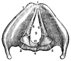
Fig. 106. A view of the larynx from above, showing the vocal ligaments. 1, The anterior edge of the larynx. 4, The posterior face of the thyroid cartilage. 5, 5, The arytenoid cartilages. 6, 6, The vocal ligaments. 7, Their origin, within the angle of the thyroid cartilage. 9, Their termination, at the base of the arytenoid cartilages. 8, 10, The glottis.
594. The larynx is connected by muscles with the sternum, œsophagus, base of the skull, hyoid bone, lower jaw, and tongue. This organ is supplied with a large number of blood-vessels, and it likewise receives nerves from the sympathetic system, and two large nerves from the tenth pair. The number and size of the nervous filaments distributed to the mucous membrane of the larynx, render it more sensitive than any other portion of the respiratory organs.
How is the glottis enlarged or contracted? Explain fig. 106. 594. By what means and to what organs is the larynx connected? Why is the larynx more sensitive than other parts of the respiratory organs?
595. The larynx is much more developed and prominent in man than in woman. In the former, the anterior angle of the thyroid cartilage is acute, while in the latter it is rounded, and the central slope of the superior border of the same cartilage is less deep, and the epiglottis smaller and less prominent, than in man.
596. The difference in the formation of the larynx in infancy is less striking; but at a later period, it is more developed in the male than in the female. It is very remarkable that this increase is not progressive, like that of other organs, but, on the contrary, develops itself at once at the period of puberty.
597. In the formation of the voice, each part already described performs an important office. The cricoid and thyroid cartilages give form and stability to the larynx; the arytenoid cartilages, by their movement, vary the width of the glottis. The epiglottis is flexible and elastic. When it is erect, the chink of the glottis is open, as in inspiration; when depressed, as in swallowing food and drink, it covers and closes this aperture. It prevents the introduction of articles of food into the trachea, and probably modifies sound as it issues from the glottis.
598. The muscles of the neck elevate and depress the larynx; the muscles of the larynx increase or diminish the width of the glottis; at the same time, the vocal cords are 273 relaxed or tightened, while the muscles of the face open and close the mouth.
595. What difference between the formation of the larynx of the female and that of the male? 596. Does this difference exist in childhood? Is its development progressive? 597–600. Give the physiology of the vocal organs. 597. Which cartilages give stability and form to the larynx? Which vary the width of the glottis? What is the function of the epiglottis? 598. What effect have the muscles of the neck upon the larynx? The use of the muscles of the larynx?
599. The elasticity of the ribs and the contraction of the abdominal muscles diminish the cavity of the chest, and the air, in consequence, is pressed from the air-cells into the bronchial tubes and trachea. It then rushes by the vocal cords, and causes a peculiar vibration, which produces sound.
Observations. 1st. Experiments have satisfactorily shown that the vocal cords are the principal agents in the formation of the voice. The tongue, which many have supposed to be the most important organ in speaking, is not essential to sound. In several instances it has been removed, and the persons thus mutilated could speak with fluency.
2d. When the vocal cords are ulcerated, or inflamed, however slightly, as in sore throat produced by a cold, the voice will be changed. The loss of speech among public speakers is generally produced by a relaxation of the vocal ligaments. Hence, bronchitis is a misnomer for this affection.
600. Sound is varied by the velocity of the expelled current of air, and the tension of the vocal ligaments. The size of the larynx, the volume and health of the lungs, the condition of the fauces and nasal passages, the elevation and depression of the chin, the development and freedom of action of the muscles which are attached to the larynx, the opening of the mouth, the state of the mind, and general health of the system, influence the modulations of sound.
What effect has the combined action of these muscles? 599. How is sound produced? What have experiments shown? What effect has disease of the vocal ligaments upon the voice? 600. How is sound varied? Mention other conditions that contribute to the modulation of sound.
601. The voice can be changed and modified by habit. Sailors, smiths, and others, who are engaged in noisy occupations, exert their vocal organs more strongly than those of more quiet pursuits. This not only affects the structure of the vocal organs, but varies the intonation of the voice.
602. The voice is strong in proportion to the development of the larynx, and the capacity of the chest. Singing and reading aloud improve and strengthen the vocal organs, and give a healthy expansion to the chest. The enunciation of the elementary sounds of the English language, aids in developing the vocal organs, as well as preventing disease of the throat and lungs. This exercise also conduces to the acquisition of musical sounds.
603. The attitude affects the modulation of the voice. When an individual stands erect, the movements of the whole respiratory apparatus are most free and effective. The larynx is brought forward by the erect position of the head and the elevation of the chin. The muscles of the arytenoid cartilages are then brought to a proper relation for action, by which a tension of the vocal cords is produced, that favors clear and harmonious enunciation.
Experiment. Read with the head bowed forward and the chin depressed; then read with the head erect and the chin elevated, and the difference in the movement of the vocal 275 organs, together with the difference in the voice, will be manifest.
601–616. Give the hygiene of the vocal organs. 602. How may the voice be strengthened? 603. What effect has the erect attitude upon the modulations of the voice? Give the experiment.
Fig. 107. An improper position; but one not unfrequently seen in some of our common schools, and in some of our public speakers.
Fig. 108. The proper position for reading, speaking, and singing.
604. If an individual or class read or sing when sitting, let the position represented by fig. 109 be adopted, and not the one represented by fig. 110; for the erect position in sitting conduces to the free and effective action of the respiratory and vocal organs, and is as important as the erect attitude in standing.
604. What position should be adopted when a person reads or sings when sitting? Why?
605. The muscles of the neck should not be compressed. If the muscles of the neck and larynx are compressed by a high cravat, or other close dressing, not only will the free and energetic movements of these parts be impeded, but the tones will be feeble and ineffective. Therefore the dress of the neck, particularly of public speakers and singers, should be loose and thin. For a warm dress upon the neck, when the vocal organs are in action, will induce too great a flow of blood to these parts, which will be attended by subsequent debility.
Fig. 109.
Observations. 1st. The loss of voice, (lar-yn-gi´tis,) which is prevalent among public speakers, may be ascribed in part to the injudicious dressing of the neck, and improper position in standing.
605. How should public speakers dress their necks? Why? What is a common cause of the loss of voice?
2d. When individuals have been addressing an audience in a warm room, or engaged in singing, they should avoid all impressions of a cold atmosphere, unless adequately protected by an extra garment.
Fig. 110.

606. The condition of the air modifies speaking and singing. As pure air is more elastic and resonant than impure, and as easy, melodious speaking or singing requires atmospheric elasticity, so school-rooms and singing-halls should be well ventilated, if we would be entertained with soft intonations in reading, or sonorous singing.
Observation. The imperfect ventilation of churches and vestries is another cause of laryngitis among clergymen. 278 This affection is almost unknown among those who speak in very open rooms, where stoves are not used.
Give 2d. observation. 606. Why does easy and melodious speaking require pure air? What is another cause laryngitis among clergymen?
607. The condition of the nasal passages and throat modifies the voice. The enunciation of words is rendered more or less distinct, in proportion as the jaws are separated in speaking, and the fauces and nasal passages are free from obstruction. For these reasons, the scholar should be taught to open the mouth adequately when reading, speaking, or singing, that the sounds formed in the larynx and modified in the fauces may have an unobstructed egress.
Observations. 1st. If the fauces are obstructed by enlarged tonsils, (a condition by no means uncommon in children,) they should be removed by a surgical operation, which is not only effective, but safe, and attended with little suffering. The tonsils are situated on each side of the base of the tongue, and, when enlarged, they obstruct the passage through which the air passes to and from the lungs, and the respiration is not only laborious, but distressing.
2d. When the nasal passages are obstructed, there is a peculiar sound of the voice, which is called “talking through the nose.” This phenomenon arises, not from the expired air passing through the nose, but from its not being able to pass through the nasal passages.
608. The state of the mind and health exerts an influence upon the vocal organs. “The organs of the voice, in common with all other parts of the bodily frame, require the vigor and pliancy of muscle, and the elasticity and animation of mind, which result from good health, in order to perform their appropriate functions with energy and effect. But these indispensable conditions to the exercise of vocal organs, are, in the case of most learners, very imperfectly supplied.”
607. Does the condition of the throat and nasal passages modify the voice? Name the influences that produce clear enunciation of words. What is the effect when the nasal passages are obstructed? 608. How are the vocal organs influenced? What do they require?
609. “A sedentary mode of life, the want of invigorating exercise, close and long-continued application of mind, and, perhaps, an impaired state of health, or a feeble constitution, prevent, in many instances, the free and forcible use of those muscles on which voice is dependent. Hence arises the necessity of students of elocution practising physical exercises adapted to promote general muscular vigor, as a means of attaining energy in speaking; the power of any class of muscles being dependent on the vigor of the whole system.”
610. “Gymnastic and calisthenic exercises are invaluable aids to the culture and development of the voice, and should be sedulously practised when opportunity renders them accessible. But even a slight degree of physical exercise, in any form adapted to the expansion of the chest and to the freedom and force of the circulation, will serve to impart energy and glow to the muscular apparatus of voice, and clearness to its sound.”
611. “There is, therefore, a great advantage in always practising some preliminary muscular actions, as an immediate preparation for vocal exercises. The art of cultivating the voice, however, has, in addition to the various forms of corporeal exercise, practised for the general purpose of promoting health, its own specific prescription for securing the vigor of the vocal organs, and modes of exercise adapted to the training of each class of organs separately.”
612. The results of such practice are of indefinite extent. They are limited only by the energy and perseverance of the student, excepting perhaps in some instances of imperfect organization. A few weeks of diligent cultivation are usually sufficient to produce such an effect on the vocal organs, that 280 persons who commence practice with a feeble and ineffective utterance, attain, in that short period, the full command of clear, forcible, and varied tone.
609. Why are students of elocution in general necessitated to practise physical exercise? 610. What are invaluable aids in the culture of the voice? 611. What is said of the art of cultivating the voice? 612. Are the results of such practices limited? What exception?
613. Repetition is essential to distinct articulation of words. In teaching a child to articulate a letter or word, in the first instance, make an effort to induce a proper state of the vocal organs by which the particular sound is produced. Repeat the letter or word again and again, until all the parts of the vocal apparatus harmonize in their movements to produce the given sound. This repetition is as necessary in learning to read as in singing.
Observations. 1st. There is nothing gained by trying to teach a child to pronounce the letters of the alphabet, before the vocal organs are so developed that distinct utterance can be given to the proper sounds.
2d. The drawling method of talking to young children, as well as using words that are not found in any written language, (called child’s talk,) is decidedly wrong. A child will pronounce and understand the application of a correct word as quickly as an incorrect one.
614. No part of the vocal organs is wanting, with those individuals that stammer, or who have an impediment in their speech. Some parts may be more developed than others, but they generally are but imperfectly under the control of the will, and assume an irregular and rapid movement, while other parts, the motions of which are essential, remain comparatively inactive. This can be seen by comparing the movements of the lips, tongue, and larynx, while attempting to speak, in a person who stammers, with the movements of the corresponding parts, while speaking, in an individual who has no such impediment.
613. Is repetition essential to distinct articulation? What method is suggested in teaching a child to articulate letters or words? Give observation 1st. Observation 2d. 614. Are the vocal organs wanting in stammerers? Why the defect in their articulation of words?
615. Surgical operations and medical treatment are not highly advantageous in a majority of these cases. In the young and middle aged, this defect can be remedied by patient and judicious training. At first, only those letters and words should be spoken that can be articulated with distinctness. Let there be repetition, until the words can be spoken at any time with readiness. Then take for a lesson other words, more difficult to articulate; and pursue a similar process of training and repetition, until every part of the vocal organs can be called into a ready and harmonious action in giving utterance to any word in common use.
616. The method of removing foreign bodies from the throat. It is not necessary to ascertain which passage the foreign body is in, for the immediate treatment ought in either case to be the same. Some person should place one hand on the front of the chest of the sufferer, and, with the other, give two or three smart blows upon the back, allowing a few seconds to intervene between them. This treatment will generally be successful, and cause the substance to be violently thrown from the throat.
Observation. If the foreign body passes into the larynx violent spasmodic coughing immediately succeeds, which continues until it is removed or life is extinct. Such cases demand the prompt opening of the trachea below the larynx by a skilful surgeon.
615. How can stammering be remedied? 616. What is the method of removing foreign bodies from the throat?
617. The skin is a membrane which envelops the muscles and other parts of the system. In youth, and in females particularly, it is smooth, soft, and elastic. In middle age, and in males, it is firm and rough to the touch. In old age, in persons who are emaciated, and about the flexions of the joints, it is thrown into folds. The interior of the body, like the exterior, is covered by a skin, which, from the constantly moistened state of its surface, is called the mucous membrane. At the various orifices of the body, the exterior skin is continuous with the internal.
618. The SKIN, to the naked eye, appears composed of one membrane. But examination has shown that it consists of two layers of membrane, namely, the Cu´ti-cle, (scarf-skin,) and the Cu´tis Ve´ra, (true skin.) These layers are widely different from each other in structure, and perform very different offices in the animal economy.
619. The CUTICLE (sometimes called the ep-i-derm´is) is the external layer of the skin. This membrane is thin and 283 semi-transparent, and resembles a thin shaving of soft, clear horn, and bears the same relation to other parts of the skin that the rough bark of a tree does to the liber, or living bark. The cuticle has no perceptible nerves or blood-vessels; consequently, if it is cut or abraded, no pain will be felt, and no fluid will ooze from it.
617. What is the skin? Mention its different appearances in its different conditions in the human frame. Is the interior of the body, as well as the exterior, covered by a skin? What is the interior membrane called? Why has it received this name? 618–636. Give the anatomy of the skin. 618. What is said of the skin? What is said relative to these layers of membrane? 619. Describe the cuticle. What name is sometimes applied to the cuticle?
Experiment. Pass a pin through the portion of the cuticle that skirts the nails, or remove a thin shaving from the palm of the hand, and no painful sensation will be experienced unless the pin or knife penetrates deeper than the cuticle.
620. This membrane varies in thickness on different parts of the body,—from the thin, delicate skin upon the internal flexions of the joints, to the thickened covering of the soles of the feet. The greater thickness of the cuticle of the palms of the hands and soles of the feet, is manifestly the intentional work of the Creator; for it is perceptible in infants, even at birth, before exercise can have had any influence.
621. The CUTIS VERA (sometimes called the co´ri-on) is composed of minute fibres, which are collected into small bundles or strands. These are interwoven with each other so as to constitute a firm, strong, and flexible web. In the superficial part of the true skin, the web is so close as to have the appearance of felt-cloth; but more deeply, the pores become progressively larger, and, upon the lower surface, have a diameter of about a line, or one twelfth of an inch. This gives the under surface the appearance of a coarse web. The strands of the under surface of the true skin are connected with the fibrous web, in which the sub-cutaneous fat of the body is deposited; while the upper surface gives 284 support to the sensitive, or papillary layer, which is bedded upon it.
Give the experiment. 620. What is said of the thickness of the cuticle in different parts of the body? 621. Describe the cutis vera. By what name is it sometimes called? What is the appearance of the upper surface of the cutis vera? Of the under surface?
Observation. When the skins of animals are immersed in a strong solution of oak or hemlock bark, a chemical union takes place between the gelatin, of which the true skin is mostly composed, and the tannin of the bark. By this process leather is formed, and its peculiar markings are owing to the papillary layer.
Fig. 111.

Fig. 111. An ideal representation of the papillæ. 1, 1, The cutis vera. 2, 2, The papillary layer. 3, 3, The arteries of the papillæ. 4, 4, The veins of the papillæ. 5, 5, The nerves of the papillæ.
622. The sensitive layer of the skin is thin, soft, uneven, pinkish in hue, and composed of blood-vessels, which confer its various tints of red; and of nerves, which give it the faculty of sensation. The unevenness of this layer is produced by small, elongated, conical prominences, called Pa-pil´læ.
623. Each PAPILLA is composed of a minute artery, vein, and nerve. Some of the prominences are arranged in concentric 285 ovals, as may be seen on the ends of the fingers; others are more or less parallel, and pursue a serpentine course; some suddenly diverge, and again reunite, as may be seen in the palm of the hand. Papillæ are found in every part of the skin. Consequently, their number is very great.
How is leather formed? 622. What is the appearance of the sensitive layer? What causes the unevenness of this layer? Explain fig. 111. 623. Describe the papillæ.
624. The cutis vera contains not only Arteries, Veins, and Nerves but Lymphatics, Oil-Glands and Tubes, and Perspiratory Glands and Tubes.
Fig. 112.

Fig. 112. The arteries and veins of a section of the skin. A, A, Arterial branches. B, B, Capillary, or hair-like vessels, in which the large branches terminate. C, The venous trunk, collecting the blood from the capillaries.
625. The ARTERIES AND VEINS of the skin are very numerous. The larger branches of the arteries pass through the open meshes of the true skin, and are subdivided into a myriad of minute capillary vessels, which form a beautiful net-work on the upper surface of the true skin. This vascular net sends a branch to each of the papillæ, which opens into and terminates in a minute vein. The capillary veins are 286 as numerous as the arteries which they accompany. They unite and form larger trunks, as small springs from the hill side coalesce to form rivulets.
624. What vessels are found in the cutis vera? Explain fig. 112. 625. What is said of the cutaneous arteries? Of the cutaneous veins?
626. The NERVES that are spread over every part of the sensitive layer of the true skin, proceed from the spinal cord. As a proof of the great number of nervous filaments in the skin, no part of this tissue can be punctured with a fine needle without transfixing a nerve, and inducing pain. In some parts of the system, however, the nerves are more abundant than in others; where the sense of feeling is most acute, we find the greatest number of nerves, and those of the largest size. Those parts that are most exposed to injury are most sensitive.
Examples. 1st. The conjunctiva, or skin of the eye, is pained by the presence of a particle of dust, because it would render vision imperfect.
2d. The lungs, also, would be injured by the smallest particle of matter; they are therefore protected by the exquisite sensitiveness of the lining membrane of the trachea, so that a particle of food or dust is ejected by a convulsive cough before it reaches the lungs.
627. The nerves are more numerous in the upper than lower extremities; in greater numbers upon the palm than the back of the hand. They are, likewise, more abundant and larger at the extremities of the fingers, and in the lips, than in any other part of the skin.
Observation. The proboscis of the elephant, the extremities of the tails of certain species of monkeys, and the tentacula of some kinds of fish, receive a more abundant supply of sensitive nerves than other parts of their systems.
626. Where do the nerves of the skin proceed from? Are they numerous in this membrane? How is it proved? What is said of those parts most exposed to injury? Give example 1st. Example 2d. 627. Mention the difference in the distribution of the nerves in various parts of the body. Is this difference found in the lower order of animals?
628. In the small papillæ, the nerve forms a single loop, while in papillæ of larger size, and endowed with a power of more exalted sensation, the nerve is bent several times upon itself previous to completing the loop. These little loops spring from a net-work of nerves, imbedded in the upper porous layer of the true skin, at the base of the papillæ. This net-work of nerves receives its influence through nerves which take their winding course through the fat distended openings of the deeper layers of the true skin.
Fig. 113.
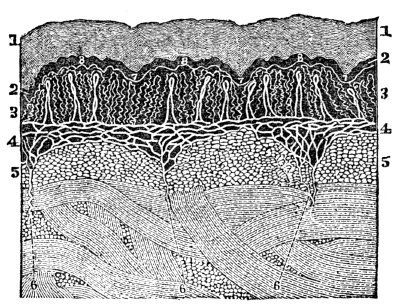
Fig. 113. 1, 1, The cuticle. 2, 2, The colored layer of the cuticle. 3, 3, The papillary layer, exhibiting the nerves as they form loops. 4, 4, The net-work of nerves. 5, 5, The true skin. 6, 6, 6, Three nerves that divide to form the net-work (4, 4.) 7, 7, 7, The furrows between the papillæ. 8, 8, 8, Three papillæ magnified fifty diameters.
629. The LYMPHATICS are found in great numbers in the true skin, and they are so minute that they cannot be seen with the naked eye; but when these hair-like vessels are injected with quicksilver, (a work of great difficulty,) the surface injected 288 resembles a sheet of silver. In this way their existence can be imperfectly demonstrated. They are a part of the vascular net-work situated upon the upper surface of the true skin. Each papilla is supplied with a lymphatic filament, the mouth of which opens beneath, and lies in contact with the under surface of the cuticle. This net-work of vessels communicates through the open meshes of the true skin with larger lymphatic trunks, that open into the venous system.
628. How are the nerves of the small papillæ arranged? How in the large papillæ? What does fig. 113 represent? 629. What is said of the cutaneous lymphatics? How is their existence proved?
Fig. 114.
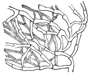
Fig. 114. A plexus of lymphatic vessels in the skin, considerably magnified from an injected preparation.
630. The OIL-GLANDS are small bodies imbedded in the true skin. They connect with the surface of the skin by small tubes, which traverse the cuticle. In some parts, these glands are wanting; in others, where their office is most needful, they are abundant, as on the face and nose, the head, the ears, &c. In some parts, these tubes are spiral; in others, straight. These glands offer every shade of complexity, from the simple, straight tube, to a tube divided into numberless 289 ramifications, and constituting a little rounded tree-like mass, about the size of a millet seed.
Of what are they a part? 630. Describe the oil-glands. With what do they connect? Do they exist in every part of the body? Of what form are their tubes?
631. In a few situations, these small glands are worthy of particular notice, as in the eyelids, where they possess great elegance of distribution and form, and open by minute pores along the lids; in the ear-passages, where they produce that amber-colored substance, known as the ce-ru´men, (wax of the ears,) and in the scalp, where they resemble small clusters of grapes, and open in pairs into the sheath of the hair, supplying it with a pomatum of Nature’s own preparing. The oil-tubes are sometimes called the se-ba´ceous fol´li-cles.
Fig. 115.
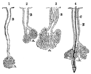
Fig. 115. 1, An oil-tube and gland from the scalp. A, The gland. B, The tube slightly twisted.
2. An oil-tube and gland from the skin of the nose. The gland (A) is double, and communicates with the main tube (B) by means of two smaller tubes.
3. Another oil-tube and gland from the nose. A, The gland. B, The tube filled with the peculiar animalculæ of the oily substances. Their heads are directed inward.
4. A small hair from the scalp, with its oil-glands. The glands (A) form a cluster around the shaft of the hair-tube, (C.) These ducts open into the sheath of the hair, (B.) All the figures, from 1 to 4, are magnified thirty-eight diameters.
631. What is said of these tubes in the eyelids? In the ear? In the scalp? What are these glands sometimes called?
Observation. Among the inhabitants of cities, and especially in persons who have a torpid state of the skin, the contents of the oil-tubes become too dense and dry to escape in the usual manner. Thus it collects, distends the tube, and remains until removed by art. When this impacted matter reaches the surface, dust and smoke mix with it, then it is recognized by small, round, dark spots. These are seen on the forehead, nose, and other parts of the face. When this matter is pressed out, the tube gives it a cylindrical form. The parts around the distended tubes sometimes inflame. This constitutes the disease called, “ac´ne punc-ta´ta.”
632. The PERSPIRATORY APPARATUS consists of minute cylindrical tubes, which pass inward through the cuticle, and terminate in the deeper meshes of the cutis vera. In their course, each little tube forms a beautiful spiral coil; and, on arriving at its destination, coils upon itself in such a way as to constitute an oval-shaped, or globular ball, called the perspiratory gland.
633. The opening of the perspiratory tube on the surface of the cuticle, namely, “the pores,” is also deserving of attention. In consequence of its extremity being a section of a spirally-twisted tube, the aperture is oblique in direction, and possesses all the advantages of a valvular opening, preventing the ingress of foreign injurious substances to the interior of the tube and gland.
634. “To arrive at something like an estimate of the value of the perspiratory system, in relation to the rest of the organism, I counted the perspiratory pores on the palm of the hand, and found 3528 in a square inch. Now each of these pores being the aperture of a little tube about a quarter of an inch 292 long, it follows, that in a square inch of skin on the palm of the hand there exists a length of tube equal to 882 inches, or 73½ feet. Surely such an amount of drainage as seventy-three feet in every square inch of skin—assuming this to be the average for the whole body—is something wonderful and the thought naturally intrudes itself, What if this drainage be obstructed?
What is said of the retention of the unctuous matter in the oil-tubes? 632. Of what does the perspiratory apparatus consist? 633. What is peculiar in the opening of the perspiratory tubes on the surface of the cuticle? 634. How many perspiratory pores did Dr. Wilson count upon a square inch of skin on the palm of the hand?
Fig. 116.

Fig. 116. A perspiratory gland from the palm of the hand, magnified forty diameters. 1, 1, A twisted tube composing the gland. 2, 2, The two excretory ducts from the gland. These unite to form one spiral tube, that perforates the cuticle, (3,) and opens obliquely on its surface at 4. The gland is imbedded in cells filled with fat, which are seen at 5, 5.
What does fig. 116 represent?
635. “Could we need a stronger argument for enforcing the necessity of attention to the skin? On the pulps of the fingers, where the ridges of the sensitive layer of the true skin are somewhat finer than in the palm of the hand, the number of pores on a square inch a little exceeded that of the palm; and on the heels, where the ridges are coarser, the number of pores on the square inch was 2268, and the length of the tube 567 inches, 47¼ feet.
636. “To obtain an estimate of the length of tube of the perspiratory system of the whole surface of the body, I think that 2800 might be taken as a fair average of the number of pores in the square inch; and consequently, 700, the number of inches in length. Now, the number of square inches of surface in a man of ordinary height and bulk is 2500; the number of pores, therefore, 7,000,000; and the number of inches of perspiratory tube is 1,750,000; that is, 145,833 feet, or 48,611 yards, or nearly TWENTY-EIGHT miles!”—Wilson.
Give other computations in this paragraph. 635. What is said of the number of these pores on the pulp of the fingers? On the heels? 536. What is an average number of pores and length of tube of the whole surface of the body? Give the summary of the number of pores, and number or inches of perspiratory tube.
637. The skin invests the whole of the external surface of the body, following all its prominences and curves, and gives protection to all the organs it encloses, while each of its several parts has a distinct use.
638. The cuticle is insensible, and serves as a sheath of protection to the highly sensitive skin (cutis vera) situated beneath it. The latter feels; but the former blunts the impression which occasions feeling. In some situations, the cuticle is so dense and thick, as wholly to exclude ordinary impressions. Of this we see an example in the ends of the fingers, where the hard and dense nail is the cuticle modified for the purpose referred to. Were the nervous tissue of the true skin not thus protected, every sensation would be so acute as to be unpleasant, and contact with external bodies would cause pain.
639. The cuticle, also, prevents disease, by impeding the evaporation of the fluids of the true skin, and the absorption of the poisonous vapors, which necessarily attend various employments. It, however, affords protection to the system only when unbroken, and then, to the greatest degree, when covered with a proper amount of oily secretion from the oil-glands.
640. The cuticle is, originally, a transparent fluid, exuded 294 by the blood-vessels, and distributed as a thin layer on the surface of the true skin. While successive layers are formed on the exterior of the true skin, the external cuticular layers are converted into dry, flattened scales, by the evaporation of their fluid contents. The thickness of the cuticle is formed mainly from these scales.
637–656. Give the physiology of the skin. 637. What is said of the skin? 638. Give a function of the cuticle. Does it vary in thickness on different parts of the body? Give examples. 630. Mention another use of the cuticle. 640. What is the cuticle originally?
641. The cuticle is, therefore, undergoing a constant process of formation and growth at its under part, to compensate for the wear that is taking place continually on its surface. A proper thickness of the cuticle is in this manner preserved; the faculty of sensation and that of touch are properly regulated; the places of the little scales, which are continually falling off under the united influence of friction and ablution, are supplied; and an action necessary, not merely to the health of the skin, but to that of the entire body, is established.
642. Whenever the cuticle is exposed to moderate and repeated friction, it becomes thicker and tougher, as may be seen in the cuticle of the lady’s finger that plies the needle and in the hard or callous appearance of the hands of farmers masons, and other mechanics. This enables them to handle the utensils and materials used in their vocations without pain or inconvenience.
Observations. 1st. When the joints of the feet are subjected to moderate and continued pressure or friction, frequently one or more of the papillæ enlarge. This is accompanied with a thickening of the layers of the cuticle, which is termed a “callosity,” or “corn.” These thickened layers of the cuticle are broad at the top and narrow at the bottom, and the enlarged mass is conical, with the point 295 innermost. When pressed upon by a tight shoe, these sensitive papillæ cause pain.
How is the thickness of the cuticle mainly formed? 641. Describe the changes of this membrane. Show the necessity of this constant growth. 642. How does moderate and repeated friction affect the cuticle? Give examples. What is the benefit derived from having the cuticle thus changed? What is the result if the joints of the feet are subjected to moderate and continued pressure? What is the form of a “corn”?
2d. To remove these painful excrescences, take a thick piece of soft leather, somewhat larger than the corn; in the centre punch a hole of the size of the summit of the corn, spread the leather with adhesive plaster, and apply it around the corn. The hole in the leather may be filled with a paste made of soda and soap, on going to bed. In the morning, remove it, and wash with warm water. Repeat this for several successive nights, and the corn will be removed. The only precaution is, not to repeat the application so as to cause pain.
643. Let a person unaccustomed to manual labor, trundle the hand-cart, or row a boat, for several successive hours, and the cuticle upon the palms of the hands, instead of becoming thicker by use, is frequently separated from the subjacent tissues, by an effusion of serum, (water,) thrown out by the vessels of the true skin. Had the friction been moderate, and applied at regular intervals, instead of blisters being formed upon the inside of the hands, material would have been thrown out to form new layers upon the lower surface of the cuticle.
644. The cuticle is interesting to us in another point of view, as being the seat of the color of the skin. The difference of color between the blonde and the brunette, the European and the African, lies in the cuticle;—in the deeper, and softer, and newly-formed layers of that structure. In the whitest skin, the cells of the cuticle always contain more or less of a peculiar pigment, incorporated with the elementary granules which enter into their composition. In the white 296 races, the pigmentary tint is extremely slight, and less in winter than in the summer season. In the darker races, on the contrary, it is deep and strongly marked.
How can they be removed? What precaution is given? 643. Explain why those persons unaccustomed to labor, blister their hands in rowing a boat or performing ordinary manual employment for several successive hours. 644. In what other point of view is the cuticle interesting? In what part of it do we find the coloring matter?
645. The various tints of color exhibited by mankind, are, therefore, referable to the amount of coloring principle contained within the elementary granules of the cuticle, and their consequent depth of hue. In the negro, the granules are more or less black; in the European of the south, they are amber-colored; and in the inhabitants of the north, they are pale and almost colorless.
646. Color of the skin has relation to energy in its action; thus, in the equatorial region, where light and heat are most powerful, the skin is stimulated by these agents to vigorous action, and color is very deep; while in the temperate regions, where light and heat are not so intense, the lungs, liver, and kidneys relieve the skin of part of its duties. The colored layer of the cuticle has been called the re´te mu-co´sum, (mucous coat of the skin,) and described as a distinct layer by many physiologists.
Observation. “The various coloring of the inner layer of the cuticle gives to some animals their varied hues; the serpent, the frog, the lizard, and some fishes have a splendor of hue almost equal to polished metal. The gold-fish and the dolphin owe their difference of color and the brilliancy of their hues to the color of this layer of the skin.”
647. The nerves of the skin are the organs of the sense of touch and feeling. Through them we receive many impressions that enhance our pleasures, as the grateful sensations imparted by the cooling breeze in a warm day. In 297 consequence of their sensitiveness, we are individually protected, by being admonished of the proximity of destructive agents.
In what season of the year is the coloring matter less in the white race? 645. To what is the color of the skin referable? 646. Why have the races of the torrid zone darker complexions than those of the temperate or frigid zones? What is this colored layer called by many physiologists? To what is the different hues in animals owing? 647. Of what use are the nerves of the skin?
Illustration. A man who had been afflicted some years with a severe disease of a portion of the brain and spinal cord, was deprived of feeling in the lower extremities. He was directed by his attending physician to use a warm footbath. Intending to follow the directions given him, he immersed his feet in boiling water, which he supposed of a proper temperature. While his feet were immersed in the water, he experienced no sensation of an unpleasant nature. On withdrawing them, he was astonished to find the cuticle separated from the other tissues, by the effusion of serum, and thus producing a blister over the whole surface.
648. Portions of the skin would suffer every day, were it not for the sentinel-like care exercised by the nerves, by which all impressions are transmitted to the brain. As the skin is continually exposed to the influence of destructive agents, it is important that the nerves, provided for its protection, should be kept in a healthy state.
649. A large proportion of the waste of the body passes through the outlets of the skin; some portions in the form of oil, others in the form of water and carbonic acid.
650. The oil-glands secrete an oil, partly free and diffused, and partly mixed with albumen. When the cells are fully formed, that is, fully distended, they yield their contents, and the fluid matter they contain is set free, and passes along the tubes to the surface; this fluid matter constitutes the oily element of the economy of the skin.
651. The uses of the unctuous product of the oil-glands are twofold: 1st. The protection; 2d. The removal of waste 298 matter from the system. In the exercise of these offices the oily substance is diffused over those parts of the skin which are naturally exposed to vicissitudes of temperature and moisture,—as the nose, face, and head;—to the injurious attrition of contiguous surfaces,—as the flexures of joints;—or the contact of acrid fluids,—as in the excoriations to which infants are liable.
Give the illustration. 648. Why is it necessary that the cutaneous nerves be kept in a healthy state? 649. Through what membrane does a large proportion of the waste material of the system pass? 650. What is the function of the oil-glands? 651. What are the uses of the oily product of these glands?
652. The oil of the unctuous substance is the principal agent in effecting these purposes: 1st. It prevents the evaporation or congelation of the water of the cuticle, which would cause it to become parched and peel off, thus leaving the sensitive skin exposed. 2d. It affords a soft medium to the contact of moving substances. 3d. It repels moisture and fluids. 4th. The action of these glands removes the waste atoms and purifies the blood.
653. In considering the purpose of the oily matter of the skin, there are two situations in which it deserves especial remark. 1st. Along the edges of the eyelids, where it is poured out in considerable quantity. Here, it is the means of confining the tears and moisture of the eyes within the lids, defending the skin from the irritation of that fluid, and preventing the adhesion of the lids, which is liable to occur upon slight inflammation. 2d. In the ears, where the unctuous wax not only preserves the membrane of the drum and the passage of the ear moist, but also, by its bitterness, prevents the intrusion of small insects.
654. The use of the perspiratory glands is to separate from the blood that portion of the waste matter which is carried off through the skin in the form of vapor. Sanctorius, a celebrated medical writer, daily, for thirty years, weighed 299 himself, his food, and excretions. He estimated that five of every eight pounds of food and drink passed from the system through the many outlets upon the skin. Many place the estimate much lower. All physiologists agree that from twenty to forty ounces of matter pass off from the skin of an adult every twenty-four hours.
652. What prevents the evaporation of the water of the cuticle? Give its 2d use. Its 3d. Its 4th. 653. What is said in reference to the distribution of the oily matter along the edges of the eyelids? In the ears? 654. Of what use are the perspiratory glands? How long did Sanctorius daily weigh his food, to ascertain the amount of secretion that passed through the skin?
655. The average amount of perspiration is about thirty ounces; and it passes off in such minute portions, and mixes so rapidly with the surrounding air, that it is not perceived. For this reason, it is called insensible perspiration. When this excretion is increased, it forms into drops, and is called sensible perspiration. The following experiments prove the existence of this excretion from the skin.
Experiments. 1st. Take a cold bell-glass, or any glass vessel large enough to admit the hand, and introduce it perfectly dry; at the same time close the mouth by winding a napkin about the wrist; in a short time, the insensible perspiration from the hand, will be seen deposited on the inside of the glass. At first, the deposit is in the form of mist; but, if the experiment be continued a sufficient time, it will collect in drops.
2d. Hold the apparently dry hand near a looking-glass, and the invisible vapor will soon be condensed, and cover the glass with a slight dew.
656. It is important that this excretion be maintained with steadiness and regularity. When the action of the perspiratory glands is suppressed, all the vessels of the different organs will suffer materially, and become diseased, by the redundant waste matter that should be carried from the system. If a person is vigorous, the action of the organs, whose 300 functions are similar to those of the skin, as channels for the exit of waste matter, will be increased, and thus relieve the diseased state of the body. But the over-taxing of these organs, to relieve the system, often produces a diseased action in themselves.
What were his conclusions? 655. What is the average amount of perspiration every twenty-four hours? What is insensible perspiration? What is sensible perspiration? How can the existence of the excretion of the skin be shown? Give the 2d experiment. 656. Why is it important that these excretions be maintained regularly?
Fig. 117

Fig. 117. 1, 1, The lines, or ridges of the cuticle, cut perpendicularly. 2, 2, 2, 2, 2, The furrows, or wrinkles of the same. 3, The cuticle. 4, 4, 4, The colored layer of the cuticle. 5, 5, The cutis vera. 6, 6, 6, 6, 6, The papillæ. 7, 7, Small furrows between the papillæ. 8, 8, 8, 8, The deeper furrows between each couple of the papillæ. 9, 9, Cells filled with fat. 10, 10, 10, The adipose layer, with numerous fat vesicles. 11, 11, 11, Cellular fibres of the adipose tissue. 12, Two hairs. 13, A perspiratory gland, with its spiral duct. 14, Another perspiratory gland, with a duct less spiral. 15, 15, Oil-glands with ducts opening into the sheath of the hair, (12.)
Note.—Let the pupil review the anatomy and physiology of the skin from Fig. 117 or from anatomical outline plate No. 9.
657. The sensibility of the skin, and the activity of the oil and perspiratory glands, are modified by the condition of the cuticle, the temperature of the skin and body, the purity and warmth of the air, and the character of the light to which the body is exposed. Thus, to maintain a healthy action of every part of this membrane, attention should be given to Clothing, Bathing, Light, and Air.
658. Clothing, in itself, does not bestow heat, but is chiefly useful in preventing the escape of heat from the body, and in defending it from the temperature of the atmosphere. In selecting and applying clothing to our persons, the following suggestions should be observed.
659. The material for clothing should be a bad conductor of heat; that is, it should have little tendency to conduct or remove heat from the body. This depends mainly on the property possessed by the material in retaining atmospheric air in its meshes.
660. The material for clothing should not possess the property of absorbing and retaining moisture. Dampness, or moisture, renders apparel a good conductor of heat; beside, if the perspired fluid, and the saline material it holds in solution, are readily absorbed by the clothing, they become 302 sources of irritation to the skin with which the apparel comes in contact.
657–716. Give the hygiene of the skin. 657. What influences modify the action of the oil and perspiratory glands? To what must attention be given to maintain a healthy action of the skin? 658. What is said in regard to the clothing? 659. Mention a property that the material for clothing should possess. 660. What property in the selection of clothing should we avoid? Why?
661. Furs contain a greater amount of air in their meshes, than any other article, and they absorb no moisture; consequently, as an article of dress, they are best adapted to those who are exposed to great vicissitudes of heat and cold.
662. Woollen cloth retains more air in its meshes than any other article except furs and eider down, and it absorbs but very little moisture. These properties, together with its comparative cheapness, render it a good article of apparel for all classes of persons. The only objection to its general use is, the disturbance of the electricity of the system, and the irritation to delicate skins from the roughness of its fibres.
Observation. Flannels are not only beneficial, during the cold season, in preventing colds and rheumatism, but they are of great utility in the warm season, in shielding the system from the chills at evening, that induce disease of the alimentary canal. Their general use among children and delicate females, would be a preventive of the “season complaints” prevalent in the months of August and September.
663. Cotton contains less air in its meshes than woollen, but much more than linen. In texture, it is smoother than wool, and less liable to irritate the skin. This fabric absorbs moisture in a small degree. In all respects, it is well adapted for garments worn next the skin. When woollen flannels irritate the skin, they may be lined with cotton.
664. Silk is not as good a conductor of heat as cotton, nor does it absorb moisture to any considerable degree; its texture is smooth, and does not irritate the skin; consequently, when the garment of this fabric has sufficient body or thickness, it 303 is a good article for clothing. The greatest objection to its use is the disturbance of the electricity of the system, and its high price.
661. Give the properties of fur. As an article of dress, to whom are they best adapted? 662. Give the properties of woollen cloth. Is this a good article for clothing? What objection? What are the advantages of wearing flannels? 663. What are the qualities of cotton as an article of dress? 664. Of silk?
665. Linen is not only a good conductor of heat, and consequently a poor article of apparel, but it likewise absorbs the fluids carried from the system by the agency of the oil and perspiratory glands. When garments are made of this material, the body is not surrounded by a layer of air, but by one of moisture. This still further increases its power to conduct heat from the system, rendering it a very objectionable article of apparel, even in warm weather and in hot climates, where the dress is usually thin.
666. Clothing differs in its power of radiating heat. This is influenced by the color; those articles that radiate heat freely also absorb it readily. A black surface is a good radiator, while a white surface is not, because it reflects the calorific rays. It is obvious that those colors which render the transmission of external heat difficult, must impede the transmission of caloric from the body. Thus it is manifest, that light-colored apparel is best adapted for every season and every climate.
Observation. Coach-drivers are practically aware, that in cold weather, light-colored over-coats are warmest, except when they are exposed to the direct rays of the sun, or when seated before a warm fire. On the other hand, when the temperature is elevated, light-colored apparel is coolest, because the sun’s rays are then reflected.
667. The clothing should be of a porous character. The skin is not only an important agent in separating from the blood those impurities that otherwise would oppress the system and occasion death, but it exercises great influence upon the system, by receiving oxygen through its tissues, and giving 304 back carbonic acid in return. Consequently, the apparel should be made of a material that will permit free transpiration from the skin, and likewise convey the excreted fluids from the surface.
665. What is said of linen as an article of apparel? 666. Why is light-colored apparel best adapted for every season? What is said of the apparel of coach-drivers? 667. Why should we wear porous clothing?
668. The necessity for this is illustrated in wearing India rubber over-shoes. If they are worn over boots ten or twelve hours, not only the hose, but the boots will be moist from retained perspiration, and the residual matter left in contact with the skin may be reconveyed into the system by absorption, causing headache and other diseases. Cotton and woollen fabrics are not only bad conductors of heat, but are also porous; for these reasons, they are well adapted to transmit the excretions of the skin.
669. The clothing should be not only porous, but fitted loosely. The garments should retain a layer of air between them and the body. Every one is practically aware that a loose dress is much warmer than one which fits closely; that a loose glove is warmer than a tight one; and that a loose boot or shoe affords greater warmth than one of smaller dimensions. The explanation is obvious; the loose dress encloses a thin layer of air, which the tight dress is incapable of doing; and what is required, is, that the dress should be closed at the upper part, to prevent the dispersion of the warm air, by the ventilating current which would be established from below.
Observation. As the purpose of additional garments is to maintain a series of strata of warm air within our clothing, we should, in going from a warm room into the cold air, put on our defensive coverings some little time previous, in order that the layers of air which we carry with us may be sufficiently warmed by the heat of the room, and not borrowed from the body on exposure to the cold.
668. How is the necessity of porous clothing illustrated? 669. Why should we wear loose garments? What is the use of additional garments when going from a warm to a cold air? When should they be put on?
670. The clothing should be suited to the temperature of the atmosphere and the condition of the individual. The invariable rule should be, to wear enough to maintain an equal and healthy action of the skin. Care should be taken, however, that the action of the cutaneous vessels is not inordinately increased, as this would debilitate, not only the skin, but the internal organs of the system, as the stomach and lungs.
671. No rule as to the quantity of clothing can be given, as the demand will vary with different individuals. The following are among the most prominent causes of this variation: Those persons who have large, active brains, full chests, well developed lungs, breathe an adequate amount of pure air, and take sufficient food to supply the wants of the system, require less clothing than those of an opposite character, because more heat is generated in the system.
672. The child and the aged person require more clothing than the vigorous adult. “Should we judge from observation, the inference would be, that children require less clothing than adults. This is an error, for the temperature in infancy is not only lower than in manhood, but the power of creating heat is feebler. The same remarks are applicable to those persons who have outlived the energies of adult life.”
Observation. The system of “hardening” children, by an inadequate supply of clothing, and keeping them uncomfortably cold throughout the whole day, is inhuman, as well as unprofitable. It operates upon the child somewhat like the long-continued chill upon a certain portion of the farmer’s herd, that are kept shivering under the thatched shed, retarding the growth of their systems, which require more food to satisfy the keen cravings of hunger than when they are comfortably 306 sheltered. To make the boy robust and active, he must have nutritious food at stated hours, and free exercise in the open air, and his system must be guarded from chills by a due amount of apparel.
670. What should be the invariable rule in reference to the amount of clothing that should be worn? What precaution should be observed? 671. What are some of the causes of the variation of the demand for clothing? 672. Why do the child and aged person require more clothing than the vigorous adult? What is said of the system of hardening children?
673. More clothing is needed when a vital organ is diseased. It may be observed that in consumption, dyspepsia, and even in headache, the skin is pale and the extremities cold, because less heat is generated. Thus persons affected with these complaints, when exposed to cold air, need more clothing than those individuals whose organs are not diseased, and the functions of which are properly performed.
674. More clothing is required in the evening, than during the day. In the evening we have less vital energy, and therefore less heat is generated in the system, than in the early part of the day; beside, the atmosphere is damp, the skin has become moist from free perspiration, and heat, in consequence, is rapidly removed from the system. For this reason, when returning from crowded assemblies, we should be provided with an extra garment.
Observations. 1st. If there is a chill upon the system after having arrived home, warmth should be restored as speedily as possible. This can be done by friction with warm flannels, and by using the warm or vapor bath. By this procedure, the pernicious effects of the chill will be prevented before any disease is fixed upon the system. Is it not the duty of the parent and the guardian to learn these facts, and to see that they are not only learned, but reduced to practice?
2d. The farmer and industrious mechanic would be freed from many a rheumatic pain, if, while resting from their labors at evening, or taking the ordinary meal after hard toil, they would put on an extra garment. The coat might not 307 feel so agreeable for the first few minutes, but it would ultimately conduce to health and longevity.
673. Why do dyspeptic and consumptive persons require more clothing than those who have healthy vital organs? 674. Why do we need more clothing in the evening than during the day? How can the pernicious effects of a chill be prevented? Give the 2d observation.
675. The person of active habits requires less clothing than one of sedentary employments. Exercise increases the circulation of the blood, which is always attended by the disengagement of a greater quantity of heat; consequently, an increase of warmth is felt throughout the system. We likewise need more clothing while riding, than when we are walking; because the exercise of the former is less than that of the latter. The same is true when resting in the field or shop, after laborious exercise.
Observation. We need a greater amount of clothing while asleep, than during the day; as not only the action of the body, but that of the brain, during sleep, is suspended.
676. Less clothing is required when the cutaneous surface is clean. A film of impurities obstructs the perspiratory ducts, and diminishes the action of their glands; consequently, less heat is generated. For this reason, the hands or feet when clean are less liable to become chilled or frozen.
677. The sensitiveness of the skin to the influence of cold, is much modified by habit. A person who has been habituated to the temperature of a warm room, or warm climate, suffers more when exposed to cold, than an individual who has been accustomed to colder air. Thus a person who labors or studies in a warm room, should wear more clothing when exposed to the air, while walking or riding, than an individual who labors in a cooler atmosphere. Not only is the sensibility of the skin increased by a warm atmosphere, but the activity of the digestive, respiratory, and nervous systems, in generating heat, is much diminished. This is an additional reason why an increased amount of clothing is demanded 308 during exposure to cold air. In all cases where practicable the heat of the system should be maintained by exercise, in preference to the use of fur or flannel.
675. Why does the person of active habits require less clothing than one of sedentary employments? 676. Why do we need less clothing when the skin is clean? 677. Show the effect of habit on the sensitiveness of the skin.
678. Those parts of the skin usually covered, uniformly need that protection. The power of generating heat is diminished, and the impressibility to cold is increased, on those portions of the skin usually clothed. If a person wears the dress high and close about the neck, he suffers from exposure to a cold atmosphere if a dress is worn that is not as high or more open. As a general rule, it is preferable that those parts of the system, as the larynx, be exposed that are not uniformly protected by clothing.
679. The clothing should be kept clean. No article of apparel is entirely free from absorption; even wool and cotton possess it in a small degree. They take up a portion of the transpired fluids which contain saline and animal matter, and thus the fibres of the garments become covered with the cutaneous excretions. We are practically aware of the retention of these secretions from the soiled appearance of those garments worn next the skin, which are so covered as to preclude the particles of dust from lodging upon them.
680. The porosity of the clothing is lessened when soiled, and its power of conducting heat from the system in consequence, is increased. The residual matter with which the clothing is coated is brought in contact with the skin, which causes irritation, and not unfrequently re-absorption of the elements, thrown off from the system through this avenue. Hence warmth, cleanliness, and health require that the clothing, particularly the garments worn next to the skin, should be frequently and thoroughly washed. This should not be forgotten in regard to children, for their blood circulates 309 with greater rapidity than that of adults, and a proportionably greater amount of waste matter is thrown off from their systems.
681. The under-garments worn during the day should not be worn at night, or the reverse. When under-garments are worn several successive days or nights, they should not be put in drawers, or hung up in a close closet, as soon as taken from the body, but should be exposed to a current of air.
682. Occupied beds should be thoroughly aired in the morning. The excretions from the skin are most abundant during the hours of sleep; and if the sheets and blankets, together with the bed, are not aired every morning, by being so arranged that both surfaces may be exposed to the air, the materials eliminated from the skin will be retained in the meshes of the bed-clothing, and may be conveyed into the system of the next occupant, by absorption. Oftentimes diseases of a disagreeable nature are contracted in this way. This fact should be instilled into every mother’s and daughter’s mind.
Observation. Bed-linen should not be put on a bed when it is not sufficiently dried, or contains moisture from the excretions of the skin, nor should beds or bedding be slept in, that have remained in a damp room that has not been occupied for many weeks, unless the dampness is removed from the bed-linen by a warming-pan, or in some other way.
683. Changes of dress, from thick to thin, should always be made in the morning. At this time the vital powers are usually in full play. Many a young lady has laid the foundation of a fatal disease, by disregarding this rule, in exchanging the thick dress, with woollen stockings, for the flimsy dress and hose of silk or cotton, which are considered suitable 310 for the ball-room or party. Sudden changes in wearing-apparel, as well as in food and general habits, are attended with hazard; and this is proportionate to the weakness or exhaustion of the system when the change is made.
681. Should the garments worn during the day be worn at night? 682. What is said respecting the cleanliness of beds and bedding? Why should not bed-linen that is damp be slept in? 683. When should change of dress from thick to thin be made? Why?
684. When the clothing has become wet, it is best to change it immediately. The skin should then be rubbed with a dry crash towel, until reaction, indicated by redness, is produced. If the garments are not changed, the person should exercise moderately, so that sufficient heat may continue to be generated in the system to dry the clothing and skin without a chill. Sitting in a cool shade, or current of air, should, by all means, be avoided; as colds are not contracted by free and excessive exercise, but by injudicious management after such exercise.
Observation. When an individual has been thrown into a profuse perspiration by violent exercise, though the skin and clothing may become wet, he feels no inconvenience from the dampness, as long as he continues that amount of exercise for the reason that the circulation of the blood being increased heat is generated in sufficient quantity to replace the amount abstracted from the system in evaporating the free perspiration; but as soon as the exercise is discontinued, the increased circulation subsides, and with it the extra amount of generated heat. This accounts for the chill we experience, when the damp clothing is permitted to dry on the body, after the cessation of exercise.
684. What suggestion when the clothing has become wet? What should be done if the garments are not changed? What causes the chill that is experienced when damp clothing is permitted to dry on the body?
685. Bathing, its necessity and expediency, is obvious from the structure and the functions of the skin. The cuticle is cast off in minute, powdery scales, many of which are retained upon the surface by the pressure of clothing. These mingle with the oily and saline products of the skin, and form a thin crust. This crust, on account of its adhesiveness, collects particles of dust and soot from the atmosphere, and particles of foreign matter from our dress; so that in the course of the day the whole body becomes coated with impurities. If this coating remains, becomes thick and established upon the skin, it will produce the following effects:—
686. 1st. The pores will be obstructed, consequently transpiration impeded, and the influence of the skin as an excretory entirely prevented. When the pores are obstructed, and transpiration is checked, the elements of the transpired fluids will necessarily be retained in the system; and, as they are injurious and poisonous if retained, they must be removed by those organs whose functions in the animal economy are similar, as the lungs, kidneys, liver, intestines, &c.
687. When these organs are called upon to perform their offices, and in addition that of another, the healthy equilibrium is destroyed, and the oppressed organ will suffer from exhaustion, and become the prey of disease. Thus, obviously, habits of uncleanliness are a cause of consumption and other serious 312 diseases of the vital organs. Again, obstruction of the pores will prevent respiration through the skin, thus depriving the blood of one source of its oxygen, and one outlet of its carbonic acid, which will diminish the temperature of the system, and the same results follow as when the clothing is inadequate.
685. Show the necessity for bathing. 686. What effect upon the body if the pores of the skin are obstructed? 687. What is the effect when an organ not only performs its own specific function, but that of another?
688. 2d. The retained perspirable matter will irritate the skin, both mechanically and chemically; and this membrane will be kept damp and cold, from attraction and detention of moisture; and foreign material, as before adverted to, once removed from the system, may be reconveyed into it by absorption. As a consequence, cutaneous eruptions and diseases will be produced, and the re-absorption of matter once separated from the system, will be the exciting cause of other injurious disorders.
689. 3d. A film of foreign substance on the skin will inevitably become the seat of detention of miasmata and infectious vapors. These will remain until absorbed, and engender the diseases of which they are the peculiar cause. This is one reason why filthy persons contract infectious diseases more frequently than individuals of cleanly habits.
690. Bathing is useful to promote cleanliness. In this capacity, it enables us to remove the coating of impurities from the exterior of our persons. It effects this purpose by dissolving saline matters, and holding in temporary suspension those substances which are insoluble.
691. The cuticle is composed of a substance resembling the dried white of egg, or, in a word, albumen. This is soluble in alkalies, and these are the agents which are commonly employed for purifying the skin. Soap is a compound of the alkali soda with oil, the former being in excess. When used 313 for washing, the excess of alkali combining with the oily fluid, with which the skin is naturally bedewed, removes it, in the form of an emulsion, and with it a portion of any adhering matter. Another portion of the alkali softens and dissolves the superficial layer of the cuticle; and when this is removed the cuticle is free from impurities.
688. How are cutaneous eruptions frequently produced? 689. How are infectious vapors transmitted to the system? 690. How does bathing promote cleanliness? 691. Why is it necessary to use soap in bathing?
692. Every washing of the skin with soap removes the old face of the cuticle, and leaves a new one; and were the process repeated to excess, the latter would become so thin as to render the body sensible to impressions too slight to be felt through its ordinary thickness. On the other hand, when the cuticle and its accumulated impurities are rarely disturbed, the sensitiveness of the skin is impaired. The proper inference to be drawn from the preceding remarks, is in favor of the moderate use of soap to cleanse the skin.
Observation. If any unpleasant sensations are felt after the use of soap, they may be immediately removed by washing the surface with water slightly acidulated with lemon juice or vinegar, which neutralizes the alkali that may remain on the skin. This is effective treatment for “chapped hands.”
693. Bathing may be partial or general, and the water used may be cold, temperate, tepid, warm, or hot. A person may apply it to his system with a sponge, it may be poured upon him, or he may immerse himself in it. The simplest mode of bathing is to apply water to a small extent of surface, by means of a wet sponge, and after being wiped dry, again cover with the dress. In this way the whole body may be speedily subjected to the influence of water, and to no less useful friction. The water used may be warm or cold. This species of bathing may be practised by any invalid, and 314 always with benefit, if the bathing is succeeded by a glow of warmth over the surface; and this is the test by which the benefit of all forms of bathing is to be estimated.
692. Why should only a moderate amount of soap be used in bathing? If unpleasant sensations are felt from too free use of soap, how can they be counteracted? 693. Give the different forms of bathing. What is the simplest mode of bathing? Can this mode be adopted by invalids with safety?
694. When the heat of the system is adequate, the bather may stand or sit in a shallow tub, while he receives the water from a sponge squeezed over the shoulders or against the body. In this form of bathing, the person is more exposed to the cold air, and on this account it is less suitable for very feeble individuals than the first-mentioned method. In the early use of this form of the sponge-bath, the bather should content himself with a single affusion from the sponge; the body should be quickly wiped with a soft towel, and friction applied with a crash towel or a brush.
695. The third kind of bathing is that of the shower-bath, which provides a greater amount of affusion than the former, combined with a greater shock to the nervous system. The concussion of the skin by the fall of water, particularly distinguishes this from the previous modes of bathing. The degree of concussion is modified by the size of the openings through which the water issues, and the height of the reservoir. The shower-bath admits of modification, adapting it to the most delicate as well as the robust. The extent of fall, the size of the apertures, the quantity and temperature of the water, may be regulated at pleasure.
Observation. In using the shower-bath, it would be judicious to commence with warm or tepid water, for which, by a gradual process, cold water may be substituted. In this way the system may be inured to cold water. After bathing, the skin should be wiped dry and rubbed briskly.
696. The fourth form of bathing is that in which the body, or a portion of it, is immersed in water. The temperature 315 of water in this form of bathing may be modified according to the sensations and purposes of the bather. This form of bathing is designated according to the heat of the water. When the temperature is below 75°, it is termed a cold bath; when from 75° to 85°, a temperate bath; from 85° to 95°, a tepid bath; from 95° to 98°, a warm bath; from 98° to 105°, a hot bath. In using this form of bathing, the skin should be wiped perfectly dry, and briskly rubbed.
What is the test by which to estimate, the benefit of all modes of bathing? 694. Give another method of sponge-bathing. 695. What is said of the shower-bath? What caution is given? 696. Give the fourth form of bathing.
Observation. The length of time a person may remain in a cold bath with benefit varies from two to ten minutes; while a person may remain in a temperate, tepid, or warm bath, from ten to thirty minutes, or until special indications are exhibited.
697. In the vapor-bath, the vapor is not only applied to the exterior of the system, but it is inhaled and brought in contact with every part of the interior of the lungs. The bather is seated upon a chair, and the vapor gradually turned on around him, until the proper temperature (90° to 110°) is attained. The bath may be continued from ten to thirty minutes. After leaving the bath, attention should be given to the skin, as in other forms of bathing.
698. In order to increase and promote reaction of the skin, various measures and processes are used, some of which are practised in, and others after, quitting the bath. Of the former, the rubbing and brushing the skin are the most common and important. The brisk and efficient friction of the skin with a coarse towel and flesh-brush, after quitting the bath, should never be omitted. This short catalogue embraces all the appliances requisite for the purpose.
699. Bathing promotes health by its immediate and remote 316 physiological effects on the system. When the body is moistened with a sponge wet with cold water, or when an affusion by the sponge or shower-bath is used, the skin instantly shrinks, and the whole of its tissue contracts. This contraction diminishes the capacity of the cutaneous system of blood-vessels, and a portion of the blood circulating through them is suddenly thrown upon the more internal parts of the body. The nervous system, among others, participates in it, and is stimulated by the afflux, and communicates its stimulus to the whole system. This causes a more energetic action of the heart and blood-vessels, and a consequent rush of blood back to the skin. This is the state termed reaction, the first object and purpose of every form of bathing.
What degree of temperature of water is termed a cold bath? A temperate? A tepid? A warm? A hot bath? State the length of time that a person should remain in the different baths. 697. What is said of the vapor bath? 698. Mention the different methods for promoting reaction of the skin.
700. This condition of the skin is known by the redness of the surface, the glow, comfort, and warmth which follow the bath. The bather should direct all his care to insure this effect. By it the internal organs are relieved, respiration is lightened, the heart is made to beat calm and free, the mind is clear and strong, the tone of the muscular system is increased, the appetite is sharpened, and the whole system feels invigorated. This is the end and aim of the bather, and to this all his training tends. The error is, to expect the result without the preparation.
701. In order to promote reaction, and to be efficient in preserving health, bathing should be regular, should be commenced by degrees, and increased by a process of training, and should not be permitted to intrude upon hours devoted to some important function, as digestion. It must not precede or follow too closely a meal, or severe mental or muscular exercise, as reaction is less certain and vigorous when important 317 internal organs are employed, than when they are at rest. When the vital powers are greatest, and the system most free from exhaustion, bathing is most beneficial; hence the morning is preferable to the evening, and the middle of the forenoon to the middle of the afternoon, for this healthful and agreeable duty; as the vital action of the system is most energetic in the early part of the day.
699. What is the effect upon the skin when cold water is applied? What is the first object and purpose of every form of bathing? 700. How is this condition of the skin known? Mention the salutary effects that this condition has on the body. 701. How should bathing be performed, in order to be efficient in preserving health?
702. In regard to the frequency of bathing, the face and neck, from their necessary exposure to the atmosphere, and the impurities which the latter contains, should receive at least two washings in twenty-four hours, one of which should be with soap; the feet, from the confined nature of the coverings which are worn over them, require at least one; the armpits, from the detention, as well as from the peculiar properties of the secretions, at least one; and the hands and arms, as many as seem proper. The whole person should be bathed at least every second day, but the most perfect health of every part of the body would be maintained, if the excretions from the skin were removed daily.
703. In diseases of the skin and internal organs, bathing is a remedial measure of great power. It should never be neglected or omitted. It is not only pleasant and safe, but is really more effective than any medicine administered internally. This, like other curative means, should be applied by the direction and under the eye of the medical adviser, that it may be adapted to the condition of the patient.
704. “From the first hour of man’s existence to his latest breath, in health and in sickness, rich or poor, water is always requisite. Baths were dedicated by the ancients to the divinities of medicine, strength, and wisdom, namely, Æsculapius, Hercules, and Minerva, to whom might properly be added the goddess of health, Hygeia. The use of water has been 318 enforced as a religious observance, and water has been adopted as one of the symbols of Christianity.”
When should bathing be performed? 702. How often should we bathe? 703. What is said of bathing in disease? Who should direct the kind of bath proper in different diseases? 704. Were baths dedicated by the ancients?
705. The AIR is an agent of importance in the functions of the skin. It imparts to this membrane oxygen, and receives from it carbonic acid. It likewise removes from it a large portion of the perspiration and the more fluid portions of the oily secretion. In order that the air may accomplish these ends, it is necessary that it come in contact with the body. This is one of the many reasons why we should wear loose and porous clothing.
706. Again, the air should be pure, and free from redundant moisture. In the warm mornings of July and August, the air is loaded with moisture and impurities, and the perspirable matter is not removed from the system as it is when the air is pure and dry. This is the cause of the general lassitude that is experienced during such mornings. As soon as the fog is dispelled, these unpleasant sensations are removed. To sustain the functions of the skin in a healthy state, the parlor, kitchen, sleeping-room, school-house, and work-shop, should be well ventilated. The blood of the system will be purer, and its color of a brighter scarlet, if the skin is surrounded by fresh and pure air, than when it is foul or moist.
707. The LIGHT permeating the skin, not only exercises a salutary influence upon this membrane, but upon the blood, and, through this fluid, upon the whole system. For this reason, the kitchen and the sitting-room, which are the apartments most used by ladies, should be selected from the most pleasant and well-lighted rooms in the house. On the other hand, dark rooms and damp cellar-kitchens should be avoided, as exercising an injurious influence upon both body and mind.
708. The dark, damp rooms, so much used in cities and 319 large villages, by indigent families and domestics, are fruitful causes of disease, as well as of vice, poverty, and suffering. Common observation shows that solar light also exercises much influence upon the vigor and color of vegetables. Plants that are kept in well-lighted rooms, have darker and more brilliant colors than those that grow in darkened apartments.
705. Give the reasons why pure air should be supplied to the skin. 706. What is the cause of the general lassitude in a damp, warm morning? 707. Show the salutary effects of light on the skin. 708. What is one cause of disease and suffering in large villages?
709. Burns and SCALDS are terms applied to those conditions of the skin which are produced by the application of an undue amount of heat, which changes the action of its vessels.
710. A small degree of heat will irritate the nerves, and cause an increased action of the blood-vessels. This is attended with severe smarting pain, and will be followed by the deposition of serum under the cuticle, unless applications are made immediately, to prevent vesication, or blistering. To prevent or suppress this state of arterial action, wet some folds of cotton or woollen cloth with cold water, and apply them to the parts scalded; continue to apply cold water, so as to steadily maintain the low temperature of the applications, as long as the smarting pain is experienced. The steady application of cold dressing also tends to prevent an increased action of the blood-vessels, and will suppress it, if it already exist.
711. When blisters are formed, the cuticle is separated from the other tissues of the skin by the effusion of serum. In all cases, if this layer of the skin is not removed, a small opening should be made in the raised cuticle, by which the serum deposited may be removed. Under such circumstances, never remove the cuticle, as it makes the best possible covering for the blood-vessels and nerves of the true skin. The cold water dressing, recommended in the preceding 320 paragraph, may then be applied as long as the smarting sensation continues. After the pain has subsided, the blistered part may be covered by a patch of cotton or linen cloth, upon which an ointment, made of lard and bees-wax, has been spread.
709. To what condition of the skin are the terms burns and scalds applied? 710. What is the effect when only a small degree of heat is applied to the skin? How can vesication be prevented? 711. What should be the treatment when blisters are formed?
712. If the cuticle has been removed, there will be much suffering, because the nerves are unduly stimulated by the air. The cuticle is the sheath or covering of the vessels and nerves of the skin, and when it is removed, a substitute should be applied. This substitute should be soothing, and cover the denuded surface. Linseed-meal or ground slippery-elm bark poultice, fresh cream, or lard and bees-wax, spread upon linen or cotton cloth, would make a good dressing. When dressings are applied, they should not be removed until they become dry and irritating.
713. If there is much suffering, administer to an adult from twenty-five to sixty drops of laudanum, according to the severity of the pain. If the patient is a child, from fifteen drops to a tea-spoonful of paregoric may be administered. When there is much prostration, some hot peppermint tea or other stimulant may be found necessary to bring on reaction.
714. The hands, feet, ears, &c., are subject, in cold latitudes, to be frozen, or frost-bitten. This may occur when the patient, at the moment, is not aware of it. The part affected at first assumes a dull red color, which gradually gives place to a pale, waxy appearance, and becomes quite insensible. The first thing to be done in such cases, is to reëstablish circulation. This should be effected very gradually. If a large quantity of blood is thrown suddenly into the chilled and debilitated vessels of the frozen part, inflammation may be produced that will destroy the vitality of the limb.
712. That should be the treatment if the cuticle has been removed? How often should the dressing of burns be removed? 713. What may be necessary when there is much suffering? 714. What is the appearance of limbs while freezing? How should the circulation be at first reëstablished? What should be avoided?
715. The circulation and sensibility may be restored by rubbing the frozen limb, with snow, or, when this is not to be obtained, cold water; but snow is always to be preferred. The fire should be avoided; and it would be better for the patient to be kept in a cold room, for a time, where there is no fire, or where the temperature is moderate.
716. When a person is found benumbed with cold, and almost or quite insensible, he should be taken into a cold room, the clothing removed, and friction commenced and continued for some time, with snow. When warmth begins to be restored, the individual should be rubbed with dry flannel, and the friction continued until reaction takes place.
Observation. When the toes and heels have been repeatedly chilled, there may be produced a disease called chilblains. This affection is attended with tenderness of the parts, accompanied with a peculiar and troublesome itching. The prevention of this disease is in wearing warm hose and thick shoes of ample size. Bathing the feet morning and evening is also a prevention of this disagreeable affection. When chilblains exist, apply cold water, warm camphorated spirits, or turpentine linament.
715. How may the circulation and sensibility be restored? 716. What treatment should be adopted when a person is benumbed with cold? What treatment should be adopted when warmth begins to be restored? What is said of chilblains?
717. The HAIRS are appendages of the skin, and, like the cuticle, they are a product of secretion. They have no blood-vessels or nerves, and, consequently, no vitality. The hairs take their origin from the cellular membrane, in the form of bulbs. Each hair is enclosed beneath the surface by a vascular secretory follicle, which regulates its form during growth. In texture, it is dense, and homogeneous toward the circumference, and porous and cellular in the centre, like the pith of a plant. Every hair has on its surface pointed barbs, arranged in a spiral manner, and directed toward the root of the hair; so that, if a hair be rolled between the fingers, it moves only in one direction.
Fig. 118.

Fig. 118. The hair follicle (1) is represented as imbedded in the cellular membrane, (2,) which is situated beneath the skin. 3, 3, The membranous sac, which has a narrow neck, opening externally by a contracted orifice, through which the hair (4) passes. Its internal surface is smooth, and not adherent to the hair, but separated from it by a reddish fluid. From the bottom of the sac (5) the pulp of the hair arises, and passes through the skin at 6.
717–723. Describe the appendages of the skin. 717. Why have not hairs vitality? Where do they take their origin? Give their structure. What is represented by fig. 118?
718. The color of the hair varies in different individuals, and is generally supposed to depend on the fluids contained in the pith. There are two causes which act in changing the hair gray. The first is, defective secretion of the coloring fluid. The second is, the canals, which convey the fluid into the hair, become obliterated. In the first instance, the hair will remain; in the second, it dies, and drops out; the cuticle of the scalp grows over the canal, which is soon obliterated, and the head becomes bald.
Observation. It is related that the hair of Marie Antoinette, Queen of France, and others, from excessive mental agitation, changed from black to gray in a single night. This is not strictly true; the secretion may be arrested, but that already deposited in the pith will require days or weeks to be removed.
719. Upon the upper part of the head, the oil-tubes open into the hair-sacs; consequently, the secretion of the oil-glands is spread over the surface of the hair, and not upon the cuticle. This is the cause of the dry, white, branny scales, called “scurf,” or “dandruff,” upon the head. This is natural, and cannot be prevented. When scurf exists, the only necessary application to remove it, is the frequent use of the hair-brush, and washing with pure water.
Observation. The secretion of the oil-glands may become impacted around the hairs as they issue from the skin, and thus prevent their outward movement in growing. The pressure of the matter deposited at their bulbs will then cause itching. The comb and the brush may be used to remove the impacted matter, and relieve the disagreeable sensation.
720. The oil is most abundant near the roots of the hair 324 A free use of the brush spreads it along the hairs, and gives them a smooth, glossy appearance. Soap should rarely be used in washing the head, as it will remove the oil which is essential to the health and appearance of the hair.
718. Upon what does the color of the hair depend? What are the causes of the hair becoming gray? What is the cause of the hair dropping out? What is related of Marie Antoinette? 719. How is “dandruff” on the scalp produced? What is the only necessary application to remove it? Give observation. 720. Where is the oil of the hair most abundant?
721. The uses of the hair vary in different regions of the body. Upon the head, it aids in shielding the brain from injury by blows, and it likewise serves to protect this part of the system from heat and cold, thus maintaining equal temperature of the cerebral organ. About the flections of the joints, as in the axilla, (armpit,) they prevent irritation of the skin from friction; in the passages to the ears and nostrils, they present an obstacle to the ingress of insects and foreign bodies; while in the eyebrows and eyelids, they serve to protect the organ of vision.
Fig. 119.
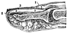
Fig. 119. A section of the end of the finger and nail. 4, Section of the last bone of the finger. 5, Fat, forming the cushion at the end of the finger. 2, The nail. 1, 1, The cuticle continued under and around the root of the nail, at 3, 3, 3.
722. The NAILS are hard, elastic, flexible, semi-transparent scales, and present the appearance of a layer of horn. The nail is divided into the root, the body, and the free portion. The root is that part which is covered on both surfaces; the body is that portion which has one surface free; the free portion projects beyond the end of the finger.
723. The nail is formed of several laminæ, or plates, that are fitted the one to the other; the deepest is that which is last formed. The nails, as well as the hoofs of animals and 325 the cuticle, are products of secretion. They receive no blood-vessels or nerves. If the cuticle be removed in severe scalds they will separate with it, as the hoofs of animals are removed by the agency of hot water. The nails increase in length and thickness, by the deposition of albumen upon their under surface, and at their roots, in a manner similar to the growth of the cuticle, of which they constitute a part.
How can it be spread along the hairs? Why should soap not be used in washing the hair? 721. Of what use is the hair upon the head? About the flexions of the joints? In the nasal and ear passages? Upon the eyebrows and eyelids? 722. Describe the nails. 723. How are they formed?
Observations. 1st. The nail upon its under surface is fashioned into thin vertical plates, which are received between the folds of the sensitive skin. In this manner, the two kinds of laminæ reciprocally embrace each other, and the firmness of connection of the nail is maintained. If we look on the surface of the nail, we see an indication of this structure in the alternate red and white lines which are there observed. The former of these correspond with the sensitive laminæ; the latter with the horny plates. The ribbed appearance of the nail is due to the same circumstance. These sensitive laminæ are provided with an unusual number of capillary vessels for the formation of the nail, and hence they give a red tint to the portion under which they lie.
2d. Near the root of the nail there is a part that is not laminated, and it is less abundantly supplied with blood-vessels. This portion consequently looks pale compared with the laminated portion, and from its half-moon shape is technically termed lunula. Beyond the lunula, the root of the nail is imbedded in the fold of the sensitive skin, and has the same relation to that structure that any single one of the thin horny plates of its under surface has to its corresponding pair of sensitive laminæ.
724. The nails, from their position, are continually receiving knocks, which produce a momentary disturbance of their cell formation, followed by a white spot. The care of the nails should be strictly limited to the knife or scissors, to 326 their free border, and an ivory presser, to prevent adhesion of the free margin of the scarf-skin to the surface of the nail. This edge of the cuticle should never be pared, the surface of the nail never scraped, nor the nails cleaned with any instrument whatever, except the nail-brush, aided by water and soap. An observance of these suggestions, will prevent irregularities and disorders of the nails.
Give observation 1st. Observation 2d. 724. How should the nails be treated to prevent irregularities and disease?
Observations. 1st. When we wear a shoe that is too short for the foot, the edge of the nail is brought against the leather. This interrupts the forward growth of the nail, and it spreads out on the sides, and becomes unusually thick. It then presses upon the soft parts, and is said to “grow into the flesh.” The prevention is, to wear shoes of ample size.
2d. Instances are by no means unfrequent in which the power of production of the nail at the root becomes entirely destroyed, and it then grows in thickness only. When this affection occurs, it is often remarkable what a mass the nail presents. Instances are on record, where the nail is regularly shed; and, whenever the old nail falls off, a new one is found beneath it, perfectly formed. Sometimes the growth in length is not entirely checked, although growth in thickness is induced; the nail then presents a peculiar appearance.
What causes the edge of the nail “to grow into the flesh” of the toe? How prevented?
725. In the preceding chapters, we have seen how various and complex are many of the motions necessary to maintain the life of an animal whose organization is superior to all others. We have noted the wonderful mechanism of the muscular system, in producing the varied movements of the body, the different processes by which the food is converted into chyle and mixed with the blood, and the circulation of this fluid to every organ and tissue of the system, that each may select from it the very principles which it requires for its growth.
726. Lymphatic absorption commences as soon as nutrition is completed, and conveys the useless, worn-out particles of different tissues back into the circulating fluid; while the respiratory organs and secretory glands perform the work of preparing the waste products to be eliminated from the body. Each of these processes effects a single object, and is performed in a regular manner.
727. “They must succeed each other in proper order in propelling every particle to its proper destination, or life would be sacrificed almost at the moment of its commencement. There is, therefore, a mutual dependence of all portions of the machinery of organic life upon each other, and a necessity for some medium of communication from one organ to another, by which they may convey mutual information of 328 their several conditions, if we may be permitted to employ a figurative expression. Were there no such medium, how would the stomach notify the heart that additional exertion on its part is required, because the stomach is busy in digesting food?
725. What has been noted in the preceding chapters? 726. Show the manner in which the several processes are performed. 727. How must they succeed each other?
728. “When we are exerting the muscular system for a long time in some laborious employment, how else are our members to inform the stomach that they are too much occupied with their duties to spare the blood necessary in digestion; that it is requisite that the appetite should decline; and that digestion should cease for the time, even if the stomach should be oppressed with its contents? When we are thinking, how else are the blood-vessels to be told that an unusual supply of their contents is wanting in the head? or when the whole frame is weary with exertion, how, without some regular line of intelligence between all the organs, is the brain to be instructed that circumstances require that it should go to sleep? To supply the necessary medium of communication, Providence has furnished all the animals that possess distinct organs, with a peculiar apparatus called the Nervous System.”
729. The NERVOUS SYSTEM consists of the Cer´e-bro-spi´nal Cen´tre, and of numerous rounded and flattened white cords, called nerves, which are connected at one extremity with the cerebro-spinal centre, and at the other, distributed to all the textures of the body. The sympathetic nerve is an exception to this description; for, instead of one, it has many small centres, which are called gan´gli-a, and which communicate very freely with the cerebro-spinal centre, and with its nerves.
728. What is the medium of communication from one organ to another? 729–754. Give the anatomy of the brain and cranial nerves. 729. Of what does the nervous system consist? What constitutes an exception to this?
730. The CEREBRO-SPINAL CENTRE consists of two portions: The brain, and the spinal cord. For convenience of description, the nervous system may be divided into the Brain, Cranial Nerves, Spinal Cord, Spinal Nerves, and the Sympathetic Nerve.
731. The term BRAIN designates those parts of the nervous system, exclusive of the nerves themselves, which are contained within the cranium, or skull-bones; they are the Cer´e-brum, Cer-e-bel´lum, and Me-dul´la Ob-lon-ga´ta. These are invested and protected by the membranes of the brain, which are called the Du´ra Ma´ter, A-rach´noid, and Pi´a Ma´ter.
Fig. 120.

Fig. 120. 1, 1, The scalp turned down. 2, 2, 2, The cut edge of the bones of the skull. 3, The external strong membrane of the brain (dura mater,) suspended by a hook. 4, The left hemisphere of the brain, showing its convolutions.
730. Of what does the cerebro-spinal centre consist? How is the nervous system divided? 731. What does the term brain designate? Name them. How are they protected? Describe fig. 120.
732. The CEREBRUM IS divided into two hemispheres, by a cleft, or fissure. Into this cleft dips a portion of the dura mater, called the falx cer´e-bri, from its resembling a sickle. The apparent design of this membrane is to relieve the one side from the pressure of the other, when the head is reclining to either side. Upon the superior surface of the cerebrum are seen undulating windings, called con-vo-lu´tions. Upon its inferior, or lower surface, each hemisphere admits of a division into three lobes—the anterior, middle, and posterior. (Fig. 122, 123)
Fig. 121
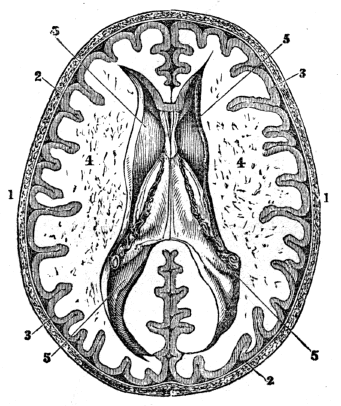
Fig. 121. A section of the skull-bones and cerebrum. 1, 1, The skull. 2, 2, the dura mater. 3, 3, The cineritious portion of the cerebrum. 4, 4, The medullary portion. The dark points indicate the position of divided blood-vessels. 5, 5, The lateral ventricles.
732. How is the cerebrum divided? What is the use of the falx cerebri? What is seen upon the superior surface of the brain? Its inferior?
733. When the upper part of the hemispheres is removed horizontally with a sharp knife, a centre of white substance is brought to view. This is surrounded by a border of gray, which follows the depressions of the convolutions, and presents a zigzag outline. The divided surface will be seen studded with numerous small red points, which are produced by the escape of blood from the division of the minute arteries and veins. The gray border is called the cortical, or cineritious portion, while the white central portion is called the medullary. The two hemispheres are connected by a dense layer of transverse fibres, called cor´pus cal-lo´sum.
734. In the interior of the brain there are several cavities, two of which are of considerable size, and are called the lateral ventricles. They extend from the anterior to the posterior part of the brain, and wind their way into other parts of the cerebral organ.
Observation. In the disease called “dropsy of the brain,” (hydrocephalus internus,) the serum, or water, is usually deposited in these ventricles. This is effused from the many small blood-vessels of the membrane in these cavities.
735. The brain is of a pulpy character, quite soft in infancy and childhood; but it gradually becomes more and more consistent, and in middle age it assumes the form of determinate structure and arrangement. It is more abundantly supplied with blood than any organ of the system. No lymphatics have been detected, but it is to be presumed that they exist in this organ.
736. The CEREBELLUM is about seven times smaller than the cerebrum. Like that organ, it is composed of white and 332 gray matter, but the gray constitutes the larger portion. Its surface is formed of parallel plates separated by fissures. The white matter is so arranged, that when cut vertically, the appearance of the trunk and branches of a tree (ar´bor vi´tæ) is presented. It is situated under the posterior lobe of the cerebrum, from which it is separated by a process of the dura mater, called the ten-to´ri-um.
733. Describe the appearance of the brain when a horizontal section has been made. What is the gray border often called? What connects the hemispheres? 734. Describe the ventricles of the brain. In the disease called “dropsy of the brain,” where is the water deposited? 735. What is the character of the brain in childhood? In adults? 736. How does the cerebellum compare in size with the cerebrum?
Fig. 122.
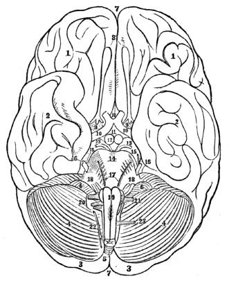
Fig. 122. The under surface, or base, of the brain and origin of the cranial nerves. 1, 1, The anterior lobes of the cerebrum. 2, 2, The middle lobes. 3, 3, The posterior lobes, almost concealed by the cerebellum. 4, 4, The cerebellum. 7, 7, The longitudinal fissure that divides the brain into two hemispheres. 8, The first pair of nerves. 9, 9, The second pair of nerves. 10, The decussation, or crossing, of its fibres. 13, 13, The third pair of nerves. 14, The pons varolii. 15, 15, The fourth pair of nerves. 16, 16, The fifth pair of nerves. 17, The sixth pair of nerves. 18, 18, The seventh and eighth pair of nerves. 19, The medulla oblongata, with the crossing of some of its fibres exhibited. 20, The ninth pair of nerves. 21, The tenth pair of nerves, 22, The eleventh pair of nerves. 23, The twelfth pair of nerves.
Describe this portion of the brain. Explain fig. 122.
737. The MEDULLA OBLONGATA, or that portion of the spinal cord which is within the skull, consists of three pairs of bodies, (cor´pus py-ram-i-da´le, res-ti-for´me, and ol-i-va´re,) united in a single bulb.
Fig. 123.
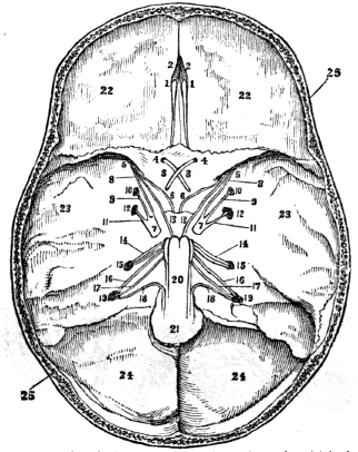
Fig. 123. The base of the skull and the openings through which the cranial nerves pass. 1, 1, The first pair of nerves. 2, 2, The cribriform plate of the ethmoid bone through which this nerve passes. 3, 3, The second pair of nerves. 4, 4, The optic foramen in the sphenoid bone; through which passes the second pair of nerves. 5, 5, The sphenoidal fissure. 6, 6, The third pair of nerves. 7, 7, The fifth pair of nerves. 8, 8, The ophthalmic branch of the fifth nerve. The third, the ophthalmic branch of the fifth and the sixth nerve pass from the brain through the sphenoidal fissure to the eye. 9, 9, The superior maxillary branch of the fifth nerve. 10, 10, The foramen rotundum, (round opening,) through which the nerve 9, 9, passes to the upper jaw. 11, 11, The inferior maxillary branch of the fifth pair. 12, 12, The foramen ovale, (oval opening,) through which the nerve 11, 11, passes to the lower jaw. 13, 13, The sixth pair of nerves. 14, 14, The seventh and eighth pair of nerves. 15, 15, The opening in the temporal bone, through which the seventh and eighth nerves pass to the face and ear. 16, 16, The ninth pair of nerves. 17, The tenth pair of nerves. 18, 18, The eleventh pair of nerves. 19, 19, The foramen lacerum (rough opening.) The ninth, tenth, and eleventh nerves pass from the brain through this opening. 20, The spinal cord. 21, The foramen spinalis, through which the spinal cord passes. 22, 22, The position of the anterior lobe of the brain. 23, 23, The middle lobe. 24, 24, The posterior lobe. 25, 25, A section of the skull-bones.
737. Describe the medulla oblongata. Explain fig. 123.
738. The DURA MATER is a firm, fibrous membrane, which is exposed on the removal of a section of the skull-bones. This lines the interior of the skull and spinal column, and likewise sends processes inward, for the support and protection of the different parts of the brain. It also sends processes externally, which form the sheaths for the nerves, as they quit the skull and spinal column. The dura mater is supplied with arteries and nerves.
Fig. 124.
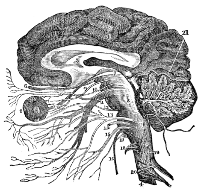
Fig. 124. A vertical section of the cerebrum, cerebellum, and medulla oblongata, showing the relation of the cranial nerves at their origin. 1, The cerebrum. 2, The cerebellum, with its arbor vitæ represented. 3, The medulla oblongata. 4, The spinal cord. 5, The corpus callosum. 6, The first pair of nerves. 7, The second pair. 8, The eye. 9, The third pair of nerves. 10, The fourth pair. 11, The fifth pair. 12, The sixth pair. 13, The seventh pair. 14, The eighth pair. 15, The ninth pair. 16, The tenth pair. 10, The eleventh pair. 18, The twelfth pair. 20, Spinal nerves. 21, The tentorium.
738. Describe the dura mater. What is its use? Explain fig. 124.
739. The ARACHNOID, so called from its extreme tenuity, is the serous membrane of the brain and spinal cord, and is, like other serous membranes, a closed sac. It envelops these organs, and is reflected upon the inner surface of the dura mater, giving to that membrane its serous investment.
740. The PIA MATER is a vascular membrane, composed of innumerable vessels, held together by cellular membrane. It invests the whole surface of the brain, and dips into its convolutions. The pia mater is the nutrient membrane of the brain, and receives its blood from the carotid and vertebral arteries. Its nerves are minute branches of the sympathetic, which accompany the branches of the arteries.
741. The CRANIAL NERVES, that connect with the brain, are arranged in twelve pairs. They are called: 1st. The Ol-fact´o-ry. 2d. The Op´tic. 3d. The Mo-to´res Oc-u-lo´rum. 4th. The Pa-thet´i-cus. 5th. The Tri-fa´cial. 6th. The Ab-du-cen´tes. 7th. The Por´ti-o Du´ra. 8th. The Por´ti-o Mol´lis. 9th. The Glos´so-pha-ryn´gi-al. 10th. The Pneu-mo-gas´tric. 11th. The Spi´nal Ac´ces-so-ry. 12th. The Hy´po-glos´sal.
742. The OLFACTORY NERVE (first pair) passes from the cavity of the skull through many small openings in a plate of the eth´moid bone. (This plate is called crib´ri-form, from its resemblance to a sieve.) This nerve ramifies upon the membrane that lines the nasal passages. It is the softest nerve of the body. (Fig. 136.)
743. The OPTIC NERVE (second pair) passes from the interior of the cranium, through an opening in the base of the skull, (fo-ra´men op´ti-cum,) to the cavity for the eye. It pierces the coats of the eye, and expands in the retina.
744. The MOTORES OCULORUM (third pair) pass from the 336 brain, through an opening of the sphe´noid bone, (sphe-noid´al fis´sure,) to the muscles of the eye.
739. Describe the arachnoid membrane. 740. What is said respecting the pia mater? 741. How many pairs of cranial nerves? Name them. 742. Describe the olfactory nerve. 743. The optic nerve. 744. Describe the motores oculorum.
745. The PATHETICUS (fourth pair) passes from the brain, through the sphenoidal fissure, to the superior oblique muscle of the eye.
Fig. 125.

Fig. 125. The distribution of the third, fourth, and sixth pairs of nerves, to the muscles of the eye. 1, The ball of the eye and rectus externus muscle. 2, The upper jaw. 3, The third pair, distributed to all the muscles of the eye, except the superior oblique, and external rectus. 4, The fourth pair passes to the superior oblique muscle. 6, The sixth pair, is distributed to the external rectus muscle.
746. The TRIFACIAL NERVE (fifth pair) is analogous to the spinal nerves in its origin by two roots, from the anterior and posterior columns of the spinal cord. It has a ganglion, like the spinal nerves upon its posterior root. For these reasons, it ranges with the spinal nerves, and is considered the cranial spinal nerve. This nerve divides into three branches:—The oph-thal´mic, superior max´il-la-ry, and inferior max´il-la-ry.
745. The patheticus. What does fig. 125 represent? 746. What is the trifacial nerve sometimes called? Why is it classed with the cranial spinal nerves? Give the names of its branches.
747. The ophthalmic nerve passes from the cranial cavity through the sphenoidal fissure. It sends branches to the forehead, eye, and nose. The superior maxillary nerve passes through an opening in the base of the skull, (foramen ro-tund´dum,) and sends branches to the eye, the teeth of the upper jaw, and the muscles of the face. The inferior maxillary nerve escapes from the cranial cavity through an opening called foramen o-va´le. It sends branches to the muscles of the lower jaw, the ear, the tongue, and the teeth of the lower jaw.
Fig. 126.
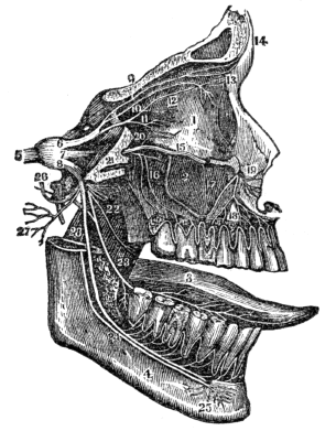
Fig. 126. The distribution of the fifth pair of nerves. 1, The orbit for the eye. 2, The upper jaw. 3, The tongue. 4, The lower jaw. 5, The fifth pair of nerves. 6, The first branch of this nerve, that passes to the eye. 9, 10, 11, 12, 13, 14, Divisions of this branch. 7, The second branch of the fifth pair of nerves is distributed to the teeth of the upper jaw. 15, 16, 17, 18, 19, 20, Divisions of this branch. 8, The third branch of the fifth pair, that passes to the tongue and teeth of the lower jaw. 23. The division of this branch that passes to the tongue, called the gust´a-to-ry. 24. The division that is distributed to the teeth of the lower jaw.
747. Where do the filaments of the ophthalmic branch ramify? The superior maxillary? The inferior maxillary? Explain fig. 126.
748. The ABDUCENTES (sixth pair) passes through the opening by which the carotid artery enters the cranial cavity. It is the smallest of the cerebral nerves, and is appropriated to the external straight muscle of the eye.
749. The PORTIO MOLLIS (seventh pair) enters the hard portion of the tem´po-ral bone at the internal auditory opening, and is distributed upon the internal ear. (Fig. 147, 148.)
Fig. 127.

Fig. 127. A representation of the distribution of the eighth pair of nerves with some branches of the fifth. 1, 2, 3, 5, 7, 9, Are branches of the eighth pair. They are distributed over the face in a radiated manner, which constitutes the pes anserinus, (foot of a goose.) The nerves 4, 6, 8, are branches of the fifth pair. 10, 11, 12, 13, 14, 15, 16, Are branches of nerves from the upper part of the spinal cord, (cervical.)
750. The FACIAL NERVE (eighth pair) passes from the skull through an opening situated below the ear, (mas´toid foramen.) It is distributed over the face, supplying the muscles with nervous filaments.
748. What is said of the abducentes, or sixth pair of nerves? 749. Of the portio mollis? Explain fig. 127. 750. Of the facial nerve?
751. The GLOSSO-PHARYNGEAL NERVE (ninth pair) passes from the brain, through an opening with the jugular vein, (foramen lac´e-rum.) It is distributed to the mucous membrane of the tongue and throat, and also to the mucous glands of the mouth.
752. the PNEUMOGASTRIC NERVE (tenth pair) escapes from the brain through the foramen lacerum. It sends branches to the larynx, pharynx, œsophagus, lungs, spleen, pancreas, liver, stomach, and intestines. (Fig. 132.)
753. The SPINAL ACCESSORY NERVE (eleventh pair) has its origin in the respiratory tract of the spinal cord. It connects with the ninth and tenth pairs of nerves, and is distributed to the muscles about the neck.
754. The HYPO-GLOSSAL NERVE (twelfth pair) passes from the brain, through a small opening, (con´dy-loid foramen.) It ramifies upon the muscles of the tongue, and is its motor nerve.
Observation. The cranial nerves, with the exception of the olfactory, optic, and auditory, connect with each other by means of filaments. They also send connecting nervous filaments to the upper spinal nerves, (cervical,) and the sympathetic nerve.
751. Describe the glosso-pharyngeal nerve. 752. The pneumogastric nerve. 753. The spinal accessory nerve. 754. The hypo-glossal nerve.
755. The spinal column contains the spinal cord, the roots of the spinal nerves, and the membranes of the cord.
756. The SPINAL CORD extends from the medulla oblongata to the second lumbar vertebra, where it terminates in a rounded point. It presents a difference of diameter in different parts of its extent, and exhibits three enlargements. The uppermost of these is the medulla oblongata. There is no distinct demarkation between this enlargement and the spinal cord. The next corresponds with the origin of the nerves distributed to the upper extremities; the third enlargement is situated near the termination of the cord, and corresponds with the attachment of the nerves which are intended for the supply of the lower extremities.
757. An anterior and posterior fissure divides the spinal cord into two lateral cords. These are united by a thin layer of white substance. The lateral cords are each divided by furrows into three distinct sets of fibres, or columns; namely the anterior, lateral, and posterior columns. The anterior are the motor columns; the posterior are the columns of sensation; the lateral columns are divided in their function between motion and sensation. They contain the fasciculus described, by Sir Charles Bell, as the respiratory tract. 341
Fig. 128. A section of the brain and spinal column. 1, The cerebrum. 2, The cerebellum. 3, The medulla oblongata. 4, 4, The spinal cord in its canal.
Fig. 129. Anterior view of the brain and spinal cord. 1, 1, The two hemispheres of the cerebrum. 3, 3, The cerebellum. 4, The olfactory nerve. 5, The optic nerve. 7, The third pair of nerves. 8, The pons varolii. 9, The fourth pair of nerves. 10, The lower portion of the medulla oblongata. 11, 11, The spinal cord. 12, 12, Spinal nerves. 13, 13, The brachial plexus. 14, 14, The lumbar and sacral plexus.
755–767. Give the anatomy of the spinal cord, spinal nerves, and the sympathetic nerve. 755. What does the spinal column contain? 756. Give the extent of the spinal cord. How many enlargements has this cord? What is said of each enlargement? 757. Into how many parts is the spinal cord divided? Give the function of these columns.
758. The SPINAL NERVES, that connect with the spinal cord, are arranged in thirty-one pairs, each arising by two 342 roots; an anterior, or motor root, and a posterior, or sensitive root. Each nerve, when minutely examined, is found to consist of an aggregate of very delicate filaments, enclosed in a common cellular envelope.
758. How many pairs of nerves issue from the spinal cord? Explain fig. 128. Fig. 129.
759. The anterior roots arise from a narrow white line upon the anterior columns of the spinal cord. The posterior roots arise from a narrow gray band formed by the internal gray substance of the cord. They are larger, and the filaments of origin more numerous than those of the anterior roots. A ganglion is found upon each of the posterior roots in the openings between the bones of the spinal column through which the nerve passes.
Fig. 130.

Fig. 130. A section of the spinal cord, surrounded by its sheath. B, A spinal nerve, formed by the union of the motor root (C) and the sensitive root (D.) At D, the ganglion upon this root is seen.
760. After the formation of the ganglion, the two roots unite, and constitute a spinal nerve, which passes through the opening between the vertebræ on the sides of the spinal column. The nerves divide and subdivide, until their minute filaments ramify on the tissues of the different organs.
759. Give the origin of the anterior roots. Of the posterior roots. In what respect do the posterior roots differ from the anterior? 760. When do the two roots unite, and where do they pass?
761. The spinal nerves are divided into—
| Cervical, | 8 | pairs, |
| Dorsal, | 12 | “ |
| Lumbar, | 5 | “ |
| Sacral, | 6 | “ |
762. The four lower cervical and upper dorsal pass into each other and then separate to reunite. This is called the brach´i-al plex´us. From this plexus six nerves proceed, which ramify upon the muscles and skin of the upper extremities.
763. The last dorsal and the five lumbar nerves form a plexus called the lumbar, similar to that of the cervical. Six nerves pass from this plexus, which ramify upon the muscles and skin of the lower extremities.
764. The last lumbar and the four upper sacral unite to form the sacral plexus. From this plexus five nerves proceed, that are distributed to the muscles and skin of the hip and lower extremities.
765. The SYMPATHETIC NERVE[19] consists of a series of Gan´gli-a, or knots, extending each side of the spinal column, forming a chain its whole length. It communicates with both the cranial and spinal nerves. With the exception of the neck, there is a ganglion for each intervertebral space. These ganglia are composed of a mixture of cineritious and medullary matter, and are supposed to be productive of peculiar nervous power.
761. Give the division of the spinal nerves. 762. What nerves constitute the brachial plexus? How many nerves pass from this plexus? 763. How many nerves from the lumbar plexus, and where do they ramify? 764. How is the sacral plexus formed? 765. Of what does the sympathetic nerve consist? How is the sympathetic nerve distributed? What exception? Of what are the ganglia composed?
Fig. 131.
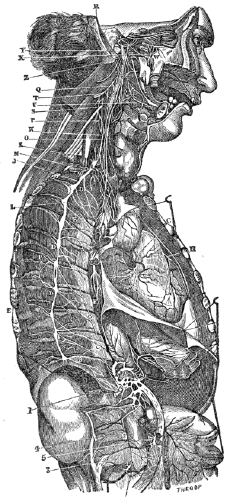
Fig. 131. A beautiful representation of the sympathetic ganglia and their connection with other nerves. It is from the grand engraving of Manec, reduced in size. A, A, A, The semilunar ganglion and solar plexus, situated below the diaphragm and behind the stomach. This ganglion is situated in the region (pit of the stomach) where a blow gives severe suffering. D, D, D, The thoracic ganglia, ten or eleven in number. E, E, The external and internal branches of the thoracic ganglia. G, H, The right and left coronary plexus, situated upon the heart. I, N, Q, The inferior, middle, and superior cervical ganglia. 1, The renal plexus of nerves that surrounds the kidneys. 2, The lumbar ganglion. 3, Their internal branches. 4, Their external branches. 5, The aortic plexus of nerves that lies upon the aorta. The other letters and figures represent nerves that connect important organs and nerves with the sympathetic ganglia.
766. The GANGLIA may be considered as distinct centres, giving off branches in four directions; namely, the superior, or ascending, to communicate with the ganglion above; the inferior, or descending, to communicate with the ganglion below; the external, to communicate with the spinal nerves; and the internal, to communicate with the sympathetic filaments. It is generally admitted that the nerves that pass from the ganglia are larger than those that entered them; as if they imparted to the nerve some additional power.
767. The branches of distribution accompany the arteries which supply the different organs, and form communications around them, which are called plexuses, and take the name of the artery with which they are associated. Thus we have the mesenteric plexus, hepatic plexus, splenic plexus, &c. All the internal organs of the head, neck, and trunk, are supplied with branches from the sympathetic, and some of them exclusively; for this reason, it is considered a nerve of organic life.
What is the design of fig, 131? 766. How may the ganglia be considered? 767. What is said of the branches of the sympathetic nerve?
768. The brain is regarded by physiologists and philosophers as the organ of the mind. Most writers consider it as an aggregate of parts, each charged with specific functions, and that these functions are the highest and most important in the animal economy. To the large brain, or cerebral lobes, they ascribe the seat of the faculties of thinking, memory, and the will. In man, this lobe extends so far backward as to cover the whole of the cerebellum. To the cerebellum, or little brain, is ascribed the seat of the animal, or lower propensities.
769. “The constant relation between mental power and development of brain, explains why capacities and dispositions are so different. In infancy, for example, the intellectual powers are feeble and inactive. This arises partly from the inaptitude of a still imperfect brain; but in proportion as the latter advances toward its mature state, the mental faculties also become vigorous and active.”
770. We are able, in most instances, at least, to trace a correspondence between the development of the cerebral lobes and the amount of intelligence possessed by the person. The weight of the brain in man to that of the whole body varies 347 in different individuals. The heaviest brain on record was that of Cuvier, which weighed 4 pounds and 13½ ounces.
768–772. Give the physiology of the nervous system. 768. How is the brain regarded by physiologists and philosophers? What do they ascribe to the cerebrum? To the cerebellum? 769. What does the relation between mental powers and development of brain explain? 770. What is said respecting the correspondence between the development of the brain and the amount of intelligence possessed by the person? What is said of the weight of the brain?
771. The brain likewise holds an important relation to all the other organs of the system. To the muscular system it imparts an influence which induces contraction of the fibres. By this relation they are brought under the control of the will. To the skin, eye, and ear, it imparts an influence that gives sensibility, or the power of feeling, seeing, hearing, &c.
772. Again, the involuntary functions of the different portions of the system are more or less influenced by the brain. If the action of this central organ of the nervous system is destroyed, the functions of the digestive, respiratory, and circulatory apparatuses will be much disturbed or entirely suppressed.
773. The brain is the seat of sensation. It receives the impressions made on all parts of the body, through the medium of the sensitive nerves. That the impressions of external objects, made on these nerves, be communicated to the brain, where sensation is perceived, it is necessary that they be not diseased or injured.
Observation. There is a plain distinction between sensations and impressions; the latter are the changes produced in the extremities of the nerve; the former, the changes produced in the brain and communicated to the mind.
774. What part of the brain receives the impressions or has the most intimate relation with the intellectual faculties is unknown. Some portions, however, are of less importance than others. Large portions of the cortical, or outer part, are frequently removed without affecting the functions of this 348 organ. Pieces of the medullary, or central parts, have been removed by injuries without impairing the intellect or destroying life. This organ, although it takes cognizance of every sensation, is, of itself, but slightly sensible. It may be cut, or parts may be removed without pain, and the individual, at the same times retain his consciousness.
771. What is said of the relation of the brain to all of the organs of the body? 772. Are the involuntary functions of different parts of the system influenced by the brain? 773. Where is sensation perceived? By what agency are the impressions of external objects conveyed to the brain? What is the difference between sensations and impressions? 774. Is it known what part of the brain has the most intimate relation with the intellectual faculties?
775. The brain is the seat of the will. It superintends the physical as well as the mental movements, and the medium of communication from this organ to the muscles, or the parts to be moved, is the motor nerves. If the brain is in a quiescent state, the muscles are at rest; if, by an act of the will, the brain sends a portion of nervous influence to a voluntary muscle, it immediately contracts, and those parts to which the muscle is attached move. There is no perceptible interval between the act of the will and the motion of the part.
776. Some physiologists assert, that the medulla oblongata is the point at which excitement to motion commences, and sensation terminates; and also, that it possesses the power of originating motion in itself.
Observation. The medulla oblongata, unlike the brain, is highly sensitive; if slightly punctured, convulsions follow; if much injured, respiration, or breathing, immediately ceases.
777. It is remarkable that the nerves which arise from the right side of the spinal cord communicate with the left hemisphere of the cerebrum, and vice versa; this results from the crossing of the fibres in the medulla oblongata. It follows from this, that if the right side of the brain receives an injury, the parts of the opposite side of the body lose their sensibility and motion.
Observations. 1st. If the cranial nerves which are connected 349 by a single root are divided, only the sensation of the part to which they are distributed is lost. Thus, if the optic nerve is divided, the sense of vision disappears, but the motions of the eye are performed as readily as before. But, if the spinal nerves are divided, both sensation and motion of the part to which they lead are destroyed.
What portions have been removed without impairing the intellect? What is remarkable of the brain? 775. What is the influence of the brain upon the muscles? 776. What do some physiologists assert of the medulla oblongata? 777. What is remarkable of the nerves? Give the 1st observation relative to the cranial nerves.
2d. When the spinal cord is divided or compressed, as in fractures of the spinal column, all parts below the fracture are paralyzed, though the nerves leading to these parts may be uninjured.
3d. Again, one side of the body or one limb may become insensible, and the power to move it, be perfectly retained; or the reverse of this may happen—the power of motion will be lost while sensation remains. In the former instance, the function of the posterior, or sensitive column of the spinal cord on one side is destroyed; in the latter, the anterior, or motor column is affected.
4th. In some cases, both sensation and motion of one side of the body or one limb are destroyed. In such instances, both the anterior and the posterior columns of one side of the spinal cord are diseased.
778. Vigorous and controllable muscular contraction requires a sound and well-developed brain. If this organ is defective in these particulars, the movements will be inefficient, and may be irregular. The central organ of the nervous system must, likewise, be in an active condition, to induce regular, steady, and controllable muscular movements.
Observations. 1st. Persons who have suffered from apoplexy and other severe diseases of the brain, have an involuntary trembling of the limbs, which results from a weakened state of the nervous system.
To the spinal nerves. What is said of the compression of the spinal cord? Give the 3d observation relative to the spinal nerves. The 4th observation. 778. Upon what does vigorous controllable muscular contraction depend? What causes the involuntary trembling of the limbs in persons who have suffered from apoplexy?
2d. The tremor of the hand, that lessens the usefulness or incapacitates the fine artist or skilful mechanic, in the prime of life, from pursuing their vocations, may be, and is often, induced by the influence of intoxicating drink, which debilitates and disorganizes the brain.
3d. The tottering step, trembling hand, and shaking head of the aged invalid, are the results of diminished nervous energy, so that steady muscular contraction, so essential to regular movements, cannot be maintained.
779. No difference can be discovered in the structure of the several kinds of nerves in any part of their course, and the functions they are designed to perform can only be known by ascertaining the place of their origin. The nerves may be functionally divided into five groups.
780. 1st. Nerves of special sensation. These are the first, second, eighth, and it may be one of the branches of the fifth pair of cranial nerves. The function of these nerves is particularly described in the chapters upon the senses of smell, vision, hearing, and taste.
781. 2d. Nerves of general sensation. These embrace the fifth pair of cranial nerves, and the thirty-one pairs of spinal nerves. In those parts that require sensation for their safety and the performance of their functions, there is an abundant supply of sensitive nervous filaments. The nerves of sensation are mostly distributed upon the skin. Few filaments ramify upon the mucous membranes and muscles.
Observations. 1st. The painful sensations experienced in the face, and in the teeth or jaws, (tic douloureux and toothache,) are induced by irritation and disease of a portion of the filaments of the fifth pair of cranial nerves.
The tremor of the hand among some mechanics in the prime of life? The tottering step of the aged invalid? 779. What is said relative to the structure of the nerves? How may they be divided? 780. Give the nerves of special sense. 781. Those of general sensation. Where are the nerves of sensation distributed? What causes tic douloureux?
2d. The unpleasant sensation sometimes experienced when we hear the grating of a file or saw, is produced by the connection of the nerve that passes across the drum of the ear with the fifth cranial nerve.
3d. When pressure is made on the trunk of a nerve, the sensibility of the part where the nerve ramifies is modified. This is illustrated, when pressure is made upon the large nerve of the lower extremity (sciatic) in sitting upon a hard bench. The foot is then said to be “asleep.”
4th. When the trunk of a nerve is diseased or injured, the pain is experienced in the outer extremity of the nerve. A blow upon the elbow, which causes a peculiar sensation in the little finger and one side of the ring finger, affords a familiar illustration. This sensation is produced by injuring the ulnar nerve, which is distributed to the little finger.
782. 3d. Nerves of motion. These are the third, sixth, and twelfth pairs of cranial nerves, and the thirty-one pairs of spinal nerves. These nerves are distributed to the fibres of the five hundred muscles of the body. The functions of the muscular are different from those of the sensitive nerves. The former are provided for the purpose of motion, and not of feeling. Hence, muscles may be cut, and the pain will be slight, compared with the cutting of the skin. This may be called muscular pain. Weariness is a sensation recognized by one set of muscular nerves.
783. So uniformly is a separate instrument provided for every additional function, that there is strong reason to regard the muscular nerves, although running in one sheath, as in reality double, and performing distinct functions. Sir Charles Bell, in his work on the Nervous System, endeavors to show, 352 that one set of nervous fibres conveys the mandate from the brain to the muscle, and excites the contraction; and that another set conveys, from the muscle to the brain, a peculiar sense of the state or degree of contraction of the muscle, by which we are enabled to judge of the amount of stimulus necessary to accomplish the end desired. This is obviously an indispensable piece of information to the mind in regulating the movements of the body.
How is the peculiar sensation accounted for when we hear the grating of a file or saw? What produces the sensation when the foot is said to be “asleep?” What is the effect when the ulnar nerve is injured by a blow? 782. Give the nerves of motion. What is said of the functions of the muscular nerves? 783. What does Sir Charles Bell endeavor to show?
784. 4th. Nerves of respiration. These are the fourth, seventh, ninth, tenth, and eleventh pair of cranial nerves, also the phrenic and the external respiratory nerve. All of these nerves have their origin in a distinct tract or column, called the lateral, in the upper part of the spinal cord. Hence it is sometimes named the respiratory column. These nerves are distributed to one of the muscles of the eye; to the muscles of the face; to the tongue, pharynx, œsophagus, stomach, heart, lungs, diaphragm, and some of the muscles of the neck and chest.
785. It is through the instrumentality of the accessory, phrenic, and external respiratory nerves, (10, 11, 12, 13, fig. 132,) that the muscles employed in respiration are brought into action without the necessity of the interference of the mind. Though to a certain extent they may be under the influence of the will, yet it is only in a secondary degree. No one can long suspend the movements of respiration;[20] for in a short time, instinctive feeling issues its irresistible mandate, which neither requires the aid of erring wisdom, nor brooks the capricious interference of the will.
Fig. 132.

Fig. 132. The distribution of the respiratory nerves. a, Section of the brain and medulla oblongata. b, The lateral columns of the spinal cord. c, c, The respiratory tract of the spinal cord. d, The tongue. e, The larynx. f, The bronchia. g, The œsophagus. h, The stomach. i, The diaphragm. 1, The pneumogastric nerve. 2, The superior laryngeal nerve. 3, The recurrent laryngeal nerve. (These two ramify on the larynx.) 4, The pulmonary plexus of the tenth nerve. 5, The cardiac plexus of the tenth nerve. These two plexuses supply the heart and lungs with nervous filaments. 7, The origin of the fourth pair of nerves, that passes to the superior oblique muscle of the eye. 8, The origin of the facial nerve, that is spread out on the side of the face and nose. 9, The origin of the glosso-pharyngeal nerve, that passes to the tongue and pharynx. 10, The origin of the spinal accessory nerve. 11, This nerve penetrating the sterno-mastoideus muscle. 12, The origin of the internal respiratory or phrenic nerve, that is seen to ramify on the diaphragm. 13, The origin of the external respiratory nerve, that ramifies on the pectoral and scaleni muscles.
784. Give the respiratory nerves. What is said in reference to the respiratory nerves? 785. Through the agency of what nerves are the respiratory muscles brought into action? Explain fig. 132. 354
786. The fourth, seventh, and tenth pairs of nerves, (7, 8, 9, fig. 132,) with the spinal accessory, phrenic, and external respiratory, are not only connected with the function of respiration, but contribute to the expression of the passions and emotions of the mind.
787. The influence of this order of nerves in the expression of the passions, is strikingly depicted in Sir Charles Bell’s Treatise on the Nervous System. “In terror,” he remarks, “we can readily conceive why a man stands with his eyes intently fixed on the object of his fears—the eyebrows elevated, and the eyeballs largely uncovered; or why, with hesitating and bewildered steps, his eyes are rapidly and wildly in search of something. In this way, we only perceive the intense application of his mind to the objects of his apprehension, and its direct influence on the outward organs.”
Can respiration be suspended for any considerable length of time? 786. What nerves contribute to the expression of the passions and emotions of the mind? 787, 788. What does Sir Charles Bell say of the influence of this order of nerves in the expression of the passions?
788. “But when we observe him further, there is a spasm in his breast; he cannot breathe freely; the chest remains elevated, and his respiration is short and rapid. There is a 355 gasping and convulsive motion of his lips, a tremor on his hollow cheeks, a gasping and catching of his throat; his heart knocks at his ribs, while yet there is no force in the circulation—the lips and cheeks being ashy pale.”
789. “These nerves are the instruments of expression, from the smile upon the infant’s cheek, to the last agony of life. It is when the strong man is subdued by this mysterious influence of soul on body, and when the passions may be truly said to tear the heart, that we have the most afflicting picture of human frailty, and the most unequivocal proof that it is the order of functions we have been considering, that is thus affected. In the first struggle of the infant to draw breath, in the man recovering from a state of suffocation, and in the agony of passion, when the breast labors from the influence at the heart, the same system of parts is affected, the same nerves, the same muscles, and the symptoms or character have a strict resemblance.”
790. The seventh pair of nerves not only communicates the purposes of the will to the muscles of the face, but at the same time it calls them into action, under the influence of instinct and sympathy. On this subject a late writer remarks, “How expressive is the face of man! How clearly it announces the thoughts and sentiments of the mind! How well depicted are the passions on his countenance! tumultuous rage, abject fear, devoted love, envy, hatred, grief, and every other emotion, in all their shades and diversities, are imprinted there, in characters so clear that he that runs may read! How difficult, nay, how impossible, is it to hide or falsify the expressions which indicate the internal feelings! Thus conscious guilt shrinks from detection, innocence declares its confidence, and hope anticipates with bright expectation.”
Observation. The fifth pair of nerves (fig. 126) is distributed 356 to the parts of the face on which the seventh pair ramifies. The former serves for sensation, the latter for motion. Thus, when the seventh pair of nerves is divided, or its functions destroyed by disease, the side affected loses all power of expression, though sensation remains unaffected. On the contrary, if we divide the fifth pair, sensation is entirely destroyed, while expression remains.
789. Are they also the instruments of expression, either of joy or grief? 790. What is said in reference to the seventh pair of nerves? Where is the fifth pair of nerves distributed?
791. 5th. The sympathetic nerve. This nerve confers vitality on all the important portions of the system. It exerts a controlling influence over the involuntary functions of digestion, absorption, secretion, circulation, and nutrition. Every portion of the body is, to a certain extent, under its influence, as filaments from this system of nerves accompany the blood-vessels throughout their course.
792. An important use of the sympathetic nerve is to form a communication of one part of the system with another, so that one organ can take cognizance of the condition of every other, and act accordingly. If, for example, disease seizes the brain, the stomach, by its sympathetic connection, knows it; and as nourishment would add to the disease, it refuses to receive food, and perhaps throws off what has already been taken. Loss of appetite in sickness is thus a kind provision of nature, to prevent our taking food when it would be injurious; and following this intimation, we, as a general rule, should abstain from food until the appetite returns.
Fig. 133.
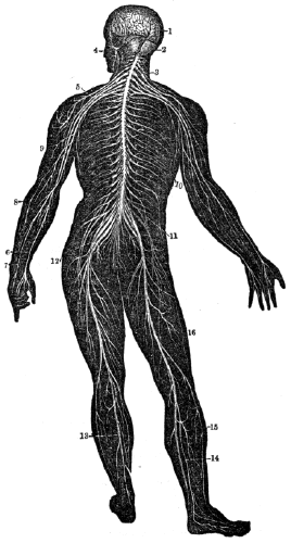
Fig. 133. A back view of the brain and spinal cord. 1, The cerebrum. 2, The cerebellum. 3, The spinal cord. 4, Nerves of the face. 5, The brachial plexus of nerves. 6, 7, 8, 9, Nerves of the arm. 10, Nerves that pass under the ribs, 11, The lumbar plexus of nerves. 12, The sacral plexus of nerves 13, 14, 15, 16, Nerves of the lower limbs.
What is the function of this nerve? What is the effect if the seventh pair is divided, or its function destroyed by disease? 791. What is said of the sympathetic nerve? 792. What is the use of the sympathetic system? Explain fig. 133.
Note. Let the anatomy and physiology of the nervous system be reviewed from figs. 131, 132, 133, or from anatomical outline plate. No. 8.
793. As the different organs of the system are dependent on the brain and spinal cord for efficient functional action, and as the mind and brain are closely associated during life, the former acting in strict obedience to the laws which regulate the latter, it becomes an object of primary importance in education, to discover what these laws are, that we may escape the numerous evils consequent on their violation.
794. For healthy and efficient action, the brain should be primarily sound; as this organ is subject to the same general laws as other parts of the body. If the brain of the child is free from defects at birth, and acquires no improper impressions in infancy, it will not easily become diseased in after life. But, if the brain has inherited defects, or has acquired a proneness to disease by mismanagement in early life, it will more easily yield to influences that cause diseased action. The hereditary tendency to disease is one of the most powerful causes that produce nervous and mental affections. Consequently, children have a strong tendency to the diseases from which the parents suffered.
795. When both parents have similar defects, or have descended from tainted families, the children are usually more deeply impressed with their imperfections than when only one 359 possesses the defect. This is the reason of the frequency of nervous disease and imbecility among the opulent, as intermarriages among near relations are more frequent with this class than among the poor.
793–850. Give the hygiene of the nervous system. 793. Why is it important to know the laws which regulate the action of the brain? 794. What is necessary that the action of the brain be healthy and efficient? What follows if the brain of the child has inherited defects? 795. What is the effect when both parents possess similar defects?
Observation. Among some of the reigning families of Europe, particularly the Spanish, the folly of intermarriage among themselves is strongly illustrated. The high and noble talents that characterized their progenitors are not seen, but there is now exhibited, among their descendants, imbecility and the most revolting forms of nervous disease.
796. “Unhappily, it is not merely as a cause of disease, that hereditary predisposition is to be dreaded. The obstacles which it throws in the way of permanent recovery are even more formidable, and can never be entirely removed. Safety is to be found only in avoiding the perpetuation of the mischief.”
797. “Therefore, if two persons, each naturally of excitable and delicate nervous temperament, choose to unite for life, they have themselves to blame for the concentrated influence of similar tendencies in destroying the health of their offspring, and subjecting them to all the miseries of nervous disease, madness, or melancholy.” The command of God not to marry within certain degrees of consanguinity, is in accordance with the organic laws of the brain, and the wisdom of the prohibition is confirmed by observation.
Observation. The inhabitants, females particularly, of the sea-girt islands of America, are more affected with nervous diseases, than those who reside upon the mainland. The prevalence of these affections is ascribed to the frequent intermarriage of persons closely related by blood.
What is one cause of nervous disease among the higher classes? What is true of some of the reigning families of Europe? 796. Why is hereditary predisposition to be dreaded? 797. Is the prohibition of God respecting intermarriage in accordance with the organic laws of the brain? What is said of the inhabitants of the sea-girt islands of America?
798. The brain requires a due supply of pure blood. This organ receives an unusually large supply of blood, in comparison with the rest of the body. It is estimated that one tenth of all the blood sent from the heart goes to this organ. If the arterial blood be altogether withdrawn, or a person breathes air that is filled with carbonic gas, the brain ceases its proper action, and sensibility with consciousness becomes extinct.
Illustrations. 1st. If a person lose a considerable quantity of blood, dizziness and loss of consciousness follow. This results from the brain not receiving a sufficient amount of blood to sustain its functions.
2d. When an individual descends into a well or pit that contains carbonic acid, the blood is not changed or purified in the lungs, and loss of consciousness and death soon follow.
799. The slighter variations in the state of the blood have equally sure, though less palpable effects. If its vitality is impaired by breathing an atmosphere so much vitiated as to be insufficient to produce the proper degree of oxygenation, the blood then affords an imperfect stimulus to the brain. As a necessary consequence, languor and inactivity of the mental and nervous functions ensue, and a tendency to headache, fainting, or hysteria, makes its appearance.
Observations. 1st. Let a person remain, for a time, in a crowded, ill-ventilated, hall or church, and headache or faintness is generally produced. This is caused by the action of impure blood upon the brain.
2d. If a school-teacher wishes to have his pupils, on the day of examination, appear creditably, he will be careful to have the room well ventilated. Ventilating churches might 361 prevent the inattention and sleepiness that are observed during the afternoon service.
798. Why does the brain require a due supply of pure blood? What is the effect when a person loses a considerable quantity of blood? What causes the loss of consciousness when carbonic acid is breathed? 799. What effects are produced by slight variations in the quality of the blood? From the following observations, give some of the effects of impure blood on the brain.
3d. In many instances, the transmission of imperfectly oxygenated blood to the brain, is an influential cause in the production of nervous disease and delicacy of constitution. The only efficient remedy for these conditions is a supply of pure blood to the brain.
800. The brain should be called into action. This organ, like the muscles, should be used, and then allowed to rest, or cease from vigorous thought. When the brain is properly called into action by moderate study, it increases in size and strength; while, on the other hand, if it is not used, the action of this organ is enfeebled, thereby diminishing the function of all parts of the body.
801. The brain, being an organized part, is subject, so far as regards exercise, to the same laws as the other organs of the body. If it is doomed to inactivity, its size diminishes, its health decays, and the mental operations and feelings, as a necessary consequence, become dull, feeble, and slow. If it is duly exercised after regular intervals of repose, the mind acquires readiness and strength. Lastly, if it is overtasked, either in the force or duration of its activity, its functions become impaired, and irritability and disease take the place of health and vigor.
802. The consequences of inadequate exercise will first be explained. We have seen that by disuse the muscles become emaciated, the bones soften, and the blood-vessels are obliterated. The brain is no exception to this general rule. It is impaired by permanent inactivity, and becomes less fit to manifest the mental powers with readiness and energy. Nor will this surprise any reflecting person, who considers that the 362 brain, as a part of the same animal system, is nourished by the same blood and regulated by the same vital laws as the muscles, bones, and arteries.
800. Why should the brain be called into action? 801. What is the effect if the brain is doomed to inactivity? 802. Show the consequences of disuse of the organs mentioned in preceding chapters. Does the same principle apply to the brain?
803. It is the weakening and depressing effect which is induced by the absence of the stimulus necessary for the healthy exercise of the brain, that renders solitary confinement so severe a punishment, even to the most daring minds. Keeping the above principle in view, we shall not be surprised to find that non-exercise of the brain and nervous system, or, in other words, inactivity of intellect and feeling, is a very frequent predisposing cause of every form of nervous disease.
804. For demonstrative evidence of this position, we have only to look at the numerous victims to be found among females of the middle and higher ranks, who have no calls to exertion in gaining the means of subsistence, and no objects of interest on which to exercise their mental faculties, and who, consequently, sink into a state of mental sloth and nervousness, which not only deprives them of much enjoyment, but subjects them to suffering, both of body and mind from the slightest causes.
805. But let the situation of such persons be changed; bring them, for instance, from the listlessness of retirement to the business and bustle of the city; give them a variety of imperative employments, and so place them in society as to supply to their cerebral organs that extent of exercise which gives health and vivacity of action, and in a few months the change produced will be surprising. Health, animation, and energy, will take the place of former insipidity and dulness.
806. An additional illustration, involving an important principle in the production of many distressing forms of disease 363 will be found in the case of a man of mature age, and of active habits, who has devoted his life to the toils of business, and whose hours of leisure have been few and short. Suppose such a person to retire to the country in search of repose, and to have no moral, religious, or philosophical pursuits to occupy his attention and keep up the active exercise of his brain; this organ will lose its health, and the inevitable result will be, weariness of life, despondency, or some other variety of nervous disease.
803. What renders solitary confinement so severe a punishment to the most daring minds? What is a predisposing cause of nervous disease? 804. In what classes do mental and nervous debility prevail? 805. How can this be counteracted? 806. Give another illustration, showing how disease of the brain is induced.
807. One great evil attending the absence of some imperative employment or object of interest, to exercise the mind and brain, is the tendency which it generates to waste the mental energies on every trifling occurrence which presents itself, and to seek relief in the momentary excitement of any sensation, however unworthy. The best remedy for these evils is to create occupation to interest the mind, and give that wholesome exercise to the brain, which its constitution requires.
808. The evils arising from excessive or ill-timed exercise of the brain, or any of its parts, are numerous. When we use the eye too long, or in too bright a light, it becomes bloodshot. The increased action of its vessels and nerves gives rise to a sensation of fatigue and pain, requiring us to desist. If we relieve the eye, the irritation gradually subsides and the healthy state returns. But, if we continue to look intently, or resume our employment before the eye has regained its natural state by repose, the irritation at last becomes permanent, and disease, followed by weakness of vision, or even blindness, may ensue.
809. Phenomena precisely analogous occur, when, from 364 intense mental excitement, the brain is kept long in a state of excessive activity. The only difference is, that we can always see what happens in the eye, but rarely what takes place in the brain; occasionally, however, cases of fracture of the skull occur, in which, part of the bone being removed, we can see the quickened circulation in the vessels of the brain, as easily as those of the eye.
807. What is one great evil attending the absence of some imperative employment to exercise the mind and brain? What is the true remedy for these evils? 808. From what other cause do evils arise to the brain? Explain the evil of it by the excessive use of the eye. 809. What is the only difference in the analogy of the phenomena of the eye and brain? Has the analogy been verified?
810. Sir Astley Cooper had a young man brought to him, who had lost a portion of his skull, just above the eyebrow. “On examining the head,” says Sir Astley, “I distinctly saw that the pulsation of the brain was regular and slow; but at this time he was agitated by some opposition to his wishes, and directly the blood was sent with increased force to the brain, and the pulsation became frequent and violent.”
811. Indeed, in many instances, the increased circulation in the brain, attendant on mental excitement, reveals itself when least expected, and leaves traces after death, which are very perceptible. When tasked beyond its strength, the eye becomes insensible to light, and no longer conveys any impressions to the mind. In like manner, the brain, when much exhausted, becomes incapable of thought, and consciousness is almost lost in a feeling of utter confusion.
812. At any time of life, excessive and continued mental exertion is hurtful; but in infancy and early youth, when the structure of the brain is still immature and delicate, permanent mischief is more easily produced by injudicious treatment than at any subsequent period. In this respect, the analogy is as complete between the brain and the other parts of the body, as that exemplified in the injurious effects of premature exercise of the bones and muscles.
813. Scrofulous and rickety children are the most usual 365 sufferers in this way. They are generally remarkable for large heads, great precocity of understanding, and small, delicate bodies. But in such instances, the great size of the brain, and the acuteness of the mind, are the results of morbid growth. Even with the best of management, the child passes the first years of its life constantly on the brink of active disease.
810. Relate the case detailed by Sir Astley Cooper. 811. May the increased functional action of the brain change its structure? 812. At what age particularly is excessive and continued mental exertion hurtful? 813. What is said of scrofulous and rickety children?
814. Instead, however, of trying to repress its mental activity, the fond parents, misled by the early promise of genius too often excite it still further, by unceasing cultivation, and the never-failing stimulus of praise. Finding its progress for a time equal to their warmest wishes, they look forward with ecstasy to the day when its talents will break forth and shed lustre on its name.
815. But in exact proportion as the picture becomes brighter to their fancy, the probability of its being realized becomes less; for the brain, worn out by premature exertion, either becomes diseased, or loses its tone, leaving the mental powers imbecile and depressed for the remainder of life. The expected prodigy is thus easily outstripped in the social race by many whose dull outset promised him an easy victory.
816. Taking for our guide the necessities of the constitution, it will be obvious that the modes of treatment commonly resorted to ought to be reversed. Instead of straining to the utmost the already irritable powers of the precocious child, and leaving his dull competitor to ripen at leisure, a systematic attempt ought to be made, from early infancy, to rouse to action the languid faculties of the latter, while no pains ought to be spared to moderate and give tone to the activity of the former.
817. Instead of this, however, the prematurely intelligent child is sent to school and tasked with lessons at an unusually 366 early age, while the healthy but more backward boy, who requires to be stimulated, is kept at home in idleness, perhaps for two or three years longer, merely on account of his backwardness. A double error is here committed. The consequences to the intelligent boy are, frequently, the permanent loss both of health and of his envied superiority of intellect.
814. How are such children usually managed? 815. What is the cause of their early promise and subsequent disappointment? 816. What mode of treatment should be adopted in educating precocious children? 817. How should the dull or less active child be treated? What is the usual course?
818. In youth, too, much mischief is done by the long daily period of attendance at school, and the continued application of the mind which the ordinary system of education requires. The law of exercise—that long-sustained action exhausts the vital powers of the organ—applies as well to the brain as to the muscles. Hence the necessity of varying the occupations of the young, and allowing frequent intervals of exercise in the open air, instead of “enforcing the continued confinement now so common.”
Observation. It is no unusual occurrence, that on examination day, the best scholars appear indifferently. This may be the result of nervous exhaustion, produced by extra mental effort in preparing for the final examination. It is advisable for such pupils to divert their minds from close study for a few days previous to examination. During this time, the student may indulge in physical recreation, social intercourse, and a moderate amount of reading.
819. “In early and middle life, fever, an unusual degree of cerebral disorder, is a common consequence of the excessive and continued excitement of the brain. This unhappy result is brought on by severe study, unremitted mental exertion, anxiety, and watching. Nervous disease, from excessive mental labor and high mental excitement, sometimes shows itself in another form.
What are the consequences of the error? 818. What error prevails in the present system of education? Why should youths be allowed frequent intervals to exercise in the open air? Give observation. 819. What is a frequent consequence of continued and excessive excitement of the brain?
820. “From the want of proper intervals of rest, the vascular excitement of the brain has not time to subside. A restless irritability of temper and disposition comes on, attended with sleeplessness and anxiety, for which no external cause can be assigned. The symptoms gradually become aggravated, the digestive functions give way, nutrition is impaired, and a sense of wretchedness is constantly present, which often leads to attempts at suicide.”
Observations. 1st. Moderation in mental exertion is more necessary in old age than in early or mature years. In youth and manhood, the exhaustion of the brain from over-excitement may be repaired, but no such result follows over-exertion in the decline of life. “What is lost then, is lost forever.” At that period, the brain becomes excited, and is soon exhausted when forced to protracted and vigorous thought. Sir Walter Scott and President Harrison afford sad examples of premature death from overtasked brains at an advanced period of their lives.
2d. If the mind is incessantly engaged in the contemplation of the same object, there is danger from over-exertion of the brain at any period of life, but more particularly in old age. The more limited the sphere of mental action, the greater the danger of the brain being over-exercised. Hence the frequency of nervous diseases in poets, mathematicians, and musicians.
820. What often manifests itself from the want of proper intervals of rest? Why is moderation in mental action necessary in old age? What is the effect if the mind is incessantly engaged in the contemplation of the same object?
821. Having pointed out the evils arising both from inadequate and from excessive mental exertion, it remains to direct the attention to some of the rules which should guide us in the exercise of the brain.
822. We should not enter upon continued mental exertion, or arouse deep feeling, immediately before or after a full meal. Such is the connection between the mind and body, that even in a perfectly healthy person, unwelcome news, sudden anxiety, or mental excitement, occurring soon after eating, will impede digestion, and cause the stomach to loathe the masticated food.
823. The worst forms of indigestion and nervous depression are those which arise from excessive mental application, or depressed feeling, conjoined with unrestrained indulgence in the pleasures of the table. In such circumstances, the stomach and brain react upon and disturb each other, till all the horrors of nervous disease make their unwelcome appearance, and render life miserable. Too many literary men and students know this from sad experience.
824. We should engage in intense study in the early part of the day. Nature has allotted the darkness of the night for repose, and for restoration by sleep of the exhausted energies of mind and body. In the early part of the evening, if study or composition be ardently engaged in, the increased action 369 of the brain, which always accompanies activity of mind, requires a long time to subside. If the individual possesses a nervous temperament, he will be sleepless for hours after he has retired, or perhaps be tormented by unpleasant dreams.
822. Why should we not arouse deep feeling immediately before or after eating a full meal? 823. How are the worst forms of indigestion and nervous depression produced? What class of men know this from sad experience? 824. What evils arise from studious application at night?
825. It is, therefore, of great advantage to enter upon intense mental application early in the day, and to devote several of the hours which precede bedtime to entertaining conversation, music, and lighter reading. The vascular excitement previously induced in the brain by study, has then time to subside, and sound, refreshing sleep is much more certainly obtained. This rule is of great consequence to those who are obliged to undergo much mental labor.
Observation. The idea of gathering wisdom by burning the “midnight oil,” is more poetical than profitable. The best time to use the brain is during the day.
826. The close student and the growing child need more sleep than the idler or the adult. As steep is the natural repose of all organs, it follows that the more the brain and other organs of the system are employed, the more repose they require. The organs of the child, beside sustaining their proper functions, are busy in promoting its growth. This nutritive process is attended with a certain degree of exhaustion. The impaired health of children often results from a disregard of this principle. But, on the other hand, an excess of sleep produces feebleness, by preventing the proper exercise of the mind as well as the body.
827. The length of time the brain may be advantageously used, is modified by many circumstances. The power of the brain in different persons to endure action, is various. This is modified by its primary character; by development and age; by habits of action; by the health of the cerebral organ 370 and general system; by the moral feelings and other conditions.
825. Why should we engage in intense study in the early part of the day? 826. What persons require the most sleep? Why? 827. What is said relative to the length of time that the brain can be advantageously used? Give a condition that modifies the amount of mental labor.
828. The primary physical organization of some individuals is such, that they are enabled to endure with impunity an amount of mental labor that would disorder, if not destroy functionally, the cerebral organ of others differently constituted. Napoleon Bonaparte was of this number. There can be no fixed period for mental labor, that may be adopted as a rule for all persons whose systems are maturely developed. Much less is there a proper definite period for study, that is applicable to all children.
Observation. The practice of retaining pupils of all ages, from five to twenty years, in the school-room the same period of time, for the purpose of study, is not predicated upon any law of physiology. An exercise of three hours, with one or two recesses of ten minutes each, may profit the eldest class; two hours with a recess of ten minutes, the middle class; while one hour, or one hour and a half, with one recess, would be as long a period as the youngest pupils should be retained in the study-room at one session.
829. A person who is accustomed to muscular exertion will endure a longer period of physical toil than one who is not inured to it. So it is with mental labor. If the brain has been habituated to mental action and profound study, it will not be so soon fatigued as when not accustomed to such exertions; consequently, an amount of mental labor may be performed with impunity at one time, that would exhaust and cause serious disease of the cerebral organ at another.
Observation. Persons that commence a course of study at a late period in life, frequently evince their zeal at the 371 commencement by poring over their books twelve or more hours each day. The progress of such students is soon arrested by physical and mental depression. In such instances, it would be more judicious to commence with only three or four hours’ vigorous application each day, and gradually protract the period of study five or more minutes every successive day, until the brain may be called into vigorous action six or eight hours with impunity.
828. Why can there be no fixed period for mental labor? What is said of the practice of retaining pupils of all ages the same period of time in the school-room? 829. Show that the action of the brain is influenced by habit, as well as the muscular system. What suggestion to those persons that commence a course of study at a late period in life?
830. The amount of mental power is greatly influenced by the general health. Such is the intimate connection of the different parts of the system, particularly the digestive apparatus, with the cerebral organs, that except there be vigor of constitution, and freedom from disease, mental efforts will be feeble and of little avail.
Observation. The prevalent opinion, that individuals who are feeble or diseased may acquire a collegiate education, and thus become useful to themselves and the community, is very generally erroneous. Such persons should enter upon a daily and systematic course of physical training, and their labor should be in the open air, in order that the system may be invigorated and freed from disease.
831. The moral feelings exert a controlling influence over the functions of the muscular, digestive, and respiratory organs. They also exert an influence, perhaps, more powerful upon the nervous system. While fear and anxiety depress, hope and the enlivening emotions, facilitate the functional activity of the brain, and increase its power for mental exertion. By a proper and systematic education of the moral feelings, they are not only a source of happiness, and productive of right conduct, but aid in the culture of the 372 intellect. Consequently, we should cultivate a feeling of hopeful trust in the future, and a firm reliance upon the laws which the Creator has given us for our guidance.
830. Show that the amount of mental power is modified by the general health. What is said of feeble persons acquiring a collegiate education? 831. Do the moral feelings exert a controlling influence over the principal functions of the system? What is the effect of a proper and systematic culture of the moral feelings?
832. Regularity is very important in exercising the moral and intellectual powers. Periodicity, or a tendency to resume the same mode of action at stated times, is peculiarly the characteristic of the nervous system. If we repeat any kind of mental effort every day at the same hour, we at last find ourselves entering upon it without premeditation when the time approaches. In like manner, if we arrange our studies in accordance with this law, and take up each in the same order, a natural aptitude is soon produced, which renders application more easy than by resuming the subjects as accident may direct.
Observation. When engaged in abstruse studies, it may be found advantageous to pursue others that are less difficult. The intense application of the brain, which is requisite in the one instance, is relieved by directing the attention to a study that requires less thought. By this change, there is mental relaxation attended with invigoration of the cerebral organ. Or, it may be explained by assuming, that the brain is composed of an aggregate of distinct organs, each of which is called into action in pursuing different studies.
833. Effective study is impossible if the powers of the brain are depressed. When the cerebral organ has been temporarily debilitated by protracted intellectual efforts, it is ineffectual to attempt any concentrated mental exercise. This condition of the nervous system is indicated by confusion of thought and inability to attain results that usually follow similar efforts. Mental rest in these cases is required.
Observation. Students frequently fail in solving mathematical 373 problems when the mind is prostrated by continued and excessive effort to obtain a solution. Not unfrequently after a night’s rest the problem is quickly solved, and the pupil thinks he “dreamed it out.” The true explanation is rest invigorated the exhausted brain, which fitted it for vigorous and successful thought.
832. Why is regularity of great importance in exercising the moral and intellectual powers? What suggestion when pursuing abstruse studies? How explained? 833. When is effective study impossible? How is this condition of the nervous system indicated?
834. The intellect should not be cultivated to the neglect of the moral and physical powers. All the faculties require for their development regular exercise, alternated with intervals of rest. This is as necessary to the due development of the moral feelings of a child as in physical training and mental culture. Consequently, those schools are to be preferred in the education of youth, where the physical, intellectual, and moral faculties receive each day a due share of attention and culture.
835. The continuance of healthy and vigorous action in the matured physical, mental, and moral powers, requires frequent and regular action, alternated with rest, as much as in their development. Consequently, those who cultivate one or two of these faculties, to the neglect of the others, exhibit a marked deficiency of acuteness and vigor in those not exercised. This defect reacts on the powers that are vigorous, diminishing the energy and deteriorating all the other faculties of man.
Observations. 1st. If the principles before mentioned are true, the adult, as well as the child, should spend a part of each day in some proper physical employment; another portion should be appropriated to intellectual pursuits; while another should be sedulously devoted to the cultivation of the moral feelings.
2d. Disease of the corporeal system more frequently occurs 374 when only one set of faculties is used than when all are equally employed. This is particularly true of nervous and mental disease, which follows and is caused by either high intellectual action, or intense moral emotions, without a due amount of physical exercise.
How is the “dreaming out” of problems explained? 834. What is said of the culture of the intellect? What schools are preferable in the education of youth? Why? 835. What is the effect of cultivating only one faculty of the mind? Give observation 1st. Observation 2d.
836. The brain can exercise its full force upon only one object at a time. If its energies are directed to two or more operations, neither will receive that full power of exertion that it would if only one object had engaged the mind. Although the brain will direct several operations at the same time when only slight mental effort is required, yet when one operation becomes difficult, or demands special attention of the mind, the other will be suspended. This is illustrated in social conversation while walking. Let it become necessary to concentrate the nervous power upon the motor organs, and the conversation declines or ceases.
837. In acquiring an education, or in pursuing any profession or trade, none of those influences that promote the proper functions of the body, and tend to increase physical ease, should be neglected. For, if the brain is occupied with disagreeable sensations, it cannot concentrate its power as effectively in the various employments of man.
Observations. 1st. The situation, ventilation, light, and warmth of a school-room, together with the arrangement of the benches, do much to influence the concentration or distraction of the operations of the mind. Let there be attached to the school-house a spacious yard planted with trees; let its architecture be attractive; let the windows be arranged with regularity, and not with the elevation of a convict’s cell, and the benches, in every respect, be adapted to the different scholars, so that the position of each may be comfortable, and 375 we mistake if there is not a greater improvement, in a given time, in such a school, than where there is an apparent disregard to the pleasure or comfort of the scholars.
836. What is the effect if the brain concentrates its energies on more than one object at a time? How illustrated? 837. What should be regarded in pursuing any employment? Why? What is said in reference to the arrangement of school-rooms?
2d. Mechanics’ shops should receive as much attention, relative to their situation, light, warmth, &c., as school-rooms. If these are duly observed, the nervous influence transmitted from the brain to the muscles will be more stimulating, as well as more abundant; consequently, labor will be performed with less exhaustion.
838. Repetition is necessary to make a durable impression on the mind. “The necessity of judicious repetition in mental and moral education, is, in fact, too little adverted to, because the principle which renders it efficacious has not been understood. To induce facility of action in the organs of the mind, practice is as essential as it is in the organs of motion.
839. “In physical education we are aware of the advantages of repetition. We know that if practice in dancing, fencing, skating, and riding, is persevered in for a length of time sufficient to give the muscles the requisite promptitude and harmony of action, the power will be ever afterward retained, although little called into use; whereas, if the muscles have not been duly trained, we may reiterate practice at different intervals, without proportionate advancement. The same principle applies equally to the moral and intellectual powers, because these operate by means of material organs.
840. “According to this principle, it follows, that in learning a language or science, six successive months of application will be more effectual in fixing it in the mind and making it a part of its furniture, than double or treble the time, if the lessons are interrupted by long intervals. Hence it is a great error to begin and study, and then break off, to finish 376 at a later period. The fatigue is thus doubled, and the success greatly diminished.
Of mechanics’ shops? 838. Is repetition necessary to make a durable impression on the mind? Why? 839. How is it with physical education? 840. What follows, according to this principle?
841. “The best way is to begin at the proper age, and to persevere till the end is attained. This accustoms the mind to sound exertion, and not to fits of attention. Hence the evil arising from long vacations; and also the evil of beginning studies before the age at which they can be understood, as in teaching children the abstract rules of grammar, to succeed in which, implies in them a power of thinking, and an amount of general knowledge, which they do not possess.”
842. The skull is susceptible of fractures from slight blows. This occurs most frequently when the blow is given on the side of the head above and anterior to the ear. Here the bone is very thin, and often quite brittle. For these reasons, no instructor, or any person, should punish a child by striking upon any portion of the head.
Observation. A few years since, a teacher in one of the Middle States gave a pupil a slight blow upon the head. It fractured the skull and ruptured a blood-vessel of the brain, causing a loss of consciousness, and finally death.
843. Concussion of the brain may be produced by blows, or by violently shaking a person. As the brain is of pulpy consistence, the atoms of which it is composed, and the circulation of blood in its minute vessels, may be disturbed by the vibration from a blow on the exterior of the skull-bones. This disturbance of the cerebral organ is attended with unpleasant sensations, dizziness, loss of memory and consciousness. These may be followed by headache and inflammation of the brain. Concussion of the brain, and the results above mentioned, may be produced by the sudden motion attendant on the violent shaking of a scholar. Consequently, a child should 377 never be seized by the arm and shaken violently as a method of chastisement.
841. What is the best way of learning the sciences? 842. Why should not a child be struck upon any portion of the head? What observation in this connection? 843. How may concussion of the brain be produced? What is the effect of each upon the brain of the child?
Observation. Most persons have experienced a disagreeable sensation and dizziness, caused by falling from a slight elevation, or by jumping from a carriage. This is the result of a moderate concussion of the brain.
844. In injuries of the brain, from blows and falls, the symptoms are usually alarming, and all should possess some information for such contingencies. In general, such accidents are attended by insensibility; the skin and extremities are pale and cold, the pulse is very weak and feeble, and the circulation is less vigorous; the respiration, also, is less frequent and full.
845. When these symptoms exist, the individual, in the first instance, should be placed in pure air, and friction and dry warmth should be applied to the pallid and cold skin. This should be assiduously persevered in until heat and color are restored to the skin and limbs, and due action of the heart and arteries has been established. Mild stimulants may also be used internally, with much advantage. The sympathizing friends should not be permitted to stand about the patient, as they vitiate the air. There should be no bleeding until the skin and extremities become warm. Send for a surgeon without delay.
Give an instance where moderate concussion of the brain is produced. 844. What are the symptoms when the brain is injured from blows and falls? 845. What treatment should be adopted?
846. Sensation is the perception of external objects by means of the senses. There are five senses, namely, Touch, Taste, Smell, Hearing, and Vision.
847. Touch is the sense by which the mind becomes acquainted with some of the properties of bodies, and enables us determine whether their surfaces are smooth or rough, their relative temperature, and, to a certain degree, their form and weight.
848. Some physiologists make a distinction between the sense of touch and tact. Tact, or feeling, is more general, extending over the whole surface of the skin and mucous membranes, while touch exists chiefly in the fingers of man and in the noses of certain quadrupeds.
849. “In the exercise of these functions, tact is considered passive; as, when any part of the system comes into contact with another body, a sensation of its presence is given, without the exercise of volition. On the contrary, touch is active, and is exercised voluntarily, for the purpose of conveying to the mind a knowledge of the qualities or properties of the surfaces of bodies; as when we feel of a piece of cloth to ascertain its qualities, or a polished surface, to prove its smoothness.”
850. In man, the hand is admirably adapted to the exercise 379 of touch. “The fineness of the skin, its great sensibility, the species of cushion formed by the sub-cutaneous fat at the extremities of the fingers, the length and flexibility of these organs, and the capability of opposing the thumb to the fingers, like a pair of forceps, are so many conditions essentially favorable to the delicacy of this sense, and enable us to appreciate with exactitude the qualities of the bodies we may feel.”
846. Define sensation. How many senses have we? 847–851. What is said of the sense of touch? 847. Define touch. 848. What is the difference between touch and tact? 849. In the exercise of these functions, which is active, and which passive? 850. Why is the hand so admirably adapted to the exercise of the sense of touch?
851. The nerves that supply the sense of touch, proceed from the anterior half of the spinal cord. Where this sense is most acute and delicate, we find the greatest number of sensitive nervous filaments, and those of the largest size.
Observation. In amputating limbs, and other surgical operations, the division of the skin causes more pain than all the subsequent steps of the operation, however protracted. The muscles, cellular membrane, and fat have but little sensibility; while the bones, tendons, and ligaments are insensible when not diseased, and may be cut without causing pain.
HYGIENE OF THE SENSE OF TOUCH.
852. The sense of touch varies in different persons, and also in individuals of different ages. Thus the sensibilities of the child are more acute than those of the aged. Although there is an original difference of sensibility from organization, still, the function of the nerves of sensation is modified by certain influences.
853. The healthy or unhealthy, active or inactive state of the brain, influences the action of the sensitive nerves. In sound and perfect sleep, the brain is inactive. In this state, ordinary impressions made upon the skin are not observed by the sleeping person. Thus the arm may be blistered while 380 sleeping, when exposed to the warm rays of the sun, and the individual will not be aware of it at the time.
851. From what do the nerves proceed that supply this sense? 852–864. Give the hygiene of the sense of touch. 852. Does this sense vary in different persons? 853. Mention a condition of the brain that influences the nerves of sensation.
854. If there is compression of the brain, as when the skull-bones are depressed, or disease of this organ exists, as in severe typhus fever, impressions made upon the nerves of the skin will not be noticed. The same is true when the mind is engaged in intense thought or study; heat or cold may be so intense as to disorganize the skin, and not to be noticed.
855. The varying health or condition of the brain usually depresses or increases the sensitiveness of the skin. This is seen in grief and fear, which diminish, while hope and joy increase the impressibility of this tissue. It is not uncommon to see the unfortunate insane endure exposure to heat and cold with seeming impunity; whereas it would induce almost insupportable suffering to the sane man. Diseases of the heart, stomach, and lungs, alter the condition of the brain, and modify, to a greater or less degree, the sensitiveness of the skin.
856. The state of the conducting nervous trunks influences the nerves of sensation. If a nervous trunk is compressed or divided, the parts supplied by nervous filaments from this branch, will be insensible to the impressions made upon them, and consequently such impressions are not transmitted to the brain.
Observation. When the inside of the arm or lower extremities rests upon a hard surface, the nerves may be compressed so as to deprive the parts of sensibility. This condition is called “numbness.”
857. The quantity of blood supplied to the skin modifies 381 its sensitiveness. If the quantity of blood is diminished, the sensibility of the skin will be impaired. This is demonstrated by noting the effects of cold upon the cutaneous tissue, the application of which contracts the blood-vessels, and drives the circulating fluid from this membrane, which is shown by the paleness, as well as by the shrivelled appearance of the skin. And, if this tissue is wounded while under the influence of cold, but little pain will be felt, and this chilling influence may be carried so far as not only to deprive the part of sensation, but of vitality.
854. Mention other conditions that affect these nerves. 855. What is the effect of the varying health or condition of the brain upon the sensitiveness of the skin? Give instances of this effect. 856. What is the result if a nervous trunk is divided or compressed? How may “numbness” in the limbs be produced? 857. Does the quantity of blood supplied to the skin affect its sensibility?
858. The influence of the blood upon the sensibility of the skin, is further demonstrated by the pain experienced when chilled extremities are suddenly exposed to heat. The nerves, by the sudden dilatation of the contracted blood-vessels, are put in vivid and rapid motion, which causes the painful and tingling sensation that we experience. In every part of the system, sudden changes produce unpleasant sensations, and frequently a diseased condition of the organs.
Observation. When the hands, or other portions of the body, are frozen, or severely chilled, safety and comfort demand that circulation be restored to the parts by moderate exercise in a cool room. Not unfrequently, the vitality of the limb is destroyed by immersing it in hot water or holding it near the fire.
859. The quality of the blood also influences sensation. If the brain and other parts of the nervous system receive impure blood, their energy is depressed, and the sensibility of the skin rendered more or less obtuse.
860. The condition of the cuticle modifies the impression made upon the cutaneous nerves. 1st. When the cuticle has 382 become thick and hard, like horn, as on the inside of the mason’s hand, it enables him to ply his tools without much suffering, because the thickened cuticle diminishes the impressions made upon the nerves.
How is it demonstrated? 858. How is the influence of the blood upon the skin further demonstrated? How should circulation be restored to limbs frozen or severely chilled? What should be avoided? 859. Show how the quality of the blood influences sensation. 860. Give the 1st condition of the cuticle that influences the impressions made on the cutaneous nerves.
861. 2d. When the cuticle is very thin and delicate, as on the hand of the lady who is unaccustomed to manual labor. Let her pursue some manual employment for several hours, and the extreme tenuity, or thinness of the cuticle, will not protect the nerves and parts below from becoming irritated and inflamed.
862. 3d. When the cuticle is removed by blistering or abrasion, the pain indicates that the naked nerves are too powerfully stimulated by the contact of external bodies. 4th. When the cuticle is coated with impurities, blended with the secretion from the oil-glands, the sensibility of the skin is lessened.
863. The sensibility of the cutaneous nerves is modified by being habituated to impressions. If, for example, an individual should immerse his feet in moderately warm water, at first it might induce a smarting sensation; in a short time, the nerves would not only become habituated to the warm water, but its warmth night be considerably increased. The same results follow, if an individual is exposed to a cold element. The impressions at first are highly disagreeable; but as soon as the nerves become accustomed to the surrounding atmosphere, it may impart the most agreeable sensations.
Illustration. 1st. Let a person from the tropical regions go to a colder climate, and the cool mornings of the latter will at first affect him unpleasantly; but, after a few days’ exposure to the cooler air, the sensation will be far from disagreeable.
2d. Let a person enter a room moderately heated; gradually increase the temperature, until it attains extreme summer 383 heat; not only the cutaneous nerves, but the whole system, become habituated to the high temperature. From these facts we learn that the sensations, are not always a correct index of the real temperature. A well-adjusted thermometer will indicate it with unerring certainty.
861. The 2d condition. 862. The 3d and 4th condition. 863. Show how habit influences the sensibility of the cutaneous nerves. Give illustration 1st. Illustration 2d.
864. Touch is modified, in a high degree, by education. Thus the blind, whose “windows of the soul” are closed to the beauties of the external world, cultivate this sense to such a degree that they can distinguish objects with great accuracy. And the rapidity with which they read books prepared for their use, is a convincing proof of the niceness and extent to which the cultivation of this sense can be carried.
Illustrations. 1st. The cloth-dresser, by the aid of this sense, distinguishes the quality, as well as the slightest difference of texture, in the different pieces of cloth.
2d. The miller, from a similar education, quickly detects the quality of flour or meal, by permitting it to pass between his fingers. The difference in the texture of cloths, or the quality of the flour, would not be distinguished by an individual whose tactile sense had not been trained to make nice comparisons.
864. Is this sense susceptible of improvement? What persons cultivate it to a high degree? Give illustration 1st. Illustration 2d.
865. The chief organ of TASTE is the upper surface of the tongue; though the lips, the palate, the internal surface of the cheeks, and the upper part of the oesophagus, participate in this function.
866. The tongue is a double organ, composed chiefly of muscular fibres, which run in almost every direction. The two sides are so perfectly distinct, that sometimes, in paralysis, one side is affected, while the function of the other remains perfect. It possesses great versatility of motion, and can be moulded into a great variety of shapes. In articulation, mastication, and deglutition, the tongue is an auxiliary to other organs.
867. This organ is abundantly supplied with blood-vessels, having a large artery sent to each side of it. It is also very largely furnished with nerves; it receives nervous filaments from the fifth, ninth, and twelfth pairs of nerves. The branch of the fifth, called the gustatory, is the nerve of taste and 385 sensibility;[21] the twelfth, called the hypo-glossal, of voluntary motion. By means of the ninth, called the glosso-pharyngeal the tongue is brought into association with the fauces, œsophagus, and larynx. It is of obvious importance that these parts should act in concert; and this is effected by the distribution of this nerve.
865. What is the chief organ of taste? What other parts participate in the function? 866–870. Give the anatomy of the organs of taste. 866. Give the structure of the tongue. 867. Is this organ abundantly supplied with blood? From what source does the tongue derive its nerves?
Fig. 134.

Fig. 134. A view of one side of the neck, showing the nerves of the tongue. 1, A fragment of the temporal bone. 2, 3, 4, 6, 7, 8, 9, 10, 11, 12, Muscles of the tongue, fauces, and neck. 5, The tongue. 13, The common carotid artery. 14, The jugular vein. 15, The external carotid. 16, The internal carotid. 17, The gustatory branch of the fifth pair of nerves. 20, The glosso-pharyngeal nerve. 21, The hypo-glossal, or the muscular nerve of the tongue. 24, The pneumogastric nerve. 25, The facial nerve.
868. What is the appearance of the surface of the tongue? Explain fig. 134.
868. The surface of the tongue is thickly studded with 386 fine papillæ, or vil´li, which give the organ a velvety appearance. These papillæ are of three varieties. The first is situated near the base of the tongue. They belong to the class of mucous follicles. They are larger than the others, and are called len-tic´u-lar, from being shaped like a lens. These, together with the tonsils, (sometimes called the almonds of the ears,) secrete mucus, to lubricate the food in the act of deglutition.
869. The instruments of taste are the two other sets of papillæ. One set consists of small, oval-shaped bodies, which are scattered over the whole surface of the tongue. They give it a rough appearance, and are called the fil´i-form papillæ.
870. The other set of papillæ is called the fun´gi-form. They are larger than the former, and consist of small, rounded heads, supported on short stalks, something in the shape of mushrooms, from which they derive their name. In the last two described sets of sensitive papillæ, the gustatory branch of the fifth pair of nerves ramifies.
Observation. By applying strong acids, as vinegar, to the tongue, with a hair pencil, these points will become curiously lengthened.
871. Taste is the sense which makes us acquainted with the savor of substances. When fluids are taken into the mouth, the papillæ dilate and erect themselves, and the particular impression excited is transmitted to the brain through 387 filaments of the gustatory nerve. This sense is closely connected with that of smell. The pleasures derived from it are strictly sensual and corporeal, and contribute in no way to the expansion of the mind, like those of hearing and seeing.
How many varieties of papillæ? Describe the first variety. What is the function of the lenticular papillæ? 869. Describe the filiform papillæ. 870. The fungiform papillæ? What nerve ramifies in the fungiform papillæ? How can these papillæ, or points, be seen? 871–875. Give the physiology of the organs of taste. 871. Define taste.
872. If dry, solid food is taken, the tongue carries it to the back side of the mouth, where it receives secretions from the salivary glands; the saliva, becoming impregnated with its flavor, flows over the sides of the tongue, and gives to the papillæ a perception of the savory juice; this impression is then communicated to the brain.
Observation. It is supposed that the salts which enter into the composition of the saliva, are very efficient agents in reducing substances to a proper state for making impressions on the nerves of taste. The fact that metals impart a peculiar taste, is owing to a galvanic shock, and not properly to what we understand by taste.
873. The primary use of taste is to guide animals in the selection of food, and to warn them against the introduction of noxious articles into the stomach. In all the inferior animals, we see that the original design of taste is still answered. But in man, this sense has been so abused and perverted, by the introduction of stimulants and condiments, and the endless admixture of different articles of food, that the simple action of this part seems to have been superseded almost entirely by acquired taste.
874. In children, this sense is usually acute, and their preference is for food of the mildest character. And it is also true, that every person has some peculiarities of taste, 388 or dislikes to particular articles of food. This may be either constitutional or from the influence of association.
With what sense is this closely connected? What is said of this sense? 872. Give the process by which we taste substances. How can we account for the taste of metals when applied to the tongue? 873. What is the primary use of taste? Where do we see it perverted? 874. How is this sense in children? What is true of every person in reference to taste?
Observation. This sense has been made to vary more than any other by the refinements of social life. Thus, the Indian’s like or dislike to particular kinds of food, generally extends to every person of the same tribe; but among civilized men, no two individuals can be found alike in all their tastes.
875. This sense is modified by habit, and not unfrequently those articles, which at first were disgusting, become highly agreeable by persevering in the use of them. By cultivation, this sense may be made very acute. Those persons whose business leads them to judge of the quality of an article by their taste, can discriminate shades of flavor not perceivable by ordinary persons. Epicures, and tasters of wines and teas, afford examples.
Observation. Many persons impair their taste by bad habits, as chewing and smoking tobacco, and using stimulating drinks, and pungent condiments with the food. These indulgences lessen the sensibility of the nerve, and destroy the natural relish for food.
What is true of the Indian? 875. Is this sense modified by habit? Give instances. How is this sense sometimes impaired?
876. This sense is located in the air-passages of the Nose. To understand the function of smell, the structure of the nose and nasal cavities, with the distribution of the olfactory nerves, must be first examined.
877. The NOSE is composed of the Bones, Fi´bro-car´tilages, and Mu´cous Mem´brane, together with its integuments.
878. The BONES of the nose are the nasal, and the nasal processes of the upper jaw.
879. The FIBRO-CARTILAGES give form and stability to the framework of the nose, providing at the same time, by their elasticity, against injuries. They are five in number.
880. The MUCOUS MEMBRANE, which lines the interior of the nose, is continuous with the skin externally, and with the lining membrane of the parts of the throat. The entrance of the nostrils is provided with numerous hairs, which serve as guardians to the delicate membrane of the nose.
881. The NASAL FOSSÆ, or nostrils, are two irregular, compressed cavities, extending from the nose to the pharynx. These cavities are bounded superiorly by the sphenoid and 390 ethmoid bones; inferiorly, by the hard palate. In the middle line they are separated from each other by a bony and fibro-cartilaginous septum; upon the outer wall of each fossa, in the dried skull, are three projecting processes, termed spongy bones. In the fresh fossa, these are covered by a mucous membrane.
876. Where is the sense of smell located? 877–884. Give the anatomy of the organs of smell. 877. Name the parts that enter into the structure of the nose? 878. What bones form the framework of the nose? 879. What is the use of the cartilages? 880. What relation has the mucous membrane with other membranes of the nose? 881. Describe the nasal cavities.
882. The space that intervenes between the superior and middle spongy bone, is called the superior me-a´tus, or channel; the space between the middle and inferior bone, is the middle meatus; and that between the inferior bone and the floor of the fossa, is the inferior meatus.
Fig. 135.

Fig. 135. A vertical section of the middle part of the nasal cavities. 7, The middle spongy bones. 8, The superior part of the nasal cavities. 10, The inferior spongy bones. 11, The vomer. 12, The upper jaw. 13. The middle channel of the nose. 14, The lower channel of the nose. 17, The palatine process of the upper jaw-bone. 18, The roof of the mouth covered by mucous membrane. 19, A section of this membrane.
883. The MEATUSES are passages that extend backward, from the nostrils, into which are several openings. They are lined by a mucous membrane, called the pi-tu´i-ta-ry, or schneiderian, from 391 Schneider, who first showed that the secretion of the nasal fossæ proceeded from the mucous membrane, and not from the brain.
882. What terms are applied to the spaces between these processes? What does fig. 135 represent? 883. Define the meatuses. By what are they lined?
884. Upon the mucous membrane of the nasal passages, the olfactory nerve ramifies, and also a branch of the fifth pair of nerves. This membrane is of considerable extent in man; and in those animals whose sense of smell is very acute, it is still more extensive.
Fig. 136.
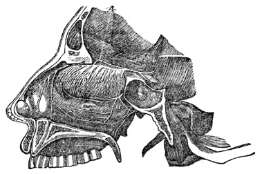
Fig. 136. A side view of the passage of the nostrils, and the distribution of the olfactory nerve. 4, The olfactory nerve. 5, The fine and curious divisions of this nerve on the membrane of the nose. 6, A branch of the fifth pair of nerves.
885. The sense of smell enables us to discern the odor or scent of any thing. When substances are presented to the nose, the air that is passing through the nostrils brings the odoriferous particles of matter in contact with the filaments of the olfactory nerves, that are spread upon the membrane 392 that lines the air-passages, and the impression is then transmitted to the brain.
884. What nerves ramify upon this membrane? What is represented by fig. 136? 885–899. Give the physiology of the organs of smell. 885. How does the mind become sensible of odoriferous particles?
886. This sense, with that of taste, aids man as well as the inferior animals, in selecting proper food, and it also gives us pleasure by the inhalation of agreeable odors. The sense of smell, like that of taste and touch, may be improved by cultivation. It likewise varies in different persons.
Observation. Sometimes this sense seems to possess a morbid degree of acuteness in respect to odors, which is highly inconvenient and even dangerous. With some individuals, the smell of certain fruits, flowers, cheese, &c., produce nausea and even convulsions.
887. In the inferior animals generally, the sense of smell is more acute than in man. Thus the bloodhound will track the hare over the ground for miles, guided only by the odor that it leaves in its flight. He also traces the progress of his master through thickly-crowded streets, distinguishing his footsteps from those of a thousand others, and amidst the odorous particles emanating from a thousand sources.
Observation. In some of the higher orders of the inferior animals, there is an astonishing acuteness of smell in regard to effluvia that come from living animals. To these animals, it possesses an importance in them far beyond what it has in man, by making them acquainted with the presence of their enemies or their prey, when the eye and ear are incapable of acting. It is related by travellers in Africa, that they were always apprised of lions in their vicinity during the night, by the moans and tremblings of their horses.
888. Smell is somewhat under the control of the will. That 393 is, we have the power of receiving or rejecting odors that are presented; thus, if odors are agreeable, we inspire forcibly, to enjoy them; but, if they are offensive, our inspirations are more cautious, or we close our nostrils. This sense is likewise modified by habit; odors which, in the first instance, were very offensive, may not only become endurable, but even agreeable.
886. What is the use of the sense of smell? Can this sense be improved by cultivation? What is said respecting this sense in some individuals? 887. What is said of this sense in the bloodhound? Mention an instance of astonishing acuteness of smell in some of the higher orders of animals. 888. Show that smell is somewhat under the control of the will.
889. Acuteness of smell requires that the brain and nerve of smell be healthy, and that the membrane that lines the nose be thin and moist. Any influence that diminishes the sensibility of the nerves, thickens the membrane, or renders it dry, impairs this sense.
Observations. 1st. Snuff, when introduced into the nose, not only diminishes the sensibility of the nervous filaments, but thickens the lining membrane. This thickening of the membrane obstructs the passage of air through the nostrils, and thus obliges “snuff-takers” to open their mouths when they breathe.
2d. The mucous membrane of the nasal passages is the seat of chronic catarrh. This affection is difficult of removal, as remedial agents cannot easily be introduced into the windings of these passages. Snuff and many other articles used for catarrh, produce more disease than they remove.
889. On what does acuteness of smell depend? What effect has snuff when introduced into the nose? What is said of chronic catarrh?
890. This sense contributes more to the enjoyment and happiness of man than any other of the senses. By it we perceive the form, color, volume, and position of objects that surround us. The eye is the organ of sight, or vision, and its mechanism is so wonderful, that it not only proves the existence of a great First Cause, but perhaps, more than other organs, the design of the Creator to mingle pleasure with our existence.
891. The apparatus of vision consists of the Op´tic Nerve, the Globe and Muscles of the eye, and its Protecting Organs.
892. The OPTIC NERVE arises by two roots from the central portion of the base of the brain. The two nerves approach each other, as they proceed forward, and some of the fibres of each cross to the nerve of the opposite side. They then diverge, and enter the globe of the eyes at their back part, where they expand, and form a soft, whitish membrane.
893. The GLOBE, or ball of the eye, is an optical instrument of the most perfect construction. The sides of the globes are composed of Coats, or membranes. The interior of the globe is filled with refracting Humors, or me´di-ums.
890. Which sense contributes most to the enjoyment of man? What do we perceive by this sense? What is said of the mechanism of the eye? 891–916. Give the anatomy of the organs of vision. 891. Of what does the apparatus of vision consist? 892. Describe the optic nerve. 893. Describe the globe of the eye.
894. The COATS are three in number: 1st. The Scle-rot´ic and Corn´e-a. 2d. The Cho´roid, Iris, and Cil´ia-ry processes. 3d. The Ret´i-na.
895 The HUMORS are also three in number: 1st. The A´que-ous, or watery. 2d. The Crys´tal-line, (lens.) 3d. The Vit´re-ous, or glassy.
Fig. 137.
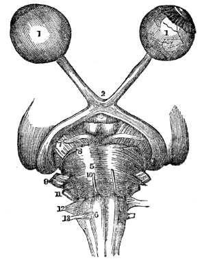
Fig. 137. The second pair of nerves. 1, 1, Globe of the eye: the one on the left is perfect, but that on the right has the sclerotic and choroid coats removed, to show the retina. 2, The crossing of the optic nerve. 5, The pons varolii. 6, The medulla oblongata. 7, 8, 9, 10, 11, 12, 13, The origin of several pairs of cranial nerves.
896. The SCLEROTIC COAT is a dense, fibrous membrane and invests about four fifths of the globe of the eye. It gives form to this organ, and serves for the attachment of the muscles that move the eye in various directions. This coat, from the brilliancy of its whiteness, is known by the name of “the 396 white of the eye.” Anteriorly, the sclerotic coat presents a bevelled edge, which receives the cornea in the same way that a watch-glass is received by the groove in its case.
894. Name the coats of the eye. 895. Name the humors of the eye. Explain fig. 137. 896. Describe the sclerotic coat.
897. The CORNEA is the transparent projecting layer, that forms the anterior fifth of the globe of the eye. In form, it is circular, convexo-concave, and resembles a watch-glass. It is received by its edge, which is sharp and thin, within the bevelled border of the sclerotic, to which it is firmly attached. The cornea is composed of several different layers; its blood-vessels are so small that they exclude the red particles altogether, and admit nothing but serum.
898. The CHOROID COAT is a vascular membrane, of a rich chocolate-brown color upon its external surface, and of a deep black color within. It is connected, externally, with the sclerotic, by an extremely fine cellular tissue, and by the passage of nerves and vessels; internally, it is in contact with the retina. The choroid membrane is composed of three layers. It secretes upon its internal surface a dark substance, called pig-ment´um ni´grum, which is of great importance in the function of vision.
899. The IRIS is so called from its variety of color in different persons. It forms a partition between the anterior and posterior chambers of the eye, and is pierced by a circular opening, which is called the pu´pil. It is composed of two layers. The radiating fibres of the anterior layer converge from the circumference to the centre. Through the action of these radiating fibres the pupil is dilated. The circular fibres surround the pupil, and by their action produce contraction of its area. The posterior layer is of a deep purple tint, and is called u-ve´a, from its resemblance in color to a ripe grape.
How are this coat and the cornea united? 897. Describe the cornea. 898. What is the color of the external surface of the choroid coat? Of the internal? How is it connected externally? How internally? What does this membrane secrete upon its internal surface? 899. Describe the iris. Of how many layers of fibres is the iris composed? What is the function of the radiating fibres? Of the circular?
900. The CILIARY PROCESSES consist of a number of triangular folds, formed, apparently, by the plaiting of the internal layer of the choroid coat. They are about sixty in number. Their external border is continuous with the internal layer of the choroid coat. The central border is free, and rests against the circumference of the crystalline lens. These processes are covered by a layer of the pigmentum nigrum.
Fig. 138.

Fig. 138. A view of the anterior segment of a transverse section of the globe of the eye, seen from within. 1, The divided edge of the three coats—sclerotic, choroid, and retina. 2, The pupil. 3, The iris: the surface presented to view in this section being the uvea. 4, The ciliary processes. 5, The scalloped anterior border of the retina.
901. The RETINA is composed of three layers: The external; middle, or nervous; and internal, or vascular. The external membrane is extremely thin, and is seen as a flocculent film, when the eye is suspended in water. The nervous membrane is the expansion of the optic nerve, and forms a thin, semi-transparent, bluish-white layer. The vascular 398 membrane consists of the ramifications of a minute artery and its accompanying vein. This vascular layer forms distinct sheaths for the nervous papillæ, which constitute the inner surface of the retina.
900. How are the ciliary processes formed? What does fig. 138 exhibit? 901. Of how many layers is the retina composed? Describe the external layer. The nervous layer.
902. The AQUEOUS HUMOR is situated in the anterior and posterior chambers of the eye. It is an albuminous fluid, having an alkaline reaction. Its specific gravity is a very little greater than distilled water. The anterior chamber is the space intervening between the cornea, in front, and the iris and pupil, behind. The posterior chamber is the narrow space, less than half a line in depth, bounded by the posterior surface of the iris and pupil, in front, and by the ciliary processes and crystalline lens, behind. The two chambers are lined by a thin layer, the secreting membrane of the aqueous humor.
903. The CRYSTALLINE HUMOR, or lens, is situated immediately behind the pupil, and is surrounded by the ciliary processes. This humor is more convex on the posterior than on the anterior surface, and, in different portions of the surface of each, the convexity varies from their oval character. It is imbedded in the anterior part of the vitreous humor, from which it is separated by a thin membrane, and is invested by a transparent elastic membrane, called the capsule of the lens. The lens consists of concentric layers, disposed like the coats of an onion. The external layer is soft, and each successive one increases in firmness until the central layer forms a hardened nucleus. These layers are best demonstrated by boiling, or by immersion in alcohol, when they separate easily from each other.
Observations. 1st. The lens in the eye of a fish is round, 399 like a globe, and has the same appearance, when boiled, as the lens of the human eye.
The vascular layer. 902. Where is the aqueous humor situated? What part of the eye is called the anterior chamber? The posterior chamber? With what are the chambers lined? 903. Where is the crystalline humor situated? With what is it surrounded? Of what does the lens consist? How are these layers best demonstrated? What is produced when the lens, or its investing membrane, is changed in structure?
2d. When the crystalline lens, or its investing membrane, is changed in structure, so as to prevent the rays of light passing to the retina, the affection is called a cataract.

Fig. 139. A section of the globe of the eye. 1, The sclerotic coat. 2, The cornea (This connects with the sclerotic coat by a bevelled edge.) 3, The choroid coat. 6, 6, The iris. 7, The pupil. 8, The retina. 10, 11, 11, Chambers of the eye that contain the aqueous humor. 12, The crystalline lens. 13, The vitreous humor. 15, The optic nerve. 16, The central artery of the eye.
904. The VITREOUS HUMOR forms the principal bulk of the globe of the eye. It is an albuminous fluid, resembling the aqueous humor, but is more dense, and differs from the aqueous in this important particular, that it has not the power of re-producing itself. If by accident it is discharged, the eye is irrecoverably lost; while the aqueous humor may be let out, and will be again restored. It is enclosed in a delicate membrane, called the hy´a-loid, which sends processes into the interior of the globe of the eye, forming the cells in which the humor is retained.
904. Describe the vitreous humor. How does this humor differ from the aqueous? What membrane encloses the vitreous humor?
Observation. The structure of this organ can be seen by first freezing the eye of a sheep or an ox; it then can be cut in various directions, and each part separately examined.
905. The MUSCLES of the eye are six in number. They are attached, at one extremity, to the bones of the orbit behind the eye; at the other extremity, they are inserted by broad, thin tendons, near the junction of the cornea with the sclerotic coat. The white, pearly appearance of the eye is caused by these tendons.
Fig. 140.
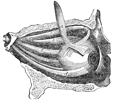
Fig. 140. A view of the eye and its muscles. a, b, c, d, e, Five of these muscles. f, The optic nerve. G, The trochlea, or pulley over which one of the muscles passes. The bone is seen above and below the eye.
Observation. If the external muscle is too short, the eye is turned out, producing the “wall eye.” If the internal 401 muscle is contracted, the eye is turned inward toward the nose. It is then called a “cross eye.”
905. How many muscles has the eye? Give their attachments. What causes the pearly appearance of the eye? What does fig. 140 represent? What is the effect if the external muscle is contracted? The internal muscle?
906. The PROTECTING ORGANS are the Or´bits, Eyebrows, Eyelids, and Lach´ry-mal Apparatus.
907. The ORBITS are deep, bony sockets, in which the globes of the eyes are situated. They have the form of a cone, the base of which is open and directed forward. The bottom of the orbits is pierced by a large hole which gives passage to the optic nerve. These cavities are lined with a thick cushion of fat, in order that the eyes may move in all directions, with perfect freedom and without friction.
908. The EYEBROWS are two projecting arches of integument, covered with short, thick hairs, which form the upper boundary of the orbits. The eyebrows are so arranged that they prevent the moisture that accumulates on the forehead, in free perspiration, from flowing into the eye, and also shade these organs from too vivid light.
909. The EYELIDS are two movable curtains placed in front of the eye. They have a delicate skin on the outside, muscular fibres beneath, and a narrow cartilage on their edges, which tends to preserve the shape of the lid. Internally, they are lined by a smooth membrane, which is reflected over the front of the eye upon the sclerotica. This membrane is called the con-junc-ti´va. It secretes the fluid that moistens and lubricates the eye, and which causes the eyelids to open and shut without friction.
Observation. When the portion of this membrane that is reflected over the globe of the eye, is inflamed, there is frequently a deposition of whitish material, called lymph. This accounts for the films, opacities, and white spots seen upon the eye after the inflammation has subsided.
906. Name the protecting organs of the eye. 907. Describe the orbits. How are the movements of the eye facilitated? 908. Describe the eyebrows. What does this arrangement prevent? 909. Describe the eyelids. What is the use of the conjunctiva? How are the white spots frequently seen upon the eye accounted for?
910. There are found several small glands on the internal surface of the cartilage, which have the appearance of parallel strings of pearls. They open by minute apertures upon the edges of the lids. The secretion from these glands prevents the edges of the eyelids from being united during sleep.
911. The edges of the eyelids are furnished with a triple row of long, thick hairs, called eyelashes, which curve upward from the upper lid, and downward from the lower, so that they may not interlace with each other in the closure of the eyelids. These appendages of the eye, by closing, not only protect it from moisture, but from dust, particularly during sleep. They likewise, by their movements in opening and shutting, spread the lubricating fluid equally over the eye.
912. The LACHRYMAL APPARATUS, which secretes the tears, consists of the Lachrymal Gland with its ducts, Lachrymal Canals, and the Nasal Duct.
913. The LACHRYMAL GLAND is situated at the upper and outer angle of the orbit. It is about three quarters of an inch in length, flattened and oval in shape, and occupies a depression in the orbital plate of the frontal bone. Ten or twelve small ducts pass from this gland, and open upon the upper eyelid, where they pour upon the conjunctiva the lachrymal fluid, or tears. This secretion is maintained while we are asleep, as well as when we are awake. The eye from this cause is kept constantly moist.
914. The LACHRYMAL CANALS commence at minute openings upon the free borders of each eyelid, near the internal 403 angle of the eye, by two small orifices, called punc´ta lach-ry-ma´li-a, (tear points.) Each of these points communicate with the sac at the upper part of the nasal duct.
910. What are found on the internal surface of the cartilage of the eyelids? Where do they open, and what is their use? 911. With what are the edges of the eyelids furnished? What are their uses? 912. Of what does the lachrymal apparatus consist? 913. Describe the lachrymal gland. How many ducts pass from this gland, and what do they convey to the eye? Why is the eye constantly moist? 914. Where do the lachrymal canals commence?
915. The NASAL DUCT is a short canal, about three quarters of an inch in length, directed downward and backward to the inferior channel of the nose, where it terminates by an expanded orifice.
Fig. 141.

Fig. 141. 1, The lachrymal gland. 2, Ducts leading from the lachrymal gland to the upper eyelid. 3, 3, The puncta lachrymalia. 4, The nasal sac. 5, The termination of the nasal duct.
916. The fluid (tears) secreted by the lachrymal gland, is conveyed to the eye by the small ducts before described. It is then imbibed by the puncta lachrymalia, and carried by the lachrymal canals into the lachrymal sac, from which it is passed to the nasal cavities by the nasal ducts.
What are they called? With what do they communicate? 915. Describe the nasal duct. 916. How are the tears conveyed from the lachrymal gland to the nose?
917. To comprehend the theory of vision, it is not sufficient to know the structure of the eye. We must be familiar with some of the properties of a subtile fluid, which is constantly emanating from all luminous bodies, called light.
918. It is the province of natural philosophy, rather than physiology, to enter minutely upon the properties of light. It may be observed, however, that, when light passes through any medium of the same density, the rays are in straight lines; but, when it passes from one medium into another of different density, it is refracted, or turned from a straight course, unless it strikes the medium in a perpendicular direction—then light passes through without a change of direction.
919. When a ray of light meets with a body, it either passes through it, or is reflected by it, or it may be absorbed. Again, in proportion as the rays of light become distant from the body from which they emanate, they diverge one from the other. In accordance with the laws of optics, the rays of light, in passing through an optical instrument like the eye, must cross each other, and thus produce an inverted image of the object from which the rays proceed. With the general 405 view of the structure of the eye, we will now examine the use of each part in the function of vision.
917–933. Give the physiology of the organs of vision. 917. What is necessary in order to understand the theory of vision? 918. When light passes through a medium of the same density, in what direction will be its rays? Of a different density? What exception? 919. When light meets with a body, what takes place? What is said in reference to rays of light in passing through the eye?
920. The sclerotic coat not only gives form to the body of the eye, but protection to the interior and more delicate parts. The choroid coat seems to be chiefly composed of a tissue of nerves and minute blood-vessels; the latter give nourishment to the different parts of the eye. One of the uses of this coat is, to absorb the rays of light immediately after they have passed through the retina. This is effected by the black pigment that lines its inner surface. Were it not for this provision, light would be too intense, and vision indistinct.
Observation. In albinos, where there is an absence of the black pigment, the rays of light traverse the iris, and even the choroid coat, and so overwhelm the eye with light, that their vision is quite imperfect, except in the dimness of evening, or at night. In the manufacture of optical instruments, care is taken to color their interior black, for the same object, namely, the absorption of scattered rays.
921. The iris, by means of its powers of expansion and contraction, regulates the quantity of light admitted through the pupil. If the iris is thin, and the rays of light pass through its substance, they are immediately absorbed by the uvea, and, if that layer be insufficient, they are taken up by the black pigment of the choroid coat.
Observation. When we look toward the bottom of the eye, the pupil appears like a black spot, instead of an opening. This is caused by seeing the black pigment through the retina and humors of the eye.
920. What is the use of the sclerotic coat? Of what is the choroid coat chiefly composed? What is the use of this coat? How is it effected? What is said of albinos? What care is taken in the manufacture of optical instruments? 921. What is the use of the iris? When we look toward the bottom of the eye, why does the pupil look like a black spot, instead of an opening?
922. The cornea, and the aqueous, crystalline, and vitreous humors, are transparent; so that rays of light traverse these parts of the eye, and fall upon the retina. The office of these humors and the cornea is to refract the rays of light in such proportion as to direct the image in the most favorable manner upon the retina.
923. The office of the retina is to receive the impression of the rays of light which leave an object at which we look, and it is upon it that a small but very clear image of that object is formed. The impression thus produced by the reflected light is transmitted by the optic nerve to the brain, which receives the sensation. This constitutes vision.
924. The optic nerve has but one function, that of sight. Sensibility is conferred on this organ by a large branch from the fifth pair of nerves, which ramifies upon the different parts of the eye and its appendages. These parts, however, receive some nervous filaments from the seventh pair.
Observations. 1st. The large number of sensitive nervous filaments renders the visual organ very impressible to bodies that cause irritation, as dust, or intense light. This compels us to use due care to shield the eye from the influence of agents that would impair or destroy vision.
2d. Although particles of dust, when in contact with the delicate parts of the eye, induce severe pain, yet these parts may be cut in surgical operations, and the patient’s sufferings are not as great as when an incision is made in the skin to remove a small tumor.
925. Different degrees of density, as already mentioned, modify the refractory power of any transparent medium. It is found, on examination, that the cornea, the vitreous, the 407 crystalline, and the aqueous humors, have each, severally, various degrees of density: and that the crystalline lens, at its circumference, is less dense than at its centre. These circumstances modify the direction of the refraction of the rays of light, in their passage from the cornea to the retina.
922. What is the use of the cornea, aqueous, crystalline, and vitreous humors? 923. What is the office of the retina? 924. What is the function of the optic nerve? How is sensibility conferred on this organ? Give the 1st observation in this connection. The 2d observation.
926. The refracting powers of the plane, convex, concave, plano-convex, plano-concave, and concavo-convex lenses,[22] are different. The cornea and aqueous humors are convexo-concave, the vitreous humor is concavo-convex, while the crystalline humor is a convexo-convex medium. (Fig. 139.)
Fig. 142.
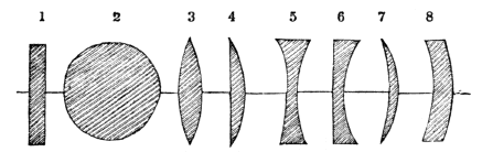
Fig. 142. The forms of the different lenses. 1, A plane lens. 2, A globe lens. 3, A convexo-convex lens. 4, A plano-convex lens. 5, A concavo-concave lens. 6, A plano-concave lens. 7, Meniscus. 8, A concavo-convex lens.
925. Have the cornea and the humors of the eye different degrees of density? What is said of the crystalline lens? What effect has the different density of the parts of the eye upon the light admitted to this organ? 926. What kind of lenses do the humors exhibit? 927. What modifies the refracting powers of transparent mediums? How does this principle apply to the humors of the eye?
927. The different degrees of convexity or concavity also modify the refracting character of transparent mediums. The crystalline lens is of different degrees of convexity on its two sides. The convex surfaces of the aqueous and vitreous humors are segments of circles, of different diameters from their concave surfaces. (Fig. 139.) All these 408 circumstances still further influence the refracting character of the visual organ. The achromatic arrangement of the transparent refracting mediums of the eye, remedies the aberration of refraction in the different portions of the eye.
928. Again, the refracting power of lenses is modified by their convexity or concavity. The more convex a lens is, the shorter the distance from the refracting medium, where the different refracted rays converge to a focus. To adapt the eye to view objects at different distances, requires a change in the refracting power of some of the transparent mediums of the eye.
929. Both surfaces of the crystalline lens are oval, not spherical, and the refraction of the rays of light is mainly effected in this portion of the eye. Change the inclination of this lens, so that different portions of its anterior surface shall be directly behind the pupil, and its refracting power is increased or diminished, as the surface presented is more or less convex.
930. To view objects at a distance, a less convex lens is needed than in examining articles very near the eye; and this organ, from its structure, has the power of adaptation to different distances. It is supposed that the muscular substance of the ciliary body and processes changes, by its contraction, the inclination of the crystalline lens. Without this, or some other adapting power, a picture of objects at different distances would not be formed upon the retina, and the vision of every person would be defective, except in reference to objects at certain definite distances from the eye.
928. What modifies the refracting power of lenses? What is necessary to adapt the eye to view objects at different distances? 929. Where is the refraction of the rays of light mainly effected? 930. When we view objects at a distance, what kind of lens is required? Has the eye the power of adapting itself to different distances? How is it effected?
Observation. It is well known that a separate image is formed on each eye, and, if they are not in the same direction, the objects will appear double. This is proved by pressing one eye, so that the rays of light cannot enter it in the same direction as they do in the other; consequently, the vision is double.
931. By the action of the muscles of the eye, it is turned in different directions, so that objects can be examined upon each side, as well as in front, without turning the body. By the slight or intense action of the straight muscles, the eye is more or less compressed, and the form of the globe is changed, together with the relative positions of the different humors. This modification also adapts the eye to view objects at different distances.
Fig. 143.
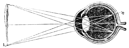
Fig. 143. 1, A pen, an inverted image of which is painted on the retina of the eye, at 2. The image of all objects upon the expansion of the optic nerve, is inverted by the crossing of the rays of light from objects as they traverse the pupil.
Observation. If the eye is fixed for a time on some object which is distinguished with difficulty, there is a painful sensation, similar to that experienced by other muscles of the body when used too long. This is called “straining the eye.”
932. When the refraction of the rays of light is too great, as in over-convexity of the cornea, or the crystalline lens, or the vitreous humor, or all of them, the image is formed a 410 little in front of the retina. Persons thus affected cannot see distinctly, except at a very short distance. This infirmity is called near, or short-sightedness. This defect is in a great measure obviated by the use of concave glasses, which scatter the luminous rays, and thus counterbalance the too strong refracting force of the eye.
What does fig. 143 represent? 931. Why can we see objects at the side as well as in front of the eye, without turning the body? What is the effect when the eye is fixed on an object that is indistinctly seen?
933. When the different parts of the eye are not sufficiently convex, the image is formed beyond the retina, and thus only distant objects are distinctly seen. This defect is called long-sightedness. The feebleness in the refracting power of the eye may be caused by disease; but usually it is a consequence of old age, and is remedied by wearing spectacles with convex glasses.
934. The eye, like other organs of the body, should be used, and then rested. If we look intently at an object for a long time, the eye becomes wearied, and the power of vision diminished. The observance of this rule is particularly needful to those whose eyes are weak, and predisposed to inflammation. On the contrary, if the eye is not called into action, its functions are enfeebled.
935. Sudden transitions of light should be avoided. The iris enlarges or contracts, as the light that falls upon the eye is faint or strong; but the change is not instantaneous. 411 Hence the imperfect vision in passing from a strong to a dim light, and the overwhelming sensation experienced on emerging from a dimly-lighted apartment to one brilliantly illuminated. A common cause of am-aur-o´sis, or paralysis of the retina, is, using the eye for a long time in a very intense light.
932. What is short-sightedness? How is the defect remedied? 933. What is long-sightedness? How is the defect remedied? 934–942. Give the hygiene of the organs of vision. 934. Do the same principles apply to the use of the eye as to other organs? What is the effect if the eye is fixed intently on an object for a long time? What results if the eye is not called into action? 935. Why should sudden transitions of light be avoided?
Note. Let the anatomy and physiology of the eye be reviewed from figs. 139 and 143, or from anatomical outline plate No. 10.
936. Long-continued oblique positions of the eye should be avoided, when viewing objects. If the eye is turned obliquely for a long time in viewing objects, it may produce an unnatural contraction of the muscle called into action. This contraction of the muscle is termed stra-bis´mus, or cross-eye. The practice of imitating the appearance of a person thus affected, is injudicious, as the imitation, designed to be temporary, may become permanent.
Observation. The vision of a “cross-eye” is always defective. In general, only one eye is called into action, in viewing the object to which the mind is directed. This defect can be remedied by a surgical operation, which also corrects the position of the eye.
937. Children should be trained to use the eye upon objects at different distances. This is necessary, in order that the vision may be correct when objects at various distances are viewed. Any action unnatural to the muscles, if frequently repeated, may and will modify the character and action of the parts so operated upon. If a limb, as the arm, be kept flexed for a long time, one set of muscles will be relaxed and elongated, and another will be shortened, and its contractile power will be increased. The same principle is true of the eye.
What causes palsy of the retina? 936. Why should we avoid oblique positions of the eye in viewing objects? What is said of the practice of imitating persons thus affected? What is said in reference to the vision of a “cross-eye”? 937. Why should children be trained to use the eye upon objects at different distances? What is the effect if an unnatural action of the muscles is frequently repeated? Does the same principle apply to the eye?
938. In viewing objects very near the eye, the ciliary processes are called into action to produce a proper inclination of the crystalline lens, so that the rays of light may be properly refracted to form a perfect image on the retina. In looking at objects at a great distance, the ciliary processes are called into a different action, to produce a different inclination of the lens. Let either of these actions be repeated, again and again, for weeks and months, and they will become natural, and the acquired inclination will be permanent.
939. From the preceding principle, a person becomes short or long sighted, as the objects to which the eye is usually directed are near or remote. This is one reason why scholars, watchmakers, and artisans, who bring minute objects near the eye to examine them, are short-sighted, and why hunters and sailors, who are habituated to view objects at a distance, are long-sighted.
Observation. In the management of children, whether in the nursery or school-room, it is very important that their books, or articles upon which they may labor, should be held at an appropriate distance from the eye. Were this attended to by the parent or instructor, we should not see so many persons with defective vision.
940. Cleanliness, as well as the health of the eye, require that it be bathed every morning with pure water, either cold or tepid, accompanied with as little rubbing or friction as possible. In all instances, the secretion from the lachrymal glands, that sometimes collects at the angle of the eye, should be removed, as it contains saline matter.
941. When small particles, or dust, get upon the eye, they produce much inconvenience, which is often increased by 413 harsh attempts to remove them. The individual should be placed before a strong light, the lids held open with one hand, or by another person, and the particles removed with the corner of a fine linen or silk handkerchief.
938. What is the effect of repeatedly using the eye in one direction? 939. Why are artisans and scholars generally short-sighted? Why are sailors and hunters long-sighted? How can defective vision in a great degree be prevented? 940. What reasons are there for bathing the eye? 941. How can dust and other small particles be removed from the eye?
942. Sometimes the substance is concealed under the upper eyelid, and it may then be exposed by turning back the lid in the following manner: Take a knitting-needle, or small, slender piece of stick, which is perfectly smooth, and place it over the upper lid, in contact with, and just under the edge of the orbit; then, holding it firmly, seize the eyelashes with the fingers of the disengaged hand, and gently turn the lid back over the stick or needle. The inner side of the lid can then be examined, and any substance removed that may have been there concealed. Too many trials ought not to be made, if unsuccessful, as much inflammation may be induced; but a surgeon should be consulted as soon as possible.
Observation. Eyestones ought never to be placed in the eye, as they often cause more pain and irritation than the evil which they are intended to remedy.
942. How removed from the upper eyelid? Why should not eyestones be used?
943. The sense of hearing is next in importance to that of vision. Through this sense we are enabled to perceive sounds, that not only subserve to our comfort and pleasure, but are instrumental in promoting our intellectual enjoyments. The organ of hearing, or the ear, is one of the most complicated in the human body.
944. The EAR is composed of three parts: 1st. The External Ear. 2d. The Tym´pan-um, or middle ear. 3d. The La´by-rinth, or internal ear.
945. The EXTERNAL EAR is composed of two parts: The Pin´na, (pavilion of the ear,) and the Me-a´tus Aud-it-o´ri-us Ex-ter´nus, (auditory canal.)
946. The PINNA is a cartilaginous plate which surrounds the entrance of the auditory canal. It presents many ridges and furrows, arising from the folds of the cartilage that form it.
Observation. The pinna, in many animals, is movable; in those that pursue their prey, it is generally directed forward; in timid animals, as the hare and rabbit, it is directed 415 backward. In man, this part is but slightly under the control of the will.
943. What is said of the importance of hearing? Is the ear complicated in its structure? 944–962. Give the anatomy of the organs of hearing. 944. Of how many parts is the ear composed? Name them. 945. Give the parts of the external ear. 946. Describe the pinna. What is said in reference to the pinna of many animals?
947. The MEATUS AUDITORIUS is a canal partly cartilaginous, and partly bony, about an inch in length, which extends inward from the pinna to the Mem´bra-na Tym´pan-i, (drum of the ear.) It is narrower in the middle than at the extremities. It is lined by an extremely thin pouch of cuticle, which, when withdrawn, after maceration, preserves the form of the canal. Some stiff, short hairs are also found in the interior of the channel, which stretch across the tube, and prevent the ingress of insects. Beneath the cuticle are a number of small follicles, which secrete the wax of the ear.
Fig. 144.

Fig. 144. A representation of the four bones of the ear. The smallest is highly magnified. This bone is early matured, and in the adult it becomes united with the incus. These bones are retained in their places and moved by three ligaments and four muscles.
948. The MEMBRANA TYMPANI is a thin, semi-transparent membrane, of an oval shape. It is about three eighths of an inch in diameter, and is inserted into a groove around the circumference of the meatus, near its termination. This membrane is placed obliquely across the area of that tube. It is concave toward the meatus, and convex toward the tympanum.
949. The TYMPANUM consists of an irregular bony cavity, situated within the temporal bone. It is bounded externally by the membrana tympani; internally by its inner wall; and in its circumference by the petrous portion of the temporal 416 bone and mastoid cells. The tympanum contains four small bones, called the os-sic´u-la au-di´tus. These are named separately, the mal´le-us, in´cus, sta´pes, and or-bic´u-lar.
947. What is the meatus auditorius? What is found in this canal? What is their use? Where is the wax of the ear secreted? 948. Describe the membrana tympani. 949. Where is the tympanum situated?
950. There are ten openings in the middle ear; five large and five small. The larger openings are, the Me-a´tus Aud-it-o´ri-us Ex-ter´nus, Fe-nes´tra O-va´lis, (oval window,) Fe-nes´tra Ro-tun´da, (round window,) Mas´toid Cells, and Eu-sta´chi-an Tube.
Fig. 145.
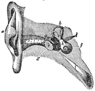
Fig. 145. A representation of the pinna, meatus, membrana tympani, bones of the ear, and semicircular canals. a, The pinna. c, The meatus auditorius externus. g, The membrana tympani. k, The tympanum. e, The bones of the ear. b, The semicircular canals. f, The cochlea. h, The vestibule. i, The Eustachian tube. d, The auditory nerve.
951. The FENESTRA OVALIS is the opening of communication between the tympanum and the vestibule. It is closed by the foot of the stapes, or bone of the ear, and by the lining membrane of both cavities.
952. The FENESTRA ROTUNDA serves to establish a communication 417 between the tympanum and the cochlea. it is closed by a proper membrane, as well as by the lining of both cavities.
What does this cavity contain? 950. How many openings in the tympanum? Explain fig. 145. 951. Describe the fenestra ovalis. 952. The fenestra rotunda.
953. The MASTOID CELLS are very numerous, and occupy the whole of the interior of the mastoid process of the temporal bone, and part of the petrous bone. They communicate, by a large, irregular opening, with the upper and posterior circumference of the tympanum.
Fig. 146.

Fig. 146. A view of the labyrinth laid open. This figure is highly magnified. 1, 1, The cochlea. 2, 3, Two channels, that wind two and a half turns around a central point, (5.) 7, The central portion of the labyrinth, (vestibule.) 8, The foramen rotundum. 9, The fenestra ovalis. 11, 12, 13, 14, 15, 16, 17, 18, The semicircular canals. The cochlea and semicircular canals open into the vestibule.
954. The EUSTACHIAN TUBE is a canal of communication, extending obliquely between the pharynx and the anterior circumference of the tympanum. In structure it is partly 418 fibro-cartilaginous and partly bony. It is broad and expanded at its pharyngeal extremity, and narrow and compressed at the tympanum.
953. Where are the mastoid cells? Explain fig. 146. 954. Describe the Eustachian tube.
955. The small openings of the middle ear are for the entrance and exit of the chorda tympani, (a small nerve that crosses the tympanum,) and for the exit of the muscles that act upon the membrana tympani and bones of the ear.
956. The LABYRINTH consists of a membranous and a bony portion. The bony labyrinth presents a series of cavities which are channelled through the substance of the petrous bone. It is situated between the cavity of the tympanum and the Aud´it-o-ry Nerve. The labyrinth is divided into the Ves´ti-bule, Sem-i-cir´cu-lar Canals, and Coch´le-a.
957. The VESTIBULE is a small, three-cornered cavity, situated immediately within the inner wall of the tympanum.
958. The SEMICIRCULAR CANALS are three bony passages which communicate with the vestibule, into which two of them open at both extremities, and the third at one extremity.
959. The COCHLEA forms the anterior portion of the labyrinth. It consists of a bony and gradually tapering canal, about one and a half inches in length, which makes two turns and a half, spirally, around a central axis, called the mo-di´o-lus. The modiolus is large near its base, where it corresponds with the first turn of the cochlea, and diminishes in diameter toward its extremity.
960. The interior of the canal of the cochlea is partially divided into two passages, by means of a bony and membranous plate. At the extremity of the modiolus, the two passages communicate with each other. At the other extremity, one opens into the vestibule; the other into the tympanum, by 419 the foramen rotundum. The internal surface of the bony labyrinth is lined by a fibro-serous membrane.
955. What passes through the small openings of the middle ear? 956. Of what does the labyrinth consist? Give the parts of the internal ear. 957. Describe the vestibule. 958. What is said of the semicircular canals? 959. Why is the cochlea so called? Of what does it consist? 960. How is the interior of the canal of the cochlea divided? Where do they communicate with each other?
961. The membranous labyrinth is smaller in size, but a perfect counterpart, with respect to form, of the bony vestibule, cochlea, and semicircular canals. Within this labyrinth are two small, elongated sacs, which are filled with a fluid.
Fig. 147.

Fig. 147. A view of the auditory nerve. 1, The spinal cord. 2, The medulla oblongata. 3, The lower part of the brain. 4, The auditory nerve. 5, A branch to the semicircular canals. 6, A branch to the cochlea.
962. The AUDITORY NERVE enters the temporal bone upon its internal surface, and divides into two branches, at the bottom of the cavity of the internal ear. These branches enter the structure of the elongated sacs and membranous labyrinth, radiating in all directions, and finally, they terminate upon the inner surface of the membrane, in minute papillæ, resembling those of the retina.
By what is the internal labyrinth lined? 961. Describe the membranous labyrinth. What does fig. 147 represent? 962. Where does the auditory nerve enter and divide? Where do the branches of the auditory nerve enter and terminate?
963. Hearing is that function by which we obtain a knowledge of the vibratory motions of bodies, which constitute sounds. The precise function of all the different parts of the ear is not known.
964. The function of that part of the external ear which projects from the head is to collect sounds and reflect them into the meatus.
965. The membrana tympani serves to facilitate the transmission of sounds, and also to moderate their intensity. It is so arranged that it can be relaxed or tightened.
Observation. This membrane, when healthy, has no opening; and it must be apparent that the apprehension which is often expressed, that insects will penetrate further, is groundless. The pain is owing to the extreme sensibility of the membrana tympani.
966. The supposed office of the tympanum is to transmit the vibrations made on the membrana tympani to the internal ear. This is effected by the air which it contains, and by the chain of small bones that are enclosed in this cavity.
967. The use of the Eustachian tube is to admit air into the tympanum, which renders the pressure on both sides equal, and thus its membrane is kept in a proper state of tension.
963–971. Give the physiology of the organs of hearing. 963. What is hearing? Are the precise functions of the different parts of the ear known? 964. What is the function of the external ear? 965. Of the membrana tympani? What observation in reference to this membrane? 966. What is the supposed office of the middle ear? 967. What is the use of the Eustachian tube?
Observation. When near a cannon, or a field-piece, about being discharged, by opening the mouth the impression upon the auditory nerve will be diminished, and the unpleasant sensation lessened. This is the result of the air in the middle ear escaping through the Eustachian tube, when the vibrations of the membrana tympani are violent.
Fig. 148.

Fig. 148. A view of all the parts of the ear. 1, The tube that leads to the internal ear. 2, The membrana tympani. 3, 4, 5, The bones of the ear. 7, The central part of the labyrinth, (vestibule.) 8, 9, 10, The semicircular canals. 11, 12, The channels of the cochlea. 13, The auditory nerve. 14, The channel from the middle ear to the throat, (Eustachian tube.)
968. But little is known of the functions of the internal ear; its parts are filled with a watery fluid, in which the filaments of the auditory nerve terminate.
What observation in this connection? 968. What is the function of the internal ear?
969. Many of the parts just enumerated aid in hearing, but are not absolutely essential to this sense. But if the vestibule and auditory nerve are diseased or destroyed, no sound is then perceived. If this sense is destroyed in early life, the person also loses the power of articulating words. Hence a man born deaf is always dumb.
970. The transmission of sound through the different parts of the ear will now be explained by aid of fig. 148. The vibrations of air are collected by the external ear, and conducted through the tube (1) to the membrana tympani, (2.) From the membrane vibrations pass along the chain of bones, (3, 4, 5.) The bone 5 communicates with the internal ear, (7, 8, 9, 10, 11, 11, 11, 12, 12, 12.) From the internal ear the impression is transmitted to the brain by the nerve, (13.)
971. The auditory nerve, like the optic, has but one function, that of special sensibility. The nerves which furnish the ear with ordinary sensibility, proceed from the fifth pair.
972. Hearing, like the other senses, is capable of very great improvement. By cultivation, the blind are enabled to judge with great accuracy the distance of bodies in motion, and even the height of buildings. It is also capable of improvement when all the other senses are perfect. Thus the Indian will distinguish sounds that are inaudible to the white man.
969. What parts of the ear are essential in order to hear sounds? What follows loss of hearing? 971. What is the office of the auditory nerve? What nerves convey ordinary sensibility to the ear? 972–978. Give the hygiene of the organs of hearing. 972. Is this sense capable of improvement? How does this sense aid the blind? Is it also capable of improvement when all the other senses are perfect? In whom is this illustrated?
Note. Let the anatomy and physiology of the organs of hearing be reviewed, from fig. 148, or from anatomical outline plate No. 10.
973. Acute hearing requires perfection in the structure and functions of the different parts of the ear, and that portion of the brain from which the auditory nerve proceeds. Deafness is by no means unfrequent. We will now advert to some of the common causes of imperfect hearing.
974. The structure or functional action of the brain may be deranged by inflammation, by compression, or by debility, and produce deafness. The first is seen during inflammatory affections of the brain, and in fevers; the second is seen in accidental injuries of the head; the third is seen in old age, and after severe diseases of the head, and fevers. In these cases, applications to, and operations upon, the ear do no good. The only remedy is to remove, if possible, the diseased condition of the brain.
975. Imperfect hearing may be produced by the destruction of the membrana tympani, or removal of the bones of the ear, or the parts within the labyrinth. In these instances, medical treatment is of no avail, as the destroyed parts cannot be restored.
976. Hearing may be rendered defective by a diminution of the vibratory character of the membrana tympani. This may result from a thickening of this membrane, or from an accumulation of wax upon its outer surface. The increased thickness is usually the result of inflammation, either acute or chronic. The proper treatment is such as is efficient to remove inflammatory action.
Observations. 1st. The introduction of heads of pins into the ear is a frequent cause of chronic inflammation of the membrana tympani. Hence this practice should never be adopted, and if acquired, should be abandoned.
973. On what does acute hearing depend? 974. State effects on the hearing in some conditions of the brain. How relieved? 975. Of the effect on hearing when the bones of the ear or the labyrinth are destroyed? Is medical treatment of any avail? 976. What conditions of the drum of the ear may impair hearing? How relieved? What is said of the introduction of pins to cleanse the ear?
2d. The accumulations of viscid wax may be softened by dropping some animal oil into the ear, and then removing it by ejecting warm soap suds a few hours subsequent to the use of the oil. This may be repeated for several successive days.
977. Hearing may be impaired by obstruction of the Eustachian tube. The closure of this canal diminishes the vibratory character of the air within the tympanum, in the same manner as closing the opening in the side of a drum. For the same reason, enlarged tonsils, inflammation and ulceration of the fauces and nasal passages during and subsequent to an attack of scarlet fever, and the inflammation attending the “sore throat” in colds, are common causes of this obstruction.
978. The treatment of such cases of defective hearing, is to have the tonsils, if enlarged, removed by a surgeon; for the inflammation and thickening of the parts remedial means should be applied, directed by a skilful physician. The nostrums for the cure of deafness are generally of an oleaginous character, and may be beneficial in cases of defective hearing caused by an accumulation of wax upon the drum of the ear, but in this respect they are no better than the ordinary animal oils.
What is the remedy where there is an accumulation of wax? 977. What is the effect on hearing if the Eustachian tube is obstructed? 978. What is the treatment when deafness is caused by inflammation or ulceration the fauces? What is said of the nostrums used for deafness?
979. Our bodies are constituted in harmony with certain laws, and every person should learn these, in order to regulate his actions and the performance of his duties, so that health may be unimpaired, and the power of enjoyment, activity, and usefulness continue while life lasts.
980. It is a law of the bones and the muscles, that they should either be used in some vocation, or called into action by some social play and active sport.
981. All admit that food is necessary to sustain life; and unless it be of a proper quality, taken in proper quantities, and at proper times, the functions of the digestive organs will be deranged, and disease produced.
982. Pure air is essential to the full enjoyment of health. The impure air of unventilated rooms may be breathed, and the effect be so gradual as not to arrest attention; yet it is a violation of the physical laws, and, sooner or later, we pay the penalty in disease and suffering.
979. Why is it incumbent on every person to learn the laws of health? 980. Give a law of the muscles. 981. In preserving the health, is it necessary to give attention to the food which is eaten? Why? 982. What beside food is essential to the full enjoyment of health? What is said of the impure air of unventilated rooms? 983. What should be observed in regard to sleep?
983. The body also requires sleep; and if it is not taken at the right time, or with regularity, we do not feel full refreshment 426 from “tired nature’s sweet restorer.” Let youth be taught that “early to bed and early to rise” gives him health and its attendant blessings. The brain, like other organs of the body, should be called into action at proper times.
984. From the extent of the surface of the skin, and the close sympathy that exists between it and those organs whose office is, to remove the waste particles of matter from the body, it therefore becomes very important in the preservation of the health, that the functions of this membrane be properly maintained.
985. The function of the circulatory and secretory organs, together with the operations of absorption and nutrition, should be steadily maintained, as vitality and the generation of animal heat are intimately connected with these processes. In the proper performance of these functions, very much depends on the observance of the laws of the muscular, digestive respiratory, dermoid, and nervous apparatuses.
REMOVAL OF DISEASE.
986. It is seldom that a physician is called in the first stages of disease. At this important period, the treatment adopted should be proper and judicious, or the sufferings of the patient are increased, and life, to a greater or less degree, is jeopardized. Hence the utility of knowing what should be done, and what should not be done, in order that the health may be rapidly regained.
987. In all instances of acute disease, it is proper to rest, not only the body, but the mind. To effect this, the patient should cease from physical exertion, and also withdraw his 427 thoughts from study and business operations. This should be done, even if the person is but slightly indisposed.
984. Why should the functions of the skin be properly maintained? 985. Show the necessity of maintaining properly other functions of the system. 986. What is important in the first stages of disease? 987. What is proper in all instances of acute disease? How can it be effected?
988. Select a room for a sick person that is exposed to as little external noise as possible, as impressions made on the organ of hearing greatly influence the nervous system. Likewise select a spacious, well-ventilated apartment, that has no superfluous furniture. The practice of placing a sick person in a small, ill-arranged sleeping-room, when a more spacious room can be used, is poor economy, not to say unkind.
989. Care is necessary in regulating the light of a sick-room. While a strong light would produce an increased action of the vessels of the brain, a moderate light would be an appropriate stimulus to this organ. It is seldom or never necessary to exclude all light from the sick-chamber.
990. A sick person, whether a child or an adult, should not be disturbed by visitors, even if their calls are short. The excitement of meeting them is followed by a depression of the nervous system. The more dangerous and apparently nearer death the sick person is, the more rigorous should be the observance of this suggestion. Nor should the sick-room be opened to privileged classes; for the excitement caused by a visit from relations and the virtuous, will do as much injury to the sick, as that produced by strangers and the vicious.
991. The custom of visiting and conversing with sick friends during the intervals of daily labor, and particularly on Sunday, is a great evil. No person will thus intrude herself in the sick-chamber who cares more for the welfare of the suffering friend than for the gratification of a sympathetic curiosity. Inquiries can be made of the family respecting the sick, and complimentary or necessary messages can be communicated through the nurse.
988. What rooms should be selected for the sick? Why? 989. What is said in reference to the quantity of light admitted into a sick-room? 990. What effect have calls on the sick? 991. What is said of the custom of calling and conversing with the sick during the intervals of daily labor?
Illustration. While attending a Miss B., of N. H., sick of fever, I pronounced her better, withdrew medicine, directed a simple, low diet, and the exclusion of all visitors. In the evening I was sent for to attend her. There was a violent relapse into the disease, which continued to increase in severity until the fourth day, when death terminated her sufferings. I learned that, soon after I gave directions that no visitors be admitted into her room, several particular friends were permitted to enter the chamber and talk with the sick girl. Their conversation produced a severe headache; and, to use the language of the patient, “it seemed as if their talk would kill me;” and it did kill her.
992. No solid food should be taken in the first stages of disease, even if the affection is slight. The thirst can be allayed by drinking cold water, barley-water, and other preparations of an unstimulating character. It is wrong to tempt the appetite of a person who is indisposed. The cessation of a desire for food, is the warning of nature, that the system is in such a state that it cannot be digested.
993. When a patient is recovering from illness, the food should be simple, and in quantities not so great as to oppress the stomach. It should also be given with regularity. “Eat little and often,” with no regard to regularity, is a pernicious practice.
994. When a physician attends a sick person, he should have the special management of the food, particularly after the medicine has been withdrawn and the patient is convalescent. The prevailing idea that every person may safely advise relative to food, or that the appetite of the convalescing person 429 is a competent guide, is dangerous; and cannot be too much censured.
Give an illustration. 992. What suggestion relative to food in the first stages of disease? How can the thirst be allayed? 993. When the patient is convalescent, how should the food be given? What is said of the practice of eating “little and often”? 994. Who should have the special management of food when medicine is withdrawn? What idea prevails in the community?
Illustration. In 1832, I attended a Miss M., sick of fever. After an illness of a few days, the fever abated, and I directed a simple, unstimulating diet. Business called me from the town two days. During my absence, a sympathizing, officious matron called; found her weak, but improving; and told her she needed food to strengthen her; and that “it would now do her good.” Accordingly, eggs and a piece of beefsteak were prepared, and given to the convalescent girl. She ate heartily, and the result was a relapse into a fever more violent than the first attack.
995. It is very important in disease that the skin be kept clean. A free action of the vessels of this part of the body exerts a great influence in removing disease from the internal organs, as well as keeping them in health. If the twenty or thirty ounces of waste, hurtful matter, that passes through the “pores” of the skin in twenty-four hours, are not removed by frequent bathing and dry rubbing, it deranges the action of the vessels that separate this waste matter from the blood, and thus increases the disease of the internal organs.
Illustration. Mrs. M. R., of N., Mass., was afflicted with disease of the lungs and cough. This was accompanied with a dry, inactive condition of the skin. As medicine had no salutary effect in relieving her cough, she was induced by the advice of the clergyman of the parish to enter upon a systematic course of bathing twice every day. Soon the skin became soft, its proper functions were restored, the disease of the lungs yielded, and the cough disappeared.
996. Every sick person should breathe pure air. The purer the blood that courses through the body, the greater the 430 energy of the system to remove disease. The confined vitiated air of the sick-chamber, not unfrequently prolongs disease; and in many instances, the affection is not only aggravated, but, even rendered fatal, by its injurious influences.
Give an illustration of the evil effects attending such an idea. 995. Does the skin exert a great influence in removing disease from the internal organs, as well as in keeping them in health? Give an illustration 996. Why should every sick person, particularly, breathe pure air?
Illustrations. 1st. In 1833, I was called, in consultation with another physician, to Mr. H., who was much debilitated, and delirious. For several successive days he had not slept. His room was kept very warm and close, for fear he would “take cold.” The only change that I made in the treatment, was to open the door and window, at a distance from the bed. In a short time, the delirium ceased, and he fell into a quiet slumber. From this time he rapidly recovered, and the delirium was probably the result of breathing impure air.
2d. Formerly, every precaution was used to prevent persons sick of the small-pox from breathing fresh air. When Mrs. Ramsay had this disease in Charleston, S.C., her friends, supposing that life was extinct, caused her body to be removed from the house to an open shed. The pure air revived the vital spark. The result probably would have been different, had she been kept a few hours longer in the vitiated air.
997. The influence of habit should not be disregarded in the removal of disease. If food or drink is to be administered, however small in quantity or simple its quality, it should be given at or about the time when the ordinary meals were taken in health.
998. Again, the usual time when the patient was in the habit of retiring for sleep should be observed, and all preparation necessary for the sick-room during the night should be made previous to this hour. Efforts should also be made to evacuate the waste matter of the digestive and urinary organs at the period which habit has formed in health. This is not 431 only a remedial agent in disease, but often precludes the necessity of laxative or drastic cathartics.
Are not diseases prolonged, and even rendered fatal, from breathing the impure, vitiated air of the sick-chamber? Give illustration 1st. Give illustration 2d. 997. What is said respecting the influence of habit in removing disease?
999. Medicine is sometimes necessary to assist the natural powers of the system to remove disease; but it is only an assistant. While emetics are occasionally useful in removing food and other articles from the stomach that would cause disease if suffered to remain, and cathartics are valuable, in some instances, to relieve the alimentary canal of irritating residuum, yet the frequent administration of either will cause serious disease.
1000. Although medicine is useful in some instances, yet, in a great proportion of the cases of disease, including fevers and inflammations of all kinds, attention to the laws of health will tend to relieve the system from disease; more certainly and speedily, and with less danger, than when medicines are administered.
1001. Thomas Jefferson, in writing to Dr. Wistar, of Philadelphia, said, “I would have the physician learn the limit of his art.” I would say, Have the matrons, and those who are continually advising “herb teas,” and other “cure-alls,” for any complaint, labelled with some popular name, learn the limits of their duty, namely, attention to the laws of health. The rule of every family, and each individual, should be, to touch not, taste not of medicine of any kind, except when directed by a well-educated and honest physician, (sudden disease from accidents excepted.)
999. What is said of the use of medicine? 1000. Of its use in fevers and many other cases of disease? 1001. What remark by Thomas Jefferson to Dr. Wistar? What should matrons learn? What should be the rule of every person in regard to taking medicine? What exception?
1002. The nurse requires knowledge and practice to enable her to discharge aright her duty to the patient, as much as the physician and surgeon do to perform what is incumbent on them. Woman, from her constitution and habits, is the natural nurse of the sick; and, in general, no small portion of her time is spent in ministering at the couch of disease and suffering.
1003. As the young and vigorous, as well as the aged and the infirm, are liable to be laid upon the bed of sickness, by an epidemic, or imprudent exposure, or by some accident, it is therefore necessary that the girl, as well as the matron, may know how she can render services in an efficient and proper manner. No girl should consider her education complete who is not acquainted with the principles of the duties of a general nurse and a temporary watcher.
1004. It is to be regretted, that while we have medical schools and colleges to educate physicians, there is no institution to educate nurses in their equally responsible station. In the absence of such institutions, the defect can be remedied, to some extent, by teaching every girl hygiene, or the laws of health. To make such knowledge more available and complete, attention is invited to the following suggestions relative to the practical duties of a nurse.
1002. Does the nurse require knowledge and practice in her employment, as well as the physician? Who is the natural nurse of the sick? 1003. What, then, is incumbent on every girl? 1004. Should there be schools to educate nurses, as well as physicians and surgeons?
1005. Bathing. The nurse, before commencing to bathe the patient, should provide herself with water, two towels, a sponge, a piece of soft flannel, and a sheet. The temperature of the room should also be observed.
1006. When the patient is feeble, use tepid or warm water. Cold water should only be used when the system has vigor enough to produce reaction upon the skin. This is shown by the increased redness of the skin, and a feeling of warmth and comfort, after a proper amount of friction. Before using the sponge to bathe, a sheet, or fold of cloth, should be spread smoothly over the bed, and under the patient, to prevent the bed-linen on which the patient lies from becoming damp or wet.
1007. Apply the wet sponge to one part of the body at a time; as the arm, for instance. By doing so, the liability of contracting chills is diminished. Take a dry, soft towel, wipe the bathed part, and follow this by vigorous rubbing with a crash towel, or, what is better, a mitten made of this material; then use briskly a piece of soft flannel, to remove all moisture that may exist on the skin, and particularly between the fingers and the flections of the joints. In this manner bathe the entire body.
1008. The sick should be thoroughly bathed, at least twice in twenty-four hours. Particular attention should be given to the parts between the fingers and toes, and about the flections of the joints, as the accumulation of the excretions is most abundant on these parts. In bathing, these portions of the system are very generally neglected. The best time for bathing, is when the patient feels most vigorous, and freest from exhaustion. The practice of daubing the face and hands with a towel dipped in hot rum, camphor, and vinegar, 434 does not remove the impurities, but causes the skin soon to feel dry, hard, and uncomfortable.
1005. What should a nurse provide herself with, before bathing a patient? 1006. When should cold water be used? 1007. How should the bathing then be performed, so that the patient may not contract a cold? 1008. How often should a sick person be bathed? What is said of daubing the face and hands merely with a wet cloth?
1009. Food. It is the duty of every woman to know how to make the simple preparations adapted to a low diet, in the most wholesome and the most palatable way. Water-gruel,[24] which is the simplest of all preparations, is frequently so ill-made as to cause the patient to loathe it. Always prepare the food for the sick, in the neatest and most careful manner.
1010. When the physician enjoins abstinence from food, the nurse should strictly obey the injunction. She should be as particular to know the physician’s directions about diet, as in knowing how and when to give the prescribed medicines, and obey them as implicitly.
1011. When a patient is convalescent, the desire for food is generally strong, and it often requires firmness and patience, together with great care, on the part of the nurse, that the food is prepared suitably, and given at proper times The physician should direct how frequently it should be taken.
1012. Pure Air. It is the duty of the nurse to see that not only the room is well ventilated in the morning, but that fresh air is constantly admitted during the day. Great care must be taken, however, that the patient does not feel the current.
1013. Bed-linen, as well as that of the body, should be aired every day, and oftener changed in sickness than in health. All clothing, when changed, should be well dried, and warmed by a fire previous to its being put on the patient or the bed.
1009. Should every woman know how to make the simple preparations adapted to a low diet? 1010. Should the nurse strictly obey the injunctions of the physician relative to food? 1011. What period of a person’s illness requires the most care in regard to the food? 1012. Give another duty of the nurse. 1013. What directions respecting the bed-linen of the patient? What is necessary when there is a change of clothing?
1014. Temperature. The warmth of the chamber should be carefully watched by the nurse. The feelings of the patient or nurse are not to be relied on as an index of the temperature of the room. There should be a well-adjusted thermometer in every sick-room. This should be frequently consulted by the nurse.
1015. The temperature of the sick-chamber should be moderate. If it is so cold as to cause a chill, the disease will be aggravated. If, on the other hand, it is too warm, the patient is enfeebled and rendered more susceptible to cold on leaving the sick-chamber. The Latin maxim, “In medio tutissimus ibis,” (in medium there is most safety,) should be regarded in the rooms of the sick.
1016. Quiet. The room of the patient should be kept free from noise. The community should be guided by this rule, that no more persons remain in the room of the sick, than the welfare of the patient demands. It is the duty of the physician to direct when visitors can be admitted or excluded from the sick-room, and the nurse should see that these directions are enforced.
1017. The movements of the attendants should be gentle and noiseless. Shutting doors violently, creaking hinges, and all unnecessary noise, should be avoided. Most persons refrain from loud talking in the sick chamber, but are not equally careful to abstain from whispering, which is often more trying than a common tone.
1018. It is the duty of the nurse to ascertain the habits of the patient as respects the period for eating and sleep, when in health, that she may prepare the food and arrange the sick-room in accordance with the practice of the patient. 436 If the person who is sick is ignorant of the necessity of the removal of the waste products from the system the nurse should invite attention to these functions at such periods as are in accordance with the previous habits of the patient.
1014. Why should there be a well-adjusted thermometer in every sick-chamber? 1015. What is said of the temperature of the sick-chamber? 1016. Why should the sick-room be kept quiet? 1017. What is said of noise in the sick-chamber? Of whispering? 1018. Should the habits of the patient be regarded in reference to the period for eating and sleep?
1019. The deportment and remarks of the nurse to the patient should be tranquil and encouraging. The illness of a friend, or persons who have recently died, should not be alluded to in the sick-room. No doubts or fears of the patient’s recovery, either by a look or by a word, should be communicated by the nurse in the chamber of the sick. When such information is necessary to be communicated, it is the duty of the physician to impart it to the sick person.
1020. The nurse should not confine herself to the sick-room more than six hours at a time. She should eat her food regularly, sleep at regular periods, and take exercise daily in the open air. To do this, let her quietly leave the room when the patient is sleeping. A watcher, or temporary nurse, may supply her place. There is but little danger of contracting disease, if the nurse attends to the simple laws of health, and remains not more than six hours at a time in the sick-room.
1021. These necessary assistants, like the nurse, should have knowledge and practice. They should ever be cheerful, kind, firm, and attentive in the presence of the patient.
1022. A simple, nutritious supper should be eaten before entering the sick-room; and it is well, during the night, to take some plain food.
1019. What should be the deportment of the nurse toward the patient? Should doubts and fears of the patient’s recovery be communicated in the sick-room? When necessary to impart such intelligence, on whom does it depend? 1020. How long should a nurse remain in the sick-chamber at a time? 1021. What qualifications are necessary in a watcher? 1022. What directions in regard to the food of the watcher?
1023. When watching in cold weather, a person should be warmly dressed, and furnished with an extra garment, as a cloak or shawl, because the system becomes exhausted toward morning, and less heat is generated in the body.
1024. Light-colored clothing should be worn by those who have care of the sick, in preference to dark-colored apparel; particularly if the disease is of a contagious character. Experiments have shown, that black and other dark colors will absorb more readily the subtile effluvia that emanate from sick persons, than white or light colors.
1025. Whatever may be wanted during the night, should be brought into the sick-chamber, or the adjoining room, before the family retires for sleep, in order that the slumbers of the patient be not disturbed by haste, or searching for needed articles.
1026. The same general directions should be observed by watchers, as are given to the nurse; nor should the watcher deem it necessary to make herself acceptable to the patient by exhausting conversation.
1027. It can hardly be expected that the farmer, who has been laboring hard in the field, or the mechanic, who has toiled during the day, is qualified to render all those little attentions that a sick person requires. Hence, would it not be more benevolent and economical to employ and pay watchers, who are qualified by knowledge and training, to perform this duty in a faithful manner, while the kindness and sympathy of friends may be practically manifested by assisting to defray the expenses of these qualified and useful assistants?
1023. When watching in cold weather, what precaution is necessary? 1024. What is said relative to the color of the clothing worn in the sick-room? 1025. What suggestions to watchers relative to the arrangement of the sick-chamber? 1026. What should watchers observe? 1027. What is said of employing those persons to watch who labor hard during the day?
1028. Poisoning, either from accident or design, is of such frequency and danger, that it is of the greatest importance that every person should know the proper mode of procedure in such cases, in order to render immediate assistance when within his power.
1029. Poisons are divided into two classes—mineral (which include the acids) and vegetable.
1030. The first thing, usually, to be done, when it is ascertained that a poison has been swallowed, is to evacuate the stomach, unless vomiting takes place spontaneously. Emetics of the sulphate of zinc, (white vitriol,) or ipecacuanha, (ipecac,) or ground mustard seed, should be given.
1031. When vomiting has commenced, it should be aided by large and frequent draughts of the following drinks: flaxseed tea, gum-water, slippery-elm tea, barley water, sugar and water, or any thing of a mucilaginous or diluent character.
1032. Ammonia.—The water of ammonia, if taken in an over-dose, and in an undiluted state, acts as a violent corrosive poison.
1033. The best and most effectual antidote is vinegar. It should be administered in water, without delay. It neutralizes the ammonia, and renders it inactive. Emetics should not be given.
1034. Antimony.—The wine of antimony and tartar emetic, if taken in over-doses, cause distressing vomiting. In addition to the diluent, mucilaginous drinks, give a tea-spoonful of the sirup of poppies, paregoric, or 440 twenty drops of laudanum, every twenty minutes, until five or six doses have been taken, or the vomiting ceases.
1025. Is it useful to know the antidotes or remedies for poison? 1029. Into how many classes are poisons divided? 1030. What is the first thing to be done when it is ascertained that poison has been swallowed? 1031. What should be taken after the vomiting has commenced? 1032. What effect has an over-dose of ammonia? 1033. The antidote? Should an emetic be given for this poison? 1034. What effect has an over-dose of the wine of antimony or tartar emetic?
1035. The antidotes are nutgalls and oak bark, which may be administered in infusion, or by steeping in water.
1036. Arsenic.—When this has been taken, administer an emetic of ipecac, speedily, in mucilaginous teas, and use the stomach-pump as soon as possible.
1037. The antidote is the hydrated peroxide of iron. It should be kept constantly on hand at the apothecaries’ shops. It may be given in any quantity, without injurious results.
1038. Copper.—The most common cause of poisoning from this metal, is through the careless use of cooking utensils made of it, on which the acetate of copper (verdigris) has been allowed to form. When this has been taken, immediately induce vomiting, give mucilaginous drinks, or the white of eggs, diffused in water.
1039. The antidote is the carbonate of soda, which should be administered without delay.
1040. Lead.—The acetate (sugar) of lead is the preparation of this metal, which is liable to be taken accidentally, in poisonous doses. Induce immediate vomiting, by emetics of ground mustard seed, sulphate of zinc, and diluent drinks.
1041. The antidote is diluted sulphuric acid. When this acid is not to be obtained, either the sulphate of magnesia, (epsom salts,) or the sulphate of soda, (glauber’s salts,) will answer every purpose.
1042. Mercury.—The preparation of this mineral by which poisoning is commonly produced, is corrosive sublimate. The mode of treatment to be pursued when this poison has been swallowed, is as follows: The whites of a dozen eggs should be beaten in two quarts of cold water, and a tumbler-full given every two minutes, to induce vomiting. When the whites of eggs are not to be obtained, soap and water should be mixed with wheat flour, and given in copious draughts, and the stomach-pump introduced as soon as possible. Emetics or irritating substances should not be given.
1043. Nitre—Saltpetre.—This, in over-doses, produces violent poisonous symptoms. Vomiting should be immediately induced by large doses 441 of mucilaginous, diluent drinks; but emetics which irritate the stomach should not be given.
1035. What is the antidote? 1036. What should immediately be done when arsenic is swallowed? 1037. What is the antidote? Can any quantity of this preparation of iron be given without injurious results? 1038. What should be given when verdigris has been taken into the stomach? 1039. What is the antidote? 1040. What should immediately be given when sugar of lead is taken? 1041. What is the antidote? 1042. Give the treatment when corrosive sublimate has been swallowed. 1043. What effect has an over-dose of saltpetre? What treatment should be adopted?
1044. Zinc.—Poisoning is sometimes caused by the sulphate of zinc, (white vitriol.) When this takes place, vomiting should be induced, and aided by large draughts of mucilaginous and diluent drinks. Use the stomach-pump as soon as possible.
1045. The antidote is the carbonate, or super-carbonate of soda.
1046. Nitric, (aqua fortis,) MURIATIC, (MARINE ACID,) OR SULPHURIC (OIL OF VITRIOL,) ACIDS, may be taken by accident, and produce poisonous effects.
1047. The antidote is calcined magnesia, which should be freely administered, to neutralize the acid and induce vomiting. When magnesia cannot be obtained, the carbonate of potash (salæratus) may be given. Chalk, powdered and given in solution, or strong soap suds, will answer a good purpose, when the other articles are not at hand. It is of very great importance that something be given speedily, to neutralize the acid. One of the substances before mentioned should be taken freely, in diluent and mucilaginous drinks, as gum-water, milk, flaxseed, or slippery-elm tea. Emetics ought to be avoided.
1048. Oxalic Acid.—This acid resembles the sulphate of magnesia, (epsom salts,) which renders it liable to be taken, by mistake, in poisonous doses. Many accidents have occurred from this circumstance. They can easily be distinguished by tasting a small quantity. Epsom salts, when applied to the tongue, have a very bitter taste, while oxalic acid is intensely sour.
1049. The antidote is magnesia, between which and the acid a chemical action takes place, producing the oxalate of magnesia, which is inert. When magnesia is not at hand, chalk, lime, or carbonate of potash, (salæratus,) will answer as a substitute. Give the antidote in some of the mucilaginous drinks before mentioned. No time should be lost in introducing the stomach-pump as soon as a surgeon can be obtained.
1050. Ley.—The ley obtained by the leaching of ashes may be taken by a child accidentally. The antidote is vinegar, or oil of any kind. The vinegar neutralizes the alkali by uniting with it, forming the acetate of potash. The oil unites with the alkali, and forms soap, which is less caustic than the ley. Give, at the same time, large draughts of mucilaginous drinks, as flaxseed tea, &c.
1044. What is the antidote for white vitriol? 1047. What is the antidote for aqua fortis and oil of vitriol? Should emetics be avoided? 1048. How can oxalic acid be distinguished from epsom salts? 1049. What is the antidote for an over-dose of oxalic acid? When magnesia cannot be obtained, what will answer as a substitute? 1050. What is the antidote when ley is swallowed?
1051. The vegetable poisons are quite as numerous, and many of them equally as virulent, as any in the mineral kingdom. We shall describe the most common, and which, therefore, are most liable to be taken.
1052. Opium.—This is the article most frequently resorted to by those wishing to commit suicide, and, being used as a common medicine, is easily obtained. From this cause, also, mistakes are very liable to be made, and accidents result from it. Two of its preparations, laudanum and paregoric, are frequently mistaken for each other; the former being given when the latter is intended.
1053. Morphia, in solution, or morphine, as it is more commonly called by the public, is a preparation of the drug under consideration, with which many cases of poisoning are produced. It is the active narcotic principle of the opium; and one grain is equal to six of this drug in its usual form.
1054. When an over-dose of opium, or any of its preparations, has been swallowed, the stomach should be evacuated as speedily as possible. To effect this, a teaspoonful of ground mustard seed, or as much tartar emetic as can be held on a five cent piece, or as much ipecacuanha as can be held on a twenty-five cent piece, should be mixed in a tumbler of warm water, and one half given at once, and the remainder in twenty minutes, if the first has not, in the mean time, operated. In the interval, copious draughts of warm water, or warm sugar and water, should be drank.
1055. The use of the stomach-pump, in these cases, is of the greatest importance, and should be resorted to without delay. After most of the poison has been evacuated from the stomach, a strong infusion of coffee ought to be given; or some one of the vegetable acids, such as vinegar, or lemon-juice, should be administered.
1056. The patient should be kept in motion, and salutary effects will often be produced by dashing a bucket of cold water on the head. Artificial respiration ought to be established, and kept up for some time. If the extremities are cold, apply warmth and friction to them. After the poison has been evacuated from the stomach, stimulants, as warm wine and water, or warm brandy and water, should be given, to keep up and sustain vital action.
1057. Stramonium—Thorn-Apple.—This is one of the most active narcotic poisons, and, when taken in over-doses, has, in numerous instances, caused death.
1051. Are vegetable poisons as numerous and as virulent in their effects as mineral? 1052. What is said of opium and its preparations? 1054, 1055, 1056. What treatment should be adopted when an over-dose of opium or any of its preparations is taken? 1057. What is said of stramonium?
1058. Hyosciamus—Henbane.—This article, which is used as a medicine, if taken in improper doses, acts as a virulent irritating and narcotic poison.
1059. The treatment for the two above-mentioned articles is similar to that of poisoning from over-doses of opium.
1060. Conium—Hemlock.—Hemlock, improperly called, by many, cicuta, when taken in an over-dose, acts as a narcotic poison. It was by this narcotic that the Athenians used to destroy the lives of individuals condemned to death by their laws. Socrates is said to have been put to death by this poison. When swallowed in over-doses, the treatment is similar to that of opium, stramonium, and henbane, when over-doses are taken.
1061. Belladonna—Deadly Nightshade.—Camphor. Aconite—Monkshood, Wolfsbane. Bryonia—Bryony. Digitalis—Foxglove. Dulcamara—Bittersweet. Gamboge. Lobelia—Indian Tobacco. Sanguinaria—Bloodroot. Oil of Savin. Spigelia—Pinkroot. Strychnine—Nux vomica. Tobacco.—All of these, when taken in over-doses, are poisons of greater or less activity. The treatment of poisoning, by the use of any of these articles, is similar to that pursued in over-doses of opium. (See Opium, page 442.)
1062. In all cases of poisoning, call a physician as soon as possible.
1058. Of henbane? 1059. What should be the treatment when an over-dose of stramonium or henbane is taken? 1060. What name is sometimes improperly given to conium, or hemlock? How was this narcotic poison used by the Athenians? How are the effects of an over-dose counteracted? 1061. What is the treatment when an over-dose of deadly nightshade, monkshood, foxglove, bittersweet, gamboge, lobelia, bloodroot, tobacco, &c., is taken? 1062. Should a physician be called in all cases when poison is swallowed? 444
The essential parts of every secretory apparatus are a simple membrane, apparently textureless, named the primary, or basement membrane, certain cells and blood-vessels. The serous and mucous membrane are examples.
The division and description of the different membranes and tissues are not well defined and settled by anatomical writers. This is not a material defect, as a clear description of the different parts of the system can be given by adopting the arrangement of almost any writer.
Fat is one of the non-nitrogenous substances. It forms the essential part of the adipose tissue. Chemical analysis shows that all fatty substances are compounds of carbon, hydrogen, and oxygen. They are lighter than water, generally fluid at the natural temperature of the body, and burn with a bright flame, forming water and carbonic acid.
Caseine is abundantly found in milk. When dried, it constitutes cheese. Alcohol, acids, and the stomach of any of the mammalia coagulate it; and it is also soluble in water. It is found in the blood, bile, saliva, and the lens of the eye.
Chondrine is a variety of gelatin. It is obtained from cartilage. It is soluble in warm water, but solidifies on cooling.
Lactic acid is common to all the solids and fluids of the system. It is found united with potash, soda, lime, or magnesia.
The word duodenum is derived from the Latin, signifying “twelve,” since the intestine, of which this is the name, is usually about twelve fingers’ breadth in length. The jejunum is also from the Latin jejunum, empty, since it is usually found in that condition after death, as the food seems to pass rapidly through this part of the intestine. The term ileum is from the Greek, signifying “to twist,” since it always appears in a contorted condition. The name cæcum is derived from the fact of its being a blind or short sack, perforated by the extremity of the ileum. The name of the next division of the intestine—colon—is from the Greek, “to prohibit,” as the contents of the alimentary canal pass slowly through this portion. The rectum is named from the straight direction that it assumes in the latter part of its course.
The food is forced through the alimentary canal by contractions of its muscular coat, produced by the nervous filaments of the sympathetic system, not being at all dependent on the cerebro-spinal centre. This is called the peristaltic, or vermicular motion. The great length of intestine in all animals, and especially in the herbivorous ones, is owing to the necessity of exposing the food to a large number of the lacteals, that the nourishment may all be taken from it.
The different processes through which the food passes before assimilation are of considerable interest. The mastication and mixture of the saliva with the food are purely of a mechanical nature. When any solid or fluid substance is placed upon the tongue, or in contact with the inner surface of the cheeks, by an involuntary act, the salivary glands are stimulated to activity, and commence pouring the saliva into the mouth through the salivary ducts. As soon as mastication commences, the contraction of the masseter and other muscles employed in mastication stimulates the salivary glands to increased action, and a still greater quantity of saliva is secreted and forced upon the food, which is constantly being ground to a finer condition, until it is sufficiently reduced for deglutition.
Whether the salivary fluid acts any other part than simply that of a demulcent to assist the gastric juice in still further dissolving the food, is yet a matter of some doubt, although it is found that no other liquid will equally well subserve the process of digestion and promote health.
After the food is in the condition ready to be swallowed, by an apparently involuntary motion, it is placed upon the back of the tongue, which carries it backwards to the top of the pharynx, where the constrictions of the pharynx, aided by the muscles of the tongue and floor of the mouth, with a sudden and violent movement thrust it beyond the epiglottis, in order to allow the least necessary time to the closure of the glottis, after which, by the compression of the œsophagus, it is forced into the stomach.
Here it is that the true business of digestion commences. For as soon as any substance except water enters the stomach, this organ, with involuntary movements, that seem almost like instinct, commences the secretion of the gastric juice, and by long-continued contractions of its muscular coat, succeeds in effecting a most perfect mixture of the food with this juice, by which the contents of the stomach are reduced to the softest pulp.
The gastric juice, in its pure state, is a colorless, transparent fluid; “inodorous, a little saltish, and perceptibly acid. It possesses the property of coagulating albumen, and separating the whey of milk from its curd, and afterwards completely dissolving the curd. Its taste, when applied to the 446 tongue, is similar to that of mucilaginous water, slightly acidulated with muriatic acid.” The organs of its secretion are an immense number of tubes or glands, of a diameter varying from one five hundredth to one three hundredth of an inch, situated in the mucous coat of the stomach, and receiving their blood from the gastric arteries. A chemical analysis shows it to consist of water, mucilage, and the several free acids—muriatic, acetic, lactic, and butyric, together with a peculiar organic matter called pepsin, which acts after the manner of ferments between the temperature of 50° and 104° F.
The true process of digestion is probably owing to the action of pepsin and the acids, especially if the presence of the chloro-hydric or muriatic be admitted; since we know, by experiments out of the body, that chlorine, one of its elements, is a powerful solvent of all organic substances.
The antiseptic properties of the gastric juice, as discovered by experiments made on Alexis St. Martin, doubtless have much influence on digestion, although their true uses are probably not yet known.
As soon as the food is reduced to a state of fluidity, the pyloric orifice of the stomach is unclosed, and it is thrust onwards through the alimentary canal, receiving in the duodenum the secretions of the liver and pancreas, after which it yields to the lacteals its nutrient portion, and the residuum is expelled from the body.
There have been many hypotheses in regard to the nature of the digestive process. Some have supposed that digestion is a mere mechanical process, produced by the motion of the walls of the stomach; while others, in later times, have considered it as under the influence of a spirit separate from the individual, who took up his residence in the stomach and regulated the whole affair; while others still would make it out to be a chemical operation, and thus constitute the stomach a sort of laboratory. But to all these ridiculous hypotheses Sir John Hunter has applied the following playful language: “Some will have it that the stomach is a mill; others that it is a fermenting vat; and others that it is a stewpan; but in my view of the matter, it is neither a mill, a fermenting vat, nor a stewpan, but a stomach, a stomach!”
At the present day this process is regarded as a complex, and not a simple operation. It seems to be a process in which the mechanical, chemical, and vital agencies must all act in harmony and order; for if one of these be withdrawn, the function cannot be sustained for any considerable length of time; and of the chemical and mechanical parts of the process, since the former is much more important, and, as a matter of course, the vital powers are indispensable, therefore digestion may be considered as a chemical operation, directly dependent on the laws of vitality, or of life; since the proper consistency of the food depends, in a great measure, upon the character of the solvents, while the secretion of these fluids, their proper amount, 447 together with the peculiar instinct—as it almost seems to be—necessary to direct the stomach in its many functions, are exclusively and entirely dependent on the laws and conditions of life.
As food is necessary to supply the waste and promote the growth of the body, it follows that that will be the best adapted to the system which contains the same chemical elements of which the body is composed; viz., oxygen, hydrogen, carbon, and nitrogen. These elements are found in greater or less quantity in all animal food, and in many vegetable products. Hence, that article of food which contains all these elements in a proper proportion will tend much more to the growth and strength of the body than those kinds which are deficient in one or more of them. Much experience on this point, and scientific research, seem to show that a reasonable amount of animal food in health tends to give greater strength of muscle, and a more general sense of fulness, than in ordinary cases a vegetable diet is able to do, owing to the presence of nitrogen in animal tissues. Yet there are examples of the healthiest and strongest men, who live years without a morsel of animal food; and the fact can only be accounted for, by supposing that the system has the power to make the most economical use of the little nitrogen offered to it in the food; or else that it has by some means the power to abstract it from the atmosphere, and transform it to the living animal substance.
The proximate principles, which are the most important in nourishing the body, are albumen and fibrin. These constitute the greater part of all the softer animal tissues, and are also found in certain classes of vegetables, such as peas, beans, lentils, and many seeds. Hence, in many cases, a vegetable diet, especially if embracing any of those articles, would be sufficient to sustain life, even if no animal food should be eaten. But no animal can exist for a long time if permitted only to eat substances destitute of nitrogen, as in the case of a dog fed entirely on sugar, which lived but thirty days. And owing to this fact, Baron Liebig proposes to call substances used for food, containing nitrogen, “elements of nutrition,” and those containing an excess of carbon, “elements of respiration;” since, according to his view, the food is necessary to support the growth of the body by replacing the effete and worn-out particles with new matter, and also to keep up the supply of fuel, in order to promote a sufficient degree of heat in the system. Accordingly, under the first division would be included all lean meats and vegetables, such as peas, &c.; while the fat of animals, vegetable oils, sugars, tubers, (as the potato,) and all other substances containing starch, would be included under the latter division.
This definition of exhalants is from the theory of Haller and others. It is now believed that the fluids exude through the thin coats of the blood vessels. This process is called exosmose, and is the exhalation of old physiologists.
It is a well-established fact, in animal and vegetable physiology, that membranes possess the property of allowing fluids and gases to pass through them in either direction, and also to permit two fluids to pass in opposite directions at the same time. This property is designated endosmose when a fluid passes from without a body inward; and exosmose when the reverse takes place. The first is called imbibition. One of the most striking instances of this, in the human system, is shown in the lungs, where carbonic acid and water pass out through the mucous membrane of the bronchial tubes and air-cells; and the oxygen of the air enters the blood through the same membrane. By this process of imbibition, the oxygenation of the blood is much more readily and faithfully accomplished; inasmuch, as by the immense number of bronchial tubes and air-cells a larger quantity of blood is exposed to a greater portion of air, than if the blood were directly laid open to the atmosphere in a mass, or the air were immediately transmitted through it.
Since the function of respiration is to free the system of superfluous carbon and hydrogen, by union with the oxygen of the air, it follows that the greater the amount of the products to be expelled, the larger the quantity of oxygen will be required to effect this purpose, as we find to be the case with those who consume large quantities of food.
The quantity of oxygen daily consumed through the lungs by an adult is about 32.5 oz., and the carbon in the food 13.9 oz. But in order to convert this whole amount of carbon into carbonic acid, which passes off through the lungs and skin, 37 oz. of oxygen are required; the remaining 4.5 oz. being absorbed by the skin. If the supply of food remain the same, while the amount of oxygen in the inspired air is diminished, the superfluous carbon will induce disease in the system, as is the case of those persons who are limited in their supply of air of a proper quality or quantity, and, consequently, have less appetite for food than those who are abundantly supplied with air of the proper standard of health; and in children, who proportionally consume more food than adults, and who are more active, thereby causing a more rapid circulation of blood, and, consequently, the removal of more superfluous particles of matter.
In children we notice the need of air, by their disposition to be much in the open air, and often inspiring more deeply than is common in older persons. 449 Also, if the carbon of the food does not have a requisite supply of oxygen from the air, or other sources, the body becomes emaciated, although nourishing food may be used. And on the other hand, if there be a diminished supply of food, but an abundance of atmospheric air, leanness and emaciation are sure to follow; owing to the fact that if the oxygen has no waste carbon from the body to unite with, it combines with the fat, and some other soft portions of the body, which the Author of nature seems to have provided for this very purpose; as is seen in the case of hibernating animals, who enter their places of winter abode sleek and fat, but crawl out in the spring not merely deprived of their fatty matter, but also with great diminution of all the softer parts, which have given up their share of carbon to supply animal heat. One important cause of emaciation in febrile diseases is the greater rapidity of the pulse and respiration, which consume more carbon than is afforded by the scanty supply of food that is taken, although profuse perspiration, which almost always occurs in some stages of fevers, greatly diminishes the full state of the body.
The theory of Baron Liebig concerning the change which the blood experiences in color, in its passage through the lungs, meets with the approbation of many physiologists, although there are some important difficulties in the way of fully receiving it. A chemical analysis of the blood shows it to be composed of albumen and fibrin, together with some other substances, in small proportions, and always perceptible traces of iron. He attributes the change in color to the iron, as this substance enters into combination with carbon and oxygen. For, as the blood passes through the trunks of the larger vessels and capillaries, it receives the carbon from the tissues which are continually transformed, and taking up the oxygen from the arterialized blood, forms carbonic acid, which unites with the iron, forming proto-carbonate of iron. This being of a gray color, he supposes it to be that which, with the other impurities of the blood, gives the venous blood the dark blue color. Then, as the blood comes in contact with the oxygen, as it is returned and exposed to this element in the lungs, the carbonic acid leaves the iron, which has a stronger affinity for oxygen than for carbonic acid, and forms the scarlet red peroxide of iron, that gives the characteristic color to the arterial blood. After this, as the blood is sent out through the smaller arteries and capillaries, it again gathers carbon and other impurities from the system, and becomes the dark, venous blood, thus completing the whole change of color in the circulation.
As already mentioned, different articles of food have been divided into the azotized and non-azotized, or those which contain nitrogen as one of their constituents, and those which are nearly destitute of it. Of these, according to Liebig, the azotized portions are simply to supply the waste that is continually going on in the body, and promote its growth in the early stages of existence, or, in other words, the nutrient portion; while the sugar, starch, &c., are mainly of use in the respiratory organs. The correctness of this view may be understood from the fact, that the inhabitants in the colder regions of the earth consume a much larger quantity of oil and fat than the residents of hotter climates; and also those dwelling in the temperate zones can eat with greater impunity a larger quantity of fat meats in the winter than in the summer, there being then so much more demand for animal heat than in the summer.
The suggestion of using the bellows in asphyxia, is from the directions of that distinguished and veteran surgeon, Valentine Mott, of New York city. The directions in the first part of the paragraph are the most practical, and best adapted to the wants of the community.
Ab-duc´tor. [L. abduco to lead away.] A muscle which moves certain parts, by separating them from the axis of the body.
Ab-do´men. [L. abdo, to hide.] That part of the body which lies between the thorax and the bottom of the pelvis.
Ab-dom´in-is. Pertaining to the abdomen.
A-ce-tab´u-lum. [L. acetum, vinegar.] The socket for the head of the thigh-bone; an ancient vessel for holding vinegar.
A-ce´tic. [L. acetum, vinegar.] Relating to acetic acid. This is always composed of oxygen, hydrogen, and carbon, in the same proportion.
A-chil´lis. A term applied to the tendon of two large muscles of the leg.
A-cro´mi-on. [Gr. ακρος, akros, highest, and ωμος, omos, shoulder.] A process of the scapula that joins to the clavicle.
Ad-duc´tor. [L. adduco, to lead to.] A muscle which draws one part of the body toward another.
Al-bu-gin´e-a. [L. albus, white.] A term applied to white textures.
Al-bu´men. [L. albus, white.] An animal substance of the same nature as the white of an egg.
A-lu´min-um. [L.] The name given to the metallic base of alumina.
Al´ve-o-lar. [L. alveolus, a socket] Pertaining to the sockets of the teeth.
Am-mo´ni-a. An alkali. It is composed of three equivalents of hydrogen and one of nitrogen.
A-nas´to-mose. [Gr. ανα, ana, through, and στομα, stoma, mouth.] The communication of arteries and veins with each other.
An-a-tom´i-cal. Relating to the parts of the body, when dissected or separated.
A-nat´o-my. [Greek ανα, ana, through, and τομη, tomē a cutting.] The description of the structure of animals. The word anatomy properly signifies dissection.
An´gu-li. [L. angulus, a corner.] A term applied to certain muscles on account of their form.
An-i-mal´cu-læ. [L. animalcula, a little animal.] Animals that are only perceptible by means of a microscope.
An´nu-lar. [L. annulus, a ring.] Having the form of a ring.
An-ti´cus. [L.] A term applied to certain muscles.
A-ort´a. [Gr. αορτη, aortē; from αηρ, aēr, air, and τηρεω, tēreo, to keep.] The great artery that arises from the left ventricle of the heart.
Ap-o-neu-ro´sis. [Gr. απο, apo, from, and νευρον, neuron, a nerve.] The membranous expansions of muscles and tendons. The ancients called every white tendon neuron, a nerve.
Ap-pa-ra´tus. [L. apparo, to prepare.] An assemblage of organs designed to produce certain results.
Ap-pend´ix. [L., an addition.] Something appended or added.
A´que-ous. [L. aqua, water.] Partaking of the nature of water.
A-rach´noid. [Gr. αραχνη, arachnē, a spider, and ειδος, eîdos, form. Resembling 452 a spider’s web. A thin membrane that covers the brain.
Ar´bor. [L.] A tree. Arbor vitæ. The tree of life. A term applied to a part of the cerebellum.
Ar´te-ry. [Gr. αηρ, aēr, air, and τηρεω, tēreo, to keep; because the ancients thought that the arteries contained only air.] A tube through which blood flows from the heart.
A-ryt-e´noid. [Gr. αρυταινα, arutaina, a ewer, and ειδος, eîdos, form.] The name of a cartilage of the larynx.
As-cend´ens. [L.] Ascending; rising.
As-phyx´i-a. [Gr. α, a, not, and σφυξις, sphyxis, pulse.] Originally, want of pulse; now used for suspended respiration, or apparent death.
As-trag´a-lus. [Gr.] The name of a bone of the foot. One of the tarsal bones.
Aud-i´tion. [L. audio, to hear.] Hearing.
Aud-it-o´ri-us. [L.] Pertaining to the organ of hearing.
Au´ri-cle. [L. auricula, the external ear; from auris, the ear.] A cavity of the heart.
Au-ric´u-lar. [L. auricula.] Pertaining to the auricle.
Ax-il´la. [L.] The armpit.
Ax´il-la-ry. Belonging or relating to the armpit.
A-zote´. [Gr. α, a, not, and ζωη, zoē, life.] Nitrogen. One of the constituent elements of the atmosphere. So named because it will not sustain life.
Ben-zo´ic. Benzoic acid. A peculiar vegetable acid, obtained from benzoin and some other balsams.
Bi´ceps. [L. bis, twice, and caput, a head.] A name applied to muscles with two heads at one extremity.
Bi-cus´pids. [L. bis and cuspis, a point.] Teeth that have two points upon their crown.
Bile. [L. bilis.] A yellow, viscid fluid secreted by the liver.
Bi-pen´ni-form. [L. bis and penna, a feather.] Bipenniform muscle. Having fibres on each side of a common tendon.
Brach´i-al. [L. brachium.] Belonging to the arm.
Bre´vis. [L.] Brevis, short; brevior, shorter.
Bronch´i-a, -æ. [L.] A division of the trachea that passes to the lungs.
Bronch´i-al. Relating to the bronchia.
Bronch-i´tis. [L.] An inflammation of the bronchia.
Buc-ci-na´tor. [L. buccinum, a trumpet.] The name of a muscle of the cheek, so named because used in blowing wind instruments.
Bur´sæ Mu-co´sa. [L. bursa, a purse, and mucosa, viscous.] Small sacs, containing a viscid fluid, situated about the joints, under tendons.
Cæ´cum. [L.] Blind; the name given to the commencement of the colon.
Calx, Cal´cis. [L.] The heel-bone.
Cal´ci-um. [L.] The metallic basis of lime.
Cap´il-la-ry. [L. capillus, a hair.] Resembling a hair; small.
Cap´su-lar. Pertaining to a capsule.
Cap´sule. [L. capsula, a little chest.] A membranous bag, enclosing a part.
Ca´put. [L.] The head. Caput coli. The head of the colon.
Car´bon. [L. carbo, a coal.] Pure charcoal. An elementary combustible substance.
Car-bon´ic. Pertaining to carbon.
Car´di-ac. [Gr. καρδια, kardia, heart.] Relating to the heart, or upper orifice of the stomach.
Car´ne-a, -æ. [L. caro, carnis, flesh.] Fleshy.
Ca-rot´id. [Gr. καρος, karos, lethargy.] The great arteries of the neck that convey blood to the heart. The ancients supposed drowsiness to be seated in these arteries.
Car´pal. [L. carpus, the wrist.] Relating to the wrist.
Car´pus, -i. [L.] The wrist.
Car´ti-lage. [L. cartilago.] Gristle. A smooth, elastic substance, softer than bone.
Car-ti-lag´in-ous. Pertaining to cartilage.
Cau-ca´sian. One of the races of men.
Ca´va. [L.] Hollow. Vena cava. A name given to the two great veins of the body.
Cel´lu-lar. [L. cellula, a little cell.] Composed of cells.
Cer-e-bel´lum. [L.] The hinder and lower part of the brain, or the little brain.
Cer´e-bral. Pertaining to the brain.
Cer´e-brum. [L.] The front and large part of the brain. The term is sometimes applied to the whole contents of the cranium.
Cer´e-bro-Spi´nal. Relating to the brain and spine.
Cer´vix. [L.] The neck.
Cer´vi-cal. Relating to the neck.
Chest. [Sax.] The thorax; the trunk of the body from the neck to the abdomen.
Chlo´rine. [Gr. χλορος, chloros, green.] Chlorine gas, so named from its color.
Chor´da, -æ. [L.] A cord. An assemblage of fibres.
Cho´roid. [Gr. χοριον, chorion.] A term applied to several parts of the body that resemble the skin.
Chyle. [Gr. χυλος, chulos, juice.] A nutritive fluid, of a whitish appearance, which is extracted from food by the action of the digestive organs.
Chyl-i-fi-ca´tion. [chyle and L. facio, to make.] The process by which chyle is formed.
Chyme. [Gr. χυμος, chumos, juice.] A kind of grayish pulp formed from the food in the stomach.
Chym-i-fi-ca´tion. [chyme and L. facio, to make.] The process by which chyme is formed.
Cil´ia-ry. [L. cilia, eyelashes.] Belonging to the eyelids.
Cin-e-ri´tious. [L. cinis, ashes.] Having the color of ashes.
Clav´i-cle. [L. clavicula, from clavis, a key.] The collar-bone; so called from its resemblance in shape to an ancient key.
Clei´do. A term applied to some muscles that are attached to the clavicle.
Co-ag´u-lum. [L.] A coagulated mass, a clot of blood.
Coc´cyx. [Gr.] An assemblage of bones joined to the sacrum.
Coch´le-a. [Gr. κοχλω, kochlo, to twist; or L. cochlea, a screw.] A cavity of the ear resembling in form a snail shell.
Co´lon. [Gr.] A portion of the large intestine.
Co-lum´na, -æ.[L.] A column or pillar.
Com-mu´nis. [L.] A name applied to certain muscles.
Com-plex´us. [L. complector, to embrace.] The name of a muscle that embraces many attachments.
Com-press´or. [L. con, together, and premo, pressus, to press.] A term applied to some muscles, that compress the parts to which they are attached.
Con´dyle. [Gr. κονδυλος, kondulos, a knuckle, a protuberance.] A prominence on the end of a bone.
Con-junc-ti´va. [L. con, together, and jungo, to join.] The membrane that covers the anterior part of the globe of the eye.
Cop´per. A metal of a pale, red color, tinged with yellow.
Cor-a´coid. [Gr. κοραξ, korax, a crow, and ειδος, eîdos, form.] A process of the scapula shaped like the beak of a crow.
Co´ri-on. [Gr. χοριον, chorion, skin.] The true skin.
Corn´e-a. [L. cornu, a horn.] The transparent membrane in the fore part of the eye.
Cos´ta. [L. costa, a coast, side, or rib.] A rib.
Crib´ri-form. [L. cribrum, a sieve, and forma, form.] A plate of the ethmoid 454 bone, through which the olfactory nerve passes to the nose.
Cri´coid. [Gr. κρικος, krikos, a ring, and ειδος, eîdos, form.] A name given to a cartilage of the larynx, from its form.
Crys´tal-line. [L. crystallinus, consisting of crystal.] Crystalline lens. One of the humors of the eye. It is convex, white, firm, and transparent.
Cu´bi-tus, -i. [L. cubitus, the elbow.] One of the bones of the forearm, also called the ulna.
Cu´boid. [Gr. κυβος, kubos, a cube, and ειδος, eîdos, form.] Having nearly the form of a cube.
Cu-ne´i-form. [L. cuneus, a wedge.] The name of bones in the wrist and foot.
Cus´pid. [L. cuspis, a point.] Having one point.
Cu-ta´ne-ous. [L. cutis, skin.] Belonging to the skin.
Cu´ti-cle. [L. cutis.] The external layer of the skin.
Cu´tis Ve´ra. [L. cutis, and vera, true.] The internal layer of the skin; the true skin.
Del´toid. [Gr. δελτα, delta, the Greek letter Δ, and ειδος, eîdos, form.] The name of a muscle, that resembles in form the Greek letter Δ.
Dens. [L.] A tooth.
Dent´al. [L. dens, tooth.] Pertaining to the teeth.
De-press´or. [L.] The name of a muscle that draws down the part to which it is attached.
Derm´oid. [Gr. δερμα, derma, the skin, and ειδος, eîdos, form.] Resembling skin.
De-scend´ens. [L. de and scando, to climb.] Descending, falling.
Di´a-phragm. [Gr. διαφραμα, diaphragma, a partition.] The midriff; a muscle separating the chest from the abdomen.
Di-ar-rhœ´a. [Gr. διαρρεω, diarrheo, to flow through.] A morbidly frequent evacuation of the intestines.
Di-as´to-le. [Gr. διαστελλω, diastello, to put asunder.] The dilatation of the heart and arteries when the blood enters them.
Di-ges´tion. [L. digestio.] The process of dissolving food in the stomach, and preparing it for circulation and nourishment.
Dig-i-to´rum. [L. digitus, a finger.] A term applied to certain muscles of the extremities.
Dor´sal. [L. dorsum, the back.] Pertaining to the back.
Du-o-de´num. [L. duodenus, of twelve fingers’ breadth.] The first portion of the small intestine.
Du´ra Ma´ter. [L. durus, hard, and mater, mother.] The outermost membrane of the brain.
Dys´en-ter-y. [Gr. δυς, dūs, bad, and εντερια, enteria, intestines.] A discharge of blood and mucus from the intestines attended with tenesmus.
Dys-pep´si-a. [Gr. δυς, dūs, bad, and πεπτω, pepto, to digest.] Indigestion, or difficulty of digestion.
En-am´el. [Fr.] The smooth, hard substance which covers the crown or visible part of a tooth.
Ep-i-derm´is. [Gr. επι, epi, upon, and δερμα, derma, the skin.] The scarf-skin; the cuticle.
Ep-i-glot´tis. [Gr. επι, epi, upon, and γλωττα, glōtta, the tongue.] One of the cartilages of the glottis.
Eu-sta´chi-an Tube. A channel from the fauces to the middle ear, named from Eustachius, who first described it.
Ex´cre-ment. [L. excerno, to separate.] Matter excreted and ejected; alvine discharges.
Ex-cre-men-ti´tial. Pertaining to excrement.
Ex´cre-to-ry. A little duct or vessel, destined to receive secreted fluids, and to excrete or discharge them; also, a secretory vessel.
Ex-ha´lant. [L. exhalo, to send forth vapor.] Having the quality of exhaling or evaporating.
Ex-tens´or. [L.] A name applied to a muscle that serves to extend any part of the body; opposed to Flexor.
Fa´cial. [L. facies, face.] Pertaining to the face.
Falx. [L. falx, a scythe.] A process of the dura mater shaped like a scythe.
Fas´ci-a. [L. fascia, a band.] A tendinous expansion or aponeurosis.
Fas-cic´u-lus, -li. [L. fascis, a bundle.] A little bundle.
Faux, -ces. [L.] The top of the throat.
Fem´o-ral. Pertaining to the femur.
Fem´o-ris. A term applied to muscles that are attached to the femur.
Fe´mur. [L.] The thigh-bone.
Fe-nes´tra, -um. [L. fenestra, a window.] A term applied to some openings into the internal ear.
Fi´bre. [L. fibra.] An organic filament, or thread, which enters into the composition of every animal and vegetable texture.
Fi´brin. A peculiar organic substance found in animals and vegetables; it is a solid substance, tough, elastic, and composed of thready fibres.
Fi´brous. Composed or consisting of fibres.
Fi´bro-Car´ti-lage. An organic tissue, partaking of the nature of fibrous tissue and that of cartilage.
Fib´u-la. [L., a clasp.] The outer and lesser bone of the leg.
Fib´u-lar. Belonging to the fibula.
Fil´a-ment. [L. filamenta, threads.] A fine thread, of which flesh, nerves, skin, &c., are composed.
Flec´tion. [L. flectio.] The act of bending.
Fol´li-cle. [L. folliculus, a small bag.] A gland; a little bag in animal bodies.
Fore´arm. The part of the upper extremity between the elbow and hand.
Fos´sa. [L., a ditch.] A cavity in a bone, with a large aperture.
Fræ´num. [L., a bridle.] Frænum linguæ. The bridle of the tongue.
Func´tion. [L. fungor, to perform.] The action of an organ or system of organs.
Fun´gi-form. [L. fungus and forma.] Having terminations like the head of a fungus, or a mushroom.
Gan´gli-on, -a. [Gr.] An enlargement in the course of a nerve.
Gas´tric. [Gr. γαστηρ, gastēr, the stomach.] Belonging to the stomach.
Gas-troc-ne´mi-us. [Gr. γαστηρ, gastēr, the stomach, and κνημη, knēmē, the leg.] The name of large muscles of the leg.
Gel´a-tin. [L. gelo, to congeal.] A concrete animal substance, transparent and soluble in water.
Gle´noid. [Gr. γληνη, glēnē, a cavity.] A term applied to some articulate cavities of bones.
Glos´sa. [Gr.] The tongue. Names compounded with this word are applied to muscles of the tongue.
Glos´so-Pha-ryn´gi-al. Relating to the tongue and pharynx.
Glot´tis. [Gr.] The narrow opening at the upper part of the larynx.
Glu´te-us. [Gr.] A name given to muscles of the hip.
Hem´or-rhage. [Gr. ἁιμα, haima, blood and ρηγνυω, rēgnuo, to burst.] A discharge of blood from an artery or vein.
Hu´mer-us. [L.] The bone of the arm.
Hy´a-loid. [Gr.] A transparent membrane of the eye.
Hy´dro-gen. [Gr. ὑδωρ, water, and γενναω, to generate.] A gas which constitutes one of the elements of water.
Hy´gi-ene. [Gr. ὑγιεινον, hugieînon, health.] The part of medicine which treats of the preservation of health.
Hy´oid. [Gr. υ and ειδος, eîdos, shape.] A bone of the tongue resembling the Greek letter upsilon in shape.
Hy-oid´e-us. Pertaining to the hyoid bone.
Hy´po-glos´sal. Under the tongue. The name of a nerve of the tongue.
Il´e-um. [Gr. ειλω, eilō, to wind.] A portion of the small intestines.
Il´i-ac. [From the above.] The flank; pertaining to the small intestine.
Il´i-um. The haunch-bone.
In-ci´sor. [L. incido, to cut.] A front tooth that cuts or divides.
In´dex. [L. indico, to show.] The fore-finger; the pointing finger.
In-nom-i-na´ta. [L. in, not, and nomen, name.] Parts which have no proper name.
In-os´cu-late. [L. in and osculatus, from osculor, to kiss.] To unite, as two vessels at their extremities.
In´ter. [L.] Between.
In-ter-cost´al. [L. inter, between, and costa, a rib.] Between the ribs.
In-ter-no´di-i. [L. inter, between, and nodus, knot.] A term applied to some muscles of the forearm.
In-ter-sti´tial. [L. inter, between, and sto, to stand.] Pertaining to or containing interstices.
In-tes´tines. [L. intus, within.] The canal that extends from the stomach to the anus.
I´ris. [L., the rainbow.] The colored circle that surrounds the pupil of the eye.
I´vo-ry. A hard, solid, fine-grained substance of a fine white color; the tusk of an elephant.
Je-ju´num. [L., empty.] A portion of the small intestine.
Ju´gu-lar. [L. jugulum, the neck.] Relating to the throat. The great veins of the neck.
La´bi-um, La´bi-i. [L.] The lips.
Lab´y-rinth. [Gr.] The internal ear, so named from its many windings.
Lach´ry-mal. [L. lachryma, a tear.] Pertaining to tears.
Lac´te-al. [L., lac, milk.] A small vessel or tube of animal bodies for conveying chyle from the intestine to the thoracic duct.
Lam´i-na, -æ. [L.] A plate, or thin coat lying over another.
Lar´ynx. [Gr. λαρυγξ, larunx.] The upper part of the windpipe.
Lar-yn-gi´tis. Inflammation of the larynx.
La-tis´si-mus, -mi. [L., superlative of latus, broad.] A term applied to some muscles.
Le-va´tor. [L. levo, to raise.] A name applied to a muscle that raises some part.
Lig´a-ment. [L. ligo, to bind.] A strong, compact substance serving to bind one bone to another.
Lin´e-a, -æ. [L.] A line.
Lin´gua, -æ. [L.] A tongue.
Liv´er. The name of one of the abdominal organs, the largest gland in the system. It is situated below the diaphragm, and secretes the bile.
Lobe. A round projecting part of an organ.
Lon´gus, Lon´gi-or. [L., long, longer.] A term applied to several muscles.
Lum´bar. [L. lumbus, the loins.] Pertaining to the loins.
Lymph. [L. lympha, water.] A colorless fluid in animal bodies, and contained in vessels called lymphatics.
Lym-phat´ic. A vessel of animal bodies that contains or conveys lymph.
Mag-ne´si-um. The metallic base of magnesia.
Mag´nus, -na, -num. [L., great.] A term applied to certain muscles.
Ma´jor. [L., greater.] Greater in extent or quantity.
Man´ga-nese. A metal of a whitish gray color.
Mar´row. [Sax.] A soft, oleaginous substance, contained in the cavities of bones.
Mas-se´ter. [Gr. μασσαομαι, massaomai, to chew.] The name of a muscle of the face.
Mas´ti-cate, Mas-ti-ca´tion. [L. mastico.] To chew; the act of chewing.
Mas´toid. [Gr. μαστος, mastos, breast, and ειδος, eîdos, form.] the name of a process of the temporal bone behind the ear.
Mas-toid´e-us. A name applied to muscles that are attached to the mastoid process.
Max-il´la. [L.] The jaw-bone.
Max´il-la-ry. Pertaining to the jaw.
Max´i-mus, -um. [L., superlative of magnus, great.] A term applied to several muscles.
Me-a´tus. [L. meo, to go.] A passage or channel.
Me-di-as-ti´num. A membrane that separates the chest into two parts.
Me´di-um, -a. [L.] The space or substance through which a body passes to any point.
Med´ul-la-ry. [L., medulla, marrow.] Pertaining to marrow.
Me-dul´la Ob-lon-ga´ta. Commencement of the spinal cord.
Me-dul´la Spi-na´lis. The spinal cord.
Mem´bra-na. A membrane; a thin, white, flexible skin formed by fibres interwoven like net-work.
Mem´bra-nous. Relating to membrane.
Mes´en-ter-y. [Gr. μεσος, mesos, the middle, and εντερον, enteron, the intestine.] The membrane in the middle of the intestines, by which they are attached to the spine.
Mes-en-ter´ic. Pertaining to the mesentery.
Met-a-car´pal. Relating to the metacarpus.
Met-a-car´pus. [Gr. μετα, meta, after, and καρπος, karpos, wrist.] The part of the hand between the wrist and fingers.
Met-a-tar´sal. Relating to the metatarsus.
Met-a-tar´sus. [Gr. μετα, meta, after, and ταρσος, tarsos, the tarsus.] The instep. A term applied to seven bones of the foot.
Mid´riff. [Sax. mid, and hrife, the belly.] See Diaphragm.
Min´i-mus, -i. [L.] The smallest. A term applied to several muscles.
Mi´nor. [L.] Less, smaller. A term applied to several muscles.
Mi´tral. [L. mitra, a mitre.] The name of the valves in the left side of the heart.
Mo-di´o-lus. [L. modus, a measure.] A cone in the cochlea around which the membranes wind.
Mo´lar. [L. mola, a mill.] The name of some of the large teeth.
Mol´lis. [L.] Soft.
Mo´tor, -es. [L. moveo, to move.] A mover. A term applied to certain nerves.
Mu´cous. Pertaining to mucus.
Mu´cus. A viscid fluid secreted by the mucous membrane, which it serves to moisten and defend.
Mus´cle. A bundle of fibres enclosed in a sheath.
Mus´cu-lar. Relating to a muscle.
My-o´des. A term applied to certain muscles of the neck.
Na´sal. Relating to the nose.
Na´sus. [L., the nose.] The nostrils.
Nerve. An organ of sensation and motion in animals.
Nerv´ous. Relating to the nerves.
Neu-ri-lem´a. [Gr. νευρον, neuron, a nerve, and λεμμα, lema, a sheath.] The sheath or covering of a nerve.
Ni´grum. [L.] Black.
Ni´tro-gen. That element of the air which is called azote.
Nu-tri´tion. The art or process of promoting the growth, or repairing the waste of the system.
Oc-cip-i-ta´lis. Pertaining to the back part of the head.
Oc´ci-put. [L. ob and caput, the head.] The hinder part of the head.
Oc-u-lo´rum. Of the eyes.
Oc´ulus, -i. [L.] The eye.
Œ-soph´a-gus. [Gr. οιω, oiō, to carry, and φαγω, phago, to eat.] The name of the passage through which the food passes from the mouth to the stomach.
O-lec´ra-non. [Gr. ωλενε, ōlene, the cubit, and κρανον, kranon, the head.] The elbow; the head of the ulna.
Ol-fact´o-ry. [L. oleo, to smell, and facio, to make.] Pertaining to smelling.
O´mo. [Gr. ωμος, ōmos, the shoulder.] Names compounded of this word are applied to muscles attached to the shoulder.
Oph-thal´mic. [Gr. οφθαλμος, ophthalmos, the eye.] Belonging to the eye.
Op-po´nens. That which acts in opposition to something. The name of two muscles of the hand.
Op´ti-cus, Op´tic. [Gr. οπτομαι, optomai, to see.] Relating to the eye.
Or-bic´u-lar. [L. orbis, a circle.] Circular.
Or-bic-u-la´ris. A name applied to several muscles.
Or´gan. A part of the system destined to exercise some particular function.
Or´i-gin. Commencement; source.
Os. [L.] A bone; the mouth of any thing.
O´ris. [L. os, oris.] Of the mouth.
Os Hy-oid´es. [Gr. See Hyoid.] The name of the bone at the base of the tongue.
Os´ma-zome. [Gr. οσμη, osmē, smell, and ζωμος, zōmos, broth.] A principle obtained from animal fibre which gives the peculiar taste to broth.
Os´sa. [L., plural of os, bone.] Bones.
Os´se-ous. Pertaining to bones.
Os-si-fi-ca´tion. The formation of bones in animals.
Os´si-fy. [L. ossa, bones, and facio, to make.] To convert into bone.
Os´sis. Of a bone.
O-va´le. [L.] The shape of an egg.
Ox-al´ic. Pertaining to sorrel. Oxalic acid is the acid of sorrel. It is composed of two equivalents of carbon and three of oxygen.
Ox´y-gen. A permanently elastic fluid invisible and inodorous. One of the components of atmospheric air.
Pa-la´tum. [L.] The palate; the roof of the mouth.
Pal-pe-bra´rum. [L. palpebra, the eyelid.] Of the eyelids.
Pal´mar. [L. palma, the palm.] Belonging to the hand.
Pal-ma´ris. A term applied to some muscles attached to the palm of the hand.
Pan´cre-as. [Gr. παν, pan, all, and κρεας, kreas, flesh.] The name of one of the digestive organs.
Pan-cre-at´ic. Belonging to the pancreas.
Pa-pil´la, -æ. [L.] Small conical prominences.
Pa-ral´y-sis. Abolition of function whether of intellect, sensation, or motion.
Pa-ren´chy-ma. [Gr. παρεγχεω, parengcheō, to pour through.] The substance contained between the blood vessels of an organ.
Pa-rot´id. [Gr. παρα, para, near, and ωτος, ōtos, the gen. of ους, ous, the ear.] The name of the largest salivary gland.
Pa-tel´la, -æ. [L.] The knee-pan.
Pa-thet´i-cus, -ci. [Gr. παθος, pathos, passion.] The name of the fourth pair of nerves.
Pec´to-ral. Pertaining to the chest.
Pec-to-ra´lis. Belonging to the chest.
Pe´dis. [L., gen. of pes, the foot.] Of the foot.
Pel´i-tongs. A term applied to masses of fat.
Pel´li-cle. [L., dim. of pellis, the skin.] A thin skin or film.
Pel´vic. Relating to the pelvis.
Pel´vis. [L.] The basin formed by the large bones at the lower part of the abdomen.
Pen´ni-form. [L. penna, a feather.] Having the form of a feather, or quill.
Per-i-car´di-um. [Gr. περι, peri, around, and καρδια, kardia, the heart.] A membrane that encloses the heart.
Per-i-chon´dri-um. [Gr. περι, peri, around, and χονδρος, chondros, cartilage.] A membrane that invests cartilage.
Per-i-cra´ni-um. [Gr. περι, and κρανιον, kranion, the cranium.] A membrane that invests the skull.
Per´ma-nent. Durable; lasting.
Per-i-stal´tic. [Gr. περιστελλω, peristello, to involve.] A movement like the crawling of a worm.
Per-spi-ra´tion. [L. per, through, and spiro, to breathe.] The excretion from the skin.
Phal´anx, -ges. [Gr. φαλαγξ, phalanx, an army.] Three rows of small bones forming the fingers or toes.
Pha-lan´gi-al. Belonging to the fingers or toes.
Pha-ryn´ge-al. Relating to the pharynx.
Phar´ynx. [Gr. φαρυγξ, pharunx.] The upper part of the œsophagus.
Phos´phor-us. [Gr. φως, phōs, the light, and φερω, pherō, to bear.] A combustible substance, of a yellowish color, semi-transparent, resembling wax.
Phren´ic. [Gr. φρην, phrēn, the mind.] Belonging to the diaphragm.
Phys-i-ol´o-gy. [Gr. φυσις, phusis, nature, and λογος, logos, a discourse.] The science of the functions of the organs of animals and plants.
Pi´a Ma´ter. [L., good mother.] The name of one of the membranes of the brain.
Pig-men´tum. [L.] Paint; a preparation of colors.
Pin´na. [L., a wing.] A part of the external ear.
Pla-tys´ma. [Gr. πλατυς, platūs, broad.] A muscle of the neck.
Pleu´ra, -æ. [Gr. πλευρα, pleura, the side.] A thin membrane that covers the inside of the thorax, and also forms the exterior coat of the lungs.
Pleu´ral. Relating to the pleura.
Plex´us. [L. plecto, to weave together.] Any union of nerves, vessels, or fibres, in the form of net-work.
Pneu-mo-gas´tric. [Gr. πνευμων, pneumōn, the lungs, and γαστηρ, gastēr, the stomach.] Belonging to both the stomach and lungs.
Pol´li-cis. [L.] A term applied to muscles attached to the fingers and toes.
Pons. [L.] A bridge. Pons varolii. A part of the brain formed by the union of the crura cerebri and cerebelli.
Pop-lit-e´al. [L. poples, the ham.] Pertaining to the ham or knee-joint. A name given to various parts.
Pos´ti-cus. [L.] Behind; posterior. A term applied to certain muscles.
Por´ti-o Du´ra. [L., hard portion.] The facial nerve; 8th pair.
Por´ti-o Mol´lis. [L., soft portion.] The auditory nerve; 7th pair.
Po-tas´si-um. [L.] The metallic basis of pure potash.
Pro-bos´cis. [Gr. προ, pro, before, and βοσκω, boskō, to feed.] The snout or trunk of an elephant or other animal.
Proc´ess. A prominence or projection.
Pro-na´tor. [L. pronus, turned downward.] The muscle of the forearm that moves the palm of the hand downward.
Pso´as. [Gr. ψοαι, psoai, the loins.] The name of two muscles of the leg.
| Pul-mon´ic. | } |
| Pul´mo-na-ry. | } [L. pulmo, the lungs.] Belonging or relating to the lungs. |
| Pul-mo-na´lis. | } |
Pu´pil. A little aperture in the centre of the iris, through which the rays of light pass to the retina.
Py-lor´ic. Pertaining to the pylorus.
Py-lor´us. [Gr. πυλωρος, pulōros, a gate keeper.] The lower orifice of the stomach, with which the duodenum connects.
Ra´di-us. [L., a ray, a spoke of a wheel.] The name of one of the bones of the forearm.
Ra-di-a´lis. Radial; belonging to the radius.
Ra´di-ate. Having lines or fibres that diverge from a point.
Ra´mus. [L.] A branch. A term applied to the projections of bones.
Rec-re-men-ti´tial. [L. re, again, and cerno, to secrete.] Consisting of superfluous matter separated from that which is valuable.
Rec´tum. The third and last portion of the intestines.
Rec´tus, -i. [L.] Straight; erect. A term applied to several muscles.
Re-sid´u-al. Pertaining to waste matter.
Re-sid´u-um. [L.] Waste matter. The fæces.
Res-pi-ra´tion. [L. re, again, and spiro, to breathe.] The act of breathing. Inspiring air into the lungs and expelling it again.
Re-spi´ra-to-ry. Pertaining to respiration; serving for respiration.
Ret´i-na. [L., rete, a net.] The essential organ of sight. One of the coats of the eye, formed by the expansion of the optic nerve.
Ro-tun´dum, -a. [L.] Round; circular.
Ru´ga, -æ. [L.] A wrinkle; a fold.
Sac´cu-lus. [L., dim. of saccus, a bag.] A little sac.
Sa´cral. Pertaining to the sacrum.
Sa´crum. [L., sacred.] The bone which forms the posterior part of the pelvis, and is a continuation of the spinal column.
Sa-li´va. [L.] The fluid which is secreted by the salivary glands, which moistens the food and mouth.
Sal´i-va-ry. That which belongs to the saliva.
San´guin-e-ous. [L. sanguis, the blood.] Bloody; abounding with blood; plethoric.
Sar-to´ri-us. [L. sartor, a tailor.] A term applied to a muscle of the thigh.
Sca´la, -æ. [L., a ladder.] Cavities of the cochlea.
Sca-le´nus. [Gr. σκαληνος, skalēnos, unequal.] A term applied to some muscles of the neck.
Scaph´oid. [Gr. σκαφη, skaphē, a little boat.] The name applied to one of the wrist-bones.
Scap´u-la. [L.] The shoulder-blade.
Scap´u-lar. Relating to the scapula.
Scarf-Skin. The outer, thin integument of the body; the cuticle.
Sci-at´ic. [Gr., pertaining to the loins.] The name of the large nerve of the loins and leg.
Scle-rot´ic. [Gr. σκληρος, sklēros, hard.] A membrane of the eye.
Se-ba´ceous. [L., sebum, tallow.] Pertaining to fat; unctuous matter.
Se-cre´tion. The act of secerning; the act of producing from the blood substances different front the blood itself, as bile, saliva. The matter secreted, as mucus, bile, &c.
Se-cre´to-ry. Performing the office of secretion.
Se-cun´dus. Second. A term applied to certain muscles.
Sem-i-cir´cu-lar. Having the form of a half circle. The name of a part of the ear.
Sem-i-ten-di-no´sus. [L. semi, half and tendo, a tendon.] The name of a muscle.
Sep´tum. [L.] A membrane that divides two cavities from each other.
Se´rous. Thin; watery. Pertaining to serum.
Se´rum. [L.] The thin, transparent part of blood.
Ser-ra´tus. [L. serro, to saw.] A term applied to some muscles of the trunk.
Sig´moid. [Gr.] Resembling the Greek σ, sigma.
Si-li´ci-um. A term applied to one of the earths.
Si´nus. [L., a bay.] A cavity, the interior of which is more expanded than the entrance.
Skel´e-ton. [Gr. σκελλω, skellō, to dry.] The aggregate of the hard parts of the body; the bones.
So´di-um. The metallic base of soda
Sphinc´ter. [Gr. σφιγγω, sphingo, to restrict.] A muscle that contracts or shuts an orifice.
Spi´nal Cord. A prolongation of the brain.
Spi-na´lis. Relating to the spine.
Spine. A thorn. The vertebral column; back-bone.
Spi´nous. Belonging to the spinal column.
Spleen. The milt. It is situated in the abdomen, and attached to the stomach.
Splen´ic. Relating to the spleen.
Sple´ni-us. The name of a muscle of the neck.
Sta´pes. The name of one of the small bones of the ear.
Ster´num. The breast-bone. The bone that forms the front of the chest from the neck to the stomach.
Stom´ach. The principal organ of the digestive apparatus.
Stra´tum. [L. sterno, to stew.] A bed; a layer.
Sty´loid. [L. stylus, a pencil.] An epithet applied to processes that resemble a style, a pen.
Sub-cla´vi-an. [L. sub, under, and clavis, a key.] Situated under the clavicle.
Sub-li´mis. High in place.
Sub-lin´gual. [L. sub, under, and lingua, the tongue.] Situated under the tongue.
Sub-max´il-la-ry. [L. sub, under, and maxilla, the jaw-bone.] Located under the jaw.
Sul´phur. A simple, mineral substance, of a yellow color, brittle, insoluble in water, but fusible by heat.
Su-pe-ri-o´ris. A term applied to certain muscles.
Su-pi-na´tor. [L.] A muscle that turns the palm of the hand upward.
Sut´ure. [L. suo, to sew.] The seam or joint that unites the bones of the skull.
Syn-o´vi-a. [Gr. συν, sūn, with, and ωον, ōon, an egg.] The fluid secreted into the cavities of joints for the purpose of lubricating them.
Syn-o´vi-al. Pertaining to synovia.
Sys´tem. An assemblage of organs composed of the same tissues, and intended for the same functions.
Sys-tem´ic. Belonging to the general system.
Sys´to-le. [Gr. συστελλω, sūstellō, to contract.] The contraction of the heart and arteries for expelling the blood and carrying on the circulation.
Tar´sal. Relating to the tarsus.
Tar´sus. [L.] The posterior part of the foot.
Ten´don. [Gr. τεινω, teino, to stretch.] A hard, insensible cord, or bundle of fibres, by which a muscle is attached to a bone.
Ten´di-na, -æ. Pertaining to a tendon.
Tens´or. A muscle that extends a part.
Ten-tac´u-la, -æ. [L. tento, to seize.] A filiform process or organ on the bodies of various animals.
Ten-to´ri-um. [L. tendo, to stretch.] A process of the dura mater which lies between the cerebrum and cerebellum.
Te´res. [L. teres, round.] An epithet given to many organs, the fibres of which are collected in small bundles.
Tho´rax. [Gr.] That part of the skeleton that composes the bones of the chest. The cavity of the chest.
Tho-rac´ic. Relating to the chest.
Thy´roid. [Gr. θυρεος, thureos, a shield.] Resembling a shield. A cartilage of the larynx.
Tib´i-a. [L., a flute.] The large bone of the leg.
Tib-i-a´lis, Tib´i-al. Relating to the tibia.
Tis´sue. The texture or organization of parts.
Ton´sil. [L.] A glandular body in the throat or fauces.
Tra´che-a. [Gr. τραχυς, trachus, rough.] The windpipe.
Tra´che-al. Belonging to the trachea.
Trans-verse´, Trans-ver-sa´lis. Lying in a cross direction.
Tra-pe´zi-us. The name of a muscle, so called from its form.
Tri´ceps. [L. tres, three, and caput, head.] Three. A name given to muscles that have three attachments at one extremity.
Tri-cus´pid. [L. tres, three, and cuspis, point.] The triangular valves in the right side of the heart.
Troch´le-a. [Gr. τροχαλια, trochalia, a pulley.] A pulley-like cartilage, over which the tendon of a muscle of the eye passes.
Troch-le-a´ris. The name of a muscle of the eye.
Trunk. The principal part of the body, to which the limbs are articulated.
Tu´ber-cle. [L. tuber, a bunch.] A small push, swelling, or tumor, on animal bodies.
Tu-ber-os´i-ty. The state of being knobbed or protuberant.
Tym´pan-um. [L.] The middle ear.
Ul´na. [L.] A bone of the forearm.
Ul´nar, Ul-na´ris. Relating to the ulna.
U´ric. [Gr. ουρον, ouron, urine.] An acid contained in urine, and in gouty concretions.
U-ve´a. [L. uva, a grape.] Resembling grapes. A thin membrane of the eye.
U´vu-la. A soft body, suspended from the palate, near the aperture of the nostrils, over the glottis.
Vac´cine Vi´rus. [L. vacca, a cow, virus, poison.] Pertaining to cows; derived from cows.
Valve. Any membrane, or doubling of any membrane, which prevents fluids from flowing back in the vessels and canals of the animal body.
Val´vu-la, -æ. A valve.
Vas´cu-lar. [L. vasculum, a vessel.] Pertaining to vessels; abounding in vessels.
Vas´tus. [L.] Great, vast. Applied to some large muscles.
Veins. Vessels that convey blood to the heart.
Ve´nous. Pertaining to veins.
Ven´tri-cle. [L. venter, the stomach.] A small cavity of the animal body.
Ven-tric´u-lar. Relating to ventricles.
Ver-mic´u-lar. [L. vermiculus, a little worm.] Resembling the motions of a worm.
Verm-i-form´is. [L. vermis, a worm, and forma, form.] Having the form and shape of a worm.
Vert´e-bra, -æ. [L. verto, to turn.] A joint of the spinal column.
Vert´e-bral. Pertaining to the joints of the spinal column.
Ves´i-cle. [L. vesica, a bladder.] A little bladder, or a portion of the cuticle separated from the cutis vera and filled with serum.
Ves´ti-bule. [L.] A porch of a house. A cavity belonging to the ear.
Vil´li. [L.] Fine, small fibres.
Vi´rus. [L. poison.] Foul matter of an ulcer; poison.
Vi´tal. [L. vita, life.] Pertaining to life.
Vit´re-ous. [L. vitrum, glass.] Belonging to glass. A humor of the eye.
Vo´lar. [L. vola, the hollow of the hand or foot.] Belonging to the palm of the hand.
Vo´mer. [L. a ploughshare.] One of the bones of the nose.
Zyg-o-mat´i-cus. [Gr. ζυγος, zugos, a yoke.] A term applied to some muscles of the face, from their attachment.
A. PAGE.
Abdomen, 34
Absorption, 181
——, Varieties of, 183
——, Cutaneous, 185
Acetabulum, 38
Acids, Acetic, 28
——, Benzoic, 28
——, Muriatic, 440
——, Nitric, 440
——, Oxalic, 28, 440
——, Sulphuric, 440
Air, Composition of the, 223
——, Influence of, on the Muscles, 90
——, Quality of the, 223, 318
——, Quantity inhaled, 222
——, Quantity exhaled, 228
——, Impure Air, the Effects of, 232
Air Vesicles, 212
Albumen, 27
Animal Heat, 252
Aorta, 159
——, Valves of the, 157
Apparatus, 18
Arteries, Structure of the, 158
——, Cutaneous, 285
——, Pulmonary, 158
Attitude, Effects of, on Digestion, 152
——, Effects of, on the Voice, 274
——, Effects of, in Respiration, 245
Auricles of the Heart, 156
Asphyxia, from Drowning, 249
——, from Electricity, 250
——, from Hanging, 250
——, from Carbonic Gas, 251
Azote, 26
B.
Bathing, Necessity of, 311
——, Methods of, 313
——, Proper Time for, 316
——, Influence of, on the System, 316
——, Frequency of, 317
Beds, 309
Bile, 122
Blood, Composition of, 154
——, Color of, 204
——, Quantity of, 171
——, Change of, 225
——, Impure, Effects of, 205
Bones, Anatomy of the, 29
——, Physiology of the, 48
——, Hygiene of the, 53
——, of the Head, 32
——, of the Trunk, 34
——, of the Upper Extremities, 39
——, of the Lower Extremities, 42
——, Composition of, 29
——, Ossification of, 30
——, Union of fractured, 62
——, Influence of Position on the, 55
Brain, 329
——, Functions of the, 346
——, Effects of Impure Blood on the, 360
——, Effects of inadequate Mental Exertion, 361
——, Effects of excessive Mental Exertion, 363
——, Directions for exercising the, 368
——, Membranes of the, 334
——, Injuries of the, 377
Bronchia, 212
Bronchitis, 214
Burns and Scalds, 319
Bursæ Mucosæ, 46
C.
Cæcum, 118
Capillaries, 163
Carbon, 26
Carbonic Gas, where formed, 224
——, Effects of, when inhaled, 230
——, Effects of, on Combustion, 230
——, Effects of, on Respiration, 231
Carpus, 41
Cartilage, 45
—— of the Larynx, 269
Caul, 123
Cellular Tissue, 19
Cerebellum, 331
464
Cerebrum, 330
Chest, 35
——, Compression of the, 56
——, Influence of the Size of the, 239
Chilblains, 321
Chlorine, 27
Chyle, 126
Chyme, 126
Circulatory Organs, Anatomy, 154
——, Physiology of the, 164
——, Hygiene of the, 172
Clavicle, 39
Clothing, Kind of, 301
——, Amount of, 305
——, Cleanliness of, 308
Coccyx, 38
Colds, Treatment of, 248
Colon, 119
Consumption, how frequently produced, 247
Corns, Treatment of, 295
Cuticle, Structure of the, 282
——, Use of the, 293
Cutis Vera, Structure of the, 283
D.
Definitions, General, 13
Diaphragm, 73, 215
Digestive Organs, Anatomy of the, 113
——, Physiology of the, 124
——, Hygiene of the, 129
——, Influence of the Mind on the, 148
——, Influence of Pure Air on the, 151
——, Influence of Position on the, 152
Drinks, how taken, 145
Drowned Persons, Treatment of, 249
Duodenum, 117
E.
Ear, Bones of, 34, 415
Epiglottis, 125, 270
Exhalants, 192
Exercise, how it should be taken, 91
——, Influence of, on the Bones, 53
——, Influence of, on Muscles, 85
——, Influence of, on the Circulation, 173
Eye, 394
Expiration, how effected, 220
F.
Face, Bones of the, 34
Fascia, 66
Fat, 67, 195
Femur, 42
Fibre, 18
Fibrin, 27
Fibula, 42
Filament, 18
Flannel, Use of, 302
Fluids, Use of, 17
Follicle, 192
Food, Quantity of the, 129
——, Quality of the, 134
——, Manner in which it is taken, 142
——, Condition of the system, when taken, 146
Foot, Bones of the, 44
Frozen Limbs, Treatment of, 320
G.
Gastric Juice, 125
Gelatin, 27
Glands, 193
——, Gastric, 116
——, Lachrymal, 402
——, Lymphatic, 183
——, Mesenteric, 121
——, Oil, 288
——, Perspiratory, 290
——, Salivary, 114
Glottis, 271
H.
Hair, 322
Heart, 154
——, Auricles of the, 156
——, Ventricles of the, 156
Heat, Animal, 252
——, Hygiene of, 261
Hearing, Anatomy of the Organs of, 414
——, Physiology of the Organs of, 420
——, Hygiene of the Organs of, 422
Humerus, 39
Hemorrhage, Means of arresting, 175
Hydrogen, 26
I.
Ileum, 118
Intestines, 117
Innominatum, 37
465
Inspiration, how effected, 219
Iron, 25
J.
Jejunum, 118
Joints, Structure of the, 45
L.
Lacteals, 120, 181
Laminæ, 17
Larynx, 268
Laryngitis, 276
Ligaments, 23, 47
——, Use of, 50
——, Capsular, 40
Light, Influence on the Skin, 318
Lime, 25
Liver, 122
Lungs, 209
Lymph, 30
Lymphatics, Anatomy of the, 181
——, Physiology of the, 183
——, Hygiene of the, 188
——, Cutaneous, 287
M.
Magnesia, 25
Marrow, Uses of, 24
Mediastinum, 211
Medulla Oblongata, 333
Membrane, 19
——, Adipose, 20
——, Cellular, 19
——, Dermoid, 22, 282
——, Mucous, 21
——, Muscular, 24
——, Serous, 21
Mesentery, 120
Metacarpus, 41
Mouth, Structure of, 113
Mucus, 28
Muscles, Anatomy of, 64
——, Physiology of, 76
——, Hygiene of, 85
——, Compression of, 93, 276
——, Exhaustion of, 87, 101
——, Effects of Pure Blood on, 89
——, Effects of Pure Air on the, 90
——, Effects of Light on the, 90
——, Influence of the Mind on, 93
——, Influence of Position on, 90
——, Intercostal, 216
——, Respiratory, 216
N.
Nails, 324
Nerves, Cranial, 335, 350
——, Cutaneous, 286
——, Respiratory, 340, 352
——, Spinal, 341, 351
——, Sympathetic, 343, 356
Nervous System, Anatomy of, 327
——, Physiology of, 346
——, Hygiene of, 358
Nitrogen, 26
Nose, Structure, 389
Nurses, Directions for, 433
Nutrition, 200
——, Hygiene of, 205
O.
ŒSOPHAGUS, 116
Oil-Glands, Structure of the, 288
——, Use of the, 297
Omentum, 123
Organ, 18
Organic and Inorganic Bodies, Difference between, 14
Orifice, Cardiac, 116
——, Pyloric, 116
Osmazome, 28
Oxygen, 26
——, Quantity at each Inspiration, 222
P.
Papilla, 284
Pancreas, 122
Parotid Gland, 114
Patella, 42
Pericardium, 155
Perichondrium, 31
Pericranium, 31
Periosteum, 31
Pelvis, Bones of the, 37
Perspiratory Apparatus, 290
—— Use of, 298
Phalanges, 42, 45
Pharynx, 115
Phosphorus, 26
Pleura, 211
Poisons, and their Antidotes, 439
466
Potash, 25
Preservation of Health, 425
R.
Radius, 41
Reading, Proper Position in, 275
Rectum, 120
Removal of Disease, 426
Respiratory Organs, Anatomy of, 209
——, Physiology of, 217
——, Hygiene of, 228
Retina, 397
Ribs, 35
Rooms, Ventilation of, 233
——, Warming of, 238
S.
Sacrum, 38
Saliva, Its Use, 124
Scapula, 39
Secretory Organs, Anatomy of, 192
——, Physiology of, 193
——, Hygiene of, 197
Senses, 378
Sick-Room, Ventilation of, 236
Sitting, Proper Position in, 99
Skeleton, 29
Skin, Anatomy of the, 282
——, Physiology of the, 293
——, Hygiene of the, 301
Skull, Structure of, 32
Sleep, Necessity of, 92
Sleeping-Rooms, Ventilation of, 235
Smell, Anatomy of the Organs of, 389
——, Physiology of the Organs of, 391
Soda, 25
Solids, Arrangement of, 17
Sound, 273
Spinal Column, Structure of, 36
——, Curvature of, 57, 60
Spinal Cord, 36, 340
Spleen, 123
Sprains, 63
Stammering, how improved, 281
Sternum, 35
Stomach, 116
Sublingual Gland, 115
Submaxillary Gland, 115
Sulphur, 26
Sutures, 33
Synovial Membrane, 46
Synovia, 49
System, 18
T.
Tarsus, 42
Taste, Anatomy of the Organs of, 384
——, Physiology of the Organs of, 386
Teeth, Anatomy of the, 105
——, Physiology of the, 109
——, Hygiene of the, 110
Tendons, 23, 65
Thoracic Duct, 120
Thorax, 35
Throat, Extraneous Bodies in, 281
Tibia, 42
Tissue, 18
——, Adipose, 20
——, Cartilaginous, 23
——, Fibrous, 22
——, Osseous, 23
——, Nervous, 24
Touch, Sense of, 378
——, Hygiene of the, 379
Trachea, 212
U.
V.
Valves of the Heart, 157
——, Use of the, 164
——, of the Veins, 162
Veins, 160
——, Cutaneous, 285
Ventilation, 233
Ventricles of the Heart, 156
Vertebra, 36
Vision, Anatomy of the Organs of, 394
——, Physiology of the Organs of, 404
——, Hygiene of the Organs of, 410
Vocal Organs, Anatomy of the, 268
——, Physiology of the, 272
——, Hygiene of the, 274
Vocal Cords, 270
W.
Watchers, Directions for, 136
Wounds, Treatment of, 178
Writing, Proper Position when, 103
In using these plates, we would suggest, that the pupil carefully examine the illustrating cuts interspersed with the text, in connection with the lesson to be recited. The similarity between these and the plates will enable the pupil to recite, and the teacher to conduct his recitation, from the latter.
Let a pupil show the situation of an organ, or part, on an anatomical outline plate, and also give its structure; while other members of the class note all omissions and misstatements. Another pupil may give the use of that organ, and if necessary, others may give an extended explanation. The third may explain the laws on which the health of the part depends, while other members of the class supply what has been omitted. After thus presenting the subject in the form Of topics, questions may be proposed promiscuously, from each paragraph, and where examples occur in the text, let other analogous ones be given.
If the physiology and hygiene of a given subject have not been studied, confine the recitation to those parts only on which the pupil is prepared. When practicable, the three departments should be united; but this can only be done when the chapter on the hygiene has been learned, while the physiology can be united with the anatomy, in all chapters upon physiology.
Bones of the Head. 7, The sphenoid bone. 8, The frontal bone. 10, The parietal bone. 11, The os unguis. 12, The superior maxillary bone, (upper jaw.) 13, The nasal bone. 14, The ethmoid bone. 15, The malar bone, (cheek-bone.) 16, The vomer. 17, The inferior maxillary bone, (the lower jaw.) a, Its body. b, Its ramus, or branch. 18, The teeth.
Bones of the Trunk. 1, 1, The spinal column. 2, The sternum. 3, 3, The ribs. 4, The sacrum. 5, The innominatum.
Bones of the Upper Extremities. 19, The clavicle, (collar-bone.) 20, The 468 scapula, (shoulder blade.) 21, The humerus. 22, The ulna. 23, The radius. 24, 25, 26, 27, 28, 29, 30, 31, The bones of the carpus (wrist.) 32, 32, 32, The five bones of the metacarpus, (the palm of the hand.) 33, 33, 33, The first range of finger-bones. 34, 34, The second range of finger-bones. 35, 35, 35, The third range of finger-bones.
Bones of the Lower Extremities. 36, The femur, (thigh-bone.) 37, The patella, (knee-pan.) 38, The tibia, (shin-bone.) 39, The fibula. 40, 40, 40, The bones of the tarsus, (instep.) 41, 41, The bones of the metatarsus, (middle of the foot.) 42, 42, The bones of the toes.
Ligaments of the Trunk. 1, 1, The common spinal ligament. 2, 2, The intervertebral ligament, (cartilage between the vertebrae.) 9, 10, 11, 12, Articulations of the ribs with the spinal column. 13, 13, 14, 15, 16, Ligaments that connect the cartilages of the ribs with the sternum.
Ligaments of the Upper Extremities. 25, The ligament that connects the clavicle and sternum. 27, The ligament that connects the upper rib and clavicle. 28, 29, 30, Ligaments that connect the clavicle and scapula. 31, 32, 33, 34, Ligaments of the shoulder-joint. 35, 35, 36, Ligaments of the elbow-joint. 37, 38, 39, 40, Ligaments of the wrist. 41, 42, 43, 44, Ligaments of the fingers.
Ligaments of the Lower Extremities. 49, 49, Ligaments of the hip-joint. 50, 50, Ligaments of the patella. 51, 52, 53, 54, 55, Ligaments of the knee-joint. 56, A large bursa mucosa. 57, The ligament of the tibia and fibula. 58, 58, The interosseous ligament. 59, 59, Ligaments of the ankle-joint 60, 61, 62, Ligaments of the metatarsus. 63, 64, Ligaments of the toes.
A, The brachial artery. B, The brachial vein. C, The radial artery D, The femoral artery. E, The femoral vein. F, G, The anterior tibia artery.
Bones of the Head. 5, The occipital bone. 6, The parietal bone. 7, The temporal bone. 8, The frontal bone. 9, The sphenoid bone. 15, The malar bone. 16, The nasal bone. 17, The superior maxillary bone, (upper jaw.) 18, The inferior maxillary bone, (lower jaw.) 19, The teeth.
Bones of the Trunk. 1, 1, The spinal column. 2, The sacrum. 3, The coccyx. 20, The innominatum. 4, 4, The ribs.
Bones of the Upper Extremities. 21, The clavicle, (collar-bone.) 22, The scapula, (shoulder-blade.) 23, The humerus. 24. The ulna, 25, The radius. 26, 27, 28, 29, 30, 31, 32, The bones of the carpus, (wrist.) 33, 33, 33, The bones of the metacarpus, (palm of the hand.) 34, 34, 34, The first range of finger-bones. 35, 35, The second range of finger-bones. 36, 36, 36, The third range of finger-bones.
Bones of the Lower Extremities. 37, The femur, (thigh-bone.) 38, The patella, (knee-pan.) 39, The tibia, (shin-bone.) 40, The fibula. 41, 42, 43, 44, 45, The bones of the tarsus, (instep.) 46, 46, The bones of the metatarsus, (middle of the foot.) 47, 47, Bones of the toes.
Ligaments of the Trunk. 1, 2, 3, 4, 5, 6, 7, 8, 9, 10, Ligaments of the spinal column. 14, 14, 15, 15, Ligaments that connect the ribs and spinal column. 11, 11, 21, 22, 23, 24, 25, 26, Ligaments that connect the sacrum and innominatum.
Ligaments of the Upper Extremities. 27, 28, Ligaments that connect the clavicle and scapula. 29, The capsular ligament of the shoulder-joint. 30, 30, Ligaments of the elbow. 31, 32, 33, 34, Ligaments of the carpus, (wrist.)
Ligaments of the Lower Extremities. 9, Tendon of the gluteus muscle. 35, The capsular ligament of the hip-joint. 36, 36, Ligaments of the knee-joint. 37, The ligament that connects the tibia and fibula. 38, The interosseous ligament. 39, 40, Ligaments of the ankle-joint.
Muscles of the Head and Neck. 7, The sterno-mastoideus muscle. 8, The sterno-hyoideus muscle. 9, The omo-hyoideus muscle. 10, The trapezius muscle. 11, The orbicularis oculi muscle. 12, The frontal muscle. 14, The orbicularis oris muscle. 15, The elevator muscle of the nostrils. 16, The zygomatic muscle. 17, The depressor of the lower lip. 18, The depressor anguli oris muscle. 19, The triangular muscle of the nose. 20, 21, The aural muscles. 22, The masseter muscle.
Muscles of the Trunk. 2, 3, The external oblique muscles.
Muscles of the Upper Extremities. 1, The grand pectoral muscle. 3, 4, The serratus muscle. 23, The deltoid muscle. 24, The biceps brachialis muscle. 25, The coraco-brachialis muscle. 26, The anterior brachial muscle. 27, The triceps brachialis muscle. 28, The long supinator muscle. 29, The external radial muscle. 30, The pronator teres muscle. 31, The anterior radial muscle. 32, The palmaris brevis muscle. 33, The anterior ulnar muscle. 35, The palmar muscle. 36, The abductor muscle of the thumb. 37, The adductor muscle of the thumb. 38, 39, Small flexor muscles of the thumb. 40, The abductor muscle of the little finger. 41, 41, The lumbricales muscles. 61, 61, The bifurcation of the tendons of the superficial flexor muscle, in the fingers.
Muscles of the Lower Extremities. 42, The fascia lata muscle. 43, The sartorius muscle. 44, The rectus femoris muscle. 45, The vastus externus muscle. 46, The vastus internus muscle. 47, The internal straight muscle. 48. The pectineus muscle. 49, The adductor muscle. 50, The psoas 470 muscle. 51, The tibialis anticus muscle. 52, The long extensor muscle of the great toe. 53, The long extensor muscle of the toes. 54, The anterior peroneal muscle. 55, The long lateral peroneal muscle. 56, 57, The gastrocnemii muscles. 58, The long flexor muscle of the great toe. 69, The short extensor muscles of the toes. 60, The abductor muscle of the great toe.
The figures and letters on the left side of the plate, indicate the position of important fasciæ, that cover the muscles and enclose the tendons.
Muscles of the Head and Neck. 4, The sterno-mastoideus muscle. 5, The complexus muscle. 6, The mylo-hyoideus muscle. 7, 8, The occipito-frontalis muscle. 9, The masseter muscle. 10, 11, 12, The anterior, middle, and posterior aural muscles. 13, The temporal muscle.
Muscles of the Trunk. 1, 1, The trapezius muscle. 2, The latissimus dorsi muscle. 3, The rhomboideus muscle. 4, The external oblique muscle.
Muscles of the Upper Extremities. 5, The deltoid muscle. 6, 7, The infra-spinatus muscle. 9, The triceps extensor muscle. 10, The internal brachial muscle. 11, The long supinator muscle. 12, The external radial muscle. 13, The second external radial muscle. 14, The anconeus muscle. 15, 16, The extensor digitorum communis muscle. 17, The extensor carpi ulnaris muscle. 18, The flexor carpi ulnaris. 19, 20, The extensor ossis metacarpi pollicis muscles. 21, An extensor muscle of the thumb. 22, 28, Interossii muscles.
Muscles of the Lower Extremities. 29, The gluteus maximus muscle. 30, The gluteus medius muscle. 31, The biceps flexor cruris muscle. 32, The semi-tendinosus muscle. 33, The semi-membranosis muscle. 34, The gracilis muscle. 35, The adductor muscle. 36, The vastus externus muscle. 37, The sartorius muscle. 38, 39, The gastrocnemii muscles. 40, The long peroneal muscle. 41, The external peroneal muscle. 42, The long flexor muscle of the great toe. 43, The long extensor muscle of the toes. 44, The short extensor muscle of the toes. 47, The short flexor muscle of the toes.
The figures and letters on the left side of the plate, indicate the position of membranous fasciæ which envelop the muscles and tendons.
Fig. 1. The Mouth and Neck. (A Side view.) 1, The upper lip. 2, The lower lip. 3, The upper jaw. 4, The lower jaw. 5, The tongue. 6, The hard palate, (roof of the mouth.) 7, The parotid gland. 8, The sublingual 471 gland. T, The larynx. 10, The pharynx. 11, The œsophagus. 12, The upper portion of the spinal column. C, The spinal cord.
The Chest and its Organs. 9, 9, The trachea. R, The right auricle of the heart. L, The left auricle. 13, The left ventricle of the heart. 14, The right ventricle. 15, The aorta. 16, The pulmonary artery. 17, The vena cava descendens. 18, The right subclavian vein. 19, The left subclavian vein. 20, The right jugular vein. 21, The left jugular vein. 22, The right carotid artery. 23, The left carotid artery. 24, 25, 26, The upper, middle, and lower lobes of the right lung. 27, 28, The upper and lower lobes of the left lung. 29, 29, 29, The diaphragm. P, P, P, P, The pleura, that lines the cavity of the chest. S, S, The clavicles. O, O, O, O, The ribs. M, M, M, M, Muscles of the chest. 40, The thoracic duct, opening into the left subclavian vein.
The Abdomen and its Organs. 30, The stomach. 31, 32, The right and left lobe of the liver. F, The fissure that separates the two lobes. 33, The gall bladder. 34, 34, The duodenum. 35, The ascending colon. 36, The transverse colon. 37, The descending colon. 38, 38, 38, 38, The small intestine. 39, 39, The walls of the abdominal cavity turned down. 41, The spleen.
Fig. 2. The Relation of the Lacteals and Thoracic Duct. 1, 1, A section of the small intestine. 2, 2, 2, 2, 2, 2, 2, 2, Mesenteric glands, through which the lacteals from the intestine pass. 3, Several lacteal vessels entering the enlarged portion and commencement of the thoracic duct. 5, 5, 5, The thoracic duct. 6, The thoracic duct opening into the left subclavian vein. 7, (See 40, Fig. 1.) 8, The right subclavian vein. 9, The vena cava descendens. 10, 11, 11, The aorta. 12, The carotid arteries. 13, 13, The jugular veins. 14, The vena azagos. 15, 15, The spinal column. 16, The diaphragm.
Fig. 3. The Relation of the Larynx, Trachea, Bronchia, and Air-cells. 1, 1, 1, An outline of the right lung. 2, 2, 2, An outline of the left lung. 3, The larynx. 4, The trachea. 5, The right bronchia. 6, The left bronchia. 7, 7, 7, 7, Divisions of the right bronchia. 8, 8, 8, 8, Divisions of the left bronchia. 9, 9, 9, 9, 9, 9, Air-cells.
Fig. 4. An ideal View of a lateral and vertical Section of the Larynx. 1, 1, The superior vocal cords, (ligaments.) 2, 2, The inferior vocal cords. 3, 3, The glottis. 4, 4, The ventricles of the larynx.
Fig. 1. The Heart and large Arteries. 1, The right auricle of the heart. 2, The right ventricle of the heart. 3, The left auricle. 4, The left ventricle. 5, The pulmonary artery. 6, The aorta. 7, 7, The descending aorta. 8, The arteria innominata. 9, The left carotid artery. 10, The left subclavian artery. 56, The right subclavian artery.
Arteries of the Neck and Head. 15, The right carotid artery. 16, The left carotid artery. 17, The right temporal artery. 50, The right facial artery. 54, The left temporal artery.
Arteries of the Upper Extremities. 11, 11, The left brachial artery. 12, The left radial artery. 13, 13, The right brachial artery. 14, The right radial artery. 51, The right ulnar artery.
Arteries of the Lower Extremities. 18, The left iliac artery. 19, The right iliac artery. 20, The left femoral artery. 21, The right femoral artery. 22, The peroneal artery. 23, The left anterior tibial artery. 24, The muscular artery. 25, 25, The right and left arteria profunda. 26, The right anterior tibial artery. 27, The right peroneal artery.
The Veins of the Neck and Head. 28, The vena cava descendens. 29, The left subclavian vein. 30, The right subclavian vein. 31, The right jugular vein. 32, The left jugular vein. 53, The right temporal vein. 55, The left temporal vein. 49, The right facial vein.
Veins of the Upper Extremities. 33, The left brachial vein. 34, The left radial vein. 35, The right brachial vein. 36, The right radial vein. 51, The right ulnar vein.
Veins of the Lower Extremities. 37, The vena cava ascendens. 38, The left iliac vein. 39. The right iliac vein. 40, The left femoral vein. 41, The right femoral vein. 42, The left anterior tibial vein. 43, The left peroneal vein. 44, The right anterior tibial vein. 45, The right peroneal vein. 46, 46, The profunda veins. 47, The muscular veins. 48, 48, 48, 48, 48, 48, Intercostal arteries and veins.
Fig. 2. The Relation of the Cavities of the Heart to the large Blood-vessels. 1, The vena cava descendens. 2, The vena cava ascendens. 3, The right auricle of the heart. 4, The opening between the right auricle and right ventricle. 5, The right ventricle. 6, The tricuspid valves. 7, The pulmonary artery. 8, 8, The branches of the pulmonary artery that pass to the right and left lung. 9, The semilunar valves of the pulmonary artery. 10, The left pulmonary veins. 11, The right pulmonary veins. 12, The left auricle. 13, The opening between the left auricle and left ventricle. 14, The left ventricle. 15, The mitral valves. 16, 16, The aorta. 17, The semilunar valves of the aorta. 18, The septum between the right and left ventricle.
Fig. 3. An ideal View of the Heart, Arteries, and Veins. A, The right auricle. B, The right ventricle. C, The tricuspid valves. D, The opening between the right auricle and right ventricle. E, The left auricle. F, the left ventricle. G, The mitral valves. H, The opening between the left auricle and left ventricle. I, The septum between the right and left ventricle. K, The pulmonary artery. L, The semilunar valves of the pulmonary artery. M, M, The right pulmonary artery. N, N, The left pulmonary artery. O, O, O, O, O, O, The capillary vessels of the lungs. P, P, P, The right pulmonary vein. Q, Q, The left pulmonary vein. R, R, The aorta. S, The semilunar valves of the aorta. T, T, A branch of the aorta to the upper extremities. U, U, U, U, A branch to the lower extremities. V, V, V, V, V, V, The capillary vessels at the extremity 473 of the branches of the aorta. W, W, The descending vena cava. X, X, X, The ascending vena cava.
In Figs. 1, 2, 3, the course of the blood through the circulatory vessels is indicated by arrows.
Fig. 1. 1, The right auricle of the heart. 2, The left auricle. 3, The right ventricle of the heart. 4, The left ventricle. 5, The pulmonary artery. 6, The branch of the pulmonary artery to the left lung. 7, The branch of the pulmonary artery to the right lung. 8, 8, 8, 8, 8, 8, 8, 8, 8, 8, Branches of the pulmonary artery in the right and left lung. 9, 9, 9, 9, 9, 9, Air-cells. 10, 10, 10, 10, 10, 10, 10, Small pulmonary veins in the right and left lung. 11, The left pulmonary vein. 12, 12, The right pulmonary vein.
Fig. 2. An ideal View of the Pulmonary Circulation. 1, 1, The right lung. 2, 2, The left lung. 3, The trachea. 4, 4, 4, 4, 4, The right bronchia. 5, 5, 5, 5, 5, The left bronchia. 6, 6, 6, 6, 6, 6, Air-cells, with arteries and veins passing around them. 7, The right auricle of the heart. 8, The right ventricle of the heart. 9, The tricuspid valves. 10, The pulmonary artery. 11, 11, 11, 11, The right pulmonary artery. 12, 12, 12, 12, 12, The left pulmonary artery. 13, 13, 13, 13, The right pulmonary vein. 14, 14, 14, 14, The left pulmonary vein. 15, The left auricle. 16, The left ventricle. 17, The mitral valves. 18, The septum between the right and left ventricles.
Fig. 3. An ideal View of the Capillaries. 1, 1, A branch of the pulmonary artery. 2, 2, A branch of the pulmonary vein. 3, 3, Capillary vessels between the artery and vein.
Fig. 4. An ideal View of the Relations of the Bronchia, Air-cells, Pulmonary Arteries, and Veins. 1, A bronchial tube. 2, 2, 2, Air-cells. 3, A branch of the pulmonary artery. 4, A branch of the pulmonary vein.
1, The cerebrum. 2, The cerebellum. 3, 3, The spinal cord. 4, The brachial plexus of nerves. 5, The lumbar plexus of nerves. 6, The sacral plexus of nerves. 7, The facial nerve. 8, 17, The radial nerve. 9, 9, 16, The ulnar nerve. 10, The median nerve. G, The circumvex nerve of the shoulder.
11, 11, The great sciatic nerve. 12, The external popliteal, or peroneal 474 nerve. 13, 13, The posterior tibial nerve. 14, The external tibial nerve. 15, The muscular branch of the external peroneal nerve. 18, The muscular branch of the sciatic nerve. P, Q, The posterior tibial nerve.
The letters and other figures indicate minor nervous filaments distributed to the various muscles and the skin.
Fig. 1. A perspiratory Tube and Gland. 1, 1, The contorted portion of the tube that forms the gland. 2, 2, Two branches which unite to form the main duct of the gland. 3, 3, The perspiratory tube. 4, The cuticle. 5. Its colored portion. 6, The cutis vera, (true skin.) 7, 7, Fat vesicles, in which the gland is imbedded.
Fig. 2. A Papilla of the Skin. 1, 1, Two papillæ, formed of an artery vein, and nerve. 2, 2, 2, 2, Nerves forming a loop in the papillæ. 3, 3, Arteries of the papillæ. 4, 4, Veins of the papillæ. 5, 5, A net-work of arteries, veins, and nerves. 6, 6, Nerves of the skin. 8, 8, Arteries of the skin. 7, 7, Veins of the skin.
Fig. 3. A Hair, and its Oil-Glands. 1, 1, The hair. 2, 2, The sheath of the hair. 3, Oil-glands that surround the bulb of the hair, the ducts of which open into the sheath of the hair, (2, 2.)
Fig. 4. A Section of the Skin. 1, 1, The cuticle. 2, 2, Its colored portion. 3, 3, The papillary layer. 4, 4, A net-work of arteries, veins, and nerves, upon the upper surface of the cutis vera. 5, 5, 5, 5, The cutis vera, (true skin.) 6, 6, 6, Hairs that originate in the cutis vera. 7, 7, 7, Oil-glands, the ducts of which connect with the sheath of the hair. 8, 8, 8, 8, 8, 8, 8, 8, Perspiratory glands and their ducts. 9, 9, 9, 9, 9, Nerves of the skin 10, 10, 10, 10, 10, Arteries of the skin. 11, 11, 11, 11, 11, Veins of the skin. 12, 12, 12, 12, Papillæ, or ridges of the skin.
Fig. 1. 1, 1, The sclerotic coat. 2, 2, The cornea. 3, 3, The choroid coat. 4, 4, The retina. 5, 5, The iris. 6, 6, The posterior chamber of the eye that contains the aqueous humor. 7, 7, The anterior chamber. 8, 8, The pupil. 9, The crystalline humor. 10, 10, The vitreous humor 11, The optic nerve. 12, A representation of a pen. 13, An inverted image of the pen (12) on the retina. 14, 14, A canal surrounding the crystalline humor. 15, 15, The bevelled junction of the cornea and sclerotic 475 coats. A, a perpendicular ray of light from the pen. B, B, oblique rays, that are refracted in passing through the humors of the eye.
Fig. 2. A View of the External, Middle, and Internal Ear. 1, 1, The external ear. 2, The meatus auditorius externus, (the tube that connects with the middle ear.) 3. The membrana tympani, (drum of the ear.) 8, 8, The tympanum, (middle ear.) 4, The malleus. 5, The incus. 6, The orbicularis. 7, The stapes, (stirrup-bone,) that connects with the vestibule of the internal ear. 9, 9, (4, 5, 6, 7, The small bones of the middle ear,) 10, 11, 12, The semicircular canals. 13, 13, The cochlea. 14, The auditory nerve. 15, The division of the auditory nerve to the semicircular canals. 16, The division to the cochlea. 17, 17, The Eustachian tube. 18, The chorda tympani nerve. 19, The seventh pair (facial) nerve. 20, The styloid process of the temporal bone. 21, 21, 21, 21, 21, The petrous or hard portion of the temporal bone, in which the parts of the middle and internal ear are situated.
Below is given the Title of a Book on a new plan, just published, intended for beginners in the study of Physiology.
HUMAN AND COMPARATIVE
ANATOMY,
PHYSIOLOGY, AND HYGIENE
BY
MRS. EUNICE P. CUTTER.
WITH ONE HUNDRED ENGRAVINGS.
NEW YORK:
CLARK, AUSTIN, AND SMITH
3 Park Row
TEXT BOOKS
UPON
Anatomy, Physiology, and Hygiene.
Recommended by the Hon. N. W. EDWARDS, School Sup’t, Ill.
HUMAN AND COMPARATIVE ANATOMY, PHYSIOLOGY, AND HYGIENE. For District Schools. With 100 Engravings. 132 pages. By Mrs. Eunice P. Cutter. Price 33 cts.
This work contains full directions for the study and teaching of Anatomy, Physiology, and Hygiene. This is a new feature. Every teacher would profit by it. The plan of the work can be gathered from the following fac-simile of the table of contents:—
Footnotes
Where examples and observations are given or experiments suggested, let the pupil mention other analogous ones.
Some anatomists reckon more than this number, others less, for the reason that, at different periods of life, the number of pieces of which one bone is formed, varies. Example. The breast-bone, in infancy, has eight pieces; in youth, three; in old age, but one.
In the plates illustrating the muscular system, the names of such muscles are given as are referred to in the paragraph “Practical Explanation.” These names need not be committed to memory. If a pupil wishes to acquire a knowledge of the general attachment of the muscles represented in the plates, he can do so by comparing the muscular plate with that of the skeleton, (fig. 28.)
It appears to be a fact, that no two important organs can be called into intense action at the same time, without injury to both, as well as to the general system. This arises from the circumstance that an organ, when in functional action, attracts fluids (sanguineous and nervous) from other organs of the system. Except in a few instances of high health in youth, the power of the system is not adequate to supply more than one organ in action with the appropriate fluids at the same time.
The process of deglutition may be comprehended by analyzing the operation of swallowing food or saliva.
The chyle is changed by the lacteals and mesenteric glands, but the nature of this change is not, as yet, well defined or understood.
The time required for the digestion of the different articles of food might vary in other persons; and would probably vary in the same individual at different periods, as the employment, health, season, &c., exert a modifying influence.
The system is gradually developed, and all changes of food, apparel, labor, exercise, or position, should be gradual. Even a change from a bad to a good habit, on this principle, should be gradual.
The urinary organs, as well as the intestinal canal, should be frequently and regularly evacuated. Some most distressing and frequently incurable complaints are caused by false customs and false delicacy in this particular. Teachers should be particularly careful, and regard this suggestion in reference to young pupils.
All vessels that carry blood to the heart, are called veins. All vessels that carry blood from the heart, are called arteries.
Transpiration is a term often used generically, to signify the passage of fluids or gases through membranes, internally or externally; but perspiration is a specific term, signifying transpiration on to the external surface.
As a substitute for a jar with a stop-cock, take a piece of lead pipe bent in the form of a siphon, and insert it in the mouth of a reversed jar. This experiment is as conclusive whether the air is inhaled once only or breathed many times.
Mr. Frederick Emerson, of Boston, has devised a simple and effective apparatus for removing vitiated air from a room. It is successfully used upon all the public school-houses of Boston. It is now being generally applied to the school-houses and other public buildings, as well as private dwellings, of New England.
Dr. Wyman’s valuable work on “Ventilation,” and the work of Henry Barnard, Esq., on “School-house architecture,” can be advantageously consulted, as they give the practical methods of ventilating and warming shops, school-rooms, dwelling-houses, public halls, &c.
The structure of this nerve is very complicated, and different physiologists ascribe to it various functions. The character of its diseases are not well understood.
Dr. Elliotson, and some other writers On physiology, have detailed cases of death from voluntary suspension of respiration. But these cases are not conclusive, as examinations were not made, so as to determine positively, that death did not result from disease of the heart, brain, or some other vital organ.
Some physiologists impute the sense of taste to the ninth pair of nerves; others, to the twelfth pair; while others, again, contend that taste is the result of a concurrent action of the fifth, ninth, and twelfth pairs of nerves.
The refracting character of differently-formed lenses is illustrated in the works on Natural Philosophy, to which the pupil is referred.
It is advised, that a thorough review of the hygiene of the preceding chapters be given from the suggestions contained in this.
Directions for making the simple preparations for the sick are found in almost every cook-book.
Typographical problems have been changed and are listed below.
Author’s archaic and variable spelling is mostly preserved.
Author’s punctuation style is mostly preserved.
This transcription is faithful to the original transliterations of Greek (which occur in italics), even when they seem incorrect.
Title page: Was ’DESIGNER’ (DESIGNED FOR)
Title page: Was ’Massachuetts’ (In the Clerk’s Office of the District Court of the District of Massachusetts.)
Title page: Added ’.’ (No. 15 Vandewater Street, N. Y.)
Page 18: Added ’,’ (Example. The digestive apparatus consists of the teeth, stomach, liver, &c., all of which aid in the digestion of food.)
Page 23, Fig. 5: Added ’.’ (Fig. 5. A section of the femur, (thigh-bone.) 1, 1, The extremities, showing a thin plate of compact texture)
Page 24: Was ’serious’ (40. How does the mucous differ from the serous tissue? What is the appearance of the external surface of this membrane?)
Page 27: Added comma (The most important compounds are Al-bu´men, Fi´brin, Gel´a-tin)
Page 27: Was ’organ ized (57. What are proximate elements? Do they exist already formed in organized bodies? Name the most important compounds.)
Page 29: Added ’.’ (The earthy portion of the bones gives them solidity and strength, while the animal part endows them with vitality.)
Page 33, Fig. 7: Added ’.’ (7. 1, 1, The coronal suture at the front and upper part of the skull, or)
Page 33, Fig. 7: Was ’cra nium’ over line break. (suture at the front and upper part of the skull, or cranium. 2, The sagittal suture on the top of the skull.)
Page 35, Fig. 9: Added ’.’ (Fig. 9. 1, The first bone of the sternum, (breast-bone.) 2. The second bone of the sternum.)
Page 36: Added ’.’ (83. Describe the thorax. Explain fig. 9. 84. Describe the sternum. 85. Describe the ribs.)
Page 36: Added ’?’ (88. Give the structure of the vertebra. Where is the spinal cord placed? 89. What is placed between each vertebra? What is its use?)
Page 37, Fig 10: Added ’.’ (5, The transverse process. 7, The inferior articulating process.)
Page 38, Fig 12: Added ’.’ (2, The sacrum. 3, The coccyx. 4, 4, The acetabulum. a, a, The pubic portion)
Page 38: Added ’.’ (In the adult? Describe the acetabulum. 93. Describe the sacrum. Explain fig. 12. 94. Describe the coccyx.)
Page 41: Was ’out side’ over page break (101. The RADIUS articulates with the bones of the carpus and forms the wrist-joint. This bone is situated on the outside of the fore-arm)
Page 41, Fig. 16: Added ’.’ (11, 11, First range of finger-bones. 12, 12, Second range of finger-bones. 13, 13, Third range of finger-bones. 14, 15, Bones of the thumb.)
Page 42: Was ’meta carpal’ over line break. (and upon the other, the first bone of the thumb. The five metacarpal bones articulate with the second range of carpal bones.)
Page 42: Added ’.’ (101. The radius. 102. How many bones in the carpus? How are they ranged? 103. Describe the)
Page 42: Added ’.’ (103. Describe the metacarpus.)
Page 42: Was ’sim ilar’ over line break. (109. The FIBULA is a smaller bone than the tibia, but of similar shape. It is firmly bound to the tibia, at each extremity.)
Page 43, Fig. 17: Added ’.’ (Fig. 17. 1, The shaft of the femur, (thigh-bone.))
Page 44: Was ’a’ (They articulate at one extremity with one range of tarsal bones; at the other extremity, with the first range of the toe-bones.)
Page 45, Fig. 21: Added ’.’ (Fig. 21 The relative position of the bones, cartilages, and synovial membrane. 1, 1, The extremities of two bones that concur to form a joint.)
Page 46: Added ’.’ (112. Describe the phalanges. 113–118. Give the anatomy of the joints. 113. What is said of the joints? Of what are the joints composed?)
Page 46: Added ’?’ (112. Describe the phalanges. 113–118. Give the anatomy of the joints. 113. What is said of the joints? Of what are the joints composed?)
Page 52, Fig. 28: Added ’.’ (14, The hand. 15, The haunch-bone. 16, The sacrum. 17, The hip-joint.)
Page 52, Fig. 28: Added ’.’ (19, The patella. 20, The knee-joint. 21, The fibula. 22, The tibia.)
Page 65: Added ’.’ (150–160. Give the anatomy of the muscles. 150. What is said of the muscles? 151. Give their structure.)
Page 70, Fig. 39: Added ’.’ (Fig. 39. A front view of the muscles of the trunk.)
Page 70, Fig. 39: Was ’superficia’ (On the left side the superficial layer is seen; on the right, the deep layer. 1, The pectoralis major muscle.)
Page 72, Fig. 41: Added ’.’ (Fig. 41 The first, second, and part of the third layer of muscles of the back. The first layer is shown on the right, and the second on the left side.)
Page 72, Fig. 41: Added ’.’ (Practical Explanation. The muscles 1, 11, 12, draw the scapula back toward the spine. The muscles 11, 12, draw the scapula upward toward the head)
Page 73, Fig. 42: Added ’.’ (Fig. 42. A representation of the under, or abdominal side of the diaphragm. 1, 2, 3, 4, The portion which is attached to the margin of the ribs.)
Page 74, Fig. 43: Added ’.’ (Fig. 43. A front view of the superficial layer of muscles of the fore-arm. 5, The flexor carpi radialis muscle.)
Page 74: Added ’.’ (That perform the delicate movements of the fingers? Give the use of some of the muscles represented by fig. 43. Those represented by fig. 44.)
Page 81: Added ’.’ (The ball and socket joints, as the shoulder, are not limited to mere flexion and extension. No joint in the system has the range of movement that is)
Page 84, Fig. 47: Added ’.’ (The muscles 9, fig. 46, and 6, fig. 47, bend the neck forward. The muscles 3, 4, fig. 47, elevate the head and chin.)
Page 84, Fig. 47: Added ’.’ (The muscles 26, 27, 28, fig. 46, bend the lower limbs on the body, at the hip. The muscle 28, fig. 46, draws one leg over the other)
Page 84, Fig. 47: Added ’.’ (The muscles 27, 28, fig. 47, extend the lower limbs on the body, at the hip. The muscles 29, 30, 31, fig. 46, extend the leg at the knee.)
Page 84, Fig. 47: Added ’,’ (The muscles 27, 28, fig. 47, extend the lower limbs on the body, at the hip. The muscles 29, 30, 31, fig. 46, extend the leg at the knee.)
Page 84, Fig. 47: Added ’.’ (The muscles 27, 28, fig. 47, extend the lower limbs on the body, at the hip. The muscles 29, 30, 31, fig. 46, extend the leg at the knee.)
Page 84, Fig. 47: Added ’.’ (The muscles 29, 30, fig. 47, bend the leg at the knee. The muscles 34, 36, fig. 46, bend the foot at the ankle, and extend the toes.)
Page 88: Added ’?’ (What class of pupils should have recesses most frequently? 179. What effect has continued muscular contraction?)
Page 95: Added ’.’ (196. Give an instance of the different effects produced by the absence and presence of the mental stimulus.)
Page 97, Fig. 49: Was ’(1.’ (the unnatural curved spinal column, and its relative position to the perpendicular, 1. The lower limbs are curved at the knee)
Page 98: Added comma. (In performing any labor, as in speaking, reading, singing, mowing, sewing, &c., there will be less exhaustion)
Page 100, Fig. 51: Added ’.’ (Fig. 51. An improper position in sitting.)
Page 104: Added ’,’ (210. What is said of the lateral and oblique movements of the arm, hand, and fingers in writing? How is this shown by experiment?)
Page 107, Fig. 55: Added ’.’ (d, e, The bicuspids. f, g, The molars, (double teeth.) h, The wisdom teeth.)
Page 108, Fig. 56: Added ’.’ (Fig. 56. A side view of the body and enamel of a front tooth.)
Page 108, Fig. 57: Added ’.’ (Fig. 57. A side view of a molar tooth. 1, The enamel. 2, The body of the tooth.)
Page 108, Fig. 57: Added ’.’ (1, The enamel. 2, The body of the tooth. 3, The cavity in the crown of the tooth that contains the pulp.)
Page 115, Fig. 59: Added ’.’ (Fig. 59. A side view of the face, œsophagus, and trachea.)
Page 118: Was ’CŒCUM’ (249. The CÆCUM is the blind pouch, or cul-de-sac, at the commencement of the large intestine. Attached to its extremity)
Page 119: Was ’cœcum’ (is the mucous membrane sometimes called the villous coat? 249. Describe the cæcum.)
Page 119, Fig. 61: Was ’cœcum’ (4, The appendix vermiformis. 5, The cæcum. 6, The ascending colon. 7, The transverse colon.)
Page 120: Was ’cœcum’ (half shorter than the intestine, and give it a sacculated appearance, which is characteristic of the cæcum and colon.)
Page 127: Moved up from the following box. (What is said in regard to the bile? 266. What becomes of the chyle? Of the residuum?)
Page 128, Fig. 65: Added ’.’ (Fig. 65. An ideal view of the organs of digestion, opened nearly the whole length.)
Page 128, Fig. 65: Added ’.’ (1, The upper jaw. 2, The lower jaw. 3, The tongue. 4, The roof of the mouth. 5, The œsophagus. 6, The trachea. 7, The parotid gland.)
Page 128, Fig. 65: Added ’.’ (8, The sublingual gland. 9, The stomach. 10, 10, The liver. 11, The gall-cyst.)
Page 128, Fig. 65: Added ’,’ (16, The opening of the small intestine into the large intestine. 17, 18, 19, 20, The large intestine. 21, The spleen.)
Page 128, Fig. 65: Added ’.’ (16, The opening of the small intestine into the large intestine. 17, 18, 19, 20, The large intestine. 21, The spleen.)
Page 128, Fig. 65: Added ’.’ (21, The spleen. 22, The upper part of the spinal column.)
Page 129: Was ’prope’ (The food that is well masticated, and has blended with it a proper amount of saliva, will induce a healthy action in the stomach.)
Page 129: Added ’.’ (will induce a healthy action in the stomach. Well-prepared chyme is the natural stimulus of the duodenum,)
Page 129: Added ’,’ (Well-prepared chyme is the natural stimulus of the duodenum, liver, and pancreas; pure chyle is the appropriate excitant of)
Page 131: Added ’.’ (another demand for food. What effect has increased exercise upon the system? 278. How are the new particles of matter supplied? What does this induce?)
Page 143: Was ’There fore’ over line break. (digested becomes mixed with that last taken. Therefore the interval between each meal should be)
Page 145: Added ’.’ (312. Why should they not be taken cold? Show some of the effects of improper food upon the inferior animals.)
Page 153: Added ’.’ (327. Why does the position of a person affect digestion? 328. Into what are different kinds of aliment separated?)
Page 154: Added ’,’ (333. The CIRCULATORY ORGANS are the Heart, Ar´te-ries, Veins, and Cap´il-la-ries.)
Page 170, Fig. 75: Added ’.’ (Fig. 75. An ideal view of the circulation in the lungs and system. From the right ventricle of the heart)
Page 179: Added ’.’ (the proper method of arresting the flow of blood from divided arteries. 382. The second incident. 383. How should “flesh wounds” be dressed?)
Page 182: Added ’.’ (What other vessels perform the office of absorption? Give observation. 389. Describe the lymphatics.)
Page 186, Fig. 85: Added ’.’ (16, 17, 18, Of the face and neck. 19, 20, Large veins. 21, The thoracic duct. 26, The lymphatics of the heart.)
Page 189: Added ’.’ (matter formed in the system of the diseased person, may be more readily conveyed into their own.)
Page 191: Was ’gen eral’ over line. (every trifling and temporary enlargement, or tumor, is a cancer. Their general remedy is arsenic; and happy is the unfortunate sufferer)
Page 191: Was ’suf ferer’ over line. (arsenic; and happy is the unfortunate sufferer who escapes destruction in their hands, for too frequently)
Page 191: Was ’frequent ly’ over line. (happy is the unfortunate sufferer who escapes destruction in their hands, for too frequently their speedy cure is death.)
Page 191: Was ’imme diately’ over line. (413. In case of an accidental wound, it is best immediately to bathe the part thoroughly in pure water, and to)
Page 192: Was ’Fol li-cles’ (415. The SECRETORY ORGANS are the Ex-ha´lants, Fol´li-cles, and the Glands.)
Page 192, Fig. 86: Added ’.’ (Fig. 86. A secretory follicle. An artery is seen, which supplies the material for its secretion. Follicles are also supplied)
Page 193: Was ’mys terious’ over line. (420. Secretion is one of the most obscure and mysterious functions of the animal economy. “It is that process)
Page 194: Was ’secre tion’ over line. (420–431. Give the physiology of the secretory organs. 420. What is secretion?)
Page 202: Was ’he’ (Very soon, minute vessels shoot out from the living parts into the coagulum of the blood, and immediately commence their operations)
Page 207: Added ’?’ (461. Mention another means by which the blood may be made impure. How remedied? 462. What is the effect of want of cleanliness upon the blood?)
Page 208, Fig. 88: Added ’.’ (7, The right auricle of the heart. 8, The left auricle. 9, The pulmonary artery. 10, The aorta. 11, The vena cava descendens. 12, The trachea.)
Page 208, Fig. 88: Added ’.’ (16, 16, The right and left lobe of the liver. 17, The gall-cyst. 18, The stomach. 26, The spleen. 19, 19, The duodenum.)
Page 208, Fig. 88: Added ’.’ (19, 19, The duodenum. 20, The ascending colon. 21, The transverse colon. 25, The descending colon.)
Page 211, Fig. 90: Added ’.’ (10, Its lower lobe. 11, The upper lobe of the right lung. 12, The middle lobe. 13, The lower lobe.)
Page 218: Was ’cavicle’ (Those which are attached to the upper rib, sternum, and clavicle, contract and elevate the lower and free extremities of the ribs.)
Page 220, Fig. 96: Added ’.’ (5, 5, The position of the walls of the abdomen in inspiration. 6, 6, The position of the abdominal walls in expiration.)
Page 223: Was ’cabonic’ (In addition, there is a small amount of vapor of water and carbonic acid. The pressure of this invisible)
Page 225, Fig. 98: Added ’.’ (Fig. 98. 1, A bronchial tube divided into three branches. 2, 2, 2, Air-cells. 3, Branches of the pulmonary artery, that spread over the air-cells.)
Page 226: Added ’to’ (In a few hours, the blood next to the membrane will have become of a bright red color.)
Page 227: Added ’.’ (reviewed from figs. 96, 97, and 99, or from anatomical outline plates Nos. 5 and 7.)
Page 232: Added ’.’ (503. Mention some reasons why different persons do not require the same amount of air.)
Page 232: Added ’.’ (Give the illustration of the effects of impure air on lighted lamps.)
Page 237: Added ’.’ (to connect with the outer walls of the building or external air. But if pure heated air is introduced into the room, it obviates)
Page 241: Added ’.’ (What does fig. 100 represent? Fig. 101? Give observation 1st.)
Page 248: Added ’.’ (535. Mention some of the effects of mental depression upon the body. What is related by Lænnec?)
Page 250: Was single-quote (Let another person press upon the projecting part of the neck, called “Adam’s apple,” while air is introduced into the lungs through the bellows.)
Page 263: Changed ’.’ to ’?’ (persons that have broad chests and voluminous lungs suffer less from cold than the narrow-chested with small lungs?)
Page 269: Added ’.’ (still broader behind, where it is connected with the thyroid cartilage. Below, it connects with the first ring of the trachea.)
Page 271: Was ’glot tis’ (The aperture, or opening between these ligaments, is called the glot´tis, or chink of the glottis.)
Page 276: Added ’.’ (vocal organs are in action, will induce too great a flow of blood to these parts, which will be attended by subsequent debility.)
Page 289, Fig. 115: Added ’.’ (These ducts open into the sheath of the hair, (B.) All the figures, from 1 to 4, are magnified thirty-eight diameters.)
Page 294: Added ’;’ (A proper thickness of the cuticle is in this manner preserved; the faculty of sensation and that of touch are properly regulated;)
Page 326: Added ’?’ (What causes the edge of the nail “to grow into the flesh” of the toe? How prevented?)
Page 329: Added ’.’ (731. What does the term brain designate? Name them. How are they protected? Describe fig. 120.)
Page 330, Fig. 121: Added ’.’ (Fig. 121. A section of the skull-bones and cerebrum. 1, 1, The skull.)
Page 330, Fig. 121: Added ’.’ (1, 1, The skull. 2, 2, the dura mater. 3, 3, The cineritious portion of the cerebrum.)
Page 330, Fig. 121: Added ’.’ (3, 3, The cineritious portion of the cerebrum. 4, 4, The medullary portion. The dark points indicate the position of divided blood-vessels.)
Page 332: Added ’.’ (733. Describe the appearance of the brain when a horizontal section has been made. What is the gray border often called? What connects the)
Page 333, Fig. 123: Added ’.’ (4, 4, The optic foramen in the sphenoid bone; through which passes the second pair of nerves. 5, 5, The sphenoidal fissure.)
Page 334, Fig. 124: Added ’.’ (5, The corpus callosum. 6, The first pair of nerves. 7, The second pair. 8, The eye. 9, The third pair of nerves.)
Page 334: Added ’.’ (738. Describe the dura mater. What is its use? Explain fig. 124.)
Page 342: Added ’.’ (758. How many pairs of nerves issue from the spinal cord? Explain fig. 128. Fig. 129.)
Page 347: Was ’13 1–2’ (The heaviest brain on record was that of Cuvier, which weighed 4 pounds and 13½ ounces.)
Page 365: Added ’.’ (what age particularly is excessive and continued mental exertion hurtful? 813. What is said of scrofulous and rickety children?)
Page 369: Added ’.’ (the more repose they require. The organs of the child, beside sustaining their proper functions,)
Page 385: Added ’.’ (868. What is the appearance of the surface of the tongue? Explain fig. 134.)
Page 387: Added ’.’ (papillæ. 870. The fungiform papillæ? What nerve ramifies in the fungiform papillæ? How can these papillæ, or points, be seen? 871–875. Give the physiology of the organs of taste. 871. Define taste.)
Page 394: Added ’.’ (892. Describe the optic nerve. 893. Describe the globe of the eye.)
Page 394: Added ’.’ (892. Describe the optic nerve. 893. Describe the globe of the eye.)
Page 395, Fig. 137: Added ’.’ (7, 8, 9, 10, 11, 12, 13, The origin of several pairs of cranial nerves.)
Page 396: Added ’.’ (In form, it is circular, convexo-concave, and resembles a watch-glass. It is received by its edge, which is sharp and thin, within the)
Page 397, Fig. 138: Added ’.’ (a transverse section of the globe of the eye, seen from within. 1, The divided edge of the three coats—sclerotic)
Page 399, Fig. 139: Added ’.’ (Fig. 139.)
Page 399, Fig. 139: Added ’.’ (Fig. 139.)
Page 399, Fig. 139: Added ’.’ (The cornea (This connects with the sclerotic coat by a bevelled edge.) 3, The choroid coat. 6, 6, The iris. 7, The pupil.)
Page 401: Added ’,’ (906. The PROTECTING ORGANS are the Or´bits, Eyebrows, Eyelids, and Lach´ry-mal Apparatus.)
Page 401: Added ’.’ (covered with short, thick hairs, which form the upper boundary of the orbits. The eyebrows are so arranged)
Page 401: Added ’.’ (909. Describe the eyelids. What is the use of the conjunctiva? How are the white spots frequently)
Page 403: Added ’.’ (913. Describe the lachrymal gland. How many ducts pass from this gland, and what do they convey to the)
Footnote 22: Added ’.’ (The refracting character of differently-formed lenses is illustrated in the works on Natural Philosophy, to which the pupil is referred.)
Page 407, Fig. 142: Added ’.’ (Fig. 142. The forms of the different lenses. 1, A plane lens. 2, A globe lens. 3, A convexo-convex lens. 4, A plano-convex lens.)
Page 407, Fig. 142: Added ’.’ (4, A plano-convex lens. 5, A concavo-concave lens. 6, A plano-concave lens. 7, Meniscus. 8, A concavo-convex lens.)
Page 416: Changed ’.’ to ’?’ (Where is the wax of the ear secreted? 948. Describe the membrana tympani.)
Page 417: Was ’, 1,’ (This figure is highly magnified. 1, 1, The cochlea. 2, 3, Two channels, that wind two and a half turns around a central point)
Page 421: Was ’Eustuchian’ (This is the result of the air in the middle ear escaping through the Eustachian tube, when the vibrations of the membrana tympani are violent.)
Page 422: Added ’.’ (969. Many of the parts just enumerated aid in hearing, but are not absolutely essential to this sense. But if the vestibule)
Page 422: Added ’.’ (Note. Let the anatomy and physiology of the organs of hearing be reviewed, from fig. 148, or from anatomical outline plate No. 10.)
Page 439: Added ’.’ (know the proper mode of procedure in such cases, in order to render immediate assistance when within his power.)
Page 441: Added ’.’ (1035. What is the antidote? 1036. What should immediately be done when arsenic is swallowed?)
Page 441: Changed ’.’ to ’?’ (When magnesia cannot be obtained, what will answer as a substitute? 1050. What is the antidote when ley is swallowed?)
Page 442: Changed ’.’ to ’?’ (What treatment should be adopted when an over-dose of opium or any of its preparations is taken? 1057. What is said of stramonium?)
Page 443: Added ’.’ (lobelia, bloodroot, tobacco, &c., is taken? 1062. Should a physician be called in all cases when poison is swallowed?)
Page 444: Added ’.’ (Caseine is abundantly found in milk. When dried, it constitutes cheese. Alcohol, acids, and the stomach of any of the mammalia coagulate it; and)
Page 444: Added ’.’ (canal pass slowly through this portion. The rectum is named from the straight direction that it assumes in the latter part of its course.)
Page 445: Was ’a’ (This is called the peristaltic, or vermicular motion. The great length of intestine in all animals, and especially in the herbivorous ones, is owing to the necessity of)
Page 448: Added ’.’ (and often inspiring more deeply than is common in older persons. Also, if the carbon of the food does not have a requisite supply of oxygen)
Page 451: Added ’.’ (Ab-duc´tor. [L. abduco to lead away.] A muscle which moves certain parts,)
Page 452: Original looks like ’Arbør’. (Ar´bor. [L.] A tree. Arbor vitæ. The tree of life. A term applied to a part)
Page 452: Added ’,’ (Bre´vis. [L.] Brevis, short; brevior, shorter.)
Page 452: Added ’]’ (Cap´il-la-ry. [L. capillus, a hair.] Resembling a hair; small.)
Page 454: Added ’.’ (Having the quality of exhaling or evaporating.)
Page 457: Added ’.’ (Mi´tral. [L. mitra, a mitre.] The name of the valves in the left side of)
Page 458: Added ’.’ (O-men´tum. [L.] The caul.)
Page 458: Added ’.’ (Op-po´nens. That which acts in opposition to something. The name of two)
Page 458: Added ’.’ (Ox-al´ic. Pertaining to sorrel. Oxalic acid is the acid of sorrel. It is composed of two equivalents of carbon)
Page 458: Added ’.’ (invisible and inodorous. One of the components of atmospheric air.)
Page 458: Added ’.’ (Pec´tus. [L.] The chest.)
Page 458: Added ’.’ (Pec´to-ral. Pertaining to the chest.)
Page 459: Added ’.’ (Plex´us, [L. plecto, to weave together.] Any union of nerves, vessels, or fibres,)
Page 459: Added ’.’ (Pos´ti-cus. [L.] Behind; posterior. A term applied to certain muscles.)
Page 459: Added ’.’ (The muscle of the forearm that moves the palm of the hand downward.)
Page 460: Added ’.’ (Ra-di-a´lis. Radial; belonging to the radius.)
Page 460: Added ’.’ (Ra´mus. [L.] A branch. A term applied to the projections of bones.)
Page 460: Added ’.’ (Sep´tum. [L.] A membrane that divides two cavities from each other.)
Page 462: Was ’Be longing’ over line. (Vit´re-ous. [L. vitrum, glass.] Belonging to glass. A humor of the eye.)
Page 462: Removed comma: was ’L.,’ (Vo´mer. [L. a ploughshare.] One of the bones of the nose.)
Page 464: Added ’,’ (——, Physiology of the, 164)
Page 464: Added ’,’ (——, Hygiene of the, 172)
Page 464: Added ’,’ (——, Influence of, on the Circulation, 173)
Page 465: Added ’,’ (Mediastinum, 211)
Page 465: Added ’,’ (Medulla Oblongata, 333)
Page 466: Added ’,’ (Preservation of Health, 425)
Page 466: Substituted ’Spinal’ for the repeat line. (Spinal Cord, 36, 340)
Page 467: Added comma (Bones of the Head. 7, The sphenoid bone. 8, The frontal bone. 10, The parietal bone. 11, The os unguis. 12, The superior maxillary bone,)
Page 468: Added ’,’ (41, 41, The bones of the metatarsus, (middle of the foot.) 42, 42, The bones of the toes.)
Page 469: Added ’.’ (27, 28, Ligaments that connect the clavicle and scapula. 29, The capsular ligament of the shoulder-joint.)
Page 469: Added ’.’ (9, Tendon of the gluteus muscle. 35, The capsular ligament of the hip-joint.)
Page 469: Added ’.’ (37, The ligament that connects the tibia and fibula. 38, The interosseous ligament.)
Page 469: Added ’.’ (38, The interosseous ligament. 39, 40, Ligaments of the ankle-joint.)
Page 469: Added ’.’ (PLATE III.)
Page 469: Added ’.’ (Muscles of the Head and Neck. 7, The sterno-mastoideus muscle. 8, The sterno-hyoideus muscle. 9, The omo-hyoideus muscle. 10, The)
Page 469: Added ’.’ (16, The zygomatic muscle. 17, The depressor of the lower lip. 18, The depressor anguli oris muscle. 19, The triangular muscle of the)
Page 469: Added ’.’ (43, The sartorius muscle. 44, The rectus femoris muscle. 45, The vastus externus muscle. 46, The vastus internus muscle.)
Page 469: Added ’.’ (46, The vastus internus muscle. 47, The internal straight muscle. 48. The pectineus muscle. 49, The adductor muscle. 50, The psoas muscle.)
Page 470: Added ’.’ (56, 57, The gastrocnemii muscles. 58, The long flexor muscle of the great toe. 69, The short extensor muscles of the toes.)
Page 470: Added ’.’ (Muscles of the Lower Extremities. 29, The gluteus maximus muscle. 30, The gluteus medius muscle. 31, The biceps flexor cruris muscle.)
Page 471: Added ’.’ (10, The pharynx. 11, The œsophagus. 12, The upper portion of the spinal column. C, The spinal cord.)
Page 471: Added ’.’ (1, 1, 1, An outline of the right lung. 2, 2, 2, An outline of the left lung. 3, The larynx. 4, The trachea.)
Page 472: Added ’.’ (Arteries of the Neck and Head. 15, The right carotid artery. 16, The left carotid artery.)
Page 472: Added ’.’ (The capillary vessels of the lungs. P, P, P, The right pulmonary vein. Q, Q, The left pulmonary vein.)
Page 473: Unclear in original (10, The median nerve. G, The circumvex nerve of the shoulder.)
Page 474: Added ’,’ (8, 8, 8, 8, 8, 8, 8, 8, Perspiratory glands and their ducts. 9, 9, 9, 9, 9, Nerves of the)
Page 475: Added ’.’ (8, 8, The tympanum, (middle ear.) 4, The malleus. 5, The incus. 6, The orbicularis.)
End of the Project Gutenberg EBook of A Treatise on Anatomy, Physiology, and
Hygiene (Revised Edition), by Calvin Cutter

*** END OF THIS PROJECT GUTENBERG EBOOK TREATISE ON ANATOMY (REVISED) ***

***** This file should be named 30541-h.htm or 30541-h.zip *****
This and all associated files of various formats will be found in:
        https://www.gutenberg.org/3/0/5/4/30541/

Produced by Bryan Ness, Dan Horwood and the Online
Distributed Proofreading Team at https://www.pgdp.net (This
file was produced from images generously made available
by The Internet Archive/American Libraries.)


Updated editions will replace the previous one--the old editions
will be renamed.

Creating the works from public domain print editions means that no
one owns a United States copyright in these works, so the Foundation
(and you!) can copy and distribute it in the United States without
permission and without paying copyright royalties.  Special rules,
set forth in the General Terms of Use part of this license, apply to
copying and distributing Project Gutenberg-tm electronic works to
protect the PROJECT GUTENBERG-tm concept and trademark.  Project
Gutenberg is a registered trademark, and may not be used if you
charge for the eBooks, unless you receive specific permission.  If you
do not charge anything for copies of this eBook, complying with the
rules is very easy.  You may use this eBook for nearly any purpose
such as creation of derivative works, reports, performances and
research.  They may be modified and printed and given away--you may do
practically ANYTHING with public domain eBooks.  Redistribution is
subject to the trademark license, especially commercial
redistribution.


*** START: FULL LICENSE ***

THE FULL PROJECT GUTENBERG LICENSE
PLEASE READ THIS BEFORE YOU DISTRIBUTE OR USE THIS WORK

To protect the Project Gutenberg-tm mission of promoting the free
distribution of electronic works, by using or distributing this work
(or any other work associated in any way with the phrase "Project
Gutenberg"), you agree to comply with all the terms of the Full Project
Gutenberg-tm License (available with this file or online at
https://gutenberg.org/license).


Section 1.  General Terms of Use and Redistributing Project Gutenberg-tm
electronic works

1.A.  By reading or using any part of this Project Gutenberg-tm
electronic work, you indicate that you have read, understand, agree to
and accept all the terms of this license and intellectual property
(trademark/copyright) agreement.  If you do not agree to abide by all
the terms of this agreement, you must cease using and return or destroy
all copies of Project Gutenberg-tm electronic works in your possession.
If you paid a fee for obtaining a copy of or access to a Project
Gutenberg-tm electronic work and you do not agree to be bound by the
terms of this agreement, you may obtain a refund from the person or
entity to whom you paid the fee as set forth in paragraph 1.E.8.

1.B.  "Project Gutenberg" is a registered trademark.  It may only be
used on or associated in any way with an electronic work by people who
agree to be bound by the terms of this agreement.  There are a few
things that you can do with most Project Gutenberg-tm electronic works
even without complying with the full terms of this agreement.  See
paragraph 1.C below.  There are a lot of things you can do with Project
Gutenberg-tm electronic works if you follow the terms of this agreement
and help preserve free future access to Project Gutenberg-tm electronic
works.  See paragraph 1.E below.

1.C.  The Project Gutenberg Literary Archive Foundation ("the Foundation"
or PGLAF), owns a compilation copyright in the collection of Project
Gutenberg-tm electronic works.  Nearly all the individual works in the
collection are in the public domain in the United States.  If an
individual work is in the public domain in the United States and you are
located in the United States, we do not claim a right to prevent you from
copying, distributing, performing, displaying or creating derivative
works based on the work as long as all references to Project Gutenberg
are removed.  Of course, we hope that you will support the Project
Gutenberg-tm mission of promoting free access to electronic works by
freely sharing Project Gutenberg-tm works in compliance with the terms of
this agreement for keeping the Project Gutenberg-tm name associated with
the work.  You can easily comply with the terms of this agreement by
keeping this work in the same format with its attached full Project
Gutenberg-tm License when you share it without charge with others.

1.D.  The copyright laws of the place where you are located also govern
what you can do with this work.  Copyright laws in most countries are in
a constant state of change.  If you are outside the United States, check
the laws of your country in addition to the terms of this agreement
before downloading, copying, displaying, performing, distributing or
creating derivative works based on this work or any other Project
Gutenberg-tm work.  The Foundation makes no representations concerning
the copyright status of any work in any country outside the United
States.

1.E.  Unless you have removed all references to Project Gutenberg:

1.E.1.  The following sentence, with active links to, or other immediate
access to, the full Project Gutenberg-tm License must appear prominently
whenever any copy of a Project Gutenberg-tm work (any work on which the
phrase "Project Gutenberg" appears, or with which the phrase "Project
Gutenberg" is associated) is accessed, displayed, performed, viewed,
copied or distributed:

This eBook is for the use of anyone anywhere at no cost and with
almost no restrictions whatsoever.  You may copy it, give it away or
re-use it under the terms of the Project Gutenberg License included
with this eBook or online at www.gutenberg.org

1.E.2.  If an individual Project Gutenberg-tm electronic work is derived
from the public domain (does not contain a notice indicating that it is
posted with permission of the copyright holder), the work can be copied
and distributed to anyone in the United States without paying any fees
or charges.  If you are redistributing or providing access to a work
with the phrase "Project Gutenberg" associated with or appearing on the
work, you must comply either with the requirements of paragraphs 1.E.1
through 1.E.7 or obtain permission for the use of the work and the
Project Gutenberg-tm trademark as set forth in paragraphs 1.E.8 or
1.E.9.

1.E.3.  If an individual Project Gutenberg-tm electronic work is posted
with the permission of the copyright holder, your use and distribution
must comply with both paragraphs 1.E.1 through 1.E.7 and any additional
terms imposed by the copyright holder.  Additional terms will be linked
to the Project Gutenberg-tm License for all works posted with the
permission of the copyright holder found at the beginning of this work.

1.E.4.  Do not unlink or detach or remove the full Project Gutenberg-tm
License terms from this work, or any files containing a part of this
work or any other work associated with Project Gutenberg-tm.

1.E.5.  Do not copy, display, perform, distribute or redistribute this
electronic work, or any part of this electronic work, without
prominently displaying the sentence set forth in paragraph 1.E.1 with
active links or immediate access to the full terms of the Project
Gutenberg-tm License.

1.E.6.  You may convert to and distribute this work in any binary,
compressed, marked up, nonproprietary or proprietary form, including any
word processing or hypertext form.  However, if you provide access to or
distribute copies of a Project Gutenberg-tm work in a format other than
"Plain Vanilla ASCII" or other format used in the official version
posted on the official Project Gutenberg-tm web site (www.gutenberg.org),
you must, at no additional cost, fee or expense to the user, provide a
copy, a means of exporting a copy, or a means of obtaining a copy upon
request, of the work in its original "Plain Vanilla ASCII" or other
form.  Any alternate format must include the full Project Gutenberg-tm
License as specified in paragraph 1.E.1.

1.E.7.  Do not charge a fee for access to, viewing, displaying,
performing, copying or distributing any Project Gutenberg-tm works
unless you comply with paragraph 1.E.8 or 1.E.9.

1.E.8.  You may charge a reasonable fee for copies of or providing
access to or distributing Project Gutenberg-tm electronic works provided
that

- You pay a royalty fee of 20% of the gross profits you derive from
     the use of Project Gutenberg-tm works calculated using the method
     you already use to calculate your applicable taxes.  The fee is
     owed to the owner of the Project Gutenberg-tm trademark, but he
     has agreed to donate royalties under this paragraph to the
     Project Gutenberg Literary Archive Foundation.  Royalty payments
     must be paid within 60 days following each date on which you
     prepare (or are legally required to prepare) your periodic tax
     returns.  Royalty payments should be clearly marked as such and
     sent to the Project Gutenberg Literary Archive Foundation at the
     address specified in Section 4, "Information about donations to
     the Project Gutenberg Literary Archive Foundation."

- You provide a full refund of any money paid by a user who notifies
     you in writing (or by e-mail) within 30 days of receipt that s/he
     does not agree to the terms of the full Project Gutenberg-tm
     License.  You must require such a user to return or
     destroy all copies of the works possessed in a physical medium
     and discontinue all use of and all access to other copies of
     Project Gutenberg-tm works.

- You provide, in accordance with paragraph 1.F.3, a full refund of any
     money paid for a work or a replacement copy, if a defect in the
     electronic work is discovered and reported to you within 90 days
     of receipt of the work.

- You comply with all other terms of this agreement for free
     distribution of Project Gutenberg-tm works.

1.E.9.  If you wish to charge a fee or distribute a Project Gutenberg-tm
electronic work or group of works on different terms than are set
forth in this agreement, you must obtain permission in writing from
both the Project Gutenberg Literary Archive Foundation and Michael
Hart, the owner of the Project Gutenberg-tm trademark.  Contact the
Foundation as set forth in Section 3 below.

1.F.

1.F.1.  Project Gutenberg volunteers and employees expend considerable
effort to identify, do copyright research on, transcribe and proofread
public domain works in creating the Project Gutenberg-tm
collection.  Despite these efforts, Project Gutenberg-tm electronic
works, and the medium on which they may be stored, may contain
"Defects," such as, but not limited to, incomplete, inaccurate or
corrupt data, transcription errors, a copyright or other intellectual
property infringement, a defective or damaged disk or other medium, a
computer virus, or computer codes that damage or cannot be read by
your equipment.

1.F.2.  LIMITED WARRANTY, DISCLAIMER OF DAMAGES - Except for the "Right
of Replacement or Refund" described in paragraph 1.F.3, the Project
Gutenberg Literary Archive Foundation, the owner of the Project
Gutenberg-tm trademark, and any other party distributing a Project
Gutenberg-tm electronic work under this agreement, disclaim all
liability to you for damages, costs and expenses, including legal
fees.  YOU AGREE THAT YOU HAVE NO REMEDIES FOR NEGLIGENCE, STRICT
LIABILITY, BREACH OF WARRANTY OR BREACH OF CONTRACT EXCEPT THOSE
PROVIDED IN PARAGRAPH F3.  YOU AGREE THAT THE FOUNDATION, THE
TRADEMARK OWNER, AND ANY DISTRIBUTOR UNDER THIS AGREEMENT WILL NOT BE
LIABLE TO YOU FOR ACTUAL, DIRECT, INDIRECT, CONSEQUENTIAL, PUNITIVE OR
INCIDENTAL DAMAGES EVEN IF YOU GIVE NOTICE OF THE POSSIBILITY OF SUCH
DAMAGE.

1.F.3.  LIMITED RIGHT OF REPLACEMENT OR REFUND - If you discover a
defect in this electronic work within 90 days of receiving it, you can
receive a refund of the money (if any) you paid for it by sending a
written explanation to the person you received the work from.  If you
received the work on a physical medium, you must return the medium with
your written explanation.  The person or entity that provided you with
the defective work may elect to provide a replacement copy in lieu of a
refund.  If you received the work electronically, the person or entity
providing it to you may choose to give you a second opportunity to
receive the work electronically in lieu of a refund.  If the second copy
is also defective, you may demand a refund in writing without further
opportunities to fix the problem.

1.F.4.  Except for the limited right of replacement or refund set forth
in paragraph 1.F.3, this work is provided to you 'AS-IS' WITH NO OTHER
WARRANTIES OF ANY KIND, EXPRESS OR IMPLIED, INCLUDING BUT NOT LIMITED TO
WARRANTIES OF MERCHANTIBILITY OR FITNESS FOR ANY PURPOSE.

1.F.5.  Some states do not allow disclaimers of certain implied
warranties or the exclusion or limitation of certain types of damages.
If any disclaimer or limitation set forth in this agreement violates the
law of the state applicable to this agreement, the agreement shall be
interpreted to make the maximum disclaimer or limitation permitted by
the applicable state law.  The invalidity or unenforceability of any
provision of this agreement shall not void the remaining provisions.

1.F.6.  INDEMNITY - You agree to indemnify and hold the Foundation, the
trademark owner, any agent or employee of the Foundation, anyone
providing copies of Project Gutenberg-tm electronic works in accordance
with this agreement, and any volunteers associated with the production,
promotion and distribution of Project Gutenberg-tm electronic works,
harmless from all liability, costs and expenses, including legal fees,
that arise directly or indirectly from any of the following which you do
or cause to occur: (a) distribution of this or any Project Gutenberg-tm
work, (b) alteration, modification, or additions or deletions to any
Project Gutenberg-tm work, and (c) any Defect you cause.


Section  2.  Information about the Mission of Project Gutenberg-tm

Project Gutenberg-tm is synonymous with the free distribution of
electronic works in formats readable by the widest variety of computers
including obsolete, old, middle-aged and new computers.  It exists
because of the efforts of hundreds of volunteers and donations from
people in all walks of life.

Volunteers and financial support to provide volunteers with the
assistance they need are critical to reaching Project Gutenberg-tm's
goals and ensuring that the Project Gutenberg-tm collection will
remain freely available for generations to come.  In 2001, the Project
Gutenberg Literary Archive Foundation was created to provide a secure
and permanent future for Project Gutenberg-tm and future generations.
To learn more about the Project Gutenberg Literary Archive Foundation
and how your efforts and donations can help, see Sections 3 and 4
and the Foundation web page at https://www.pglaf.org.


Section 3.  Information about the Project Gutenberg Literary Archive
Foundation

The Project Gutenberg Literary Archive Foundation is a non profit
501(c)(3) educational corporation organized under the laws of the
state of Mississippi and granted tax exempt status by the Internal
Revenue Service.  The Foundation's EIN or federal tax identification
number is 64-6221541.  Its 501(c)(3) letter is posted at
https://pglaf.org/fundraising.  Contributions to the Project Gutenberg
Literary Archive Foundation are tax deductible to the full extent
permitted by U.S. federal laws and your state's laws.

The Foundation's principal office is located at 4557 Melan Dr. S.
Fairbanks, AK, 99712., but its volunteers and employees are scattered
throughout numerous locations.  Its business office is located at
809 North 1500 West, Salt Lake City, UT 84116, (801) 596-1887, email
business@pglaf.org.  Email contact links and up to date contact
information can be found at the Foundation's web site and official
page at https://pglaf.org

For additional contact information:
     Dr. Gregory B. Newby
     Chief Executive and Director
     gbnewby@pglaf.org


Section 4.  Information about Donations to the Project Gutenberg
Literary Archive Foundation

Project Gutenberg-tm depends upon and cannot survive without wide
spread public support and donations to carry out its mission of
increasing the number of public domain and licensed works that can be
freely distributed in machine readable form accessible by the widest
array of equipment including outdated equipment.  Many small donations
($1 to $5,000) are particularly important to maintaining tax exempt
status with the IRS.

The Foundation is committed to complying with the laws regulating
charities and charitable donations in all 50 states of the United
States.  Compliance requirements are not uniform and it takes a
considerable effort, much paperwork and many fees to meet and keep up
with these requirements.  We do not solicit donations in locations
where we have not received written confirmation of compliance.  To
SEND DONATIONS or determine the status of compliance for any
particular state visit https://pglaf.org

While we cannot and do not solicit contributions from states where we
have not met the solicitation requirements, we know of no prohibition
against accepting unsolicited donations from donors in such states who
approach us with offers to donate.

International donations are gratefully accepted, but we cannot make
any statements concerning tax treatment of donations received from
outside the United States.  U.S. laws alone swamp our small staff.

Please check the Project Gutenberg Web pages for current donation
methods and addresses.  Donations are accepted in a number of other
ways including including checks, online payments and credit card
donations.  To donate, please visit: https://pglaf.org/donate


Section 5.  General Information About Project Gutenberg-tm electronic
works.

Professor Michael S. Hart was the originator of the Project Gutenberg-tm
concept of a library of electronic works that could be freely shared
with anyone.  For thirty years, he produced and distributed Project
Gutenberg-tm eBooks with only a loose network of volunteer support.


Project Gutenberg-tm eBooks are often created from several printed
editions, all of which are confirmed as Public Domain in the U.S.
unless a copyright notice is included.  Thus, we do not necessarily
keep eBooks in compliance with any particular paper edition.


Most people start at our Web site which has the main PG search facility:

     https://www.gutenberg.org

This Web site includes information about Project Gutenberg-tm,
including how to make donations to the Project Gutenberg Literary
Archive Foundation, how to help produce our new eBooks, and how to
subscribe to our email newsletter to hear about new eBooks.