PLATE IV.
MICROSCOPIC ANATOMY OF THE LIVER.
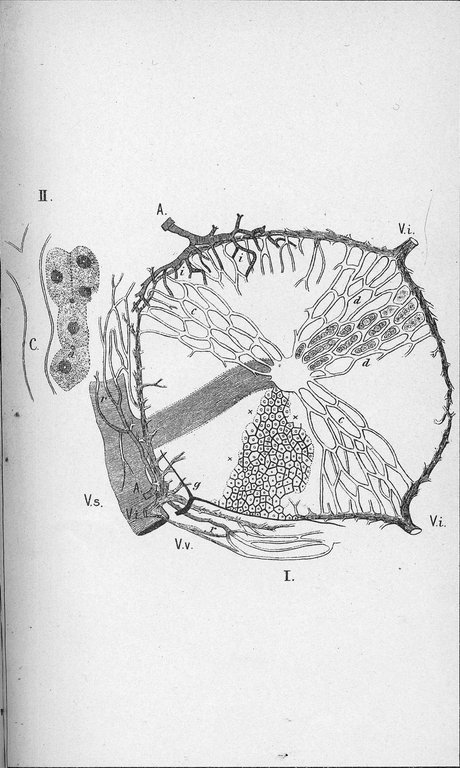
Plate IV. Microscopic anatomy of the liver. The liver is composed of innumerable small lobules, from 1/20 to 1/10 inch in diameter. The lobules are held together by a small amount of fibrous tissue, in which the bile ducts and larger blood vessels are lodged.
Fig. 1 Illustrates the structure of a lobule; v v, interlobular veins or the veins between the lobules. These are branches of the portal vein, which carries blood from the stomach and intestines to the liver; c c, capillaries, or very fine blood vessels, extending as a very fine network between the groups of liver cells from the interlobular vein to the center of the lobule and emptying there into the intralobular vein to the center of the lobule; v c, intralobular vein, or the vein within the lobule. This vessel passes out of the lobule and there becomes the sublobular vein; v s, sublobular vein. This joins other similar veins and helps to form the hepatic vein, through which the blood leaves the liver; d d, the position of the liver cells between the meshes of the capillaries; A A, branches of the hepatic artery to the interlobular connective tissue and the walls of the large veins and large bile ducts. These branches are seen at r r and form the vena vascularis; v v, vena vascularis; i i, branches of the hepatic artery entering the substance of the lobule and connecting with capillaries from the interlobular vein. The use of the hepatic artery is to nourish the liver, while the other vessels carry blood to be modified by the liver cells in certain important directions; g, branches of the bile ducts, carrying bile from the various lobules into the gall bladder and into the intestines; x x, intralobular bile capillaries between the liver cells. These form a network of very minute tubes surrounding each ultimate cell, which receives the bile as it is formed by the liver cells and carried outward as described.
Fig. 2. Isolated liver cells: c, blood capillary; a, fine bile capillary channel.