PLATE XXXI.
CONTAGIOUS PLEUROPNEUMONIA.
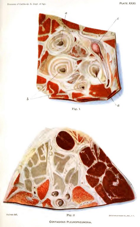
Plate XXXI. Contagious pleuropneumonia. Appearance of a cow's lung affected with contagious pleuropneumonia when sections or slices are made of it and cut surfaces examined.
Fig. 1. Transverse section through the right principal lobe in a case of acute pleuropneumonia. The area drawn includes the air tubes, veins, and arteries, and illustrates the great thickening of the interlobular connective tissue into broad whitish bands and of the walls of the air tubes, veins, and arteries: a, air tube cut obliquely; a', air tube cut directly across; b, arteries cut across; c, large vein completely occluded by a thrombus or plug formed during life. The great thickening of the walls of the artery and vein in this disease is especially brought out by stating that in the healthy lung they are so thin as to be easily overlooked.
Fig. 2. Transverse section of the principal lobe in a case of acute pleuropneumonia, illustrating the different kinds of hepatization or consolidat[Pg 388]ion of the lung. These are indicated by the different colors from dark red to reddish yellow. This variation of color is regarded by some as the real marbling characteristic of pleuropneumonia, while the whitish bands penetrating the lung tissue in all directions constitute the true marbling according to other observers.