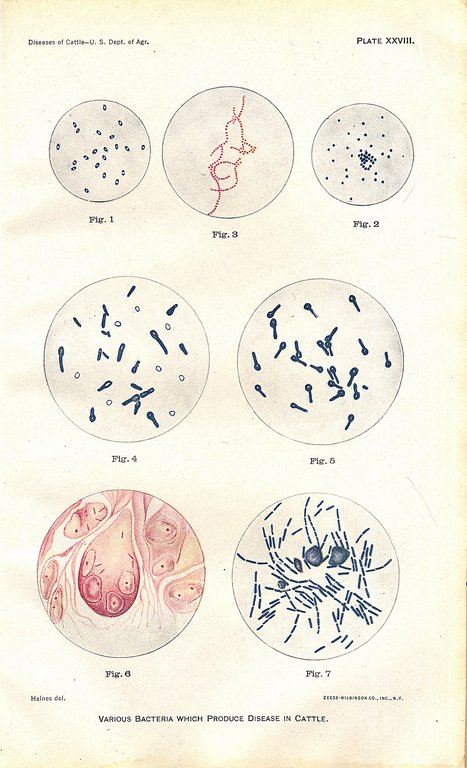
PLATE XXVIII.
VARIOUS BACTERIA WHICH PRODUCE DISEASE IN CATTLE.
The bacteria on this plate are partly from tissues, partly from cultures, and stained artificially with aniline colors (fuchsin or methylene blue). Figs 6 and 7 are copied from Fränkel and Pfeiffer's atlas. All but fig. 7 are magnified 1,000 times; fig. 7, 500 times.
Fig. 1. Bacteria from pneumonia in cattle. These are also the cause of hemorrhagic septicemia and are closely related to swine-plague bacteria. These bacteria were drawn from a piece of spleen pulp (rabbit).
Fig. 2. Micrococci (streptococcus) which produce inflammation of the lining membranes of the abdomen, thorax, heart, brain, and joints. Frequently associated with the preceding bacteria in abscesses.
Fig. 3. Micrococci (staphylococcus) which produce inflammation and suppuration; also pyemia.
Fig. 4. Bacilli of blackleg. The pale oval bodies as well as the light spots in one end of the bacilli represent spores.
Fig. 5. Bacilli which produce tetanus or lockjaw. The light spot in the enlarged end of each rod represents a spore.
Fig. 6. Bacilli of tuberculosis. Microscopic sections of a pearly nodule from the lining membrane of the chest cavity. The bacilli are stained red and appear as small straight rods within the cells of the nodule or tubercle.
Fig. 7. Bacilli of anthrax. Bacilli from the spleen of a mouse inoculated with a culture. The bacilli were obtained from the blood of a cow which died of anthrax in Mississippi. The bacilli appear as rods stained blue. The round bodies are blood corpuscles, also stained artificially.