PLATE XLV.
TEXAS FEVER.
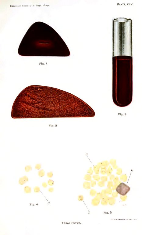
Plate XLV. Texas fever.
Fig. 1. The cut surface of a healthy liver taken from a steer slaughtered for beef.
Fig. 2. The cut surface of the liver in Texas fever.
Fig. 3. Appearance of the urine in an acute, fatal case of Texas fever.
Fig. 4. Red corpuscles, magnified 1,000 diameters, containing the parasite of Texas fever. This appears as a blue point a near the edge of the corpuscle. The blood was taken from a skin incision. The case was nonfatal and occurred late in the fall.
Fig. 5. Red corpuscles from the blood of an acute, fatal case, 20 hours before death. The Texas-fever microbes a are shown as pear-shaped bodies, stained with methylene blue, within the red corpuscles. The larger body on the right b is a white blood corpuscle, also stained with methylene blue. (Magnified 1,000 diameters.)