PLATE XLI.
ACTINOMYCOSIS OF THE LUNGS.
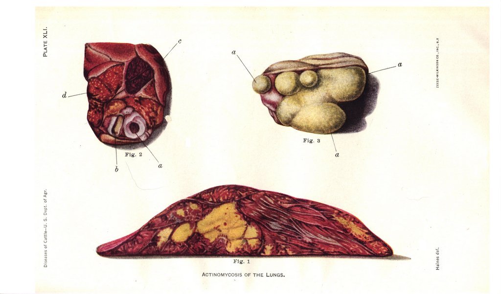
Plate XLI. Actinomycosis of the lungs.
Fig. 1. Transverse section of the ventral lobe of the right lung, from a case studied in the laboratory. The yellowish dots represent the places where the actinomyces fungus is lodged. The larger yellowish patches are produced by the confluence of a number of isolated centers. The entire lobe is of a dark flesh-red color, due to collapse and bronchopneumonia.
Fig. 2. The cut surface of a portion of the principal lobe of the same lung, showing the recent invasion of antinomycosis from the other lobe: a, large air tube; b, artery; c, a pneumatic lobule; d, lobule containing minute yellowish dots. In these the actinomyces fungus is lodged.
Fig. 3. Cut surface of a small portion of another lung, showing a few lobules, a. The fungus is sprinkled throughout the lung tissue in the form of yellowish grains, as shown in the illustration. The pleural covering of the lung tissue is shown in profile above.