PLATE XL.
ACTINOMYCOSIS OF THE JAW.
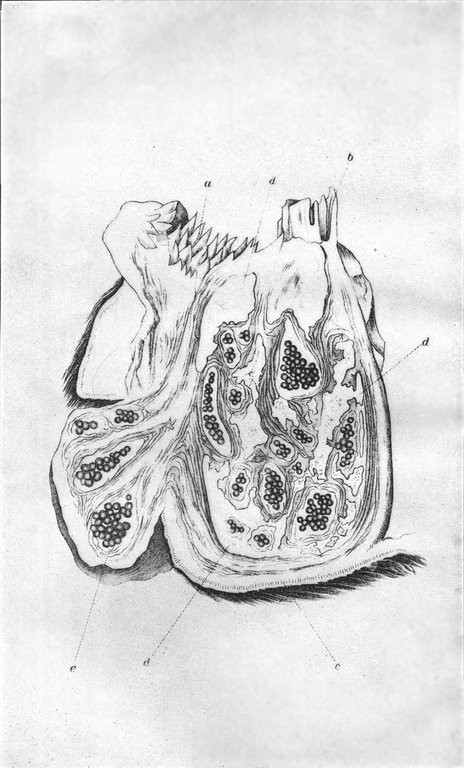
Plate XL. Actinomycosis of the jaw. (Reduced one-half. From Jöhne's Encyklopädie d. gesammt. Thierheilkunde.) The lower jaw is sawed through transversely, i.e., from right to left, and shows the disease within the jawbone itself; a, within the mouth, showing the papillæ on the mucous membrane of the cheek; b, front view of a molar tooth; c, the skin covering the lower surface of the jawbone; d, the jawbone hollowed out and enlarged by the formation of cavities within it, which are filled with the soft growth of the actinomycotic tumor. The section makes it appear as if the bone were broken into fragments and these forced apart; e[Pg 474], a portion of the tumor which has broken through the bone and the skin and appears as a tumor on the cheek. The little roundish masses represent the granulomata (minute tumors) in which the fungus vegetates.