PLATE XIII.
PREGNANT UTERUS WITH COTYLEDONS.
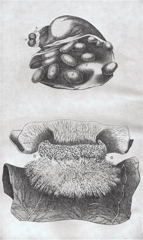
PLATE XIII. Pregnant uterus with cotyledons.
Fig. 1. Uterus of the cow during pregnancy, laid open to show the cotyledons (d) on the internal surface of uterus (c). The ovary (a) is shown cut across, and the two halves are laid open to show the position of the discharged ovum at a'.
Fig. 2 illustrates the relation of the fetal and maternal parts of a cotyledon. A portion of the uterus (A) is shown with the maternal cotyledon (BB) attached to it. The fetal portion (D) consists of a mass of very minute hairlike processes on the chorion (E), which fit into corresponding depressions or pits of the maternal portion. Each portion is abundantly supplied with blood vessels, so that a ready interchange of nutritive fluid may take place between mother and fetus.