PLATE X.
MICROSCOPIC ANATOMY OF THE KIDNEY.
Plate X. Microscopic anatomy of the kidney.
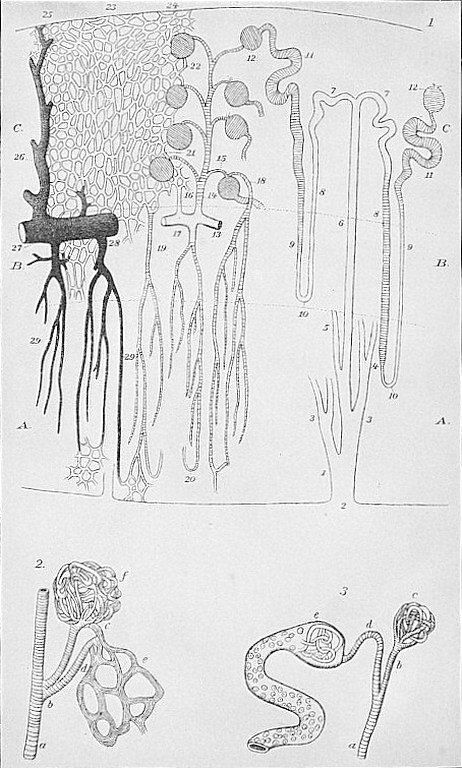
Fig. 1. In this figure the minute apparatus for the secretion, collection, and discharge of the urine into the pelvis of the kidney (see preceding plate) is shown. The course is as follows: The urine is secreted from the blood vessels in the little round bodies called glomeruli (12), and by the minute cells in the curved tubes (11, 9, 10, 8), and passes through the convoluted and straight tubes (7, 6) into the larger tube (1), and then out into the pelvis, thence through the ureters into the bladder. The fluid and salts dissolved in the urine are taken from the blood, and the minute blood vessels are therefore very abundant in the kidneys, as is shown by the branches and network on the left of the figure. The blood passes into the kidney in the artery (13), which then divides into branches which pass into the glomeruli (12) and also forms network around the secreting tubules (11, 9). The urine and salts pass from these vessels through the cells lining the tubules into the latter, and are discharged as described above. The blood is again collected in veins drawn black in the figure.
Fig. 2 illustrates the manner in which the blood is distributed in the glomerulus (f), and also to the secreting tubules (e).
Fig. 3 shows the relation between the blood vessel in the glomerulus (e) the tubule which conducts the urine therein secreted from the blood vessel; (c) represents a glomerulus from which the urinary tubule has been removed.