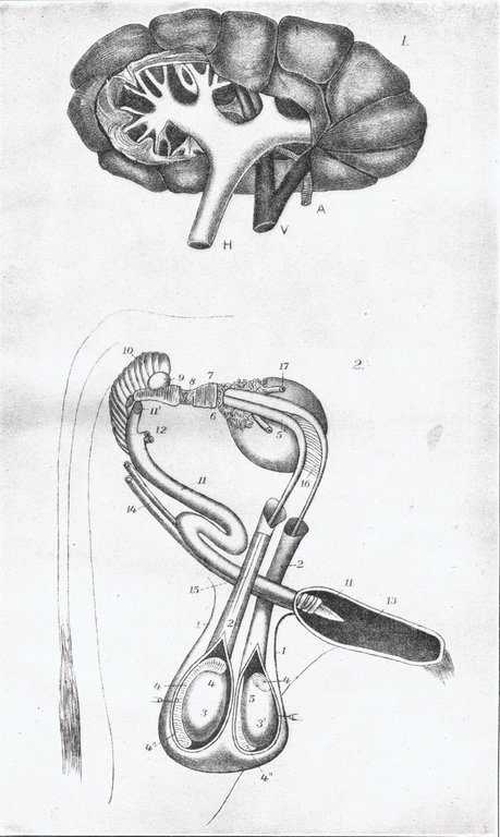
PLATE IX.
KIDNEY AND MALE GENERATIVE AND URINARY ORGANS.
Plate IX. Kidney and male generative and urinary organs.
Fig. 1. Kidney of the ox. (From Handbuch des Vergleichenden Anatomie des Haus Säugethiere, vol. 7, 1890.) A, renal artery carrying blood into the kidney; V, renal vein carrying blood from the kidney back to the heart; H, ureter, the tube carrying the urine from kidney to bladder. It is formed by the union of a number of branches which begin as cups (calices), each inclosing the tip of a conical mass of tissue from which the urine excludes.
Fig. 2. Genital and urinary organs of the bull. (From Leisering, Mueller, and Ellenberger, Handbuch des Verg. Anat. des Haus Säugethiere.) the serous membrane enveloping the testicles; 3, the right testicle, outer view; 3', left testicle, inner view; 4, epididymis, or the beginning of the excretory canal of the testicle; 4', globus major, or the head of the epididymis; 4'', globus minor, or the tail of the epididymis; 5, vas deferens, the duct through which the seminal fluid reaches the ejaculatory ducts; 5', pelvic dilation of the vas deferens; 6, vesicula seminalis. The vesiculæ seminalis are two oval pouches, which, in addition to their own secretions, receive the semen conveyed by the seminal ducts and hold it in reserve until copulation; 7, membranous or intrapelvic portion of the urethral canal covered by Wilson's muscle; 8, part of the prostate gland, covered by Wilson's muscle; 9, Cowper's gland. This gland, like the prostate gland, secretes a fluid which is thrown into the urethal canal in abundance immediately before ejaculation; by this means the expulsion of the semen is facilitated; 10, ejaculator seminis, or accelerator urinæ muscle; 11, penis; 11', cut portion of same; 12, cut suspensory ligaments of penis; 13, sheath, or prepuce laid open; 14, retractor muscles of sheath; 15, cremaster muscle cut at superior extremity; 16, duplicature of peritoneum; 17, ureters carrying urine from the kidneys to the bladder.